Metadichol ® Gel and Nanoparticle patent
45
(12)United States Patent Raghavan (54) METADICHOLO LIQUID AND GEL NANOPARTICLE FORMULATIONS (71) Applicant: NanoRx, Inc., Chappaqua, NY (US) (72) Inventor: Palayakotai R. Raghavan, Chappaqua, NY (US) (73) Assignee: NanoRx, Inc., Chappaqua, NY (US) ) Notice: S u b j e c t to any disclaimer, the term of this patent is extended or adjusted under 35 U.S.C. 154(b) by 29 days. (21) A p p l . No.: 14/205,243 (22) Filed: (65) Mar. 11, 2014 Prior Publication Data US 2014/0275285 Al S e p . 18, 2014 Related U.S. Application Data (60) Provisional application No. 61/794,490, filed on Mar. 15, 2013. (51) I n t . Cl. A61K 31/045 B82Y 30/00 A61K 9/14 A61K 47/32 (2006.01) (2011.01) (2006.01) (2006.01) 1111111111111111111111111411111 II 1111111111111 11 111 (56) (10) Patent No.: U S 9,006,292 B2 (45) Date of Patent: A p r . 14, 2015 (52) U.S. Cl. CPC A 6 1 K 31/045 (2013.01); A61K 9/14 (2013.01); A61K 47/32 (2013.01); B82Y 30/00 (2013.01); Y10S 977/773 (2013.01) (58) F i e l d of Classification Search CPC A 6 1 K 31/045; B82Y 30/00 USPC 5 1 4 / 7 2 4 ; 977/773 See application file for complete search history. References Cited U.S. PATENT DOCUMENTS 4,670,471 A 6 / 1 9 8 7 Clark 2003/0198616 Al 10/2003 Howard 2005/0074443 A l 4 / 2 0 0 5 Treadwell 2007/0196507 A l 8 / 2 0 0 7 Majeed etal. 2009/0191288 A l 7 / 2 0 0 9 Squires 2010/0215752 A l 8 / 2 0 1 0 Raghavan 2013/0045179 A l 2 / 2 0 1 3 Ciustea et als Primary Examiner — Raymond Henley, III (74) Attorney, Agent, or Firm — Morgan, Lewis & Bocldus LLP (57) A B S T R A C T The present invention provides methods of regulating physi- ological and metabolic parameters and of treating diseases by administering metadichol to a subject in need of such regu- lation and/or treatment. Metadichol can be administered as a liquid or gel formulation. 12 Claims, 15 Drawing Sheets
-
Upload
independent -
Category
Documents
-
view
0 -
download
0
Transcript of Metadichol ® Gel and Nanoparticle patent
(12) United States PatentRaghavan
(54) M E TA D I C H O L O LIQUID AND GELNANOPARTICLE FORMULATIONS
(71) Appl icant : NanoRx, Inc., Chappaqua, NY (US)
(72) Inventor: Pa layakota i R. Raghavan, Chappaqua,NY (US)
(73) Assignee: NanoRx, Inc., Chappaqua, NY (US)
) Notice: S u b j e c t to any disclaimer, the term of thispatent is extended or adjusted under 35U.S.C. 154(b) by 29 days.
(21) A p p l . No.: 14/205,243
(22) F i l ed :
(65)
Mar. 11, 2014
Prior Publication Data
US 2014/0275285 A l S e p . 18, 2014
Related U.S. Application Data
(60) Provisional application No. 61/794,490, filed on Mar.15, 2013.
(51) I n t . Cl.A61K 31/045B82Y 30/00A61K 9/14A61K 47/32
(2006.01)(2011.01)(2006.01)(2006.01)
1111111111111111111111111411111 II 1111111111111 11111
(56)
(10) Patent No.: U S 9,006,292 B2(45) Date of Patent: A p r . 14, 2015
(52) U . S . Cl.CPC A 6 1 K 31/045 (2013.01); A61K 9/14
(2013.01); A61K 47/32 (2013.01); B82Y 30/00(2013.01); Y10S 977/773 (2013.01)
(58) F i e l d of Classification SearchCPC A 6 1 K 31/045; B82Y 30/00USPC 5 1 4 / 7 2 4 ; 977/773See application file for complete search history.
References Cited
U.S. PATENT DOCUMENTS
4,670,471 A 6 / 1 9 8 7 C l a r k2003/0198616 A l 1 0 / 2 0 0 3 Howard2005/0074443 A l 4 / 2 0 0 5 Treadwell2007/0196507 A l 8 / 2 0 0 7 Majeed etal.2009/0191288 A l 7 / 2 0 0 9 Squires2010/0215752 A l 8 / 2 0 1 0 Raghavan2013/0045179 A l 2 / 2 0 1 3 Ciustea et als
Primary Examiner — Raymond Henley, III(74) Attorney, Agent, or Firm — Morgan, Lewis & BocldusLLP
(57) A B S T R A C TThe present invention provides methods of regulating physi-ological and metabolic parameters and of treating diseases byadministering metadichol to a subject in need of such regu-lation and/or treatment. Metadichol can be administered as aliquid or gel formulation.
12 Claims, 15 Drawing Sheets

-4
tf1ualud stoz st jo 1
簽
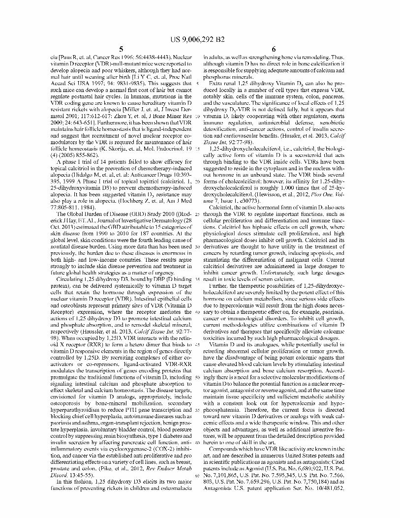
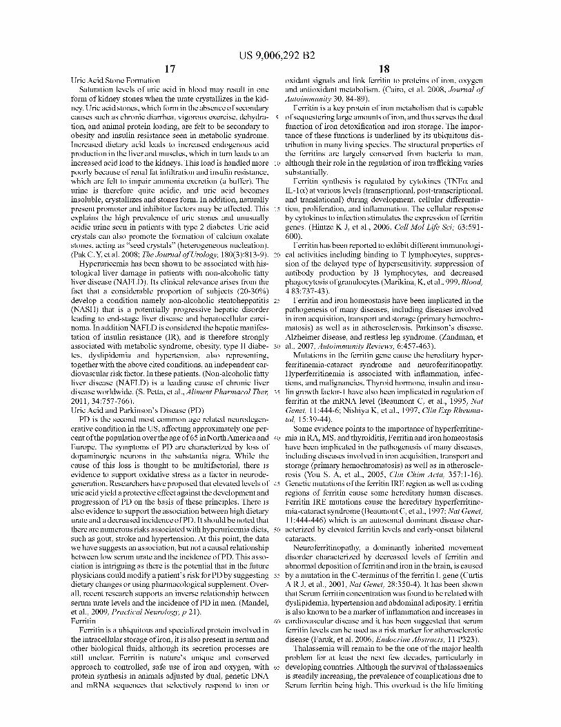
Log EC 50 EC 50Vitamin D 3 0.7854 6.1
Calcitriol -1.591 0.0255Metadichol 0.8276 6.724
Calcitriol
-2
Reference
log nm2
Vitamin D 3
4
FIGURE 1
Metadic hol
-3 - 2 - 1 0
log nm
tf1ualud stoz st jo 1
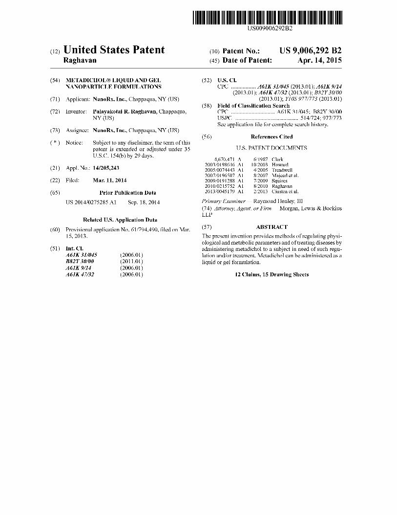
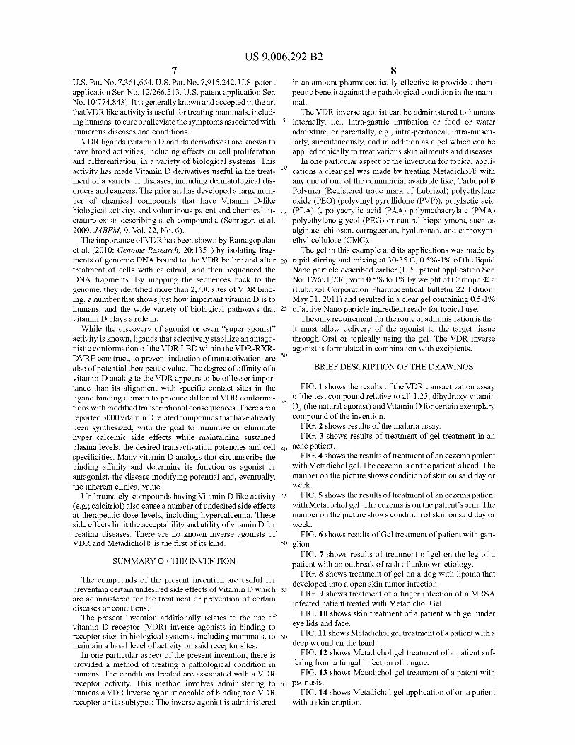
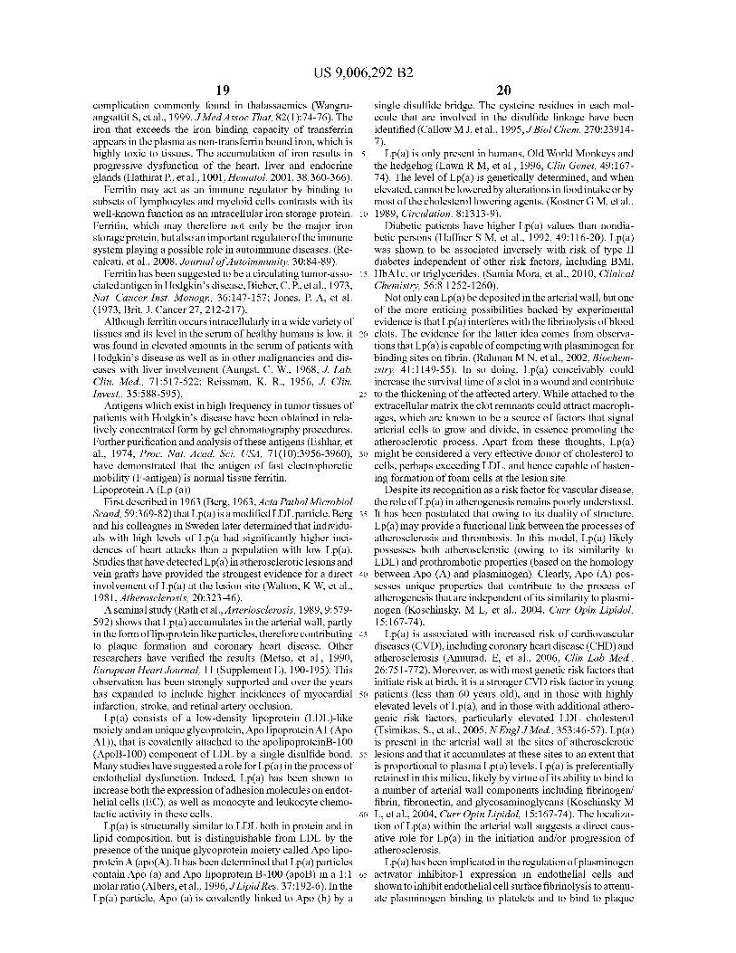
Compound I C 50 (rim)Metadichol 5 0 9
Reference compoundschloroquine 4 .3 nmArtesunate 1 . 7 nm
140
120
100
80
80
20
FIGURE 2
623 125EN.tetaip1341#104)
•25 S O O 1 0 0 0
wawa stoz SI Jo Z
U . S . P a t e n t A p r . 14, 2015 S h e e t 3 of 15 U S 9,006,292 B2

U . S . P a t e n t A p r . 14, 2015 S h e e t 4 of 15 U S 9,006,292 B2

U . S . P a t e n t A p r . 14, 2015 S h e e t 5 of 15
Lf lLI-1CC
E
US 9,006,292 B2
U . S . P a t e n t A p r . 14, 2015 S h e e t 6 of 15 U S 9,006,292 B2
U . S . P a t e n t A p r . 14, 2015 S h e e t 7 of 15 U S 9,006,292 B2

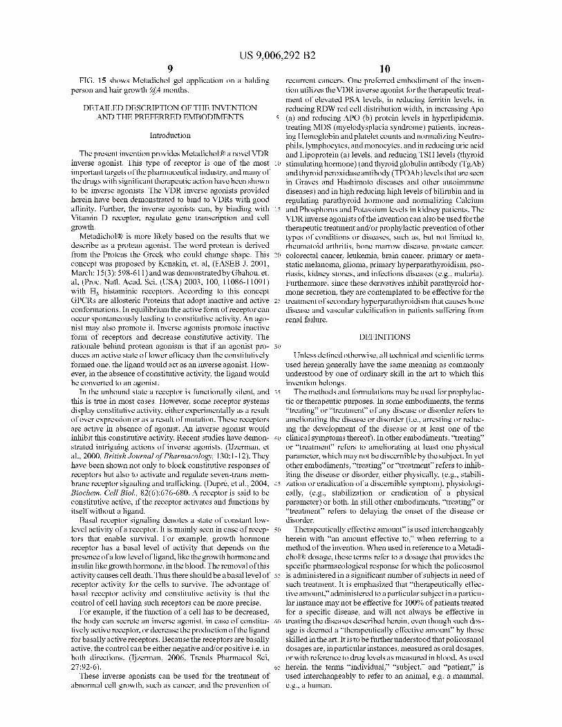
FIGURE 8
tf1ualud stoz st Jo 8
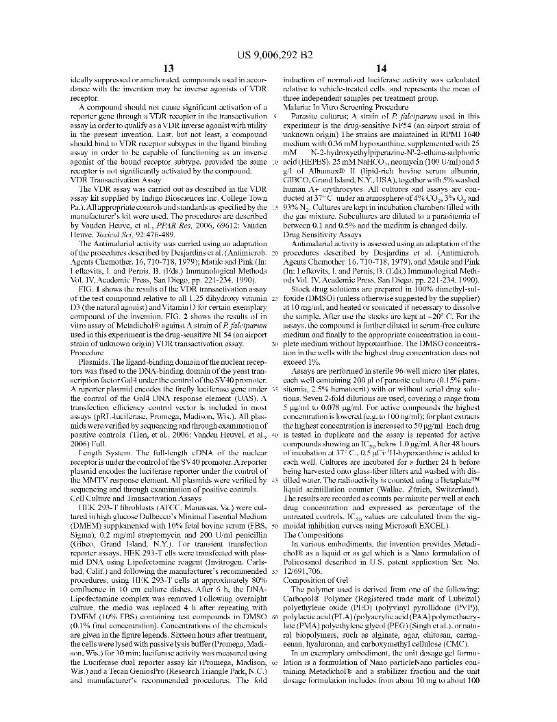
baseline
Day 6
Day 2
FIGURE 9
Day 7 Day 8 @ 3 weeks
tf1ualud stoz st Jo 6

U . S . P a t e n t A p r . 14, 2015 S h e e t 10 of 15 U S 9,006,292 B2
FIGURE 10

U . S . P a t e n t A p r . 14, 2015 S h e e t 11 of 15 U S 9,006,292 B2
FIGUR

U . S . P a t e n t A p r . 14, 2015 S h e e t 12 of 15 U S 9,006,292 B2
FIGURE 12
U . S . P a t e n t A p r . 14, 2015 S h e e t 13 of 15 U S 9,006,292 B2
U . S . P a t e n t A p r . 14, 2015 S h e e t 14 of 15 U S 9,006,292 B2
FIGUR
U . S . P a t e n t A p r . 14, 2015 S h e e t 15 of 15 U S 9,006,292 B2
FIGURE 15
US 9,006,292 B21 2
METADICHOLO LIQUID AND GELNANOPARTICLE FORMULATIONS
PRIORITY CLAIM
This application claims priority to U.S. Patent ApplicationNo. 61/794,490, filed Mar. 15, 2013, the contents of which areincorporated by reference in their entirety for all purposes.
FIELD OF THE INVENTION
The present invention relates to a novel formulation o fMetadichol® a Nano particulate in liquid oral and in poly-meric Nano gel formulation. The Metadichol® formulation(previously described in US patent application (Ser. No.12/691,706)) behaves as an inverse agonist against the Vita-min D receptor (VDR). This application relates to methods oftreating, and preventing diseases and extends the therapeuticeffects beyond what is observed with 1,25 dihydroxy VitaminD, (Calcitriol) the active form of Vitamin D in the humanbody.
BACKGROUND OF THE INVENTION
Vitamin D, acquired either from dietary sources or viaultraviolet irradiation of 7-dehydrocholesterol in the epider-mis, is metabolized to its hormonal form. The keratinocytesof the skin are unique in being not only the primary source ofvitamin D for the body, but also possessing the enzymaticmachinery to metabolize vitamin D to active metabolites.Many functions of the skin are regulated by vitamin D and/orits receptor: these include inhibition of proliferation, stimu-lation of differentiation including formation of the permeabil-ity barrier, promotion of innate immunity, regulation of thehair follicle cycle, and suppression of tumors.
When exposed to ultraviolet radiation, cells in the epider-mis convert a cholesterol related steroid to vitamin D orcholecalciferol. Vitamin D is essential for proper develop-ment o f the bones. The ultraviolet radiation necessary forvitamin D synthesis (specifically, UV-B) only reaches theEarth's surface in much abundance for a few hours a daywhen the sun is high. Much less o f it reaches the Earth'ssurface at high latitudes than at low latitudes, and very littlereaches the Earth's surface on cloudy days or during thewinter. Even so, the average fair-skinned person can makeand store several days' worth o f vitamin D with just onehour's exposure to the midday sun. Dark-skinned peopleliving at high latitudes are much more likely to suffer fromvitamin D deficiency than are light-skinned people.
The most important source o f Vitamin D is through theaction of sun on cholesterol on the skin. Vitamin D modulatesT-cell responses and has anti-inflammatory properties, andboosts innate immune responses by induction of the humangene for cathelicidin. Cathelicidin's and defensins are smallpeptides with amphipathic structures that allow them to dis-rupt the integrity of the pathogen cell membrane, resulting inits death. Most immune cells or those epithelial cells that arein contact with the environment express these proteins. Defi-ciency in these peptides results in increased susceptibility toinfection.
Vitamin D enters the circulation and is transported to theliver, where it is hydroxylated to form 25-hydroxyvitamin D,(calcidiol; the major circulating form of vitamin D). In thekidneys, the 25-hydroxyvitamin D3-1-hydroxylase enzymecatalyzes a second hydroxylation of 25-hydroxyvitamin D,resulting in the formation of 1,25-dihydroxyvitamin D, (cal-citriol, lalpha, 25-dihydroxyvitamin D] t h e most potent
form of vitamin D. Most of the physiological effects of vita-min Din the body are related to the activity of 1,25-dihydrox-yvitamin D. Keratinocytes in the epidermis possess hydroxy-lase enzymes that local ly convert v i tamin D t o 1,25
5 dihydroxyvitamin D3, (Bikle, et al. 1986, Biochemistry. 25(7): 1545-15480) the form that regulates epidermal prolifera-tion and differentiation. I n skin, the vitamin D receptor(VDR) appears to have other roles that are independent of itsassociation with 1,25 dihydroxyvitamin D3. For instance, the
lo V D R is important in regulating the growth cycle of maturehair follicles. (Bikle, et al., J. Bone. Miner. Metab, (2010)28:117-130). Certain mutations in the VDR lead to misregu-lated gene expression resulting i n aberrant hair foll iclecycling and alopecia (hair loss) i n mice (28, 29) and in
15 humans (30). The VDR also functions as a tumor suppressorin skin. The VDR is one of several factors that control thesetwo diverse roles. Moreover, 1,25-dihydroxyvitamin D3 is apotent immune modulator in skin.Functions in Healthy Skin
20 P h o t o protection photo damage refers to skin damageinduced by ultraviolet (UV) light. Depending on the dose, UVlight can lead to DNA damage, inflammatory responses, skincell apoptosis (programmed cell death), skin aging, and skincancer. Some studies, mainly in vitro (cell culture) studies
25 (Dixon, K M, et al. 2005, J Steroid Biochem Mot' Biol, 97(1-2):137-143) and mouse studies where 1,25-dihydroxyvita-min D3 was topically applied to skin before or immediatelyfollowing irradiation, have found that vitamin D exhibitsphoto protective effects (Gupta, et al., 2007, J Invest Derma-
30 tot'. 127(3):707-715). Documented effects i n sk in cellsinclude decreased D N A damage, reduced apoptosis,increased cell survival, and decreased erythema. The mecha-nisms for such effects are not known, but one mouse studyfound that 1,25-dihydroxyvitamin D3 induced expression of
35 metallothionein (a protein that protects against free radicalsand oxidative damage) in the stratum basale. It has also beenpostulated that nongenomic actions of vitamin D contributeto the photo protection; such effects o f vitamin D involvecell-signaling cascades that open calcium channels. 1,25-
,40 dihydroxyvitamin D3 regulates the expression of cathelicidin(LL-37/hCAP18) (40, 41), an antimicrobial protein thatappears to mediate innate immunity in skin by promotingwound healing and tissue repair. One human study found thatcathelicidin expression is up regulated during early stages of
45 normal wound healing (Gombart A F, Faseb J., 2005, 19(9):1067-1077). Other studies have shown that cathelicidinmodulates inflammation in skin Kratz, G, et al., 2003,11-westDermata 120(3):379-389), induces angiogenesis (Kupatt C,et al., J Gin Invest., 2003, 111(11):1665-1672), and improves
50 re-epithelialization (the process o f restoring the epidermalbarrier to re-establish a functional barrier that protects under-lying cells from environmental exposures). The active form ofvitamin D and its analogs have been shown to up regulatecathelicidin expression in cultured keratinocytes (Sthale M,
55 2005, J Invest Dermata, 124(5):1080-1082).Diseases such as rosacea may require lower levels of vita-
min D or even locally active serine proteases inhibitors andvitamin D antagonists to prevent harm. In rosacea, patientsmight benefit from therapies blocking cathelicidin expression
60 and processing. Polymorphisms in the vitamin D receptorgene have been described in patients with severe rosaceaindicating that vitamin D3 signaling is involved in pathogen-esis (Jansen T, et. al, J Dermatol 2004; 31:244-246.). Block-ing cathelicidin expression by targeting the vitamin D3 path-
65 way might represent a novel therapeutic approach in rosacea.As an example, vitamin D3 analogues without intrinsic activ-ity at the vitamin D receptor have been shown to inhibit
31,25D3-induced cathelicidin in keratinocytes in vitro (Liu P.T, et. al, Science 2006; 311:1770-1773).
In psoriasis, blocking cathelicidin peptide could break thevicious cycle of increased LL-37 expression, dendritic cellactivation and cutaneous inflammation. Again strategies todecrease cathelicidin in keratinocytes could target vitamin D3signaling. Paradoxically, for a long time vitamin D3 ana-logues have been used in the therapy of psoriasis. Vitamin D3analogues bind to and activate the vitamin D receptor andshould therefore increase cathelicidin in keratinocytes pre-sumably worsening inflammation in psoriasis. However, theopposite is true: vitamin D analogues resemble one o f thepillars o f topical psoriasis treatment. They ameliorate cuta-neous inflammation and reverse morphological changeswithin skin lesions. (Lebwohl M, et. al, J Am Acad Dermatol2004; 50:416-430). Understanding the molecular effects o fvitamin D3 analogues on cutaneous innate immune functionwill eventually also lead to better treatment. In summary,influencing cathelicidin expression via vita-min D3 signalingmight offer a new treatment angle in the therapy o f verycommon skin diseases. However, until the 'sunshine vitamin'can be targeted additional experimental work and clinicalstudies have to be performed to prove its safety and benefits.Overall, current data overwhelmingly support the importanceof AMPs to healthy human skin but the key steps to put thisinformation to therapeutic use remain to be done.
Eczema (Bjorn Hartmann, et al, Journal o f InvestigativeDermatology (2012), Volume 132) is a chronic inflammatoryskin disease that has reached nearly epidemic proportions inchildhood. Moreover, it is a difficult disease to control and,with its onset in childhood, is often the first manifestation ofatrophy. The clinical features of eczema include itchy red skinaccompanied by dryness and lichenification. In the past, treat-ment options consisted primarily of avoidance of soap andwater. These options have considerably improved with bothnon-pharmacologic and pharmacologic approaches. How-ever, eczema is still a treatment challenge. Part of the problemin developing new treatment options has been the relativefailure in translating basic science information into clinicalapplication. I t is hoped that the newer biologics wi l l helpbridge this gap and lead to greater success rates.
Atopic dermatitis (AD) is a common chronic inflammatoryskin disease that has increased in prevalence over the lastseveral decades in industrialized countries. AD is a multifac-torial, heterogeneous disease with a variety of defects in theimmune system, in antimicrobial defense mechanisms andepidermal barrier integrity, which collectively contribute tothe risk and severity of AD development. (J Innate Immun2011; 3:131-141).
Topical corticosteroids have been the gold standard for thetreatment of atopic dermatitis for many decades. The emer-gence o f the immuno-modulatory drugs Tacrolimus andPicrolimus represented the first major advance in the treat-ment of this disease in 40 years. Numerous other therapeuticmodalities have been studied and whereas some have beenfound to have beneficial effects, none have exceeded theefficacy o f topical corticosteroids. Less severe forms o feczema are generally treated successfully with topical ste-roids or immuno-modulatory drugs, however,
Steroid-resistant eczema presents a problem because mostof the other adjunctive treatments do not completely resolvethe condition.
Warts are a benign proliferation of the skin and mucosacaused by infection with human papillomavims (HPV). HPVis ubiquitous, and renal transplant recipients (RTRs) maynever totally clear HPV infections, which are the most fre-quently recurring infections. This infection is important
US 9,006,292 B24
because of its link to the development of certain skin cancers,in particular, squamous cell carcinoma. Regular surveillance,sun avoidance, and patient education are important aspects ofthe management strategy. Warts are usually treated by tradi-
5 t iona l destructive modalities such as cryotherapy with liquidnitrogen, local injection o f bleomycin, electrocoagulation,topical application of glutaraldehyde, and local and systemicinterferon-3 therapy [S. Gibbs, et. al, British Medical Journal,vol. 325, no. 7362, pp. 461-464, 2002. However, the tolerance
io o f patients to these treatment modalities is poor, because theyoften cause pain, especially in children, and sometimes scar-ring or pigmentation after treatment. No treatment has beenuniformly effective, and warts are often refractory, especiallyin immuno-compromised patients where their quality of life
15 i s threatened. Researchers have reported an RTR with a rightindex finger wart, which was successfully treated with a topi-cal activated vitamin D. (Luciano Moscarelli, et. al, CaseReports in Transplantation Volume 2011, Article ID 368623).
Hair loss (alopecia) is a much-feared side effect of many20 chemotherapy protocols and is one of the most psychological
devastating aspects of cancer therapy. So far, no satisfactorystrategy for suppressing chemotherapy-induced alopecia is athand. During the last decade, some progress in understandingmolecular mechanisms o f chemotherapy-induced hair loss
25 has been achieved using rodent models. However, the patho-biology o f the response o f human hair follicle to chemo-therapy remains largely unknown. (Vladimir A Botchkarev,Journal of Investigative Dermatology Symposium Proceed-ings (2003) 8, 72-75).
30 And rogene t i c alopecia is the most common hair loss dis-order in men and is largely determined by genetic factors andthe peripheral action of androgens. Others mechanisms suchas chronic inflammation and several hormones or vitaminslike aldosterone, insulin or vitamin D have been implicated in
35 the pathogenesis of Androgenetic alopecia. The diagnosis o fAndrogenetic alopecia is made by clinical history and clinicalexamination. Minoxidil and finasteride are the main drugsapproved for the treatment of Androgenetic alopecia. Andro-genetic alopecia has been associated with cardiovascular risk
40 factors and benign pro static hyperplasia. Alopecia is a featureof vitamin D receptor (VDR) mutations in humans and inVDR null mice. This alopecia results from an inability toinitiate the anagen phase of the hair cycle after follicle mor-phogenesis is complete.
45 T h u s , once the initial hair is shed it does not regrow. VDRexpression in the epidermal component of the hair follicle, thekeratinocyte, is critical for maintenance of the hair cycle. Todetermine which functional domains of the VDR are requiredfor hair cycling, mutant VDR transgenes were targeted to the
50 keratinocytes o f V D R nu l l mice. Keratinocyte-specificexpression of a VDR transgene with a mutation in the hor-mone binding domain that abolishes ligand binding restoresnormal hair cycling in VDR null mice, whereas a VDR trans-gene with a mutation in the activation function 2 domain that
55 impairs nuclear receptor co-activator recruitment results in apartial rescue. Mutations in the nuclear receptor co-repressorHairless are also associated wi th alopecia in humans andmice. Hairless binds the VDR, resulting in transcriptionalrepression. Neither VDR mutation affects Hairless interac-
60 tions or its ability to repress transcription. These studies dem-onstrate that the effects o f the VDR on the hair follicle areligand independent and point to novel molecular and cellularactions of this nuclear receptor (Kristi Skorij a, et. al, Molecu-lar Endocrinology 19: 855-862, 2005).
65 P r e v i o u s reports have described the effects of vitamin D onhair follicles. Topical pretreatment o f VD3 enhanced hairregrowth in a mouse model of chemotherapy-induced alope-
5cia [Paus R, et. al, Cancer Res 1996; 56:4438-4443). Nuclearvitamin D receptor (VDR)-null-mutant mice were reported todevelop alopecia and poor whiskers, although they had nor-mal hair until weaning after birth [Li Y C, et. al, Proc NatiAccad Sci USA 1997; 94: 9831-9835). This suggests thatsuch mice can develop a normal first coat of hair but cannotregulate postnatal hair cycles. In humans, mutations in theVDR coding gene are known to cause hereditary vitamin Dresistant rickets with alopecia [Miller J, et. al, J Invest Der-matol 2001; 117:612-617: Zhou Y, et. al, J Bone Miner Res2009; 24: 643-6511. Furthermore, it has been shown that VDRmaintains hair follicle homeostasis that is ligand-independentand suggest that recruitment o f novel nuclear receptor co-modulators by the VDR is required for maintenance of hairfollicle homeostasis (K. Skorija, et. al, Mol. Endocrinol. 19(4) (2005) 855-862).
A phase I trial o f 14 patients failed to show efficacy fortopical calcitriol in the prevention of chemotherapy-inducedalopecia (Hidalgo M, et. al, et. al: Anticancer Drugs 10:393-395, 1999 A Phase I trial o f topical topitriol (calcitriol, 1,25-dihydroxyvitamin D3) to prevent chemotherapy-inducedalopecia. I t has been suggested vitamin D, resistance mayalso play a role in alopecia. (Hochberg Z, et. al, Am J Med77:805-811, 1984).
The Global Burden of Disease (GBD) Study 2010 ((Rod-erick J Hay, ET. AL, Journal of Investigative Dermatology (28Oct. 2013) estimated the GBD attributable to 15 categories ofskin disease from 1990 to 2010 for 187 countries. A t theglobal level, skin conditions were the fourth leading cause ofnonfatal disease burden. Using more data than has been usedpreviously, the burden due to these diseases is enormous inboth high- and low-income countries. These results arguestrongly to include skin disease prevention and treatment infuture global health strategies as a matter of urgency.
Circulating 1,25 dihydroxy D3, bound by DBP (D bindingprotein), can be delivered systemically to vitamin D targetcells that retain the hormone through expression o f thenuclear vitamin D receptor (VDR). Intestinal epithelial cellsand osteoblasts represent primary sites of VDR (Vitamin DReceptor) expression, where the receptor mediates theactions of 1,25 dihydroxy D3 to promote intestinal calciumand phosphate absorption, and to remodel skeletal mineral,respectively (Haussler, et al. 2013, Calcif Tissue Int, 92:77-98). When occupied by 1,25D, VDR interacts with the retin-oid X receptor (RXR) to form a hetero dimer that binds tovitamin D responsive elements in the region of genes directlycontrolled by 1,25D. By recruiting complexes of either co-activators o r co-repressors, ligand-activated VDR-RXRmodulates the transcription of genes encoding proteins thatpromulgate the traditional functions of vitamin D, includingsignaling intestinal calcium and phosphate absorption toeffect skeletal and calcium homeostasis. The disease targets,envisioned for vitamin D analogs, appropriately, includeosteoporosis b y bone-mineral mobilization, secondaryhyperparathyroidism to reduce PTH gene transcription andblocking chief cell hyperplasia, autoimmune diseases such aspsoriasis and asthma, organ-transplant rejection, benign pros-tate hyperplasia, involuntary bladder control, blood pressurecontrol by suppressing renin biosynthesis, type 1 diabetes andinsulin secretion by affecting pancreatic cell function, anti-inflammatory events via cyclooxygenase-2 (COX-2) inhibi-tion, and cancer via the established anti proliferative and prodifferentiating effects on a variety of cell lines, such as breast,prostate and colon. (Pike, et al., 2012, Rev Endocr MetabDisord, 13:45-55).
In this fashion, 1,25 dihydroxy D3 elicits its two majorfunctions o f preventing rickets in children and osteomalacia
US 9,006,292 B26
in adults, as well as strengthening bone via remodeling. Thus,although vitamin D has no direct role in bone calcification itis responsible for supplying adequate amounts of calcium andphosphorus minerals.
5 E x t r a renal 1,25 dihydroxy Vitamin D, can also be pro-duced locally in a number of cell types that express VDR,notably skin, cells o f the immune system, colon, pancreas,and the vasculature. The significance of local effects of 1,25dihydroxy D,-VDR is not defined fully, but it appears that
o vitamin D, likely cooperating with other regulators, exertsimmuno regulation, antimicrobial defense, xenobioticdetoxification, anti-cancer actions, control o f insulin secre-tion and cardiovascular benefits. (Hussler, et al. 2013, CalcifTissue Int, 92:77-98).
15 1,25-dihydroxycholecalciferol, i.e., calcitriol, the biologi-cally active form o f vitamin D is a secosteroid that actsthrough binding to the VDR inside cells. VDRs have beensuggested to reside in the cytoplasm and in the nucleus with-out hormone in an unbound state. The VDR binds several
20 forms of cholecalciferol; however, its affinity for 1,25-dihy-droxycholecalciferol is roughly 1,000 times that o f 25-hy-droxycholecalciferol. (Hewisson, et al., 2012, Plos One, Vol-ume 7, Issue 1, e30773).
Calcitriol, the active hormonal form of vitamin D, also acts25 through the VDR to regulate important functions, such as
cellular proliferation and differentiation and immune func-tions. Calcitriol has biphasic effects on cell growth, wherephysiological doses stimulate cell proliferation, and highpharmacological doses inhibit cell growth. Calcitriol and its
30 derivatives are thought to have util ity in the treatment o fcancers by retarding tumor growth, inducing apoptosis, andstimulating the differentiation o f malignant cells. Currentcalcitriol derivatives are administered in large dosages toinhibit cancer growth. Unfortunately, such large dosages
35 result in toxic levels of serum calcium.Further, the therapeutic possibilities of 1,25-dihydroxyc-
holecalciferol are severely limited by the potent effect of thishormone on calcium metabolism, since serious side effectsdue to hypercalcemia will result from the high doses neces-
40 sary to obtain a therapeutic effect on, for example, psoriasis,cancer or immunological disorders. To inhibit cell growth,current methodologies utilize combinations o f vitamin Dderivatives and therapies that specifically alleviate calcemictoxicities incurred by such high pharmacological dosages.
45 V i t a m i n D and its analogues, while potentially useful inretarding abnormal cellular proliferation or tumor growth,have the disadvantage of being potent calcemic agents thatcause elevated blood calcium levels by stimulating intestinalcalcium absorption and bone calcium resorption. Accord-
50 ing ly there is a need for a selective molecular modifications ofvitamin D to balance the potential function as a nuclear recep-tor agonist, antagonist or reverse agonist, and at the same timemaintain tissue specificity and sufficient metabolic stabilitywith a constant look out f o r hypercalcemia and hypo-
55 phoosphatemia. Therefore, the current focus i s directedtoward new vitamin D derivatives or analogs with weak cal-cemic effects and a wide therapeutic window. This and otherobjects and advantages, as well as additional inventive fea-tures, will be apparent from the detailed description provided
60 herein to one of skill in the art.Compounds which have VDR like activity are known in the
art, and are described in numerous United States patents andin scientific publications as agonists and as antagonists; Citedpatents include as Agonist (U.S. Pat. No. 6,689,922, U.S. Pat.
65 No . 7,101,865, U.S. Pat. No. 7,595,345, U.S. Pat. No. 7,566,803, U.S. Pat. No. 7,659,296, U.S. Pat. No. 7,750,184) and asAntagonists U.S. patent application Ser. No. 10/481,052,
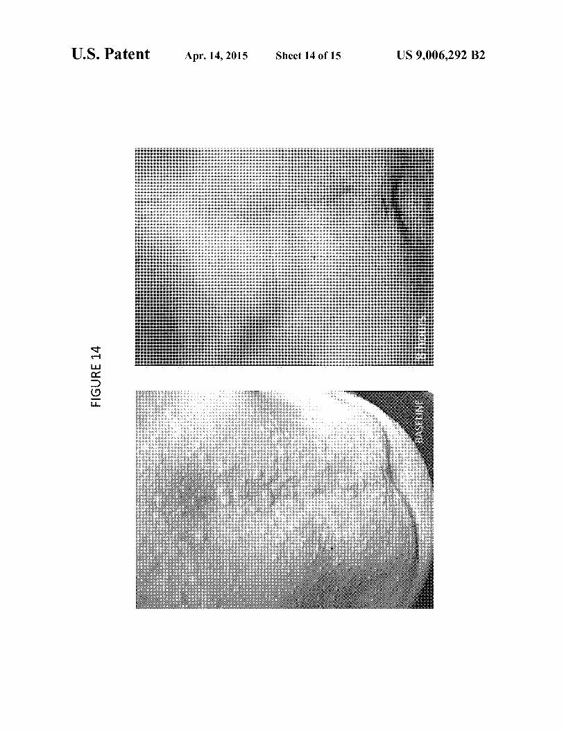
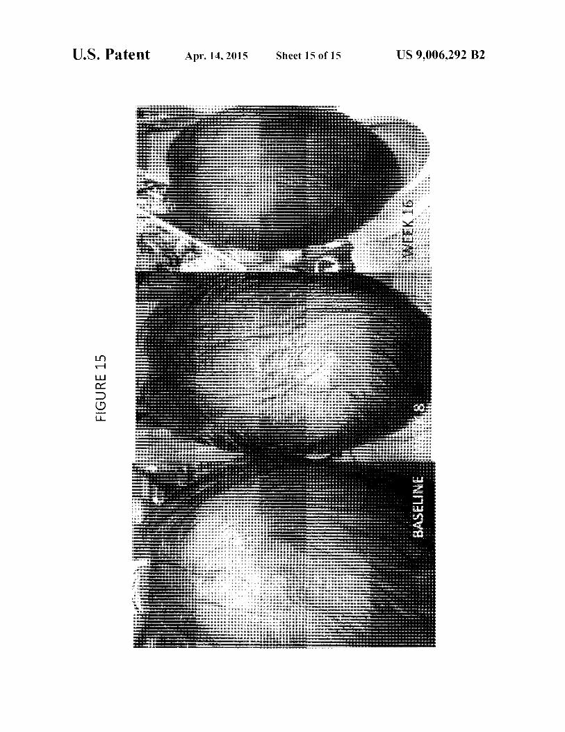
9FIG. 15 shows Metadichol gel application on a balding
person and hair growth g 4 months.
DETAILED DESCRIPTION OF THE INVENTIONAND THE PREFERRED EMBODIMENTS
Introduction
US 9,006,292 B2
The present invention provides Metadichol® a novel VDRinverse agonist. This type o f receptor is one o f the most 1important targets of the pharmaceutical industry, and many ofthe drugs with significant therapeutic action have been shownto be inverse agonists. The VDR inverse agonists providedherein have been demonstrated to bind to VDRs with goodaffinity. Further, the inverse agonists can, by binding with 1Vitamin D receptor, regulate gene transcription and cellgrowth.
Metadicholt is more likely based on the results that wedescribe as a protean agonist. The word protean is derivedfrom the Proteas the Greek who could change shape. This 2concept was proposed by Kenakin, et. al, (FASEB J. 2001,March: 15(3): 598-611) and was demonstrated by Gbahou. et.al, (Proc. Natl. Acad. Sci. (USA) 2003, 100, 11086-11091)with H , histaminic receptors. According to this conceptGPCRs are allosteric Proteins that adopt inactive and active 2conformations. In equilibrium the active form of receptor canoccur spontaneously leading to constitutive activity. An ago-nist may also promote it. Inverse agonists promote inactiveform o f receptors and decrease constitutive activity. Therationale behind protean agonism is that i f an agonist pro- 30duces an active state of lower efficacy than the constitutivelyformed one, the ligand would act as an inverse agonist. How-ever, in the absence of constitutive activity, the ligand wouldbe converted to an agonist.
In the unbound state a receptor is functionally silent, andthis is true in most cases. However, some receptor systemsdisplay constitutive activity, either experimentally as a resultof over expression or as a result of mutation. These receptorsare active in absence of agonist. An inverse agonist wouldinhibit this constitutive activity. Recent studies have demon-strated intriguing actions o f inverse agonists. (llzerman, etal., 2000, British Journal of Pharmacology, 130:1-12). Theyhave been shown not only to block constitutive responses ofreceptors but also to activate and regulate seven-trans mem-brane receptor signaling and trafficking. (Dupre, et al., 2004,Biochetn. Cell Biol., 82(6):676-680. A receptor is said to beconstitutive active, i f the receptor activates and functions byitself without a ligand.
Basal receptor signaling denotes a state of constant low-level activity of a receptor. It is mainly seen in case of recep-tors that enable survival. For example, growth hormonereceptor has a basal level o f activity that depends on thepresence of a low level of ligand, like the growth hormone andinsulin like growth hormone, in the blood. The removal of thisactivity causes cell death. Thus there should be a basal level ofreceptor activity for the cells to survive. The advantage ofbasal receptor activity and constitutive activity is that thecontrol of cell having such receptors can be more precise.
For example, i f the function of a cell has to be decreased,the body can secrete an inverse agonist, in case of constitu-tively active receptor, or decrease the production of the ligandfor basally active receptors. Because the receptors are basallyactive, the control can be either negative and/or positive i.e. inboth directions. (Ijzerman, 2006, Trends Pharmacol Sci,27:92-6).
These inverse agonists can be used for the treatment o fabnormal cell growth, such as cancer, and the prevention of
10recurrent cancers. One preferred embodiment of the inven-tion utilizes the VDR inverse agonist for the therapeutic treat-ment o f elevated PSA levels, in reducing ferritin levels, inreducing RDW red cell distribution width, in increasing Apo
5 ( a ) and reducing APO (b) protein levels in hyperlipidemia,treating MDS (myelodysplacia syndrome) patients, increas-ing Hemoglobin and platelet counts and normalizing Neutro-phils, lymphocytes, and monocytes, and in reducing uric acidand Lipoprotein (a) levels, and reducing TSH levels (thyroid
0 stimulating hormone) (and thyroid globulin antibody (TgAb)and thyroid peroxidase antibody (TP0Ab) levels that are seenin Graves and Hashimoto diseases and other autoimmunediseases) and in high reducing high levels of bilimbin and inregulating parathyroid hormone and normalizing Calcium
5 and Phosphorus and Potassium levels in kidney patients. TheVDR inverse agonists of the invention can also be used for thetherapeutic treatment and/or prophylactic prevention of othertypes o f conditions or diseases, such as, but not limited to,rheumatoid arthritis, bone marrow disease, prostate cancer,
0 colorectal cancer, leukemia, brain cancer, primary or meta-static melanoma, glioma, primary hyperparathyroidism, pso-riasis, kidney stones, and infections diseases (e.g., malaria).Furthermore, since these derivatives inhibit parathyroid hor-mone secretion, they are contemplated to be effective for the
5 treatment of secondary hyperparathyroidism that causes bonedisease and vascular calcification in patients suffering fromrenal failure.
DEFINITIONS
Unless defined otherwise, all technical and scientific termsused herein generally have the same meaning as commonlyunderstood by one of ordinary skill in the art to which thisinvention belongs.
35 T h e methods and formulations may be used for prophylac-tic or therapeutic purposes. In some embodiments, the terms"treating" or "treatment" of any disease or disorder refers toameliorating the disease or disorder (i.e., arresting or reduc-ing the development o f the disease or at least one o f the
40 clinical symptoms thereof). In other embodiments, "treating"or "treatment" refers to ameliorating at least one physicalparameter, which may not be discernible by the subject. In yetother embodiments, "treating" or "treatment" refers to inhib-iting the disease or disorder, either physically, (e.g., stabili-
45 zation or eradication of a discernible symptom), physiologi-cally, (e.g., stabilization o r eradication o f a physicalparameter) or both. In still other embodiments, "treating" or"treatment" refers to delaying the onset o f the disease ordisorder.
50 Therapeut ica l ly effective amount" is used interchangeablyherein with "an amount effective to," when referring to amethod of the invention. When used in reference to a Metadi-cho l t dosage, these terms refer to a dosage that provides thespecific pharmacological response for which the policosanol
55 i s administered in a significant number of subjects in need ofsuch treatment. It is emphasized that "therapeutically effec-tive amount," administered to a particular subject in a particu-lar instance may not be effective for 100% of patients treatedfor a specific disease, and wi l l not always be effective in
60 treating the diseases described herein, even though such dos-age is deemed a "therapeutically effective amount" by thoseskilled in the art. It is to be further understood that policosanoldosages are, in particular instances, measured as oral dosages,or with reference to drug levels as measured in blood. As used
65 herein, the terms "individual," "subject," and "patient," isused interchangeably to refer to an animal, e.g. a mammal,e.g., a human.

11The CompositionsMetadichott Nano Gel
An aging population in the developing world has led to anincrease in musculo skeletal diseases such as osteoporosis andbone metastases. Left untreated, many bone diseases causedebilitating pain and in the case of cancer, death. Many poten-tial drugs are effective in treating diseases but result in sideeffects preventing their efficacy in the clinic. Bone, and skinhowever, provides a unique environment of inorganic solids,which can be exploited to effectively target drugs to diseasedtissue. By integration o f bone targeting moieties to drug-carrying water-soluble polymers, the payload to diseased areacan be increased while side effects decreased.
Nanometer-sized polymeric hydrogels, Nano gels, o rhydrogel Nano particles (NPs, size from 1 to 1000 nm) areswollen networks composed of amphiphilic or hydrophilicpoly ionic polymers, either natural or synthetic. Nano gels arepromising multifunctional polymeric NPs with potential asdelivery systems because of their unique properties. Theseinclude tunable chemical and physical structures, flexibleNano size, large surface area for multivalent conjugation,high water content, biocompatibility, loading capacity, stabil-ity, ability to target specific cells and specific cell compart-ments, immune modulatory properties, and responsiveness toenvironmental factors. (Oh J K, et al., Prog Polym Sci, 2009,34:1261-82; Oh J K., et al. 2010, Can J Chem, 88:173-84;Hubbell J A, et al., Nature, 2009, 462:449-60).
As Nano carriers must be delivered to specific sites uponinjection into body fluids, the possibility of modulating thechemical and physical properties of NPs could be most help-ful in overcoming major biological barriers such as the reticu-loendothelial system, clearance through kidney glomemli,and nonspecific accumulation in different organs. Nano gelsare still a new and rapidly developing group o f materials,gaining wide application in many fields, especially pharmacy,medicine and agriculture. An exemplary hydrogel is a mate-rial made when a water-insoluble polymer absorbs a largeamount o f water, or i t is simply a water-swollen polymernetwork.
The terms gels and hydrogels are used interchangeably byfood and biomaterials scientists to describe polymeric cross-linked network structures. Although the water content i nhydrogels may be as little as a few percent to over 99%,hydrogels retain the properties o f solids (Truong N., et al.,2002, Biomaterials, 23:4307, Glyn 0 Phillips, et al., 2011,Hydrogels: Methods o f Preparation, Characterization andApplications, Progress i n Molecular and EnvironmentalBioengineering F r o m Analysis and Modeling to Technol-ogy Applications, Angelo Carpi (Ed.), ISBN: 978-953-307-268-5, InTech).
Due to their high water absorption capacity and biocom-patibility these gels have been used in wound dressing, drugdelivery, agriculture, sanitary pads as well as trans-dermalsystems, dental materials, implants, injectable polymeric sys-tems, ophthalmic applications, hybrid-type organs (encapsu-lated living cells (Table 1 below) They are used in wound care,in drug delivery, dental materials, tissue engineering implantsand injectable polymeric systems to name a few. (VinogradovS V, et al., 2009, Angew. Chem. Intern. Ed., 48:5418-5429).
TherapeuticMoieties Polymer
TABLE 1
Insulin T r i polymer of N-vinyl pyrrolidone methacrylamide anditaconic acid
US 9,006,292 B2
TherapeuticMoieties
5 C a f f e i n eCamptothecinCalcitonin
Ketoprofen
10 HumanGrowthHormoneAdenochrome(Bloodcoagulating
15 agent)Proteins andpeptide5-FluouracilInsulinVaginalMicrobicide
20
Polymer
12TABLE 1-continued
Poly dimethyaminoethylmethyacrylatepolyethylene glycolcopolymer of polymethylacrylic acid and polyethyleneglycolCopolymer of cationic guar gum and acrylic acidpolymerPoly organophosphazene with alpha-amino omega methylpolyethylene glycol
Copolymer of poly-PNIPA and poly PNIPA-Co-AA
Polyepsilon caprolactone-co-lactide-polyethylene glycol:ChitosanCo-polymer of poly-PNIPA and poly-PNIPA-Co-AANIPAAm-Co-AAmNIPAAm-Co-AAm
Nano gels have found applications in several fields such assensing diagnostics and bioengineering, but i ts greatestimpact has been in the area of drug delivery. Nano gels and
25 other Nano-sized drug delivery systems have several advan-tages over macro-sized ones. Nano gels can also be inherentlyuseful in systems that require a burst release. Nano systems,unlike bulk drug delivery systems, can enter cells to deliverdrugs and can be designed to respond to intracellular cues. (S.
30 Thayumanavan, e t a l . 2912, Advanced Drug DeliveryReviews, 64, 836-851).
The tables below show some applications of hydrogel intherapeutics. (Kohli, et al., Scientific Research and Essay,2009, 3(10:1175-1183).35
Dispersed in aqueous media, swollen Nano gel networksare soft and can encapsulate a considerable volume of water.Biological agents and drugs can be loaded into Nano gels viaa spontaneous process including interactions between the
40 agent and the polymer matrix, forming hydrophilic particleswith high dispersion stability. Nano gels are able to physicallyprotect biological molecules from degradation in vivo andhave been pre-clinically investigated for many types of activemolecules, ranging from small drugs to bio-macromolecules.
45 The water holding capacity and permeability are the mostimportant characteristic features of a hydrogel (S. Vinogra-dov, et al., Nanomedicine, 2010, 166, 5(2)).
Accordingly, the present invention provides a compound orformula as described in U.S. patent application Ser. No.
50 12/691,706 published Aug. 26, 2010 and as a polymerichydrogel formulation f o r skin and topical applications.Described herein are compositions and methods for prevent-ing and/or treating skin diseases including, but not limited to,psoriasis MRSA infections, and atopic dermatitis as well as
55 providing anti-aging benefits wh ich results i n reducedappearance of wrinkles and aged skin, improved skin color,treatment of photo damaged skin, growing hair, improvementin skin's radiance and clarity and finish, and an overallhealthy and youthful appearance of the skin, involving aber-
60 rant angiogenesis and hyperplasia employing the said formu-lation.Biological Activity, Modes of Administration
As noted above, the compounds o f the present inventionare inverse agonists of the VDR receptor. This means that the
65 compounds o f the invention bind to the same site as thenatural receptor 1,25 dihydroxy vitamin D3. Depending onthe site and nature o f undesirable side effects, which are
13ideally suppressed or ameliorated, compounds used in accor-dance with the invention may be inverse agonists o f VDRreceptor.
A compound should not cause significant activation of areporter gene through a VDR receptor in the transactivationassay in order to qualify as a VDR inverse agonist with utilityin the present invention. Last, but not least, a compoundshould bind to VDR receptor subtypes in the ligand bindingassay in order to be capable o f functioning as an inverseagonist o f the bound receptor subtype, provided the samereceptor is not significantly activated by the compound.VDR Transactivation Assay
The VDR assay was carried out as described in the VDRassay kit supplied by Indigo Biosciences Inc. College TownPa.). All appropriate controls and standards as specified by themanufacturer's kit were used. The procedures are describedby Vanden Heuve, et al., PPAR Res, 2006, 69612; VandenHeuve, Toxicol Sci, 92:476-489.
The Antimalarial activity was carried using an adaptationof the procedures described by Desjardins et al. (Antimicrob.Agents Chemother. 16, 710-718, 1979); Matile and Pink (In:Lefkovits, I. and Pemis, B. (Eds.) Immunological MethodsVol. IV, Academic Press, San Diego, pp. 221-234, 1990).
FIG. 1 shows the results of the VDR transactivation assayof the test compound relative to all 1,25 dihydroxy vitaminD3 (the natural agonist) and Vitamin D for certain exemplarycompound o f the invention. FIG. 2 shows the results o f invitro assay of Metadicholt against A strain of P. falciparumused in this experiment is the drug-sensitive NF54 (an airportstrain of unknown origin) VDR transactivation assay.Procedure
Plasmids. The ligand-binding domain of the nuclear recep-tors was fused to the DNA-binding domain of the yeast tran-scription factor Ga14 under the control of the 5V40 promoter.A reporter plasmid encodes the firefly luciferase gene underthe control o f the Ga14 DNA response element (UAS). Atransfection efficiency control vector is included in mostassays (pRL-luciferase, Promega, Madison, Wis.). Al l plas-mids were verified by sequencing and through examination ofpositive controls. (Tien, et al., 2006; Vanden Heuvel, et al.,2006) Full.
Length System. The full-length cDNA o f the nuclearreceptor is under the control of the 5V40 promoter. A reporterplasmid encodes the luciferase reporter under the control ofthe MMTV response element. All plasmids were verified bysequencing and through examination of positive controls.Cell Culture and Transactivation Assays
HEK 293-T fibroblasts (ATCC, Manassas, Va.) were cul-tured in high glucose Dulbecco's Minimal Essential Medium(DMEM) supplemented with 10% fetal bovine serum (FBS,Sigma), 0.2 mg/ml streptomycin and 200 Ulml penicillin(Gibco, Grand Island, N.Y.) . F o r transient transfectionreporter assays, HEK 293-T ells were transfected with plas-mid DNA using Lipofectamine reagent (Invitrogen, Carls-bad, Calif.) and following the manufacturer's recommendedprocedures, using HEK 293-T cells at approximately 80%confluence in 10 cm culture dishes. After 6 h, the DNA-Lipofectamine complex was removed Following overnightculture, the media was replaced 4 h after repeating withDMEM (10% FBS) containing test compounds in DMSO(0.1% final concentration). Concentrations of the chemicalsare given in the figure legends. Sixteen hours after treatment,the cells were lysed with passive lysis buffer (Promega, Madi-son, Wis.) for 30 min; luciferase activity was measured usingthe Luciferase dual reporter assay kit (Promega, Madison,Wis.) and a Tecan GeniosPro (Research Triangle Park, N.C.)and manufacturer's recommended procedures. The fo ld
US 9,006,292 B214
induction o f normalized luciferase activity was calculatedrelative to vehicle-treated cells, and represents the mean ofthree independent samples per treatment group.Malaria: In Vitro Screening Procedure
5 P a r a s i t e cultures; A strain o f P falciparum used in thisexperiment is the drug-sensitive NF54 (an airport strain ofunknown origin) The strains are maintained in RPMI 1640medium with 0.36 mM hypoxanthine, supplemented with 25mM N-2-hydroxyethylpiperazine-N'-2-ethane-sulphonic
io acid (HEPES), 25 mM NaHCO3, neomycin (100 U/ml) and 5gil o f Albumax® I I ( l ipid-rich bovine serum albumin,GIBCO, Grand Island, N.Y., USA), together with 5% washedhuman A+ erythrocytes. A l l cultures and assays are con-ducted at 37° C. under an atmosphere of 4% CO2, 3% 02 and
15 93% N2. Cultures are kept in incubation chambers filled withthe gas mixture. Subcultures are diluted to a parasitemia ofbetween 0.1 and 0.5% and the medium is changed daily.Drug Sensitivity Assays
Antimalarial activity is assessed using an adaptation of the20 procedures described b y Desjardins e t al . (Antimicrob.
Agents Chemother. 16, 710-718, 1979), and Matile and Pink(In: Lefkovits, I. and Pemis, B. (Eds ) Immunological Meth-ods Vol. IV, Academic Press, San Diego, pp. 221-234, 1990).
Stock drug solutions are prepared in 100% dimethyl-sul-25 foxide (DMSO) (unless otherwise suggested by the supplier)
at 10 mg/ml, and heated or sonicated if necessary to dissolvethe sample. After use the stocks are kept at —20° C. For theassays, the compound is further diluted in serum-free culturemedium and finally to the appropriate concentration in com-
30 plete medium without hypoxanthine. The DMSO concentra-tion in the wells with the highest drug concentration does notexceed 1%.
Assays are performed in sterile 96-well micro titer plates,each well containing 200 i l of parasite culture (0.15% para-
35 sitemia, 2.5% hematocrit) with or without serial drug solu-tions. Seven 2-fold dilutions are used, covering a range from5 lig/m1 to 0.078 F o r active compounds the highestconcentration is lowered (e.g. to 100 ng/m1); for plant extractsthe highest concentration is increased to 50 lig/mi. Each drug
40 i s tested in duplicate and the assay is repeated for activecompounds showing an IC50 below 1.011g/1111. After 48 hoursof incubation at 37° C., 0.5 liCi•3H-hypoxanthine is added toeach well. Cultures are incubated for a further 24 h beforebeing harvested onto glass-fiber filters and washed with dis-
45 t i l led water. The radioactivity is counted using a BetaplateTMliquid scintillation counter (Wallac, Zurich, Switzerland).The results are recorded as counts per minute per well at eachdrug concentration and expressed as percentage o f theuntreated controls. IC50 values are calculated from the sig-
50 moidal inhibition curves using Microsoft EXCEL).The Compositions
In various embodiments, the invention provides Metadi-cho l t as a liquid or as gel which is a Nano formulation ofPolicosanol described in U.S. patent application Ser. No.
55 12/691,706.Composition of Gel
The polymer used is derived from one o f the following:Carbopolt Polymer (Registered trade mark o f Lubrizol)polyethylene oxide (PEO) (polyvinyl pyrollidone (PVP)),
60 polylactic acid (PLA) (polyacrylic acid (PAA) polymethacry-late (PMA) polyethylene glycol (PEG) (Singh et al.), or natu-ral biopolymers, such as alginate, agar, chitosan, carrag-eenan, hyaluronan, and carboxymethyl cellulose (CMC).
In an exemplary embodiment, the unit dosage gel formu-65 lat ion is a formulation of Nano particleNano particles con-
taining Metadicholt and a stabilizer fraction and the unitdosage formulation includes from about 10 mg to about 100

15mg, for example from about 1 mg to about 20 mg per mL orfrom about 10 mg to about 30 mg per mL. In various embodi-ments, the unit dosage is a daily dosage. One of ordinary skillwil l appreciate that therapeutically effective amounts o fMetadichol® gel can be determined empirically and can beemployed in pure form or, where such forms exist, in phar-maceutically acceptable salt, ester, or pro-drug form. Actualdosage levels o f Metadichol® in the Nano particulate com-positions of the invention may be varied to obtain an amountof Metadichol® that is effective to obtain a desired therapeu-tic response for a particular composition and method o fadministration. The selected dosage level therefore dependsupon the desired therapeutic effect, the route of administra-tion, the potency of the administered policosanol, the desiredduration of treatment, and other factors.
Dosage unit compositions may contain such amounts o fsuch submultiples thereof as may be used to make up the dailydose. I t wil l be understood, however, that the specific doselevel for any particular patient will depend upon a variety offactors: the type and degree of the cellular or physiologicalresponse to be achieved; activity o f the specific agent orcomposition employed; the specific agents or compositionemployed; the age, body weight, general health, sex, and dietof the patient; the time of administration, route of adminis-tration, and rate of excretion of the agent; the duration of thetreatment; drugs used in combination or coincidental with thespecific agent; and like factors well known in the medical arts.The Methods
The present invention provides methods o f using theseNano particles o f Metadichol® in liquid or gel form andprevent disease and to regulate metabolism. I n variousembodiments, the Nano particleNano particles of the inven-tion in liquid form are of use to regulating, Lp (a), Apo (a) andApo (b) protein levels, Uric acid, Parathyroid hormone levels,decreasing bun ratios in kidney patients, Regulating Phos-phorous and Calcium levels, Potassium levels in hypertensionpatients, Ferritin levels, TSH levels, neutropenia, modulatingaspartate aminotransferase (AST) or serum glutamic-oxalo-acetic transaminase [SGOT]), alanine aminotransferase(ALT) or serum glutamate pymvate transaminase [SGPT]),levels, modulating absolute neutrophil and Lymphocyteratios and modulating albumin in hyperlipidemia patients.
The Metadichol® gel formulations is topically used intreating various skin diseases like acne, MRSA infection,Eczema, Psoriasis, and preventing and/or treating skin dis-eases including, but not limited to, psoriasis and atopic der-matitis as well as providing anti-aging benefits which resultsin reduced appearance of wrinkles and aged skin, improvedskin color, treatment of photo damaged skin, improvement inskin's radiance and clarity and finish, and an overall healthyand youthful appearance o f the skin, involving aberrantangiogenesis and hyperplasia.
In an exemplary embodiment, the formulations are admin-istered in a therapeutically effective amount to a subject totreat a particular disease or disorder and wherein the subjectis not otherwise in need of treatment with Metadichol®. Invarious embodiments, the Metadichol® is administered totreat a single disease or regulate a single metabolic factor.Thus, in an exemplary embodiment, the invention provides amethod to treat Lipoprotein (a) in a subject not in need oftreatment for hyperlipidemia, hypercholesterolemia, hyper-tension, etc. In an exemplary embodiment, the invention pro-vides a method of regulating parathyroid hormones levels ina subject not in need of treatment for kidney disease, hyper-lipidemia, hypercholesterolemia, etc. I n various embodi-ments, the invention provides a method of treating Potassiumlevels in a subject not in need of treatment for hypertension,
US 9,006,292 B216
diabetes etc. In various embodiments, the invention providesa method to decrease or prevent neutropenia in a subject whois not in need of treatment for treatment for kidney diseases,Inflammation etc., In an exemplary embodiment, the inven-tion provides a method of increasing Apo Protein (a) levels ina subject not in need of treatment for hyperlipidemia, hyper-cholesterolemia, etc. In other embodiments, the inventionprovides a method of modulating AST and ALT levels in asubject not in need of treatment for hyperlipidemia, hyperc-
lo holesterolemia, etc.In various embodiments, the Metadichol® gel is used in
Eczema, Psoriasis, Lipoma tumors on removing wrinkles andwarts and in MRSA and other skin infections.
Non-limiting examples of methods of the invention are set15 for th below:
Uric AcidThe invention provides a method of decreasing Uric acid
levels in a subject and, therefore, reducing the deleteriousconsequences of this oxidation. The method includes admin-
20 istering to a subject a therapeutically effective amount o fMetadichol® to decrease protein oxidation in a subject.
Uric acid is present in small amounts in everyone's bodiesand is made from the breakdown o f purines, which arereleased as part of the body's normal functioning and can be
25 absorbed by the body from certain types of food (such as meator seafood). In addition a number of epidemiological studieshave reported a relation between serum uric acid levels and awide variety of cardiovascular conditions, including hyper-tension, (Acosta, et al. 2005, J Am Soc Nephrol, 16,909-
30 1919; Heinig M, et al. 2006, Cleveland Clinic Journal o fMedicine, 73(12):1059-64).
Uric acid may play a role in the metabolic syndrome.Historically, the elevated level o f uric acid observed in themetabolic syndrome has been attributed to hyperinsulinemia,
35 since insulin reduces renal excretion of uric acid. Hypemri-cemia, however, often precedes the development of hyperin-sulinemia, obesity, and diabetes. Hypemricemia may also bepresent in the metabolic syndrome in people who are notoverweight or obese. (Tyagi, et al., Nutrition & Metabolism,
40 2004, 1:10). Hypemricemia i s strongly associated wi thperipheral, carotid, and coronary vascular disease, with thedevelopment of stroke, with preeclampsia, and with vasculardementia. The relationship of uric acid with cardiovascularevents is particularly strong, especially in patients at high risk
45 f o r heart disease and in women. In coronary artery disease,(Viazzi, et al., 2006, The Journal of Clinical Hypertension,8(7):510).
Serum uric acid is a strong predictor of stroke in patientswith non-insulin dependent diabetes mellitus ND cerebrovas-
50 cular disease, (Puig, et. al., 2007, Nutrition, Metabolism &Cardiovascular Diseases, 17, 409e414).
Gout is a type of arthritis caused by uric acid build-up in thejoints (the places where two or more bones come together).When uric acid levels in the body are high (known as hype-
55 mricemia), uric acid crystals can form in the fluid around thejoints. I f there is too much uric acid around the joints, inflam-mation (a condition in which a part of your body can becomered, swollen, and painful) can occur, which may lead to a goutattack (Tausche A K, et al., 2006, Der Internist, 47(5):509-
60 20).Lesch-Nyhan Syndrome
Lesch-Nyhan syndrome, an extremely rare inherited dis-order, is also associated with very high serum uric acid levels.Spasticity, involuntary movement and cognitive retardation
65 as well as manifestations o f gout are seen in cases o f thissyndrome. (Nyhan W. L, 2005, Journal of the History of theNeurosciences, 14 (1): 1-10).
17US 9,006,292 B2
Uric Acid Stone FormationSaturation levels o f uric acid in blood may result in one
form of kidney stones when the urate crystallizes in the kid-ney. Uric acid stones, which form in the absence of secondarycauses such as chronic diarrhea, vigorous exercise, dehydra- 5tion, and animal protein loading, are felt to be secondary toobesity and insulin resistance seen in metabolic syndrome.Increased dietary acid leads to increased endogenous acidproduction in the liver and muscles, which in turn leads to anincreased acid load to the kidneys. This load is handled more i opoorly because of renal fat infiltration and insulin resistance,which are felt to impair ammonia excretion (a buffer). Theurine i s therefore quite acidic, and ur ic acid becomesinsoluble, crystallizes and stones form. In addition, naturallypresent promoter and inhibitor factors may be affected. This 15explains the high prevalence o f uric stones and unusuallyacidic urine seen in patients with type 2 diabetes. Uric acidcrystals can also promote the formation of calcium oxalatestones, acting as "seed crystals" (heterogeneous nucleation).(Pak C. Y, et al. 2008; The Journal of Urology, 180(3):813-9). 20
Hypemricemia has been shown to be associated with his-tological liver damage in patients with non-alcoholic fattyliver disease (NAFLD). Its clinical relevance arises from thefact that a considerable proportion o f subjects (20-30%)develop a condition namely non-alcoholic steatoheppatitis 25(NASH) that is a potentially progressive hepatic disorderleading to end-stage liver disease and hepatocellular carci-noma. In addition NAFLD is considered the hepatic manifes-tation o f insulin resistance (IR), and is therefore stronglyassociated with metabolic syndrome, obesity, type II diabe- 30tes, dyslipidemia a n d hypertension, a lso representing,together with the above cited conditions, an independent car-diovascular risk factor. In these patients. (Non-alcoholic fattyliver disease (NAFLD) is a leading cause o f chronic liverdisease worldwide. (S. Petta, et al., Aliment Pharmacol Ther, 352011, 34:757-766).Uric Acid and Parkinson's Disease (PD)
PD is the second most common age related neurodegen-erative condition in the US, affecting approximately one per-cent of the population over the age of 65 in North America and 40Europe. The symptoms o f PD are characterized by loss o fdopaminergic neurons i n the substantia nigra. While thecause o f this loss is thought to be multifactorial, there isevidence to support oxidative stress as a factor in neurode-generation. Researchers have proposed that elevated levels of 45uric acid yield a protective effect against the development andprogression of PD on the basis of these principles. There isalso evidence to support the association between high dietaryurate and a decreased incidence of PD. It should be noted thatthere are numerous risks associated with hypemricemia diets, 50such as gout, stroke and hypertension. At this point, the datawe have suggests an association, but not a causal relationshipbetween low serum urate and the incidence of PD. This asso-ciation is intriguing as there is the potential that in the futurephysicians could modify a patient's risk for PD by suggesting 55dietary changes or using pharmacological supplement. Over-all, recent research supports an inverse relationship betweenserum urate levels and the incidence of PD in men. (Mandel,et al., 2009, Practical Neurology, p 21).Ferritin 6 0
Ferritin is a ubiquitous and specialized protein involved inthe intracellular storage of iron; it is also present in serum andother biological fluids, although its secretion processes arestill unclear. Ferrit in i s nature's unique and conservedapproach to controlled, safe use o f iron and oxygen, with 65protein synthesis in animals adjusted by dual, genetic DNAand mRNA sequences that selectively respond to iron or
18oxidant signals and link ferritin to proteins of iron, oxygenand antioxidant metabolism. (Cairo, et al. 2008, Journal o fAutoimmunity 30, 84-89).
Ferritin is a key protein of iron metabolism that is capableof sequestering large amounts of iron, and thus serves the dualfunction of iron detoxification and iron storage. The impor-tance of these functions is underlined by its ubiquitous dis-tribution in many living species. The structural properties ofthe ferritins are largely conserved from bacteria to man,although their role in the regulation of iron trafficking variessubstantially.
Ferritin synthesis is regulated by cytokines (TNFa andIL- la ) at various levels (transcriptional, post-transcriptional,and translational) during development, cellular differentia-tion, proliferation, and inflammation. The cellular responseby cytokines to infection stimulates the expression of ferritingenes. (Hintze K J, et al., 2006, Cell Mol Life Sci; 63:591-600).
Ferritin has been reported to exhibit different immunologi-cal activities including binding to T lymphocytes, suppres-sion of the delayed type of hypersensitivity, suppression o fantibody production b y B lymphocytes, and decreasedphagocytosis of granulocytes (Marikina, K, et al., 999, Blood,4 83:737-43).
Ferritin and iron homeostasis have been implicated in thepathogenesis of many diseases, including diseases involvedin iron acquisition, transport and storage (primary hemochro-matosis) as well as in atherosclerosis, Parkinson's disease,Alzheimer disease, and restless leg syndrome. (Zandman, etal., 2007, Autoimmunity Reviews, 6:457-463).
Mutations in the ferritin gene cause the hereditary hyper-ferritinemia-cataract syndrome a n d neuroferritinopathy.Hyperferritinemia is associated with inflammation, infec-tions, and malignancies. Thyroid hormone, insulin and insu-lin growth factor-1 have also been implicated in regulation offerritin at the mRNA level (Beaumont C, et al., 1995, NatGenet, 11:444-6; Nishiya K, et al., 1997, Clin Exp Rheuma-tol, 15:39-44).
Some evidence points to the importance of hyperferritine-mia in RA, MS, and thyroiditis, Ferritin and iron homeostasishave been implicated in the pathogenesis o f many diseases,including diseases involved in iron acquisition, transport andstorage (primary hemochromatosis) as well as in atheroscle-rosis (You S. A, et al., 2005, Clin Chim Acta, 357:1-16).Genetic mutations of the ferritin IRE region as well as codingregions o f ferritin cause some hereditary human diseases.Ferritin IRE mutations cause the hereditary hyperferritine-mia-cataract syndrome (Beaumont C, et al., 1997; Nat Genet,11:444-446) which is an autosomal dominant disease char-acterized by elevated ferritin levels and early-onset bilateralcataracts.
Neuroferritinopathy, a dominantly inherited movementdisorder characterized by decreased levels o f ferritin andabnormal deposition of ferritin and iron in the brain, is causedby a mutation in the C-terminus of the ferritin L gene (CurtisA R J, et al., 2001, Nat Genet, 28:350-4). It has been shownthat Serum ferritin concentration was found to be related withdyslipidemia, hypertension and abdominal adiposity. Ferritinis also known to be a marker of inflammation and increases incardiovascular disease and it has been suggested that serumferritin levels can be used as a risk marker for atheroscleroticdisease (Famk, et al. 2006; Endocrine Abstracts, 11 P323).
Thalassemia will remain to be the one of the major healthproblem for at least the next few decades, particularly indeveloping countries. Although the survival of thalassaemicsis steadily increasing, the prevalence of complications due toSerum ferritin being high. This overload is the life limiting
19complication commonly found in thalassaemics (Wangm-angsattit S, et al., 1999, J Med Assoc That, 82(1):74-76). Theiron that exceeds the iron binding capacity o f transferrinappears in the plasma as non-transferrin bound iron, which ishighly toxic to tissues. The accumulation of iron results inprogressive dysfunction o f the heart, l iver and endocrineglands (Hathirat P., et al., 1001, Hematol, 2001, 38:360-366).
Ferritin may act as an immune regulator by binding tosubsets of lymphocytes and myeloid cells contrasts with itswell-known function as an intracellular iron storage protein.Fen-Ain, which may therefore not only be the major ironstorage protein, but also an important regulator of the immunesystem playing a possible role in autoimmune diseases. (Re-calcati, et al., 2008, Journal ofAutoimmunity, 30:84-89).
Ferritin has been suggested to be a circulating tumor-asso-ciated antigen in Hodgkin's disease, Bieber, C. P., et al., 1973,Nat. Cancer Inst. Monogr., 36:147-157; Jones, P. A , et al.(1973, Brit. J. Cancer 27, 212-217).
Although ferritin occurs intracellularly in a wide variety oftissues and its level in the serum of healthy humans is low, i twas found in elevated amounts in the serum of patients withHodgkin's disease as well as in other malignancies and dis-eases with liver involvement (Aungst, C. W., 1968, J. Lab.Chit. Med., 71:517-522; Reissman, K . R., 1956, J. Clin.Invest., 35:588-595).
Antigens which exist in high frequency in tumor tissues ofpatients with Hodgkin's disease have been obtained in rela-tively concentrated form by gel chromatography procedures.Further purification and analysis of these antigens (Eshhar, etal., 1974, Proc. Nat. Acad. Sci. USA, 71(10):3956-3960),have demonstrated that the antigen o f fast electrophoreticmobility (F-antigen) is normal tissue ferritin.Lipoprotein A (Lp (a))
First described in 1963 (Berg, 1963, Acta Pathol MicrobiolScand, 59:369-82) that Lp(a) is a modified LDL particle. Bergand his colleagues in Sweden later determined that individu-als with high levels o f Lp(a had significantly higher inci-dences o f heart attacks than a population with low Lp(a).Studies that have detected Lp (a) in atherosclerotic lesions andvein grafts have provided the strongest evidence for a directinvolvement of Lp(a) at the lesion site (Walton, K W, et al.,1981, Atherosclerosis, 20:323-46).
A seminal study (Rath et al., Arteriosclerosis, 1989, 9:579-592) shows that Lp(a) accumulates in the arterial wall, partlyin the form of lipoprotein like particles, therefore contributingto plaque formation and coronary heart disease. Otherresearchers have verified the results (Metso, et al., 1990,European Heart Journal, 11 (Supplement E), 190-195). Thisobservation has been strongly supported and over the yearshas expanded to include higher incidences o f myocardialinfarction, stroke, and retinal artery occlusion.
Lp(a) consists o f a low-density lipoprotein (LDL)-likemoiety and an unique glycoprotein, Apo lipoproteinAl (ApoAl)) , that is covalently attached to the apolipoproteinB-100(ApoB-100) component o f LDL by a single disulfide bond.Many studies have suggested a role for Lp(a) in the process ofendothelial dysfunction. Indeed, Lp(a) has been shown toincrease both the expression of adhesion molecules on endot-helial cells (EC), as well as monocyte and leukocyte chemo-tactic activity in these cells.
Lp(a) is structurally similar to LDL both in protein and inlipid composition, but is distinguishable from LDL by thepresence of the unique glycoprotein moiety called Apo lipo-protein A (apo(A). It has been determined that Lp(a) particlescontain Apo (a) and Apo lipoprotein B-100 (apoB) in a 1:1molar ratio (Albers, et al., 1996, JLipid Res, 37:192-6). In theLp(a) particle, Apo (a) is covalently linked to Apo (b) by a
US 9,006,292 B220
single disulfide bridge. The cysteine residues in each mol-ecule that are involved in the disulfide linkage have beenidentified (Callow M J, et al., 1995, J Biol Chem, 270:23914-7).
5 L p ( a ) is only present in humans, Old World Monkeys andthe hedgehog (Lawn R M, et al., 1996, Clin Genet, 49:167-74). The level of Lp(a) is genetically determined, and whenelevated, cannot be lowered by alterations in food intake or bymost of the cholesterol lowering agents. (Kostner GM, et al.,
io 1989, Circulation, 8:1313-9).Diabetic patients have higher Lp(a) values than nondia-
betic persons (Haffner S M, et al., 1992, 49:116-20). Lp(a)was shown to be associated inversely with risk o f type I Idiabetes independent o f other risk factors, including BMI,
15 H b A l c , or triglycerides. (Sarnia Mora, et al., 2010, ClinicalChemistry, 56:8 1252-1260).
Not only can Lp(a) be deposited in the arterial wall, but oneof the more enticing possibilities backed by experimentalevidence is that Lp(a) interferes with the fibrinolysis of blood
20 clots. The evidence for the latter idea comes from observa-tions that Lp (a) is capable of competing with plasminogen forbinding sites on fibrin. (Rahman MN, et al., 2002, Biochem-istry, 41:1149-55). I n so doing, Lp(a) conceivably couldincrease the survival time of a clot in a wound and contribute
25 t o the thickening of the affected artery. While attached to theextracellular matrix the clot remnants could attract macroph-ages, which are known to be a source of factors that signalarterial cells to grow and divide, in essence promoting theatherosclerotic process. Apart from these thoughts, Lp(a)
30 might be considered a very effective donor of cholesterol tocells, perhaps exceeding LDL, and hence capable of hasten-ing formation of foam cells at the lesion site.
Despite its recognition as a risk factor for vascular disease,the role of Lp(a) in atherogenesis remains poorly understood.
35 I t has been postulated that owing to its duality of structure,Lp(a) may provide a functional link between the processes ofatherosclerosis and thrombosis. In this model, Lp(a) likelypossesses both atherosclerotic (owing to its similarity toLDL) and prothrombotic properties (based on the homology
40 between Apo (A) and plasminogen). Clearly, Apo (A) pos-sesses unique properties that contribute to the process o fatherogenesis that are independent of its similarity to plasmi-nogen (Koschinsky, M L, et al., 2004, Curr Opin Lipidol,15:167-74).
45 L p ( a ) is associated with increased risk of cardiovasculardiseases (CVD), including coronary heart disease (CHD) andatherosclerosis (Anuurad, E, et al., 2006, Clin Lab Med.,26:751-772). Moreover, as with most genetic risk factors thatinitiate risk at birth, it is a stronger CVD risk factor in young
50 patients (less than 60 years old), and in those with highlyelevated levels of Lp(a), and in those with additional athero-genic r isk factors, particularly elevated L D L cholesterol(Tsimikas, S., et al., 2005, N Engl J Med., 353:46-57). Lp(a)is present in the arterial wall at the sites o f atherosclerotic
55 lesions and that it accumulates at these sites to an extent thatis proportional to plasma Lp(a) levels. Lp(a) is preferentiallyretained in this milieu, likely by virtue of its ability to bind toa number of arterial wall components including fibrinogen/fibrin, fibronectin, and glycosaminoglycans (Koschinsky M
60 L , et al., 2004, Curr Opin Lipidol, 15:167-74). The localiza-tion of Lp(a) within the arterial wall suggests a direct caus-ative role for Lp(a) in the initiation and/or progression o fatherosclerosis.
Lp (a) has been implicated in the regulation of plasminogen65 activator inhibitor-1 expression i n endothelial cells and
shown to inhibit endothelial cell surface fibrinolysis to attenu-ate plasminogen binding to platelets and to bind to plaque

21matrix components. Autopsy studies in humans have docu-mented the presence of Lp(a) in aortic and coronary athero-sclerotic plaques and an apparent localization with fibrinogen(Hoefler G, 1998, Arteriosclerosis, 8(4): 398-401).
Lp(a) levels are frequently elevated in patients receivingchronic hemodialysis treatment o f end-stage renal disease(Quashing T, et al., 2001, Am J Kid Dis, 38, suppl 1, 514-9).It has been suggested that kidney have an important role inLp (a) metabolism. In renal failure, there is a decrease in Lp (a)catabolism or increase in Lp(a) production by liver. In hemo-dialysis patients, Lp(a) has been shown to have the charac-teristics o f an acute phase reactant (Maeda S, et al., 1989,Atherosclerosis, 78:145-50). Patients with Peripheral arterydisease (PAD) showed significantly higher median serumconcentrations of Lp(a) than controls (Dieplinger, et al. 2007,Clinical Chemistry, 53:7 1298-1305).
Lp(a) concentration and Apo B to ApoAI ratio in 55 SouthAsian subjects with ischemic stroke and 85 controls. Theanalysis of the data showed that both parameters were asso-ciated with ischemic stroke (Sharobeem, K. M, et al., 2007,Int. J. Chit. Pract, 61(11): 1824-1828).
Lp(a) levels in 100 patients with acute ischemic stroke and100 healthy subjects were compared and noted that even aslight elevation in Lp(a) plasma concentration was stronglyand independently associated with ischemic stroke in men,but not in women. (Rigal, M, et al., 2007, J. Neurol. Sci.,252(1):39-44).
The role of Lp(a) in silent cerebral infarction (SCI) wasinvestigated in patients with chronic renal failure who weremaintained on hemodialysis. Lp(a) was found to be signifi-cantly associated with the presence of SCI (Fukunaga, N, etal., 2008, Metabolism, 57(10):1323-1327).
In a study of homocysteine and Lp(a) in ischemic stroke itwas seen plasma Lp(a) concentration patients with ischemicstroke and controls and found that these two parameters areindependently associated with ischemic stroke with a signifi-cant positive correlation between them (Dhamij a, R. K, et al.,2009, J. Neurol. Sci., 281(1-2):64-68). Increased Lp (a) levelsin acute phases, such as after surgery, inflammation, preg-nancy, myocardial infarction, psoriasis, gout and others havebeen reported. Lp(a) has been shown to be a potent chemoattractant for human peripheral monocytes. (Syrovets, et al.,1997, Blood, 90:2027-2036).
Hypothyroid patients have increased, while hyperthyroidpatients have decreased plasma Lp(a) levels in comparison toeuthyroid controls. Human growth hormone drasticallyincreases Lp (a) by up to 120% (Laron Z, et al., 1997, JPediatrEndocr Met, 10:143-149).
Patients with many forms of kidney disease exhibit strikingelevations of plasma Lp(a) levels. In patients with end-stagerenal disease (ESRD), Lp(a) and the Apo(a) phenotype arepredictors for both the degree of preclinical atherosclerosisand atherosclerotic events. In ESRD and nephrotic syndromeelevations o f Lp(a) are not only due to overproduction o fLp(a) by the liver but also to diminished excretion of Apo (a)fragments into the urine (Kostner and Kostner, 2002, CurrOpin Lipidol, 13:391-396).
There is a highly significant association between Lp (a) andthe presence and progression of breast cancer, and the serumLp (a) determination may provide an aid in patients withbreast cancer for both diagnostic purposes and the follow-upof the disease (Kok8glu E, et al., 1994, Cancer BiochemBiophys., 1994, 4(2):133-6.)
Recent studies have suggested a link between Lp(a) andoxidized phospholipids. Specifically, it has been shown thatin human plasma, oxidized phospholipids are preferentially
US 9,006,292 B222
associated with Lp(a) compared to free LDL (Tsimikas S, etal., 2003, J Am Coll Cardiol, 41:360-70).
Most patients with the nephrotic syndrome have Lp(a)concentrations that are substantially elevated compared with
5 controls of the same Apo(a) isoforni. Because Lp(a) concen-trations are substantially reduced when remission o f thenephrotic syndrome is induced, it is likely that the nephroticsyndrome results directly in elevation o f Lp(a) by an as yetunknown mechanism. The high levels of Lp(a) in the neph-
lo rot ic syndrome could cause glomemlar injury as wel l asincrease the risk for atherosclerosis and thrombotic eventsassociated with this disorder (Wanner. L, et al., 1993, AnnIntern Med, 119:263-269).
Lp (a) is commonly reported to be significantly increased in15 cancer patients as compared to healthy controls, irrespective
of source and degree o f malignancy o f the tumor. Patientssuffering from cancer of different locations and origin exhibitup to two fold elevated plasma Lp(a) concentrations. Patientswith acute myelotic leukemia have very high Lp(a) levels
20 (Wright, et al., 1989, Int J Cancer, 43:241-244).At present, the one. albeit costly and invasive method of
proven value is LDL apheresis (Armstrong V W, et al., 1989,Eur J. Clin. Invest., 19:235-40), resembling dialysis, to elimi-nate the cholesterol-containing particle low-density lipopro-
25 te in (LDL) from the bloodstream. Which should be reservedfor individuals with extreme elevations of Lp(a). The proce-dure takes 2-4 hours and must be repeated every severalweeks to keep the LDL levels from accumulation and causingcardiovascular disease. It is an expensive procedure, limiting
30 i ts use to severe cases of hyperlipidemia. Unfortunately, thereis no drug therapy that is currently available to specificallylower Lp(a) levels without affecting other lipoproteins.APO A-1 and APO B
Although LDL cholesterol (LDL-C) is thought to be asso-35 ciated with an increased risk of coronary heart disease, other
lipoproteins and their constituents, Apo lipoproteins, mayplay an important role in atherosclerosis.
Elevated levels ofApo lipoprotein (Apo) B, a constituent ofatherogenic lipoproteins, and reduced levels o f Apo A-I, a
40 component o f anti-atherogenic HDL, are associated wi thincreased cardiac events. Apo B, Apo A-1 and the Apo B/ApoA-I ratio have been reported as better predictors of cardiovas-cular events than LDL-C and they even retain their predictivepower in patients receiving lipid-modifying therapy. Mea-
45 surement of these Apo lipoproteins could improve cardiovas-cular risk prediction. (Jungner, et al., Journal o f InternalMedicine, 2004, 255:188-205).
Levels o f Apo A-I are strongly correlated with those o fHDL-C, and expression of Apo A-I may be largely respon-
50 sible for determining the plasma level of HDL (XV Interna-tional Symposium onAtherosclerosis, Jun. 14-18, 2009, Bos-ton, Mass., USA). Apo A-I also acts as a cofactor for lecithincholesterol acyl transferase (LCAT) [Phillips M C, et. a l(1998; Atherosclerosis 137(Suppl): S 13-7), which is impor-
55 tant in removing excess cholesterol from tissues and incor-porating it into HDL for reverse transport to the liver. Fur-thermore, Apo A-1 is the ligand for the ATP-binding cassette(ABC) protein, ABCA1, and hence is involved in the dockingprocedure by which excess cholesterol in peripheral cells is
60 externalized to HDL. (Oram J F, et al., 2000, JBiol Chem, 27534508-11), f o r further reverse cholesterol transport eitherdirectly or indirectly via LDL back to the liver (Tailleux A, etal., 2002, Atherosclerosis, 164: 1-13).
The Prospective Epidemiological Study of Myocardial Inf-65 arction (PRIME) study examined the association between the
incidence o f CHD and several H D L related parameters,including HDL-C itself, Apo A-I, HDL A-I, and HDL A-I:A-
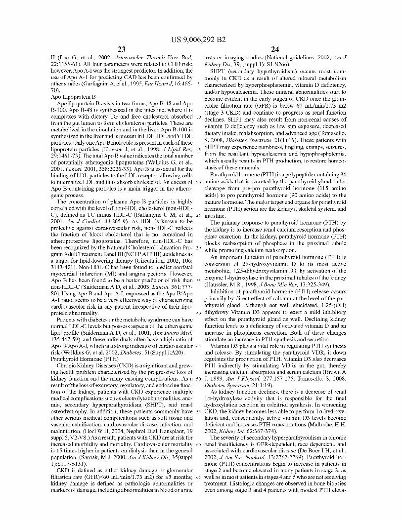
23II (Luc G, et al., 2002, Arterioscler Thromb Vasc Rio",22:1155-61). Al l four parameters were related to CHD risk;however, Apo A-I was the strongest predictor. In addition, theuse of Apo A-1 for predicting CAD has been confirmed byother studies (Garfagnini A, et al., 1995, Eur Heart J, 16:465-70).Apo Lipoprotein B
Apo lipoprotein B exists in two forms, Apo B-48 and ApoB-100. Apo B-48 is synthesized in the intestine, where it iscomplexes with dietary TG and free cholesterol absorbedfrom the gut lumen to form chylomicron particles. These aremetabolized in the circulation and in the liver. Apo B-100 issynthesized in the liver and is present in LDL, IDL and VLDLparticles. Only one Apo B molecule is present in each of theselipoprotein particles (Elovson J, et al., 1998, J Lipid Res,29:1461-73). The total Apo B value indicates the total numberof potentially atherogenic lipoproteins (Walldius G, et al.,2001, Lancet, 2001, 358:2026-33). Apo B is essential for thebinding of LDL particles to the LDL receptor, allowing cellsto internalize LDL and thus absorb cholesterol. An excess ofApo B-containing particles is a main trigger in the athero-genic process.
The concentration o f plasma Apo B particles is highlycorrelated with the level of non-HDL cholesterol (non-HDL-C), defined as TC minus HDL-C (Ballantyne C M, et al.,2001, Am J Cardiol, 88:265-9). As HDL is known to beprotective against cardiovascular risk, non-HDL-C reflectsthe fraction o f blood cholesterol that is not contained inatheroprotective lipoproteins. Therefore, non-HDL-C hasbeen recognized by the National Cholesterol Education Pro-gram Adult Treatment Panel III (NCEP ATP III) guidelines asa target for lipid-lowering therapy (Circulation, 2002, 106:3143-421). Non-HDL-C has been found to predict nonfatalmyocardial infarction (MI) and angina pectoris. However,Apo B has been found to be a better predictor of risk thannon-HDL-C (Sniderman A D, et al., 2003, Lancet, 361:777-80). Using Apo B and Apo A-I, expressed as the Apo B/ApoA-1 ratio, seems to be a very effective way of characterizingcardiovascular risk in any patient irrespective of their lipo-protein abnormality.
Patients with diabetes or the metabolic syndrome can havenormal LDL-C levels but possess aspects of the atherogeniclipid profile (Sniderman A D, et al., 1001, Ann Intern Med,135:447-59), and these individuals often have a high ratio ofApo B/Apo A-I, which is a strong indicator of cardiovascularrisk (Walldius G, et al, 2002, Diabetes, 51(Suppl.):A20).Parathyroid Hormone (PTH)
Chronic Kidney Diseases (CKD) is a significant and grow-ing health problem characterized by the progressive loss o fkidney function and the many ensuing complications. As aresult of the loss of excretory, regulatory, and endocrine func-tion of the kidney, patients with CKD experience multiplemedical complications such as electrolyte abnormalities, ane-mia, secondary hyperparathyroidism (SHPT), and renalosteodystrophy. In addition, these patients commonly haveother serious medical complications such as soft tissue andvascular calcification, cardiovascular disease, infection, andmalnutrition. (Horl W H, 2004, Nephrol Dial Transplant, 19suppl 5, V2-V8.) As a result, patients with CKD are at risk forincreased morbidity and mortality. Cardiovascular mortalityis 15 times higher in patients on dialysis than in the generalpopulation. (Samak, M J, 2000, Am J Kidney Dis, 35(suppl1):S117-S131).
CKD is defined as either kidney damage or glomerularfiltration rate (GFR)<60 mUmin/1.73 m2) for m o n t h s ;kidney damage is defined as pathologic abnormalities o rmarkers of damage, including abnormalities in blood or urine
US 9,006,292 B224
tests or imaging studies (National guidelines, 2002, Am JKidney Dis, 39, (suppl 1): S 1 -S266).
SHPT (secondary hypothyroidism) occurs most com-monly in CKD as a result o f altered mineral metabolism
5 characterized by hyperphosphatemia, vitamin D deficiency,and/or hypocalcaemia. These mineral abnormalities start tobecome evident in the early stages of CKD once the glom-erular filtration rate (GFR) is below 60 mLimin/1.73 m2(stage 3 CKD) and continue to progress as renal functiondeclines. SHPT may also result from non-renal causes o fvitamin D deficiency such as low sun exposure, decreaseddietary intake, malabsorption, and advanced age (Tomasello,S, 2008, Diabetes Spectrum, 21(1):19). These patients with
15 SHPT may experience numbness, tingling, cramps, seizures,from the resultant hypocalcaemia and hypophosphatemia,which usually results in PTH production, to restore homeo-stasis of these minerals.
Parathyroid hormone (PTH) is a polypeptide containing 8420 amino acids that is secreted by the parathyroid glands after
cleavage f rom pre-pro parathyroid hormone (115 aminoacids) to pro parathyroid hormone (90 amino acids) to themature hormone. The major target end organs for parathyroidhormone (PTH) action are the kidneys, skeletal system, and
25 intestine.The primary response to parathyroid hormone (PTH) by
the kidney is to increase renal calcium resorption and phos-phate excretion. In the kidney, parathyroid hormone (PTH)blocks reabsorption o f phosphate i n the proximal tubule
30 while promoting calcium reabsorption.An important function of parathyroid hormone (PTH) is
conversion o f 25-hydroxyvitamin D t o i t s most activemetabolite, 1,25-dihydroxyvitamin D3, by activation of theenzyme 1-hydroxylase in the proximal tubules of the kidney35(Haussler, MR., 1998, J Bone Min Res, 13:325-349).
Inhibition of parathyroid hormone (PTH) release occursprimarily by direct effect of calcium at the level of the par-athyroid gland. Although not wel l elucidated, 1,25-(OH)
40 dihydroxy Vitamin D3 appears to exert a mild inhibitoryeffect on the parathyroid gland as well. Declining kidneyfunction leads to a deficiency of activated vitamin D and anincrease in phosphorus excretion. Both o f these changesstimulate an increase in PTH synthesis and secretion.
45 V i t a m i n D3 plays a vital role in regulating PTH synthesisand release. By stimulating the parathyroid VDR, i t downregulates the production of PTH. Vitamin D3 also decreasesPTH indirectly by stimulating VDRs i n the gut, therebyincreasing calcium absorption and serum calcium (Brown A
50 J, 1999, Am J Physiol, 277:157-175; Tomasello, S, 2008,Diabetes Spectrum, 21:1:19).
As kidney function declines, there is a decrease of renalla-hydroxylase activity that is responsible fo r the finalhydroxylation reaction in calcitriol synthesis. In worsening
55 CKD, the kidney becomes less able to perform la-hydroxy-lation and, consequently, active vitamin D3 levels becomedeficient and increases PTH concentrations (Malluche, H H,2002, Kidney Int, 62:367-374).
The severity of secondary hyperparathyroidism in chronic60 renal insufficiency is GFR-dependent, race dependent, and
associated with cardiovascular disease (De Boer I H, et al.,2002, J Am Soc Nephrol, 13:2762-2769). Parathyroid hor-mone (PTH) concentrations begin to increase in patients instage 2 and become elevated in many patients in stage 3, as
65 we l l as inmost patients in stages 4 and 5 who are not receivingtreatment. Histologic changes are observed in bone biopsieseven among stage 3 and 4 patients with modest PTH eleva-

25tion. I n patients on dialysis, the SHPT and bone diseaseworsen and become more difficult to treat as the duration ofdialysis increases.
Elevated Phosphate level is common in patients with CKDdue to their impaired ability to excrete phosphorus renally.Phosphorus retention and the resultant increase o f serumphosphorus concentrations directly suppress the productionof calcitriol. Furthermore, the shrinkage in renal mass resultsin reduced activation o f vitamin D to calcitriol. Calciumabsorption from the gastrointestinal (GI) tract is reduced,resulting in hypocalcaemia. These derangements are interre-lated and can either individually or collectively stimulate thesynthesis and/or secretion of PTH by the parathyroid gland.The continuous increased production of PTH subsequentlyleads to hyperplasia of the parathyroid gland. Eventually, thegland may become autonomous in secreting PTH and lesslikely to respond to therapy.Phosphorus Metabolism
As the glomerular filtration rate (GFR) declines to <60mlimin/1.73 m2, phosphorus excretion becomes altered inthe nephron. Although half of the nephrons are not working toexcrete phosphorus, the remaining nephrons compensate byhyper-excreting the daily phosphorus load to maintain nor-mal serum phosphorus concentrations. Compensation cangenerally continue until the GFR declines to <25-40 mlimini1.73 m2. With progressive CKD, when the remaining neph-rons can no longer sufficiently excrete the phosphorus load,and hyperphosphatemia results.
Calcium, a divalent cation, and phosphorus, a monovalentanion, have a high binding affinity for each another. In theserum, as the concentration o f one or both ions increases,there is an increased risk for an ionic bonds to form, creatingan insoluble complex. This process may lead to extra skeletalcalcification and potentially calciphylaxis or cardiac disease(Ketteler, et al., Pediatr Nephrol, 2011, 26:7-18). Addition-ally, the precipitation may decrease serum calcium concen-trations, further stimulating PTH secretion. In fact, PTH pro-duction and secretion may be stimulated by hypocalcaemia,hyperphosphatemia, and vitamin D deficiency (Friedman EA, 2005, Kidney Int, 65 (Suppl.): S1-S7).
Because PTH is chiefly responsible for preventing hypoc-alcaemia, i t stimulates osteoclasts to lyse bone, releasingcalcium into the serum. Under normal conditions, there ishomeostasis involving osteoclast activity and osteoblast syn-thetic activity. SHPT produces an imbalance of these activi-ties leading to enhanced bone breakdown that eventuates inrenal osteodystrophy (Tomasello, S, 2008, Diabetes Spec-trum, 21AA9).Calcification
In addition to bone mineral defects and disease, alterationsin calcium, phosphorus, vitamin D, and PTH cause otherdeleterious consequences in patients with CKD. Extra skel-etal calcification (primarily cardiovascular calcification) hasbeen documented in patients with CKD (Goodman W G,2000, N Engl J Med, 342:1478-1483) and is directly corre-lated to an increase in cardiovascular morbidity and mortality.Patients with CKD, especially with end-stage renal disease(ESRD), have an increased risk of cardiovascular morbidityand mortality
Research has shown that the primary cause o f death inpatients with End stage renal disease (ESRD) is cardiovascu-lar disease (U.S. Renal Data System: USRDS 2006 AnnualData Report NIDDK 2006). A study of patients on hemodi-alysis found that even when stratified for variables such assex, race, and presence of diabetes, dialysis patients still had
US 9,006,292 B226
a cardiovascular mortality rate nearly 30 times greater thanthe general population (Block, GA, 2004, J Am Soc Nephrol,15:2208-2218).
The balance of calcium, phosphorus, vitamin D, and Intact5 P T H (I-PTH) is complex and interrelated. Patients must
adhere to dietary restrictions, dialysis therapies, and compli-cated medication regimens. These factors create barriers toachieving and maintaining control of SHPT. A study of nearly200 chronic hemodialysis outpatients revealed that <10% of
io patients could be simultaneously maintained within the targetranges of the above parameters. (Tomasello, S, 2004, DialysisTransplant, 33:236-242).eGFR
Glomerular filtration rate (GFR which is also referred to as15 "e-GFR" or "eGFR") is considered by medical professionals
to be the best measure of kidney function. Knowing some-one's GFR helps the medical professional figure out the stageof kidney disease. Doctors wil l use this information to plantheir patient's treatment.
20 T h e proportional variation in the GFR is larger in popula-tions with the disease (by a factor of approximately 10, from6 to 60 ml per minute per 1.73 ails/min/1.73 m2) than inpopulations without the disease (by a factor of approximately3, from 60 to 180 ml per minute per 1.73 ails/min/1.73 ma).
25 A s a result, larger proportion of the variation in serum crea-tinine levels among patients with the disease is due to avariation in the GFR, not to a variation in the other determi-nants as compared with healthy people.
The GFR assesses the excretory function of the kidneys30 and is considered the gold standard used to evaluate renal
function. The National Kidney Foundation's guidelines forchronic kidney disease, a GFR exceeding 90 mLimin/1.73 m2is considered normal; GFR of 60 to 89 is mildly decreased; aGFR of 30 to 59 is moderately decreased and may signify
35 "renal insufficiency"; a GFR of 15 to 29 mLimin/1.73 m2 isconsidered severely decreased; and a GFR of less than 15 isconsidered kidney failure. Decreased kidney function is asso-ciated with many complications, such as hypertension, ane-mia, malnutrition, bone disease, and a decreased quality o f
40 l i f e (National Kidney Foundation, 2000, Am J Kidney Dis,2:39, (suppl 1): S1-266).Prostate Specific Antigen (PSA)
Prostate cancer is the most commonly diagnosed cancer inmen and a leading cause of cancer death in the United States
45 and Europe (Boyle P, et al., 2005, Ann Oncol, 16:481-488).Prostate-specific antigen (PSA) is a protein produced by
the cells of the prostate gland. The PSA test measures the levelof PSA in the blood. It is normal for men to have low levels ofPSA in their blood; however, prostate cancer or benign (not
50 cancerous) conditions can increase PSA levels. As men age,both benign prostate conditions and prostate cancer becomemore frequent. The most common benign prostate conditionsare prostatitis (inflammation of the prostate) and benign pro-static hyperplasia (BPH) (enlargement of the prostate). There
55 i s no evidence that prostatitis or BPH cause cancer, but it ispossible for a man to have one or both of these conditions andto develop prostate cancer as well. A PSA level of 4.0 ng/m1_,is considered an appropriate cutoff for selecting men forprostate biopsy (Catalona W J, 1994, J Urol ; 152: 2037-42).A
60 recent report f rom the Prostate Cancer Prevention Trialshowed that prostate cancer occurs in men with low PSAlevels (Thompson I M, et al., 2004, N Engl J Med, 350:2239-46), suggesting that there is no "normal" PSA but rather acontinuum of risk of prostate cancer based on an individual's
65 PSA level (Thompson I M, 2005, JAMA, 294:66-70).Vitamin D receptor expression is inversely associated with
prostate cancer progression; VDR levels in tumor tissue may

27influence prostate cancer prognosis (Nutrition and Health:Vitamin D Edited by: M. F. Holick, 2010, PP 797; SpringerScience-Business Media, LLC).
Many epidemiological studies, indicate that vitamin Ddeficiency increases the risk of pro state cancer and that higherlevels of vitamin D are associated with better prognosis andimproved outcomes (Young, et al., 2011, Advances in Pre-ventive Medicine: Volume, Article ID 281863). The outcomeof clinical trials using vitamin D, calcitriol or various vitaminD analogs in men with prostate cancer have thus far beendisappointing.Liver Enzymes (AST, ALP, ALP and Bilimbin)
Abnormalities in liver function tests are elevated levels ofbiochemical tests, including aspartate aminotransferase(AST) o r s e r u m glutamic-oxaloacetic transaminase[SGOT]), a lanine aminotransferase ( A LT ) o r serumglutamate pymvate transaminase [SGPT]), alkaline phos-phatase, bilimbin, and albumin
Cellular injury in the liver causes release of AST and ALT.ALT is a more specific indication of liver disease, whereasAST elevations may be secondary to damage of other organs(heart, kidney, brain, intestine, placenta).
The ALT is found in the cytosol of liver, whereas two ASTisoenzymes are located in the cytosol and mitochondria,respectively. Both enzymes are released into the blood inincreasing amounts when the liver cell membrane is dam-aged. Necrosis of liver cells is not required for the release ofthe aminotransferase. I n fact, there i s poor correlationbetween the degree of liver-cell damage and the level of theaminotransferase. The ALT is found in low concentrations intissues other than liver, so it is frequently considered specificfor hepatocellular injury. However, this specificity is notabsolute because serum ALT elevations can occur in non-hepatic conditions such as myopathic diseases. Liver cellnecrosis is indicated by highly elevated ALT levels (Tung, BY, 1999, Clin Liver Dis, 3:585-601).
The AST also is abundantly expressed in several nonhepatic tissues including heart, skeletal muscle, and blood.Nonetheless, both the ratio and absolute elevation of the ASTand ALT can provide important information regarding theextent and etiology of liver disease.
While no single biochemical liver function test is suffi-ciently specific to allow a definite diagnosis in patients withliver disease, we have been impressed by the consistency withwhich the serum glutamic oxaloacetic transaminase (SGOT)activity exceeds the serum glutamic pymvic transaminase(SGPT) activity in patients with alcoholic liver disease.
Nonalcoholic fatty liver disease (NAFLD) refers to a widespectrum o f liver disease ranging from simple fatty liver(steatosis), to nonalcoholic steatoheppatitis (NASH), to cir-rhosis (irreversible, advanced scarring of the liver). All of thestages of NAFLD have in common the accumulation of fat(fatty infiltration) in the liver cells (hepatocytes). In NASH,the fat accumulation is associated with varying degrees o finflammation (hepatitis) and scarring (fibrosis) of the liver.
It was shown (Cohen et al., 1979, Digestive Diseases andSciences, 24(11) that SGOT/SGPT ratio i s significantlyelevated in patients with alcoholic hepatitis and cirrhosis(2.85+-0.2) compared with patients with post necrotic cir-rhosis (1.74+ or —0.2), chronic hepatitis (1.3+ or —0.17),obstructive jaundice (0.81+ o r —0.06) and viral hepatitis(0.74+ or —0.07). An SGOT/SGPT ratio greater than 2 ishighly suggestive o f alcoholic hepatitis and cirrhosis. I toccurs i n 70% o f these patients compared with 26% o fpatients with post necrotic cirrhosis, 8% with chronic hepa-titis, 4% with viral hepatitis and none with obstructive jaun-dice. (Sorbi, et al., 1999, Am J Gastroenterol, 94:1018-1022).
US 9,006,292 B228
The AST to ALT ratio has been reported to be a usefulparameter supporting the diagnosis o f alcoholic hepatitis(Finlayson, e t al., 1993, Baill iere's C l in Gastroenterol,7:627-640; 40: Harrison, D J, et al., Bailliere's Clin Gastro-
5 enterol, 1993, 7:641-62).An AST level more than twice the ALT level has been
reported in as many as 83% of patients hospitalized for alco-holic hepatitis (Pinto, H C, et al., 1996, Dig Dis Sci, 1996,41:172-9; Bird G LA , et al., 1993, Bailliere's Clin Gastro-
io enterol, 1997, 663-82).The AST to ALT ratio appears to be a useful index for
distinguishing nonalcoholic steatoheppatitis (NASH) fromalcoholic l iver disease. Subset analysis o f patients w i thNASH revealed mean AST to ALT ratios of 0.7, 0.9, and 1.4
15 f o r subjects wi th no fibrosis, m i ld fibrosis, o r cirrhosis,respectively (Sorbii. D, et. al., 1999, The American Journal ofGastroenterology, 94(4)).Alkaline Phosphatase
Alkaline phosphatase (ALP) is associated with cellular20 membranes, and elevated levels may be caused by injury to
the liver, bone, kidneys, intestines, placenta, or leukocytes. Inthe liver, the enzyme is located in the bile canaliculi. Biliaryobstruction induces increased synthesis o f alkaline phos-phatase and spillage into the circulation. Elevations in serum
25 A L P levels originate predominantly from two sources, liverand bone. I t is also present in kidneys, small bowel andplacenta. Levels vary with age. Rapidly growing adolescentscan have serum ALP levels that are twice those of healthyadults as a result of the leakage of bone. Also, serum ALP
30 levels normally increase gradually between the ages of 40 and65 years, particularly in women. Women in the third trimesterof pregnancy have elevated serum level ALP because of aninflux of placental ALP into their blood. In persons with bloodtype 0 or B, serum ALP levels may increase after the inges-
35 t ion of a fatty meal; because of an influx o f intestinal ALPthere are also reports of a benign familial elevation in serumALP levels because o f increased levels o f intestinal ALP(Wolf P L, 1978, Arch. Pathol. Lab. Med, 102:497-501).Bilirubin
40 T h e human body produces about 4 mg per kg of bilirubinper day from the metabolism o f heme. Approximately 80percent of the heme moiety comes from catabolism of redblood cells, with the remaining 20 percent resulting fromineffective erythropoiesis and breakdown of muscle myoglo-
45 b i n and cytochromes. B i l imbin is transported f rom theplasma to the liver for conjugation and excretion.
Hyperbilimbinemia may signify hepatobiliary disease orhemolysis. Mild degrees of indirect hyperbilimbinemia maybe found in as many as 10% o f asymptomatic patients with
50 Gilbert's syndrome (Lidofsky, SD, Jaundice, In: Feldman M,et al., Sleisenger and Fordtran's Gastrointestinal and LiverDisease, 8th ed. Philadelphia, Pa.: Saunders Elsevier, 2006).
Prior to age 30, hepatitis causes 75% of hyperbilirubine-mia. After age 60, extra hepatic obstruction causes 50% of
55 hyperbilimbinemia (e.g., gallstones or pancreatic cancer).Intra-hepatic disorders can lead to unconjugated or conju-
gated hyperbilimbinemia. The conjugated (direct) bilirubinlevel is often elevated by alcohol, infectious hepatitis, drugreactions, and autoimmune disorders. Post hepatic disorders
60 also can cause conjugated hyperbilimbinemia. Gallstone for-mation is the most common and benign post hepatic processthat causes jaundice; however, the differential diagnosis alsoincludes serious conditions such as biliary tract infection,pancreatitis, and malignancies (S. P. Roche, et al., 2004, Am
65 _Pam Physician, 2004, 69:299-304). Biliary obstruction is acondition where b lood levels o f conjugated bi l i rubinincrease.

29Symptoms of hemolytic anemia are similar to other forms
of anemia (fatigue and shortness of breath), but in addition,the breakdown of red cells leads to jaundice and increases therisk of particular long-term complications, such as gallstonesand pulmonary hypertension.
Hemolytic jaundice can lead increased production o fbilimbin. In this case bilimbin is conjugated and excretednormally, but the conjugation mechanism is overwhelmed,and an abnormally large amount of unconjugated bilirubin isfound in the blood.
Gilbert syndrome is a common, benign, hereditary disorderthat affects approximately 5 percent of the U.S. population.(Schreiber R. A., et al., 2001, Pediatr Rev, 22:219-26). Typi-cally, the disease results in a mild decrease in the activity ofthe enzyme glucuronosyltransferase, causing an increase inthe indirect fraction of serum bilimbin. Gilbert syndrome istypically an incidental finding on routine liver function tests,when the bilirubin level is slightly increased and all other liverfunction values are within normal limits. Jaundice and furtherelevation of the bilirubin level may occur during periods ofstress, fasting, or illness.
Crigler-Najjar Syndrome or CNS is a rare disorder affect-ing the metabolism of bilimbin, a chemical formed from thebreakdown of blood. The disorder results in an inherited formof non-hemolytic jaundice, which results in high levels o funconjugated bilimbin and often leads to brain damage ininfants. This syndrome is divided into type I and type II, withthe latter sometimes referred to as Arias syndrome. These twotypes, along with Gilbert's syndrome, Dubin-Johnson syn-drome, and Rotor syndrome, make up the five known heredi-tary defects in bilirubin metabolism. Unlike Gilbert's syn-drome, only a few hundred cases of CNS are known (JansenP L, et al., 1999, European Journal o 'Pediatrics, 158, Suppl2, S89-S94, 0154).
Dubin-Johnson syndrome is an autosomal recessive disor-der that causes an increase o f conjugated bilimbin in theserum without elevation of liver enzymes (ALT, AST). Thiscondition is associated with a defect in the ability of hepato-cytes to secrete conjugated bilimbin into the bile, and issimilar to Rotor syndrome. I t is usually asymptomatic butmay be diagnosed in early infancy based on laboratory tests.
(Rotor syndrome, also called Rotor type hyperbilirubine-mia, is a rare, relatively benign autosomal recessive bilimbindisorder of unknown origin. It is a distinct disorder; yet simi-lar to Dubin-Johnson syndrome both diseases cause anincrease in conjugated bilimbin (Wolkoff, A W, et al., 1976,The American Journal of Medicine, 60(2):173-179).Thyroid Diseases
According to the World Health Organization (WHO), thy-roid diseases have the second-highest prevalence among allendocrine disorders, right behind diabetes mellitus. Over 665million people suffer from endemic goiter or other thyroiddiseases worldwide; 1.5 bill ion people are at risk for devel-oping an iodine-deficient condition. Statistical data show thatthe annual increment in the number of thyroid disease cases is5%. Hypothyroidism is a common endocrine disorder result-ing from deficiency of thyroid hormone. Worldwide, iodinedeficiency remains the foremost cause of hypothyroidism. Inthe United States and other areas of adequate iodine intake,autoimmune thyroid disease (Hashimoto disease) is the mostcommon cause of hypothyroidism; worldwide, iodine defi-ciency remains the foremost cause. Among all methods avail-able to treat diseases of the thyroid gland (thyroid), a prefer-ence i s given to hormone replacement therapy (HRT),treatment with antithyroid drugs, surgical intervention (thy-roidectomy), and radioactive iodine therapy. An estimated 20millionAmericans have some form of thyroid disease accord-
US 9,006,292 B230
ing to the American Thyroid association. One in eight womenwill develop a thyroid disorder during her lifetime. Levothy-roxine, a synthetic form of thyroid hormone, is the 4th highestselling drug in the U.S. 13 of the top 50 selling drugs are either
5 d i rect ly or indirectly related to hypothyroidism.Al l these treatment standards characteristically have a con-
siderable number of contraindications and shortcomings. Inparticular, hormone replacement therapy requires lifelongadministration o f thyroid hormone preparations and, as a
io result, the patient's own thyroid eventually stops functioningat all thus making the patient a "lifelong client" of pharma-ceutical companies. Besides, administration of thyroid hor-mone preparations has a number of contraindications, andmultiple adverse effects, including tachycardia, cardiac
15 arrhythmia, allergic reactions, excitation, insomnia, etc.,accompany this treatment.
The aim of antithyroid therapy practiced today is partial orcomplete suppression of the patient's own thyroid functionafter which the patient is switched to hormone replacement
20 therapy. Adverse effects o f thyro static therapy include inhi-bition o f hematopoiesis, nausea, vomiting, l iver functionimpairment, allergic reactions, etc.
Surgical intervention is often accompanied by complica-tions responsible for a near-10% disability rate. Other signifi-
25 cant disadvantages of thyroid ablation (or removal of a part ofthe thyroid) include lifelong administration of hormones anda high risk of damage to the parathyroid glands during sur-gery. Medicine today is unable to offer patients with thyroiddiseases any alternatives, and prefers to ignore the disadvan-
30 tages of current treatment regimens and standards.Thyroid Regulation
The thyroid is a butterfly shaped organ located just belowthe Adam's apple in the neck. Made up o f small sacs, thisgland is filled with an iodine-rich protein called thyroglobulin
35 along with the thyroid hormones thyroxine (T4) and smallamounts of triiodothyronine (T3).
The primary function of these two hormones is to regulatemetabolism by controlling the rate at which the body convertsoxygen and calories to energy. In fact, the metabolic rate of
40 every cell in the body is regulated by thyroid hormones,primarily T3. (Videla L A, Fernandez V, Tapia G, Varela P.Thyroid hormone calorigenesis and mitochondrial redox sig-naling: up regulation of gene expression. Front Biosci. 2007Jan. 1; 12:1220-8).
45 I n healthy individuals the gland is imperceptible to thetouch. A visibly enlarged thyroid gland is referred to as agoiter. Historically, goiter was most frequently caused by alack o f dietary iodine. However, in countries where salt isiodized, goiter of iodine deficiency is rare.
50 T h e production of T4 and T3 in the thyroid gland is regu-lated by the hypothalamus and pituitary gland. To ensurestable levels of thyroid hormones, the hypothalamus monitorscirculating thyroid hormone levels and responds to low levelsby releasing thyrotropin-releasing hormone (TRH). This
55 T R H then stimulates the pituitary to release thyroid-stimulat-ing hormone (TSH). (Segerson T P, et. al, Science. 1987 Oct.2; 238(4823): 78-80; Dyess E M, et. al, Endocrinology. 1988November; 123(5): 2291-7).
When thyroid hormone levels increase, production of TSH60 decreases, which in turn slows the release of new hormone
from the thyroid gland. Cold temperatures can also increaseTRH levels. This is thought to be an intrinsic mechanism thathelps keep us warm in cold weather.
Elevated levels o f cortisol, as seen during stress and in65 conditions such as Cushing's syndrome, lowers TRH, TSH
and thyroid hormone levels as well. (Roelfsema F, et. al, EurEndocrinol. 2009 November; 161(5): 695-703.

31The thyroid gland needs iodine and the amino acid L-ty-
rosine to make T4 and T3. A diet deficient in iodine can limithow much T4 the thyroid gland can produce and lead tohypothyroidism. (Angermayr L, et. al, Cochrane DatabaseSyst Rev. 2004; (2): CD003819).
T3 is the biologically active form of thyroid hormone. Themajority of T3 is produced in the peripheral tissues by con-version of T4 to T3 by a selenium-dependent enzyme.
Various factors including nutrient deficiencies, drugs, andchemical toxicity may interfere with conversion of T4 to T3.(Kelly G S. Altem Med Rev. 2000 August; 5(4): 306-33).
Another related enzyme converts T4 to an inactive form ofT3 called reverse T3 (rT3). Reverse T3 does not have thyroidhormone activity; instead i t blocks the thyroid hormonereceptors in the cell hindering action of regular T3. (Kohrle J.Acta Med Austriaca. 1996; 23(1-2): 17-30).
Ninety-nine percent o f circulating thyroid hormones arebound to carrier proteins, rendering them metabolically inac-tive. The remaining "free" thyroid hormone, the majority ofwhich is T3, binds to and activates thyroid hormone receptors,exerting biological activity. (Nussey S, et. al, Endocrinology:An Integrated Approach. Oxford: BIOS Scientific Publishers,2001). Very small changes in the amount of carrier proteinswill affect the percentage of unbound hormones. Oral contra-ceptives, pregnancy, and conventional female hormonereplacement therapy may increase thyroid carrier protein lev-els and, thereby, lower the amount of free thyroid hormoneavailable. (Arafah B M., Increased need for thyroxine inwomen with hypothyroidism during estrogen therapy. N EnglJ. Med. 2001 Jun. 7; 344(23): 1743-9).
The thyroid gland is the biggest gland in the neck. The solefunction of the thyroid is to make thyroid hormone. Thyroidhormones regulate our body's metabolism and influence vir-tually every organ system in the body. They tell organs howfast or slow they should work. Thyroid hormones also regu-late the body's consumption o f oxygen and production o fheat. Thyroid problems, such as an overactive or under activethyroid, can severely affect metabolism (Franklyn, et al.,2005, Journal of Endocrinology, 187:1-15).Hyperthyroidism
Too much thyroid hormone from an overactive thyroidgland is called hyperthyroidism, because i t speeds up thebody's metabolism. This hormone imbalance occurs in about1 percent of all women, who get hyperthyroidism more oftenthan men. One of the most common forms of hyperthyroidi smis known as Graves' disease. Because the thyroid gland isproducing too much hormone in hyperthyroidism, the bodydevelops an increased metabolic state, with many body sys-tems developing abnormal function (Wondisford, ClinicalManagement of Thyroid Diseases, ISBN: 978-1-4160-4745-2, Copyright 0 2009 by Saunders. Philadelphia).Hypothyroidism
Too little thyroid hormone from an under active thyroidgland i s called hypothyroidism. I n hypothyroidism, thebody's metabolism is slowed. Several causes for this condi-tion exist, most o f which affect the thyroid gland directly,impairing its ability to make enough hormone. More rarely,there may be a pituitary gland tumor, which blocks the pitu-itary from producing TSH. As a consequence, the thyroid failsto produce a sufficient supply of hormones needed for goodhealth. Whether the problem is caused by the thyroid condi-tions or y the pituitary gland, the result is that the thyroid isunder producing hormones, causing many physical and men-tal processes to become sluggish. The body consumes lessoxygen and produces less body heat.
Hypothyroidism is a condition in which the thyroid glanddoes not make enough thyroid hormones, characterized by a
US 9,006,292 B232
reduction in metabolic rate. The main symptoms of hypothy-roidism are fatigue, weakness, increased sensitivity to cold,constipation, unexplained weight gain, dry skin, hair loss orcoarse dry hair, muscle cramps and depression. However,
5 m o s t symptoms take years to develop. The slower the metabo-lism gets, the more obvious the signs and symptoms wi l lbecome. I f hypothyroidism goes untreated, the signs andsymptoms could become severe, such as a swollen thyroidgland (goiter), slow thought processes, or dementia. (Hy-
io pothyroidism. The American ThyroidAssociation). Subclini-cal hypothyroidism, an often under-diagnosed thyroid disor-der, manifests as elevated TSH, normal T4 and normal T3levels. Individuals with subclinical hypothyroidism are atgreater risk for developing overt hypothyroidism. (Garduno-
15 Garcia et al. Eur J Endocrinol. 2010 August; 163(2): 273-8.It has been estimated that about 20% of women over the age
of 60 suffer from subclinical hypothyroidism. (Wilson G R,et. al, Am Fam Physician. 2005 Oct. 15; 72(8): 1517-24.
There is evidence that the standard blood TSH test refer-20 ence range may cause many cases of hypothyroidism to be
missed. Most physicians accept a reference range for TSHbetween 0.45 and 4.5 [IIIRmL to indicate normal thyroidfunction. In reality, though, a TSH reading of more than 2.0may indicate lower-than-optimal thyroid hormone levels.
25 Various TSH levels that fall within normal range are associ-ated with adverse health outcomes.
TSH greater than 2.0 leads to increased 20-year risk o fhypothyroidism and increased risk o f thyroid autoimmunedisease. TSH between 2.0 and 4.0: hypercholesterolemia and
30 cholesterol levels decline in response to T4 therapy TSHgreater than 4.0: greater risk of heart disease. ((Tunbridge WM, et. al, Clin Endocrinol (Oxf). 1977 December; 7(6): 481-93).Consequences of Hypothyroidism
35 Gastrointest inal problems. Hypothyroidism is a commoncause of constipation. Constipation in hypothyroidism mayresult from diminished motility o f the intestines. In somecases, this can lead to intestinal obstruction or abnormalenlargement of the colon.36 Hypothyroidism is also associ-
40 ated with decreased motility in the esophagus, which causesdifficulty swallowing, heartburn, indigestion, nausea, o rvomiting. Abdominal discomfort, flatulence, and bloatingoccur in those with small intestinal bacterial growth second-ary to poor digestion. Depression and psychiatric disorders.
45 Panic disorders, depression, and changes in cognition arefrequently associated with thyroid disorders. Hypothyroid-ism is often misdiagnosed as depression. (Hennessey J V, et.al, J Fam Pract. 2007 August; 56(8 Suppl Hot Topics): S31-9.
Cognitive decline. Patients with low thyroid function can50 suffer from slowed thinking, delayed processing of informa-
tion, difficulty recalling names, etc. Patients with subclinicalhypothyroidism show signs o f decreased working memory,and decreased speed of sensory and cognitive processing. Anevaluation o f thyroid hormones along with TSH may help
55 avoid misdiagnosis as being depressed. (Kritz-Silverstein D,et. al, J Nutr Health Aging. 2009 April; 13(4): 317-21.
Hypothyroidism and subclinical hypothyroidism are asso-ciated with increased levels o f blood cholesterol, increasedblood pressure, and increased risk of cardiovascular disease.
60 Even those with subclinical hypothyroidism were almost 3.4times as likely to develop cardiovascular disease than thosewith healthy thyroid function. Hypertension is relativelycommon among patients with hypothyroidism. (Duntas L H,et. al, Semin Thromb Hemost. 2011 February; 37(1): 27-34.
65 T h e risk o f heart disease increases proportionally withincreasing T S H , even i n subclinical hypothyroidism.Hypothyroidism that is caused by autoimmune reactions is
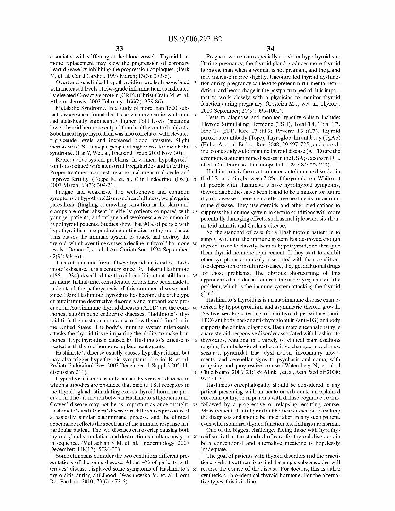
33associated with stiffening of the blood vessels. Thyroid hor-mone replacement may slow the progression o f coronaryheart disease by inhibiting the progression of plaques. (PerkM, et. al, Can J Cardiol. 1997 March; 13(3): 273-6).
Overt and subclinical hypothyroidism are both associatedwith increased levels of low-grade inflammation, as indicatedby elevated C-reactive protein (CRP). (Christ-CraM M, et. al,Atherosclerosis. 2003 February; 166(2): 379-86).
Metabolic Syndrome. In a study of more than 1500 sub-jects, researchers found that those with metabolic syndromehad statistically significantly higher TSH levels (meaninglower thyroid hormone output) than healthy control subjects.Subclinical hypothyroidism was also correlated with elevatedtriglyceride levels and increased blood pressure. Sl ightincreases in TSH may put people at higher risk for metabolicsyndrome. (Lai Y, Wet. al, Endocr J. Epub 2010 Nov. 30).
Reproductive system problems. In women, hypothyroid-ism is associated with menstrual irregularities and infertility.Proper treatment can restore a normal menstrual cycle andimprove fertility. (Poppe K, et. al, Cl in Endocrinol (Oxf).2007 March; 66(3): 309-21.
Fatigue and weakness. The well-known and commonsymptoms of hypothyroidi sm, such as chilliness, weight gain,paresthesia (tingling or crawling sensation in the skin) andcramps are often absent in elderly patients compared withyounger patients, and fatigue and weakness are common inhypothyroid patients. Studies show that 90% of people withhypothyroidism are producing antibodies to thyroid tissue.This causes the immune system to attack and destroy thethyroid, which over time causes a decline in thyroid hormonelevels. (Doucet J, et. al, J Am Geriatr Soc. 1994 September;42(9): 984-6).
This autoimmune form of hypothyroidism is called Hash-imoto's disease. It is a century since Dr. Hakam Hashimoto(1881-1934) described the thyroid condition that still bearshis name. In that time, considerable efforts have been made tounderstand the pathogenesis o f this common disease and,since 1956; Hashimoto thyroiditis has become the archetypeof autoimmune destructive disorders and autoantibody pro-duction. Autoimmune thyroid diseases (AITD) are the com-monest autoimmune endocrine diseases. Hashimoto's thy-roiditis is the most common cause of low thyroid function inthe United States. The body's immune system mistakenlyattacks the thyroid tissue impairing the ability to make hor-mones. Hypothyroidism caused by Hashimoto's disease istreated with thyroid hormone replacement agents.
Hashimoto's disease usually causes hypothyroidism, butmay also trigger hyperthyroid symptoms. (Lorini R, et. al,Pediatr Endocrinol Rev. 2003 December; 1 Suppl 2:205-11;discussion 211).
Hyperthyroidism is usually caused by Graves' disease, inwhich antibodies are produced that bind to TSH receptors inthe thyroid gland, stimulating excess thyroid hormone pro-duction. The distinction between Hashimoto' s thyroiditis andGraves' disease may not be as important as once thought.Hashimoto's and Graves' disease are different expressions ofa basically similar autoimmune process, and the clinicalappearance reflects the spectrum of the immune response in aparticular patient. The two diseases can overlap causing boththyroid gland stimulation and destruction simultaneously orin sequence. (McLachlan S M, et. al, Endocrinology. 2007December; 148(12): 5724-33).
Some clinicians consider the two conditions different pre-sentations o f the same disease. About 4% o f patients withGraves' disease displayed some symptoms of Hashimoto'sthyroiditis during childhood. (Wasniewska M, et. al, HornRes Paediatr. 2010; 73(6): 473-6).
US 9,006,292 B234
Pregnant women are especially at risk for hypothyroidism.During pregnancy, the thyroid gland produces more thyroidhormone than when a woman is not pregnant, and the glandmay increase in size slightly. Uncontrolled thyroid dysfunc-
5 t i o n during pregnancy can lead to pretenn birth, mental retar-dation, and hemorrhage in the postpartum period. It is impor-tant to work closely with a physician to monitor thyroidfunction during pregnancy. (Costeira M J, wet. al, Thyroid.2010 September; 20(9): 995-1001).
Tests to diagnose and monitor hypothyroidism include:Thyroid Stimulating Hormone (TSH), Total T4, Total T3,Free T4 (fT4), Free T3 (fT3), Reverse T3 (rT3), Thyroidperoxidase antibody (Tope), Thyroglobulin antibody (TgAb)
15 (Huber A, et. al, Endocr Rev. 2008; 29:697-725), and accord-ing to one study Auto immune thyroid disease (AITD) are thecommonest autoimmune diseases in the USA; (Jacobson D L,et. al, Clin Immunol Immunopathol. 1997; 84:223-243).
Hashimoto's is the most common autoimmune disorder in20 the U.S., affecting between 7-8% of the population. While not
all people with Hashimoto's have hypothyroid symptoms,thyroid antibodies have been found to be a marker for futurethyroid disease. There are no effective treatments for autoim-mune disease. They use steroids and other medications to
25 suppress the immune system in certain conditions with morepotentially damaging effects, such as multiple sclerosis, rheu-matoid arthritis and Crolm's disease.
So the standard o f care for a Hashimoto's patient is tosimply wait until the immune system has destroyed enough
30 thyroid tissue to classify them as hypothyroid, and then givethem thyroid hormone replacement. I f they start to exhibitother symptoms commonly associated with their condition,like depression or insulin resistance, they get additional drugsfor those problems. T h e obvious shortcoming o f this35approach is that it doesn't address the underlying cause of theproblem, which is the immune system attacking the thyroidgland.
Hashimoto's thyroiditis is an autoimmune disease charac-40 terized by hypothyroidism and asymmetric thyroid growth.
Positive serologic testing o f antithyroid peroxidase (anti-TPO) antibody and/or anti-thyroglobulin (anti-TG) antibodysupports the clinical diagnosis. Hashimoto encephalopathy isa rare steroid-responsive disorder associated with Hashimoto
45 thyroiditis, resulting in a variety o f clinical manifestationsranging from behavioral and cognitive changes, myoclonus,seizures, pyramidal tract dysfunction, involuntary move-ments, and cerebellar signs to psychosis and coma, wi threlapsing and progressive course (Watemberg N, et. al, J
50 Chi ld Neurol 2006; 21:1-5; Alink J, et. al, Acta Paediatr 2008;97:451-3).
Hashimoto encephalopathy should be considered in anypatient presenting with an acute or sub acute unexplainedencephalopathy, or in patients with diffuse cognitive decline
55 fol lowed by a progressive o r relapsing-remitting course.Measurement of antithyroid antibodies is essential to makingthe diagnosis and should be undertaken in any such patient,even when standard thyroid function test findings are normal.
One of the biggest challenges facing those with hypothy-60 roidism is that the standard of care for thyroid disorders in
both conventional and alternative medicine is hopelesslyinadequate.
The goal of patients with thyroid disorders and the practi-tioners who treat them is to find that single substance that will
65 reverse the course of the disease. For doctors, this is eithersynthetic or bio-identical thyroid hormone. For the alterna-tive types, this is iodine.
1
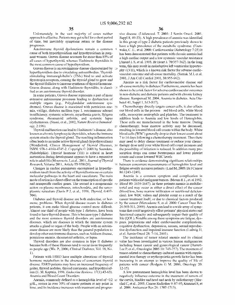
35Unfortunately, i n the vast majority o f cases neither
approach is effective. Patients may get relief for a short periodof time, but inevitably symptoms return o r the diseaseprogresses.
Autoimmune thyroid dysfunctions remain a commoncause of both hyperthyroidism and hypothyroidism in preg-nant women. Graves disease accounts for more than 85% ofall cases of hyperthyroid, whereas Hashimoto thyroiditis isthe most common cause of hypothyroidism.
Graves disease is an autoimmune disease characterized byhyperthyroidism due to circulating autoantibodies. Thyroid-stimulating immunoglobulin's (TSIs) bind to and activatethyrotropin receptors, causing the thyroid gland to grow andthe thyroid follicles to increase synthesis of thyroid hormone.Graves disease, along with Hashimoto thyroiditis, is classi-fied as an autoimmune thyroid disorder.
In some patients, Graves disease represents a part of moreextensive autoimmune processes leading to dysfunction ofmultiple organs (e.g., Polyglandular autoimmune syn-dromes). Graves disease is associated with pernicious ane-mia, vitiligo, diabetes mellitus type 1, autoimmune adrenalinsufficiency, systemic sclerosis, myasthenia gravis, Sjogrensyndrome, rheumatoid arthrit is, a n d systemic l u p u serythematous. (Stassi, et al., 2002, Nature Reviews Immunol-ogy, 2:196).
Thyroid malfunction can lead to Hashimoto' s disease, alsoknown as chronic lymphocytic thyroiditis, where the immunesystem attacks the thyroid gland. The resulting inflammationoften leads to an under active thyroid gland (hypothyroidism)(Wondisford, Clinica Management o f Thyroid Diseases,ISBN: 978-1-4160-4745-2, Copyright 0 2009 by Saunders.Philadelphia). Thyroid hormone (TH) is critical i n heartmaturation during development appears to have a reparativerole in adult life (Mourouzis. I, et al., 2011, Journal ofThyroidResearch, Volume 2011, Article ID 958626).
Changes in cardiac parameters encountered in hyperthy-roidism result from the activity of thyroid hormone on certainmolecular pathways in the heart and vasculature. The mainmode of action is a direct effect on the transcription of specificand nonspecific cardiac genes. The second is a nongenomicaction on plasma membranes, mitochondria, and the sarco-plasmic reticulum (Davis P J, et al., 1996, Thyroid, 6:497-504).
Diabetes and thyroid disease are both endocrine, or hor-mone, problems. When thyroid disease occurs in diabetespatients, i t can make blood glucose control more difficult.Almost one third of people with type 1 diabetes, have beenfound to have thyroid disease. This is because type 1 diabetesand the most common thyroid disorders are autoimmunediseases, which are diseases in which the immune systemattacks a gland or organ of the body. People with an autoim-mune disease are more likely than the general population todevelop other autoimmune diseases, such as Addison disease,pernicious anemia, rheumatoid arthritis, or lupus.
Thyroid disorders are also common in type I I diabetesbecause both of these illnesses tend to occur more frequentlyas people age (Wu. P., 2000, Clinical Diabetes, Vol. 18, No.1).
Patients with ESRD have multiple alterations o f thyroidhormone metabolism in the absence o f concurrent thyroiddisease. ESRD patients may have an increased frequency ofgoiter, thyroid nodules, thyroid carcinoma, and hypothyroid-ism (E. M. Kaptein, 1996, Endocrine Reviews, 17(1):45-63).Anemia and Blood Count Disorders
Anemia, commonly defined as a hemoglobin level of <12gidL, occurs in over 30% of cancer patients at any point intime, and its incidence increases with treatment and progres-
US 9,006,292 B236
sive disease. (Littlewood. T. 2001: J Semin Oncol. 2001,Suppl 8, 49-53). A high prevalence of anemia was identifiedin this group of type 2 diabetic patients previously shown tohave a high prevalence o f the metabolic syndrome. (Ezen-
5 waka, C. E., et al. 2008: Cardiovascular Diabetology 7:25) Ithas been demonstrated that patients with chronic anemia hada high cardiac output and a low systemic vascular resistance[Anand I. S, et al. 1993, Br Heart J, 70:357-362). In the longterm, this may result in maladaptive left ventricular hypertro-phy (LVH), which is a known risk factor for adverse cardio-vascular outcome and all-cause mortality. (Samak M J, et al.2001, J Am Coll Cardiol 2001, 38:955-962).
Anemia as a risk factor for cardiovascular disease and15 all-cause mortality in diabetes: Furthermore, anemia has been
shown to be a risk factor for adverse cardiovascular outcomesin non-diabetic and diabetic patients and with chronic kidneydisease. Rampersad M, 2004, Anemia in diabetes. Acta Dia-betol 41, Suppl 1, S13-S17).
20 Chemotherapy directly targets cancer cells, it also affectsour blood cells in the process r e d blood cells, white bloodcells, monocytes neutrophils and platelets. The treatment inaddition leads to Anemia and low levels o f Hemoglobin.These cells are manufactured in the bone marrow. During
25 chemotherapy, bone marrow activity may be decreased,resulting in lowered blood cell counts within the body. Whiteblood cells (WBC) generally drop to their lowest count about7 to 14 days following a chemotherapy treatment. Thus theremay need to delay chemo treatment or reduce your chemo-
30 therapy dose until your white blood cell count increases andthe possibility of infection is reduced. In addition many pre-scription drugs can cause Neutropenia and lower plateletcounts and cause lowered WBC levels
There is evidence demonstrating significant relationships35 between concurrent measurements of hemoglobin level and
fatigue severity in cancer patients. (Lind M, 2003: Br J Cancer86:1243-1249).
Anemia i s a common symptom and complication i npatients with solid malignant tumors. (Means R T., et. all 992,
40 Blood 80: 1639-1647). In these patients anemia is multifac-torial and may occur as either a direct effect o f the cancer(blood loss, bone marrow infiltration or nutritional deficien-cies), low WBC values and platelet count as a result of thecancer treatment itself, or due to chemical factors produced
45 b y the cancer (Mercadante S, et al. 2000: Cancer Treat Rev26:303-311, 2000). Anemia can lead to a wide array of symp-toms that could negatively affect patients' physical status andfunctional capacity and subsequently impair their quality oflife (Q0L). Notable among these symptoms are fatigue, dys-
50 pnea, palpitations and other cardiovascular complications,cognitive dysfunction, depression, nausea, sexual/reproduc-tive dysfunction and impaired immune function (Ludwig H,et al. Semin Oncol 28: 7-14, 2001).
The incidence o f tumor related anemia and its clinical55 value has been investigated in various human malignancies
including breast cancer and gynecological cancer (Barrett-Lee P, et al., Oncologist 2005 10: 743-757). The treatment oftumor-related or chemotherapy-induced anemia with supple-mental iron therapy or erythropoietin growth factor has been
60 increasing in an attempt to improve the quality o f life o fpatients with cancer (Rodgers G M, 2006, Oncology 20:12-15).
A low pretreatment hemoglobin level has been shown tonegatively influence outcome in the treatment o f tumors of
65 the cervix, bladder and head and neck by radiotherapy (Mar-chal C, et al., 2005, Cancer Radiother 9: 87-95; Kummel S, etal. 2006: Anticancer Res 26: 1707-1713).
1
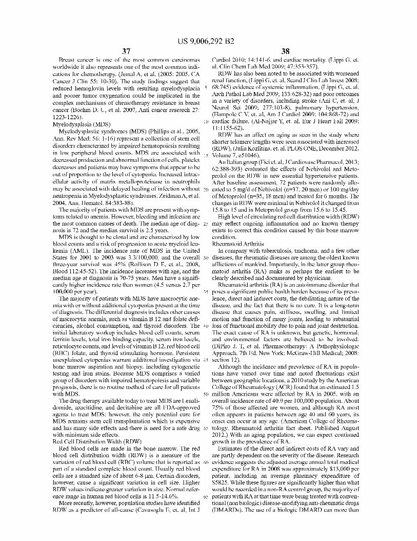
37Breast cancer is one o f the most common carcinomas
worldwide it also represents one of the most common indi-cations for chemotherapy. (Jemal A, et al. (2005: 2005. CACancer J Clin 55: 10-30). The study findings suggest thatreduced hemoglovin levels with resulting myelodysplaciaand poorer tumor oxygenation could be implicated in thecomplex mechanisms o f chemotherapy resistance in breastcancer (Boehm D. U, et al. 2007, Anti cancer research 27:1223-1226).Myelodysplasia (MDS)
Myelodysplastic syndromes (MDS) (Phillips et al., 2005,Ann. Rev Med; 56: 1-16) represent a collection of stem celldisorders characterized by impaired hematopoiesis resultingin low peripheral blood counts. MDS are associated withdecreased production and abnormal function of cells, plateletdecreases and patients may have symptoms that appear to beout of proportion to the level of cytopenia. Increased intrac-ellular activity o f matrix metalloproteinase in neutrophilsmay be associated with delayed healing of infection withoutneutropenia in Myelodysplastic syndromes. ZeidmanA, et al.2004, Ann. Hematol. 84:383-88).
The majority of patients with MDS are present with symp-toms related to anemia. However, bleeding and infection arethe most common causes of death. The median age of diag-nosis is 72 and the median survival is 2.5 years.
MDS is thought to be clonal and are characterized by lowblood counts and a risk of progression to acute myeloid leu-kemia (AML). The incidence rate o f MDS in the UnitedStates for 2001 to 2003 was 3.3/100,000, and the overallthree-year survival was 45% (Rollison D E, et al., 2008,Blood 112:45-52). The incidence increases with age, and themedian age at diagnosis is 70-75 years. Men have a signifi-cantly higher incidence rate than women (4.5 versus 2.7 per100,000 per year).
The majority of patients with MDS have macrocytic ane-mia with or without additional cytopenias present at the timeof diagnosis. The differential diagnosis includes other causesof macrocytic anemia, such as vitamin B 12 and folate defi-ciencies, alcohol consumption, and thyroid disorders. Theinitial laboratory workup includes blood cell counts, serumferritin levels, total iron binding capacity, serum iron levels,reticulocyte counts, and levels of vitamin B 12, red blood cell(RBC) folate, and thyroid stimulating hormone. Persistentunexplained cytopenias warrant additional investigation viabone marrow aspiration and biopsy, including cytogenetictesting and iron stains. Because MDS comprises a variedgroup of disorders with impaired hematopoiesis and variableprognosis, there is no routine method of care for all patientswith MDS.
The drug therapy available today to treat MDS are Lenali-domide, azacitidine, and decitabine are all FDA-approvedagents to treat MDS; however, the only potential cure forMDS remains stem cell transplantation which is expensiveand has many side effects and there is need for a safe drugwith minimum side effects.Red Cell Distribution Width (RDW)
Red blood cells are made in the bone marrow. The redblood cell distribution width (RDW) is a measure o f thevariation of red blood cell (RBC) volume that is reported aspart of a standard complete blood count. Usually red bloodcells are a standard size of about 6-8 vim. Certain disorders,however, cause a significant variation in cell size. HigherRDW values indicate greater variation in size. Normal refer-ence range in human red blood cells is 11.5-14.6%.
More recently, however, population studies have identifiedRDW as a predictor of all-cause (Cavusoglu E, et. al, Int J
US 9,006,292 B238
Cardiol 2010; 14:141-6. and cardiac mortality. (Lippi G, et.al, Clin Chem Lab Med 2009; 47:353-357).
RDW has also been noted to be associated with worsenedrenal function, (Lippi G, et. al, Scand J Clin Lab Invest 2008;
5 68:745) evidence of systemic inflammation, (Lippi G, et. al,Arch Pathol Lab Med 2009; 133:628-32) and poor outcomesin a variety o f disorders, including stroke (Ani C, et. al, JNeurol Sc i 2009; 277:103-8), pulmonary hypertension,(Hampole C V, et. al, Am J Cardiol 2009; 104:868-72) and
10 cardiac failure. (Al-Najjar Y, et. al, Eur J Heart Fail 2009;11:1155-62).
RDW has an affect on aging as seen in the study whereshorter telomere lengths were seen associated with increased(RDW), (Julia Kozlitina, et. al, PLOS ONE, December 2012,
15 Volume 7, e51046).An Italian group (Fici et. al, J Cardiovasc Phannacol, 2013;
62:388-393) evaluated the effects o f Nebivolol and Meto-prolol on the RDW in new essential hypertensive patients.After baseline assessment, 72 patients were randomly alio-
20 cated to 5 mg/d of Nebivolol (n-37, 20 men) or 100 mg/dayof Metoprolol (n-35, 18 men) and treated for 6 months. Thechanges in RDW were minimal in Nebivolol it changed from15.8 to 15 and in Metoprolol group from 15.6 to 15.45.
High level of circulating red cell distribution width (RDW)25 may reflect ongoing inflammation and no known therapy
exists to correct this condition caused by this bone marrowcondition.Rheumatoid Arthritis
In company with tuberculosis, trachoma, and a few other30 diseases, the rheumatic diseases are among the oldest known
afflictions of mankind Importantly, in the latter group rheu-matoid arthritis (RA) ranks as perhaps the earliest to beclearly described and documented by physicians.
Rheumatoid arthritis (RA) is an autoimmune disorder that35 poses a significant public health burden because of its preva-
lence, direct and indirect costs, the debilitating nature of thedisease, and the fact that there is no cure. I t is a long-termdisease that causes pain, stiffness, swelling, and limitedmotion and function of many joints, leading to substantial
40 loss of functional mobility due to pain and joint destruction.The exact cause of RA is unknown, but genetic, hormonal,and environmental factors are believed to be involved.(DiPiro J. T, et al. Phannacotherapy: A PathophysiologicApproach. 7th Ed. New York: McGraw-Hill Medical; 2008:
45 section 12).Although the incidence and prevalence of RA in popula-
tions have varied over time and noted fluctuations existbetween geographic locations, a 2010 study by the AmericanCollege of Rheumatology (ACR) found that an estimated 1.5
50 mi l l ion Americans were affected by RA in 2005, with anoverall incidence rate of 40.9 per 100,000 population. About75% o f those affected are women, and although RA mostoften appears in patients between age 40 and 60 years, itsonset can occur at any age. (American College of Rheuma-
55 tology. Rheumatoid arthritis fact sheet. Published August2012.) With an aging population, we can expect continuedgrowth in the prevalence of RA.
Estimates of the direct and indirect costs of RA vary andare partly dependent on the severity of the disease. Research
60 evidence suggests the adjusted average annual total medicalexpenditure for RA in 2008 was approximately $13,000 perpatient, including an average pharmacy expenditure o f$5825. While these figures are significantly higher than whatwould be recorded in a non-RA control group, the majority of
65 patients with RA at that time were being treated with conven-tional (non biologic) disease-modifying anti-rheumatic drugs(DMARDs). The use of a biologic DMARD can more than

39double pharmacy costs; the annual cost of therapy for thosepatients using tumor necrosis factor (TNF) antagonists in2008 ranged from roughly $10,000 to $14,000 annually, 5 atwhich time the estimated total direct medical expenditures forRA were $73.4 billion. Indirect costs of the disease includelost productivity due to work-related disability, increasedmorbidity, and shortened survival. Between 25% and 50% ofall patients with RA are unable to work within 20 years ofbeing first diagnosed. (Mikuls T. Arthritis Rheum. 2010;62(6); 1565-1567).
RA is considered to be a chronic progressive systemiccondition that affects mainly diarthrodial joints. The mostcommon, but by no means universal, mode of onset involvessymmetrical pain and swelling in small joints of hands andfeet, and its inception can and does vary widely among indi-viduals. R A may begin with marked systemic symptomsincluding fatigue, fever, and weight loss, and slowly overweeks to months present the more classic symptoms of jointpain and swelling. Some common extra-articular manifesta-tions o f RA include parenchymal lung disease, secondarySjogren's syndrome, cutaneous vasculitis, and pericarditis.(Turesson C., Ann Rheum Dis. 2003; 62: 722-727).
The primary therapeutic strategy is to detect the relevantsynovial and other pathogenic processes early in their devel-opment, and to treat quickly and aggressively to prevent long-term joint damage. A full metabolic panel and complete bloodcount with differential and inflammatory biomarkers (eryth-rocyte sedimentation rate [ESR]/C-reactive protein) are oftendone, and they often are elevated in patients with RA. Twoantibodies, rheumatoid factor (RF) and anti cyclic citrulli-nated peptide (anti-CCP) also appear elevated and accom-pany the diagnostic protocol; however RF, and to a lesserextent anti-CCP, are sometimes seen in other disease states oreven in healthy individuals, and thus these may not serve ascompletely specific markers for RA in each and every case.(Van Venrooij W J, Ann NY Acad Sci. 2008; 1143:268-285;Lee A N, Cl in Lab Sci. 2008; 21:15-18: Renaudineau Y,Autoimmunity. 2005; 38:11-16).
RA pathogenesis also includes a significant an importantautoimmune component of populations of T cells which rec-ognize self-tissue escape clonal deletion in the thymus andthereafter are available to react with self-peptides in the RAjoint. Auto reactive T cells are known to persist in normalindividuals, and under the appropriate stimulus such autoreactive cells can initiate anti self-immune responses. (Kreu-wel H T, et. al, Curr Opin Immunol. 2001; 13:639-643.)Further, pathogen peptides that have a similar sequence and/or structure to host peptides may be expressed in the synoviaof some RA patients where they are recognized by auto reac-tive T cells. (Prakken B J, et. al, Curr Dir Autoimmun 2001;3:51-63).
Malfunctioning B cel ls also can produce antibodiesdirected against host peptides. For example RF, an antibodydirected against the Fc portion of immunoglobulin G (IgG)molecules, is present in approximately two thirds of patientswith RA. (Lee A N, et. al, Clin Lab Sci. 2008; 21:15-18).
Although the presence of RF is used for diagnostic pur-poses, 10% o f healthy individuals and many patients wi thother autoimmune disorders, including Sjogren's syndrome,systemic lupus erythematous (SLE), and mixed connectivetissue disease, also express RR In addition, approximately70% of patients with disease due to chronic hepatitis C virusare RF positive. (Newkirk M M . J . Rheumatol. 2002;29:2034-2040; Nowak U, et. al, Clin Exp Immunol. 2007;147: 324-329).
The diagnostic protocol for RA has included laboratorytesting for RF for more than 50 years, but the presence of RF
US 9,006,292 B240
as a marker for RA in patients is considered unreliable due toits presence in the general population and in other autoim-mune and infectious diseases. Decline in RF levels may beuseful, however, as an indicator of a given patient's response
5 t o treatment when various disease modifying anti rheumaticdrugs and other biologics, such as Infliximab or Rituximab,are administered.
Autoantibodies to citrullinated proteins provide a usefuland quite specific diagnostic indicator for RA. The anti-CCPantibody is present in approximately 80% of RA patients andis quite specific for the disorder. (Van Venrooij W J, et. al,Anti-CCP antibody, a marker for the early detection of rheu-matoid arthritis. Ann N Y Acad Sci. 2008; 1143:268-285.);
15 Moreover, this autoantibody is detected in less then 1% o fhealthy individuals; it can even appear before RA is clinicallyevident and detectable.
The treatment of RA has evolved dramatically over the pastfew decades. The main goal of treatment is to control pain and
20 inflammation, and ultimately slow the progression of jointdestruction, on the basis of a stepped-care approach. Patientsare initially treated with rest, exercise, physical and occupa-tional therapy, and non-steroidal anti-inflammatory drugs.This step is followed by more aggressive treatment; American
25 College o f Rheumatology (ACR) recommends the use o fconventional DMARDs and/or biologic DMARDs (biolog-ics) depending on the stage/progression of the disease, effi-cacy of the current treatment, and presence of other comorbidconditions, among other factors. (Saag K G, et al; Arthritis
30 Rheum. 2008; 59(6): 762-784). Therapeutic agents in currentuse for treatment o f patients with RA, Auranofin, azathio-prime, g o l d sod ium Thiomalate, hydroxychloroquine,Leflunomide, and methotrexate. Biologics used are the anti-TNFs Adalimumab, Anakinra, Certolizumab, Etanercept,
35 Golimumab, and Inffiximab, and the non-TNFs Abatacept,Rituximab, and Tocilizumab.
Therapeutic agents in current use for treatment of patientswith RA including those targeting TNFa or its receptor, lackuniversal efficacy, indicating significant heterogeneity in the
40 detailed pathogenic processes among those patients.Malaria
Malaria remains one o f the most prevalent and deadlyinfectious diseases across Africa, Asia, and the Americas. TheWorld Health Organization (WHO) estimates 154-289 mil-
45 l i on malaria cases in 2010, with 660,000 associated deaths(WHO, World Malaria Report 2012) The mortality is twice ashigh when including cases of malaria that are undiagnosed oruntreated (Murray, C. J.; et. al, Lancet 2012, 379, 413). Eightypercent of the estimated cases occur in sub-Saharan Africa
50 and 86% of deaths occur in children less than 5 years of age.Several species of Plasmodium cause malaria in humans:
Plasmodium falciparum, Plasmodium vivax, Plasmodiumovale, Plasmodium malariae and the simian Plasmodiumknowlesi. The most lethal species is P falciparum, found
55 predominantly in Africa (Gething, P W., and et. al, Malan J.2011, 10, 378). I f left untreated, P. falciparum causes organfailures (severe malaria) and accumulate s in the brain capil-laries (cerebral malaria), leading to coma and eventuallydeath.
60 T h e parasite has a complex life cycle and in order to eradi-cate the disease, every stage should be considered for treat-ment:a. Liver stage. Once the mosquito inoculates the parasites(sporozoites) into the blood stream, the parasites invade the
65 l iver within 30 min and start replicating there. Drugs thattarget the liver stages are important to prevent the diseasefrom developing (prophylactic treatment)
10
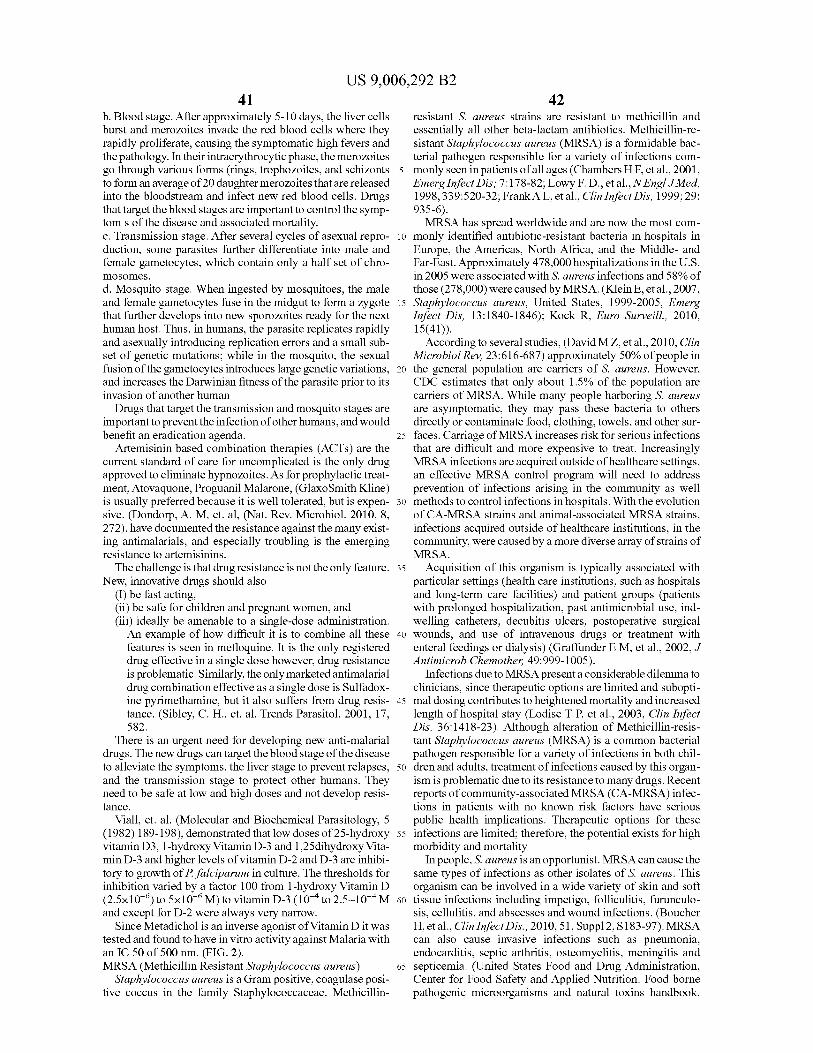
41b. Blood stage. After approximately 5-10 days, the liver cellsburst and merozoites invade the red blood cells where theyrapidly proliferate, causing the symptomatic high fevers andthe pathology. In their intraerythrocytic phase, the merozoitesgo through various forms (rings, trophozoites, and schizontsto form an average of 20 daughter merozoites that are releasedinto the bloodstream and infect new red blood cells. Drugsthat target the blood stages are important to control the symp-tom s of the disease and associated mortality.c. Transmission stage. After several cycles of asexual repro-duction, some parasites further differentiate into male andfemale gametocytes, which contain only a half set of chro-mosomes.d. Mosquito stage. When ingested by mosquitoes, the maleand female gametocytes fuse in the midgut to form a zygotethat further develops into new sporozoites ready for the nexthuman host. Thus, in humans, the parasite replicates rapidlyand asexually introducing replication errors and a small sub-set o f genetic mutations; while in the mosquito, the sexualfusion of the gametocytes introduces large genetic variations,and increases the Darwinian fitness of the parasite prior to itsinvasion of another human
Drugs that target the transmission and mosquito stages areimportant to prevent the infection of other humans, and wouldbenefit an eradication agenda.
Artemisinin based combination therapies (ACTs) are thecurrent standard of care for uncomplicated is the only drugapproved to eliminate hypnozoites. As for prophylactic treat-ment, Atovaquone, Proguanil Malarone, (GlaxoSmith Kline)is usually preferred because it is well tolerated, but is expen-sive. (Dondorp, A. M, et. al, (Nat. Rev. Microbiol. 2010, 8,272). have documented the resistance against the many exist-ing antimalarials, and especially troubling is the emergingresistance to artemisinins
The challenge is that drug resistance is not the only feature.New, innovative drugs should also
(I) be fast acting,(ii) be safe for children and pregnant women, and(iii) ideally be amenable to a single-dose administration.
An example o f how difficult i t is to combine all thesefeatures is seen in mefloquine. It is the only registereddrug effective in a single dose however, drug resistanceis problematic. Similarly, the only marketed antimalarialdrug combination effective as a single dose is Sulfadox-ine pyrimethamine, but it also suffers from drug resis-tance. (Sibley, C. H., et. al. Trends Parasitol. 2001, 17,582.
There is an urgent need for developing new anti-malarialdrugs. The new drugs can target the blood stage of the diseaseto alleviate the symptoms, the liver stage to prevent relapses,and the transmission stage to protect other humans. Theyneed to be safe at low and high doses and not develop resis-tance.
Viall, et. al. (Molecular and Biochemical Parasitology, 5(1982) 189-198), demonstrated that low doses of 25-hydroxyvitamin D3, 1-hydroxy Vitamin D-3 and 1,25dihydroxy Vita-min D-3 and higher levels of vitamin D-2 and D-3 are inhibi-tory to growth of P. falciparum in culture. The thresholds forinhibition varied by a factor 100 from 1-hydroxy Vitamin D(2.5x10⁻⁶) to 5x10⁻⁶ M) to vitamin D-3 (10-4 to 2.5-10-4 Mand except for D-2 were always very narrow.
Since Metadichol is an inverse agonist of Vitamin D it wastested and found to have in vitro activity against Malaria withan IC 50 of 500 nm. (FIG. 2).MRSA (Methicillin Resistant Staphylococcus aureus)
Staphylococcus aureus is a Gram positive, coagulase posi-tive coccus in the family Staphylococcaceae. Methicillin-
US 9,006,292 B242
resistant S. aureus strains are resistant to methicillin andessentially all other beta-lactam antibiotics. Methicillin-re-sistant Staphylococcus aureus (MRSA) is a formidable bac-terial pathogen responsible for a variety of infections com-monly seen inpatients of all ages (Chambers H F, et al., 2001,Emerg Infect Dis; 7:178-82; Lowy F. D., et al., N Engl J Med,1998, 339:520-32; FrankA L, et al., Clin Infect Dis, 1999; 29:935-6).
MRSA has spread worldwide and are now the most com-o monly identified antibiotic-resistant bacteria in hospitals in
Europe, the Americas, North Africa, and the Middle- andFar-East. Approximately 478,000 hospitalizations in the U.S.in 2005 were associated with S. aureus infections and 58% ofthose (278,000) were caused by MRSA. (Klein E, et al., 2007,
15 Staphylococcus aureus, United States, 1999-2005, EmergInfect Dis, 13:1840-1846); Kock R, Euro Surveill., 2010,15(41)).
According to several studies, (David M Z, et al., 2010, ClinMicrobiol Rev, 23:616-687) approximately 50% of people in
20 the general population are carriers o f S. aureus. However,CDC estimates that only about 1.5% o f the population arecarriers of MRSA. While many people harboring S. aureusare asymptomatic, they may pass these bacteria to othersdirectly or contaminate food, clothing, towels, and other sur-
25 faces. Carriage of MRSA increases risk for serious infectionsthat are difficult and more expensive to treat. IncreasinglyMRSA infections are acquired outside of healthcare settings,an effective MRSA control program wi l l need to addressprevention o f infections arising in the community as well
30 methods to control infections in hospitals. With the evolutionof CA-MRSA strains and animal-associated MRSA strains,infections acquired outside of healthcare institutions, in thecommunity, were caused by a more diverse array of strains ofMRSA.
35 A c q u i s i t i o n of this organism is typically associated withparticular settings (health care institutions, such as hospitalsand long-term care facilities) and patient groups (patientswith prolonged hospitalization, past antimicrobial use, ind-welling catheters, decubitis ulcers, postoperative surgical
40 wounds, and use o f intravenous drugs o r treatment wi thenteral feedings or dialysis) (Graffunder E M, et al., 2002, JAntimicrob Chemother, 49:999-1005).
Infections due to MRSA present a considerable dilemma toclinicians, since therapeutic options are limited and subopti-
45 ma l dosing contributes to heightened mortality and increasedlength of hospital stay (Lodise T P. et al., 2003, Clin InfectDis, 36:1418-23). Although alteration o f Methicillin-resis-tant Staphylococcus aureus (MRSA) is a common bacterialpathogen responsible for a variety of infections in both chil-
50 dren and adults, treatment of infections caused by this organ-ism is problematic due to its resistance to many drugs. Recentreports of community-associated MRSA (CA-MRSA) infec-tions in patients with no known risk factors have seriouspublic health implications. Therapeutic options fo r these
55 infections are limited; therefore, the potential exists for highmorbidity and mortality
In people, S. aureus is an opportunist. MRSA can cause thesame types of infections as other isolates of S. aureus. Thisorganism can be involved in a wide variety of skin and soft
60 tissue infections including impetigo, folliculitis, fumnculo-sis, cellulitis, and abscesses and wound infections. (BoucherH, et al., Clin Infect Dis., 2010, 51, Suppl 2, S183-97). MRSAcan also cause invasive infections such as pneumonia,endocarditis, septic arthritis, osteomyelitis, meningitis and
65 septicemia. (United States Food and Drug Administration,Center for Food Safety and Applied Nutrition. Food bornepathogenic microorganisms and natural toxins handbook.
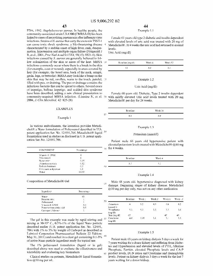
43FDA; 1992. Staphylococcus aureus I n healthy people, thecommunity-associated strain USA300 (CMRSA10) has beenlinked to cases of necrotizing pneumonia after influenza virusinfections. Strains ofS. aureus that carry the exotoxin TSST-1can cause toxic shock syndrome, a life-threatening diseasecharacterized by a sudden onset of high fever, rash, desqua-mation, hypotension and multiple organ failure (Fitzgerald JR, et al., 2001, Proc Nati Acad Sci USA, 98(15): 8821-6). Skininfections caused by S. aureus are generally believed to fol-low colonization o f the skin or nares o f the host. MRSA 10infections commonly occur where there is a break in the skin(for example, a cut or wound), especially in areas covered byhair (for example, the beard area, back of the neck, armpit,groin, legs, or buttocks). MRSA may look like a bump on theskin that may be red, swollen, warm to the touch, painful, 15filled with pus, or draining. The pus or drainage contains theinfectious bacteria that can be spread to others. Several casesof impetigo, bullous impetigo, and scalded skin syndromehave been described, adding a new clinical presentation tocommunity-acquired MRSA infection. (Liassine N, et al.2004, J. Clin Microbiol, 42: 825-28).
Baseline Week 1 Week 2 Week 6 Week 12
Potassium(nurio1/1)
6 5.2 4.3 4.8 4.2
Phosphorus(mg/dI)
5.2 5.2 3.3 3.3 3.6
Bun (mg/dI) 67 47 45Creatinine(mg/dI)
4.6 2.99 3.1 3.1 3.3
Vitamin E TPGSPolicosanolSugar esterPotassium sorbateSodium benzoateCitric acid anhydrousWater
EXAMPLES
Example 1
In various embodiments, the invention provides Metadi-chot t a Nano formulation of Policosanol described in U.S.patent application Ser. No. 12/691,706. Metadicholt liquidformulation used in studies as disclosed in U.S. patent appli-cation Ser. No. 12/691,706.
COMPONENT P e r c e n t a g e
Composition of Metadichott Gel
Ingredient P e r c e n t a g e
WaterSucrose esterPolicosanolVitamin E TPGSPreservatives citric acidCarbopol Polymer
4
0.950.120.20.1
93.63
93113.50.5
The gel in this example was made by rapid stirring andmixing at 30-35° C., 0.5%4% o f the liquid Nano particledescribed earlier (U.S. patent application Ser. No. 12/691,706) with 1% to 3% by weight of Carbopol as described inLubrizol Corporation Pharmaceutical Bulletin 22 Edition:May 31, 2011) and resulted in a clear gel containing 0.1-3%of active Nano particle ingredient ready for topical use.
The 1 % policosanol formulation ( l iqu id o r i n gel )described above was used to evaluate the effectiveness andtolerability and evaluating key biomarkers
Clinical studies on patients, Metadicholt liquid formula-tion g10 mg per ml.
US 9,006,292 B2
Female 61 years old type 2 diabetic and insulin dependentwith elevated levels o f uric acid was treated with 20 mg of
5 Metad icho l t A t 4 weeks the uric acid had returned to normallevels.Uric Acid (mg/di)
Female 60 years old; Diabetic, Type 2 insulin dependent20 w i t h mildly elevated Uric acid levels treated with 20 mg
Metadicholt per day for 24 weeks.
25
30
35
40
45 damage. Beginning stages o f kidney disease. Metadicholg10 mg per day only, was not on any other medication
50
Baseline (mg/dI)
13
55
60
44Example 1.1
Example 1.2
Uric Acid (mg/di)
Example 1.3
Potassium (mmo1/1)
Example 1.4
Example 1.5
Week 4 W e e k 52
4.9 3 . 4
Baseline W e e k 24
8.1 3 . 9
Patient male 66 years o l d hypertensive patient w i thelevated potassium levels treated with Metadicholt g40 mgfor 4 weeks.
Baseline W e e k 4
5.7 4 . 1
Male 68 years old, hypertensive diagnosed with kidney
Patient male 63 years on kidney dialysis 3 days a week for7 years waiting for a donor kidney and suffering from Diabe-tes and Hypertension and elevated levels o f PTH, AlkalinePhosphate, Ferritin. elevated Phosphate levels and CA-P
65 product levels, BUN ratios and Creatinine and Hemoglobinlevels. Patient on kidney dialysis 3 days a week for the last 7years waiting for a donor kidney.

Ferritin (ng/ml)
Week Week We e k WeekBaseline 6 10 14 18
1033 873 1022 672 342
Baseline
Potassium (mmo1/1)
Alkaline Phosphatase (IU/L)
Phosphorus Levels (mg/di)
PTH Levels (pg/ml)
Creatinine (mg/di)
45
5.7 5 . 9 5 . 9
Week 18 W e e k 32
532 4 9 2 3 2 5
Baseline W e e k 10 W e e k 24 W e e k 30 W e e k 48
Baseline W e e k 18 W e e k 30 W e e k 38 W e e k 44
148 1 1 1 8 5 7 4 6 5
Week We e k Week We e k Week24 3 2 3 8 4 0 4 6
212 2 6 1 1 6 5 1 2 5 8 5
CA*P Product Levels. Normal Level Below 55 for DialysisPatients and for Normal Patients 25-40.
Blood Urea Nitrogen (mg/di)
Baseline W e e k 6 W e e k 10 W e e k 18 W e e k 32 W e e k 48
50 4 5 4 0 3 3 3 3 2 1
Week W e e k W e e k W e e k W e e k W e e kBaseline 1 0 1 8 2 5 3 0 3 2 4 8
10.4 9 . 9 8 8 . 9 8 . 7 8 . 7
Hemoglobin Levels (mg/di)
5.1 4 . 3
8.7 1 . 1
12.1 1 0 . 1 1 1 1 1 . 5 1 1 . 7 1 4 . 4
US 9,006,292 B2
Female 61 years insulin dependent diabetic treated with 20mg of Metadicholt per day for 52 weeks.
5 L p ( a ) (mg/di)
10
20
Baseline W e e k 6 W e e k 14 W e e k 24 W e e k 30 W e e k 32 W e e k 48 2 5
6.8 5 . 6 6 . 3 4 . 8 4 . 9 4 . 9 4 . 0
30
35
45
50
60
65
Baseline
Baseline
14
Patient
46Example 1.6
Week 4 W e e k 24 W e e k 52
13 1 0 . 4 8 . 3 5 . 0
Example 1.7
15 M a l e 61 years old insulin dependent diabetic with elevatedLp(a) levels treated with 20 mg Metadicholt for 24 weeksLp (a) (mg/di)
Week 4 W e e k 24
1.9 1 . 2
Example 1.8
Female 55 years old insulin dependent type 2 diabetictreated with 40 mg per day Metadichott for 24 weeks.Lp(a) (mg/di)
Baseline W e e k 24
27 1 0 . 1
Example 1.9
4 0 M a l e 4 2 yearso l d insulindependenttype2 diabeticw i t hWeek Week Week W e e k W e e k We e k e l e v a t e d Lp (a) levels treated with Metadicholt 20 mg per day
Baseline 6 1 0 1 8 2 4 3 2 4 8 f o r 4 w e e k s .
61.20 5 0 . 9 6 5 1 . 0 4 5 1 . 9 2 3 8 . 3 0 4 4 . 6 4 0 . 4 L p (a) ( m g / d i )
Baseline W e e k 4
10.9 3 . 8 9
Example 2.0
Both Patients were type 2 diabetic and insulin dependent.Treated with Metadichott g20 mg for 24 weeks.
55 A p o A Protein Levels (mg/di)
Female 60 7 2 1 4 1Female 55 1 0 2 1 5 5
Baseline W e e k 14 W e e k 32 W e e k 38 W e e k 44 W e e k 48 E x a m p l e 2.1
Baseline W e e k 24
Both Patients were type 2 diabetic and insulin dependent.Treated with Metadichott g20 mg for 24 weeks.

ApoB: Apo A Ratio
Patient Baseline ratio
Male 60 years old 1.1Female 55 years old 1.53Female 60 years old 0.8Male 52 years old 0.74
US 9,006,292 B247 4 8
61 9 9
Example 2.3
47 6 7
Week 24
0.790.830.40.4
Patient Male 58 years old suffering from uncontrolled10 Hypertension for last 15 years unable to control with drugs
Example 2.2 T h a t only made it worse and led to chronic Hepatitis with aelevated SGOT/SGPT ratios was treated with Metadichott
eGFR Levels (mlimin/1.73 M2) f o r 12 weeks at 20 mg per day.Liver Enzymes SGOT/SGPT Ratio
Female 55 years old insulin dependent type 2 diabetic. 1 5
Baseline W e e k 24
eGFR Levels (mlimin/1.73 M2)
Male 67 early stages o f kidney disease was treated withMetadichott 40 mg per day for 24 weeks. Normal for nonAfrican American male is above 60.
Baseline W e e k 24
Example 2.4
eGFR Levels (mlimin/1.73 M2)
Male 68 years old type 2 non insulin diabetic treated withMetadichott 40 mg for 60 weeks.
Example 2.5
eGFR Levels (mlimin/1.73 M2)
62 year old female type 2 non insulin dependent treatedwith Metadicholt g20 mg per day for 60 weeks.
Baseline W e e k 60
Example 2.6
eGFR Levels (mlimin/1.73 M2)
5
20
Patient Male 49 years old, alcoholic, Diabetic, Hyperten-25 sive, Obese and hyperlipidemic and diagnosed as suffering
from post necrotic Cirrhosis with a elevated SGOP/SGPTratio treated with Metadicholt g20 mg per day for 60 weeks.Liver Enzymes SGOT/SGPT Ratio
30
35
Baseline W e e k 604 5
70 1 3 5
50
60
Baseline
Baseline
Baseline W e e k 39
42 8 7
Example 2.7
Week 4 W e e k 12
1.35 1 . 2 1 0 . 7 2
Example 2.8
Week 12 W e e k 60
1.89 0 . 7 2 0 . 6 4
Example 2.9
Male-62 years old was on Statins for 12 years diagnosed40 w i t h a moderately elevated SGOT/SGPT ratio treated with
Metadicholt 20 mg for 48 weeks.Liver Enzymes SGOT/SGPT Ratio
Baseline Week 24 W e e k 48
1.3 1 . 2 0 0 . 7 8
Example 3.0
84 year old Male lung cancer patient after chemotherapyhas elevated SGOT/SGPT ratio. Metadichott treatment for24 weeks at 40 mg per day.
55 L iver Enzymes SGOT/SGPT Ratio
Baseline before L e v e l after 5 M e t a d i c h o l61 1 0 4 C h e m o t h e r a p y d a y s of Chemo t r e a t m e n t for 24 weeks
1.07 1 . 6 7 0 . 7 7
Example 3.165
Female 70, type 2 non insulin dependent diabetic treated F e m a l e 35, diagnosed with Gilbert's syndrome elevatedwith Metadicholt 40 mg for 39 weeks. B i l i r u b i n levels Metadicholt g20 mg per day.

Total Bilimbin (mg/di)
Baseline Week 3 Week 24 Week 84
1.7 1.4 0.6 0.5
D. Bilimbin (mg/di)
5.4Abs. Neutrophil 0.67
Baseline Week 3 Week 24 Week 84
Abs. Neutrophil
Baseline Week 1 Week 3 Week 4
WBC 3.2 4.1 4.8 5.4Abs. Neutrophil 0.67 1.2 1.92 2.04Platelet Count 106000 140000 176000 153000Absolutemonocytes
0.3 0.39 0.70 1.35
Abs. Neutrophil 0.4 0.36 0.43 0.90Abs monocyte 0.019 0.36 0.50 0.46
0.4 0 . 4
Baseline
Baseline
13.2 1 3 . 9
Baseline
13.2 1 3 . 9
49
0.2 0 . 1
Example 3.2
Hemoglobin Levels (mg/di)
Male 59 years Type 2 Insulin dependent Diabetic, withsymptoms of constant fatigue with low Hemoglobin treatedwith Metadicholt 40 mg for 32 weeks.
Patient 69 year old male diagnosed with Neutropenia andextremely low monocyte count was diagnosed as in the begin-ning stages of Leukemia. Treated with Metadichott 40 mgper day while waiting for a matching donor for a bone marrow
W e e k 12 W e e k 32 2 5 t r a n s p l a n t .
13.3 1 4 . 2 1 6
Example 3.3
Hemoglobin Level (g/dl)
Male-59 years old Type 2 diabetic 6 years and chronicallyfatigued treated with Metadicholt 20 mg for 24 weeks
Week 3 W e e k 6 W e e k 24
14.3 1 5
Example 3.4
Hemoglobin Levels (mg/di)
Male 83 Lung cancer patient undergoing chemotherapyMetadichott g40 mg per day.
Baseline H e m o g l o b i n after 5 M e t a d i c h o lbefore Chemo d a y s chemotherapy t r e a t m e n t for 24 weeks
14 1 0 . 7 1 4
Example 3.5
Hemoglobin Level (mg/di)
Type 2 diabetic 6 years and chronically fatigued treatedwith Metadicholt 40 mg for 24 weeks.
Week 3 W e e k 6 W e e k 24
14.3 1 5
US 9,006,292 B2
5
10 B a s e l i n e W e e k 6 W e e k 26
15
20
30
35
50Example 3.6
Platelet Count
Female 64 type 2 diabetic with a low platelet count treatedwith Metadicholt 20 mg daily for 36 weeks.
60 T S H Levels
65
123000
Baseline
TSH (mIU/L) 3 . 1 9
145000 1 8 9 0 0 0
Example 3.7
Blood Count Disorders
Baseline 4 Weeks 6 Weeks 1 2 Weeks
Example 3.8
MDS Patient
65 year-old female with history of breast carcinoma, now40 with diagnosed wi th Myelodysplacia (MDS). Diagnosed
with breast cancer 10 years ago and treatment was surgery,followed by chemo and radiation. Recent bone marrow testshowed hypo-cellular marrow f o r age w i t h progressivehematopoiesis with trilineage dysplastic megakaryocytes and45increase in myeloblasts (-10%), consistent with Myelodys-placia (MDS) with excess of blasts. Diagnosis was that bonemarrow is not functioning and needed chemotherapy andbone marrow transplant. Treated with Metadicholt g10 mg
50 per day. Patient also suffered from hypothyroidism.
55
Week 40 W e e k 52
3.48 0 . 7 2

Fen-Ain Levels
Ca X P product
BaselineWeek
4Week
6Week
8Week
10Week
14Week
20
iPTH (pg/ml) 510 457 386 702 513 736 249RDW (%) 23.1 20.1
4.9
15.1 14.6
3.825 Potassium (nimo1/1)
Ca * P product 38.22 40.94 35.1 37.8 34.8 44.8 34.1Creatinine (mg/dI) 8.37 6.85 7.87 7.47 8.08 7.72 7.39Phosphorus (mg/dI) 4.6 4.2 3.9 4.3 3.9 5.1 3.8Potassium (nimo1/1) 5.2 4.46 5 4 4.2 4.3 4Iron (ug/dI) 49 62 94 93 100 81 93BUN (blood ureaand nitrogen) (mg/dI)
53 54 63 60 50 43
Ca X P product 40.67 36.96 33.6 34
Other Improvements in Key Biomarkers20
14.2 1 2 . 879.9 7 7 . 6
Ca X P Product Levels.
Baseline Week 4 Week 10 Week 24
Creatinine (mg/dI) 7.01 5.65 5.66 3.6Phosphorus (mg/dI) 4.9 4.4 4 3.8
25 Potassium (nimo1/1) 4.4 4 3.3 3.5Iron (UG/DL) 38 63 114 116Hemoglobin (g/dI) 10 11.9 12.3 14
30 Example 4.2
Base- Week Weekline 2 4
Week6
Week We e k Week We e k10 1 4 1 8 2 4
RDW (%) 1 5 . 7 1 5 . 6 1 5 . 6post 9 6 . 6 9 3 . 4 9 0 . 3dialysisweight (kg)
1588.5
14.4 1 4 . 788.6 8 6 . 3
14.2 1 2 . 879.9 7 7 . 6
Ca X P Product Levels.
Baseline Week 2 Week 6 Week 24
51
Ferritin 2 8 4 7 1 1 1 1 8 5 8 1 3 2 6 1 0 5 4 8 4 8 4 0(ng/ml)
Example 3.9
Baseline
Hypothyroidism
Female 74 years o l d diagnosed wi th hypothyroidismtreated with Metadichol® 20 mgs for 24 weeks.TSH (mIU/L)
Example 4.0
Male 83 Lung cancer patient with Hypothyroidism under-going Chemotherapy treated with Metadichol® g40 mg perday.TSH Levels (mIU/L)
Week 4 W e e k 12
7.6 6 . 1 2 . 8 3
Example 4.1
Kidney Disease Patients
Male 62 Male 62 years, obese, hypertensive, type 2 dia-betic and on peritoneal dialysis for 1 year with kidney dis-eases, with symptoms of constant fatigue with low Hemoglo-bin treated with Metadichol® 10 mg per day. Improvementsare noted below.iPTH (Intact Parathyroid Hormone)
Baseline W e e k 6 W e e k 14 w e e k 24
iPTH (pg/ml) 4 8 9 3 4 0 4 9 5 7 6
US 9,006,292 B2
Week W e e k W e e k W e e k We e k WeekBaseline 2 4 6 8 1 0 1 6 5
10
50
52RDW (Red Cell Distribution Width)
15
Baseline W e e k 4 W e e k 12 W e e k 16 2 4 W e e k 32
21.8 1 9 1 0 . 6 6 9 . 0 2 9 . 0 3 7 . 5
Male 70 years on peritoneal dialysis for the past 2 yearswith elevated iPTH, RDW Ferritin, creatinine levels treatedwith Metadichol g10 mg per day. Improvements are notedbelow
Example 4.3
Hashimoto Disease
38 year old male with episodes of syncope associated with55 seizure activity, beginning in 2000(13 years ago when he was
26). Neurological tests were normal. He has been averaging150-155 systolic and 105-110 diastolic for the last 12 years.Was on anti seizure medications for 14 years. but episodes ofseizure continued. In addition he suffered from double vision,
60 auras and episodes of dizziness Most episodes occurred dur-ing night while sleeping. Patient diagnosed with Hashimotoencephalopathy a year ago. Had elevated Thyroid hormone(TSH), elevated T4 and undetectable Thyroglobulin (TgAB)and elevated Thyroglobulin antibodies and Thyroperoxidase
65 antibodies (TP0ab). Treated with Metadichol g 1 0 mg perday. After 4 weeks patient's original symptoms o f doublevision, aura and double vision and dizziness disappeared. His
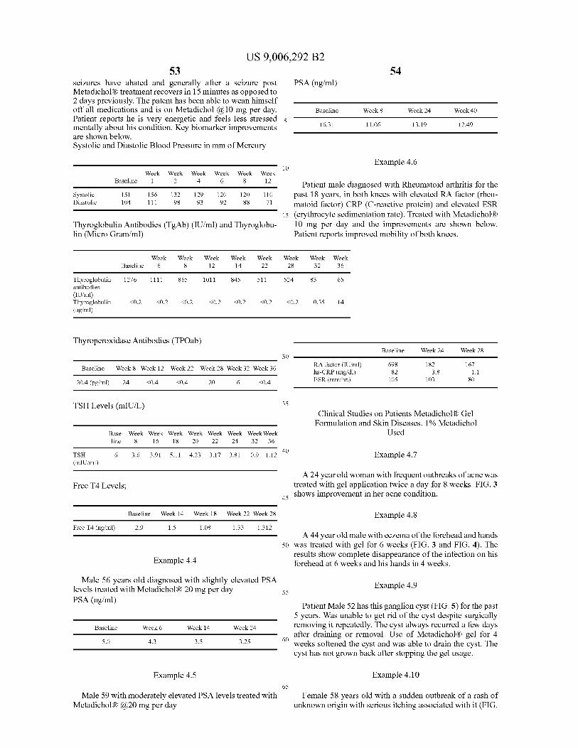
US 9,006,292 B253 5 4
seizures have abated and generally after a seizure post P S A (ng/ml)Metadichott treatment recovers in 15 minutes as opposed to2 days previously. The patent has been able to wean himselfoff all medications and is on Metadichol g 1 0 mg per day. B a s e l i n e W e e k 8 W e e k 24 W e e k 40Patient reports he is very energetic and feels less stressed 5mentally about his condition. Key biomarker improvements 1 6 . 3 1 1 1 . 0 6 1 3 . 1 9 1 2 . 4 9
RA factor (11J/m1)
BaselineWeek
6Week
8Week
12Week
14Week
22Week
28Week
32Week
36
Thyroglobulinantibodies(11J/m1)Thyroglobulin(ug/ml)
1276
<0.2
1111
<0.2
865
<0.2
1011
<0.2
845
<0.2
511
<0.2
524
<0.2
83
0.35
65
14
RA factor (11J/m1) 698 182 167hs-CRP (mg/d1) 82 3.8 1.1ESR (nun/hr.) 105 103 80
Systolic 151 156 132 129 120Diastolic 104 111 98 93 92
are shown below.Systolic and Diastolic Blood Pressure in mm of Mercury
10Week W e e k W e e k W e e k W e e k W e e k
Baseline 1 2 4 6 8 1 2 Patient male diagnosed with Rheumatoid arthritis for the120 1 1 0 p a s t 18 years, in both knees with elevated RA factor (rheu-88 7 1 m a t o i d factor) CRP (C-reactive protein) and elevated ESR
15 (erythrocyte sedimentation rate). Treated with MetadichottThyroglobulin Antibodies (TgAb) (IU/ml) and Thyroglobu- 1 0 mg per day and the improvements are shown below.lin (Micro Gramiml) P a t i e n t reports improved mobility of both knees.
Thyroperoxidase Antibodies (TP0ab)
Baseline W e e k 8 We e k 12 W e e k 22 W e e k 28 Week 32 Week 36
20.4 (pg/ml) 2 4 < 0 . 4 < 0 . 4 2 0 6 < 0 . 4
TSH Levels (mIU/L)
Free T4 Levels;
Free T4 (ng/ml)
Baseline
Base- We e k Week Week We e k Week Week WeekWeekline 8 1 6 1 8 2 0 2 2 2 4 3 2 3 6
Baseline W e e k 14 W e e k 18 W e e k 22 Week 28
2.9 1 . 5
5.0 4 . 3
Example 4.4
1.08 1 . 3 3 1 . 3 1 2
Week 6 W e e k 14 W e e k 24
3.5 3 . 2 5
30
35
40TSH 6 3 . 6 3 . 9 1 5 . 1 1 4 . 2 3 3 . 1 7 3 . 8 1 0 . 9 1 . 1 2(rnIU/m1)
45
Male 56 years old diagnosed with slightly elevated PSAlevels treated with Metadichott 20 mg per day 5 5PSA (ng/ml)
Example 4.6
Baseline W e e k 24 W e e k 28
Clinical Studies on Patients Metadicholt GelFormulation and Skin Diseases. 1% Metadichol
Used
Example 4.7
A 24 year old woman with frequent outbreaks of acne wastreated with gel application twice a day for 8 weeks. FIG. 3shows improvement in her acne condition.
Example 4.8
A 44 year old male with eczema of the forehead and hands50 was treated with gel for 6 weeks (FIG. 3 and FIG. 4). The
results show complete disappearance of the infection on hisforehead at 6 weeks and his hands in 4 weeks.
60
Example 4.9
Patient Male 52 has this ganglion cyst (FIG. 5) for the past5 years. Was unable to get rid of the cyst despite surgicallyremoving it repeatedly. The cyst always recurred a few daysafter draining or removal. Use o f Metadichott gel for 4weeks softened the cyst and was able to drain the cyst. Thecyst has not grown back after stopping the gel usage.
Example 4.5 E x a m p l e 4.1065
Male 59 with moderately elevated PSA levels treated withMetadichott g20 mg per day
Female 58 years old with a sudden outbreak of a rash ofunknown origin with serious itching associated with it (FIG.
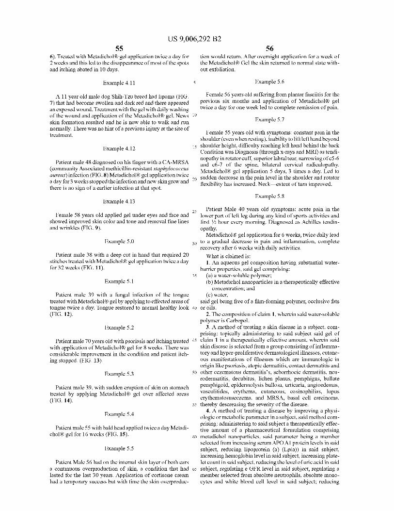
556). Treated with Metadichol8 gel application twice a day for2 weeks and this led to the disappearance of most of the spotsand itching abated in 10 days.
Example 4.11
A 11 year old male dog Shih-Tzu breed had lipoma (FIG.7) that had become swollen and dark red and there appearedan exposed wound. Treatment with the gel with daily washingof the wound and application of the Metadichol8 gel. Newsskin formation resulted and he is now able to walk and runnormally. There was no hint of a previous injury at the site oftreatment.
Example 4.12
Patient male 48 diagnosed on his finger with a CA-MRSA(community Associated methicillin-resistant staphylococcusaureus) infection (FIG. 8) Metadichol8 gel application twicea day for 3 weeks stopped the infection and new skin grew andthere is no sign of a earlier infection at that spot.
Example 4.13
Female 58 years old applied gel under eyes and face andshowed improved skin color and tone and removal fine linesand wrinkles (FIG. 9).
Example 5.0
Patient male 38 with a deep cut in hand that required 20stitches treated with Metadichol8 gel application twice a dayfor 32 weeks (FIG. 11).
Example 5.1
Patient male 39 wi th a fungal infection o f the tonguetreated with Metadichol8 gel by applying to effected areas oftongue twice a day. Tongue restored to normal healthy look(FIG. 12).
Example 5.2
Patient male 70 years old with psoriasis and itching treatedwith application of Metadichol8 gel for 8 weeks. There wasconsiderable improvement in the condition and patient itch-ing stopped. (FIG. 13).
Example 5.3
Patient male 39, with sudden eruption of skin on stomachtreated by applying Metadichol8 gel over affected areas(FIG. 14).
Example 5.4
Patient male 55 with bald head applied twice a day Metadi-chol8 gel for 16 weeks (FIG. 15).
Example 5.5
Patient Male 56 had on the internal skin layer of both earsa continuous overproduction o f skin, a condition that hadlasted for the last 30 years. Application of cortisone creamhad a temporary success but with time the skin overproduc-
US 9,006,292 B2
5
10
56tion would return. After overnight application for a week ofthe Metadichol8 Gel the skin returned to normal state with-out exfoliation.
Example 5.6
Female 56 years old suffering from plantar fasciitis for theprevious six months and application o f Metadichol8 geltwice a day for one week led to complete remission of pain.
Example 5.7
Female 55 years old with symptoms: constant pain in theshoulder (even when resting), inability to lift left hand beyond
15 shoulder height, difficulty reaching left hand behind the backCondition was Diagnosis (through x-rays and MRI) as tendi-nopathy in rotator cuff, superior labral tear, narrowing of c5-6and c6-7 o f the spine, bilateral cervical radiculopathy.Metadichol8 gel application 5 days, 3 times a day. Led to
20 sudden decrease in the pain level in the shoulder and rotatorflexibility has increased. Neck ex tent of turn improved.
Example 5.8
25 P a t i e n t Male 40 years old symptoms: acute pain in thelower part of left leg during any kind of sports activities andfirst 1/2 hour every morning. Diagnosed as Achilles tendin-opathy.
Metadichol8 gel application for 6 weeks, twice daily lead30 t o a gradual decrease in pain and inflammation, complete
recovery after 6 weeks with daily activities.What is claimed is:1. An aqueous gel composition having substantial water-
barrier properties, said gel comprising:35 ( a ) a water-soluble polymer;
(b) Metadichol nanoparticles in a therapeutically effectiveconcentration; and
(c) water,said gel being free of a film-forming polymer, occlusive fats
40 o r oils.2. The composition of claim 1, wherein said water-soluble
polymer is Carbopol.3. A method of treating a skin disease in a subject, com-
prising: topically administering to said subject said gel o f45 c la im 1 in a therapeutically effective amount, wherein said
skin disease is selected from a group consisting of inflamma-tory and hyper-proliferative dermatological illnesses, cutane-ous manifestations o f illnesses which are immunologic inorigin like psoriasis, atopic dermatitis, contact dermatitis and
50 other eczematous dermatitis's, seborrhoeic dermatitis, neu-rodermatitis, decubitus, lichen planus, pemphigus, bullatepemphigoid, epidermolysis bullosa, urticaria, angioedemas,vasculitides, erythema, cutaneous, eosinophilias, lupuserythematosuseczema, and MRSA, basal cell carcinoma,
55 thereby descreasing the severity of the disease.4. A method of treating a disease by improving a physi-
ologic or metabolic parameter in a subject, said method com-prising: administering to said subject a therapeutically effec-tive amount o f a pharmaceutical formulation comprising
60 metadichol nanoparticles, said parameter being a memberselected from increasing serum APO Al protein levels in saidsubject, reducing lipoprotein (a) (Lp(a)) i n said subject,increasing hemoglobin level in said subject, increasing plate-let count in said subject, reducing the level of uric acid in said
65 subject, regulating e GFR level in said subject, regulating amember selected from absolute neutrophils, absolute mono-cytes and white blood cell level in said subject; reducing

US 9,006,292 B257
elevated AST:ALT ratios in said subject, reducing ferritinlevel in said subject, reducing bilimbin level in said subject,regulating thyroid hormone (TSH) level in said subject, andlowering the level of a member selected from PTH, calcium,creatinine, alkaline phosphatase Bun levels and serum phos- 5phoms levels in the subject, thereby regulating said parameterin said subject.
5. The method of claim 4 wherein said disease is kidneydisease.
6. The method of claim 4 wherein said disease is a MRSA i oinfection.
7. The method of claim 4 wherein said disease is a MDS.8. The method of claim 4 wherein said disease is a hema-
tological disease.9. The method of claim 4 wherein said disease is prostate 15
cancer.10. The method of claim 3 wherein said disease is MRSA
infection.11. The method of claim 3 where the disease is Eczema.12. The method of claim 3 wherein the subject is a human. 20


![Ulllted States Patent [19] [11] Patent Number: 6,138,671](https://static.fdokumen.com/doc/165x107/6317fbc571e3f2062906ff7d/ulllted-states-patent-19-11-patent-number-6138671.jpg)