

A preliminary proteomic evaluation of smooth muscle cells in 2 thoracic aortic aneurysms 3 4
37
1 A preliminary proteomic evaluation of smooth muscle cells in 2 thoracic aortic aneurysms 3 4 Ceyda AÇILAN AYHAN 1 , Ph.D., Betül BAYKAL 1,2 , MSc., Müge SERHATLI 1 , MSc., 5 Ömer KAÇAR 1 , MSc., Zelal ADIGÜZEL 1 , MSc., Serpil TAŞ 3 , M.D., Kemal BAYSAL 1,4 , 6 M.D., Ph.D., Ahmet Tarık BAYKAL 5* , Ph.D. 7 8 1 TUBITAK, Marmara Research Center, Genetic Engineering and Biotechnology Institute, 9 41470, Gebze - Kocaeli, TURKEY 10 2 International Centre for Genetic Engineering and Biotechnology, AREA Science Park, 11 34149, Trieste, ITALY 12 3 Turkish Ministry of Health, Kartal Kosuyolu, Advanced Training and Research Hospital, 13 38846, Kartal - Istanbul, TURKEY 14 4 Dokuz Eylul University, Medical Faculty, Department of Biochemistry, 35340, Inciraltı - 15 Izmir, TURKEY 16 5 Istanbul Medipol University, School of Medicine, Department of Medical Biochemistry, 17 34083, Unkapani-Fatih, Istanbul, TURKEY 18 19 * To whom correspondence should be addressed 20 Tel: +90 212 453 4803 21 Fax: +90 212 531 75 55 22
Transcript of A preliminary proteomic evaluation of smooth muscle cells in 2 thoracic aortic aneurysms 3 4

1
A preliminary proteomic evaluation of smooth muscle cells in 2
thoracic aortic aneurysms 3
4
Ceyda AÇILAN AYHAN1, Ph.D., Betül BAYKAL
1,2, MSc., Müge SERHATLI
1, MSc., 5
Ömer KAÇAR1, MSc., Zelal ADIGÜZEL
1, MSc., Serpil TAŞ
3, M.D., Kemal BAYSAL
1,4, 6
M.D., Ph.D., Ahmet Tarık BAYKAL5*
, Ph.D. 7
8
1TUBITAK, Marmara Research Center, Genetic Engineering and Biotechnology Institute, 9
41470, Gebze - Kocaeli, TURKEY 10
2International Centre for Genetic Engineering and Biotechnology, AREA Science Park, 11
34149, Trieste, ITALY 12
3Turkish Ministry of Health, Kartal Kosuyolu, Advanced Training and Research Hospital, 13
38846, Kartal - Istanbul, TURKEY 14
4Dokuz Eylul University, Medical Faculty, Department of Biochemistry, 35340, Inciraltı - 15
Izmir, TURKEY 16
5Istanbul Medipol University, School of Medicine, Department of Medical Biochemistry, 17
34083, Unkapani-Fatih, Istanbul, TURKEY 18
19
*To whom correspondence should be addressed 20
Tel: +90 212 453 4803 21
Fax: +90 212 531 75 55 22
3
1. ABSTRACT: 1
Aortic aneurysms are characterized as localized degeneration of the aorta leading to 2
advanced weakening and widening of the vessel. While the exact mechanisms are yet to be 3
determined, current studies indicate that the degradation of extracellular matrix (ECM) 4
proteins and apoptosis of vascular smooth muscle cells (SMCs) may result in extendibility, 5
dilatation and rupture of the vessel. Within the aortic wall, SMCs are implicated as key 6
components involved in disease development as numerous molecular changes have been 7
reported to occur. Most current studies involve either investigation of proteins comprising a 8
group or pathway in SMCs or analyses in the whole aortic tissue. In order to determine 9
which proteins are important in the development of thoracic aortic aneurysms (TAA), we 10
performed comparative proteomic analyses using cultured SMCs from TAAs versus 11
controls. Label-free nanoLC-MS/MS analysis of cell extracts resulted in the identification 12
of 256 proteins, 26 of which were differentially regulated ≥1.4 fold. Both previously 13
described and new proteins were identified that were involved in oxidative stress, ECM 14
formation, energy metabolism, or 14-3-3 pathway. Among these, differential expression of 15
SerpinH1, a protease inhibitor for collagenases, was further verified via immunoblotting. 16
Here, we attempted to shed light on the cellular mechanisms of TAA. 17
18
Keywords: Label-free proteomics, thoracic aortic aneurysm, smooth muscle cell, 19
SerpinH1/HSP47, oxidative stress, protein expression 20
21
4
2. INTRODUCTION: 1
Aortic aneurysms can be defined as a localized structural degeneration of a part of the aorta 2
which leads to weakening of the wall and its progressive dilatation (MacSweeney et al., 3
1994; Boddy et al., 2008). In time, the weakened and dilated vessel tends to rupture, which 4
is the most dangerous outcome of this disease with a considerably high mortality rate 5
(31.7/100.000 individuals) (Booher and Eagle, 2011; CDC). Unfortunately, today, there are 6
still many undiscovered questions on how aneurysms begin and develop; hence identifying 7
the molecular factors involved in disease development is an active research area. 8
The aortic wall is a dynamic and firmly regulated structure mainly made up of three major 9
types of cells (endothelial cells, fibroblasts and the smooth muscle cells (SMCs)), and the 10
surrounding extracellular matrix (ECM) (Thompson et al., 2002). Currently, it has been 11
reported that vessel dilation is not just a passive enlargement process, but rather a 12
combination of multiple factors including increased degradation and decreased buildup of 13
ECM proteins, and loss of SMCs through apoptosis (Weintraub, 2009; Lindsay and Dietz, 14
2011). During the pathogenesis of aneurysms, inflammation also appears to play a role 15
contributing to the above mentioned processes by the release of cytokines, the increase in 16
the expression matrix metalloproteinases (MMPs), and the increase in oxidative stress 17
resulting in the death of SMCs (Newman et al., 1994; McCormick et al., 2007). Yet, 18
inflammatory aneurysms are frequent in the abdominal, and are very rare in the thoracic 19
aorta, which are mostly characterized with medial degeneration (He et al., 2006). In 20
addition, while abdominal aortic aneurysms (AAAs) present later in life, thoracic aortic 21
aneurysms (TAAs) may be observed at younger ages, indicating the presence of genetic 22
factors (Lindsay and Dietz, 2011). Hence, the two types of aneurysms are different both 23
5
pathologically and mechanistically for the factors that lead to disease development. 1
Two cytoskeletal gene mutations specific to SMCs have been implicated in the formation 2
of nonsyndromic familial TAAs: the SMC-specific beta-myosin (MYH11) gene (Zhu et al., 3
2006) and the alpha-actin (ACTA2) gene (Guo et al., 2007). Mutations in FBN1, TGFßR2, 4
and TGFßR1 have also been linked to cause TAAs, resulting in Marfan’s disease in the first 5
gene and Loeyz-Dietz syndrome in the last two (Milewicz et al., 1996; Pannu et al., 2005; 6
Loeys et al., 2006). Mutations affecting SMC functioning may have serious outcomes. In 7
addition to being a structural component populating the medial layer of the aorta, SMCs 8
have various roles within the vessel including contraction and secretion of ECM molecules 9
(El-Hamamsy and Yacoub, 2009). Furthermore, SMCs link the outside (the ECM) to the 10
inside of the cell (the cytoskeleton) via cell surface receptors (integrins, G-protein coupled 11
receptors and the discoidin domain receptors), regulating cell migration, proliferation, 12
shape or contraction (Alenghat and Ingber, 2002; Berrier and Yamada, 2007; Luo et al., 13
2007). Consecutively, SMCs can sense the mechanical stress and respond by changing the 14
microenvironment through both intra and extracellular arrangements including realignment 15
of stress fibres or cytoskeleton or by the synthesis of new ECM components 16
(mechanotransduction) (Wang et al., 1993; Kim et al., 1999; Alenghat and Ingber, 2002; 17
Parker and Ingber, 2007). Thus, SMCs provide the necessary hemodynamic environment 18
for the proper function of the aortic wall. Indeed, during TAA development, the loss of 19
SMCs through apoptosis results in major changes due to physical absence in the dilated 20
wall as well as differentially regulated gene and protein profiles, i.e. increased MMP 21
expression (Koullias et al., 2004; Schmoker et al., 2007; Phillippi et al., 2009), further 22
contributing to disease development. 23
6
Development of effective strategies for early diagnosis and treatment of aneurysms depends 1
on determining the cell specific changes rather than the whole aortic section (Abdulkareem 2
et al., 2013). Assessing expression from a mixture of cells as in tissue samples carries the 3
risk of missing real differences when genes are differentially regulated in different types of 4
cells, or over interpretation of results when highly expressing cells exist albeit few in 5
number. Hence, studying a single cell type could yield different results and be more 6
informative than studying a mixture of different cell types. 7
In order to have a comprehensive understanding of the molecular changes that lead to TAA 8
development, we concentrated on SMCs and performed a label-free differential proteome 9
analysis by comparing SMCs from TAAs to SMCs from normal non dilated aorta. We were 10
able to identify 26 significant differentially regulated proteins, which were involved in the 11
energy metabolism, protein folding, oxidative stress response, regulatory pathways, 12
cytoskeleton, ECM organization or DNA packaging. Among the proteins that are found to 13
be significantly differentially expressed, we found that SerpinH1 (or HSP47), a human 14
serine protease inhibitor, is down regulated in the TAA samples and may be one of the 15
proteins that contribute to aneurysm development. To our knowledge, this is the first 16
proteomic study focused on vascular SMCs in TAAs and may form a basis on new 17
hypotheses for development of TAAs. 18
19
3. MATERIALS AND METHODS: 20
3.1. Materials: 21
Acetonitrile (LC-MS grade), water (LC-MS grade), dithiothreitol (DTT), Tris, 22
trifluoroacetic acid (TFA), formic acid (FA), iodoacetamide (IAA), sequencing grade 23
7
modified trypsin (proteomic grade) were purchased from Sigma-Aldrich. SDS and 1
Acrylamide-Bis (40%) were from BioRad. Ammonium bicarbonate (NH4HCO3) was from 2
Fluka. RapiGest, an MS compatible detergent and the internal standard MassPREP alcohol 3
dehydrogenase digest, Uniprot Accession # P00330 were from Waters Corp., Milford, MA, 4
USA. 5
6
3.2. Aortic Tissue and Cell Culture: 7
During this study two control samples and 2 TAA samples were used. The study was 8
approved by the ethics committee of Turkish Ministry of Health, Kartal Kosuyolu, 9
Advanced Training and Research Hospital (ethics report number: 23, dated 21-03-2008 and 10
protocol number: 184-04). Informed consents were signed by all participants. Both samples 11
were transported to our laboratory within 12h after surgery and smooth muscle cell 12
extraction procedures were started. In fact, both control samples were obtained at the same 13
time, hence is not likely to pose an expression problem between controls. Similar timelines 14
were also applied for aneurysm samples. 15
SMCs were prepared from thoracic aortic tissue samples or aorta from organ donor 16
cadavers without aneurysm (one donor and one acceptor individual were used as control 17
samples). TAA specimens removed during surgery were transferred to the laboratory in 18
cold PBS solution with Penicillin (10.000 U/ml) and streptomycin (10 mg/ml) (Biol. 19
Industries, Cat # 03-031) and all samples were processed within a few hours of operation. 20
SMCs were isolated using the explant method (Leik et al., 2004). Cells were isolated on the 21
same day the surgery was performed. After the adventitia and endothelial layer of the 22
vessel were removed, the tissue was cut into small pieces (~2 mm2), which were kept at 5% 23
8
CO2, 37ºC for 0.5-1 h on gelatin coated plates for attachment before the addition of culture 1
medium. The medium was changed every 2-3 days, and cells were passaged once ~70-80% 2
confluency was reached. For initial experiments, non-enriched medium formulations 3
(DMEM/F12, Gibco Cat # 32500-035 + 10% fetal bovine serum, Biochrom AG Cat # 4
S0115) were used. For the proteomic analyses, only cells grown in commercially purchased 5
enriched growth medium (Cell Applications, Smooth Muscle Cell Growth Medium) were 6
used from the first day of the isolation process and were always kept in the enriched 7
medium. We routinely run tryphan blue exclusion assay while harvesting cells and the 8
cultures used in this study was performed with >95% viable cells. Cells were harvested 9
when they were 90% confluent. 10
11
3.3. Immunofluorenscence Staining: 12
Cells were fixed at approximately 80% confluency using -20ºC methanol for 10 min at -13
20ºC. Briefly, after washing with 1X PBS, cells were blocked using 1% BSA and stained 14
using anti-alpha smooth muscle actin (Abcam, ab5694, 1/100, 25ºC, 2 h) and anti-rabbit 15
(Invitrogen, Alexa Fluor 546, 1/50, 25ºC, 1 h). Cells were visualized under fluorescence 16
microscope, scored for smooth muscle actin positivity and imaged using the same camera 17
settings. 18
19
3.4. Immunoblotting: 20
20 μg of total cell lysate (prepared as described in sample preparation) was loaded in each 21
well, separated by 10% SDS-PAGE and transferred to 0.22 μm PVDF membranes 22
(Millipore, PSQ #ISEQ00010), which were then blocked for 1 h at 25ºC in TBS (Tris-HCl 23
9
20 mM, pH: 7.4, NaCl 150 mM) containing 3% BSA (Amresco, #0332-100G). The 1
membrane was incubated overnight at 4ºC with anti-SerpinH1 antibodies (1/1000, Abcam, 2
#ab109117) and anti-actin (1/200, pan Ab-5, Labvision, #MS1295-P1) diluted in TBS/0.1% 3
Tween20 (v/v), and for 1 h at 25ºC with secondary antibodies (1:5000, Labvision #TR-001-4
HR, and 1/1250, Pierce, cat #32400). The proteins were detected using the SuperSignal 5
chemiluminescent substrate (Pierce #34080). 6
7
3.5. Sample preparation for analysis: 8
Approximately 250.000 SMCs were scraped from 25 cm2 cell culture flasks when they 9
were between 80-90% conluency and washed twice with cold 50 mM ammonium 10
bicarbonate and lysed with an ultrasonic homogenizor (5 s on, 5 s off, 3 cycles). The 11
mixture was centrifuged at 15.000 rpm and the protein concentration measurement was 12
done for the supernatant based on the Bradford method. 50 µg of total protein extract was 13
transferred to a 1.5 mL eppendorf vial. Proteins were reduced with 5 mM dithiothreitol at 14
60ºC for 15 min, alkylated with 10 mM iodoacetamide in the dark at room temperature for 15
30 min and digested with trypsin (50 µl, 20 ng/µl, Sigma Proteomics Grade) overnight at 16
37ºC. The hydrolysis of the acid-labile MS compatible detergent, RapiGest (Waters Corp., 17
Milford, Maryland, USA), was done by the addition of TFA and ACN to 1% final volume 18
and incubating at 60ºC (600 rpm shaker, 2 h). During Rapigest removal standart internal 19
calibrant digest (alcohol dehydrogenase, uniprot # P00330, Waters Corp., Milford, 20
Maryland, USA) was added to the samples (25 fmol/µl final concentration). The resulting 21
mixtures had 250 ng/µl tryptic peptide mixture. After Rapigest hydrolysis the mixture was 22
centrifuged (15000 rpm, 15 min) and an aliquot was taken into a LC vial for analysis and 23

10
the rest of the tryptic peptides were stored at -80ºC. 1
2
3.6. LC-MS/MS analysis: 3
Each sample was analysed in triplicate to eliminate technical errors. A 2 μl volume of 4
sample (containing 500 ng of tryptic peptide mixture) was loaded on the LC-MS/MS 5
system (nanoACQUITY UPLC coupled to SYNAPT high definition mass spectrometer 6
with nanolockspray ion source, Waters Corp., Milford, MA). Prior to the injection, the 7
columns were equilibrated with 97% mobile phase A (water with 0.1% FA) and 3% mobile 8
phase B (acetonitrile containing 0.1% FA). The column temperature was set to 35ºC. First, 9
peptides were trapped on a nanoACQUITY UPLC Symmetry C18 Trap column (5 µm 10
particle size, 180 µm i.d. × 20 mm length) at 5 μl/min flow rate for 5 min. Peptides were 11
eluted from the trap column by gradient elution onto analytical column (nanoACQUITY 12
UPLC BEH C18 Column, 1.7 µm particle size, 75 µm i.d. × 250 mm length), at 300 nl/min 13
flow rate with a linear gradient from 5 to 40% acetonitrile over 90 min. Data independent 14
acquisition mode (MSE) was carried out by operating the instrument at positive ion V 15
mode, applying the MS and MS/MS functions over 1.5 sec intervals with 6 V low collusion 16
energy and 15-40 V high collusion energy ramp to collect the peptide mass to charge ratio 17
(m/z) and the product ion information to deduce the amino acid sequence. To correct for the 18
mass drift, the internal mass calibrant Glu-fibrinopeptide (500 pmol/µl) was infused every 19
45 sec through the nanolockspray ion source at 300 nl/min flow rate. Peptide signal data 20
between 50-1600 m/z values were collected. 21
22
3.7. LC-MS/MS data processing: 23

11
Tandem mass spectra extraction, charge state deconvolution and deisotoping steps were 1
processed with ProteinLynx Global Server v2.4 (Waters Corp., Milford, MA) and searched 2
with the IDENTITYE algorithm against the homo sapiens reviewed protein database from 3
Uniprot (December 1st 2010, 25690 entiries). Identity
E was set up to search null assuming 4
the digestion enzyme trypsin and searched with a fragment ion mass tolerance of 20 ppm 5
and a parent ion tolerance of 10 ppm. The aminoacid sequence of the internal standard 6
(yeast alcohol dehydrogenase, Uniprot accession # P00330) was included in the FASTA 7
file of the database. The Apex3D data preparation parameters were set to 0.2 min 8
chromatographic peak width, 10000 MS TOF resolution, 150 counts for low energy 9
threshold, 50 counts for elevated energy threshold, and 1200 counts for the intensity 10
threshold. Databank search query was set to minimum 3 fragment ion matches per peptide, 11
minimum 7 fragment ion matches per protein, minimum 1 peptide matches per protein and 12
1 missed cleavage. Carbamidomethyl-cysteine fixed modification and Acetyl N-TERM, 13
deamidation of asparagine and glutamine, oxidation of methionine variable modifications 14
were set. Absolute quantification of the peptides was calculated with the Hi3 functionality 15
of the IDENTITYE system using the spiked known amount of the internal standard. The 16
false positive rate of the IdentityE algorithm is around 3-4% with a randomized database 17
(D'Aguanno et al., 2010). The quantitative analysis was based on the identified proteins, 18
which were detected in 2 out of the 3 technical replicate injections. Normalization of the 19
proteins was achieved against the digest of the internal calibrant P00330. The acquired 20
protein fold changes were used in the IPA analysis (IPA version 8.5). The canonical 21
pathways used to construct the protein-protein interaction map were generated with protein 22
identifications having a p-value < 0.05 and greater than or equal to 40% (≥1.4 fold) 23
12
expression change. The peptide m/z values and sequence information related to the 1
identified proteins are provided in Supplementary Table 1. 2
3
4. RESULTS: 4
4.1. Sample Characterization: 5
SMCs were isolated from aortic tissue via the explant technique (Leik et al., 2004) and 6
maintained under cell culture conditions until growth was observed (Figure 1A). During 7
initial experiments, cells were explanted in DMEM/F12 supplemented with 10% fetal 8
bovine serum. However, our analyses showed that medium formulations that were not 9
enriched (no additional growth factors other than serum) affect the proteome of the cells 10
(Baykal et al, 2013, accepted manuscript in press). Therefore, cells grown in commercially 11
purchased medium that is enriched for growth factors (Cell Applications, Smooth Muscle 12
Cell Growth Medium) were used. Using this medium formulation, we were able to isolate 2 13
control and 2 patient cells and the SMCs were always maintained in the enriched medium 14
from the first day of the isolation process. 15
The mean age of the control subjects were lower than the TAA patients (Table 2). The 16
controls did not have any known cardiovascular diseases and there were no pathological 17
signs of atherosclerosis. The aortic diameter for control individuals was not measured, but 18
was reported to be less than 3 cm. The diameters for patients were 4.75 and 5.7 cm at the 19
time of operation respectively. All subjects were males. 20
The cells were passaged until enough material for the experiment was collected (passage 2 21
or 3, see materials and methods for details). There was no apparent morphological 22
difference in cultured SMCs between control and patient samples (Figure 1B). SMCs were 23
13
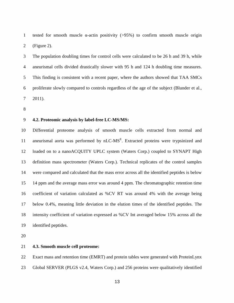
tested for smooth muscle α-actin positivity (>95%) to confirm smooth muscle origin 1
(Figure 2). 2
The population doubling times for control cells were calculated to be 26 h and 39 h, while 3
aneurismal cells divided drastically slower with 95 h and 124 h doubling time measures. 4
This finding is consistent with a recent paper, where the authors showed that TAA SMCs 5
proliferate slowly compared to controls regardless of the age of the subject (Blunder et al., 6
2011). 7
8
4.2. Proteomic analysis by label-free LC-MS/MS: 9
Differential proteome analysis of smooth muscle cells extracted from normal and 10
aneurismal aorta was performed by nLC-MSE. Extracted proteins were trypsinized and 11
loaded on to a nanoACQUITY UPLC system (Waters Corp.) coupled to SYNAPT High 12
definition mass spectrometer (Waters Corp.). Technical replicates of the control samples 13
were compared and calculated that the mass error across all the identified peptides is below 14
14 ppm and the average mass error was around 4 ppm. The chromatographic retention time 15
coefficient of variation calculated as %CV RT was around 4% with the average being 16
below 0.4%, meaning little deviation in the elution times of the identified peptides. The 17
intensity coefficient of variation expressed as %CV Int averaged below 15% across all the 18
identified peptides. 19
20
4.3. Smooth muscle cell proteome: 21
Exact mass and retention time (EMRT) and protein tables were generated with ProteinLynx 22
Global SERVER (PLGS v2.4, Waters Corp.) and 256 proteins were qualitatively identified 23
14
from 36415 EMRTs. Normalization of the absolute peptide intensities was based on the 1
three most intense internal calibrant peptides. Quantitative calculation was processed only 2
for the proteins identified that were detected in two out of three injections. Among the 256 3
proteins; actin, vimentin and desmin were determined as the most abundant proteins in 4
SMCs constituting almost 50% of the whole cell extract and were not depleted from the 5
samples. 62 proteins out of 256 were found to be significantly differentially regulated using 6
PLGS. Among the 62 identified proteins 9 of them had a PLGS identification score below 7
100 and were eliminated after manually re-evaluation of their protein spectra. Furthermore, 8
intensity cut-off was set to 40% and only the proteins that are up or down regulated more 9
than the 40% cut-off is reported. Following the limiting criteria above, we identified 26 10
differentially regulated proteins that were statistically significant (Table 1). 11
12
4.4. Differentially regulated proteins in TAAs: 13
We classified the identified differentially regulated proteins that were detected in all the 14
samples into eight groups as regulatory proteins, skeletal proteins, extracellular matrix 15
related proteins, histones, ones that are involved in protein folding, oxidative stress, energy 16
metabolism and two proteins classified as “others” that did not fall into any of these 17
categories (Table 1). Out of the 26 differentially regulated proteins, 9 were abundant in one 18
group, and below our detection limit in the other. Furthermore, while there was a general 19
downregulation of proteins (15/26), only 3 were upregulated in aneurysm samples. A brief 20
summary of all expression changes is given in Figure 3. 21
22
4.5. Ingenuity Pathway Analysis: 23
15
In order to understand the signal transduction pathways and associated proteins with these 1
differentially expressed proteins, the data were further analyzed using Ingenuity Pathway 2
Analysis (IPA version 8.5) software, which is a knowledge database relying on published 3
literature assessing protein function, localization, relevant interactions and biological 4
mechanisms. Interestingly, most of these proteins (18 out of 26) were merged strongly in a 5
single network (Figure 4A, Table 3) with a score of 49 (score ≥2 is significant; the score 6
indicates the log of the probability of network eligible proteins appearing in a network by 7
random chance. The higher the score, the lower the probability of randomness) The second 8
network had a score of 8 and 4 out of 26 molecules were included in this signaling pathway 9
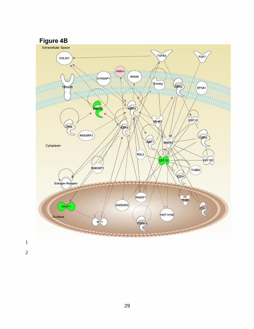
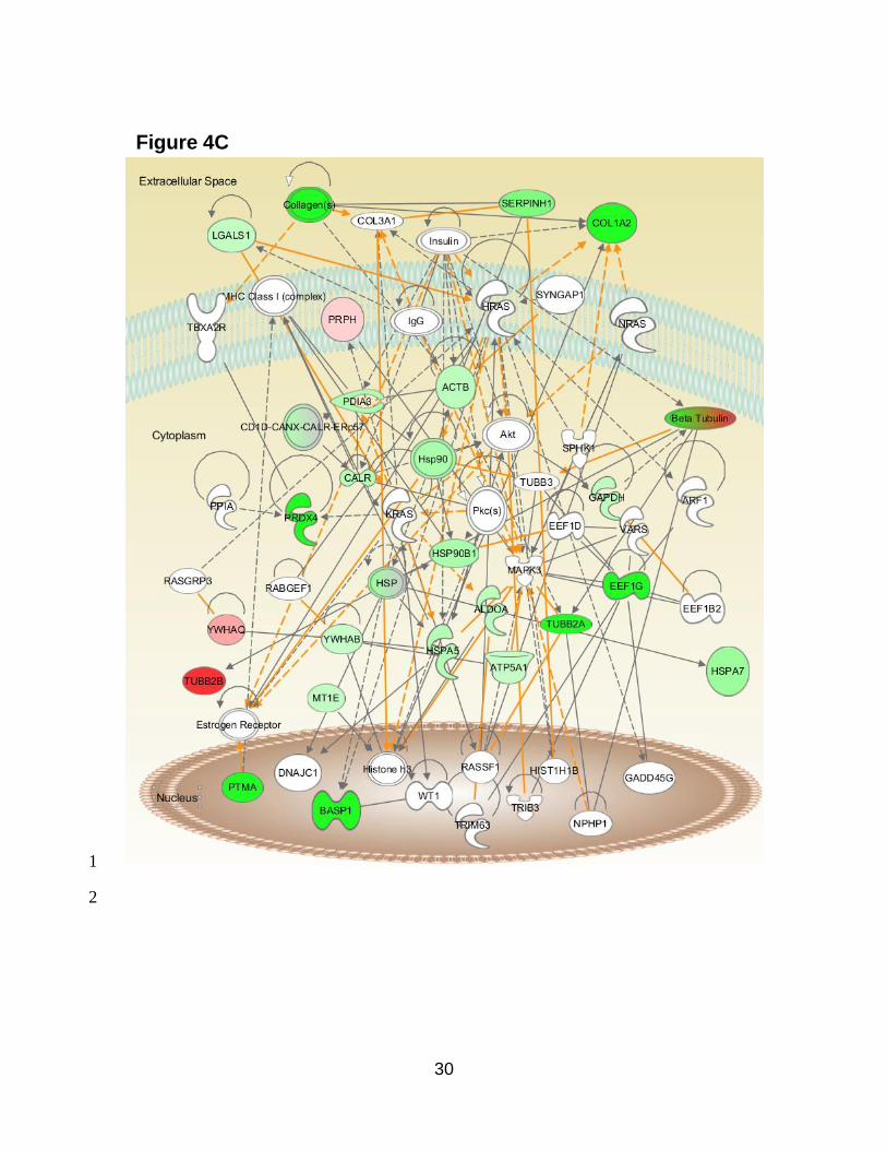
(Figure 4B, Table 3). Figure 4C shows the merged networks 1 and 2. 10
While the top canonical pathway appeared as the 14-3-3 signaling pathway including 11
proteins such as protein disulfide isomerase A3, Tubulin beta 2A and 2B chains, 14-3-3 12
theta and 14-3-3 beta (p: 4.68x10⁻⁷
), 15 molecules were identified that are implicated to 13
play a role in skeletal and muscular disorders. (Actin cytoplasmic 1, Fructose bisphosphate 14
aldolase A, Brain acid soluble protein 1, Calreticulin, Collagen alpha 2 I chain, HUMAN 15
Elongation factor 1 gamma, Glyceraldehyde 3 phosphate dehydrogenase, Endoplasmin, 78 16
kDa glucose regulated protein, Galectin, Tubulin beta 2A and 2B chains, Metallothionein, 17
Protein disulfide isomerase A3, Prothymosin alpha) (p: 1.39x10⁻⁵
). The top ten canonical 18
pathways and biological functions are given in Figure 5. 19
20
4.6. Implication for SerpinH1 downregulation in TAAs: 21
Our analyses showed that one of the downregulated proteins was SerpinH1/HSP47 which 22
belongs to a serine protease inhibitor family and is involved in the correct folding of 23

16
collagens (Nagai et al., 2000). In accordance, the expression of one of the collagens, 1
Collagen A1 (Col1A1), was also reduced in our study (Table 1). Due to its potential role in 2
disease development as a previously unidentified molecule in aneurysms we focused on 3
SerpinH1. Immunoblotting for SerpinH1 confirmed that this protein was downregulated in 4
aneurysm samples (Figure 6). 5
6
5. DISCUSSION: 7
In this study, we aimed to conduct a global comparison of protein expression profiles of 8
SMCs in TAAs to cells of aorta from transplant tissue without aneurysm. Hence, a label-9
free differential proteome analysis of the whole cell extracts was run on a nanoLC-MS/MS 10
setup. Proteomic analysis resulted in identifying 26 differentially expressed proteins greater 11
than 1.4 fold with significance. Importantly, to our knowledge, this is the first report 12
focusing on SMCs in TAAs analyzing the global proteome. 13
14
Although this study was performed in a cell culture model with limited number of samples, 15
we believe the data revealed important results that might be relevant to in vivo alterations. 16
In accordance, proteins involved in main TAA pathological mechanisms (i.e. extracellular 17
matrix, oxidative stress) were identified by LC-MS/MS consistent with previous reports. 18
Furthermore, several differentially expressed proteins are in accordance with literature as 19
10 of the identified proteins were implicated in connective tissue disorders and 15 in 20
skeletal and muscle disorders (IPA analysis) implying that even small number of samples 21
can reveal disease related changes. 22
23
17
Alterations in the ECM components are the key factors in medial degeneration in aortic 1
aneurysms. Collagens, as one of the main components that make up the ECM that is crucial 2
for maintaining the arterial wall structure and stiffness, determines the mechanical 3
properties of the aorta. Our data presents evidence of a reduction in collagen protein levels 4
(Col1A2, Table 1) that is accompanied by a reduction in SerpinH1/HSP47 levels, a protein 5
involved in the correct folding of collagen triple helix (Nagai et al., 2000). Although 6
complete knock out of this gene is embryonically lethal (Nagai et al., 2000), mutations in 7
SerpinH1 have been associated with other connective tissue disorders (Drogemuller et al., 8
2009; Christiansen et al., 2010). The proposed mechanism of action for SerpinH1 is via its 9
interactions with triple helical procollagen molecules that is distinct from protein disulfide 10
isomerase (PDIA3), another molecule observed to be diminished in our study. We 11
confirmed the downregulation of SerpinH1 through Western blotting. Indeed, a larger 12
sample size is necessary to extrapolate SerpinH1 as a biomarker, yet it appears that 13
downregulation of SerpinH1 could be one of the factors leading to TAA development. 14
15
Interestingly, apart from SerpinH1, despite the increased stress within the aorta, SMCs 16
isolated from TAAs expressed less cytoprotective molecules involved in protein folding 17
(Table 1) suggesting that correct folding of cellular proteins might be perturbed, perhaps 18
leading to an accumulation of misfolded proteins that are essential for aortic wall integrity. 19
It has been previously shown that artificial elevation molecular chaperones can exhibit 20
cardioprotection (Marber et al., 1995; Shamaei-Tousi et al., 2007). Thus, the opposite could 21
be true for the reduction in the protein quality control mechanism by downregulation of 22
chaperones, which might directly or indirectly contribute to disease development and 23
18
aneurysm formation. 1
2
To further elucidate how these proteins are regulated and which molecular networks are in 3
play, IPA analysis was performed. Our IPA analyses suggested that one of the pathways 4
that did not previously receive much attention in relation to aneurysm could be the 14-3-3 5
pathway. This regulatory pathway has a large plethora of targets that play a role in 6
regulation of cell cycle, mitogenic signaling and cell death (Gardino and Yaffe, 2011). 7
Supportively, our analyses showed greatly increased population doubling times for TAA 8
SMCs and expression changes in the 14-3-3 pathway proteins could partially be responsible 9
for the observed difference in the cell cycle. Abnormal expression of 14-3-3 proteins and/or 10
dysregulation of 14-3-3/target interactions have been previously reported and were 11
associated with various human diseases (Freeman and Morrison, 2011; Steinacker et al., 12
2011; Zhao et al., 2011), making 14-3-3 proteins a potential target for therapeutic agents 13
(Zhao et al., 2011). Interestingly, similar to TAAs, as a result of a proteomic study in 14
AAAs, many 14-3-3 subunits were proposed as potential biomarkers in many biological 15
samples and were patented for possible uses in diagnosis (Smalley et al., 2009). 16
17
The limitations in this study include the small sample size, the containment of only one 18
gender (males), and the difference in age between the groups at the time of surgery. The 19
low proliferative capacity of SMCs and availability of control tissue have been the major 20
restrictive factors for the small sample size. Despite these limitations, we were able to 21
identify previously reported pathways implying that this study can provide relevant 22
information. However, these results should be confirmed using larger study groups and 23

19
validated by immunoblotting of each protein. Accordingly, this study should be considered 1
a supportive base for new hypotheses. 2
3
In conclusion, SMCs isolated from TAAs differentially express proteins that appear to be 4
potentially important for disease development and were not previously reported for TAAs. 5
In the future, it will also be interesting to see whether these results are valid in vivo, where 6
SMCs are isolated via laser capture microdissection microscopy and further examined. 7
Indeed, studies like this one and others will serve as a database before large clinical studies 8
take place for evaluation of the value of such biomarkers. 9
10
6. LIST OF ABBREVIATIONS: 11
AA: Aortic aneurysm; TAA: Thoracic aortic aneurysm; AAA: Abdominal aortic aneurysm; 12
SMC: Smooth muscle cell; ECM: Extracellular matrix; MMP: Matrix metalloproteinase; 13
IPA: Ingenuity Pathway Analysis 14
15
7. COMPETING INTERESTS: 16
The authors declare that they have no competing interests. 17
18
8. ACKNOWLEDGEMENTS: 19
This work was supported by the European Union 7th Framework Programme Project 20
entitled Fighting Aneurysmal Disease (FAD), Health-2007-2.4.2-2, Grant agreement no: 21
200647 and internal funding through TUBITAK. 22
23
20
9. REFERENCES: 1
Abdulkareem N, Skroblin P, Jahangiri M, Mayr M (2013). Proteomics in Aortic Aneurysm 2
- What Did we Learn so Far? Proteom Clin Appl. doi: 10.1002/prca.201300016 3
Alenghat FJ, Ingber DE (2002). Mechanotransduction: all signals point to cytoskeleton, 4
matrix, and integrins. Sci STKE 2002: pe6. 5
Berrier AL, Yamada KM (2007). Cell-matrix adhesion. J Cell Physiol 213: 565-573. 6
Blunder S, Messner B, Aschacher T, Zeller I, Turkcan A, Wiedemann D, Andreas M, 7
Bluschke G, Laufer G, Schachner T, et al. (2011). Characteristics of TAV- and BAV-8
associated thoracic aortic aneurysms--smooth muscle cell biology, expression profiling, and 9
histological analyses. Atherosclerosis 220: 355-361. 10
Boddy AM, Lenk GM, Lillvis JH, Nischan J, Kyo Y, and Kuivaniemi H (2008). Basic 11
research studies to understand aneurysm disease. Drug News Perspect 21: 142-148. 12
Booher AM, Eagle KA (2011). Diagnosis and management issues in thoracic aortic 13
aneurysm. Am Heart J 162: 38-46 e31. 14
CDC. Centers for Disease Control and Prevention, National Center for Health Statistics. 15
Compressed Mortality File 1999-2008. CDC WONDER Online Database, compiled from 16
Compressed Mortality File 1999-2008 Series 20 No. 2N, 2011. Accessed at 17
http://wonder.cdc.gov/cmf-icd10.html. 18
Christiansen HE, Schwarze U, Pyott SM, AlSwaid A, Al Balwi M, Alrasheed S, Pepin MG, 19
Weis MA, Eyre DR, Byers PH (2010). Homozygosity for a missense mutation in 20
SERPINH1, which encodes the collagen chaperone protein HSP47, results in severe 21
recessive osteogenesis imperfecta. Am J Hum Genet 86: 389-398. 22
D'Aguanno S, D'Alessandro A, Pieroni L, Roveri A, Zaccarin M, Marzano V, De Canio M, 23
21
Bernardini S, Federici G, Urbani A (2010). New insights into neuroblastoma cisplatin 1
resistance: a comparative proteomic and meta-mining investigation. J Proteome Res 10: 2
416-428. 3
Drogemuller C, Becker D, Brunner A, Haase B, Kircher P, Seeliger F, Fehr M, Baumann 4
U, Lindblad-Toh K, Leeb T (2009). A missense mutation in the SERPINH1 gene in 5
Dachshunds with osteogenesis imperfecta. PLoS Genet 5: e1000579. 6
El-Hamamsy I, Yacoub MH (2009). Cellular and molecular mechanisms of thoracic aortic 7
aneurysms. Nat Rev Cardiol 6: 771-786. 8
Freeman AK, Morrison DK (2011). 14-3-3 Proteins: Diverse functions in cell proliferation 9
and cancer progression. Semin Cell Dev Biol 7: 681-7. 10
Gardino AK, Yaffe MB (2011). 14-3-3 proteins as signaling integration points for cell 11
cycle control and apoptosis. Semin Cell Dev Biol 7: 688-95. 12
Guo DC, Pannu H, Tran-Fadulu V, Papke CL, Yu RK, Avidan N, Bourgeois S, Estrera AL, 13
Safi HJ, Sparks E, et al. (2007). Mutations in smooth muscle alpha-actin (ACTA2) lead to 14
thoracic aortic aneurysms and dissections. Nat Genet 39: 1488-1493. 15
He R, Guo DC, Estrera AL, Safi HJ, Huynh TT, Yin Z, Cao SN, Lin J, Kurian T, Buja LM, 16
et al. (2006). Characterization of the inflammatory and apoptotic cells in the aortas of 17
patients with ascending thoracic aortic aneurysms and dissections. J Thorac Cardiov Sur 18
131: 671-678. 19
Kim BS, Nikolovski J, Bonadio J, Mooney DJ (1999). Cyclic mechanical strain regulates 20
the development of engineered smooth muscle tissue. Nat Biotechnol 17: 979-983. 21
Koullias GJ, Ravichandran P, Korkolis DP, Rimm DL, Elefteriades JA (2004). Increased 22
tissue microarray matrix metalloproteinase expression favors proteolysis in thoracic aortic 23
22
aneurysms and dissections. Ann Thorac Surg 78: 2106-2110; discussion 2110-2101. 1
Leik CE, Willey A, Graham MF, Walsh SW (2004). Isolation and culture of arterial smooth 2
muscle cells from human placenta. Hypertension 43: 837-840. 3
Lindsay ME, Dietz HC (2011). Lessons on the pathogenesis of aneurysm from heritable 4
conditions. Nature 473: 308-316. 5
Loeys BL, Schwarze U, Holm T, Callewaert BL, Thomas GH, Pannu H, De Backer JF, 6
Oswald GL, Symoens S, Manouvrier S, et al. (2006). Aneurysm syndromes caused by 7
mutations in the TGF-beta receptor. New Engl J Med 355: 788-798. 8
Luo BH, Carman CV, Springer TA (2007). Structural basis of integrin regulation and 9
signaling. Annu Rev Immunol 25: 619-647. 10
MacSweeney ST, Powell JT, Greenhalgh RM (1994). Pathogenesis of abdominal aortic 11
aneurysm. Brit J Surg 81: 935-941. 12
Marber MS, Mestril R, Chi SH, Sayen MR, Yellon DM, and Dillmann WH (1995). 13
Overexpression of the rat inducible 70-kD heat stress protein in a transgenic mouse 14
increases the resistance of the heart to ischemic injury. J Clin Invest 95: 1446-1456. 15
McCormick ML, Gavrila D, Weintraub NL (2007). Role of oxidative stress in the 16
pathogenesis of abdominal aortic aneurysms. Arterioscler Thromb Vasc Biol 27: 461-469. 17
Milewicz DM, Michael K, Fisher N, Coselli JS, Markello T, Biddinger A (1996). Fibrillin-18
1 (FBN1) mutations in patients with thoracic aortic aneurysms. Circulation 94: 2708-2711. 19
Nagai N, Hosokawa M, Itohara S, Adachi E, Matsushita T, Hosokawa N, Nagata K (2000). 20
Embryonic lethality of molecular chaperone hsp47 knockout mice is associated with 21
defects in collagen biosynthesis. J Cell Biol 150: 1499-1506. 22
Newman KM, Jean-Claude J, Li H, Ramey WG, Tilson MD (1994). Cytokines that activate 23
23
proteolysis are increased in abdominal aortic aneurysms. Circulation 90: II224-227. 1
Pannu H, Fadulu VT, Chang J, Lafont A, Hasham SN, Sparks E, Giampietro PF, Zaleski C, 2
Estrera AL, Safi HJ, et al. (2005). Mutations in transforming growth factor-beta receptor 3
type II cause familial thoracic aortic aneurysms and dissections. Circulation 112: 513-520. 4
Parker KK, Ingber DE (2007). Extracellular matrix, mechanotransduction and structural 5
hierarchies in heart tissue engineering. Philos T Roy Soc B 362: 1267-1279. 6
Phillippi JA, Klyachko EA, Kenny JPt, Eskay MA, Gorman RC, Gleason TG (2009). Basal 7
and oxidative stress-induced expression of metallothionein is decreased in ascending aortic 8
aneurysms of bicuspid aortic valve patients. Circulation 119: 2498-2506. 9
Schmoker JD, McPartland KJ, Fellinger EK, Boyum J, Trombley L, Ittleman FP, Terrien 10
C, 3rd, Stanley A, Howard A (2007). Matrix metalloproteinase and tissue inhibitor 11
expression in atherosclerotic and nonatherosclerotic thoracic aortic aneurysms. J Thorac 12
Cardiov Sur 133: 155-161. 13
Shamaei-Tousi A, Halcox JP, Henderson B (2007). Stressing the obvious? Cell stress and 14
cell stress proteins in cardiovascular disease. Cardiovasc Res 74: 19-28. 15
Smalley DM, Harthun NL, Ley KF, Sarembock IJ, Little K (2009). Protein-based 16
biomarkers for abdominal aortic aneurysm (USA, University Of Virginia Patent 17
Foundation). 18
Steinacker P, Aitken A, Otto M (2011). 14-3-3 proteins in neurodegeneration. Semin Cell 19
Dev Biol 7: 696-704. 20
Thompson RW, Geraghty PJ, Lee JK (2002). Abdominal aortic aneurysms: basic 21
mechanisms and clinical implications. Curr Prob Surg 39: 110-230. 22
Wang N, Butler JP, Ingber DE (1993). Mechanotransduction across the cell surface and 23

24
through the cytoskeleton. Science 260: 1124-1127. 1
Weintraub NL (2009). Understanding abdominal aortic aneurysm. New Engl J Med 361: 2
1114-1116. 3
Zhao J, Meyerkord CL, Du Y, Khuri FR, Fu H (2011). 14-3-3 proteins as potential 4
therapeutic targets. Semin Cell Dev Biol 7: 705-12. 5
Zhu L, Vranckx R, Khau Van Kien P, Lalande A, Boisset N, Mathieu F, Wegman M, 6
Glancy L, Gasc JM, Brunotte F, et al. (2006). Mutations in myosin heavy chain 11 cause a 7
syndrome associating thoracic aortic aneurysm/aortic dissection and patent ductus 8
arteriosus. Nat Genet 38: 343-349. 9
10
25
10. FIGURES and TABLES : 1 Figure 1
A
Control 1 Control 2
Patient 1 Patient 2
B
Tissue
2
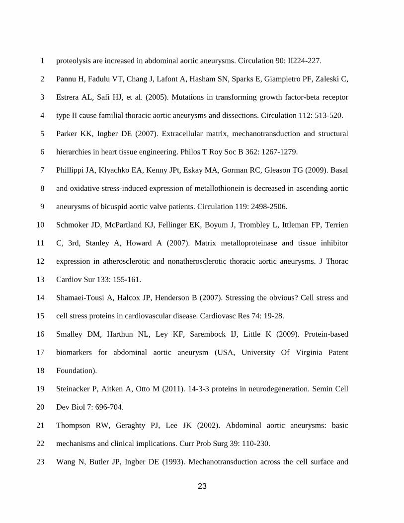
Figure 1: A) Smooth muscle cells were isolated using the explant technique: A 3
representative phase contrast image is shown in the picture with the tissue seen dark and the 4
cells growing around. (10X magnification). B) Morphological appearance of SMCs used in 5
this study. Top panel shows images from control samples, bottom panel shows images from 6
TAA samples, the identities are labeled on the images. Phase contrast microscopy, 10X 7
magnification 8
26
Control 2Control 1
Figure 2
Patient 1 Patient 2
1
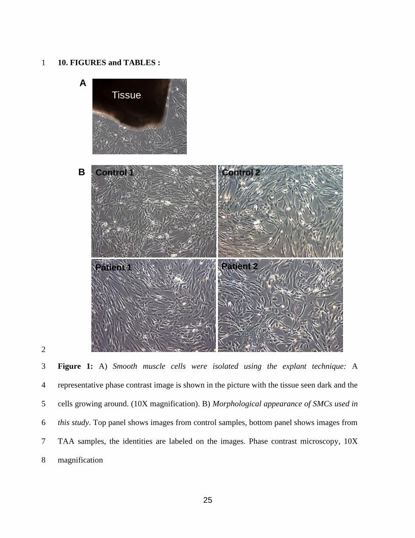
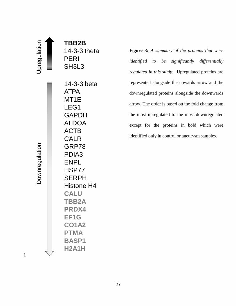
Figure 2: α-actin staining of smooth muscle cells used in this study: Cells were 2
characterized by α-actin positivity for smooth muscle origin. >95% positivity was 3
observed. Fluorescent images were taken with Leica DMI 6000 microscope and Hitachi 3-4
CCD HV-D20P color camera using the same settings and exposure times (40X 5
magnification). 6
7
27
TBB2B
14-3-3 theta
PERI
SH3L3
14-3-3 beta
ATPA
MT1E
LEG1
GAPDH
ALDOA
ACTB
CALR
GRP78
PDIA3
ENPL
HSP77
SERPH
Histone H4
CALU
TBB2A
PRDX4
EF1G
CO1A2
PTMA
BASP1
H2A1H
Up
reg
ula
tion
Dow
nre
gula
tion
Figure 3
1
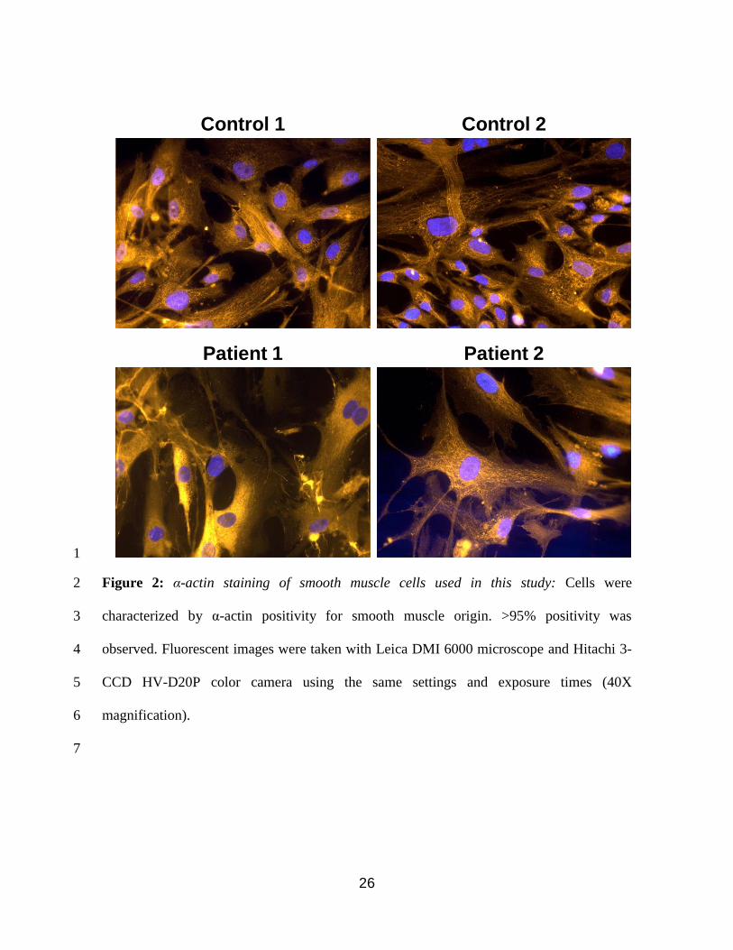
Figure 3: A summary of the proteins that were
identified to be significantly differentially
regulated in this study: Upregulated proteins are
represented alongside the upwards arrow and the
downregulated proteins alongside the downwards
arrow. The order is based on the fold change from
the most upregulated to the most downregulated
except for the proteins in bold which were
identified only in control or aneurysm samples.

28
Figure 4A
1
2
29
1
2
30
Figure 4C
1
2
31
IPA Legends
Figure 4D
1
2
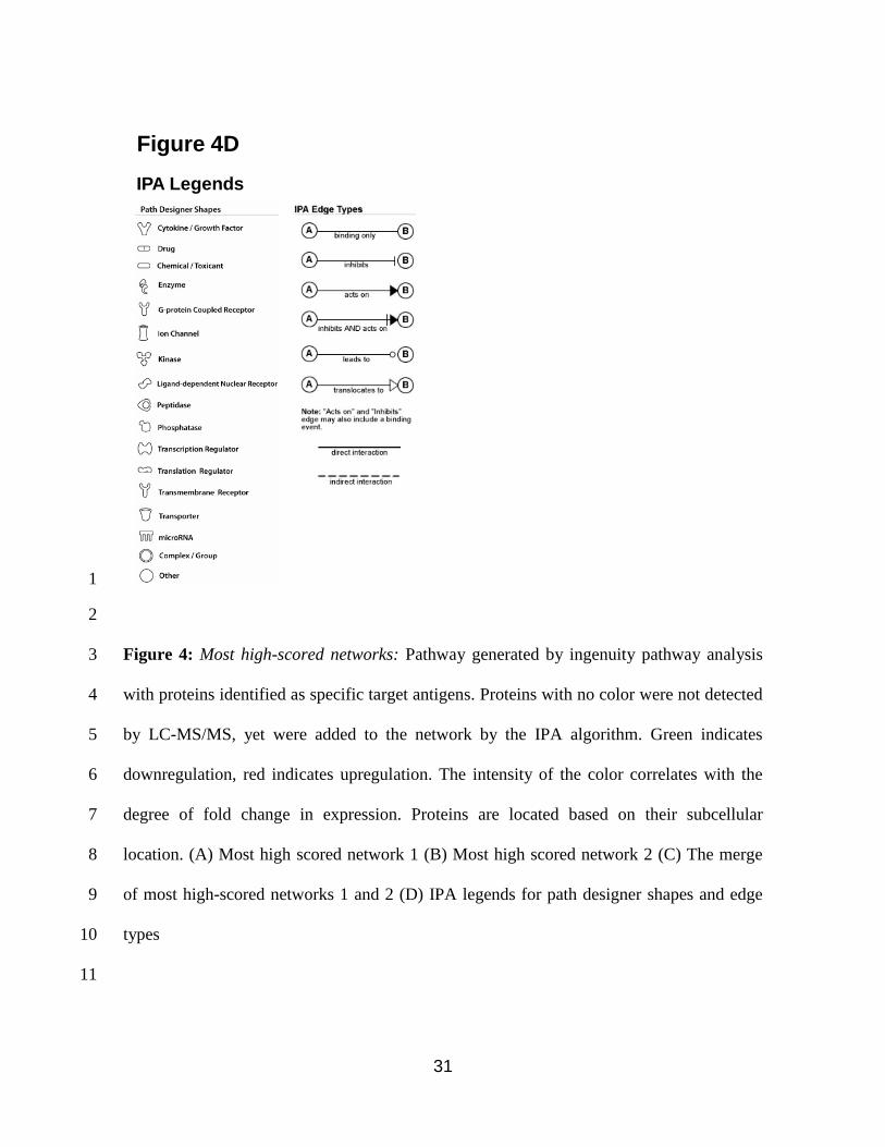
Figure 4: Most high-scored networks: Pathway generated by ingenuity pathway analysis 3
with proteins identified as specific target antigens. Proteins with no color were not detected 4
by LC-MS/MS, yet were added to the network by the IPA algorithm. Green indicates 5
downregulation, red indicates upregulation. The intensity of the color correlates with the 6
degree of fold change in expression. Proteins are located based on their subcellular 7
location. (A) Most high scored network 1 (B) Most high scored network 2 (C) The merge 8
of most high-scored networks 1 and 2 (D) IPA legends for path designer shapes and edge 9
types 10
11
32
Molecules IdentifiedA
Top Canonical Pathways
# Molecules Identified
5 PDIA3, TUBB2A, TUBB2B, YWHAB, YWHAQ
4 HSP90B1, HSPA5, HSP7, PDIA3
2 CALR, PDIA3
3 HSP90B1, YWHAB, YWHAQ
3 PDIA3, YWHAB, YWHAQ
3 ACTB, TUBB2A, TUBB2B,
2 CALR, PDIA3
2 YWHAB, YWHAQ
2 YWHAB, YWHAQ
2 YWHAB, YWHAQ
Figure 5
BTop Functions
# Molecules Identified
5 ACTB, ALDOA, HSPA5, PDIA3, TUBB2A
13 ACTB, ALDOA, ATP5A1, CALR, COL1A2, EEF1G, HSP90B1, HSPA5, LGALS1, PDIA3, PTMA, TUBB2A, YWHAQ
12 ACTB, ALDOA, CALR, EEF1G, GAPDH, HSP90B1, HSPA5, LGALS1, MT1E, PDIA3, PTMA, TUBB2A
7 ACTB, ALDOA, GAPDH, HSPA5, HSP90B1, PDIA3, TUBB2A
5 CALR, HSP90B1, HSPA5, MT1E, PRPH
11 CALR, HSP90B1, HSPA5, SERPINH1, ALDOA, COL1A2, LGALS1, PRPH, MT1E, PTMA, GAPDH
15 ACTB, ALDOA, BASP1, CALR, COL1A2, EEF1G, GAPDH, HSP90B1, HSPA5, LGALS1, TUBB2A, TUBB2B, MT1E, PDIA3, PTMA
4 CALR, GAPDH, HSP90B1, HSPA5
5 CALR, EEF1G, GAPDH, HSP90B1, HSPA5
9 CALR, COL1A2, GAPDH, HSP90B1, HSPA5, LGALS1, TUBB2A, YWHAB, YWHAQ
1
2
3
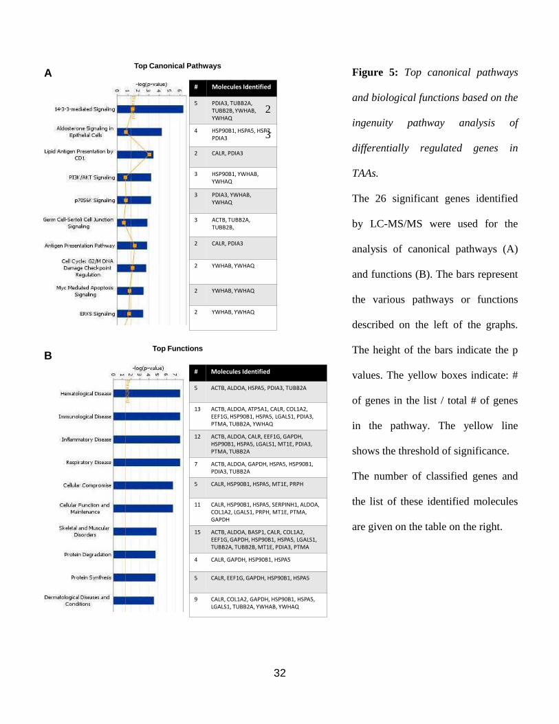
Figure 5: Top canonical pathways
and biological functions based on the
ingenuity pathway analysis of
differentially regulated genes in
TAAs.
The 26 significant genes identified
by LC-MS/MS were used for the
analysis of canonical pathways (A)
and functions (B). The bars represent
the various pathways or functions
described on the left of the graphs.
The height of the bars indicate the p
values. The yellow boxes indicate: #
of genes in the list / total # of genes
in the pathway. The yellow line
shows the threshold of significance.
The number of classified genes and
the list of these identified molecules
are given on the table on the right.

33
-actin
Serpin H1
Figure 6
Control 1 Control 2Patient 1 Patient 2
1
2
Figure 6: SerpinH1/Hsp47 expression: The sample identity in each lane is indicated above 3
the gel picture. The reduction in SerpinH1/Hsp47 expression in patient SMCs was 4
confirmed via Western blotting. α-actin was used as loading control. 5

34
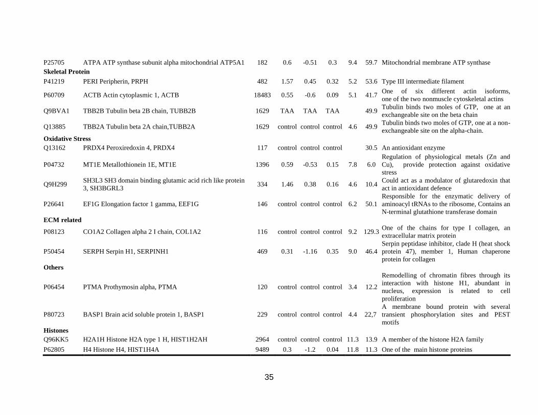
Table 1: Proteins identified to be differentially expressed in this study. Ratio represents the absolute intensities of the aneurismal
samples divided by the control samples. Values below 1 indicate down regulation and values above 1 indicate upregulation in the
aneurismal samples. Control protein was only detected in control samples, Aneurysm protein was only detected in
aneurysm samples. Log(e)Ratio and Log(e)StdDev are natural logarithm values and standard deviations for the ratio of a given
protein. pI is the isoelectric point and MW is the molecular weight of proteins. The functions may include statements from
proteins database such as UniProt.
Accession Description Score Ratio Log(e)
Ratio
Log(e)
StdDev pI
MW
(kDa) Molecular Function
Protein folding
P48741 HSP77 Putative heat shock 70 kDa protein 7, HSPA7 235 0.41 -0.88 0.68 7.9 40.2 A member of the Hsp70 family of heat shock
proteins
P30101 PDIA3 Protein disulfide isomerase A3, PDIA3 574 0.5 -0.7 0.08 5.9 56.7
ECM-related, catalyzes protein folding, prolyl
4-hydroxylase, a highly abundant isomerase
multifunctional enzyme that belongs to the
protein precursor disulfide isomerase family
P14625 ENPL Endoplasmin, HSP90B1/GRP94 572 0.47 -0.76 0.08 4.6 92.4 A heat shock 90Kda protein
O43852 CALU Calumenin CALU 122 control control control 4.5 37.1 Involved in ER functions such as protein
folding and sorting
P27797 CALR Calreticulin CALR 387 0.51 -0.68 0.1 4.1 48.1
Binds to misfolded proteins, prevents export
from the Endoplasmic reticulum to the Golgi
apparatus
P11021 GRP78 78 kDa glucose regulated protein HSPA5 1479 0.51 -0.67 0.06 4.9 72.3 Heat shock 70 kDa protein 5
Regulatory Proteins
P27348 1433T 14 3 3 protein theta YWHAQ 341 2.34 0.85 0.31 4.5 27.8 Regulatory protein, mediate signal
transduction
P31946 1433B 14 3 3 protein beta alpha YWHAB 472 0.61 -0.49 0.15 4.6 28.0 Regulatory protein, mediate signal
transduction
P09382 LEG1 Galectin 1 LGALS1 1506 0.58 -0.54 0.06 5.1 14.7 Beta-galactoside-binding protein, modulates
cell-cell and cell-matrix interactions
Energy Metabolism
P04406 G3P Glyceraldehyde 3 phosphate dehydrogenase GAPDH 6442 0.55 -0.6 0.04 8.7 36.0 Catalyses 6th step of glycolysis
P04075 ALDOA Fructose bisphosphate aldolase A, ALDOA 2195 0.55 -0.59 0.07 8.1 39.4 Catalyses a reverse aldol reaction
35
P25705 ATPA ATP synthase subunit alpha mitochondrial ATP5A1 182 0.6 -0.51 0.3 9.4 59.7 Mitochondrial membrane ATP synthase
Skeletal Protein
P41219 PERI Peripherin, PRPH 482 1.57 0.45 0.32 5.2 53.6 Type III intermediate filament
P60709 ACTB Actin cytoplasmic 1, ACTB 18483 0.55 -0.6 0.09 5.1 41.7 One of six different actin isoforms,
one of the two nonmuscle cytoskeletal actins
Q9BVA1 TBB2B Tubulin beta 2B chain, TUBB2B 1629 TAA TAA TAA 49.9 Tubulin binds two moles of GTP, one at an
exchangeable site on the beta chain
Q13885 TBB2A Tubulin beta 2A chain,TUBB2A 1629 control control control 4.6 49.9 Tubulin binds two moles of GTP, one at a non-
exchangeable site on the alpha-chain.
Oxidative Stress
Q13162 PRDX4 Peroxiredoxin 4, PRDX4 117 control control control 30.5 An antioxidant enzyme
P04732 MT1E Metallothionein 1E, MT1E 1396 0.59 -0.53 0.15 7.8 6.0
Regulation of physiological metals (Zn and
Cu), provide protection against oxidative
stress
Q9H299 SH3L3 SH3 domain binding glutamic acid rich like protein
3, SH3BGRL3 334 1.46 0.38 0.16 4.6 10.4
Could act as a modulator of glutaredoxin that
act in antioxidant defence
P26641 EF1G Elongation factor 1 gamma, EEF1G 146 control control control 6.2 50.1
Responsible for the enzymatic delivery of
aminoacyl tRNAs to the ribosome, Contains an
N-terminal glutathione transferase domain
ECM related
P08123 CO1A2 Collagen alpha 2 I chain, COL1A2 116 control control control 9.2 129.3 One of the chains for type I collagen, an
extracellular matrix protein
P50454 SERPH Serpin H1, SERPINH1 469 0.31 -1.16 0.35 9.0 46.4
Serpin peptidase inhibitor, clade H (heat shock
protein 47), member 1, Human chaperone
protein for collagen
Others
P06454 PTMA Prothymosin alpha, PTMA 120 control control control 3.4 12.2
Remodelling of chromatin fibres through its
interaction with histone H1, abundant in
nucleus, expression is related to cell
proliferation
P80723 BASP1 Brain acid soluble protein 1, BASP1 229 control control control 4.4 22,7
A membrane bound protein with several
transient phosphorylation sites and PEST
motifs
Histones
Q96KK5 H2A1H Histone H2A type 1 H, HIST1H2AH 2964 control control control 11.3 13.9 A member of the histone H2A family
P62805 H4 Histone H4, HIST1H4A 9489 0.3 -1.2 0.04 11.8 11.3 One of the main histone proteins

36
Table 2: Patients’ demographic and clinical properties. NA: Not available.
Gender Age Hypertension Diabetus mellitus Smoking
Patient 1 M 40 1 0 1
Patient 2 M 63 1 1 1
Control 1 M 28 NA NA NA
Control 2 M 30 NA NA NA
37
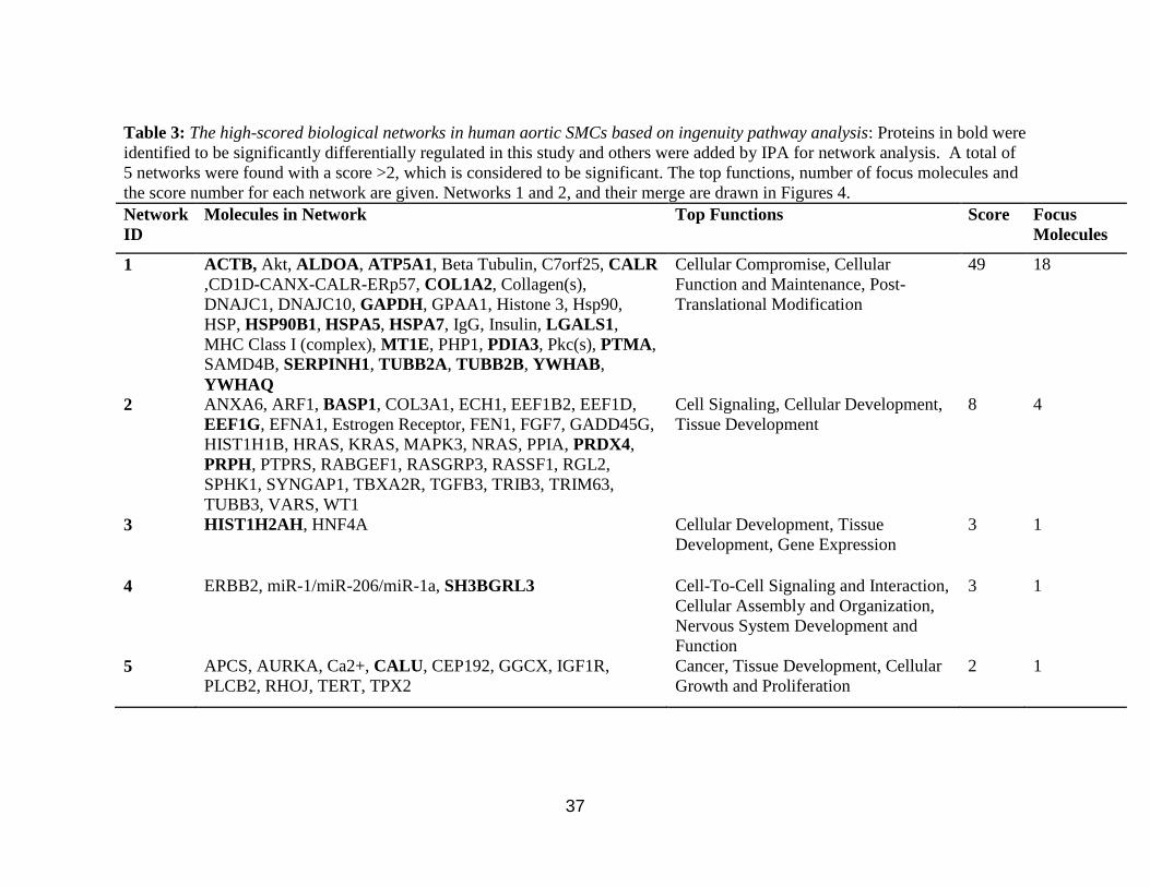
Table 3: The high-scored biological networks in human aortic SMCs based on ingenuity pathway analysis: Proteins in bold were
identified to be significantly differentially regulated in this study and others were added by IPA for network analysis. A total of
5 networks were found with a score >2, which is considered to be significant. The top functions, number of focus molecules and
the score number for each network are given. Networks 1 and 2, and their merge are drawn in Figures 4.
Network
ID
Molecules in Network Top Functions Score Focus
Molecules
1 ACTB, Akt, ALDOA, ATP5A1, Beta Tubulin, C7orf25, CALR
,CD1D-CANX-CALR-ERp57, COL1A2, Collagen(s),
DNAJC1, DNAJC10, GAPDH, GPAA1, Histone 3, Hsp90,
HSP, HSP90B1, HSPA5, HSPA7, IgG, Insulin, LGALS1,
MHC Class I (complex), MT1E, PHP1, PDIA3, Pkc(s), PTMA,
SAMD4B, SERPINH1, TUBB2A, TUBB2B, YWHAB,
YWHAQ
Cellular Compromise, Cellular
Function and Maintenance, Post-
Translational Modification
49 18
2 ANXA6, ARF1, BASP1, COL3A1, ECH1, EEF1B2, EEF1D,
EEF1G, EFNA1, Estrogen Receptor, FEN1, FGF7, GADD45G,
HIST1H1B, HRAS, KRAS, MAPK3, NRAS, PPIA, PRDX4,
PRPH, PTPRS, RABGEF1, RASGRP3, RASSF1, RGL2,
SPHK1, SYNGAP1, TBXA2R, TGFB3, TRIB3, TRIM63,
TUBB3, VARS, WT1
Cell Signaling, Cellular Development,
Tissue Development
8 4
3 HIST1H2AH, HNF4A Cellular Development, Tissue
Development, Gene Expression
3 1
4 ERBB2, miR-1/miR-206/miR-1a, SH3BGRL3 Cell-To-Cell Signaling and Interaction,
Cellular Assembly and Organization,
Nervous System Development and
Function
3 1
5 APCS, AURKA, Ca2+, CALU, CEP192, GGCX, IGF1R,
PLCB2, RHOJ, TERT, TPX2
Cancer, Tissue Development, Cellular
Growth and Proliferation
2 1