Zinc induces disorder-to-order transitions in free and membrane-associated Thellungiella salsuginea...
18
ORIGINAL ARTICLE Zinc induces disorder-to-order transitions in free and membrane-associated Thellungiella salsuginea dehydrins TsDHN-1 and TsDHN-2: a solution CD and solid-state ATR-FTIR study Luna N. Rahman • Vladimir V. Bamm • Janine A. M. Voyer • Graham S. T. Smith • Lin Chen • Mahmoud W. Yaish • Barbara A. Moffatt • John R. Dutcher • George Harauz Received: 14 June 2010 / Accepted: 21 September 2010 Ó Springer-Verlag 2010 Abstract Dehydrins are intrinsically unstructured pro- teins that are expressed in plants experiencing extreme environmental conditions such as drought or low temper- ature. Although their role is not completely understood, it has been suggested that they stabilize proteins and mem- brane structures during environmental stress and also sequester metals such as zinc. Here, we investigate two dehydrins (denoted as TsDHN-1 and TsDHN-2) from Thellungiella salsuginea. This plant is a crucifer that thrives in the Canadian sub-Arctic (Yukon Territory) where it grows on saline-rich soils and experiences periods of both extreme cold and drought. We show using circular dichroism and attenuated total reflection-Fourier transform infrared spectroscopy that ordered secondary structure is induced and stabilized in these proteins, both in free and vesicle-bound form, by association with zinc. In mem- brane-associated form, both proteins have an increased proportion of b-strand conformation induced by the cation, in addition to the amphipathic a-helices formed by their constituent K-segments. These results support the hypoth- esis that dehydrins stabilize plant plasma and organellar membranes in conditions of stress, and further that zinc may be an important co-factor in stabilization. Whereas dehydrins in the cytosol of a plant cell undergoing dehy- dration or temperature stress form bulk hydrogels and remain primarily disordered, dehydrins with specific membrane- or protein-associations will have induced ordered secondary structures. Keywords Dehydrins Late embryogenesis abundant (LEA) Cold tolerance Drought tolerance Intrinsically-disordered protein Induced folding Poly-proline type II CD spectroscopy FTIR spectroscopy ATR-FTIR spectroscopy Abbreviations ATR Attenuated total reflection CD Circular dichroism spectroscopy Chol Cholesterol TsDHN-1 Acidic Thellungiella salsuginea dehydrin 1 TsDHN-2 Basic Thellungiella salsuginea dehydrin 2 ddH 2 O Distilled, deionised water DGDG Digalactosyldiacylglycerol DMPG 1, 2-dimyristoyl-sn-glycero-3-[phospho-rac- (1-glycerol)] FTIR Fourier transform infrared HCA Hydrophobic cluster analysis IDP Intrinsically disordered protein L. N. Rahman V. V. Bamm J. A. M. Voyer G. S. T. Smith L. Chen M. W. Yaish G. Harauz (&) Department of Molecular and Cellular Biology, University of Guelph, 50 Stone Road East, Guelph, ON N1G 2W1, Canada e-mail: [email protected] L. N. Rahman V. V. Bamm J. A. M. Voyer G. S. T. Smith L. Chen J. R. Dutcher G. Harauz Biophysics Interdepartmental Group, University of Guelph, Guelph, ON N1G 2W1, Canada L. Chen J. R. Dutcher Department of Physics, University of Guelph, Guelph, ON N1G 2W1, Canada M. W. Yaish B. A. Moffatt Department of Biology, University of Waterloo, Waterloo, ON N2L 3G1, Canada Present Address: M. W. Yaish Department of Biology, College of Science, Sultan Qaboos University, 123 Muscat, P.O. Box 36, Muscat, Oman 123 Amino Acids DOI 10.1007/s00726-010-0759-0
Transcript of Zinc induces disorder-to-order transitions in free and membrane-associated Thellungiella salsuginea...

ORIGINAL ARTICLE
Zinc induces disorder-to-order transitions in freeand membrane-associated Thellungiella salsuginea dehydrinsTsDHN-1 and TsDHN-2: a solution CD and solid-state ATR-FTIRstudy
Luna N. Rahman • Vladimir V. Bamm • Janine A. M. Voyer • Graham S. T. Smith •
Lin Chen • Mahmoud W. Yaish • Barbara A. Moffatt • John R. Dutcher •
George Harauz
Received: 14 June 2010 / Accepted: 21 September 2010
� Springer-Verlag 2010
Abstract Dehydrins are intrinsically unstructured pro-
teins that are expressed in plants experiencing extreme
environmental conditions such as drought or low temper-
ature. Although their role is not completely understood, it
has been suggested that they stabilize proteins and mem-
brane structures during environmental stress and also
sequester metals such as zinc. Here, we investigate two
dehydrins (denoted as TsDHN-1 and TsDHN-2) from
Thellungiella salsuginea. This plant is a crucifer that
thrives in the Canadian sub-Arctic (Yukon Territory) where
it grows on saline-rich soils and experiences periods of
both extreme cold and drought. We show using circular
dichroism and attenuated total reflection-Fourier transform
infrared spectroscopy that ordered secondary structure is
induced and stabilized in these proteins, both in free and
vesicle-bound form, by association with zinc. In mem-
brane-associated form, both proteins have an increased
proportion of b-strand conformation induced by the cation,
in addition to the amphipathic a-helices formed by their
constituent K-segments. These results support the hypoth-
esis that dehydrins stabilize plant plasma and organellar
membranes in conditions of stress, and further that zinc
may be an important co-factor in stabilization. Whereas
dehydrins in the cytosol of a plant cell undergoing dehy-
dration or temperature stress form bulk hydrogels and
remain primarily disordered, dehydrins with specific
membrane- or protein-associations will have induced
ordered secondary structures.
Keywords Dehydrins � Late embryogenesis abundant
(LEA) � Cold tolerance � Drought tolerance �Intrinsically-disordered protein � Induced folding �Poly-proline type II � CD spectroscopy �FTIR spectroscopy � ATR-FTIR spectroscopy
Abbreviations
ATR Attenuated total reflection
CD Circular dichroism spectroscopy
Chol Cholesterol
TsDHN-1 Acidic Thellungiella salsuginea dehydrin 1
TsDHN-2 Basic Thellungiella salsuginea dehydrin 2
ddH2O Distilled, deionised water
DGDG Digalactosyldiacylglycerol
DMPG 1, 2-dimyristoyl-sn-glycero-3-[phospho-rac-
(1-glycerol)]
FTIR Fourier transform infrared
HCA Hydrophobic cluster analysis
IDP Intrinsically disordered protein
L. N. Rahman � V. V. Bamm � J. A. M. Voyer �G. S. T. Smith � L. Chen � M. W. Yaish � G. Harauz (&)
Department of Molecular and Cellular Biology,
University of Guelph, 50 Stone Road East,
Guelph, ON N1G 2W1, Canada
e-mail: [email protected]
L. N. Rahman � V. V. Bamm � J. A. M. Voyer �G. S. T. Smith � L. Chen � J. R. Dutcher � G. Harauz
Biophysics Interdepartmental Group, University of Guelph,
Guelph, ON N1G 2W1, Canada
L. Chen � J. R. Dutcher
Department of Physics, University of Guelph,
Guelph, ON N1G 2W1, Canada
M. W. Yaish � B. A. Moffatt
Department of Biology, University of Waterloo,
Waterloo, ON N2L 3G1, Canada
Present Address:M. W. Yaish
Department of Biology, College of Science, Sultan Qaboos
University, 123 Muscat, P.O. Box 36, Muscat, Oman
123
Amino Acids
DOI 10.1007/s00726-010-0759-0
LEA Late embryogenesis abundant
MGDG Monogalactosyldiacylglycerol
PC Phosphatidylcholine
PE Phosphatidylethanolamine
PI Phosphatidylinositol
PPII Poly-proline type II conformation
PS Phosphatidylserine
SQDG Sulfoquinovosyl diacylglycerol
Introduction
Many plants have evolved with the ability to withstand
environmental stresses such as low temperature and
drought. Some plants respond to these stresses by inducing
genes that encode the late embryogenesis-abundant (LEA)
proteins (Battaglia et al. 2008; Caramelo and Iusem 2009;
Hundertmark and Hincha 2008). Most LEA proteins are
intrinsically disordered proteins (IDPs, also referred to as
intrinsically unstructured proteins; Eom et al. 1996;
Goldgur et al. 2007; Hundertmark and Hincha 2008; Lisse
et al. 1996; Mouillon et al. 2006; Soulages et al. 2003;
Thalhammer et al. 2010; Tompa and Kovacs 2010). Such
proteins are distinguished by their lack of a defined tertiary
fold, but they adopt altered conformations in association
with other molecules or under different environmental
conditions (Mohan et al. 2009; Uversky 2009; Vucetic
et al. 2007; Xie et al. 2007a, b). The way in which LEA
proteins protect plants from environmental stress is not
completely understood, although numerous mechanisms
have been described (Battaglia et al. 2008; Rajesh and
Manickam 2006; Tunnacliffe and Wise 2007; Wise and
Tunnacliffe 2004). Amongst many macromolecular asso-
ciations, LEA proteins have been suggested to stabilize
plasma and organellar membranes (Beck et al. 2007;
Danyluk et al. 1998; Han et al. 1997; Hincha et al. 1990;
Ismail et al. 1999; Puhakainen et al. 2004; Steponkus et al.
1998; Thalhammer et al. 2010; Tolleter et al. 2007, 2010;
Zhang et al. 2010). This current study is performed within
the theme of membrane-stabilization by particular LEA
proteins called dehydrins.
Dehydrins: intrinsically disordered group 2 LEA
proteins
The group 2 LEA proteins are also known as dehydrins
(Allagulova et al. 2003; Battaglia et al. 2008; Beck et al.
2007; Campbell and Close 1997; Close 1997; Garay-Arroyo
et al. 2000; Kosova et al. 2007, 2008; Mouillon et al. 2006;
Puhakainen et al. 2004; Zhu et al. 2000). Dehydrins contain
three conserved sequences: the K-segment, a lysine-rich
domain that has the potential for electrostatic and hydro-
phobic interactions with membranes attributed to the for-
mation of amphipathic a-helices (Allagulova et al. 2003;
Bravo et al. 2003; Campbell and Close 1997; Close 1997;
Koag et al. 2003, 2009; Rorat et al. 2006); the S-segment, a
serine-rich domain which may be phosphorylated, modu-
lating the dehydrin protein’s ability to bind ligands such as
metal ions (Alsheikh et al. 2003; Heyen et al. 2002); and the
Y-segment, which is found at the N-terminus and is similar
to the nucleotide-binding sites of plant and bacterial chap-
erone proteins (Allagulova et al. 2003; Battaglia et al. 2008;
Campbell and Close 1997; Close 1997; Jepson and Close
1995). Dehydrins can be categorised into different classes
based on their combination of K, S, and Y segments, and
some are interspersed with u segments. On this basis,
several major classes have been previously described
(Kn, SKn, KnS, YnSK2, and Y2Kn) (Allagulova et al. 2003;
Battaglia et al. 2007; Campbell and Close 1997; Close
1997).
Dehydrins in the cytosol of plant cells form highly stable
hydrated gels and remain primarily disordered (Mouillon
et al. 2008; Tompa et al. 2006; Wolkers et al. 2001). How-
ever, the K-segment forms an amphipathic a-helix that can
associate with membrane surfaces (Allagulova et al. 2003;
Rorat et al. 2006). Association of dehydrins with detergents
and lipids can lead to ordered secondary structure (Ceccardi
et al. 1994; Ismail et al. 1999; Koag et al. 2003, 2009; Kovacs
et al. 2008; Rahman et al. 2010; Soulages et al. 2002;
Soulages et al. 2003). (See also reference (Thalhammer et al.
2010) which is a study of intrinsically disordered group 3
LEA proteins interacting with lipid membranes, and refer-
ence (Tolleter et al. 2010) on a mitochondrial LEA protein.)
Citrus dehydrins are known to have a scavenging role against
hydroxyl and peroxyl radicals that are generated in plants
during environmental stress (Hara et al. 2004; Ueda et al.
2003), a role that has been suggested to involve metal- and
DNA-binding (Hara et al. 2005, 2009; Kalifa et al. 2004;
Rom et al. 2006). The metals reported to interact with citrus
dehydrins, by virtue of histidine-rich domains, are Fe3?,
Co2?, Ni2?, Cu2?, and Zn2?, but not Mg2?, Ca2?, and Mn2?
(Hara et al. 2005). However, Ca2?-binding has been reported
for other dehydrins and shown to depend on their phos-
phorylation state (Alsheikh et al. 2003; Heyen et al. 2002). In
general, then, the associations of dehydrins with Ca2? and
Zn2? appear to be the most physiologically significant, and
we focus on the latter cation in this study (Goldgur et al.
2007; Hara et al. 2005, 2009; Rom et al. 2006).
Thellungiella salsuginea dehydrins
We report here on studies of two dehydrin proteins TsDHN-1
and TsDHN-2 derived from the plant Thellungiella
salsuginea (salt-lick mustard, saltwater cress; also called
L. N. Rahman et al.
123

Thellungiella halophila) (Wong et al. 2005). This plant is a
member of the Brassicaceae family and has been proposed as
a new model plant for research on abiotic stress tolerance
(Amtmann 2009; Griffith et al. 2007; Inan et al. 2004; Pedras
and Zheng 2010). The classification of TsDHN-1 (acidic,
calculated pI 5.25) is conservatively S1K3, or Y1S1K4 if one
interprets two additional segments more liberally; for
TsDHN-2 (slightly basic, calculated pI 7.91), the classifi-
cation is conservatively Y2S1K3 or potentially Y3S1K3, with
roughly 9 glycine-rich u segments as well (Fig. 1). The
cloning, over-expression, purification, and first character-
ization of recombinant TsDHN-1 and TsDHN-2 have been
recently described (Rahman et al. 2010). These proteins were
also shown to undergo partial ordering in association with
membranes of various lipid compositions, mimicking the
plant plasma, mitochondrial, and chloroplast membranes.
Reducing the temperature also appeared to induce and/or
stabilize ordered secondary structure.
We extend our first study here by investigating the
effects of zinc on the secondary structures of TsDHN-1 and
K’Y K’ K KS
SY’ Y KY K K
B Basic dehydrin TsDHN-2
A Acidic dehydrin TsDHN-1
1 MAEEYKNASE EFKNVPEHET TPKISTTEEP SAEVKDRGFF DFLGKKKEEV51 KPQETTTPLE SEFEHKAQIS EPPAFVAKHE EEQETKENKP TLVEQLHQKH
101 VEEEENKPSL FDKLHRSSSS SSSSSDEEGE DGEKRKKKKE KKKTVEGEDK151 TEEENKGVMD KIKEKFPHAK KTEDDHAPVV TGVPETEKIG MTEKIKEKLP201 GHGKKPEDSP VVDTAPVVET ATPITAEHSA EHPAENKGFL EKIKEKLPGH251 HAKGTEEMEK KEKESDA
1 MASYQNRPGA QATDEYGNPM QQLDEYGNPI GGVGATGGGG AGYGTGGGYG51 GGATGGEGYG TGALGAGAGA RHHGQEQLHK EGGGGLGGML HRSGSGSSSS
101 SEDDGQGGRR KKGITQKIKE KLPGQHDQSG QSQGMGMGTT TGYDAGGYGG151 QHHEKKGITD KIKEKLPGQD QSGQSQGMGM GATTGYDAGG YGGERHEKKG201 MMDKIKDKLP GGGGR
Fig. 1 Amino acid sequences and classification of Thellungiellasalsuginea dehydrins (a) TsDHN-1 (acidic, 267 residues, Mr
30,140.3 Da, theoretical pI 5.25, net charge -19 at neutral pH) and
(b) TsDHN-2 (basic, 215 residues, Mr 21,435.1 Da, theoretical pI
7.91, net charge ?1 at neutral pH). In each panel, two representations
are used. First, the sequences are given with the following colour
scheme: (red P and acidic residues D, E, N, Q; blue basic residues H,
K, R; green hydrophobic residues V, L, I, F, W, M, Y; and black for
all other residues G, S, T, C, A). Shaded regions show our
interpretation of Y, S, and K motifs for both proteins, with additional
glycine-rich u segments for TsDHN-2. Second, each primary
structure representation is visually enhanced via hydrophobic cluster
analysis (HCA) (Callebaut et al. 1997; Gaboriaud et al. 1987), using
symbols (opened square T; opened square with dot S; filled diamondG; asterisk P), and colours (red P and acidic residues D, E, N, Q; blue
basic residues H, K, R; green hydrophobic residues V, L, I, F, W,
M, Y; and black for all other residues G, S, T, C, A). The Y, K, S,
and u segments are marked. The Y (consensus sequence (V/T)D
(E/Q)YGNP), K (consensus sequence EKKGIMDKIKEKLPG), S
(run of 5 or 6 Ser, a phosphorylation sink), and u (run of polar
residues, many Gly) segments are identified. At the amino terminus of
each sequence is a Y-like segment that we denote Y0. Dehydrin
TsDHN-1 can be classified as S1K3, or possibly Y1S1K4 if one
includes the lysine-rich cluster (denoted K0) that does not exactly
match the consensus, and if one includes the Y-like segment
(denoted Y0) at the amino-terminus. By the same reasoning, dehydrin
TsDHN-2 can be classified as Y2S1K3u9, or possibly Y3S1K3u9. The
sequence panels in this figure have been partially adapted from
Rahman et al. (2010). This figure is available in colour in the web
version of the article
Dehydrin-zinc interactions
123

TsDHN-2 in both free and membrane-associated form
(using lipid compositions mimicking the plasma, mito-
chondrial, and chloroplast membranes). Using circular
dichroism (CD) and attenuated total reflection (ATR)-
Fourier transform infrared (FTIR) spectroscopy, we
demonstrate that membrane- and/or zinc-association sig-
nificantly increases the proportion of ordered secondary
structure in each protein (induced disorder-to-order tran-
sition), and further that low temperatures increase the
degree of order of each dehydrin. These results support the
hypothesis that dehydrins stabilize plant outer and organ-
ellar membranes in conditions of reduced water content
such as low temperature, and further that zinc sequestration
may be an important co-factor in stabilization.
Materials and methods
Materials
Most chemicals were reagent grade and acquired from
either Fisher Scientific (Unionville, ON) or Sigma–Aldrich
(Oakville, ON). Electrophoresis grade chemicals were
purchased from ICN Biomedicals (Costa Mesa, CA) or
Bio-Rad Laboratories (Mississauga, ON). Protein over-
expression, purification, and reconstitution were performed
as previously described (Rahman et al. 2010). The stable
isotopic compound D2O was obtained from Cambridge
Isotope Laboratories (C.I.L., Andover, MA). The lipids
phosphatidylcholine (PC), phosphatidylethanolamine (PE),
1,2-dimyristoyl-sn-glycero-3-[phospho-rac-(1-glycerol)]
(DMPG), phosphatidylinositol (PI), phosphatidylserine
(PS), and cholesterol (Chol) were obtained from Avanti
Polar Lipids (Alabaster, AL). The lipids DGDG (diga-
lactosyldiacylglycerol), MGDG (monogalactosyldiacyl-
glycerol), and SQDG (sulfonoquinovosyl diacylglycerol)
were obtained from Lipid Products (Nutfield Nurseries,
Redhill, Surrey, UK).
Protein over-expression and purification
The TsDHN-1 and TsDHN-2 proteins were purified as
previously described (Rahman et al. 2010). Protein prepa-
rations were dialyzed extensively against 50 mM ammo-
nium bicarbonate (pH 7.3), which is volatile, and were
lyophilized for long-term storage. There were thus no salts
in the lyophilized proteins, and they were reconstituted by
dissolving them in a suitable buffer. Here, the TsDHN-1
and TsDHN-2 samples were dissolved in HEPES buffer
(20 mM HEPES–NaOH, pH 7.3, 100 mM NaCl) at a
concentration of 10 mg/ml (0.36 and 0.48 mM, respec-
tively). Since these proteins lacked tryptophanyl residues,
the extinction coefficients were exceedingly low and the
protein concentration was determined by weighing lyoph-
ilized proteins in large scale (*10 mg) and aliquotting
known amounts. Protein solutions were stored at -20�C.
Binding of Zn2? by TsDHN-1 and TsDHN-2:
isothermal titration calorimetry
Isothermal titration calorimetry (ITC) experiments were
carried out using a VP-ITC instrument from Microcal Inc.
(Northampton, MA). Lyophilized variants of TsDHN-1 and
TsDHN-2 were dissolved in buffer (20 mM HEPES–
NaOH, pH 7.3, 100 mM NaCl), and extensively dialyzed
against the same solution (at least 2 changes). Following
the dialysis, the protein was filtered (0.22 lm pore size)
and the concentration was estimated using a bicinchoninic
acid assay (BCA), where TsDHN-1 and TsDHN-2 at
known concentrations were used as standards. The stock
solutions of 5 mM ZnCl2 or 2.5 mM ZnCl2 were prepared
in the same buffer prior to each experiment. Samples were
degassed in a Thermovac (Northhampton, MA) at 23�C for
10 min. The Zn2? solution was injected into the sample
cell, containing 100 lM of TsDHN-1 or TsDHN-2 variant
in the above solution. Typically, for TsDHN-1, the titra-
tions were carried out with a preliminary injection of 2 ll
followed by 29 injections of 10 ll of 5 mM ZnCl2, with a
300-s spacing between each injection. For TsDHN-2, the
titrations were carried out with a preliminary injection of
3 ll followed by 24 injections of 5 ll of 2.5 mM ZnCl2,
and then 17 injections of 7 ll of 2.5 mM ZnCl2, with a
300-s spacing between each injection. All experiments
were carried out in triplicate at 23�C.
Before analysis, data from the preliminary 3 ll injec-
tions were discarded, and heats of dilution of the ligand
into solution of 20 mM HEPES–NaOH, pH 7.3, 100 mM
NaCl (in the absence of protein) were subtracted from the
Zn2? into TsDHN-1/TsDHN-2 titration experiments. In
the analyses presented here, the last several points in the
titration curves were fitted with linear regression for the
purpose of baseline correction. The corrected data were
integrated and plotted as a function of the molar ratio, and
the binding isotherms obtained were fitted to the Origin
‘‘one set of sites’’ model for both proteins (Origin 5.0,
Microcal) (cf., Majava et al. 2008; Tong et al. 2006;
Velazquez-Campoy et al. 2004).
Circular dichroism spectroscopy
The effects of Zn2? on the protein secondary structure in
buffer alone (in 20 mM HEPES–NaOH, pH 7.3, 100 mM
NaCl) were studied by CD spectroscopy on a JASCO J-815
spectropolarimeter (Japan Scientific, Tokyo), equipped
with a re-circulating water bath. The scan rates were
50 nm/min, and the band resolution was 1 nm. The protein
L. N. Rahman et al.
123

concentration was 1.4 mg/ml (0.047 mM) for TsDHN-1
(Mr = 30.1 kDa), and 1.3 mg/ml (0.06 mM) for TsDHN-2
(Mr = 21.4 kDa). The sample volume was 70 ll in a
demountable quartz cuvette with a path-length of 0.01 cm.
The CD experiments in the presence of Zn2? were done
with a molar protein to Zn2? ratio of 1:10, in order to
ensure saturation of all binding sites. The CD spectra were
collected from 30 to 5�C with 5�C intervals. Four succes-
sive scans were recorded, the sample blank was subtracted,
and the scans were averaged. The data averaging and
smoothing (using the Savitzky-Golay algorithm) operations
were accomplished with OriginPro (Version 8, OriginLab
Corporation, Northampton, MA).
Lipid vesicle preparation and protein reconstitution
for ATR-FTIR spectroscopy
Various lipid stocks in chloroform or in chloro-
form:H2O:methanol (1:2:1 by volume) mixtures were
prepared at the desired mass ratio. The solvent was then
evaporated under a mild flow of nitrogen gas and subse-
quently kept under vacuum overnight for complete removal
of the residual solvent. Lipid mixtures (10 mg) were
rehydrated in 1 ml buffer (20 mM HEPES–NaOH, pH 7.3,
100 mM NaCl) at room temperature overnight with
vigorous shaking and three freeze–thaw cycles.
Large unilamellar vesicles (LUVs) were formed by
extruding lipid mixtures (61 times at 45�C) through a
polycarbonate membrane with a 100-nm pore size. The
lipid compositions were chosen to mimic one of the
following plant membranes: plasma (PC:PS:PI, 33:47:20
by wt%), mitochondrial (PC:PS:PE:Chol, 27:25:29:20
by wt%), or chloroplast (MGDG:DGDG:SQDG:PC:
DMPG:PI, 51:26:7:3:9:1 by wt%) (Harwood 1980). The
sizes of vesicles were measured to be approximately
100 nm, using a dynamic light scattering (DLS) Zetasizer
Nano-S model ZEN1600 instrument (633 nm ‘‘red’’ laser;
Malvern Instruments).
For reconstitution, the desired amount of protein (in
20 mM HEPES–NaOH, pH 7.3, 100 mM NaCl) was added
to the LUVs at a lipid-to-protein ratio of 1:1 by weight. The
protein-LUV complexes were used within 1 h of prepara-
tion for FTIR measurements. The lipid-to-protein ratio was
chosen to assure a significant signal-to-noise ratio.
First, 5 mM Zn2? in 20 mM HEPES–NaOH, pH 7.3,
100 mM NaCl was added to the protein solution to achieve a
concentration of 0.36 mM for TsDHN-1 and 0.48 mM for
TsDHN-2 (to reach the molar zinc to protein ratio of 10:1).
After allowing the proteins to interact with Zn2? for 5 min at
room temperature, LUVs were added to reach a protein-lipid
ratio of 1:1 (wt:wt), followed by a further incubation of
10–15 min at room temperature. After incubation, the pro-
tein-LUV complexes (with or without Zn2?) were spun
down in a table-top centrifuge at 14,000 rpm (18,0009g) for
1 h. After removing the supernatant, the aggregate was used
for ATR-FTIR analysis.
Attenuated total reflection-Fourier transform infrared
spectroscopy
The effects of Zn2? on the protein secondary structure
when associated with membranes were studied with a
Bruker Optics Vertex 70 FTIR spectrometer equipped with
a liquid nitrogen-cooled mercury cadmium telluride (MCT)
detector. A vertical PIKE MIRacle Micro ATR accessory
(PIKE Technologies, Madison, WI) combined with a
1-reflection diamond ATR crystal unit with a diameter of
6 mm was used. The crystal was cleaned by isopropanol,
followed by double-distilled H2O. The crystal surface was
dried under nitrogen flow before use. All experiments were
conducted at room temperature (*22�C). For each spec-
trum, 1,000 interferograms were collected and Fourier-
transformed to give a resolution of 2 cm-1. To minimize
the spectral contributions from atmospheric water vapour,
the optic and sample compartments of the spectrometer
were purged continuously with dry nitrogen.
Aliquots of about 30 ll of this protein-lipid complex
solution were added onto a one-reflection diamond ATR
crystal one at a time, followed by 45 min incubation in a
desiccator under vacuum to form a dry film. A second layer
of deposition was necessary to yield a high signal-to-noise
ratio spectrum with the amide I signal around 0.6 O.D.
(optical density). Spectra ranging from 950 to 1,750 cm-1
were collected while a stream of nitrogen gas, containing
rich D2O vapour, flowed over the sample on the crystal
with the aid of a home-built bubbler. The spectra, collected
after the complete H2O/D2O exchange, were used for
secondary structure analysis. The completion of H2O/D2O
exchange was confirmed when the spectra in the amide II
region did not change further. A typical measurement
needed about 90 to 120 lg of either TsDHN-1 or
TsDHN-2.
ATR-FTIR data analysis
The overlapping bands in the ATR-FTIR spectra were
resolved by Fourier self-deconvolution (FSD) using OM-
NIC software (Thermo Fisher Scientific, Waltham, MA).
The bandwidth at half-height was set to 15 cm-1, and the
enhancement value was set to 1.8. The number and the
location of peaks of the secondary structure components
were verified by the second derivative method using the
PeakFit program (version 4.12, Seasolve Software Inc., San
Jose, CA). The conditions were chosen to minimize the
increase in noise, and the appearance of side chain lobes,
while achieving maximum band narrowing.
Dehydrin-zinc interactions
123

The wavenumbers of these component bands were
subsequently used in the PeakFit program as input
parameters for curve-fitting analysis of the amide I original
spectrum. The parameters were left free to adjust itera-
tively, with each wavenumber restricted to vary within a
range of ±2 cm-1 (Arrondo et al. 1993). The amide I
region (*1,600 to *1,700 cm-1) arises due to the peptide
backbone C=O stretching, and some in-plane N–H bending
in a pure H2O environment. Since all the FTIR measure-
ments were done here in a saturated D2O environment, the
band located between 1,600 and 1,700 cm-1 can be con-
sidered to be due to C=O stretching only.
Results and discussion
Metal-binding by dehydrins: physiological significance
Hydroxyl peroxyl radicals are generated in plants during
environmental stress. Citrus dehydrins are known to have a
scavenging role against hydroxyl radicals and peroxyl
radicals (Ueda et al. 2003). It has been suggested that citrus
dehydrins play this role through metal ion binding (Hara
et al. 2005, 2009), by virtue of their enhanced proportion of
histidyl residues (Herzer et al. 2003; Kruger et al. 2002;
Svensson et al. 2000). The metal-binding activity of the
citrus dehydrin CuCOR15 has been studied by immobilized
metal affinity chromatography, showing the importance of
a double-His and a His-X3-His motifs (Hara et al. 2005,
2009). It had been previously shown that a double-His
motif binds metal ions more strongly than a single His
residue (Gusman et al. 2001).
In turn, the physiological significance of metal-binding
by dehydrins has been proposed to revolve around DNA-
binding. One of the best-characterized DNA-binding
motifs is present in the C2H2-type zinc finger proteins, of
which 176 members have been revealed by in silico anal-
ysis to be present in Arabidopsis thaliana (Englbrecht et al.
2004). Most (about two-thirds) of these proteins are plant-
specific. Plant zinc finger domains differ from those found
in yeast and animals by being separated by long spacers of
various length and sequences, instead of being clustered.
They also contain a conserved QALGGH motif (not found
in yeast or animals), which has been shown to have a
crucial role in DNA binding in vitro.
Although the C2H2 type zinc finger proteins are
well characterized with regard to their zinc-dependent
DNA-binding activity, recent studies have suggested that
dehydrins (which are not zinc finger proteins) also have zinc-
enhanced DNA-binding activity. For example, five histidine
residues near the N-terminus of the tomato abscisic acid
stress ripening protein ASR1 were first suggested to be
responsible for its DNA-binding, in the absence of any other
well-known DNA-binding motif (Kalifa et al. 2004). Sub-
sequently, the central or C-terminal regions were also shown
to play a role (Rom et al. 2006). Zinc-dependent DNA-
binding activity was also observed in the citrus dehydrin
CuCOR15, with other cations (Mg2?, Ca2?, Mn2?) having
little or no effect (Goldgur et al. 2007; Hara et al. 2009).
Furthermore, zinc induces ordered secondary structure in
both the CuCOR15 and Apo-ASR1 dehydrins, further
examples of ligand-induced order in IDPs. We concentrate in
this study on the interactions of Thellungiella dehydrins with
zinc in the absence and presence of membranes.
Binding of Zn2? by TsDHN-1 and TsDHN-2:
isothermal titration calorimetry
The association of TsDHN-1 or TsDHN-2 with Zn2? was
elucidated by isothermal titration calorimetry. Figure 2 pre-
sents results from a typical ITC experiment, which was per-
formed in triplicate for each dehydrin protein. In each
experimental run, Zn2? was injected into the sample cell
containing a known amount of TsDHN-1 or TsDHN-2 to
reach the final protein to ligand molar ratio of 1–11 or 1–5,
respectively. The Origin Microcal software for isothermal
titration calorimetry integrates the areas under the peaks in
isothermal titration curves to obtain the change in heat (qi)
associated with ligand injection. Throughout the experiment,
the total concentrations of reactants, [M1]T and [M2]T, are
known and used as independent variables. Nonlinear regres-
sion analysis of qi, the dependent variable, allows estimation
of the thermodynamic parameters (Ka and DH and, thus,
DS) as follows. First, we used the following (Eq. 1):
qi ¼ VDH M1M2½ �i� M1M2½ �i�1
� �; ð1Þ
where DH is the enthalpy of binding of reactants M1 and
M2 in the constant calorimetric cell volume (V) during
titration i. The analytical solution for the concentration of
complex M1M2 upon titration i is described by Eq. 2:
M1M2½ �i¼1þ n M2½ �T ;iKa M1½ �T ;i�
ffiffiffiffiffiffiffiffiffiffiffiffiffiffiffiffiffiffiffiffiffiffiffiffiffiffiffiffiffiffiffiffiffiffiffiffiffiffiffiffiffiffiffiffiffiffiffiffiffiffiffiffiffiffiffiffiffiffiffiffiffiffiffiffiffiffiffiffiffiffiffiffiffiffiffiffiffiffiffiffiffiffiffiffiffiffiffiffiffiffiffiffiffiffiffiffiffiffiffi
1þ n M2½ �T ;iKa þ Ka M1½ �T ;i� �2
�4n M2½ �T ;iK2a M1½ �T ;i
r
2Ka
ð2Þ
L. N. Rahman et al.
123

where n is the number of independent binding sites, [M1]T,i
and [M2]T,i are the total concentrations of macromolecule
and ligand, respectively, in the cell after injection
i. Finally, DS is calculated from Eq. 3:
DG ¼ �RT ln Ka ¼ DH � TDS ð3ÞThe Origin 5.0 software package (Microcal option)
applies the above equations to fit the binding isotherm. The
data were fitted to the Origin ‘‘one set of sites’’ model for
both proteins, although the ‘‘two sets of independent sites’’
model was also investigated for TsDHN-1 (cf., Majava
et al. 2008; Tong et al. 2006; Velazquez-Campoy et al.
2004). The parameters derived from these fittings are
summarized in Tables 1 and 2 (Velazquez-Campoy et al.
2004; Wiseman et al. 1989). Standard errors originated
from the fitting procedures at the 95% confidence level in
the determination of the parameters.
We first describe the data and analysis for TsDHN-1.
According to the isothermal titration curve obtained for
titration of TsDHN-1 with Zn2?, the binding sites in this
protein could not be completely saturated with Zn2?
(Fig. 2a). The last six points in the isothermal titration
curve were linearly fitted to determine the heat of dilutions
and were subtracted from the raw data. Therefore, even
though we are confident here about the values for n and Ka
that are calculated for TsDHN-1 (Table 1), we do not
consider that either the DH or -TDS values are reliable
(cf., Velazquez-Campoy et al. 2004). These data were fitted
to the ‘‘one set of sites model’’, but could also be fitted to a
binding model with two sets of binding sites (Tong et al.
2006; Velazquez-Campoy et al. 2004). Here, for simplicity,
we present the results only with the ‘‘one set of sites
model’’, which yielded n = 2.3 (meaning at least 2 zinc
ions per protein, and a dissociation constant also in the low-
micromolar range, 45 lM. Most probably for TsDHN-1,
after the first two sites are saturated, less specific binding of
Zn2? occurs.
Dehydrin TsDHN-2 associated with Zn2? at a 1:1 M
ratio (Fig. 2b; Table 2). For this protein, the last five points
in the isothermal titration curve were linearly fitted to
determine the heat of dilutions and were subtracted from
the raw data (Fig. 2b). The data were fitted to the Origin
‘‘one set of sites’’ model (Majava et al. 2008), yielding
n = 1.03 (essentially 1 zinc ion per protein) and a disso-
ciation constant in the low-micromolar range, 26 lM
(Table 2). The association was accompanied by a decrease
in DH and DG, suggesting that these interactions are
enthalpy driven, and probably predominantly electrostatic.
The slight increase of entropy may be due to the release of
previously oriented water of solvation (Pierce et al. 1999;
Sobhany and Negishi 2006).
Effect of Zn2? on the TsDHN-1 and TsDHN-2
structures: CD spectroscopy
We studied the effects of Zn2? on the secondary structure
compositions of TsDHN-1 and TsDHN-2 by CD spec-
troscopy of the proteins in buffer alone (Figs. 3, 4). There
was no detectable precipitation or aggregation of either
protein upon addition of cation. At 30�C and in the absence
of Zn2?, a deep trough at 197 nm is observed for both
TsDHN-1 and TsDHN-2 (Figs. 3a, 4a), indicating that
these proteins are mostly random coil under these condi-
tions, with the TsDHN-2 being slightly more ordered than
TsDHN-1. This trough shifts towards 205 nm and becomes
weaker with decreasing temperature, suggesting a slight
cold-stabilization of ordered structure.
A B
Fig. 2 Isothermal titration calorimetry (ITC) of the interaction of
zinc with (a) TsDHN-1, and (b) TsDHN-2, in 20 mM HEPES–NaOH,
pH 7.3, 100 mM NaCl. The isothermal titration calorimetry curves for
both proteins were fitted to a model representing one set of binding
sites. Thermodynamic parameters thereby derived are given in
Tables 1 and 2
Table 1 Thermodynamic parameters for association of TsDHN-1 with Zn2?
n* Ka (M-1) DH (kcal mol-1) -TDS (kcal mol-1)
2.3 ± 0.07 (2.2 ± 0.2) 9 104 -3.04 ± 0.01 (not reliable) 2.8 (not reliable)
Standard errors originated from the fitting procedures at the 95% confidence level in the determination of the parameters
* All three parameters (n, Ka, and DH) were iterated to produce the best fit to the experimental data using a model representing one set of binding
sites
Dehydrin-zinc interactions
123
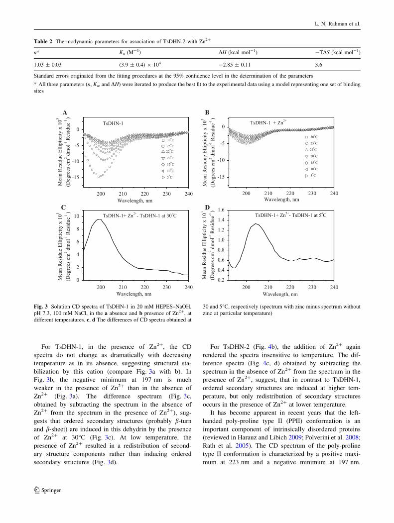
For TsDHN-1, in the presence of Zn2?, the CD
spectra do not change as dramatically with decreasing
temperature as in its absence, suggesting structural sta-
bilization by this cation (compare Fig. 3a with b). In
Fig. 3b, the negative minimum at 197 nm is much
weaker in the presence of Zn2? than in the absence of
Zn2? (Fig. 3a). The difference spectrum (Fig. 3c,
obtained by subtracting the spectrum in the absence of
Zn2? from the spectrum in the presence of Zn2?), sug-
gests that ordered secondary structures (probably b-turn
and b-sheet) are induced in this dehydrin by the presence
of Zn2? at 30�C (Fig. 3c). At low temperature, the
presence of Zn2? resulted in a redistribution of second-
ary structure components rather than inducing ordered
secondary structures (Fig. 3d).
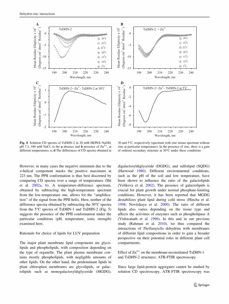
For TsDHN-2 (Fig. 4b), the addition of Zn2? again
rendered the spectra insensitive to temperature. The dif-
ference spectra (Fig. 4c, d) obtained by subtracting the
spectrum in the absence of Zn2? from the spectrum in the
presence of Zn2?, suggest, that in contrast to TsDHN-1,
ordered secondary structures are induced at higher tem-
perature, but only redistribution of secondary structures
occurs in the presence of Zn2? at lower temperature.
It has become apparent in recent years that the left-
handed poly-proline type II (PPII) conformation is an
important component of intrinsically disordered proteins
(reviewed in Harauz and Libich 2009; Polverini et al. 2008;
Rath et al. 2005). The CD spectrum of the poly-proline
type II conformation is characterized by a positive maxi-
mum at 223 nm and a negative minimum at 197 nm.
Table 2 Thermodynamic parameters for association of TsDHN-2 with Zn2?
n* Ka (M-1) DH (kcal mol-1) -TDS (kcal mol-1)
1.03 ± 0.03 (3.9 ± 0.4) 9 104 -2.85 ± 0.11 3.6
Standard errors originated from the fitting procedures at the 95% confidence level in the determination of the parameters
* All three parameters (n, Ka, and DH) were iterated to produce the best fit to the experimental data using a model representing one set of binding
sites
A B
DC
Fig. 3 Solution CD spectra of TsDHN-1 in 20 mM HEPES–NaOH,
pH 7.3, 100 mM NaCl, in the a absence and b presence of Zn2?, at
different temperatures. c, d The differences of CD spectra obtained at
30 and 5�C, respectively (spectrum with zinc minus spectrum without
zinc at particular temperature)
L. N. Rahman et al.
123
However, in many cases the negative minimum due to the
a-helical component masks the positive maximum at
223 nm. The PPII conformation is thus best discerned by
comparing CD spectra over a range of temperatures (Shi
et al. 2002a, b). A temperature-difference spectrum,
obtained by subtracting the high-temperature spectrum
from the low-temperature one, allows for the ‘‘amplifica-
tion’’ of the signal from the PPII helix. Here, neither of the
difference spectra obtained by subtracting the 30�C spectra
from the 5�C spectra of TsDHN-1 and TsDHN-2 (Fig. 5)
suggests the presence of the PPII conformation under the
particular conditions (pH, temperature, ionic strength)
examined here.
Rationale for choice of lipids for LUV preparation
The major plant membrane lipid components are glyco-
lipids and phospholipids, with composition depending on
the type of organelle. The plant plasma membrane con-
tains mostly phospholipids, with negligible amounts of
other lipids. On the other hand, the predominant lipids in
plant chloroplast membranes are glycolipids, or galac-
tolipids such as monogalactosyldiglyceride (MGDG),
digalactosyldiglyceride (DGDG), and sulfolipid (SQDG)
(Harwood 1980). Different environmental conditions,
such as the pH of the soil and low temperature, have
been shown to influence the ratio of the galactolipids
(Velikova et al. 2002). The presence of galactolipids is
crucial for plant growth under normal phosphate-limiting
conditions. However, it has been reported that MGDG
destabilizes plant lipid during cold stress (Hincha et al.
1998; Novitskaya et al. 2000). The ratio of different
lipids also varies depending on the tissue type and
affects the activities of enzymes such as phospholipase A
(Vishwanath et al. 1996). In this and in our previous
study (Rahman et al. 2010), we thus compared the
interactions of Thellungiella dehydrins with membranes
of different lipid compositions in order to gain a broader
perspective on their potential roles in different plant cell
compartments.
Effect of Zn2? on the membrane-reconstituted TsDHN-1
and TsDHN-2 structures: ATR-FTIR spectroscopy
Since large lipid-protein aggregates cannot be studied by
solution CD spectroscopy, ATR-FTIR spectroscopy was
A B
DC
Fig. 4 Solution CD spectra of TsDHN-2 in 20 mM HEPES–NaOH,
pH 7.3, 100 mM NaCl, in the a absence and b presence of Zn2?, at
different temperatures. c, d The differences of CD spectra obtained at
30 and 5�C, respectively (spectrum with zinc minus spectrum without
zinc at particular temperature). In the presence of zinc, there is a gain
of ordered secondary structure at 30�C under these conditions
Dehydrin-zinc interactions
123

applied to study the effect of Zn2? on the membrane-
associated TsDHN-1 and TsDHN-2 proteins. The proce-
dure of peak-fitting is illustrated in Fig. 6.
Our first experiment was a control to see the effects of
the cation on the lipids alone. The ATR-FTIR spectra of
LUVs derived from lipid mixtures mimicking the compo-
sitions of plasma, chloroplast, or mitochondrial membranes
all collected in the absence of protein, contained m(C=O),
symmetric d(CH2), and asymmetric d(CH2) modes at
1,734, 1,465, and 1,414 cm-1, respectively (Fig. 7a–c). In
the presence of Zn2?, an additional peak at 1,593 cm-1
was observed. None of the LUV-alone (no protein) spectra
showed significant absorption in the amide region, except
for that mimicking the mitochondrial membrane (Fig. 7c).
For these LUVs, there is a new unique band located at
around 1,620 cm-1 due to the m(COO-) of PS, which shifts
to 1,640 cm-1 upon addition of Zn2?. The LUV samples
mimicking the composition of plasma and mitochondrial
membranes became more compact (condensed acyl chains)
upon interaction with Zn2?, due to charge screening,
causing them to attach more strongly to the ATR crys-
tal (cf., Smith et al. 2010). This effect is indicated by
larger carbonyl lipid peak areas in the presence of Zn2?
(Fig. 7a, c).
The addition of Zn2? to either membrane-reconstituted
TsDHN-1 or TsDHN-2 caused significant changes in the
ATR-FTIR spectra (Figs. 8, 9). All infrared spectra
(Figs. 8a–c, 9a–c) were normalized by the carbonyl peak
areas after base line corrections. In the presence of Zn2?,
TsDHN-1 and TsDHN-2 interact with membranes much
less than in its absence, except in the case of TsDHN-2
interacting with LUVs mimicking the composition of the
plasma membrane. It has been reported that Zn2? forms
bridges between neighbouring negatively charged
phospholipid head groups, thus stabilizing the lipid mem-
branes in their gel phase (Binder et al. 2000).
When TsDHN-1 or TsDHN-2 bound to Zn2? comes in
contact with lipid membranes, the Zn2? ions are perhaps
removed from the proteins and interact instead with the
phospholipid head groups, limiting the interactions of the
proteins with lipid membranes. For TsDHN-1, peaks at
1,593 cm-1 were observed, which might be due to
lipids.
In order to determine the effect of Zn2? on the sec-
ondary structures of the proteins themselves, the FTIR
spectra were corrected for the LUVs background, fol-
lowed by normalization, such that the area between the
linear baseline and the spectra between the region 1,700
and 1,500 cm-1 was unity (Figs. 8d–f, 9d–f). The peak
at 1,564 cm-1 (Fig. 8d, e) or 1,550 cm-1 (Fig. 8f), for
TsDHN-1, could arise due to the m(COO-) vibration
of Glu or Asp side chains, respectively. The peak at
1,575 cm-1 for TsDHN-2 (Fig. 9d–f), could arise due to
the m(COO-) vibration of Asp side chains. In the pres-
ence of Zn2?, the peak at 1,564 cm-1 for TsDHN-1
disappears, and instead a peak at 1,605–1,610 cm-1 is
observed, indicating the involvement of the side chain
m(COO-) for interaction with Zn2? (Fig. 8d, e; Mizuguchi
et al. 2001; Nara et al. 1995). In addition, peaks at
around 1674 cm-1 are also observed for plasma
A
B
Fig. 5 The differences of solution CD spectra obtained in the
presence of zinc at 5 and 30�C (lower temperature spectrum minus
higher temperature spectrum) for (a) TsDHN-1, and (b) TsDHN-2,
discounting the presence of a zinc-induced (or zinc-stabilized) poly-
proline type II (PPII) conformation in TsDHN-1 and TsDHN-2 in
solution
L. N. Rahman et al.
123

membrane or chloroplast membrane reconstituted
TsDHN-1 (Fig. 8d, e).
In the FTIR spectra of membrane-reconstituted TsDHN-2,
a peak shifted from 1,648 to 1,632 cm-1 in the presence of
Zn2? suggesting a change in conformation from random
coil to b-sheet (Fig. 9d–f; Bandekar 1992; Byler and Susi
1986; Krimm and Bandekar 1986; Surewicz et al. 1993;
Surewicz and Mantsch 1988). There was little change
observed in the peak located at 1,575 cm-1 in the presence
of Zn2? for TsDHN-2, suggesting that the side chains of
this dehydrin are not involved in interacting with Zn2? to
any significant extent (Mizuguchi et al. 2001).
Zinc-induced ordered secondary structure in TsDHN-1
and TsDHN-2
All FTIR spectra were fitted with multiple Gaussian and
Lorentzian peaks for detailed secondary structure analysis
as illustrated in Fig. 6 and as previously described in detail
(Rahman et al. 2010; Smith et al. 2010). Partial overlaps of
the amide I band with the deuterated side chain at around
1,564 cm-1 for TsDHN-1, and at 1,575 cm-1 for TsDHN-2,
were observed. Therefore, all peaks located in the region
between 1,600 and 1,500 cm-1, after baseline correction,
were included during curve fitting. Detailed predictions of
the proportions of different types of secondary structures
(a-helix, b-strand, and random coil) are given in Tables 3,
4, 5. It should be cautioned that the calculations of overall
secondary structure composition from such spectra will
vary depending on how peak identification, baseline cor-
rection, and peak-fitting are performed. Our method is
summarized in Fig. 6. Therefore, we restrict ourselves here
to reporting on clear trends, as we have previously done for
these dehydrins and for myelin basic protein (Rahman et al.
2010; Smith et al. 2010).
Detailed spectral analysis shows that both TsDHN-1
and TsDHN-2 gain more ordered secondary structure in
the presence of Zn2?, particularly b-sheet (Fig. 10). The
anti-parallel b-sheet becomes more pronounced in the
secondary structure of TsDHN-1, whereas the parallel
b-sheet is more pronounced in TsDHN-2. However, the
extent in increase of ordered structure is more pronounced
for TsDHN-2. For TsDHN-1, there is no significant
change in the random coil structure, but there is an
A B
DC
Fig. 6 Demonstration of peak-fitting process for ATR-FTIR spectra.
a Attenuated total reflection Fourier-transform infrared spectra of
TsDHN-2, associated with chloroplast-LUVs at a wt:wt lipid-to-
protein ratio of 1:1 in D2O (solid line) at 22�C. b Fourier self-
deconvolution of an unprocessed ATR-FTIR spectrum of TsDHN-2,
associated with chloroplast-LUVs at a wt:wt lipid-to-protein ratio of
1:1 in D2O at 22�C, using OMNIC software with a bandwidth at a
half-height of 15 cm-1 and an enhancement value of 1.8. c Second
derivative of the ATR-FTIR spectrum (a) for secondary structure
determination by PEAKFIT software. d Analysis of the ATR-FTIR
spectrum (a) for secondary structure determination by PEAKFIT
software—the baseline-corrected spectra of the TsDHN-1 and
TsDHN-2 proteins reconstituted in LUVs of differing lipid compo-
sitions, were deconvoluted using a mixed Gaussian and Lorentzian
band shape. Auto-fits of the self-deconvoluted spectra of the original
spectra were performed until the coefficient of determination (r2) was
larger than 0.99
Dehydrin-zinc interactions
123

increase of turn structures and a decrease in the side chain
contributions. Little change in side chain contributions
due to Zn2? is observed for TsDHN-2. There is also a
synergistic effect of lipid composition, as TsDHN-1 gains
more ordered secondary structure when associated with
LUVs mimicking the lipid compositions of chloroplast
membranes (Fig. 10a, b). The FTIR spectra for TsDHN-1
associated with LUVs mimicking the composition of
mitochondria were not of suitable quality for secondary
structure analysis with the above method and are not
presented.
It has been suggested that dehydrins retain their intrin-
sically disordered conformation in the formation of
hydrogels in conditions of drought, thereby avoiding
structural collapse that well-folded proteins would undergo
(Mouillon et al. 2008). Our results here demonstrate that
dehydrins undergo a disorder-to-order transition in asso-
ciation with membranes and zinc. These new data extend
our previous study in which we demonstrated induced
order in these same dehydrins upon membrane association
at low temperatures (Rahman et al. 2010). Thus, the con-
formational space sampled by dehydrins in vivo would
depend on their particular environment within the cell—the
proteins at a membrane interface would be more ordered
than those within the bulk hydrogel.
Significance of zinc sequestration by Th. salsuginea
dehydrins
The sequestration of heavy metals such as zinc has been
documented for several LEA proteins and dehydrins
(Alsheikh et al. 2003; Heyen et al. 2002; Xu et al. 2008;
Zhang et al. 2006). More recently, it has been reported
that Zn2? promotes the association of citrus dehydrins
with DNA, and that this association was mediated by
domains enriched with histidyl and lysyl residues (Hara
et al. 2009). This mechanism makes sense since poly-
lysine chains have long been used as a coating to fix DNA
to solids (Eisen and Brown 1999), and it is known that
His-His or His-X3(4)-His motifs, or combinations thereof,
can bind to macromolecules chelating metal ions (such
as DNA) through Zn2? intermediates (Vallee and Auld
1995).
Here, TsDHN-1 is an acidic S1K3 type dehydrin con-
taining a His-His pair (residues 250 and 251, counting the
N-terminal Met as #1) which may play a role in promoting
the ordered secondary structure in the presence of Zn2?. In
contrast, TsDHN-2 contains two His-His pairs (residues 72
and 73, and 152 and 153), which may play a role in pro-
moting ordered secondary structure in the presence of
Zn2?. In addition, the negatively charged side chains in
TsDHN-1 may also interact with Zn2?. Previously and
independently, studies on Brassica juncea dehydrins led to
the suggestion that they confer heavy metal tolerance (Xu
et al. 2008). Our results here indicate that zinc sequestra-
tion by free or membrane-associated Th. salsuginea
dehydrins results in induced ordered structure, the physi-
ological significance of which remains to be determined.
We suggest here that zinc may be important also for
membrane-stabilization via dehydrins in plants subjected to
extremes of cold or drought.
A
B
C
Fig. 7 Effects of Zn2? on ATR-FTIR spectra, absorbance versus
wavenumber, at room temperature (22�C), for LUVs alone in the
absence of protein. The LUV lipid compositions mimic the plant
a plasma (PM_LUV), b chloroplast (CHLOR_LUV), or c mitochon-
drial (MITOCH_LUV) membranes
L. N. Rahman et al.
123
A B C
FED
Fig. 8 Effects of Zn2? on ATR-FTIR spectra of TsDHN-1 at room
temperature (22�C). In panels (a–c), the protein is membrane-
associated with LUV lipid compositions mimicking the plant
(a) plasma (PM_LUV), b chloroplast (CHLOR_LUV), and
c mitochondrial (MITOCH_LUV) membranes. Panels (d–f) represent
the corresponding spectra of protein alone, after subtraction of the
lipid spectrum
A B C
FED
Fig. 9 Effects of Zn2? on ATR-FTIR spectra of TsDHN-2 at room
temperature (22�C). In panels (a–c), the protein is membrane-
associated with LUV lipid compositions mimicking the plant
a plasma (PM_LUV), b chloroplast (CHLOR_LUV), and
c mitochondrial (MITOCH_LUV) membranes. Panels (d–f) represent
the corresponding spectra of protein alone, after subtraction of the
lipid spectrum
Dehydrin-zinc interactions
123

Conclusions
We have investigated the interactions of Th. salsuginea
dehydrins TsDHN-1 and TsDHN-2 with Zn2? and mem-
branes, in vitro, to gain further insight into their physio-
logical roles. Using a combination of complementary
spectroscopic methods, we have shown that both proteins
associate with membranes of different lipid compositions,
and thereby gain ordered secondary structure, consistent
with other dehydrins that have been investigated (Koag
et al. 2003, 2009; Soulages et al. 2002, 2003). These strong
membrane interactions of both dehydrins, with concomi-
tant induced folding (ordered secondary structure forma-
tion) support the hypothesis that they protect plant plasma
and organellar membranes under conditions of extreme
cold (Beck et al. 2007; Zhang et al. 2010). Zinc has been
shown to stabilize ordered secondary structure in many
intrinsically disordered proteins (briefly reviewed in, e.g.,
Smith et al. 2010). Here, the divalent cation Zn2? has been
shown to have a strong stabilizing effect on both dehydrin
Table 3 Effect of zinc on secondary structure components of TsDHN-1 and TsDHN-2 bound to large unilamellar vesicles mimicking the plant
plasma membrane (PC:PE:PI at 33:47:20 by wt%), with a 1:10 M ratio of TsDHN-x:Zn2?
Components of TsDHN-1 kmax (cm-1) Absence
of Zn2?Presence
of Zn2?Components
of TsDHN-2
kmax
(cm-1)
Absence
of Zn2?Presence
of Zn2?
Side chain contribution Side chain contribution 1,517 3.3 ± 0.5 2.7 ± 1
Side chain contribution 1,535 6.2 ± 0.7 Side chain contribution 1,546 2.6 ± 0.1 5.2 ± 1
Side chain contribution 1,555 1.3 ± 0.7 Side chain contribution 1,562 7.1 ± 0.1 9.1 ± 1
Side chain contribution 1,565 17.0 ± 1.0 6.4 ± 0.2 Side chain contribution 1,579 11.3 ± 0.2 9.5 ± 0.3
Side chain contribution 1,609 11.3 ± 0.6 7.1 ± 1.1 Side chain contribution 1,609 11.8 ± 0.2 12.6 ± 0.3
b-strand 1,625 12.9 ± 0.2 19.6 ± 0.3 b-strand 1,627 17.2 ± 0.5 25.4 ± 0.5
Random coil 1,647 37.5 ± 0.7 31.5 ± 0.9 Random coil 1,644 28.3 ± 0.6 15.8 ± 4
Turn 1,672 17.8 ± 0.6 23.7 ± 0.5 Turn 1,658 12.4 ± 4
Side chain contribution 1,699 2.4 ± 0.2 5.5 ± 0.4 Turn 1,671 14.0 ± 0.5 5.7 ± 3
Anti parallel b-strand 1,684 4.3 ± 0.1 2.8 ± 1
Total 100.2% 100.0% 99.9% 101.2%
The columns show the percentage area of each peak, with errors representing the standard deviation of two replicates of data sets. The values will
not necessarily add up precisely to 100% because of rounding errors
Table 4 Effect of zinc on secondary structure components of TsDHN-1 and TsDHN-2 bound to large unilamellar lipid vesicles mimicking the
plant chloroplast membrane (MGDG:DGDG:SQDG:PC:DMPG:PE:PI at 51:26:7:3:9:1 by wt%), with a 1:10 M ratio of TsDHN-x:Zn2?
Components of TsDHN-1 kmax (cm-1) Absence
of Zn2?Presence
of Zn2?Components
of TsDHN-2
kmax
(cm-1)
Absence
of Zn2?Presence
of Zn2?
Side chain contribution 1,514 1.2 ± 0.5 1.6 ± 4.0
Side chain contribution 1,540–1,543 3.3 ± 0.6 2.7 ± 0 Side chain contribution 1,543 3.5 ± 2.0
Side chain contribution 1,562–1,565 17.8 ± 0.7 6.8 ± 0.3 Side chain contribution 1,562 9.6 ± 1.0 7.2 ± 0.7
Side chain contribution 1,582–1,585 10.5 ± 1.4 5.1 ± 0.4 Side chain contribution 1,578 8.7 ± 2.0 8.8 ± 4.0
Side chain contribution 1,602 10.8 ± 0.2 Side chain contribution 1,596 7.5 ± 3.0
Side chain contribution 1,611–1,614 5.5 ± 0.4 2.4 ± 0.1 Side chain contribution 1,613 8.5 ± 1.0 10.8 ± 0.5
b-strand 1,627–1,628 12.6 ± 1.5 17.2 ± 0.5 b-strand 1,629 13.3 ± 2.0 17.6 ± 1.4
Random coil 1,642–1,645 25.7 ± 1.2 22.4 ± 0.2 Random coil 1,643 25.7 ± 1.0 12.4 ± 2.2
Alpha Helices 1,659 8.9 ± 1.2 7.0 ± 0.3 Alpha Helices 1,651 11.8 ± 1.0 10.7 ± 1.6
Turn 1,671–1,673 15.7 ± 1.2 25.5 ± 0.2 Turn 1,663 13.6 ± 1.7 12.0 ± 1.0
Anti parallel b-strand 1,679 7.7 ± 1.6 7.8 ± 1.3
Total 100.0% 99.9% 100.1% 100.0%
The columns show the percentage area of each peak, with errors representing the standard deviation of two replicates of datasets. The values will
not necessarily add up precisely to 100% because of rounding errors
L. N. Rahman et al.
123

Table 5 Effect of zinc on secondary structure components of TsDHN-1 and TsDHN-2 bound to large unilamellar lipid vesicles mimicking the
plant mitochondrial membrane (PC:PS:PE:Chol at 27:25:29:20 by wt%), with a 1:10 M ratio of TsDHN-x:Zn2?
Components of TsDHN-2 kmax (cm-1) Absence of Zn2? Presence of Zn2?
Side chain contribution 1,551 2.9 ± 1.0 3.6 ± 0.2
Side chain contribution 1,567 6.3 ± 0.7 9.4 ± 0.11
Side chain contribution 1,581 5.9 ± 1.0 8.3 ± 0.5
Side chain contribution 1,593 6.3 ± 1.0 3.0 ± 0.6
Side chain contribution 1,611 7.5 ± 0.7 9.1 ± 0.4
b-strand 1,629 19.2 ± 1.0 24.7 ± 1.3
Random coil 1,646 25.7 ± 1.0 19.9 ± 1.5
Turn 1,660 17.4 ± 0.7 14.2 ± 1.5
1,672 5.5 ± 0.9
b-strand 1,678 9.1 ± 1.0 2.1 ± 0.3
Total 100.3% 99.9%
The columns show the percentage area of each peak, with errors representing the standard deviation of two replicates of datasets. The values will
not necessarily add up precisely to 100% because of rounding errors
A B
D
E
C
Fig. 10 Effect of Zn2? on
secondary structure composition
of membrane-associated
(a, c) TsDHN-1, and (b, d, e)
TsDHN-2 at room temperature
(22�C). The LUV lipid
compositions mimic the plant
(a, b) plasma (PM_LUV),
(c, d) chloroplast
(CHLOR_LUV), or
(e) mitochondrial
(MITOCH_LUV) membranes.
Error bars represent the
standard deviation of triplicate
measurements
Dehydrin-zinc interactions
123
structures, both free and in membrane-bound form, indi-
cating a synergistic interaction and potentially a further
mechanism for cold-stabilization.
Acknowledgments This work was supported by the Natural
Sciences and Engineering Research Council of Canada (G.H.) and
initially by the Advanced Food and Materials Network (AFMNet,
B.A.M., J.R.D., G.H.), a national Network of Centres of Excellence.
J.R.D. acknowledges support from the Canada Research Chair Pro-
gram. The authors are grateful to Dr. David Libich for assistance with
Fig. 1, to Mr. Miguel De Avila for help with the isothermal titration
calorimetry and to Dr. Leonid Brown of the University of Guelph for
many helpful discussions on FTIR data collection and analysis.
References
Allagulova C, Gimalov FR, Shakirova FM, Vakhitov VA (2003) The
plant dehydrins: structure and putative functions. Biochemistry
(Moscow) 68:945–951
Alsheikh MK, Heyen BJ, Randall SK (2003) Ion binding properties of
the dehydrin ERD14 are dependent upon phosphorylation. J Biol
Chem 278:40882–40889
Amtmann A (2009) Learning from evolution: Thellungiella generates
new knowledge on essential and critical components of abiotic
stress tolerance in plants. Mol Plant 2:3–12
Arrondo JL, Muga A, Castresana J, Goni FM (1993) Quantitative
studies of the structure of proteins in solution by Fourier-
transform infrared spectroscopy. Prog Biophys Mol Biol
59:23–56
Bandekar J (1992) Amide modes and protein conformation. Biochim
Biophys Acta 1120:123–143
Battaglia M, Solorzano RM, Hernandez M, Cuellar-Ortiz S, Garcia-
Gomez B, Marquez J, Covarrubias AA (2007) Proline-rich cell
wall proteins accumulate in growing regions and phloem tissue
in response to water deficit in common bean seedlings. Planta
225:1121–1133
Battaglia M, Olvera-Carrillo Y, Garciarrubio A, Campos F, Cova-
rrubias AA (2008) The enigmatic LEA proteins and other
hydrophilins. Plant Physiol 148:6–24
Beck EH, Fettig S, Knake C, Hartig K, Bhattarai T (2007) Specific
and unspecific responses of plants to cold and drought stress.
J Biosci 32:501–510
Binder H, Arnold K, Ulrich AS, Zschornig O (2000) The effect of
Zn(2?) on the secondary structure of a histidine-rich fusogenic
peptide and its interaction with lipid membranes. Biochim
Biophys Acta 1468:345–358
Bravo LA, Gallardo J, Navarrete A, Olave N, Martinez J, Alberdi M,
Close TJ, Corcuera LJ (2003) Cryoprotective activity of a cold-
induced dehydrin purified from barley. Physiol Plant
118:262–269
Byler DM, Susi H (1986) Examination of the secondary structure of
proteins by deconvolved FTIR spectra. Biopolymers 25:469–487
Callebaut I, Labesse G, Durand P, Poupon A, Canard L, Chomilier J,
Henrissat B, Mornon JP (1997) Deciphering protein sequence
information through hydrophobic cluster analysis (HCA): cur-
rent status and perspectives. Cell Mol Life Sci 53:621–645
Campbell SA, Close TJ (1997) Dehydrins: genes, proteins, and
associations with phenotypic traits. New Phytol 137:61–74
Caramelo JJ, Iusem ND (2009) When cells lose water: lessons from
biophysics and molecular biology. Prog Biophys Mol Biol
99:1–6
Ceccardi TL, Meyer NC, Close TJ (1994) Purification of a maize
dehydrin. Protein Expr Purif 5:266–269
Close TJ (1997) Dehydrins: a commonality in the response of plants
to dehydration and low temperature. Physiol Plant 100:291–296
Danyluk J, Perron A, Houde M, Limin A, Fowler B, Benhamou N,
Sarhan F (1998) Accumulation of an acidic dehydrin in the
vicinity of the plasma membrane during cold acclimation of
wheat. Plant Cell 10:623–638
Eisen MB, Brown PO (1999) DNA arrays for analysis of gene
expression. Methods Enzymol 303:179–205
Englbrecht CC, Schoof H, Bohm S (2004) Conservation, diversifi-
cation and expansion of C2H2 zinc finger proteins in the
Arabidopsis thaliana genome. BMC Genomics 5:39
Eom JW, Baker WR, Kintanar A, Wurtele ES (1996) The embryo-
specific EMB-1 protein of Daucus carota is flexible and
unstructured in solution. Plant Sci 115:17–24
Gaboriaud C, Bissery V, Benchetrit T, Mornon JP (1987) Hydropho-
bic cluster analysis: an efficient new way to compare and analyse
amino acid sequences. FEBS Lett 224:149–155
Garay-Arroyo A, Colmenero-Flores JM, Garciarrubio A, Covarrubias
AA (2000) Highly hydrophilic proteins in prokaryotes and
eukaryotes are common during conditions of water deficit. J Biol
Chem 275:5668–5674
Goldgur Y, Rom S, Ghirlando R, Shkolnik D, Shadrin N, Konrad Z,
Bar-Zvi D (2007) Desiccation and zinc binding induce transition
of tomato abscisic acid stress ripening 1, a water stress- and salt
stress-regulated plant-specific protein, from unfolded to folded
state. Plant Physiol 143:617–628
Griffith M, Timonin M, Wong AC, Gray GR, Akhter SR, Saldanha M,
Rogers MA, Weretilnyk EA, Moffatt B (2007) Thellungiella: an
Arabidopsis-related model plant adapted to cold temperatures.
Plant Cell Environ 30:529–538
Gusman H, Lendenmann U, Grogan J, Troxler RF, Oppenheim FG
(2001) Is salivary histatin 5 a metallopeptide? Biochim Biophys
Acta 1545:86–95
Han B, Hughes DW, Galau GA, Bewley JD, Kermode AR (1997)
Changes in late-embryogenesis-abundant (LEA) messenger
RNAs and dehydrins during maturation and premature drying
of Ricinus communis L. seeds. Planta 201:27–35
Hara M, Fujinaga M, Kuboi T (2004) Radical scavenging activity and
oxidative modification of Citrus dehydrin. Plant Physiol Bio-
chem 42:657–662
Hara M, Fujinaga M, Kuboi T (2005) Metal binding by Citrusdehydrin with histidine-rich domains. J Exp Bot 56:2695–2703
Hara M, Shinoda Y, Tanaka Y, Kuboi T (2009) DNA binding of
Citrus dehydrin promoted by zinc ion. Plant Cell Environ
32:532–541
Harauz G, Libich DS (2009) The classic basic protein of myelin—
conserved structural motifs and the dynamic molecular barcode
involved in membrane adhesion and protein-protein interactions.
Current Protein Pept Sci 10:196–215
Harwood JL (1980) Plant acyl lipids: structure, distribution, and
analysis. In: Stumpf PK, Conn EE (eds) Lipids: structure and
function. Academic Press, New York, pp 2–56
Herzer S, Kinealy K, Asbury R, Beckett P, Eriksson K, Moore P
(2003) Purification of native dehydrin from Glycine Max cv.,
Pisum sativum, and Rosmarinum officinalis by affinity chroma-
tography. Protein Expr Purif 28:232–240
Heyen BJ, Alsheikh MK, Smith EA, Torvik CF, Seals DF, Randall
SK (2002) The calcium-binding activity of a vacuole-associated,
dehydrin-like protein is regulated by phosphorylation. Plant
Physiol 130:675–687
Hincha DK, Heber U, Schmitt JM (1990) Proteins from frost-hardy
leaves protect thylakoids against mechanical freeze-thaw dam-
age in vitro. Planta 180:416–419
Hincha DK, Oliver AE, Crowe JH (1998) The effects of chloroplast
lipids on the stability of liposomes during freezing and drying.
Biochim Biophys Acta 1368:150–160
L. N. Rahman et al.
123

Hundertmark M, Hincha DK (2008) LEA (late embryogenesis
abundant) proteins and their encoding genes in Arabidopsisthaliana. BMC Genomics 9:118
Inan G, Zhang Q, Li P, Wang Z, Cao Z, Zhang H, Zhang C, Quist
TM, Goodwin SM, Zhu J, Shi H, Damsz B, Charbaji T, Gong Q,
Ma S, Fredricksen M, Galbraith DW, Jenks MA, Rhodes D,
Hasegawa PM, Bohnert HJ, Joly RJ, Bressan RA, Zhu JK (2004)
Salt cress. A halophyte and cryophyte Arabidopsis relative
model system and its applicability to molecular genetic analyses
of growth and development of extremophiles. Plant Physiol
135:1718–1737
Ismail AM, Hall AE, Close TJ (1999) Purification and partial
characterization of a dehydrin involved in chilling tolerance
during seedling emergence of cowpea. Plant Physiol
120:237–244
Jepson SG, Close TJ (1995) Purification of a maize dehydrin protein
expressed in Escherichia coli. Protein Expr Purif 6:632–636
Kalifa Y, Gilad A, Konrad Z, Zaccai M, Scolnik PA, Bar-Zvi D
(2004) The water- and salt-stress-regulated Asr1 (abscisic acid
stress ripening) gene encodes a zinc-dependent DNA-binding
protein. Biochem J 381:373–378
Koag MC, Fenton RD, Wilkens S, Close TJ (2003) The binding of
maize DHN1 to lipid vesicles. Gain of structure and lipid
specificity. Plant Physiol 131:309–316
Koag MC, Wilkens S, Fenton RD, Resnik J, Vo E, Close TJ (2009)
The K-segment of maize DHN1 mediates binding to anionic
phospholipid vesicles and concomitant structural changes. Plant
Physiol 150:1503–1514
Kosova K, Vitamvas P, Prasil IT (2007) The role of dehydrins in plant
response to cold. Biol Plant 51:601–617
Kosova K, Holkova L, Prasil IT, Prasilova P, Bradacova M, Vitamvas
P, Capkova V (2008) Expression of dehydrin 5 during the
development of frost tolerance in barley (Hordeum vulgare).
J Plant Physiol 165:1142–1151
Kovacs D, Kalmar E, Torok Z, Tompa P (2008) Chaperone activity of
ERD10 and ERD14, two disordered stress-related plant proteins.
Plant Physiol 147:381–390
Krimm S, Bandekar J (1986) Vibrational spectroscopy and confor-
mation of peptides, polypeptides, and proteins. Adv Protein
Chem 38:181–364
Kruger C, Berkowitz O, Stephan UW, Hell R (2002) A metal-binding
member of the late embryogenesis abundant protein family
transports iron in the phloem of Ricinus communis L. J Biol
Chem 277:25062–25069
Lisse T, Bartels D, Kalbitzer HR, Jaenicke R (1996) The recombinant
dehydrin-like desiccation stress protein from the resurrection
plant Craterostigma plantagineum displays no defined three-
dimensional structure in its native state. Biol Chem 377:555–561
Majava V, Petoukhov MV, Hayashi N, Pirila P, Svergun DI, Kursula
P (2008) Interaction between the C-terminal region of human
myelin basic protein and calmodulin: analysis of complex
formation and solution structure. BMC Struct Biol 8:10
Mizuguchi M, Fujisawa R, Nara M, Nitta K, Kawano K (2001)
Fourier-transform infrared spectroscopic study of Ca2?-binding
to osteocalcin. Calcif Tissue Int 69:337–342
Mohan A, Uversky VN, Radivojac P (2009) Influence of sequence
changes and environment on intrinsically disordered proteins.
PLoS Comput Biol 5:e1000497
Mouillon JM, Gustafsson P, Harryson P (2006) Structural investiga-
tion of disordered stress proteins. Comparison of full-length
dehydrins with isolated peptides of their conserved segments.
Plant Physiol 141:638–650
Mouillon JM, Eriksson SK, Harryson P (2008) Mimicking the plant-
cell interior under water stress by macromolecular crowding:
Disordered dehydrin proteins are highly resistant to structural
collapse. Plant Physiol 148:1925–1937
Nara M, Tanokura M, Yamamoto T, Tasumi M (1995) A comparative
study of the binding effects of Mg2?, Ca2?, Sr2?, and Cd2? on
calmodulin by Fourier-transform infrared-spectroscopy. Bio-
spectroscopy 1:47–54
Novitskaya GV, Suvorova TA, Trunova TI (2000) Lipid composition
of tomato leaves as related to plant cold tolerance. Russ J Plant
Physiol 47:728–733
Pedras MS, Zheng QA (2010) Metabolic responses of Thellungiellahalophila/salsuginea to biotic and abiotic stresses: metabolite
profiles and quantitative analyses. Phytochemistry 71:581–589
Pierce MM, Raman CS, Nall BT (1999) Isothermal titration
calorimetry of protein-protein interactions. Methods
19:213–221
Polverini E, Rangaraj G, Libich DS, Boggs JM, Harauz G (2008)
Binding of the proline-rich segment of myelin basic protein to
SH3-domains—spectroscopic, microarray, and modelling studies
of ligand conformation and effects of post-translational modifi-
cations. Biochemistry 47:267–282
Puhakainen T, Hess MW, Makela P, Svensson J, Heino P, Palva ET
(2004) Overexpression of multiple dehydrin genes enhances
tolerance to freezing stress in Arabidopsis. Plant Mol Biol
54:743–753
Rahman LN, Chen L, Nazim S, Bamm VV, Yaish MWF, Moffatt BA,
Dutcher JR, Harauz G (2010) Interactions of intrinsically
disordered Thellungiella salsuginea dehydrins TsDHN-1 and
TsDHN-2 with membranes—synergistic effects of lipid compo-
sition and temperature on secondary structure. Biochem Cell
Biol 88(5):791–807
Rajesh S, Manickam A (2006) Prediction of functions for two LEA
proteins from mung bean. Bioinformation 1:133–138
Rath A, Davidson AR, Deber CM (2005) The structure of ‘‘unstruc-
tured’’ regions in peptides and proteins: role of the polyproline II
helix in protein folding and recognition. Biopolymers
80:179–185
Rom S, Gilad A, Kalifa Y, Konrad Z, Karpasas MM, Goldgur Y,
Bar-Zvi D (2006) Mapping the DNA- and zinc-binding domains
of ASR1 (abscisic acid stress ripening), an abiotic-stress
regulated plant specific protein. Biochimie 88:621–628
Rorat T, Szabala BM, Grygorowicz WJ, Wojtowicz B, Yin Z, Rey P
(2006) Expression of SK(3)-type dehydrin in transporting organs
is associated with cold acclimation in Solanum species. Planta
224:205–221
Shi Z, Olson CA, Rose GD, Baldwin RL, Kallenbach NR (2002a)
Polyproline II structure in a sequence of seven alanine residues.
Proc Natl Acad Sci USA 99:9190–9195
Shi Z, Woody RW, Kallenbach NR (2002b) Is polyproline II a major
backbone conformation in unfolded proteins? Adv Protein Chem
62:163–240
Smith GST, Chen L, Bamm VV, Dutcher JR, Harauz G (2010) The
interaction of zinc with membrane-associated 18.5 kDa myelin
basic protein: an attenuated total reflectance-Fourier transform
infrared spectroscopic study. Amino Acids 39:739–750
Sobhany M, Negishi M (2006) Characterization of specific donor
binding to alpha1, 4-N-acetylhexosaminyltransferase EXTL2
using isothermal titration calorimetry. Methods Enzymol
416:3–12
Soulages JL, Kim K, Walters C, Cushman JC (2002) Temperature-
induced extended helix/random coil transitions in a group 1 late
embryogenesis-abundant protein from soybean. Plant Physiol
128:822–832
Soulages JL, Kim K, Arrese EL, Walters C, Cushman JC (2003)
Conformation of a group 2 late embryogenesis abundant protein
from soybean. Evidence of poly (L-proline)-type II structure.
Plant Physiol 131:963–975
Steponkus PL, Uemura M, Joseph RA, Gilmour SJ, Thomashow MF
(1998) Mode of action of the COR15a gene on the freezing
Dehydrin-zinc interactions
123
tolerance of Arabidopsis thaliana. Proc Natl Acad Sci USA
95:14570–14575
Surewicz WK, Mantsch HH (1988) New insight into protein
secondary structure from resolution-enhanced infrared spectra.
Biochim Biophys Acta 952:115–130
Surewicz WK, Mantsch HH, Chapman D (1993) Determination of
protein secondary structure by Fourier transform infrared
spectroscopy: a critical assessment. Biochemistry 32:389–394
Svensson J, Palva ET, Welin B (2000) Purification of recombinant
Arabidopsis thaliana dehydrins by metal ion affinity chroma-
tography. Protein Expr Purif 20:169–178
Thalhammer A, Hundertmark M, Popova AV, Seckler R, Hincha DK
(2010) Interaction of two intrinsically disordered plant stress
proteins (COR15A and COR15B) with lipid membranes in the
dry state. Biochim Biophys Acta 1798:1812–1820
Tolleter D, Jaquinod M, Mangavel C, Passirani C, Saulnier P, Manon
S, Teyssier E, Payet N, Avelange-Macherel MH, Macherel D
(2007) Structure and function of a mitochondrial late embryo-
genesis abundant protein are revealed by desiccation. Plant Cell
19:1580–1589
Tolleter D, Hincha DK, Macherel D (2010) A mitochondrial late
embryogenesis abundant protein stabilizes model membranes in
the dry state. Biochim Biophys Acta 1798:1926–1933
Tompa P, Kovacs D (2010) Intrinsically disordered chaperones in
plants and animals. Biochem Cell Biol 88:167–174
Tompa P, Banki P, Bokor M, Kamasa P, Kovacs D, Lasanda G,
Tompa K (2006) Protein-water and protein-buffer interactions in
the aqueous solution of an intrinsically unstructured plant
dehydrin: NMR intensity and DSC aspects. Biophys J
91:2243–2249
Tong KI, Katoh Y, Kusunoki H, Itoh K, Tanaka T, Yamamoto M
(2006) Keap1 recruits Neh2 through binding to ETGE and DLG
motifs: characterization of the two-site molecular recognition
model. Mol Cell Biol 26:2887–2900
Tunnacliffe A, Wise MJ (2007) The continuing conundrum of the
LEA proteins. Naturwissenschaften 94:791–812
Ueda EK, Gout PW, Morganti L (2003) Current and prospective
applications of metal ion-protein binding. J Chromatogr A
988:1–23
Uversky VN (2009) Intrinsically disordered proteins and their
environment: Effects of strong denaturants, temperature, pH,
counter ions, membranes, binding partners, osmolytes, and
macromolecular crowding. Protein J 28:305–325
Vallee BL, Auld DS (1995) Zinc metallochemistry in biochemistry.
EXS 73:259–277
Velazquez-Campoy A, Leavitt SA, Freire E (2004) Characterization
of protein-protein interactions by isothermal titration calorime-
try. Methods Mol Biol 261:35–54
Velikova V, Ivanova A, Yordanov I (2002) Changes in lipid
compistion of Phaseolus vulgaris leaves after simulating acid
rain treatment. Bulg J Plant Physiol 28:59–65
Vishwanath BS, Eichenberger W, Frey FJ, Frey BM (1996) Interac-
tion of plant lipids with 14 kDa phospholipase A2 enzymes.
Biochem J 320(Pt 1):93–99
Vucetic S, Xie H, Iakoucheva LM, Oldfield CJ, Dunker AK,
Obradovic Z, Uversky VN (2007) Functional anthology of
intrinsic disorder. 2. Cellular components, domains, technical
terms, developmental processes, and coding sequence diversities
correlated with long disordered regions. J Proteome Res
6:1899–1916
Wise MJ, Tunnacliffe A (2004) POPP the question: what do LEA
proteins do? Trends Plant Sci 9:13–17
Wiseman T, Williston S, Brandts JF, Lin LN (1989) Rapid
measurement of binding constants and heats of binding using a
new titration calorimeter. Anal Biochem 179:131–137
Wolkers WF, McCready S, Brandt WF, Lindsey GG, Hoekstra FA
(2001) Isolation and characterization of a D-7 LEA protein from
pollen that stabilizes glasses in vitro. Biochim Biophys Acta
1544:196–206
Wong CE, Li Y, Whitty BR, Diaz-Camino C, Akhter SR, Brandle JE,
Golding GB, Weretilnyk EA, Moffatt BA, Griffith M (2005)
Expressed sequence tags from the Yukon ecotype of Thellungi-ella reveal that gene expression in response to cold, drought and
salinity shows little overlap. Plant Mol Biol 58:561–574
Xie H, Vucetic S, Iakoucheva LM, Oldfield CJ, Dunker AK,
Obradovic Z, Uversky VN (2007a) Functional anthology of
intrinsic disorder. 3. Ligands, post-translational modifications,
and diseases associated with intrinsically disordered proteins.
J Proteome Res 6:1917–1932
Xie H, Vucetic S, Iakoucheva LM, Oldfield CJ, Dunker AK, Uversky
VN, Obradovic Z (2007b) Functional anthology of intrinsic
disorder. 1. Biological processes and functions of proteins with
long disordered regions. J Proteome Res 6:1882–1898
Xu J, Zhang YX, Wei W, Han L, Guan ZQ, Wang Z, Chai TY (2008)
BjDHNs confer heavy-metal tolerance in plants. Mol Biotechnol
38:91–98
Zhang Y, Li J, Yu F, Cong L, Wang L, Burkard G, Chai T (2006)
Cloning and expression analysis of SKn-type dehydrin gene
from bean in response to heavy metals. Mol Biotechnol
32:205–218
Zhang C, Ding Z, Xu X, Wang Q, Qin G, Tian S (2010) Crucial roles
of membrane stability and its related proteins in the tolerance of
peach fruit to chilling injury. Amino Acids 39:181–194
Zhu B, Choi DW, Fenton R, Close TJ (2000) Expression of the barley
dehydrin multigene family and the development of freezing
tolerance. Mol Gen Genet 264:145–153
L. N. Rahman et al.
123