Investigation into an outbreak of Shiga toxin producing E coli
Wild Ungulates as Disseminators of Shiga Toxin-Producing Escherichia coli in Urban AreasAlan B. Franklin1*, Kurt C. VerCauteren1, Hugh Maguire2, Mary K. Cichon2, Justin W. Fischer1,
Michael J. Lavelle1, Amber Powell3, J. Jeffrey Root1, Elaine Scallan4
1 United States Department of Agriculture, National Wildlife Research Center, Fort Collins, Colorado, United States of America, 2 Laboratory Services Division, Colorado
Department of Public Health and Environment, Denver, Colorado, United States of America, 3 Division of Biostatistics and Bioinformatics, National Jewish Health, Denver,
Colorado, United States of America, 4 Department of Epidemiology, Colorado School of Public Health, Aurora, Colorado, United States of America
Abstract
Background: In 2008, children playing on a soccer field in Colorado were sickened with a strain of Shiga toxin-producingEscherichia coli (STEC) O157:H7, which was ultimately linked to feces from wild Rocky Mountain elk. We addressed whetherwild cervids were a potential source of STEC infections in humans and whether STEC was ubiquitous throughout wild cervidpopulations in Colorado.
Methodology/Principal Findings: We collected 483 fecal samples from Rocky Mountain elk and mule deer in urban andnon-urban areas. Samples testing positive for STEC were higher in urban (11.0%) than non-urban (1.6%) areas. Elk fecalsamples in urban areas had a much higher probability of containing STEC, which increased in both urban and non-urbanareas as maximum daily temperature increased. Of the STEC-positive samples, 25% contained stx1 strains, 34.3% containedstx2, and 13% contained both stx1 and stx2. Additionally, eaeA genes were detected in 54.1% of the positive samples.Serotypes O103, and O146 were found in elk and deer feces, which also have the potential to cause human illness.
Conclusions/Significance: The high incidence of stx2 strains combined with eaeA and E-hyl genes that we found in wildcervid feces is associated with severe human disease, such as hemolytic uremic syndrome. This is of concern because thereis a very close physical interface between elk and humans in urban areas that we sampled. In addition, we found a strongrelationship between ambient temperature and incidence of STEC in elk feces, suggesting a higher incidence of STEC in elkfeces in public areas on warmer days, which in turn may increase the likelihood that people will come in contact withinfected feces. These concerns also have implications to other urban areas where high densities of coexisting wild cervidsand humans interact on a regular basis.
Citation: Franklin AB, VerCauteren KC, Maguire H, Cichon MK, Fischer JW, et al. (2013) Wild Ungulates as Disseminators of Shiga Toxin-Producing Escherichia coliin Urban Areas. PLoS ONE 8(12): e81512. doi:10.1371/journal.pone.0081512
Editor: Markus M. Heimesaat, Charite, Campus Benjamin Franklin, Germany
Received July 12, 2013; Accepted October 22, 2013; Published December 11, 2013
This is an open-access article, free of all copyright, and may be freely reproduced, distributed, transmitted, modified, built upon, or otherwise used by anyone forany lawful purpose. The work is made available under the Creative Commons CC0 public domain dedication.
Funding: This study was funded by the United States Department of Agriculture. The manuscript was reviewed for general policy statements committing theUSDA to action, but otherwise the findings were independently developed by the authors.
Competing Interests: The authors have declared that no competing interests exist.
* E-mail: [email protected]
Introduction
In 2008, eight children playing on a soccer field in Evergreen,
Colorado were sickened with the same strain of Shiga toxin-
producing Escherichia coli (STEC) O157:H7; five of these children
were subsequently hospitalized. Ultimately, the source of these
infections was genetically linked to feces from wild Rocky
Mountain elk (Cervus elaphus), which used the soccer field for
foraging [1]. In 2011, 5% of the elk feces collected from the same
field and a nearby golf course were positive for non-O157 STEC,
a level 2–3 times higher than previously published estimates for
wild cervids [2,3]. These two events motivated the questions: Are
wild cervids a potential source of STEC infections in humans in
Colorado and are STEC infections in cervids ubiquitous
throughout the wild population?
While E. coli is common in human intestinal flora and is
generally non-pathogenic, STEC are strains of E. coli that produce
potent cytotoxins referred to as Shiga toxin (stx), which are further
characterized as stx1 and stx2 [4]. Stx2-producing strains, especially
when coupled with expression of eaeA and E-hyl genes, are
considered to have a high likelihood of causing severe disease in
humans [4]. STEC have been implicated in many high-profile
outbreaks of disease in humans and cause an estimated 265,000
clinical cases of enteric illnesses, 3,700 hospitalizations and 31
deaths in humans in the U.S. each year though the origins of these
infections are incompletely understood and rarely attributed to a
specific source [5]. STECs are estimated to result in about $280
million in costs of illness each year [6]. Thus, determining the
sources of STEC infections is critical to developing effective,
evidence-based public health interventions. While the public
health focus is often on O157 STEC because of its pathogenicity,
non-O157 STEC serogroups caused twice as many acute
infections in humans in the U.S. as O157 STEC [6]. Non-O157
serogroups primarily implicated in human disease include O26,
O103, O111, O121 and O145 [4]. Both O157 and non-O157
STEC infections are a particularly important problem in Colorado
where infection rates in humans are substantially higher than the
PLOS ONE | www.plosone.org 1 December 2013 | Volume 8 | Issue 12 | e81512
national average [7]. Colorado also has the largest elk population
in the U.S. [8] with elk and mule deer (Odocileus hemionus) living in
proximity to humans in many urban and suburban areas. They
also frequent agricultural areas, recreational parks, and green
spaces. Although the 2008 outbreak documented an association of
wild cervids with STEC transmission to humans in Colorado, the
magnitude and risk has never been adequately demonstrated.
Outbreaks of STEC in humans have also been linked to feces of
wild cervids elsewhere, most recently in Oregon where deer feces
may have contaminated strawberries with STEC [9].
While STEC has been identified in feces of deer and elk [3,10–
14], it is unknown whether their carriage of STEC is geograph-
ically uniform or is higher in animals living near humans or
agriculture. For example, deer and elk could acquire STEC
through contact with free-ranging cattle, drinking contaminated
water, or foraging in urban and suburban green spaces irrigated
with untreated water. If deer and elk acquire STEC from other
sources, they may be intermediate reservoirs for STEC in a
complex transmission cycle rather than the ultimate source for
STEC infections in humans. To initially examine this issue, we
addressed whether the prevalence and microbial serotypes of
STEC in wild elk and mule deer in urban/suburban areas were
similar to those in more remote areas of Colorado. Addressing this
question begins to elucidate whether wild cervids are proximate or
ultimate sources of STEC and identify whether potential spillback
of STEC into elk populations occurs in areas of human habitation.
Methods
Ethics StatementPermissions were obtained for all private areas that were
sampled in this study.
SamplingFrom August through December 2012, we sampled wild cervid
feces at 4 sites within areas used by wild elk that had a low
likelihood of interchange with urban areas (Wild areas), 2 sites that
were similar to wild areas but had evidence of use by free-ranging
livestock (Livestock areas), and 6 sites within the towns of Estes
Park and Evergreen (Urban areas). In the urban areas, the sites
sampled included areas used by town residents, such as public
parks and school grounds. Cervid feces were collected using
disposable gloves and deposited in sterile Whirl-PaksH. Disposable
gloves were changed before collecting each sample of $5 fecal
pellets from each fecal pellet group to prevent cross contamination
among fecal samples. A descriptive scale was used to age feces [15]
and facilitate the collection of only fresh fecal samples. Samples
were placed on ice in the field and sent on the day of collection to
the Laboratory Services Division of the Colorado Department of
Public Health and Environment (CDPHE), Denver, Colorado for
analysis.
Laboratory AnalysisFecal samples were placed in 10 mL of GN enrichment broth
(BD BBLTM, Franklin Lakes, New Jersey) and incubated at 37uCin a rotating/shaking incubator for 18–24 hours. DNA was
extracted from the enrichment broth culture using the MagNA
Pure LC 2.0 (Roche Diagnostics, Indianapolis, Indiana). Using
primers and the PCR protocol described by Reischl et al [16],
nucleic acid extracts from the broth cultures were analyzed by
PCR for the presence of the genes producing stx1 and stx2 Shiga
toxin [16]. Broths yielding positive findings by PCR for Shiga
toxin genes were cultured using MacConkey Agar with Sorbitol
(Becton Dickinson, Franklin Lakes, New Jersey) and CHROMa-
garTM STEC (CHROMagar, Paris, France) agarose plates in
order to isolate the toxigenic E. coli colonies. Multiple colonies
were selected for repeat PCR testing to confirm the presence of stx
genes prior to identifying O157 or non-O157 serogroups. Upon
isolation of the Shiga-toxin producing colonies, PCR for eaeA and
E-hyl genes was also performed on each isolate [16]. Shiga toxin-
producing isolates were also characterized by group-specific latex
agglutination. Pure bacterial cultures were grown 18–24 hours at
37uC in Brain Heart Infusion Broth (Becton Dickinson, Franklin
Lakes, New Jersey). Broth cultures were boiled for 1 hour in order
to remove capsular (K) antigens. Cultures were tested with E. coli
monospecific antisera (Statens Serum Institut, Copenhagen,
Denmark) according to package instructions using the microtiter
plate method. Microtiter plates were incubated at 52uC overnight
and reactions were read as positive or negative.
Statistical AnalysisWe analyzed the presence or absence of STEC in wild cervid
feces using generalized linear models with a binomial distribution
and logit link function using PROC GENMOD [17] in program
SASH (SAS Institute, Carey, North Carolina). We used a model
selection framework to assess multiple competing statistical models
using a version of Akaike’s Information Criterion adjusted for
small sample sizes (AICc) [18]. The models we examined included
effects of area type (urban vesus non-urban), month, season
(summer [August], fall [September and October], and winter
[November and December]), and maximum daily temperature
(uC), which was the maximum temperature on the day of
collection from the nearest NOAA National Climatic Data Center
(http://gis.ncdc.noaa.gov/map/viewer/) weather station to the
site of collection.
Results
We collected samples from 483 pellet groups from elk and mule
deer in wild areas (12.0% of samples), wild areas with evidence of
livestock grazing (26.1% of samples), and urban areas (61.2% of
samples). The percentage of samples positive for either stx1 or stx2
(n = 36) was much higher in urban areas (11.0%) than either wild
areas (0%) or areas used by free-range cattle (2.4%) (Table 1).
Fecal samples from mule deer were a small percentage (3.1%) of
the total samples collected but appeared to have a higher incidence
of positive samples in livestock and urban areas than elk (Table 1).
However, the sample size from mule deer was too small to make
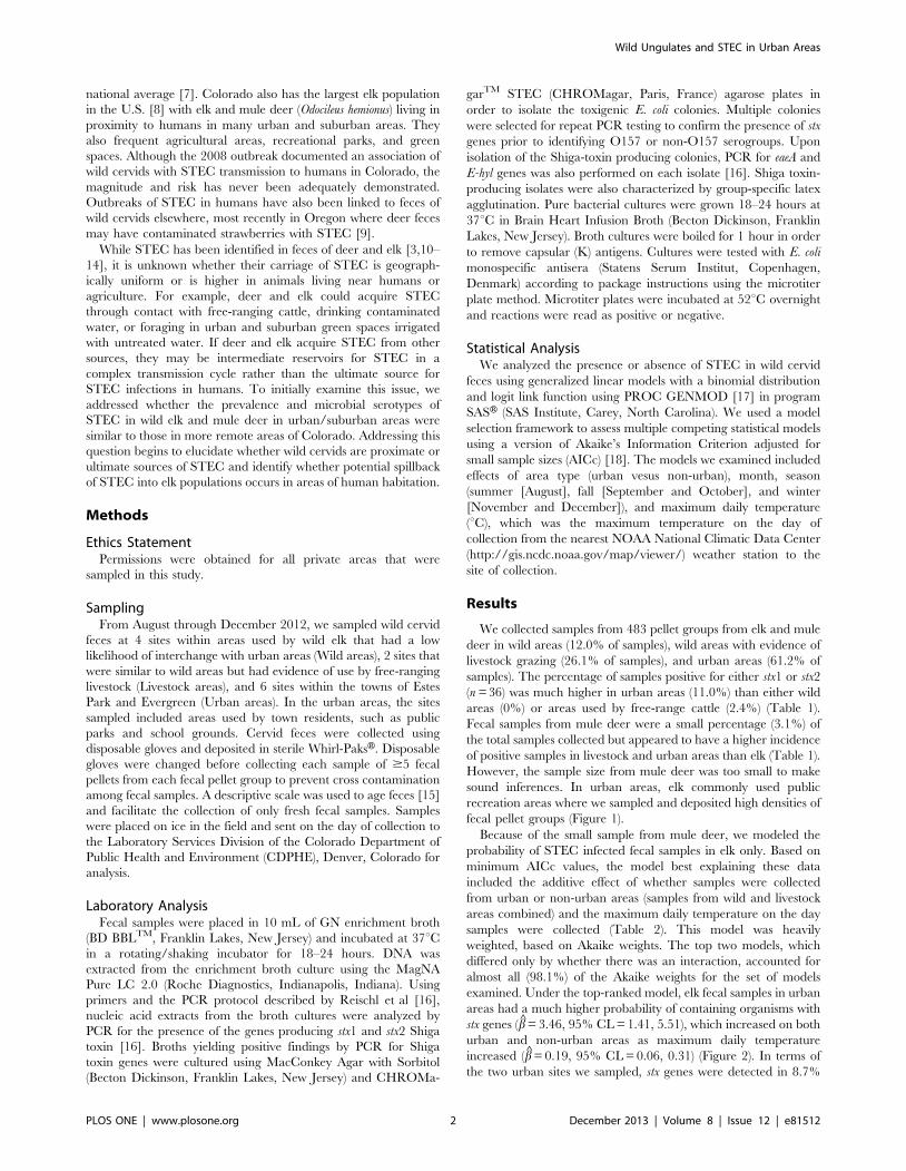
sound inferences. In urban areas, elk commonly used public
recreation areas where we sampled and deposited high densities of
fecal pellet groups (Figure 1).
Because of the small sample from mule deer, we modeled the
probability of STEC infected fecal samples in elk only. Based on
minimum AICc values, the model best explaining these data
included the additive effect of whether samples were collected
from urban or non-urban areas (samples from wild and livestock
areas combined) and the maximum daily temperature on the day
samples were collected (Table 2). This model was heavily
weighted, based on Akaike weights. The top two models, which
differed only by whether there was an interaction, accounted for
almost all (98.1%) of the Akaike weights for the set of models
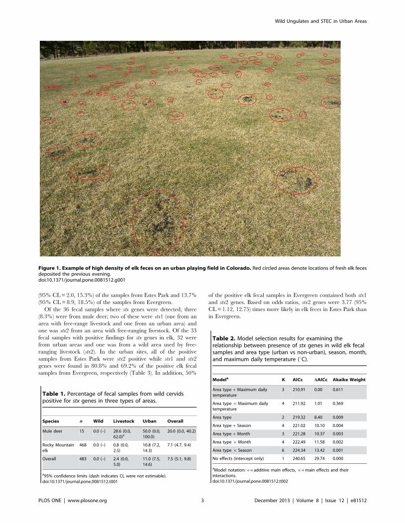
examined. Under the top-ranked model, elk fecal samples in urban
areas had a much higher probability of containing organisms with
stx genes (b = 3.46, 95% CL = 1.41, 5.51), which increased on both
urban and non-urban areas as maximum daily temperature
increased (b = 0.19, 95% CL = 0.06, 0.31) (Figure 2). In terms of
the two urban sites we sampled, stx genes were detected in 8.7%
Wild Ungulates and STEC in Urban Areas
PLOS ONE | www.plosone.org 2 December 2013 | Volume 8 | Issue 12 | e81512
(95% CL = 2.0, 15.3%) of the samples from Estes Park and 13.7%
(95% CL = 8.9, 18.5%) of the samples from Evergreen.
Of the 36 fecal samples where stx genes were detected, three
(8.3%) were from mule deer; two of these were stx1 (one from an
area with free-range livestock and one from an urban area) and
one was stx2 from an area with free-ranging livestock. Of the 33
fecal samples with positive findings for stx genes in elk, 32 were
from urban areas and one was from a wild area used by free-
ranging livestock (stx2). In the urban sites, all of the positive
samples from Estes Park were stx2 positive while stx1 and stx2
genes were found in 80.8% and 69.2% of the positive elk fecal
samples from Evergreen, respectively (Table 3). In addition, 50%
of the positive elk fecal samples in Evergreen contained both stx1
and stx2 genes. Based on odds ratios, stx2 genes were 3.77 (95%
CL = 1.12, 12.75) times more likely in elk feces in Estes Park than
in Evergreen.
Figure 1. Example of high density of elk feces on an urban playing field in Colorado. Red circled areas denote locations of fresh elk fecesdeposited the previous evening.doi:10.1371/journal.pone.0081512.g001
Table 1. Percentage of fecal samples from wild cervidspositive for stx genes in three types of areas.
Species n Wild Livestock Urban Overall
Mule deer 15 0.0 (–) 28.6 (0.0,62.0)a
50.0 (0.0,100.0)
20.0 (0.0, 40.2)
Rocky Mountainelk
468 0.0 (–) 0.8 (0.0,2.5)
10.8 (7.2,14.3)
7.1 (4.7, 9.4)
Overall 483 0.0 (–) 2.4 (0.0,5.0)
11.0 (7.5,14.6)
7.5 (5.1, 9.8)
a95% confidence limits (dash indicates CL were not estimable).doi:10.1371/journal.pone.0081512.t001
Table 2. Model selection results for examining therelationship between presence of stx genes in wild elk fecalsamples and area type (urban vs non-urban), season, month,and maximum daily temperature (uC).
Modela K AICc DAICc Akaike Weight
Area type + Maximum dailytemperature
3 210.91 0.00 0.611
Area type 6Maximum dailytemperature
4 211.92 1.01 0.369
Area type 2 219.32 8.40 0.009
Area type + Season 4 221.02 10.10 0.004
Area type + Month 3 221.28 10.37 0.003
Area type 6Month 4 222.49 11.58 0.002
Area type 6 Season 6 224.34 13.42 0.001
No effects (intercept only) 1 240.65 29.74 0.000
aModel notation: + = additive main effects, 6= main effects and theirinteractions.doi:10.1371/journal.pone.0081512.t002
Wild Ungulates and STEC in Urban Areas
PLOS ONE | www.plosone.org 3 December 2013 | Volume 8 | Issue 12 | e81512
Of the 36 samples positive for stx, we were able to culture
isolates from 24 of those samples for the detection of eaeA and E-hyl
genes. Of these, 66.7% of the stx1 PCR-positive samples, 30% of
the stx2 PCR-positive samples, and 75.0% of the samples PCR-
positive for both stx1 and stx2 contained eaeA genes, E-hyl genes or
both (Table 4). All but one of the 24 samples contained the eaeA
gene (Table 4). Of the 6 samples we were able to serologically
identify, four were O146, one was O103.
Discussion
Both direct and indirect contact with animals has been implicated
for causing both O157 and non-O157 STEC illnesses in humans
[19,20]. Indirect contact with wild cervids infected with STEC or
ingestion of contaminated game meat have been implicated in at
least 9 outbreaks of STEC in humans since 1995 (www.marlerblog.
com/uploads/image/Deer%20Table%5B1%5D. pdf). In addition,
a number of studies have found non-O157 STEC prevalence rates
of 15.0252.5% in individual wild cervids [11,12,21] and incidence
rates of 19.4% in fecal samples from wild cervids [13]. Incidence
rates of O157 STEC in fecal samples from wild cervids are much
lower, ranging from 0.022.4% [3,10,14]. Our estimate of incidence
rates of non-O157 STEC in wild elk, the focus serogroups in our
study, was at the lower end of this range. However, our limited data
on mule deer suggest higher incidence in this species, which warrant
further examination. Additionally, not all these studies [11,12]
targeted the eaeA gene whereas in our study this gene was detected in
54.1% of the positive samples collected; this in combination with the
presence of stx2-positive strains detected suggests the potential for
transmission of these pathogens to humans. However, the potential
for this to occur may be greater in specific geographic areas based
on the genetic make-up of serogroups carried by a given elk herd.
We previously documented an outbreak of human disease
associated with direct contact with elk feces that occurred in one
of the areas (Evergreen) we sampled in this study. Thus, prevalence
or incidence rates alone of STEC in wild herds may not be a useful
indicator for the potential to cause human disease; additional
information on presence of stx1, stx2, eaeA and E-hyl genes is also
required.
Our results strongly suggest that wild elk in urban areas have a
higher incidence of STEC than those in either wild areas with little
contact with urban areas or wild areas used by free-ranging
livestock. There are a number of public health concerns resulting
from our findings. First, we found a high incidence of stx2
combined with eaeA and E-hyl genes in wild cervid feces. In
general, stx2 strains are 1,000 times more cytotoxic in humans
than stx1 strains or strains carrying both stx1 and stx2; presence of
stx2 is also the most important virulence factor associated with
severe human disease, such as hemolytic uremic syndrome [22–
24]. Bacterial stx2 strains that also have the eaeA and E-hyl genes
are strongly associated with the capacity of these strains to cause
severe human disease [25]. Although we did not find STEC
serotypes that are commonly implicated in human disease, such as
O157, we did find several serotypes that are recognized as causing
human illness. Specifically, serotypes O103 and O146 were found
in elk feces in our study. These serotypes were collectively 17% of
isolates found responsible for causing human illness from
Figure 2. Effects of area type and maximum daily temperature (6C) on probability of infection of elk fecal pellets with Shiga toxin-producing Esherichia coli. Solid lines are estimates and dotted lines are 95% confidence limits.doi:10.1371/journal.pone.0081512.g002
Table 3. Percentages of stx gene variants found in positiveelk fecal samples from two urban sites in Colorado.
Stx Gene Estes Park (n = 6) Evergreen (n = 26)
Stx1 0.0 (–) 30.8 (0.0, 62.8)a
Stx2 100.0 (–) 19.2 (0.0, 53.8)
Stx1 & stx2 0.0 (–) 50.0 (22.8, 77.2)
a95% confidence limits (dash indicates CL were not estimable).doi:10.1371/journal.pone.0081512.t003
Wild Ungulates and STEC in Urban Areas
PLOS ONE | www.plosone.org 4 December 2013 | Volume 8 | Issue 12 | e81512
non-O157 STEC in the U.S. between 1983 and 2002, in some
cases with severe symptoms in humans comparable to those
caused by STEC O157 strains [26].
Second, there is a very close physical interface between elk and
humans in the two urban areas that we sampled. Both urban areas
experience high elk populations with wild elk frequently foraging
and resting in public areas, such as playgrounds, golf courses, and
public parks [27,28]. For example, Estes Park has a resident elk
population of about 2,500 animals [28]. In addition, these elk
populations have grown substantially in the last 20230 years so
the coexistence with human residents has been relatively recent
[27]. Thus, there is increased potential for human contact with elk
feces in public areas within these two Colorado towns.
Third, we found a strong relationship between ambient
temperature and incidence of STEC in elk feces. This relationship
has also been observed in studies on livestock [4] and may be a
function of shedding rates or proliferation of STEC populations in
feces once they have been deposited into the environment with
conducive temperatures. Regardless of the mechanism, a higher
incidence of STEC in elk feces in public areas on warmer days
(when people tend to utilize those areas more frequently) will
further increase the likelihood that people will become infected
with STEC.
The question remains as to whether wild cervids, especially elk,
are proximate or ultimate sources of STEC contamination of the
environment. Given the localized distribution of STEC infections
of wild cervids around urban areas, we hypothesize that they are
proximate sources with the ultimate source being localized factors,
such as water contaminated with bacteria. Water contamination is
plausible because some of the areas where we collected feces were
irrigated with untreated water from natural sources, such as
nearby rivers. Wildlife feces, including those from wild cervids, can
be significant contributors to E. coli contamination of water [29]
and at least one human STEC outbreak in Wyoming was
suspected from elk and deer feces contaminating drinking water
[30]. However, the reverse situation where cervids are infected
from water sources has not, to our knowledge, been documented.
In addition, both communities where we sampled also have very
high visitation rates by tourists from around the world. For
example, Estes Park receives over 2 million visitors a year with
peak visitation rates in July and August [31]. The coupling of
environmental contamination of public areas grazed by urban elk
through water contaminated by sewage systems used by tourists
magnifies the public health problem through possible introduction
of novel strains of STEC, some of which may be of foreign origin,
and which may be subsequently spread through wild cervids. We
are currently examining this hypothesis in more detail.
We have identified a number of factors that may contribute to
increased contact by humans with STEC shed in wild cervid feces,
with at least one outbreak attributed to this type of contact. We
suggest that outbreaks may not occur on a regular basis but rather
may occur sporadically as a ‘‘perfect storm’’ of optimal conditions
occurring at the same time and place. This situation also has
implications to other urban areas where high densities of
coexisting wild cervids and humans interact on a regular basis, a
phenomenon that is becoming more commonplace [32,33].
Acknowledgments
Various local agencies and organizations kindly cooperated in the
collection of samples.
Author Contributions
Conceived and designed the experiments: ABF KCV HM ES. Performed
the experiments: ABF KCV HM MKC JWF MJL AP JJR ES. Analyzed
the data: ABF. Contributed reagents/materials/analysis tools: HM MKC.
Wrote the paper: ABF KCV HM MKC JWF MJL AP JJR ES.
References
1. ProMED (2008) E. coli O157, elk droppings - USA (Colorado). ProMED:
International Society for Infectious Diseases.
2. Fischer JR, Zhao T, Doyle MP, Goldberg MR, Brown CA, et al. (2001)
Experimental and field studies of Escherichia coli O157:H7 in white-tailed deer.
Applied and Environmental Microbiology 67: 1218–1224.
3. Sargeant JM, Hafer DJ, Gillespie JR, Oberst RD, Flood SJA (1999) Prevalence
of Escherichia coli O157:H7 in white-tailed deer sharing rangeland with cattle.
Journal of the American Veterinary Medical Association 215: 792–794.
4. Gyles CL (2007) Shiga toxin-producing Escherichia coli: An overview. Journal of
Animal Science 85: E45–E62.
5. Scallan E, Hoekstra RM, Angulo FJ, Tauxe RV, Widdowson M-A, et al. (2011)
Foodborne illness acquired in the United States–Major pathogens. Emerging
Infectious Diseases 17: 7–15.
6. Hoffmann S, Batz MB, Morris JG Jr (2012) Annual cost of illness and quality-
adjusted life year losses in the United States due to 14 foodborne pathogens.
Journal of Food Protection 75: 1292–1302.
7. Centers for Disease Control and Prevention (2010) Preliminary FoodNet data on
the incidence of infection with pathogens transmitted commonly through food -
10 States, 2009. Morbidity and Mortality Weekly Report 59: 418–422.
8. Bunnell SD, Wolfe ML, Brunson MW, Potter DR (2002) Recreational use of elk.
In: Toweill DE, Thomas JW, editors. North American elk: Ecology and
management. Washington, D.C.: Smithsonian Institution Press. 701–747.
9. ProMED (2011) E. coli O157 - USA (06): (Oregon) Strawberry, deer dropping
source. ProMED: International Society for Infectious Diseases.
10. Garcıa-Sanchez A, Sanchez S, Rubio R, Pereira G, Alonso JM, et al. (2007)
Presence of Shiga toxin-producing E. coli O157:H7 in a survey of wild
artiodactyls. Veterinary Microbiology 121: 373–377.
11. Bardiau M, Gregoire F, Muylaert A, Nahayo A, Duprez JN, et al. (2010)
Enteropathogenic (EPEC), enterohaemorragic (EHEC) and verotoxigenic
(VTEC) Escherichia coli in wild cervids. Journal of Applied Microbiology 109:
2214–2222.
12. Sanchez S, Garcıa-Sanchez A, Martınez R, Blanco J, Blanco JE, et al. (2009)
Detection and characterisation of Shiga toxin-producing Escherichia coli other
than Escherichia coli O157:H7 in wild ruminants. The Veterinary Journal 180:
384–388.
13. Gilbreath JJ, Shields MS, Smith RL, Farrell LD, Sheridan PP, et al. (2009) Shiga
toxins, and the genes encoding them, in fecal samples from native Idaho
ungulates. Applied and Environmental Microbiology 75: 862–865.
14. Branham LA, Carr MA, Scott CB, Callaway TR (2005) E. coli O157 and
Salmonella spp. in white-tailed deer and livestock. Current Issues in Intestinal
Microbiology 6: 25–29.
15. Hibert F, Maillard D, Fritz H, Garel M, Abdou H, et al. (2011) Ageing of
ungulate pellets in semi-arid landscapes: how the shade of colour can refine
pellet-group counts. European Journal of Wildlife Research 57: 495–503.
16. Reischl U, Youssef MT, Kilwinski J, Lehn N, Zheng WL, et al. (2002) Real-time
flourescence PCR assaysfor detection and characterization of Shiga toxin, intim,
and enterohemolysin genes from Shiga toxin-producing Escherichia coli. Journal of
Clinical Microbiology 40: 2555–2565.
17. Liu D (2012) Using SAS to extend logistic regression. SAS Global Forum 2012
Paper 317–2012; Cary: SAS Institute.
Table 4. EaeA and E-hyl genes detected and serogroups forstx gene variants found in elk fecal samples from two urbansites in Colorado.
Number of samples
Stx Gene n eaeA E-hyl Both Serogroups
Stx1 6 1 1 2 O103
Stx2 10 1 0 2 O146
Stx1 & stx2 8 1 0 5 –
doi:10.1371/journal.pone.0081512.t004
Wild Ungulates and STEC in Urban Areas
PLOS ONE | www.plosone.org 5 December 2013 | Volume 8 | Issue 12 | e81512
18. Hurvich CM, Tsai C-L (1995) Model selection for extended quasi-likelihood
models in small samples. Biometrics 51: 1077–1084.19. Hale CR, Scallan E, Cronquist AB, Dunn J, Smith K, et al. (2012) Estimates of
enteric illness attributable to contact with animals and their environments in the
United States. Clinical Infectious Diseases 54: S472–S479.20. Henderson H (2008) Direct and indirect zoonotic transmission of Shiga toxin-
producing Escherichia coli. Journal of the American Veterinary MedicalAssociation 232: 848–859.
21. Mora A, Lopez C, Dhabi G, Lopez-Beceiro AM, Fidalgo LE, et al. (2012)
Seropathotypes, phylogroups, stx subtypes, and intimin types of wildlife-carried,Shiga toxin-producing Escherichia coli strains with the same characteristics as
human-pathogenic isolates. Applied and Environmental Microbiology 78: 2578–2585.
22. de Sablet T, Bertin Y, Vareille M, Girardeau J-P, Garrivier A, et al. (2008)Differential expression of stx2 variants in Shiga toxin-producing Escherichia coli
belonging to seropathotypes A and C. Microbiology 154: 176–186.
23. Donohue-Rolfe A, Kondova I, Oswald S, Hutto D, Tzipori S (2000) Escherichia
coli O157:H7 strains that express Shiga toxin (Stx) 2 alone are more neurotropic
for gnotobiotic piglets than are isotypes producing only Stx1 or both Stx1 andStx2. Journal of Infectious Diseases 181: 1825–1829.
24. Karmali MA (2004) Prospects for preventing serious systemic toxemic
complications of Shiga toxin-producing Escherichia coli infections using Shigatoxin receptor analogues. Journal of Infectious Diseases 189: 355–359.
25. Paton JC, Paton AW (1998) Pathogenesis and diagnosis of Shiga toxin-producing Escherichia coli infections. Clinical Microbiology Reviews 11: 450–479.
26. Brooks JT, Sowers EG, Wells JG, Greene KD, Griffin PM, et al. (2005) Non-
O157 Shiga toxin-producing Escherichia coli infections in the United States, 1983–2002. Journal of Infectious Diseases 192: 1422–1429.
27. Chase LC, Siemer WF, Decker DJ (2002) Designing stakeholder involvement
strategies to resolve wildlife management controversies. Wildlife Society Bulletin30: 937–950.
28. Lubow BC, Singer FJ, Johnson TL, Bowden DC (2002) Dynamics of interactingelk populations within and adjacent to Rocky Mountain National Park. The
Journal of Wildlife Management 66: 757–775.
29. Meays CL, Broersma K, Nordin R, Mazumder A, Samadpour M (2006) Spatialand annual variability in concentrations and sources of Escherichia coli in multiple
watersheds. Environmental Science & Technology 40: 5289–5296.30. Olsen SJ, Miller G, Breuer T, Kennedy M, Higgins C, et al. (2002) A
waterborne outbreak of Escherichia coli O157:H7 infections and hemolytic uremicsyndrome: Implications for rural water systems. Emerging Infectious Diseases 8:
370–375.
31. National Park Service (2007) Final environmental impact statement: Elk andvegetation management plan, Rocky Mountain National Park. Estes Park,
Colorado: Rocky Mountain National Park. 214 p.32. Grund MD, McAninch JB, Wiggers EP (2002) Seasonal movements and habitat
use of female white-tailed deer associated with an urban park. The Journal of
Wildlife Management 66: 123–130.33. Walter DW, Beringer J, Hansen LP, Fischer JW, Millspaugh JJ, et al. (2011)
Factors affecting space use overlap by white-tailed deer in an urban landscape.International Journal of Geographical Information Science 25: 379–392.
Wild Ungulates and STEC in Urban Areas
PLOS ONE | www.plosone.org 6 December 2013 | Volume 8 | Issue 12 | e81512
Copyright © 2022 FDOKUMEN