Serotypes, virulence genes and intimin types of Shiga toxin (verocytotoxin)-producing Escherichia...
9
BioMed Central Page 1 of 9 (page number not for citation purposes) BMC Microbiology Open Access Research article Serotypes, virulence genes and intimin types of Shiga toxin (verocytotoxin)-producing Escherichia coli isolates from minced beef in Lugo (Spain) from 1995 through 2003 Azucena Mora 1 , Miguel Blanco 1 , Jesús E Blanco 1 , Ghizlane Dahbi 1 , Cecilia López 1 , Paula Justel 1 , María Pilar Alonso 1,2 , Aurora Echeita 3 , María Isabel Bernárdez 1 , Enrique A González 1,4 and Jorge Blanco* 1 Address: 1 Laboratorio de Referencia de E. coli (LREC), Departamento de Microbioloxía e Parasitoloxía, Universidade de Santiago de Compostela (USC), 27002 Lugo, Spain, 2 Unidade de Microbioloxía Clínica, Complexo Hospitalario Xeral-Calde, 27004 Lugo, Spain, 3 Laboratorio de Bacteriología, Centro Nacional de Microbiología, Instituto de Salud Carlos III, Madrid, Spain and 4 Deceased Email: Azucena Mora - [email protected]; Miguel Blanco - [email protected]; Jesús E Blanco - [email protected]; Ghizlane Dahbi - [email protected]; Cecilia López - [email protected]; Paula Justel - [email protected]; María Pilar Alonso - [email protected]; Aurora Echeita - [email protected]; María Isabel Bernárdez - [email protected]; Enrique A González - [email protected]; Jorge Blanco* - [email protected] * Corresponding author Abstract Background: Shiga toxin-producing Escherichia coli (STEC) have emerged as pathogens that can cause food-borne infections and severe and potentially fatal illnesses in humans, such as haemorrhagic colitis (HC) and haemolytic uraemic syndrome (HUS). In Spain, like in many other countries, STEC strains have been frequently isolated from ruminants, and represent a significant cause of sporadic cases of human infection. In view of the lack of data on STEC isolated from food in Spain, the objectives of this study were to determine the level of microbiological contamination and the prevalence of STEC O157:H7 and non-O157 in a large sampling of minced beef collected from 30 local stores in Lugo city between 1995 and 2003. Also to establish if those STEC isolated from food possessed the same virulence profiles as STEC strains causing human infections. Results: STEC were detected in 95 (12%) of the 785 minced beef samples tested. STEC O157:H7 was isolated from eight (1.0%) samples and non-O157 STEC from 90 (11%) samples. Ninety-six STEC isolates were further characterized by PCR and serotyping. PCR showed that 28 (29%) isolates carried stx 1 genes, 49 (51%) possessed stx 2 genes, and 19 (20%) both stx 1 and stx 2 . Enterohemolysin (ehxA) and intimin (eae) virulence genes were detected in 43 (45%) and in 25 (26%) of the isolates, respectively. Typing of the eae variants detected four types: γ1 (nine isolates), β1 (eight isolates), ε1 (three isolates), and θ (two isolates). The majority (68%) of STEC isolates belonged to serotypes previously detected in human STEC and 38% to serotypes associated with STEC isolated from patients with HUS. Ten new serotypes not previously described in raw beef products were also detected. The highly virulent seropathotypes O26:H11 stx 1 eae-β1, O157:H7 stx 1 stx 2 eae-γ1 and O157:H7 stx 2 eae-γ1, which are the most frequently observed among STEC causing human infections in Spain, were detected in 10 of the 96 STEC isolates. Furthermore, phage typing of STEC O157:H7 isolates showed that the majority (seven of eight isolates) belonged to the Published: 1 March 2007 BMC Microbiology 2007, 7:13 doi:10.1186/1471-2180-7-13 Received: 3 October 2006 Accepted: 1 March 2007 This article is available from: http://www.biomedcentral.com/1471-2180/7/13 © 2007 Mora et al; licensee BioMed Central Ltd. This is an Open Access article distributed under the terms of the Creative Commons Attribution License (http://creativecommons.org/licenses/by/2.0 ), which permits unrestricted use, distribution, and reproduction in any medium, provided the original work is properly cited.
-
Upload
independent -
Category
Documents
-
view
1 -
download
0
Transcript of Serotypes, virulence genes and intimin types of Shiga toxin (verocytotoxin)-producing Escherichia...
BioMed CentralBMC Microbiology
ss
Open AcceResearch articleSerotypes, virulence genes and intimin types of Shiga toxin (verocytotoxin)-producing Escherichia coli isolates from minced beef in Lugo (Spain) from 1995 through 2003Azucena Mora1, Miguel Blanco1, Jesús E Blanco1, Ghizlane Dahbi1, Cecilia López1, Paula Justel1, María Pilar Alonso1,2, Aurora Echeita3, María Isabel Bernárdez1, Enrique A González1,4 and Jorge Blanco*1Address: 1Laboratorio de Referencia de E. coli (LREC), Departamento de Microbioloxía e Parasitoloxía, Universidade de Santiago de Compostela (USC), 27002 Lugo, Spain, 2Unidade de Microbioloxía Clínica, Complexo Hospitalario Xeral-Calde, 27004 Lugo, Spain, 3Laboratorio de Bacteriología, Centro Nacional de Microbiología, Instituto de Salud Carlos III, Madrid, Spain and 4Deceased
Email: Azucena Mora - [email protected]; Miguel Blanco - [email protected]; Jesús E Blanco - [email protected]; Ghizlane Dahbi - [email protected]; Cecilia López - [email protected]; Paula Justel - [email protected]; María Pilar Alonso - [email protected]; Aurora Echeita - [email protected]; María Isabel Bernárdez - [email protected]; Enrique A González - [email protected]; Jorge Blanco* - [email protected]
* Corresponding author
AbstractBackground: Shiga toxin-producing Escherichia coli (STEC) have emerged as pathogens that cancause food-borne infections and severe and potentially fatal illnesses in humans, such ashaemorrhagic colitis (HC) and haemolytic uraemic syndrome (HUS). In Spain, like in many othercountries, STEC strains have been frequently isolated from ruminants, and represent a significantcause of sporadic cases of human infection. In view of the lack of data on STEC isolated from foodin Spain, the objectives of this study were to determine the level of microbiological contaminationand the prevalence of STEC O157:H7 and non-O157 in a large sampling of minced beef collectedfrom 30 local stores in Lugo city between 1995 and 2003. Also to establish if those STEC isolatedfrom food possessed the same virulence profiles as STEC strains causing human infections.
Results: STEC were detected in 95 (12%) of the 785 minced beef samples tested. STEC O157:H7was isolated from eight (1.0%) samples and non-O157 STEC from 90 (11%) samples. Ninety-sixSTEC isolates were further characterized by PCR and serotyping. PCR showed that 28 (29%)isolates carried stx1 genes, 49 (51%) possessed stx2 genes, and 19 (20%) both stx1 and stx2.Enterohemolysin (ehxA) and intimin (eae) virulence genes were detected in 43 (45%) and in 25(26%) of the isolates, respectively. Typing of the eae variants detected four types: γ1 (nine isolates),β1 (eight isolates), ε1 (three isolates), and θ (two isolates). The majority (68%) of STEC isolatesbelonged to serotypes previously detected in human STEC and 38% to serotypes associated withSTEC isolated from patients with HUS. Ten new serotypes not previously described in raw beefproducts were also detected. The highly virulent seropathotypes O26:H11 stx1 eae-β1, O157:H7stx1stx2 eae-γ1 and O157:H7 stx2eae-γ1, which are the most frequently observed among STECcausing human infections in Spain, were detected in 10 of the 96 STEC isolates. Furthermore, phagetyping of STEC O157:H7 isolates showed that the majority (seven of eight isolates) belonged to the
Published: 1 March 2007
BMC Microbiology 2007, 7:13 doi:10.1186/1471-2180-7-13
Received: 3 October 2006Accepted: 1 March 2007
This article is available from: http://www.biomedcentral.com/1471-2180/7/13
© 2007 Mora et al; licensee BioMed Central Ltd. This is an Open Access article distributed under the terms of the Creative Commons Attribution License (http://creativecommons.org/licenses/by/2.0), which permits unrestricted use, distribution, and reproduction in any medium, provided the original work is properly cited.
Page 1 of 9(page number not for citation purposes)
BMC Microbiology 2007, 7:13 http://www.biomedcentral.com/1471-2180/7/13
main phage types previously detected in STEC O157:H7 strains associated with severe humanillnesses.
Conclusion: The results of this study do not differ greatly from those reported in other countrieswith regard to prevalence of O157 and non-O157 STEC in minced beef. As we suspected,serotypes different from O157:H7 also play an important role in food contamination in Spain,including the highly virulent seropathotype O26:H11 stx1 eae-β1. Thus, our data confirm mincedbeef in the city of Lugo as vehicles of highly pathogenic STEC. This requires that control measuresto be introduced and implemented to increase the safety of minced beef.
BackgroundShiga toxin-producing Escherichia coli (STEC), also calledverocytotoxin-producing E. coli (VTEC), have emerged aspathogens that can cause food-borne infections andsevere and potentially fatal illnesses in humans, such ashaemorrhagic colitis (HC) and haemolytic uraemic syn-drome (HUS). STEC strains that cause human infectionsbelong to a large number of O:H serotypes. Certain STECstrains belonging to serotypes O26:H11, O103:H2,O111:H8, O145:H28, and O157:H7 have been more fre-quently isolated from humans with severe illnesses. Fur-thermore, most outbreaks of HC and HUS have beenattributed to strains of the enterohemorrhagic serotypeO157:H7. However, as non-O157 STEC are more preva-lent in animals and are food contaminants, humans areprobably more exposed to these strains [1-3]. In Spain,STEC represent a significant cause of sporadic cases ofhuman infection [3]. STEC isolates have caused eight out-breaks in Spain: six produced by the serotype O157:H7(mainly of PT2), one by the serotype O26:H11, and oneby the serotype O111:H-[3].
Pathogenicity of STEC is associated with various virulencefactors. The main factor is the capacity to form two potentphage-encoded cytotoxins called Shiga-toxins (Stx1 andStx2) or verocytotoxins (VT1 and VT2) [1]. In addition totoxin production, another virulence-associated factorexpressed by STEC is a protein called intimin encoded bythe eae gene and responsible for intimate attachment ofSTEC to the intestinal epithelial cells, and which causesattaching and effacing (A/E) lesions in the intestinalmucosa [4,5]. The association between infections withintimin-positive STEC and severe illnesses in humans wasdemonstrated previously, but it was also shown thatintimin is not essential for the virulence of certain STECstrains. At present, 21 (α1, α2, β1, ξR/β2B, δ/β2O, κ, γ1,γ2, θ, ε1, νR/ε2, ζ, η1, η2, ι1, µR/ι2, λ, µB, νB, ξB and ο)types and subtypes of intimins encoding in the eae genehave been identified [5-8]. It is discussed that differentintimins may be responsible for different host- and tissuecell tropism [9]. A factor that may also affect virulence ofSTEC is the enterohemolysin (Ehly), also called enterohe-morrhagic E. coli haemolysin (EHEC-HlyA), encoded bythe ehxA gene [10].
Ruminants, and especially cattle, represent one of the larg-est reservoirs of STEC, undercooked minced beef and rawmilk being the major vehicles of STEC outbreaks [6,11-15]. The results of our previous studies indicated thatSTEC colonization is widespread among cattle in Spain.Between 1993 and 1995, 1,069 healthy cattle were exam-ined for STEC colonization. STEC-positive animals werefound in 95% of the farms examined and the estimatedproportion of positive cattle in each farm ranged from 0to 100%. The overall prevalence rates of non-O157 STECcolonization were estimated to be 37% in calves and 27%in cows. In a survey between 1998 and 1999, STECO157:H7 was isolated from 55 (12%) of 471 feedlotcalves (four to eight months of age). Although only amean of three animals per herd were sampled, STECO157:H7 isolates were detected in 32 (22%) of the 145feedlots examined [11,13].
Our current investigation is the first large study in Spainon prevalence of STEC O157 and non-O157 in raw beefproducts. In view of the increasing importance of STEC asemerging food-borne pathogens, the high STEC preva-lence in cattle and the lack of data from STEC isolatedfrom food in Spain, the objective of this study were todetermine the prevalence of STEC O157:H7 and non-O157 in a large sampling of minced beef collected from30 local stores in Lugo city between 1995 and 2003.Another objective was to establish the serotypes, virulencegenes and intimin types of those STEC isolated fromminced beef to determine their potential pathogenicity tohumans.
Results and discussionIn the present study STEC were detected in 95 (12%) ofthe 785 minced beef samples collected. STEC O157:H7was isolated from eight (1.0%) samples and non-O157STEC were isolated from 90 (11%) (Table 1). In threesamples, both STEC O157:H7 and non-O157 (O8:H21,O22:H8, O110:H-) were isolated. The best results fordetection and isolation of STEC were obtained when thecells were plated onto cefixime tellurite sorbitol MacCo-nkey (CTSMAC) agar (18 h at 44°C) and MacConkey(MAC) agar (18 h at 44°C). The highest prevalence ofSTEC was detected in the samples with higher level of
Page 2 of 9(page number not for citation purposes)

BMC Microbiology 2007, 7:13 http://www.biomedcentral.com/1471-2180/7/13
fecal contamination. Thus, STEC were detected in 33% ofminced beef samples with more than 999 E. coli pergramme (Table 2).
The comparison of data on STEC prevalence in food issubject to certain limitations. First, the use of differentsampling methods on different types of products. Second,protocols for isolation and detection of these pathogensvary and are not uniform among and within laboratories.Third, most studies are focused on O157:H7, and thusunderestimate STEC prevalence by ignoring the presenceof non-O157 STEC. In any case, figures found in our lab-oratory do not differ greatly from those reported abroad(12% in New Zealand, 11% in Canada, 11% in the UnitedKingdom, 9% in Thailand) with studies carried out on theprevalence of both O157 and non-O157 STEC in mincedbeef [16-19]. Higher prevalences were reported in othercountries (i.e. USA, Argentina, India). Thus, three mainstudies were carried out in Washington state with preva-lences of 23% [20], 17% [21], and a quite different 1%[22]. In Argentina, Parma et al. [23] detected a prevalenceof 30% in 50 minced beef samples. In India, Khan et al.[24] found 50% of 111 minced beef samples positive forstx by PCR.
In our study STEC O157:H7 was isolated from eight(1.0%) samples. A similar prevalence was detected in dif-ferent countries: 1% and 0.4% in United Kingdom[25,26]; 1% in The Netherlands [27]; 0.4% and 2% inItaly [28,29]; 0.3% in Czeck Republic [30]. In USA, prev-alences reported varied from 0% [31], 0.7% [32] and2.8% [33]. Higher prevalences were detected in Argentina3.8% [34], China 5% [35], Perú 19% [36], and in Canada,with two studies of 2% and 29% of prevalence, respec-tively [32,37].
Analyzing Table 1, we can appreciate a longitudinal main-tenance of the prevalence of non-O157 STEC in mincedbeef samples, with a decline prevalence occurred during2002. However, it should be considered that only 20 sam-ples were analysed during this year. The same happenswith STEC O157:H7, whose prevalence fluctuates from
0% to 1.3%, with the exception of 1995, when it increasedto 5%. The present study comprises data from 1995 to2003, and data from this longitudinal period does notreflect any control measures to reduce contamination inraw minced beef sold in different stores from Lugo.
STEC strains that cause human infections belong to a largenumber of O:H serotypes (a total of 472 serotypes arelisted in the authors' website [38]. Most outbreaks of HCand HUS have been attributed to strains of the enterohe-morrhagic serotype O157:H7 [39]. However, as Non-O157 STEC are more prevalent in animals and as contam-inant in foods, humans are probably more exposed tothese strains. Infections with some non-O157 STEC types,such as O26:H11 or H-(non-motile), O91:H21 or H-,O103:H2, O111:H-, O113:H21, O117:H7, O118:H16,O121:H19, O128:H2 or H-, O145:H28 or H- andO146:H21 are frequently associated with severe illnessesin humans, but the role of other non-O157 STEC types inhuman illnesses needs further examination [2,3].
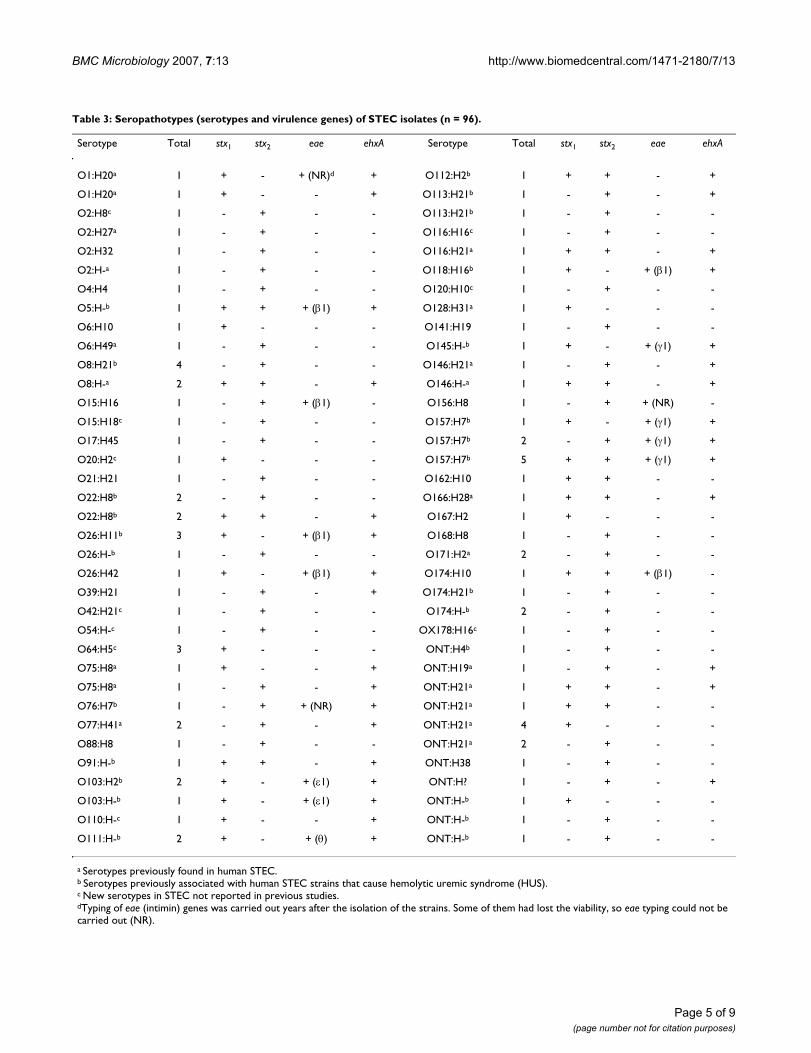
In the present study we have characterized 96 STEC iso-lates (Table 3). These isolates belonged to 42 O sero-groups, 61 O:H serotypes (including 10 new serotypes).The majority (68%) of STEC isolates belonged to sero-types previously found in human STEC and 38% to sero-types (O5:H-, O8:H21, O22:H8, O26:H11, O26:H-,O76:H7, O91:H-, O103:H2, O103:H-, O111:H-,O112:H2, O113:H21, O118:H16, O145:H-, O157:H7,O174:H21, O174:H-, ONT:H4 and ONT:H-) (ONT = Onot typeable) associated with STEC isolated from patientswith HUS. By PCR the 96 STEC isolates showed that 28(29%) carried stx1 genes, 49 (51%) stx2, and 19 (20%)both stx1 and stx2. Enterohemolysin (ehxA) and intimin(eae) virulence genes were detected in 43 (45%) and 25(26%) of the isolates, respectively. A total of 71 differentseropathotypes (associations between serotypes and viru-lence genes) were detected, being seropathotype O157:H7stx1stx2eae-γ1 ehxA the most common (five isolates), fol-lowed by O8:H21 stx2 and ONT:H21 stx1 (four isolates,respectively), O26:H11 stx1eae-β1 ehxA, and O64:H5 stx1(three isolates, respectively). Like other authors [40], we
Table 1: Prevalence of STEC in minced beef in Spain, from 1995 through 2003
Year No. of samples analysed STEC O157:H7 Non-O157 STEC Total STEC
1995 58 3 5% 8 14% 10 17%1996 91 0 0% 8 9% 8 9%1997 173 1 0.6% 20 12% 21 12%1998 133 1 0.8% 18 14% 18 14%2001 80 1 1.3% 6 8% 7 9%2002 20 0 0% 1 5% 1 5%2003 230 2 0.9% 29 13% 30 13%
Total 785 8 1.0% 90 11% 95 12%
Page 3 of 9(page number not for citation purposes)
BMC Microbiology 2007, 7:13 http://www.biomedcentral.com/1471-2180/7/13
have observed that STEC isolated from beef and humanSTEC of the same serotype have similar known virulence-associated properties.
At least 90 phage types have been reported for STECO157:H7 [41] but only seven of these (PT2, PT4, PT8,PT14, PT21/28, PT32 and PT54) accounted for the major-ity (>75%) of the human strains isolated in Europe andCanada [39]. Phage types PT2 and PT8 were predominantin human STEC O157:H7 strains in Spain as well as inmany other European countries, including Belgium, Fin-land, Germany, Italy, England and Scotland; whereasPT14 was the most frequently isolated in Canada. Six dif-ferent phage types were detected among the eightO157:H7 strains isolated in this study: PT2 and PT8 (twoisolates, respectively); PT21/28, PT32, PT54, PT atypical(one isolate, each). Interestingly, seven of those eight iso-lates belonged to the most common phage types associ-ated with severe human illnesses in Europe and Canada[39].
The eae gene, which has been shown to be necessary forattaching and effacing activity, encodes a 94- to 97-kDaouter membrane protein (OMP) which is termed intimin[4]. Numerous investigators have underlined the strongassociation between the carriage of eae gene and thecapacity of STEC strains to cause severe human illnesses,especially HUS [1-3]. This important virulence gene wasdetected in 100% of STEC O157:H7 and in 19% of non-O157 isolates from beef in the present study. Neverthe-less, production of intimin is not essential for pathogene-sis, because a number of sporadic cases of HUS have beencaused by eae-negative non-O157 STEC strains. Thus,STEC O104:H21 and O113:H21 strains lacking eae genewere responsible for an outbreak and a cluster of threeHUS cases in USA and Australia, respectively [1,42].
Differentiation of intimin alleles represents an importanttool for STEC typing in routine diagnostics as well as inpathogenesis, epidemiological, clonal and immunologi-cal studies [5,43-48]. The C-terminal end of intimin isresponsible for receptor binding, and it has been sug-gested that different intimins may be responsible for dif-ferent host tissue cell tropism. The 5'regions of eae genes
are conserved, whereas the 3'regions are heterogeneous.This observation led to the construction of universal PCRprimers and allele-specific PCR primers, which has madepossible to differentiate at present 21 variants of the eaegene encoding 21 different intimin types and subtypes(α1, α2, β1, ξR/β2B, δ/β2O, κ, γ1, γ2, θ, ε1, νR/ε2, ζ, η1,η2, ι1, µR/ι2, λ, µB, νB, ξB and ο) [5,8,48]. In this study,four eae variants were differentiated: γ1 (in nine isolates),β1 (eight isolates), ε1 (three isolates), and θ (two iso-lates).
ConclusionSeropathotypes O26:H11 stx1 eae-β1, O157:H7 stx1 stx2eae-γ1 and O157:H7 stx2 eae-γ1 are the most frequentlyobserved in STEC that cause human infections in Spain[3]. These highly virulent seropathotypes were detected in10 of 96 STEC isolates characterized in the present study,confirming minced beef as potential source of STEC iso-lates pathogenic for humans and reinforces the need ofHACCP (Hazard Analysis and Critical Control Point) pro-grams all through food chain, from abattoirs to markets,in order to reduce contamination and consequently, ill-nesses related to minced beef. In addition, consumersshould reduce their risk for STEC foodborne illnesses byfollowing safe food-handling recommendations and byavoiding consumption of raw or undercooked meat prod-ucts.
MethodsSample collection, culture, and STEC screeningA total of 785 of samples of minced beef were collectedfrom 30 local stores in Lugo city (Spain) between 1995and 2003. A maximum of 10 samples for week wereobtained at random from 10 markets located in differentdistricts. Some stores from those 30 were sampled alongthe seven years, while others only during a period of thestudy. In most cases, stores had different meat suppliers.
All minced beef samples were packaged individually insterile plastic bags, refrigerated for transportation to thelaboratory, and processed within 2 h of collection. For iso-lation of STEC O157:H7 a 25 g portion of each mincedbeef sample was homogenized in a stomacher with 225ml of buffered peptone water supplemented with vanco-
Table 2: Most probable number of E. coli and detection of STEC
MPNa of E. coli No. of samples analysed STEC O157:H7 Non-O157 STEC Total STEC
<10 488 2 (0.4%) 43 (9%) 43 (9%)10–99 183 2 (1%) 27 (15%) 29 (16%)
100–999 71 2 (3%) 8 (11%) 9 (13%)>999 43 2 (5%) 12 (28%) 14 (33%)
aMost probable number of E. coli per gram of minced beef.
Page 4 of 9(page number not for citation purposes)
BMC Microbiology 2007, 7:13 http://www.biomedcentral.com/1471-2180/7/13
Page 5 of 9(page number not for citation purposes)
Table 3: Seropathotypes (serotypes and virulence genes) of STEC isolates (n = 96).
Serotype Total stx1 stx2 eae ehxA Serotype Total stx1 stx2 eae ehxA
O1:H20a 1 + - + (NR)d + O112:H2b 1 + + - +
O1:H20a 1 + - - + O113:H21b 1 - + - +
O2:H8c 1 - + - - O113:H21b 1 - + - -
O2:H27a 1 - + - - O116:H16c 1 - + - -
O2:H32 1 - + - - O116:H21a 1 + + - +
O2:H-a 1 - + - - O118:H16b 1 + - + (β1) +
O4:H4 1 - + - - O120:H10c 1 - + - -
O5:H-b 1 + + + (β1) + O128:H31a 1 + - - -
O6:H10 1 + - - - O141:H19 1 - + - -
O6:H49a 1 - + - - O145:H-b 1 + - + (γ1) +
O8:H21b 4 - + - - O146:H21a 1 - + - +
O8:H-a 2 + + - + O146:H-a 1 + + - +
O15:H16 1 - + + (β1) - O156:H8 1 - + + (NR) -
O15:H18c 1 - + - - O157:H7b 1 + - + (γ1) +
O17:H45 1 - + - - O157:H7b 2 - + + (γ1) +
O20:H2c 1 + - - - O157:H7b 5 + + + (γ1) +
O21:H21 1 - + - - O162:H10 1 + + - -
O22:H8b 2 - + - - O166:H28a 1 + + - +
O22:H8b 2 + + - + O167:H2 1 + - - -
O26:H11b 3 + - + (β1) + O168:H8 1 - + - -
O26:H-b 1 - + - - O171:H2a 2 - + - -
O26:H42 1 + - + (β1) + O174:H10 1 + + + (β1) -
O39:H21 1 - + - + O174:H21b 1 - + - -
O42:H21c 1 - + - - O174:H-b 2 - + - -
O54:H-c 1 - + - - OX178:H16c 1 - + - -
O64:H5c 3 + - - - ONT:H4b 1 - + - -
O75:H8a 1 + - - + ONT:H19a 1 - + - +
O75:H8a 1 - + - + ONT:H21a 1 + + - +
O76:H7b 1 - + + (NR) + ONT:H21a 1 + + - -
O77:H41a 2 - + - + ONT:H21a 4 + - - -
O88:H8 1 - + - - ONT:H21a 2 - + - -
O91:H-b 1 + + - + ONT:H38 1 - + - -
O103:H2b 2 + - + (ε1) + ONT:H? 1 - + - +
O103:H-b 1 + - + (ε1) + ONT:H-b 1 + - - -
O110:H-c 1 + - - + ONT:H-b 1 - + - -
O111:H-b 2 + - + (θ) + ONT:H-b 1 - + - -
a Serotypes previously found in human STEC.b Serotypes previously associated with human STEC strains that cause hemolytic uremic syndrome (HUS).c New serotypes in STEC not reported in previous studies.dTyping of eae (intimin) genes was carried out years after the isolation of the strains. Some of them had lost the viability, so eae typing could not be carried out (NR).
BMC Microbiology 2007, 7:13 http://www.biomedcentral.com/1471-2180/7/13
mycin 8 mg liter -1, cefixime 0.05 mg liter -1, and cefsulo-din 10 mg liter -1 (BPWvcc) and incubated for 6 h at 37°C.One millilitre of this enriched culture was added to 20 µlof magnetic beads coated with O157-antibodies (Dyna-beads anti-E. coli O157, Dynal, Oslo) and the immu-nomagnetic separation (IMS) was performed according tothe manufacturer's instructions. The concentrated targetcells were plated onto sorbitol MacConkey (SMAC) agarand on cefixime tellurite sorbitol MacConkey (CTSMAC)agar (18 h at 37°C). For isolation of non-O157 STEC a 25g portion of each minced beef sample was homogenizedin an stomacher with 225 ml of buffered peptone waterwithout antibiotics (BPW) and incubated for 6 h at 37°C.The cells were plated onto CTSMAC and MacConkey(MAC) agar (18 h at 37°C and 44°C).
STEC were detected by PCR using specific primers foramplification of stx1, stx2, and eae genes (Table 4). ForPCR, a loopful of bacterial growth taken from the firststreaking area of the culture plates was suspended in 0.5ml of sterile distilled water and boiled for 5 min to releasethe DNA. For each PCR-positive culture, 10 E. coli-like col-onies obtained from MAC, SMAC and/or CTSMAC plateswere analysed by PCR in order to obtain the STEC isolatesfor further characterization. If no positive single colonywas found among the first 10 colonies, at least 40 morewere tested. If still none of the assayed coliform colonieswas positive in the PCR reaction, the sample was reportedas PCR positive without STEC isolation. Production ofShiga toxins (verocytotoxins) by PCR-positive isolates wasconfirmed by cytotoxicity tests on Vero and HeLa cells[13].
All STEC isolates were subsequently characterized bio-chemically with the API 20E system (bioMerieux, France)and serotyped. In samples from which all isolates wereidentical with respect to the profile of virulence genes andO:H serotypes, only one colony was selected. When onesample yielded colonies with different virulence genes orO:H serotypes, one of each was selected for further char-acterization.
Enumeration of E. coli (MPN)Most probable number (MPN) determination was per-formed by standard method using Petrifilm™ Select E. coliCount Plates (3M Microbiology Products, USA), accord-ing to manufacturer's instructions.
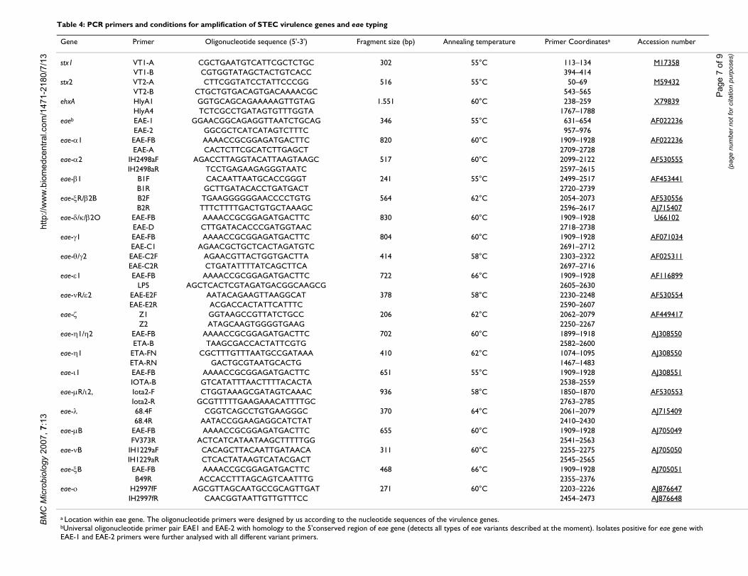
Detection of virulence genes and typing of eae (intimin) genes by PCRThe methodology used for the detection of virulencegenes and typing of intimin genes into eae α1, α2, β1, ξR/β2B, δ/β2O, κ, γ1, γ2, θ, ε1, νR/ε2, ζ, η1, η2, ι1, µR/ι2, λ,µB, νB, ξB and ο has been described elsewhere[7,8,12,13,48]. Base sequences and predicted sizes of
amplified products for the specific oligonucleotide prim-ers used in this study are shown in Table 4. The oligonu-cleotide primers were designed by us according to thenucleotide sequences of the virulence genes. Isolates pos-itive for eae gene with EAE-1 and EAE-2 primers were fur-ther analysed with all different variant primers. Due to thehigh sequence similarity, specific primers could not bedesigned for distinguishing between eae-γ2 and eae-θgenes, eae-δ and eae-κ, or eae-η1 and eae-η2. In those casesit was necessary to establish the nucleotide sequence of afragment from the 3' variable region of the eae gene. Thenucleotide sequence of the amplification products puri-fied with a QIAquick DNA purification kit (Qiagen) wasdetermined by the dideoxynucleotide triphosphate chaintermination method of Sanger, with the BigDye Termina-tor v3.1 Cycle Sequencing Kit and an ABI 3100 GeneticAnalyzer (Applied Bio-Systems).
SerotypingThe determination of O and H antigens was carried out bythe method described by Guinιe et al. [49] employing allavailable O (O1-O185) and H (H1-H56) antisera. Allantisera were obtained and absorbed with the correspond-ing cross-reacting antigens to remove the nonspecificagglutinins. The O antisera were produced in the LREC(Lugo, Spain) and the H antisera were obtained from theStatens Serum Institut (Copenhagen, Denmark).E. colistrains representing the new O groups O182 to O185(unpublished data) were kindly provided by FlemmingScheutz (Statens Serum Institut).
Phage typing of STEC O157:H7Phage typing was performed with the method of Khakriaet al. [41] in the Centro Nacional de Microbiología(Madrid) [39,50] using phages provided by The NationalLaboratory for Enteric Pathogens, Laboratory for DiseaseControl, Otawa, Ontario (Canada). The sixteen differentphages used were capable of identifying 90 phage types.
E. coli control strainsE. coli strains used as controls were: EPEC-2348 (human,O127:H6, eae-α1), AEEC-IH2498a (human, O125:H6,eae-α2), EPEC-337 (human, O111:H2, eae-β1), EPEC-359(human, O119:H6, eae-ξR/β2B), EPEC-BL152.1 (human,O86:H34, eae-δ/β2O), AEEC-6044/95 (human,O118:H5, eae-κ), STEC-EDL933 (human, O157:H7, stx1,stx2, eae-γ1), STEC-TW07926 (human, O111:H8, stx1, stx2,eae-θ), STEC-VTB-286 (bovine, O103:H2, stx1, eae-ε1),AEEC-IH3205a (human, O123:H19, eae-νR/ε2), STEC-VTO-50 (ovine, O156:H-, stx1, eae-ζ), AEEC-CF11201(human, O125:H-, eae-η1), H03/53199a (human,ONT:H45, eae-η2), AEEC-7476/96 (human, O145:H4,eae-ι1), AEEC-217-2 (human, O101:H-, eae-µR/ι2),AEEC-68-4 (human, O34:H-, eae-λ), EPEC-373 (human,O55:H51, eae-µB), AEEC-IH1229a (human, O10:H-, eae-
Page 6 of 9(page number not for citation purposes)
BM
C M
icro
biol
ogy
2007
, 7:1
3ht
tp://
ww
w.b
iom
edce
ntra
l.com
/147
1-21
80/7
/13
Page
7 o
f 9(p
age
num
ber n
ot fo
r cita
tion
purp
oses
)
Table 4: PCR primers and conditions for amplification of STEC virulence genes and eae typing
Gene Primer Oligonucleotide sequence (5'-3') Fragment size (bp) Annealing temperature Primer Coordinatesa Accession number
stx1 VT1-A CGCTGAATGTCATTCGCTCTGC 302 55°C 113–134 M17358VT1-B CGTGGTATAGCTACTGTCACC 394–414
stx2 VT2-A CTTCGGTATCCTATTCCCGG 516 55°C 50–69 M59432VT2-B CTGCTGTGACAGTGACAAAACGC 543–565
ehxA HlyA1 GGTGCAGCAGAAAAAGTTGTAG 1.551 60°C 238–259 X79839HlyA4 TCTCGCCTGATAGTGTTTGGTA 1767–1788
eaeb EAE-1 GGAACGGCAGAGGTTAATCTGCAG 346 55°C 631–654 AF022236EAE-2 GGCGCTCATCATAGTCTTTC 957–976
eae-α1 EAE-FB AAAACCGCGGAGATGACTTC 820 60°C 1909–1928 AF022236EAE-A CACTCTTCGCATCTTGAGCT 2709–2728
eae-α2 IH2498aF AGACCTTAGGTACATTAAGTAAGC 517 60°C 2099–2122 AF530555IH2498aR TCCTGAGAAGAGGGTAATC 2597–2615
eae-β1 B1F CACAATTAATGCACCGGGT 241 55°C 2499–2517 AF453441B1R GCTTGATACACCTGATGACT 2720–2739
eae-ξR/β2B B2F TGAAGGGGGGAACCCCTGTG 564 62°C 2054–2073 AF530556B2R TTTCTTTTGACTGTGCTAAAGC 2596–2617 AJ715407
eae-δ/κ/β2O EAE-FB AAAACCGCGGAGATGACTTC 830 60°C 1909–1928 U66102EAE-D CTTGATACACCCGATGGTAAC 2718–2738
eae-γ1 EAE-FB AAAACCGCGGAGATGACTTC 804 60°C 1909–1928 AF071034EAE-C1 AGAACGCTGCTCACTAGATGTC 2691–2712
eae-θ/γ2 EAE-C2F AGAACGTTACTGGTGACTTA 414 58°C 2303–2322 AF025311EAE-C2R CTGATATTTTATCAGCTTCA 2697–2716
eae-ε1 EAE-FB AAAACCGCGGAGATGACTTC 722 66°C 1909–1928 AF116899LP5 AGCTCACTCGTAGATGACGGCAAGCG 2605–2630
eae-νR/ε2 EAE-E2F AATACAGAAGTTAAGGCAT 378 58°C 2230–2248 AF530554EAE-E2R ACGACCACTATTCATTTC 2590–2607
eae-ζ Z1 GGTAAGCCGTTATCTGCC 206 62°C 2062–2079 AF449417Z2 ATAGCAAGTGGGGTGAAG 2250–2267
eae-η1/η2 EAE-FB AAAACCGCGGAGATGACTTC 702 60°C 1899–1918 AJ308550ETA-B TAAGCGACCACTATTCGTG 2582–2600
eae-η1 ETA-FN CGCTTTGTTTAATGCCGATAAA 410 62°C 1074–1095 AJ308550ETA-RN GACTGCGTAATGCACTG 1467–1483
eae-ι1 EAE-FB AAAACCGCGGAGATGACTTC 651 55°C 1909–1928 AJ308551IOTA-B GTCATATTTAACTTTTACACTA 2538–2559
eae-µR/ι2, Iota2-F CTGGTAAAGCGATAGTCAAAC 936 58°C 1850–1870 AF530553Iota2-R GCGTTTTTGAAGAAACATTTTGC 2763–2785
eae-λ 68.4F CGGTCAGCCTGTGAAGGGC 370 64°C 2061–2079 AJ71540968.4R AATACCGGAAGAGGCATCTAT 2410–2430
eae-µB EAE-FB AAAACCGCGGAGATGACTTC 655 60°C 1909–1928 AJ705049FV373R ACTCATCATAATAAGCTTTTTGG 2541–2563
eae-νB IH1229aF CACAGCTTACAATTGATAACA 311 60°C 2255–2275 AJ705050IH1229aR CTCACTATAAGTCATACGACT 2545–2565
eae-ξB EAE-FB AAAACCGCGGAGATGACTTC 468 66°C 1909–1928 AJ705051B49R ACCACCTTTAGCAGTCAATTTG 2355–2376
eae-ο H2997fF AGCGTTAGCAATGCCGCAGTTGAT 271 60°C 2203–2226 AJ876647IH2997fR CAACGGTAATTGTTGTTTCC 2454–2473 AJ876648
a Location within eae gene. The oligonucleotide primers were designed by us according to the nucleotide sequences of the virulence genes.bUniversal oligonucleotide primer pair EAE1 and EAE-2 with homology to the 5'conserved region of eae gene (detects all types of eae variants described at the moment). Isolates positive for eae gene with EAE-1 and EAE-2 primers were further analysed with all different variant primers.
BMC Microbiology 2007, 7:13 http://www.biomedcentral.com/1471-2180/7/13
νB), STEC-B49 (bovine, O80:H-, stx1,eae-ξB), AEEC-IH2997f (human, O129:H-, eae-ο) and K12-185 (negativefor stx1, stx2, bfpA, and eae genes). Strains were stored atroom temperature in nutrient broth with 0.75% of agar.
Authors' contributionsAM, CL, and PJ carried out the isolation of the E. coli andperformed the detection of virulence genes. MB, EAG andGD carried out the typing of the eae gene. JEB, MPA andMIB performed the serotyping. AE carried out the phagetyping. JB carried out the design of the study and, togetherwith AM, drafted the manuscript. All authors read andapproved the final manuscript.
AcknowledgementsThis paper is dedicated to the memory of Dr. Enrique A. González, an emi-nent scientist, an excellent Professor of Microbiology and a very good friend. We thank to A. Román and M. Núñez for their collaboration in this study. We thank Monserrat Lamela for skillful technical assistance. This work was supported by grants from the Fondo de Investigación Sanitaria (grants FIS G03-025-COLIRED-O157 and PI052023), from the Xunta de Galicia (grants PGIDIT02BTF26101PR, PGIDIT04RAG261014PR, PGIDIT05BTF26101PR, and PGIDIT065TAL26101PR), from the Comisión Interministerial de Ciencia y Tecnología (grants CICYT-ALI98-0616 and CICYT-FEDER-1FD1997-2181-C02-01), and from European Commission (FAIR programme grants CT98-4093 and CT98-3935). A. Mora, G. Dahbi and C. López acknowledge the Agencia Española de Cooperación Interna-cional (AECI) and the Xunta de Galicia for research fellowships.
References1. Paton JC, Paton AW: Pathogenesis and diagnosis of shiga toxin-
producing Escherichia coli infections. Clin Microbiol Rev 1998,11:450-479.
2. Beutin L, Krause G, Zimmermann S, Kaulfuss S, Gleier K: Charater-ization of Shiga toxin-producing Escherichia coli strains iso-lated from human patients in Germany over a 3-year period.J Clin Microbiol 2004, 42:1099-1108.
3. Blanco JE, Blanco M, Alonso MP, Mora A, Dahbi G, Coira MA, BlancoJ: Serotypes, virulence genes and intimin types of Shiga toxin(verotoxin)-producing Escherichia coli isolates from humanpatients: prevalence in Lugo, Spain, from 1992 through 1999.J Clin Microbiol 2004, 42:311-319.
4. Kaper JB, Elliott S, Sperandio V, Perna NT, Mayhew GF, Blattner FR:Attaching and effacing intestinal histopathology and thelocus of enterocyte effacement. In Escherichia coli O157:H7 andother Shiga toxin-producing E. coli strains Edited by: Kaper JB, O'BrienAD. American Society for Microbiology, Washington, DC;1998:163-182.
5. Garrido P, Blanco M, Moreno-Paz M, Briones C, Dahbi G, Blanco JE,Blanco J, Parro V: STEC-EPEC oligonucleotide microarray: anew tool for typing genetic variants of the LEE pathogenicityisland of human and animal Shiga toxin-producingEscherichia coli (STEC) and enteropathogenic E. coli (EPEC)strains. Clin Chem 2006, 52:192-201.
6. Blanco M, Podola NL, Krüger A, Sanz ME, Blanco JE, González EA,Dahbi G, Mora A, Bernárdez MI, Etcheverria AL, Arroyo GH, Luc-chesi PMA, Parma AE, Blanco J: Virulence genes and intimintypes of Shiga-toxin-producing Escherichia coli isolated fromcattle and beef products in Argentina. Int Microbiol 2004,7:269-276.
7. Blanco M, Schumacher S, Tasara T, Zweifel C, Blanco JE, Dahbi G,Blanco J, Stephan R: Serotypes, intimin variants and other viru-lence factors of eae positive Escherichia coli strains isolatedfrom healthy cattle in Switzerland. Identification of a newintimin variant gene (eae-η2). BMC Microbiol 2005, 5:23.
8. Blanco M, Blanco JE, Dahbi G, Alonso MP, Mora A, Coira MA, MadridC, Juárez A, Bernárdez MI, González EA, Blanco J: Identification of
two new intimin types in atypical enteropathogenicEscherichia coli. Int Microbiol 2006, 9(2):103-110.
9. Torres AG, Zhou G, Kaper JB: Adherence of diarrheagenicEscherichia coli strains to epithelial cells. Infect Immun 2005,73:18-29.
10. Schmidt H, Beutin L, Karch H: Molecular analysis of the plasmid-encoded hemolysin of Escherichia coli O157:H7 strain EDL933. Infect Immun 1995, 66:1055-1061.
11. Blanco J, Blanco M, Blanco JE, Mora A, Alonso MP, González EA,Bernárdez MI: Epidemiology of verocytotoxigenic Escherichiacoli (VTEC) in ruminants. In Verocytotoxigenic Escherichia coliEdited by: Duffy G, Garvey P, McDowell D. Food & Nutrition PressInc, Trumbull, USA; 2001:113-148.
12. Blanco M, Blanco JE, Mora A, Rey J, Alonso JM, Hermoso M, HermosoJ, Alonso MP, Dhabi G, González EA, Bernárdez MI, Blanco J: Sero-types, virulence genes, and intimin types of Shiga toxin(verotoxin)-producing Escherichia coli isolates from healthysheep in Spain. J Clin Microbiol 2003, 41:1351-1365.
13. Blanco M, Blanco JE, Mora A, Dahbi G, Alonso MP, González EA,Bernárdez MI, Blanco J: Serotypes, virulence genes, and intimintypes of Shiga toxin (Verotoxin)-producing Escherichia coliisolates from cattle in Spain and identification of a newintimin variant gene (eae-ξ). J Clin Microbiol 2004, 42:645-651.
14. Zweifel C, Blanco JE, Blanco M, Blanco J, Stephan R: Serotypes andvirulence genes of ovine non-O157 Shiga toxin-producingEscherichia coli in Switzerland. Int J Food Microbiol 2004,95:19-27.
15. Zweifel C, Schumacher S, Blanco M, Blanco JE, Tasara T, Blanco J,Stephan R: Phenotypic and genotypic characteristics of non-O157 Shiga toxin-producing Escherichia coli (STEC) fromSwiss cattle. Vet Microbiol 2005, 105:37-45.
16. Read SC, Gyles CL, Clarke RC, Lior H, McEwen S: Prevalence ofverocytotoxigenic Escherichia coli in ground beef, pork, andchicken in southwestern Ontario. Epidemiol Infect 1990,105:11-20.
17. Suthienkul O, Brown E, Seriwatana J, Tienthongdee S, Sastravaha S,Echeverria P: Shiga-like-toxin-producing Escherichia coli inretail meats and cattle in Thailand. Appl Environ Microbiol 1990,56:1135-1139.
18. Willshaw GA, Smith HR, Roberts D, Thirlwell J, Cheasly T, Rowe B:Examination of raw beef products for the presence of verocytotoxin producing Escherichia coli, particularly those ofserogroup O157. J Appl Bacteriol 1993, 75:20-426.
19. Brooks HJL, Mollison BD, Bettelheim KA, Matejka K, Paterson KA,Wark VK: Occurrence and virulence factors of non-O157Shiga toxin-producing Escherichia coli in retail meat in Dun-edin, New Zealand. Lett Appl Microbiol 2001, 32:118-122.
20. Samadpour M, Ongerth JE, Liston J, Tran N, Nguyen D, Whittam TS,Wilson RA, Tarr PI: Occurrence of Shiga-like toxin-producingEscherichia coli in retail fresh seafood, beef, lamb, pork, andpoultry from grocery stores in Seattle, Washington. ApplEnviron Microbiol 1994, 60:1038-1040.
21. Samadpour M, Kubler M, Buck FC, Depavia GA, Mazengia E, StewartJ, Yang P, Alfi D: Prevalence of Shiga toxin-producingEscherichia coli in ground beef and cattle feces from KingCounty, Washington. J Food Prot 2002, 65:1322-1325.
22. Schroeder CM, White DG, Ge B, Zhang Y, McDermott PF, Ayers S,Zhao S, Meng J: Isolation of antimicrobial-resistant Escherichiacoli from retail meats purchased in Greater Washington,DC, USA. Int J Food Microbiol 2003, 85:197-202.
23. Parma AE, Sanz ME, Blanco JE, Blanco J, Viñas MR, Blanco M: Viru-lence genotypes and serotypes of verotoxigenic Escherichiacoli isolated from cattle and foods in Argentina. Eur J Epidemiol2000, 16:757-762.
24. Khan A, Yamasaki S, Sato T, Ramamurthy T, Pal A, Datta S, Chowd-hury NR, Das SC, Sikdar A, Tsukamoto T, Bhattacharya SK, TakedaY, Nair GG: Prevalence and genetic profiling of virulencedeterminants of non-O157 Shiga toxin-producing Escherichiacoli isolated from cattle, beef, and humans, Calcutta, India.Emerg Infect Dis 2002, 8:54-62.
25. Chapman PA, Siddons CA, Cerdan Malo AT, Harkin MA: A one yearstudy of Escherichia coli O157 in raw beef and lamb products.Epidemiol Infect 2000, 124:207-213.
26. Chapman PA, Cerdán Malo AT, Ellin M, Ashton R, Harkin MA:Escherichia coli O157 in cattle and sheep at slaughter, on beef
Page 8 of 9(page number not for citation purposes)
http://www.ncbi.nlm.nih.gov/entrez/query.fcgi?cmd=Retrieve&db=PubMed&dopt=Abstract&list_uids=9665978
http://www.ncbi.nlm.nih.gov/entrez/query.fcgi?cmd=Retrieve&db=PubMed&dopt=Abstract&list_uids=2200697
http://www.ncbi.nlm.nih.gov/entrez/query.fcgi?cmd=Retrieve&db=PubMed&dopt=Abstract&list_uids=2200697
http://www.ncbi.nlm.nih.gov/entrez/query.fcgi?cmd=Retrieve&db=PubMed&dopt=Abstract&list_uids=2187402
http://www.ncbi.nlm.nih.gov/entrez/query.fcgi?cmd=Retrieve&db=PubMed&dopt=Abstract&list_uids=2187402
http://www.ncbi.nlm.nih.gov/entrez/query.fcgi?cmd=Retrieve&db=PubMed&dopt=Abstract&list_uids=8161171
BMC Microbiology 2007, 7:13 http://www.biomedcentral.com/1471-2180/7/13
Publish with BioMed Central and every scientist can read your work free of charge
"BioMed Central will be the most significant development for disseminating the results of biomedical research in our lifetime."
Sir Paul Nurse, Cancer Research UK
Your research papers will be:
available free of charge to the entire biomedical community
peer reviewed and published immediately upon acceptance
cited in PubMed and archived on PubMed Central
yours — you keep the copyright
Submit your manuscript here:http://www.biomedcentral.com/info/publishing_adv.asp
BioMedcentral
and lamb carcasses and in raw beef and lamb products inSouth Yorkshire, UK. Int J Food Microbiol 2001, 64:139-150.
27. Heuvelink AE, Zwartkruis-Nahuis JTM, Beumer RR, De Boer E:Occurrence and survival of verocytotoxin-producingEscherichia coli O157 in meats obtained from retail outlets inThe Netherlands. J Food Prot 1999, 62:1115-1122.
28. Conedera G, Dalvit P, Martín M, Galiero G, Gramaglia M, Goffredo E,Loffredo G, Morabito S, Ottaviani D, Paterlini F, Pezzotti G, Pisanu M,Semprini P, Caprioli A: Verocytotoxin-producing Escherichiacoli O157 in minced beef and dairy products in Italy. Int J FoodMicrobiol 2004, 96:67-73.
29. Stampi S, Caprioli A, De Luca G, Quaglio P, Sacchetti R, Zanetti F:Detection of Escherichia coli O157 in bovine meat productsin northern Italy. Int J Food Microbiol 2004, 90:257-262.
30. Lukásová J, Abraham B, Cupáková S: Occurrence of Escherichiacoli O157 in raw material and food in Czech Republic. J VetMed B 2004, 51:77-81.
31. Tarr PI, Tran TN, Wilson RA: Escherichia coli O157:H7 in retailground beef in Seattle: results of a one-year prospectivestudy. J Food Prot 1999, 62:133-139.
32. Doyle MP, Schoeni JL: Isolation of Escherichia coli O157:H7 fromretail fresh meats and poultry. Appl Environ Microbiol 1987,53:2394-2396.
33. Padhye NV, Doyle MP: Rapid procedure for detecting Entero-hemorrhagic Escherichia coli O157:H7 in food. Appl EnvironMicrobiol 1991, 57:2693-2698.
34. Chinen I, Tanaro JD, Miliwebsky E, Lound LH, Chillemi G, Ledri S,Baschkier A, Scarpin M, Manfredi E, Rivas M: Isolation and charac-terization of Escherichia coli O157:H7 from retail meats inArgentina. J Food Prot 2001, 64:1346-1351.
35. Zhou Z, Nishikawa Y, Zhu P, Hong S, Hase A, Cheasty T, Smith HR,Zheng M, Haruki K: Isolation and characterization of Shigatoxin-producing Escherichia coli O157:H7 from beef, pork,and cattle fecal samples in Changchun, China. J Vet Med Sci2002, 64:1041-1044.
36. Mora A, León SL, Blanco M, Blanco JE, López C, Dahbi G, Echeita A,González EA, Blanco J: Phage types, virulence genes and PFGEprofiles of Shiga toxin-producing Escherichia coli O157:H7isolated from raw beef meat, soft cheese and vegetables inLima (Peru). Int J Food Microbiol 2007, 114:204-210.
37. Sekla L, Milley D, Stackiw W: Verotoxin-producing Escherichiacoli in ground beef in Manitoba. Can Med Assoc J 1990,143:519-521.
38. [http://www.lugo.usc.es/ecoli/SEROTIPOSHUM.htm].39. Mora A, Blanco M, Blanco JE, Alonso MP, Dahbi G, Thompson-Carter
F, Usera MA, Bartolomé R, Prats G, Blanco J: Phage Types andGenotypes of Human and Animal Shiga Toxin-ProducingEscherichia coli O157:H7 in Spain. Identification of two pre-dominating phage types (PT2 and PT8). J Clin Microbiol 2004,42:4007-4015.
40. Meng J, Zhao S, Doyle MP: Virulence genes of shiga toxin-pro-ducing Escherichia coli isolated from food, animals andhumans. Int J Food Microbiol 1998, 45:229-235.
41. Khakria R, Duck D, Lior H: Extended phage-typing scheme forEscherichia coli O157:H7. Epidemiol Infect 1990, 105:511-520.
42. Paton AW, Woodrow MC, Doyle RM, Lanser JA, Paton JC: Molecu-lar characterization of a shiga toxigenic Escherichia coliO113:H21 strain lacking eae responsible for a cluster of casesof hemolytic-uremic syndrome. J Clin Microbiol 1999,37:3357-3361.
43. Adu-Bobie J, Frankel G, Bain C, Goncalves AG, Trabulsi LR, Douce G,Knutton S, Dougan G: Detection of intimins α, β, γ, and δ, fourintimin derivatives expressed by attaching and effacingmicrobial pathogens. J Clin Microbiol 1998, 36:662-668.
44. Oswald E, Schmidt H, Morabito S, Karch H, Marchès O, Caprioli A:Typing of intimin genes in human and animal enterohemor-rhagic and enteropathogenic Escherichia coli: characteriza-tion of a new intimin variant. Infect Immun 2000, 68:64-71.
45. Tarr CL, Whittam S: Molecular evolution of the intimin gene inO111 clones of pathogenic Escherichia coli. J Bacteriol 2002,184:479-487.
46. Zhang WL, Köhler B, Oswald E, Beutin L, Karch H, Morabito S, Cap-rioli A, Suerbaum S, Schmidt H: Genetic diversity of intimingenes of attaching and effacing Escherichia coli strains. J ClinMicrobiol 2002, 40:4486-4492.
47. Ramachandran V, Brett K, Hornitzky MA, Dowton M, Bettelheim KA,Walker MJ, Djordjevic SP: Distribution of intimin subtypesamong Escherichia coli isolates from ruminant and humansources. J Clin Microbiol 2003, 41:5022-5032.
48. Blanco M, Blanco JE, Dahbi G, Mora A, Alonso MP, Varela G, GadeaMP, Schelotto F, Gonzalez EA, Blanco J: Typing of intimin (eae)genes from enteropathogenic Escherichia coli (EPEC) iso-lated from children with diarrhoea in Montevideo, Uruguay:identification of two novel intimin variants (µB and ξR/β2B).J Med Microbiol 2006, 55:1165-1174.
49. Guinée PAM, Jansen WH, Wadström T, Sellwood R: Escherichia coliassociated with neonatal diarrhoea in piglets and calves. InLaboratory Diagnosis in Neonatal Calf and Pig diarrhoea: Current Topics inVeterinary and Animal Science Edited by: Leeww PW, Guinée PAM.Martinus-Nijhoff, The Hague, Netherlands; 1981:126-162.
50. Mora A, Blanco JE, Blanco M, Alonso MP, Dhabi G, Echeita A,Gonzalez EA, Bernardez MI, Blanco J: Antimicrobial resistance ofShiga toxin (verotoxin)-producing Escherichia coli O157:H7and non-O157 strains isolated from humans, cattle, sheepand food in Spain. Res Microbiol 2005, 156:793-806.
Page 9 of 9(page number not for citation purposes)
http://www.ncbi.nlm.nih.gov/entrez/query.fcgi?cmd=Retrieve&db=PubMed&dopt=Abstract&list_uids=3322190
http://www.ncbi.nlm.nih.gov/entrez/query.fcgi?cmd=Retrieve&db=PubMed&dopt=Abstract&list_uids=3322190
http://www.ncbi.nlm.nih.gov/entrez/query.fcgi?cmd=Retrieve&db=PubMed&dopt=Abstract&list_uids=1768144
http://www.ncbi.nlm.nih.gov/entrez/query.fcgi?cmd=Retrieve&db=PubMed&dopt=Abstract&list_uids=9927001
http://www.ncbi.nlm.nih.gov/entrez/query.fcgi?cmd=Retrieve&db=PubMed&dopt=Abstract&list_uids=9927001
http://www.ncbi.nlm.nih.gov/entrez/query.fcgi?cmd=Retrieve&db=PubMed&dopt=Abstract&list_uids=2249715
http://www.ncbi.nlm.nih.gov/entrez/query.fcgi?cmd=Retrieve&db=PubMed&dopt=Abstract&list_uids=9508292
http://www.ncbi.nlm.nih.gov/entrez/query.fcgi?cmd=Retrieve&db=PubMed&dopt=Abstract&list_uids=9508292


![Debuxo e ornamentación na arquitectura do sur da provincia de Lugo [in Galician]](https://static.fdokumen.com/doc/165x107/6331ea76576b626f850d29ed/debuxo-e-ornamentacion-na-arquitectura-do-sur-da-provincia-de-lugo-in-galician.jpg)