Characterization of Biofilm Formation by Clinically Relevant Serotypes of Group A Streptococci
13
10.1128/AEM.72.4.2864-2875.2006. 2006, 72(4):2864. DOI: Appl. Environ. Microbiol. Kreikemeyer Hidalgo-Grass, Ludwig Jonas, Emanuel Hanski and Bernd Cordula Lembke, Andreas Podbielski, Carlos Streptococci Clinically Relevant Serotypes of Group A Characterization of Biofilm Formation by http://aem.asm.org/content/72/4/2864 Updated information and services can be found at: These include: SUPPLEMENTAL MATERIAL Supplemental material REFERENCES http://aem.asm.org/content/72/4/2864#ref-list-1 at: This article cites 58 articles, 28 of which can be accessed free CONTENT ALERTS more» articles cite this article), Receive: RSS Feeds, eTOCs, free email alerts (when new http://journals.asm.org/site/misc/reprints.xhtml Information about commercial reprint orders: http://journals.asm.org/site/subscriptions/ To subscribe to to another ASM Journal go to: on September 9, 2014 by guest http://aem.asm.org/ Downloaded from on September 9, 2014 by guest http://aem.asm.org/ Downloaded from
-
Upload
independent -
Category
Documents
-
view
1 -
download
0
Transcript of Characterization of Biofilm Formation by Clinically Relevant Serotypes of Group A Streptococci

10.1128/AEM.72.4.2864-2875.2006.
2006, 72(4):2864. DOI:Appl. Environ. Microbiol. KreikemeyerHidalgo-Grass, Ludwig Jonas, Emanuel Hanski and Bernd Cordula Lembke, Andreas Podbielski, Carlos StreptococciClinically Relevant Serotypes of Group A Characterization of Biofilm Formation by
http://aem.asm.org/content/72/4/2864Updated information and services can be found at:
These include:
SUPPLEMENTAL MATERIAL Supplemental material
REFERENCEShttp://aem.asm.org/content/72/4/2864#ref-list-1at:
This article cites 58 articles, 28 of which can be accessed free
CONTENT ALERTS more»articles cite this article),
Receive: RSS Feeds, eTOCs, free email alerts (when new
http://journals.asm.org/site/misc/reprints.xhtmlInformation about commercial reprint orders: http://journals.asm.org/site/subscriptions/To subscribe to to another ASM Journal go to:
on Septem
ber 9, 2014 by guesthttp://aem
.asm.org/
Dow
nloaded from
on Septem
ber 9, 2014 by guesthttp://aem
.asm.org/
Dow
nloaded from
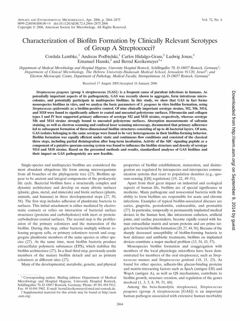
APPLIED AND ENVIRONMENTAL MICROBIOLOGY, Apr. 2006, p. 2864–2875 Vol. 72, No. 40099-2240/06/$08.00�0 doi:10.1128/AEM.72.4.2864–2875.2006Copyright © 2006, American Society for Microbiology. All Rights Reserved.
Characterization of Biofilm Formation by Clinically Relevant Serotypesof Group A Streptococci†
Cordula Lembke,1 Andreas Podbielski,1 Carlos Hidalgo-Grass,2 Ludwig Jonas,3Emanuel Hanski,2 and Bernd Kreikemeyer1*
Department of Medical Microbiology and Hospital Hygiene, University Hospital Rostock, Schillingallee 70, D-18057 Rostock, Germany1;Department of Clinical Microbiology, The Hebrew University-Hadassah Medical School, Jerusalem 91120, Israel2; and
Electron Microscopic Centre, Department of Pathology, Medical Faculty, Strempelstrasse 14, D-18057 Rostock, Germany3
Received 17 August 2005/Accepted 16 January 2006
Streptococcus pyogenes (group A streptococcus [GAS]) is a frequent cause of purulent infections in humans. Aspotentially important aspects of its pathogenicity, GAS was recently shown to aggregate, form intratissue micro-colonies, and potentially participate in multispecies biofilms. In this study, we show that GAS in fact formsmonospecies biofilms in vitro, and we analyze the basic parameters of S. pyogenes in vitro biofilm formation, usingStreptococcus epidermidis as a biofilm-positive control. Of nine clinically important serotype strains, M2, M6, M14,and M18 were found to significantly adhere to coated and uncoated polystyrene surfaces. Fibronectin and collagentypes I and IV best supported primary adherence of serotype M2 and M18 strains, respectively, whereas serotypeM6 and M14 strains strongly bound to uncoated polystyrene surfaces. Absorption measurements of safraninstaining, as well as electron scanning and confocal laser scanning microscopy, documented that primary adherenceled to subsequent formation of three-dimensional biofilm structures consisting of up to 46 bacterial layers. Of note,GAS isolates belonging to the same serotype were found to be very heterogeneous in their biofilm-forming behavior.Biofilm formation was equally efficient under static and continuous flow conditions and consisted of the classicalthree steps, including partial disintegration after long-term incubation. Activity of the SilC signaling peptide as acomponent of a putative quorum-sensing system was found to influence the biofilm structure and density of serotypeM14 and M18 strains. Based on the presented methods and results, standardized analyses of GAS biofilms andtheir impact on GAS pathogenicity are now feasible.
Single-species and multispecies biofilms are considered themost abundant ubiquitous life form among microorganismsfrom all branches of the phylogenetic tree (27). Biofilms ap-pear to be ancient and integral components of the prokaryoticlife cycle. Bacterial biofilms have a structurally complex anddynamic architecture and develop on many abiotic surfaces(plastic, glass, metal, and minerals) and biotic surfaces (plants,animals, and humans) in three developmental steps (27, 50,58). The first step includes adhesion of planktonic bacteria tosurfaces. This initial attachment is either mediated by electro-static contacts or relies on interaction of bacterial surfacestructures (proteins and carbohydrates) with inert or protein-carbohydrate-coated surfaces. The second step is the prolifer-ation of the primary colonizers and the maturation of thebiofilm. During this step, either bacteria multiply without re-leasing progeny cells, or primary colonizers recruit and coag-gregate planktonic members of the same species or other spe-cies (27). At the same time, most biofilm bacteria produceextracellular polymeric substances (EPS), which stabilize thebiofilm architecture (27). In a final third step, previously sessilemembers of the mature biofilm detach and act as primarycolonizers at different sites (27).
Many of the developmental, metabolic, genetic, and physical
properties of biofilm establishment, maturation, and disinte-gration are regulated by intraspecies and interspecies commu-nication systems that react to population densities (e.g., quo-rum-sensing [QS] regulation) (19, 22, 49–51).
Apart from their great impact on industrial and ecologicalaspects of human life, biofilms are of special significance inmedicine. Many pathogenic and nosocomial bacteria with theability to form biofilms are responsible for acute and chronicinfections. Examples of typical biofilm-associated diseases arecaries, gingivitis, periodontitis, endocarditis, and prostatitis(27). In particular, temporally or permanently implanted medicaldevices in the human host, like intravenous catheters, artificialjoints, and cardiac pacemakers, become rapidly coated with hu-man extracellular matrix and serum proteins and are prime tar-gets for bacterial biofilm formation (20, 27, 44, 56). Because of thesharply decreased susceptibility of biofilm-forming bacteria tohost defenses and antibiotic treatments, biofilms on implanteddevices constitute a major medical problem (13, 34, 43, 57).
Monospecies biofilm formation and coaggregation withmembers of the local physiologic microflora have been dem-onstrated for members of the oral streptococci, such as Strep-tococcus mutans and Streptococcus gordonii (18, 21, 23). Asfound for other species, adhesin-like glucan-binding proteinsand matrix-interacting factors such as SpaA (antigen I/II) andWapA (antigen A), as well as QS mechanisms, contribute tobiofilm growth, structure creation, and regulation of the genesinvolved (1, 3, 5, 8, 39, 51, 60).
Among the beta-hemolytic streptococci, Streptococcuspyogenes (group A streptococcus [GAS]) is an importanthuman pathogen associated with extensive human morbidity
* Corresponding author. Mailing address: Department of MedicalMicrobiology and Hospital Hygiene, University Hospital Rostock,Schillingallee 70, D-18057 Rostock, Germany. Phone: 49 381 494 5912.Fax: 49 81494 5902. E-mail: [email protected].
† Supplemental material for this article may be found at http://aem.asm.org/.
2864
on Septem
ber 9, 2014 by guesthttp://aem
.asm.org/
Dow
nloaded from

worldwide (17). GAS invades host cells (15) and causesprimary infections of the skin, throat, and other mucosalsurfaces, such as pharyngitis and impetigo. Particularly,pharyngitis is a primary infection that can occur in multipleepisodes, due to antibiotic treatment failure in up to 30% ofaffected individuals. A recent study by Conley et al. (11)revealed that GAS strains recovered from patients withtreatment failure in fact form biofilms in vitro with variableefficiency and that, compared to planktonic cultures, GASorganized in biofilms have higher MICs for all standardantibiotics used for treatment.
GAS biofilm-like structures (microcolonies) were also re-ported to exist in vivo by Hirota et al. (33) and Akiyama et al.(2). The latter study documented GAS microcolonies in skinsections from patients with impetigo and atopic dermatitis.Takemura and colleagues identified GAS as members of rootcanal multispecies biofilms (52). Additionally, another recentstudy published during the review process of this paper iden-tified biofilm-like tissue communities in GAS-infected zebra-fish, studied the transcriptome of a serotype M5 GAS strainfrom tissue communities and biofilms, and identified the M5protein and Mga and CovR regulators as important for biofilmformation of this serotype (9). One step that could contributeto midstage biofilm formation by GAS has been studied in vitrowith GAS serotypes M1, M4, M12, and M49, which formmicrocolonies when grown in liquid medium. The aggregationinto microcolonies is mediated by a conserved 19-amino-acidresidue peptide (designated AHP) present in M protein andprotein H (24).
The present study investigated several aspects of in vitroGAS biofilm formation employing different clinically relevantGAS serotype strains. By using a variety of microscopic tech-niques under static and flow conditions, we demonstrate thatsome serotype strains have the capacity to form biofilms. Yet,these serotype strains have an adherence tendency toward dif-ferent substrates, which constitutes the first step in their tran-sition from planktonic to sessile lifestyle structures. Further-more, isolates of the same serotype are heterogeneous in theirbiofilm-building capacity. Finally we show that silC, a compo-nent of the putative QS system (31, 32), plays an important rolein biofilm formation by the M14 and M18 serotypes.
MATERIALS AND METHODS
Bacterial strains and growth conditions. The serotype M49 GAS strain 591 isa skin isolate originally obtained from R. Lutticken, Aachen, Germany. Thedifferent M serotype GAS strains (M1, M2, M3, M6, M12, M18, and M28) usedin this study are all clinical isolates obtained from the collection of the Centre ofEpidemiology and Microbiology, National Institute of Public Health, Prague,Czech Republic, and have been previously described by Podbielski et al. (46).GAS serotype strain M14 and its corresponding silC-deficient mutant were de-scribed by Hidalgo-Grass et al. (31). The biofilm formation of Staphylococcusepidermidis DSM 1798 was used as an internal control for all experiments.Escherichia coli strain DH5� was purchased from GibcoBRL (Eggenstein, Ger-many) and served as a host for plasmids pAT::egfp and pO-KK. E. coli was grownin Luria broth (LB) or on LB agar plates at 37°C for all experiments. All GASstrains were routinely grown in Todd-Hewitt (TH) broth or TH agar platessupplemented with 0.5% yeast extract. For biofilm experiments, GAS and S.epidermidis were grown in LB, TH agar plates supplemented with 0.5% yeastextract, brain heart infusion (BHI) medium, or chemically defined medium. Thegrowth medium of recombinant E. coli strains and GAS mutants was supple-mented with 100 and 60 mg of spectinomycin · liter�1, respectively. GAS strainswere grown as standing cultures at 37°C under a 5% CO2–20% O2 atmosphereunless otherwise indicated.
Construction of the silC mutants and GAS strains carrying enhanced greenfluorescent protein gene (egfp). The construction of the serotype M14 GAS strainsilC mutant was described in a previous publication (31). To construct a plasmidthat would allow an allelic replacement of silC by aad9 (spectinomycin resistancecassette) in serotype M18, we amplified a 1,572-bp fragment containing silC, 695bp upstream and 511 bp downstream, using primers SilB-f2 (5�-TTATTGGATCGGAACTTACGC-3�) and SilD-r2 (5�-TGCTTCCCAACAACTTACCAC-3�),designed according to the serotype M14 chromosomal DNA. The PCR productwas then cloned into pORI280, and a StyI-BspEI fragment encompassing silCwas replaced by aad9. The resulting plasmid (pO-KK) was electroporated intoserotype M18, and mutants with resistance to spectinomycin but susceptibility toerythromycin were selected. The fidelity of the replacement and the integrity ofsilB and silD were confirmed by PCR using primers SilB-f2 and SilD-r2. The PCRproduct was sequenced to confirm the sites of silC replacement.
For construction of the egfp expression plasmid, the egfp gene was releasedfrom vector pKen (12) by restriction digestion with XbaI and PstI. The fragmentwas cloned into pAT28 (55) downstream of a constitutively active recA promoterfrom GAS, which was cloned as an EcoRI/BamHI fragment into pAT28. Theresulting plasmid pAT::egfp was sequenced to confirm its integrity.
Biofilm assays. All biofilm experiments that used safranin staining as a detec-tion method were performed in 96-well polystyrene microtiter plates (GreinerBio-One, Frickenhausen, Germany). For all microscopic detection methods,biofilms were allowed to grow on round coverslips (Nunc, Wiesbaden, Germany)placed in 24-well polystyrene cell culture plates (Greiner Bio-One). Plastic sur-faces of the 96-well plates and coverslips were either used uncoated or werecoated with human fibronectin (Roche), human fibrinogen (Sigma), human col-lagen types I and IV (Biomol), or human laminin (Sigma) at a concentration of50 �g · ml�1 overnight at 4°C. After being washed three times with phosphate-buffered saline (PBS), wells were used for inoculation with bacteria. GAS overnightcultures were harvested, and 100 �l of a suspension containing 104 CFU · ml�1 wasinoculated into 100 �l fresh medium per well in 96-well plates and 900 �l freshmedium for 24-well plates, respectively. After desired incubation time points (from12 h up to 120 h), the remaining planktonic bacteria were removed by aspiration ofthe liquid.
For quantitative measurements, we used a modified method of Christensen etal. (10). Essentially, 96-well plates with biofilms were washed twice with PBS andstained in a 0.1% safranin solution for 30 min. Stained biofilms were washedthree times in PBS and allowed to dry, and staining was quantified in a spectro-photometer at 492 nm. In parallel experiments, unstained biofilm bacteria wereremoved from the wells by vigorous washing and vortexing, and bacterial num-bers were quantified in a Thomae counting chamber by light microscopy. Like inmany other studies that used safranin staining, we found a good correlationbetween increasing total cell numbers and increasing optical density of safranin-stained biofilms over the investigated time course (data not shown). Individualassays always consisted of three replicates. All experiments were performed atleast four times. The data shown represent the means and standard deviations offour or more experiments.
Microscopic techniques. For visualization of biofilms by light microscopy, theincubation of the bacteria on the uncoated or coated plastic coverslips wasstopped at the indicated time points. Safranin staining was performed as de-scribed above. Stained coverslips were placed on slides with the biofilm pointingup and were inspected by light or fluorescence microscopy at magnifications of�100 and �200 (BX60 microscope; Olympus, Hamburg, Germany). Visiblebiofilms were documented with an attached digital camera (Leica, Solms, Ger-many).
Sample preparation for scanning electron microscopy (SEM) studies was per-formed as follows. The biofilms on the coverslips were fixed for 1 h in a solutioncontaining 2.5% glutardialdehyde. Coverslips were washed in 0.1 M Na acetatebuffer (pH 7.3) and dehydrated in a graded series of ethanol. Subsequently,coverslips were subjected to critical point drying with CO2, covered with gold toa 10-nm layer, and examined with a Zeiss DSM 960A electron microscope.
For confocal laser scanning microscopy (CLSM) studies, biofilms of GASserotype strains M2, M6, and M18, all transformed with an egfp-expressingplasmid (the strains resulting were TM2, TM6, and TM18, respectively), weregrown in glass-bottom chamber slides (Nunc). GAS serotype strain TM6 wasgrown on uncoated glass, TM2 was grown on fibronectin-coated glass, and TM18was grown on collagen IV-coated glass. The coating was done as described above.A total of 200 �l of a suspension containing 104 CFU · ml�1 (each) TM2, TM6,and TM18 GAS bacteria was inoculated in 200 �l fresh BHI medium containedin the chamber slide wells. Preparations were inspected after desired time pointsof growth with a Zeiss inverted microscope attached to a Leica TCS SP2 AOBSlaser scanning confocal imaging system with an Argon laser at a 488-nm excita-tion wavelength.
VOL. 72, 2006 BIOFILM FORMATION BY STREPTOCOCCUS PYOGENES 2865
on Septem
ber 9, 2014 by guesthttp://aem
.asm.org/
Dow
nloaded from

Flow chamber system. For investigation of GAS biofilm development underconstant flow conditions, a flow chamber system (FCS1c; Oligene, Berlin, Ger-many) was used. This chamber consisted of a main corpus with tubing adaptors,clip-on spring, silicon gaskets, plastic coverslips (diameter, 25 mm) and siliconetubing with a luer fitting on each side. The channel length was 15 mm, thechannel width was 2 mm, and the channel height was 300 �m. Shear stresses of0 to 3 Pa were applied. For a complete description of the system please refer tohttp://www.oligene.de/Flow_Chamber_System_FCS_1c.95.0.html. The plasticcoverslips were coated with fibronectin and collagen IV for GAS serotype strainsM2 and M18, respectively. The sterilized and assembled flow chamber systemwas attached to a pump, which was subsequently connected via tubing with aninlet of a flask containing fresh sterile BHI. The system was closed by connectingthe flask outlet via tubing to the chamber inlet. The medium was inoculated withthe respective GAS strains under sterile conditions, and the complete system wasincubated at 37°C for 72 h. A constant medium flow of 1 ml · min�1 was foundappropriate in preliminary experiments. To monitor biofilm growth after 72 h,the coverslips were removed from the flow chamber system and subsequentlyprepared for SEM as described above.
Statistics. The two-tailed Mann-Whitney U test was used to calculate statis-tical significance between data sets.
RESULTS
Evaluation of basic parameters of GAS biofilm formation.To initially assess the effect of growth medium, oxygen partialpressure, and incubation time on potential biofilm assembly, S.epidermidis (positive control) and serotype M6 and M49 GASstrains were used with uncoated polystyrene microtiter platesurfaces as solid supports. Safranin staining was employed, asit is an established method for staphylococcal biofilm researchand in parallel experiments the total number of biofilm bacte-ria was enumerated by light microscopy, giving concordantdata (data not shown). Since the staining and quantificationtechniques did not allow the visualization of three-dimensionalstructures, potential biofilms will be referred to as “adherentbacteria” or “primary colonizers” unless experimentally provenotherwise.
Preliminary results documented that BHI medium best sup-ported adherence to uncoated plastic of all GAS strains tested.The oxygen partial pressure had no significant influence (datanot shown). As illustrated in Fig. 1, the S. epidermidis-positivecontrol measurably adhered as early as 24 h postinoculation(p.i.) to the plastic surface. Judged by safranin staining andabsorption measurements, the serotype M49 GAS strain failedto adhere in detectable numbers during the whole investigationperiod (Fig. 1). Increasing numbers of bacteria, assembled in apotential biofilm, were detected for serotype M6 GAS up to72 h p.i., when an absorption maximum was reached (Fig. 1). Inaddition, change of the growth medium at daily intervals didnot increase the number of adhered bacteria (data not shown).
As a consequence of these data, BHI medium and staticcultures (unless stated otherwise) in ambient air at 37°C wereused for all further experiments. Samples at 24, 48, and 72 hp.i. were routinely analyzed in the subsequent experiments.Whether the adherence led to formation of a multilayeredbiofilm needed further investigation.
The effect of matrix substratum on biofilm development byvarious GAS serotypes. Primary adhesion of planktonic bacte-rial cells is a crucial step in biofilm formation. So far, GAS havenot been described as colonizing implanted medical devices,but they bind to surfaces coated with extracellular matrix pro-teins during the infectious process. Thus, one isolate each ofnine different clinically important GAS serotype strains (M1,
M2, M3, M6, M12, M14, M18, M28, and M49), including an S.epidermidis strain as a positive control, was tested for its pri-mary adhesion to uncoated plastic surfaces and plastic surfacescoated with fibronectin, fibrinogen, collagen types I and IV,and laminin, using the previously established assay conditionsand quantitative measurements of safranin staining.
The experiments showed that the GAS isolates of serotypesM1 (Fig. 2A), M12, M28, and M49 (data not shown) did notdisplay significant primary adhesion on any of the uncoated ormatrix protein-coated plastic surfaces tested throughout theinvestigated sampling time points, suggesting that these strainsare unable to form potential biofilms. In sharp contrast, theisolates of serotypes M2 (Fig. 2B), M6 (Fig. 2C), M14, andM18 (Fig. 2D) GAS strains were able to initially adhere to thedifferent matrix protein surfaces in measurable amounts.
Fibronectin-coated surfaces supported the transition fromplanktonic to sessile cells of the serotype M2 (Fig. 2B) and M6(Fig. 2C) GAS isolates and, to a lesser extent, those of serotypeM14 and M3 GAS strains (data not shown). For all fourstrains, fibrinogen was equally as efficient as the primary ad-hesion substrate. Collagen I, collagen IV, and laminin sup-ported the primary adhesion of the serotype M2 GAS isolate(Fig. 2B), but only after 72 h of incubation. The serotype M18strain exclusively adhered to collagen type I- or IV-coatedsurfaces (Fig. 2D). Matrix proteins were dispensable for sero-type M6 (Fig. 2C) and M14 GAS strains, since both isolates didadhere to uncoated plastic surfaces and thus qualified as po-tential biofilm builders. The S. epidermidis-positive controlstrain adhered equally well to the uncoated plastic surface andall substrates tested (Fig. 2E).
Additionally, investigation of four independent isolates eachof the serotypes M1, M3, and M6 revealed heterogeneousbiofilm formation behavior (see Fig. S1 in the supplemental
FIG. 1. Quantification of primary adherence of different strains ofS. pyogenes (serotypes M6 and M49) and S. epidermidis (positive con-trol) to uncoated polystyrene surfaces. Bacteria were grown in BHIunder static conditions at 37°C in ambient air. Adhesion of the bacteriawas quantified by safranin staining of potential biofilms and subse-quently by measuring absorbance at 492 nm at the indicated timepoints. The mean values of six independent experiments and standarddeviations are shown.
2866 LEMBKE ET AL. APPL. ENVIRON. MICROBIOL.
on Septem
ber 9, 2014 by guesthttp://aem
.asm.org/
Dow
nloaded from

material). This strongly suggested that biofilm formation is atrait of individual GAS isolates rather than a general attributeof defined GAS serotypes.
In conclusion, the majority of serotype M2, M6, M14, andM18 GAS strains revealed strong primary adhesion to theirpreferred substrates, suggesting that these isolates fulfill thefirst requirement in the process of biofilm formation. Whether
this led to formation of typical biofilm architectures neededfurther investigation.
Evaluation and detection of GAS biofilm formation by mi-croscopy. In the next series of experiments, we intended todetermine whether the adherent bacteria were organized instructures resembling biofilms. As a first approach, we usedlight microscopy to study the architecture of potential GASbiofilms. Strains were allowed to adhere to and form putativebiofilms on plastic coverslips coated with their preferred sub-strates for up to 72 h and were inspected by visual light mi-
FIG. 2. Quantification of characteristic primary adhesion profilesof different S. pyogenes serotype strains and S. epidermidis (positivecontrol) to immobilized matrix proteins and polystyrene surfaces.(A) Serotype M1 GAS strain; (B) serotype M2 GAS strain; (C) sero-type M6 GAS strain; (D) serotype M18 GAS strain; (E) S. epidermidis(positive control strain). Bacteria were grown in BHI under staticconditions at 37°C in ambient air. Adhesion of the bacteria was quan-tified by safranin staining of potential biofilms and subsequently bymeasuring absorbance at 492 nm at the indicated time points. Themean values of six independent experiments and standard deviationsare shown. Immobilized matrix proteins were as follows: Fn, fibronec-tin; Fo, fibrinogen; Co I, collagen type I; Co IV; collagen type IV; Lam,laminin; wPrt, without proteins (i.e., polystyrene surface).
VOL. 72, 2006 BIOFILM FORMATION BY STREPTOCOCCUS PYOGENES 2867
on Septem
ber 9, 2014 by guesthttp://aem
.asm.org/
Dow
nloaded from

croscopy at a magnification of �200 after safranin staining ofthe bacterial coverslips.
As illustrated in Fig. 3A to C, the S. epidermidis-positivecontrol strain revealed a strong and dense adherence after 24 hof incubation. The serotype M6 GAS strain formed a denselayer on uncoated polystyrene, which was increased in a time-dependent manner, reaching maximal levels at 48 h (Fig. 3D toF). Similar results were obtained for the serotype M2, M14,and M18 GAS strains exposed to fibronectin, uncoated plastic,and collagen types I and IV, respectively (data not shown).Serotype M1, M12, and M49 GAS strains did not form dense
layers on any substrate (Fig. 3G to I). These data are consistentwith the results presented in Fig. 1 and 2.
To ascertain that the structures visualized by light micros-copy indeed represent biofilm-like structures, we employedSEM to elucidate the potential GAS biofilm architecture.The serotype M49 GAS strain did not bind to uncoated orcoated surfaces, as revealed by microtiter plate assay (Fig. 1)and direct visualization by light microscopy (Fig. 3I). There-fore, we did not expect to see any biofilm-like structures. Infact, the SEM identified only a few bacterial chains on thesurface and no multilayered meshwork for serotype M49
FIG. 3. Inspection of adherence and biofilm development by safranin staining and light microscopy at a magnification of �140. (A to C) S.epidermidis (positive control strain) biofilms grown in static culture on uncoated polystyrene coverslips at 24 h p.i. (A), 48 h p.i. (B), and 72 h p.i.(C). (D to E) Serotype M6 GAS biofilms grown in static culture on uncoated polystyrene coverslips at 24 h p.i. (D), 48 h p.i. (E), and 72 h p.i.(F). (G to I) Light microscopic images of potential biofilms of different S. pyogenes serotype strains grown in static culture. Serotype M1 GAS strain(72 h) (G), serotype M12 GAS strain (72 h) (H), and and serotype M 49 GAS strain (48 h) (I) are shown.
2868 LEMBKE ET AL. APPL. ENVIRON. MICROBIOL.
on Septem
ber 9, 2014 by guesthttp://aem
.asm.org/
Dow
nloaded from

FIG. 4. SEM of S. pyogenes biofilms. (A and B) Serotype M49 GAS cells from a 72-h static culture on an uncoated plastic surface. Images revealprimary bacterial adherence without subsequent formation of typical biofilm structures, with magnifications of �500 (A) and �5,000 (B). (C and D)Serotype M6 GAS grown on plastic coverslips for 72 h in static culture, with magnifications of �500 (C) and �5,000 (D). (E and F) Biofilms of theserotype M18 GAS strain grown for 72 h in static culture on collagen type IV-coated coverslips, with magnifications of �200 (E) and �2,000 (F).
VOL. 72, 2006 BIOFILM FORMATION BY STREPTOCOCCUS PYOGENES 2869
on Septem
ber 9, 2014 by guesthttp://aem
.asm.org/
Dow
nloaded from

bacteria grown 72 h on a fibronectin-coated plastic coverslip(Fig. 4A and B).
A different picture emerged from SEM of serotype M6 andM18 GAS strains, grown on plastic- and collagen type IV-coated surfaces, respectively. They revealed the formation ofthree-dimensional, multilayered, and dense biofilms (Fig. 4Cto F). At high resolutions, it was apparent that at the lowersurface only a single bacterial layer was in contact with thesupporting matrix. The additional layers on top of this layer(“secondary colonizers”) were probably integrated into thebiofilm architecture by interbacterial binding. Taken together,the results in Fig. 1 to 4 not only show that GAS attaches tocoated or uncoated support matrices but document that thisprimary adhesion leads to the formation of typical biofilmstructures. In contrast to the S. epidermidis biofilms (data notshown), no EPS matrix material could be detected for thebiofilm-positive GAS serotype strains under these test condi-tions.
In the following experiments, we used confocal laser scan-ning microscopy to gain further insight into biofilm architec-ture and biofilm depth. The biofilm-forming serotype M2, M6,and M18 GAS strains were transformed with a plasmid con-stitutively expressing EGFP (12), yielding the strains TM2,TM6, and TM18, respectively. The fluorescent bacterial strainswere allowed to form biofilms under the same conditions usedfor prior experiments. Figure 5A depicts a 72-h biofilm of TM2taken from the x-y axis using the CLSM “snapshot” mode.Figures 5B and C illustrate a 72-h biofilm of GAS serotypestrain TM6 taken in the “AnimTrans” mode. These experi-ments confirm the three-dimensional structure of GAS bio-films. Using the support software, we determined that thethickness of the biofilm layers was 13.3, 13.6, and 28 �m forTM2, TM6, and TM18, respectively. By analyzing four slidesfor each transformant and assuming that the GAS cell diam-eter ranges from 0.6 to 1 �m, we calculated that GAS biofilmscontained 13 to 46 cell layers.
Biofilm dynamics during extended incubations. To study theGAS biofilm structure and stability after longer incubationperiods, we extended our experimental setup to 120 h. Safraninstaining and adsorption assays or SEM of biofilms of serotypeM2, M6, and M18 GAS strains was used. Quantitative mea-surements and SEM were performed after 72 h, 96 h, and 120 hof incubation. As shown in Fig. S2 in the supplemental mate-rial, the attachment of serotype M2 and M6 GAS strainsreached a peak at 96 h, after which it appeared to decline.However, the attachment of the serotype M18 GAS strainreached its maximum at 72 h of incubation. In accordance withresults shown in Fig. S2 in the supplemental material, thebiofilm mass formed by the serotype M2 GAS strain increasedduring incubation from 72 h to 96 h (see Fig. S3 in the sup-plemental material). After 120 h of incubation, a partial dis-integration of the biofilm was observed, and it was accompa-nied by formation of significantly shorter chains (see Fig. S3 inthe supplemental material). Together, these results show mor-phological changes in GAS biofilms during lengthy incubationtimes. The partial disintegration of the biofilm observed after120 h could be required for a return to planktonic life styleinitiated by nutrient limitation (42).
GAS biofilm formation in a flow chamber. GAS are exposedto a constant flow of secretions at their colonization or infec-
FIG
.5.
Con
foca
llas
ersc
anni
ngm
icro
scop
yim
ages
ofdi
ffere
nteg
fp-e
xpre
ssin
gS.
pyog
enes
sero
type
stra
ins
asse
mbl
edin
tobi
ofilm
s.(A
)T
his
snap
shot
imag
esh
ows
abi
ofilm
ofa
sero
type
M2
GA
Sst
rain
grow
nst
atic
ally
for
72h
inch
ambe
rsl
ides
with
inte
grat
edfib
rone
ctin
-coa
ted
glas
sco
vers
lips.
The
cent
ralp
ortio
nof
the
pict
ure
give
san
over
view
ofth
ebi
ofilm
arch
itect
ure.
The
side
pane
lssh
owth
em
icro
colo
nies
asso
ciat
edw
ithth
esu
ppor
tm
atri
x.(B
and
C)
Thr
ee-d
imen
sion
alre
cons
truc
tion
ofth
ebi
ofilm
form
edby
the
egfp
-exp
ress
ing
sero
type
M6
GA
Sst
rain
,w
ithtw
odi
ffere
ntvi
ews
ofth
ebi
ofilm
show
n.D
imen
sion
sof
the
regi
ons
inal
lpan
els
are
238.
1by
238.
1�
m(x
-ype
rspe
ctiv
e).
2870 LEMBKE ET AL. APPL. ENVIRON. MICROBIOL.
on Septem
ber 9, 2014 by guesthttp://aem
.asm.org/
Dow
nloaded from

FIG
.6.
Scanningelectron
microscopy
ofS.pyogenes
biofilmdevelopm
entunder
continuousflow
conditionsin
aflow
chamber
system.(A
toC
)Serotype
M18
GA
Sstrain;(D
toF
)serotype
M2
GA
Sstrain.B
iofilms
were
formed
for72
hon
coverslipscoated
with
collagentype
IV(A
toC
)or
fibronectin(D
toF
)in
aflow
chamber
systemunder
continuousflow
conditionsfor
them
edium.
The
development
ofthe
biofilmarchitecture
isshow
nat
magnifications
of�
140(D
),�
350(A
),�
1,400(B
andE
),and�
3,500(C
andF
).
2871
on Septem
ber 9, 2014 by guesthttp://aem
.asm.org/
Dow
nloaded from

tion sites, i.e., the upper respiratory tract of their host. Thus,formation of biofilms in a flow chamber, rather than formationunder static conditions, is more related to the situation whichexists in vivo during the GAS infection process. Consequently,we monitored the biofilm architecture of serotype M2 and M18GAS strains under in vitro flow conditions by SEM. For thispurpose, serotype M2 and M18 GAS strains were allowed togrow and form biofilms on plastic coverslips mounted in a flowchamber system. The slips were precoated with fibronectin orcollagen type IV, respectively, and were incubated for 72 hunder constant flow conditions of 1.2 ml · min�1 BHI medium.Again, the S. epidermidis strain was used as a positive control(data not shown). SEM analysis shown in Fig. 6 clearly dem-onstrates that GAS form biofilms under flow conditions. Thestructure of the serotype M18 GAS biofilm (Fig. 6A to C) wasnot as dense as observed under static conditions (Fig. 4E andF). The individual serotype M18 GAS chains within the biofilmwere oriented in the direction of the medium flow. No obviousEPS material could be detected (Fig. 6A to C). Compared withserotype M18 GAS, serotype M2 GAS displayed a more com-pact biofilm structure under flow conditions (Fig. 6D and E).In addition, unlike the M18 strain, the chains of the serotypeM2 strain were connected by threadlike structures of an as-yet-unknown chemical composition (magnification, �3,500)(Fig. 6F).
Role of the GAS SilC signaling peptide in biofilm develop-ment. The development of biofilms is usually regulated by QSsystems (20, 50, 59). The GAS sil gene locus (streptococcalinvasion locus) is a putative QS system demonstrated to play(for serotype M14 strains) an important role in the pathogen-
esis of highly invasive soft tissue infection (31, 32). A similarlocus exists in serotype M18 GAS strains, but its role in viru-lence has not been elucidated (48). silC is a component of thesil locus, which is required for virulence; recently, it was dis-covered to encode a protein that regulates gene expression (Y.Eran and E. Hanski, unpublished data).
To test a possible participation of silC in biofilm develop-ment, we compared the biofilms of M14 and M18 wild-type(wt) strains to those of the corresponding silC-deficient mu-tants. Using the quantitative safranin-staining measurements,we could demonstrate a reduced adherence to fibronectin,fibrinogen, and plastic of the M14 silC mutant compared to thecorresponding wt strain (Fig. 7A). However, the differenceswere not statistically significant. In contrast, the M18 silC-deficient mutant displayed significantly reduced primary adhe-sion on collagen type I and IV substrates, compared to thecorresponding wt (Fig. 7B).
The SEM analysis (see Fig. S4A to H in the supplementalmaterial) confirmed these results. The biofilm structure of theM14 silC mutant was more cleft than that of the M14 wt strain(see Fig. S4E to H in the supplemental material). Much morepronounced differences were discovered for the M18 silC mu-tant, which displayed a patchy, thin biofilm compared to thesolid and thick biofilm of the corresponding parental strain(magnification, �500) (see Fig. S4C and D in the supplementalmaterial).
Together, these experiments documented an involvement ofthe potential signaling peptide SilC in GAS biofilm assemblyand architecture. The molecular background of this involve-ment needs to be determined in future experiments.
FIG. 7. Comparative biofilm formation of serotype M14 and M18 GAS strains with their corresponding silC-deficient peptide mutants. Biofilmswere grown in static culture on fibronectin-coated plastic wells (serotype M14 GAS strain) (A) or collagen type IV-coated plastic wells (serotypeM18 GAS strain) (B) for 24, 48, and 72 h. Biofilms were detected by safranin staining. The Mann-Whitney U test was used to calculate statisticalsignificance from six independent assays with three replicates each time. An asterisk indicates a significant difference between the two isogenicstrains at a given incubation period.
2872 LEMBKE ET AL. APPL. ENVIRON. MICROBIOL.
on Septem
ber 9, 2014 by guesthttp://aem
.asm.org/
Dow
nloaded from

DISCUSSION
Streptococcus pyogenes is an important human pathogen, butit is not considered a classical biofilm-building species. Thefacts that GAS can be found in microcolony-like structures inskin sections from impetigo patients (2), as tissue communitiesin skin sections of GAS-infected zebrafish (9), and as membersof multispecies biofilms in root canals (52) prompted us toinvestigate the basic parameters of GAS biofilm development.
This study revealed that the majority of clinically importantserotype M2, M6, M14, and M18 GAS strains produced de-tectable biofilms on different coated and uncoated plastic sur-faces in vitro. As documented for typical biofilm-forming bacte-ria (24), the developmental process of GAS biofilm formation canbe characterized as a three-step process of adherence of plank-tonic cells to the surface, biofilm maturation, and return to theplanktonic life style after complete or partial disintegration of thebiofilm. These three steps were all documented in our study byquantification of safranin-stained cells and various microscopictechniques.
Uncoated plastic or glass surfaces supported primary adher-ence of serotype M6 and M14 GAS isolates, whereas serotypeM2 and M18 GAS isolates preferentially adhered to humanmatrix protein-coated surfaces. After comparison with resultsobtained for initial adherence of the S. epidermidis-positivecontrol strain, the latter four isolates were designated biofilmpositive. For S. epidermidis, the major adhesins important forinitial adherence during biofilm formation are AtlE (majorautolysin), Bap (biofilm-associated protein), and AAP (accu-mulation-associated protein) (16, 25, 29, 30). So far, homolo-gous genes have not been detected in the eight GAS genomesthat have been completely sequenced (B. Kreikemeyer, M.Nakata and A. Podbielski, unpublished data).
The heterogeneous primary adherence behavior of the GASisolates in this study therefore raised the question of whether(i) a specific but variable adhesin or (ii) a serotype- or evenstrain-specific array of several adhesins could be responsiblefor this initial step in the biofilm process.
The obvious candidate molecule for the first option wouldbe the GAS M protein, which is expressed by almost everyclinical isolate and displays an N-terminal sequence variabilitythat allows the discrimination of �120 variants (7). The Mprotein of a serotype M5 GAS strain was recently implicated inprimary adherence during biofilm development (9). Consistentwith the idea of M protein as a general primary adhesion inbiofilm formation, Conley and coworkers reported biofilm-positive phenotypes of 99 GAS isolates representing nine dif-ferent M serotypes (11). However, these authors defined bio-films by the number of recovered CFU after 24 h of incubationand only randomly performed SEM to correlate CFU andbiofilm thickness or architecture.
In contrast to the statements on the association of M proteinexpression and biofilm building, our consistent results fromsafranin staining and microscopic techniques revealed thepresence of biofilm-negative serotype strains (M1, M12, M28,and M49) and heterogeneous behavior of isolates with identi-cal serotypes. These findings argue against the M protein as ageneral primary biofilm adhesin.
The current most probable group of candidate molecules forthe second option are commonly termed MSCRAMMs (for
microbial surface components recognizing adhesive matrixmolecule) (45). At present, more than a dozen surface proteinshave been characterized that enable GAS to adhere to surfacescoated with matrix molecules and to eukaryotic cells (14, 15,36). Considering the binding strength of such GAS MSCRAMMsfor matrix molecules, the genes of the most important surfacemolecules are located in the FCT region (6) and are controlledby specific transcriptional regulators (4, 26, 35, 40, 41, 47). TheFCT region has a serotype-specific gene content, with genesencoding MSCRAMMs responsible for fibronectin and colla-gen binding (28, 37, 38, 53, 54). At present, both moleculegroups could explain the serotype-specific primary binding ofGAS to matrix-protein-coated surfaces. The isolate-specificpatterns within a certain serotype could be due to diverseregulatory mechanisms leading to differential expression of theprimary adhesins. The adhesins and corresponding regulatorsthat enable the minority of the tested strains to directly interactwith uncoated plastic are currently uncharacterized.
During the three-dimensional development of biofilms formedby serotype M2, M6, M14, and M18 GAS isolates, we observed anaggregation of cells into microcolonies by SEM and CLSM. Mi-crocolony formation appears to be a common feature of manyGAS strains, among them serotype M1 and M49 strains, whengrown in vitro in liquid medium (24). This aggregation is me-diated by homophilic protein-protein interactions betweenAHP-containing surface proteins of neighboring bacteria (24).However, serotype M49 GAS strains form aggregates under invitro conditions in liquid medium but do not assemble intostable and detectable biofilms under the experimental setupused in this study. This suggested that additional mechanismsresponsible for interbacterial binding and subsequent biofilmmaturation probably exist.
In biofilm-forming bacteria expressing QS regulatory sys-tems, these systems have consistently been shown to be in-volved in one or several steps of biofilm development (18, 59).Sil, a putative QS system in GAS, has so far exclusively beenfound in serotype M14 and M18 GAS strains (31, 48). The sillocus encodes an ABC transporter composed of the proteinsSilD and SilE, a two-component regulatory system (SilAB),and two small converging, overlapping open reading framestermed SilC and SilCR. The latter open reading frame encodesa competence-like stimulating peptide, but in serotype M14 itis not formed due to a missense mutation in the start codon(31). silC is required for virulence of the serotype M14 GASstrains (31), and it was recently shown to encode a small pep-tide that regulates gene expression (Eran and Hanski, unpub-lished). A synthetic SilCR peptide counteracts SilC virulenceactivity. In this study, we demonstrate that deletion of silC inthe serotype M14 GAS strain has small effects on primaryadhesion, biofilm assembly, and architecture. However, in theserotype M18 GAS strain, a deletion of silC causes a drasticallydecreased ability to form biofilms. It would be interesting tocompare the profiles of genes that are regulated by SilC in M14and M18 strains, in an attempt to delineate the cause for suchpronounced strain-specific differences in biofilm formation. Sofar, such prominent strain-specific differences in the influenceof a QS peptide have not been observed in other gram-positivecocci.
In summary, this study has collected significant evidence thatindividual isolates of several GAS serotypes grow in biofilm
VOL. 72, 2006 BIOFILM FORMATION BY STREPTOCOCCUS PYOGENES 2873
on Septem
ber 9, 2014 by guesthttp://aem
.asm.org/
Dow
nloaded from

structures in vitro, under either static or flow conditions. Pref-erential primary adhesion substrates have been identified, thethree-dimensional structure of GAS biofilms was documented,and the thicknesses of the biofilms were determined. The clin-ical significance of these observations for GAS pathogenesis isunderscored by recent preliminary reports showing GAS mi-crocolonies in skin sections from patients with impetigo oratopic dermatitis, implications of GAS biofilm formation inantibiotic treatment failure cases (11), and identification ofGAS as members of multispecies biofilms in root canals (2, 52).It has to be considered that this newly emerging GAS virulencetrait potentially renders these important pathogens more re-sistant to antibiotic therapy and to innate, as well as adaptive,immune responses. In particular, GAS biofilms could play asignificant role in recurrent and chronic infections and shouldbe investigated. With the basic parameters evaluated in thisstudy, it will be possible to analyze the biofilm phenotype ofGAS isolates from more M serotypes at the transcriptome andproteome levels to develop novel prevention strategies andtherapy concepts.
ACKNOWLEDGMENTS
The work was supported by BMBF grant BE031-03U213B, as a partof the German PathoGenomik Competence Network, awarded to A.P.and B.K., and by a grant (I-741-106.2/2002) from the German-IsraelScience Foundation (GIF) awarded to A.P. and E.H.
We especially thank Jana Normann for expert technical assistance.We also thank Peter Lorenz, Institute of Immunology, University Hos-pital Rostock, for his instructions when using the confocal laser scan-ning microscope, and we thank Brendan Cormack, Johns HopkinsUniversity, for providing the EGFP expression plasmids.
REFERENCES
1. Ajdic, D., W. M. McShan, R. E. McLaughlin, G. Savic, J. Chang, M. B.Carson, C. Primeaux, R. Tian, S. Kenton, H. Jia, S. Lin, Y. Qian, S. Li, H.Zhu, F. Najar, H. Lai, J. White, B. A. Roe, and J. J. Ferretti. 2002. Genomesequence of Streptococcus mutans UA159, a cariogenic dental pathogen.Proc. Natl. Acad. Sci. USA 99:14434–14439.
2. Akiyama, H., S. Morizane, O. Yamasaki, T. Oono, and K. Iwatsuki. 2003.Assessment of Streptococcus pyogenes microcolony formation in infected skinby confocal laser scanning microscopy. J. Dermatol. Sci. 32:193–199.
3. Banas, J. A., and M. M. Vickerman. 2003. Glucan-binding proteins of theoral streptococci. Crit. Rev. Oral Biol. Med. 14:89–99.
4. Beckert, S., B. Kreikemeyer, and A. Podbielski. 2001. Group A streptococcalrofA gene is involved in the control of several virulence genes and eukaryoticcell attachment and internalization. Infect. Immun. 69:534–537.
5. Bensing, B. A., J. A. Lopez, and P. M. Sullam. 2004. The Streptococcusgordonii surface proteins GspB and Hsa mediate binding to sialylated car-bohydrate epitopes on the platelet membrane glycoprotein Ib�. Infect. Im-mun. 72:6528–6537.
6. Bessen, D. E., and A. Kalia. 2002. Genomic localization of a T serotype locusto a recombinatorial zone encoding extracellular matrix-binding proteins inStreptococcus pyogenes. Infect. Immun. 70:1159–1167.
7. Bisno, A. L., M. O. Brito, and C. M. Collins. 2003. Molecular basis of groupA streptococcal virulence. Lancet Infect. Dis. 3:191–200.
8. Blehert, D. S., R. J. Palmer, Jr., J. B. Xavier, J. S. Almeida, and P. E.Kolenbrander. 2003. Autoinducer 2 production by Streptococcus gordoniiDL1 and the biofilm phenotype of a luxS mutant are influenced by nutri-tional conditions. J. Bacteriol. 185:4851–4860.
9. Cho, K. H., and M. G. Caparon. 2005. Patterns of virulence gene expressiondiffer between biofilm and tissue communities of Streptococcus pyogenes.Mol. Microbiol. 57:1545–1556.
10. Christensen, G. D., W. A. Simpson, J. J. Younger, L. M. Baddour, F. F.Barrett, D. M. Melton, and E. H. Beachey. 1985. Adherence of coagulase-negative staphylococci to plastic tissue culture plates: a quantitative modelfor adherence of staphylococci to medical devices. J. Clin. Microbiol. 22:996–1006.
11. Conley, J., M. E. Olson, L. S. Cook, H. Ceri, V. Phan, and H. D. Davies. 2003.Biofilm formation by group A streptococci: is there a relationship withtreatment failure? J. Clin. Microbiol. 41:4043–4048.
12. Cormack, B. P., R. H. Valdivia, and S. Falkow. 1996. FACS-optimizedmutants of the green fluorescent protein (GFP). Gene 173:33–38.
13. Costerton, J. W., P. S. Stewart, and E. P. Greenberg. 1999. Bacterial biofilms:a common cause of persistent infections. Science 284:1318–1322.
14. Courtney, H. S., D. L. Hasty, and J. B. Dale. 2002. Molecular mechanisms ofadhesion, colonization, and invasion of group A streptococci. Ann. Med.34:77–87.
15. Courtney, H. S., and A. Podbielski. 2004. Group A streptococcal invasion ofhost cells, p. 239–273. In R. J. Lamont (ed.), Bacterial invasion of host cells.Cambridge University Press, Cambridge, United Kingdom.
16. Cucarella, C., C. Solano, J. Valle, B. Amorena, I. Lasa, and J. R. Penades.2001. Bap, a Staphylococcus aureus surface protein involved in biofilm for-mation. J. Bacteriol. 183:2888–2896.
17. Cunningham, M. W. 2000. Pathogenesis of group A streptococcal infections.Clin. Microbiol. Rev. 13:470–511.
18. Cvitkovitch, D. G., Y. H. Li, and R. P. Ellen. 2003. Quorum sensing andbiofilm formation in streptococcal infections. J. Clin. Investig. 112:1626–1632.
19. Davies, D. G., M. R. Parsek, J. P. Pearson, B. H. Iglewski, J. W. Costerton,and E. P. Greenberg. 1998. The involvement of cell-to-cell signals in thedevelopment of a bacterial biofilm. Science 280:295–298.
20. Donlan, R. M., and J. W. Costerton. 2002. Biofilms: survival mechanisms ofclinically relevant microorganisms. Clin. Microbiol. Rev. 15:167–193.
21. Egland, P. G., R. J. Palmer, Jr., and P. E. Kolenbrander. 2004. Interspeciescommunication in Streptococcus gordonii-Veillonella atypica biofilms: signal-ing in flow conditions requires juxtaposition. Proc. Natl. Acad. Sci. USA101:16917–16922.
22. Federle, M. J., and B. L. Bassler. 2003. Interspecies communication inbacteria. J. Clin. Investig. 112:1291–1299.
23. Foster, J. S., and P. E. Kolenbrander. 2004. Development of a multispeciesoral bacterial community in a saliva-conditioned flow cell. Appl. Environ.Microbiol. 70:4340–4348.
24. Frick, I. M., M. Morgelin, and L. Bjorck. 2000. Virulent aggregates ofStreptococcus pyogenes are generated by homophilic protein-protein interac-tions. Mol. Microbiol. 37:1232–1247.
25. Goetz, F. 2002. Staphylococcus and biofilms. Mol. Microbiol. 43:1367–1378.26. Granok, A. B., D. Parsonage, R. P. Ross, and M. G. Caparon. 2000. The
RofA binding site in Streptococcus pyogenes is utilized in multiple transcrip-tional pathways. J. Bacteriol. 182:1529–1540.
27. Hall-Stoodley, L., J. W. Costerton, and P. Stoodley. 2004. Bacterial biofilms:from the natural environment to infectious diseases. Nat. Rev. Microbiol.2:95–108.
28. Hanski, E., and M. Caparon. 1992. Protein F, a fibronectin-binding protein,is an adhesin of the group A streptococcus Streptococcus pyogenes. Proc.Natl. Acad. Sci. USA 89:6172–6176.
29. Heilmann, C., O. Schweitzer, C. Gerke, N. Vanittanakom, D. Mack, and F.Goetz. 1996. Molecular basis of intercellular adhesion in the biofilm-formingStaphylococcus epidermidis. Mol. Microbiol. 20:1083–1091.
30. Heilmann, C., M. Hussain, G. Peters, and F. Goetz. 1997. Evidence forautolysin-mediated primary attachment of Staphylococcus epidermidis to apolystyrene surface. Mol. Microbiol. 24:1013–1024.
31. Hidalgo-Grass, C., M. Ravins, M. Dan-Goor, J. Jaffe, A. E. Moses, and E.Hanski. 2002. A locus of group A streptococcus involved in invasive diseaseand DNA transfer. Mol. Microbiol. 46:87–99.
32. Hidalgo-Grass, C., M. Dan-Goor, A. Maly, Y. Eran, L. A. Kwinn, V. Nizet, M.Ravins, J. Jaffe, A. Peyser, A. E. Moses, and E. Hanski. 2004. Effect of abacterial pheromone peptide on host chemokine degradation in group Astreptococcal necrotising soft-tissue infections. Lancet 363:696–703.
33. Hirota, K., K. Murakami, K. Nemoto, T. Ono, T. Matsuo, H. Kumon, and Y.Miyake. 1998. Fosfomycin reduces CD15s-related antigen expression ofStreptococcus pyogenes. Antimicrob. Agents Chemother. 42:1083–1087.
34. Hoyle, B. D., and J. W. Costerton. 1991. Bacterial resistance to antibiotics:the role of biofilms. Prog. Drug Res. 37:91–105.
35. Kreikemeyer, B., S. Beckert, A. Braun-Kiewnick, and A. Podbielski. 2002.Group A streptococcal RofA-type global regulators exhibit a strain-specificgenomic presence and regulation pattern. Microbiology 148:1501–1511.
36. Kreikemeyer, B., M. Klenk, and A. Podbielski. 2004. The intracellular statusof Streptococcus pyogenes: role of extracellular matrix-binding proteins andtheir regulation. Int. J. Med. Microbiol. 294:177–188.
37. Kreikemeyer, B., S. Oehmcke, M. Nakata, R. Hoffrogge, and A. Podbielski.2004. Streptococcus pyogenes fibronectin-binding protein F2: expression pro-file, binding characteristics, and impact on eukaryotic cell interactions.J. Biol. Chem. 279:15850–15859.
38. Kreikemeyer, B., M. Nakata, S. Oehmcke, C. Gschwendtner, J. Normann,and A. Podbielski. 2005. Streptococcus pyogenes collagen type I-binding Cpasurface protein: expression profile, binding characteristics, biological func-tions, and potential clinical impact. J. Biol. Chem. 280:33228–33239.
39. Li, Y., and R. A. Burne. 2002. Regulation of the gtfBC and ftf genes ofStreptococcus mutans in biofilms in response to pH and carbohydrate. Mi-crobiology 147:2841–2848.
40. Molinari, G., M. Rohde, S. R. Talay, G. S. Chhatwal, S. Beckert, and A.Podbielski. 2001. The role played by the group A streptococcal negativeregulator Nra on bacterial interactions with epithelial cells. Mol. Microbiol.40:99–114.
2874 LEMBKE ET AL. APPL. ENVIRON. MICROBIOL.
on Septem
ber 9, 2014 by guesthttp://aem
.asm.org/
Dow
nloaded from

41. Nakata, M., A. Podbielski, and B. Kreikemeyer. 2005. MsmR, a specificpositive regulator of the Streptococcus pyogenes FCT pathogenicity regionand cytolysin-mediated translocation system genes. Mol. Microbiol. 57:786–803.
42. O’Toole, G. A., K. A. Gibbs, P. W. Hager, P. V. Phibbs, Jr., and R. Kolter.2000. The global carbon metabolism regulator Crc is a component of a signaltransduction pathway required for biofilm development by Pseudomonasaeruginosa. J. Bacteriol. 182:425–431.
43. Pajkos, A., K. Vickery, and Y. Cossart. 2004. Is biofilm accumulation onendoscope tubing a contributor to the failure of cleaning and decontamina-tion? J. Hosp. Infect. 58:224–229.
44. Parsek, M. R., and P. K. Singh. 2003. Bacterial biofilms: an emerging link todisease pathogenesis. Annu. Rev. Microbiol. 57:677–701.
45. Patti, J. M., B. L. Allen, M. J. McGavin, and M. Hook. 1994. MSCRAMM-mediated adherence of microorganisms to host tissues. Annu. Rev. Micro-biol. 48:585–617.
46. Podbielski, A., A. Kaufhold, and P. P. Cleary. 1993. PCR-mediated ampli-fication of group A streptococcal genes encoding immunoglobulin-bindingproteins. ImmunoMethods 2:55–64.
47. Podbielski, A., M. Woischnik, B. A. Leonard, and K. H. Schmidt. 1999.Characterization of nra, a global negative regulator gene in group A strep-tococci. Mol. Microbiol. 31:1051–1064.
48. Smoot, J. C., K. D. Barbian, J. J. Van Gompel, L. M. Smoot, M. S. Chaussee,G. L. Sylva, D. E. Sturdevant, S. M. Ricklefs, S. F. Porcella, L. D. Parkins,S. B. Beres, D. S. Campbell, T. M. Smith, Q. Zhang, V. Kapur, J. A. Daly,L. G. Veasy, and J. M. Musser. 2002. Genome sequence and comparativemicroarray analysis of serotype M18 group A Streptococcus strains associatedwith acute rheumatic fever outbreaks. Proc. Natl. Acad. Sci. USA 99:4668–4673.
49. Stanley, N. R., and B. A. Lazazzera. 2004. Environmental signals and regu-latory pathways that influence biofilm formation. Mol. Microbiol. 52:917–924.
50. Stoodley, P., K. Sauer, D. G. Davies, and J. W. Costerton. 2002. Biofilms ascomplex differentiated communities. Annu. Rev. Microbiol. 56:187–209.
51. Suntharalingam, P., and D. G. Cvitkovitch. 2005. Quorum sensing in strep-tococcal biofilm formation. Trends. Microbiol. 13:3–6.
52. Takemura, N., Y. Noiri, A. Ehara, T. Kawahara, N. Noguchi, and S. Ebisu.2004. Single species biofilm-forming ability of root canal isolates on gutta-percha points. Eur. J. Oral Sci. 112:523–529.
53. Talay, S. R., P. Valentin-Weigand, P. G. Jerlstrom, K. N. Timmis, and G. S.Chhatwal. 1992. Fibronectin-binding protein of Streptococcus pyogenes: se-quence of the binding domain involved in adherence of streptococci toepithelial cells. Infect. Immun. 60:3837–3844.
54. Terao, Y., S. Kawabata, M. Nakata, I. Nakagawa, and S. Hamada. 2002.Molecular characterization of a novel fibronectin-binding protein of Strep-tococcus pyogenes strains isolated from toxic shock-like syndrome patients.J. Biol. Chem. 277:47428–47435.
55. Trieu-Cuot, P., C. Carlier, C. Poyart-Salmeron, and P. Courvalin. 1990. Apair of mobilizable shuttle vectors conferring resistance to spectinomycin formolecular cloning in Escherichia coli and in gram-positive bacteria. NucleicAcids Res. 18:4296.
56. Vaudaux, P. E., D. P. Lew, and F. A. Waldvogel. 1994. Host factors predis-posing to and influencing therapy of foreign body infections, p. 1–29. In A. L.Bisno and F. A. Waldvogel (ed.), Infections associated with indwelling med-ical devices, 2nd ed. ASM Press, Washington, D.C.
57. Vickery, K., A. Pajkos, and Y. Cossart. 2004. Removal of biofilm fromendoscopes: evaluation of detergent efficiency. Am. J. Infect. Control 32:170–176.
58. Watnick, P., and R. Kolter. 2000. Biofilm, city of microbes. J. Bacteriol.182:2675–2679.
59. Yarwood, J. M., and P. M. Schlievert. 2003. Quorum sensing in Staphylo-coccus infections. J. Clin. Investig. 112:1620–1625.
60. Yoshida, A., T. Ansai, T. Takehara, and H. K. Kuramitsu. 2005. LuxS-basedsignaling affects Streptococcus mutans biofilm formation. Appl. Environ. Mi-crobiol. 71:2372–2380.
VOL. 72, 2006 BIOFILM FORMATION BY STREPTOCOCCUS PYOGENES 2875
on Septem
ber 9, 2014 by guesthttp://aem
.asm.org/
Dow
nloaded from