Crystal Toxin Activity Bacillus thuringiensis Role of Receptors in
Upload
independentCategory
view
0download
0
1
The Effects of Shiga Toxin Type 2 on a Bioengineered 3D Model of Human Renal Tissue 1
Teresa M. DesRochersa*
, Erica Palma Kimmerlinga, Dakshina M. Jandhyala
bc, Wassim El-2
Jounid, Jing Zhou
d, Cheleste Thorpe
c, John M. Leong
b, and David L. Kaplan
a# 3
Department of Biomedical Engineering, Tufts University, Medford, MA, USAa; Department of 4
Molecular Biology and Microbiology, Tufts University School of Medicine, Boston, MA, USAb; 5
Division of Geographic Medicine and Infectious Disease, Tufts Medical Center, Boston, MA, 6
USAc, Harvard Medical School, Boston, MA
d 7
Running Head: Exposure of 3D Renal Tissue to Shiga Toxin Type 2 8
#Address correspondence to David L. Kaplan, [email protected] 9
*Present Address: Teresa M. DesRochers, KIYATEC, Inc., Greenville, SC, USA 10
11
IAI Accepts, published online ahead of print on 13 October 2014Infect. Immun. doi:10.1128/IAI.02143-14Copyright © 2014, American Society for Microbiology. All Rights Reserved.

2
Abstract 12
Shiga toxins (Stx) are a family of cytotoxic proteins that can cause Hemolytic Uremic Syndrome 13
(HUS), a thrombotic microangiopathy, following infections by Shiga toxin producing 14
Escherichia coli (STEC). Renal failure is a key feature of HUS and a major cause of childhood 15
renal failure worldwide. There are currently no specific therapies for STEC-associated HUS and 16
the mechanism of Stx-induced renal injury is not well understood primarily due to a lack of fully 17
representative animal models and an inability to monitor disease progression on a molecular or 18
cellular level in humans at early stages. 3D tissue models have been shown to be more in vivo-19
like in their phenotype and physiology than 2D cultures for numerous disease models including 20
cancer and polycystic kidney disease. It is unknown if exposure of a 3D renal tissue model to Stx 21
will yield a more in vivo-like response than 2D cell culture. In this study, we have characterized 22
Stx2-mediated cytotoxicity in a bioengineered 3D human renal tissue model previously shown to 23
be a predictor of drug-induced nephrotoxicity and compared its response to Stx2 exposure in 2D 24
cell culture. Our results demonstrate that although many mechanistic aspects of cytotoxicity were 25
similar between 3D and 2D, treatment of the 3D tissues with Stx resulted in an elevated secretion 26
of the kidney injury marker 1 (Kim-1) and the cytokine IL-8 compared to the 2D cell cultures. 27
This study represents the first application of 3D tissues for the study of Stx-mediated kidney 28
injury. 29
30
3
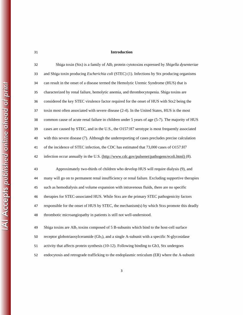
Introduction 31
Shiga toxin (Stx) is a family of AB5 protein cytotoxins expressed by Shigella dysenteriae 32
and Shiga toxin producing Escherichia coli (STEC) (1). Infections by Stx producing organisms 33
can result in the onset of a disease termed the Hemolytic Uremic Syndrome (HUS) that is 34
characterized by renal failure, hemolytic anemia, and thrombocytopenia. Shiga toxins are 35
considered the key STEC virulence factor required for the onset of HUS with Stx2 being the 36
toxin most often associated with severe disease (2-4). In the United States, HUS is the most 37
common cause of acute renal failure in children under 5 years of age (5-7). The majority of HUS 38
cases are caused by STEC, and in the U.S., the O157:H7 serotype is most frequently associated 39
with this severe disease (7). Although the underreporting of cases precludes precise calculation 40
of the incidence of STEC infection, the CDC has estimated that 73,000 cases of O157:H7 41
infection occur annually in the U.S. (http://www.cdc.gov/pulsenet/pathogens/ecoli.html) (8). 42
Approximately two-thirds of children who develop HUS will require dialysis (9), and 43
many will go on to permanent renal insufficiency or renal failure. Excluding supportive therapies 44
such as hemodialysis and volume expansion with intravenous fluids, there are no specific 45
therapies for STEC-associated HUS. While Stxs are the primary STEC pathogenicity factors 46
responsible for the onset of HUS by STEC, the mechanism(s) by which Stxs promote this deadly 47
thrombotic microangiopathy in patients is still not well-understood. 48
Shiga toxins are AB5 toxins composed of 5 B-subunits which bind to the host-cell surface 49
receptor globotriaosylceramide (Gb3), and a single A-subunit with a specific N-glycosidase 50
activity that affects protein synthesis (10-12). Following binding to Gb3, Stx undergoes 51
endocytosis and retrograde trafficking to the endoplasmic reticulum (ER) where the A-subunit 52

4
separates from the B-subunits and is then transported across the ER membrane to the cytosol 53
(13). Once in the cytosol, the A-subunit catalyzes the cleavage of an adenine from the 28S rRNA 54
thus inhibiting protein synthesis and activating pro-inflammatory signaling (11,12). Certain types 55
of protein synthesis inhibitors, including Stx, which interact with and/or damage the 28S rRNA, 56
have been shown to activate the mitogen activated protein kinases (MAPKs), jun-N-terminal 57
kinases (JNKs) and p38 (14,15). This activation of MAPKs by Stx and other protein synthesis 58
inhibitors has been termed the ribotoxic stress response or RSR. Stx-mediated activation of the 59
RSR has been shown to contribute to induction of cytokines such as IL-8 at both the 60
transcriptional and post-transcriptional level in intestinal epithelial cells (16,17). 61
While renal failure is a key feature of HUS, and renal samples from patients with HUS show 62
extensive damage (2), the temporal pattern of Stx-mediated renal damage has not been 63
determined and it remains unclear which renal cells are the primary targets during disease. This 64
is largely due to an inability to monitor renal damage on a molecular and cellular level during the 65
course of disease in human patients and to access patients during the early stages of disease 66
progression. To overcome these limitations, investigators have used either animal models (18,19) 67
or 2D monolayer cell cultures to study Stx-mediated disease (20-22). 2D cell culture systems, 68
while simple and low in cost, lack the architecture required to play a major role in cell-cell and 69
cell-ECM interactions, and are thus incapable of recapitulating the complexity of the in vivo 70
environment (23,24) as well as inadequate at predicting cellular toxicity (25,26). Studies using 71
2D human renal proximal tubule cells have demonstrated that Stx increases apoptosis in a time- 72
and dose-dependent manner along with the secretion of IL-1β, IL-6, and IL-8, inhibition of 73
protein synthesis, and water absorption (20,21,27,28). Animal models are incapable of fully 74
recapitulating human disease for numerous reasons, including differences in physiology and 75

5
experimental conditions (29). Importantly, differences in Stx receptor expression patterns 76
between mice and humans contribute to the failure of murine models of HUS to completely 77
mimic human sequelae (30). While nonhuman primates have been shown to recapitulate the 78
clinical manifestation of HUS and respond to therapeutics (31), these models are expensive and 79
require specialized staff and facilities. 80
The advantages of using 3D human tissues over 2D cell culture have already been 81
demonstrated in studies of cancer progression, signal transduction, and drug toxicity (23,32-35). 82
They have revealed advantages of 3D tissue models over 2D cell cultures due to differences in 83
cell-cell signaling, ECM interactions, ECM composition, gene expression, and epigenetic gene 84
regulation (23,36). Bioengineered 3D kidney tissues produced in our lab using human cells have 85
been shown to better reflect human renal physiology than 2D cell culture (37). These tissues are 86
composed of human telomerase reverse transcriptase (hTERT) immortalized human renal 87
proximal tubule cells embedded within a hydrogel. Following 2 weeks of culture, they form an 88
interconnecting network of tubules (Fig. 1A). These bioengineered kidney tissues are capable of 89
revealing toxic effects of both acute and chronic exposure to insult as they are viable for up to 8 90
weeks, unlike 2D cell cultures, which are viable and relevant for only days before increased cell 91
density results in cell death. 92
The goal of this study was to adapt our 3D bioengineered human renal tissue model for the study 93
of Stx-induced renal toxicity and to compare the responses with responses by 2D cultures. We 94
examined cytotoxicity, activation of the RSR, renal injury biomarker expression, and 95
inflammatory cytokine secretion. While the overall cytotoxicity profile, protein synthesis 96
inhibition, and RSR activation were similar between 2D and 3D cultures, there were differences 97
in the IC50 at different time points, the secretion of renal injury biomarkers, and cytokine 98

6
secretion between the two systems. Importantly, treatment of the 3D tissues with Stx resulted in 99
an elevated secretion of the kidney injury marker 1 (Kim-1) and the cytokine IL-8 compared to 100
the 2D cell cultures. IL-8 secretion levels have been shown to significantly increase in the urine 101
of baboons following Stx2 treatment (31) and from intestinal epithelial cells exposed to Stx2 102
(38). Kim-1 has been shown to be induced upon infection of mice with Stx-producing 103
Citrobacter rodentium, a murine model for EHEC disease (39); the current study is the first to 104
show a Kim-1 renal injury biomarker response to Stx2 in vitro. This study is the first utilization 105
of a 3D renal tissue model for the study of Stx-mediated nephrotoxicity and reveals significant 106
benefits of a 3D tissue model compared to 2D cell culture. 107
Materials and Methods 108
Cell culture. The NKi-2 cells used in all studies were derived by the serial passaging in low 109
serum media of hTERT immortalized human renal cortical cells. Cells were grown in high 110
glucose DMEM/F12 (Invitrogen) containing 2% FBS (Invitrogen), 20ng/mL hEGF (Invitrogen), 111
72 ng/mL T3 (Sigma), 1% ITS (Invitrogen), 100 ng/mL hydrocortisone (Sigma), and 1% 112
penicillin/streptomycin (Invitrogen). For 2D studies, cells were seeded at a density of 25,000 113
cells per well of a 12-well tissue culture plate 24 hours before exposure to Stx2. Every effort was 114
taken to ensure matching passages were used for 2D and 3D cultures. 115
Shiga Toxin. Stx2 was purchased from Dr. Anne Kane of the Phoenix Laboratory at Tufts 116
Medical Center. The toxin is purified by P1 glycoprotein affinity chromatography based on the 117
method of Donahue-Rolfe et al., 1989(40). Lipopolysaccharide (LPS) was removed using Pierce 118
High-capacity Endotoxin Removal Resin (Thermo) on a column with a 4 hour incubation. The 119
removal of lipopolysaccharides (LPS) was confirmed with the ToxinSensorTM
Chromogenic 120
LAL Endotoxin Assay Kit (Genscript). As a control, Stx2 was inactivated by boiling for 8 121

7
hours. For dosing, Stx2 was reconstituted in sterile PBS at 1mg/mL and then diluted in cell 122
culture media for concentrations ranging from 1fg/mL to 1μg/mL. For 2D cultures, 2 mL of Stx2 123
containing media was applied to each well and 350μL was removed at each time point. For 3D 124
cultures, 1.5 mL of Stx2 culture media was applied below the tissue and 0.5 mL on top of the 125
tissue. 350μL of media was removed from the bottom at each time point. The cellular 126
supernatants were stored at -80°C and used for LDH, Kim-1, NGAL, and cytokine assays. 127
3D tissue constructs. 3D tissue constructs were formed as previously describes (37). Briefly, 128
100,000 NKi-2 cells were combined with a 50:50 mix of Matrigel and rat tail collagen I at a final 129
concentration of 1 mg/mL in a 12-well transwell insert. The tissues were allowed to polymerize 130
at 37°C for 1 hour after which NKi-2 cell culture media was added to both the bottom well of the 131
transwell and inside the insert. Tissues were grown at 37°C, 5% CO2 with media changes every 2 132
days. 133
Cytotoxicity. LDH secretion was assayed in 100μL of cell culture supernatant from both 2D 134
NKi-2 cells and 3D tissues using LDH cytotoxicity assay from Clontech and following the 135
manufacturer’s instructions. 2% Triton was used as the positive control for cell death. Percent 136
cytotoxicity was calculated as (Stx2 treated – untreated)/(2% Triton treated – untreated)x100. 137
Samples from 3 independent experiments were analyzed and graphed using GraphPad Prism 5 138
software. The data are expressed as averages ± standard errors of the mean (SEM). 139
IC50 calculations. IC50, the dosage at which 50% toxicity was achieved, was calculated using 140
LDH data following 48 and 72 hours of treatment for 2D cells and 3D tissues. Values were 141
calculated with GraphPad Prism 5 software using nonlinear regression analysis. 142
Protein synthesis inhibition. Protein synthesis inhibition was determined by measuring the 143
decrease in 3H-leucine incorporation. For 2D culture, cells were plated in 96-well plates at a 144

8
density of 0.6-1 x 10⁵ cells/well. The following day media was removed, and fresh media 145
without toxin or with Stx2 at concentrations ranging from 1g/ml-1pg/ml was added and the 146
cells were incubated overnight. The next day (24-26 hours post-intoxication) the media was 147
removed, and the cells were gently washed two times with pre-warmed PBS. To the cells, media 148
made 1:1 with RPMI w/25 mM HEPES w/o leucine (Chemicon International R-078-B) and 149
DMEM low glucose w/o Leu, Lys, Arg, (Sigma cat. # SLBH1080) and containing 5 ul/ml of 3H-150
Leucine (Leucine [3, 4, 5-3H(N)], Perkin Elmer cat # NGT460001MC, 1 mCi, 106.2 Ci/mMol) 151
was added and the cells incubated at 37C in 5% CO2 for 1 hour. Following this incubation, 152
media was removed and the cells were washed two times with 200 l of PBS. 50 l of 0.2 M 153
KOH was added to each well of cells and incubated at room temperature for 15 minutes followed 154
by the addition of 150 l of 10 % trichloroacetic acid (TCA). The plates were then incubated at 155
room temperature for 30 minutes followed by transfer of the well contents to a 96-well vacuum 156
manifold-coupled filter plate (Millipore cat # MAHVN4550). Wells were washed two times with 157
200 l of 5% TCA transferring the each wash to the appropriate filter well on the vacuum 158
manifold. Filters were washed one time with 5% TCA 200 l/filter followed by a wash with 200 159
l of 1 % acetic acid. Filters were allowed to dry and then were punched into vials for 160
scintillation counting. 161
The 3D tissues in 12-well plates were treated similar to 2D-cultures except for the 162
following: 1) Following removal of toxin containing media from the wells, tissues were washed 163
2 times in prewarmed PBS and then incubated in leucine-free media for 90 min to dilute out 164
leucine containing media from the matrix prior to adding the 3H-leucine containing media. 2)
3H-165
leucine containing media was incubated for 7:30 hours to allow for sufficient detectable 166
incorporation of radioactivity. Tissues were then washed 2 times with PBS. 3) 300 l of Dispase 167

9
(BD Scientific cat # 354235) containing 100 g/ml collagenase type I was added to the upper 168
chamber of the tissue containing transwell to breakdown the tissue containing matrix. This was 169
incubated for 1:30 hours at 37C, 5 % CO2. 4) The protein in the upper chamber was then 170
precipitated by adding 100 % TCA to a final concentration of 25 %, incubating this at 4oC for 30 171
minutes and spinning in a microfuge for 5 minutes at maximum speed. One ml of ice cold 172
acetone was added to the pellet and the tubes were centrifuged at maximum speed for 5 minutes. 173
The resulting pellet was allowed to dry and suspended in scintillation fluid prior to being 174
transferred to a scintillation vial for counting. 175
Immunohistochemistry. Following 72 hours of Stx2 exposure, tissues were fixed with 10% 176
buffered formalin for 48 hours at 4°C and embedded in paraffin. H&E staining was performed on 177
8 µm tissue sections and images were captured with Leica Application Suite v4, using a Leica 178
DMIL microscope equipped with a Leica DFC340FX camera for fluorescent images and a Leica 179
DFC295FX camera for brightfield images. 180
Kidney injury biomarkers. Kim-1 and NGAL secretion was assayed in 50uL of the cell culture 181
supernatant from both the NKi-2 cells and 3D tissues using ELISA assays from R&D systems 182
and following the manufacturer’s instructions. All measurements were performed with Softmax 183
Pro 5.4 software on a SpectraMax M2 plate reader (Molecular Devices). Experiments were 184
analyzed and graphed using GraphPad Prism 5 software and are expressed as mean concentration 185
± SEM. Statistical significance was assessed at p<0.05 by two-way ANOVA with Bonferroni 186
posttest. 187
Cytokine secretion. Cytokine secretion was measured in 25uL of the cell culture supernatant 188
using the Human Cytokine/Chemokine Milliplex® MAP kit from Millipore customized to 189
measure IL-1β, IL-6, IL-8, TNFα, and MCP-1 and following the manufacturer’s instructions. 190

10
Experiments were analyzed and graphed using GraphPad Prism 5 software and are expressed as 191
mean concentration ± SEM. Statistical significance was assessed at p<0.05 by two-way ANOVA 192
with Bonferroni posttest. 193
MAPK assays. Phospho-JNKs and Phospho-p38 levels were determined using 194
immunoprecipitation based assays by Cell Signaling Technology (cat. #s 8794 and 9820 for 195
Phospho JNKs and Phospho-p38 respectively) according to the manufacturer’s instructions. To 196
200 g of protein-containing cell lysates were added phospho-specific JNKs or p38 antibody-197
conjugated beads and incubated overnight. The beads were then washed twice in lysis buffer and 198
twice in kinase reaction buffer followed by resuspension of the beads in kinase reaction buffer 199
containing 200 M ATP and 1 g kinase substrate (c-Jun fusion protein for phosphor-JNKs and 200
ATF-2 fusion protein for phsopho-p38). 201
MTT Assay. Cells were plated in 96-well tissue culture plates at a density of 0.6 x 10⁵ cells/well. 202
The next day Stx2 was added at concentrations varying from 1 pg/ml to 1 g/ml. After 24 and 48 203
hours of incubation at 37oC, 5 % CO2, media was removed and 100 l of 0.5 mg/ml 3-(4,5-204
dimethylthiazol-2-yl)-2,5-diphenyltetrazolium bromide in PBS was added to each well. 205
Following incubation at 37oC, 5 % CO2 for 1 hour, the MTT solution was removed and 50 l of 206
isopropanol containing 4 mM HCL and 0.1 % NP40 Nonidet was added followed by gentle 207
shaking at room temperature for 15 min. Optical density measurements were made at 550 nm 208
and 620 nm. 209
210
Results 211
Shiga Toxin Induced Nephrotoxicity 212

11
In order to validate our bioengineered 3D human renal model for the study of Stx-213
induced nephrotoxicity, we began by comparing cytotoxicity in 3D to cytotoxicity in the same 214
cells grown in 2D (Fig. 1). 3D tissues containing immortalized renal proximal tubule cells (NKi-215
2) were formed and allowed to grow for 2 weeks prior to treatment with Stx2 while 2D cultures 216
of the NKi-2 cells were seeded 24 hours prior to Stx2 treatment. Stx2 was purified free of LPS, 217
which has been used as a co-stimulus in a murine model of HUS (41,42), and may potentiate a 218
Stx response by increasing the inflammatory response or increasing Gb3 receptor on the cell 219
surface (43-46). Both the cells and the tissues were treated with a range of Stx2 concentrations 220
(1fg -1μg /mL) for up to 72 hours with supernatant sampling occurring at 6, 24, 48, and 72 hours 221
post-treatment (Fig. 1B). 222
To first test whether Stx2 had the predicted inhibitory effect on protein synthesis, we 223
added increasing concentrations of the toxin to either 2D or 3D cultures. Protein synthesis of 224
both cultures, measured by 3H-leucine incorporation (see Materials and Methods), was inhibited 225
in a similar dose-dependent manner (Fig. 1C). The predicted Stx2-induced cytotoxicity was 226
demonstrated for 3D cultures by H&E staining, which revealed the loss of nuclei at 72 h post-227
exposure (Fig. 1D), and for 2D cultures by an MTT cell viability assay (Fig. S1). Importantly, 228
cell viability at 24 hours after Stx2 exposure was greater than 50% for all Stx2 concentrations 229
tested, indicating that the inhibition of protein synthesis observed at 24 h (Fig. 1C) was not 230
simply a reflection of cell death. Heat-treatment of purified Stx2 abrogated the cytotoxic effect 231
(data not shown), suggesting that cytotoxicity was not a reflection of possible contamination by 232
LPS. To discern possible dramatic differences in the kinetics or dose-dependence of cytoxicity 233
between 2D and 3D cultures, we measured the IC50 for lactate dehydrogenase (LDH) release at 234
various times after Stx2 exposure. We found that at 48 h post-exposure, 3D cultures were 235

12
somewhat (seven-fold) less sensitive than 2D cultures; at 72 h, 3D cultures were somewhat 236
(three-fold) more sensitive (Fig. 1E). We conclude that although the cytotoxic responses of the 237
two cultures may differ somewhat in kinetics and relative sensitivity, Stx2 inhibits protein 238
synthesis and triggers cytotoxicity similarly in both 2D and 3D cultures.. 239
Finally, since Stx-mediated damage to actively translating ribosomes results in activation 240
of the RSR, we investigated whether a RSR was induced in 2D and 3D renal cultures. Whole cell 241
lysates were made from 2D and 3D cultures untreated, treated with heat-inactivated-Stx2 (HI), 242
Stx2 without LPS removed, and Stx2 following LPS removal for 6 hours. Phospho-JNKs were 243
immunoprecipitated from whole cell lysates and immunoprecipitates were incubated with c-jun 244
substrate and phosphorylated c-jun was determined by western blot. Activation of JNKs (Fig. 245
2A) was increased in Stx2 treated 2D and 3D samples, but not in HI samples. Similarly, 246
immunoprecipitation of phosphorylated p38 (Fig. 2B) demonstrates that p38 activation was also 247
increased by Stx2 in both 2D and 3D cultures. Taken together, these data indicate that there are 248
similarities in the general cytotoxic response to Stx2 between 2D and 3D cultures in terms of 249
dose response, protein synthesis inhibition, and activation of the RSR. 250
Kidney Injury Marker Secretion in Response to Stx2 Treatment 251
A number of biomarkers have been identified to monitor kidney damage. These include 252
both Kim-1 and neutrophil gelatinase-associated lipocalin (NGAL). Kim-1 secretion in the urine 253
of patients or by kidney epithelial cells in response to Stx2 has not been examined and only two 254
studies have examined the secretion of NGAL in patient urine and serum in response to Stx 255
(47,48). NGAL has been examined for its ability to predict renal injury in HUS (48) and the need 256
for renal replacement therapy following Stx exposure (47). We have previously shown its utility 257
as a marker of drug-induced nephrotoxicity in 3D tissues and shown that it was predictive of 258

13
renal proximal tubule cytotoxicity for highly toxic agents at high doses but incapable of 259
distinguishing more subtle differences in toxicity in vitro due to high expression from untreated 260
controls (37). None of the Stx2 doses resulted in NGAL secretion in excess of untreated controls 261
in either 3D or 2D cultures (Fig. 3A). Interestingly, the highest doses of Stx2, 1μg/mL and 262
1ng/mL, which resulted in almost 100% cytotoxicity in 3D by LDH secretion (Fig. 1C), resulted 263
in a significant decrease in NGAL secretion (p<0.001) 72 hours post-treatment. This may be due 264
to the almost complete inhibition in protein synthesis seen at these doses by 24 hours post-265
treatment. These data raise the possibility that low levels of NGAL, rather than high levels, may 266
be predictive of Stx-induced cytotoxicity in vitro. 267
Kim-1 has been shown to be a very sensitive biomarker of kidney injury and we have 268
previously shown its utility in measuring cytotoxicity in 3D with highly nephrotoxic compounds 269
(37). However, we have also shown that secreted Kim-1 increases over time in untreated control 270
cultures (37) making it only useful at extremely toxic concentrations and not sensitive or precise 271
enough to reveal subtle changes in cytotoxicity in vitro. Consistent with these previous findings, 272
Kim-1 increased in expression in both the 2D and 3D untreated controls over time (data not 273
shown). When assayed at 72 h after exposure to 1 pg/ml Stx2 or greater, Kim-1 production by 274
2D cultures was inhibited, perhaps due to the protein synthesis inhibition induced by the toxin, 275
whereas 3D cultures were largely unaffected by Stx2 exposure (Fig. 3B). When assayed at an 276
earlier time point, i.e. 24 h after exposure, 1μg/mL Stx2 induced Kim-1 production increased 277
almost two-fold above the untreated controls (Fig. 3C). In contrast, at 24 h post-exposure no 278
induction of Kim1 was observed for 2D cultures at any Stx2 concentration (Fig. 3B). The Stx2-279
induced Kim-1 secretion response by 3D but not 2D cultures suggest that 3D tissues may provide 280
a better experimental model for examining renal damage using this marker. 281

14
Stx2-Induced Cytokine Secretion 282
Since cytokines have been shown to play a role in Stx2-induced renal damage (for review 283
(49)), are secreted by a number of different cell types in response to Stx2 (17,20,50-52), and are 284
differentially secreted based upon the toxin sensitivity of the cell (20,53), we examined whether 285
3D culturing would elicit a different response compared to 2D in the secretion of a set of 286
cytokines previously shown to be secreted by cells in response to Stx, IL-1β, IL-6, IL-8, MCP-1, 287
and TNFα (Fig. 4) (20,21,31,54-56). All cytokines were measured at 24 and 48 hours after 288
treatment with increasing concentrations of Stx2. IL-1β was not detectable in either 2D or 3D 289
cultures (data not shown), and untreated controls had baseline secretion of IL-6, IL-8, TNFα, and 290
MCP-1 that increased between 24 and 48 hours for both 2D and 3D cultures (Fig. 4A-D; see 291
open and filled bars, “untreated”). 292
Upon intoxication of either model with increasing concentrations of Stx2, MCP-1 293
secretion was not induced. However, MCP-1 production exhibited a dramatic difference between 294
the two models. MCP-1 was almost undetectable in 2D cultures, and while the secreted levels 295
did increase over time, the levels never exceeded the levels of the untreated controls (Fig. 4A). 296
In striking contrast, MCP-1 was produced at levels 20- to 40-fold higher in 3D cultures, a result 297
that may reflect the observation that increased MCP-1 is detected in the urine of children with 298
HUS (57). 299
For both 3D and 2D cultures, the secretion of IL-6 (Fig. 4B), TNFα (Fig. 4C) and IL-8 300
(Fig. 4D) were each induced 48 hours after exposure to 1pg/mL Stx2. Cytokines were not 301
induced by 1ng/mL or 1g/ml Stx2. The fact that a significant change in expression was only 302
seen at 1pg/mL may be due to the fact that at the higher concentrations protein synthesis 303
15
inhibition prevented cytokine production immediately. In contrast, 1pg/mL is most similar to the 304
IC50 values determined in Figure 1B and may not intoxicate cells rapidly enough to prevent the 305
production and secretion of inflammatory factors. That Stx2 induces cytokine production despite 306
promoting protein synthesis inhibition is consistent with the ability of Stx to induce gene 307
expression and protein production via signaling pathways not directly activated by protein 308
synthesis inhibition (16,58,59). 309
The 3D tissue IL-8 responses to Stx2 deserves particular note. First, unlike IL-6 and 310
TNFα, but like MCP-1, basal production of IL-8 in the absence of Stx2 was 4- to 8- fold higher 311
in 3D tissues compared to 2D cells (Fig. 4D). Second, this already high level of secretion of IL-8 312
was further induced ~1.8-fold by 1pg/mL Stx2, resulting in IL-8 levels higher than that induced 313
in 2D cultures by Stx2. This increase in 3D versus 2D could indicate a property of the 3D culture 314
system that up-regulates both basal and induced IL-8 levels or it could reflect a difference in cell 315
numbers able to secrete IL-8 at this time point. Importantly, while the exact role of IL-8 in HUS 316
is unknown, increases in concentration have been noted in the urine of children with HUS, the 317
urine of Stx2 treated baboons, and in human intestinal epithelial cells following Stx2 exposure 318
(31,38,57). 319
320
Discussion 321
In this study, we have shown the utility of a 3D bioengineered human renal tissue model for the 322
examination of the mechanisms of Stx-induced renal cytotoxicity. While many responses were 323
similar between the 2D and 3D cultures, including overall cytotoxicity, IC50, protein synthesis 324
inhibition, RSR, and IL-6 and TNFα secretion, there were differences indicating that a 3D 325

16
system may provide a more physiologically relevant model than a 2D system for examination of 326
cellular responses to Stx2. Two distinct and notable differences were observed between the 2D 327
and 3D systems. First, higher basal levels of Kim-1, NGAL, MCP-1, and IL-8 were observed 328
from the 3D tissues compared to the 2D cells indicating that these inflammatory or renal injury 329
markers may be better expressed in the 3D tissues. Second, higher induction levels of Kim-1 and 330
IL-8 were observed from the 3D tissues compared to the 2D cells indicating that the 3D tissues 331
maybe be a better system for monitoring Stx2-mediated renal injury in vitro through renal 332
biomarker secretion (Kim-1) and characteristic cytokine secretion (IL-8). Taken together, these 333
data suggest differential cellular mechanisms functioning between 2D and 3D cultures in 334
response to Stx2. 335
Initially, the general features of cytotoxicity of Stx2 appeared to be very similar between 336
the 2D and 3D cultures. For example, several studies have demonstrated that intoxication with 337
Stx activates pro-inflammatory and pro-apoptotic signaling pathways (60,61), in part through the 338
activation of the mitogen-activated protein kinase (MAPK) pathway (62-64). This activation of 339
the RSR contributes to both pro-inflammatory and pro-apoptotic signaling in cultured cells 340
(62,63,65,66). We showed that human renal 2D and 3D tissue both underwent an RSR following 341
Stx2 treatment suggesting that this signaling cascade may participate in Stx-associated 342
inflammation and cell death in the human kidney. Whether the kinetics of the RSR in 2D and 3D 343
models differs will be a subject of future studies as will defining the relative contribution of the 344
RSR to cytokine production and cell death. 345
A second similarity was the general trend of inflammatory cytokine expression in the cell 346
culture supernatant. Inflammatory signaling is associated with HUS, as patients show increased 347
urinary levels of pro-inflammatory cytokines, and renal biopsies have revealed infiltration of 348

17
neutrophils and monocytes into the kidney during disease (57,67-69). In 2D cell culture models, 349
Stx treatment of human proximal tubular cells has been shown to induce the secretion of IL-1β, 350
IL-6, and IL-8 (27), and evidence suggests that IL-1, IL-8, TNFα, and IL-6 may be elevated in 351
the serum of D+HUS patients (55,56). Also, IL-1 and TNF have been shown to upregulate the 352
Stx cell surface receptor globotriaosylceramide (Gb3) and increase cellular sensitivity to Stx 353
cytotoxicity in endothelial cells (70,71). In nonhuman primates treated with Stx, kidney tissue 354
had up-regulated levels of IL-8, MCP-1, and TNFα mRNA along with increased urinary levels of 355
IL-6, IL-8, and MCP-1 (31). Our studies revealed an increase in IL-6, IL-8, and TNFα over time 356
following Stx2 treatment in both 2D and 3D models. The three detectable cytokines all showed 357
an increase above untreated controls at 48 hours with 1pg/mL Stx2 while the higher 358
concentrations did not secrete any cytokines above the level of the untreated control. This was 359
most likely due to the relatively high rate of cytotoxicity and protein synthesis inhibition seen 360
with the higher doses; in contrast, the 1pg/mL dose was close to the calculated IC50 and therefore 361
did not result in complete cytotoxicity of the cultures. Studies designed to examine cytokine 362
secretion at earlier time points following Stx2 treatment may reveal increased secretion at higher 363
doses. However, studies of cytokine secretion from HK-2 cells at early time points (<4 hours) 364
did not reveal an increase in IL-1β, TNFα, or IL-8 protein levels although increases in mRNA 365
transcripts were observed (20). 366
Examination of the IC50 at both 48 and 72 hours revealed a difference between the 2D 367
and 3D culture systems. In particular, there was a difference in the response rate to Stx2 between 368
the 2D and 3D cultures. The 2D cultures were seven-fold more sensitive to Stx2 than the 3D 369
cultures at 48 hours post-treatments, but by 72 hours the two cultures exhibited roughly similar 370
Stx2 sensitivity. This difference does not simply reflect delayed diffusion of Stx2 into the 3D 371

18
culture, because at 24 hours post-treatment Stx2 induced a similar degree of protein synthesis 372
inhibition in both the 2D and 3D cultures. Thus it may be that the difference in cellular 373
organization and structure has an impact upon Stx2-mediated cytotoxicity. Interestingly, HK-2 374
cells, a HPV immortalized human renal proximal tubule cell line, were shown to have an IC50 of 375
20 pg/mL at 72 hours post-treatment with Stx2, compared to 2.2 pg/mL and 0.7 pg/mL at the 376
same time point for 2D and 3D NKi-2 cells respectively in this study. HK-2 cells have also been 377
shown to lack sensitivity to drug-induced toxicity (26) indicating they may not be the best cell 378
line for studying renal cytotoxicity. 379
A second significant difference between the 2D and 3D cultures was the inability to 380
detect MCP-1 in the 2D cultures, and while it was detectable in the 3D cultures and increased 381
with time, following Stx2 treatment it was secreted at levels significantly lower than the 382
untreated controls. Interestingly, while MCP-1 mRNA has been shown to be elevated in the 383
kidneys of nonhuman primates exposed to Stx2 and the protein has been shown to be elevated in 384
the urine, its mRNA was barely detectable in cultured renal endothelial cells (31). This may 385
indicate that the secreted MCP-1 is originating from a different cell type or the specifically high 386
basal levels of MCP-1 might also reflect a physiologic response to the accumulation of metabolic 387
waste products. In vivo, tubules exist adjacent to both a vasculature and a lumen through which 388
metabolites can be either taken up into the blood stream, or discarded through the urine. Since 389
our current 3D-tubule system is not perfused, it is reasonable to hypothesize that the increase in 390
MCP-1 over the course of 48 hours reflects a normal physiological response to the accumulation 391
of metabolic waste products. In this scenario, the tubular epithelium is signaling for resident 392
tissue phagocytes to aid in the removal of these waste products in an effort to maintain a 393
homeostatic environment. Furthermore, the high basal MCP-1 level does not appear to be part of 394

19
an inflammatory response since neither TNFα nor IL-6 appear to be upregulated in our model in 395
the absence of Stx. This hypothesis is supported by the observation that the kidney maintains 396
distinct populations of resident mononuclear phagocytes that produce anti-inflammatory and 397
homeostatic factors (72), and therefore, the high basal levels of MCP-1 might represent a 398
homeostatic response not present in 2D cell culture. 399
A third significant difference between the 2D and 3D cultures was the secretion of IL-8. 400
While the secretion of IL-8 increased from 24 to 48 hours for both the 3D tissues and the 2D 401
cells, the amounts were approximately four-fold higher from the 3D tissues compared to the 2D 402
cells. Importantly, this induction occurred in response to 1 pg/mL Stx2, which may be present in 403
sera of STEC-infected patients at very low levels, i.e., often lower than the 6 pg/ml limit of 404
detection (73). IL-8 has been shown in the urine of nonhuman primates 48 hours after exposure 405
to Stx2 at approximately 600 pg/mL (31). Our cells in 2D culture did not reach that amount at 406
any time or dose while the 3D tissues more than doubled that amount at 48 hours with a 1 pg/mL 407
dose. Thus, the 3D tissue system may be more representative of the in vivo response than the 408
cells in 2D. 409
Biomarkers of renal injury are extremely important for the prediction of drug-induced 410
nephrotoxicity and the early assay of acute renal injury. A number of potential markers have 411
been identified and are in different stages of validation and use. Kim-1 and NGAL are two such 412
markers. While they have been shown to be predictive of drug-induced nephrotoxicity in vivo 413
and have been examined as biomarkers for HUS (47,48,74), we have previously shown that they 414
may only be minimally useful to assess toxicity in cultured renal cells in vitro primarily due to 415
the high levels secreted by untreated controls (37). What was notable in our studies was an 416
increase in Kim-1 secretion at 1μg/mL Stx2 compared to the untreated controls at 24 hours that 417

20
then later decreased. This spike was only evident in the 3D cultures. The 2D cultures never 418
reached a secretion level above the untreated controls. This may indicate an initial early spike in 419
secretion in relation to Stx2-mediated cytotoxicity that then decreases due to the inhibition of 420
protein synthesis which we show occurred by 24 hours post-treatment. Further studies at early 421
time points need to be conducted. This spike would never have been noticed if we had only been 422
examining the cells in 2D culture. Our 3D studies indicate that Kim-1 has the potential to act as 423
an early biomarker of renal injury for infected patients and in vivo studies need to be performed 424
to confirm this. If it is confirmed in vivo, then our studies also indicate a larger relevance of the 425
3D system over 2D cell culture in mimicking Stx2-mediated renal injury. 426
In conclusion, we have presented the first use of a 3D bioengineered human kidney tissue 427
model to study renal damage upon Stx2 exposure. We have shown that the 3D tissue responds to 428
Stx2 with a dose- and time-dependent cytotoxicity that is related to the inhibition of protein 429
synthesis. We have also shown changes in renal injury biomarkers in a dose-dependent manner 430
along with changes in the secretion of cytokines and activation of the RSR. Key differences 431
between the 3D tissue over 2D cell culture included differences in the IC50 and the ability to 432
measure significantly high levels of kidney injury biomarkers and cytokines, particularly IL-8, 433
MCP-1 and Kim-1. Further use of this model may save on the use of inaccurate animal models 434
while making in vitro studies more relevant to in vivo conditions. 435
436
437
Acknowledgments 438
21
This work was supported by the NIH (P41 EB002520, R01 AI46454, R37 DK51050, and R01 439
DK099532) along with the Tufts Collaborates! Seed Grant Program. We would like to thank Dr. 440
Anne Kane of the Phoenix Laboratory for her assistance with the Stx2 and Alexander Histed for 441
his insight on MCP-1. We would also like to thank Milva Ricci, Carmen Preda, and Darin 442
Goodwin for their assistance. 443
Figure Legends 444
Figure 1: Shiga toxin mediated cytotoxicity. (A) A representative image of the interconnecting 445
tubules present in the 3D bioengineered human kidney tissue model. (B) Timeline of tissue 446
formation, treatment, and sampling. (C) 3H-leucine incorporation assay was used to determine 447
the sensitivity of 2D and 3D cultures to Stx2 at 24 hours post treatment. (D) H&E of 448
representative 3D tissues following 72 hours of no treatment or treatment with noted 449
concentrations of Stx2. Arrows indicate the hematoxylin stained cells within the matrix. (E) IC50 450
values at specific time points for 2D and 3D tissues. 451
Figure 2: Ribotoxic Stress Response. Activation of JNKs (A) or p38 (B) was determined by 452
western blot, comparing in vitro phosphorylation of c-jun (JNKs) or ATF-2 (p38) following 453
precipitation of phosphoyrlated JNKs or p38 from whole cell lysates. Bar graphs represent blot 454
band intensity of P-c-jun or P-ATF-2 as normalized against loading controls. 455
Figure 3: Kidney Injury Markers. (A) NGAL production in response to Stx2 at 72 hours post-456
toxin treatment. Kim-1 production in response to Stx2 is shown at (B) 24 hours and (C) 72 hours 457
post-toxin treatment. 458

22
Figure 4: Cytokine expression. (A) MCP-1, (B) IL-6, (C) TNFα, and (D) IL-8 secretion into 459
the cell culture supernatant following 24 and 48 hour Stx2 treatment of 3D and 2D cultures. 460
*p<0.05, **p<0.01, ***p<0.001. 461
462

23
Reference List 463 464 1. Strockbine, N. A., L. R. Marques, J. W. Newland, H. W. Smith, R. K. Holmes, and A. 465
D. O'Brien. 1986. Two toxin-converting phages from Escherichia coli O157:H7 strain 466 933 encode antigenically distinct toxins with similar biologic activities. Infect. Immun. 467 53:135-140. 468
2. Obrig, T. G. and D. Karpman. 2012. Shiga toxin pathogenesis: kidney complications 469 and renal failure. Curr. Top. Microbiol. Immunol. 357:105-136. 470
3. Karmali, M. A., M. Petric, C. Lim, P. C. Fleming, G. S. Arbus, and H. Lior. 1985. The 471 association between idiopathic hemolytic uremic syndrome and infection by verotoxin-472 producing Escherichia coli. J. Infect. Dis. 151:775-782. 473
4. Mora, A., S. L. Leon, M. Blanco, J. E. Blanco, C. Lopez, G. Dahbi, A. Echeita, E. A. 474 Gonzalez, and J. Blanco. 2007. Phage types, virulence genes and PFGE profiles of 475 Shiga toxin-producing Escherichia coli O157:H7 isolated from raw beef, soft cheese and 476 vegetables in Lima (Peru). Int. J. Food Microbiol. 114:204-210. 477
5. Ong, K. L., M. Apostal, N. Comstock, S. Hurd, T. H. Webb, S. Mickelson, J. 478 Scheftel, G. Smith, B. Shiferaw, E. Boothe, and L. H. Gould. 2012. Strategies for 479 surveillance of pediatric hemolytic uremic syndrome: Foodborne Diseases Active 480 Surveillance Network (FoodNet), 2000-2007. Clin. Infect. Dis. 54 Suppl 5:S424-S431. 481
6. Gould, L. H., L. Demma, T. F. Jones, S. Hurd, D. J. Vugia, K. Smith, B. Shiferaw, S. 482 Segler, A. Palmer, S. Zansky, and P. M. Griffin. 2009. Hemolytic uremic syndrome 483 and death in persons with Escherichia coli O157:H7 infection, foodborne diseases active 484 surveillance network sites, 2000-2006. Clin. Infect. Dis. 49:1480-1485. 485
7. Tarr, P. I., C. A. Gordon, and W. L. Chandler. 2005. Shiga-toxin-producing Escherichia 486 coli and haemolytic uraemic syndrome. Lancet 365:1073-1086. 487
8. Mead, P. S., L. Slutsker, V. Dietz, L. F. McCaig, J. S. Bresee, C. Shapiro, P. M. 488 Griffin, and R. V. Tauxe. 1999. Food-related illness and death in the United States. 489 Emerg. Infect. Dis. 5:607-625. 490
9. Scheiring, J., S. P. Andreoli, and L. B. Zimmerhackl. 2008. Treatment and outcome 491 of Shiga-toxin-associated hemolytic uremic syndrome (HUS). Pediatr. Nephrol. 23:1749-492 1760. 493
10. Fraser, M. E., M. Fujinaga, M. M. Cherney, A. R. Melton-Celsa, E. M. Twiddy, A. D. 494 O'Brien, and M. N. James. 2004. Structure of shiga toxin type 2 (Stx2) from Escherichia 495 coli O157:H7. J. Biol. Chem. 279:27511-27517. 496
11. Endo, Y., K. Tsurugi, T. Yutsudo, Y. Takeda, T. Ogasawara, and K. Igarashi. 1988. 497 Site of action of a Vero toxin (VT2) from Escherichia coli O157:H7 and of Shiga toxin on 498 eukaryotic ribosomes. RNA N-glycosidase activity of the toxins. Eur. J. Biochem. 171:45-499 50. 500

24
12. Fraser, M. E., M. M. Cherney, P. Marcato, G. L. Mulvey, G. D. Armstrong, and M. N. 501 James. 2006. Binding of adenine to Stx2, the protein toxin from Escherichia coli 502 O157:H7. Acta Crystallogr. Sect. F. Struct. Biol. Cryst. Commun. 62:627-630. 503
13. Sandvig, K. and D. B. van. 2002. Transport of protein toxins into cells: pathways used 504 by ricin, cholera toxin and Shiga toxin. FEBS Lett. 529:49-53. 505
14. Iordanov, M. S., D. Pribnow, J. L. Magun, T. H. Dinh, J. A. Pearson, S. L. Chen, and 506 B. E. Magun. 1997. Ribotoxic stress response: activation of the stress-activated protein 507 kinase JNK1 by inhibitors of the peptidyl transferase reaction and by sequence-specific 508 RNA damage to the alpha-sarcin/ricin loop in the 28S rRNA. Mol. Cell Biol. 17:3373-509 3381. 510
15. Smith, W. E., A. V. Kane, S. T. Campbell, D. W. Acheson, B. H. Cochran, and C. M. 511 Thorpe. 2003. Shiga toxin 1 triggers a ribotoxic stress response leading to p38 and JNK 512 activation and induction of apoptosis in intestinal epithelial cells. Infect. Immun. 71:1497-513 1504. 514
16. Thorpe, C. M., W. E. Smith, B. P. Hurley, and D. W. Acheson. 2001. Shiga toxins 515 induce, superinduce, and stabilize a variety of C-X-C chemokine mRNAs in intestinal 516 epithelial cells, resulting in increased chemokine expression. Infect. Immun. 69:6140-517 6147. 518
17. Thorpe, C. M., B. P. Hurley, L. L. Lincicome, M. S. Jacewicz, G. T. Keusch, and D. 519 W. Acheson. 1999. Shiga toxins stimulate secretion of interleukin-8 from intestinal 520 epithelial cells. Infect. Immun. 67:5985-5993. 521
18. Mohawk, K. L. and A. D. O'Brien. 2011. Mouse models of Escherichia coli O157:H7 522 infection and shiga toxin injection. J. Biomed. Biotechnol. 2011:258185. 523
19. Ochoa, F., N. R. Lago, E. Gerhardt, C. Ibarra, and E. Zotta. 2010. Characterization of 524 stx2 tubular response in a rat experimental model of hemolytic uremic syndrome. Am. J. 525 Nephrol. 32:340-346. 526
20. Lentz, E. K., D. Leyva-Illades, M. S. Lee, R. P. Cherla, and V. L. Tesh. 2011. 527 Differential response of the human renal proximal tubular epithelial cell line HK-2 to 528 Shiga toxin types 1 and 2. Infect. Immun. 79:3527-3540. 529
21. Creydt, V. P., C. Silberstein, E. Zotta, and C. Ibarra. 2006. Cytotoxic effect of Shiga 530 toxin-2 holotoxin and its B subunit on human renal tubular epithelial cells. Microbes. 531 Infect. 8:410-419. 532
22. Nestoridi, E., R. I. Kushak, D. Duguerre, E. F. Grabowski, and J. R. Ingelfinger. 533 2005. Up-regulation of tissue factor activity on human proximal tubular epithelial cells in 534 response to Shiga toxin. Kidney Int. 67:2254-2266. 535
23. Bissell, M. J., D. C. Radisky, A. Rizki, V. M. Weaver, and O. W. Petersen. 2002. The 536 organizing principle: microenvironmental influences in the normal and malignant breast. 537 Differentiation 70:537-546. 538
25
24. Guo, Q., B. Xia, S. Moshiach, C. Xu, Y. Jiang, Y. Chen, Y. Sun, J. M. Lahti, and X. A. 539 Zhang. 2008. The microenvironmental determinants for kidney epithelial cyst 540 morphogenesis. Eur. J. Cell Biol. 87:251-266. 541
25. El, M. M., G. Laurent, M. P. Mingeot-Leclercq, and P. M. Tulkens. 2000. Gentamicin-542 induced apoptosis in renal cell lines and embryonic rat fibroblasts. Toxicol. Sci. 56:229-543 239. 544
26. Wu, Y., D. Connors, L. Barber, S. Jayachandra, U. M. Hanumegowda, and S. P. 545 Adams. 2009. Multiplexed assay panel of cytotoxicity in HK-2 cells for detection of renal 546 proximal tubule injury potential of compounds. Toxicol. In Vitro 23:1170-1178. 547
27. Hughes, A. K., P. K. Stricklett, and D. E. Kohan. 1998. Shiga toxin-1 regulation of 548 cytokine production by human proximal tubule cells. Kidney Int. 54:1093-1106. 549
28. Silberstein, C., C. Pistone, V, E. Gerhardt, P. Nunez, and C. Ibarra. 2008. Inhibition of 550 water absorption in human proximal tubular epithelial cells in response to Shiga toxin-2. 551 Pediatr. Nephrol. 23:1981-1990. 552
29. Knight, A. 2008. Systematic reviews of animal experiments demonstrate poor 553 contributions toward human healthcare. Rev Recent Clin. Trials 3:89-96. 554
30. Psotka, M. A., F. Obata, G. L. Kolling, L. K. Gross, M. A. Saleem, S. C. Satchell, P. 555 W. Mathieson, and T. G. Obrig. 2009. Shiga toxin 2 targets the murine renal collecting 556 duct epithelium. Infect. Immun. 77:959-969. 557
31. Stearns-Kurosawa, D. J., S. Y. Oh, R. P. Cherla, M. S. Lee, V. L. Tesh, J. Papin, J. 558 Henderson, and S. Kurosawa. 2013. Distinct renal pathology and a chemotactic 559 phenotype after enterohemorrhagic Escherichia coli shiga toxins in non-human primate 560 models of hemolytic uremic syndrome. Am. J. Pathol. 182:1227-1238. 561
32. Andriani, F., A. Margulis, N. Lin, S. Griffey, and J. A. Garlick. 2003. Analysis of 562 microenvironmental factors contributing to basement membrane assembly and 563 normalized epidermal phenotype. J. Invest Dermatol. 120:923-931. 564
33. Segal, N., F. Andriani, L. Pfeiffer, P. Kamath, N. Lin, K. Satyamurthy, C. Egles, and 565 J. A. Garlick. 2008. The basement membrane microenvironment directs the 566 normalization and survival of bioengineered human skin equivalents. Matrix Biol. 27:163-567 170. 568
34. Mammoto, T. and D. E. Ingber. 2010. Mechanical control of tissue and organ 569 development. Development 137:1407-1420. 570
35. Lan, S. F. and B. Starly. 2011. Alginate based 3D hydrogels as an in vitro co-culture 571 model platform for the toxicity screening of new chemical entities. Toxicol. Appl. 572 Pharmacol. 256:62-72. 573
36. Desrochers, T. M., Y. Shamis, A. lt-Holland, Y. Kudo, T. Takata, G. Wang, L. 574 Jackson-Grusby, and J. A. Garlick. 2012. The 3D tissue microenvironment modulates 575 DNA methylation and E-cadherin expression in squamous cell carcinoma. Epigenetics. 576 7. 577
26
37. Desrochers, T. M., L. Suter, A. Roth, and D. L. Kaplan. 2013. Bioengineered 3D 578 Human Kidney Tissue, a Platform for the Determination of Nephrotoxicity. PLoS One 579 8:e59219. 580
38. Jandhyala, D. M., T. J. Rogers, A. Kane, A. W. Paton, J. C. Paton, and C. M. Thorpe. 581 2010. Shiga toxin 2 and flagellin from shiga-toxigenic Escherichia coli superinduce 582 interleukin-8 through synergistic effects on host stress-activated protein kinase 583 activation. Infect. Immun. 78:2984-2994. 584
39. Mallick, E. M., M. E. McBee, V. K. Vanguri, A. R. Melton-Celsa, K. Schlieper, B. J. 585 Karalius, A. D. O'Brien, J. R. Butterton, J. M. Leong, and D. B. Schauer. 2012. A 586 novel murine infection model for Shiga toxin-producing Escherichia coli. J. Clin. Invest 587 122:4012-4024. 588
40. Donohue-Rolfe, A., D. W. Acheson, A. V. Kane, and G. T. Keusch. 1989. Purification 589 of Shiga toxin and Shiga-like toxins I and II by receptor analog affinity chromatography 590 with immobilized P1 glycoprotein and production of cross-reactive monoclonal 591 antibodies. Infect. Immun. 57:3888-3893. 592
41. Keepers, T. R., M. A. Psotka, L. K. Gross, and T. G. Obrig. 2006. A murine model of 593 HUS: Shiga toxin with lipopolysaccharide mimics the renal damage and physiologic 594 response of human disease. J. Am. Soc. Nephrol. 17:3404-3414. 595
42. Stahl, A. L., M. Svensson, M. Morgelin, C. Svanborg, P. I. Tarr, J. C. Mooney, S. L. 596 Watkins, R. Johnson, and D. Karpman. 2006. Lipopolysaccharide from 597 enterohemorrhagic Escherichia coli binds to platelets through TLR4 and CD62 and is 598 detected on circulating platelets in patients with hemolytic uremic syndrome. Blood 599 108:167-176. 600
43. Clayton, F., T. J. Pysher, R. Lou, D. E. Kohan, N. D. Denkers, V. L. Tesh, F. B. 601 Taylor, Jr., and R. L. Siegler. 2005. Lipopolysaccharide upregulates renal shiga toxin 602 receptors in a primate model of hemolytic uremic syndrome. Am. J. Nephrol. 25:536-603 540. 604
44. Louise, C. B. and T. G. Obrig. 1991. Shiga toxin-associated hemolytic-uremic 605 syndrome: combined cytotoxic effects of Shiga toxin, interleukin-1 beta, and tumor 606 necrosis factor alpha on human vascular endothelial cells in vitro. Infect. Immun. 607 59:4173-4179. 608
45. Hughes, A. K., P. K. Stricklett, and D. E. Kohan. 1998. Cytotoxic effect of Shiga toxin-609 1 on human proximal tubule cells. Kidney Int. 54:426-437. 610
46. Hughes, A. K., P. K. Stricklett, D. Schmid, and D. E. Kohan. 2000. Cytotoxic effect of 611 Shiga toxin-1 on human glomerular epithelial cells. Kidney Int. 57:2350-2359. 612
47. Lukasz, A., J. Beneke, J. Menne, F. Vetter, B. M. Schmidt, M. Schiffer, H. Haller, P. 613 Kumpers, and J. T. Kielstein. 2014. Serum neutrophil gelatinase-associated lipocalin 614 (NGAL) in patients with Shiga toxin mediated haemolytic uraemic syndrome (STEC-615 HUS). Thromb. Haemost. 111:365-372. 616

27
48. Trachtman, H., E. Christen, A. Cnaan, J. Patrick, V. Mai, J. Mishra, A. Jain, N. 617 Bullington, and P. Devarajan. 2006. Urinary neutrophil gelatinase-associated lipocalcin 618 in D+HUS: a novel marker of renal injury. Pediatr. Nephrol. 21:989-994. 619
49. Ramegowda, B., J. E. Samuel, and V. L. Tesh. 1999. Interaction of Shiga toxins with 620 human brain microvascular endothelial cells: cytokines as sensitizing agents. J. Infect. 621 Dis. 180:1205-1213. 622
50. Ramegowda, B. and V. L. Tesh. 1996. Differentiation-associated toxin receptor 623 modulation, cytokine production, and sensitivity to Shiga-like toxins in human monocytes 624 and monocytic cell lines. Infect. Immun. 64:1173-1180. 625
51. Harrison, L. M., W. C. van Haaften, and V. L. Tesh. 2004. Regulation of 626 proinflammatory cytokine expression by Shiga toxin 1 and/or lipopolysaccharides in the 627 human monocytic cell line THP-1. Infect. Immun. 72:2618-2627. 628
52. Tesh, V. L., B. Ramegowda, and J. E. Samuel. 1994. Purified Shiga-like toxins induce 629 expression of proinflammatory cytokines from murine peritoneal macrophages. Infect. 630 Immun. 62:5085-5094. 631
53. Fujii, J., T. Matsui, D. P. Heatherly, K. H. Schlegel, P. I. Lobo, T. Yutsudo, G. M. 632 Ciraolo, R. E. Morris, and T. Obrig. 2003. Rapid apoptosis induced by Shiga toxin in 633 HeLa cells. Infect. Immun. 71:2724-2735. 634
54. Hughes, A. K., P. K. Stricklett, and D. E. Kohan. 2001. Shiga toxin-1 regulation of 635 cytokine production by human glomerular epithelial cells. Nephron 88:14-23. 636
55. Tesh, V. L. 1998. Virulence of enterohemorrhagic Escherichia coli: role of molecular 637 crosstalk. Trends Microbiol. 6:228-233. 638
56. Karpman, D., A. Andreasson, H. Thysell, B. S. Kaplan, and C. Svanborg. 1995. 639 Cytokines in childhood hemolytic uremic syndrome and thrombotic thrombocytopenic 640 purpura. Pediatr. Nephrol. 9:694-699. 641
57. van Setten, P. A., H. van, V, L. P. van den Heuvel, F. Preyers, H. B. Dijkman, K. J. 642 Assmann, d. van, V, and L. A. Monnens. 1998. Monocyte chemoattractant protein-1 643 and interleukin-8 levels in urine and serum of patents with hemolytic uremic syndrome. 644 Pediatr. Res. 43:759-767. 645
58. Colpoys, W. E., B. H. Cochran, T. M. Carducci, and C. M. Thorpe. 2005. Shiga toxins 646 activate translational regulation pathways in intestinal epithelial cells. Cell Signal. 647 17:891-899. 648
59. Petruzziello-Pellegrini, T. N., D. A. Yuen, A. V. Page, S. Patel, A. M. Soltyk, C. C. 649 Matouk, D. K. Wong, P. J. Turgeon, J. E. Fish, J. J. Ho, B. M. Steer, V. Khajoee, J. 650 Tigdi, W. L. Lee, D. G. Motto, A. Advani, R. E. Gilbert, S. A. Karumanchi, L. A. 651 Robinson, P. I. Tarr, W. C. Liles, J. L. Brunton, and P. A. Marsden. 2012. The 652 CXCR4/CXCR7/SDF-1 pathway contributes to the pathogenesis of Shiga toxin-653 associated hemolytic uremic syndrome in humans and mice. J. Clin. Invest 122:759-776. 654

28
60. Jandhyala, D. M., C. M. Thorpe, and B. Magun. 2012. Ricin and Shiga toxins: effects 655 on host cell signal transduction. Curr. Top. Microbiol. Immunol. 357:41-65. 656
61. Tesh, V. L. 2012. The induction of apoptosis by Shiga toxins and ricin. Curr. Top. 657 Microbiol. Immunol. 357:137-178. 658
62. Obrig, T. G., R. M. Seaner, M. Bentz, C. A. Lingwood, B. Boyd, A. Smith, and W. 659 Narrow. 2003. Induction by sphingomyelinase of shiga toxin receptor and shiga toxin 2 660 sensitivity in human microvascular endothelial cells. Infect. Immun. 71:845-849. 661
63. Cherla, R. P., S. Y. Lee, P. L. Mees, and V. L. Tesh. 2006. Shiga toxin 1-induced 662 cytokine production is mediated by MAP kinase pathways and translation initiation factor 663 eIF4E in the macrophage-like THP-1 cell line. J. Leukoc. Biol. 79:397-407. 664
64. Hurley, B. P., M. Jacewicz, C. M. Thorpe, L. L. Lincicome, A. J. King, G. T. Keusch, 665 and D. W. Acheson. 1999. Shiga toxins 1 and 2 translocate differently across polarized 666 intestinal epithelial cells. Infect. Immun. 67:6670-6677. 667
65. Jetzt, A. E., J. S. Cheng, N. E. Tumer, and W. S. Cohick. 2009. Ricin A-chain requires 668 c-Jun N-terminal kinase to induce apoptosis in nontransformed epithelial cells. Int. J. 669 Biochem. Cell Biol. 41:2503-2510. 670
66. Shifrin, V. I. and P. Anderson. 1999. Trichothecene mycotoxins trigger a ribotoxic 671 stress response that activates c-Jun N-terminal kinase and p38 mitogen-activated 672 protein kinase and induces apoptosis. J. Biol. Chem. 274:13985-13992. 673
67. Inward, C. D., M. Varagunam, D. Adu, D. V. Milford, and C. M. Taylor. 1997. 674 Cytokines in haemolytic uraemic syndrome associated with verocytotoxin-producing 675 Escherichia coli infection. Arch. Dis. Child 77:145-147. 676
68. Inward, C. D., A. J. Howie, M. M. Fitzpatrick, F. Rafaat, D. V. Milford, and C. M. 677 Taylor. 1997. Renal histopathology in fatal cases of diarrhoea-associated haemolytic 678 uraemic syndrome. British Association for Paediatric Nephrology. Pediatr. Nephrol. 679 11:556-559. 680
69. Zoja, C., S. Buelli, and M. Morigi. 2010. Shiga toxin-associated hemolytic uremic 681 syndrome: pathophysiology of endothelial dysfunction. Pediatr. Nephrol. 25:2231-2240. 682
70. Kaye, S. A., C. B. Louise, B. Boyd, C. A. Lingwood, and T. G. Obrig. 1993. Shiga 683 toxin-associated hemolytic uremic syndrome: interleukin-1 beta enhancement of Shiga 684 toxin cytotoxicity toward human vascular endothelial cells in vitro. Infect. Immun. 685 61:3886-3891. 686
71. van de Kar, N. C., L. A. Monnens, M. A. Karmali, and H. van, V. 1992. Tumor 687 necrosis factor and interleukin-1 induce expression of the verocytotoxin receptor 688 globotriaosylceramide on human endothelial cells: implications for the pathogenesis of 689 the hemolytic uremic syndrome. Blood 80:2755-2764. 690
72. Kawakami, T., J. Lichtnekert, L. J. Thompson, P. Karna, H. Bouabe, T. M. Hohl, J. 691 W. Heinecke, S. F. Ziegler, P. J. Nelson, and J. S. Duffield. 2013. Resident renal 692

29
mononuclear phagocytes comprise five discrete populations with distinct phenotypes 693 and functions. J. Immunol. 191:3358-3372. 694
73. Lopez, E. L., M. M. Contrini, E. Glatstein, S. G. Ayala, R. Santoro, G. Ezcurra, E. 695 Teplitz, Y. Matsumoto, H. Sato, K. Sakai, Y. Katsuura, S. Hoshide, T. Morita, R. 696 Harning, and S. Brookman. 2012. An epidemiologic surveillance of Shiga-like toxin-697 producing Escherichia coli infection in Argentinean children: risk factors and serum 698 Shiga-like toxin 2 values. Pediatr. Infect. Dis. J. 31:20-24. 699
74. Vinken, P., S. Starckx, E. Barale-Thomas, A. Looszova, M. Sonee, N. Goeminne, L. 700 Vermissen, K. Buyens, and A. Lampo. 2012. Tissue Kim-1 and Urinary Clusterin as 701 Early Indicators of Cisplatin-Induced Acute Kidney Injury in Rats. Toxicol. Pathol. 702
703 704


Copyright © 2022 FDOKUMEN