Remarks on the taxonomy of Megapodagrionidae with emphasis on the larval gills (Odonata)

Vascular Anatomy of the Gills in a HighEnergy Demand Teleost, the SkipjackTuna (Katsuwonus pelamis)
KENNETH R. OLSON*1, HEIDI DEWAR2, JEFFREY B. GRAHAM3,and RICHARD W. BRILL4
1Indiana University School of Medicine, South Bend Center for MedicalEducation, University of Notre Dame, Notre Dame, Indiana 465562Pfleger Institute of Environmental Research, Oceanside, California 920543Center for Marine Biotechnology and Biomedicine and Marine Biology ResearchDivision, Scripps Institution of Oceanography, University of California,San Diego, La Jolla, California 920934National Marine Fisheries Service, Southwest Fisheries Science Center,Honolulu, Hawaii 96822
ABSTRACT Tunas (family: Scombridae, Tribe: Thunnini) exhibit anatomical, physiological,and biochemical adaptations that dramatically increase the ability of their cardiorespiratory systemsto transfer oxygen from the water to the tissues. In the present study the vascular anatomy of theskipjack tuna, Katsuwonus pelamis, gill was examined by light and scanning electron microscopicanalysis of methyl methacrylate vascular corrosion replicas prepared under physiological pressure.The gill filament contains three distinct blood pathways, respiratory, interlamellar, and nutrient.The respiratory, or arterio-arterial (AA) pathway, is the site of gas exchange and consists of theafferent and efferent filamental arteries (AFA and EFA) and arterioles (ALA and ELA) and thelamellae. Each ALA in the basal filament supplies ten or more lamellae and they anastomose withtheir neighbor to form a continuous vascular arcade. Four modifications in the lamellar circulationappear to enhance gas exchange efficiency. 1) The ALA deliver blood directly to the outer margin ofthe lamellae where unstirred boundary layer effects are predicted to be minimal and water PO2
highest. 2) Pillar cells are closely aligned along the outer boundary of the inlet side and the innerboundary of the outlet side of the lamellae to form multiple distributing and receiving bloodchannels. 3) Elsewhere in the lamella, pillar cells are aligned to form diagonal channels that directblood from the outer to the inner lamellar margins, thereby reducing vascular resistance. 4) Thelamellar sinusoid is especially widened near the efferent end to augment oxygen saturation of bloodflowing through the inner margin. These adaptations, plus the presence of a bow-shapedinterlamellar septum, and a thinned filament core appear to decrease gill vascular resistance andmaximize gas-exchange efficiency. The interlamellar (IL) and nutrient systems originate from post-lamellar vessels and are arterio-venous (AV) pathways. IL vessels form an extensive ladder-likelattice on both sides of the filamental cartilage and are supplied in part by narrow-bore vessels fromthe medial wall of the EFA. Their function is unknown. Nutrient vessels are formed from theconfluence of a myriad of tortuous, narrow-bore vessels arising from the basal region of the EFA andfrom efferent branchial arteries. They re-enter the filament and eventually drain into the IL systemor filamental veins. As these AV pathways are retained despite considerable reduction in filamentaltissue, it is evident that they are integral components of other non-respiratory homeostatic activitiesof the gill. J. Exp. Zool. 297A:17–31, 2003. r 2003 Wiley-Liss, Inc.
INTRODUCTION
Tunas (family: Scombridae, Tribe: Thunnini)are pelagic, highly mobile fish with the ability toreach exceptionally high rates of oxygen consump-tion. This high-level performance has beenattained through a number of anatomical, physio-
Grant sponsor: NSF; Grant numbers: IBN 91-05247, IBN 97-23306(K.O.); Grant sponsor: Honolulu Laboratory of the National MarineFisheries, Southwest Fisheries Science Center, NOAA (R.B.); Grantsponsor: UCSD Academic Senate and the Birch Aquarium at ScrippsInstitution of Oceanography (JBG).
nCorrespondence to: K.R. Olson, SBCME, B-19 Haggar Hall,University of Notre Dame, Notre Dame, Indiana 46556.
E-mail: [email protected] 13 May 2002; Accepted 7 October 2002Published online in Wiley InterScience (www.interscience.wiley.
com). DOI: 10.1002/jez.a.10262
r 2003 WILEY-LISS, INC.
JOURNAL OF EXPERIMENTAL ZOOLOGY 297A:17–31 (2003)

logical and biochemical adaptations includingchanges in body shape, position of red muscle,and capacities for regional endothermy and in-creased aerobic metabolism (Reviewed in: Bernalet al., 2001; Brill and Bushnell 2001; Korsmeyerand Dewar, 2001).A number of modifications of tuna gill structure
to enhance gas transfer, increase mechanicalstrength, and augment convective flow of ambientwater and blood have been described. Respiratorysurface area relative to body weight is nearly tentimes greater in tunas than typical teleosts and itis more in line with that found in mammals (Muirand Hughes, ’69; Hughes, ’84). Water to blooddiffusion distances are also reduced in tunas andare around one-tenth of that in most other teleosts(Hughes, ’84). To resist mechanical deformation inthe water current, the leading edges (water entryside of the filament) of adjacent lamellae on eachhemibranch are fused, thereby forming a rigidscreen (Muir and Kendall, ’68; Muir and Brown,’71). Resistance to water flow across the lamellaehas been decreased by reducing lamellar size,especially lamellar width (the distance betweenthe lamellar attachment to the filament and theouter-most edge; Muir and Brown, ’71), however,this advantage is somewhat offset by a decreasedspacing between lamellae (Muir and Hughes, ’69;Muir and Brown, ’71).Fish gills are one of, if not the most physiolo-
gically diverse organs in vertebrates and in orderto subserve these functions the gill vasculaturemay exceed even the mammalian kidney in itscomplexity (Olson, ’91). It is not unanticipatedthat the additional structural modifications intuna gills described above would be accompaniedby vascular remodeling, however, this has receivedsurprisingly little attention. Muir and Brown (’71)observed in whole mounts of lamellae dissectedfrom gills of skipjack (Euthynnus ¼ Katsuwonuspelamis), and bluefin, (Thunnus thynnus), tunasthat pillar cells were closely spaced and aligned toform vascular channels which direct blood diag-onally across the lamella from the outer to theinner margin. Thus, a red blood cell would onlytraverse one-fourth of the entire length of thelamellar sinusoid in the skipjack tuna and one-ninth of the bluefin tuna lamella (Muir andBrown, ’71). Muir and Brown (’71) predicted thatthis would decrease gill vascular resistance 16 and80 fold in these two species. To our knowledge, thestudy by Muir and Brown (’71) is the onlydescription of tuna gill vasculature. While theirwork clearly shows that the lamellar circulation is
modified to allow high rates of oxygen transfer, itdoes not provide any information on othercomponents of the arterio-arterial circulation oron other vascular pathways within the gill.
In the present study we employ vascularcorrosion casting to examine the macro- andmicrocirculation of the skipjack tuna gill in great-er detail. With these methods rigid, three-dimen-sional methyl methacrylate replicas of thevasculature are obtained that are amenable tolight and scanning electron microscopic (SEM)analysis (Gannon, ’78; Olson, ’80, ’85). To ourknowledge this is the first comprehensive exam-ination of the gill vascular anatomy from anytuna.
MATERIALS AND METHODS
Live skipjack tuna, Katsuwonus pelamis (900–1500 g), were purchased from local commercialfishermen and held for one to three weeks at theNational Marine Fisheries Service, Kewalo Re-search Facility, Honolulu, HI, USA, as describedby Nakamura (’72). Tuna were maintained atambient temperature (251 C) and photoperiod andfed squid and fresh fish daily.
Individual tuna were removed from the holdingtank and quickly given an intramuscular injectionof 35 mg of pentobarbital and returned to thetank. When swimming ceased, the fish were movedinto an indoor experimental area and placeddorsal side up in a cradle and their gills wereirrigated with aerated sea water. The tuna werethen injected with a neuromuscular blockingagent (Flaxadil, 0.2 ml), placed ventral side up,and the heart and ventral aorta were exposed by amid-line incision. Approximately 500 USP ofheparin in 0.5 ml of 0.9% saline (NaCl) wasinjected into the ventricle and allowed to circulatefor 3 min. The ventral aorta was then occlusivelycannulated with polyethylene tubing (PE 140) andthe sinus venosus was cut to permit venousoutflow and simulate presumed in vivo centralvenous pressure (0 kPa). The fish was thenperfused at a constant physiological ventral aorticpressure (9.3–10.7 kPa; Bushnell and Brill, ’92;Olson and Brill, unpublished observation) withRinger solution (composition is detailed in Brilland Dizon, ’79).
Preparation of vascular corrosion replicas in fishhas been described previously (Gannon, ’78;Olson, ’85). When the perfusate draining the sinusvenosus was free of red blood cells, the fish wasperfused with 25 –50 ml of partially pre-polymer-
K.R. OLSON ET AL.18

ized methyl methacrylate (Mercox) at 10.7 kPa. Atthe onset of polymerization, the ventral aorta wasclamped and the fish was transferred to warm(601C) water for an additional 2 hr to expeditepolymerization. The carcass was then maceratedin alternate rinses of 20% NaOH (wt:vol) and 5%HNO3 (vol:vol). The replicas were then rinsed intap water until clean and rinsed two more times indistilled water and air-dried. Gill replicas wereinitially examined with a stereo microscope andfilaments were removed singly or in pairs from thegill arch with a scissors or hot nichrome wire. Gillarches from five fish were examined.Several gill arches were excised from freshly
killed tuna and prepared for light microscopy. Thefilaments were removed from the arch-supporttissue and fixed for a minimum of 24 h in cold(4 1C) fixative (2% paraformaldehyde, 2% glutar-aldehyde, 0.1 g/l CaCl2 in 50% Sorensen’s phos-phate buffer, pH 7.8). Groups of 6–10 filamentswere then isolated, rinsed 6 times in 100%Sorensen’s buffer (30 min/rinse), dehydrated in25% increments to 100% ethanol over 24 h, andthen embedded in plastic (JB-4, Polysciences, Inc.,Warrington, PA). Semi-thin (0.5–2.0 mm) sectionswere mounted on glass slides and stained witheither toluidine blue or Lee’s methylene blue-basicfuchsin (Polysciences Inc. data sheet #123) andexamined with a light microscope.
RESULTS
Figure 1 provides a descriptive orientation ofthe gill filament. Afferent and efferent filamentalarteries (AFA and EFA, respectively) form theplane of the filament body. Relative to this plane,the gill-arch support tissue and branchial arteriesare located basally and the tips of the filamentsperipherally. Medial and lateral refer to positionrelative to the filament plane; medial is close tothe core of the filament body and lateral is towardthe outer margin of the lamella.All micrographs (except Figs. 3, 5 and 13) are
arranged with the same spatial orientation as Fig.1; the long axis of the filament is orientedvertically, the direction of water flow is from rightto left (EFA toward AFA), and the direction ofblood flow is left to right (AFA toward EFA).Figures 4, 13, 14, 22, 23 are light micrographs, allothers are SEM of vascular corrosion replicas. Thegeneral aspects of the filamental circulation areshown in Figs. 2–5 and details of the lamellarcirculation in Figs. 6–9. Figure 10 shows theintralamellar and nutrient circulations within
the filament and details of these along the efferentand afferent margins of the filament are shown inFigs. 11–16 and 17–23, respectively.
As in other teleosts, three vascular networks,arterio-arterial, interlamellar, and nutrient, areevident. The arterio-arterial circulation is com-prised of the afferent and efferent branchialarteries (ABA, EBA), filamental arteries (AFA,EFA), lamellar arterioles (ALA, ELA), and therespiratory lamellae. The arterio-venous circula-tion arises from post-lamellar vessels and consistsof an extensive interlamellar circulation thatunderlies much of the filamental epithelium, anda nutrient circulation that perfuses arch andfilamental tissues.
Arterio-arterial circulation
Blood is supplied to the gill arch by the afferentbranchial artery (ABA; Fig. 2), and from thisalternating afferent filamental arteries (AFA)perfuse the adjacent hemibranchs. Near theintrabranchial septum (bracketed area in Fig. 2and viewed from the side in Fig. 5), the AFA
Fig. 1. Reference planes of gill filament. Afferent andefferent filamental arteries (AFA and EFA) lie in the plane ofthe filament body and provide reference lines to each side ofthe filament. The basal filament joins the gill-arch supporttissue and peripheral to this is the free tip of the filament. Thelateral-medial plane is perpendicular to the filament body andto the lamellae (L), and parallel to the long axis of thefilament. Dashed lines show lamella on opposite of filament.Dashed arrows indicate direction of water and blood flowacross filament; dotted arrow on lamella shows blood flowacross lamella.
TUNAGILL VASCULAR ANATOMY 19

appear to be compressed against some tissue,probably the filamental cartilaginous supportwithin the filament (Fig 4). This flattens them tothe point where the peripheral ends become nearlyindistinct. AFA in the interbranchial septum are aconsiderable distance from the respiratory lamel-lae and the ALA are correspondingly long (over 1mm; Figs. 2, 3). The ALA bifurcate sendingbranches to opposite sides of the filament (insetFig. 2). Near the lamellae, each branch bifurcatesagain into a basal and peripheral vessel. Thesebranches anastomose with their neighbors, there-by forming a continuous arterial arcade (Fig. 2;
arrowheads). Thus each ALA may perfuse twentyor more lamellae, half of which are on oppositesides of the filament. Peripheral to the septum, theAFA turn towards the lamellae (Fig. 3) and theALA are shorter (Fig. 6). ALA in the peripheralfilament supply only one to three lamellae, all onthe same side of the filament. Here, anastomosesbetween adjacent ALA are less common andconsiderably smaller in diameter (Fig. 6). A thinblade-like cartilaginous support runs the length ofthe filament and spans almost the entire distancebetween the AFA and EFA (Fig. 4). Near the AFA,the cartilage is triangular in cross section, else-
K.R. OLSON ET AL.20

where it appears as a thin sheet (Figs. 4, 13, 22).AFA remain so close to the cartilaginous supportin the peripheral filament that they appearpartially embedded in it (Fig. 22).Respiratory lamellae are tightly spaced along
the filament (40 lamella/mm filament; Fig. 6).Near the EBA, they are imbedded in the interfila-mental septum, an otherwise avascular tissue thatis parallel to, and between adjacent filaments(Fig. 4). The outer vascular margin of the lamella(outer marginal channel; OMC) is nearly straightfor the proximal two-thirds along the length of thelamella, whereas it projects further out into thewater current (towards lamellae from the adjacentfilament) in the distal third (Fig. 6). A secondsmaller, tab-like projection on the OMC at theefferent end of the filament extends into theinterfilamental septum (Fig. 8). There was noevidence of any additional vascularization in theinterfilamental septum. ALA enter the lamellanear the outermost margin (Figs. 7, 9) and herepillar cells are so closely spaced that they formnumerous (seven in Fig. 7) channels parallel to thelong axis of the lamella. There is only a singleinner marginal channel (IMC; Fig. 7) in thisregion. Pillar cells between the OMC and IMCare also aligned, albeit less regularly, formingdiagonal channels (Figs. 7, 9) that send bloodobliquely across the lamellar sinusoid and intochannels that empty into the ELA. Because thisdiagonal pathway is approximately 25% of thelength of the long axis of the lamellae, the red cell
residence time in the lamellar sinusoids is prob-ably reduced by 75%. The number of OMCdecreases along the longitudinal axis of thelamella (Figs. 7, 9) and eventually only a singlediscrete channel (Fig. 8) remains. There is acorresponding increase in inner channels towardsthe efferent end. Where multiple OMC arepresent, the diameter of the most lateral channelis greater and channel diameter progressivelydecreases medially (Fig. 7).
ELA are shorter than ALA and do not anasto-mose (i.e., each lamella is connected to the EFA bya single ELA; Fig. 8). EFA are found along theouter (water inlet border) of the filament and afterentering the arch support tissue the vessel curvesinward and anastomoses with the EBA (Figs. 2,16). The ABA lies between the EBA and thefilaments. Typically, there is a single EBA. How-ever, in the anterioventral arch, where the ABAenter the arch proper, the EBA birfucate. TheEBA continue anteriorly from their bifurcation aspaired vessels with each branch receiving EFAfrom a single hemibranch (Fig. 2).
Arterio-venous circulation: interlamellarsystem
The interlamellar system (IL) consists of repe-titive vessels that traverse the filament, like rungsof a ladder, between the AFA and EFA (Figs. 3,10). IL vessels are found on both sides of thefilamental cartilage (Fig.15) and, in effect, are
Fig. 2. Cross section through a gill arch vascular replicashowing blood supply to two filaments on adjacent hemi-branchs. Bracket shows extent of interbranchial septum. ABA,afferent branchial artery; AFA, afferent filamental artery; D,debris; EBA, efferent branchial artery (paired in anterioven-tral gill arch); EFA, efferent filamental artery; L, lamellae; V,veins; arrowheads, anastomotic branches of afferent lamellararterioles (ALA); dotted arrow, direction of blood flow alongEFA; *, nutrient vessels along AFA; **, nutrient vessel in gillarch. Open arrow indicates view of EFA-EBA junction shownin Fig. 16. Inset: enlarged area of dashed box showingbifurcation of ALA to supply lamellae on both sides offilament.Fig. 3. Cross section through a gill arch vascular replica
showing filamental vasculature in region of intrabranchialseptum (bracket) and peripheral to septum. Lamellae havebeen removed from near side of filament revealing theinterlamellar circulation in the peripheral filament. Theinterlamellar vessels have also been removed from thefilament in the septal region exposing the inner margins ofthe lamellae on the opposite of the filament. Note long afferentlamellar arterioles (*) in the septal region. N, nutrient vessel
in peripheral margin of septum. Arrow, nutrient artery alongEFA; arrowheads indicate space between interlamellar vesselsnormally occupied by nutrient vessels (not filled with plastic inthis filament).
Fig. 4. Light micrograph of a section through threefilaments, peripheral to the interbranchial septum. Thesection is not quite perpendicular to the filaments thus itcuts through about 10 lamellae. Arrowhead indicates theinterfilamental septum which is parallel to the filaments andis attached to the outer margin of lamellae from adjacentfilaments. Dilated sinuses in the efferent margin of thefilament (*) lateral to (above and below in figure) the efferentfilamental artery (EFA) and the bow-shaped septum appear todirect water toward the filament body (dashed arrows). Thecartilage support (CS; in bracket) spans the filament.
Fig. 5. Afferent filamental arteries of one hemibranch inthis vascular replica were exposed by removing filaments ofthe contralateral hemibranch. In the region of the intrabran-chial septum (lower half of micrograph) the vessels areflattened (*). A large nutrient artery (dotted arrow) traversesthe interbranchial septum and sends branches peripherallyinto the filaments (arrowheads).
3
TUNAGILL VASCULAR ANATOMY 21
paired as are the lamellae. Vascular replicas of thelumenal surface of these vessels are strikinglyirregular (Figs. 10, 11, 15, 17, 18, 21) suggestive ofa low-pressure, highly distensible system. Thereare numerous anastomoses between adjacent ILvessels on the same side of the filament. Near the
EFA there are also interconnections between thetwo IL networks from opposite sides of thefilament. Thus, the IL system is loosely inter-connected near the EFA to form a longitudinalvessel that extends the length of the filament(Fig. 15). Other branches extend from the
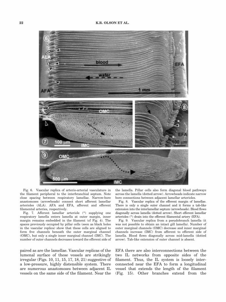
Fig. 6. Vascular replica of arterio-arterial vasculature inthe filament peripheral to the interbranchial septum. Noteclose spacing between respiratory lamellae. Narrow-boreanastomoses (arrowheads) connect short afferent lamellararterioles (ALA). AFA and EFA, afferent and efferentfilamental arteries, respectively.Fig. 7. Afferent lamellar arteriole (*) supplying one
respiratory lamella enters lamella at outer margin, innermargin remains embedded in the filament (cf Fig. 4). Thespaces previously occupied by pillar cells (seen as black holesin the vascular replica) show that these cells are aligned toform five channels beneath the outer marginal channel(OMC), but only a single inner marginal channel (IMC). Thenumber of outer channels decreases toward the efferent side of
the lamella. Pillar cells also form diagonal blood pathwaysacross the lamella (dotted arrow). Arrowheads indicate narrowbore connections between adjacent lamellar arterioles.
Fig. 8. Vascular replica of the efferent margin of lamellae.There is only a single outer channel and it forms a tab-likeextension into the interlamellar septum (arrowheads). Blood flowsdiagonally across lamella (dotted arrow). Short efferent lamellararterioles (*) drain into the efferent filamental artery (EFA).
Fig. 9. Vascular replica from a pseudobranch lamella (itwas not possible to obtain an intact gill lamella). Number ofouter marginal channels (OMC) decrease and inner marginalchannels increase (IMC) from afferent to efferent side oflamella. Blood flows diagonally across mid-lamella (dottedarrow). Tab-like extension of outer channel is absent.
K.R. OLSON ET AL.22
longitudinal vessel in between each ELA, therebyforming a vascular web around the ELA and thelateral wall of the EFA (Fig. 15). In the peripheralfilament the IL vessels and lamellae are parallel toeach other, however, there is approximately 1 ILvessel for every 2.5 lamellae (which is about thesame ratio of ALA to lamellae). Near the AFA, theIL system is condensed into a dense network withlittle space between vessels (Fig. 10) and herethere is one IL vessel for every lamella. The ILsystem also forms an anastomotic web around theALA and the lateral wall of the AFA (Fig. 19). TheIL system is less organized in the portion of thefilament contained within the intrabranchialseptum and becomes especially web-like towardthe efferent side. Closer to the afferent side thereare relatively large gaps in the IL vessels thatallow nutrient vessels to traverse across and downthe filament (Fig. 17). Adjacent to the AFA,however, the IL vessels become condensed andform a tight array with little spacing between theparallel vessels (Fig. 17) with an IL:ALA ratio ofapproximately 1, similar to that observed in theperipheral filament.A well-defined collateral vessel runs along the
filament and appears almost embedded in thelateral aspects of the AFA and filamental cartilage(Figs. 20, 22, 23). Although this vessel hasnumerous anastomotic connections with the ILsystem (Fig. 20), the relatively smooth andordered relief evident in vascular replicas is notconsistent with that of IL vessels, suggesting thatthe collateral vessel is a vein that drains the ILnetwork from the filament.The IL system is supplied almost exclusively by
post-lamellar vessels coming either directly fromthe ELA and EFA or indirectly via the nutrientcirculation (described in the following section), thelatter arising from the EFA in the basal filamentand from the EBA. Vessels arising directly fromthe EFA emanate from its medial wall (Fig. 12)and usually extend less than 300 mm into thefilament (toward the AFA) to anastomose with theIL system. These vessels are narrow-bore (o20 mmdia) and their lumen is irregular (Figs. 12, 15),although none of the narrow-bore vessels ap-peared distended and they may not be as dis-tensible as IL vessels. The narrow-bore vesselsappear somewhat constricted at the point of theirorigin from the EFA (Fig. 15) which suggests thatthey are a low-pressure network. Figure 15 showsthese vessels anastomosing with the IL system onthe near side of the filament. Anastomoses with ILvessels on the opposite side of the filament were
technically difficult to observe, but are likely tooccur here as well. These vessels are also relativelyinfrequent, approximately one vessel for every4–6 pair of ELA.
Short, narrow-bore (o20 mm dia) vessels alsoarise from the lateral wall of the ELA close to theanastomoses of the ELA with the EFA (Fig. 12).These vessels also have an irregular lumenindicative of a low-pressure network. They aremore common than those arising directly from theEFA (described above); approximately one vesselfor every 2–4 ELA. It was not possible todetermine their exact destination, although it isquite likely that they anastomose with the largesinuses lateral to the EFA (Fig. 14).
Arterio-venous circulation: nutrientvessels
The nutrient circulation is formed from narrow-bore (o20 mm dia) tortuous vessels that arise fromthe walls of the EFA near its terminal anasto-moses with the EBA and also directly from theEBA (Fig. 16). After repeated anastomoses, theyform the nutrient arteries that supply the gill archand filament. At their point of origin, the lumen ofthe tortuous vessels resembles a funnel in thatthere is an initial short dilated segment followedby a longer, albeit twisting segment of uniformdiameter (Fig. 16). These vessels do not appear tohave any specific orientation or direction; they areapproximately100 –300 mm long yet their lineartransit is less than one third of their length. Thetortuous vessels repeatedly anastomose with eachother to form progressively larger vessels. Tortu-ous vessels arising from the EFA condense to formone or two vessels that continue both peripherallyto re-enter the filament and form the filamentalnutrient arteries and basally to anastomose withthe larger arteries that comprise the nutrientsupply to the arch support tissue and interbran-chial septum (described below). Nutrient arteriesin the basal filament are closely associated withthe EFA, but peripherally branches of the nutrientarteries traverse the filament and are associatedwith the AFA. These main distributing nutrientarteries continue peripherally the length of thefilament and they are usually closely associatedwith either the AFA (Figs. 19, 22, 23) or EFA (Fig.11). Periodically they send branches into thefilament body between the filamental arteries(Figs. 10, 11, 17, 19). In many instances, branchesfrom nutrient vessels along the AFA traversenearly all the way across the filament before
TUNAGILL VASCULAR ANATOMY 23

anastomosing with the IL system (Fig. 10). In fact,it appears that most nutrient-IL anastomoses arecloser to the efferent side of the filament. Thus,branches from the nutrient artery near the AFAare considerably longer than branches originatingnear the EFA (compare Figs. 10 and 11). Dis-
tributing nutrient arteries (arterioles) are usuallyless than 25 mm in diameter, whereas branchvessels are 7 mm or less.
Nutrient vessels in the portion of the basalfilament residing in the interbranchial septumappear to arise predominantly from nutrient
K.R. OLSON ET AL.24

arteries near the AFA. We could not discern if thelatter were supplied from the tortuous vesselsoriginating from the basal EFA, or if they were fedfrom nutrient arteries in the intrabranchialseptum whose origins are mainly from the EBA(described below). These nutrient vessels traversethe filament in wide channels through the ILsystem and may run in a longitudinal direction forbrief distances (Fig. 17). They often extend the fulldistance across the filament (Fig. 17) giving offnumerous short branches that anastomose withthe IL vessels (Figs. 17, 18, 19). The long nutrientvessels in the filament body are usually less than10 mm in diameter; their terminal branches areconsiderably smaller, generally less than 5 mm(Figs. 10, 11, 18, 19). Figure 18 shows a smallnutrient vessel and three of its terminal branches.In this figure it appears that the diameter of theterminal branches becomes larger nearer theanastomoses with the IL system. It is not clearwhether the change in vessel diameter is due to aconstriction in the proximal region of the vessel,transition of the vessel from a systemic vessel toan IL-type vessel, or retrograde filling of the vesselfrom the IL system. In another terminal vessel(Fig. 21) there appears to be a sphincter-likeconstriction at the junction with the IL vessel. Inall situations, the lumenal relief of the nutrientvessels is relatively smooth (Figs. 11, 21). This isconsistent with that of a systemic vessel anddistinct from the irregular appearance of the ILvessels.The nutrient system of the gill-arch support
tissue is derived from tortuous vessels arisingfrom the EBA and basal branches from thefilamental nutrients. These vessels condense to
form one or several large arteries that coursealong the axis of the gill arch. The largest nutrientarteries (often paired) are located between theABA and EBA and are parallel to the branchialarteries (Figs. 2, 16). Large branches from thesearteries turn and pass between the EFA to supplyarch-support tissue surrounding the branchialarteries. Other nutrient vessels are found in theinterbranchial septum between the AFA of adja-cent hemibranchs. The most notable of these is arelatively large artery at the peripheral margin ofthe septum where the AFA turns toward thefilament core (Figs. 3,5). Perpendicular branchesfrom this vessel follow the AFA out into theperiphery of the filament. They may contribute tothe nutrient circulation that ultimately suppliesthe core of the filament, or they may nourish theouter margin of the filament along the AFA.However, neither of these possibilities could beconfirmed.
The arterio-venous pathway in the gill arch isdrained by large veins, two of which are foundbetween the ABA and EBA (Fig. 2). Nutrientvessels in the arch-support tissue, interbranchialseptum, and along the afferent margin of thefilament drain into these veins. It is presumed thatthe IL vessels anastomose with the branchial veinsas well, although, other than connections betweenthe IL system and an apparent vein along theAFA, the pathway of effluent from the IL could notbe determined.
DISCUSSION
Laurent (’84) and Olson (’91) provided detaileddescriptions of the teleost gill vascular anatomy
Fig. 10. Vascular replica showing intralamellar (*) andnutrient vessels (arrowheads) between afferent and efferentfilamental arteries (AFA and EFA). Arrows indicate anasto-moses of nutrient with intralamellar system.Fig. 11. Vascular replica showing the relationship between
interlamellar and nutrient vessels, lamellae were removed(* indicates broken efferent lamellar arterioles). A smallnutrient artery (N) runs along the longitudinal axis of thefilament and one of its branches traverses the filament andanastomoses with the interlamellar (IL) system (at arrow-heads). Another nutrient-IL anastomosis is also evident(double arrowheads). Arrow points to inner marginal channelsof lamellae on contralateral side of filament;. Large vessel (X)to right of nutrient may be a filamental vein, but this could notbe confirmed.Fig. 12. Vascular replica of the margin of an efferent
filamental artery facing body of filament mid-way along
filament. Small vessels originating from the medial wall ofthe artery (*) supply the interlamellar system betweenafferent and efferent filamental arteries. Other vessels (arrow-heads) arise from efferent lamellar arterioles and supply sinusvessels that surround the efferent filamental artery andconnect to interlamellar system.
Fig. 13. Longitudinal section of filament, perpendicular tolamellae (L) approximately mid-way between afferent andefferent filamental arteries. The central cartilage (arrows) iscovered by a thin filamental epithelium. A single, large innermarginal channel (arrowheads) is embedded in the filamentbody.
Fig. 14. Cross section of filament perpendicular toefferent filamental artery (EFA). Two lateral sinus vessels(S) and a central interlamellar vessel (arrowhead) are evident.Section is tangential to two efferent lamellar arterioles(ELA).
3
TUNAGILL VASCULAR ANATOMY 25

Fig. 15 a, b. Stereo pair of a vascular replica showing smallfeeder vessels (arrowheads) that originate from efferentfilamental artery (EFA) and supply interlamellar system.Several of these appear slightly constricted near their origin.The two interlamellar networks normally separated by thefilamental cartilage as well as the inner marginal channels (*)of lamellae on the far side of the filament are evident. The ILanastomose extensively and near the EFA they form acontinuous vessel (arrow) that extends the length of thefilament, other branches (double arrowheads) extend to form a
network around the lateral margin of the EFA. Broken endsof efferent lamellar arterioles are just visible at right marginof micrograph.
Fig. 16. A myriad of small tortuous vessels (*) arise fromthe efferent branchial and filamental arteries (EBA andEFA) in this vascular replica and anastomose to form the gillnutrient circulation. A large nutrient artery (N) is parallelto the EBA and supplies gill-arch support tissue, otherbranches (arrowheads) re-enter the filament as the filamentalnutrient system.
K.R. OLSON ET AL.26

and it is evident from the present study that thevascular pathways in the skipjack tuna gill followthe basic teleost design. As in other teleosts,skipjack tuna gills have a respiratory (arterio-arterial; AA) pathway and a post-lamellar, non-respiratory (arterio-venous; AV) system that isactually comprised of two distinct circulations.Minor vascular remodeling was observed in boththe AA and AV pathways that may accommodatethe over-all need of tunas to enhance gas trans-port. However, no major alterations of either AAor AV pathways were evident.
Arterio-arterial circulation
The respiratory (arterio-arterial) pathway con-sists of distributing vessels, afferent and efferentbranchial (ABA, EBA) and filamental (AFA, EFA)arteries and lamellar arterioles (ALA, ELA) aswell as the vascular sinusoids of the respiratorylamellae. A few modifications were observed in theAFA, ALA and lamellar circulation that mayincrease respiratory efficiency and these aredescribed below. Post-lamellar vessels did notexhibit any remarkable alterations.The flattened segment of the AFA in the
interbranchial septum suggests that this vessel iseither distensible or compressed against someother tissue, such as the filamental cartilage. Thisappears similar to the ampullae (blebs) found inAFA within the interbranchial septum of otherteleosts (Fromm, ’74). Ampullae may bufferarterial pulse pressure or they may boost arterialpressure when they are compressed against thecartilage by contraction of the filamental adductormuscles (Hughes, ’84; Olson, ’91; Gannon, perso-nal communication). Ampullae of other teleostsare richly innervated with adrenergic nerves(Laurent, ’84) and this may contribute to theirphysiological attributes.ALA in the basal filament (within the inter-
branchial septum) are more highly branched inskipjack tuna than other teleosts and they areinterconnected. In most teleosts ALA in the basalfilament supply four or five lamellae (Olson, ’91),whereas skipjack tuna ALA may supply twenty ormore, half of which are on opposite sides of thefilament (Fig. 2). The extensive anastomoses ofadjacent ALA in the basal filament thus appearunique. These interconnections form a vascularmanifold that may ensure uniform perfusion oflamellae. Such an arrangement may be necessaryto compensate for the unusually long ALA in thisregion. Clearly these modifications in ALA are
somehow related to their location in the inter-branchial septum as they are not similarlymodified in the peripheral filament. ALA in theperipheral filament only perfuse lamellae on oneside of the filament. This feature is found in some,but not all, other teleosts (Olson, ’91).
There are at least four modifications of thevasculature leading into and within the lamellaethat appear to augment gas exchange and reducevascular resistance; diagonal vascular channels inthe lamellae, direct delivery of blood to the outerlamellar margin, multiple marginal channels, andwidening of the lamellae near the efferent end.
Diagonal lamellar channels
The diagonal arrangement of pillar cells inlamellae, originally shown by Muir and Brown(’71) in whole-mount lamellar tissue, are clearlydemonstrated in our corrosion replicas (Figs. 7–9).Theoretically, this substantially reduces lamellarresistance by creating more in-parallel lamellarchannels and by reducing the length of eachchannel (Muir and Brown, ’71; Hughes, ’84).
Delivery of blood to outer margin
The skipjack tuna ALA delivers blood directly tothe outermost (lateral) margin of the lamellae(Figs. 7, 9) enabling blood to percolate mediallyinward toward the inner margin. To our knowl-edge the skipjack tuna is the only fish known tohave this lamellar-blood flow pattern. In otherfish, the ALA enters from the inner margin of thelamella and blood is distributed laterally into thelamellar sinusoids and circumferentially along theouter marginal channel (cf. Fig 14; in Hughes ’84).However, as in other teleosts, the ELA drain thelamellae from the medial (Fig. 8) border or mid-way between medial and lateral (Fig. 9) borders.
Multiple marginal channels
Pillar cells on the afferent end of the lamella areso closely aligned that they appear to form discreteblood vessels resembling multiple parallel outermarginal channels (Fig. 7). The innermost of thesechannels proceeds only a short distance across thelamella. There is a progressive increase in channellength from the medial to the outermost channeland only the latter is continuous along the lengthof the lamella. Because the number of medialchannels progressively decreases from the afferenttoward the efferent side of the lamella it appearsthat each channel delivers blood to a segment ofthe lamella. The diameter of these channels also
TUNAGILL VASCULAR ANATOMY 27

increases from the inner to the outermost chan-nels. Because blood in the outermost channels hasthe longest transit across the lamella beforeentering the diagonal lamellar sinusoids, anincrease in channel diameter appears to offsetthe effect of channel length on channel resistance.The number of channels on the medial margin ofthe lamella progressively increase toward theEFA to collect blood as it drains from the sinusoids(Fig. 9).
Lamellar widening
The outer marginal channels of the lamella arcaway from the filament near the efferent end ofthe lamella (Fig. 6), thereby increasing the medial-lateral lamellar width. The functional significanceof this is not known. We propose, however, that itmay increase blood oxygen saturation in thelamellar sinusoids closest to the afferent margin.Tuna gills do not have a strict countercurrent
K.R. OLSON ET AL.28

blood-water flow because blood flows diagonallyacross the lamella. If the lamellae were of uniformwidth, red cells traversing the sinusoids in theefferent region would be maximally oxygenated,whereas red cells traversing in the afferent regionwould encounter partially deoxygenated water andmight not be able to load as much oxygen.However, because the lamella is wider near itsefferent end, red blood cells have a greater chanceof becoming oxygen saturated before they reachthe inner margins. This decreases the amount ofoxygen removed from water flowing closest to thefilament on the efferent end and allows fullyoxygenated water to reach the afferent side of thelamella. In retrospect, this may be why theefferent margin of nearly all teleost lamellae isextended.The function of the small tab-like extension in
the outer margin of the lamella (Fig. 8) remainsunclear. Cross sections through the interlamellarseptum show that the septal blood supply isderived from the lamellar circulation as no othervasculature is evident. However, it is not apparentfrom our samples whether the projections oradjacent areas of the lamella are embedded inthe septum. It seems more likely that the projec-tions are not in the septum and that they areexposed to the inhalant water current where theycan affect water flow across the gill. Similarprojections have been found in elasmobranch gills(Cook, ’80; Olson and Kent, ’80) and these fishlack an interlamellar septum.The general structure of the filament and
presence of an interlamellar septum may also
enhance gas-exchange. In cross-section, the fila-ments of most fish are dumbbell shaped (Hughes,’84; Laurent, ’84; Olson, 2001). In skipjack tunathis is even more dramatic as the efferent (waterinlet) leading edge is enlarged by vascular sinuses,while the mid-region has been trimmed to anarrow cartilage sheet and a relatively thin layerof cells (Fig. 4, 13; see also Muir and Kendall, ’68).It is likely that this creates an eddy in the inhalantwater flowing around the efferent margin of thefilament such that flow along the filament bodynear the inner border of the lamella is increased(Stevens et al., ’92). Furthermore, the bow-shapedinterlamellar septum (Fig. 4) may also directinhalant water toward the filament body. Keepingin mind the diagonal pattern of blood flow in thelamella, an increase in water flow along the innerlamellar margin should favorably impact gasexchange by maintaining blood oxygen saturationin the inner marginal channels.
The thinness of the filament body is probablyrelated to another adaptation to enhance gasexchange. In most teleosts the medial margin ofthe lamella is imbedded in the filament to theextent that as much as 20 percent of the lamellarvasculature is probably not close enough to therespiratory epithelium to participate in gas ex-change (Olson, ’98, 2001). Clearly, this is not thecase for skipjack tuna where only the innermostchannel is embedded in the filamental epithelium(Fig. 13). Furthermore, because the skipjack tunafilamental epithelium in this region is thin, eventhe inner channel may be close enough to thewater boundary to participate in gas exchange.
Fig. 17. Vascular replica of the interlamellar system nearafferent filamental artery (AFA; artery lies beneath IL vesselsat left margin of micrograph) and within region of interbran-chial septum. IL vessels are irregular and leave wide spacingfor nutrient vessels (arrowheads) fed from nutrient arteries inafferent margin of filament. Dashed rectangle is enlarged inFig. 18.Fig. 18. Higher magnification of dashed rectangle from
Fig. 17 showing multiple anastomoses (arrowheads) ofnutrient vessel with interlamellar system.Fig. 19. Vascular replica of nutrient arteries (N) and
anastomotic network (*) from the interlamellar system (IL)surrounding an afferent filamental artery (not visible beneaththe other vessels). Note branches of nutrient arteries (arrow-heads) that supply body of filament and appearance of afferentlamellar arterioles (arrows) as they pass through IL.Fig. 20. Vascular replica showing relationship between
afferent filamental artery (AFA), collateral vessel (C) andinterlamelar system (IL). Medial (arrowheads) and broken
ends of lateral (*) anastomoses between C and IL are evident.Note also broken ends of afferent lamellar arterioles. Striatedrelief in AFA is produced by endothelial cells projecting intolumen of vessel.
Fig. 21. Vascular replica of an anastomosis (arrowhead) ofa nutrient vessel (N) with the interlamellar system (IL). Thesmooth relief of the nutrient is clearly distinguished fromthe irregular shape of the IL.
Fig. 22. Cross section through the filament near theafferent filamental artery (AFA). The collateral vessel (C) isnear the AFA and the filamental cartilagenous support (CS). Apair of vascular bundles (brackets) on either side of the AFAcontain one nutrient artery and several thin-walled vesselsthat may be either filamental veins, IL vessels, or both.
Fig. 23. Higher magnification of serial section near Figure22, although an afferent lamellar arteriole (ALA) is sectionedtangentially in this figure. The nutrient artery (arrowhead)is filled with red blood cells. Compare with Figure 19, otherabbreviations as in Figure 22.
3
TUNAGILL VASCULAR ANATOMY 29
Arterio-venous circulation
The arterio-venous circulation arises from thepost-lamellar vessels and it is ultimately returnedto the heart via the branchial veins. By definition,these are systemic vessels. This vasculature iswell-developed in the filament body and has beencalled the central sinus, central venous sinus, orvenolymphatic system (Laurent, ’84; Olson, ’91).The arterio-venous vasculature can be furtherdivided into two networks the intralamellar andnutrient (Boland and Olson, ’79; Olson, ’80, ’81;Olson and Kent, ’80; Olson et al., ’86, ’90, ’94, ’95).This distinction is based on the origin of the post-lamellar vessels and the type of vessel that theysupply in the filament body. We have clearlyshown that both vascular networks are present inskipjack tuna gills.
Interlamellar (IL) system
In many fish, such as the channel catfish,Ictalurus punctatus (Boland and Olson, ’79, Olson,’80); rainbow trout, Oncorhynchus mykiss (Olson,’83); bowfin, Amia calva (Olson, ’81); climbingperch, Anabas testudineus (Olson et al., ’86); singicatfish, Heteropneustes fossilis (Olson et al., ’90);snakeheads, Channa punctata, C. gachua and C.marulius (Olson et al., ’94); walking catfish,Clarias batrachus (Olson et al., ’95); and dogfishshark Squalus acanthias (Olson and Kent, ’80) theIL system comprises a repetitive vasculaturenetwork whose vessels traverse the filament likeladder rungs. The system is so named because thevessels are in register with, but spaced betweenthe lamellar inner marginal channels. Vascularcorrosion replicas of IL vessels are noticeablyirregular and the system appears highly disten-sible; if filled with resin at supraphysiologicalpressure the vessels appear to coalesce into a largesack-like vessels (Olson, ’83). The IL system issupplied by narrow-bore vessels from the wall ofthe EFA and in numerous species also by thenutrient vessels (described below). The filamentalvasculature in skipjack tuna gills fulfills thisdescription with one exception; across much ofthe peripheral filament there is only one IL vesselfor approximately 2.5 lamellae. Perhaps this is dueto the greater number of lamellae in tuna gills(twice as many lamellae per mm filament length)compared to other teleosts (Hughes, ’84). It thusappears that most, if not all, of the selectionpressure for high performance was placed on therespiratory circulation. This also implies that thephysiological function of the IL system, although
enigmatic, is not related to an increased metabolicdemand of the fish.
Nutrient vessels
The nutrient system is formed from the con-fluence of numerous narrow-bore tortuous vesselsthat arise from the basal wall of the EFA and fromthe EBA and like the IL system is a commonfeature in many teleosts (see above references).These vessels are the proximal component part ofthe secondary circulation (Olson, ’96) and havealso been found in the skipjack tuna heat-exchanger vessels (Dewar et al., ’94). Feedervessels from this system supply the gill-archsupport tissue and other branches return intothe filament. Their relatively smooth lumen andreduction to capillary-size vessels suggest that thissystem has a nutrient function.
Nutrient capillaries do not re-anastomose intoprogressively larger venules and veins, but insteadconnect directly into the large diameter IL system,which in turn may anastomose with a filamentalvein (Fig. 20). Perhaps this is the best option fordraining from the filament, although the locationof these anastomoses suggests otherwise. Nutrientvessels near the AFA send narrow branches nearlytwo-thirds of the way across the filament (towardthe EFA) before they anastomose with IL vessels.Because these anastomoses are somewhat distantfrom the longitudinal vessels that appear to drainthe filament, it is tempting to speculate that they,and the small vessels that arise from the medialwall of the EFA, create flow through the IL vesselsthat is countercurrent to lamellar blood flow andnutrient flow and that is concurrent to the flow ofrespiratory water. A possibility similar situationhas been noted in the channel catfish (Boland andOlson, ’79). Unfortunately, it was not possible todetermine the direction of flow in the vascularreplicas.
It is clear from the present study that thevasculature of the skipjack tuna gill is essentiallythe same as that found in other teleosts. The needto increase gas exchange has resulted in minoralterations in the arterio-arterial pathway, espe-cially within the lamellae, to reduce vascularresistance while maintaining, or increasing, theefficiency of gas exchange. There has been little ifany modification of the two arterio-venous path-ways suggesting that these have little function ingas exchange. Furthermore, our findings suggestthat the arterio-venous pathways plays an im-portant role in other, nonrespiratory, homeostatic
K.R. OLSON ET AL.30

activities in the tuna gill because both pathwaysare retained as elaborate and extensive vascularnetworks while other tissue elements in thefilament body have been greatly reduced.
ACKNOWLEDGMENTS
H.D. thanks the Bushnell-Gettleman family fortheir hospitality.
LITERATURE CITED
Bernal D, Dickson KA, Shadwick RE, Graham JB. 2001.Review: Analysis of the evolutionary convergence for highperformance swimming in lamnid sharks and tunas. CompBiochem Physiol [A] 129:695–726.
Boland EJ, Olson KR. 1979. Vascular organization of thecatfish gill filament. Cell Tiss Res 198:487–500.
Brill RW, Dizon AE. 1979. Effect of temperature on isotonictwitch of white muscle and predicted maximum swimmingspeed of skipjack tuna, Katsuwonus pelamis. Environ BiolFishes 4:199–205.
Brill RW. 1996. Selective advantages conferred by the highperformance physiology of tunas, billfishes, and dolphin fish.Comp Biochem Physiol 113A:3–15.
Brill RW, Bushnell PG. 2001. Cardiovascular system of tunas.In: Fish Physiology Vol. XIX, Tuna Physiology, Ecology, andEvolution. Block BA, Stevens ED, Eds. Pp 79 –120, SanDiego: Academic Press, Inc.
Bushnell PG, Brill RW. 1992. Oxygen transport and cardio-vascular responses in skipjack tuna (Katsuwonus pelamis)and yellowfin tuna (Thunnus albacares) exposed to acutehypoxia. J Comp Physiol [B] 162:131–143.
Cooke IRC. 1980. Functional aspects of the morphology andvascular anatomy of the gills of the endeavour dogfish,Centrophorus scalpratus (McCulloch) (elasmobranchii:squalidae). Zoomorphologie 94:167–183.
Dewar H, Brill RW, Olson KR. 1994. The secondary circula-tion of the vascular heat exchangers in skipjack tuna,Katsuwonus pelamis. J Exp Zool 269:566–570.
Korsmeyer KE, Dewar H. 2001. Cardiovascular system oftunas. In: Fish Physiology Vol. XIX, Tuna Physiology,Ecology, and Evolution. Block BA, Stevens ED, Eds.pp. 36–78, San Diego: Academic Press, Inc.
Fromm PO. 1974. Circulation in trout gills: Presence of ‘blebs’in afferent filament vessels. J Fish Res Board Can 31:1793–1796.
Gannon BJ. 1978. Vascular casting. In: Principles andTechniques of Scanning Electron Microscopy. Vol. 6 (ed.Hayatt, M.A.), pp. 170–193. New York: Van NostrandReinhold.
Hughes GM. 1984. General anatomy of the gills. In: FishPhysiology, Vol. X Gills, Part A, Anatomy, Gas Transfer, andAcid-Base Regulation (eds. Hoar, W.S. and Randall, D.J.),pp. 1 –72. New York: Academic Press, Inc.
Laurent P. 1984. Gill internal morphology. In: Fish Physiol-ogy, Vol. X Part A, Anatomy, Gas Transfer, and Acid-BaseRegulation (eds. Hoar, W.S. and Randall, D.J.), pp. 73–183.New York: Academic Press, Inc.
Muir BS, Brown CE. 1971. Effects of blood pathway on thepressure drop in fish gills, with special reference to tunas.J Fish Res Bd Canada 28:947 –955.
Muir BS, Hughes GM. 1969. Gill dimensions for three speciesof tunny. J Exp Biol 51:271 –285.
Muir BS, Kendall JI. 1968. Structural modifications in thegills of tunas and some other oceanic fishes. Copeia1968:388–398.
Nakamura EL. 1972. Development and use of facilities forstudying tuna behavior. In: Behavior of Marine Animals,Vol. 2 (eds. Winn, H.E. and Olla, B.L.), New York: PlenumPress.
Olson KR. 1980. Application of corrosion casting proceduresin identification of perfusion distribution in a complexmicrovasculature. In: Scanning Electron Microscopy Vol. 3(ed. Johari, O.), pp. 357 –364. Chicago: SEM, Inc.
Olson KR. 1981. Morphology and vascular anatomy of the gillsof a primitive air breathing fish, the bowfin (Amia calva).Cell Tiss Res 218:499–517.
Olson KR. 1983. Effects of perfusion pressure on themorphology of the central sinus in the trout gill filament.Cell Tiss Res 232:319–325.
Olson KR. 1985. Preparation of fish tissues for electronmicroscopy. J Electron Microsc Technique 2:217–228.
Olson KR. 1991. Vasculature of the fish gill: anatomicalcorrelates of physiological function. J Electron MicroscTechnique 19:389–405.
Olson KR. 1996. The secondary circulation in fish: anatomicalorganization and physiological significance. J Exp Zool275:172–185.
Olson KR. 1998. Hormone metabolism by the fish gill. CompBiochem Physiol 119A:55 –65.
Olson KR. 2001. Microscopic functional anatomy: respiratorysystem. In: The Laboratory Fish (ed. Ostrander, GK),London: Academic Press.
Olson KR, Ghosh TK, Roy PK, Munshi JSD. 1995. Micro-circulation of gills and accessory respiratory organs of thewalking catfish Clarias batrachus. Anat Rec 242:383–399.
Olson KR, Kent B. 1980. The microvasculature of theelasmobranch gill. Cell Tiss Res 209:49 –63.
Olson KR, Munshi JSD, Ghosh TK. 1990. Vascular organiza-tion of the head and respiratory organs of the air-breathingcatfish, Heteropneustes fossilis. J Morphol 203:165–179.
Olson KR, Munshi JSD, Ghosh TK, Ojha J. 1986. Gillmicrocirculation of the air-breathing climbing perch, Ana-bas testudineus (Bloch): Relationships with the accessoryrespiratory organs and systemic circulation. Am J Anat176:305–320.
Olson KR, Roy PK, Ghosh TK, Munshi JSD. 1994. Micro-circulation of gills and accessory respiratory organs from theair-breathing snakehead fish, Channa punctata, C. gauchaand C. marulius (Ophiocephalidae, Ophiocephaliformes).Anat Rec 238:92–107.
Stevens ED. 1992. Oxygen molecules as units to dimensionthe sieve of fish gills. Env Biol Fish 33:317–318.
TUNAGILL VASCULAR ANATOMY 31
Copyright © 2022 FDOKUMEN