Spermatogenesis and spermatology of some teleost fish ... - HAL
45
HAL Id: hal-00898500 https://hal.archives-ouvertes.fr/hal-00898500 Submitted on 1 Jan 1986 HAL is a multi-disciplinary open access archive for the deposit and dissemination of sci- entific research documents, whether they are pub- lished or not. The documents may come from teaching and research institutions in France or abroad, or from public or private research centers. L’archive ouverte pluridisciplinaire HAL, est destinée au dépôt et à la diffusion de documents scientifiques de niveau recherche, publiés ou non, émanant des établissements d’enseignement et de recherche français ou étrangers, des laboratoires publics ou privés. Spermatogenesis and spermatology of some teleost fish species (1) Roland Billard To cite this version: Roland Billard. Spermatogenesis and spermatology of some teleost fish species (1). Reproduction Nutrition Développement, 1986, 26 (4), pp.877-920. 10.1051/rnd:19860601. hal-00898500
-
Upload
khangminh22 -
Category
Documents
-
view
4 -
download
0
Transcript of Spermatogenesis and spermatology of some teleost fish ... - HAL

HAL Id: hal-00898500https://hal.archives-ouvertes.fr/hal-00898500
Submitted on 1 Jan 1986
HAL is a multi-disciplinary open accessarchive for the deposit and dissemination of sci-entific research documents, whether they are pub-lished or not. The documents may come fromteaching and research institutions in France orabroad, or from public or private research centers.
L’archive ouverte pluridisciplinaire HAL, estdestinée au dépôt et à la diffusion de documentsscientifiques de niveau recherche, publiés ou non,émanant des établissements d’enseignement et derecherche français ou étrangers, des laboratoirespublics ou privés.
Spermatogenesis and spermatology of some teleost fishspecies (1)Roland Billard
To cite this version:Roland Billard. Spermatogenesis and spermatology of some teleost fish species (1). ReproductionNutrition Développement, 1986, 26 (4), pp.877-920. �10.1051/rnd:19860601�. �hal-00898500�

Spermatogenesis and spermatologyof some teleost fish species (1)
R. BILLARD
Station de Ph ysiologie animale, /. N. R. A.,78350 Jouy-en-Josas, France.
Summary. In teleost fish, the whole of the processes involved in the formation of
spermatozoa and their release and in fecundation are extremely diverse.Two main types of testicular structure are distinguished : a tubular and a lobular. The
organization of the lobules and genital duct structure is very diverse in the latter type.Spermatogenesis can be continuous, as in guppy, discontinuous with overlapping
cycles, as in carp, or show well separated cycles as in trout.Spermiogenesis shows increasing degrees of complexity, leading to the formation of
extremely simple spermatozoa as in carp (spherical head, very little nuclear histonetransformation, very simple mid-piece with untransformed cytoplasmic and mitochondrialremains) or much more complex spermatozoa as those of guppy (elongated head, histone-protamine transformation, transformation of the centriolar complex in the deep nuclearnotch, very developed mid-piece with glycogen stores).
Spermatogenetic production is extremely variable (GSI : 0.1-10 % ; annual pro-duction : 1 .10$ to 7.109 spermatozoa/g body weight).
Spermiation yield varies according to species but also within the same species(20-90 % in trout).
The endocrine model of spermatogenesis and spermiation is very different in carp andtrout. The main environmental factor influencing spermatogenesis is photoperiod in trout andtemperature in carp ; spermiation depends on temperature in trout and on socialenvironment as well as temperature in carp.
Sperm physiology (survival time in vivo and in vitro, motility time, metabolism) is verydifferent in the several species studied. Two groups with a fundamentally different
physiology (internal or external fecundation), sperm biology and morphology (structure oftestis and spermatozoon) can be grossly distinguished. These groups are fish with lobulartestes (guppy) and those with tubular testes (teleosts).
Introduction.
The reproductive function in male teleosts is original in several ways.
1. There is a wide variation in the amount of spermatozoa produced. This isindicated by the increase in testicular weight at the same time as body weight
(1) This paper reviews the personal work of the author and co-workers ; their articles are noted inparenthesis in the text and given in a list at the end.
(2) Present address : Museum national d’Histoire naturelle, lchtyologie, 43, rue Cuvier 75231 Paris.

(fish growth is a continuous or subcontinuous process) ; testicular weight is
usually expressed as a percentage of empty body weight or as gonadosomaticindex (GSI). In annually reproducing species this gonad growth reaches 5 to 10 %in salmonids (Hiroi and Yamamoto, 1968, 1970) and cyprinids (Solewski, 1957 ;Weil, 1981), 2 to 3 % in pike (Medford and Mackay, 1978) and 0.2 to 2 % in
various tilapia (Peters, 19711. Spermatogenetic production has been studied indetail in only a small number of cases (Turdakov, 1968), but since spermatozoaconstitute the majority of cells in the mature testis, testicular weight is thought tobe a good criterion of the quantity of spermatozoa produced. It is thereforeevident that spermatogenetic activity varies greatly from one species to another.The reasons for these differences are not clear and they have been little discussedby authors, even in recent reviews on spermatogenesis (Nagahama, 1983). Otherthan aspects of quantitative production, it is necessary to consider sperm qualityand the true amounts of spermatozoa available for fertilization.
2. The periods during which spermatogenesis occurs differ widely as wellas the time at which the gametes are released (review by Billard and Breton,1978). It is supposed that spermatogenesis is continuous in male guppies in thelaboratory because females seem to be gravid all year long (Rosenthal, 1952 ; Billard,1966). In nature, spermatogenesis is seasonal in temperate zone species ; Atlanticsalmon (Jones and Orton, 1940), brook trout (Henderson, 1962) and pike (Loftsand Marshall, 1957) show spermatogenesis in summer and the sea-bream in
spring (Papadopol, 1962). In some cases, spermatogenesis may begin in autumnand finish in spring as in the stickleback (Craig-Bennett, 1931) ; it appears to alsobe seasonal in some subtropical and tropical species (Barbus liberiensis : Payne,1975 ; tilapia : Hyder, 1970 ; see review by Lam, 1983). Sperm release or
spermiation also takes place at very different seasons (autumn and winter in
salmonids and spring or summer in cyprinids) ; it coincides with the rainy seasonin the tropics. In temperate zone species, it is interesting that spermiation andspawning occur at such different seasons although conditions favoring maximaljuvenile survival, such as availability of food that is sufficient in quality (prey sizecorresponding to larval mouth size) as well as quantity, only occur in spring orsummer.
3. The modes of reproduction are very diverse. This question has beenreviewed in detail by Breder and Rosen (1966). Some species are viviparous andhave internal fecundation, while other more numerous species present externalfecundation in very diverse media (fresh water, salt water, briny water) where thegametes are subjected to considerable osmotic shock. Laying and incubationsubstrates (grass, algae, gravel) also vary. The male sometimes incubates the
eggs. Reproductive behavior is very diversified ; some species spawn in couples,while others spawn in groups. This variety of reproductive modes is often
associated with important body changes (permanent or temporary) or with
endocrine adaptation directly related with the reproductive function. The hippo-
Abbreviations : GA : type A spermatogonia ; GB : type B spermatogonia ; GSI : gonadosomaticindex ; PGC : primary germ cell.

campus male, which incubates the eggs, presents changes in tegument due tospecific endocrine determinism (Boisseau, 1967). These general observations,usually based on field work, seldom contain any detailed analysis of the dynamicsof spermatogenesis or spermiation or of the quantity and biology of the
spermatozoa involved in reproduction.The present work uses several freshwater species of the temperate zone,
such as salmonids and cyprinids, as examples and describes and quantifies theformation, release, morphology and viability of their spermatozoa. These specieswill be compared with the guppy, a tropical species which reproduces continually.Spermatogenesis, its endocrine regulation and sperm biology will be analysedusing some examples to illustrate widely diverse modes of reproduction. We shallalso define the positive and negative environmental factors that determine thetimes of the year at which spermatogenesis and spermiation occur in the speciesstudied. The literature on spermatogenesis in teleost fish will not be exhaustivelyreviewed here ; readers are referred to several recent reviews on this subjectwhich summarize the state of the art (Grier et al., 1980 ; Grier, 1981 ; Brusl6,1982 ; Nagahama, 1983 ; Fostier et al., 1983 ; Stoss, 1983 ; Van Thienoven,19831.
The aim is to better understand the regulatory mechanisms initiating seasonalreproduction and the emergence of juveniles at the same time that qualitativelyand quantitatively suitable food supplies become available in the environment.
1. The morphological and dynamic aspects of spermatogenesis
1.1. Testis morphology.
The morphology of the testis has been studied macroscopically (1) and
microscopically (2). The testis is usually paired but the two lobes may be partiallyfused, as in perch, or totally fused, as in guppy. Two types of testicular structureare distinguished.
1.1.1. Tubular type (guppy) (1). - At the center of the testis is a large cavity(fig. 1) where the spermatozoa are stored ; they are grouped into bundles called

spermatozeugmas. Tubules radiate from this cavity towards the periphery(fig. 2) ; the blind end (apex) of these tubules is apposed against the testicularcapsule. Isolated germ cells (3), the stem spermatogonia, are found concentratedat the apex. Immediately below the germ cells, the B spermatogonia organize intocysts which progress towards the center of the testis during spermatogenesis(fig. 2) (4).
1.1.2. Lobular type. ― In this type, connective tissue extends from thetesticular capsule to form irregular « tubes » lined with a Sertoli cell epitheliumincluding germ cells (fig. 3). The blind end is apposed against the testicular
capsule ; the tubes converge ventrally towards a sperm collection system. Lobularstructure is thus different from tubular structure ; type A spermatogonia (GA’s)are found all along the lobule, while in the tubule they are localized in the apex.
During spermatogenesis, the cysts move only slightly towards the center of thelobule ; the spermatozoa are released into the lobule lumen from which theyreach the efferent and deferent systems (fig. 2) (4, 5). The complexity of lobulartestis structure varies with the species since the extensions of connective tissuemay overlap more or less.
1.1.3. Interstitial cells and the interlobular space. ― The lamina of
connective tissue from the capsule is composed of fibroblasts bordered on thelobule side by a basal lamina against which Sertoli cells are apposed. The

fibroblasts form a quasi-continuous layer around the tubules and a discontinuousone around the lobules (fig. 21. Using morphological criteria, interstitial cells can bedistinguished between the fibroblast layers. They are found in all studied speciessuch as guppy, trout, pike, goldfish, roach, carp and tilapia (6).
1.1.4. Genital tract. - The male genital tract is very simplified in guppy ; asingle duct, equivalent to a vas deferens, connects the central lumen of the testiswith a genital papilla (1) (fig. 4).
At the place where the open ends of the lobules converge in other teleostswith lobular testes, there is an epithelial area which has no germ cells and delimitsa space where the spermatozoa accumulate. This area, found all along the testis,is connected at its posterior part to a duct that joins it to the genital papilla. Thetwo original ducts in both testes fuse at the genital papilla. The whole of thiscollection system, intratesticular and extratesticular, acts as a spermiduct. Thestructure of the epithelium lining the extratesticular part varies according to thespecies and the sex cycle stage. In trout, the epithelium is secretory and mayphagocytose the spermatozoa (7) and the spermiduct is long (as long as the testisitself) and distends to become a true sperm storage organ, attaining a space of10 % of the body weight. On the contrary, in carp the length of the free

spermiduct is very reduced and in no case is it a large storage organ. No
accessory gland, except for a small diverticulum at the base of the spermiduct in
pike, can be microscopically identified in the species studied ; the function of thisdiverticulum is unknown. The spermiduct in trout contracts spontaneously, theamplitude of the contractions decreasing with the temperature (4). These
contractions do not occur in the testis itself.
1.2. Morphology of Sertoli cells and germ cells.
1.2.1. Sertoli cells. - In the guppy (8) as in other teleosts with lobular testes
(5), germ cells other than spermatozoa, whether isolated or in groups, are
surrounded by Sertoli cells ; the germ cells are never in contact with the basal
lamina. Several roles, based on morphological criteria, have been attributed to theSertoli cells : (i) the support and structuration of lobules, tubules and cysts ;(ii) the spermatozeugma morphogenesis in guppy ; (iii) the transfer and eventualconversion of metabolites or hormones towards germ cells or the central cavity ;(iv) the phagocytosis of germ cells and residual spermatozoa. The Sertoli cells
seem to be permanent in lobular testes but degenerate in the guppy, and we haveno clear understanding of their renewal or origin ; (v) the isolation of the cystcompartment beyond the spermatocyte stage due to the appearance of tightjunctions which prevent peroxidase penetration fMarcaillou and Szollosi, 1980).
1.2.2. Primordial germ cells (PGC) and spermatogonia.Tubular testis. - Several large GA’s found in the guppy apex have nuclei
measuring between 8 and 10 11m in histological preparations. These nuclei haveonly one rather large, more or less central nucleolus and several chromatinfilaments that radiate towards the periphery (11. These cells are interpreted astype A (GA) spermatogonia. Ultrastructural analysis (3) shows that each one is
almost entirely surrounded by a layer of Sertoli cytoplasm. They have fewstructured organelles, only a few mitochondria and little reticulum (3). The
presence of PGC in adult guppy was not cleared.Lobular testis. - Some germ cells in trout similar to those in guppy are
inserted in the Sertoli layer all along the lobules. They are more numerous in thelobular than in the tubular apex. The morphology seen in photonic microscopy ofthe nucleus (10 11m) of embryonic gonad cells in newly-hatched alevins is similarand the cells are considered to be primordial germ cells (PGC1. Their total numberis about 50 (9). They show mitotic activity and yield two types of gonocyteswhich are morphologically different in photonic microscopy (9) and least one ofwhich (G2) may occur in adult males.
Spermatogonia grouped in cysts appear to be irreversibly engaged in
spermatogenesis and are interpreted as type B spermatogonia (GB). Byestimating the number of cells in various types of cysts, it has been determinedthat there are 14 spermatogonial generations in guppy. During these generations,cell diameter decreases from 7 to 3 11m (measured on histological sections) andthe chromatin becomes more dense (1) as the organelles diversify (3) (8).
The cysts of species with lobular testes cannot be dissected and they aretherefore difficult to identify in seriated sections ; this makes it impossible to

routinely count the cells they contain and thus the number of spermatogonialgenerations.
There appears to be fewer spermatocytes per cyst in trout than in guppy,suggesting a reduced number of spermatogonial generations.
1.2.3. Spermatocytes. ― Guppy spermatocytes have been thoroughlyanalysed (1-3). Leptotene spermatocytes are distinguished from final spermato-gonia by their larger nuclei (4 !m). The cysts increase in size. Changes in thevarious leptotene-zygotene and pachytene phases occur synchronously in each
cyst and all the cells of a cyst seem to be linked by cytoplasmic junctions. Thereare few spermatocyte II cysts, indicating that this phase is short. Spermatocytemorphology is similar in species with lobular testes.
1.2.4. Spermatids. - Spermatid morphology, spermiogenetic profile andfinal sperm structure show diverse degrees of complexity depending on the
species (fig. 5). The most complex type, occurring in guppy (10), shows anextremely developed mid-piece formed during spermiogenesis at the same timethat the nuclear head elongates along with the microtubular structures. Duringelongation, dense chromatin elements appear in the nuclear periphery whichcorrespond with the appearance of protamine in the nuclear proteins (11).Glycogen particles are visible in the mid-piece (12).
In trout, the nucleus elongates slightly as the chromatin becomes more dense(13). The centriolar complex changes little (14) and the mid-piece is reduced to aring-shaped mitochondrion (13-15). A septum appears in the A microtubule ofsome doublets (16). The spermatozoon head shows symmetric revolution.
Spermiogenesis in carp and pike is very brief ; the sperm head generally remainsspherical with the flagellum inserted on one side of the sphere by an unmodifiedcentriolar complex. The mid-piece is reduced (14). An original type of
spermiogenesis has been described in the eel (17) where the nucleus elongatesgreatly and a mitochondrion is found in the anterior part of the sperm head.
In conclusion, fish spermatozoa, even when very complicated as in guppy,are primitive compared with those of higher vertebrates ; the chromatin is lesstransformed and condensed and the mid-piece is very reduced.
1.3. Dynamics of spermatogenesis.
1.3.1. Spermatogenetic cycle. - An examination of the guppy testes showsthat there is continuous spermatogenetic activity throughout the year ; spermato-zeugmas can be collected all year long (Billard, 1966) without modifying thereserves, showing that production is permanent. This is not true in species withlobular testes where spermatogenesis is not continuous. Two main types of
spermatogenetic cycles have been distinguished.- In trout there are two successive, distinct cycles. All the spermatozoa of onecycle are eliminated before the next cycle begins ; only the GA’s are permanent(fig. 6-7) (24, 25). The situation is similar in roach (fig. 7) (26), tench (27) and pike(28-32).- In carp the two successive cycles are not as distinct. GB’s and spermatozoaare always present in the lobules as well as GA’s. Spermatozoa and spermatids,


found in summer, correspond to the main spermatogenetic activity which is
accompanied by a rise in the GSI (fig. 7) (25).The histological images of these two types of spermatogenesis are very
different. Well-defined phases succeed each other in trout : spermatogenesis andthe presence of cysts, GA’s + spermatozoa, GA’s alone. In carp, cysts and
spermatozoa occur permanently (22).The period when active spermatogenesis occurs varies with the species. In
carp and trout it takes place in summer and in tench in spring ; in roach it beginsin autumn and finishes in spring. The different types of spermatogenetic cycle canthus be identified by the season at which they occur (fig. 8) (29). Spermato-genesis lasts 36 days at 25 °C (1) in guppy and the leptotene-spermiation interval14 days (181. In roach, the latter interval lasts 30 days at 10 °C (Gillet and Billard,unpublished).
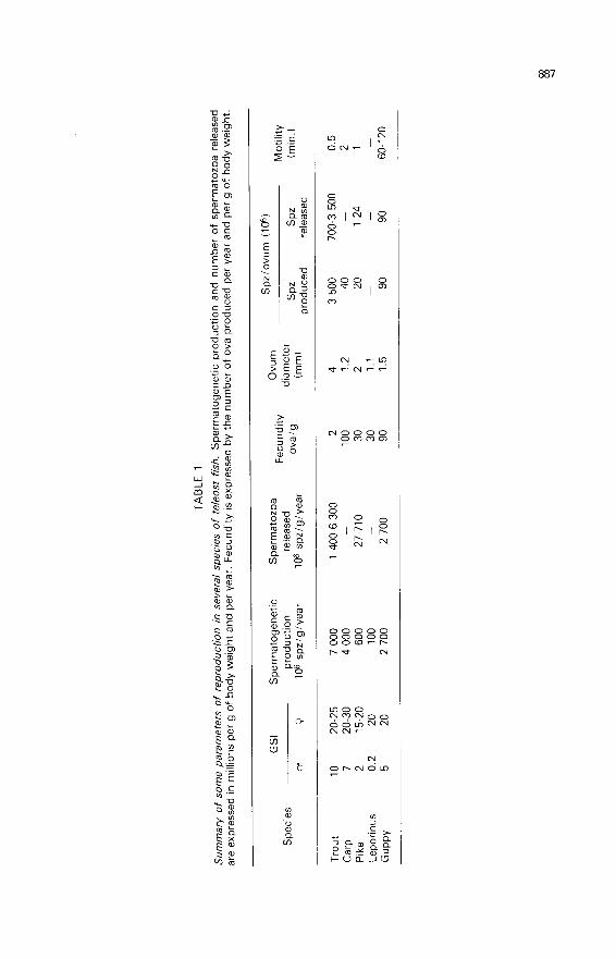
1.3.2. Spermatogenetic production. ― The guppy produces an average of36 spermatozeugmas per day (19) or 750 000 spermatozoa daily, counting thenumber of spermatozoa per spermatozeugma (1 The values obtained for several
species with lobular testes are given in table 1.
1.3.3. Spermiation. - The term o spermiation » in guppy means the sameas in mammals, i.e. the detachment of spermatozoa from the Sertoli cells. In
lobular testes, this term has no morphological basis since the spermatids are notinserted in the Sertoli cell layer. The term « spermiation » could correspond to theopening of the cysts and the release of spermatozoa into the lobule lumen, but it
is the usage to refer to the release of spermatozoa by the testis and their arrival inthe spermiduct. The expulsion of spermatozoa from the spermiducts (spermrelease) by abdominal massage shows that the process exists ; its intensity is
estimated by measuring sperm volume after massage, the sperm being collectedin a test-tube placed under the abdominal pore. In this way, most of the sperm inthe spermiducts can be collected. The volume of milt and sperm concentration,estimated by spermatocrit (32) and optic density (33), are measured during thereproductive period in trout (30, 31) and pike (32).
In both species, the total number of spermatozoa collected is much lower
than spermatogenetic production (table 11. No systematic study has been done incarp, but regular measurements at given times have shown that sperm volume islow when there is no hormonal stimulation (see below). In species such as trout

and pike with distinct spermatogenetic cycles, the residual spermatozoa are
resorbed in the testis itself where they are phagocytosed by Sertoli cells (5) andby some macrophages identified in the lobular lumen (4, 21 Moreover, somespermatozoa are lysed in the lobular lumen (13). Lysis and resorption byphagocytosis also occur in the spermiduct (7).
1.4. Conclusion.
Spermatogenesis seems to be continuous in a species like guppy which hasan original testicular structure consisting of tubules in which cysts move. This
process is seasonal in such species as trout, carp and tench but occurs at
different seasons ; three groups can be distinguished which include a number oftemperate zone species (41) (fig. 8) : group 1 (trout, carp, pike) : spermato-genesis occurs in summer ; group 2 (roach) : spermatogenesis begins in autumnbut is interrupted in winter due to low temperatures and then recommences andterminates in spring ; group 3 (tench) : the testes are quiescent in autumn and
winter and spermatogenesis occurs entierely in spring.While there is little difference in spermatogenetic production among the
species (between 68 000 and 150 000 spermatozoa per day and per g of testis, orbetween 1.7 and 7.5 106 spermatozoa per day and per g of body weight), thespermiation yield is much more variable (between 3 and nearly 100 % of thespermatogenetic production). Although testicular weight alone is a good criterionof spermatogenetic production, it cannot be used to determine the efficiency ofthe male reproductive function, particularly the number of spermatozoa releasedduring reproduction.
Spermatogenesis is a very diversified process in teleost fish, as seen above.This diversity is evident in the structures of the testis and genital tract, thedynamics of spermatogenesis ― which may be continuous, quasicontinuous orvery seasonal ― spermatogenetic effectiveness (up to 14 spermatogonialgenerations), spermatogenetic production and spermiation yield.
This diversity also occurs in the morphology of the spermatids and the
spermatozoa may reflect an adaptation to the various biotopes the fish havecolonized.
We shall see how the different components of these diverse environmentsinfluence and determine the various phases of the reproductive function.
2. Some aspects of the endocrine regulationof spermatogenesis and spermiation
2.1. Material and methods.
Radioimmunoassay (34) and biological assay (35) of gonadotropins havebeen developed for endocrine studies by Breton and Jalabert, radioimmunoassayof androgens by Fostier (36) and the immunocytochemical analysis of pituitarycells by Dubois (37). The techniques of hypophysectomy (38), castration (39) andstereotaxy (40) have also been used. Hypophysectomy was widely practiced onguppy and goldfish but not in trout due to technical problems. Castration is

difficult in cyprinids and has only been done in trout. Gonadotropins(glycoproteins) used for standard RIA and hormone supplements were providedas follows : Burzawa-G6rard (1973) : carp gonadotropin (cGTH) ; Breton et al.,(1976) : trout and salmon gonadotropins (tGTH and SGTH).
2.2. Profiles of trout and carp gonadotropins and androgens during the annualcycle.The study was based on regular samplings of plasma and pituitary at given
times once or twice a month in rainbow and brown trout and in carp (25). Inbrown trout the levels of pituitary and plasma GTH rose markedly in April-Maywhile testis weight was minimal ; GB first appeared at this time. During the restof the cycle, pituitary GTH varied little but tended to increase during spermiation.Plasma GTH increased during spermatogenesis, reaching a maximum before
spermiation began. Similar profiles were found in rainbow trout (25) where plasmaGTH tended to decrease just before the first spermatozoa appeared in the
spermiducts. Other experiments have confirmed this trend and showed that totalandrogen, and in particular 11-ketotestosterone (11-KT1, reached the highest peakat spermiation. The disappearance of 11-KT from the plasma at the end of the cyclecoincided with the arrest of spermiation (411. ).
2.3. Endocrine control of spermatogenesis and spermiation,2.3.1. Spermatogenesis. ― Hypophysectomy has shown that the depen-
dence of various spermatogenetic stages varies with the species.- In guppy the last spermatogonial generations degenerate rapidly when the
pituitary has been removed. On the contrary, primary generations of GB (G,-G4)disappear more slowly after hypophysectomy. Spermatocytes I do not developinto spermatocytes II and spermatids do not elongate. However, the GA’s subsistin the apex and the first stages of meiosis do not seem to be pituitary-dependent(42) ; methallibure treatment does not inhibit spermatogenesis as much (43).Supplements of the crude pituitary extracts of gambusia, a species of the samegroup, maintain or at least qualitatively regenerate spermatogenesis in hypo-physectomized guppies (44), and limited, partial regeneration is obtained with
carp pituitary extracts and steroids (4-44).- In goldfish, hypophysectomy carried out in full spermatogenesis causes a
considerable decrease in testicular weight (4) and the total disappearance of all
germ cells, except GA’s and spermatozoa, within four weeks (45). A milder
inhibitory treatment using methallibure showed that higher levels of gonadotropichormones were needed to maintain spermatocytes and spermatids than to
maintain GB’s (46). Spermiogenesis did not require the presence of GTH (45).Some experiments on maintenance or restoration after hypophysectomy or
methallibure treatment have shown that spermatogenesis is completely restored(45) by carp total pituitary extracts (47-48) and by a highly purified gonadotropicfactor (49). In goldfish, preparations of cyprinid crude pituitary gonadotropinsseem to be more active than those of salmonids, suggesting specific zoologicactivity (44). Mammalian gonadotropic hormones have no effect on spermato-genesis in goldfish ; the GSI has been only partially maintained with HCG, but

this hormone has no effect on the germ cells themselves. Testosterone in the
form of propionate may maintain spermatogenesis after hypophysectomy but thedose varies with the stage : 1 to 10 pg per injection are enough to maintain theGB’s but 200 pg are needed for meiosis and spermiogenesis (45).
2.3.2. Spermiation. - Goldfish spermiation is completely inhibited byhypophysectomy (50-51 ) and does not seem to occur when GTH levels decreaseto less than 10 ng/ml with methallibure treatment (46). Intact males in captivityshowing circulating levels of 8 ng release no sperm (52). Hormone supplementsgiven to intact or hypophysectomized males show that purified gonadotropichormones and crude pituitary preparations as well as HCG markedly stimulategoldfish spermiation ; pituitary hormones other than GTH have little or no effect(50). Among the sex steroids, only methyltestosterone and progesterone have aclear effect (511. In carp, the injection of cGTH is followed by a temporary rise in11-KT, suggesting that this steroid mediates the action of GTH. Attempts toneutralize circulating GTH in trout by antibodies did not notably inhibit
spermiation (fig. 9). However, GTH supplementation caused an increase in the
volume of sperm released (41. Supplements of testosterone and 11-KT in the formof silastic implants in the testes had no marked effects on spermiation (4) but the

levels of the steroids released might not have been high enough. In pike, theadministration of pike or carp crude pituitary extract (53) or of progesterone (54)stimulated spermiation while testosterone and HCG had no significant effects.
2.3.3. Conclusion. ― The degree to which spermatogenesis depends on thepituitary varies with the spermatogenetic stage. GA morphology or physiology isnot affected by hypophysectomy since the GA’s are restored after the operation.However, GB development, especially in the older generations, is pituitary-dependent. The same is true of meiotic prophase.
Guppy spermiogenesis seems to require gonadotropin but goldfish spermio-genesis does not. This points to species differences that correspond withdifferences in the complexity of spermiogenesis, goldfish, as carp, producing onlyround spermatids which show little transformation until sperm are formed.
The maintenance of spermatozoa in the lobules or the testicular duct of
guppy does not require the presence of GTH since spermatozoa subsist afterhypophysectomy ; their fertilizing ability has not been tested.
The specific zoological action of gonadotropins is clear in HCG which cannotmaintain or restore spermatogenesis in hypophysectomized goldfish. This
conclusion has not been confirmed in eel because HCG induces completespermatogenesis in the hypophysectomized animal (Khan, 1983). The existence ofzoological specificity within the same teleost group, suggested by this work, hasyet to be confirmed by using purified preparations of known biological activityand a more complex approach, as for example, in vitro studies.
Spermiation is initially gonadotropin-dependent but, contrary to carp, troutdoes not need high amounts of -GTH to maintain this activity : It should be
pointed out that after injection of gonadotropin a rise of 11-KT and 17a-20iJ-dihydroxy-4-pregnen-3-one (17a-20iJP) (Scott and Baynes, 1982) is observed. As
injection of 11-KT was not followed by a stimulation of spermiation, it seems
likely that 17a-20aP is more directly involved in this process. In fact, progesteronewas shown to have some effect on spermiation.
Differences between trout and carp as to the gonadotropic determinism ofspermiation suggest divergent regulations which will be examined in relation totheir physical and social environments.
2.4. Feedback of the male gonad on the hypothalamo-pituitary axis.Before analysing the stimulatory, inhibitory or synchronizing effects of the
external environment on the male reproductive function, it is necessary to
understand how the central nervous system and the pituitary perceive informationfrom the testes or other organs, both adult and juvenile ; the prepuberty possiblycorrespond to a period of sexual quiescence in seasonally reproducing species.
Bilateral castration causes a rise in circulating GTH in adult rainbow trout butits amplitude and duration of response vary with the cycle stage (55-56).Supplements of estradiol (E2) and testosterone (T) given to castrated or intactmales affect the levels of circulating GTH which also vary with the cycle stage. InMarch during sexual rest, castration causes a rise in GTH which is not inhibited
by T implant ; when T is implanted in intact animals, GTH also increases (55). In

June, when spermatogenetic activity intensifies, the GTH rise after castration is
inhibited by E2 and T. In full spermiation in December, the strong reaction tocastration is only partially inhibited by implants of T and E2 in the general cavityand by 11-KT in the pituitary, but this reaction is not significantly modified bytesticular extracts, whether they contain steroids or not (55). On the contrary,administration of a factor extracted from seminal fluid and treated with charcoalpartially inhibits the GTH rise which follows castration (56).
Hemicastration in May causes no changes in the levels of circulating tGTHwhile bilateral castration results in an increase ; after hemicastration, the contra-lateral testis shows compensatory hypertrophy and its weight is equal to the sumof the two testes of the intact controls.
Testicular fragments invisible to the naked eye and resulting from incompletebilateral castration carried out on male trout in May, regenerated testes in
September that weighed the same as those of the sham-operated control fish.Thus, the gonad seemed to act on the higher centers by modulating pituitary
gonadotropin secretion ; this modulation varied with the stage of the reproductivecycle. Non-steroid factors, perhaps of an inhibin-type, appeared to be involved,and we wished to test the putative role of androgens on both spermatogenesisand the regulation of gonadotropin secretion by continuously administering anti-androgens (cyproterone acetate and oxymetholone) in the feed of intact maletrout. Testicular growth was inhibited when the treatment began in May beforethe active phase of spermatogenesis was initiated (57). Measurements of
circulating GTH showed that the concentrations tended to be substantially higherin treated than in untreated fish. If the treatment began in June, after testiculargrowth had started, it had no effects on either spermatogenesis or plasma GTHconcentrations (57). This experiment shows that the anti-androgens had a directeffect on the gonad before spermatogenesis was established. The effects of thegonad on the central level could thus vary during the annual cycle.
In trout juveniles several months old and weighing more than 10 g, the testisreacted to a sGTH injection by producing testosterone and 11-KT. At the sametime, the pituitary level of GTH increased ; this did not occur in the castratedcontrols (58). Intraperitoneal implantation of testosterone, E2 and cortisol in
juveniles also caused a rise in pituitary GTH (59, 60). It can therefore beconcluded there was positive gonadal feedback on the pituitary. However,juveniles castrated when immature showed a pituitary GTH level one year laterthat was comparable to that of the sham-operated controls which were thenmature. The castrated animals also showed higher levels of circulating GTH thanthe controls (611, suggesting that a negative feedback was established. Removingthe gonad thus does not inhibit the rise in pituitary GTH over a long period but itis possible that non-sexual steroids, such as cortisol, might be involved sincecortisol could increase the level of pituitary GTH if implanted in the pituitary (59).
The sites and modes of the action of steroids and other gonadal factors havenot been studied in detail. In the case of cortisol, the effects differ with theadministration site : intraperitoneal injection to juvenile trout has no effect on therise in pituitary GTH, contrary to an intrapituitary implant (59). Testosterone andE2 are effective in both cases (59). The action appears to be centered in the

pituitary. In adult goldfish which show a negative estrogen feedback on centrallevels (60), the site of action is mainly in the pituitary and less in the hypo-thalamus (601. It may be that the steroid effect seen in the pituitary does occur inneuroendocrine components such as the LRH fibers that are abundant in that
gland (62).In conclusion, the type of testicular feedback on the hypothalamo-pituitary
axis in trout is not the same in adults in spermatogenesis and spermiation as inthose in sexual rest and in immature juveniles. However, in all cases the testis is
receptive to gonadotropic stimulation, indicating that the reasons for sexual
inactivity are to be found at high levels such as the pituitary and the centralnervous system ; these latter appear to be subject to endocrine control as well asexogeneous factors.
2.5. Effect of some environmental factors on spermatogenesis and spermiation
The effect of the environment varies according to the species and the
development of the testis (spermatogenesis or spermiation).In tench, increasing the rearing temperature of 6 °C accelerates the process
of spermatogenesis (27) ; 700 degrees-days are needed to initiate spermatogonialdivision and 1 000 degrees-days for spermiogenesis.
In goldfish, the temperature plays a major role in spermatogenesis ; after3 months of rearing under long days (16L : 8D) and at 30 °C between Februaryand May, only GA’s are found in the testes while GTH levels remain high in theplasma and pituitary (63). Spermatogenesis occurs normally at temperaturesbetween 10 and 24 °C, but if rearing at 30 °C is continued beyond the month ofMay, spermatogenesis is reinitiated 7 months later. The GSI is then higher in
males kept under 16L : 8D than under 8L : 16D (64). Photoperiod has little effecton spermatogenesis in goldfish but in general long days are more favorable thanshort ones (65). Among other factors influencing spermatogenesis and
spermiation are feeding and the concentration of dissolved 02. A fast of 3 monthscauses the testes to regress at 12 as at 20 °C (65). Spermatogenesis, complete at5 mg of 02/liter, is inhibited when the concentration drops to between 1.5 and3 mg (64).
Goldfish do not spermiate under a constant rearing temperature (10, 17,24 °C) but in a natural environment, fluctuating temperatures favor spermiation(52). The social environment is also important. In goldfish, GTH levels and
spermiation are stimulated after the males are put with females (66). The same istrue for carp.
In rainbow trout, spermatogenesis occurs within a wide temperature range(8-18 °C) (67, 68), but needs a well-defined photoperiod as, for example, a regimedecreasing from 16L : 8D to 8L : 16D in 6 months (68, 69). During spermiation,environmental requirements seem to be the opposite ; the photoperiod has noeffect while at rearing temperatures of 15-18 °C the volume of released sperm is
lower than at 10 °C (67). When the temperature is raised from 10 to 18 °C ; heatshock causes a considerable rise in plasma GTH but spermiation is not intensified(fig. 10) (67).

2.6. Attempts to control spermatogenesis and spermiation.When the above-mentioned decreasing photoperiod is advanced in rainbow
trout for example between January and June, spermatogenesis is advanced as
well as spermiation. Early spermatogenesis can be experimentally induced in
juvenile trout by repeated sGTH injections (70) or by intraperitoneal implant of200 pg of testosterone in cocoa-butter. When various sex steroids are
incorporated in the feed and given continuously for 150 days from the first feedintake, sex reversal, hermaphrodites and sterile individuals result (71). When asimilar treatment is given to adults from the onset of spermatogenesis, that
process is inhibited and severe testicular necrosis results (72, 73).Theoretically, there are several ways of controlling spermatogenesis : by
advancing or inhibiting it and by reversing the sex. Some, such as advancementby modifying the photoperiod, have been used in practice ; others, such as sexreversal, have opened interesting perspectives in the genetic control of sex.
As concerns spermiation, it is necessary to be able to stimulate sperm releasein species with low spermiation yield. This implies better management of broodfish in hatcheries. Gonadotropic treatment, i.e. carp pituitary extract for pike (43)or LHRH-A combined (74) or not (75) with pimozide for carp (which suppress thedopaminergic inhibition of GTH secretion), increases the volume of spermavailable for insemination.
2.7. Conclusion.
Spermatogenesis and spermiation depend on such diverse environmentalfactors as photoperiod, temperature, dissolved O2 and the social milieu but their
effect differs with the species. In carp, spermatogenesis is initiated immediately

after spawning, but no clear GTH variations are observed. The effect of photo-period has not been studied much in carp, but it is generally conceded thatgametogenesis in species reproducing in spring or summer is stimulated by longphotoperiods (Lam, 1983). In trout a GTH peak in spring precedes the onset ofspermatogenesis. This GTH surge, which occurs at the same time in the pituitaryand plasma, could be due to such an environmental factor as exceeding thecritical daylength. Spermiation depends more on temperature than on photo-period. Low temperatures favor trout spermiation while high and sometimesfluctuating ones favor cyprinid spermiation. In the latter, social factors such asthe presence of females are also important.
3. Data concerning sperm physiology
3.1. Material and methods.
Sperm motility is evaluated microscopically by reference to an arbitrary scalewhich associates motility intensity with the percentage of motile spermatozoa(76). More objective techniques, such as inelastic light diffusion (77) and
stroboscopy, have also been used to analyse the beating frequency of the
flagellum.Fertilizing ability is determined by inseminating lots of about 200 ova with a
given amount of sperm (sperm dilution : 10-2-10-4) in a diluent. The percentageof eyed-eggs gives the approximate fecundation percentage.
3.2. Motility.3.2.1. Duration of motility. - The spermatozoa of teleosts are immotile in the
genital tract and only become motile after dilution in the external medium or afterthey are put into the female genital tract, as in viviparous species. The factorsinhibiting motility are K+ in salmonids and osmotic pressure in cyprinids. Theduration and intensity of motility vary according to species, temperature anddiluent composition and may fluctuate during spermiation. Spermatozoa surviveonly a short time in fresh water (about 1 min) and considerable structuralmodification may occur, including even plasma membrane rupture (78). Themotility of the spermatozoa of some sea fish is also brief in salt water of 35 °%°salinity but structural changes are not as marked. Generally, when isotonic
physiological solutions are used to dilute the sperm, sperm structure is not
modified (78) but motility time still varies greatly, lasting 1 to 2 h in guppy vs onlyone or several minutes in other species. After the spermatozoon swims for30 min, the amount of glycogen in the mid-piece is considerably depleted (79).
3.2.2. Motility and fertilizing ability in rainbow trout. - There is no clear
relationship between motility estimated under a microscope and fertilizing ability(80). On the other hand, changes in the beating frequency of flagella after dilutiondo correspond to changes in fertilizing ability (fig. 11). The frequency measured at20 °C is 50 Hz at the onset of motility (fig. 11) (80) and it decreases linearly to15 Hz in 30 sec : a second phase then begins in which only a part (50 % or less)of the spermatozoa remains motile for 1 or more min with beating frequencies of

about 10-15 Hz. These spermatozoa may fertilize ova but only when the dilutionrate is low. It is thus understandable that increasing the number of spermatozoamay compensate for the decrease in motility. This has been clearly observed afterdeep-freezing when thawed spermatozoa showing short, low motility can fertilizethe same proportion of ova as untreated spermatozoa, if the number used toinseminate is 10-fold higher (81). This may be due to the fact that the
spermatozoon can penetrate the ovum at only one place, i.e. through the
micropyle ; the chances of fecundation depend on the distance the spermatozoonmust cover to reach the micropyle and possible on the form of its trajectory. Thelower the motility of the spermatozoon, the less distance it can cover ; this can becompensated for by increasing the sperm concentration.
3.2.3. Effect of sperm age. ― In species with discontinuous reproduction,the spermatozoa all appear over a relatively short period of one month. However,they are released over several months, that is they age as the reproductive periodadvances. This aging process is particularly evident in sea-bass since motility timeand intensity decrease rapidly in one month (83) as well as sperm fitness for
storage and deep-freezing (84). cAMP stores, motility time and fitness for storageand deep-freezing also decrease in trout at the end of spermiation.
Moreover, at the onset of spermiation, the fertilizing ability of the
spermatozoa is different at various genital tract levels ; this ability is higher in thespermiducts than in the testes (82). This would support the hypothesis of extra-testicular maturation ; it should be noted that the more concentrated intra-testicular spermatozoa are not « protected » by the seminal fluid (see below).
3.2.4. Effect of dilution medium composition. ― Experiments on sperma-tozoa washed in potassium-rich (50 mM) media to prevent the initiation of motilityhave shown that elimination of the seminal fluid and washing causes a decreasein fertilizing ability (85, 86). This process is amplified even more if the ova are alsowashed (87). It is thus evident, at least in trout, that the seminal fluid besides
immobilizing the spermatozoa also protects them. The levels of minerals andamino acids in the seminal fluid of trout (88, 89) and carp (90, 91) show widespecies differences and very original compositions ; the amino acid levels of theseminal fluid are almost 400-fold higher in carp than in trout. It has been
experimentally proven that adding BSA to the saline solutions enhances trout

sperm survival and allows an increase of the dilution rate, i.e. that each ovum canbe fertilized using fewer spermatozoa (86). In carp, in which sperm motility isinhibited by the osmotic pressure of the seminal fluid, the osmotic contribution ofthe mineral fraction alone is not enough because sperm motility can be initiated ina saline solution that re-constitutes the mineral composition of the seminal fluid.To obtain total inhibition the osmotic contribution of the peptide part must beadded.
The oxygen level of the medium is also important in sperm survival, at leastin salmonids. Although the respiration of both immotile and motile spermatozoa islow, in vitro storage under 02 prolongs the conservation time (92, 93). Further-
more, spermatozoa do not survive in the genital tract for more than several hoursafter a male rainbow (94) or fario (95) trout is killed, while an aliquot of theirsperm can be stored several days in vitro.
The pH of the external medium plays a determinant role in the motility oftrout spermatozoa. At pH 7 or less, the spermatozoa are immotile and at pH 9,motility intensity is maximal (fig. 12). The internal pH of the spermatozoa varies inparallel with the external pH and is about 1 pH unit less (Christen et al.,unpublished data). When media are buffered at different pH’s for insemination,maximal fertility rate is between pH 8 and 9.5 (96, 97, 98).
3.2.5. Effect of temperature. ― Temperature has a strong influence on
sperm motility and fertilizing ability in rainbow trout. The beating frequency of theflagella is low at 5 °C ; it increases rapidly at about 10 °C and is maintained at50 Hz up to 20-25 °C (80). When insemination is carried out within a temperaturerange of 1 to 20 °C, the fertility rate is highest at 5 °C and at 10-4 to 10-5dilution (99). At 0-5 °C, the in vitro storage time of the spermatozoa is prolonged(76). High intermale variability in sperm reponse to temperature is without doubtrelated with differences in sperm quality (67, 80, 99).
3.2.6. Effects of micropollutants on spermatozoa. - The spermatozoa ofspecies with external fertilization, such as trout, are vulnerable to micropollutantslike PCB (100), lindane (101) and heavy metals (102) or other compounds likefatty alcohols (103). The effect of these micropollutants is amplified when thetemperature rises (1011. Pesticides like parathion inhibit spermatogenesis in guppy

( i041. Most of these effects occur at low doses which do not kill the larvae oradults. Fish spermatozoa and even spermatogenesis are thus sensitive to
anthropic changes in the environment.
3.3. Data on sperm metabolism.
The ATP stores of rainbow trout spermatozoa are quickly depleted after
motility begins. This internal ATP decreases from 2 to 0.3 mM in one min thenregenerates in the following minutes, although motility is not re-initiated and theflagellar mechanism continues to function. Once motility is initiated and then
stops, sperm flagellar beating can be re-initiated after the sperm membrane hasbeen removed by a detergent and the sperm diluted in a reactivation medium
containing Mg++, ATP and cyclic AMP.The addition of theophylline to the dilution medium may prolong motility and
fertilizing ability (105), but this type of motility is similar to the residual motilityobserved after 30 sec with low flagellar beating frequencies (fig. 11). ).
3.4. Conclusions.
The gametes of freshwater species with external fecundation are releasedinto a particularly hostile medium. In a few minutes, the spermatozoon is
hydrated so that its plasma membrane may rupture. The egg, however, is
protected against hypo-osmotic shock by the cortical reaction related with ion-proton exchange that renders the plasma membrane-chorion-perivitelline spacecomplex impermeable. This process also blocks the micropyle in several minutes,making further fecundation impossible. As the gametes are released synchron-ously, the spermatozoon needs only a brief time to fertilize the egg ; this wouldexplain why such spermatozoa have short motility time, no extensive energystores or elaborate metabolic mechanisms, and no device for resisting hypo-tonicity.
On the other hand, guppy spermatozoa, which have no contact with theexternal medium and are transmitted to the female in a spermatozeugma, show
longer motility coupled with stores and a more complex metabolism. They areespecially suited to internal fecundation.
4. Discussion.
Testicular structure has been discussed recently by Grier et a/. (1980) andGrier (1981) who divide teleost testes into two groups, restricted and unrestricted,based on the distribution of stem spermatogonia ; these groups correspond totubular and lobular-type testes, respectively. The restricted type has beenextended to include the whole atheriniform group. Brock (1978) identified two
types of lobular-type structure, acini in cyprinids and tubules in percoids, but
Roosen-Runge (1977) noted that these two forms represent two extremes of thesame type. In fact, syngnathids have an even simpler testicular structure in whichthe gonad is reduced to a tube which has few connective extensions and is veryshort (fig. 13). The tubular-lobular distinction probably corresponds to two

filiations, one the tubule type (not found in higher vertebrates), and the other thelobule type which announces amniote structure.
In mammals, connective tissue extensions originating from the testicular
capsule delimit lobules in which the germ tissue is no longer apposed to the wallsbut to the inside of a neoformation called the seminiferous tubule. This complexin the efferent system is also complicated by the appearance of a rete-testis (fig.13). The term seminiferous tubule cannot then be used in fish.
The existence of interstitial cells (identified by morphological criteria) in the
interlobular or intertubular space and of a Sertoli-germinal compartment does,however, relate the fish testis to the type of structure found in vertebrates.
Not being able to find interstitial cells in the interlobular space of some fish
species (for example, pike), Lofts and Marshall (1957) attributed steroidogenicfunction to the somatic cells in the germinal compartment, calling them « lobuleboundary cells ». Since then, interstitial cells have been identified in all speciesstudied (Gresik, 1975 ; Nagahama et al., 1978 ; Grier et al., 1980 ; Van den Hurket al., 1982), including pike (6). At present, authors do tend not to use the
terminology of Lofts and Marshall but refer to the cells as Sertoli cells (5) (Gresiket al., 1973 ; Gardiner, 1978a ; Grier, 1976, 1981 ; Nagahama, 1983). The use of« cyst cell » proposed by Roosen-Runge (1977) and mentioned by Gresik et al.(1973) and (4), has not been retained. The steroidogenic function of the interstitialcells has been reviewed and discussed by Fostier et a/. (1983).
The origin of Sertoli cells in teleosts has not been thoroughly described. In
species with lobular testes such as trout, the female gonad is easily identified bythe appearance of meiosis at about 6 to 10 weeks after hatching, while the malegonads are those which remain undifferentiated beyond 10 weeks (Takashima et
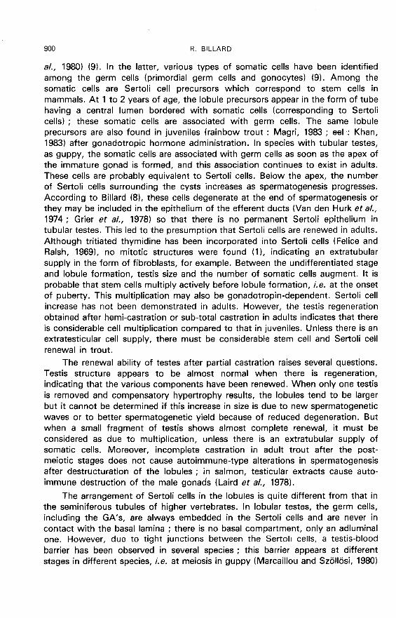
a/., 1980) (9). In the latter, various types of somatic cells have been identified
among the germ cells (primordial germ cells and gonocytes) (9). Among thesomatic cells are Sertoli cell precursors which correspond to stem cells in
mammals. At 1 to 2 years of age, the lobule precursors appear in the form of tube
having a central lumen bordered with somatic cells (corresponding to Sertoli
cellsl ; these somatic cells are associated with germ cells. The same lobule
precursors are also found in juveniles (rainbow trout : Magri, 1983 ; eel : Khan,1983) after gonadotropic hormone administration. In species with tubular testes,as guppy, the somatic cells are associated with germ cells as soon as the apex ofthe immature gonad is formed, and this association continues to exist in adults.These cells are probably equivalent to Sertoli cells. Below the apex, the numberof Sertoli cells surrounding the cysts increases as spermatogenesis progresses.According to Billard (8), these cells degenerate at the end of spermatogenesis orthey may be included in the epithelium of the efferent ducts (Van den Hurk et al.,1974 ; Grier et al., 1978) so that there is no permanent Sertoli epithelium in
tubular testes. This led to the presumption that Sertoli cells are renewed in adults.Although tritiated thymidine has been incorporated into Sertoli cells (Felice and
Ralsh, 1969), no mitotic structures were found (1), indicating an extratubularsupply in the form of fibroblasts, for example. Between the undifferentiated stageand lobule formation, testis size and the number of somatic cells augment. It is
probable that stem cells multiply actively before lobule formation, i.e. at the onsetof puberty. This multiplication may also be gonadotropin-dependent. Sertoli cellincrease has not been demonstrated in adults. However, the testis regenerationobtained after hemi-castration or sub-total castration in adults indicates that thereis considerable cell multiplication compared to that in juveniles. Unless there is anextratesticular cell supply, there must be considerable stem cell and Sertoli cellrenewal in trout.
The renewal ability of testes after partial castration raises several questions.Testis structure appears to be almost normal when there is regeneration,indicating that the various components have been renewed. When only one testisis removed and compensatory hypertrophy results, the lobules tend to be largerbut it cannot be determined if this increase in size is due to new spermatogeneticwaves or to better spermatogenetic yield because of reduced degeneration. Butwhen a small fragment of testis shows almost complete renewal, it must beconsidered as due to multiplication, unless there is an extratubular supply ofsomatic cells. Moreover, incomplete castration in adult trout after the post-meiotic stages does not cause autoimmune-type alterations in spermatogenesisafter destructuration of the lobules ; in salmon, testicular extracts cause auto-immune destruction of the male gonads (Laird et al., 1978).
The arrangement of Sertoli cells in the lobules is quite different from that inthe seminiferous tubules of higher vertebrates. In lobular testes, the germ cells,including the GA’s, are always embedded in the Sertoli cells and are never incontact with the basal lamina ; there is no basal compartment, only an adiuminalone. However, due to tight junctions between the Sertoli cells, a testis-blood
barrier has been observed in several species ; this barrier appears at different
stages in different species, i.e. at meiosis in guppy (Marcaillou and Szbll6si, 1980)

and later after spermiogenesis in Aphanius dispar (Abraham et al., 1980 ; also seeBergmann et al., 1984).
The endocrine function of the Sertoli cells is still not clear in fish. Criteria for
defining its steroidogenic activity (metabolism or synthesis) are relatively limitedand consist of histoenzymological evidence of various enzymes or the presence oflipids (Mattei et al., 1982). At present, there is not clear answer to the question ofwhether the Sertoli cell is the target of a gonadotropic hormone and, if so, which
hormone. Only an in vitro study using isolated Sertoli cells can provide a validanswer.
In lobular and tubular types, germ cells occur at various sites ; the GA’s areisolated, the GB’s, spermatocytes and spermatids are found in cysts, and the
spermatozoa are grouped at the base of tubules but are free in the lumen of
globules. The term « GA » probably includes several types of cells that have notbeen distinguished by morphological criteria. Their number remains almost
constant during the annual cycle, although it increases in some species such ascarp. Such an increase could mean the appearance of intermediate spermato-gonia. The mode of GA renewal is controversial, some authors are of opinionthey are of extralobular origin (Van den Hurk et al., 1978) and suggest that theSertoli cells send extensions outside the lobules to gather the stem spermatogonia(Ruby and McMillan, 1975). However, many authors think that the GA’s arerenewed from a stock that is permanently present inside the lobules ; we agreewith this hypothesis proposed by Wiart (1936) because we have never observedGA’s in the extralobular or extratubular space in any of the species we havestudied. It is often believed that primary germ cells (PGC) and GA’s are
characterized by the presence of ringed lamellae and granular material associatedwith mitochondria (Nagahama, 1983). However, these structures are also found inGB’s and granular material even occurs in spermatocytes.
The grouping of germ cells into cysts is considered to be characteristic of
anamniotes. The cells in the group develop isogenically from a GA spermato-gonia. However, Brusl6 (1982) studying Liza aurata and Wiebe (1968)
Cymatogaster aggregata reported that the cells were organized into cysts only atthe spermatocyte stage. In the latter species, Gardiner (1978b) observed
spermatogonia in cysts. In the species described in the present paper, the
spermatogonia that were linked together by intercellular junctions were groupedinto cysts with a syncitial structure. The concept of groups of isogenic cells
having cytoplasmic continuity is general (Roosen-Runge, 1977) and is also appliedin mammals (Dym and Fawcett, 1971) although the cysts are not morphologicallyidentifiable.
The number of spermatogonial generations (fourteen) is very high in guppy ;similar values (ten-twelve) were found in gambusia, another species with tubulartestes, but they are lower in species with lobular testes (Roosen-Runge, 1977).
Higher values were reported in Scleropages by Scott and Fuller (1976). Generally,PGC multiplication is high in fish. About 50 PGC’s are found in the undifferen-tiated gonad of the juvenile trout and 8 to 80.106 GA in the adult testis (13) ; thisis equal to at least 17 divisions. The process is even more marked in the capacitiesof trout for compensatory hypertrophy ; when a testis is removed at the

beginning of the cycle, the remaining testis doubles in size at the end of
spermatogenesis, giving a GSI comparable to the controls. When prepuberaljuvenile trout are castrated under a binocular, castration sometimes proves to beincomplete and the remaining bits of gonad regenerate testes of a size andstructure comparable to the controls (61). It is hypothetised that PGC mayactively contribute in the adult testis and might also explain the presence of pre-vitellogenetic oocytes in adult testes (unless oocytes in meiosis have subsistedfrom the juvenile gonad) or the presence of small testicular nodules sometimesfound in the ovaries. Such PGC’s have been described in the gonads of adultmugilids (gonochoric) and serranids (hermaphrodites) by Brusl6 (1982). Michibata(1975) distinguished stem (3-4 11m) and differentiated (5-10 ym) spermatogonia.
Primordial germ cells or undifferentiated gonocytes present in adults mightmultiply many times, more than the spermatogonia whose number of divisions isbetter defined.
While overall cell morphology is comparable among species up to the
spermatocyte stage, notable differences appear at spermiogenesis. This point hasbeen thoroughly discussed (Billard, 8, 10, 13) in relation to the final morphologyof the spermatozoon and its physiology (see below). The degree of chromatincondensation and of nuclear change is more marked when spermiogenesis is
complicated. The significance of these structures is still not well known.
it is necessary to question the resistance of the sperm structure to manipu-lation. For example, it has been shown that after deep-freezing, the chromatin oftrout spermatozoa is modified. This may be why a large number of carp motilespermatozoa lose their fertilizing ability after deep-freezing, and it is not sure thatthese alterations would not cause chromosome anomalies in the progeny.
The most complex spermatozoa with an elongated nucleus and developedmid-piece are not found exclusively in species with lobular testes ; they also occurin other groups such as Cymatogaster aggregata, a viviparous perciform. Gardiner(1978a) studying this species concluded that the elongated head and mid-piecewere specialized for internal fecundation. In the anterior part of the spermatozoa,specialized areas have been described in some species such as viviparousXiphophorus helleri in which lectins are specifically bound at an exact point in theplasma membrane having a glycocalyx that adheres closely to the nuclear
envelope (Jonas-Davies et al., 1983). These lectins, concanavalin A and Ricinuscommunis, indicate the presence of D-mannosyl and D-galactosyl residues. Theplasma membrane of trout spermatozoa is also closely apposed to the nuclearenvelope which itself adheres to the chromatin lamina inside the nucleus (131. Thespecialized area probably represent sites at which the membrane fuses at
fecundation. This type of plasma membrane anchorage to the nuclear envelope,observed in trout, may also represent an adaptation to the mode of fertilization inwhich the spermatozoa rest a short time in the water ; the anchorage wouldprevent the plasma membrane from detaching from the nucleus after hypo-osmotic shock.
The fish spermiduct appears to have a composite structure. The free, well-individualized part between the testis and the genital papilla is thought to be aneoformation originating in the urogenital area, while the intra or juxtatesticular

part which varies in morphology and position is not very structured, being a
testicular area with no germinal epithelium (Grier et al., 1980). In carp, Dragotoiu(1963) reported that the two spermiducts met between the intestine and the
urinary bladder in a single short tube having no musculature but a common
outlet with the urethra. This poses the question of the mode of « ejaculation »and of a putative urinary sperm supply.
Due to its limited extension, the carp spermiduct is not as well developed forstorage as in trout. However, in the goldfish, another cyprinid, ventral extensionsof the juxtatesticular part of the spermiducts have been interpreted by Takahashiand Takano (1972) as a sperm reservoir. Besides the function of sperm storageattributed to spermiducts, the literature also mentions the function of residual
spermatozoon resorption (Hayashi, 1969) and the secretion of protein-like seminalmaterial (pH 6.5) that contributes to spermatophore cohesion in the viviparousfish, Cymatogaster aggregata (Gardiner, 1978b). After transfer into the female
genital tract, the spermatozoa dissociate from the spermatophores when the pH ishigher than 7.3-7.9 and they are in the presence of ovarian fluid (Gardiner,1978b). In view of results obtained in trout, spermatophore dissociation in
C. aggregata may be due to the initiation of sperm motility due to the higher pH.The spermatozoa of viviparous species are transferred directly into the female
genital tract and seem to have no contact with the exterior medium. Specializedstructures at the anal fin (gonopod) consit of a simple groove in species wherethe spermatozoa are grouped into spermatozeugmas or spermatophores and of areal tube in species where the spermatozoa are free, as in the atheriniform,A. anableps (Grier et al., 19811. Thus, the spermatozoa of viviparous fish are notnecessarily grouped together but when they are free, the transfer structures in thegonopod are more elaborate. Atheriniform testes (« unrestricted » or tubular) canalso produce free spermatozoa. Moreover, viviparous species, such as C.
aggregata which is a perciform, are found in other groups than atheriniforms.The reproductive cycle includes two main events, spermatogenesis and
spermiation, that appear to differ greatly as to length, endocrine determinism andmodulating environmental factors, the two latter being subject to family variation.Spermatogenesis and spermiation and, more generally, gametogenesis and
gamete release-fecundation, can occur continuously as associated events or, onthe contrary, as events separated by a more or less long time interval and thusdissociated. Some of the species studied may be included in Crews’ (1984) plan.
Different combinations are found which suppose, in the case of dis-
continuity, that there are some particularities, especially in relation to the storagefitness of one of the gametes while waiting for its sexual partner to be availableand/or for environmental conditions to be favorable for reproduction.
During spermatogenesis, basic plasma GTH levels are low. The pulsatilecharacter of this secretion has not yet been clarified in males ; in male trout Zohar(1982) detected some pulses which were less evident than in females.
Secretory pulses have been recorded in female carp but have not yet beenstudied in males. It is not known if the GTH pulses are predeced by those ofGnRH secretion but, in view of results in higher vertebrates, they are likely to be.

It is also known that continuous GnRH administration by an implant, for example,sustains GTH secretion (Weil and Crim, 1983).
The low basal GTH levels found in males during spermatogenesis may berelated with the nature of the hormone measured. The GTH mentioned is purifiedmaturational (glycoprotein) GTH as far as its biological action on oocytematuration and spermiation or on the final phase of gametogenesis is concerned.It may be that the more specific non-glycoprotein hormone of vitellogenesis(vitellus incorporation in females) postulated by Idler and Ng (1983) also occurs inmales. However, the actual data available show that maturation GTH alone can atleast restore the quality of spermatogenesis in hypophysectomized goldfish (45).This would indicate that a single gonadotropic hormone stimulates spermato-genesis. According to Fontaine (1984), there is only a single gonadotropichormone in teleosts but another hormone like TSH might play a role. This wouldcorrespond to the survival of its double function (gonadotropic and thyreotropic)postulated by this author in agnaths. Experimental results tend to show that
spermatogenesis in eel is thyreotropin-dependent (Khan, 1983 ; Leloup-Hatey,personal communication).
GTH requirements could vary at various times during spermatogenesis,depending on the complexity of this cycle, as do the number of spermatogonialgenerations and the degree of spermatid and sperm transformation. GTH is
indispensable to spermatid elongation in guppy but does not appear necessary tothe simplified spermiogenesis of goldfish. It should be noted that mammalian
gonadotropic hormones have no effect on goldfish spermatogenesis. Specificityof zoological action also seems to be true within a class of fish. In contrast to
mammals, this process might result from a wider species difference due to longerevolution of the species. Salmonids as cyprinids show several spermatogeneticwaves so that several cell types are present in the testis at the same time ;therefore it is difficult to relate a given hormone level to a cell type and to exactlydeduce hormonal requirements at various spermatogenetic stages.
At the onset of and during spermiation, circulating levels of GTH and steroidsare markedly higher than during spermatogenesis. In trout a temporary rise inGTH just before spermiation appears to initiate a rise in 11-KT, the latter
remaining high throughout spermiation. Other steroid hormones such as 17 a-20 a-dihydroxy-4-pregnen-3-one (17 a-20 ¡3-P) have been identified in trout
plasma during this period (Scott and Baynes, 1982). It is possible that 11-KTcould madoulate 17 a-20 ¡3-P production (Leprêtre, personal communication) andthat the latter could be directly involved in the process of spermiation, its role
being limited to the secretion of seminal fluid and the control of its composition.Scott and Baynes (1982) reported a high correlation between K+ levels in theseminal fluid and circulating levels of 17 a-20 ¡3-P. Moreover, progesteronestimulates spermiation in trout, goldfish and pike. Therefore, progesterone (rarelyfound in a male vertebrate) could be associated with spermiation, an originalevent in fish with lobular testes.
The arrest of spermiation coincides exactly with the clearance of plasma 11-KT (41) but also probably with that of 17 a-20 ¡3-P according to the data of Scott

and Baynes (1982). This would tend to also confirm the role of these steroids inspermiation.
From this moment, residual spermatozoa are phagocytosed by either Sertolicells, spermiduct cells or macrophages. The antigenic recognition of spermatozoaby Sertoli cells or spermiduct epithelial cells could be modified when these
steroids are absent. The spermatozoa would be recognized as foreign cells and bephagocytosed, and the testis barrier would no longer be uniquely morphologicalbut also cytophysiological.
Gonadotropins also show zoological specificity in the case of spermiation (4)but hormones like HCG that have no effect on spermatogenesis in goldfishstrongly stimulate spermiation in that fish but not in carp.
Spermiation seems to be a complex stage in the male reproductive cyclewhich is modulated by widely diverse hormonal mechanisms and modes of
secretion. Spermiation is also the key stage of reproduction, resulting in the
release of good quality spermatozoa at the time the most favorable environmentalconditions occur simultaneously.
It is therefore necessary to determine the main environmental factors thataffect the processes of spermatogenesis and spermiation in various species orgroups of species. Two general types of reproductive cycle can be identified witheach action of specific environmental factors.
In salmonids gametogenesis mainly depends on the photoperiod, while
spermiation, and especially embryogenesis, has strict thermal requirements( < 12° for embryogenesis). Thus, reproduction occurs after gametogenesis withno clear evident stimuli or homonal response inducing spermiation. It is surprisingthat these species reproduce in winter ; in fact, salmonids eggs are large (up to1 cm in diameter) and are autonomous at least two months before the alevins canfeed. The temperature regulates embryogenesis and delays the swimming upstage until food is available in the environment.
In cyprinids, gametogenesis and spermiation-ovulation may be dissociated(example : carp). Temperature is one of the major environmental factors,although the role of photoperiod has not been analysed in detail. Below certain
thermic limits, gametogenesis is inhibited. As concerns spermiation, the
temperature rise in spring, which fluctuates continuously, causes plasma GTH toincrease and stimulates spermiation. Another social-type environmental factor is
also evident via pheromones. The males seem to know the sexual state of thefemales and particularly when ovulation is initiated. In cyprinids the direct effectof temperature on spermiation and ovulation is an efficient way of obtaining egglaying at the time when food for the larvae is already present because the eggs ofthis species are small and hatch only several days after they are laid. A social
stimulation coincides with the rise in temperature, leading to a rise in plasma GTHpermitting a specific volume of sperm to be mobilized for fecundation (66 and
Kyle and Stacey, personal communication). In this way, cyprinids, as carp andgoldfish, can compensate for the absence of large sperm reserves in the
spermiducts.Another type of essential information in fish with external fecundation is that
which induces the synchronous release of the sexual products at fertilization. The

simultaneous release of spermatozoa and ova has a quasi-obligatory characterdue to the short life of the gametes once they are in contact with the water.
Various hypotheses have been proposed. The stimuli causing synchronous releasemay be transmitted via the lateral line during spawning behavior ; cutting thelateral streak in medaka suppresses oviposition. According to the pheromonehypothesis, the laying of the first eggs, accompanied by pheromone-type factors,would release an immediate ejaculation reflex. Demski and Hornby (1982)consider that this is possible and that the stimuli-response interval (contraction ofthe spermiduct and abdominal musculature) is very brief.
Species differences in spe natogenetic production and spermiation yield areparticularly crucial in seasonal reproducing teleosts. First (table 11, there is no
relationship between the male and female GSI. Neither is there any betweenfemale fecundity (number of ova laid per g of liveweight) and spermatogeneticproduction, i.e. the ratio of spermatozoa produced/ovum, does not tend to beconstant. On the contrary, the process is amplified with 3.6 billion spermatozoaper ovum in trout. Estimating the real number of spermatozoa released by usingthe coefficient of spermiation yield to express spermatogenetic production insteadof the number of spermatozoa produced in the testis does not change the basicdifferences.
Sperm motility is very short in freshwater species with external fecundation(30 to 60 sec in salmonids and cyprinids). It is slightly longer in saltwater fish,being 2 min in sea-bass gilthead sea-bream and reaching several hours in
herrings (Dushkina, 1973). Trout spermatozoa rapidly become immotile when thelevel of ATP drops, indicating that dynein ATPase activity is higher than
mitochondrial synthetase activity. The low levels of internal ATP could inactivatethe membrane pump and phosphorylation implicated in flagellar beating ; this
inactivation is irreversible because motility is not re-initiated when the ATP level
rises. Inhibition of motility by KCI or by acid pH is apparently not due to a simpleacidification of the internal medium as in sea-urchin (Christen et al., 1982). The
process of membrane permeability are probably necessary to the regulation ofmotility ; present studies emphasize membrane potential and Ca+ + movement.
Sperm aging in trout is expressed by a decrease in cyclic AMP (Benau andTerner, 1980) and we have observed that cAMP had to be added into the
reactivation medium to activate demembranated spermatozoa near the end of
spermiation. These aging processes, causing a change in sperm quality, probablyoccur in other species such as carp and pike in which spermatogenesis andspermiation are dissociated, but this point still has to be studied.
The number of spermatozoa also appears to compensate for short motilitytime, as seen in trout, but these data cannot be applied to pike and carp sincemotility time is shorter in pike which produces fewer spermatozoa. Egg diameteris also a factor since in order to be fertilized a large egg (compared to a small egg)requires more spermatozoa having the same motility time or longer. This
argument has been shown in salmonids, the high number of spermatozoacompensating for the short motility time and the large egg diameter, but it cannotbe applied to pike which have relatively large eggs, short motility and a smallnumber of spermatozoa.

Another factor could be differences in sexual behavior. The two species inthis study which have the highest spermatogenetic production and the highestgonadal weight, carp and trout, are polyandrous (4-6 males/female), while pike isnot (1-3 males/female) and tilapia spawns in couples.
Carp and trout males participate more often in reproduction and release moresperm than pike and tilapia. Much has been written on salmonids ; several largemales participate in the ferlitization of one female with chances of fecundationwhich vary depending on their position in the social system (example : cohosalmon), but all the males release sperm (Gross, 1984).
Small, precocious males, not included in the social system of the large malesbecause they are not dominated due to their small size and lack of color, activelyparticipate in reproduction with the same chances of success as the dominantmales (Gross, 19841. These precocious males might be important in the sense thatthey contribute to mix sperm from several males at reproduction.
The testicular weight of primates has been related with the mode of
reproduction, polygamous species having a higher testicular weight than speciesin which the female mates with only one male (Harcourt et al., 1981 ; Harvey andHarcourt, 19821. This hypothesis might also be valid for fish, i.e. testicular weight,which shows higher variations than in mammals, and spermatogenetic productionmight be related with the mode of reproduction, whether isolated or in groups.Tilapia which mate in couples have a low testicular weight : 0.4-2 % of bodyweight (Peters, 1971). Peters has shown a relationship between the lower GSIand the smaller number of eggs laid but noted that testicular weight did not varybefore or after spawning ; this author concluded that the quantity of spermreleased must be extremely low and mentioned that for insemination to beefficient, the spermatozoa must be released very close to the eggs. Leporinusscrofa, a South American species, also has a very low GSI (0.2 %) (Godinho,personal communication), but its mode of reproduction is not known.
Large volumes of sperm are released during spawning in carp and trout,making the medium opaque (Breder and Rosen, 1966). These amounts of spermare mobilized in very different ways in the two species. In trout, the spermatozoaaccumulate in the spermiducts from the onset of spermiation, while in carp thespermatozoa are only mobilized a few hours before spawning. The males seem toknow the physiological state of the females ; giving an injection of gonadotropichormone to female carp or HCG to female goldfish stimulates spermiationwithin a few hours via a rise in gonadotropin (Kyle and Stacey, personalcommunication). The males must perceive a preovulatory peak and mobilizesperm that accumulates in the genital tract, probably in the system of lacunaethat constitute the intratesticular « spermiduct » because the free spermiduct isnot very developed in carp, and spermiation may continue throughout spawning.The mode of communication is not known ; it may be accomplished by sexualpheromones such as glucuronides which are released by the females and inducemating behavior in Brachydanio and gobies (Van den Hurk and Lambert, 1983 ;Colombo et al., 19821.
The amount of sperm released would ensure a sufficient sperm/egg ratio in

species such as salmonids which reproduce in running water or in carp which stirup the water by rapid swimming, causing the sperm to be quickly diluted.
However, seminal fluid, as ovarian fluid, might create a favorable micro-environment for fertilization by limiting hypo-osmotic shock or by maintaining afavorable pH for fecundation. This would explain why salmonids reproduce in
water of low alkalinity or at pH of less than 7, although it is known that at this pHthe spermatozoa are not motile.
The method used to measure the amount of spermatozoa released by troutthroughout spermiation leads to an overestimate of the number actually availablefor fecundation. The values measured correspond to a production of severalweeks, or even several months, in pisciculture conditions, while in nature the
period during which the females ovulate is extremely limited. Deroche (1969)studying lake trout, reported a case in which the total length of the spawningperiod was 18 days but noted that most of the females reproduced during onlytwo nights. Strictly speaking, the spawning period is usually shorter in salmonidsthan the period during which the males spermiate. Spermiation begins shortlybefore the females ovulate (in a same population). It is therefore not surprisingthat the sperm accumulates beforehand in the spermiducts of the male. Femalecarp have a longer spawning period (several months) and it depends a great dealon the temperature. The strategy of the male carp is different ; there is no
preaccumulation of sperm in the genital tract (where the sperm may age), butthere is preovulatory mobilization which can be explained by the longer spawningperiod that depends directly on environmental factors. This is also related with thefact that carp testicular spermatozoa mobilized experimentally throughout theyear by hormonal stimulation retain their fertilizing ability, while good quality troutspermatozoa are available for only several months.
5. Conclusion.
Some conclusions and hypotheses have been put forward concerning thereproductive function in several groups of teleost fish, and we have tried to definethe significance of various modes of reproduction.
Two large groups can be distinguished by their physiology (internal andexternal fecundation, sperm biology), morphology (structure of the testis andspermatozoon) and biochemical characteristics (transformation of nuclear protein,presence or absence of sperm energy stores) (fig. 14).
The first group includes species such as guppy with tubular testes andinternal fecundation. The spermatozoa have an elongated head but no glycogenstores and remain motile for several hours. They are usually associated in sperma-tozeugmas, thus facilitating transfer to the female genital tract. Transfer occursthrough the anal fin that is transformed into a gonopod. In some species thespermatozoa remain free and are transferred by the gonopod in the form of a
pseudopenis.The second group of fish includes those with lobular testes and external
fecundation. Sperm morphology is simple since there is no mid-piece or large

energy stores, and motility is short. The spermatozoa are very vulnerable in freshwater where they undergo severe hypo-osmotic shock, including rupture of theplasma membrane. They are « saved » by the rapidity with which they penetratethe egg. The spermatozoon seeks protection in a structure which resists
hypotonicity due to the cortical reaction and the resulting impermeability.Therefore this structure only remains open for several minutes. The short life ofthe spermatozoon is thus related with the short time the eggs are fertilizable once
they are in fresh water.In spite of the fact that internal fecundation protects the qametes, it is not
more economical in terms of sperm than external fecundation ; sperm productionin guppy (which has internal fecundation) is higher than in pike which has
external fertilization. Moreover, species with tubular testes have an original modeof internal fecundation which is found only in teleosts but not in highervertebrates. However, some structural elements prefiguring the testicular
organization of higher vertebrates occur in lobular testes.

The wide differences in spermatogenetic production occurring among specieshave no satisfactory answer, if only the physiological parameters are considered ;there is no relationship between spermatogenetic production and spermiationyield, between the number of spermatozoa produced or released and female
fecundity or egg diameter. However, there is a behavioral relationship, specieswith a high GSI spawning in a group and those with a low GSI spawning in
couples. The number of spermatozoa needed for fertilization is higher in theformer than in the latter, the males participating more often in reproduction. Thelarge volumes of sperm released in group spawning might create a favorablemicroenvironment for fertilization.
Spermatogenesis and spermiation, the major events of the annual cycle,have very different time lengths, the first being shorter than the second ; theyalso occur at different seasons. Both are continuous in trout and tench but theyare separated by a resting period in winter in carp and pike. In roach, evenspermatogenesis is inhibited in winter. The differences found between theseasons of spermiation may be related with species ecological requirements. Thelarge eggs of salmonids (autumn and winter spawning) have a 2 to 3-monthautonomy between reproduction and the trophic life of the juveniles that occursat the end of winter or in spring. On the contrary, the small eggs of cyprinidshave very little autonomy (not more than several days) between hatching and firstfood intake. Cyprinids reproduce at a time when food is already found in the
medium or there are indications that it soon will be. The temperature rise in the
spring seems to be a logical reference for spermiation and ovulation. Spermato-genesis in pike and carp in spring and autum occurs separately from spermiation,while spermatogenesis in tench begins at the end of winter and is rapidly followedby spermiation.
It is probable that various environmental factors play a role in the regulationof spermatogenesis and spermiation. Spermatogenesis depends more on wide,long-term variations such as seasonal changes in daylength and temperature.Spermiation is more directly influenced by short-term variations causing an
immediate response, such as exceeding the critical thermic threshold or the
appearance of environmental (laying substrates) or social (presence of femalesready to ovulate) stimuli.
The intensity of spermiation in salmonids depends on the presence offemales and on low temperatures, the optimum ( < 12-13 °C) corresponding tothe thermic requirements for incubation. Spermiation is arrested at 18 °C in troutin spite of a high level of GTH ; this may be due to the inactivation of androgensconverted into conjugates and glucuronides (Kime, 1979) and would be a way ofregulating spermiation by inhibiting it when thermic conditions do not favor eggsurvival.
In cyprinids, various regularoty mechanisms are superimposed that lead tospermiation and sperm release. As favorable environmental conditions
(particularly thermic conditions) may occur late, the brood fish can remain in a
holding state which does not affect sperm survival. In carp and goldsfish, whichhave no storage structures and whose spermatozoa are vulnerable when released

into the spermiduct, the male mobilizes large volumes of sperm only a few hoursbefore spawning. In this case, the female gives a social signal probably in
response to the environment (heat fluctuations, presence of water plants). Thus,spermiation requires two distinct conditions : the temperature that is facilitatingand must reach an optimal level and social stimulus that is determinant.
Why are the meeting of the sexes and of the gametes and the developmentof the resulting eggs and larvae guaranteed by such diverse modes of
reproduction, morphological structures and regulatory mechanisms ? Teleostsinclude more than 20 000 species and thus constitute the largest, and probablythe most diversified an oldest, group of vertebrates. On the other hand, aquaticmedia are extremely varied. Therefore, it may be that fish, by a sort of adaptivedialogue with the environment during their long history, have managed to
colonize a very wide variety of biotopes. The diversification in modes of
reproduction in the large teleost group would be a response to the diverseenvironments.
Requ en juillet 1!85.Accept6 en mai 1986.
Résumé. Spermatogenèse et spermiogenèse chez des Poissons Téléostéens.
L’ensemble des processus qui concourent à la formation des spermatozoïdes, à leurlibération et à la fécondation apparaissent extrêmement diversifiés chez les poissons Téléos-téens.
On identifie d’abord deux grands types de structure testiculaire, tubulaire et lobulaire,ce dernier présentant lui-même une grande diversité dans l’organisation des lobules et lastructure des conduits génitaux.- La spermatogenèse peut revêtir un aspect continu (guppy) ou discontinu avec descycles qui s’interpénètrent (carpe) ou sont bien séparés dans le temps (truite).- La spermiogenèse présente des degrés de complexification croissants conduisant à laformation de spermatozoïdes extrêmement simplifiés, comme ceux de la carpe (tête sphéri-que, transformation très sommaire des histones nucléaires, pièce intermédiaire très simpli-fiée avec restes de cytoplasme et mitochondries non transformées) ou beaucoup plus com-plexes comme ceux du guppy (allongement de la tête spermatique, transformation
histones-protamines, transformation du complexe centriole inséré dans une profonde enco-che nucléaire, pièce intermédiaire très développée comportant des réserves de glycogène).- La production spermatogénétique est extrêmement variable (RGS variant entre 0,2 et10 % et production annuelle comprise entre 100 et 7 000 106 spermatozoïdes/g de poidscorporel).- Le rendement de la spermiation varie fortement entre espèces, mais aussi à l’intérieurd’une même espèce (20 à 90 % chez la truite).- Le modèle endocrinien de la spermatogenèse et de la spermiation est très différent chezla carpe et la truite. Les facteurs de l’environnement prépondérants sont la photopériodechez la truite et la température chez la carpe pour la spermatogenèse, la température chezla truite à laquelle s’ajoute l’environnement social chez la carpe pour la spermiation.- La physiologie du sperme (durée de survie in vivo et in vitro, durée de motilité, métabo-lisme) est très différente dans les quelques espèces étudiées.
De cet ensemble de caractéristiques on peut identifier deux groupes qui sont fonda-mentalement différents tant sur le plan physiologique (fécondation interne ou externe,physiologie du sperme) que morphologique (structure du testicule et du spermatozoïde), Il
s’agit du groupe de Athériniformes (type guppy à testicule tubulaire) et du groupe desautres Téléostéens à reproduction saisonnière à testicule lobulaire.
1. - Author’s referencescalled in text by number
(1) BILLARD R., 1969. La spermatogen6se de Poecilia reticulata. I. Estimation du nombre de
generations goniales et rendement de la spermatogen6se. Ann. Biol. anim. Bioch. Biophys.,9, 251-271.
(2) GILLARD R., FLECHON J.-E., 1969. Spermatogonies et spermatocytes flagell6s chezPoecilia reticulata (T616ost6en cyprinodontiforme). Ann. Biol. anim. Bioch. Biophys., 9, 281-286.
(3) BILLARD R , 1984. Ultrastructural changes in the spermatogonia and spermatocytes ofPoecilia reticulata during spermatogenesis. Cell Tissue Res., 237, 219-226.
(4) BILLARD R., FOSTIER A., WEIL C., BRETON B., 1982. Endocrine control of spermato-genesis in teleost fish. Can. J. Fish. Aquat. Sci., 39, 65-79.
(5) BILLARD R., JALABERT B., 1972. Les cellules de Sertoli des poissons T616ost6ens.I. Etude ultrastructurale. Ann. Biol. anim. Bioch. Biophys., 12, 19-32.
(6) BILLARD R., MEUSY-DESSOLLES N., FLECHON J.-E., 1971. Les cellules interstitielles de
quelques poissons T616ost6ens (abstr.). J. Microscopie, 11, 30.(7) BILLARD D., TAKASHIMA F., 1983. Resorption of spermatozoa in the sperm duct of rainbow
trout during the post-spawning period. Bull. jap. Soc. Sci. Fish, 49, 387-392.(8) BILLARD R., 1970. La spermatogen6se de Poecilia reticulata. 111. Ultrastructure des cellules
de Sertoli. Ann. Bio/. anim. Bioch. Biophys., 10, 37-50.(9) LEBRUN C., BILLARD R., JALABERT B., 1982. Changes in the number of germ cells in
the gonads of the rainbow trout (Salmo gairdneri) during the first 10 post-hatching weeks.Reprod. Nutr. Deve%p., 22, 405-412.
(10) BILLARD R., 1970. La spermatogen6se de Poecilia reticulata. IV. La spermiogen6se. Etudeultrastructurale. Ann. Biol. anim. Bioch. Biophys., 10, 493-510.
(11) PICHERAL B., BILLARD R., BASSEZ Th., 1972. Localisation des protamides dans le noyaudes spermatides du T616ost6en Poecilia reticulata. Etude autoradiographique (abstr.). J.
Microscopie, 14, 80-81.(12) BILLARD R., JALABERT B., 1973. Le glycogbne au cours de la formation des spermato-
zoides et de leur transit dans le tractus g6nital femelle chez le Guppy (Poisson Poecilidae).Ann. Biol. anim. Bioch. Biophys., 13, 313-320.
(13) BILLARD R., 1983. Spermiogenesis in the rainbow trout (Salmo gairdneri). An ultrastructuralstudy. Cell Tissue Res., 233, 265-284.
(14) BILLARD R., 1969. Ultrastructure comparée de spermatozoides de quelques poissonsT616ost6ens. Quaderno, N. 137, 71-79, Baccetti Edit.
(15) BILLARD R., 1983. Ultrastructure of trout spermatozoa : changes after dilution and deep-freezing. Cell Tissue Res., 228, 205-218.
(16) MATTEI C., MATTEI X., MARCHAND B., BILLARD R., 1981. R6investigation de la structuredes flagelles spermatiques. Cas particulier des spermato20ides mitochondrie annulaire. J.Ultrastruct. Res., 74, 307-312.
(17) BILLARD R., GINSBURG A. S., 1973. La spermiogen6se et le spermatozoide d’Anguillaanguilla L. Ann. Bio/. anim. Bioch. Biophys., 13, 523-534.
(18) BILLARD R., 1968. Influence de la temp6rature sur la durée et 1’efficacit6 de la spermato-genbse de Guppy Poecilia reticulata. C.R. Acad. Sci. Paris, S6r. D, 266, 2287-2290.
(19) BILLARD R., 1969. La spermatogen6se de Poecilia reticulata. II. La production spermato-g6n6tique. Ann. Biol. anim. Bioch. Biophys., 9, 307-313.
(20) BILLARD R., SOLARI A., ESCAFFRE A. M., 1974. Sur une m6thode d’analyse quantitativede la spermatogen!se des T616ost6ens. Ann. Biol. anim. Bioch. Biophys., 14, 87-104.
(21) BILLARD R., 1983. A quantitative analysis of spermatogenesis in the trout Salmo trutta
fario. Cell Tissue Res., 230, 495-502.(22) BILLARD R., ESCAFFRE A.-M., 1975. Identification des stades de la spermatogen6se de
la truite fario d’apr6s la morphologie des gonades et la spermiation. Bull. Fr. Pisci., 256, 111-113.

(23) BILLARD R., BRETON B., JALABERT B., 1971. La production spermatogénétique chez la
truite. Ann. Bio/. anim. Bioch. Biophys., 11, 190-212.(24) BILLARD R., 1986. Gametogenesis in the brown trout (Salmo trutta fario) bred in captivity :
a quantitative study (in preparation).(25) BILLARD R., BRETON B., FOSTIER A., JALABERT B., WEIL C., 1978. Endocrine control
of the teleost reproductive cycle and its relation to external factors : Salmonid and Cyprinidmodels. In : Comparative endocrino%gy, GAILLARD P. J. and BOER H. H. Eds., Elsevier.North Holland, Biochem. Press, Amsterdam, 37-48.
(26) ESCAFFRE A.-M., BILLARD R., 1976. Le cycle spermatogénétique du gardon Rutilus rutilus.Cahiers Lab. Montereau, 3, 43-46.
(27) BRETON B., HOROSZEWICZ L., BILLARD R., BIENIARZ K., 1980. Temperature and
reproduction in tench, effect or a rise in the annual temperature regime on gonadotropin, levelgametogenesis and spawning. I. Case in the male. Reprod. Nutr. Dwelop., 20, 105-118.
(28) BILLARD R., MACKAY W. C., MARCEL J., 1983. Evolution de la gam6togen6se, du poidsdu corps et des gonades au cours du cycle reproducteur du brochet. (Esox lucius), 53-61. In :R. BILLARD Le Brochet, gestion dans le milieu naturel et 6levage, INRA Publ., Paris.
(29) BILLARD R., 1981. The reproductive cycle in teleost fish. Riv. it. Pisci. Ittiop., A.XVI, 79-80,106-118.
(30) BILLARD R., 1974. La production spermatog6n6tique de la truite arc-en-ciel au cours du
premier cycle reproducteur. Bull. fr. Pisci., 253, 139-149.(31) BOURLIER A., BILLARD R., 1984. Delayed gametogenesis and spawning in rainbow trout
fSalmo gairdneri) kept under permanent light during the first and second reproductive cycles.Aquaculture, 43, 259-268.
(32) DE MONTALEMBERT G., MARCEL J., BILLARD R., 1980. La spermiation chez le brochet. I.Evolution de la quantité de sperme r6colt6 au cours de la saison de reproduction. Bull. fr.
Pisci., 276, 90-103.(33) TAKASHIMA F., WEIL C., BILLARD R., CRIM L. W., FOSTIER A., 1984. Acceleration of
spermiation in carp by LH-RH analogue. Bull. jap. Soc. Sci. Fish., 50, 1323-1329.(34) BRETON B., KANN G., BURZAWA-G!RARD E., BILLARD R., 1971. Dosage radioimmuno-
logique d’une hormone gonadotrope de carpe Cyprinus carpio. C.R. Acad. Sci., Paris,S6r. D, 272, 1515-1517.
(35) JALABERT B., BRETON B., BILLARD R., 1974. Dosage biologique des hormones gonado-tropes de poissons par le test de maturation in vivo des ovocytes de truite. Ann. Biol. anim.Bioch. Biophys., 14, 217-228.
(36) FOSTIER A., BILLARD R., BRETON B., LEGENDRE M., MARLOT S., 1982. Plasma 11-oxo-
testosterone and gonadotropin during the beginning of spermiation in rainbow trout ISalmogairdneri R.l Gen. comp. Endocrinol., 46, 428-434.
(37) BILLARD R., BRETON B., DUBOIS M. P., 1971. Immunocytologie et histochimie des
cellules gonadotropes et thyr6otropes hypophysaires chez la carpe CVprinus carpio. C.R.Acad. Sci., Paris, S6r. D, 272, 981-983.
(38) JALABERT B., BILLARD R., 1968. Hypophysectomie de Poecilia reticulata (PoissonTeleosttsenl. Ann. Biol. anim. Bioch. Biophys., 8, 99-105.
(39) BILLARD R., RICHARD M., BRETON B., 1976. Stimulation of gonadotropin secretionafter castration in rainbow trout. Gen. comp. Endocrinol., 33, 163-165 et C.R. Acad. Sci.,Paris, S6r. 0, 283, 171-174.
(40) BILLARD R., PETER R. E., 1982. A stereotaxic atlas and technique for nuclei of the
diencephalon of rainbow trout (Salmo gairdneri). Reprod. Nutr. Dwe%p., 22, 1-25:
(41) FOSTIER A., BILLARD R., BRETON B., 1984. Plasma 11-oxotestosterone and gonadotropinin relation to the arrest of spermiation in rainbow trout (Salmo gairdneri). Gen. comp.
Endocrinol., 54, 378-381.
(42) BILLARD R., 1969. Hypophysectomie et spermatogen6se chez Poecilia reticulata (PoissonCyprinodontidae). C.R. Acad. Sci., Paris, S6r. D, 268, 1856-1859.
(43) BILLARD R., BRETON B., JALABERT B., 1970. Inhibition de la spermatogen6se de Guppy(Poisson Cyprinodontidae) par le m6thallibure. Ann. Biol. anim. Bioch. Biophys., 10, 511-515.

(44) BRETON B., BILLARD R., JALABERT B., 1973. Specificite d’action et relations immunolo-giques des hormones gonadotropes de quelques T616ost6ens. Ann. Biol. anim. Bioch.
Biophys., 13, 347-362.(45) BILLARD R., 1974. Testosterone : effects on the maintenance of spermatogenesis in intact
and hypophysectomized goldfish Carassius auratus. l. R. C. S., 2, 1231.(46) BRETON B., JALABERT B., BILLARD R., 1973. Pituitary and plasma gonadotrophin levels
and spermatogenesis in the goldfish Carassius auratus after methallibure treatment. J.Endocr., 59, 415-420.
(47) BILLARD R., BRETON B., ESCAFFRE A.-M., 1971. Maintien et restauration de la sperma-togen6se par un extrait ac6tonique hypophysaire de Carpe chez le Cyprin (Carassius auratus)trait6 au m6thallibure. Ann. Biol. anim. Bioch. Biophys., 11, 167-174.
(48) BILLARD R., ESCAFFRE A.-M., 1973. Effects of HCG and carp gonadotropin on themaintenance of spermatogenesis in hypophysectomized goldfish Carassius auratus. /. R. C. S.,(73-12), 15-1-20.
(49) BILLARD R., BURZAWA-GTRARD E., BRETON B., 1970. Regeneration de la spermatogen6sedu Cyprin hypophysectomis6 Carassius auratus L. par un facteur gonadotrope hautementpurify de carpe. C.R. Acad. Sci., Paris, S6r. D, 271, 1896-1899.
(50) BILLARD R., 1977. Effect of various hormones on sperm release in the hypophysectomizedgoldfish. /. R. C. S., 5, 188.
(51) BILLARD R., 1976. Induction of sperm release in the goldfish by some steroids. /.R.C.S.,4, 42.
(52) BRETON B., BILLARD R., JALABERT B., KANN G., 1972. Dosage radioimmunologique desgonadotropines plasmatiques chez Carassius auratus au cours du nycth6mbre et pendantl’ovulation. Gen. comp. Endocrinol., 18, 463-468.
(53) BILLARD R., MARCEL J., 1980. Stimulation of spermiation and induction of ovulation inNorthern pike (Esox luciusl. Aquaculture, 21, 181-195.
(54) DE MONTALEMBERT G., BRY C., BILLARD R., 1978. Control of reproduction in Northernpike (Esox luciusl. Trans. amer. Fish. Soc., Special Publ., 11, 217-225.
(55) BILLARD R., 1978. Testicular feed back on the hypothalamo-pituitary axis in rainbow trout
(Salmo gairdnenl. Ann. Biol, anim. Bioch. Biophys., 18, 813-818.(56) BRETON B., BILLARD R., 1980. Mise en evidence d’une r6gulation non-st6rdidienne d’origine
testiculaire sur la s6cr6tion d’hormone gonadotrope (GTH) chez la truite arc-en-ciel. C. R.Acad. Sci., Paris, 290, 1091-1094.
(57) BILLARD R., 1982. Attempts to inhibit testicular growth in rainbow trout with antiandrogens(Cyproterone, cyproterone acetate, oxymetholone) and Busulfan given during the period ofspermatogenesis. Gen. comp. Endocrinol., 48, 33-38.
(58) CRIM L. W., BILLARD R., GENGE P. D., IDLER D. R., 1982. The influence of immature
gonads on onset of gonadotropic hormone accumulation in the juvenile rainbow troutpituitary gland. Gen. comp. Endocrinol., 32, 213-220.
(59) CRIM L. W., PETER R. E., BILLARD R., 1981. Onset of gonadotropic hormone accumulationin the immature trout pituitary gland in response to estrogen or aromatizable androgen steroidhormones. Gen. comp. Endocrinol., 44, 374-381.
(60) BILLARD R., PETER R. E., 1977. Gonadotropin release after implantation of antiestrogens inpituitary and hypothalamus of goldfish Carassius auratus. Gen. comp. Endocrinol., 32, 213-220.
(61) BILLARD R., CRIM L. W., PETER R. E., BRETON B., 1982. Long-term changes in plasmaand pituitary GTH after castration of rainbow trout at an immature stage, 50. InC. J. J. RICHTER and H. J. Th. GOOS. Reproductive physio%gy of fish. PUDOC Wage-ningen.
(62) DUBOIS M. P., BILLARD R., BRETON B., 1978. Use of immunofluorescence for localisation
of somatostatin-like antigen in the rainbow trout ( Salmo gairdnenl. Comparative distributionof LHRH and neurophysin. Ann. Biol. anim. Bioch. Biophys., 18, 843-850.
DUBOIS M. P., BILLARD R., BRETON B., PETER R. E., 1979. Comparative distribution ofsomatostatin LH-RH neurophysin, and a-endorphin in rainbow trout : an immunocytologicalstudy. Gen. comp. Endocrinol., 37, 220-232.

(63) GILLET C., BILLARD R., BRETON B., 1977. Effets de la temp6rature sur le taux de gonado-tropine plasmatique et la sperniatogen6se du poisson rouge Carassius auratus. Can. J. Zool.,55, 242-245.
GILLET C., BILLARD R., 1977. Stimulation of gonadotropin secretion in goldfish by elevationof rearing temperature. Ann. Biol. anim. Bioch. Biophys., 17, 673-678.
(64) GILLET C., BILLARD R., BRETON B., 1981. La reproduction du poisson rouge Carassiusauratus élevé à 30 °C. Effet de la photop6riode, de I’alimentation et de l’oxyg6nation. CahiersLab. Montereau, 11, 49-56.
(65) GILLET C., BILLARD R., BRETON B., 1977. Influence de la temp6rature sur la reproductiondu poisson rouge (Carassius auratus L.). Cahiers Lab. Montereau, 5, 25-42.
GILLET C., BILLARD R., 1981. Effets de la temp6rature, de la photopériode et niveauxalimentaires sur la gonadotropine plasmatique et hypophysaire et la gam6togen6se duPoisson rouge. Cahiers Lab. Montereau, 11, 41-48.
(66) KYLE A. L., STACEY N. E., PETER R. E., BILLARD R., 1984. Elevations in gonadotropinconcentrations and milt volumes as a result of spawning behavior in the goldfish. Gen. comp.Endocrinol., 57, 10-22.
(67) BILLARD R., BRETON B., 1977. Sensibiiite la temp6rature des diff6rentes 6tapes de la
reproduction chez la truite arc-en-ciel. Cahiers Lab. Montereau, 5, 5-24.(68) BRETON B., BILLARD R., 1977. Effects of photoperiod and temperature on plasma gonado-
tropin and spermatogenesis in the rainbow trout Salmo gairdneri Richardson. Ann. Biol.anim. Bioch. Biophys., 17, 331-340.
(69) BILLARD R., REINAUD P., LE BRENN P., 1981. Effects of changes of a photoperiod ongametogenesis in the rainbow trout (Salmo gairdnenl. Reprod. Nutr. D6velop., 21,1009-1014.
(70) MAGRI M. H., BILLARD R., REINAUD P., FOSTIER A., 1981. Induction of gametogenesisin the juvenile rainbow trout. Gen. comp. Endocrinol., 46, 394 (Abstr. 1411. ).
(71) JALABERT B., BILLARD R., CHEVASSUS B., 1975. Preliminary on sex control in trout :
production of sterile fishes and stimultaneous self-fertilizable hermaphrodites. Ann. Biol.anim. Bioch. Biophys., 15, 19-28.
(72) BILLARD R., RICHARD M., ROMBAUTS P., 1982. Inhibition of spermatogenesis and
vitellogenesis in rainbow trout by hormonal additives (estradiol and methyltestosterone)included in the diet. Progr. Fish Cult., 44, 15-18.
(73) BILLARD R., BRETON B., RICHARD M., 1981. On the inhibitory effects of some steroids onspermatogenesis in adult rainbow trout (Salmo gairdneril. Can. J. Zool., 59, 1479-1487.
(74) BILLARD R., ALAGARSWAMI K., PETER R. E., BRETON B., 1983. Potentialisation par le
pimozide des effets du LHRH-A sur la s6cr6tion gonadotrope hypophysaire, l’ovulation et laspermiation chez la carpe commune (Cyprinus carpio). C. R. Acad. Sci., Paris, S6r. D, 296,181-184.
(75) BILLARD R., CHOISIS J. P., REINAUD P., 1983. Stimulation of spermiation in carp in
response to LHRH and D-ALA6-LHRH 6thylamide. Aquaculture, 35, 173-176.
(76) SANCHEZ-RODRIGUEZ M., BILLARD R., 1977. Conservation de la motilit6 et du pouvoirf6condant du sperme de la truite arc-en-ciel maintenu des temperatures voisines de 0 °C.Bull. fi. Pisci., 265, 144-152.
(77) BERGE P., VOLOCHINE B., BILLARD R., HAMELIN A., 1967. Mise en evidence du mou-vement propre de microorganismes vivants grâce 6 1’6tude de la diffusion in6lastique de la
lumi6re. C. R. Acad. Sci., Paris, S6r. D., 265, 889-892.
(78) BILLARD R., 1978. Changes in structure and fertilizing ability of marine and freshwater fishspermatozoa diluted in media of various salinities. Aquaculture, 14, 187-198.
(79) BILLARD R., BRETON B., 1970. Modifications ultrastructurales et cytochimiques de
spermatozoides apr6s dilution, chez les Poissons d’eau douce. Soc. fse. Microsc. 6lectron.VIle Congr. intern. Microsc. 6lectron., Grenoble 1970, 637-638.
(80) BILLARD R., BRETON B., 1976. Sur quelques probl6mes de physiologie du sperme, chezles poissons T616ost6ens. Rev. Trav. Inst. P6ches Marit., 40, 501-503.
(81) LEGENDRE M., BILLARD R., 1980. Cryopreservation of rainbow trout sperm by deep-freezing. Reprod. Nutr. D6velop., 20, 1859-1868.

(82) BILLARD R., 1976. Variation de la qualit6 des gametes a differents niveaux du tractus genitalchez la truite arc-en-ciel. Ann. Hydrobiol., 7, 97-104.
(83) BILLARD R., DUPONT J., BARNABT G., 1977. Diminution de la motilit6 et de la duréede conservation du sperme de Dicentrarchuslabrax (poisson Teleosteen) pendant la p6riode despermiation. Aquaculture, 11, 363-367.
(84) BILLARD R., 1984. La conservation des gametes et l’ins6mination artificielle chez le bar et ladaurade. In G. BARNAB! et R. BILLARD, L’Aquaculture du bar et des sparidés, I.N.R.A.
Publ., Paris.
(85) BILLARD R., 1982. L’importance des prot6ines du liquide coblomique sur la fertilit6 des
gametes chez la truite arc-en-ciel et possibilit6 de substitution. Bull. fr. Pisci., 284, 168-177.(86) BILLARD R., 1983. Effects of coelomic and seminal fluids and various saline diluents on the
fertilizing ability of spermatozoa in the rainbow trout Salmo gairdneri. J. Reprod. Fert., 68,77-84.
(87) BILLARD R., 1981. Les effets du lavage des gam6tes sur leur aptitude à la fécondation chezla truite arc-en-ciel. Rev. can. Biol., 40, 317-321.
(88) BILLARD R., JALABERT B., 1974. L’ins6mination artificielle de la truite Salmo gairdneriRichardson. it. Comparaison des effets des diff6rents dilueurs sur la conservation de lafertilit6 des gambtes avant et apr6s insemination. Ann. Biol. anim. Bioch. Biophys., 14, 601-610.
(89) BILLARD R., MTN!ZO Y., 1984. The amino-acid composition of rainbow trout seminal fluidblood plasma : a comparison with carp. Aquaculture, 41, 255-258.
(90) PLOUIDY M. G., BILLARD R., 1982. The chemical composition of the companion fluidsof the gametes in the common carp (Cyprinus carpio L.1, 134. In C. J. J. RICHTER andH. J. Th. GOOS, Reproductive physiology of fish. PUDOC Wageningen.
(91) MTN!ZO Y., PLOUIDY M. G., BILLARD R., 1983. Sur la composition en acides amineslibres et peptidiques du liquide s6minal de Carpe (Cyprinus carpio L.). C. R. Acad. Sci. Paris,série ///, 296, 67-70.
(92) BILLARD R., 1981. Short-term preservation of sperm under oxygen atmosphere in rainbowtrout (Salmo gairdneril. Aquaculture, 23, 287-293.
(93) BILLARD R., LEGENDRE M., 1982. Conservation à court terme des gametes de truite arc-en-ciel en condition in vitro sous atmosphere d’oxygene. Bull. fr. Pisci., 284, 162-167.
(94) MATEI D., MARCEL J., BILLARD R., 1980. Survie des gam6tes en conditions in vitro
et post mortem chez la truite arc-en-ciel. Bull. Acad. Sci. Agr. For. Roum., 10, 211-217.(95) BILLARD R., MARCEL J., MATEI D., 1980. Survie post mortem des gam6tes de truite fario,
Salmo trutta fario. Can. J. Zool., 52, 29-33.(96) PETIT J., JALABERT B., CHEVASSUS B., BILLARD R., 1973. L’ins6mination artificielle
de la Truite. I. Effets du taux de dilution du pH et de la pression osmotique du dilueur sur lafécondation. Ann. Hydrobiol., 4, 201-210.
(97) BILLARD R., PETIT J., JALABERT B., SZOLLOSI D., 1974. Artificial insemination in trout
using a sperm diluant, 715-723. In The early life history fish, J. H. S. BLAXTER, Edit.
Springer Verl8g, Berlin, Heidelberg, New York.(98) BILLARD R., JALABERT B., BRETON B., 1974. L’ins6mination artificielle de la truite Salmo
gairdneri Richardson. III. Definition de la nature et de la molarit6 du tampon employer avecles dilueurs d’ins6mination et de conservation. Ann. Biol. anim. Bioch. Biophys., 14, 611-621.
(99) BILLARD R., GILLET C., 1975. Effets de la temperature sur la fécondation et la surviedes gam6tes chez la truite arc-en-ciel Salmo gairdneri. Bull. fr. Pisci., 259, 53-65.
(100) BILLARD R., CAZIN J. C., DEQUIDT J., ERB F., COLEIN P., 1978. Toxicit6 du Pyra-16ne 3010 sur les ovules et les spermatozdfdes de truite arc-en-ciel avant et pendantl’ins6mination. Bufl. fr. Pisci., 270, 238-249.
(101) BILLARD R., GILLET C., 1981. Vieillissement des ovules et potentialisation par la temperaturedes effets des micropolluants du milieu aqueux sur les gambtes chez la truite. Cahiers Lab.Montereau, 12, 35-42.
(102) BILLARD R., ROUBAUD P., 1984. The effect of metals and cyanide ions on fertilization inrainbow trout (Salmo gairdneri). Water Res., 19, 209-214.
(103) BILLARD R., 1978. Effets des alcools gras sur la fécondation et les gam6tes de la truite
arc-en-ciel. Bull. fr. Pisci., 271, 3-8.
(104) BILLARD R., de KINKELIN P., 1970. Starvation des testicules de Guppies par des dosesnon 16thales de parathion. Ann. Hydrobiol., 1, 91-99.
(105) BILLARD R., 1980. Prolongation de la durée de motilitb et du pouvoir f6condant des sperma-tozoides de truite arc-en-ciel par addition de th6ophylline au milieu de dilution. C. R. Acad.
Sci., Paris, 291, 649-652.
’
II. - Other references
ABRAHAM M., RAHAMIM E., TIBIKA H., GOLENSER E., KIESELSTEIN M., 1980. The blood-
testis barrier in Aphanius dispar (Teleosteil. Cell Tissue Res., 211, 207-214.BARNABE G., 1976. Contribution 6 la connaissance de la biologie du loup Dicentrarchus labrax
(Poissons Serranidael. Th. Doct. Etat. Sci. nat., Univ. Sci. Tech. du Languedoc.BENAU D., TERNER C., 1980. Initiation, prolongation and reactivation of salmonid spermatozoa.
Gamete Res., 3, 247-257.BERGMANN M., SCHINDELMEISER J., GREVEN H., 1984. The blood-testis barrier in vertebrates
having different testicular organization. Cell Tissue Res., 238, 145-150.BILLARD R., 1966. Contribution 6 1’6tude de la reproduction chez le poisson téléostéen Lebistes
reticulatus, au moyen de l’insemination artificielle. Th. 3e Cycle, Fac. Sci. Lyon.BILLARD R., BRETON B., 1978. Rhythms of reproduction in teleost fish, 31-53. In J. E. THORPE,
Rhythmic activity of fishes, Acad. Press.
BOISSEAU J., 1967. Les regulations hormonales de l’incubation chez un Vertebre male : recherchessur la reproduction de I’Hippocampe. Th. Doct. Etat. Sci. Nat., Fac. Sci. Bordeaux.
BREDER C. M., ROSEN D. E., 1966. Modes of reproduction in fishes. Am. Mus. Nat. Hist. Publ.,New York.
BRETON B., JALABERT B., REINAUD P., 1976. Purification of gonadotropin from rainbow trout(Sa/mo gairdneri Richardson). Pituitary glands. Ann. Bio/. anim. Bioch. Biophys., 16, 25-36.
BROCK J., 1878. Beitrage zur Anatomie und Histologie der Geschtolchtsorgane der Knockenfische.Morphol. Jahr., 4, 505-572.
BRUSLE S., 1982. Contribution a la connaissance de la sexualit6 de poissons téléostéens marins
gonochoriques (Mugilides) et hermaphrodites (Serranid6s). Th. Doct. Sci. Nat., Univ.
Perpignan.BURZAWA-G!RARD E., 1973. Etude biologique et biochimique de l’hormone gonadotrope
d’un poisson téléostéen, la Carpe (Cyprinus carpio L. I. Th. Doct. Sci. Nat., Univ. Paris VI.CHRISTEN R., SCHACKMANN R. W., SHAPIRO B. M., 1982. Elevation of the intracellular pH
activates respiration and motility of sperm of the sea urchin, Strongylocentrotus purpuratus.J. biol. Chem., 257, 14881-14890.
COLOMBO L., BELVEDERE P. C., MARCONATO A., BENTIVEGNA F., 1982. Pheromones in
teleost fish, 84. In Reproductive physio%gy of fish, C. J. J. RICHTER and H. J. Th. GOOS.
PUDOC Wageningen.CRAIG-BENNETT A., 1931. The reproductive cycle of the three-spined stickleback, Gasterosteus
aculaetus L. Phil. Trans. roy Soc. Lond., B 219, 197-279.CREWS D., 1984. Gamete production, sex hormone secretion, and mating behavior uncoupled.
Hormones and behavior, 18, 22-28.DAVIS J. R., LANGFORD G. A., KIRBY P. J., 1970. The testicular capsule, 282-334. In
A. D. JOHNSON, W. R. GOMES and N. L. VANDEMARD, The testis, Acad. Press. New
York, London.DEMSKI D. J., HORNBY P. J., 1982. Hormonal control of fish reproductive behavior : brain-
gonadal steroid interactions. Can. J. Fish. Aouat. Sci., 39. 36-47.DEROCHE S. E., 1969. Observations on the spawning habits and early life of lake trout. Progr.
Fish Cult., 31, 109-113.DRAGOTOIU C., 1963. Contributions 6 1’6tude des organes reproducteurs mhles de Cyprinus
carpio L. Trav. Mus. Hist. nat., Bucarest, 4, 325-338.DUSHKINA L. A., 1973. Influence of salinity on eggs, sperm and larvae of low-vertebrates
herring reproducing in the coastal water of the Soviet Union. Marine Bio/., 19, 210-223.

DYM M., FAWCETT D. W., 1971. Further observations on the numbers of spermatogonia,spermatocytes, and spermatids connected by intercellular bridges in the mammalian testis.Biol. Reprod., 4, 195-215.
FELICE De D. A., RASCH E. M., 1969. Chronology of spermatogenesis and spermiogenesis in
Poeciliid fishes. J. exp. Zool., 171, 191-207.FONTAINE Y. A., 1984. Les hormones et I’evolution. La Recherche, 153, 311-320.FOSTIER A., JALABERT B., BILLARD R., BRETON B., ZOHAR Y., 1983. The gonadal steroids,
277-312. In Fish physio%gy W. S. HOAR, D. J. RANDALL and E. M. DONALDSON Eds.,Acad. Press, New York, London (vol. IXAI.
GARDINER D. M., 1978a. Fine structure of the spermatozoon of the viviparous teleost, Cymato-gaster aggregata. J. Fish Bio/., 13, 435-438.
GARDINER D. M., 1978b. The origin and fate of spermatophores in the viviparous Teleost Cymato-gaster aggregata (Perciformes : Embiotocidae). J. Morphol., 155, 157-171.
GRESIK E. W., 1975. Homologs of Leydig and Sertoli cells in the testis of the teleost Oryziaslatipes, 231-242. In Electron microscopic concepts of secretion, Hess and J. Wiley Eds.
GRESIK E. W., QUIRK J. G., HAMILTON J. B., 1973. Fine structure of the Sertoli cell of the testisof the teleost Oryzias latipes. Gen. comp. Endocrinol., 21, 341-352.
GRIER H. J., 1976. Sperm development in the teleost Oryzias latipes. Cell Tissue Res., 168,419-431.
GRIER H. J., 1981. Cellular organization of the testis and spermatogenesis in fishes. Am. Zool.,21, 345-357.
GRIER H. J., FITZSIMONS J. M., LINTON J. R., 1978. Structure and ultrastructure of the testisand sperm formation in goodeid teleosts. J. Morphol., 156, 419-438.
GRIER H. J., LINTON J. R., LEATHERLAND J. F., De VLAMING V. L., 1980. Structural evidence
for two different testicular types in teleost fishes. Am. J. Anat., 159, 331-345.GRIER H. J., BURNS J. R., FLORES J. A., 1981. Testis structure in three species of teleosts with
tubular gonopodia. Copeia, (4), 797-801.GROSS M. R., 1984. Sunfish, salmon, and the evolution of alternative reproductive strategies and
tactics in fishes. 50 p. in Fish reproduction. Strategies and tactics, R. J. WOOTON andG. POTTS Eds., Acad. Press.
GUYOMARC’H J. P., 1970. La spermatogenèse chez Nerophis lumbricitormis (Téléostéen Syngna-thid6s). Th. 3e Cycle, Fac. Sci. Brest. 92 pp.
HARCOURT A. H., HARVEY P. H., LARSON S. G., SHORT R. V., 1981. Testis weight, bodyweiqht and breeding system in primates. Nature, 293, 55-57.
HARVEY P. H., HARCOURT A. H., 1982. Sperm competition, testes size and breeding systemsin primates. In R. L. SMITH, Sperm competition and the evolution of animal mating systems.Acad. Press.
HAYASHI 1., 1969. Some observations on the reproductive duct of the japanese sea bass,Lateolabrax japonicus (Cuvier and Valenciennes). Jap. J. Ichthyol., 16, 68-76.
HENDERSON N. E., 1962. The annual cycle in the testis of eastern brook trout Salvelinus foniinalis(Mitchill). Can. J. Zoo/., 40, 631-641.
HIROI 0., YAMAMOTO K., 1968. Studies on the maturation of salmonid fishes. I. Changes in thetestis of the chum salmon Oncorhynchus keta, during anadromous migration. Bull. Fac. FishHokkaido Univ., 19, 173-184.
HIROI 0., YAMAMOTO K., 1970. Studies on the maturation of salmonid fishes. II. Changes in thetestis of the masu salmon, Oncorhynchus masou, during andromous migration. Bull. Fac.
Fish Hokkaido Univ., 20, 252-264.HURK Van Den R., LAMBERT J. G. D., 1983. Ovarian steroid glucuronides function as sex
pheromones for male zebrafish, Brachydanio rerio. Can. J. Zool., 61, 2381-2387.HURK Van Den R., MEEK J., PEUTE J., 1974. Ultrastructural study of the testis of the black molly
(Mollienisia latipinna). 11. Sertoli cells and Leydig cells. Koninkl. Nederl. Akad.
Wetenschappen-Amsterdam, Ser. C, 77, 470-476.HURK Van Den R., PEUTE J., VERMEIJ J. A. J., 1978. Morphological and enzyme cytochemical
aspects of the testis and vas deferens of the rainbow trout Salmo gairdneri. Cell Tissue Res.,186, 309-325.

HURK Van Den R., LAMBERT J. G. D., PEUTE J., 1982. Steroidogenesis in the gonads ofrainbow trout fry ( Salmo gairdneri) before and after the onset of gonadal sex differentiation.Reprod. Nutr. Dé, velop. 22, 413-425.
HYDER M., 1970. Gonadal and reproductive pattern in Tilapia leucosticta (Teleostei : Cichlid6s).J. Zool., 162, 179-195.
IDLER D. R., NG T. B., 1983. Teleost gonadotropins : isolation, biochemistry and function, 187-221.In Fish physiology (vol. IXA), W. S. HOAR, D. J. RANDALL and E. M. DONALDSON Eds.,Acad. Press, New York, London.
JONAS-DAVIES J. A. C., WINFREY V., OLSON G. E., 1983. Plasma membrane structure in
spermatogenic cells of the swordtail (Teleostei, Xiphophorus helleri). Gamete Res., 4,309-324.
JONES J. W., ORTON J. H., 1940. The paedogenetic male cycle in Salmo salar L. Proc. roy. Soc.,London, 128, 485-498.
KHAN I. A., 1983. Contr6le hormonal du fonctionnement testiculaire chez un poisson t6l6osteen(Anguilla anguilla L.) ; structure histo%gique, morpho%gique et androgenes plasmatiques.Th. 3&dquo; Cycle, Univ. Paris VI.
KIME D. E., 1979. The effect of temperature on the steroidogenic enzyme of the rainbow trout.Gen. comp. Endocrinol., 35, 322-328.
LAIRD L. M., ELLIS A. E., WILSON A. R., HOLLIDAY F. G. T., 1978. The development of thegonadal and immune systems in the atlantic salmon (Sa/mo salar L.) and a consideration ofthe possibility of inducing autoimmune destruction of the testis. Ann. Biol. anim. Bioch.
Biophys., 18, 1101-1106.LAM T. J., 1983. Environmental influences on gonadal activity in fish, 65-116. In Fish physiology,
Vol. IX, W. S. HOAR, D. J. RANDALL and E. M. DONALDSON Eds., Acad. Press, NewYork, London.
LOFTS B., MARSHALL A. J., 1957. Cyclical changes in the distribution of the testis lipids of ateleost fish, Esox luclus. Quart. J. micr. Sci., 98, 79-98.
MAGRI M. H., 1983. Action facilitante de l’hormone gonadotrope ou de st6roides sexuels surle d6clenchement de la puberte chez la truite arc-en-ciel. lnterference avec la photoperiode.Th. 3e Cycle, Univ. Paris VI.
MARCAILLOU C., SZOLLOSI A., 1980. The « Blood-testis » barrier in a nematode and a fish :a generalizable concept. J. Ultrast. Res., 70, 128-136.
MATTEI X., MATTEI C., MARCHAND B., LEUNG TACK KIT D., 1982. Ultrastructure des cellules
de sertoli d’un poisson t6l6ost6en : Abudefduf marginatus. J. Ultrast. Res., 81, 333-340.MEDFORD B. A., MACKAY W. C., 1978. Protein and lipid content of gonad, liver and muscle of
northern pike ( Esox lucius) in relation to gonad growth. J. Fish Res. Board Can., 35, 213-219.MICHIBATA H., 1975. Cell population kinetics of primary spermatogonia activated by warm
temperatures in the teleost, Oryzias latipes during the winter months. J. Fac. Sci. Univ.
Tokyo, Sec. 4, 13, 299-309.NAGAHAMA Y., 1983. The functional morphology of teleost gonads, 223-265. In Fish physio%gy,
vol. IXA, W. S. HOAR, D. J. RANDALL and E. M. DONALDSON Eds. Acad. Press, NewYork, London.
NAGAHAMA Y., CLARKE W. C., HOAR W. S., 1978. Ultrastructure of putative steroid-producingcells in the gonads of coho (Oncorhynchus kisutch) and pink salmon (Oncorhynchusgorbuschal. Can. J. Zool., 56, 2508-2519.
PAPADOPOL M., 1962. Nouvelles donn6es concernant la biologie de la reproduction de la breme
(Abramis brama L.) du cours inf6rieur du Danube (en roumainl. Studii. Cercet. Biol., Ser.Biol. anim., 14, 393-400.
PAYNE A. I., 1975. The reproductive cycle, condition and feeding in Barbus liberiensis, a tropicalstream-dwelling cyprinid. J. Zool., 176, 247-269.
PETERS H. M., 1971. Testis weights in Tilapia (Pisces : Cichlidae). Copeia (11, 13-17.
PETIT J., 1974. Recyclage et 6puration en pisciculture : 6tude th6orique et bibliographique. Coll.
Aquac., CNEXO Brest, 445-470.PHILIPPI E., 1908. Spermatophoren bei Fischen. Verh. d. deutsch Zool. Gesellsch., 17, 105-108.
ROOSEN-RUNGE E. C., 1977. The process of spermatogenesis in animals. Cambridge Univ.
Press, Cambridge, London, New York, Melbourne.

ROSENTHAL H. L., 1952. Observations on reproduction of the poeciliid Lebistes. Biol. Bull, 102, 30-38.RUBY S. M., McMILLAN D. B., 1975. The interstitial origin of germinal cells in the testis of the
stickleback. J. Morphol., 145, 295-317.SANCHEZ-RODRIGUEZ M., ESCAFFRE A.-M., MARLOT S., REINAUD P., 1978. The spermiation
period in the rainbow trout ( Salmo gairdneri). Plasma gonadotropin and androgen levels,sperm production and biochemical changes in the seminal fluid. Ann. Bio/. anim. Bioch.
Biophys., 18, 943-948.SCOTT A. P., BAYNES S. M., 1982. Plasma levels of sex steroids in relation to ovulation and
spermiation in rainbow trout (Salmo gairdneril, 103-106. In Reproductive physiology of fish,C. J. J. RICHTER and H. J. Th. GOOS. PUDOC, Wageningen.
SCOTT D. B. C., FULLER J. 0., 1976. The reproductive biology of Scleropages formosus(Mullet fr Schlegel) (Osteoglossomorpha osteoglossidae) in Malaya and the morphology of itspituitary gland. J. Fish. Biol., 8, 45-53.
SOLEWSKI W., 1957. Rozwoj gruczolow plciowych u karpia (Cyprinus carpio L.) w ciagu pierwszychtrzech lat zycia. Bnl. Zaktadu Bio/. Stavev Polskiy Akad. Nauk Krakov, 24-30. 3-24.
STOSS J., 1983. Fish gamete preservation and spermatozoan physiology, 305-350. In HOAR W. S.,RANDALL D. J., DONALDSON E. M., Fish physiology, vol. IX. Acad. Press, New York,London.
TAKAHASHI H., TAKANO K., 1972. Morphogenesis of accessory reproductive organs in male
goldfish, Carassius auratus. Bull. Fac. Fish, Hokkaido Univ., 23, 53-64.TAKASHIMA F., PATINO R., NOMURA M., 1980. Histological studies on the sex differentiation in
rainbow trout. Bull. jap. Soc. Sci. Fish, 46, 1317-1322.THIENOVEN B. Van, 1983. Reproductive physiology in vertebrates (2nd Edition). WB Saunders,
Philadelphia, London, Toronto.TURDAKOV A. F., 1968. Production of sperm by the males of lssyk Kuli trout. Vopr. lcht., 8,
253-265.WIEBE J. P., 1968. The effects of temperature and daylength on the reproductive physiology of
the viviparous seaperch, Cymatogaster aggregata Gibbons. Can. J. Zoot, 46, 1207-1219.WEIL C., 1981. La fonction gonadotrope de I’hypophyse au cours du cycle sexuel chez deux
poissons t6l6ost6ens : la carpe commune (Cyprinus carpio), et la truite arc-en-ciel (Salmogairdneri), son contr6le par /,hypothalamus, les gonades et les facteurs externes. Th. Doc. Etat,Univ. Paris VI.
WEIL C., CRIM L. W., 1983. Administration of LHRH analogues in various ways : effect on theadvancement of spermiation in prespawning Landlocked salmon Salmo salar. Aquaculture,35, 103-115.
WIART P., 1936. Variations saisonni6res du testicule des t6l6ost6ens. Trav. Stat. Bio;. Roscoff,14-16, 79-86.
ZOHAR Y., 1982. La dynamique nycth6m6rale de la secretion gonadotrope chez la truite arc-en-cielfemelle au cours du cycle annuel.- analyse descriptive et analyse experimentale de sa fonctiondans la régulation ovarienne. Th. Doc. Etat, Univ. Paris VI.