Toxic impact of lethal concentration of lead nitrate on the gills ...
17
167 VETERINARSKI ARHIV 72 (3), 167-183, 2002 * Contact address: Tarun Kumar Banerjee, Histochemistry and Histopathology Laboratory, Department of Zoology, Centre of Ad- vanced Study, Banaras Hindu University, Varanasi - 221005, India. Fax: +91 542 367174; E-mail: [email protected] ISSN 0372-5480 Printed in Croatia Toxic impact of lethal concentration of lead nitrate on the gills of air-breathing catfish Heteropneustes fossilis (Bloch) Ram Sanehi Parashar, and Tarun Kumar Banerjee* Histochemistry and Histopathology Laboratory, Department of Zoology, Centre of Advanced Study, Banaras Hindu University, India PARASHAR, R. S., T. K. BANERJEE: Toxic impact of lethal concentration of lead nitrate on the gills of air-breathing catfish Heteropneustes fossilis (Bloch). Vet. arhiv 72, 167-183, 2002. ABSTRACT Toxicopathological impact of lethal concentration (925 mg/l) of lead nitrate on the gills of air-breathing catfish Heteropneustes fossilis was analysed. Following exposure the gills exhibited rapid alterations that include detachment and lifting of the epithelial linings from the surfaces of the gill filament (primary, PL) and respiratory (secondary, SL) lamellae. This led to extensive haemorrhage from the gills. Thus the quantity of blood flowing across the gills decreased substantially. Simultaneously, uncontrolled regeneration of the PL and SL occured, leading to extensive hyperplasia of the epithelial cells lining the PL, and SL. Consequently, the gill filaments appeared as a cylindrical solid mass of cells with very little or almost no free surface left on the SL for gaseous exchange. The goblet mucous cells also exhibited periodic fluctuations in their density and staining behaviour. The chloride cells showed periodic fluctuation in their number at different stages of exposure. The density of the chloride cells is inversely proportional to the thickness of the epithelial lining of the PL and SL. Due to prolonged exposure, the neighbouring SL fused together and the entire gills appeared as solid mass of undifferentiated cells. Subsequently, the ladder-like arrangement of the pillar cells-blood capillaries of the gills also collapsed, causing asphyxiation and the death of the fish. Key words: gills, Heteropneustes fossilis, histopathology, lead nitrate toxicity, India
-
Upload
khangminh22 -
Category
Documents
-
view
3 -
download
0
Transcript of Toxic impact of lethal concentration of lead nitrate on the gills ...
167
VETERINARSKI ARHIV 72 (3), 167-183, 2002
* Contact address:Tarun Kumar Banerjee, Histochemistry and Histopathology Laboratory, Department of Zoology, Centre of Ad-vanced Study, Banaras Hindu University, Varanasi - 221005, India. Fax: +91 542 367174; E-mail:[email protected]
ISSN 0372-5480Printed in Croatia
Toxic impact of lethal concentration of lead nitrate on the gills ofair-breathing catfish Heteropneustes fossilis (Bloch)
Ram Sanehi Parashar, and Tarun Kumar Banerjee*
Histochemistry and Histopathology Laboratory, Department of Zoology, Centre of AdvancedStudy, Banaras Hindu University, India
PARASHAR, R. S., T. K. BANERJEE: Toxic impact of lethal concentration of leadnitrate on the gills of air-breathing catfish Heteropneustes fossilis (Bloch). Vet.arhiv 72, 167-183, 2002.
ABSTRACTToxicopathological impact of lethal concentration (925 mg/l) of lead nitrate on the gills
of air-breathing catfish Heteropneustes fossilis was analysed. Following exposure the gills exhibitedrapid alterations that include detachment and lifting of the epithelial linings from the surfacesof the gill filament (primary, PL) and respiratory (secondary, SL) lamellae. This led to extensivehaemorrhage from the gills. Thus the quantity of blood flowing across the gills decreasedsubstantially. Simultaneously, uncontrolled regeneration of the PL and SL occured, leading toextensive hyperplasia of the epithelial cells lining the PL, and SL. Consequently, the gillfilaments appeared as a cylindrical solid mass of cells with very little or almost no free surfaceleft on the SL for gaseous exchange. The goblet mucous cells also exhibited periodic fluctuationsin their density and staining behaviour. The chloride cells showed periodic fluctuation in theirnumber at different stages of exposure. The density of the chloride cells is inversely proportionalto the thickness of the epithelial lining of the PL and SL. Due to prolonged exposure, theneighbouring SL fused together and the entire gills appeared as solid mass of undifferentiatedcells. Subsequently, the ladder-like arrangement of the pillar cells-blood capillaries of the gillsalso collapsed, causing asphyxiation and the death of the fish.
Key words: gills, Heteropneustes fossilis, histopathology, lead nitrate toxicity, India
168 Vet. arhiv 72 (3), 167-183, 2002
IntroductionThe gills are not only the prime organs for gaseous exchange; they also
perform several other physiological functions including osmoregulation andexcretion. Changes in environmental parameters often damage this vitalorgan because of its delicate structure. Recent review articles (DUTTA etal., 1996; WENDALAAR BONGA, 1997) on ambient toxicants in fish have clearlydemonstrated that increased concentrations of several heavy metals seri-ously damage the gills of teleostean fish. HEMALATHA and BANERJEE (1997a,1997b) have studied the toxic impact of the trace element zinc (ZnCl2) onthe gills and accessory respiratory organs of Heteropneustes fossilis. How-ever, almost no data are available on the toxic impact of lead salts on therespiratory organs of air breathing fish (including Heteropneustes fossilis).The data concerning lead toxicity are mainly related to studies with mam-malian subjects and to air-borne pollutants. Therefore in this study effortshave been made to examine the toxicity of lead nitrate on the branchialrespiratory organs of H. fossilis. PARASHAR and BANERJEE (1999a) haverecently attempted to investigate the toxic impact of this lead salt on theaccessory respiratory system of this air-breathing teleost.
Materials and methodsHealthy specimens of H. fossilis (38-42 g body weight, 18-20 cm in
length) were collected from a single population from a local fish market atVaranasi and were acclimated for one month in tap water in large plasticaquaria in the laboratory. They were fed with minced goat liver on alternatedays and the water was renewed after every 24 h, leaving no faecal matter,unconsumed food or dead fish, if any. Feeding ceased 24 h prior to thecommencement of the experiment with the starvation regime continuingthroughout the period of the experiment.
Prior to the commencement of the experiment, 96h median lethalconcentration (96h LC50) of lead nitrate (Glaxo India Ltd., Mumbai; 99%pure) was estimated following the Trimmed Spearman-Karber Method(HAMILTON et al., 1977) and was determined at 925 ppm.
R. S. Parashar and T. K. Banerjee: Toxic impact of lethal concentration of lead nitrate onthe gills of air-breathing catfish Heteropneustes fossilis (Bloch)

169Vet. arhiv 72 (3), 167-183, 2002
Eight groups of 14 fish each were exposed to 925 mg/l (96h LC50value) of lead nitrate for estimation of lethal toxicity. Each group wasexposed separately to 50 l of lead salt solution, prepared in tap water (havingdissolved O2, 6.2 mg/l, pH 7.5, water hardness 23.4 mg/l and watertemperature 24 ± 2o C). Eight identical groups of 10 fish each were kept inseparate aquaria containing 50 l of plain tap water (without lead salt) ascontrols.
After each of the exposure periods of 0 h, 3 h, 6 h, 12 h, 24 h, 48 h, 72h, and 96 h, five fish each from the respective experimental, as well ascontrol, aquaria were sacrificed. The entire second gill from both the sidesof the fish were fixed in 10% neutral formalin, aqueous Bouin’s fluid andHelly’s fluid. Six µm paraffin sections were stained with Ehrlich‘s or Harrishaematoxylin and eosin (H&E) for routine histopathological analysis. Certaincarbohydrate moieties were visualised by periodic acid-Schiff (PAS), alcianblue pH 2.5 (AB 2.5), AB 2.5/PAS, alcian blue pH 1.0 (AB 1.0), Bismarckbrown (BB) and salivary amylase/PAS techniques (Table 1) (PEARSE, 1985).
ResultsControl. H. fossilis possesses four pairs of typical teleostean gill arches,
each bearing two rows of gill filaments (primary gill lamellae, PL). EachPL bears a series of alternately arranged secondary lamellae (SL) (Fig. 1)
Fig. 1. Structural organization of a part ofthe second gill of control fish. Note thebranching of the gill filament. AB&PAS;×125.
Fig. 2. Normal distribution of carbohydratemoieties in its different cellular constituentsespecially in its mucous cells (MCs) of thegill. Note the presence of MCs in the second-ary lamellae (SL) also (arrows). AB&PAS;×520.
R. S. Parashar and T. K. Banerjee: Toxic impact of lethal concentration of lead nitrate onthe gills of air-breathing catfish Heteropneustes fossilis (Bloch)

170 Vet. arhiv 72 (3), 167-183, 2002
on both its sides. The SL at the distal end of the gill filament (PL) mostlyremain fused together. The respiratory lining of the SL on each side con-sists of a thin layer of epithelium which rests on a basement membranecovering the pillar cell - blood channel (PC-BC) system and which consti-tutes the main vascular component of the gills (Figs. 2, 3). The mucouscells (MCs) are mostly observed in the inter-lamellar epithelium betweentwo SL (Fig. 2) and at the distal tip of the PL. The histochemical propertiesof the MCs of the gill epithelia are summarized in Table 1.
Exposure periods Staining techniques Control 3h 6h 12h 24h 48h 72h 96h
PAS 4+C; 4+P
2-3+C; 2-4+P
1-2+C; 2-3+P
3+C; 3-4+P
3+C; 3-4+P
3+C; 3-4P
3-4+C; 3-4+P
3+C; 3-4+P
AB 2.5 1-2+C; 1-2+P
4+C; 4+P
3-4+C; 3-4+P
3-4+C; 3-4+P
3+C; 4+P
3+C; 3+P
3-4+C; 3-4+P
3-4+C; 3-4+P
AB 2.5/PAS**
3+C; 4+P
3-4+C; 4+P
3-4+C; 4+P
3-4+C; 4+P
3+C; 4+P
4+C; 4+P
4+C; 4+P
3-4+C; 4+P
BB 4+C; 4+P
4+C; 4+P
4+C; 4+P
3-4+C; 3-4+P
3-4+C; 3-4+P
2-3+C; 3+P
2-3+C; 3+P
2+C; 2-3+P
Table 1. Summary of histochemical alterations shown by the goblet mucous cells of thegills of H. fossilis exposed to different periods of exposure to lethal concentration of lead
nitrate solution
Symbols and abbreviations: ** = variously stained reactions from various shades of ma-genta, violet to greenish blue, in the same or different mucocytes; C = cell content; P = cellperiphery; AB 2.5 = alcian blue at pH 2.5 for acidic mucopolysaccharides; BB = Bismarckbrown reaction for water-stable mucoproteins; h = hour; PAS = per iodic acid/Schiff reac-tion for neutral glycoproteins; AB 2.5/ PAS = alcian blue pH 2.5/periodic acid Schiff foracidic and neutral glycoproteins; - = to; 0 = negative reaction; 1+ = weak reaction; 2+ =moderate reaction; 3+ = strong reaction; 4+ = very strong reaction.
Experimental gill. Following exposure the gills showed extensive dam-age in their lamellar configuration, even though the gills continue to regen-erate repeatedly after every wear and tear, especially during the initialstages. The ionocytes (chloride cells) showed fluctuation in their density atdifferent stages of exposure. Generally, the density of these ionocytes de-creases, invariably in the thickened epithelia.
R. S. Parashar and T. K. Banerjee: Toxic impact of lethal concentration of lead nitrate onthe gills of air-breathing catfish Heteropneustes fossilis (Bloch)
171Vet. arhiv 72 (3), 167-183, 2002
Table 1. Specification of myomorphus mammals examined by renoculture andmicroscopic agglutination acording to the trapping area with corresponding results
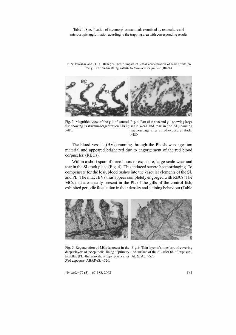
The blood vessels (BVs) running through the PL show congestionmaterial and appeared bright red due to engorgement of the red bloodcorpuscles (RBCs).
Within a short span of three hours of exposure, large-scale wear andtear in the SL took place (Fig. 4). This induced severe haemorrhaging. Tocompensate for the loss, blood rushes into the vascular elements of the SLand PL. The intact BVs thus appear completely engorged with RBCs. TheMCs that are usually present in the PL of the gills of the control fish,exhibited periodic fluctuation in their density and staining behaviour (Table
Fig. 4. Part of the second gill showing largescale wear and tear in the SL, causinghaemorrhage after 3h of exposure. H&E;×480.
Fig. 3. Magnified view of the gill of controlfish showing its structural organization. H&E;×480.
Fig. 6. Thin layer of slime (arrow) coveringthe surface of the SL after 6h of exposure.AB&PAS; ×520.
Fig. 5. Regeneration of MCs (arrows) in thedeeper layers of the epithelial lining of primarylamellae (PL) that also show hyperplasia after3hof exposure. AB&PAS; ×520.
R. S. Parashar and T. K. Banerjee: Toxic impact of lethal concentration of lead nitrate onthe gills of air-breathing catfish Heteropneustes fossilis (Bloch)
172 Vet. arhiv 72 (3), 167-183, 2002
1) at different stages of exposure (Figs. 5, 6). However, a thin layer of AB2.5 positive slimy coating continued to cover the surface of the SL at severalstages of exposure (Fig. 6). Even though their number decreased marginallyafter 3 h, MCs were very often found in the inner, as well as in the outerlayers of the hyperplastic epithelial lining of the PL, indicating regenerationof new MCs. Simultaneously, regeneration and healing of the damagedgills also takes place at this stage. Uncontrolled regeneration of epithelialcells (ECs) causes hyperplasia of PL, resulting in their increased thickness
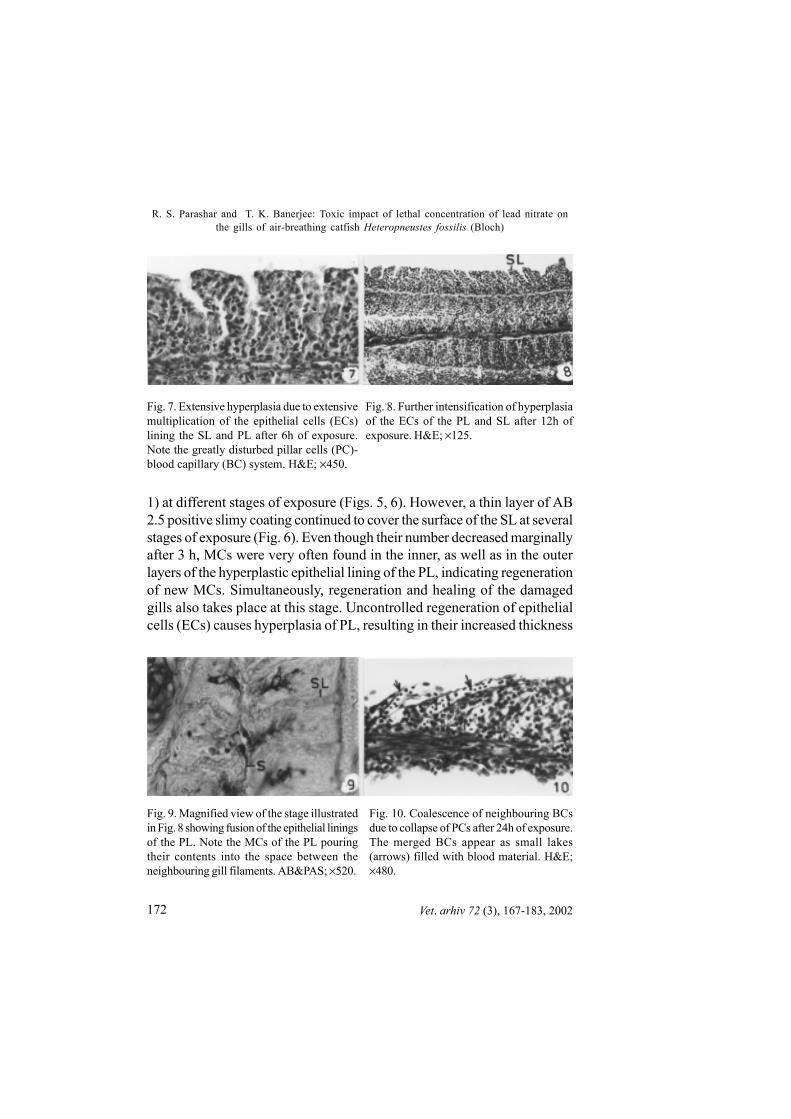
Fig. 8. Further intensification of hyperplasiaof the ECs of the PL and SL after 12h ofexposure. H&E; ×125.
Fig. 7. Extensive hyperplasia due to extensivemultiplication of the epithelial cells (ECs)lining the SL and PL after 6h of exposure.Note the greatly disturbed pillar cells (PC)-blood capillary (BC) system. H&E; ×450.
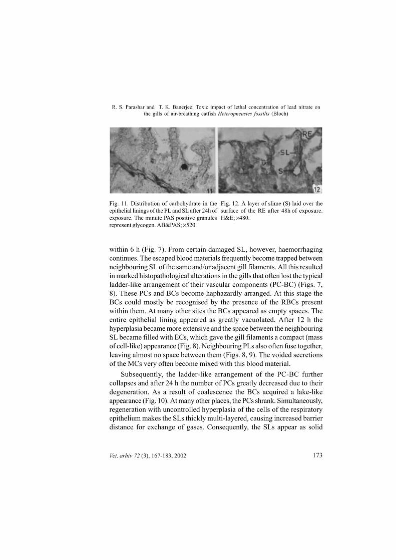
Fig. 10. Coalescence of neighbouring BCsdue to collapse of PCs after 24h of exposure.The merged BCs appear as small lakes(arrows) filled with blood material. H&E;×480.
Fig. 9. Magnified view of the stage illustratedin Fig. 8 showing fusion of the epithelial liningsof the PL. Note the MCs of the PL pouringtheir contents into the space between theneighbouring gill filaments. AB&PAS; ×520.
R. S. Parashar and T. K. Banerjee: Toxic impact of lethal concentration of lead nitrate onthe gills of air-breathing catfish Heteropneustes fossilis (Bloch)
173Vet. arhiv 72 (3), 167-183, 2002
within 6 h (Fig. 7). From certain damaged SL, however, haemorrhagingcontinues. The escaped blood materials frequently become trapped betweenneighbouring SL of the same and/or adjacent gill filaments. All this resultedin marked histopathological alterations in the gills that often lost the typicalladder-like arrangement of their vascular components (PC-BC) (Figs. 7,8). These PCs and BCs become haphazardly arranged. At this stage theBCs could mostly be recognised by the presence of the RBCs presentwithin them. At many other sites the BCs appeared as empty spaces. Theentire epithelial lining appeared as greatly vacuolated. After 12 h thehyperplasia became more extensive and the space between the neighbouringSL became filled with ECs, which gave the gill filaments a compact (massof cell-like) appearance (Fig. 8). Neighbouring PLs also often fuse together,leaving almost no space between them (Figs. 8, 9). The voided secretionsof the MCs very often become mixed with this blood material.
Subsequently, the ladder-like arrangement of the PC-BC furthercollapses and after 24 h the number of PCs greatly decreased due to theirdegeneration. As a result of coalescence the BCs acquired a lake-likeappearance (Fig. 10). At many other places, the PCs shrank. Simultaneously,regeneration with uncontrolled hyperplasia of the cells of the respiratoryepithelium makes the SLs thickly multi-layered, causing increased barrierdistance for exchange of gases. Consequently, the SLs appear as solid
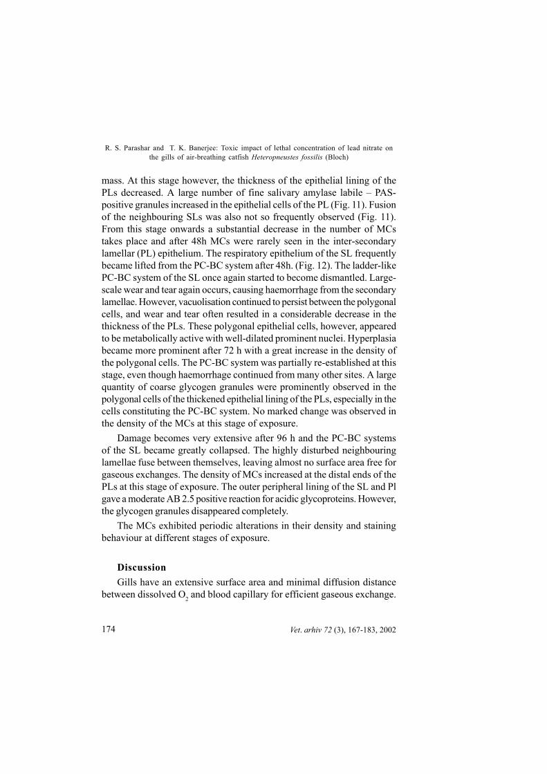
Fig. 11. Distribution of carbohydrate in theepithelial linings of the PL and SL after 24h ofexposure. The minute PAS positive granulesrepresent glycogen. AB&PAS; ×520.
Fig. 12. A layer of slime (S) laid over thesurface of the RE after 48h of exposure.H&E; ×480.
R. S. Parashar and T. K. Banerjee: Toxic impact of lethal concentration of lead nitrate onthe gills of air-breathing catfish Heteropneustes fossilis (Bloch)
174 Vet. arhiv 72 (3), 167-183, 2002
mass. At this stage however, the thickness of the epithelial lining of thePLs decreased. A large number of fine salivary amylase labile – PAS-positive granules increased in the epithelial cells of the PL (Fig. 11). Fusionof the neighbouring SLs was also not so frequently observed (Fig. 11).From this stage onwards a substantial decrease in the number of MCstakes place and after 48h MCs were rarely seen in the inter-secondarylamellar (PL) epithelium. The respiratory epithelium of the SL frequentlybecame lifted from the PC-BC system after 48h. (Fig. 12). The ladder-likePC-BC system of the SL once again started to become dismantled. Large-scale wear and tear again occurs, causing haemorrhage from the secondarylamellae. However, vacuolisation continued to persist between the polygonalcells, and wear and tear often resulted in a considerable decrease in thethickness of the PLs. These polygonal epithelial cells, however, appearedto be metabolically active with well-dilated prominent nuclei. Hyperplasiabecame more prominent after 72 h with a great increase in the density ofthe polygonal cells. The PC-BC system was partially re-established at thisstage, even though haemorrhage continued from many other sites. A largequantity of coarse glycogen granules were prominently observed in thepolygonal cells of the thickened epithelial lining of the PLs, especially in thecells constituting the PC-BC system. No marked change was observed inthe density of the MCs at this stage of exposure.
Damage becomes very extensive after 96 h and the PC-BC systemsof the SL became greatly collapsed. The highly disturbed neighbouringlamellae fuse between themselves, leaving almost no surface area free forgaseous exchanges. The density of MCs increased at the distal ends of thePLs at this stage of exposure. The outer peripheral lining of the SL and Plgave a moderate AB 2.5 positive reaction for acidic glycoproteins. However,the glycogen granules disappeared completely.
The MCs exhibited periodic alterations in their density and stainingbehaviour at different stages of exposure.
DiscussionGills have an extensive surface area and minimal diffusion distance
between dissolved O2 and blood capillary for efficient gaseous exchange.
R. S. Parashar and T. K. Banerjee: Toxic impact of lethal concentration of lead nitrate onthe gills of air-breathing catfish Heteropneustes fossilis (Bloch)

175
This organ system remains protected by a thin layer of mucous coating(HUGHES et. al., 1979; POWELL et al., 1992). Electron microscopic investigationshave shown that the surface of the gill epithelia is provided with numerousmicro-ridges which anchor the mucus film (HUGHES and WRIGHT, 1970).The number and pattern of the microridges are disturbed following treatmentof the fish with various stress conditions, including exposure to heavy metals,and may diminish the capacity of gas exchange by reducing both the lamellararea and micro-turbulence (KARLSSON-NORRGREN et al., 1986a; 1986b).
The immediate morpho-pathological response of the gills of fish exposedto ambient xenobiotics (including metal salts) is often manifested by asignificant increase in the density of its MCs (BAKER, 1969; CARDEILHAC etal., 1979; MATEY, 1984; WISE et al., 1987; DUTTA, 1997; HEMALATHA andBANERJEE, 1997a; 1997b). The large quantity of mucous secretion acts as adefence mechanism against several toxic substances (SELLERS et al., 1975;McDONALD, 1983; HANDY and EDDY, 1991; MAZON et al., 1999). The regularsloughing of mucus from the surface of gills into the media helps to removethe bound pathogens, toxicants and foreign matters (POWELL et. al., 1992)(including metal-compounds, LOCK and VAN OVERBEEKE, 1981) which adhereto the gills. Lead is also known to bind with COOH, PO4, SH and amidazolegroups and causes the precipitation of proteins due to the insolubility oflead-protein complex (LUCKEY and VENUGOPAL, 1977). Thus, the periodicsloughing of mucus might be one of the important means for elimination oftoxic lead salt from the surface of the gills. WITTERS et al. (1990) noticed thatcomplexion with organic material (e.g. humic acid, PEURANEN et al., 1994)lowers (but not completely prevents) the toxicity of the metals on the gillsurface. Thus, the quantity of the xenobiotics available immediately aroundthe gill surface for absorption becomes continually reduced, at least in theinitial stages of exposure. PÄRT and LOCK (1983) inferred that the mucouslayer creates a microenvironment that may act as ion trap for concentratingtrace elements in the water. PLAYLE and WOODS (1989) suggested that becausethe pH at the gill microenvironment differs considerably from that of thesurrounding media, precipitation of the metals on the gills possibly takesplace. While substantiating the protective function of fish mucus againsthexavalent chromium contamination, ARILLO and MELODIA (1990) suggestedthat some components of mucus, probably the protein-bound sulphydryl
Vet. arhiv 72 (3), 167-183, 2002
R. S. Parashar and T. K. Banerjee: Toxic impact of lethal concentration of lead nitrate onthe gills of air-breathing catfish Heteropneustes fossilis (Bloch)

176
groups, have a detoxifying function against ambient toxins. These SH groupsof mucus seem to bind with the toxicants, playing a fundamental role intheir reduction mechanism, especially for occasional and short-termexposure. The mucous coat covering the gill epithelia is composed mainlyof glycoproteins that have electro-negative charges (as shown by AB 2.5and BB positive reactions) at neutral pH. It is perhaps also due to the wellestablished ability of these glycoproteins to trap heavy metal ions (McKONEet al., 1971; COOMBS et al., 1972; VARANASI and MARKEY, 1977; LOCK and VANOVERBEEKE, 1981). The present investigation has shown that the same ordifferent MCs in the PL of the gills at the same or different stages of lethal(present investigation) as well as sub-lethal concentrations (BANERJEE andPARASHAR, unpublished) of lead salt exposure show varying intensities ofPAS and/or AB 2.5 positive reactions indicating synthesis, storage andsecretion of mucus containing mostly acidic, or a mixture of neutral andacidic, glycoproteins (Table 1). A shift in the nature of the mucus secretedby the MCs towards acidity and/or weak sulphation as revealed by increasedAB 2.5 reaction has been observed following lead salt exposure (Table 1).According to DAOUST et al. (1984), exposure to heavy metals very oftenalters the chemical composition or thickness of mucous layer that maydisturb the normal ability to recognise different cell types. They concludedthat this is due to contact stress and may also be due to transformation ofelectrically charged properties of the epithelial cells which favour adhesionbetween the cells of two neighbouring SL, which has been a very commonmanifestation of the toxic impact of a large number of xenobiotics, includinglead salt. This leads to extensive fusion of SL, causing a drastic reductionin the respiratory surface area. Several other xenobiotics are also knownto induce fusion of the SL of gills (LEINO et al., 1987; DUTTA et al., 1996;WENDELAAR BONGA, 1997). During the present study also, the slimy coatingsover the gills showed compositional alterations (Table 1) and sloughed offseveral times, which might have led to the fusion of the SL (Figs. 7, 8).
According to MALLATT (1985) induced alterations in gill histology aremostly non-specific in nature, which partially represent the damage, andpartially the compensatory response of the fish. Examples of the first(representing damage) are necrosis of the epithelial cells of the SL, epitheliallifting, dilatation of the blood sinuses of the SL, and lamellar aneurysm. The
Vet. arhiv 72 (3), 167-183, 2002
R. S. Parashar and T. K. Banerjee: Toxic impact of lethal concentration of lead nitrate onthe gills of air-breathing catfish Heteropneustes fossilis (Bloch)

177
main compensatory responses are hypertrophy and hyperplasia of therespiratory epithelial and chloride cells, hyperplasia of the MCs (includingdecrease due to exhaustion, followed by an increase in their density) andinfiltration of the dilated intercellular spaces by leukocytes. DUTTA et al.(1996), categorised the structural alteration in the gill morphology into twogroups: (1) direct deleterious effect of the xenobiotics causing necrosisand rupture of the branchial epithelium. Such type of effect is mostly dosedependent and very often reported under lethal conditions (MALLATT, 1985).They also suggested that death of branchial cells and their rupture usuallydevelops either by autolysis or by rapid lyses caused by the direct action oftoxicants on the cells’ constituents (ABEL, 1976), and (2) branchial defenceresponse achieved by mucus hyper secretion, chloride cell proliferation,epithelial lifting, swelling, hyperplasia and lamellar fusion. Other changesemphasised by them include vascular stasis, leukocyte infiltration, andlymphatic space dilation. However, very little is known about the toxicimpact of lead salts on the functional morphology of the gills. The presentinvestigation indicates that due to continuation of the toxic impact of leadsalt, the protective role of the thin layer of slime collapses and fails toprevent the penetration of lead salt, subjecting the cellular constituents liningthe extensive surface area of the gills to the toxicity of the heavy metal.This leads to various degrees of wear and tear, which causes damage tothe delicate protective device of the gill epithelia of H. fossilis (Figs. 4, 5).The epithelial layer covering the PC-BC systems of the SL is finally ruptured,causing direct exposure of the blood in the BCs to the surrounding media,resulting in loss of blood and other body fluids. Subsequently, the typicalladder-like structures of the PC-BC infrastructure of the SL of the gills arealso lost. Very often, after 24h the damaged PCs degenerate and becomelost, causing merging of the neighbouring BCs, which appear like lakes.According to PEURANEN et al. (1994) any discontinuity of epithelial lining ofthe gill due to massive wear and tear may lead to a negative ion balanceand to changes in the haematocrit and mean cellular haemoglobin values ofthe blood. The lead salt in H. fossilis also causes an overall decreasedquantity of blood material (including those in the gills and air-breathingorgans), leading to anaemia. Other important damage to the gill epitheliafollowing exposure to various xenobiotics includes detachment of the
Vet. arhiv 72 (3), 167-183, 2002
R. S. Parashar and T. K. Banerjee: Toxic impact of lethal concentration of lead nitrate onthe gills of air-breathing catfish Heteropneustes fossilis (Bloch)
178
respiratory epithelium from the basement membrane, which causes theformation of non-tissue spaces and increased width of the SL (KARLSSON-NORRGREN et al., 1985; HEMALATHA and BANERJEE, 1997a; 1997b). Thesenon- tissue spaces in the gills were filled with a fluid consisting of myelinbodies and cellular debris. The non-tissue spaces, along with hypertrophiedepithelial lining, results in inadequate gas exchange and consequently areduced diffusion capacity, although they also create an additional barrierfor the prevention of penetration of the water-borne xenobiotics. Identicallifting of the respiratory epithelia of the SL of the gills and/or ARO has alsobeen observed in H. fossilis subjected to desiccation stress (PARASHARand BANERJEE, 1999b; 1999c) and lead nitrate exposure (PARASHAR andBANERJEE, 1999a).
Due to continuation of lethal lead nitrate exposure, uncontrolledregeneration of the gills takes place and the PCs become haphazardlyarranged. Consequently, the space between the neighbouring SL becomesalmost entirely filled with polygonal epithelium and the gill filaments appearas a solid mass of cells. The tips of the neighbouring PL also fuse, leavingno space between them. Uncontrolled hyperplasia of the polygonal cells ofthe respiratory epithelium makes the SL multi-layered thick, the free surfaceof the SL is entirely lost and the SLs appear as solid mass. This decreasesthe respiratory efficiency of the fish by increasing the blood-oxygen barrierdistance. However, this also delays the penetration of the xenobiotics byincreasing the diffusion distance of the xenobiotics via the gill epithelium.Between the polygonal cells extra-cellular vacuolisation is regularly seenand these polygonal cells appear to be metabolically active, while lifting ofthe respiratory epithelium from the gill surface is not generally seen followingexposure to a lethal concentration.
During the present investigation, the number of the chloride cells in theepithelial linings of both the PL and SL of H. fossilis increased significantlyfollowing exposure to lead nitrate solution. RAJBANSHI and GUPTA (1988)also observed an increased number of chloride cells in H. fossilis followingexposure to water-borne copper. An increased number of chloride cells inthe SL following exposure to heavy metal salts has also been observed bySHEPARD and SIMKISS (1978), LAUREN and McDONALD (1987a; 1987b),KARLSSON-NORRGREN et al. (1985). The increased number of chloride cells
Vet. arhiv 72 (3), 167-183, 2002
R. S. Parashar and T. K. Banerjee: Toxic impact of lethal concentration of lead nitrate onthe gills of air-breathing catfish Heteropneustes fossilis (Bloch)

179
in the gills of fishes following exposure to a variety of toxicants have beensummarised by DUTTA et al. (1996). Following ZnCl2 exposure, the numberof such cells present in the respiratory epithelia of the SL of the gills alsoincreases at several stages of exposure (HEMALATHA and BANERJEE, 1997a).An increase in the number of chloride cells following exposure to severalmetal salts (SARDET et al., 1979; ORONSAYE and BRAFIELD, 1984; KARLSSON-NORRGREN et al., 1986a; 1986b) and other xenobiotics (LEINO and McCORMICK,1984; LEINO et al., 1987; FISCHER-SCHERL and HOFFMANN, 1988) and bacterialgill diseases (KUDO and KIMURA, 1983; DUTTA et al., 1996) has commonlybeen observed. An increase in the concentration of any positive charge-bearing element in the environment (either H+, metallic ions or flocculants,etc.) causes multiplication of chloride cells.
AcknowledgementsThe present work was supported by a grant-in-aid from the University Grants Commission, Government. of India,New Delhi. Research projects No. F 3-55/93 (SR-II)
ReferencesABEL, P. D. (1976): Toxic action of several lethal concentrations 9of an anionic detergent
on the gills of the brown trout (S L. ). J. Fish Biol. 9, 442-446.ARILLO, A., F. MELODIA (1990): Protective effect of fish mucus against Cr (VI) pollution.
Chemosphere 20, 397-402.BAKER, J. T. P. (1969): Histological and electron microscopical observations on copper
poisoning in the winter flounder (Pseudopleuronectes americanus. ) J. Fish. Res.Board. Can. 26, 2785-2793
CARDEILHAC, P. T., C. P. SIMPSON, R. L. LOVERLOCK, S. E. YOSHA, H. W.CALDERWOOD, J. C. GUDAT (1979): Failure of osmoregulation with apparentpotassium intoxication in marine teleosts: primary toxic effect of copper. Aquaculture17, 231-239.
COOMBS, T. L., T. FLETCHER, C. A. WHITE (1972): Interaction of metal ions withmucus from the plaice Pleuronectes platessa (L.). Biochem. J. 128, 128-129.
DAOUST, P. Y., G. WOBESER, J. D. NEWSTEAD (1984): Acute pathological effects ofinorganic mercury and copper in gills of rainbow trout. Vet. Pathol. 21, 91-101.
DUTTA, H. M., J. S. D. MUNSHI, P. K. ROY, N. K. SINGH, S. ADHIKARI, J. KILLIUS(1996): Ultrastructural changes in the respiratory lamellae of the catfish, Heteropneustesfossilis after sublethal exposure to melathion. Environ. Poll. 92, 329-341.
Vet. arhiv 72 (3), 167-183, 2002
R. S. Parashar and T. K. Banerjee: Toxic impact of lethal concentration of lead nitrate onthe gills of air-breathing catfish Heteropneustes fossilis (Bloch)

180
DUTTA, H. M. (1997): A composite approach for evaluation of the effect of pesticides onfish. In Fish Morphology: Horizon of new research (Munshi, J. S. D., H. M. Dutta,Eds.) Science Publisher Inc., USA. pp. 249-277.
FISCHER-SCHERL, T., R. W. HOFFMANN (1988): Gill morphology of native browntrout, Salmo trutta, and m. fario experiencing acute and chronic acidification of a brookin Bavaria, F. R. G. Dis. Aquat. Org. 4, 43-51.
HAMILTON, M. A., R. C. RUSSO, R. V. THURSTON (1977): Trimmed Spearman-Karber method for estimating median lethal concentrations in toxicity bioassays.Environ. Sci. Technol. 11, 714-719.
HANDY, R. D., F. B. EDDY (1991): The absence of mucous on the secondary lamellae ofunsrtessed rainbow trout, Oncorhynchus mykiss. J. Fish Biol. 1991, 38,153-155.
HEMALATHA, S., T. K. BANERJEE (1997a): Histopathological analysis of sublethaltoxicity of zinc chloride to the respiratory organs of the air-breathing catfishHeteropneustes fossilis (Bloch). Biol. Res. 30, 11-21.
HEMALATHA, S., T. K. BANERJEE (1997b): Histopathological analysis of acute toxicityof zinc chloride to the respiratory organs of the air-breathing catfish Heteropneustesfossilis (Bloch). Vet. arhiv. 67, 11-24.
HUGHES, G. M., S. F. PERRY, V. M. BROWN (1979): A morphometric study of effectsof nickel, chromium and cadmium on secondary lamellae of rainbow trout. Water Res.13, 665-679.
HUGHES, G. M., D. E. WRIGHT (1970): A comparative study of the ultrastructure of thewater-blood pathway in the secondary lamellae of teleost and elasmobranch benethicforms. Z. Zellforsch. Mikrosk. Anat. 104, 478-493.
KARLSSON-NORRGREN, L., P. RUNN, C. HAUX, L. FORLIN (1985): Cadmium-induced changes in gill morphology of zebra fish, Brachydanio rerio (Hamilton-Buchanan), and rainbow trout, Salmo gairdneri (Richardson). J. Fish Biol. 27, 81-95.
KARLSSON-NORRGREN, L., L. BJORKLUND, O. LJUNDBERG, P. RUNN (1986a):Acid water and aluminium exposure: experimentally induced gill lesions in browntrout, Salmo trutta L. J. Fish Dis. 9, 11-25.
KARLSSON-NORRGREN, L., W. DICKSON, O. LJUNDBERG, P. RUNN (1986b):Acid water and aluminium exposure: gill lesions and aluminium accumulation in browntrout, Salmo trutta L. J. Fish Dis. 9, 1-9.
KUDO, S., N. KIMURA (1983): Transmission electron microscopic studies on bacterialdisease in rainbow trout fingerlings, Japan. J. Ichthyol. 30, 277-260.
LAUREN, D. J., D. G. McDONALD (1987a): Acclimation to copper by rainbow trout,Salmo gairdneri: biochemistry. Ca., J. Fish. Aqua. Sci. 44, 99-104.
LAUREN, D. J., D. G. McDONALD (1987b): Acclimation to copper by rainbow trout,Salmo gairdneri: physiology. Ca., J. Fish. Aqua. Sci. 44,105-111.
Vet. arhiv 72 (3), 167-183, 2002
R. S. Parashar and T. K. Banerjee: Toxic impact of lethal concentration of lead nitrate onthe gills of air-breathing catfish Heteropneustes fossilis (Bloch)

181
LEINO, R. L., J. H. McCORMICK (1984): Morphological and morphometrical changes inchloride cells of the gills of Peepholes promelas after chronic exposure to acid water.Cell Tissue Res. 236, 121-128.
LEINO, R. L., P. WILKINSON, D. G. ANDERSON (1987): Histopathological changes inthe gills of pearl dace, Semotilus margarita, and fathead minnows, Pimephales promleas,from experimentally acidified lakes. Ca., J. Fish. Aqua. Sci. 44, 126-134.
LOCK, R. A. C., A. P. VAN OVERBEEKE (1981): Effects of mercuric chloride andmethyl mercuric chloride on mucus secretion in rainbow trout, Salmo gairdneri(Richardson). Comp. Biochem. Physiol. 69, 67-73.
LUCKEY, T. D., B. VENUGOPAL (1977): Metal toxicity in mammals. 1. Physiologicand Chemical Basis for Metal Toxicity. New York/London, Plenum Press, pp. 1-238.
MALLATT, J. (1985): Fish gill structural changes induced by toxicants and other irritants:A statistical review. Can. J. Fish. Aquat. Sci. 42, 630-648.
McDONALD, D. G. (1983): The effects of H+ upon the gills of freshwater fish Can. J.Zool. 65, 691-703.
McKONE, C. E., R. G. YOUNG, C. A. BACHE, D. J. LISK (1971): Rapid uptake ofmercuric ion by goldfish. Environ. Sci. Techno. 5, 1138-1139.
MATEY, V. E. (1984) Comparative analysis of the gill epithelium ultrastructure in theperch, Perca fluviatillis, from basins with different composition. Tsitologiya 778-772.
MAZON, A. F., C. C. C. CERQUEIRA, E. A. S. MONTEIRO, M. N. FERNANDES(1999): Acute copper exposure in freshwater fish: Morphological and physiologicaleffects. In: Biology of Tropical Fishes, (Val, A. L., V. M. F. Almeida-Val, Eds.) INPA,Manaus, pp. 263-275.
ORONSAYE, J. A. O., A. E. BRAFIELD (1984): The effect of dissolved cadmium on thechloride cells of the gills of the stickle back, Gasterosteus aculeatus L. J. Fish Biol. 24,253-258.
PARASHAR, R. S., T. K. BANERJEE (1999a): Histopathological analysis of sublethaltoxicity induced by lead nitrate to the accessory respiratory organs of the air-breathingteleost, Heteropneustes fossilis (Bloch) Pol. Arch. Hydrobiol. 46, 194-205.
PARASHAR, R. S., T. K. BANERJEE (1999b): Response of the aerial respiratory organsof the air-breathing catfish Heteropneustes fossilis (Bloch) to extreme stress ofdesiccation. Vet. arhiv 69, 63-68.
PARASHAR, R. S., T. K. BANERJEE (1999c): Response of the gills of the air-breathingcatfish Heteropneustes fossilis (Bloch) to acute stress of desiccation J. Exp. Zool.India 2, 169-174.
PÄRT, P., R. A. C. LOCK (1983) Diffusion of calcium, cadmium and mercury inmucous solution from rainbow trout. Comp. Biochem. Physiol. 76, 259-263.
PEARSE, A. G. E. (1985): Histochemistry-theoretical and applied. Vol. II. Edinburgh/London, Churchill Livingston Inc., pp. 441-1055.
Vet. arhiv 72 (3), 167-183, 2002
R. S. Parashar and T. K. Banerjee: Toxic impact of lethal concentration of lead nitrate onthe gills of air-breathing catfish Heteropneustes fossilis (Bloch)
182
PEURANEN, S., P. J. VUORINEN, M. VUORINEN, A. HOLLENDER (1994): Theeffect of iron, humic acids and low pH on the gills and physiology of brown trout(Salmo trutta). Ann Zool. Fennici 31, 389-396.
PLAYLE, R., C. M. WOOD (1989): Water chemistry changes in the gill microenvironmentof rainbow trout: experimental observations and theory. J. Comp. Physiol. B. 159,527-537.
POWELL, M. D., D. J. SPEARE, J. F. BURKA (1992): Fixation of mucus on rainbow trout(Onchorhynchus mykiss Walbaum) gills for light and electron microscopy. J. Fish Biol.41, 813-824.
RAJBANSHI, V. K., A. K. GUPTA (1988): Alternations in the architecture of gill surfaceproduced by water-borne copper in Heteropneustes fossilis (Bloch). Acta hydrochem.hydrobiol. 16, 325-333.
SARDET, C., M. PISAM, J. MAETZ (1979): The surface epithelium of teleostean fishgills cellular and junctional adaptations of the chloride cell in relation to salt adaptation.J. Cell Biol. 80, 96-117.
SELLERS, C. M., A. G. HEATH, M. L. BASS (1975): The effect of sublethal concentrationsof copper and zinc on ventillatory activity, blood oxygen and pH in rainbow trout(Salmo gairdneri) . Water Research 9, 410-418.
SHEPARD, K., K. SIMKISS (1978): The effects of heavy metal ion on Ca2+ ATPaseextracted from fish gills. Comp. Biochem. Physiol. 61B, 69-72.
VARANASI, U., D. MARKEY (1977) Uptake and release of lead and cadmium in skinand mucus of Coho salmon (Oncorhynchus kisutch). Comp Biochem Physiol. 60C,187-191
WENDELAAR BONGA, S. E. (1997) The stress response in Fish. Physiol. Rev. 77, 591-625.
WISE, M. L, C. L. STIEBEL, J. M. GRIZZP (1987) Acute toxicity of nitrofurazone tochannel catfish, Ictalurus punctatus and goldfish Carassius auratus. Bull Environ.Contam. Toxicol 38, 42-46.
WITTERS, H. E., S. VAN PUYMBROECK,, J. H. D. VANGENECHTEN, O. L. J.VANDERBORGHT (1990): The effect of humic substances on the toxicity ofaluminium to adult rainbow trout Oncorhynchus mykiss (Walbaum). J. Fish Bio. 37,43-53.
Vet. arhiv 72 (3), 167-183, 2002
R. S. Parashar and T. K. Banerjee: Toxic impact of lethal concentration of lead nitrate onthe gills of air-breathing catfish Heteropneustes fossilis (Bloch)
Received: 6 February 2002Accepted: 28 June 2002
183Vet. arhiv 72 (3), 167-183, 2002
R. S. Parashar and T. K. Banerjee: Toxic impact of lethal concentration of lead nitrate onthe gills of air-breathing catfish Heteropneustes fossilis (Bloch)
PARASHAR, R. S., T. K. BANERJEE: Toksični učinak letalne koncentracijeolovnog nitrata na škrge indijskog smeđeg soma Heteropneustes fossilis (Bloch).Vet. arhiv 72, 167-183, 2002.
SAŽETAKAnaliziran je toksopatološki učinak letalne koncentracije (925 mg/l) olovnog nitrata na
škrge indijskog smeđeg soma Heteropneustes fossilis (Bloch). Ubrzo nakon izloženosti olovnomnitratu na škrgama je utvrđeno odvajanje i podizanje epitelnog sloja od površine škržnogfilamenta (primarno, PL) i respiratornih lamela (sekundarno, SL). To je dovelo do opsežnogkrvarenja na škrgama pa se bitno smanjila količina krvi što protječe kroz njih. Istodobno jedošlo do nekontrolirane regeneracija PL i SL što je dovelo do snažne hiperplazije epitelnihstanica koje okružuju PL i SL. Škržni filamenti su izgledali kao cilindrične nakupine stanica svrlo malo ili gotovo bez slobodne površine preostale za izmjenu plinova na SL. Promjene subile utvrđene i na vrčastim stanicama s obzirom na njihovu gustoću i svojstva bojenja. Urazličitim fazama izloženosti utvrđene su i povremene promjene u broju kloridnih stanice.Gustoća kloridnih stanica bila je obrnuto proporcionalna debljini epitelnog sloja PL i SL. Nakonprodužene izloženosti spojile su se susjedne SL pa su škrge izgledale poput čvrste masenediferenciranih stanica. Kao posljedica toga kolabirale su stupnjevito poredane stanice krvnihkapilara škrga što je dovelo do uginuća ribe.
Ključne riječi: škrge, Heteropneustes fossilis, histopatologija, olovni nitrat, toksičnost,Indija