Use of Ultrasound to Assess the Response to Therapy for Secondary Hyperparathyroidism
7
Imaging Teaching Case Use of Ultrasound to Assess the Response to Therapy for Secondary Hyperparathyroidism Mario Meola, MD, PhD, 1,2 Ilaria Petrucci, MD, 2 Elisa Colombini, MD, 2 and Giuliano Barsotti, MD 2 Secondary hyperparathyroidism (SHPT) is a common complication in patients with chronic kidney disease. In SHPT, the biology of parathyroid cells changes significantly toward diffuse nodular hyperplasia. Currently, diagnosis of SHPT is based on intact parathyroid hormone serum levels and parameters of mineral metabo- lism. The morphologic diagnosis of SHPT relies on high-resolution ultrasonography with color Doppler imaging. This report describes a maintenance hemodialysis patient with severe SHPT treated using conventional therapy (phosphate binders and oral/intravenous vitamin D or analogues) and the subsequent addition of a calcimimetic. The role of color Doppler ultrasonography in the diagnosis, clinical follow-up, and assessment of therapeutic response of SHPT is discussed. This case suggests that the availability of calcimimetics has changed the natural history of clinical SHPT and may change the therapeutic utility of parathyroidectomy. Use of color Doppler ultrasonography further supports these therapeutic advances, allowing evaluation of the morphologic and vascular changes in hyperplastic parathyroid glands and aiding clinical, pharmacologic, and surgical strategies. Am J Kidney Dis. 58(3):485-491. © 2011 by the National Kidney Foundation, Inc. INDEX WORDS: Parathyroid sonography; neck high-resolution sonography; secondary hyperparathyroidism; calcimimetic cinacalcet; parathyroid hyperplasia. INTRODUCTION Secondary hyperparathyroidism (SHPT) is a seri- ous complication of chronic kidney disease and main- tenance hemodialysis (HD) therapy. Hypocalcemia, phosphate retention, and 1,25 dihydroxyvitamin D 3 deficiency are the stimuli behind the synthesis and release of parathyroid hormone (PTH). 1,2 Loss of renal mass results in persistent overstimulation of the parathyroid glands, resulting in changes to their cell biology, triggering cell hypertrophy-hyperplasia, and leading to the selection of cell clones with decreased calcium receptor and vitamin D receptor density. 1 Long-term hyperstimulation of the parathyroid glands produces glandular hyperplasia, which is at first polyclonal, then monoclonal and nodular, pro- gressing to a growth disorder (tertiary hyperparathy- roidism). 3 Nodular hyperplasia does not involve all glands or affect all patients with chronic kidney dis- ease equally; therefore, it can be assumed that un- known genetic mechanisms may be involved. 4,5 The identification of healthy parathyroid glands is difficult. However, high-resolution ultrasonography (US) with color Doppler imaging can be used to localize hyperplastic glands. Color Doppler US is the only technique that measures volumetric variations of the hyperplastic glands and provides semiquantitative parameters on glandular perfusion. 6 In this report, we describe a maintenance HD patient with severe SHPT treated using conventional therapy (phosphate binders and oral/intravenous vita- min D or analogues), then percutaneous ethanol injec- tion, and finally the calcimimetic cinacalcet. We em- phasize the role of color Doppler US to assess the response to therapy for SHPT. CASE REPORT Clinical History and Initial Laboratory Data We present the case of a 51-year-old woman undergoing mainte- nance HD therapy since 1981 because of bladder exstrophy, urinary obstructive, and reflux uropathy with recurrent urinary tract infections. At 12 years of age, after multiple reconstructive surgeries, serum creatinine was 2.5 mg/dL [221 mol/L]). Cystog- raphy showed bilateral vesicoureteral reflux with grade IV hydro- nephrosis. A ureteral-sigmoidal-cutaneostomy was performed, but glomerular filtration rate progressively worsened. At the age of 22 years (serum creatinine, 12 mg/dL [1,060.8 mol/L], her estimated glomerular filtration rate, calculated using the MDRD (Modifica- tion of Diet in Renal Disease) Study equation, was 5 mL/min [0.08 mL/s], so she started maintenance HD treatment. In 1990, after 9 years of maintenance HD therapy and continuous oral therapy with calcium carbonate, 4 g/d, serum calcium level was 11 mg/dL (2.64 mmol/L), serum phosphorus level was 4.9 mg/dL (1.58 mmol/L), From the 1 S. Anna School of Advanced Studies and 2 Nephrology and Dialysis Unit, Department of Internal Medicine, University of Pisa, Italy. Received October 7, 2010. Accepted in revised form March 4, 2011. Originally published online June 30, 2011. Address correspondence to Mario Meola, MD, PhD, Nephrol- ogy and Dialysis Unit, Department of Internal Medicine, Univer- sity of Pisa, Hospital of Cisanello, Via Paradisa, 2-56127 Pisa, Italy. E-mail: [email protected] © 2011 by the National Kidney Foundation, Inc. 0272-6386/$36.00 doi:10.1053/j.ajkd.2011.03.030 Am J Kidney Dis. 2011;58(3):485-491 485
Transcript of Use of Ultrasound to Assess the Response to Therapy for Secondary Hyperparathyroidism

Imaging Teaching Case
Use of Ultrasound to Assess the Response to Therapy forSecondary Hyperparathyroidism
Mario Meola, MD, PhD,1,2 Ilaria Petrucci, MD,2 Elisa Colombini, MD,2 andGiuliano Barsotti, MD2
Secondary hyperparathyroidism (SHPT) is a common complication in patients with chronic kidney disease.In SHPT, the biology of parathyroid cells changes significantly toward diffuse nodular hyperplasia. Currently,diagnosis of SHPT is based on intact parathyroid hormone serum levels and parameters of mineral metabo-lism. The morphologic diagnosis of SHPT relies on high-resolution ultrasonography with color Doppler imaging.This report describes a maintenance hemodialysis patient with severe SHPT treated using conventionaltherapy (phosphate binders and oral/intravenous vitamin D or analogues) and the subsequent addition of acalcimimetic. The role of color Doppler ultrasonography in the diagnosis, clinical follow-up, and assessment oftherapeutic response of SHPT is discussed. This case suggests that the availability of calcimimetics haschanged the natural history of clinical SHPT and may change the therapeutic utility of parathyroidectomy. Useof color Doppler ultrasonography further supports these therapeutic advances, allowing evaluation of themorphologic and vascular changes in hyperplastic parathyroid glands and aiding clinical, pharmacologic, andsurgical strategies.Am J Kidney Dis. 58(3):485-491. © 2011 by the National Kidney Foundation, Inc.
INDEX WORDS: Parathyroid sonography; neck high-resolution sonography; secondary hyperparathyroidism;calcimimetic cinacalcet; parathyroid hyperplasia.
INTRODUCTION
Secondary hyperparathyroidism (SHPT) is a seri-ous complication of chronic kidney disease and main-tenance hemodialysis (HD) therapy. Hypocalcemia,phosphate retention, and 1,25 dihydroxyvitamin D3
deficiency are the stimuli behind the synthesis andrelease of parathyroid hormone (PTH).1,2 Loss ofrenal mass results in persistent overstimulation of theparathyroid glands, resulting in changes to their cellbiology, triggering cell hypertrophy-hyperplasia, andleading to the selection of cell clones with decreasedcalcium receptor and vitamin D receptor density.1
Long-term hyperstimulation of the parathyroidglands produces glandular hyperplasia, which is atfirst polyclonal, then monoclonal and nodular, pro-gressing to a growth disorder (tertiary hyperparathy-roidism).3 Nodular hyperplasia does not involve allglands or affect all patients with chronic kidney dis-ease equally; therefore, it can be assumed that un-known genetic mechanisms may be involved.4,5
The identification of healthy parathyroid glands isdifficult. However, high-resolution ultrasonography(US) with color Doppler imaging can be used tolocalize hyperplastic glands. Color Doppler US is theonly technique that measures volumetric variations ofthe hyperplastic glands and provides semiquantitativeparameters on glandular perfusion.6
In this report, we describe a maintenance HDpatient with severe SHPT treated using conventionaltherapy (phosphate binders and oral/intravenous vita-
min D or analogues), then percutaneous ethanol injec-Am J Kidney Dis. 2011;58(3):485-491
tion, and finally the calcimimetic cinacalcet. We em-phasize the role of color Doppler US to assess theresponse to therapy for SHPT.
CASE REPORT
Clinical History and Initial LaboratoryData
We present the case of a 51-year-old woman undergoing mainte-nance HD therapy since 1981 because of bladder exstrophy,urinary obstructive, and reflux uropathy with recurrent urinarytract infections. At 12 years of age, after multiple reconstructivesurgeries, serum creatinine was 2.5 mg/dL [221 �mol/L]). Cystog-raphy showed bilateral vesicoureteral reflux with grade IV hydro-nephrosis. A ureteral-sigmoidal-cutaneostomy was performed, butglomerular filtration rate progressively worsened. At the age of 22years (serum creatinine, 12 mg/dL [1,060.8 �mol/L], her estimatedglomerular filtration rate, calculated using the MDRD (Modifica-tion of Diet in Renal Disease) Study equation, was 5 mL/min [0.08mL/s], so she started maintenance HD treatment. In 1990, after 9years of maintenance HD therapy and continuous oral therapy withcalcium carbonate, 4 g/d, serum calcium level was 11 mg/dL (2.64mmol/L), serum phosphorus level was 4.9 mg/dL (1.58 mmol/L),
From the 1S. Anna School of Advanced Studies and 2Nephrologyand Dialysis Unit, Department of Internal Medicine, University ofPisa, Italy.
Received October 7, 2010. Accepted in revised form March 4,2011. Originally published online June 30, 2011.
Address correspondence to Mario Meola, MD, PhD, Nephrol-ogy and Dialysis Unit, Department of Internal Medicine, Univer-sity of Pisa, Hospital of Cisanello, Via Paradisa, 2-56127 Pisa,Italy. E-mail: [email protected]
© 2011 by the National Kidney Foundation, Inc.0272-6386/$36.00
doi:10.1053/j.ajkd.2011.03.030485

Meola et al
calcium-phosphorous product was 49 mg2/dL2 (4.17 mmol2/L2),intact PTH (iPTH) level was 611 pg/mL (611 ng/L), and alkalinephosphatase level was 580 U/L.
ImagingStudies
In 1990, color Doppler US showed nodular hyperplasia of theright inferior parathyroid gland. The gland measured 13 � 10 � 8mm with a calculated volume of 543 mm3. In 1993, the volume ofthe right inferior gland was 4,260 mm3, and a small left hy-poechoic parathyroid with abnormal size (5 � 3 � 4 mm; volume,31 mm3) became evident.
In 1994, color Doppler US showed a further increase in volumeof the right inferior parathyroid (23 � 19 � 20 mm; volume, 4,556mm3) and the left gland (81 mm3). Subtraction scintigraphyshowed only hyperaccumulation of radiotracer near the rightinferior pole of the thyroid. From 1997 to 2003, both glandsincreased in size (5,299 and 210 mm3) and an enlarged parathyroidgland (65 mm3) was noted at the inferior pole of the left thyroidlobe.
In January 2005, color Doppler US showed 4 hyperplasticparathyroid glands. On the right side, the biggest gland was 22 �20 � 21 mm with a volume of 4,827 mm3 and appeared hypervas-cularized. The second gland was in the superior mediastinum onthe right side and measured 9 � 7 � 5 mm with a volume of 164mm3. On the left side, there were 2 parathyroid glands measuring11 � 8 � 7 mm with a volume of 321 mm3 (superior) and 9 � 8 �7 mm with a volume of 263 mm3 (inferior). Color Doppler showeda hypervascularized pattern.
Figure 1. Volumetric variations of the pa
486
In 2006, color Doppler US showed a small volume increase inthe right inferior parathyroid gland due to cyst-like involutions andwidespread hypovascularization. The decrease in volume of theother glands was not significant; however, intraglandular cyst-likeareas were noted in the bigger glands.
In 2007, color Doppler US showed wide cystic degeneration ofthe largest gland, which appeared completely avascularized oncolor Doppler. A decrease in volume and structural alterations ofthe smaller gland (cystic areas and loss of color Doppler signal)became more evident. To evaluate the functional significance ofthe cyst-like involutions, technetium 99m (99mTc) methoxyisobu-tyl isonitrile scintigraphy was performed. This showed an area ofearly and late hyperaccumulation of tracer near the inferior pole ofthe thyroid, but did not identify other glands.
In January 2010, color Doppler US showed that the right inferiorgland was 22 � 17 � 20 mm with a volume of 3,979 mm3, and itsmorphologic and vascular patterns were unchanged. The mediasti-nal gland was hyperechoic and measured 6 � 5 � 3 mm with avolume of 47 mm3, the left superior gland was 9.9 � 5.3 � 4.2 mmwith a volume of 214 mm3 and absence of vascularization, and theleft inferior gland was no longer distinguishable (Fig 1).
Diagnosis
SHPT in a maintenance HD patient.
Clinical Follow-up
Laboratory data, imaging findings, and therapeutic interventionsare listed in Table 1.
rathyroid glands from 1990 to 2010.
Am J Kidney Dis. 2011;58(3):485-491
Ultrasound in Secondary Hyperparathyroidism
DISCUSSION
Color Doppler US of the parathyroid glands has apivotal role in the diagnosis of SHPT, assessing theseverity of disease and evaluating response to medicaltreatment. However, in clinical practice, color Dopp-ler US is not commonly performed and SHPT diagno-sis is based on iPTH serum levels and parameters ofmineral metabolism.
Because parathyroid glands normally are heavilypopulated with adipose cells, they can be difficult todistinguish from thyroid parenchyma even with theuse of high-resolution probes.7 However, cellular hy-perplasia makes the glands diffusely hypoechoic andeasily detectable in the thyroid lodge. There is no setsize threshold above which a parathyroid gland isidentified as pathologic; however, if each of 2 diam-eters is �5 mm, a hypoechoic gland is judged hyper-
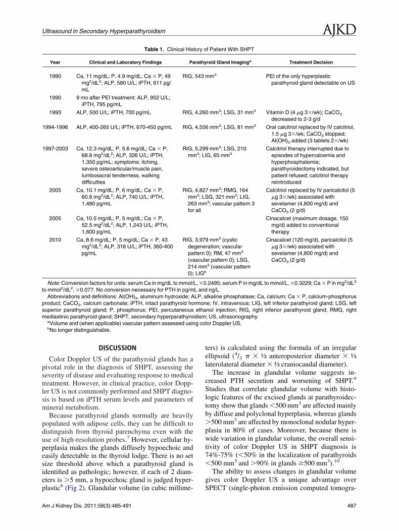
Table 1. Clinical His
Year Clinical and Laboratory Findings Pa
1990 Ca, 11 mg/dL; P, 4.9 mg/dL; Ca � P, 49mg2/dL2; ALP, 580 U/L; iPTH, 611 pg/mL
RIG,
1990 9 mo after PEI treatment: ALP, 952 U/L;iPTH, 795 pg/mL
1993 ALP, 500 U/L; iPTH, 700 pg/mL RIG,
1994-1996 ALP, 400-265 U/L; iPTH, 670-450 pg/mL RIG,
1997-2003 Ca, 12.3 mg/dL; P, 5.6 mg/dL; Ca � P,68.8 mg2/dL2; ALP, 326 U/L; iPTH,1,350 pg/mL; symptoms: itching,severe osteoarticular/muscle pain,lumbosacral tenderness, walkingdifficulties
RIG,mm
2005 Ca, 10.1 mg/dL; P, 6 mg/dL; Ca � P,60.6 mg2/dL2; ALP, 740 U/L; iPTH,1,480 pg/mL
RIG,mm26for
2005 Ca, 10.5 mg/dL; P, 5 mg/dL; Ca � P,52.5 mg2/dL2; ALP, 1,243 U/L; iPTH,1,600 pg/mL
2010 Ca, 8.6 mg/dL; P, 5 mg/dL; Ca � P, 43mg2/dL2; ALP, 316 U/L; iPTH, 360-400pg/mL
RIG,depa(va210);
Note: Conversion factors for units: serum Ca in mg/dL to mmol/to mmol2/dL2, �0.077. No conversion necessary for PTH in pg/m
Abbreviations and definitions: Al(OH)3, aluminum hydroxide; Aproduct; CaCO3, calcium carbonate; iPTH, intact parathyroid hosuperior parathyroid gland; P, phosphorus; PEI, percutaneousmediastinic parathyroid gland; SHPT, secondary hyperparathyro
aVolume and (when applicable) vascular pattern assessed usinbNo longer distinguishable.
plastic8 (Fig 2). Glandular volume (in cubic millime-
Am J Kidney Dis. 2011;58(3):485-491
ters) is calculated using the formula of an irregularellipsoid (4/3 � � ½ anteroposterior diameter � ½laterolateral diameter � ½ craniocaudal diameter).
The increase in glandular volume suggests in-creased PTH secretion and worsening of SHPT.9
Studies that correlate glandular volume with histo-logic features of the excised glands at parathyroidec-tomy show that glands �500 mm3 are affected mainlyby diffuse and polyclonal hyperplasia, whereas glands�500 mm3 are affected by monoclonal nodular hyper-plasia in 80% of cases. Moreover, because there iswide variation in glandular volume, the overall sensi-tivity of color Doppler US in SHPT diagnosis is74%-75% (�50% in the localization of parathyroids�500 mm3 and �90% in glands �500 mm3).10
The ability to assess changes in glandular volumegives color Doppler US a unique advantage over
f Patient With SHPT
roid Gland Imaginga Treatment Decision
mm3 PEI of the only hyperplasticparathyroid gland detectable on US
0 mm3; LSG, 31 mm3 Vitamin D (4 �g 3�/wk); CaCO3
decreased to 2-3 g/d
6 mm3; LSG, 81 mm3 Oral calcitriol replaced by IV calcitriol,1.5 �g 3�/wk; CaCO3 stopped;Al(OH)3 added (3 tablets 2�/wk)
9 mm3; LSG, 210IG, 65 mm3
Calcitriol therapy interrupted due toepisodes of hypercalcemia andhyperphosphatemia;parathyroidectomy indicated, butpatient refused; calcitriol therapyreintroduced
7 mm3; RMG, 164SG, 321 mm3; LIG,
3; vascular pattern 3
Calcitriol replaced by IV paricalcitol (5�g 3�/wk) associated withsevelamer (4,800 mg/d) andCaCO3 (2 g/d)
Cinacalcet (maximum dosage, 150mg/d) added to conventionaltherapy
9 mm3 (cysticration; vascular0); RM, 47 mm3
ar pattern 0); LSG,3 (vascular pattern
Cinacalcet (120 mg/d), paricalcitol (5�g 3�/wk) associated withsevelamer (4,800 mg/d) andCaCO3 (2 g/d)
.2495; serum P in mg/dL to mmol/L, �0.3229; Ca � P in mg2/dL2
ng/L.lkaline phosphatase; Ca, calcium; Ca � P, calcium-phosphoruse; IV, intravenous; LIG, left inferior parathyroid gland; LSG, leftnol injection; RIG, right inferior parathyroid gland; RMG, right; US, ultrasonography.lor Doppler US.
tory o
rathy
543
4,26
4,55
5,293; L
4,823; L
3 mmall
3,97genetternscul4 mmLIGb
L, �0L andLP, armonetha
idismg co
SPECT (single-photon emission computed tomogra-
487

of vas
Meola et al
phy)–scintigraphy, computed tomography, and mag-netic resonance imaging.
The increase in parathyroid gland volume is associ-ated with a widespread increase in vascularity, evidenthistologically.11-13 Usually color Doppler distin-guishes 3 different patterns,10 as shown in Fig 2D-F.
According to international guidelines, SHPT diag-nosis is based on iPTH serum levels and parameters ofmineral metabolism. However, the optimal values foriPTH in maintenance HD patients are still uncer-tain,14,15 and hormone levels show great variability.16
Because levels are influenced by drug therapy, iPTHlevel does not provide information for grading hyper-plasia. Color Doppler US evaluates the degree ofhyperplasia and number of glands involved, complet-ing the biochemical diagnosis. We routinely performcolor Doppler US of the parathyroid glands wheniPTH values are consistently �400 pg/mL (�400ng/L) and repeat this study annually in maintenanceHD patients to evaluate the progression of disease.B-Mode parameters important for the assessment ofSHPT progression include: (1) the gland’s echogenic-ity, (2) gland diameter17 and volume, (3) appearanceof anechoic areas with no vascularization suggestinginvolutive cystic phenomena, and (4) presence of
Figure 2. Progression of parathyroid gland hyperplasia. (A) Lthat makes the glands enlarged and diffusely hypoechoic (arrhyperplasia is at first diffuse and polyclonal, then monoclonal anda widespread increase in vascularity. Color Doppler distinguhypovascularized glands with only a few color spots in the hilar/eglands fed by an enlarged hilar artery that have a peripheral arccommon carotid; T, thyroid.
fibrocalcifications.13
488
In our case, nodular hyperplasia of a single glandappeared after a long period of maintenance HDtherapy. Color Doppler US was used to performchemical ablation with percutaneous ethanol injec-tion, but biochemical/morphologic results were poor.Afterward, other hyperplastic glands appeared, show-ing the slow but continuous progression of SHPTdespite oral/intravenous vitamin D treatments. At thisstage, the evidence of nodular hyperplasia on colorDoppler US, severity of SHPT, and refractoriness totherapy with vitamin D/binders indicated the neces-sity of parathyroidectomy, which was refused by thepatient.
The role of color Doppler US in the presurgicalevaluation of SHPT is very different from primaryhyperparathyroidism. The sensitivity of color DopplerUS in the localization of adenoma/carcinoma is veryhigh (90%-92%) for primary hyperparathyroidism,similar to that of conventional parathyroidectomy(93%-99%).18 In a patient with SHPT, the role ofimaging is less critical because parathyroidectomy isperformed using conventional cervicotomy. More-over, because there is wide variation in glandularvolume and the overall sensitivity of color DopplerUS for SHPT is lower than for primary hyperparathy-
term hyperstimulation of parathyroid glands causes hyperplasia, easily distinguishable in the thyroid lodge. (B, C) Glandularlar. The increase in parathyroid gland volume is associated with3 different patterns: (D) glands lacking Doppler signal, (E)
odular region (weak Doppler signal), and (F) hypervascularizedcularity and/or ray-like endonodular vessels. Abbreviations: Cc,
ong-ows)nodu
ishesndon
roidism, the role of color Doppler US therefore should
Am J Kidney Dis. 2011;58(3):485-491
Ultrasound in Secondary Hyperparathyroidism
not be to localize the glands, but instead to determinesurgical timing. Parathyroidectomy is indicated whencolor Doppler US evidences one or more hyperplasticglands �500 mm3, serum iPTH level is �700 pg/mL(�700 ng/L), and calcium, phosphorus, and calcium-phosphorous product values are no longer controlledusing conventional therapy.19,20
Glandular volume is related directly to the numberof glands detected; it also may be highly variable andnot entirely correlated with iPTH level at greater thana certain size/volume threshold. Calculated volumeshows a linear correlation with iPTH value only whengland volume is �2,000 mm3 (ie, �2 g).8,21 Thisobservation suggests that the biggest glands tend todisengage from receptor control mechanisms (ie, up-regulation of calcium receptor and vitamin D recep-tor) and grow independently.
In our patient, the combination of conventionaltherapy and cinacalcet marked a reversal in the progres-sion of disease: PTH serum levels were decreased,mineral metabolism was stabilized, and color DopplerUS showed progressive changes in volume and vascu-larization. The regression resembled a “chemical para-thyroidectomy,” and 5 years after beginning therapy,surgical parathyroidectomy was not necessary.
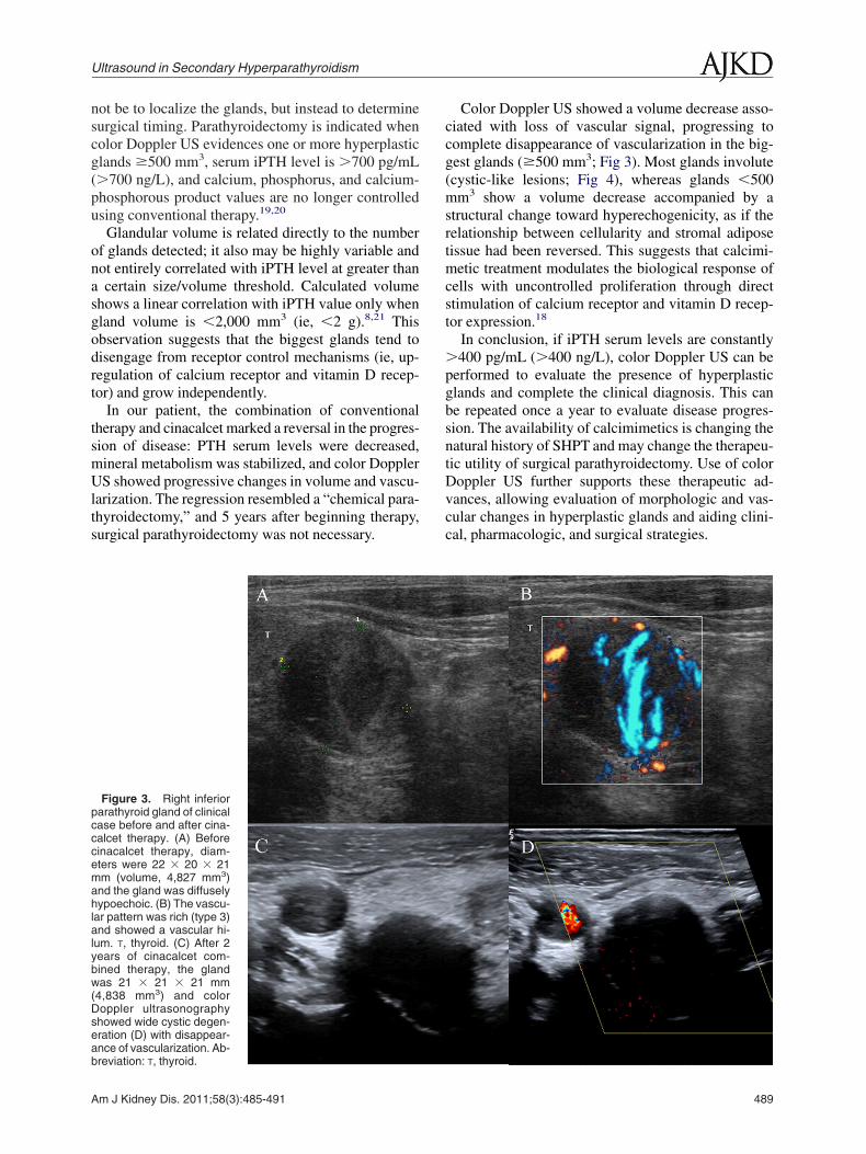
Figure 3. Right inferiorparathyroid gland of clinicalcase before and after cina-calcet therapy. (A) Beforecinacalcet therapy, diam-eters were 22 � 20 � 21mm (volume, 4,827 mm3)and the gland was diffuselyhypoechoic. (B) The vascu-lar pattern was rich (type 3)and showed a vascular hi-lum. T, thyroid. (C) After 2years of cinacalcet com-bined therapy, the glandwas 21 � 21 � 21 mm(4,838 mm3) and colorDoppler ultrasonographyshowed wide cystic degen-eration (D) with disappear-ance of vascularization. Ab-
breviation: T, thyroid.Am J Kidney Dis. 2011;58(3):485-491
Color Doppler US showed a volume decrease asso-ciated with loss of vascular signal, progressing tocomplete disappearance of vascularization in the big-gest glands (�500 mm3; Fig 3). Most glands involute(cystic-like lesions; Fig 4), whereas glands �500mm3 show a volume decrease accompanied by astructural change toward hyperechogenicity, as if therelationship between cellularity and stromal adiposetissue had been reversed. This suggests that calcimi-metic treatment modulates the biological response ofcells with uncontrolled proliferation through directstimulation of calcium receptor and vitamin D recep-tor expression.18
In conclusion, if iPTH serum levels are constantly�400 pg/mL (�400 ng/L), color Doppler US can beperformed to evaluate the presence of hyperplasticglands and complete the clinical diagnosis. This canbe repeated once a year to evaluate disease progres-sion. The availability of calcimimetics is changing thenatural history of SHPT and may change the therapeu-tic utility of surgical parathyroidectomy. Use of colorDoppler US further supports these therapeutic ad-vances, allowing evaluation of morphologic and vas-cular changes in hyperplastic glands and aiding clini-cal, pharmacologic, and surgical strategies.
489

Meola et al
ACKNOWLEDGEMENTSWe thank Sara Samoni, MD, for collecting clinical data.Support: An unrestricted grant from Amgen Europe GmbH
funded English translation by Apothecom ScopeMedical Ltd.Financial Disclosure: The authors declare that they have no
relevant financial interests.
REFERENCES1. Drüeke TB. Cell biology of parathyroid gland hyperplasia
in chronic renal failure. J Am Soc Nephrol. 2000;11(6):1141-1152.
2. Bethmore JB, Quarles LD. Calcimimetics or vitamin Danalogs for suppressing parathyroid hormone in end-stage renaldisease: time for a paradigm shift? Nat Clin Pract Nephrol.2009;5(1):24-33.
3. Fukagawa M. Cell biology of parathyroid hyperplasia inuremia. Am J Med Sci. 1999;317(6):377-382.
4. Arnold A, Brown MF, Urena P, et al. Monoclonality ofparathyroid tumors in chronic renal failure and in primary parathy-roid hyperplasia. J Clin Invest. 1995;95(5):2047-2053.
5. Afonso S, Santamaría I, Guinsburg ME, et al. Chromosomalaberrations, the consequence of refractory hyperparathyroidism:its relationship with biochemical parameters. Kidney Int Suppl.2003;63(85):S32-38.
6. Meola M, Petrucci I, Barsotti G. Long-term treatment withcinacalcet and conventional therapy reduces parathyroid hyperpla-sia in severe secondary hyperparathyroidism. Nephrol Dial Trans-plant. 2009;24(3):982-989.
7. Pavlovic D, Brzac HT. Prevention and treatment of second-ary hyperparathyroidism: still a challenge for the nephrologist?
Nephrol Dial Transplant. 2003;18(suppl 5):v45-46.490
8. Indridason OS, Heath H III, Khosla S, Yohay DA, QuarlesLD. Non suppressible parathyroid hormone secretion is related togland size in uremic secondary hyperparathyroidism. Kidney Int.1996;50(5):1663-1667.
9. Périé S, Fessi H, Tassart M, et al. Usefulness of combina-tion of high-resolution ultrasonography and dual-isotope iodine123/technetium Tc 99m sestamibi scintigraphy for the preo-perative localization of hyperplastic parathyroid glands inrenal hyperparathyroidism. Am J Kidney Dis. 2005;45(2):344-352.
10. Meola M, Petrucci I, Calliada F, et al. Presurgical settingof secondary hyperparathyroidism using high-resolution sonog-raphy and color Doppler. Ultraschall Med. 2011;32(suppl 1):S74-82.
11. Calliada F, Sala G, Conti MP, et al. Clinical applications ofcolor-Doppler: the parathyroid glands. Radiol Med. 1993;85(5)(suppl 1):S114-119.
12. Lane MJ, Desser TS, Weigel RJ, Jeffrey RB. Use of colorand power Doppler sonography to identify feeding arteries associ-ated with parathyroid adenomas. AJR Am J Roentgenol. 1997;171(3):819-823.
13. Gooding GA, Clark OH. Use of color Doppler imaging inthe distinction between thyroid and parathyroid lesions. Am J Surg.1992;164(1):51-56.
14. National Kidney Foundation. K/DOQI Clinical PracticeGuidelines for Bone Metabolism and Disease in Chronic Kid-ney Disease. Guidelines evaluation of calcium and phosphorousmetabolism. Am J Kidney Dis. 2003;42(suppl 3):S52-57.
15. Moe SM, Drüeke TB, Block GA, et al. KDIGO clinicalpractice guidelines for diagnosis, evaluation, prevention, and treat-
Figure 4. (A) Morphologic changesin the left superior parathyroid gland ofthe clinical case. (B) Before cinacalcettherapy, the gland was hypoechoic witha solid appearance and showed diffusedhypervascularization. (C) After 5 yearsof conventional therapy associated withcinacalcet, the gland shows areas ofcystic degeneration (arrows) (D) withno Doppler signal.
ment of chronic kidney disease-mineral and bone disorder
Am J Kidney Dis. 2011;58(3):485-491

Ultrasound in Secondary Hyperparathyroidism
(CKD-MBD). Treatment of CKD–MBD targeted at lowering highserum phosphorus and maintaining serum calcium. Kidney IntSuppl. 2009;76(113):S50-90.
16. Souberbielle JC, Boutten A, Carlier M-C, et al. WorkingGroup on PTH and Vitamin D. Société Francaise de BiologieClinique. Inter-method variability in PTH measurement: impli-cation for the care of CKD patients. Kidney Int. 2006;70(2):345-350.
17. Vulpio C, Bossola M, De Gaetano A, et al. Ultrasoundpatterns of parathyroid glands in chronic hemodialysis patientswith secondary hyperparathyroidism. Am J Nephrol. 2008;28(4):
589-597.Am J Kidney Dis. 2011;58(3):485-491
18. Elder GJ. Parathyroidectomy in the calcimimetic era. Ne-phrology. 2005;10(5):511-515.
19. Schomig M, Ritz E. Indications for parathyroidectomy.Nephrol Dial Transplant. 2000;15(suppl 5):S25-29.
20. Tominaga Y, Matsuoka S, Sato T. Surgical indications andprocedures of parathyroidectomy in patients with chronic kidneydisease. Ther Apher Dial. 2005;9(1):44-47.
21. Tominaga Y, Matsuoka S, Sato T, et al. Clinical features andhyperplastic patterns of parathyroid glands in hemodialysis pa-tients with advanced secondary hyperparathyroidism refractory tomaxacalcitol treatment and required parathyroidectomy. Ther Apher
Dial. 2007;11(4):266-273.491