Use of Replication-Conditional Adenovirus as a Helper System to Enhance Delivery of P450...
13
2004;64:292-303. Cancer Res Youssef Jounaidi and David J. Waxman Genes for Cancer Therapy System to Enhance Delivery of P450 Prodrug-Activation Use of Replication-Conditional Adenovirus as a Helper Updated version http://cancerres.aacrjournals.org/content/64/1/292 Access the most recent version of this article at: Cited Articles http://cancerres.aacrjournals.org/content/64/1/292.full.html#ref-list-1 This article cites by 68 articles, 25 of which you can access for free at: Citing articles http://cancerres.aacrjournals.org/content/64/1/292.full.html#related-urls This article has been cited by 8 HighWire-hosted articles. Access the articles at: E-mail alerts related to this article or journal. Sign up to receive free email-alerts Subscriptions Reprints and . [email protected] Department at To order reprints of this article or to subscribe to the journal, contact the AACR Publications Permissions . [email protected] Department at To request permission to re-use all or part of this article, contact the AACR Publications Cancer Research. on September 30, 2014. © 2004 American Association for cancerres.aacrjournals.org Downloaded from Cancer Research. on September 30, 2014. © 2004 American Association for cancerres.aacrjournals.org Downloaded from
-
Upload
independent -
Category
Documents
-
view
0 -
download
0
Transcript of Use of Replication-Conditional Adenovirus as a Helper System to Enhance Delivery of P450...
2004;64:292-303. Cancer Res Youssef Jounaidi and David J. Waxman Genes for Cancer TherapySystem to Enhance Delivery of P450 Prodrug-Activation Use of Replication-Conditional Adenovirus as a Helper
Updated version
http://cancerres.aacrjournals.org/content/64/1/292
Access the most recent version of this article at:
Cited Articles
http://cancerres.aacrjournals.org/content/64/1/292.full.html#ref-list-1
This article cites by 68 articles, 25 of which you can access for free at:
Citing articles
http://cancerres.aacrjournals.org/content/64/1/292.full.html#related-urls
This article has been cited by 8 HighWire-hosted articles. Access the articles at:
E-mail alerts related to this article or journal.Sign up to receive free email-alerts
SubscriptionsReprints and
To order reprints of this article or to subscribe to the journal, contact the AACR Publications
Permissions
To request permission to re-use all or part of this article, contact the AACR Publications
Cancer Research. on September 30, 2014. © 2004 American Association forcancerres.aacrjournals.org Downloaded from
Cancer Research. on September 30, 2014. © 2004 American Association forcancerres.aacrjournals.org Downloaded from
[CANCER RESEARCH 64, 292–303, January 1, 2004]
Use of Replication-Conditional Adenovirus as a Helper System to Enhance Deliveryof P450 Prodrug-Activation Genes for Cancer Therapy
Youssef Jounaidi and David J. WaxmanDivision of Cell and Molecular Biology, Department of Biology, Boston University, Boston, Massachusetts
ABSTRACT
Cytochrome P450 (CYP) gene transfer sensitizes tumor xenografts toanticancer prodrugs such as cyclophosphamide (CPA) without a detecta-ble increase in host toxicity. Optimal prodrug activation is achieved whena suitable P450 gene (e.g., human CYP2B6) is delivered in combinationwith NADPH-cytochrome P450 reductase (P450R), which encodes theflavoenzyme P450 reductase. We sought to improve this gene therapy bycoordinated delivery and expression of P450 and P450R on a singlebicistronic vector using an internal ribosomal entry site (IRES) sequence.Retrovirus encoding a CYP2B6-IRES-P450R expression cassette wasshown to induce strong P450-dependent CPA cytotoxicity in a populationof infected 9L gliosarcoma cells. Adeno-P450, a replication-defective,E1/E3 region-deleted adenovirus engineered to express CYP2B6-IRES-P450R, induced intracellular CPA 4-hydroxylation, and CPA cytotoxicity,in a broad range of human cancer cell lines. However, limited Adeno-P450gene transfer and CPA chemosensitization was seen with certain humantumor cells, notably PC-3 prostate and HT-29 colon cancer cells. Remark-able improvements could be obtained by coinfecting the tumor cells withAdeno-P450 in combination with Onyx-017, an E1b-55k gene-deletedadenovirus that selectively replicates in p53 pathway-deficient cells. Sub-stantial increases in gene expression were observed during the early stagesof viral infection, reflecting an apparent coamplification of the Adeno-P450 genome, followed by enhanced viral spread at later stages, as dem-onstrated in cultured tumor cells, and in A549 and PC-3 solid tumorxenografts grown in scid mice. This combination of the replication-defec-tive Adeno-P450 with a replication-conditional and tumor cell-targetedhelper adenovirus dramatically improved the low gene transfer observedwith some human tumor cell lines and correspondingly increased tumorcell-catalyzed CPA 4-hydroxylation, CPA cytotoxicity, and in vivo antitu-mor activity in a PC-3 tumor xenograft model. The use of tumor-selective,replicating adenovirus to promote the spread of replication-defective genetherapy vectors, such as Adeno-P450, substantially increases the thera-peutic potential of adenoviral delivery systems, and should lead to in-creased activity and enhanced tumor selectivity of cytochrome P450 andother gene-directed enzyme prodrug therapies.
INTRODUCTION
Gene-directed enzyme prodrug therapy (GDEPT) is designed tosensitize tumor cells to cancer chemotherapeutic prodrugs. ManyGDEPT strategies have been introduced over the last several years;however, only a few have advanced to the stage of human clinicaltrials (1–3). The success of prodrug activation gene therapy is influ-enced by multiple factors, including the activity and specificity of theprodrug activating enzyme, the efficiency and selectivity of genetransfer, the potency of the activated prodrug, and the strength of thebystander effect. One emerging GDEPT strategy utilizes cytochromeP450 (CYP) enzymes in combination with the flavoenzyme P450R toactivate established anticancer prodrugs with demonstrated clinicalutility, such as cyclophosphamide (CPA) and its isomer ifosfamide(IFA; Refs. 4, 5; reviewed in Refs. 6, 7). CPA and IFA are alkylating
agent prodrugs that induce DNA cross-linking in a cell cycle-inde-pendent manner, and ultimately trigger a mitochondrial pathwayapoptotic cell death in the case of CPA (8) and either apoptosis ornecrosis in the case of IFA (8, 9). CPA and IFA are activated by asubset of liver-expressed P450 enzymes (10, 11), which catalyze a4-hydroxylation reaction that yields cell membrane-permeable, cyto-toxic metabolites. These metabolites are transported from the liver totumor cells and also normal host tissues, where they induce deleteri-ous side effects that limit therapeutic effectiveness (12, 13).
The anticancer activity of CPA in cultured tumor cells, and inrodent and human xenograft models is substantially increased byintroduction of cDNAs encoding the P450 enzymes CYP2B1 andCYP2B6, which are major catalysts of CPA activation in rat andhuman liver, respectively (14–17). The efficacy of this P450 GDEPTstrategy may be additionally enhanced in several ways: (a) by usingP450/P450R-activated bioreductive drugs that target hypoxic regionsof solid tumors, either alone or in combination with CPA (18, 19); (b)by increasing the tumor/liver partition of P450 prodrugs, using thyroidhormone antagonists to suppress liver P450R expression and therebydecrease liver P450 metabolic activity (20); and (c) by administrationof CPA in frequent, low doses (21) using a metronomic scheduleassociated with antiangiogenic activity (22).
In the present study we sought to enhance the efficiency of P450gene delivery and P450 prodrug activation. We report improved P450prodrug activity using an internal ribosome entry site (IRES) sequence(23) to achieve coordinate expression of P450 and P450R. We addi-tionally show that a replication-defective adenovirus armed with aCYP2B6-IRES-P450R expression cassette (Adeno-P450) can sensi-tize human tumor cells of diverse tissue origin to CPA. Finally, wedemonstrate that a tumor cell-targeted, conditionally-replicating E1b-55k-deleted adenovirus, closely related to the Onyx-015 oncolyticadenovirus now in Phase II/Phase III clinical trials (24–26), can beused as a helper virus to greatly increase the efficiency of tumor cellexpression and spread of Adeno-P450. This effect is demonstratedboth in cell culture and in vivo in a human xenograft model, and isshown to enhance production of cytotoxic, P450-activated CPA me-tabolites and thereby significantly increase tumor cell kill.
MATERIALS AND METHODS
Chemicals. CPA, chloroquine, fetal bovine serum (FBS), and puromycinwere purchased from Sigma Chemical Co. (St. Louis, MO). 4OOH-CPA and4OOH-IFA, chemically activated derivatives of CPA and IFA, respectively,were obtained from Asta Pharma (Bielefeld, Germany).
Retroviral Plasmid Construction. The retroviral plasmids pBabe-puroand pWzl-bleo are based on the pBabe series (27), and encode puromycin andbleomycin resistance genes, respectively, both being transcribed from the3�-long terminal repeat of the viral vector. Human P450R cDNA cloned intothe EcoRI site of pUV1 (28), obtained from Dr. Frank Gonzalez (NationalCancer Institute, Bethesda, MD), was excised with EcoRI, and then bluntended and subcloned into the blunt-ended SnaBI and SalI sites of pBabe-puroto yield pBabe-P450R-puro. The presence of the correct ATG initiation codonin the cloned P450R cDNA was verified by DNA sequencing. To constructpBabe-2B6-IRES-P450R-puro, the IRES sequence of pWzl-bleo was excisedusing BglII and NcoI, blunt ended, and then recloned into the SnaBI site ofpWzl-bleo to yield a pWzl-bleo derivative with two IRES sequences intandem. The first IRES sequence was removed using BamHI and BglII, and
Received 6/18/03; revised 10/17/03; accepted 10/24/03.Grant support: Supported by NIH Grant CA49248 (to D. J. W.).The costs of publication of this article were defrayed in part by the payment of page
charges. This article must therefore be hereby marked advertisement in accordance with18 U.S.C. Section 1734 solely to indicate this fact.
Requests for reprints: David J. Waxman, Department of Biology, Boston University,5 Cummington Street, Boston, MA 02215. Fax: (617) 353-7404; E-mail: [email protected].
292
Cancer Research. on September 30, 2014. © 2004 American Association forcancerres.aacrjournals.org Downloaded from
subcloned into the BamHI site of pBabe-P450R-puro. The resulting plasmid,pBabe-IRES-P450R-puro, was linearized by digestion with SpeI and EcoRI, bluntended, and then ligated to a blunt-ended SpeI-SphI fragment encompassing theopen reading frame of CYP2B6 excised from pBabe-2B6-puro (15). The resultingconstruct is designated pBabe-2B6-IRES-P450R-puro. The CYP2B6 cDNA usedin these studies (GenBank accession no. M29874) corresponds to the wild-type(CYP2B6*) allele (29), i.e., Arg22/Lys139/Gln172/Ser259/Lys262/Arg487.CYP2C18 cDNA was cut from pBabe-2C18-Met-puro (15) using SpeI and VspI,blunt ended, and then subcloned into SpeI- and EcoRI-linearized and blunt-endedpBabe-IRES-P450R-puro. CYP2C19 cDNA was cut from pBabe-2C19-puro (15)using SpeI and BglII, followed by blunt-end subcloning into the SpeI and EcoRIsites of pBabe-IRES-P450R-puro.
9L Cell Lines Coexpressing P450 and P450R cDNAs. Transfection of theecotropic packaging cell line Bosc 23 (30) with pBabe-2B6-IRES-P450R-puro,pBabe-2C18-IRES-P450R-puro, pBabe-2C19-IRES-P450R-puro, pBabe-P450R-puro, and pBabe-IRES-P450R-puro retroviral plasmids, harvesting of retro-viral supernatants, and infection of rat 9L gliosarcoma cells were carried out asdescribed (15), with the following modifications. Bosc 23 cells were plated at2.5 � 106 cells in a 60-mm dish. Fresh media (4 ml) containing chloroquine(25 �M) was added to the cells 16 h later. The cells were cotransfected 1 h laterwith 5 �g of retroviral plasmid DNA and 2.5 �g of the helper plasmid pKat(31) using 9 �l of the cationic liposome Fugene 6 (Boehringer-Mannheim) ina total volume of 40 �l DMEM without FBS. Pools of cells resistant topuromycin (2.5 �g/ml) were selected 48 h later over a 2–3-day period asdescribed (15). Drug-resistant pools of cells were propagated and then assayedfor P450R-catalyzed, NADPH-dependent cytochrome C reduction (A550 mea-sured at 30°C) in isolated microsomes (15). A 3–4-fold increase in P450Ractivity was exhibited by the 9L/pBabe-IRES-P450R cells versus a 2–3-foldincrease in the pools of 9L/P450-IRES-P450R transfectants.
Growth Inhibition Assay. The chemosensitivity of P450-expressing 9Lcell lines was determined using a 4-day growth inhibition assay, with cellsplated in triplicate at 4000 cells/well of a 48-well plate 1 day before treatmentwith CPA continuously for 4 days (15). The same assay was used to evaluatethe intrinsic sensitivity of tumor cells to the chemically activated CPA and IFAderivatives 4OOH-CPA and 4OOH-IFA (0–5 �M). Cells remaining at the endof 4 dats were stained and quantified using a crystal violet/alcohol-extractionassay (15). Data are expressed as cell number (A595) relative to drug-freecontrols, mean � SD values for triplicate samples, unless indicated otherwise.
Construction of Recombinant Adenovirus. A replication-defective, E1and E3 region-deleted adenovirus encoding CYP2B6 and P450R, designatedAdeno-P450, was constructed using the AdenoX expression system (ClontechLaboratories Inc., Palo Alto, CA). pBabe-2B6-IRES-P450R-puro was linear-ized using BamHI, blunt ended, and the CYP2B6-IRES-P450R cassette thencut with DraI and ligated into the DraI site of pShuttle (Clontech Labs). Theresulting plasmid, pShuttle-2B6-IRES-P450R, was used to construct the re-combinant adenovirus, Adeno-P450, as described in the manufacturer’s kit.The cytomegalovirus-IE promoter of pShuttle drives Adeno-P450-directedexpression of CYP2B6 and P450R, and ensures robust expression in a widerange of cell lines and tissues. The conditional replicating virus Onyx-017,which contains wild-type viral E3 region and an E1b-55k gene deletionequivalent to that of Onyx-015 (25), was obtained from Onyx Pharmaceuticals,Inc. (Richmond, CA). Adeno-�-galactosidase (Adeno-�Gal) is an E1- andE3-region deleted, replication-deficient adenovirus that contains a �-galacto-sidase reporter under the control of a cytomegalovirus promoter and was usedin our previous studies (14).
Adenovirus Purification. Adenoviral stocks were propagated in humankidney 293 cells grown in 100-mm plates at 37°C in a humidified, 5% CO2
atmosphere in high glucose DMEM containing 10% FBS. Cells were grown to�90% confluence and then infected with Adeno-P450 at a multiplicity ofinfection (MOI) of �5 viral particles per cell. Alternatively, 3–5 ml of �80°Cfrozen culture supernatant obtained from Adeno-P450-infected 293 cells wasused to reinfect the 293 cells. Seventy-two h after infection, �80–90% of thecells became rounded, and 10–20% of the cells were floating. The cells werethen collected by centrifugation and resuspended in 20 ml of buffer A [10 mM
Tris-base (pH 8.0) and 1 mM MgCl2]. The virus was released by threefreeze-thaw cycles, alternating between an alcohol-dry ice bath and a 37°Cbath. The cell lysate was centrifuged at 4°C for 10 min at 3,000 rpm, and thesupernatant was placed on ice. The residual cell pellet was re-extracted in thesame manner with 10 ml of buffer A, and the combined supernatants were treated
with Benzonase (0.5–1 units/ml) at room temperature for 30 min. The treatedsuspension was layered carefully onto a cold CsCl step gradient comprised of 10ml of light CsCl [1.20 g/ml: 22.39 g CsCl �77.6 ml of 10 mM Tris-base (pH 8.0)]layered on top of 10 ml of heavy CsCl [1.45 g/ml: 42.2 g CsCl �57.8 ml of 10 mM
Tris-base (pH 8.0)]. Samples were centrifuged in a Sorval Pro ultracentrifuge in anSW28 rotor at 4°C for 2 h at 20,000 rpm. The banded virus was collected anddiluted with an equal volume of 10 mM Tris-base (pH 8.0), and rebanded on asecond CsCl step gradient consisting of 4 ml each of light CsCl and heavy CsClas described above, then centrifuged overnight at 4°C at 20,000 rpm in a SorvalSW41Ti rotor. The purified virus was desalted by dialysis against 10 mM Tris-base(pH 8.0), 1 mM MgCl2, and 10% glycerol, with the effectiveness of desaltingverified by conductivity measurement. Alternatively, the purified virus was pas-saged through a Bio-Rad Econo-Pac 10DG Column pre-equilibrated with 30 ml ofcolumn buffer [10 mM Tris-base (pH 8.0), 1 mM MgCl2, and 10% glycerol]. Thevirus suspension was loaded onto the column, and samples were collected imme-diately after the first 2.5-ml eluent. Viral titers were quantitated using the Adeno-XRapid Titer kit (Clontech Labs), as described in the manufacturer’s kit. Virusaliquots were stored at �80°C.
Adenoviral Infection of Human Tumor Cell Lines and CPA Cytotox-icity Assays. Thirteen human tumor cell lines selected from the NCI-60 panel(see Fig. 2, below; Ref. 32) were obtained from Dr. Dominic Scudiero(National Cancer Institute). The A549 lung cancer cell line was also obtainedfrom Onyx Pharmaceuticals, Inc. and was used in all of the helper adenovirusexperiments. In the experiments shown in Fig. 2 (see below), cells were seededin six-well plates at 75,000 cells per well. Sixteen h later the cells were infectedfor 1 h with Adeno-P450 at an MOI of 90 plaque-forming units (pfu)/cell in 1ml of RPMI 1640 containing 5% FBS, after which the medium was removedand replaced with fresh medium. Cells were incubated for an additional 48 hand cell lysate then prepared by sonication. P450R activity in 20 �g cell lysatewas assayed to obtain an overall indication of viral transduction efficiency. Inother experiments (Fig. 3B; below), human tumor cells were plated in triplicateat 8,000 cells/well of a 24-well plate 18–24 h before adenovirus infection.Cells were infected for 1 h with Adeno-�Gal at MOI 50 or with Adeno-P450at MOI values of 25, 65, and 125 pfu/cell in DMEM containing 5% FBS. Freshmedium (1 ml) was then added to each well, followed by an additional 24-hincubation. The cells were then treated with CPA (0–1 mM, as specified ineach experiment) and cultured up to 4 days. Cells remaining at the end of theexperiment were stained with crystal violet and quantified (A595) as percentageof survival relative to untreated cells.
Onyx-017 Helper Virus Experiments. In a typical experiment, A549 cellswere plated in six-well plates at 75,000 cells/well. Cells were infected for 1 hwith Adeno-�Gal or Adeno-P450, either alone or in combination with Onyx-017, at MOIs specified in each experiment. The medium was then replacedwith virus-free medium. Individual plates of cells were stained with 5-bromo-4-chloro-3-indolyl-�-D-galactopyranoside (X-gal) 1–5 days later to evaluatethe spread of Adeno-�Gal. �-Galactosidase activity was quantitated using amicrotiter plate reader (A650) after resuspending the X-gal stain in 1 mlDMSO/well of a six-well plate (18). To determine whether the infected cellscontinue to release infective Adeno-�Gal viral particles, the supernatant fromday 4- or day 5-infected cells was used to infect fresh A549 cells, which werestained with X-gal 48 h later.
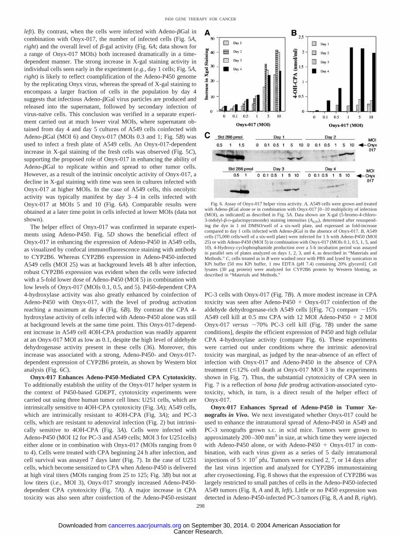
In other experiments, the effect of Onyx-017 on Adeno-P450-dependentCYP2B6 expression and CYP2B6-catalyzed 4-hydroxy-cyclophosphamide(4OH-CPA) production was assayed, as follows. Twenty-four h after infectionwith adenovirus, the cells were treated with 1 mM CPA in 3 ml of fresh RPMI1640 containing 5% FBS and 0.5 mM semicarbazide to stabilize the 4OH-CPAmetabolite. After 5 h of CPA treatment, 0.5 ml of culture supernatant wasremoved and assayed for the presence of 4OH-CPA released into the culturemedium (33). The cells were then washed once with PBS, scraped from thewells, and lysed by sonication in 50 mM potassium phosphate buffer and1 mM EDTA (pH 7.4) containing 20% glycerol. The procedure was re-peated on days 2– 4 after viral infection in parallel sets of plates. Westernblot analysis of CYP2B6 protein (30 �g sonicated cell lysate protein/well) wasperformed using mouse anti-CYP2B6 monoclonal antibody and lymphoblast-expressed CYP2B6 protein (0.6–1 pmol/lane) as standard (BD-Gentest, Inc.,Woburn, MA).
To assess the impact of Onyx-017 on Adeno-P450-mediated CPA cytotox-icity, human tumor cell lines U251, PC-3, and A549 were plated in 24-wellplates at 14,000 cells/well and infected 24 h later with Adeno-P450 (A549 and
293
P450 GENE THERAPY FOR CANCER
Cancer Research. on September 30, 2014. © 2004 American Association forcancerres.aacrjournals.org Downloaded from
PC-3, MOI 12; U251 cells, MOI 3), either alone or in combination withOnyx-017 (MOIs 0, 0.3, 1, 2, 3, and 4). Virus was incubated with the cells for3.5 h in RPMI 1640 medium (0.2 ml/well), after which an additional 1 ml offresh RPMI 1640 was added to each well. The medium was removed 24 h laterand 1 ml of fresh medium containing CPA (0, 0.25, 0.5, and 1 mM) was addedto the cells. After 2 days of CPA treatment, the medium was replaced with 1 mlof fresh CPA-containing medium for an additional 5 days, after which cellsurvival was assayed by crystal violet staining.
Visualization of Helper Virus Dynamics by Immunochemistry. Confo-cal immunofluorescence microscopy was used to assay the expression ofCYP2B6 in A549 cells infected with Adeno-P450 (MOI 25) or infected withAdeno-P450 (MOI 5) in combination with Onyx-017 (MOIs 0.1, 0.5, and 5).A549 cells (4000 cells/well) were plated in an eight-well LAB-TEK ChamberSlides (Nalgen Nunc, International). Cells were infected with adenovirus for1 h at the indicated MOIs, after which the medium was replaced with 0.2 mlof fresh RPMI 1640 containing 5% FBS. Cells were then cultured for anadditional 48 h. CYP2B6 immunostaining was then carried out as follows.Cells were washed with PBS buffer and fixed in 100% methanol at roomtemperature for 10 min. Samples were air-dried and then incubated twice for10 min in PBS containing 3% FCS for blocking. Monoclonal anti-CYP2B6antibody diluted 1/1000 into PBS containing 3% FCS (50 �l) was then addedto each well. The plates were covered with aluminum foil and incubated for 1 hat 37°C in a tissue culture incubator. Cells were washed twice for 5 min with100 �l of 3% FCS in PBS and then incubated with FITC-conjugated antimouseantibody (Molecular Probes, Inc., Eugene, OR) diluted 1:1000 in 3% FCS inPBS and incubated for 1 h at 37°C. Cells were washed under dim light with 3%
FCS in PBS. To visualize cell nuclei, 50 �l of propidium iodide (5 ng/ml inPBS) was added to the cells for 5 min, followed by two PBS washes. Slideswere treated with a drop of Fluoroguard anti-fade reagent (Bio-Rad), coveredwith a coverslip sealed with nail polish and stored at �20°C before confocalmicroscopy. Cells were scanned with a BX-50 confocal laser-scanning micro-scope (Olympus Corp., New Hyde Park, NY) equipped with �60 objective(Carl Zeiss, Thornwood, NY) and photographed.
Adeno-P450 Spread in A549 and PC-3 Xenografts in Scid Mice. Seven-week-old (25–29 g) male ICR/Fox Chase mice, an outbred scid immunodefi-cient strain (Taconic Farms, Germantown, NY), were injected s.c. at eachposterior flank with 4 � 106 A549 or PC-3 tumor cells in a volume of 0.5 mlof serum-free DMEM using a 0.5-inch, 29-gauge needle and a 1-ml insulinsyringe. Tumor sizes were measured twice a week using external calipers.Tumors reaching approximately 200–300 mm3 in size were injected withAdeno-P450, alone or in combination with Onyx-017, using an establishedprotocol (intratumoral injection of 50 �l containing 5 � 107 pfu of each virus,daily for 5 days). Tumors were excised on day 2, 7, and 14 after the last virusinjection. The efficiency of Adeno-P450 gene delivery was monitored byimmunofluorescence analysis of cryosectioned tumor slices using anti-CYP2B6 antibody. Mice were killed by cervical dislocation, tumors wereexcised and frozen in dry ice-cooled isopentane for 3 min, and were sectionedwith a cryostat to give 25 �m sections. Sections were fixed immediately in�10°C acetone and processed for immunohistochemical analysis as describedabove. Tumor extracts from PC-3 tumors were prepared by homogenization in50 mM KPi buffer and 1 mM EDTA (pH 7.4) containing 20% glycerol. Toassess the Onyx helper virus effect in vivo, 30 �g of cell lysate was analyzedby Western blotting using anti-2B6 mouse monoclonal antibody.
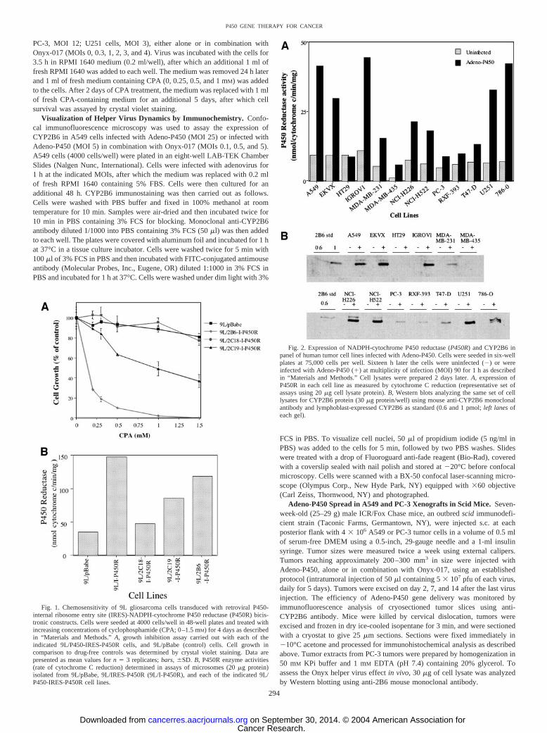
Fig. 1. Chemosensitivity of 9L gliosarcoma cells transduced with retroviral P450-internal ribosome entry site (IRES)-NADPH-cytochrome P450 reductase (P450R) bicis-tronic constructs. Cells were seeded at 4000 cells/well in 48-well plates and treated withincreasing concentrations of cyclophosphamide (CPA; 0–1.5 mM) for 4 days as describedin “Materials and Methods.” A, growth inhibition assay carried out with each of theindicated 9L/P450-IRES-P450R cells, and 9L/pBabe (control) cells. Cell growth incomparison to drug-free controls was determined by crystal violet staining. Data arepresented as mean values for n � 3 replicates; bars, �SD. B, P450R enzyme activities(rate of cytochrome C reduction) determined in assays of microsomes (20 �g protein)isolated from 9L/pBabe, 9L/IRES-P450R (9L/I-P450R), and each of the indicated 9L/P450-IRES-P450R cell lines.
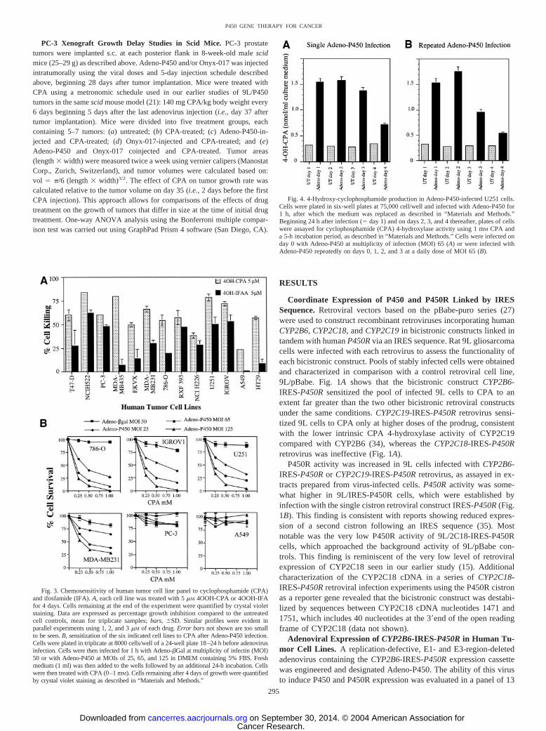
Fig. 2. Expression of NADPH-cytochrome P450 reductase (P450R) and CYP2B6 inpanel of human tumor cell lines infected with Adeno-P450. Cells were seeded in six-wellplates at 75,000 cells per well. Sixteen h later the cells were uninfected (�) or wereinfected with Adeno-P450 (�) at multiplicity of infection (MOI) 90 for 1 h as describedin “Materials and Methods.” Cell lysates were prepared 2 days later. A, expression ofP450R in each cell line as measured by cytochrome C reduction (representative set ofassays using 20 �g cell lysate protein). B, Western blots analyzing the same set of celllysates for CYP2B6 protein (30 �g protein/well) using mouse anti-CYP2B6 monoclonalantibody and lymphoblast-expressed CYP2B6 as standard (0.6 and 1 pmol; left lanes ofeach gel).
294
P450 GENE THERAPY FOR CANCER
Cancer Research. on September 30, 2014. © 2004 American Association forcancerres.aacrjournals.org Downloaded from
PC-3 Xenograft Growth Delay Studies in Scid Mice. PC-3 prostatetumors were implanted s.c. at each posterior flank in 8-week-old male scidmice (25–29 g) as described above. Adeno-P450 and/or Onyx-017 was injectedintratumorally using the viral doses and 5-day injection schedule describedabove, beginning 28 days after tumor implantation. Mice were treated withCPA using a metronomic schedule used in our earlier studies of 9L/P450tumors in the same scid mouse model (21): 140 mg CPA/kg body weight every6 days beginning 5 days after the last adenovirus injection (i.e., day 37 aftertumor implantation). Mice were divided into five treatment groups, eachcontaining 5–7 tumors: (a) untreated; (b) CPA-treated; (c) Adeno-P450-in-jected and CPA-treated; (d) Onyx-017-injected and CPA-treated; and (e)Adeno-P450 and Onyx-017 coinjected and CPA-treated. Tumor areas(length � width) were measured twice a week using vernier calipers (ManostatCorp., Zurich, Switzerland), and tumor volumes were calculated based on:vol � �/6 (length � width)3/2. The effect of CPA on tumor growth rate wascalculated relative to the tumor volume on day 35 (i.e., 2 days before the firstCPA injection). This approach allows for comparisons of the effects of drugtreatment on the growth of tumors that differ in size at the time of initial drugtreatment. One-way ANOVA analysis using the Bonferroni multiple compar-ison test was carried out using GraphPad Prism 4 software (San Diego, CA).
RESULTS
Coordinate Expression of P450 and P450R Linked by IRESSequence. Retroviral vectors based on the pBabe-puro series (27)were used to construct recombinant retroviruses incorporating humanCYP2B6, CYP2C18, and CYP2C19 in bicistronic constructs linked intandem with human P450R via an IRES sequence. Rat 9L gliosarcomacells were infected with each retrovirus to assess the functionality ofeach bicistronic construct. Pools of stably infected cells were obtainedand characterized in comparison with a control retroviral cell line,9L/pBabe. Fig. 1A shows that the bicistronic construct CYP2B6-IRES-P450R sensitized the pool of infected 9L cells to CPA to anextent far greater than the two other bicistronic retroviral constructsunder the same conditions. CYP2C19-IRES-P450R retrovirus sensi-tized 9L cells to CPA only at higher doses of the prodrug, consistentwith the lower intrinsic CPA 4-hydroxylase activity of CYP2C19compared with CYP2B6 (34), whereas the CYP2C18-IRES-P450Rretrovirus was ineffective (Fig. 1A).
P450R activity was increased in 9L cells infected with CYP2B6-IRES-P450R or CYP2C19-IRES-P450R retrovirus, as assayed in ex-tracts prepared from virus-infected cells. P450R activity was some-what higher in 9L/IRES-P450R cells, which were established byinfection with the single cistron retroviral construct IRES-P450R (Fig.1B). This finding is consistent with reports showing reduced expres-sion of a second cistron following an IRES sequence (35). Mostnotable was the very low P450R activity of 9L/2C18-IRES-P450Rcells, which approached the background activity of 9L/pBabe con-trols. This finding is reminiscent of the very low level of retroviralexpression of CYP2C18 seen in our earlier study (15). Additionalcharacterization of the CYP2C18 cDNA in a series of CYP2C18-IRES-P450R retroviral infection experiments using the P450R cistronas a reporter gene revealed that the bicistronic construct was destabi-lized by sequences between CYP2C18 cDNA nucleotides 1471 and1751, which includes 40 nucleotides at the 3�end of the open readingframe of CYP2C18 (data not shown).
Adenoviral Expression of CYP2B6-IRES-P450R in Human Tu-mor Cell Lines. A replication-defective, E1- and E3-region-deletedadenovirus containing the CYP2B6-IRES-P450R expression cassettewas engineered and designated Adeno-P450. The ability of this virusto induce P450 and P450R expression was evaluated in a panel of 13
Fig. 3. Chemosensitivity of human tumor cell line panel to cyclophosphamide (CPA)and ifosfamide (IFA). A, each cell line was treated with 5 �M 4OOH-CPA or 4OOH-IFAfor 4 days. Cells remaining at the end of the experiment were quantified by crystal violetstaining. Data are expressed as percentage growth inhibition compared to the untreatedcell controls, mean for triplicate samples; bars, �SD. Similar profiles were evident inparallel experiments using 1, 2, and 3 �M of each drug. Error bars not shown are too smallto be seen. B, sensitization of the six indicated cell lines to CPA after Adeno-P450 infection.Cells were plated in triplicate at 8000 cells/well of a 24-well plate 18–24 h before adenovirusinfection. Cells were then infected for 1 h with Adeno-�Gal at multiplicity of infectin (MOI)50 or with Adeno-P450 at MOIs of 25, 65, and 125 in DMEM containing 5% FBS. Freshmedium (1 ml) was then added to the wells followed by an additional 24-h incubation. Cellswere then treated with CPA (0–1 mM). Cells remaining after 4 days of growth were quantifiedby crystal violet staining as described in “Materials and Methods.”
Fig. 4. 4-Hydroxy-cyclophosphamide production in Adeno-P450-infected U251 cells.Cells were plated in six-well plates at 75,000 cell/well and infected with Adeno-P450 for1 h, after which the medium was replaced as described in “Materials and Methods.”Beginning 24 h after infection (� day 1) and on days 2, 3, and 4 thereafter, plates of cellswere assayed for cyclophosphamide (CPA) 4-hydroxylase activity using 1 mM CPA anda 5-h incubation period, as described in “Materials and Methods.” Cells were infected onday 0 with Adeno-P450 at multiplicity of infection (MOI) 65 (A) or were infected withAdeno-P450 repeatedly on days 0, 1, 2, and 3 at a daily dose of MOI 65 (B).
295
P450 GENE THERAPY FOR CANCER
Cancer Research. on September 30, 2014. © 2004 American Association forcancerres.aacrjournals.org Downloaded from
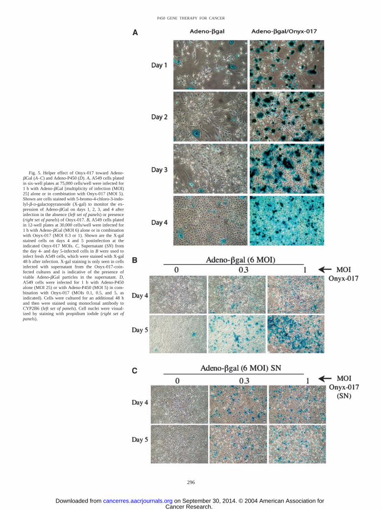
Fig. 5. Helper effect of Onyx-017 toward Adeno-�Gal (A–C) and Adeno-P450 (D). A, A549 cells platedin six-well plates at 75,000 cells/well were infected for1 h with Adeno-�Gal [multiplicity of infection (MOI)25] alone or in combination with Onyx-017 (MOI 5).Shown are cells stained with 5-bromo-4-chloro-3-indo-lyl-�-D-galactopyranoside (X-gal) to monitor the ex-pression of Adeno-�Gal on days 1, 2, 3, and 4 afterinfection in the absence (left set of panels) or presence(right set of panels) of Onyx-017. B, A549 cells platedin 12-well plates at 30,000 cells/well were infected for1 h with Adeno-�Gal (MOI 6) alone or in combinationwith Onyx-017 (MOI 0.3 or 1). Shown are the X-galstained cells on days 4 and 5 postinfection at theindicated Onyx-017 MOIs. C, Supernatant (SN) fromthe day 4- and day 5-infected cells in B were used toinfect fresh A549 cells, which were stained with X-gal48 h after infection. X-gal staining is only seen in cellsinfected with supernatant from the Onyx-017-coin-fected cultures and is indicative of the presence ofviable Adeno-�Gal particles in the supernatant. D,A549 cells were infected for 1 h with Adeno-P450alone (MOI 25) or with Adeno-P450 (MOI 5) in com-bination with Onyx-017 (MOIs 0.1, 0.5, and 5, asindicated). Cells were cultured for an additional 48 hand then were stained using monoclonal antibody toCYP2B6 (left set of panels). Cell nuclei were visual-ized by staining with propidium iodide (right set ofpanels).
296
P450 GENE THERAPY FOR CANCER
Cancer Research. on September 30, 2014. © 2004 American Association forcancerres.aacrjournals.org Downloaded from

cell human tumor lines that represents the broad range of tumor tissuetypes included in the full NCI-60 panel (Ref. 32; Fig. 2). Up to a7-fold increase in total cellular P450R expression (rate of P450R-catalyzed cytochrome C reduction) was observed. Six of the 13 celllines showed at least a 4-fold increase in P450R activity and 9 showedat least a 2-fold increase. CYP2B6 protein was also increased signif-icantly in 9 of the cell lines after Adeno-P450 infection (Fig. 2B). Theextent of this latter increase generally correlated with the cell line-dependent increases in P450R expression (Fig. 2A), consistent withthe coordinate expression of CYP2B6 and P450R linked by the IRESsequence. Little or no increase in P450R activity or CYP2B6 proteinexpression was detected in the four remaining cell lines. This reflectspoor adenoviral infectivity and/or expression in the case of PC-3prostate cancer and HT-29 colon carcinoma cells, as demonstrated bythe weak (PC-3 cells) and extremely weak staining with the �-galac-tosidase substrate X-gal (HT29 cells) after infection with 50 MOI ofAdeno-�Gal, which encodes �-galactosidase (data not shown). X-galstaining was intermediate in A549 cells and strongest in cell linesU251, IGROV, and 786-O, where 50–70% of the cells stained underthe same conditions of Adeno-�Gal infection (data not shown). Thesefindings may indicate substantial cell line-dependent differencesin expression of the integrins and CAR proteins that serve as recep-tors for adenovirus and/or differences in the intrinsic strength of thecytomegalovirus promoter used to regulate the �-galactosidasereporter.
Adeno-P450 Sensitizes Human Tumor Cells to CPA. The 13human tumor cell lines were characterized with respect to theirintrinsic sensitivities to 4OOH-CPA and 4OOH-IFA, chemically ac-tivated derivatives of CPA and IFA that decompose in aqueous media
to form the active metabolites 4OH-CPA and 4OH-IFA, respectively.Growth inhibition assays demonstrated that all 13 of the cell lineswere more sensitive to 4OOH-CPA than 4OOH-IFA (5 �M; Fig. 3A),as are rat gliosarcoma 9L cells (15). These differences in intrinsicsensitivity to 4OOH-CPA versus 4OOH-IFA may reflect differencesin the cellular response to drug-induced DNA damage, which involvesa 5-atom cross-link in the case of phosphoramide mustard, derivedfrom CPA, and a 7-atom cross-link in the case of isophosphoramidemustard, derived from IFA. A549 lung carcinoma cells were the leastsensitive to both oxazaphosphorines. This finding is consistent withthe expression in A549 cells of high levels of aldehyde dehydrogenaseforms 1 and 3, which convert 4OH-CPA and 4OH-IFA to the corre-sponding inactive carboxyphosphamides (36), and would render thiscell line particularly resistant to P450-activated CPA.
Next, six of the tumor cells lines were additionally investigated todetermine whether Adeno-P450 infection results in a level ofCYP2B6 and P450R expression that is sufficient to sensitize the cellsto CPA. U251 (brain), IGROV1 (ovarian), 786-O (renal), PC-3 (pros-tate), MDA-MB-231 (breast), and A549 (lung) cells were infectedwith Adeno-P450 at MOIs of 25, 65, and 125, or with Adeno-�Gal(MOI 50) as a control (Fig. 3B). Increased CPA chemosensitivity wasachieved with increasing MOI of Adeno-P450 in four of the six celllines. The control virus, Adeno-�Gal, had no effect, demonstratingthat the increase in CPA cytotoxicity is cytochrome P450-dependent.Adeno-P450 did not appreciably sensitize PC-3 cells to CPA, whichlikely reflects the poor intrinsic adenoviral gene transfer in these cells(Fig. 2, A and B). A549 cells were readily infected by Adeno-P450 atthe high MOIs used in these experiments (Fig. 2, A and B), but werenevertheless resistant to CPA, consistent with the low intrinsic sen-sitivity of these cells to activated CPA (Fig. 3A).
Repeat Infection of Tumor Cells Does Not Enhance Adeno-P450 Infection or CPA Activation. Experiments were carried out inU251 cells to determine whether the expression of P450 and P450Rfollowing Adeno-P450 infection leads to sustained production of theP450-generated cytotoxic CPA metabolite 4OH-CPA. Fig. 4A showsthat the capacity of U251 cells for CPA 4-hydroxylation increasedrapidly and became near maximal 1 day after Adeno-P450 infection.CPA 4-hydroxylase activity was maintained for 3 days, after whichthe cell capacity for CPA activation declined. Repeat infection of thecells with Adeno-P450 (MOI 65 applied each 24 h) did not addition-ally increase the cell capacity to activate CPA (Fig. 4B) nor did itincrease the level of transgene expression, as monitored by P450Ractivity (data not shown).
Replication-Conditional Onyx-017 Virus Promotes Amplifica-tion and Tumor Cell Spread of Adeno-�Gal and Adeno-P450. Incells infected with wild-type adenovirus, the viral E1b protein mustbind to and inactivate the p53 protein of the host cell in order for thevirus to initiate replication (26). However, when the viral E1b-55kgene is deleted, e.g., as in the case of the Onyx-015 adenovirus and itswild-type E3 region counterpart Onyx-017, efficient viral replicationcannot proceed in cells that retain a functional, wild-type p53 path-way. Consequently, replication of the Onyx virus is largely restrictedto tumor cells, which are generally deficient in p53 function (37).Given the ability of the Onyx adenovirus to spread by tumor cell lysis,we investigated whether Onyx-017 can be used as a helper virus tofacilitate amplification and cell-to-cell spread of replication-defectiveviruses such as Adeno-�Gal and Adeno-P450. We first tested thishypothesis in A549 cells coinfected with Adeno-�Gal (MOI 25) andOnyx-017 (MOI 5). In the absence of Onyx-17, �Gal expression wasdetectable in comparatively few A549 cells, as judged by stainingwith X-gal. Moreover, the level of transgene expression increasedmarginally whereas the number of infected cells remained essentiallyunchanged as the cell population grew over a 4-day period (Fig. 5A,
Fig. 5. Continued.
297
P450 GENE THERAPY FOR CANCER
Cancer Research. on September 30, 2014. © 2004 American Association forcancerres.aacrjournals.org Downloaded from
left). By contrast, when the cells were infected with Adeno-�Gal incombination with Onyx-017, the number of infected cells (Fig. 5A,right) and the overall level of �-gal activity (Fig. 6A; data shown fora range of Onyx-017 MOIs) both increased dramatically in a time-dependent manner. The strong increase in X-gal staining activity inindividual cells seen early in the experiment (e.g., day 1 cells; Fig. 5A,right) is likely to reflect coamplification of the Adeno-P450 genomeby the replicating Onyx virus, whereas the spread of X-gal staining toencompass a larger fraction of cells in the population by day 4suggests that infectious Adeno-�Gal virus particles are produced andreleased into the supernatant, followed by secondary infection ofvirus-naı̈ve cells. This conclusion was verified in a separate experi-ment carried out at much lower viral MOIs, where supernatant ob-tained from day 4 and day 5 cultures of A549 cells coinfected withAdeno-�Gal (MOI 6) and Onyx-017 (MOIs 0.3 and 1; Fig. 5B) wasused to infect a fresh plate of A549 cells. An Onyx-017-dependentincrease in X-gal staining of the fresh cells was observed (Fig. 5C),supporting the proposed role of Onyx-017 in enhancing the ability ofAdeno-�Gal to replicate within and spread to other tumor cells.However, as a result of the intrinsic oncolytic activity of Onyx-017, adecline in X-gal staining with time was seen in cultures infected withOnyx-017 at higher MOIs. In the case of A549 cells, this oncolyticactivity was typically manifest by day 3–4 in cells infected withOnyx-017 at MOIs 5 and 10 (Fig. 6A). Comparable results wereobtained at a later time point in cells infected at lower MOIs (data notshown).
The helper effect of Onyx-017 was confirmed in separate experi-ments using Adeno-P450. Fig. 5D shows the beneficial effect ofOnyx-017 in enhancing the expression of Adeno-P450 in A549 cells,as visualized by confocal immunofluorescence staining with antibodyto CYP2B6. Whereas CYP2B6 expression in Adeno-P450-infectedA549 cells (MOI 25) was at background levels 48 h after infection,robust CYP2B6 expression was evident when the cells were infectedwith a 5-fold lower dose of Adeno-P450 (MOI 5) in combination withlow levels of Onyx-017 (MOIs 0.1, 0.5, and 5). P450-dependent CPA4-hydroxylase activity was also greatly enhanced by coinfection ofAdeno-P450 with Onyx-017, with the level of prodrug activationreaching a maximum at day 4 (Fig. 6B). By contrast the CPA 4-hydroxylase activity of cells infected with Adeno-P450 alone was stillat background levels at the same time point. This Onyx-017-depend-ent increase in A549 cell 4OH-CPA production was readily apparentat an Onyx-017 MOI as low as 0.1, despite the high level of aldehydedehydrogenase activity present in these cells (36). Moreover, thisincrease was associated with a strong, Adeno-P450- and Onyx-017-dependent expression of CYP2B6 protein, as shown by Western blotanalysis (Fig. 6C).
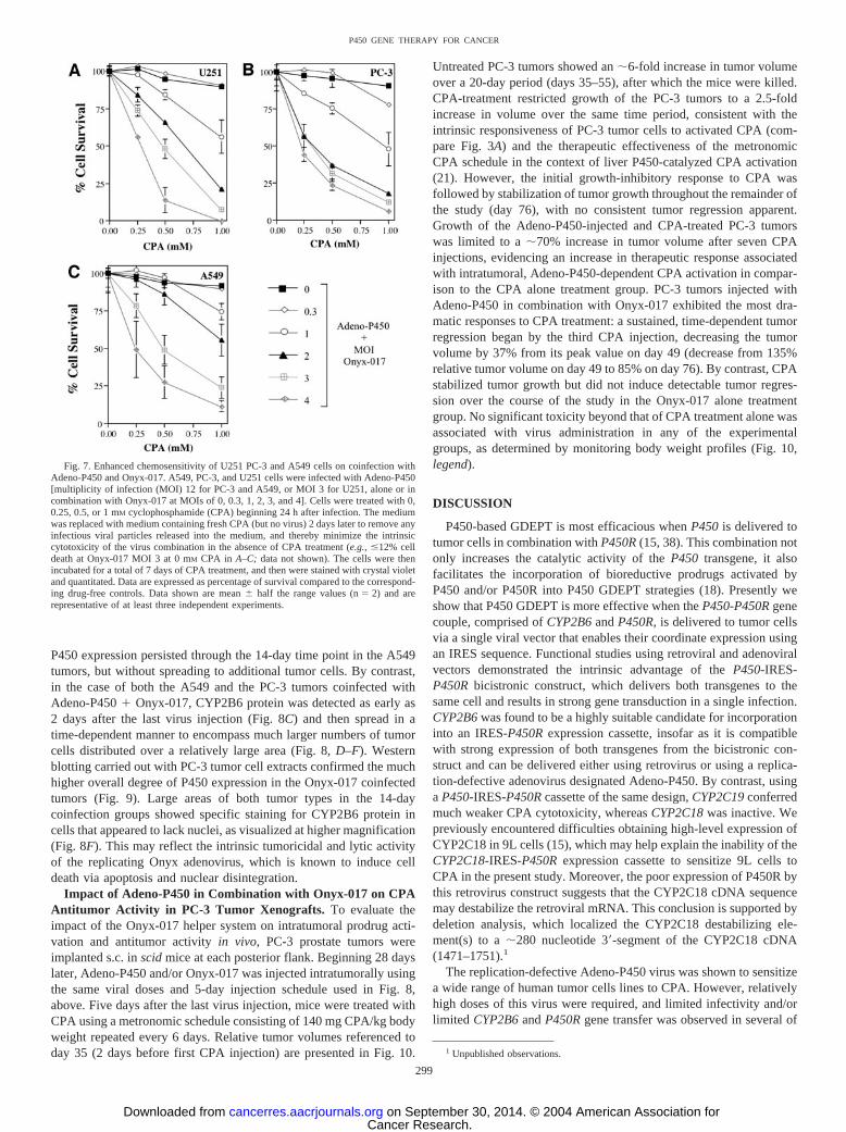
Onyx-017 Enhances Adeno-P450-Mediated CPA Cytotoxicity.To additionally establish the utility of the Onyx-017 helper system inthe context of P450-based GDEPT, cytotoxicity experiments werecarried out using three human tumor cell lines: U251 cells, which areintrinsically sensitive to 4OH-CPA cytotoxicity (Fig. 3A); A549 cells,which are intrinsically resistant to 4OH-CPA (Fig. 3A); and PC-3cells, which are resistant to adenoviral infection (Fig. 2) but intrinsi-cally sensitive to 4OH-CPA (Fig. 3A). Cells were infected withAdeno-P450 (MOI 12 for PC-3 and A549 cells; MOI 3 for U251cells)either alone or in combination with Onyx-017 (MOIs ranging from 0to 4). Cells were treated with CPA beginning 24 h after infection, andcell survival was assayed 7 days later (Fig. 7). In the case of U251cells, which become sensitized to CPA when Adeno-P450 is deliveredat high viral titers (MOIs ranging from 25 to 125; Fig. 3B) but not atlow titers (i.e., MOI 3), Onyx-017 strongly increased Adeno-P450-dependent CPA cytotoxicity (Fig. 7A). A major increase in CPAtoxicity was also seen after coinfection of the Adeno-P450-resistant
PC-3 cells with Onyx-017 (Fig. 7B). A more modest increase in CPAtoxicity was seen after Adeno-P450 � Onyx-017 coinfection of thealdehyde dehydrogenase-rich A549 cells [(Fig. 7C) compare �15%A549 cell kill at 0.5 mM CPA with 12 MOI Adeno-P450 � 2 MOIOnyx-017 versus �70% PC-3 cell kill (Fig. 7B) under the sameconditions], despite the efficient expression of P450 and high cellularCPA 4-hydroxylase activity (compare Fig. 6). These experimentswere carried out under conditions where the intrinsic adenoviraltoxicity was marginal, as judged by the near-absence of an effect ofinfection with Onyx-017 and Adeno-P450 in the absence of CPAtreatment (�12% cell death at Onyx-017 MOI 3 in the experimentsshown in Fig. 7). Thus, the substantial cytotoxicity of CPA seen inFig. 7 is a reflection of bona fide prodrug activation-associated cyto-toxicity, which, in turn, is a direct result of the helper effect ofOnyx-017.
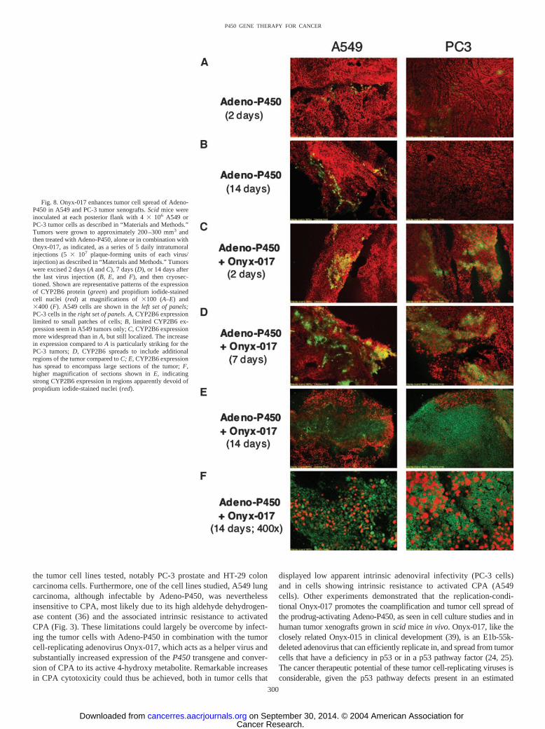
Onyx-017 Enhances Spread of Adeno-P450 in Tumor Xe-nografts in Vivo. We next investigated whether Onyx-017 could beused to enhance the intratumoral spread of Adeno-P450 in A549 andPC-3 xenografts grown s.c. in scid mice. Tumors were grown toapproximately 200–300 mm3 in size, at which time they were injectedwith Adeno-P450 alone, or with Adeno-P450 � Onyx-017 in com-bination, with each virus given as a series of 5 daily intratumoralinjections of 5 � 107 pfu. Tumors were excised 2, 7, or 14 days afterthe last virus injection and analyzed for CYP2B6 immunostainingafter cryosectioning. Fig. 8 shows that the expression of CYP2B6 waslargely restricted to small patches of cells in the Adeno-P450-infectedA549 tumors (Fig. 8, A and B, left). Little or no P450 expression wasdetected in Adeno-P450-infected PC-3 tumors (Fig. 8, A and B, right).
Fig. 6. Assay of Onyx-017 helper virus activity. A, A549 cells were grown and treatedwith Adeno-�Gal alone or in combination with Onyx-017 [0–10 multiplicity of infection(MOI), as indicated] as described in Fig. 5A. Data shown are X-gal (5-bromo-4-chloro-3-indolyl-�-D-galactopyranoside) staining intensities (A620), determined after resuspend-ing the dye in 1 ml DMSO/well of a six-well plate, and expressed as fold-increasecompared to day 1 cells infected with Adeno-�Gal in the absence of Onyx-017. B, A549cells (75,000 cells/well of a six-well plate) were infected for 1 h with Adeno-P450 (MOI25) or with Adeno-P450 (MOI 5) in combination with Onyx-017 (MOIs 0.1, 0.5, 1, 5, and10). 4-Hydroxy-cyclophosphamide production over a 5-h incubation period was assayedin parallel sets of plates analyzed on days 1, 2, 3, and 4, as described in “Materials andMethods.” C, cells treated as in B were washed once with PBS and lysed by sonication inKPi buffer [50 mM KPi buffer, 1 mM EDTA (pH 7.4) containing 20% glycerol]. Celllysates (30 �g protein) were analyzed for CYP2B6 protein by Western blotting, asdescribed in “Materials and Methods.”
298
P450 GENE THERAPY FOR CANCER
Cancer Research. on September 30, 2014. © 2004 American Association forcancerres.aacrjournals.org Downloaded from
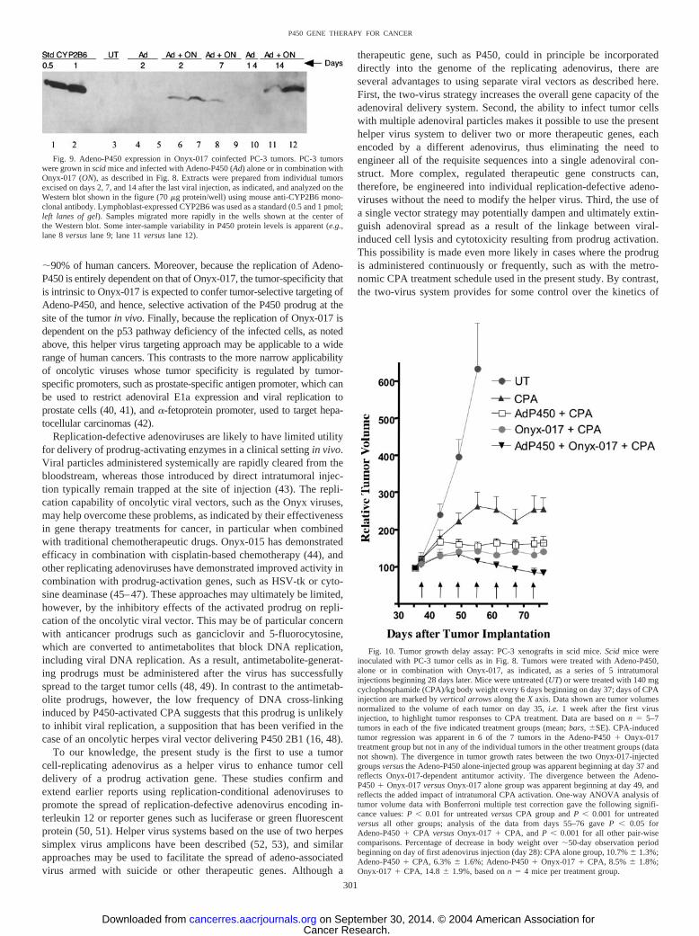
P450 expression persisted through the 14-day time point in the A549tumors, but without spreading to additional tumor cells. By contrast,in the case of both the A549 and the PC-3 tumors coinfected withAdeno-P450 � Onyx-017, CYP2B6 protein was detected as early as2 days after the last virus injection (Fig. 8C) and then spread in atime-dependent manner to encompass much larger numbers of tumorcells distributed over a relatively large area (Fig. 8, D–F). Westernblotting carried out with PC-3 tumor cell extracts confirmed the muchhigher overall degree of P450 expression in the Onyx-017 coinfectedtumors (Fig. 9). Large areas of both tumor types in the 14-daycoinfection groups showed specific staining for CYP2B6 protein incells that appeared to lack nuclei, as visualized at higher magnification(Fig. 8F). This may reflect the intrinsic tumoricidal and lytic activityof the replicating Onyx adenovirus, which is known to induce celldeath via apoptosis and nuclear disintegration.
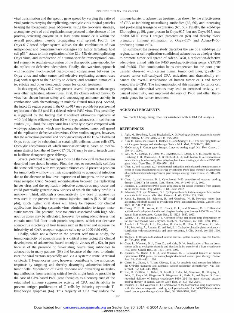
Impact of Adeno-P450 in Combination with Onyx-017 on CPAAntitumor Activity in PC-3 Tumor Xenografts. To evaluate theimpact of the Onyx-017 helper system on intratumoral prodrug acti-vation and antitumor activity in vivo, PC-3 prostate tumors wereimplanted s.c. in scid mice at each posterior flank. Beginning 28 dayslater, Adeno-P450 and/or Onyx-017 was injected intratumorally usingthe same viral doses and 5-day injection schedule used in Fig. 8,above. Five days after the last virus injection, mice were treated withCPA using a metronomic schedule consisting of 140 mg CPA/kg bodyweight repeated every 6 days. Relative tumor volumes referenced today 35 (2 days before first CPA injection) are presented in Fig. 10.
Untreated PC-3 tumors showed an �6-fold increase in tumor volumeover a 20-day period (days 35–55), after which the mice were killed.CPA-treatment restricted growth of the PC-3 tumors to a 2.5-foldincrease in volume over the same time period, consistent with theintrinsic responsiveness of PC-3 tumor cells to activated CPA (com-pare Fig. 3A) and the therapeutic effectiveness of the metronomicCPA schedule in the context of liver P450-catalyzed CPA activation(21). However, the initial growth-inhibitory response to CPA wasfollowed by stabilization of tumor growth throughout the remainder ofthe study (day 76), with no consistent tumor regression apparent.Growth of the Adeno-P450-injected and CPA-treated PC-3 tumorswas limited to a �70% increase in tumor volume after seven CPAinjections, evidencing an increase in therapeutic response associatedwith intratumoral, Adeno-P450-dependent CPA activation in compar-ison to the CPA alone treatment group. PC-3 tumors injected withAdeno-P450 in combination with Onyx-017 exhibited the most dra-matic responses to CPA treatment: a sustained, time-dependent tumorregression began by the third CPA injection, decreasing the tumorvolume by 37% from its peak value on day 49 (decrease from 135%relative tumor volume on day 49 to 85% on day 76). By contrast, CPAstabilized tumor growth but did not induce detectable tumor regres-sion over the course of the study in the Onyx-017 alone treatmentgroup. No significant toxicity beyond that of CPA treatment alone wasassociated with virus administration in any of the experimentalgroups, as determined by monitoring body weight profiles (Fig. 10,legend).
DISCUSSION
P450-based GDEPT is most efficacious when P450 is delivered totumor cells in combination with P450R (15, 38). This combination notonly increases the catalytic activity of the P450 transgene, it alsofacilitates the incorporation of bioreductive prodrugs activated byP450 and/or P450R into P450 GDEPT strategies (18). Presently weshow that P450 GDEPT is more effective when the P450-P450R genecouple, comprised of CYP2B6 and P450R, is delivered to tumor cellsvia a single viral vector that enables their coordinate expression usingan IRES sequence. Functional studies using retroviral and adenoviralvectors demonstrated the intrinsic advantage of the P450-IRES-P450R bicistronic construct, which delivers both transgenes to thesame cell and results in strong gene transduction in a single infection.CYP2B6 was found to be a highly suitable candidate for incorporationinto an IRES-P450R expression cassette, insofar as it is compatiblewith strong expression of both transgenes from the bicistronic con-struct and can be delivered either using retrovirus or using a replica-tion-defective adenovirus designated Adeno-P450. By contrast, usinga P450-IRES-P450R cassette of the same design, CYP2C19 conferredmuch weaker CPA cytotoxicity, whereas CYP2C18 was inactive. Wepreviously encountered difficulties obtaining high-level expression ofCYP2C18 in 9L cells (15), which may help explain the inability of theCYP2C18-IRES-P450R expression cassette to sensitize 9L cells toCPA in the present study. Moreover, the poor expression of P450R bythis retrovirus construct suggests that the CYP2C18 cDNA sequencemay destabilize the retroviral mRNA. This conclusion is supported bydeletion analysis, which localized the CYP2C18 destabilizing ele-ment(s) to a �280 nucleotide 3�-segment of the CYP2C18 cDNA(1471–1751).1
The replication-defective Adeno-P450 virus was shown to sensitizea wide range of human tumor cells lines to CPA. However, relativelyhigh doses of this virus were required, and limited infectivity and/orlimited CYP2B6 and P450R gene transfer was observed in several of
1 Unpublished observations.
Fig. 7. Enhanced chemosensitivity of U251 PC-3 and A549 cells on coinfection withAdeno-P450 and Onyx-017. A549, PC-3, and U251 cells were infected with Adeno-P450[multiplicity of infection (MOI) 12 for PC-3 and A549, or MOI 3 for U251, alone or incombination with Onyx-017 at MOIs of 0, 0.3, 1, 2, 3, and 4]. Cells were treated with 0,0.25, 0.5, or 1 mM cyclophosphamide (CPA) beginning 24 h after infection. The mediumwas replaced with medium containing fresh CPA (but no virus) 2 days later to remove anyinfectious viral particles released into the medium, and thereby minimize the intrinsiccytotoxicity of the virus combination in the absence of CPA treatment (e.g., �12% celldeath at Onyx-017 MOI 3 at 0 mM CPA in A–C; data not shown). The cells were thenincubated for a total of 7 days of CPA treatment, and then were stained with crystal violetand quantitated. Data are expressed as percentage of survival compared to the correspond-ing drug-free controls. Data shown are mean � half the range values (n � 2) and arerepresentative of at least three independent experiments.
299
P450 GENE THERAPY FOR CANCER
Cancer Research. on September 30, 2014. © 2004 American Association forcancerres.aacrjournals.org Downloaded from
the tumor cell lines tested, notably PC-3 prostate and HT-29 coloncarcinoma cells. Furthermore, one of the cell lines studied, A549 lungcarcinoma, although infectable by Adeno-P450, was neverthelessinsensitive to CPA, most likely due to its high aldehyde dehydrogen-ase content (36) and the associated intrinsic resistance to activatedCPA (Fig. 3). These limitations could largely be overcome by infect-ing the tumor cells with Adeno-P450 in combination with the tumorcell-replicating adenovirus Onyx-017, which acts as a helper virus andsubstantially increased expression of the P450 transgene and conver-sion of CPA to its active 4-hydroxy metabolite. Remarkable increasesin CPA cytotoxicity could thus be achieved, both in tumor cells that
displayed low apparent intrinsic adenoviral infectivity (PC-3 cells)and in cells showing intrinsic resistance to activated CPA (A549cells). Other experiments demonstrated that the replication-condi-tional Onyx-017 promotes the coamplification and tumor cell spread ofthe prodrug-activating Adeno-P450, as seen in cell culture studies and inhuman tumor xenografts grown in scid mice in vivo. Onyx-017, like theclosely related Onyx-015 in clinical development (39), is an E1b-55k-deleted adenovirus that can efficiently replicate in, and spread from tumorcells that have a deficiency in p53 or in a p53 pathway factor (24, 25).The cancer therapeutic potential of these tumor cell-replicating viruses isconsiderable, given the p53 pathway defects present in an estimated
Fig. 8. Onyx-017 enhances tumor cell spread of Adeno-P450 in A549 and PC-3 tumor xenografts. Scid mice wereinoculated at each posterior flank with 4 � 106 A549 orPC-3 tumor cells as described in “Materials and Methods.”Tumors were grown to approximately 200–300 mm3 andthen treated with Adeno-P450, alone or in combination withOnyx-017, as indicated, as a series of 5 daily intratumoralinjections (5 � 107 plaque-forming units of each virus/injection) as described in “Materials and Methods.” Tumorswere excised 2 days (A and C), 7 days (D), or 14 days afterthe last virus injection (B, E, and F), and then cryosec-tioned. Shown are representative patterns of the expressionof CYP2B6 protein (green) and propidium iodide-stainedcell nuclei (red) at magnifications of �100 (A–E) and�400 (F). A549 cells are shown in the left set of panels;PC-3 cells in the right set of panels. A, CYP2B6 expressionlimited to small patches of cells; B, limited CYP2B6 ex-pression seem in A549 tumors only; C, CYP2B6 expressionmore widespread than in A, but still localized. The increasein expression compared to A is particularly striking for thePC-3 tumors; D, CYP2B6 spreads to include additionalregions of the tumor compared to C; E, CYP2B6 expressionhas spread to encompass large sections of the tumor; F,higher magnification of sections shown in E, indicatingstrong CYP2B6 expression in regions apparently devoid ofpropidium iodide-stained nuclei (red).
300
P450 GENE THERAPY FOR CANCER
Cancer Research. on September 30, 2014. © 2004 American Association forcancerres.aacrjournals.org Downloaded from
�90% of human cancers. Moreover, because the replication of Adeno-P450 is entirely dependent on that of Onyx-017, the tumor-specificity thatis intrinsic to Onyx-017 is expected to confer tumor-selective targeting ofAdeno-P450, and hence, selective activation of the P450 prodrug at thesite of the tumor in vivo. Finally, because the replication of Onyx-017 isdependent on the p53 pathway deficiency of the infected cells, as notedabove, this helper virus targeting approach may be applicable to a widerange of human cancers. This contrasts to the more narrow applicabilityof oncolytic viruses whose tumor specificity is regulated by tumor-specific promoters, such as prostate-specific antigen promoter, which canbe used to restrict adenoviral E1a expression and viral replication toprostate cells (40, 41), and �-fetoprotein promoter, used to target hepa-tocellular carcinomas (42).
Replication-defective adenoviruses are likely to have limited utilityfor delivery of prodrug-activating enzymes in a clinical setting in vivo.Viral particles administered systemically are rapidly cleared from thebloodstream, whereas those introduced by direct intratumoral injec-tion typically remain trapped at the site of injection (43). The repli-cation capability of oncolytic viral vectors, such as the Onyx viruses,may help overcome these problems, as indicated by their effectivenessin gene therapy treatments for cancer, in particular when combinedwith traditional chemotherapeutic drugs. Onyx-015 has demonstratedefficacy in combination with cisplatin-based chemotherapy (44), andother replicating adenoviruses have demonstrated improved activity incombination with prodrug-activation genes, such as HSV-tk or cyto-sine deaminase (45–47). These approaches may ultimately be limited,however, by the inhibitory effects of the activated prodrug on repli-cation of the oncolytic viral vector. This may be of particular concernwith anticancer prodrugs such as ganciclovir and 5-fluorocytosine,which are converted to antimetabolites that block DNA replication,including viral DNA replication. As a result, antimetabolite-generat-ing prodrugs must be administered after the virus has successfullyspread to the target tumor cells (48, 49). In contrast to the antimetab-olite prodrugs, however, the low frequency of DNA cross-linkinginduced by P450-activated CPA suggests that this prodrug is unlikelyto inhibit viral replication, a supposition that has been verified in thecase of an oncolytic herpes viral vector delivering P450 2B1 (16, 48).
To our knowledge, the present study is the first to use a tumorcell-replicating adenovirus as a helper virus to enhance tumor celldelivery of a prodrug activation gene. These studies confirm andextend earlier reports using replication-conditional adenoviruses topromote the spread of replication-defective adenovirus encoding in-terleukin 12 or reporter genes such as luciferase or green fluorescentprotein (50, 51). Helper virus systems based on the use of two herpessimplex virus amplicons have been described (52, 53), and similarapproaches may be used to facilitate the spread of adeno-associatedvirus armed with suicide or other therapeutic genes. Although a
therapeutic gene, such as P450, could in principle be incorporateddirectly into the genome of the replicating adenovirus, there areseveral advantages to using separate viral vectors as described here.First, the two-virus strategy increases the overall gene capacity of theadenoviral delivery system. Second, the ability to infect tumor cellswith multiple adenoviral particles makes it possible to use the presenthelper virus system to deliver two or more therapeutic genes, eachencoded by a different adenovirus, thus eliminating the need toengineer all of the requisite sequences into a single adenoviral con-struct. More complex, regulated therapeutic gene constructs can,therefore, be engineered into individual replication-defective adeno-viruses without the need to modify the helper virus. Third, the use ofa single vector strategy may potentially dampen and ultimately extin-guish adenoviral spread as a result of the linkage between viral-induced cell lysis and cytotoxicity resulting from prodrug activation.This possibility is made even more likely in cases where the prodrugis administered continuously or frequently, such as with the metro-nomic CPA treatment schedule used in the present study. By contrast,the two-virus system provides for some control over the kinetics of
Fig. 9. Adeno-P450 expression in Onyx-017 coinfected PC-3 tumors. PC-3 tumorswere grown in scid mice and infected with Adeno-P450 (Ad) alone or in combination withOnyx-017 (ON), as described in Fig. 8. Extracts were prepared from individual tumorsexcised on days 2, 7, and 14 after the last viral injection, as indicated, and analyzed on theWestern blot shown in the figure (70 �g protein/well) using mouse anti-CYP2B6 mono-clonal antibody. Lymphoblast-expressed CYP2B6 was used as a standard (0.5 and 1 pmol;left lanes of gel). Samples migrated more rapidly in the wells shown at the center ofthe Western blot. Some inter-sample variability in P450 protein levels is apparent (e.g.,lane 8 versus lane 9; lane 11 versus lane 12).
Fig. 10. Tumor growth delay assay: PC-3 xenografts in scid mice. Scid mice wereinoculated with PC-3 tumor cells as in Fig. 8. Tumors were treated with Adeno-P450,alone or in combination with Onyx-017, as indicated, as a series of 5 intratumoralinjections beginning 28 days later. Mice were untreated (UT) or were treated with 140 mgcyclophosphamide (CPA)/kg body weight every 6 days beginning on day 37; days of CPAinjection are marked by vertical arrows along the X axis. Data shown are tumor volumesnormalized to the volume of each tumor on day 35, i.e. 1 week after the first virusinjection, to highlight tumor responses to CPA treatment. Data are based on n � 5–7tumors in each of the five indicated treatment groups (mean; bars, �SE). CPA-inducedtumor regression was apparent in 6 of the 7 tumors in the Adeno-P450 � Onyx-017treatment group but not in any of the individual tumors in the other treatment groups (datanot shown). The divergence in tumor growth rates between the two Onyx-017-injectedgroups versus the Adeno-P450 alone-injected group was apparent beginning at day 37 andreflects Onyx-017-dependent antitumor activity. The divergence between the Adeno-P450 � Onyx-017 versus Onyx-017 alone group was apparent beginning at day 49, andreflects the added impact of intratumoral CPA activation. One-way ANOVA analysis oftumor volume data with Bonferroni multiple test correction gave the following signifi-cance values: P 0.01 for untreated versus CPA group and P 0.001 for untreatedversus all other groups; analysis of the data from days 55–76 gave P 0.05 forAdeno-P450 � CPA versus Onyx-017 � CPA, and P 0.001 for all other pair-wisecomparisons. Percentage of decrease in body weight over �50-day observation periodbeginning on day of first adenovirus injection (day 28): CPA alone group, 10.7% � 1.3%;Adeno-P450 � CPA, 6.3% � 1.6%; Adeno-P450 � Onyx-017 � CPA, 8.5% � 1.8%;Onyx-017 � CPA, 14.8 � 1.9%, based on n � 4 mice per treatment group.
301
P450 GENE THERAPY FOR CANCER
Cancer Research. on September 30, 2014. © 2004 American Association forcancerres.aacrjournals.org Downloaded from
viral transmission and therapeutic gene spread by varying the ratio ofviral particles carrying the replicating, oncolytic virus to viral particlesbearing the therapeutic gene. Moreover, using the two-virus strategy,a complete cycle of viral replication may proceed in the absence of theprodrug-activating enzyme in at least some tumor cells within theoverall population, thereby propagating viral spread. Fourth, theOnyx-017-based helper system allows for the combination of twoindependent and complementary strategies for tumor targeting, hostcell p53� status to limit replication of the E1b-55k-deleted replicatingOnyx virus, and introduction of a tumor-specific transcriptional con-trol element to regulate expression of the therapeutic gene encoded bythe replication-defective adenovirus. Finally, the two-virus approachwill facilitate much-needed head-to-head comparisons between theOnyx virus and other tumor cell-selective replicating adenoviruses(54) with respect to their ability to deliver, and sensitize tumor cellsto, suicide and other therapeutic genes for cancer treatment.
In this regard, Onyx-017 may present several important advantagesover other replicating adenoviruses. First, the closely related Onyx-015virus has shown human safety and encouraging antitumor activity incombination with chemotherapy in multiple clinical trials (55). Second,the intact E3 region present in the Onyx-017 may provide for preferentialreplication of the E3 (and E1)-deleted Adeno-P450 virus. This possibilityis suggested by the finding that E3-deleted adenovirus replicates at�10-fold higher efficiency than E3 wild-type adenovirus in coinfectionstudies (56). Third, the Onyx virus has a slow lytic cycle compared withwild-type adenovirus, which may increase the desired tumor cell spreadof the replication-defective adenovirus. Other studies suggest, however,that the replication potential and oncolytic activity of the E1b-55k-deletedOnyx virus may be suboptimal in certain p53-deficient tumor cells (57).Oncolytic adenoviruses of which tumor-selectivity is based on mecha-nisms distinct from that of Onyx-017 (58, 59) may be useful in such casesto facilitate therapeutic gene delivery.
Several potential disadvantages to using the two viral vector systemdescribed here should be noted. First, the need to successfully coinfectthe same cell target with two separate viruses could present a problemfor tumor cells with low intrinsic susceptibility to adenoviral infectiondue to the absence or low-level expression of integrins, or the adeno-viral receptor CAR. Second, recombination between the replicatinghelper virus and the replication-defective adenovirus may occur andcould potentially generate new viruses of which the safety profile isunknown. Third, although a comparatively low dose of adenoviruswas used in the present intratumoral injection studies (5 � 108 totalpfu), much higher viral doses will likely be required for clinicalapplications involving systemic virus administration to target meta-static tumors. The potential host toxicities associated with high ade-novirus doses may be alleviated, however, by using adenoviruses thatcontain modified fiber knob protein sequences, which can decreaseadenovirus infectivity of host tissues such as the liver and increase theinfectivity of CAR receptor-negative cells up to 1000-fold (60).
Finally, while not a factor in the present scid mouse study, theimmunogenicity of adenoviruses is a critical issue facing the clinicaldevelopment of adenovirus-based oncolytic viruses (61, 62), in partbecause of the presence of pre-existing neutralizing antibodies toadenovirus in many patients (63) and because of the need to admin-ister the viral vectors repeatedly and via a systemic route. Antiviralcytotoxic T lymphocytes may, however, contribute to the anticancerresponse by targeting and thereby destroying adenovirus-infectedtumor cells. Modulation of T-cell response and preventing neutraliz-ing antibodies from reaching critical levels might both be possible inthe case of CPA-based P450 gene therapies as a consequence of theestablished immune suppressive activity of CPA and its ability toprevent antigen proliferation of T cells by inducing cytotoxic T-lymphocyte apoptosis (64). This property of CPA may reduce the
immune barrier to adenovirus treatment, as shown by the effectivenessof CPA at inhibiting neutralizing antibodies (65, 66), and increasingand prolonging transgene expression (67, 68). Finally, the wild-typeE3b region gp19k gene present in Onyx-017, but not Onyx-015, mayinhibit MHC class I antigen presentation (69) and thereby blockpremature immune elimination of Onyx-017- and Adeno-P450-producing tumor cells.
In summary, the present study describes the use of a wild-type E3region, tumor cell replication-conditional adenovirus as a helper virusto promote tumor cell spread of Adeno-P450, a replication-defectiveadenovirus armed with the P450 prodrug-activating genes CYP2B6and P450R. This combination helps compensate for the poor genetransfer observed with certain human tumor cell lines, greatly in-creases tumor cell-catalyzed CPA activation, and dramatically en-hances the overall sensitization of human tumor cells and tumorxenografts to CPA. The implementation of this strategy for tumor celltargeting of adenoviral vectors may lead to increased activity, en-hanced selectivity, and improved delivery of P450 and other thera-peutic genes for cancer treatment.
ACKNOWLEDGMENTS
We thank Chong-Sheng Chen for assistance with 4OH-CPA analysis.
REFERENCES
1. Aghi, M., Hochberg, F., and Breakefield, X. O. Prodrug activation enzymes in cancergene therapy. J. Gene Med., 2: 148–164, 2000.
2. Kirn, D., Niculescu-Duvaz, I., Hallden, G., and Springer, C. J. The emerging fields ofsuicide gene therapy and virotherapy. Trends Mol. Med., 8: S68–73, 2002.
3. McCormick, F. Cancer gene therapy: fringe or cutting edge? Nat. Rev. Cancer, 1:130–141, 2001.
4. Wei, M. X., Tamiya, T., Chase, M., Boviatsis, E. J., Chang, T. K. H., Kowall, N. W.,Hochberg, F. H., Waxman, D. J., Breakefield, X. O., and Chiocca, E. A. Experimentaltumor therapy in mice using the cyclophosphamide-activating cytochrome P450 2B1gene. Hum. Gene Ther., 5: 969–978, 1994.
5. Chen, L., and Waxman, D. J. Intratumoral activation and enhanced chemotherapeuticeffect of oxazaphosphorines following cytochrome P450 gene transfer: developmentof a combined chemotherapy/cancer gene therapy strategy. Cancer Res., 55: 581–589,1995.
6. Chen, L., and Waxman, D. J. Cytochrome P450 gene-directed enzyme prodrugtherapy (GDEPT) for cancer. Curr. Pharm. Des., 8: 1405–1416, 2002.
7. Jounaidi, Y. Cytochrome P450-based gene therapy for cancer treatment: from conceptto the clinic. Curr. Drug Metab., 3: 609–622, 2002.
8. Schwartz, P. S., and Waxman, D. J. Cyclophosphamide induces caspase 9-dependentapoptosis in 9L tumor cells. Mol. Pharmacol., 60: 1268–1279, 2001.
9. Karle, P., Renner, M., Salmons, B., and Gunzburg, W. H. Necrotic, rather thanapoptotic, cell death caused by cytochrome P450- activated ifosfamide. Cancer GeneTher., 8: 220–230, 2001.
10. Chang, T. K. H., Weber, G. F., Crespi, C. L., and Waxman, D. J. Differentialactivation of cyclophosphamide and ifosphamide by cytochromes P450 2B and 3A inhuman liver microsomes. Cancer Res., 53: 5629–5637, 1993.
11. Weber, G. F., and Waxman, D. J. Activation of the anti-cancer drug ifosphamide byrat liver microsomal P450 enzymes. Biochem. Pharmacol., 45: 1685–1694, 1993.
12. Ayash, L. J., Wright, J. E., Tretyakov, O., Gonin, R., Elias, A., Wheeler, C., Eder,J. P., Rosowsky, A., Antman, K., and Frei, E. I. Cyclophosphamide pharmacokinetics:correlation with cardiac toxicity and tumor response. J. Clin. Oncol., 10: 995–1000,1992.
13. Thigpen, T. Ifosphamide-induced central nervous system toxicity. Gynecol Oncol.,42: 191–192, 1991.
14. Chen, L., Waxman, D. J., Chen, D., and Kufe, D. W. Sensitization of human breastcancer cells to cyclophosphamide and ifosfamide by transfer of a liver cytochromeP450 gene. Cancer Res., 56: 1331–1340, 1996.
15. Jounaidi, Y., Hecht, J. E. D., and Waxman, D. J. Retroviral transfer of humancytochrome P450 genes for oxazaphosphorine-based cancer gene therapy. CancerRes., 58: 4391–4401, 1998.
16. Chase, M., Chung, R. Y., and Chiocca, E. A. An oncolytic viral mutant that deliversthe CYP2B1 transgene and augments cyclophosphamide chemotherapy. Nat. Bio-technol., 16: 444–448, 1998.
17. Kan, O., Griffiths, L., Baban, D., Iqball, S., Uden, M., Spearman, H., Slingsby, J.,Price, T., Esapa, M., Kingsman, S., Kingsman, A., Slade, A., and Naylor, S. Directretroviral delivery of human cytochrome P450 2B6 for gene- directed enzymeprodrug therapy of cancer. Cancer Gene Ther., 8: 473–482, 2001.
18. Jounaidi, Y., and Waxman, D. J. Combination of the bioreductive drug tirapazaminewith the chemotherapeutic prodrug cyclophosphamide for P450/P450-reductase-based cancer gene therapy. Cancer Res., 60: 3761–3769, 2000.
302
P450 GENE THERAPY FOR CANCER
Cancer Research. on September 30, 2014. © 2004 American Association forcancerres.aacrjournals.org Downloaded from
19. McCarthy, H. O., Yakkundi, A., McErlane, V., Hughes, C. M., Keilty, G., Murray,M., Patterson, L. H., Hirst, D. G., McKeown, S. R., and Robson, T. BioreductiveGDEPT using cytochrome P450 3A4 in combination with AQ4N. Cancer Gene Ther.,10: 40–48, 2003.
20. Huang, Z., Raychowdhury, M. K., and Waxman, D. J. Impact of liver P450 reductasesuppression on cyclophosphamide activation, pharmacokinetics and antitumoral ac-tivity in a cytochrome P450-based cancer gene therapy model. Cancer Gene Ther., 7:1034–1042, 2000.
21. Jounaidi, Y., and Waxman, D. J. Frequent, moderate-dose cyclophosphamide admin-istration improves the efficacy of cytochrome P-450/cytochrome P-450 reductase-based cancer gene therapy. Cancer Res., 61: 4437–4444, 2001.
22. Browder, T., Butterfield, C. E., Kraling, B. M., Shi, B., Marshall, B., O’Reilly, M. S.,and Folkman, J. Antiangiogenic scheduling of chemotherapy improves efficacyagainst experimental drug-resistant cancer. Cancer Res., 60: 1878–1886, 2000.
23. Martinez-Salas, E. Internal ribosome entry site biology and its use in expressionvectors. Curr. Opin. Biotechnol., 10: 458–464, 1999.
24. Bischoff, J. R., Kirn, D. H., Williams, A., Heise, C., Horn, S., Muna, M., Ng, L., Nye,J. A., Sampson-Johannes, A., Fattaey, A., and McCormick, F. An adenovirus mutantthat replicates selectively in p53-deficient human tumor cells [see comments]. Sci-ence (Wash. DC), 274: 373–376, 1996.
25. Heise, C., Sampson-Johannes, A., Williams, A., McCormick, F., Von Hoff, D. D., andKirn, D. H. ONYX-015, an E1B gene-attenuated adenovirus, causes tumor-specificcytolysis and antitumoral efficacy that can be augmented by standard chemothera-peutic agents [see comments]. Nat. Med., 3: 639–645, 1997.
26. Yew, P. R., and Berk, A. J. Inhibition of p53 transactivation required for transfor-mation by adenovirus early 1B protein. Nature (Lond.), 357: 82–85, 1992.
27. Morgenstern, J. P., and Land, H. Advanced mammalian gene transfer: high titreretroviral vectors with multiple drug selection markers and a complementary helper-free packaging cell line. Nucleic Acids Res., 18: 3587–3596, 1990.
28. Yamano, S., Aoyama, T., McBride, O. W., Hardwick, J. P., Gelboin, H. V., andGonzalez, F. J. Human NADPH-P450 oxidoreductase: complementary DNA cloning,sequence and vaccinia virus-mediated expression and localization of the CYPORgene to chromosome 7. Mol. Pharmacol., 36: 83–88, 1989.
29. Lang, T., Klein, K., Fischer, J., Nussler, A. K., Neuhaus, P., Hofmann, U., Eichel-baum, M., Schwab, M., and Zanger, U. M. Extensive genetic polymorphism in thehuman CYP2B6 gene with impact on expression and function in human liver.Pharmacogenetics, 11: 399–415, 2001.
30. Pear, W. S., Nolan, G. P., Scott, M. L., and Baltimore, D. Production of high-titerhelper-free retroviruses by transient transfection. Proc. Natl. Acad. Sci. USA, 90:8392–8396, 1993.
31. Finer, M. H., Dull, T. J., Qin, L., Farson, D., and Roberts, M. R. kat: a high-efficiencyretroviral transduction system for primary human T lymphocytes. Blood, 83: 43–50,1994.
32. Monga, M., and Sausville, E. A. Developmental therapeutics program at the NCI:molecular target and drug discovery process. Leukemia (Baltimore), 16: 520–526, 2002.
33. Huang, Z., and Waxman, D. J. HPLC-fluorescent method to determine chloroacetal-dehyde, a neurotoxic metabolite of the anti-cancer drug ifosfamide, in plasma and inliver microsomal incubations. Anal. Biochem., 273: 117–125, 1999.
34. Huang, Z., Roy, P., and Waxman, D. J. Role of human liver microsomal CYP3A4 andCYP2B6 in catalyzing N-dechloroethylation of cyclophosphamide and ifosfamide.Biochem. Pharmacol., 59: 961–972, 2000.
35. Mizuguchi, H., Xu, Z., Ishii-Watabe, A., Uchida, E., and Hayakawa, T. IRES-dependent second gene expression is significantly lower than cap-dependent first geneexpression in a bicistronic vector. Mol. Ther., 1: 376–382, 2000.
36. Sreerama, L., and Sladek, N. E. Class 1 and class 3 aldehyde dehydrogenase levels inthe human tumor cell lines currently used by the National Cancer Institute to screenfor potentially useful antitumor agents. Adv. Exp. Med. Biol., 414: 81–94, 1997.
37. Ries, S., and Korn, W. M. ONYX-015: mechanisms of action and clinical potentialof a replication-selective adenovirus. Br. J. Cancer, 86: 5–11, 2002.
38. Chen, L., Yu, L. J., and Waxman, D. J. Potentiation of cytochrome P450/cyclophos-phamide-based cancer gene therapy by coexpression of the P450 reductase gene.Cancer Res., 57: 4830–4837, 1997.
39. Nemunaitis, J., Khuri, F., Ganly, I., Arseneau, J., Posner, M., Vokes, E., Kuhn, J.,McCarty, T., Landers, S., Blackburn, A., Romel, L., Randlev, B., Kaye, S., and Kirn,D. Phase II trial of intratumoral administration of ONYX-015, a replication-selectiveadenovirus, in patients with refractory head and neck cancer. J. Clin. Oncol., 19:289–298, 2001.
40. Yu, D. C., Chen, Y., Seng, M., Dilley, J., and Henderson, D. R. The addition ofadenovirus type 5 region E3 enables calydon virus 787 to eliminate distant prostatetumor xenografts. Cancer Res., 59: 4200–4203, 1999.
41. Rodriguez, R., Schuur, E. R., Lim, H. Y., Henderson, G. A., Simons, J. W., andHenderson, D. R. Prostate attenuated replication competent adenovirus (ARCA)CN706: a selective cytotoxic for prostate-specific antigen-positive prostate cancercells. Cancer Res., 57: 2559–2563, 1997.
42. Hallenbeck, P. L., Chang, Y. N., Hay, C., Golightly, D., Stewart, D., Lin, J., Phipps, S.,and Chiang, Y. L. A novel tumor-specific replication-restricted adenoviral vector for genetherapy of hepatocellular carcinoma. Hum. Gene Ther., 10: 1721–1733, 1999.
43. Vile, R., Ando, D., and Kirn, D. The oncolytic virotherapy treatment platform forcancer: Unique biological and biosafety points to consider. Cancer Gene Ther., 9:1062–1067, 2002.
44. Heise, C., Lemmon, M., and Kirn, D. Efficacy with a replication-selective adenovirusplus cisplatin-based chemotherapy: dependence on sequencing but not p53 functionalstatus or route of administration. Clin. Cancer Res., 6: 4908–4914, 2000.
45. Wildner, O., Blaese, R. M., and Morris, J. C. Therapy of colon cancer with oncolyticadenovirus is enhanced by the addition of herpes simplex virus-thymidine kinase.Cancer Res., 59: 410–413, 1999.
46. Wildner, O., Morris, J. C., Vahanian, N. N., Ford, H., Jr., Ramsey, W. J., and Blaese,R. M. Adenoviral vectors capable of replication improve the efficacy of HSVtk/GCVsuicide gene therapy of cancer. Gene Ther., 6: 57–62, 1999.
47. Freytag, S. O., Rogulski, K. R., Paielli, D. L., Gilbert, J. D., and Kim, J. H. A novelthree-pronged approach to kill cancer cells selectively: concomitant viral, doublesuicide gene, and radiotherapy. Hum. Gene Ther., 9: 1323–1333, 1998.
48. Pawlik, T. M., Nakamura, H., Yoon, S. S., Mullen, J. T., Chandrasekhar, S., Chiocca,E. A., and Tanabe, K. K. Oncolysis of diffuse hepatocellular carcinoma by intravas-cular administration of a replication-competent, genetically engineered herpesvirus.Cancer Res., 60: 2790–2795, 2000.
49. Lambright, E. S., Amin, K., Wiewrodt, R., Force, S. D., Lanuti, M., Propert, K. J.,Litzky, L., Kaiser, L. R., and Albelda, S. M. Inclusion of the herpes simplexthymidine kinase gene in a replicating adenovirus does not augment antitumorefficacy. Gene Ther., 8: 946–953, 2001.
50. Habib, N. A., Mitry, R., Seth, P., Kuppuswamy, M., Doronin, K., Toth, K., Krajcsi,P., Tollefson, A. E., and Wold, W. S. Adenovirus replication-competent vectors(KD1, KD3) complement the cytotoxicity and transgene expression from replication-defective vectors (Ad-GFP, Ad-Luc). Cancer Gene Ther., 9: 651–654, 2002.
51. Motoi, F., Sunamura, M., Ding, L., Duda, D. G., Yoshida, Y., Zhang, W., Matsuno,S., and Hamada, H. Effective gene therapy for pancreatic cancer by cytokinesmediated by restricted replication-competent adenovirus. Hum. Gene Ther., 11:223–235, 2000.
52. Saeki, Y., Fraefel, C., Ichikawa, T., Breakefield, X. O., and Chiocca, E. A. Improvedhelper virus-free packaging system for HSV amplicon vectors using an ICP27-deleted, oversized HSV-1 DNA in a bacterial artificial chromosome. Mol. Ther., 3:591–601, 2001.
53. Fraefel, C., Jacoby, D. R., and Breakefield, X. O. Herpes simplex virus type 1-basedamplicon vector systems. Adv. Virus Res., 55: 425–451, 2000.
54. Nemunaitis, J., and Edelman, J. Selectively replicating viral vectors. Cancer GeneTher., 9: 987–1000, 2002.
55. Kirn, D. Oncolytic virotherapy for cancer with the adenovirus dl1520 (Onyx-015):results of phase I and II trials. Exp. Opin. Biol. Ther., 1: 525–538, 2001.
56. Berkner, K. L., and Sharp, P. A. Generation of adenovirus by transfection of plasmids.Nucleic Acids Res., 11: 6003–6020, 1983.
57. Dix, B. R., O’Carroll, S. J., Myers, C. J., Edwards, S. J., and Braithwaite, A. W.Efficient induction of cell death by adenoviruses requires binding of E1B55k and p53.Cancer Res., 60: 2666–2672, 2000.
58. Bauerschmitz, G. J., Barker, S. D., and Hemminki, A. Adenoviral gene therapy forcancer: From vectors to targeted and replication competent agents (Review). Int. J.Oncol., 21: 1161–1174, 2002.
59. Yoon, T. K., Shichinohe, T., Laquerre, S., and Kasahara, N. Selectively replicatingadenoviruses for oncolytic therapy. Curr. Cancer Drug Targets, 1: 85–107, 2001.
60. Koizumi, N., Mizuguchi, H., Hosono, T., Ishii-Watabe, A., Uchida, E., Utoguchi, N.,Watanabe, Y., and Hayakawa, T. Efficient gene transfer by fiber-mutant adenoviralvectors containing RGD peptide. Biochim. Biophys. Acta, 1568: 13–20, 2001.
61. Cartmell, T., Southgate, T., Rees, G. S., Castro, M. G., Lowenstein, P. R., andLuheshi, G. N. Interleukin-1 mediates a rapid inflammatory response after injectionof adenoviral vectors into the brain. J. Neurosci., 19: 1517–1523, 1999.
62. Worgall, S., Wolff, G., Falck-Pedersen, E., and Crystal, R. G. Innate immunemechanisms dominate elimination of adenoviral vectors following in vivo adminis-tration. Hum. Gene Ther., 8: 37–44, 1997.
63. Chen, Y., Yu, D. C., Charlton, D., and Henderson, D. R. Pre-existent adenovirusantibody inhibits systemic toxicity and antitumor activity of CN706 in the nudemouse LNCaP xenograft model: implications and proposals for human therapy. Hum.Gene Ther., 11: 1553–1567, 2000.
64. Strauss, G., Osen, W., and Debatin, K. M. Induction of apoptosis and modulation ofactivation and effector function in T cells by immunosuppressive drugs. Clin. Exp.Immunol., 128: 255–266, 2002.
65. Smith, T. A., White, B. D., Gardner, J. M., Kaleko, M., and McClelland, A. Transientimmunosuppression permits successful repetitive intravenous administration of anadenovirus vector. Gene Ther., 3: 496–502, 1996.
66. Christ, M., Lusky, M., Stoeckel, F., Dreyer, D., Dieterle, A., Michou, A. I., Pavirani,A., and Mehtali, M. Gene therapy with recombinant adenovirus vectors: evaluation ofthe host immune response. Immunol. Lett., 57: 19–25, 1997.
67. Jooss, K., Yang, Y., and Wilson, J. M. Cyclophosphamide diminishes inflammationand prolongs transgene expression following delivery of adenoviral vectors to mouseliver and lung. Hum. Gene Ther., 7: 1555–1566, 1996.
68. Bouvet, M., Fang, B., Ekmekcioglu, S., Ji, L., Bucana, C. D., Hamada, K., Grimm,E. A., and Roth, J. A. Suppression of the immune response to an adenovirus vectorand enhancement of intratumoral transgene expression by low-dose etoposide. GeneTher., 5: 189–195, 1998.
69. Bruder, J. T., Jie, T., McVey, D. L., and Kovesdi, I. Expression of gp19K increasesthe persistence of transgene expression from an adenovirus vector in the mouse lungand liver. J. Virol., 71: 7623–7628, 1997.
303
P450 GENE THERAPY FOR CANCER
Cancer Research. on September 30, 2014. © 2004 American Association forcancerres.aacrjournals.org Downloaded from