Ultrasonic tissue characterization of vulnerable carotid plaque: correlation between...
7
BioMed Central Page 1 of 7 (page number not for citation purposes) Cardiovascular Ultrasound Open Access Research Ultrasonic tissue characterization of vulnerable carotid plaque: correlation between videodensitometric method and histological examination Liz Andréa V Baroncini* 1 , Antonio Pazin Filho 1 , Luiz O Murta Junior 2 , Antonio R Martins 3 , Simone G Ramos 4 , Jesualdo Cherri 5 and Carlos E Piccinato 5 Address: 1 Department of Internal Medicine – Faculdade de Medicina de Ribeirão Preto, University of São Paulo, São Paulo, Brazil, 2 Department of Physics and Math – Faculdade de Filosofia, Ciências e Letras de Ribeirão Preto, University of São Paulo, São Paulo, Brazil, 3 Department of Pharmacology – Faculdade de Medicina de Ribeirão Preto, University of São Paulo, São Paulo, Brazil, 4 Department of Pathology – Faculdade de Medicina de Ribeirão Preto, University of São Paulo, São Paulo, Brazil and 5 Department of Surgery and Anatomy – Faculdade de Medicina de Ribeirão Preto, University of São Paulo, São Paulo, Brazil Email: Liz Andréa V Baroncini* - [email protected]; Antonio Pazin Filho - [email protected]; Luiz O Murta Junior - [email protected]; Antonio R Martins - [email protected]; Simone G Ramos - [email protected]; Jesualdo Cherri - [email protected]; Carlos E Piccinato - [email protected] * Corresponding author Abstract Background: To establish the correlation between quantitative analysis based on B-mode ultrasound images of vulnerable carotid plaque and histological examination of the surgically removed plaque, on the basis of a videodensitometric digital texture characterization. Methods: Twenty-five patients (18 males, mean age 67 ± 6.9 years) admitted for carotid endarterectomy for extracranial high-grade internal carotid artery stenosis (≥ 70% luminal narrowing) underwent to quantitative ultrasonic tissue characterization of carotid plaque before surgery. A computer software (Carotid Plaque Analysis Software) was developed to perform the videodensitometric analysis. The patients were divided into 2 groups according to symptomatology (group I, 15 symptomatic patients; and group II, 10 patients asymptomatic). Tissue specimens were analysed for lipid, fibromuscular tissue and calcium. Results: The first order statistic parameter mean gray level was able to distinguish the groups I and II (p = 0.04). The second order parameter energy also was able to distinguish the groups (p = 0,02). A histological correlation showed a tendency of mean gray level to have progressively greater values from specimens with < 50% to >75% of fibrosis. Conclusion: Videodensitometric computer analysis of scan images may be used to identify vulnerable and potentially unstable lipid-rich carotid plaques, which are less echogenic in density than stable or asymptomatic, more densely fibrotic plaques. Published: 17 August 2006 Cardiovascular Ultrasound 2006, 4:32 doi:10.1186/1476-7120-4-32 Received: 25 June 2006 Accepted: 17 August 2006 This article is available from: http://www.cardiovascularultrasound.com/content/4/1/32 © 2006 Baroncini et al; licensee BioMed Central Ltd. This is an Open Access article distributed under the terms of the Creative Commons Attribution License (http://creativecommons.org/licenses/by/2.0 ), which permits unrestricted use, distribution, and reproduction in any medium, provided the original work is properly cited.
-
Upload
independent -
Category
Documents
-
view
0 -
download
0
Transcript of Ultrasonic tissue characterization of vulnerable carotid plaque: correlation between...

BioMed CentralCardiovascular Ultrasound
ss
Open AcceResearchUltrasonic tissue characterization of vulnerable carotid plaque: correlation between videodensitometric method and histological examinationLiz Andréa V Baroncini*1, Antonio Pazin Filho1, Luiz O Murta Junior2, Antonio R Martins3, Simone G Ramos4, Jesualdo Cherri5 and Carlos E Piccinato5Address: 1Department of Internal Medicine – Faculdade de Medicina de Ribeirão Preto, University of São Paulo, São Paulo, Brazil, 2Department of Physics and Math – Faculdade de Filosofia, Ciências e Letras de Ribeirão Preto, University of São Paulo, São Paulo, Brazil, 3Department of Pharmacology – Faculdade de Medicina de Ribeirão Preto, University of São Paulo, São Paulo, Brazil, 4Department of Pathology – Faculdade de Medicina de Ribeirão Preto, University of São Paulo, São Paulo, Brazil and 5Department of Surgery and Anatomy – Faculdade de Medicina de Ribeirão Preto, University of São Paulo, São Paulo, Brazil
Email: Liz Andréa V Baroncini* - [email protected]; Antonio Pazin Filho - [email protected]; Luiz O Murta Junior - [email protected]; Antonio R Martins - [email protected]; Simone G Ramos - [email protected]; Jesualdo Cherri - [email protected]; Carlos E Piccinato - [email protected]
* Corresponding author
AbstractBackground: To establish the correlation between quantitative analysis based on B-modeultrasound images of vulnerable carotid plaque and histological examination of the surgicallyremoved plaque, on the basis of a videodensitometric digital texture characterization.
Methods: Twenty-five patients (18 males, mean age 67 ± 6.9 years) admitted for carotidendarterectomy for extracranial high-grade internal carotid artery stenosis (≥ 70% luminalnarrowing) underwent to quantitative ultrasonic tissue characterization of carotid plaque beforesurgery. A computer software (Carotid Plaque Analysis Software) was developed to perform thevideodensitometric analysis. The patients were divided into 2 groups according to symptomatology(group I, 15 symptomatic patients; and group II, 10 patients asymptomatic). Tissue specimens wereanalysed for lipid, fibromuscular tissue and calcium.
Results: The first order statistic parameter mean gray level was able to distinguish the groups Iand II (p = 0.04). The second order parameter energy also was able to distinguish the groups (p =0,02). A histological correlation showed a tendency of mean gray level to have progressively greatervalues from specimens with < 50% to >75% of fibrosis.
Conclusion: Videodensitometric computer analysis of scan images may be used to identifyvulnerable and potentially unstable lipid-rich carotid plaques, which are less echogenic in densitythan stable or asymptomatic, more densely fibrotic plaques.
Published: 17 August 2006
Cardiovascular Ultrasound 2006, 4:32 doi:10.1186/1476-7120-4-32
Received: 25 June 2006Accepted: 17 August 2006
This article is available from: http://www.cardiovascularultrasound.com/content/4/1/32
© 2006 Baroncini et al; licensee BioMed Central Ltd.This is an Open Access article distributed under the terms of the Creative Commons Attribution License (http://creativecommons.org/licenses/by/2.0), which permits unrestricted use, distribution, and reproduction in any medium, provided the original work is properly cited.
Page 1 of 7(page number not for citation purposes)

Cardiovascular Ultrasound 2006, 4:32 http://www.cardiovascularultrasound.com/content/4/1/32
BackgroundCarotid artery atherosclerosis is responsible for 20% to30% of ischemic strokes. Several large randomized multi-center trials [1-11] have demonstrated the benefit ofcarotid endarterectomy (CEA) and recently with carotidartery stenting (CAS) [11-13] in the prevention of stroke,in both symptomatic and asymptomatic disease. In thesestudies, the degree of internal carotid artery stenosis wasthe only criterion for selection of patients at high risk forstroke. However, these trials also noted that most patientswith high-grade stenosis (>70%) remained stroke freeeven with medical therapy alone [3]. Factors in additionto the degree of stenosis, such as the histological compo-sition of the plaque, may be responsible for the determi-nation of stroke risk. The composition of plaques frompatients with symptoms is significantly different from thatof plaques from those without [14-23]. The former con-tain more total lipid and cholesterol, and less collagenand calcium. Plaque echogenicity as assessed by B-modeultrasound has been found to reliably predict the contentof soft tissue and the amount of calcification in carotidplaques. Fibrous plaques have a highly echogenic qualityand the presence of calcium provides a markedly hypere-choic image with shadowing formation. As the lipid con-tent of the plaque increases, the plaque becomes moreecholucent [14,24,25]. Nevertheless, the subjective visualanalysis of echogenicity provides only a qualitative classi-fication, which can be difficult to reproduce [26]. Thepresent study was designed to establish the correlationbetween quantitative analysis of ultrasound B-modeimages of vulnerable carotid plaque on the basis of a vid-eodensitometric digital texture characterization. and his-tological examination of the surgically removed plaque.
MethodsA. PatientsThirty-six nonconsecutive surgical inpatients admitted forcarotid endarterectomy for extracranial high-grade (≥70%) internal carotid artery stenosis were entered intothis study between February 2003 and July 2005 from 3participating hospitals. Local ethical committee approvalwas obtained for the study and procurement of speci-mens. Written informed consent was obtained from allpatients before each examination. Exclusion criteria were:a disorder that could seriously complicate surgery (3patients); terminal cancer (1 patient); patient refusal ofoperation (1 patient); suboptimal ultrasonographic visu-alization of the atherosclerotic plaque contour/border (1patient); and surgical specimen inadequate to histologicaland immunocytochemical analysis (5 patients). The studywas conducted on 25 common or internal carotid arteryplaques from the 25 remaining patients (18 men and 7women; mean age 67 ± 6.9 years). A clinical examination,including neurological exam, with particular care taken toestablish the number and duration of ischemic events,
and a record of the time from the last symptom and theoperation, was obtained from each patient. Before sur-gery, all patients underwent a: 1 – either cerebral angiog-raphy or magnetic resonance angiography and Duplexultrasound for grading carotid artery stenosis and assess-ment of intracranial arterial system; and 2 – either compu-ter tomography (CT) or magnetic resonance brain scan.The presence or absence of infarction in the correspond-ing middle cerebral artery territory was noted. Focal cere-bral ischemic events were defined as transient ischemicattack (TIA), amaurosis fugax (AF), central retinal arteryocclusion (CRAO), or cerebrovascular accident. Patientswere considered to be symptomatic if they had experi-enced AF, TIA or stroke ipsilateral to the carotid lesionbeing studied. Silent infarcts and lacunar symptomatol-ogy, diagnosed by a neurologist based on clinical andbrain computer tomography (CT) scan and/or magneticresonance imaging (MRI) located ipsilateral to the steno-sis, were also considered symptomatic. On the otherhand, patients without any history of recent neurologicsymptoms or with nonspecific, nonhemispheric symp-toms such as dizziness and vertigo were consideredasymptomatic. Each patient was then assigned preopera-tively to 1 of 2 groups on the basis of their symptoms:group I symptomatic patients (n = 15; mean age 67.4 ± 6.4years) and group II (n = 10; mean age 65.2 ± 7.9 years)consisting of all asymptomatic patients. At the baselineexamination, measurements of height, weight, body massindex, blood pressure, fasting serum total cholesterol,HDL cholesterol, LDL cholesterol, triglycerides, fastingplasma glucose, electrocardiograms and informationabout coronary artery disease, diabetes mellitus andsmoking habits was collected. Percentages of carotiddiameter reduction, procedural methods, concomitanttherapy, age, sex, and risk factors did not differ betweenthe 2 groups (Table 1).
Table 1: Patient's Characteristics
Group I (n = 15) Group II (n = 10)
Age, years 67.4 ± 6.4 65.2 ± 7.9Sex, M/F 11/4 7/3Hypertension 10 1Diabetes mellitus 2 3Active Smoking 3 3Hypercholesterolemia 2 1CAD 4 0Aspirin 15 10Statin 5 4ACE inhibitors 9 8Ticlopidine 4 1
CAD = coronary artery disease
Page 2 of 7(page number not for citation purposes)

Cardiovascular Ultrasound 2006, 4:32 http://www.cardiovascularultrasound.com/content/4/1/32
B. Ultrasonographic image acquisition and preprocessingThe patients underwent carotid endarterectomy 1 to 2days after ultrasound assessment. Conventional echoimages were acquired with a commercially available 2Dultrasonic imaging system (Hewlett-Packard Sonos 5500,Andover, Massachusetts). The system characterized arte-rial tissue at the bedside using a 5- to 12- MHz multifre-quency linear transducer for all studies. This softwareenables the acquisition; storing and retrieving of asequence of continuous 2D conventional images, forminga continuous loop digital recording of 2 s (60 frames in 2s). Anterior, lateral and posterior projections were used toimage the plaque longitudinally. The position of theprobe was adjusted so that the ultrasonic beam was verti-cal to the artery wall. Offline analysis of the 2D imageswas performed by retrieving the previously stored datafrom the built-in optical disc drive in the system. For vid-eodensitometric analysis, the images from the magneticaloptical disk were loaded into a computer where a specificsoftware program (CaPAS – Carotid Plaque Analysis Soft-ware) was designed. Selection was done, such that plaquecontour/border, area, and contrast were optimized, sub-jectively judged. Only the image plaque at anterior vesselwall was analysed. The selected static frames consideredappropriate for analysis, should fulfil these criteria: 1) theblood, in the vicinity of the plaques, was dark and echoi-cally uniform, and 2) the atherosclerotic plaque was welldelineated, horizontal, and with maximum thickness.
C. Quantitative texture analysisAll plaque images were evaluated by the software CaPASfor texture parameters including a set of first-order (meangray level; and standard deviation) and of second-order(entropy, energy, and homogeneity) parameters. Themean gray level (MGL) represents the median of the fre-quency distribution of gray tones of the pixels included inthe region of interest (gray scale median of the region) ina scale of 256 gray tones (0 = darkest tone; 255 = brightesttone) [25]. Dark (hypoechoic) regions were associatedwith a gray scale median (GSM) that tends to approach 0,whereas bright (hyperechoic) regions were associatedwith a GSM that tended to approach 255. The standarddeviation (SD) is an expression of the spreading of the dis-tribution from the mean value, i.e., the overall contrast.The energy or angular second-moment value increaseswhen the co-occurrence matrix elements are very unequal.Entropy and homogeneity reflects the coarseness of theimage, as its value increases when homogeneity isreduced, i.e., when co-occurrence matrix elements tend tobe equal and the diagonal concentration lowers. Themathematical definitions of these texture parameters aredescribed in previous articles [27]. All plaque images werenormalized by using two echo-anatomic points: the grayscale median (GSM) of the blood and the GSM of the peri-adventitia region. After normalization, each image plaque
was outlined manually three times by the same examinerin its longitudinal section. Mean score of these threesequential measurements was used as a final value.
D. Procurement of tissue specimens and histological analysisCarotid plaques were obtained immediately after endar-terectomy. All surgeries were performed with standardsurgical techniques, and with minimal manipulation ofthe specimen. No attempts were made to evaluate thepresence and the degree of surface ulceration or throm-bus. The plaque should be removed in bloc, without frag-mentation or significant distortion. After removal, thesection of plaque for histological analysis was placed infresh 4% paraformaldehyde solution and partly decalci-fied overnight, in order to be sectioned subsequently. Thesamples were transected transversely at 3 to 4 mm, andembedded in paraffin. For the most of the specimens, fiveto six blocks were avaiable. Histological analysis was per-formed by an experience pathologist (SGR) who was una-ware to the ultrasound results. Tissue specimens wereanalysed for lipid, fibromuscular tissue and calcium andexpressed as the percentage of the total plaque areaobtained.
Statistical analysisCategorical variables were expressed as percentages andcontinuous variables were expressed as mean ± SD(median). The comparison of the histological and vide-odensitometric parameters among the groups was doneby non-parametric test of Wilcoxon rank-sum test or Chi-square test as appropriate and the correlation between his-tological and videodensitometric data was done by thenon-parametric Spearman test. Statistical significance wasindicated by a value of P < 0.05. The intra and inter-exam-iner variability in ultrasonographic measurements wastested in all carotid images as proposed by Lin [28].
ResultsA. Histological examinationTissue specimens were analysed for lipid, fibromusculartissue and calcium. The percentage of these three tissuecomponents was determined in each plaque section. Thepercentage of fibromuscular tissue had higher prevalencein the group II (75,86 ± 11,46) and lower prevalence inthe group I (51,87 ± 13,01) with statistic significance (p =0.04). The percent of lipid tissue had the opposite results(40,10 ± 15,80 for group I and 19,57 ± 9,96 for group II;p = 0,05). The percentage of calcium did not differbetween the two groups (Table 2).
B. Videodensitometric analysisAmong first-order parameters, the MGL was effective indistinguishing group I versus group II (p = 0.04), showingsignificant lower values in group I. The standard deviation
Page 3 of 7(page number not for citation purposes)
Cardiovascular Ultrasound 2006, 4:32 http://www.cardiovascularultrasound.com/content/4/1/32
had the same behaviour but without statistic significance.Among second order parameters, energy distinguishedgroups I and II with lower values in group I (p = 0.02).Homogeneity and entropy did not find any significant dif-ference (Table 2).
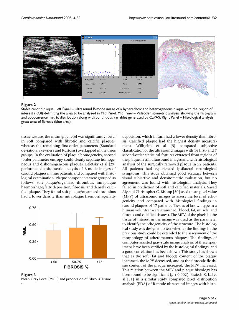
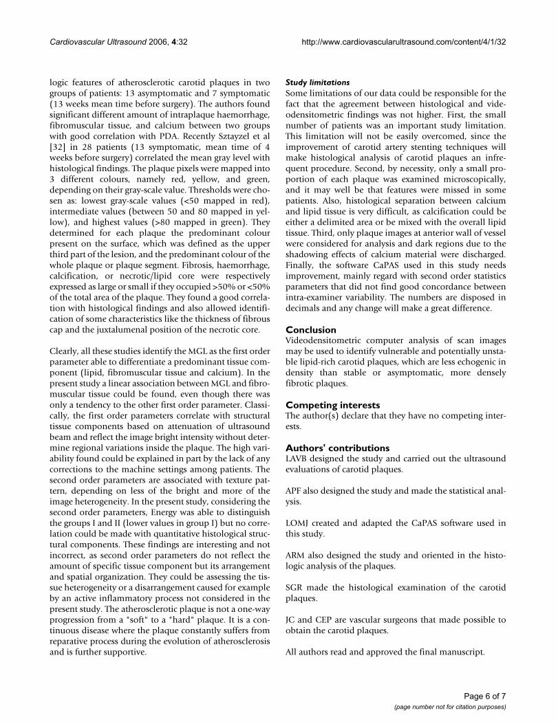
C. Correlation between histological examination and videodensitometric analysisThere was no statistical significant correlation between thehistological examination and videodensitometric param-eters. However, considering MGL and proportion offibrous tissue, progressively greater values was found fromspecimens with < 50% to >75% of fibrosis. Figures 1 and2 show examples of ultrasound B-mode images, CaPASparameters and histological analysis. Figure 3 shows MGLvalues related to proportion of fibrous tissue.
D. Intra and inter-examiner variabilityThe inter-examiner variability acquired good agreementbetween the measurements. The intra-examiner variabil-
ity found concordance for first order videodensitometricparameters and for entropy, but not for energy and homo-geneity. The mean inter-examiner variability ranged: forMGL from 0,1607 to 0,9622; for SD from 0,3236 to0,9809; for entropy from 0,5406 to 0,9459; for energyfrom 0,1811 to 0,6981; and for homogeneity from 0,3244to 0,9049. The mean intra-examiner variability ranged: forMGL from 0,4280 to 0,8150; for SD from 0,3958 to0,7996; for entropy from 0,1085 to 0,6141; for energyfrom - 0,0342 to 0,4546; and for homogeneity from -0,1181 to 0,2073.
DiscussionComparison with previous studiesMazzone et al [27] selected 47 images of carotid plaquesfrom 10 patients and correlated visual assessment ofplaque echodensity and homogeneity with the resultsobtained by mathematical descriptors of tissue texture.The plaques were first assigned as soft, fibrotic and calcificaccording visual approach. In the first-order parameters of
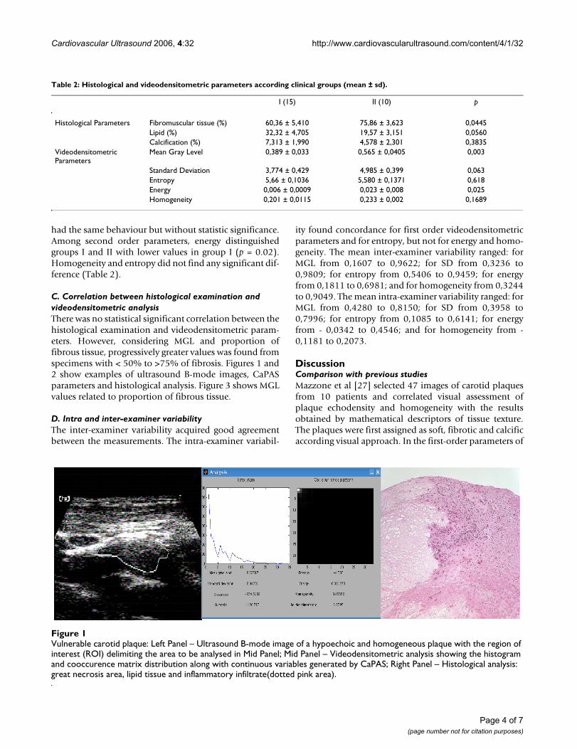
Vulnerable carotid plaqueFigure 1Vulnerable carotid plaque: Left Panel – Ultrasound B-mode image of a hypoechoic and homogeneous plaque with the region of interest (ROI) delimiting the area to be analysed in Mid Panel; Mid Panel – Videodensitometric analysis showing the histogram and cooccurence matrix distribution along with continuous variables generated by CaPAS; Right Panel – Histological analysis: great necrosis area, lipid tissue and inflammatory infiltrate(dotted pink area).
Table 2: Histological and videodensitometric parameters according clinical groups (mean ± sd).
I (15) II (10) p
Histological Parameters Fibromuscular tissue (%) 60,36 ± 5,410 75,86 ± 3,623 0,0445Lipid (%) 32,32 ± 4,705 19,57 ± 3,151 0,0560Calcification (%) 7,313 ± 1,990 4,578 ± 2,301 0,3835
Videodensitometric Parameters
Mean Gray Level 0,389 ± 0,033 0,565 ± 0,0405 0,003
Standard Deviation 3,774 ± 0,429 4,985 ± 0,399 0,063Entropy 5,66 ± 0,1036 5,580 ± 0,1371 0,618Energy 0,006 ± 0,0009 0,023 ± 0,008 0,025Homogeneity 0,201 ± 0,0115 0,233 ± 0,002 0,1689
Page 4 of 7(page number not for citation purposes)
Cardiovascular Ultrasound 2006, 4:32 http://www.cardiovascularultrasound.com/content/4/1/32
tissue texture, the mean gray-level was significantly lowerin soft compared with fibrotic and calcific plaques,whereas the remaining first-order parameters (Standarddeviation, Skewness and Kurtosis) overlapped in the threegroups. In the evaluation of plaque homogeneity, second-order parameter entropy could clearly separate homoge-neous and dishomogeneous plaques. Beletsky et al [29]performed densitometric analysis of B-mode images ofcarotid plaques in nine patients and compared with histo-logical examination. Plaque components were grouped asfollows: soft plaque/organized thrombus, intraplaquehaemorrhage/fatty deposition, fibrosis, and densely calci-fied plaque. They found soft plaque/organized thrombushad a lower density than intraplaque haemorrhage/fatty
deposition, which in turn had a lower density than fibro-sis. Calcified plaque had the highest density measure-ment. Wilhjelm et al [5] compared subjectiveclassification of the ultrasound images with 16 first- and 7second-order statistical features extracted from regions ofthe plaque in still ultrasound images and with histologicalanalysis of the surgically removed plaque in 52 patients.All patients had experienced ipsilateral neurologicalsymptoms. This study obtained good accuracy betweenvisual subjective and densitometric evaluation, but noagreement was found with histological analysis. Theyfailed in prediction of soft and calcified materials. SayedAly and Christopher C. Bishop [30] used mean pixel value(MPV) of ultrasound images to assess the level of echo-genicity and compared with histological findings incarotid plaques of 17 patients. Tissues of known type in ahuman volunteer were examined (blood, fat, muscle, andfibrous and calcified tissues). The MPV of the pixels in thetissue of interest in the image was used as the parameterto identify the echogenicity of the structure. The histolog-ical study was designed to test whether the findings in theprevious study could be extended to the assessment of themorphology of atheromatous plaques. The findings ofcomputer-assisted gray-scale image analysis of these spec-imens have been verified by the histological findings, anda good correlation has been shown. This study has shownthat as the soft (fat and blood) content of the plaqueincreased, the MPV decreased, and as the fibrocalcific tis-sue content of the plaque increased, the MPV increased.This relation between the MPV and plaque histology hasbeen found to be significant (p < 0.002). Brajesh K. Lal etal [31] in a similar study compared pixel distributionanalysis (PDA) of B-mode ultrasound images with histo-
Mean Gray Level (MGL) and proportion of Fibrous TissueFigure 3Mean Gray Level (MGL) and proportion of Fibrous Tissue.
< 50 50-75 >75
0.00
0.25
0.50
0.75
FIBROSIS %
MG
L
Stable carotid plaqueFigure 2Stable carotid plaque: Left Panel – Ultrasound B-mode image of a hyperechoic and heterogeneous plaque with the region of interest (ROI) delimiting the area to be analysed in Mid Panel; Mid Panel – Videodensitometric analysis showing the histogram and cooccurence matrix distribution along with continuous variables generated by CaPAS; Right Panel – Histological analysis: great area of fibrosis (blue area).
Page 5 of 7(page number not for citation purposes)
Cardiovascular Ultrasound 2006, 4:32 http://www.cardiovascularultrasound.com/content/4/1/32
logic features of atherosclerotic carotid plaques in twogroups of patients: 13 asymptomatic and 7 symptomatic(13 weeks mean time before surgery). The authors foundsignificant different amount of intraplaque haemorrhage,fibromuscular tissue, and calcium between two groupswith good correlation with PDA. Recently Sztayzel et al[32] in 28 patients (13 symptomatic, mean time of 4weeks before surgery) correlated the mean gray level withhistological findings. The plaque pixels were mapped into3 different colours, namely red, yellow, and green,depending on their gray-scale value. Thresholds were cho-sen as: lowest gray-scale values (<50 mapped in red),intermediate values (between 50 and 80 mapped in yel-low), and highest values (>80 mapped in green). Theydetermined for each plaque the predominant colourpresent on the surface, which was defined as the upperthird part of the lesion, and the predominant colour of thewhole plaque or plaque segment. Fibrosis, haemorrhage,calcification, or necrotic/lipid core were respectivelyexpressed as large or small if they occupied >50% or <50%of the total area of the plaque. They found a good correla-tion with histological findings and also allowed identifi-cation of some characteristics like the thickness of fibrouscap and the juxtalumenal position of the necrotic core.
Clearly, all these studies identify the MGL as the first orderparameter able to differentiate a predominant tissue com-ponent (lipid, fibromuscular tissue and calcium). In thepresent study a linear association between MGL and fibro-muscular tissue could be found, even though there wasonly a tendency to the other first order parameter. Classi-cally, the first order parameters correlate with structuraltissue components based on attenuation of ultrasoundbeam and reflect the image bright intensity without deter-mine regional variations inside the plaque. The high vari-ability found could be explained in part by the lack of anycorrections to the machine settings among patients. Thesecond order parameters are associated with texture pat-tern, depending on less of the bright and more of theimage heterogeneity. In the present study, considering thesecond order parameters, Energy was able to distinguishthe groups I and II (lower values in group I) but no corre-lation could be made with quantitative histological struc-tural components. These findings are interesting and notincorrect, as second order parameters do not reflect theamount of specific tissue component but its arrangementand spatial organization. They could be assessing the tis-sue heterogeneity or a disarrangement caused for exampleby an active inflammatory process not considered in thepresent study. The atherosclerotic plaque is not a one-wayprogression from a "soft" to a "hard" plaque. It is a con-tinuous disease where the plaque constantly suffers fromreparative process during the evolution of atherosclerosisand is further supportive.
Study limitationsSome limitations of our data could be responsible for thefact that the agreement between histological and vide-odensitometric findings was not higher. First, the smallnumber of patients was an important study limitation.This limitation will not be easily overcomed, since theimprovement of carotid artery stenting techniques willmake histological analysis of carotid plaques an infre-quent procedure. Second, by necessity, only a small pro-portion of each plaque was examined microscopically,and it may well be that features were missed in somepatients. Also, histological separation between calciumand lipid tissue is very difficult, as calcification could beeither a delimited area or be mixed with the overall lipidtissue. Third, only plaque images at anterior wall of vesselwere considered for analysis and dark regions due to theshadowing effects of calcium material were discharged.Finally, the software CaPAS used in this study needsimprovement, mainly regard with second order statisticsparameters that did not find good concordance betweenintra-examiner variability. The numbers are disposed indecimals and any change will make a great difference.
ConclusionVideodensitometric computer analysis of scan imagesmay be used to identify vulnerable and potentially unsta-ble lipid-rich carotid plaques, which are less echogenic indensity than stable or asymptomatic, more denselyfibrotic plaques.
Competing interestsThe author(s) declare that they have no competing inter-ests.
Authors' contributionsLAVB designed the study and carried out the ultrasoundevaluations of carotid plaques.
APF also designed the study and made the statistical anal-ysis.
LOMJ created and adapted the CaPAS software used inthis study.
ARM also designed the study and oriented in the histo-logic analysis of the plaques.
SGR made the histological examination of the carotidplaques.
JC and CEP are vascular surgeons that made possible toobtain the carotid plaques.
All authors read and approved the final manuscript.
Page 6 of 7(page number not for citation purposes)

Cardiovascular Ultrasound 2006, 4:32 http://www.cardiovascularultrasound.com/content/4/1/32
AcknowledgementsThe authors thank Elaine Medeiros Floriano and Ana Maria Anselmi Dori-gan for their excellent technical assistance.
References1. Barnett HJM, Taylor DW, Eliasziw M, Fox AJ, Ferguson GG, Haynes
RB, Rankin RN, Clagett GP, Hachinski VC, Sackett DL, Thorpe KE,Math M, Meldrum HE: Benefit of carotid endarterectomy inpatients with symptomatic moderate or severe stenosis. NEngl J Med 1998, 339:1415-1425.
2. North American Symptomatic Carotid Endarterectomy Trial collabo-rators: Beneficial effect of carotid endarterectomy in sympto-matic patients with high – grade carotid stenosis. N Engl J Med1991, 325:445-453.
3. Executive Committee for the Asymptomatic Carotid AtherosclerosisStudy: Endarterectomy for asymptomatic carotid artery ste-nosis. JAMA 1995, 273:1421-1428.
4. Barnett HJM, Meldrum HE, Eliasziw M: The dilemma of surgicaltreatment for patients with asymptomatic carotid disease.Ann Intern Med 1995, 123:723-725.
5. Wilhjelm JE, Grønholdt MLM, Wiebe B, Jespersen SK, Hansen LK,Sillesen H: Quantitative analysis of ultrasound B-mode imagesof carotid atherosclerotic plaque: correlation with visualclassification and histological examination. IEEE Trans MedImag 1998, 17:910-922.
6. North American Symptomatic Carotid Endarterectomy Trial (NAS-CET) Steering Committee: North American SymptomaticCarotid Endarterectomy Trial: methods, patient character-istics, and progress. Stroke 1991, 22:711-720.
7. Paciaroni M, Eliasziw M, Sharpe BL, Kappelle LJ, Chaturvedi S, Mel-drum H, Barnett HJM, the North American Symptomatic CarotidEndarterectomy Trial (NASCET) Group: Long-Term clinical andangiographic outcomes in symptomatic patients with 70% to90% carotid artery stenosis. Stroke 2000, 31:2037-2042.
8. Nadareishvili ZG, Rothwell PM, Beletsky V, Pagniello A, Norris JW:Long-term risk of stroke and other vascular events inpatients with asymptomatic carotid artery stenosis. Arch Neu-rol 2002, 59:1162-1166.
9. Rothwell PM, Eliasziw M, Gutnikov SA, Phil D, Warlow CP, BarnettHJM: Sex difference in the effect of time from symptoms tosurgery on benefit from carotid endarterectomy for tran-sient ischemic attack and nondisabling stroke. Stroke 2004,35:2855-2861.
10. Rothwell PM, Gutnikov SA, Warlow CP: Reanalysis of the finalresults of the European Carotid Surgery Trial. Stroke 2003,34:514-523.
11. Roubin GS, New G, Iyer SS, Vitek JJ, Al-Mubarak N, Liu MW, Yadav J,Gomez C, Kuntz RE: Immediate and late clinical outcomes ofcarotid artery stenting in patients with symptomatic andasymptomatic carotid artery stenosis. A 5-year prospectiveanalysis. Circulation 2001, 103:532-537.
12. Biasi GM, Froio A, Diethrich EB, Deleo G, Galimberti S, Mingazzini P,Nicolaides AN, Griffin M, Raithel D, Reid DB, Valsecchi MG: Carotidplaque echolucency increases the risk of stroke in carotidstenting. The imaging in carotid angioplasty and risk ofstroke (ICAROS) study. Circulation 2004, 110:756-762.
13. Yadav JS, Wholey MH, Kuntz RE, Fayad P, Katzen BT, Mishkel GJ,Bajwa TK, Whitlow P, Strickman NE, Jaff MR, Popma JJ, Snead DB,Cutlip DE, Firth BG, Ouriel K: Protected carotid-artery stentingversus endarterectomy in high-risk patients. N Eng J Med 2004,351:1493-501.
14. Geroulakos G, Ramaswami G, Nicolaides A, James K, Labropoulos N,Belcaro G, Holloway M: Characterization of symptomatic andasymptomatic carotid plaques using high-resolution real-time ultrasonography. Br J Surg 1993, 80:1274-1277.
15. Cipollone F, Prontera C, Pini B, Marini M, Fazia M, De Cesare D, IezziA, Ucchino S, Boccoli G, Saba V, Chiarelli F, Cuccurullo F, Mezzetti A:Overexpression of functionally coupled cyclooxygenase-2and prostaglandin E synthase in symptomatic atheroscle-rotic plaques as a basis of prostaglandin E(2)-dependentplaque instability. Circulation 2001, 104:921-27.
16. Gray-Weale AC, Graham JC, Burnett JR, Byrne K, Lusby RJ: Carotidartery atheroma: comparison of preoperative B-mode ultra-sound appearance with carotid endarterectomy specimenpathology. J Cardiovasc Surg 1988, 29:676-681.
17. Nandalur KR, Baskurt E, Hagspiel KD, Phillips CD, Kramer CM: Cal-cified carotid atherosclerotic plaque is associated less withischemic symptoms than is noncalcified plaque on MDCT.AJR 2005, 184:295-298.
18. Mathiesen EB, Bonaa KH, Joakimsen O: Echolucent plaques areassociated with high risk of ischemic cerebrovascular eventsin carotid stenosis: the tromso study. Circulation 2001,103:2171-2175.
19. Gronholdt MLM, Nordestgaard BG, Schroeder TV, Vorstrup S,Sillesen H: Ultrasonic echolucent carotid plaques predictfuture strokes. Circulatio 2001, 104:68-73.
20. Tegos TJ, Stavropoulos P, Sabetai MM, Khodabakhsh P, Sassano A,Nicolaides AN: Determinants of carotid plaque instability:echoicity versus heterogeneity. Eur J Vasc Endovasc Surg 2001,22:22-30.
21. Sabetai MM, Tegos TJ, Nicolaides AN, El-Atrozy TS, Dhanjil S, GriffinM, Belcaro G, Geroulakos G: Hemispheric symptoms andcarotid plaque echomorphology. J Vasc Surg 2000, 31:39-49.
22. AbuRahma AF, Thiele SP, Wulu JT: Prospective controlled studyof the natural history of asymptomatic 60% to 69% carotidstenosis according to ultrasonic plaque morphology. J VascSurg 2002, 36:437-442.
23. Pedro LM, Pedro MM, Gonçalves I, Carneiro TF, Balsinha C, Fern-andes e Fernandes R, Fernandes e Fernandes J: Computer -assistedcarotid plaque analysis: characteristics of plaques associatedwith cerebrovascular symptoms and cerebral infarction. EurJ Vasc Endovasc Surg 2000, 19:118-123.
24. Liapis CD, Kakisis JD, Kostakis AG: Carotid stenosis. Factorsaffecting symptomatology. Stroke 2001, 32:2782-2786.
25. Tegos TJ, Sohail M, Sabetai MM, Robless P, Akbar N, Pare G, StansbyG, Nicolaides AN: Echomorphologic and histopathologic char-acteristics of unstable carotid plaques. Am J Neuroradiol 2000,21:1937-1944.
26. Sabetai MM, Tegos TJ, Nicolaides AN, Dhanjil S, Pare GJ, Stevens JM:Reproducibility of computer – quantified carotid plaqueechogenicity. Can we overcome the subjectivity? Stroke 2000,31:2189-2196.
27. Mazzone AM, Urbani MP, Picano E, Paterni M, Borgatti E, De FabritiisA, Landini L: In vivo ultrasonic parametric imaging of carotidatherosclerotic plaque by videodensitometric technique.Angiology 1995, 46:663-672.
28. Lin LI: A concordance correlation coefficient to evaluatereproducibility. Biometrics 1989, 45:255-268.
29. Beletsky VY, Kelley RE, Fowler M, Phifer T: Ultrasound densito-metric analysis of carotid plaque composition. Stroke 1996,27:2173-2177.
30. Aly S, Bishop CC: An objective characterization of atheroscle-rotic lesion. An alternative method to identify unstableplaque. Stroke 2000, 31:1921-1924.
31. Lal BK, Hobson II RW, Pappas PJ, Kubicka R, Hameed M, ChakhturaEY, Jamil Z, Padberg FT Jr, Haser PB, Duran WN: Pixel distributionanalysis of B – mode ultrasound scan images predicts histo-logic features of atherosclerotic carotid plaques. J Vasc Surg2002, 35:1210-1217.
32. Sztajel R, Momjian S, Momjian-Mayor I, Murith N, Djebaili K, BoissarG, Comelli M, Pizolato G: Stratified gray-scale median analysisand color mapping of the carotid plaque. Correlation withendarterectomy specimen histology of 28 patients. Stroke2005, 36:742-745.
Page 7 of 7(page number not for citation purposes)
http://www.ncbi.nlm.nih.gov/entrez/query.fcgi?cmd=Retrieve&db=PubMed&dopt=Abstract&list_uids=9811916
http://www.ncbi.nlm.nih.gov/entrez/query.fcgi?cmd=Retrieve&db=PubMed&dopt=Abstract&list_uids=9811916
http://www.ncbi.nlm.nih.gov/entrez/query.fcgi?cmd=Retrieve&db=PubMed&dopt=Abstract&list_uids=1852179
http://www.ncbi.nlm.nih.gov/entrez/query.fcgi?cmd=Retrieve&db=PubMed&dopt=Abstract&list_uids=1852179
http://www.ncbi.nlm.nih.gov/entrez/query.fcgi?cmd=Retrieve&db=PubMed&dopt=Abstract&list_uids=7723155
http://www.ncbi.nlm.nih.gov/entrez/query.fcgi?cmd=Retrieve&db=PubMed&dopt=Abstract&list_uids=7723155
http://www.ncbi.nlm.nih.gov/entrez/query.fcgi?cmd=Retrieve&db=PubMed&dopt=Abstract&list_uids=7574229
http://www.ncbi.nlm.nih.gov/entrez/query.fcgi?cmd=Retrieve&db=PubMed&dopt=Abstract&list_uids=7574229
http://www.ncbi.nlm.nih.gov/entrez/query.fcgi?cmd=Retrieve&db=PubMed&dopt=Abstract&list_uids=2057968
http://www.ncbi.nlm.nih.gov/entrez/query.fcgi?cmd=Retrieve&db=PubMed&dopt=Abstract&list_uids=2057968
http://www.ncbi.nlm.nih.gov/entrez/query.fcgi?cmd=Retrieve&db=PubMed&dopt=Abstract&list_uids=2057968
http://www.ncbi.nlm.nih.gov/entrez/query.fcgi?cmd=Retrieve&db=PubMed&dopt=Abstract&list_uids=8242296
http://www.ncbi.nlm.nih.gov/entrez/query.fcgi?cmd=Retrieve&db=PubMed&dopt=Abstract&list_uids=8242296
http://www.ncbi.nlm.nih.gov/entrez/query.fcgi?cmd=Retrieve&db=PubMed&dopt=Abstract&list_uids=8242296
http://www.ncbi.nlm.nih.gov/entrez/query.fcgi?cmd=Retrieve&db=PubMed&dopt=Abstract&list_uids=7639412
http://www.ncbi.nlm.nih.gov/entrez/query.fcgi?cmd=Retrieve&db=PubMed&dopt=Abstract&list_uids=7639412
http://www.ncbi.nlm.nih.gov/entrez/query.fcgi?cmd=Retrieve&db=PubMed&dopt=Abstract&list_uids=2720055
http://www.ncbi.nlm.nih.gov/entrez/query.fcgi?cmd=Retrieve&db=PubMed&dopt=Abstract&list_uids=2720055
http://www.ncbi.nlm.nih.gov/entrez/query.fcgi?cmd=Retrieve&db=PubMed&dopt=Abstract&list_uids=8969775