A review of routing strategies for optical burst switched networks
Upload
independentCategory
view
0download
0
Journal of Computational Neuroscience 14, 329–342, 2003c© 2003 Kluwer Academic Publishers. Manufactured in The Netherlands.
Type I Burst Excitability
CARLO R. LAING, BRENT DOIRON AND ANDRE LONGTINDepartment of Physics, University of Ottawa, Ottawa, Canada K1N 6N5
LIZA NOONAN AND RAY W. TURNERDepartment of Cell Biology and Anatomy, University of Calgary, Calgary, Canada T2N 4N1
LEONARD MALERDepartment of Cellular and Molecular Medicine, University of Ottawa, Ottawa, Canada K1H 8M5
Received June 6, 2002; Revised November 14, 2002; Accepted January 9, 2003
Action Editor: Xiao-Jing Wang
Abstract. We introduce the concept of “type I burst excitability”, which is a generalization of the “normal”excitability that is well-known in cardiac and neural systems. We demonstrate this type of burst excitability ina specific model system, a pyramidal cell from the electrosensory lateral line lobe of the weakly electric fishApteronotus leptorhynchus. As depolarizing current is increased, a saddle-node bifurcation of periodic orbits occurs,which separates tonic and burst activity. This bifurcation is responsible for the excitable nature of the system, andis the basis for the “type I” designation. We verify the existence of this transition from in vitro recordings of anumber of actual pyramidal cells. A scaling relationship between the magnitude and duration of a current pulserequired to induce a burst is derived. We also observe this type of burst excitability and the scaling relationshipsin a multicompartmental model that is driven by realistic stochastic synaptic inputs mimicking sensory input. Weconclude by discussing the relevance of burst excitability to communication between weakly electric fish.
Keywords: bursting, excitable systems, pyramidal cells, electric fish, bifurcation
1. Introduction
Bursting, in which a cell periodically switches fromquiescent behavior to a rapidly spiking state and backagain, is an important and common form of electri-cal activity (de Vreis, 1998; Izhikevich, 2000; Keenerand Sneyd, 1998; Rinzel and Ermentrout, 1998). Inthis paper we introduce a specific example of what weterm “burst excitability”. Burst excitability is analo-gous to the “normal” excitability seen in neural, cardiacand other systems (Bub et al., 2002; Ermentrout, 1996;Glass and Mackey, 1988; Goldbeter, 1996; Izhikevich,2000; Keener and Sneyd, 1998; Rinzel and Ermentrout,
1998), where a small, transient change in the inputto a system causes it to undergo a large, stereotyp-ical excursion in phase space before returning to itsrest state. In neural systems, this large excursion corre-sponds to an action potential, while in cardiac systemsit is a heart beat. In burst excitability, the large excur-sion in phase space is what would normally be classi-fied as a “burst” in the bursting system, and the “reststate” that the system returns to may be periodic fir-ing, as opposed to a true fixed point. Normal excitabil-ity often appears in systems that are close in parame-ter space to a saddle-node bifurcation of fixed points(Guckenheimer and Holmes, 1990; Kuznetsov, 1995),
330 Laing et al.
and which also have a “global connection” which thesystem approximately follows during the large excur-sion in phase space (Ermentrout, 1996; Gutkin andErmentrout, 1998).
We discuss burst excitability in a system whichalso has a saddle-node bifurcation, although of peri-odic orbits rather than fixed points, and which alsohas a “global connection” in phase space. Specifically,we discuss the “ghostburster” model of Doiron et al.(2002), a model of a pyramidal cell from the electrosen-sory lateral line lobe (ELL) of a weakly electric fish thatis capable of burst discharge (Lemon and Turner, 2000).Previous work on a low-dimensional ODE model forthis pyramidal cell showed that the transition from pe-riodic to bursting behavior as the injected current wasincreased was a saddle-node bifurcation of periodic or-bits (Doiron et al., 2002), and it is the presence of this bi-furcation that provides a necessary ingredient for burstexcitability. We refer to the type of burst excitabilityinvestigated here as “type I” in analogy with “normal”type I excitability, which involves a saddle-node bifur-cation of fixed points (Ermentrout, 1996; Gutkin andErmentrout, 1998).
In Section 2 we briefly review “normal” excitabil-ity. In Section 3 we introduce the pyramidal cell modelthat shows type I burst excitability, and in Section 4 wediscuss the saddle-node bifurcation of periodic orbitsand show experimental data verifying the existence ofa qualitative change from tonic to bursting behavior inELL pyramidal cells as injected current is increased.In Section 5 we demonstrate type I burst excitability inboth the ghostburster model and a large multicompart-ment model (Doiron et al., 2001a). Section 6 containsan analysis of some of the properties of excitability, asderived from the normal form of the saddle-node bifur-cation, and numerical verification of these results forboth the low-dimensional pyramidal cell model we arestudying and the multicompartment model of Doironet al. (2001a). Finally we conclude in Section 7 with adiscussion of our results, in particular, with respect tocoding of communication signals between fish.
2. “Normal” Excitability
Excitability is a well-known phenomenon, observed inneural, cardiac and other systems. The usual notion ofexcitability is that a system is at rest and there existsa threshold such that if a perturbation pushes the sys-tem over threshold, it undergoes a large, stereotypical
excursion in phase space (corresponding to e.g. the pro-duction of an action potential in a spiking neuron) be-fore returning to rest. If the perturbation fails to pushthe system over threshold, the variables return directlyto their resting values.
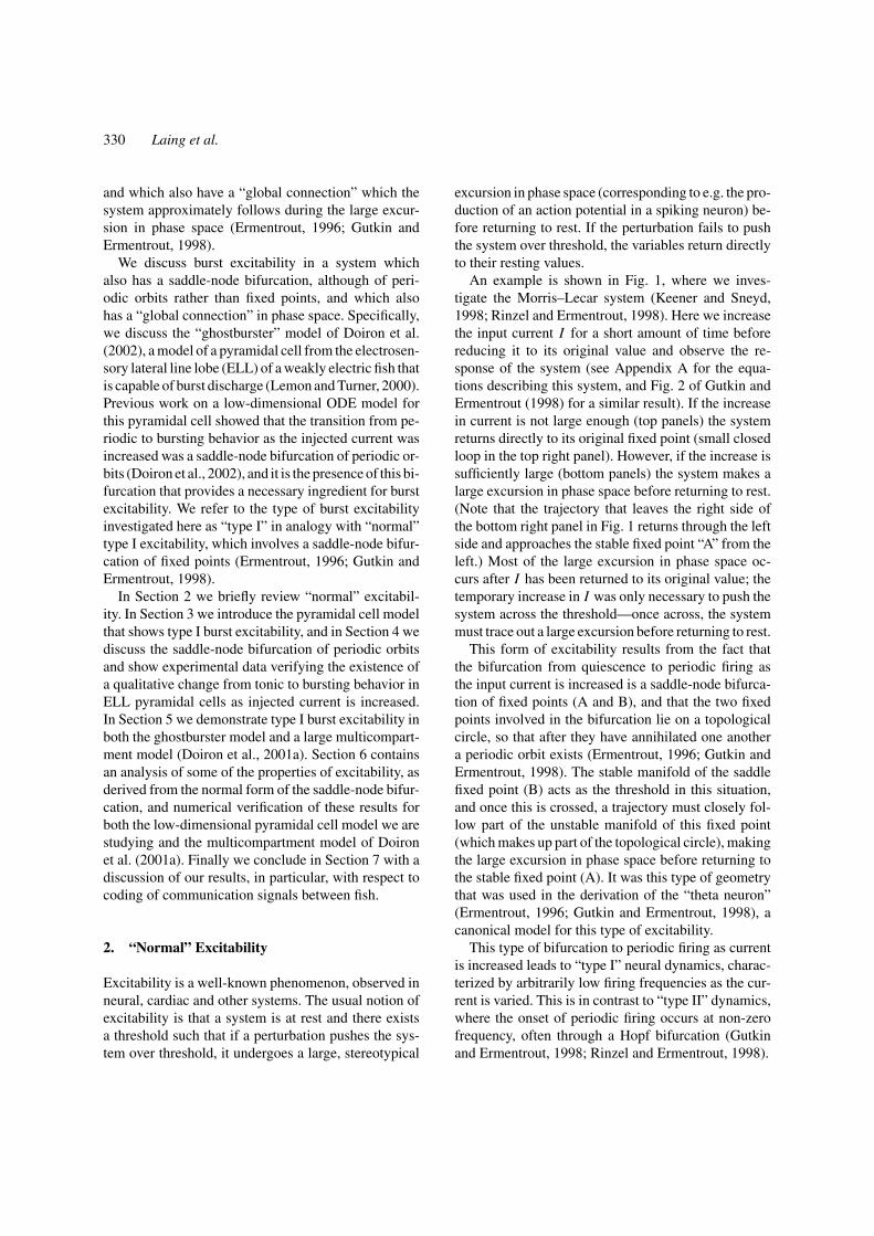
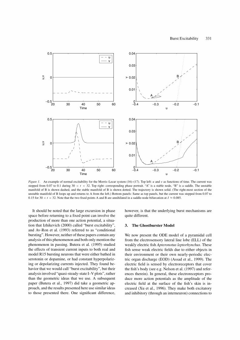
An example is shown in Fig. 1, where we inves-tigate the Morris–Lecar system (Keener and Sneyd,1998; Rinzel and Ermentrout, 1998). Here we increasethe input current I for a short amount of time beforereducing it to its original value and observe the re-sponse of the system (see Appendix A for the equa-tions describing this system, and Fig. 2 of Gutkin andErmentrout (1998) for a similar result). If the increasein current is not large enough (top panels) the systemreturns directly to its original fixed point (small closedloop in the top right panel). However, if the increase issufficiently large (bottom panels) the system makes alarge excursion in phase space before returning to rest.(Note that the trajectory that leaves the right side ofthe bottom right panel in Fig. 1 returns through the leftside and approaches the stable fixed point “A” from theleft.) Most of the large excursion in phase space oc-curs after I has been returned to its original value; thetemporary increase in I was only necessary to push thesystem across the threshold—once across, the systemmust trace out a large excursion before returning to rest.
This form of excitability results from the fact thatthe bifurcation from quiescence to periodic firing asthe input current is increased is a saddle-node bifurca-tion of fixed points (A and B), and that the two fixedpoints involved in the bifurcation lie on a topologicalcircle, so that after they have annihilated one anothera periodic orbit exists (Ermentrout, 1996; Gutkin andErmentrout, 1998). The stable manifold of the saddlefixed point (B) acts as the threshold in this situation,and once this is crossed, a trajectory must closely fol-low part of the unstable manifold of this fixed point(which makes up part of the topological circle), makingthe large excursion in phase space before returning tothe stable fixed point (A). It was this type of geometrythat was used in the derivation of the “theta neuron”(Ermentrout, 1996; Gutkin and Ermentrout, 1998), acanonical model for this type of excitability.
This type of bifurcation to periodic firing as currentis increased leads to “type I” neural dynamics, charac-terized by arbitrarily low firing frequencies as the cur-rent is varied. This is in contrast to “type II” dynamics,where the onset of periodic firing occurs at non-zerofrequency, often through a Hopf bifurcation (Gutkinand Ermentrout, 1998; Rinzel and Ermentrout, 1998).
Burst Excitability 331
20 30 40 50 60−0.5
0
0.5
Time
u,v
uv
−0.4 −0.3 −0.2 −0.10
0.01
0.02
0.03
0.04
B
A
u
v
20 30 40 50 60−0.5
0
0.5
Time
u,v
uv
−0.4 −0.3 −0.2 −0.10
0.01
0.02
0.03
0.04
u
v
A
B
Figure 1. An example of normal excitability for the Morris–Lecar system (16)–(17). Top left: u and v as functions of time. The current wasstepped from 0.07 to 0.1 during 30 < t < 32. Top right: corresponding phase portrait. “A” is a stable node, “B” is a saddle. The unstablemanifold of B is shown dashed, and the stable manifold of B is shown dotted. The trajectory is shown solid. (The right-most section of theunstable manifold of B loops up and returns to A from the left.) Bottom panels: Same as top panels, but the current was stepped from 0.07 to0.15 for 30 < t < 32. Note that the two fixed points A and B are annihilated in a saddle-node bifurcation at I ≈ 0.085.
It should be noted that the large excursion in phasespace before returning to a fixed point can involve theproduction of more than one action potential, a situa-tion that Izhikevich (2000) called “burst excitability”,and Av-Ron et al. (1993) referred to as “conditionalbursting”. However, neither of these papers contain anyanalysis of this phenomenon and both only mention thephenomenon in passing. Butera et al. (1995) studiedthe effects of transient current inputs to both real andmodel R15 bursting neurons that were either bathed inserotonin or dopamine, or had constant hyperpolariz-ing or depolarizing currents injected. They found be-havior that we would call “burst excitability”, but theiranalysis involved “quasi-steady-state I–V plots”, ratherthan the geometric ideas that we use. A subsequentpaper (Butera et al., 1997) did take a geometric ap-proach, and the results presented here use similar ideasto those presented there. One significant difference,
however, is that the underlying burst mechanisms arequite different.
3. The Ghostburster Model
We now present the ODE model of a pyramidal cellfrom the electrosensory lateral line lobe (ELL) of theweakly electric fish Apteronotus leptorhynchus. Thesefish sense weak electric fields due to either objects intheir environment or their own nearly-periodic elec-tric organ discharge (EOD) (Assad et al., 1999). Theelectric field is sensed by electroreceptors that coverthe fish’s body (see e.g. Nelson et al. (1997) and refer-ences therein). In general, these electroreceptors pro-duce more action potentials as the amplitude of theelectric field at the surface of the fish’s skin is in-creased (Xu et al., 1996). They make both excitatoryand inhibitory (through an interneuron) connections to
332 Laing et al.
50 100 150−80
−60
−40
−20
0
20
40
Time (ms)
Vs (
mV
)
50 100 150
−60
−40
−20
0
Time (ms)
Vd (
mV
)
50 100 1500.05
0.1
0.15
0.2
Time (ms)
p d
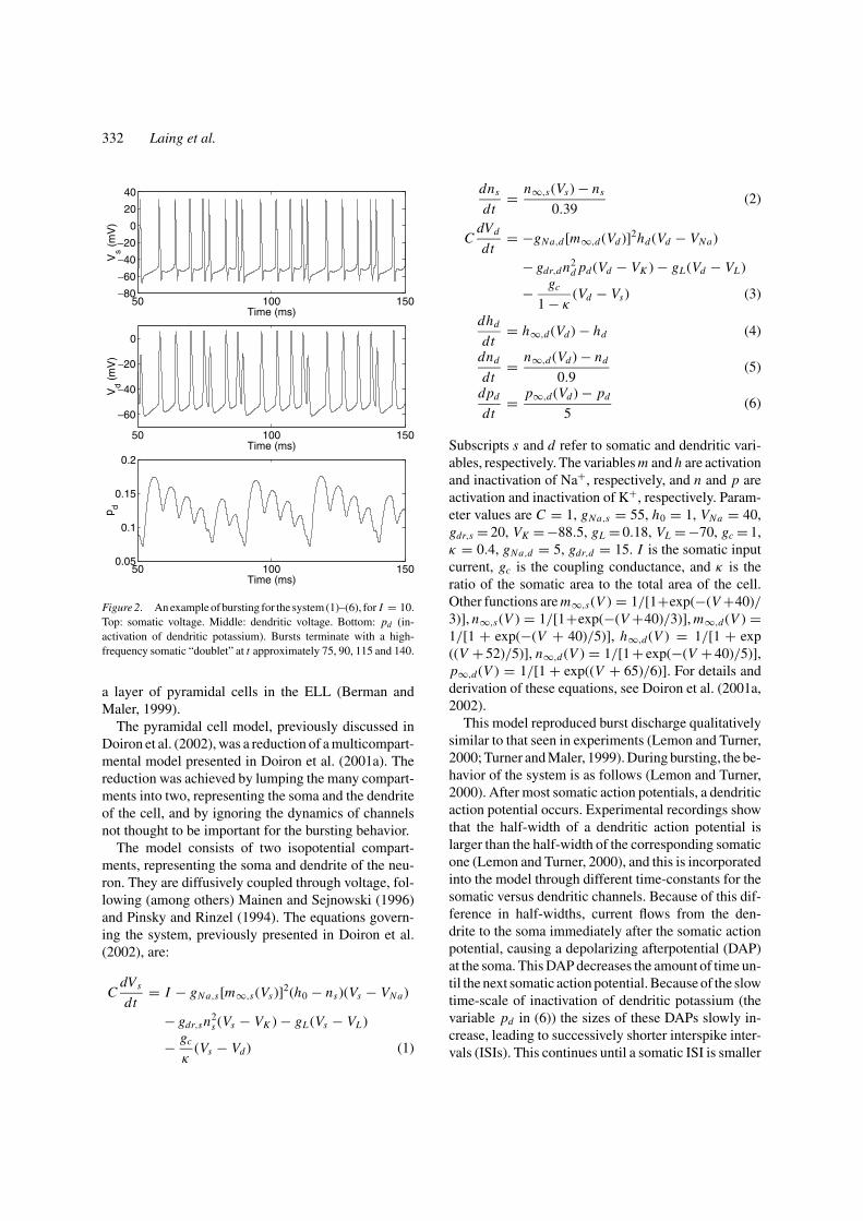
Figure 2. An example of bursting for the system (1)–(6), for I = 10.Top: somatic voltage. Middle: dendritic voltage. Bottom: pd (in-activation of dendritic potassium). Bursts terminate with a high-frequency somatic “doublet” at t approximately 75, 90, 115 and 140.
a layer of pyramidal cells in the ELL (Berman andMaler, 1999).
The pyramidal cell model, previously discussed inDoiron et al. (2002), was a reduction of a multicompart-mental model presented in Doiron et al. (2001a). Thereduction was achieved by lumping the many compart-ments into two, representing the soma and the dendriteof the cell, and by ignoring the dynamics of channelsnot thought to be important for the bursting behavior.
The model consists of two isopotential compart-ments, representing the soma and dendrite of the neu-ron. They are diffusively coupled through voltage, fol-lowing (among others) Mainen and Sejnowski (1996)and Pinsky and Rinzel (1994). The equations govern-ing the system, previously presented in Doiron et al.(2002), are:
CdVs
dt= I − gNa,s[m∞,s(Vs)]2(h0 − ns)(Vs − VNa)
− gdr,sn2s (Vs − VK ) − gL (Vs − VL )
− gc
κ(Vs − Vd ) (1)
dns
dt= n∞,s(Vs) − ns
0.39(2)
CdVd
dt= −gNa,d [m∞,d (Vd )]2hd (Vd − VNa)
− gdr,dn2d pd (Vd − VK ) − gL (Vd − VL )
− gc
1 − κ(Vd − Vs) (3)
dhd
dt= h∞,d (Vd ) − hd (4)
dnd
dt= n∞,d (Vd ) − nd
0.9(5)
dpd
dt= p∞,d (Vd ) − pd
5(6)
Subscripts s and d refer to somatic and dendritic vari-ables, respectively. The variables m and h are activationand inactivation of Na+, respectively, and n and p areactivation and inactivation of K+, respectively. Param-eter values are C = 1, gNa,s = 55, h0 = 1, VNa = 40,gdr,s = 20, VK = −88.5, gL = 0.18, VL = −70, gc = 1,κ = 0.4, gNa,d = 5, gdr,d = 15. I is the somatic inputcurrent, gc is the coupling conductance, and κ is theratio of the somatic area to the total area of the cell.Other functions are m∞,s(V ) = 1/[1+exp(−(V +40)/3)], n∞,s(V ) = 1/[1+exp(−(V +40)/3)], m∞,d (V ) =1/[1 + exp(−(V + 40)/5)], h∞,d (V ) = 1/[1 + exp((V + 52)/5)], n∞,d (V ) = 1/[1 + exp(−(V + 40)/5)],p∞,d (V ) = 1/[1 + exp((V + 65)/6)]. For details andderivation of these equations, see Doiron et al. (2001a,2002).
This model reproduced burst discharge qualitativelysimilar to that seen in experiments (Lemon and Turner,2000; Turner and Maler, 1999). During bursting, the be-havior of the system is as follows (Lemon and Turner,2000). After most somatic action potentials, a dendriticaction potential occurs. Experimental recordings showthat the half-width of a dendritic action potential islarger than the half-width of the corresponding somaticone (Lemon and Turner, 2000), and this is incorporatedinto the model through different time-constants for thesomatic versus dendritic channels. Because of this dif-ference in half-widths, current flows from the den-drite to the soma immediately after the somatic actionpotential, causing a depolarizing afterpotential (DAP)at the soma. This DAP decreases the amount of time un-til the next somatic action potential. Because of the slowtime-scale of inactivation of dendritic potassium (thevariable pd in (6)) the sizes of these DAPs slowly in-crease, leading to successively shorter interspike inter-vals (ISIs). This continues until a somatic ISI is smaller
Burst Excitability 333
than the refractory period of the dendrite and the den-drite fails to produce an action potential in responseto a somatic one, so little current flows from the den-drite to the soma, no DAP appears, and the next ISI islong. The process then repeats. These long ISIs, eachof which immediately follows a very short one, dividea train of action potentials into bursts. Figure 2 showsa typical series of bursts from the model (1)–(6). A de-tailed description of this burst mechanism is presentedin Lemon and Tuner (2000) and Doiron et al. (2001a).
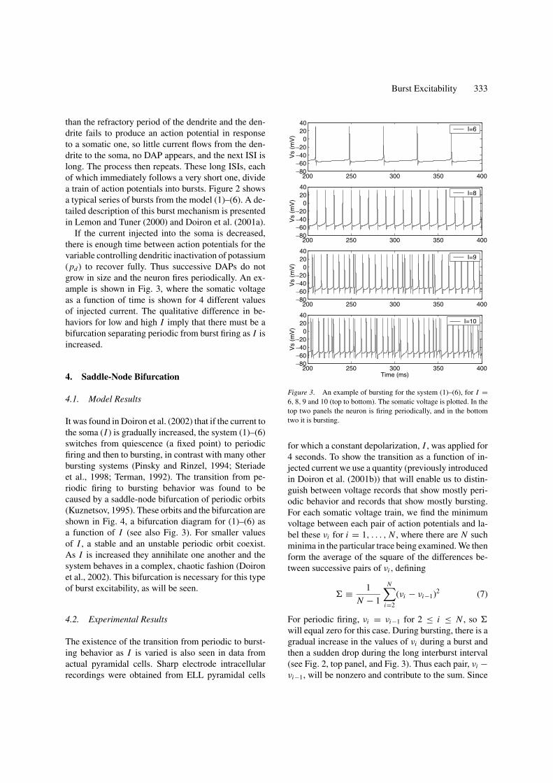
If the current injected into the soma is decreased,there is enough time between action potentials for thevariable controlling dendritic inactivation of potassium(pd ) to recover fully. Thus successive DAPs do notgrow in size and the neuron fires periodically. An ex-ample is shown in Fig. 3, where the somatic voltageas a function of time is shown for 4 different valuesof injected current. The qualitative difference in be-haviors for low and high I imply that there must be abifurcation separating periodic from burst firing as I isincreased.
4. Saddle-Node Bifurcation
4.1. Model Results
It was found in Doiron et al. (2002) that if the current tothe soma (I ) is gradually increased, the system (1)–(6)switches from quiescence (a fixed point) to periodicfiring and then to bursting, in contrast with many otherbursting systems (Pinsky and Rinzel, 1994; Steriadeet al., 1998; Terman, 1992). The transition from pe-riodic firing to bursting behavior was found to becaused by a saddle-node bifurcation of periodic orbits(Kuznetsov, 1995). These orbits and the bifurcation areshown in Fig. 4, a bifurcation diagram for (1)–(6) asa function of I (see also Fig. 3). For smaller valuesof I , a stable and an unstable periodic orbit coexist.As I is increased they annihilate one another and thesystem behaves in a complex, chaotic fashion (Doironet al., 2002). This bifurcation is necessary for this typeof burst excitability, as will be seen.
4.2. Experimental Results
The existence of the transition from periodic to burst-ing behavior as I is varied is also seen in data fromactual pyramidal cells. Sharp electrode intracellularrecordings were obtained from ELL pyramidal cells
200 250 300 350 400−80−60−40−20
02040
Vs
(mV
)
I=9
200 250 300 350 400−80−60−40−20
02040
Vs
(mV
)
I=6
200 250 300 350 400−80−60−40−20
02040
Vs
(mV
)
I=8
200 250 300 350 400−80−60−40−20
02040
Time (ms)
Vs
(mV
)
I=10
Figure 3. An example of bursting for the system (1)–(6), for I =6, 8, 9 and 10 (top to bottom). The somatic voltage is plotted. In thetop two panels the neuron is firing periodically, and in the bottomtwo it is bursting.
for which a constant depolarization, I , was applied for4 seconds. To show the transition as a function of in-jected current we use a quantity (previously introducedin Doiron et al. (2001b)) that will enable us to distin-guish between voltage records that show mostly peri-odic behavior and records that show mostly bursting.For each somatic voltage train, we find the minimumvoltage between each pair of action potentials and la-bel these νi for i = 1, . . . , N , where there are N suchminima in the particular trace being examined. We thenform the average of the square of the differences be-tween successive pairs of νi , defining
≡ 1
N − 1
N∑i=2
(νi − νi−1)2 (7)
For periodic firing, νi = νi−1 for 2 ≤ i ≤ N , so
will equal zero for this case. During bursting, there is agradual increase in the values of νi during a burst andthen a sudden drop during the long interburst interval(see Fig. 2, top panel, and Fig. 3). Thus each pair, νi −νi−1, will be nonzero and contribute to the sum. Since

334 Laing et al.
7 7.5 8 8.5 9 9.5 100
0.1
0.2
0.3
0.4
0.5
0.6
0.7
0.8
0.9
I
h d
Figure 4. The bifurcation diagram for (1)–(6) as a function of I .For I less than ∼8.5 there are two periodic orbits, one stable (solidline) and one unstable (dash-dotted). The maximum and minimumvalues of hd over one period are plotted vertically. For I greater than∼8.5 the value of hd at each local maximum and minimum during aperiod of 400 msec are plotted.
it is mainly the interburst intervals that contribute tothe sum, and the number of action potentials per burstdecreases as I is increased above the bifurcation value(Doiron et al., 2002), will increase as I increasespast the bifurcation value (∼8.5, from Fig. 4).
In Fig. 5 we show for both experimental andmodel data. The top and middle panels of Fig. 5 show, defined in (7), as a function of somatic input currentfor two different pyramidal cells during recordings ofduration four seconds, and the bottom panel shows thesame quantity for the model (1)–(6). Data from sixother cells was recorded and shows similar behavior tothat in the top two panels (data not shown). A number ofother cells were recorded from but did not show burst-ing for the range of currents tested, which is in agree-ment with previous studies (Lemon and Turner, 2000).There was a large variability in the maximum value of and the value of I at which the transition occurred.This is to be expected, due to the known variabil-ity in dendritic arborization (Bastian and Courtright,1991; Bastian and Nguyenkim, 2001). This variabilityaffects the threshold for bursting and the range of pos-sible spiking patterns, among other properties (Lainget al., 2002).
The existence of current values at which both theactual and model cells switch from periodic to burstingbehavior is clear. The experimental data presented hereand in Lemon and Turner (2000) strongly suggests that
0.2 0.3 0.4 0.5 0.60
1
2
3
I (nA)
Σ (m
V2 )
Neuron 1
0 0.1 0.2 0.3 0.40
0.5
1
1.5
I (nA)Σ
(mV
2 )
Neuron 2
7.5 8 8.5 9 9.50
1
2
3
4
5
I
Σ (m
V2 )
Ghostburster model
Figure 5. Top and middle: , (7), as a function of somatic inputcurrent, I , for two different pyramidal cells. The number of spikes,N , was typically about 400. See Lemon and Turner (2000) for detailsof the methods used to record the data presented here. Bottom: asa function of I for the system (1)–(6).
the actual cells undergo a saddle-node bifurcation ofperiodic orbits as the current injected to the soma is var-ied. Furthermore, the average burst length in data fromthese cells has been observed to scale as the inverse ofthe square root of the amount by which the current issuperthreshold (Doiron et al., 2003). This saddle-nodebifurcation of periodic orbits is a necessary ingredientfor type I burst excitability.
5. Burst Excitability
Since the bifurcation terminating periodic firing as I isincreased in the system (1)–(6) (see Fig. 4) is a saddle-node bifurcation of periodic orbits (Doiron et al., 2002),in analogy with normal type I excitability we have thepossibility of excitable behavior. The other necessaryingredient is a “global reinjection” mechanism, analo-gous to the topological circle mentioned in Section 2,which, after the periodic orbits have been annihilated,provides a mechanism for a large excursion in phase

Burst Excitability 335
space and return to a periodic orbit. We do have this“reinjection”, as can be seen in the behavior of pd dur-ing a burst (Fig. 2, bottom panel). The variable pd grad-ually decreases through a burst, but cannot continue todo this forever, as eventually the effect of the dendriticrefractory period come into play, the burst terminates,and pd rapidly increases—this is the reinjection.
(For the study of burst excitability in the ghost-burster, we are making an analogy between the saddle-node bifurcation of fixed points in the Morris–Lecarsystem (Fig. 1) and the saddle-node bifurcation of pe-riodic orbits in the ghostburster. However, for burst ex-citability in other bursting systems, other bifurcationsmay relevant.)
For a value of I just below that corresponding to theperiodic to bursting transition, the stable manifold ofthe unstable periodic orbit acts as a threshold—if thisis crossed, pd starts to decrease and continues to do sountil the burst terminates and the trajectory returns tothe stable periodic orbit. An example of this is shownin Fig. 6. At a current of 8.3, the neuron fires periodi-cally. The current is stepped from 8.3 to either 10.5 or11 for 10 ms, and then returned to 8.3. The step to 10.5fails to induce a burst and the variables return directly totheir previous (periodic) values, but a step to 11 pushesthe system over threshold, pd decreases until the end ofthe burst, and then the variables return to their previousvalues. Note that most of the burst occurs after I hasbeen returned to 8.3, another signature of the excitablenature of the system.
Thus, for the system (1)–(6), we have a new formof excitability, analogous to the usual form with theassociations
Normal excitability Type I burst excitability
Fixed point Periodic firing
Action potential Burst
However, the analogy is not exact since before the per-turbation the system (1)–(6) is periodically oscillating,rather than at a fixed point, and the phase of the os-cillation at which the perturbation is applied must betaken into consideration. The value of this phase cangreatly affect the resulting burst. This is demonstratedin Fig. 7, where two identical current pulses are ap-plied but at slightly different phases of the underlyingoscillation (the actual difference in phases for the twosituations is approximately 1/8 of a cycle). Note thatthe time between the onset of the pulse and the termi-
280 300 320 340 360 380 400 420 440−80
−60
−40
−20
0
20
40
Time (ms)
Vs (
mV
)
280 300 320 340 360 380 400 420 440−80
−60
−40
−20
0
20
40
Time (ms)V
s (m
V)
280 300 320 340 360 380 400 420 4400
0.1
0.2
0.3
0.4
Time (ms)
p d, I/3
0
Figure 6. Burst excitability in (1)–(6). Two different current pulses(shown in the bottom panel, 1/30 their actual size) were applied. Thelarger one (bold line) induced a burst (top panel) that appeared about30 ms after the stimulus, while the smaller one (thin line) did not(middle panel). Top: Vs for the large stimulus. Middle: Vs for thesmall stimulus. Bottom: pd and I/30.
nation of the burst is very different in the two cases.Another difference, also related to the more complexnature of the underlying system, is that the bursts arenot highly stereotyped. They all terminate in a highfrequency “doublet”, but as mentioned, the number ofaction potentials between the stimulus onset and thedoublet is highly variable.
The duration of the applied current steps is com-patible with the synaptic input to the pyramidal cellresulting from the detection of a “chirp”, a transientmodulation of both the frequency and amplitude of afish’s EOD. Zupanc and Maler (1993) found that chirpscould be evoked in Apteronotus leptorhynchus by im-itating the EOD of a neighboring fish, and proposedthat chirps are involved in communication between fish.They found that chirps typically last about 15 ms and in-volve an increase in the frequency of the chirping fish ofabout 100 Hz. Chirps produce transient increases in ex-citatory synaptic input to pyramidal cells (unpublished

336 Laing et al.
450 500 550 600 650 700−80−60−40−20
02040
Time (ms)
Vd (
mV
)
450 500 550 600 650 7000.05
0.1
0.15
0.2
Time (ms)
p d
950 1000 1050 1100 1150 1200−80−60−40−20
02040
Time (ms)
Vd (
mV
)
950 1000 1050 1100 1150 12000.05
0.1
0.15
0.2
Time (ms)
p d
Figure 7. Different bursts induced by identical current pulses (in-dicated by the solid bars in the voltage panels) for the system (1)–(6).The current was stepped from 8.3 to 10.8 for 10 ms at t = 500 (toptwo panels) and again at t = 1000 (bottom two panels). The differ-ence in the underlying phases at the two stimuli is approximately 1/8of a cycle, corresponding to approximately 1 ms.
observations) that result in the production of a smallnumber of spikes (Metzner and Heiligenberg, 1991).This provides motivation for the form of the currentpulses used in Figs. 6 and 7.
To demonstrate type I burst excitability in these pyra-midal cells in a more realistic manner, we have simu-lated a transient input to a realistic multicompartmen-tal model of a pyramidal cell in vivo. The model usedwas presented in Doiron et al. (2001a). The cell hasboth proximal and apical dendritic compartments, con-structed from a digitized image of a Lucifer yellowstained basilar pyramidal cell; the apical compartmentscontain channels responsible for the active backpropa-gation necessary for bursting. A single basilar dendriteextends from the soma for approximately 200 µm, afterwhich the dendrite branches to form a bush-like struc-ture. The electroreceptor afferents synapse excitatorilyto this bush.
Lateral segment pyramidal cells have been shown tobe required for the detection of communication signals
(chirps) (Metzner, 1999). To simulate one of these cells,we spread 1000 excitatory synapses over the basilarbush of the neuron—these model afferent connections.This number is estimated from data on receptive fieldsizes (Bastian et al., 2002) and receptor density (Carret al., 1982). The synaptic events are modeled as cur-rents of the form −g(t)(V (t) − 0), where V (t) is thevoltage of the compartment to which the synapse is at-tached (i.e. the reversal potential is 0) and g(t) followsan alpha function
g(t) = g
(t
τ
)e1−t/τ (8)
after each firing time, where g = 6 pS and τ = 1.5 msec(i.e. AMPA synapses). These parameters are appropri-ate for modeling afferent-evoked EPSPs in pyramidalcells (Berman and Maler, 1999). (The maximal con-ductance was chosen so as to produce realistic changesin the firing rate of the pyramidal cell, given the firingrates of the afferent inputs.) The synaptic firing timeswere chosen from independent Poisson distributions.The firing rate of each synapse was originally set at150 Hz, and then stepped to either 475 or 400 Hz when250 < t < 270, i.e. for 20 msec. These values areconsistent with the responses of electroreceptors dur-ing chirp stimuli (unpublished observations) (Metznerand Heiligenberg, 1991).
As can be seen in Fig. 8, the step to 400 Hz did notinduce a burst, but the step to 475 Hz did. The first andthird panels of Fig. 8 show the somatic voltage, and thesecond and fourth show the sum of the conductancesfrom all 1000 synapses as a function of time (comparewith Fig. 6). Thus in a much more realistic model ofthe pyramidal cell, and with more realistic stochasticinputs, we also have type I burst excitability.
This form of burst excitability was also observedby Laing and Longtin (2002) in a reduced model ofthe system (1)–(6). The model presented there used anintegrate-and-fire neuron to produce action potentials,and a second variable that mimicked the effects of theslowly changing dendritic potassium in (1)–(6).
6. Analysis
6.1. Theory
Both the normal and burst excitability that we haveanalyzed arise from saddle-node bifurcations—of fixedpoints in normal excitability and of periodic orbits in

Burst Excitability 337
200 250 300 350 400−80−60−40−20
020
Time (ms)
Vs
(mV
)
200 250 300 350 4000
5
10
15
Time (ms)
Tota
l Con
duct
ance
(nS
)
200 250 300 350 400−80−60−40−20
020
Time (ms)
Vs
(mV
)
200 250 300 350 4000
5
10
15
Time (ms)
Tota
l Con
duct
ance
(nS
)
Figure 8. Burst excitability in the multicompartmental model ofDoiron et al. (2001a). In the top two panels an increase in the firingrate of randomly occurring excitatory synaptic events induced a burst,whereas in the bottom two panels a slightly smaller increase in firingrate did not induce a burst. See text for details.
burst excitability. The normal form of this bifurcationcan be used to derive the minimum pulse strength andduration needed to induce a burst (or action potential).We first examine normal excitability.
The normal form of the saddle-node bifurcation offixed points in a continuous-time dynamical system is(Guckenheimer and Holmes, 1990; Kuznetsov, 1995)
dx
dt= µ + x2 (9)
This governs the behavior on the center manifold. Forµ < 0 there are two fixed points, x± ≡ ±√−µ. Thepoint x− is stable with basin of attraction (−∞, x+),and x+ is unstable. For µ > 0 there are no fixed points.The current pulses discussed above for normal ex-citability correspond to switching µ from a negativevalue to a positive one for a short amount of time, andthen back again. Assume for concreteness that beforethe pulse, µ = −ν, (ν > 0), and that during the pulse
µ = λ, (λ > 0). For µ > 0, the solution of (9) is
x(t) = õ tan
[√µt + tan−1
(x(0)√
µ
)](10)
where x(0) is the value of x at time 0. For a pulseof duration T to cause an action potential, we needx(T ) > x+, where x(0) = x−, i.e. for x to escapethe basin of attraction of the stable fixed point duringthe pulse, having started at the stable fixed point. Oncex has escaped the basin of attraction it will make alarge excursion and then return to the stable fixed point.(The normal form (9) is only a local description of thedynamics, and does not describe the large excursion.)The above inequality is equivalent to
√ν <
√λ tan
[√λT + tan−1
(−√ν√
λ
)](11)
which, under the assumption that√
ν/λ is small so thattan x ≈ tan−1 x ≈ x , can be written
2√
ν
λ< T (12)
Thus the minimum value of T that will induce an actionpotential scales as
T ∼√
ν
λ(13)
The analysis of type I burst excitability is very simi-lar, except that one should now study the normal formof the saddle-node bifurcation of fixed points in a map:
xn+1 = xn + µ + x2n (14)
where n is an integer. This map can be derived by plac-ing a Poincare section in the flow, reducing the study ofa periodic orbit in a continuous-time system to the studyof a fixed point of a discrete-time map (Guckenheimerand Holmes, 1990; Kuznetsov, 1995). For µ < 0, themap (14) has two fixed points, ±√−µ, which are de-stroyed in a saddle-node bifurcation at µ = 0 in thesame way as for (9). A bifurcation of this form (whichcorresponds to a saddle-node bifurcation of periodicorbits in a flow) can be observed in (1)–(6) by placinga Poincare section in the flow, as was shown in Doironet al. (2002). The scaling relationship obtained from(14) is also given by (13).

338 Laing et al.
6.2. Numerical Results
In Fig. 9 we show numerical results for the system(1)–(6) that are consistent with the scaling of (13). Here,the current before a pulse (effectively ν) is held constantwhile the height of the pulse (effectively λ) is increased.We measured the minimum duration of a pulse neededto produce a burst with probability 0.5. We have to takethis probabilistic approach for burst excitability as the“rest state” is now periodic firing, and the time of theonset of the pulse relative to the phase of the periodicfiring has to be considered (see Fig. 7). (This wouldalso be the case if, e.g. the system shown in Fig. 1 wasweakly periodically forced.)
To obtain Fig. 9, a number of pulses were applied atrandom phases of the periodic oscillation. (The time be-tween pulses was sufficient for the system to relax backto the periodic orbit.) The average number of bursts perpulse is a continuous function of both pulse durationand strength, and curves for probabilities other than0.5 are similar to that shown in Fig. 9. As predicted by(13), the minimum pulse duration required to induce aburst for a fixed baseline current (effectively ν) is in-versely proportional to the height of the pulse above thevalue needed to produce bursting. (The offset, 0.1235,in the caption of Fig. 9 is a result of the pulse heightbeing ν + λ, not just λ.) The baseline current was setat I = 8.3.
Note that a plot of the form shown in Fig. 9 but withthe axes interchanged (i.e. a plot of the stimulus inten-
0 1 2 3 4 50
10
20
30
40
50
60
Pulse height
Pul
se d
urat
ion
(ms)
Figure 9. Minimum duration of a pulse required to generate anaverage of one burst per two pulses for the system (1)–(6), as afunction of pulse height. Circles are numerically measured values,the solid curve is y = 24.14/(x − 0.1235). The baseline current isI = 8.3.
8 8.1 8.2 8.3 8.4 8.50
5
10
15
20
25
I
Tim
e (m
s)
Figure 10. Minimum duration of a pulse required to generate an av-erage of one burst per two pulses for the system (1)–(6), as a functionof baseline current. I = 10 during a pulse. Circles are numericallymeasured values, the solid curve is y = 33.69
√8.481 − x and the
dashed curve is y = 32.02 tan−1 (1.213√
8.476 − x).
sity necessary to evoke a response as a function of stim-ulus duration) is often known as a “strength-durationcurve” (Koch, 1999). However, these are usually con-structed for normal excitability, where the event is asingle action potential rather than a burst of them.
Figure 10 is further confirmation that the relation-ship (13) holds for (1)–(6). Here the height of the pulse(effectively λ) is held constant while the current beforethe pulse (effectively ν) is varied. Note that ν increasesas I decreases. As in Fig. 9, we measure the minimumduration of a pulse needed to produce, on average, oneburst for every two pulses applied. We see that this du-ration is proportional to the square root of the differencebetween the baseline current and the current needed toproduce bursting (solid line), in agreement with (13).An even better fit can be obtained by keeping the fullexpression (11), which gives
T ∼ tan−1 (α√
ν) (15)
for some α, assuming that λ is fixed (dashed line inFig. 10).
The scaling results of (13) also hold for the multi-compartment model of Doiron et al. (2001a), discussedin Section 5. Recall that we change the input strengthby modulating the firing rate of the independent Pois-son processes associated with each of the 1000 excita-tory synapses. In Fig. 11 we show a plot of the mini-mum duration of a pulse required to generate a burstas a function of the strength of the pulse. The firing

Burst Excitability 339
250 300 350 400 450 50015
20
25
30
35
40
45
50
Pulse Height (Hz)
Pul
se d
urat
ion
(ms)
Figure 11. Minimum duration of a pulse required to generate aburst for the model described in Doiron et al. (2001a) as a functionof pulse height. Circles are numerically measured values, the solidcurve is the function y = 7107/(x − 141). Ten noise realizations weused to obtain each data point. Compare with Fig. 9.
180 190 200 210 220 2300
5
10
15
Baseline Intensity (Hz)
Tim
e (m
s)
Figure 12. Minimum duration of a pulse required to generate aburst for the model described in Doiron et al. (2001a) as a functionof the intensity before stimulation. Circles are numerically measuredvalues, the solid curve is the function y = 1.996
√225 − x . Ten noise
realizations we used to obtain each data point. Compare with Fig. 10.
rate prior to the pulse, and after it, was 150 Hz. Wesee that the simulation results are consistent with thetheory.
In Fig. 12 we show further confirmation that thescaling given (13) holds for the multicompartmentmodel of Doiron et al. (2001a). We have kept the in-tensity of each pulse constant at 475 Hz, but variedthe rate of synaptic activity before and after the pulse(they are the same) and determined how this affectsthe duration of a pulse required to produce one burst.
We see that again the simulations agree well with thetheory, particularly when the baseline activity is closeto the threshold between tonic and bursting behavior(225 Hz, from Fig. 12).
Note that while the numerically obtained resultsabove are consistent with there being a saddle-nodebifurcation of periodic orbits separating periodic frombursting behavior, other model neurons could haveother bifurcations that separate these types of behaviorand produce the same form of scaling.
7. Discussion and Conclusion
We have introduced and analyzed the new phenomenonof type I burst excitability, presenting it in a specificsystem, a “ghostburster” pyramidal cell, and in a spe-cific context, the detection of communicatory signals.Briefly, the existence of a saddle-node bifurcation ofperiodic orbits, together with a “global reinjection”through phase space, enable us to make an analogywith normal excitability:
Normal excitability Type I burst excitability
Fixed point Periodic firing
Action potential Burst
The analogy is not exact, as the underlying phase ofthe periodic oscillation at the onset of the transientinput must be taken into account, and the bursts arenot as highly sterotyped as action potentials in normalexcitability. We have shown data from actual pyrami-dal cells which strongly suggests that these cells pos-sess the necessary ingredients for this type of burstexcitability, and we have also simulated it in a realisticmulticompartment model of a pyramidal cell driven bystochastic inputs. We have derived and verified a scal-ing law relating the strength and duration of the currentpulses required to observe a superthreshold response ofan excitable system.
The pyramidal cells studied are sensory neurons,receiving input directly from electroreceptors on thesurface of the fish, and thus it is reasonable to as-sume that they process electrosensory information insome way and pass it to higher brain regions. Asmentioned, the type of input current required to in-duce a burst in these cells is compatible with the out-put of electroreceptors (unpublished observations) thatdetect a chirp from another fish (Zupanc and Maler,1993).
340 Laing et al.
The production of a burst by a pyramidal cell in theELL in response to a sudden artificial increase in EODamplitude has recently been seen in vivo (J. Bastian,personal communication). These bursts were observedby recording from the dendrite of a pyramidal cell,and both the decreasing ISIs during the burst and thesudden decrease in spike amplitude at the end of theburst indicate that this was the same type of burst asdiscussed here and elsewhere (Doiron et al., 2001a,2002; Lemon and Turner, 2000). These burst responseswere suppressed by hyperpolarization of the cell, whichis consistent with the existence of a threshold in currentfor burst behavior.
More generally, the current input to a ghostburstingcell will not be constant, but will have slow modula-tions. These could be due to objects passing close tothe fish, or a result of the “beat” frequency that occurswhen two fish with different EOD frequencies are closeto one another. (The beat frequency is equal to the dif-ference between the EOD frequencies of the two fish;recall that EODs are quasi-sinusoidal.) The results inFig. 7 show that the response of a pyramidal cell to iden-tical inputs depends on the phase of the periodic firingof the cell, but it may also be the case that the responsewill be different during different parts of the cycle asso-ciated with the beating frequency. Indeed, Zupanc andMaler (1993) showed that the form of chirps emitted bymale fish in response to a simulated EOD depended onthe phase of the beat cycle created between the fish’sown EOD and the simulated EOD, and any system fordetecting such chirps may also take into account thephase of the beat cycle.
Lisman (1997) recently proposed that bursts, ratherthan individual action potentials, may be fundamentalunits of neural information. One reason behind thisidea is that many synapses are unreliable, but due tofacilitation a burst of action potentials may be signaledreliably in a situation where a single action potentialmay not. The system described in this paper may bean example for which this principle applies, since theburst terminates with a high-frequency “doublet” event.Simultaneous burst discharge by a number of pyramidalcells that synapse onto a common target cell wouldpresumably be more reliable than a transient increasein the rate of asynchronous firing of those cells.
Other bursting systems also have bifurcations thatseparate periodic or quiescent behavior from burst-ing as input current is changed (Terman, 1992). Wehave simulated the square-wave, parabolic and ellipticbursters described in Rinzel and Ermentrout (1998),
and found that these systems also have such bifurca-tions. These bursters all follow the pattern quiescence→bursting→ tonic as current is increased (not shown),in contrast with the system (1)–(6). We have found thattransient changes in input current across the bifurcationpoints separating these regimes also cause behavior thatis qualitatively similar to that discussed above—a mo-tion in phase space that would be labeled a burst if thesystem was bursting, followed by a return to either pe-riodic or quiescent behavior, depending on the state ofthe system before the perturbation (results not shown).This is the behavior observed by Izhikevich (2000) andAv-Ron et al. (1993); their systems were close to thequiescent/bursting boundary. Butera et al. (1995) sawsimilar behavior in model and real R15 neurons and ina subsequent paper analyzed the model in a way similarto that performed here (Butera et al., 1997). However,the bifurcations involved in the transitions from burst-ing to other types of behavior in these models are oftenvery complicated (Terman, 1992), rather than simplesaddle-node bifurcations. Also, the models describedin Rinzel and Ermentrout (1998) are not models ofsensory neurons, so it is harder to speculate about the“meaning” of burst excitability in such neurons. Thisis in contrast with the system (1)–(6), which representsa neuron thought to play an important role in sensoryprocessing.
It should also be mentioned that there are othermodel neurons which are capable of bursting, but whichdo not follow the pattern quiescence → bursting →tonic as injected current is increased. An example isintegrate-and-fire-or-burst model of a thalamocorticalrelay neuron (Smith et al., 2000), which moves fromquiescence to a single burst to quiescence and finally toperiodic firing as the injected DC current is graduallyincreased.
The simulations mentioned above, and the resultsof others (Av-Ron et al., 1993; Butera et al., 1995;Izhikevich, 2000), suggest that burst excitability is ageneral phenomenon in systems capable of bursting.We have analyzed the behavior of one particular typein a particular model, as did Butera et al. (1997); theanalysis of other bursting systems under time-varyinginput remains to be done.
Appendix: Equations
The Morris–Lecar system (Keener and Sneyd, 1998;Rinzel and Ermentrout, 1998) is the following pair of
Burst Excitability 341
ODEs:
du
dt= I − g1m(u)(u − 1) − gK (u − uK )
− gL (u − uL ) (16)dv
dt= φ[v(u) − v]
τ (u)(17)
where
m(u) = 1 + tanh [(u − u1)/u2]
2(18)
v(u) = 1 + tanh [(u − u3)/u4]
2(19)
and
τ (u) = 1
cosh [(u − u3)/(2u4)](20)
We use the following parameter values: g1 = 1, gK =2, uK = −0.7, gL = 0.5, uL = −0.5, φ = 1/3, u1 =−0.01, u2 = 0.15, u3 = 0.1, u4 = 0.145.
Acknowledgments
We acknowledge financial support from NSERC (B.D.,L.N., A.L), OPREA (C.L., A.L.) and CIHR (A.L., R.T.,L.M.). R.T. is an Alberta Heritage Senior Scholar. Wethank the referees for bringing Butera et al. (1995,1997) to our attention.
References
Assad C, Rasnow B, Stoddard PK (1999) Electric organ dischargesand electric images during electrolocation J. Exp. Biol. 202:1185–1193.
Av-Ron E, Parnas H, Segel LA (1993) A basic biophysical modelfor bursting neurons. Biol. Cybern. 69: 87–95.
Bastian J, Chacron MJ, Maler L (2002) Receptive field organiza-tion determines pyramidal cell stimulus encoding capability andspatial stimulus selectivity. J. Neurosci. 22: 4577–4590.
Bastian J, Courtright J (1991) Morphological correlates of pyra-midal cell adaptation rate in the electrosensory lateral linelobe of weakly electric fish. J. Comp. Physiol. A 168(4): 393–407.
Bastian J, Nguyenkim J (2001) Dendritic modulation of burst-likefiring in sensory neurons. J. Neurophysiol. 85: 10–22.
Berman NJ, Maler L (1999) Neural architecture of the electrosen-sory lateral line lobe: Adaptations for coincidence detection, asensory searchlight and frequency-dependent adaptive filtering.J. Exp. Biol. 202: 1243–1253.
Bub G, Shrier A, Glass L (2002) Spiral wave generation in hetero-geneous excitable media. Phys. Rev. Lett. 88: 058101.
Butera RB, Clark JW, Byrne JH (1997) Transient responses of a mod-eled bursting neuron: Analysis with equilibrium and averagednullclines. Biol. Cyber. 77: 307–322.
Butera RJ, Clark JW, Canavier CC, Baxter DA, Byrne JH (1995)Analysis of the effects of modulatory agents on a model burst-ing neuron: Dynamic interactions between voltage and calciumdependent systems. J. Comput. Neurosci. 2: 19–44.
Carr C, Maler L, Sas E (1982) Peripheral organization and centralprojections of the electrosensory nerves in gymnotiform fish. J.Comp. Neurol. 211: 139–153.
de Vreis G (1998) Multiple bifurcations in a polynomial model ofbursting oscillations. J. Nonlinear Sci. 8: 281–316.
Doiron B, Laing CR, Longtin A, Maler L (2002) “Ghostbursting”: Anovel bursting mechanism in pyramidal cells. J. Comput. Neu-rosci. 12(1): 5–25.
Doiron B, Longtin A, Turner RW, Maler L (2001a) Model of gammafrequency burst discharge generated by conditional backpropa-gation. J. Neurophysiol. 86(4): 1523–1545.
Doiron B, Noonan L, Lemon N, Turner RW (2003) Persistant Na+current modifies burst discharge by regulating conditional back-propagation of dendritic spikes. J. Neurophysiol. 89: 324–337.
Doiron B, Noonan L, Turner RW, Longtin A, Maler L (2001b) Shift-ing burst threshold with dendritic conductances. XXXI Proc.Soc. Neurosci, San Diego.
Ermentrout GB (1996) Type I membranes, phase resetting curves,and synchrony. Neural Comp. 8: 979–1001.
Glass L, Mackey MC (1988) From Clocks to Chaos: The Rhythmsof Life. Princeton University Press.
Goldbeter A (1996) Biochemical Oscillations and Cellular Rhythms:The Molecular Bases of Periodic and Chaotic Behaviour.Cambridge University Press.
Guckenheimer J, Holmes P (1990) Nonlinear oscillations, dynamicalsystems and bifurcations of vector fields. Applied MathematicalSciences, Springer-Verlag, Vol. 42.
Gutkin BS, Ermentrout GB (1998) Dynamics of membrane excitabil-ity determine inter-spike interval variability: A link betweenspike generation mechanisms and cortical spike train statistics.Neural Comp. 10: 1047–1065.
Izhikevich EM (2000) Neural excitability, spiking, and bursting. Int.J. Bifn. Chaos 10: 1171–1266.
Keener J, Sneyd J (1998) Mathematical Physiology. InterdisciplinaryApplied Mathematics, Vol. 8. Springer-Verlag, New York.
Koch C (1999) Biophysics of Computation: Information Processingin Single Neurons. Oxford University Press.
Kuznetsov YA (1995) Elements of Applied Bifurcation Theory. Ap-plied Mathematical Sciences, Springer-Verlag, Vol. 112.
Laing CR, Doiron B, Longtin A, Maler L (2002) Ghostbursting: Theeffects of dendrites on spike patterns. Neurocomputing 44–46:127–132.
Laing CR, Longtin A (2002) A two-variable model of somatic-dendritic interactions in a bursting neuron. Bull. Math. Biol,64: 829–860.
Lemon N, Turner R (2000) Conditional spike backpropagation gen-erates burst discharge in a sensory neuron. J Neurophysiol 84:1519–1530.
Lisman JE (1997) Bursts as units of neural information: Makingunreliable synapses reliable. Trends Neurosci 20: 38–43.
Mainen ZF, Sejnowski TJ (1996) Influence of dendritic structure onfiring pattern in model neocortical neurons. Nature 382: 363–366.

342 Laing et al.
Metzner W (1999) Neural circuitry for communication and jammingavoidance in gymnotiform electric fish. J. Exp. Biol. 202: 1365–1375.
Metzner W, Heiligenberg W (1991) The coding of signals in theelectric communication of the gymnotiform fish Eigenmannia:From electroreceptors to neurons in the torus semicircularis ofthe midbrain. J. Comp. Physiol. [A] 169: 135–150.
Nelson, ME, Xu Z, Payne JR (1997) Characterization and modelingof P-type electrosensory afferent responses to amplitude mod-ulations in a wave-type electric fish. J. Comp. Physiol. A. 181:532–544.
Pinsky PF, Rinzel J (1994) Intrinsic and network rhythmogenesis ina reduced Traub model for CA3 neurons. J. Comput. Neurosci.1: 39–60.
Rinzel J, Ermentrout GB (1998) Analysis of neural excitability andoscillations. In: C Koch, I Segev, eds. Methods in Neuronal Mod-eling: From Ions to Networks. MIT Press.
Smith GD, Cox CL, Sherman SM, Rinzel J (2000) Fourier analysis
of sinusoidally driven thalamocortical relay neurons and a mini-mal integrate-and-fire-or-burst model. J. Neurophysiol. 83: 588–610.
Steriade M, Timofeev I, Durmuller N, Grenie F (1998) Dynamicproperties of corticothalamic neurons and local cortical interneu-rons generating fast rhythmic (30–40 Hz) spike bursts. J. Neu-rophysiol. 79: 483–490.
Terman D (1992) The transition from bursting to continuous spikingin excitable membrane models. J. Nonlinear Sci. 2(2): 135–182.
Turner RW, Maler L (1999) Oscillatory and burst discharge in theApteronotid electrosensory lateral line lobe. J. Exp. Biol. 202:1255–1265.
Xu Z, Payne JR, Nelson ME (1996) Logarithmic time course ofsensory adaptation in electrosensory afferent nerve fibers in aweakly electric fish. J. Neurophysiol. 76: 2020–2032.
Zupanc GKH, Maler L (1993) Evoked chirping in the weakly elec-tric fish (Apteronotus leptorhynchus): A quantitative biophysicalanalysis. Can. J. Zool. 71: 2301–2310.
Copyright © 2022 FDOKUMEN