Hearing Loss Raises Excitability in the Auditory Cortex
11
Behavioral/Systems/Cognitive Hearing Loss Raises Excitability in the Auditory Cortex Vibhakar C. Kotak, 1 Sho Fujisawa, 1 Fanyee Anja Lee, 1 Omkar Karthikeyan, 1 Chiye Aoki, 1,2 and Dan H. Sanes 1,2 1 Center for Neural Science and 2 Department of Biology, New York University New York, New York 10003 Developmental hearing impairments compromise sound discrimination, speech acquisition, and cognitive function; however, the ad- justments of functional properties in the primary auditory cortex (A1) remain unknown. We induced sensorineural hearing loss (SNHL) in developing gerbils and then reared the animals for several days. The intrinsic membrane and synaptic properties of layer 2/3 pyramidal neurons were subsequently examined in a thalamocortical brain slice preparation with whole-cell recordings and electron microscopic immunocytochemistry. SNHL neurons displayed a depolarized resting membrane potential, an increased input resistance, and a higher incidence of sustained firing. They also exhibited significantly larger thalamocortically and intracortically evoked excitatory synaptic responses, including a greater susceptibility to the NMDA receptor antagonist AP-5 and the NR2B subunit antagonist ifenprodil. This correlated with an increase in NR2B labeling of asymmetric synapses, as visualized ultrastructurally. Furthermore, decreased frequency and increased amplitude of miniature EPSCs (mEPSCs) in SNHL neurons suggest that a decline in presynaptic release properties is compensated by an increased excitatory response. To verify that the increased thalamocortical excitation was elicited by putative mono- synaptic connections, minimum amplitude ventral medial geniculate nucleus-evoked EPSCs were recorded. These minimum-evoked responses were of larger amplitude, and the NMDAergic currents were also larger and longer in SNHL neurons. These findings were supported by significantly longer AP-5-sensitive durations and larger amplitudes of mEPSCs. Last, the amplitudes of intracortically evoked monosynaptic and polysynaptic GABAergic inhibitory synaptic responses were significantly smaller in SNHL neurons. These alterations in cellular properties after deafness reflect an attempt by A1 to sustain an operative level of cortical excitability that may involve homeostatic mechanisms. Key words: homeostasis; synaptic plasticity; GABA A ; NMDA receptor; mEPSC; disuse Introduction Hearing impairments produce significant changes in the human cortex and may permanently diminish auditory perceptual skills and language acquisition (Mody et al., 1999; Vernon-Feagans, 1999; Psarommatis et al., 2001; Kidd et al., 2002; Emmorey et al., 2003; Iverson, 2003). The effects of sensorineural hearing loss (SNHL) are particularly severe in children with the longest peri- ods of auditory deprivation (Sharma et al., 2002). Recordings from hearing-impaired animals have also revealed many pro- found changes in auditory processing (Syka, 2002). Unilaterally deafened animals display increased acoustically evoked activity in the ipsilateral inferior colliculus (IC) and auditory cortex, likely attributable to a change in the balance between existing excitatory and inhibitory inputs (Kitzes and Semple, 1985; McAlpine et al., 1997; Mossop et al., 2000). Processing after SNHL has also been examined in cats using electrical stimulation with cochlear pros- thetic devices. Although some findings suggest that auditory depri- vation leads to decreased synaptic drive, particularly to layer 5 (Klinke et al., 1999; Kral et al., 2000), there are also signs of increased excitability. For example, electrode-evoked thresholds are lower, spatial tuning curves are broader, and cochleotopy appears impre- cise after 2 weeks of deafness (Raggio and Schreiner, 1999, 2003). These in vivo studies do not distinguish between changes that may have occurred in the brainstem (Snyder et al., 2000; Moore et al., 2002) and those that have occurred locally in the cortex, nor do they identify alterations to synaptic and intrinsic membrane properties that may attend deafness. For example, our previous work in the auditory brainstem shows that unilateral hearing loss not only decreases synaptic inhibition but also enhances NMDA receptor (NMDA-R)-mediated excitation (Kotak and Sanes, 1996). Similarly, in the inferior colliculus, bilateral SNHL de- creases the inhibitory postsynaptic conductance and increases EPSC amplitude (Vale and Sanes, 2002). These findings are con- sistent with experiments performed on invertebrate circuits and dissociated cortical neurons showing that synapses and ion chan- nels are regulated by electrical activity (Marder and Prinz, 2002). In general, such changes tend to resist the manipulation. Thus, when cultures are grown in activity blockers, it is found that excitatory synaptic and sodium currents increase and inhibitory synaptic and potassium currents decrease (Rao and Craig, 1997; Desai et al., 1999; Murthy et al., 2001; Burrone et al., 2002; Kil- man et al., 2002). The present studies were designed to assess both intrinsic and synaptic properties in the primary auditory cortex (A1) after bi- lateral hearing loss. Only one previous study has examined alter- ations in biophysical properties of cochlear nucleus (CN) neu- Received July 2, 2004; revised March 7, 2005; accepted March 9, 2005. This work was supported by National Institutes of Health Grants DC00540 (D.H.S.) and DC006864 (D.H.S. and V.C.K.), National Eye Institute Grant EY 13145 (C.A.), National Institute of Neurological Disorders and Stroke Grant NS041091 (C.A.), and Japan Foundation for Aging and Health Grant R6189 (C.A.). We thank Dr. Patricio Huerta for his useful advice. Correspondence should be addressed to Dr. Vibhakar C. Kotak, Center for Neural Science, 4 Washington Place, New York University, New York, NY 10003. E-mail: [email protected]. DOI:10.1523/JNEUROSCI.5169-04.2005 Copyright © 2005 Society for Neuroscience 0270-6474/05/253908-11$15.00/0 3908 • The Journal of Neuroscience, April 13, 2005 • 25(13):3908 –3918
-
Upload
khangminh22 -
Category
Documents
-
view
3 -
download
0
Transcript of Hearing Loss Raises Excitability in the Auditory Cortex

Behavioral/Systems/Cognitive
Hearing Loss Raises Excitability in the Auditory Cortex
Vibhakar C. Kotak,1 Sho Fujisawa,1 Fanyee Anja Lee,1 Omkar Karthikeyan,1 Chiye Aoki,1,2 and Dan H. Sanes1,2
1Center for Neural Science and 2Department of Biology, New York University New York, New York 10003
Developmental hearing impairments compromise sound discrimination, speech acquisition, and cognitive function; however, the ad-justments of functional properties in the primary auditory cortex (A1) remain unknown. We induced sensorineural hearing loss (SNHL)in developing gerbils and then reared the animals for several days. The intrinsic membrane and synaptic properties of layer 2/3 pyramidalneurons were subsequently examined in a thalamocortical brain slice preparation with whole-cell recordings and electron microscopicimmunocytochemistry. SNHL neurons displayed a depolarized resting membrane potential, an increased input resistance, and a higherincidence of sustained firing. They also exhibited significantly larger thalamocortically and intracortically evoked excitatory synapticresponses, including a greater susceptibility to the NMDA receptor antagonist AP-5 and the NR2B subunit antagonist ifenprodil. Thiscorrelated with an increase in NR2B labeling of asymmetric synapses, as visualized ultrastructurally. Furthermore, decreased frequencyand increased amplitude of miniature EPSCs (mEPSCs) in SNHL neurons suggest that a decline in presynaptic release properties iscompensated by an increased excitatory response. To verify that the increased thalamocortical excitation was elicited by putative mono-synaptic connections, minimum amplitude ventral medial geniculate nucleus-evoked EPSCs were recorded. These minimum-evokedresponses were of larger amplitude, and the NMDAergic currents were also larger and longer in SNHL neurons. These findings weresupported by significantly longer AP-5-sensitive durations and larger amplitudes of mEPSCs. Last, the amplitudes of intracorticallyevoked monosynaptic and polysynaptic GABAergic inhibitory synaptic responses were significantly smaller in SNHL neurons. Thesealterations in cellular properties after deafness reflect an attempt by A1 to sustain an operative level of cortical excitability that mayinvolve homeostatic mechanisms.
Key words: homeostasis; synaptic plasticity; GABAA ; NMDA receptor; mEPSC; disuse
IntroductionHearing impairments produce significant changes in the humancortex and may permanently diminish auditory perceptual skillsand language acquisition (Mody et al., 1999; Vernon-Feagans,1999; Psarommatis et al., 2001; Kidd et al., 2002; Emmorey et al.,2003; Iverson, 2003). The effects of sensorineural hearing loss(SNHL) are particularly severe in children with the longest peri-ods of auditory deprivation (Sharma et al., 2002). Recordingsfrom hearing-impaired animals have also revealed many pro-found changes in auditory processing (Syka, 2002). Unilaterallydeafened animals display increased acoustically evoked activity inthe ipsilateral inferior colliculus (IC) and auditory cortex, likelyattributable to a change in the balance between existing excitatoryand inhibitory inputs (Kitzes and Semple, 1985; McAlpine et al.,1997; Mossop et al., 2000). Processing after SNHL has also beenexamined in cats using electrical stimulation with cochlear pros-thetic devices. Although some findings suggest that auditory depri-vation leads to decreased synaptic drive, particularly to layer 5(Klinke et al., 1999; Kral et al., 2000), there are also signs of increased
excitability. For example, electrode-evoked thresholds are lower,spatial tuning curves are broader, and cochleotopy appears impre-cise after 2 weeks of deafness (Raggio and Schreiner, 1999, 2003).
These in vivo studies do not distinguish between changes thatmay have occurred in the brainstem (Snyder et al., 2000; Moore etal., 2002) and those that have occurred locally in the cortex, nordo they identify alterations to synaptic and intrinsic membraneproperties that may attend deafness. For example, our previouswork in the auditory brainstem shows that unilateral hearing lossnot only decreases synaptic inhibition but also enhances NMDAreceptor (NMDA-R)-mediated excitation (Kotak and Sanes,1996). Similarly, in the inferior colliculus, bilateral SNHL de-creases the inhibitory postsynaptic conductance and increasesEPSC amplitude (Vale and Sanes, 2002). These findings are con-sistent with experiments performed on invertebrate circuits anddissociated cortical neurons showing that synapses and ion chan-nels are regulated by electrical activity (Marder and Prinz, 2002).In general, such changes tend to resist the manipulation. Thus,when cultures are grown in activity blockers, it is found thatexcitatory synaptic and sodium currents increase and inhibitorysynaptic and potassium currents decrease (Rao and Craig, 1997;Desai et al., 1999; Murthy et al., 2001; Burrone et al., 2002; Kil-man et al., 2002).
The present studies were designed to assess both intrinsic andsynaptic properties in the primary auditory cortex (A1) after bi-lateral hearing loss. Only one previous study has examined alter-ations in biophysical properties of cochlear nucleus (CN) neu-
Received July 2, 2004; revised March 7, 2005; accepted March 9, 2005.This work was supported by National Institutes of Health Grants DC00540 (D.H.S.) and DC006864 (D.H.S. and
V.C.K.), National Eye Institute Grant EY 13145 (C.A.), National Institute of Neurological Disorders and Stroke GrantNS041091 (C.A.), and Japan Foundation for Aging and Health Grant R6189 (C.A.). We thank Dr. Patricio Huerta for hisuseful advice.
Correspondence should be addressed to Dr. Vibhakar C. Kotak, Center for Neural Science, 4 Washington Place,New York University, New York, NY 10003. E-mail: [email protected].
DOI:10.1523/JNEUROSCI.5169-04.2005Copyright © 2005 Society for Neuroscience 0270-6474/05/253908-11$15.00/0
3908 • The Journal of Neuroscience, April 13, 2005 • 25(13):3908 –3918
rons after deafness (Francis and Manis, 2000); none haveexamined A1 neurons. By manipulating hearing in vivo and ex-amining neuronal properties in the auditory thalamocorticalbrain slice preparation (Cruikshank et al., 2002), we provide ev-idence that membrane and synaptic properties become scaled tofavor excitability. These results point to a homeostatic responseafter cochlear damage and may significantly affect corticalprocessing.
Materials and MethodsSurgery for SNHL. All protocols were reviewed and approved by the NewYork University Institutional Animal Care and Use Committee. Cochlearablations were performed using procedures similar to those describedpreviously (Sanes et al., 1992; Vale and Sanes, 2002). Gerbil (Merionesunguiculatus) pups at postnatal day 10 (P10) were anesthetized with thehalogenated ethyl methyl ether methoxyflurane (Metofane). Anestheticinduction occurred within 10 min and produced complete elimination ofresponses to nociceptive stimuli. A small hole was made in the cochlearwall, and the contents were rapidly removed with a forceps. A piece ofGelfoam was then placed in the cavity, and the wound was closed. Ablationswere performed bilaterally. After surgery, animals were warmed on a heatingpad and returned to the litter when respiration and motor activity had re-covered. Animals were reared for 3–13 d with their parents under conditionsidentical to those for normal pups. Before each brain slice experiment, suc-cessful ablations were confirmed by opening the inner wall of the cochleaunder a dissecting microscope and observing the absence of cochlear tissueand the presence of a Gelfoam insert. Thus, the recordings were not per-formed blind. The age of surgery was chosen based on the work of Tierneyand Moore (1997) showing that anteroventral cochlear nucleus cell numberis unaffected by cochlear ablation at P9.
Thalamocortical brain slice recordings and pharmacology. Brain sliceswere generated from P13–P23 animals in a manner similar to that re-ported for the mouse (Cruikshank et al., 2002). This horizontal thalamo-cortical slice preparation retains much of the connectivity between theventral medial geniculate nucleus (MGv) and the primary auditory cor-tex. The artificial CSF (ACSF) contained the following (in mM): 125NaCl, 4 KCl, 1.2 KH2PO4, 1.3 MgSO4 26 NaHCO3, 15 glucose, 2.4 CaCl2,and 0.4 L-ascorbic acid, pH 7.3 (when bubbled with 95% O2-05% CO2).ACSF was superfused in the recording chamber at 5 ml/min at 32°C.Whole-cell current-clamp recordings were obtained from layer 2/3 py-ramidal neurons in A1 (PC-501A; Warner Instruments, Hamden, CT).Data were acquired from neurons with VREST of less than or equal to �50mV and overshooting action potentials. The internal patch solution con-tained the following: 127.5 mM potassium gluconate, 0.6 mM EGTA, 10mM HEPES, 2 mM MgCl2, 5 mM KCl, 2 mM ATP, 0.3 mM GTP, and 5 mM
phosphocreatine, pH 7.2, and 0.5% biocytin (Sigma, St. Louis, MO).Bipolar platinum wire stimulating electrodes were placed over the MGvand either layer 5/6 or layer 2/3 to stimulate the thalamocortical andintracortical pathways, respectively. These stimulating electrodes werefabricated from 0.004-inch-diameter Teflon-coated platinum wires(A-M Systems, Carlsborg, WA) that were inserted into a �2-inch-longdouble-barrel glass electrode. The tips of the wires were bared of theTeflon coat (the exposed tip was 0.002 inches in diameter). Intracorti-cally evoked monosynaptic IPSPs were recorded in the presence of iono-tropic glutamate receptor blockers, whereas mixed monosynaptic andpolysynaptic IPSPs were recorded in the presence of AP-5 alone (below).
Standard pharmacological agents were used to block the followingreceptors: NMDA-R (AP-5, 50 �M; Tocris Cookson, Ballwin, MO),AMPA-R (DNQX, 20 �M; Sigma), GABAA-R (bicuculline methoiodide,10 �M; Sigma), GABAB-R [SCH-50911 ((2S)(�)5,5-dimethyl-2-morpholineacetic acid)] (20 �M; Tocris Cookson), and NR2B subunit-containing NMDA-R (ifenprodil, 20 �M; Sigma). A 6 min applicationtime was given for each of the drugs, except for ifenprodil, in which caserecordings were made after 18 min of application. For NR2B subunit-mediated currents and miniature EPSCs (mEPSCs) two changes weremade: (1) Mg 2�-free ACSF was used, and (2) for the internal solution,K-gluconate was replaced by equimolar Cs-gluconate (Sigma) to blockpotassium channels and QX-314 [2(triethylamino)-N-(2,6-dimethyl-
phenyl) acetamine] (5 mM; Alamone Labs, Jerusalem, Israel) was addedto block sodium channels. The NR2B subunit-mediated currents wererecorded at VHOLD of �50 mV as inward currents, in voltage-clampmode, in the presence of DNQX (20 �M) and glycine (10 �M). mEPSCswere recorded in Mg �2-free ACSF in the presence of glycine (10 �M),bicuculline (10 �M), and tetrodotoxin (500 nM). mEPSCs were recordedfor 15 min after superfusion of the modified ACSF. At least 10 sweeps of20 s duration were acquired for each neuron at VHOLD of �70 mV.Similarly, recordings were obtained in the presence of bath-applied AP-5(50 �M), and the change in mEPSC duration was calculated.
Minimal amplitude MGv-evoked EPSCs were recorded in normalACSF under voltage-clamp conditions at VHOLD of �70 and �20 mV.First, at VHOLD of �70 mV, incremental stimulus intensities were deliv-ered to the MGv at 0.5– 0.2 Hz until an evoked EPSC was discernible(putative AMPAergic EPSC) (see Fig. 6 A). Stimulation at this intensityproduced 50 – 80% failures in eliciting EPSCs. The intensity varied some-what between preparations. Increasing the minimal intensity by �5%decreased the failure rate without changing the EPSC amplitude. Addi-tional increases in the stimulus intensity above this level evoked incre-ments in EPSC size with potential polysynaptic involvement (see Fig.6 A). Therefore, the minimal stimulation intensity was kept constantthroughout each recording. The failures were not included in the analysisof EPSCs.
Data collection and analysis. Data were collected using a Macintosh G4personal computer running a custom-designed IGOR (version 3.14;WaveMetrics, Lake Oswego, OR) macro called SLICE. The data on firingproperties and evoked synaptic potentials/currents were analyzed off-line using a second IGOR macro called SLICE ANALYSIS.
A customized macro was added to the SLICE ANALYSIS software foroff-line measurement of mEPSC amplitude and frequency. The macroused a semiautomated procedure to identify events, which used a thresh-old value based on the SD of the acquired trace. To ensure that all eventswere included, the macro also permitted the user to manually appendevents that were not identified by the automated process. In practice, ourthreshold value was no less than 8 pA, and smaller events were thereforeexcluded from our analyses. The amplitude of each event was calculatedby the macro, and the frequency was calculated using a statistical package(JMP; SAS Institute, Cary, NC). The mEPSC durations were calculatedmanually on events that were �30 pA. This amplitude criterion waschosen such that events were well above the baseline noise and the fullduration could be easily ascertained. Durations were calculated from allmEPSC recordings.
Both the data acquisition and analysis macros are available with com-plete documentation on-line at http://www.cns.nyu.edu/�sanes/slice_software/. Statistical tests (ANOVA, Student’s t test, and Wilcoxon’s)were performed using statistical software (JMP; SAS Institute). Biocytin-filled neurons were processed with a DAB-ABC protocol, and labeledneurons (SNHL, n � 30 slices; normal, n � 36 slices) were used toconfirm the location of each recording, a pyramidal morphology, and anintact dendritic arborization.
Electron microscopic immunocytochemistry. Two normal and twoSNHL animals (cochlear ablation performed at P10) were perfused tran-scardially with 4% paraformaldehyde/1% glutaraldehyde at P17. A1 re-gion was cut into 80 �m free-floating sections and processed for electronmicroscopy using the osmium-free method described by Phend et al.(1995). They were infiltrated with the resin (Electron Microscopy Sci-ences, Fort Washington, PA), flat embedded, capsule embedded, and cutinto 80 nm ultrathin sections. Sections from the four animals were simul-taneously immunolabeled for NR2B using postembedding gold method(Fujisawa and Aoki, 2003). The final concentration of rabbit anti-NR2B(Upstate Biotechnology, Lake Placid, NY) was 67 �g/ml. The sectionswere counterstained with Reynold’s lead citrate (Electron MicroscopySciences) for 1 min. Under the electron microscope (1200XL; JEOL,Peabody, MA), asymmetric synapses within layer 2/3 in A1 wereexamined.
The gold labels were categorized into the following groups accordingto the ultrastructural position: within axon “terminal” bouton but awayfrom synapse, on/near “presynaptic” membrane, “at postsynaptic den-sity” (PSD), “near PSD,” on “extrasynaptic” membrane, and within
Kotak et al. • Deafness Favors Excitation in the Auditory Cortex J. Neurosci., April 13, 2005 • 25(13):3908 –3918 • 3909
“spine/dendrite.” An additional category called“synaptic” pooled the data from presynaptic, atPSD, and near PSD categories. The tallied goldcounts from the two animals of the same con-dition were pooled together as “normal” and“SNHL” data. The average number of gold par-ticles per synapse encountered and its SEMwere calculated for each category. The electronmicroscopic examination and analyses wereperformed blind to the experimental condition.Data from normal and SNHL conditions werecompared using Student’s t test (two-tailed)(SPSS software; SPSS, Chicago, IL).
ResultsSNHL alters passive and activemembrane propertiesThe data were collected from 164 neuronsrecorded in 143 brain slices from experi-ments performed in an approximately equalnumber of normal and SNHL neurons. Anintact thalamocortical projection to A1 wasverified by recording a robust MGv-evokedextracellular response (Cruikshank et al.,2002). The cortex was first observed at 100�magnification, in which the laminar struc-ture is clear, and the recording electrodeposition was established to be within layer2/3. Neurons were visually identified un-der infrared-differential interference con-trast optics at 400� magnification, andwhole-cell recordings were then obtained.The search strategy excluded neurons insparsely populated layer 1 and targeted py-ramidal neurons that are densely packed inlayer 2/3. A blind analyses of over 53 ran-domly chosen biocytin-filled pyramidalneurons showed that there was no differ-ence in the soma depth from the pial sur-face between normal and SNHL neurons(normal, 402 � 16 �m vs SNHL, 392 � 18�m; Wilcoxon’s test; � 2 � 0.14; df � 52;p � 0.7). Therefore, there was no bias inrecording site depth between the twogroups. The depth range of these cell bod-ies varied between 125 and 575 �m fromthe pial surface.
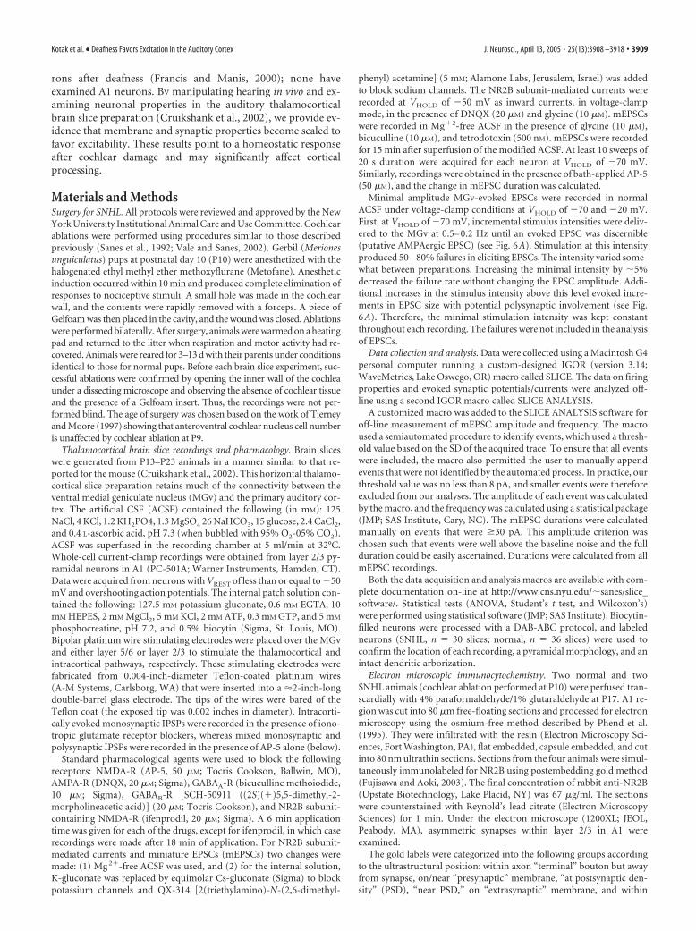
The mean resting membrane potentialof neurons from the SNHL animals dis-played a small but significant depolariza-tion when compared with the value fromnormal animals (Fig. 1A) (mean � SEM;normal, �63.4 � 0.8 mV vs SNHL,�59.9 � 0.8 mV; t test; t � 3; df � 100; p �0.002). SNHL also led to a significant in-crease in the mean input resistance (Fig.1B) (mean � SEM; normal, 277 � 13.5M� vs SNHL, 409 � 29 M�; t test; t � 4;df � 92; p 0.001). A linear fit betweenresting potential and input resistance did not reveal a significantcorrelation in either normal (R 2 � 0.01; p 0.05) or SNHL(R 2 � 0.02; p 0.05) neurons. Furthermore, we compared cer-tain properties of adapting only neurons after hearing loss. Thisanalysis showed a trend similar to that seen in the entire neuron
population (resting potential: normal adapting, 64 � 0.8 vsSNHL adapting, 59.4 � 0.9 mV, t test, t � 3.4, df � 73, p 0.0008; input resistance: normal adapting, 271 � 14 vs SNHLadapting, 452 � 29 M�, t test, t � 4.5, df � 67, p 0.0001).Whereas there was no effect of age on VREST, there was a small
Figure 1. SNHL increases membrane excitability. The resting membrane potential of SNHL neurons is significantly depolarized(A), and the input resistance is significantly higher (B) compared with neurons recorded in normal animals. In this and subsequentfigures, each open symbol represents the measurement from one neuron (n values shown on the x-axis). Filled symbols aremean � SEM. C, Examples of neurons that respond to suprathreshold current injection with an onset (top), sustained (middletrace), and adapting (bottom trace) response. Five suprathreshold pulses (10 pA, 1500 ms) were injected, and firing pattern wasestablished if the fifth pulse did not alter the firing pattern. D, Bar graphs of firing patterns in normal and SNHL neurons showpercentile distribution. After SNHL, onset-type neurons were not observed, and there was a significant rise in the sustained and adecrease in adapting neuron patterns, suggesting greater excitability. E, Linear fit of number of spikes by depolarizing currentinjection. Five 10 pA, 1500 ms depolarizing current steps were injected, and the resultant spikes were counted. The fit shows nosignificant difference between the stimulus–response characteristics of normal and SNHL adapting neurons; the SNHL sustainedneuron fits had a greater correlation coefficient, similar to the correlation in sustained neuron fits of normal neurons (see Results).Therefore, the characteristics of firing patterns did not change; rather, the incidence of sustained firing increased and adaptingneurons decreased in SNHL neurons, as shown in D.
3910 • J. Neurosci., April 13, 2005 • 25(13):3908 –3918 Kotak et al. • Deafness Favors Excitation in the Auditory Cortex
effect for RINPUT. Therefore, a linear fit was performed for datafrom normal and SNHL neurons that revealed a very small effectof age on RINPUT of SNHL neurons (R 2 � 0.11; p 0.05) but notnormal neurons (R 2 � 0.06; p 0.05).
The firing patterns of A1 neurons were characterized in re-sponse to suprathreshold depolarizing pulses (1500 ms) and fellinto three broad categories: (1) onset (phasic) neurons that fireda single action potential; (2) sustained neurons that fired at a highrate with little or no adaptation; and (3) adapting (regular spik-ing) neurons that fired at a relatively lower rate exhibited a sig-nificant degree of spike frequency adaptation (Fig. 1C). Whereas
15% of normal neurons were onset type,this response was not found in SNHLcases. Furthermore, the adapting patterndecreased by 13% (Fig. 1D), whereas theincidence of sustained type of firing in-creased by 30% after SNHL.
Table 1 presents a comparison of addi-tional measures, including spike thresholdand half-spike width. These measuresstrongly suggest that all included record-ings were obtained from pyramidal neu-rons and that differences in firing patternswere induced by hearing loss. For example,there were no significant differences be-tween the half-spike width ( p � 0.7; df �15), input resistance ( p � 0.4; df � 15), orresting membrane potential ( p � 0.11;df � 15) between the adapting and sus-tained SNHL neurons. Likewise, there wasno difference between the half-spikewidths of normal and SNHL neurons ( p �0.14; df � 20). In contrast, the spikethreshold (i.e., magnitude of voltage de-flection from the resting potential beforethe neurons fired) and the threshold cur-rent (i.e., the amount of current requiredto elicit a threshold response) were signif-icantly lower in SNHL neurons comparedwith normals. Additional comparisons be-tween the resting potentials and input re-sistance between all adapting neurons(normal vs SNHL) showed a highly signif-icant difference as seen in the entire popu-lation of normal versus SNHL neurons(Table 1). To compare the stimulus–re-sponse curves, a correlation coefficientwas obtained for injected current (five cur-rent steps of 1500 ms in 10 pA increments)
and number of elicited spikes for each firing pattern in Table 1.SNHL sustained neurons exhibited a stronger correlation (R 2 �0.6) compared with adapting neurons (normal, R 2 � 0.42;SNHL, R 2 � 0.43). The stimulus–response curve for normal sus-tained firing neurons is not plotted in Figure 1E (R 2 � 0.59). Themaximum instantaneous firing frequency in response to depolar-izing current injections (during the first 100 ms) for adaptingneurons was 25 Hz (n � 6), whereas that for sustained neuronswas 50 Hz (n � 6). The stimulus–response plots (Fig. 1E) fornormal and SNHL adapting neurons showed a similar incrementin firing rate. Therefore, hearing loss led to a net increase in the
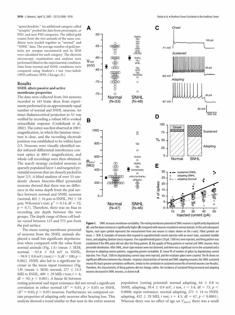
Figure 2. Layer 2/3 spiny pyramidal neurons. A selection of nine biocytin-filled layer 2/3 neurons with different firing patternsreveal cell and dendritic architecture. A, A normal adapting neuron. B, A normal sustained neuron. C, A normal onset neuron. D,Dendritic apical spines from the neuron shown in B. E, An SNHL sustained neuron. F, SNHL adapting (left) and sustained (right)neurons recorded in the same brain slice. G, Normal adapting (left) and onset (right) neurons recorded in the same slice. H, AnSNHL adapting neuron. Scale bar: A–C, E–H, 100 �m; D, 10 �m.
Table 1. Membrane properties of adapting and sustained firing neurons
GroupResting potential(mV)
Input resistance(M�)
Half-spikewidth (ms)
Threshold currentto elicit spike (pA)
Spike threshold(�Vm in mV)
Normal adapting �64 � 2.2 265 � 26 1.8 � 0.18 58 � 5 22 � 1.5(n � 11)SNHL adapting �61 � 1.4 410 � 29 2.2 � 0.19 36 � 2 16 � 1.4(n � 10)SNHL sustained �59.8 � 1.4 375 � 17 2.4 � 0.37 20 � 3.6 12.8 � 1.7(n � 6)
Statistical comparisons of membrane properties of �100 recorded neurons appear in Figure 1 and Results. Properties of a subset adapting and sustained neurons were additionally compared (columns 1, 2). Furthermore, �25% of neuronsfrom each group (left column) were blindly selected to compare the half-spike width, spike thresholds (in picoamperes, subthreshold current injected before the cell spiked and the resultant Vm). Statistical analyses: half-spike width (notdifferent) among the three groups, one-way ANOVA, p � 0.22, df � 26; spike thresholds (in picoamperes), normal adapting versus SNHL adapting, t test, t � 3.4, df � 20, p 0.002; SNHL adapting versus SNHL sustained, t test, t � 3.5,df � 15, p 0.003; spike threshold (Vm: the difference between the VREST and spike threshold); normal adapting versus SNHL adapting, t test, t � 2.6, df � 20, p 0.01; SNHL adapting versus SNHL sustained (not different), p � 0.19.For frequency–intensity (F–I) correlation coefficients, see Results and Figure 1E.
Kotak et al. • Deafness Favors Excitation in the Auditory Cortex J. Neurosci., April 13, 2005 • 25(13):3908 –3918 • 3911

incidence of the sustained firing patternand a net decrease in the adapting and on-set patterns.
To confirm that nonpyramidal cellswere not included in our sample, biocytinfilled neurons were examined after eachexperiment under 40 – 600� magnifica-tion to identify the dendritic structure andto confirm the presence of spines. Thisqualitative examination of normal andSNHL neurons indicated the presence ofintact basal and apical dendrites withspines in each recovered neuron includedin this study (Fig. 2).
SNHL enhances synaptic excitationMGv stimulation was used to evoke EPSPsin layer 2/3 pyramidal neurons (Fig. 3A). Acomparison of maximum EPSP ampli-tudes between normal and SNHL neuronsshowed no difference, both before and af-ter treatment with the NMDA receptorblocker AP-5 (mean � SEM before AP-5application: normal, 10.9 � 1.5 mV vsSNHL, 12.1 � 1.7 mV, t test, t � 0.54, df �26, p � 0.5; mean � SEM after AP-5 ap-plication: normal, 5.7 � 1 mV vs SNHL,4.2 � 0.7mV, t test, t � �1.1, df � 26, p �0.2).
In contrast to maximum EPSP ampli-tude, the EPSP duration was significantlylonger in SNHL neurons. Figure 3B illus-trates that the reduction in EPSP durationby the NMDA receptor blocker AP-5 wassignificantly greater after SNHL. Themean EPSP duration (milliseconds �SEM) of normal and SNHL neurons wassignificantly different before the applica-tion of AP-5 (normal, 238 � 22 ms vsSNHL, 412 � 69 ms; Wilcoxon’s test; � 2 �5.89; p � 0.02). Furthermore, the absolutereduction in EPSP duration by applicationof AP-5 was significantly greater in SNHLneurons (normal, 110 � 13 ms vs SNHL,315 � 44 ms; Wilcoxon’s test; � 2 � 16.62;p 0.0001). However, the duration of theremainder EPSP, presumably carried byAMPA receptors, was not different be-tween normal and SNHL neurons (nor-mal, 129 � 20 ms vs SNHL, 123 � 31 ms).
A similar trend was found for intracor-tically evoked EPSPs. SNHL led to longerEPSP durations and greater reduction byAP-5 than in normals (mean � SEM re-duction in duration of intracorticallyevoked EPSPs after AP-5 treatment: nor-mal, 70 � 14 ms vs SNHL, 190 � 39 ms; ttest; t � 2.8; df � 17; p � 0.01). There wasno significant difference in the maximumamplitude of intracortically evoked EPSPs before or after AP-5treatment.
To test whether the extended EPSP duration in SNHL neuronswas attributable to a change in the subunit composition of
NMDA receptors, MGv-evoked maximum EPSCs were recordedafter blocking sodium and potassium channels in an Mg�2-freeACSF (VHOLD of �50 mV). The NMDA receptor-mediated com-ponent was recorded in the presence of DNQX (20 �M), bicucul-
Figure 3. SNHL augments NMDA receptor function. A, A Schematic of the thalamocortical brain slice showing the position of astimulating electrode in the MGv (square pulse), the pathway (dark line) from MGv to A1, and a recording electrode within A1(Vm). The approximate distance the afferents travel from the MGv around the lateral geniculate (LGN) and hippocampus, radiatingto the recording site in layer 2/3, is �5.5 mm. B, Maximum EPSP evoked by stimulating MGv (arrowhead). Note the significantduration (in milliseconds) reduction by the NMDA receptor antagonist AP-5 and a greater reduction in an SNHL neuron. Restingmembrane potentials (in millivolts) indicated at the left of the traces. Inset shows MGv-evoked EPSP and EPSP-elicited spike(clipped); in all cases, the subthreshold maximum EPSPs were analyzed. C, Scatter plot of the magnitude of reduction in EPSPduration by AP-5 between normal and SNHL neurons shows a significantly greater AP-5-sensitive NMDA receptor-mediatedcomponent among SNHL cases. D, EPSCs evoked by stimulating at MGv (arrowhead). Note the reduction in amplitude by the NR2Bsubunit-specific antagonist ifenprodil and a greater reduction in an SNHL neuron. We do not rule out an effect of ifenprodil onpresynaptic NMDA receptors. E, Scatter plot of the magnitude of reduction in EPSC amplitude by ifenprodil between normal andSNHL neurons shows a significantly greater ifenprodil-sensitive NR2B subunit component in SNHL neurons.
3912 • J. Neurosci., April 13, 2005 • 25(13):3908 –3918 Kotak et al. • Deafness Favors Excitation in the Auditory Cortex
line (10 �M), and glycine (10 �M). In these experiments, we chosea stimulus intensity that produced an initial (before ifenprodil)EPSC amplitude of 400 –500 pA for both normal and SNHL neu-rons (mean � SEM EPSC amplitude: normal, 437 � 89 pA vsSNHL, 474 � 80 mV; Wilcoxon’s test; � 2 � 3.84; p � 0.05). Wemaintained this stimulus intensity throughout the experiments.In the absence of the preceding AMPAergic EPSC component(DNQX in bath), we measured the reduction in EPSC amplitudeby the antagonist to the NR2B subunit-containing NMDA recep-tors, ifenprodil, as a function of change in NR2B subunits.
The reduction of EPSC amplitude by the antagonist specificfor NR2B subunit-containing NMDA receptors, ifenprodil, wassignificantly greater in SNHL neurons (Fig. 3D,E) (mean � SEM
reduction after application of ifenprodil:normal, 201 � 41 pA vs SNHL, 318 � 38pA; t test; t � 2.1; df � 18; p � 0.05). Theremaining EPSC amplitudes, presumablycarried by the NR2A subunit, were not sig-nificantly different between normal andSNHL neurons. In two normal and twoSNHL neurons recorded from four differ-ent slices, these remainder EPSCs werecompletely and reversibly abolished byAP-5 (data not shown), demonstratingthat the analyzed EPSCs in this experimentwere exclusively NMDAergic.
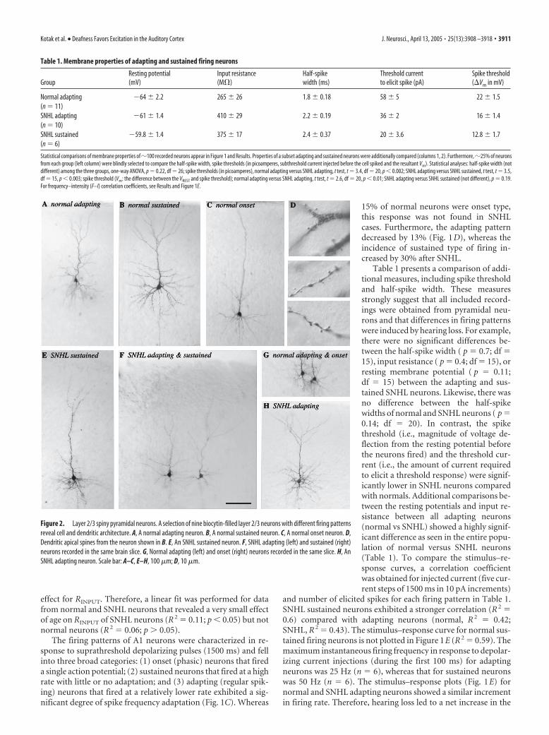
To assess whether increased sensitivityof EPSCs to ifenprodil in SNHL neuronswas attributable to increased expression ofNR2B subunits at synapses in layer 2/3,postembedding immunogold staining wasperformed to examine the localization ofthese subunits at synapses using an elec-tron microscope (Fig. 4). Gold particles re-flecting immunolabeling for NR2B that re-sided in the vicinity of asymmetricsynapses were counted and grouped intothe following mutually exclusive catego-ries: (1) on presynaptic membrane, (2) atPSD, (3) near PSD, (4) on extrasynapticmembrane, and (5) within spine/dendriteor within axon terminal. For each cate-gory, the average number of gold particlesper synapse was more numerous for theSNHL neurons (Table 2). The differencewas most pronounced at the PSD and overthe total synaptic region (i.e., the averageof the sum of gold particles in categories1–3).
If the maximum EPSP amplitude is de-termined primarily by the AMPAergiccomponent (Fig. 3), there could have beena masking effect attributable to the depo-
larized resting membrane potential (Fig. 1) and decreased firing
threshold (Table 1). Therefore, a series of voltage-clamp record-ings were performed. First, to examine whether such postsynap-tic alterations are accompanied by a presynaptic change, mEPSCswere recorded in nominal Mg 2�-ACSF and in the presence ofblockers of Na� and K� channels and GABAA receptors undervoltage-clamp conditions (see Materials and Methods). Analysesshowed a significant decrease in the frequency and an increase inthe peak amplitude of mEPSCs in SNHL neurons (Fig. 5)(mEPSC frequency, mean � SEM: normal, 3.3 � 0.2 Hz vsSNHL, 1.9 � 0.4 Hz, t test, t � 2.9, df � 7, p � 0.02; mEPSCamplitude, mean � SEM: normal, 12.7 � 1.4 pA vs SNHL, 20 �
Figure 4. Greater occurrence of immunolabeling of NR2B subunits at synapses from SNHL animals. A, B, Electron micrographsfrom normal brain. C, D, Micrographs from SNHL brain. Arrowheads indicate PSDs of asymmetric synapses. S and T representpostsynaptic spine and presynaptic axon terminal, respectively. Black dots are 10 nm immunogold particles labeling for NR2Bs.The categories of labeling are as follows: Pre, on presynaptic membrane; At, at PSD; Near, near PSD; Ex, on extrasynaptic mem-brane; S/D, within postsynaptic spine or dendrite away from the synapse; Tml, within axon terminal away from the synapse. Scalebar, 200 nm.
Table 2. Average � SEM number of NR2B-labeling immunogold particles per synapse in layer 2/3 of A1
Terminal Presynaptic At PSD Near PSD Synaptic Extrasynaptic Spine/Dendrite
Normal 1.95 � 0.19 0.20 � 0.05 0.12 � 0.04 0.16 � 0.05 0.48 � 0.08 0.18 � 0.04 0.97 � 0.15(n � 153)SNHL 2.85 � 0.22 0.51 � 0.06 0.56 � 0.07 0.38 � 0.05 1.45 � 0.11 0.60 � 0.10 1.48 � 0.18(n � 175)p values 0.0022 0.0001 0.0001 0.0018 0.0001 0.0001 0.0336
n denotes the number of synapses analyzed for each condition. Synaptic category was calculated by summing the values in presynaptic, at PSD, and near PSD categories. p values were obtained by performing nonpaired Student’s t test,two-tailed.
Kotak et al. • Deafness Favors Excitation in the Auditory Cortex J. Neurosci., April 13, 2005 • 25(13):3908 –3918 • 3913

2.6 pA, t test, t � 2.6, df � 7, p � 0.03).Furthermore, the duration of total mEP-SCs, and reduction in duration by AP-5,was greater in SNHL neurons than in nor-mals (Fig. 5) (total mEPSC duration,mean � SEM: normal, 51 � 4.7 vs SNHL,100.9 � 4.4, t test, t � 7.6, p 0.0001;mEPSC duration after AP-5, mean �SEM: normal, 34.5 � 3 ms vs SNHL, 46 �7 ms; t test, t � 2.8, p 0.01). Statisticalcomparisons confirmed that AP-5 signifi-cantly reduced mEPSC duration withinboth the normal and the SNHL groups(data not shown).
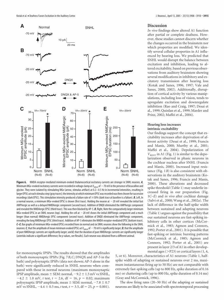
To assess whether putative monosyn-aptic connections from the MGv to layer2/3 pyramidal neurons exhibit NMDAer-gic alterations and whether there was aparallel change in AMPAergic function,minimum MGv-evoked EPSCs (�50%failure rates) were recorded in voltageclamp with normal ACSF, bicuculline (10�M), and glycine (10 �M) in the bath (Fig.6A). The results showed that the ampli-tude of minimum EPSCs at VHOLD of �70was significantly larger in SNHL neurons(minimum MGv-evoked amplitudes atVHOLD of �70 mV, mean � SEM: normal,11 � 2 pA vs SNHL, 28 � 5 pA; Wilcoxon’stest; � 2 � 5; p � 0.025). To examine theNMDAergic component, cells were held at�20 mV. Comparison of such EPSC dura-tions at this holding potential showed thatSNHL neuron EPSCs were significantlylonger (minimum MGv-evoked EPSC du-ration at VHOLD of �20 mV, mean � SEM:normal, 22 � 3 ms vs SNHL, 182 � 40 ms;Wilcoxon’s test; � 2 � 8.3; p � 0.003). Fi-nally, after blockade of AMPA receptors byDNQX, the pure NMDA receptor-mediated minimum-evoked EPSC cur-rents revealed at VHOLD of �20 mV weresignificantly larger and longer in SNHLneurons (minimum MGv-evoked ampli-tude in the presence of DNQX at VHOLD of�20 mV, mean � SEM: normal, 8 � 1.8pA vs SNHL, 49 � 19 pA, Wilcoxon’s test,� 2 � 7.4, p � 0.006; minimum MGv-evoked duration in thepresence of DNQX at VHOLD of �20 mV, mean � SEM: normal,17 � 2 ms vs SNHL, 191 � 38 ms, Wilcoxon’s test, � 2 � 8.3, p �0.003) (Fig. 6). In all neurons (n � 6 each normal and SNHLneurons), the EPSCs were abolished by AP-5, demonstrating thatthey were pure minimum-evoked MGv-evoked NMDAergic cur-rents (Fig. 6B). For each recorded neuron, we calculated the max-imum difference in response latency as a measure of variability inminimum-evoked EPSCs. We compared this measure of vari-ability between normal and SNHL neurons and found no signif-icant difference (normal, 1.5 � 0.3 ms vs SNHL, 1.1 � 0.2 ms;n � 12; p � 0.3).
SNHL reduces synaptic inhibitionBecause the resting potential of layer 2/3 pyramidal neurons innormal animals is close to ECl (data not shown), it was often
difficult to observe the evoked IPSPs. Furthermore, cortical stim-ulation produces a highly variable mixed excitatory–inhibitory syn-aptic response (Cruikshank et al., 2002). To circumvent these issues,recorded neurons were held at �55 mV to enhance the driving forcefor IPSPs, and excitatory ionotropic transmission was blocked par-tially with AP-5. This procedure revealed polysynaptic IPSPs. Inthree each normal and SNHL neurons, these IPSPs could then beblocked by the sequential addition of GABAB and GABAA receptorantagonists, respectively, demonstrating that they were GABAergic(data not shown). In another set of experiments, the AMPA receptorblocker DNQX was also added with AP-5; this strategy blocked re-cruitment of intracortical excitatory neurons that would drive inhib-itory interneurons. However, because such monosynaptic IPSPswere not recorded consistently, IPSP data were first examined withAP-5 application alone.
Figure 7A is a schematic of the stimulation and recording site
Figure 5. Frequency and amplitude of mEPSCs, respectively, decreases and increases in SNHL neurons. A, The left panel showstwo sweeps of mEPSCs recorded for 20 s each in a normal (left) and an SNHL neuron (right) at a holding potential of �70 mV. Eachrecording was acquired �2 min apart. Note that amplitudes of mEPSCs in an SNHL neuron (right) appear to be larger, whereas thefrequency of mEPSCs have decreased. At the bottom panel of A, expanded mEPSCs from a normal (left) and an SNHL neuron areshown, both before and after AP-5 treatment. The traces indicate that the amplitude, duration, and AP-5 sensitivity in the SNHLneuron are greater. B–D, Bar graphs summarizing the mean amplitude and duration of mEPSCs recorded from five normal andthree SNHL neurons. B, The amplitude of mean mEPSCs was significantly larger in SNHL neurons. C, The mean frequency of mEPSCsdeclined significantly in SNHL neurons. D, The total mEPSC duration was significantly greater in SNHL neurons. Asterisks (and barin D) indicate that the differences are significant (for p values, see Results). Each neuron was obtained from a different animal.
3914 • J. Neurosci., April 13, 2005 • 25(13):3908 –3918 Kotak et al. • Deafness Favors Excitation in the Auditory Cortex
for monosynaptic IPSPs. The results showed that the amplitudesof both monosynaptic IPSPs (Fig. 7B,C; DNQX and AP-5 in thebath) and polysynaptic IPSPs (data not shown; AP-5 alone in thebath) were significantly reduced in SNHL neurons when com-pared with those in normal neurons (maximum monosynapticIPSP amplitude, mean � SEM: normal, �9.2 � 1.5 mV vs SNHL,�4.2 � 1.0 mV, t test, t � 2.6, df � 16, p � 0.02; maximumpolysynaptic IPSP amplitude, mean � SEM: normal, �7.8 � 0.7mV vs SNHL, �4.4 � 0.5 ms, t test, t � 3.5, df � 25, p � 0.001).
DiscussionIn vivo findings show altered A1 functionafter partial or complete deafness. How-ever, these studies cannot discern whetherthe changes occurred in the brainstem norwhich properties are modified. We iden-tify several cellular properties in A1 influ-enced by hearing loss. We predicted thatSNHL would disrupt the balance betweenexcitation and inhibition, leading to al-tered excitability, based on previous obser-vations from auditory brainstem showingseveral modifications in inhibitory and ex-citatory transmission after hearing loss(Kotak and Sanes, 1996, 1997; Vale andSanes, 2000, 2002). Additionally, disrup-tion of cortical activity by various manip-ulations, including loss of vision, tends toupregulate excitation and downregulateinhibition (Rao and Craig, 1997; Desai etal., 1999; Quinlan et al., 1999; Marder andPrinz, 2002; Maffei et al., 2004).
Hearing loss increasesintrinsic excitabilityOur findings support the concept that ex-citability increases after deprivation of af-ferent activity (Desai et al., 1999; Francisand Manis, 2000; Murthy et al., 2001;Maffei et al., 2004). Depolarization ofVREST in A1 (Fig. 1) is similar to the depo-larization observed in phasic neurons inthe cochlear nucleus after SNHL (Francisand Manis, 2000). Increased input resis-tance (Fig. 1B) is also consistent with ob-servations in the auditory brainstem (Ko-tak and Sanes, 1996; Francis and Manis,2000). These alterations and decreasedspike threshold (Table 1) may underlie in-creased firing in our preparation (Fig.1C–E) and in vivo after cochlear damage(Salvi et al., 2000; Wang et al., 2002a). Thelack of difference in the half-spike widthbetween sustained and adapting neurons(Table 1) argues against the possibility thatour sustained neurons are fast-spiking in-hibitory cells in other cortices (McCor-mick et al., 1985; Agmon and Connors,1992; Porter et al., 2001). It is possible thatfast-spiking or intrinsic bursting patterns(McCormick et al., 1985; Agmon andConnors, 1992; Porter et al., 2001) arepresent in layer 2/3 of A1 in other develop-mental ages (P23) or cortical layers (1, 4,
5, or 6). Moreover, characteristics of A1 neurons (Table 1, half-spike width of adapting or sustained neurons over 2 ms, maxi-mum instantaneous firing up to 50 Hz) are not comparable withextremely fast-spiking cells (up to 800 Hz, spike duration of 0.16ms) or chattering cells (up to 800 Hz, spike duration of 0.14 ms)(Gray and McCormick, 1996).
The slow firing rates (20–50 Hz) of the adapting or sustainedneurons are likely to be associated with spectrotemporal processing
Figure 6. NMDA receptor-mediated minimum-evoked thalamocortical excitatory currents are stronger in SNHL neurons. A,Minimum MGv-evoked excitatory currents were recorded in voltage clamp at VHOLD of �70 mV in the presence of bicuculline andglycine. They were isolated by stimulating MGv (arrow, stimulus artifact) at 0.2– 0.5 Hz in incremental intensities, resulting inlarger EPSCs at each stimulus step (gray traces); the intensity at which minimum EPSC was recorded was then chosen for successiverecordings (dark EPSC). This stimulation intensity produced a failure rate of �50% (dark trace at baseline is a failure). B, Left, Ina normal neuron, a minimum MGv-evoked EPSC is shown (first trace). Holding the neuron at �20 mV revealed the initial fastAMPAergic as well as a delayed NMDAergic component (second trace). Addition of DNQX eliminated the AMPAergic componentand revealed the NMDAergic EPSC (third trace). This was then blocked by AP-5. B, Right, Note the comparatively larger minimumMGv-evoked EPSC in an SNHL neuron (top). Holding the cell at �20 mV shows the initial AMPAergic component and a muchlonger (than normal) NMDAergic EPSC component (second trace). Addition of DNQX eliminated the AMPAergic component,revealing the long NMDAergic EPSC (third trace). Addition of AP-5 eliminates the NMDA receptor-mediated EPSC (bottom trace).C–E, Bar graphs of minimum MGv-evoked EPSCs recorded from six normal and six SNHL neurons show the following in the SNHLneurons: C, that the amplitude of mean minimum evoked EPSCs at VHOLD of �70 mV is significantly larger; D, that the amplitudeof pure NMDAergic currents are significantly larger; and E, that the duration of pure NMDAergic currents are significantly longer(asterisks indicate a significant difference; for p values, see Results). Each neuron was obtained from a different animal.
Kotak et al. • Deafness Favors Excitation in the Auditory Cortex J. Neurosci., April 13, 2005 • 25(13):3908 –3918 • 3915

of static or dynamic auditory cues that elicit low discharge rates(Semple and Scott, 2003). This may contrast with integration ofimage-specific synchronous oscillations in the visual cortex (for re-view, see Singer and Gray, 1995; Gray and McCormick, 1996).
The changes in voltage-gated channels or molecular mechanismsafter hearing impairments that cause firing properties to alter remainunidentified. Because potassium and calcium currents regulate fir-ing patterns, hearing loss may modify these channels. Thus, down-regulation of a potassium leakage conductance after SNHL coulddepolarize VREST and increase input resistance, making the neuronselectrically compact. Additionally, downregulation of a low- or high-
threshold K� conductance could underlie a lack of phase lockingand heightened firing rates (Fig. 1) (Svirskis et al., 2002).
SNHL increases synaptic excitation and decreases inhibitionLonger EPSPs and larger reduction by AP-5 after SNHL demon-strates an enhanced NMDA receptor-mediated excitatory re-sponse (Fig. 3B,C). Furthermore, greater ifenprodil sensitivity ofSNHL EPSCs (Fig. 3D,E) and higher NR2B-positive immuno-gold counts at synapses (Fig. 4, Table 2) demonstrate that in-creased postsynaptic glutamate sensitivity may compensate forthe loss of cochlear activity. These findings agree with an aug-mented NMDA receptor function in evoked excitation in thelateral superior olive (LSO) and IC after hearing loss (Kotak andSanes, 1996; Vale and Sanes, 2002) and increased NMDAergicEPSCs in the CN of deaf mutant mice (Oleskevich and Walmsley,2002).
An augmentation of NR2B subunits (Figs. 3, 4) parallels thefinding in visual cortex that high expression of synaptic NR2Bsubunits occurs after visual deprivation. In the visual cortex, thetermination of the critical period, demonstrated by reduction inNR2B expression and increase in NR2A expression, can be de-layed by sensory deprivation (Erisir and Harris, 2003). Our re-sults support the notion that similar mechanisms operate duringA1 development. NR2A and NR2B subunit expression at corticaland hippocampal synapses follow disparate trafficking and deliv-ery rules, depending on activity (Rao and Craig, 1997; Quinlan etal., 1999; Barria and Malinow, 2002; Fong et al., 2002; Aoki et al.,2003; Fujisawa and Aoki, 2003).
The increase of mEPSC amplitude and duration in SNHLneurons suggests an upregulation of both AMPA and NMDAreceptors (Fig. 5) (Myme et al., 2003). Furthermore, the parallelincrease in DNQX-sensitive (AMPAergic) and AP-5-sensitive(NMDAergic) components in MGv-evoked minimum-evokedEPSCs (Fig. 6) demonstrates that the increased excitation is me-diated, in part, by thalamic afferents. The lack of change in MGv-evoked maximum EPSP amplitude could be attributable to sev-eral confounding variables, including depolarized VREST (Fig. 1)and decreased spike threshold (Table 1).
Activity deprivation alters the scaling of excitatory synapticinputs at a network level in an age-dependent manner (Murthy etal., 2001; Desai et al., 2002; Maffei et al., 2004). Decreased fre-quency of mEPSCs in SNHL neurons are in agreement with re-ports showing that deprivation of activity also leads to a decreasein presynaptic release properties (Fig. 5) (Desai et al., 1999; Mur-thy et al., 2001). Moreover, in the cochlear nucleus of congeni-tally deaf mice and IC neurons of deafened gerbils, presynapticproperties have been shown to change (Oleskevich and Walms-ley, 2002; Vale and Sanes, 2002). In our preparation, an increaseof presynaptically localized NR2B subunits was also observed(Fig. 4). Presynaptic NR2B-containing NMDA receptors havebeen shown to enhance glutamate release (Berretta and Jones,1996). It remains to be determined whether a subset of presyn-aptic glutamate receptors can augment the release of transmitterafter hearing loss. Together, our observations are consistent withanalyses of synapse physiology in the CN, LSO, and IC after SNHL orin congenitally deaf mice and support a homeostatic mechanism:excitatory synapses get stronger, whereas inhibitory synapses be-come weaker (Kotak and Sanes, 1996; Vale and Sanes, 2000, 2002;Oleskevich and Walmsley, 2002).
Enhanced synaptic excitation is accompanied by diminishedinhibition in diverse experimental models. For example, disrup-tion of activity in cultured cortical neurons decreases spontane-ous IPSCs possibly by downregulation of GABAA receptors (Kil-
Figure 7. SNHL reduces monosynaptic IPSP amplitude. A, Schematic of the thalamocorticalbrain slice showing a stimulating electrode on layer 2/3 (square pulse) �1 mm rostral to therecording electrode. B, The maximum monosynaptic IPSP evoked by stimulating layer 2/3 isshown for a normal cell (top) and an SNHL cell (bottom). Note that the SNHL IPSP amplitudeappears to be smaller. These recordings were obtained in the presence of blockers of the iono-tropic glutamate receptors DNQX and AP-5. C, The plot of monosynaptic IPSP amplitudes fromall recorded neurons shows a significant reduction for SNHL neurons.
3916 • J. Neurosci., April 13, 2005 • 25(13):3908 –3918 Kotak et al. • Deafness Favors Excitation in the Auditory Cortex
man et al., 2002). In vivo studies in the auditory system show thatdecreased inhibition after auditory deprivation complementsgreater excitation (Salvi et al., 2000). Specifically, weakened side-band inhibition after noise- and drug-induced hearing loss con-tributes to enhanced IC neuron discharge, as well as expansionand increased sensitivity of low-frequency tuning curve (Wang etal., 2002a). Based on pharmacological observations in vivo, aweakening of intracortical inhibition would affect A1 firing prop-erties as well as their ability to encode sound frequency (Wang etal., 2002b). Increased firing may explain heightened sound-evoked activity in the ipsilateral IC and auditory cortex after par-tial deafening; the cause of the latter may be an imbalance be-tween excitation and inhibition (Kitzes and Semple, 1985; Realeet al., 1987; McAlpine et al., 1997; Mossop et al., 2000). In ourpreparation, reduction in maximum IPSP amplitude suggestsmodulation of the fast initial IPSP component carried bybicuculline-sensitive GABAA receptors (Fig. 6). Our results ondecreased synaptic inhibition are interpreted to mean that thestrength of GABAergic synapses decreased after hearing loss. Analternative explanation is that the inhibitory interneurons have alower threshold for activation after hearing loss. However, weused a stimulus level that produced maximal IPSP amplitude(i.e., no additional increase in amplitude could be obtained withincreasing stimulus amplitude). Therefore, a decreased firingthreshold of inhibitory interneurons is unlikely to explain thefindings.
Implications for cortical processing and plasticityIn vivo recordings from animals with hearing loss reveal pro-found changes in auditory processing (Syka, 2002). Even studiesfrom human cortex suggest that central physiology has changedafter deafness (Ponton and Eggermont, 2001; Sharma et al.,2002). The regional blood flow measured in A2 is relativelygreater in prelingually deaf cochlear implant users. Furthermore,speech tends to activate a larger fraction of temporal cortex inimplanted patients (Hirano et al., 2000; Naito et al., 2000). Anincrease in sound-evoked discharges is consistently observed af-ter cochlear damage (Kitzes and Semple, 1985; Szczepaniak andMoller, 1995; Rajan, 1998; Klinke et al., 1999; Rajan, 2001). Ourfindings indicate that three major cellular changes can accountfor increased excitability and support a homeostatic mechanism:passive membrane properties favor excitability, excitatory syn-apses become stronger, and inhibitory synapses become weakerafter SNHL. Greater NMDAergic participation could overwhelmdecreased inhibition during prostheses-evoked responses, thusrecruiting an excitable A1. Furthermore, prolonged EPSP timecourses could support temporal summation by virtue of theboosted NMDA/NR2B expression (Fig. 3), especially in associa-tion with enhanced intrinsic excitability (Fig. 1).
The processing of spectrotemporal and level-dependent prop-erties of sound are dependent on synaptic and intrinsic propertiesof A1 neurons. Developing auditory brainstem neurons dependon cochlear-evoked activity for synaptic refinements and emer-gence of adult-like tonotopic maps (Sanes and Constantine-Paton, 1985; Kapfer et al., 2002). Our data support empirical andmodeling work associated with various invertebrate and verte-brate preparations that have shown that ionic and synaptic mech-anisms are capable of homeostasis (Marder and Prinz, 2002). Thepersistent excitability we observe after SNHL may ultimatelylimit the finer integrative computations performed by A1 neu-rons, unless prosthetic devices are used early in development torestore activity (Sharma et al., 2002).
ReferencesAgmon A, Connors BW (1992) Correlation between intrinsic firing pat-
terns and thalamocortical synaptic responses of neurons in mouse barrelcortex. J Neurosci 12:319 –329.
Aoki C, Fujisawa S, Mahadomrongkul V, Shah P, Nader K, Erisir E (2003)NMDA receptor blockade in intact adult cortex increases trafficking ofNR2A subunits into spines, postsynaptic densities and axon terminals.Brain Res 963:139 –149.
Barria A, Malinow R (2002) Subunit-specific NMDA receptor trafficking tosynapses. Neuron 35:345–353.
Berretta N, Jones RS (1996) Tonic facilitation of glutamate release by pre-synaptic N-methyl-D-aspartate autoreceptors in the entorhinal cortex.Neuroscience 75:339 –344.
Burrone J, O’Byrne M, Murthy VN (2002) Multiple forms of synaptic plas-ticity triggered by selective suppression of activity in individual neurons.Nature 420:414 – 418.
Cruikshank SI, Rose HJ, Metherate R (2002) Auditory thalamocortical syn-aptic transmission in vitro. J Neurophysiol 87:361–384.
Desai NS, Rutherford LC, Turrigiano GG (1999) Plasticity in the intrinsicexcitability of cortical pyramidal neurons. Nat Neurosci 2:515–520.
Desai NS, Cudmore RH, Nelson SB, Turrigiano GG (2002) Critical periodsfor experience-dependent synaptic scaling in visual cortex. Nat Neurosci5:783–789.
Emmorey K, Allen JS, Schenker N, Damasio H (2003) A morphometricanalysis of auditory brain regions in congenitally deaf adults. Proc NatlAcad Sci USA 100:10049 –10054.
Erisir A, Harris JL (2003) Decline of the critical period of visual plasticity isconcurrent with the reduction of NR2B subunit of the synaptic NMDAreceptor in layer 4. J Neurosci 23:5208 –5218.
Fong DK, Rao A, Crump FT, Craig AM (2002) Rapid synaptic remodelingby protein kinase C: reciprocal translocation of NMDA receptor andcalcium/calmodulin-dependent kinase II. J Neurosci 15:2153–2164.
Francis HW, Manis PB (2000) Effects of deafferentation on the electrophys-iology of ventral cochlear nucleus neurons. Hear Res 149:91–105.
Fujisawa S, Aoki C (2003) In vivo blockade of N-methyl-D-aspartate recep-tors induces rapid trafficking of NR2B subunits away from synapses andout of spines and terminals in adult cortex. Neuroscience 121:51– 63.
Gray CM, McCormick DA (1996) Chattering cells: superficial pyramidalneurons contributing to the generation of synchronous oscillation in thevisual cortex. Science 274:109 –113.
Hirano S, Naito Y, Kojima H, Honjo I, Inoue M, Shoji K, Tateya I, Fujiki N,Nishizawa S, Konishi J (2000) Functional differentiation of the auditoryassociation area in prelingually deaf subjects. Auris Nasus Larynx27:303–310.
Iverson P (2003) Evaluating the function of phonetic perceptual phenom-ena within speech recognition: an examination of the perception of /d/-/t/by adult cochlear implant users. J Acoust Soc Am 113:1056 –1064.
Kapfer C, Seidl AH, Schweizer H, Grothe B (2002) Experience-dependentrefinement of inhibitory inputs to auditory coincidence-detector neu-rons. Nat Neurosci 5:247–253.
Kidd Jr G, Arbogast TL, Mason CR, Walsh M (2002) Informational maskingin listeners with sensorineural hearing loss. J Assoc Res Otolaryngol3:107–119.
Kilman V, van Rossum MC, Turrigiano GG (2002) Activity deprivation re-duces miniature IPSC amplitude by decreasing the number of postsynap-tic GABAA receptors clustered at neocortical synapses. J Neurosci22:1328 –1337.
Kitzes LM, Semple MN (1985) Single-unit responses in the inferior collicu-lus: effects of neonatal unilateral cochlear ablation. J Neurophysiol53:1483–1500.
Klinke R, Kral A, Heid S, Tillein J, Hartmann R (1999) Recruitment of theauditory cortex in congenitally deaf cats by long-term cochlear electro-stimulation. Science 285:1729 –1733.
Kotak VC, Sanes DH (1996) Developmental influence of glycinergic trans-mission: regulation of NMDA receptor-mediated EPSPs. J Neurosci16:1836 –1843.
Kotak VC, Sanes DH (1997) Deafferentation weakens excitatory synapses inthe developing central auditory system. Eur J Neurosci 9:2340 –2347.
Kral A, Hartmann R, Tillein J, Heid S, Klinke R (2000) Congenital auditorydeprivation reduces synaptic activity within the auditory cortex in a layerspecific manner. Cereb Cortex 10:714 –726.
Maffei A, Nelson SB, Turrigiano GG (2004) Selective reconfiguration of
Kotak et al. • Deafness Favors Excitation in the Auditory Cortex J. Neurosci., April 13, 2005 • 25(13):3908 –3918 • 3917
layer 4 visual cortical circuitry by visual deprivation. Nat Neurosci7:1353–1359.
Marder E, Prinz AA (2002) Modeling stability in neuron and network func-tion: the role of activity in homeostasis. BioEssays 24:1145–1154.
McAlpine D, Martin RL, Mossop JE, Moore DR (1997) Response propertiesof neurons in the inferior colliculus of the monaurally-deafened ferret toacoustic stimulation of the intact ear. J Neurophysiol 78:767–779.
McCormick DA, Connors BW, Lighthall JW, Prince DA (1985) Compara-tive electrophysiology of pyramidal and sparsely spiny stellate neuron ofthe neocortex. J Neurophysiol 54:782– 806.
Mody M, Schwartz RG, Gravel JS, Ruben RJ (1999) Speech perception andverbal memory in children with and without histories of otitis media. JSpeech Lang Hear Res 42:1069 –1079.
Moore CM, Vollmer, M Leake PA, Snyder RL, Rebscher SJ (2002) The ef-fects of chronic intracochlear electrical stimulation on inferior colliculusspatial representation in adult deafened cats. Hear Res 164:82–96.
Mossop JE, Wilson MJ, Caspary DM, Moore DR (2000) Down-regulationof inhibition following unilateral deafening. Hear Res 147:183–187.
Murthy VN, Schikorski T, Stevens CF, Zhu Y (2001) Inactivity producesincreases in neurotransmitter release and synapse size. Neuron32:673– 682.
Myme CI, Sugino K, Turrigiano GG, Nelson SB (2003) The NMDA-to-AMPA ratio at synapses onto layer 2/3 pyramidal neurons is conservedacross prefrontal and visual cortices. J Neurophysiol 90:771–779.
Naito Y, Tateya I, Fujiki N, Hirano S, Ishizu K, Nagahama Y, Fukuyama H,Kojima H (2000) Increased cortical activation during hearing of speechin cochlear implant users. Hear Res 143:139 –146.
Oleskevich S, Walmsley B (2002) Synaptic transmission in the auditorybrainstem of normal and congenitally deaf mice. J Physiol (Lond)540:447– 455.
Phend KD, Rustioni A, Weinberg RJ (1995) An osmium-free method ofepon embedment that preserves both ultrastructure and antigenicity forpost-embedding immunocytochemistry. J Histochem Cytochem43:283–292.
Ponton CW, Eggermont JJ (2001) Of kittens and kids: altered cortical mat-uration following profound deafness and cochlear implant use. AudiolNeurootol 6:363–380.
Porter JT, Johnson CK, Agmon A (2001) Diverse types of interneurons gen-erate thalamus-evoked feedforward inhibition in the mouse barrel cortex.J Neurosci 21:2699 –2710.
Psarommatis IM, Goritsa E, Douniadakis D, Tsakanikos M, KontrogianniAD, Apostolopoulos N (2001) Hearing loss in speech-language delayedchildren. Int J Pediatr Otorhinolaryngol 58:205–210.
Quinlan EM, Olstein DH, Bear MF (1999) Bidirectional, experience-dependent regulation of N-methyl-D-aspartate receptor subunit compo-sition in the rat visual cortex during postnatal development. Proc NatlAcad Sci USA 96:1276 –1280.
Raggio MW, Schreiner CE (1999) Neuronal responses in cat primary audi-tory cortex to electrical cochlear stimulation. III. Activation patterns inshort- and long-term deafness. J Neurophysiol 82:3506 –3526.
Raggio MW, Schreiner CE (2003) Neuronal responses in cat primary audi-tory cortex to electrical cochlear stimulation. IV. Activation pattern forsinusoidal stimulation. J Neurophysiol 89:3190 –3204.
Rajan R (1998) Receptor organ damage causes loss of cortical surround in-hibition without topographic map plasticity. Nat Neurosci 1:138 –143.
Rajan R (2001) Plasticity of excitation and inhibition in the receptive field ofprimary auditory cortical neurons after limited receptor organ damage.Cereb Cortex 11:171–182.
Rao A, Craig AM (1997) Activity regulates the synaptic localization of theNMDA receptor in hippocampal neurons. Neuron 19:801– 812.
Reale RA, Brugge JF, Chan JC (1987) Maps of auditory cortex in cats rearedafter unilateral cochlear ablation in the neonatal period. Brain Res431:281–290.
Salvi RJ, Wang J, Ding D (2000) Auditory plasticity and hyperactivity fol-lowing cochlear damage. Hear Res 147:261–274.
Sanes DH, Constantine-Paton M (1985) The development of stimulus fol-lowing in the cochlear nerve and inferior colliculus of the mouse. BrainRes 354:255–267.
Sanes DH, Markowitz S, Bernstein J, Wardlow J (1992) The influence ofinhibitory afferents on the development of postsynaptic dendritic arbors.J Comp Neurol 321:637– 644.
Semple MN, Scott BH (2003) Cortical mechanisms in hearing. Curr OpinNeurobiol 13:167–173.
Sharma A, Dorman MF, Spahr AJ (2002) A sensitive period for the devel-opment of the central auditory system in children with cochlear implants:implications for age of implantation. Ear Hear 23:532–539.
Singer W, Gray CM (1995) Visual feature integration and the temporal cor-relation hypothesis. Annu Rev Neurosci 18:555–586.
Snyder RL, Sinex DG, McGee JD, Walsh EW (2000) Acute spiral ganglionlesions change the tuning and tonotopicorganization of cat inferior col-liculus neurons. Hear Res 147:200 –220.
Svirskis GS, Kotak VC, Sanes DH, Rinzel JH (2002) Enhancement of signal-to-noise ratio and phase locking for small inputs by low threshold out-ward current in auditory neurons. J Neurosci 22:11019 –11025.
Syka J (2002) Plastic changes in the central auditory system after hearingloss, restoration of function, and during learning. Physiol Rev82:601– 636.
Szczepaniak WS, Moller AR (1995) Effects of L-baclofen and D-baclofen onthe auditory system: a study of click-evoked potentials from the inferiorcolliculus in the rat. Ann Otol Rhinol Laryngol 104:399 – 404.
Tierney TS, Moore DR (1997) Naturally occurring neuron death duringpostnatal development of the gerbil ventral cochlear nucleus begins at theonset of hearing. J Comp Neurol 387:421– 429.
Vale C, Sanes DH (2000) Afferent regulation of inhibitory synaptic trans-mission in the developing auditory midbrain. J Neurosci 20:1912–1921.
Vale C, Sanes DH (2002) The effect of bilateral deafness on excitatory syn-aptic strength in the auditory midbrain. Eur J Neurosci 16:2394 –2404.
Vernon-Feagans L (1999) Impact of otitis media on speech, language, cog-nition, and behavior. In: Evidence-based otitis media (Rosenfeld RM,Bluestone CD, eds), pp 353–373. St. Louis, MO: Decker.
Wang J, Ding D, Salvi RJ (2002a) Functional reorganization in chinchillainferior colliculus associated with chronic and acute cochlear damage.Hear Res 168:238 –249.
Wang J, McFadden SL, Caspary D, Salvi R (2002b) Gamma-aminobutyricacid circuits shape response properties of auditory cortex neurons. BrainRes 944:219 –231.
3918 • J. Neurosci., April 13, 2005 • 25(13):3908 –3918 Kotak et al. • Deafness Favors Excitation in the Auditory Cortex