Novel polymer nanocomposite hydrogel with natural clay nanotubes
Upload
khangminh22Category
view
5download
0
Citation: Mubarok, W.; Elvitigala,
K.C.M.L.; Sakai, S. Tuning
Myogenesis by Controlling Gelatin
Hydrogel Properties through
Hydrogen Peroxide-Mediated
Cross-Linking and Degradation. Gels
2022, 8, 387. https://doi.org/
10.3390/gels8060387
Academic Editors: Yanen Wang and
Qinghua Wei
Received: 1 June 2022
Accepted: 15 June 2022
Published: 17 June 2022
Publisher’s Note: MDPI stays neutral
with regard to jurisdictional claims in
published maps and institutional affil-
iations.
Copyright: © 2022 by the authors.
Licensee MDPI, Basel, Switzerland.
This article is an open access article
distributed under the terms and
conditions of the Creative Commons
Attribution (CC BY) license (https://
creativecommons.org/licenses/by/
4.0/).
gels
Article
Tuning Myogenesis by Controlling Gelatin Hydrogel Propertiesthrough Hydrogen Peroxide-Mediated Cross-Linkingand DegradationWildan Mubarok , Kelum Chamara Manoj Lakmal Elvitigala and Shinji Sakai *
Department of Materials Engineering Science, Graduate School of Engineering Science, Osaka University,Toyonaka 560-8531, Japan; [email protected] (W.M.);[email protected] (K.C.M.L.E.)* Correspondence: [email protected]
Abstract: Engineering skeletal muscle tissue in vitro is important to study the mechanism of myoge-nesis, which is crucial for regenerating muscle cells. The physicochemical properties of the cellularmicroenvironment are known to govern various cell behaviours. Yet, most studies utilised syntheticmaterials to model the extracellular matrix that suffers from cytotoxicity to the cells. We have previ-ously reported that the physicochemical property of hydrogels obtained from horseradish peroxidase(HRP)-catalysed cross-linking could be controlled by a simple adjustment to the exposure time to aircontaining H2O2. In this study, we evaluated the influence of physicochemical properties dynamicsin the gelatin possessing phenol groups (Gelatin-Ph) hydrogel to regulate the myogenesis in vitro.We controlled the Young’s modulus of the Gelatin-Ph hydrogel by tuning the air containing 16 ppmH2O2 exposure time for 15–60 min. Additionally, prolonged exposure to air containing H2O2 alsoinduced Gelatin-Ph degradation. Myoblasts showed higher adhesion and myotube formation onstiff hydrogel (3.53 kPa) fabricated through 30 min of exposure to air containing H2O2 compared tothose on softer hydrogel (0.77–2.79 kPa) fabricated through 15, 45, and 60 min of the exposure. Theseresults demonstrate that the myogenesis can be tuned by changes in the physicochemical propertiesof Gelatin-Ph hydrogel mediated by H2O2.
Keywords: horseradish peroxidase; myoblast; skeletal muscle; tissue engineering; gelatin
1. Introduction
Skeletal muscle is the largest component of the human body, accounting for 30–40% ofbody mass [1,2]. In the occurrence of traumatic injuries or degenerative diseases, skeletalmuscle can be damaged, which causes a physiological impairment. To rescue the physio-logical function, the formation of new muscle cells (myogenesis) is needed. While nativecells could regenerate the muscle tissue, controlling the fate of these cells to differentiateinto muscle cells is difficult due to the complex interaction between the intrinsic factorsof the cells and external factors, including their microenvironment. The tissue engineer-ing approach has garnered great interest since it provides an in vitro model to study thephysiological phenomenon regulating myogenesis that could improve the efficiency of celltherapy [3,4].
Among the important factors that govern myogenesis are the materials comprisingthe extracellular matrix (ECM) and the physicochemical properties of the ECM. To studythe physicochemical effect of the substrate on myoblasts, most studies utilised syntheticmaterials such as poly(ethylene glycol) diacrylate (PEGDA), poly(vinyl alcohol) (PVA) gels,and poly(acrylamide) [5,6]. Zahari et al. utilised laminin-coated poly(methyl methacrylate)nanofiber scaffold [7], while Shin et al. reported the use of poly(lactic-co-glycolic acid) [8].Additionally, the graphene oxide-based matrix is also widely used to control myoblast
Gels 2022, 8, 387. https://doi.org/10.3390/gels8060387 https://www.mdpi.com/journal/gels
Gels 2022, 8, 387 2 of 13
differentiation [9,10]. However, these materials are not components of natural ECM andsuffer from the toxicity of the uncross-linked monomers and photo-crosslinkers [5,6].
To model the native ECM environment, gelatin is widely used due to its excellentbiocompatibility and biodegradability [11]. In recent years, several studies have reportedthe application of gelatin-based hydrogels to control myoblasts’ behaviour. Hayashi et al.reported gelatin-conjugated supramolecular hydrogels with a switchable stiffness [12].While C2C12 adhesion showed dynamic changes according to the stiffness, this study doesnot report the myoblasts differentiation. Denes et al. fabricated micropatterned gelatinhydrogels to study the myotube orientation [13]. Du et al. also successfully achieveddirected cell migration and myotube formation using 3D-printed gelatin methacrolyl(GelMA) micropatterns on a surface coated with thermo-responsive material poly(N-isopropylacrylamide) [14]. However, these studies do not consider the effect of the physico-chemical properties of the gelatin hydrogel, which also play a key role in myogenesis.
Recently, we have reported that hydrogen peroxide (H2O2) could be used to con-trol the physicochemical property of gelatin derivatives possessing phenolic hydroxylmoieties (Gelatin-Ph) hydrogel obtained from horseradish peroxidase (HRP)-catalysedcross-linking [15]. This system exploits the contradictory effect of H2O2 that simultaneouslyinduces the HRP-catalysed cross-linking as an electron donor while degrading the polymeras an oxidant (Figure 1a) [15]. The advantage of this system is that the mechanical propertyand molecular weight of the hydrogel can be controlled by a simple adjustment of theair containing H2O2 exposure time. Using this system, the adhesion of stem cells andfibroblasts can be controlled [15]. However, there are no reports that study the effect of thedynamics of the physicochemical properties of the Gelatin-Ph hydrogel by H2O2 to controlthe myogenesis.
Figure 1. (a) The conceptual scheme of the contradictory function of hydrogen peroxide (H2O2)to induce horseradish peroxidase (HRP)-catalysed cross-linking and polymer degradation of theGelatin-Ph. (b) Experimental scheme of this study. Gelatin-Ph hydrogels fabricated by tuning aircontaining H2O2 exposure time were characterised, and the effect on myoblasts’ viability, adhesion,and differentiation was studied.
In this study, we aimed to investigate the effect of the H2O2–mediated control of thephysicochemical properties of Gelatin-Ph on modulating myogenesis. To address this, we
Gels 2022, 8, 387 3 of 13
fabricated the Gelatin-Ph hydrogel by exposing air containing H2O2 at different exposuretimes, and the hydrogel properties were characterised. The modulatory effect of thesephysicochemical changes of Gelatin-Ph on myoblast behaviour is studied to the myoblastadhesion and viability. In addition, we also report the influence of the H2O2 contradictingeffect on the formation of myotubes on the Gelatin-Ph hydrogel (Figure 1b).
2. Results and Discussion2.1. Gelatin-Ph Hydrogel Characterisation
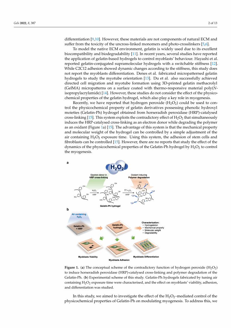
Gelatin-Ph was successfully prepared by conjugating the gelatin with HPPA viaWSCD/NHS chemistry in a DMF buffer pH 4.7 (Figure 2a). UV-Vis observation showedthe peak at 275 nm corresponding to the phenol (Ph) group (Figure S1). The Ph-contentwas measured at 2.3 × 10⁻⁴ mol-Ph g-Gelatin-Ph−1 based on the tyramine standard. Next,we investigated the hydrogelation of Gelatin-Ph. In this study, hydrogelation is induced byHRP-catalysed cross-linking in the presence of air containing H2O2 (Figure 2b). Exposingair containing 16 ppm H2O2 to a PBS solution containing 3.0% w/v Gelatin-Ph and 0.1,0.5, and 1.0 U mL−1 HRP resulted in hydrogel formation within 35 s. Additionally, theincrease in the HRP concentration resulted in a shorter gelation time (Figure 2c). Theshorter gelation time could be mediated by the higher phenolic radical generation in thehigher HRP concentration [16].
The effect of air containing H2O2 exposure time on the properties of the Gelatin-Ph hydrogel was then investigated. The hydrogel was fabricated by exposing a PBSsolution containing 3.0% w/v Gelatin-Ph and 1 U mL−1 HRP with air containing H2O2for 15, 30, 45, and 60 min. We selected these parameters considering the ease of handlingin room temperature. At 5.0% w/v, the Gelatin-Ph solution quickly forms hydrogel atroom temperature, while at 1.0% w/v, the resultant hydrogels are too weak to handle forexperiments. In addition, 1 U mL−1 HRP is used, since previous studies have reported anH2O2-mediated dynamic of the stiffening and softening of the hydrogels using a similarsetup [15,17]. The scanning electron microscope (SEM) observation on the cross-sectionof the hydrogels showed a porous structure with a pore diameter of 28–42 µm (Figure S2).The mechanical property of the hydrogel was investigated by measuring the Young’smodulus. The Young’s modulus of the Gelatin-Ph hydrogels increased as the exposuretime was extended from 0.77 ± 0.07 kPa at 15 min and peaked at 30 min at 3.53 ± 0.55 kPa.Further extending the exposure time to 45 and 60 min led to gradual decreases in theYoung’s modulus of the hydrogel to 2.79 ± 0.10 kPa and 1.97 ± 0.21 kPa, respectively(Figure 2d). The enzymatic degradation by collagenase showed a shorter time for thecomplete degradation on softer hydrogel obtained from 15 and 30 min of the exposure(Figure 2e).
The dynamic trend in the mechanical property of the hydrogel that shows an initialincrease, which peaked at 30 min, followed by a reduction in the Young’s modulus inprolonged exposure time (Figure 2d) is consistent with our previous studies on Gelatin-Ph hydrogel and Gelatin-Ph/HA-Ph composite hydrogel [15,17]. The decreased Young’smoduli of Gelatin-Ph hydrogel in prolonged exposure to air containing 16 ppm H2O2could be a consequence of cross-linking inhibition due to HRP inactivation. The prolongedexposure increased the concentration of H2O2 that generated excess phenoxy radicals. Theattack of these excess radicals induces side reactions in the peroxidase catalytic cycle thatinhibit cross-linking [18–21]. Further inactivation in higher H2O2 could also have occurreddue to HRP denaturation [20].
Additionally, the decreasing Young’s modulus at 45–60 min exposure time could alsobe caused by the degradation of Gelatin-Ph by H2O2. Molecular weight measurementsshowed a decreasing molecular weight of Gelatin-Ph following exposure with air containingH2O2 for 15–60 min (Figure 2f and Figure S3), demonstrating the Gelatin-Ph degradationin extended H2O2 exposure. H2O2 produced free radicals such as H·, O·, and OH· thatcould induce cleavage to degrade the polymer [22]. Indeed, previous studies have reportedthat H2O2 could degrade a variety of materials, including gelatin, via oxidation [23–25].
Gels 2022, 8, 387 4 of 13
Figure 2. Characterisation of gelatin derivatives containing phenol groups (Gelatin-Ph) hydrogel.(a) Schematic of Gelatin-Ph preparation by conjugating gelatin with 3-(4-hydroxyphenyl) propionicacid (HPPA) using WSCD/NHS reaction in a DMF buffer (pH 4.7). (b) Schematic of HRP-catalysedcross-linking. (c) Gelation time of Gelatin-Ph hydrogel. Bar: S.E. (n = 3). (d) Young’s modulus ofGelatin-Ph hydrogel obtained through exposing 16 ppm air containing H2O2 for 15–60 min. Bar: S.E.(n = 5). (e) Degradability of Gelatin-Ph hydrogel by collagenase. Bar: S.E. (n = 4). (f) Molecular weightof Gelatin-Ph exposed with air containing H2O2 for 0–60 min. * p < 0.05, ** p < 0.005, *** p < 0.0005,Tukey’s HSD.
2.2. Myoblasts Viability
Cell-substrate interaction is important in the formation of muscle cells (myogenesis)during embryonic development and post-injury. In addition, understanding the effect ofthe physicochemical properties of the microenvironment is also important for studyingmuscle regeneration in vitro, which provides a crucial foundation for developing functionalartificial tissues and in vivo or translational studies [3,4]. Therefore, we investigated theeffect of the Young’s moduli and molecular weight changes of the Gelatin-Ph hydrogel byair containing H2O2 to regulate myoblasts’ behaviour.

Gels 2022, 8, 387 5 of 13
In this study, we used C2C12 cells as the well-established myoblast cell line, whichhas been widely used as a skeletal muscle model [13,26–28]. Initially, we confirmed thatthe cells could attach to the hydrogel (Figure S4). Then, we investigated the viabilityof C2C12 myoblasts on the Gelatin-Ph hydrogels. The viability was analysed based onCalcein-AM/propidium iodide (PI) staining, which stained live and dead cells, respectively(Figure 3a). C2C12 cells showed the high viability (>94%) of the cells, independent of theexposure time to air containing H2O2 (Figure 3b). This result showed that, while the aircontaining H2O2 used in this study might intuitively be thought to induce cell death, theremoval by catalase is sufficient to minimise or remove the toxic effect on cells. Additionally,the high viability of the cells could also be mediated by the well-known biocompatibility ofGelatin-Ph [29,30].
Figure 3. The viability of C2C12 myoblasts on Gelatin-Ph hydrogels obtained through varying aircontaining H2O2 exposure times. (a) Fluorescence micrograph of C2C12 myoblasts on day 2 of culturestained with Calcein-AM and propidium iodide (PI), which stained live and dead cells, respectively.(b) Viability of C2C12 myoblasts on the Gelatin-Ph hydrogels. Cells cultured on the culture wellplate were used as the control. The data are presented as the means ± S.E. (n = 6). n.s.: p > 0.05,Tukey’s HSD.
2.3. Myoblasts Adhesion
Based on the Calcein-AM staining during the viability analysis, it was observed thatthe cells had different morphologies on the hydrogels (Figure 3a). Previous studies havealso reported that the Calcein, which stained the cytoplasm, allows for the observation of thecell morphology [31,32]. The difference in cell morphology could reflect the cell adhesionon the hydrogel. Therefore, the adhesion of myoblasts was investigated by analysing themorphology of the Calcein-AM-stained cells on the resultant Gelatin-Ph hydrogel. Themyoblasts showed different morphologies on the Gelatin-Ph hydrogels obtained throughdifferent H2O2 exposure times (Figure 4a). The cells cultured on the hydrogel obtainedthrough 30 min of the exposure, which has the highest mechanical property, had a largeand elongated morphology, as shown by the largest cell area (Figure 4b) and lowest cellcircularity (Figure 4c). In contrast, the cells cultured on the hydrogels obtained through aircontaining H2O2 exposure times of 15 min and 60 min had a more circular morphology, asshown by the lower cell area and higher circularity.

Gels 2022, 8, 387 6 of 13
Figure 4. Adhesion of C2C12 myoblasts on Gelatin-Ph hydrogels obtained through exposure to aircontaining 16 ppm H2O2 for 15 to 60 min. (a) Fluorescence observation of C2C12 myoblasts stainedwith Calcein-AM cultured on the hydrogel for 2 days. As the control, the cells were cultured on theculture well plate. (b) Cell area and (c) circularity of C2C12 cells cultured on the resultant hydrogel.The data are presented as the means ± S.E. (n ≥ 114 cells). N.s.: p > 0.05, * p < 0.05, ** p < 0.005,Tukey’s HSD.
These results show that the adhesion of myoblasts depends on the stiffness of theGelatin-Ph hydrogel. This phenomenon is similar to previous studies that cultured humanmesenchymal stem cells, human adipose-derived stem cells, and fibroblasts on Gelatin-Phhydrogel [15,33]. Additionally, smooth muscle cells and myoblasts also showed stiffness-dependent cell spreading on collagen-coated polyacrylamide gels and alginate hydro-gel [34–36]. For adherent cells, including myoblasts, the adhesion dynamic depends onactin polymerisation and tension. A stiffer substrate allows actin polymerisation and as-sembly to occur and the cells to maintain the cytoskeletal tension, which results in the cellelongation [37,38]. In contrast, the cells cultured on the soft substrate cannot form F-actinbundles and stress fibres; thus, the cells appear in a round morphology [17,39].
Interestingly, we also found that the cells cultured on Gelatin-Ph hydrogel obtainedthrough 30 min of exposure time have a more elongated shape than the cells on theplastic surface of the culture well plate, as observed from the significantly lower circularity(p < 0.005, Tukey’s HSD) (Figure 4c). The stiffness of the culture well plate is much higher(~1 GPa) [40], than that of the Gelatin-Ph hydrogel (0.77–3.53 kPa). Therefore, despitehaving a lower stiffness, the cell-adhesive property of Gelatin-Ph could be beneficial tocontrolling the elongation of the myoblasts. This phenomenon is possibly mediated bythe focal adhesion kinase (FAK), the regulator of the cell elongation that is activatedby the interaction between the Arg-Gly-Asp (RGD) tripeptide of the Gelatin-Ph and theintegrins [41].

Gels 2022, 8, 387 7 of 13
2.4. Myoblasts Differentiation
Finally, we investigated myoblasts’ differentiation into myotubes on the Gelatin-Phhydrogels obtained from different exposures to H2O2. First, we seeded the cells on theculture well plate (control) and Gelatin-Ph hydrogels. After 1 day in the growth medium(DMEM + 10% fetal bovine serum), the cells’ density had reached > 80% confluency.Therefore, the medium was changed to a differentiation medium consisting of DMEMsupplied with 2% horse serum (Figure 5a). After 6 days, the myotube formation wasdetermined by observing the multinucleated cells, in which the F-actin and nuclei of thecells were stained with Phalloidin and DAPI, respectively.
Figure 5. The differentiation of myoblasts to myotubes on the Gelatin-Ph hydrogel fabricatedby altering the exposure time to air containing 16 ppm H2O2 for 15–60 min. (a) Experimentalsetup for inducing differentiation. After seeding, the cells were cultured in a growth medium(GM: DMEM + 10% fetal bovine serum). After 1 day in the GM, the medium was changed to adifferentiation medium (DM: DMEM + 2% horse serum). (b) Fluorescence observation of C2C12 cellsafter 6 days in the differentiation medium stained with phalloidin and DAPI, staining F-actin andnuclei, respectively. (c) Number of myotubes (n = 6), (d) fusion index (n = 6), and (e) myotubes length(n ≥ 24). Bar: S.E. n.s.: p > 0.05, ** p < 0.005, *** p < 0.0005, Tukey’s HSD.
The fluorescence observations showed different myoblasts differentiation trends basedon air containing H2O2 exposure times to fabricate Gelatin-Ph hydrogel (Figure 5b). Thecells cultured on the Gelatin-Ph hydrogel obtained through 30 min of H2O2 exposureshowed the highest myotube formation, similar to the control on the culture well plate, asshown by the highest number of myotubes (Figure 5c) and the fusion index (Figure 5d).In contrast, on the hydrogels fabricated through 15, 45, and 60 min of exposure to aircontaining H2O2, C2C12 showed a significantly lower number of myotubes (Figure 5c) and

Gels 2022, 8, 387 8 of 13
a lower fusion index (Figure 5d). Furthermore, the myotubes cultured on hydrogel obtainedthrough 30 min of the exposure also showed the longest myotube lengths (Figure 5e). Theseresults showed that the myotube formation is also governed by the stiffness of the Gelatin-Ph hydrogel.
In this study, the myogenesis was studied on hydrogel with a stiffness range of0.77–3.53 kPa. This stiffness range is lower than that in previous studies, in which Tomaschet al. used 5–20 kPa fibrin hydrogels [42], Boonen et al. used Matrigel-coated polyacry-lamide gels with a stiffness of 3–80 kPa [43], and Romanazzo et al. used 0.9–133.2 MPapoly-ε-caprolactone film [44]. However, the stiffness of the Gelatin-Ph hydrogel in thisstudy is within the range of the reported stiffnesses of an intact (~0.5 kPa) and damagedskeletal muscle tissue (2–5 kPa) [45–47]. The stiffness-dependent myotube formation ob-served in this study is in accordance with previous reports [42–44]. A possible explanationfor the higher myogenesis in the stiffness of 3.53 kPa than that in the stiffness of 0.77 kPa inthis study is that, similar to the native skeletal muscle tissue [45], higher myoblast prolifer-ation and differentiation are observed in damaged tissue compared to those in intact tissue.A similar conclusion was also reported by Trensz et al., who modelled intact and damagedtissue stiffness using polyacrylamide gels [47]. Mechanically, the lower myogenesis on thesofter substrate could also be explained by the deformation or collapse of the substrateunder cell contraction forces, which inhibits the myotubes formation [48].
Taken together, our study demonstrates that the contradictory effect of H2O2 on induc-ing cross-linking and degrading the polymer of the Gelatin-Ph hydrogel could modulate theadhesion and differentiation of myoblasts. However, future studies should be conducted toaddress the limitations of our current study. In the future, myogenesis studies on higherstiffness that better reflect the optimum stiffness to induce myogenesis (~12 kPa) shouldbe conducted [34]. Myogenesis analysis based on specific markers such as MyoD or MF20also should be used to further confirm the myoblasts differentiation. More importantly, theintricate details of the mechanotransduction of the myoblasts in response to the physic-ochemical changes of the Gelatin-Ph hydrogel also need to be studied. The interactionbetween the RGD sequence of the gelatin and the myoblasts receptor, e.g., the integrins,could be the key regulator [49–51]. Integrins could affect the YAP/TAZ pathway, which isreported to play a role in cell adhesion [39,52] and differentiation [53].
Additionally, there is a possibility that the physicochemical changes in the Gelatin-Phhydrogel also affect the cell–cell communication that triggers myoblast fusion to formmyotubes. During myogenesis, myotubes are formed by the fusion between myoblasts orthe myoblast-myotube. Hindi et al. reported that the myoblast fusion could be mediatedby integrins that increase the expression of the β1D integrin and caveolin-3 via focal ad-hesion kinase (FAK) [54]. Alternatively, a gelatin-based scaffold could also regulate theIntercellular Adhesion Molecule-1 (ICAM-1) [55] that plays role in myoblast fusion [56,57].Further studies should be conducted to address the details of these pathways. We believethat our findings are useful in the field of biomedical engineering aimed at the regenerationof muscular tissue, which requires the knowledge of cell-substrate interaction [3]. Addi-tionally, our findings could also be applied in the development of scaffolds with tuneablephysicochemical and biological properties for biomedical applications [58,59].
3. Conclusions
The modulatory effect of H2O2 in HRP-catalysed cross-linking and the polymer degra-dation of the Gelatin-Ph hydrogel on controlling the myogenesis is reported. The myoblastsshowed a high viability on the Gelatin-Ph hydrogel. The myoblasts showed a stiffness-dependent adhesion and differentiation, with higher elongation and myotube formationobserved in higher stiffnesses. Taken together, these results showed that the H2O2-mediatedchanges in the properties of the Gelatin-Ph could govern the myogenesis. We believe thatour findings could be useful for skeletal muscle tissue engineering to control the cell fate toform new muscle cells.

Gels 2022, 8, 387 9 of 13
4. Materials and Methods4.1. Materials
Gelatin from bovine (type B, ~226 g Bloom) was purchased from Sigma-Aldrich (St.Louis, MO, USA). N-hydroxysuccinimide (NHS), N,N-Dimethylformamide (DMF), 3-(4-hydroxyphenyl) propionic acid (HPPA), aqueous hydrogen peroxide (H2O2, 31% w/w),horseradish peroxidase (HRP, 190 U mg−1), catalase (bovine liver), collagenase, and 4% w/vparaformaldehyde in PBS were purchased from FUJIFILM Wako Pure Chemical (Osaka,Japan). Water-soluble carbodiimide hydrochloride (WSCD·HCl) was purchased fromthe Peptide Institute (Osaka, Japan). Dulbecco’s Modified Eagle Medium (DMEM) waspurchased from Nissui (Tokyo, Japan). Calcein-AM was purchased from Nacalai TesqueInc., Kyoto, Japan). Propidium iodide (PI) was purchased from Dojindo, Kumamoto, Japan).Phalloidin-iFluor 647 Reagent (ab176759) was purchased from Abcam (Cambridge, UK),and -Cellstain®- DAPI solution was obtained from Dojindo (Kumamoto, Japan).
4.2. Gelatin-Ph Preparation
Gelatin-Ph was prepared based on the previously reported methods [60,61]. Briefly,the gelatin was conjugated with HPPA in the DMF buffer (pH 4.7) via WSCD/NHS chem-istry. After 20 h, the solution was dialysed in dH2O to remove the remaining HPPA,followed by freeze-drying. The presence of the Ph group introduced to the Gelatin-Ph wasobserved based on the peak at 275 nm using a UV-Vis spectrometer (UV-2600, Shimadzu,Kyoto, Japan).
4.3. Scanning Electron Microscope Observation
An aqueous solution containing 3.0% w/v Gelatin-Ph and 1 U mL−1 HRP in PBS wasadded to a polydimethylsiloxane (PDMS) mould (diameter: 8 mm, height: 4 mm). Aircontaining H2O2 was then exposed for 15, 30, 45, and 60 min. The resultant hydrogel wasthen frozen at −80 ◦C, immersed in 70% and 100% ethanol, and vacuum dried. The porousstructure of the hydrogel was then observed using a scanning electron microscope (SEM,JCM-6000 plus, JEOL, Tokyo, Japan). The pore size was measured using ImageJ (1.53f51,NIH, Bethesda, MD, USA).
4.4. Gelation Time Measurement
The gelation time was measured based on previous reports [15,62]. A phosphate-buffered solution (PBS, pH 7.4) containing 3.0% w/v Gelatin-Ph and 0.1, 0.5, and 1 U mL−1
HRP was added to a well of a 48-well plate at 200 µL well−1. Air containing H2O2 (16 ppm),which was prepared by blowing air into 1 M H2O2 solution, was then exposed to thepolymer solution, which was continuously stirred with a magnetic stirrer. Gel formationwas indicated by the swelling of the surface and the hindrance of the magnetic stirrer.
4.5. Mechanical Property Measurement
Air containing H2O2 was exposed for 15–60 min to 600 µL PBS solution containing3.0% w/v Gelatin-Ph and 1 U mL−1 HRP in a 12-well plate. The Young’s modulus of thefabricated Gelatin-Ph hydrogel was measured using a material tester (EZ-SX, Shimadzu,Kyoto, Japan). The hydrogels were compressed with a probe (ø: 8 mm) at a compressionrate of 6.0 mm min−1. The Young’s modulus was calculated based on the stress–straincurve with a compression strain of 1–3% (Figure S5).
4.6. Enzymatic Degradation
The Gelatin-Ph hydrogels were immersed in the PBS solution for 24 h in order to reachan equilibrium state. The PBS solution was then changed to PBS containing 120 µg mL−1
collagenase. The time for the complete degradation of the hydrogel was observed usingOLYMPUS Provi CM20 (Olympus, Tokyo, Japan).
Gels 2022, 8, 387 10 of 13
4.7. Molecular Weight Measurement
The PBS solution containing 3.0% w/v Gelatin-Ph was exposed with air containingH2O2 for 0, 15, 30, 45, and 60 min. The molecular weights of the Gelatin-Ph were thenmeasured using HPLC (LC-20AD, Shimadzu, Kyoto, Japan) and an RI detector (RID-20A,Shimadzu, Kyoto, Japan).
4.8. Cell Culture
The mouse muscle myoblasts C2C12 cell line was obtained from the RIKEN Cell Bank(Ibaraki, Japan). The C2C12 was cultured in a growth medium consisting of low glucoseDMEM supplied with 10% v/v fetal bovine serum (FBS). The cells were cultured at a 37 ◦Cincubator supplied with 5% CO2.
4.9. Cell Viability and Adhesion Analysis
The hydrogel was fabricated by exposing 600 µL well−1 PBS solution containing 3.0%w/v Gelatin-Ph and 1 U mL−1 HRP to air containing H2O2 for 15–60 min. One millilitre ofthe growth medium containing 1 mg mL−1 catalase was added to the hydrogel to removethe remaining H2O2. After overnight incubation in the medium containing catalase, thehydrogels were washed with the PBS and growth medium. The C2C12 cells were thenseeded on the hydrogel or culture well plate as the control at 3.6 × 10³ cells cm−2. Theviability of the cells was observed after 2 days of culture by staining the cells with 3.3 µgmL−1 Calcein-AM/3.3 µg mL−1 propidium iodide (PI) in the PBS for 10 min, which stainedthe viable and dead cells, respectively. The Calcein-AM/PI-stained cells were observedusing a fluorescence microscope (BZ-9000, Keyence, Osaka, Japan). The viability of thecells was calculated as the percentage of the number of viable cells/the total number ofcells. Cell adhesion was analysed based on the morphological characteristics of the cellsobserved on day 2 of the culture. The cells’ morphological parameters, including thecell area and circularity, were measured using ImageJ. The circularity was calculated as4π × (area/perimeter2).
4.10. Cell Differentiation Analysis
The C2C12 cells were seeded onto the culture well plate (control) and the Gelatin-Phhydrogels at a density of 2.6 × 10⁴ cells cm−2. After one day, the growth medium waschanged to a differentiation medium consisting of DMEM containing 2% v/v horse serum.The differentiation medium was replenished every two days, and the multinucleatedmyotubes were observed on day 6 post-induction [63,64]. The myotubes were observedby staining the cells with Phalloidin-iFluor 647 Reagent and -Cellstain®- DAPI solution.Briefly, the cells were fixed with 4% paraformaldehyde in the PBS for 30 min, permeabilisedin 4-(2-hydroxyethyl)-1-piperazineethanesulfonic acid (HEPES) pH 5.5 for 10 min, andstained with Phalloidin (1×) in the PBS for 60 min and with DAPI (100 nM) in the PBSfor 30 min. The number of myotubes and nuclei, as well as the myotube lengths, wereanalysed using ImageJ. The fusion index was calculated as the percentage of the number ofnuclei in the myotubes/the total number of nuclei.
4.11. Statistical Analysis
The data were analysed using Microsoft® Excel® 2019 version 1808 (Microsoft Corp.,Redmond, WA, USA). The statistical analysis was conducted using a one-way analysis ofvariance (ANOVA). A post hoc t-test was conducted using Tukey’s HSD; a p-value <0.05was considered significantly different.
Supplementary Materials: The following supporting information can be downloaded at: https://www.mdpi.com/article/10.3390/gels8060387/s1, Figure S1: UV-Vis absorbance of the unmodified gelatinand Gelatin-Ph; Figure S2: (a) Scanning electron microscope (SEM) observation of the cross-sectionof the Gelatin-Ph hydrogel. (b) Pore size of the Gelatin-Ph hydrogel obtained through differentair containing H2O2 exposure times; Figure S3: Intensity-time curve of the Gelatin-Ph exposed

Gels 2022, 8, 387 11 of 13
with air containing H2O2 for 0–60 min; Figure S4: Confocal laser-scanning microscope observationof the C2C12 myoblasts on the culture well plate (control) and the Gelatin-Ph hydrogel obtainedthrough exposure to air containing H2O2 for 15, 30, 45, and 60 min.; Figure S5: Stress-strain curve ofthe Gelatin-Ph hydrogel fabricated by exposing the solution containing 3.0% w/v Gelatin-Ph and1 U mL−1 HRP with air containing H2O2 for 15, 30, 45, and 60 min.
Author Contributions: Conceptualisation: S.S. and W.M.; Methodology: S.S.; Investigation: W.M.and K.C.M.L.E.; Data curation: W.M.; Formal analysis: W.M. and K.C.M.L.E.; Validation: W.M.and K.C.M.L.E.; Writing—original draft: W.M. and K.C.M.L.E.; Writing—review & editing: W.M.,K.C.M.L.E., and S.S.; Project administration: S.S.; Funding acquisition: S.S.; Supervision: S.S. Allauthors have read and agreed to the published version of the manuscript.
Funding: This research was funded by the PHC SAKURA 2019 program—JSPS Bilateral JointResearch Projects, Grant number 43019NM; and JSPS Fostering Joint International Research (B), Grantnumber 20KK0112.
Institutional Review Board Statement: Not applicable.
Informed Consent Statement: Not applicable.
Data Availability Statement: All data generated or analysed during this study are included in thispublished article and its supplementary files.
Conflicts of Interest: The authors declare no conflict of interest.
References1. Kim, J.; Wang, Z.M.; Heymsfield, S.B.; Baumgartner, R.N.; Gallagher, D. Total-body skeletal muscle mass: Estimation by a new
dual-energy X-ray absorptiometry method. Am. J. Clin. Nutr. 2002, 76, 378–383. [CrossRef]2. Csapo, R.; Gumpenberger, M.; Wessner, B. Skeletal Muscle Extracellular Matrix—What Do We Know About Its Composition,
Regulation, and Physiological Roles? A Narrative Review. Front. Physiol. 2020, 11, 253. [CrossRef]3. Alarcin, E.; Bal-öztürk, A.; Avci, H.; Ghorbanpoor, H.; Guzel, F.D.; Akpek, A.; Yesiltas, G.; Canak-ipek, T.; Avci-adali, M. Current
strategies for the regeneration of skeletal muscle tissue. Int. J. Mol. Sci. 2021, 22, 5929. [CrossRef]4. Rossi, C.A.; Pozzobon, M.; De Coppi, P. Advances in musculoskeletal tissue engineering: Moving towards therapy. Organogenesis
2010, 6, 167–172. [CrossRef]5. Maleiner, B.; Tomasch, J.; Heher, P.; Spadiut, O.; Rünzler, D.; Fuchs, C. The importance of biophysical and biochemical stimuli in
dynamic skeletal muscle models. Front. Physiol. 2018, 9, 1130. [CrossRef]6. Narasimhan, B.N.; Horrocks, M.S.; Malmström, J. Hydrogels with Tunable Physical Cues and Their Emerging Roles in Studies of
Cellular Mechanotransduction. Adv. NanoBiomed Res. 2021, 1, 2100059. [CrossRef]7. Zahari, N.K.; Idrus, R.B.H.; Chowdhury, S.R. Laminin-coated poly(Methyl methacrylate) (PMMA) nanofiber scaffold facilitates
the enrichment of skeletal muscle myoblast population. Int. J. Mol. Sci. 2017, 18, 2242. [CrossRef]8. Shin, Y.C.; Lee, J.H.; Jin, L.; Kim, M.J.; Kim, C.; Hong, S.W.; Oh, J.W.; Han, D.W. Cell-adhesive matrices composed of RGD
peptide-displaying M13 bacteriophage/poly(lactic-co-glycolic acid) nanofibers beneficial to myoblast differentiation. J. Nanosci.Nanotechnol. 2015, 15, 7907–7912. [CrossRef]
9. Kumar, S.; Parekh, S.H. Linking graphene-based material physicochemical properties with molecular adsorption, structure andcell fate. Commun. Chem. 2020, 3, 8. [CrossRef]
10. Shin, Y.C.; Lee, J.H.; Jin, L.; Kim, M.J.; Kim, Y.J.; Hyun, J.K.; Jung, T.G.; Hong, S.W.; Han, D.W. Stimulated myoblast differentiationon graphene oxide-impregnated PLGA-collagen hybrid fibre matrices matrices. J. Nanobiotechnol. 2015, 13, 21. [CrossRef]
11. Jaipan, P.; Nguyen, A.; Narayan, R.J. Gelatin-based hydrogels for biomedical applications. MRS Commun. 2017, 7, 416–426.[CrossRef]
12. Hayashi, K.; Matsuda, M.; Mitake, N.; Nakahata, M.; Munding, N.; Harada, A.; Kaufmann, S.; Takashima, Y.; Tanaka, M. One-StepSynthesis of Gelatin-Conjugated Supramolecular Hydrogels for Dynamic Regulation of Adhesion Contact and Morphology ofMyoblasts. ACS Appl. Polym. Mater. 2022, 4, 2595–2603. [CrossRef]
13. Denes, L.T.; Riley, L.A.; Mijares, J.R.; Arboleda, J.D.; McKee, K.; Esser, K.A.; Wang, E.T. Culturing C2C12 myotubes on micro-molded gelatin hydrogels accelerates myotube maturation. Skelet. Muscle 2019, 9, 17. [CrossRef]
14. Du, W.; Hong, S.; Scapin, G.; Goulard, M.; Shah, D.I. Directed Collective Cell Migration Using Three-Dimensional BioprintedMicropatterns on Thermoresponsive Surfaces for Myotube Formation. ACS Biomater. Sci. Eng. 2019, 5, 3935–3943. [CrossRef]
15. Mubarok, W.; Qu, Y.; Sakai, S. Influence of Hydrogen Peroxide-Mediated Cross-Linking and Degradation on Cell-AdhesiveGelatin Hydrogels. ACS Appl. Bio Mater. 2021, 4, 4184–4190. [CrossRef]
16. Ren, K.; He, C.; Xiao, C.; Li, G.; Chen, X. Injectable glycopolypeptide hydrogels as biomimetic scaffolds forcartilage tissueengineering. Biomaterials 2015, 51, 238–249. [CrossRef]

Gels 2022, 8, 387 12 of 13
17. Mubarok, W.; Elvitigala, K.C.M.L.; Nakahata, M.; Kojima, M.; Sakai, S. Modulation of Cell-Cycle Progression by HydrogenPeroxide-Mediated Cross-Linking and Degradation of Cell-Adhesive Hydrogels. Cells 2022, 11, 881. [CrossRef]
18. Huang, Q.; Huang, Q.; Pinto, R.A.; Griebenow, K.; Schweitzer-Stenner, R.; Weber, W.J. Inactivation of horseradish peroxidase byphenoxyl radical attack. J. Am. Chem. Soc. 2005, 127, 1431–1437. [CrossRef]
19. Ogushi, Y.; Sakai, S.; Kawakami, K. Phenolic hydroxy groups incorporated for the peroxidase-catalyzed gelation of a car-boxymethylcehulose support: Cellular adhesion and proliferation. Macromol. Biosci. 2009, 9, 262–267. [CrossRef]
20. Carvalho, R.H.; Lemos, F.; Lemos, M.A.N.D.A.; Vojinovic, V.; Fonseca, L.P.; Cabral, J.M.S. Kinetic modelling of phenol co-oxidationusing horseradish peroxidase. Bioprocess Biosyst. Eng. 2006, 29, 99–108. [CrossRef]
21. Reihmann, M.; Ritter, H. Synthesis of phenol polymers using peroxidases. In Enzyme-Catalyzed Synthesis of Polymers; Springer:Berlin/Heidelberg, Germany, 2006; Volume 194, pp. 1–49. [CrossRef]
22. Chen, H.; Qin, J.; Hu, Y. Efficient degradation of high-molecular-weight hyaluronic acid by a combination of ultrasound, hydrogenperoxide, and copper ion. Molecules 2019, 24, 617. [CrossRef]
23. Li, X.; Xu, A.; Xie, H.; Yu, W.; Xie, W.; Ma, X. Preparation of low molecular weight alginate by hydrogen peroxide depolymerizationfor tissue engineering. Carbohydr. Polym. 2010, 79, 660–664. [CrossRef]
24. Takahashi, S.; Itoh, N.; Kawamura, Y.; Hayashi, R. Physical and Chemical Changes of Gelatins by Oxidation Treatment. Bull. Soc.Sci. Photogr. Jpn. 1998, 51, 22–28. (In Japanese) [CrossRef]
25. Chang, K.L.B.; Tai, M.C.; Cheng, F.H. Kinetics and products of the degradation of chitosan by hydrogen peroxide. J. Agric. FoodChem. 2001, 49, 4845–4851. [CrossRef]
26. Burattini, S.; Ferri, R.; Battistelli, M.; Curci, R.; Luchetti, F.; Falcieri, E. C2C12 murine myoblasts as a model of skeletal muscledevelopment: Morpho-functional characterization. Eur. J. Histochem. 2004, 48, 223–233.
27. McMahon, D.K.; Anderson, P.A.W.; Bunting, J.B.; Saba, Z.; Oakeley, E.; Carolina, N.; Anderson, P.A.W.; Bunting, J.B.; Saba, Z.;Oakeley, A.E.; et al. C2C12 cells: Biophysical, biochemical and immunocytochemical properties. Am. J. Physiol.-Cell Physiol. 1994,266, C1795–C1802. [CrossRef]
28. Ikeda, K.; Ito, A.; Imada, R.; Sato, M.; Kawabe, Y.; Kamihira, M. In vitro drug testing based on contractile activity of C2C12 cellsin an epigenetic drug model. Sci. Rep. 2017, 7, 44570. [CrossRef]
29. Kondo, D.; Ogino, Y.; Ayukawa, Y.; Sakai, S.; Kawakami, K.; Koyano, K. Bone Regeneration of Tibial Defects in Rats withEnzymatic Hydrogelation of Gelatin Derivative and Recombinant Human Platelet-Derived Growth Factor-BB Complex. Int. J.Oral Maxillofac. Implant. 2013, 28, 1377–1385. [CrossRef]
30. Le Thi, P.; Lee, Y.; Nguyen, D.H.; Park, K.D. In situ forming gelatin hydrogels by dual-enzymatic cross-linking for enhancedtissue adhesiveness. J. Mater. Chem. B 2017, 5, 757–764. [CrossRef]
31. Agarwal, V.; Tjandra, E.S.; Iyer, K.S.; Humfrey, B.; Fear, M.; Wood, F.M.; Dunlop, S.; Raston, C.L. Evaluating the effects of nacre onhuman skin and scar cells in culture. Toxicol. Res. 2014, 3, 223–227. [CrossRef]
32. Catelas, I.; Sese, N.; Wu, B.M.; Dunn, J.C.Y.; Helgerson, S.; Tawil, B. Human mesenchymal stem cell proliferation and osteogenicdifferentiation in fibrin gels in vitro. Tissue Eng. 2006, 12, 2385–2396. [CrossRef]
33. Wang, L.S.; Boulaire, J.; Chan, P.P.Y.; Chung, J.E.; Kurisawa, M. The role of stiffness of gelatin-hydroxyphenylpropionic acidhydrogels formed by enzyme-mediated crosslinking on the differentiation of human mesenchymal stem cell. Biomaterials 2010,31, 8608–8616. [CrossRef]
34. Engler, A.J.; Griffin, M.A.; Sen, S.; Bönnemann, C.G.; Sweeney, H.L.; Discher, D.E. Myotubes differentiate optimally on substrateswith tissue-like stiffness: Pathological implications for soft or stiff microenvironments. J. Cell Biol. 2004, 166, 877–887. [CrossRef]
35. Engler, A.; Bacakova, L.; Newman, C.; Hategan, A.; Griffin, M.; Discher, D. Substrate Compliance versus Ligand Density in Cellon Gel Responses. Biophys. J. 2004, 86, 617–628. [CrossRef]
36. Boontheekul, T.; Hill, E.E.; Kong, H.J.; Mooney, D.J. Regulating myoblast phenotype through controlled gel stiffness anddegradation. Tissue Eng. 2007, 13, 1431–1442. [CrossRef]
37. Parsons, J.T.; Horwitz, A.R.; Schwartz, M.A. Cell adhesion: Integrating cytoskeletal dynamics and cellular tension. Nat. Rev. Mol.Cell Biol. 2010, 11, 633–643. [CrossRef]
38. Iskratsch, T.; Wolfenson, H.; Sheetz, M.P. Appreciating force and shape-the rise of mechanotransduction in cell biology. Nat. Rev.Mol. Cell Biol. 2014, 15, 825–833. [CrossRef]
39. Dupont, S. Role of YAP/TAZ in cell-matrix adhesion-mediated signalling and mechanotransduction. Exp. Cell Res. 2016, 343,42–53. [CrossRef]
40. Syed, S.; Karadaghy, A.; Zustiak, S. Simple polyacrylamide-based multiwell stiffness assay for the study of stiffness-dependentcell responses. J. Vis. Exp. 2015, 2015, 1–12. [CrossRef]
41. Katoh, K. FAK-Dependent Cell Motility and Cell Elongation. Cells 2020, 9, 192. [CrossRef]42. Tomasch, J.; Maleiner, B.; Heher, P.; Rufin, M.; Andriotis, O.G.; Thurner, P.J.; Redl, H.; Fuchs, C.; Teuschl-Woller, A.H. Changes in
Elastic Moduli of Fibrin Hydrogels Within the Myogenic Range Alter Behavior of Murine C2C12 and Human C25 MyoblastsDifferently. Front. Bioeng. Biotechnol. 2022, 10, 836520. [CrossRef]
43. Boonen, K.J.M.; Rosaria-Chak, K.Y.; Baaijens, F.P.T.; Van Der Schaft, D.W.J.; Post, M.J. Essential environmental cues from thesatellite cell niche: Optimizing proliferation and differentiation. Am. J. Physiol.-Cell Physiol. 2009, 296, 1338–1345. [CrossRef]
44. Romanazzo, S.; Forte, G.; Ebara, M.; Uto, K.; Pagliari, S.; Aoyagi, T.; Traversa, E.; Taniguchi, A. Substrate stiffness affects skeletalmyoblast differentiation in vitro. Sci. Technol. Adv. Mater. 2012, 13, 064211. [CrossRef]
Gels 2022, 8, 387 13 of 13
45. Lacraz, G.; Rouleau, A.J.; Couture, V.; Söllrald, T.; Drouin, G.; Veillette, N.; Grandbois, M.; Grenier, G. Increased stiffness in agedskeletal muscle impairs muscle progenitor cell proliferative activity. PLoS ONE 2015, 10, e0136217. [CrossRef]
46. Silver, J.S.; Günay, K.A.; Cutler, A.A.; Vogler, T.O.; Brown, T.E.; Pawlikowski, B.T.; Bednarski, O.J.; Bannister, K.L.; Rogowski, C.J.;McKay, A.G.; et al. Injury-mediated stiffening persistently activates muscle stem cells through YAP and TAZ mechanotransduction.Sci. Adv. 2021, 7, eabe4501. [CrossRef]
47. Trensz, F.; Lucien, F.; Couture, V.; Söllrald, T.; Drouin, G.; Rouleau, A.J.; Grandbois, M.; Lacraz, G.; Grenier, G. Increasedmicroenvironment stiffness in damaged myofibers promotes myogenic progenitor cell proliferation. Skelet. Muscle 2015, 5, 5.[CrossRef]
48. Levy-Mishali, M.; Zoldan, J.; Levenberg, S. Effect of scaffold stiffness on myoblast differentiation. Tissue Eng.-Part A 2009, 15,935–944. [CrossRef]
49. Wang, P.Y.; Thissen, H.; Tsai, W.B. The roles of RGD and grooved topography in the adhesion, morphology, and differentiation ofC2C12 skeletal myoblasts. Biotechnol. Bioeng. 2012, 109, 2104–2115. [CrossRef]
50. Gribova, V.; Gauthier-Rouvière, C.; Albigès-Rizo, C.; Auzely-Velty, R.; Picart, C. Effect of RGD functionalization and stiffnessmodulation of polyelectrolyte multilayer films on muscle cell differentiation. Acta Biomater. 2013, 9, 6468–6480. [CrossRef]
51. Robinson, P.A.; Brown, S.; McGrath, M.J.; Coghill, I.D.; Gurung, R.; Mitchell, C.A. Skeletal muscle LIM protein 1 regulatesintegrin-mediated myoblast adhesion, spreading, and migration. Am. J. Physiol.-Cell Physiol. 2003, 284, 681–695. [CrossRef]
52. Nardone, G.; Oliver-De La Cruz, J.; Vrbsky, J.; Martini, C.; Pribyl, J.; Skládal, P.; Pešl, M.; Caluori, G.; Pagliari, S.; Martino, F.; et al.YAP regulates cell mechanics by controlling focal adhesion assembly. Nat. Commun. 2017, 8, 15321. [CrossRef]
53. Van Putten, S.; Shafieyan, Y.; Hinz, B. Mechanical control of cardiac myofibroblasts. J. Mol. Cell. Cardiol. 2016, 93, 133–142.[CrossRef]
54. Hindi, S.M.; Tajrishi, M.M.; Kumar, A. Signaling mechanisms in mammalian myoblast fusion. Sci. Signal. 2013, 6, re2. [CrossRef]55. Nosenko, M.A.; Maluchenko, N.V.; Drutskaya, M.S.; Arkhipova, A.Y.; Agapov, I.I.; Nedospasov, S.A.; Moisenovich, M.M.
Induction of ICAM-1 expression in mouse embryonic fibroblasts cultured on fibroin-gelatin scaffolds. Acta Nat. 2017, 9, 89–93.[CrossRef]
56. Pizza, F.X.; Martin, R.A.; Springer, E.M.; Leffler, M.S.; Woelmer, B.R.; Recker, I.J.; Leaman, D.W. Intercellular adhesion molecule-1augments myoblast adhesion and fusion through homophilic trans-interactions. Sci. Rep. 2017, 7, 5094. [CrossRef]
57. Goh, Q.; Dearth, C.L.; Corbett, J.T.; Pierre, P.; Chadee, D.N.; Pizza, F.X. Intercellular adhesion molecule-1 expression by skeletalmuscle cells augments myogenesis. Exp. Cell Res. 2015, 331, 292–308. [CrossRef]
58. Zhang, Y.; Li, S.; Xu, Y.; Zhang, M.; Huang, Y.; Liang, Y.; Chen, Y.; Ji, W.; Kim, J.R.; Song, W.; et al. Engineering of hollow polymericnanosphere-supported imidazolium-based ionic liquids with enhanced antimicrobial activities. Nano Res. 2022, 15, 5556–5568.[CrossRef]
59. Zhang, Y.; Song, W.; Lu, Y.; Xu, Y.; Wang, C.; Yu, D.G.; Kim, I. Recent Advances in Poly(α-L-glutamic acid)-Based Nanomaterialsfor Drug Delivery. Biomolecules 2022, 12, 636. [CrossRef]
60. Hu, M.; Kurisawa, M.; Deng, R.; Teo, C.M.; Schumacher, A.; Thong, Y.X.; Wang, L.; Schumacher, K.M.; Ying, J.Y. Cell immobiliza-tion in gelatin-hydroxyphenylpropionic acid hydrogel fibers. Biomaterials 2009, 30, 3523–3531. [CrossRef]
61. Wang, L.S.; Chung, J.E.; Pui-Yik Chan, P.; Kurisawa, M. Injectable biodegradable hydrogels with tunable mechanical properties forthe stimulation of neurogenesic differentiation of human mesenchymal stem cells in 3D culture. Biomaterials 2010, 31, 1148–1157.[CrossRef]
62. Peng, H.T.; Blostein, M.D.; Shek, P.N. Experimental optimization of an in situ forming hydrogel for hemorrhage control. J. Biomed.Mater. Res.-Part B Appl. Biomater. 2009, 89, 199–209. [CrossRef]
63. Asano, T.; Ishizua, T.; Yawo, H. Optically controlled contraction of photosensitive skeletal muscle cells. Biotechnol. Bioeng. 2012,109, 199–204. [CrossRef]
64. Asano, T.; Ishizuka, T.; Morishima, K.; Yawo, H. Optogenetic induction of contractile ability in immature C2C12 myotubes. Sci.Rep. 2015, 5, 8317. [CrossRef]
Copyright © 2022 FDOKUMEN