Bioactive cell-hydrogel microcapsules for cell-based drug delivery
Upload
khangminh22Category
view
3download
0
pharmaceutics
Review
Hydrogel-Based Active Substance Release Systemsfor Cosmetology and Dermatology Application:A Review
Martyna Zagórska-Dziok 1 and Marcin Sobczak 1,2,*1 Department of Cosmetics and Pharmaceutical Products Technology, Medical College,
University of Information Technology and Management in Rzeszow, 2 Sucharskiego St.,35-225 Rzeszów, Poland; [email protected]
2 Chair of Analytical Chemistry and Biomaterials, Department of Biomaterials Chemistry,Faculty of Pharmacy, Medical University of Warsaw, 1 Banacha St., 02-097 Warsaw, Poland
* Correspondence: [email protected] or [email protected]
Received: 27 March 2020; Accepted: 22 April 2020; Published: 26 April 2020�����������������
Abstract: Hydrogels are playing an increasingly important role in medicine and pharmacy. Due totheir favorable physicochemical properties, biocompatibility, and designed interaction with livingsurroundings, they seem to be one of the most promising groups of biomaterials. Hydrogelformulations from natural, semi, or synthetic polymeric materials have gained great attention inrecent years for treating various dermatology maladies and for cosmetology procedures. The purposeof this review is to present a brief review on the basic concept of hydrogels, synthesis methods, relevantmechanisms, and applications in dermatology or cosmetology. This review discusses transdermaltherapies and the recent advances that have occurred in the field.
Keywords: biomedical materials; hydrogels; hydrogels for cosmetology; hydrogels for dermatology;controlled release of active substances; transdermal therapeutic systems
1. Introduction
Dermatology and cosmetology are dynamically developing fields that deal with the diagnosisand treatment of skin, nail, and hair diseases. Dermatology is a medical discipline dealing with thediseases of the above-mentioned parts of the body, and with the treatment of certain systemic diseases,especially those whose symptoms can be observed primarily on the skin. In turn, cosmetology focusesmainly on skin, hair, and nail care in various disease states. Treatments carried out by both are aimedat improving the external appearance of the skin by treating its various diseases and pathologicalconditions. Nowadays, attention to external appearances is very noticeable. Many people put a lotof effort into ensuring that their skin is in the best condition. Although reports indicate that skindisease rates are lower compared to other diseases, the undeniable fact is that all skin conditions havea significant impact on quality of life. Moreover, it should be noted that some of them, especially skincancers or serious infections, can pose a significant threat not only to health, but also to life [1]. Therefore,there is a need for new and innovative products that will be effective remedies for skin diseases.
A few decades ago, cosmetics were mainly applied to the surface of the skin to ensure its vitalityor younger appearance. Their importance has increased significantly and currently they also play animportant role in dermatology, supporting the fight against various skin disorders [2]. The increase inthe importance of cosmetics in skin care is mainly due to scientific and technological progress, thanksto which we observe a better understanding of skin physiology and are able to notice the significantimpact of cosmetic preparations on the modification of its physical and biological properties. The widespectrum of available research methods makes it possible to assess the skin’s response to many stimuli,
Pharmaceutics 2020, 12, 396; doi:10.3390/pharmaceutics12050396 www.mdpi.com/journal/pharmaceutics

Pharmaceutics 2020, 12, 396 2 of 43
which have a significant and noticeable impact on the development of cosmetics and dermocosmetics inthe market. The currently designed preparations are based on research aimed at a deep understandingof the physiology of skin and its reactions to various stimuli, both internal and external. In vitro studieson cell lines and in vivo studies using animal models are very useful in assessing the impact of newlydesigned preparations. There are also more and more studies involving people, thanks to which we canget information on the impact of individual cosmetics or dermocosmetics on the condition of our skin.
Skin diseases are one of the most common disorders affecting people around the world. Althoughthe mortality rates are lower than for many other diseases, skin problems have a significant impact on thequality of life of people who suffer from them. Very often, these diseases are associated with many skinlesions, persistent itching, and pain, which significantly reduce quality of life, stimulate social isolation,and are comparable to the feelings of people suffering from various chronic non-dermatologicaldiseases [3]. The prevalence of skin and skin-related disorders may vary depending on age and genderas well as geographical areas [4]. Skin conditions affect people of all ages living in both developed anddeveloping countries. Therefore, there is an urgent need to develop effective remedies and treatmentmethods to minimize the effects of these diseases. This is extremely important, because as the datashow, the percentage of treatment failures is still very high [3]. Analyses regarding global disability andmortality caused by skin diseases carried out by team members of the Institute of Health Metrics andinternational skin experts in dermatoepidemiology showed that the most common skin conditions arevarious dermatitis conditions mainly atopic, psoriasis, seborrheic and contact, pyoderma, or cellulitis.A high incidence of such diseases as scabies, viral and fungal skin disorders, decubitus ulcer, urticaria,acne vulgaris, alopecia areata, and pruritus is also observed. Among the more serious skin conditionsthat are common in the population are malignant skin melanoma and keratinocyte carcinoma, bothbasal and squamous cell carcinomas [5].
Hydrogels are one of the most interesting groups of medical materials for cosmetology anddermatology. One of the main advantages of hydrogels used in the topical treatment of skin diseasesis their ease of application and significant minimization of side effects. With oral or intravenous useof various medicinal substances, their serum concentrations often reach high values, which increasethe risk of significant complications. The application of therapeutic compounds incorporated intothe hydrogel structure directly on the skin also protects these compounds against the action of liverenzymes and the first-pass effect in the liver. During transdermal administration, the drug initiallypenetrates the stratum corneum, then the deeper epidermis until it reaches the dermis. After reachingthe dermal layer, it can be absorbed into the systemic circulation by dermal microcirculation [6].It should be noted, however, that local drug delivery depends on many factors, including skin barrierproperties as well as physicochemical properties of the embedded therapeutic compound and its carrier.Due to the high water content of hydrogel structures, these biomaterials were mainly seen as carriersof hydrophilic drugs. However, due to the multitude of hydrophobic drugs used in the treatment ofa wide spectrum of diseases, research is currently underway on the development of new matricescapable of incorporating these types of therapeutic substances [7]. Due to the fact that hydrophobiccompounds have limited loading quantity and homogeneity in hydrogel matrices, the compatibilityof hydrogels with hydrophobic drugs should be improved [8,9]. This is possible, among other ways,by introducing molecules capable of forming inclusion complexes or by incorporating hydrophobicmoieties into hydrogel structures. Other possibilities are the use of hydrogels containing micelles ornanoparticles in their structures [8,10]. Another option is to combine the particles with a hydrogeland trap the liposomes, nanoparticles, or microspheres [11]. A way to create a hydrogel carrier forhydrophobic drugs is also to build a mixed micellar gel consisting of a polymer and a surfactant [12].The interactions between the analyzed drug embedded in the carrier structure and the skin surfaceand its individual layers are also extremely important. The molecular weight of the drug and itslipophilicity or hydrophilicity also plays an important role in the effectiveness of the topical applicationof active substances. This type of drug application results in achieving therapeutic concentration inindividual layers of the skin located in the area of application, while it significantly reduces the serum

Pharmaceutics 2020, 12, 396 3 of 43
concentration of the drug, which minimizes undesirable effects. However, it should be rememberedthat the topical application of therapeutic compounds is often associated with various types of skinirritation, but these are usually mild lesions [13,14].
Therapeutic substances can penetrate the skin through two pathways: transepidermal andtransappendageal. The first focuses on the passage of molecules through the stratum corneum, whichconsists of many layers of large, polyhedral, and unnucleated cells. Intracellular penetration cantake place via the pathway involving corneocytes, which mainly allow the transport of substanceswith hydrophilic or polar properties, or through intercellular spaces that allow diffusion of lipophilicor non-polar substances through the lipid matrix. The second route, transappendageal, involvesthe passage of agents through the hair follicles and through the sweat glands [15]. Thus, topicalapplication may result in the release of the drug into the skin and into the systemic circulation throughpercutaneous absorption including the passage of topically-applied molecules to the skin, percutaneouspenetration during which the compounds move from the surface of the stratum corneum throughthe skin to the systemic circulation, and the permeation of chemical compounds through the skin bydiffusion or through the pores [16].
The dermal absorption process of various biologically active substances, both cosmetic andpharmaceutical, is a process that can be divided into three stages: penetration, permeation, andresorption. Research to assess the amount of substances absorbed through the skin is useful to obtainqualitative and quantitative information on the number of chemical compounds, including cosmeticsingredients and drugs, that can enter the relevant systemic compartments in the body. Conductingsuch analyses is very important as it prevents the administration of a dose of compound that couldhave a toxic effect. Commonly conducted skin penetration studies using isolated skin for this purposeare due to the fact that one of its elements, the stratum corneum, is the main barrier that protects thebody against penetration of foreign substances through the skin [17]. In vivo analyses on humans seemto be the most appropriate method used to assess dermal or transdermal drug delivery systems inhumans. However, these kinds of experiments are associated with many obstacles, mainly ethical andeconomic. Therefore, methods that use human skin ex vivo have become a commonly used alternativeto this type of research. This skin is usually obtained from corpses or patients undergoing plasticsurgery. Another alternative is to use animal skin ex vivo as well as many different models of artificialor reconstructed skin that can provide results correlating with the results of in vivo human studies [18].
Due to the high demand for the development of new hydrogel biomaterials with increasinglybetter mechanical and therapeutic properties, various polymers, both synthetic and natural, are sought.A noteworthy starting material for the production of hydrogels that can be used in cosmetologyand dermatology are natural polymers which, in addition to playing an important physiologicaland biological role in the human body, can also serve as substrates for the design of hydrogels.Natural polymers are particularly valuable due to their high biocompatibility, non-toxicity, similarityof their physical properties to natural tissues, numerous sites for modification with reactive groups,biofunctionality, and biodegradability. A very important feature is their relatively low immunogenicitywhich allows their use in some biomedical applications. Although these biopolymers are not alwaysable to form a gel on their own, efficient cross-linking methods have already been developed, suchas various chemical modifications, covalent cross-linking, and the use of gelling agents. Variouschemical and physical design strategies for hydrogels have been developed to obtain hydrogels withdesired properties. Among the most commonly used are enzymatic and disulfide cross-linking,supramolecular assemblies of guest–host pairs, click chemistry reactions, and supramolecular assemblythrough inclusion complexing. Recently, the efforts of researchers have focused primarily on thedevelopment of hydrogel biomaterials that, depending on the needs, would be versatile platformswith static or smart properties and responsive to stimuli. Due to numerous skin conditions thatboth cosmetologists and dermatologists struggle with, hydrogel materials are widely tested for theirapplication in these areas. The current use of various types of hydrogels, which are useful in thetreatment of various dermatological diseases and beauty deficiencies, focuses primarily on the use of

Pharmaceutics 2020, 12, 396 4 of 43
hydrogel matrices as carriers of both topical and systemic medicinal substances in tissue engineering,cell therapy, or regenerative medicine [19,20].
In recent years, there has been growing interest in hydrogels for dermatology and cosmetology.They can be used in the technology of highly controlled active substance release systems by obtaininghydrogel materials. Due to inappropriate pharmacokinetic and physicochemical properties, manyactive substances are limited in local dermatology therapy or cosmetology procedures. However,these parameters could be improved by changing the dosage forms, such as through application ofhydrogels [21,22].
Hydrogels have many features that give them a significant advantage over other forms ofpreparations used in cosmetology and dermatology. These biomaterials, due to their high swellingpotential, have a similar degree of flexibility to natural tissues and may undergo gel-sol phase changes inresponse to various types of stimuli, both physical and chemical. The release of therapeutic substancesfrom hydrogel structures can be activated at any time by changes in temperature, local pH, physicalstimuli, as well as by the presence of various types of enzymes. There are also many possibilitiesto manipulate both the pore size and the surface properties of hydrogels to ensure adequate andcontrolled kinetics of drug release as well as to obtain a hydrogel with the mechanical propertiesdesired for the application [23].
Thanks to the possibility of using electro-sensitive hydrogels, it is possible to improve and developtreatment methods based on the use of electrotherapy, which is popular in both cosmetology anddermatology. The use of this type of smart hydrogels may contribute to the controlled release ofthe therapeutic substance at the target site on the patient’s skin by adjusting the permeability andsize of micropores under the influence of electrical stimulation [24]. Hydrogels can also supportphotodynamic therapy, which is used in both cosmetology and dermatology, thanks to the abilityto change the properties of these biomaterials after exposure to light at the appropriate wavelength,which ensures controlled drug delivery [25]. Hydrogels as porous structures give the opportunity toincorporate into their structures a wide range of different types of drugs, differing in size or charge [26].The possibility of sustained release of therapeutic substances by hydrogels allows for delivering a highconcentration of active pharmaceutical substance to the target site for a long period of time. The bigadvantage of these biomaterials is also the ability to store and protect biologically active substancesagainst the adverse effects of the external environment [23]. Properly designed hydrogels also allowminimally invasive filling of free spaces in the human body and delivering medicinal substances theredue to the fact that their structures are similar to the extracellular matrix of many tissues [27]. By fillingthe space after damaged tissues and providing appropriate bioactive molecules they can also contributeto restoring new tissue [23]. Thanks to the ability to create three-dimensional polymeric hydrogelsthat are able to provide chemical and mechanical signals, these biomaterials can be an appropriateenvironment for the proliferation and differentiation of cells, which creates the possibility of deliveringthem to different places to restore damaged tissues [28]. The big advantage of hydrogel matrices is alsothe possibility of immobilizing on their surface or incorporating various enzymes used in the therapy ofskin diseases, while maintaining their active and functional structure [23]. What is more, the possibilityof designing hydrogels with high bioavailability and no immune response is also a great advantageof these biomaterials. In addition, due to the fact that many skin diseases as well as cosmetologicalor dermatological procedures are accompanied by the formation of many different types of wounds,maintaining a moist wound healing environment thanks to the use of hydrogels is extremely helpfulin obtaining the desired results of treatment [29]. The use of hydrogels can also contribute to skinregeneration and thus reduce the formation of abnormal scars due to its biomimetic nature, regulatedmechanics, and ability to crosslink at the target site. This is particularly important in the processof wound healing, especially in the case of burn wounds, where the availability of autologous skinis significantly limited [30]. Thus, the multitude of advantages that the use of hydrogel structuresin the treatment of various dermatological diseases brings, makes them excellent biomaterials forversatile use.

Pharmaceutics 2020, 12, 396 5 of 43
In this review, we aim to present some of the main current directions in developing methods forobtaining, and further application of, polymeric hydrogels intended for dermatology and cosmetology,including therapies of various types of disease. In the review we critically analyze English-languagescientific and professional literature, excluding patents.
2. Hydrogel Materials for Biomedical Applications
As is commonly known, hydrogels are polymeric networks with a three-dimensional configurationcapable of imbibing high amounts of water or biological fluids. Hydrogels are widely used as medicalmaterials due to its ease in manufacturing and self-application. They are used in contact lens production,cartilage reconstruction and regeneration, artificial organs, wound dressings providing the humidenvironment beneficial for wound healing, as materials for tissue engineering purposes, in plasticand reconstructive surgery as soft tissue fillers, and as augmentation materials. Due to their uniqueproperties, hydrogels can also be used for drug release systems (DDS) [21,22,31–35].
Some hydrogels are called “smart hydrogels”. These hydrogels react to various chemical, physical,and biological stimuli (e.g., redox reactions, pH, specific ions, solvents, temperature, light, pressure,radiation, an acoustic, magnetic or electrical field, molecular recognition events) [21,35].
Various combinations of natural, semi-synthetic, and synthetic polymers are made into hydrogelformulations to use their potential as biomaterials. There are numerous applications of hydrogelsin the medical and pharmaceutical sectors. Hydrogels can be used as contact lenses, membranesfor biosensors, materials for artificial hearts or artificial skin, and active substances or drug deliverysystems. They can also be used as carriers of drugs that can interact with the mucosa lining in thegastrointestinal tract, colon, nose, vagina, and various tumor tissues due to their ability to prolongtheir residence time at the delivery location [21,22].
There are different classifications of hydrogels. The division of these materials include, forexample, the nature of the side group, their mechanical and structural characteristics, the methodof preparation, the physical structure of the networks, and the mechanisms controlling the activesubstance release [31,34].
The various preparation methods of biomedical hydrogels are known. The physical, chemicaland radiation cross-linking, and grafting-polymerization methods are used. Cross-linked networks ofnatural biopolymers such as alginate, carboxymethyl cellulose, carrageenan, chitosan, and hyaluronan,or synthetic polymers such as poly(acrylic acid) (PAA),polyethylene glycol (PEG), poly(ethylene oxide)(PEO), polyethylene glycol methacrylate (PEGMA), polyethylene glycol dimethacrylate (PEGDMA) andpolyethylene glycol diacrylate (PEGDA), poly(hydroxyethyl methacrylate) (polyHEMA), polyimides(PI), poly(lactic acid) (PLAc), polylacide (PLA), poly(lactic acid) (PLAc), poly(vinyl pyrollidone) (PVP),poly(vinyl alcohol) (PVA), and polyurethanes (PUs) have been reported [21,31,34].
3. The Use of Hydrogels in the Treatment of Skin Diseases
In recent decades, significant progress has been observed in the development of biomedicalhydrogels. The first developed hydrogels were mainly static implants, after which dynamic scaffoldsappeared that could react to various biological stimuli. Subsequently, hydrogels began to be seen aspotential carriers of biologically active substances, which was confirmed in many scientific reports,until they became platforms enabling cell proliferation and differentiation [36]. To date, various typesof hydrogels have been developed that may find potential use in skin care and treatment of skinconditions. One type of hydrogel valuable for dermatology and cosmetology is bioadhesive hydrogel,which, thanks to its long residence time at the application site, reduces the frequency of applicationof a given product to the skin surface. One example of such hydrogels is the formulation proposedby Parenete et al., which was created by combining a carbomer homopolymer type C with xanthangum that is also able to release caffeine gradually, which can be useful in the treatment of cellulite [37].Another example is self-adhesive hydrogel patches based on sodium polyacrylate and carboxymethylcellulose, which contain the active substance Triclosan, which is a well-known compound used in acne

Pharmaceutics 2020, 12, 396 6 of 43
therapy [38]. Peel-off hydrogel masks are also used in skin care, which are dedicated to various skintypes, including patients with sensitive skin, thanks to their cooling and soothing effects [39]. Silk sericinembedded in nanocellulose or hydrogels based on carboxymethylcellulose can be used to producethese types of hydrogel masks [40,41]. Another example is microcapsule-embedded hydrogel patchesdesigned among others by Huang et al. whose application increases the permeability of diclofenacsodium through the skin, which in combination with ultrasound may support its permeability andimprove the effects of therapy of local soft tissue damage [42]. Dressings are very promising forms ofhydrogels, thanks to which it is possible, among others, to maintain a moist wound environment andthe possibility of including antimicrobials and various biological signaling molecules in their structures.These structures can be used to treat both minor and chronic wounds, which often coexist with skinconditions [43]. Chemically cross-linked hydrogels have also found dermatological applications,an example of which is a hyaluronic acid-based hydrogel designed by Monticelli et al. This groupdeveloped a hydrogel based on this polysaccharide cross-linked by polyethylene glycol diglycidylether, which shows resistance to hyaluronidase naturally present in the skin, thanks to which it couldbe used as a filler in aesthetic procedures [44].Effective dermal and transdermal drug delivery is also avery important step in the treatment of various skin conditions, so more and more research is emergingto develop forms of hydrogels that can deliver drugs to the target place. Cellulose-derivatives-basedhydrogels as vehicles can be a good solution that can efficiently deliver active substances through theskin [45]. Much attention from researchers around the world is focused on hydrogel scaffolds, whichcan act as cell scaffolds used to regenerate damaged tissues. Collagen proved to be a promising polymerfor this application, which showed satisfactory interaction and imitation of biological functions [46].Due to the fact that initially the hydrogels used inside the body were pre-formed externally andimplanted using surgical techniques which was a very invasive procedure, research has focused on thedevelopment of hydrogels that can occur in situ after delivery by standard needles. This resulted ininjectable hydrogels that reduce invasiveness and allow the delivery of biologically active substancesand the filling of various tissue defects [47]. An example of this type of hydrogel, which can be usedin cosmetology and dermatological procedures, is injectable shape-memorizing three-dimensionalhyaluronic acid cryogels; they have shown very good properties in both in vitro and in vivo tests,so they can be used in soft tissue reconstruction [48]. As shown above, there is a wide spectrum ofdifferent forms of hydrogels that can be selected depending on the needs.
As mentioned before, one of the more interesting biomedical applications of hydrogels is usedin dermatology and cosmetology. The use of hydrogels allows very effective treatment of many skindiseases and supports skin regeneration processes (Figure 1, Table 1).
Pharmaceutics 2020, 12, x FOR PEER REVIEW 6 of 49
compound used in acne therapy [38]. Peel-off hydrogel masks are also used in skin care, which are dedicated to various skin types, including patients with sensitive skin, thanks to their cooling and soothing effects [39]. Silk sericin embedded in nanocellulose or hydrogels based on carboxymethylcellulose can be used to produce these types of hydrogel masks [40,41]. Another example is microcapsule-embedded hydrogel patches designed among others by Huang et al. whose application increases the permeability of diclofenac sodium through the skin, which in combination with ultrasound may support its permeability and improve the effects of therapy of local soft tissue damage [42]. Dressings are very promising forms of hydrogels, thanks to which it is possible, among others, to maintain a moist wound environment and the possibility of including antimicrobials and various biological signaling molecules in their structures. These structures can be used to treat both minor and chronic wounds, which often coexist with skin conditions [43]. Chemically cross-linked hydrogels have also found dermatological applications, an example of which is a hyaluronic acid-based hydrogel designed by Monticelli et al. This group developed a hydrogel based on this polysaccharide cross-linked by polyethylene glycol diglycidyl ether, which shows resistance to hyaluronidase naturally present in the skin, thanks to which it could be used as a filler in aesthetic procedures [44].Effective dermal and transdermal drug delivery is also a very important step in the treatment of various skin conditions, so more and more research is emerging to develop forms of hydrogels that can deliver drugs to the target place. Cellulose-derivatives-based hydrogels as vehicles can be a good solution that can efficiently deliver active substances through the skin [45]. Much attention from researchers around the world is focused on hydrogel scaffolds, which can act as cell scaffolds used to regenerate damaged tissues. Collagen proved to be a promising polymer for this application, which showed satisfactory interaction and imitation of biological functions [46]. Due to the fact that initially the hydrogels used inside the body were pre-formed externally and implanted using surgical techniques which was a very invasive procedure, research has focused on the development of hydrogels that can occur in situ after delivery by standard needles. This resulted in injectable hydrogels that reduce invasiveness and allow the delivery of biologically active substances and the filling of various tissue defects [47]. An example of this type of hydrogel, which can be used in cosmetology and dermatological procedures, is injectable shape-memorizing three-dimensional hyaluronic acid cryogels; they have shown very good properties in both in vitro and in vivo tests, so they can be used in soft tissue reconstruction [48]. As shown above, there is a wide spectrum of different forms of hydrogels that can be selected depending on the needs.
As mentioned before, one of the more interesting biomedical applications of hydrogels is used in dermatology and cosmetology. The use of hydrogels allows very effective treatment of many skin diseases and supports skin regeneration processes (Figure 1, Table 1).
Figure 1. The effects of hydrogels in the treatment of selected skin diseases.
3.1. Acne Vulgaris
Figure 1. The effects of hydrogels in the treatment of selected skin diseases.

Pharmaceutics 2020, 12, 396 7 of 43
3.1. Acne Vulgaris
Acne vulgaris is a complex, multifactorial, and chronic disease of the pilosebaceous unit, whichoccurs mainly in people under 18 years of age, but also affects many people between the ages of 20and 40 [49]. Skin colonization by Propionibacterium acnes, ductal hyperkeratinization, the abnormaldifferentiation and desquamation of follicular keratinocytes, immunologic host reactions, inflammatorysignaling and stimulation of sebaceous gland secretion by androgens are the main factors that playa very important role in the pathogenesis of this disease [49–51]. In addition, in appropriate eatinghabits and a poor diet or frequent stress can contribute to some extent to the development of thisdisease [51,52]. Drugs such as anabolic substances, steroids, neuropsychotherapeutic and cytostaticdrugs also have an effect [53].
The method of treating acne is closely related to the severity of the disease. Topical therapyis mainly used to treat mild to moderate acne. In the treatment of acne lesions, retinoids andantimicrobials are primarily used [54]. The most popular drugs are benzoyl peroxide and preparationswith antibiotics, whose main role is to inhibit existing acne lesions and prevent the formation ofnew ones [55]. The most popular forms of medicine for oily skin are gels, lotions, and solutions;for people with dry skin, lotions, creams, and ointments are more suitable. The main side effect thatoccurs when treating acne using these preparations is local irritation [56]. The use of retinoid therapyconsists mainly of action on alveolar keratinocytes. This is to prevent excessive actinic keratosis andblockage and also to reduce the release of proinflammatory cytokines. Tretinoin, adapalene, andtazarotene are the most commonly used drugs in this group. Local antimicrobials, such as benzoylperoxide, which kills bacteria by releasing oxygen into the follicle, are highly effective in treating thiscondition. Antibiotics, which are available in the form of preparations of various concentrations, arealso very effective. As shown by the data, effective compounds from this group are erythromycinand clindamycin, which are topically administered and are well tolerated. However, it should beremembered that monotherapy with topical antibiotics should not be used routinely, because bacteria,including P. acnes, can become resistant very quickly [54]. To avoid resistance, a topical antibiotic withbenzoyl peroxide is recommended. This treatment is used because the data indicate that combinationtherapy is more effective than using retinoids and antibiotics separately [57]. However, it is importantto use these measures simultaneously only if they are compatible [54]. Many people struggling withacne, especially mild forms, use over-the-counter products. Among them, Proactiv, containing benzoylperoxide, is very popular. Washing with 2% salicylic acid or using antibacterial soaps with benzoylperoxide are also common methods [58]. In moderate to severe acne patients, topical medications areoften insufficient, so systemic therapy is used. This includes oral antibiotic therapy, hormonal therapies,and the use of isotretinoin. Tetracyclines, erythromycin, minocycline, and doxycycline are very oftenused, which effectively reduce the number of inflammatory lesions [59]. Hormonal drugs are alsoused to treat acne. Studies show that estrogen-containing oral contraceptives and preparations thatlower free testosterone levels give good results. On the other hand, progesterone-only contraceptivesmay increase acne lesions [55]. Ethinylestradiol and drospirenone, as well as ethinylestradiol withcyproterone acetate, have also shown quite good efficacy [60]. However, the use of combinationtherapy with topical agents or oral antibiotics brings better treatment results [54]. Isotretinoin is avery effective therapy, but not without side effects. It shows very good efficiency, because it can alterkeratosis, reduce sebum secretion, inhibit P. acnes colonization, and has anti-inflammatory effects. Thistherapy should be used only in the case of very severe forms of the disease, because side effects include,among others, strong teratogenicity, hepatoxicity, hyperostosis, pancreatitis, erythema multiforme,epidermal necrolysis, or night blindness. It is also possible to use herbal therapies, such as tea tree oilor other oral herbal substances. Acne therapy may also include physical treatments, among which thepopular methods are blackhead extraction, chemical peels, microdermabrasion, blue photodynamictherapy, and laser treatments for acne scars [54].
Due to the fact that one of the pathogenic factors responsible for the development of acne isskin colonization by various microorganisms, Lee et al. designed adhesive hydrogel patches for acne

Pharmaceutics 2020, 12, 396 8 of 43
treatment containing the commonly used antibacterial drug Triclosan (TS). The developed hydrogelwas based on sodium polyacrylate and carboxymethyl cellulose. To ensure greater penetration andaccumulation of this drug in the skin, this group also incorporated Transcutol CG (TC) into thestructure of the developed hydrogel. This compound as a penetration enhancer has been incorporatedinto the patch formulation. In studies focused on assessing the antibacterial properties of designedhydrogels against P. acnes, a bacterium closely related to the development of acne, they observedareas of growth inhibition on the plaque proportional to the content of antibacterial TC. The authorsalso drew attention to the fact that Triclosan as a hydrophobic compound will likelyeasily diffusethrough the lipid layers of the skin, which increases its ability to penetrate and accumulate in thelayers [38]. The legitimacy of using hydrogels in acne therapy has also been confirmed in clinicaltrials conducted by various groups of scientists [50,61,62]. In order to achieve the optimal effect ofacne treatment, it is recommended to use a combination of atopic retinoid and an antibiotic, howeveruntil now these two classes of drugs have been used mainly separately [63]. The main obstacles areproblems in the formulation of a preparation containing both these compounds, which significantlyhinders the complete cure of this disease. A solution to this problem was proposed by Leyden et al.who developed hydrogels that can be carriers of both tretinoin (0.025%) and clindamycin (1%) in onepreparation [61]. Clindamycin is a commonly used topical antibiotic used to reduce the proliferationof P. acnes and reduce inflammation, while tretinoin mainly normalizes and slows the desquamationprocess [64,65]. To confirm the applicability of the developed carriers in the treatment of acne, theyconducted 12 weekly randomized, double-blind clinical studies on 2219 women and men. Duringthese clinical trials, the subjects were divided into four groups and were treated with a clindamycin(1%) and tretinoin (0.025%) hydrogel, clindamycin (1%) hydrogel, tretinoin (0.025%) hydrogel, andhydrogel alone (vehicle). Research conducted by this group proves that the use of a combined hydrogelsignificantly reduces the number of inflammatory and non-inflammatory lesions compared to the otherthree types of hydrogels. They also observed a much shorter response time (time that resulted in a 50%reduction in the total lesion counts) and good tolerance of the developed hydrogel, which significantlyimproved the skin condition of people suffering from acne vulgaris. In these studies, no side effectsof the applied hydrogel were seen in most patients, except for a few occurring at the application sitesuch as dryness, burning, erythema, or irritation. In summary, the developed hydrogel that is a carrierof clindamycin and tretinoin may contribute to achieving better results of acne vulgaris treatmentthanks to the possibility of reducing many causes of this disorder [61]. Standard treatments for acnevulgaris, including topical antimicrobial agents, retinoids, hormone therapy, and oral antibiotics,often face obstacles related to the inability to inhibit the proliferation of P. acnes strains, which oftenbecome resistant to antibiotics and prevent effective treatment of moderate to severe acne lesions [66].In addition, the teratogenic effects of some of the retinoids used have contributed to the search for newacne treatments, including photodynamic therapy (PDT), thanks to which eradication of P. acnes andsebaceous glands has been observed through increased synthesis of porphyrin and free radicals [67,68].
The use of PDT in combination with hydrogels has been proposed by Fadel et al. as a treatmentoption for acne vulgaris. As part of randomized, controlled, and blinded studies, they used hydrogelscontaining liposomes with loaded methylene blue (MB) in patients with mild to moderate acnevulgaris [62]. Methylene blue due to its properties is perceived as a promising compound that canfind application in photodynamic therapy of many disorders and serious diseases [69]. They showedthat using this therapy significantly reduces the number of inflammatory and non-inflammatory acnelesions, by 83.3% and 63.6%, respectively. After 12 weeks, 90% of patients experienced a significantimprovement in skin condition and a reduction in acne lesions and edema. Most patients undergoingthis therapy reported no pain and no serious side effects except for slight discoloration in three patients.These authors also proved that the liposomal hydrogel is able to selectively deliver methylene blue tothe sebaceous glands, thanks to which it can significantly contribute to the success of photodynamictherapy of acne vulgaris [62]. Research on the effectiveness of the use of PDT using intense pulsedlight (IPL) in the treatment of this disease was also conducted by Moftah et al. The purpose of their

Pharmaceutics 2020, 12, 396 9 of 43
research was to compare the effect of photodynamic therapy using liposomal methylene blue comparedto the use of intensive pulsed light alone. Studies conducted on thirty-five patients with varyingdegrees of truncal acne vulgaris indicated that the use of MB significantly reduced inflammatory andnon-inflammatory lesions. Although this therapy caused more pain and caused several side effectsthan the use of intensive pulsed light alone, as indicated by the Cardiff Acne Disability Index (CADI),patients were satisfied with the treatment as the skin condition improved [70]. Another group ofscientists showed a positive effect on the effectiveness of acne therapy using a carboxymethylcellulosehydrogel with an incorporated natural compound trans-resveratrol. These studies demonstrateda much greater reduction of clinical lesions on the face after hydrogel with resveratrol applicationcompared to the hydrogel alone. Patients also observed significant or even complete disappearanceof macrocomedones, as well as a decrease in inflammation and pustules, which resulted in theirsatisfaction with the treatment [71].
Comparing conventional therapies used to treat acne and hydrogels as carriers of drugs used inits therapy, it can be concluded that the use of these biomaterials brings many benefits. A significantproblem occurring during the treatment of this disease by traditional methods is local irritation, which,as shown in the studies cited above, can be minimized thanks to the use of hydrogel carriers of drugs.These carriers can also ensure greater penetration and accumulation of medicinal substances in theskin, as well as shorten the response time and ensure better drug tolerance. The use of hydrogels alsoallows the incorporation of retinoids and antibiotics into one matrix, which is often a problem withcommonly used therapies, so that better treatment results can be obtained. As shown above, thesebiomaterials can also increase the effectiveness of photodynamic therapy and reduce the side effects ofacne treatment.
3.2. Mycosis
Fungal infections, also called mycosis, are a very common problem affecting many peopleregardless of age, gender, or region. They also often coexist with other diseases such as asthma, acquiredimmunodeficiency syndrome (AIDS), cancer, organ transplantation, or corticosteroid therapy [72].They mainly affect the skin and its appendages, because dermatophytes require keratin for growth,a protein that is found in the skin, nails, and hair [73]. Symptoms of a fungal infection of the skininclude irritated, scaly, dry, red, and flaky skin that additionally itches and may be swollen. There arevarious fungal skin infections, among which are dermatophytosis, Candida and Malassezia infection,tinea capitis, fungal keratitis, and onychomycosis [74]. Although these diseases have been affectingpeople for a long time, optimal treatment has not yet been developed, although the recovery rates arequite good and usually range from 80–90% [75]. Commonly used antifungal systemic drugs often haveserious side effects. When used topically, side effects are less common and less severe. The solution tovarious problems encountered in the treatment of fungal infections may be the use of controlled releasesystems for antifungal drugs, which, as studies show, can be achieved by incorporating drugs intohydrogel structures. By delivering the correct amount of drugs at the site of infection, these carriers canpotentially achieve very high local drug concentrations without significant systemic distribution [76].
Currently, topical therapy, which is mainly used to treat local lesions, and oral therapy, which isused for more extensive fungal infections, are used to treat skin infections caused by dermatophytes.Although a wide range of antifungal agents have been developed so far, a huge problem interferingwith complete recovery is the large irregularity in their intake and application, because many patientsstop using antifungal substances after reducing symptoms, which very often causes remission of thedisease. In dermatophyte infections, commonly used topical preparations with high efficacy includeclotrimazole, tioconazole, econazole, isoconazole, miconazole, econazole, sulconazole, sertaconazole,and ketoconazole. Equally effective are substances that belong to allylamines, such as terbinafine,naphthifin, or butenafine. The available form of azole antifungal agents are usually creams, solutions,or sprays. The substances listed above rarely cause side effects and only in a few cases may allergic orirritating contact dermatitis occur. Commonly used oral antifungal agents with fairly good efficacy

Pharmaceutics 2020, 12, 396 10 of 43
include terbinafine, itraconazole, fluconazole, ketoconazole, and griseofulvin. However, taking thesedrugs is often associated with various systemic side effects, including hepatitis. Due to the fact thatin some cases a relapse is observed despite the initial good response to treatment, various treatmentregimens are sought, which often involve a combination of oral and topical therapeutic substances [75].In topical treatment of Candida skin infections, a number of antifungal agents are used, primarilyazole drugs such as econazole, clotrimazole, ketoconazole, and miconazole, and polyene drugs such asnystatin, amphotericin B, and natamycin [77]. In contrast, fluconazole, itraconazole, voriconazole, andposaconazole are mainly used in oral therapy. Unfortunately, an important problem often extendingtherapy is primary resistance to these drugs found in some species such as Candida albicans, Candidakrusei, Candida dubliniensis, Candida glabrata,or Candida auris. For the treatment of Malassezia infections,which are often the cause of variegated dandruff, mainly topical azole antifungal agents such asmiconazole, clotrimazole, ketoconazole, sertaconazole, or allylamines such as terbinafine, naphthifin,and butenafine as well as ciclopirox are used. Oral administration of itraconazole or fluconazole is alsoeffective [75]. Itraconazole is mainly orally used to treat folliculitis caused by Malassezia infection, sincetopical administration usually does not bring the expected results, probably due to poor penetrationof the hair follicles. In seborrheic dermatitis, the topical application of ketoconazole, bifonazole andselenium sulfide has proved to be the most effective, and itraconazole can be used successfully in oraltherapy [78]. On the other hand, the treatment of tinea capitis involves oral antifungal drugs such asterbinafine, itraconazole, griseofulvin, or fluconazole, which show different efficacy depending on thespecies causing the infection [75]. For example, in the case of Trichophyton infection, terbinafine showsgood efficacy, whereas in the case of Microsporum infection, griseofulvin is more effective [79].
A major problem in the treatment of fungal infections of the nails is improper penetration of thenail plate by the drugs used. Therefore, antifungal therapy is often combined with the removal of aninfected nail plate, by surgical excision or laser ablation, and also by the use of photodynamic therapyor iontophoresis which brings better results. Basically, the use of local therapy in the treatment ofonychomycosis caused by dermatophytes is limited to cases where there is no involvement of thenail matrix and a significant thickening of the nail plate. Tioconazole and bifonazole, luliconazole,efinaconazole, ciclopirox, tavaborole, or terbinafine proved to be effective topical drugs, whichare mainly used in the form of solutions, ointments, or nail varnishes. The effectiveness of thesecompounds are associated with the improvement of nail penetration due to reduced affinity for keratinand inhibition of the synthesis of enzymes and fungal proteins. Oral medications have proved tobe more effective in treating this condition, among which terbinafine, itraconazole, fluconazole, andgriseofulvin are quite effective. Unfortunately, the use of these drugs is associated with variousgastrointestinal and nervous system side effects. Hepatotoxicity and skin rashes may also appear lessfrequently. Candida-induced nail dystrophy therapy is carried out using oral itraconazole, fluconazoleor ketoconazole or chemically removed followed by topical antifungal therapy. In some cases, oralor topical antifungal agents are used in combination with topical corticosteroids. In cases of muchless frequent nail infections caused by Fusarium, Neoscytalidium, or Scopulariopsis, a combination oforal and topical therapy is usually used. Methods of treatment using various medical devices, usedalone or in combination with other topical or oral antifungal drugs, are also helpful in treating fungalinfections. The most commonly used methods include ultraviolet radiation, photodynamic therapy,iontophoresis, and laser therapy. Surgical treatment is also used, which consists of mechanical orchemical debridement as well as surgical nail plate avulsion [75].
Due to the lack of an optimal treatment method for superficial fungal infections, which oftenalso leads to many complications, Kumar et al. attempted to develop a topical hydrogel containingluliconazole nanocrystalline (LZL), which is a broad-spectrum local antifungal drug. As it is commonlyknown, fungal infections can affect various layers of the skin, so achieving a satisfactory result oftreatment is conditioned by the delivery and retention of an appropriate dose of the drug both in theepidermis and dermis layers [80]. In recent years there has been a great interest in preparations based onvarious types of nanocarriers, including nanocrystals as potential carriers for various pharmacological

Pharmaceutics 2020, 12, 396 11 of 43
substances. Studies show that nanocrystals have better properties than other nanoparticle systems.They show a high usable capacity of the drug, lower toxicity, and higher chemical stability [81]. Anotheradvantage of these forms of the drug is that the preparation of the drug in the form of nanocrystalscan significantly improve the bioavailability of the drug and its penetration through the skin layers,which is possible due to better solubility and prolonged retention at the site of infection [82]. Due tothe low bioavailability of luliconazole as a result of its low water solubility, the study proposed theuse of a hydroalcoholic hydrogel consisting of water and PEG. The use of PEG allowed for betterdrug dissolution and enhancement of penetration, while ethanol was used as a cosolvent which wassupposed to facilitate the distribution of LZL in the hydrogel structure [83]. The results of the researchreceived by this team indicate that the developed hydroalcoholic hydrogel can be used in the localdelivery of LZL and has great potential compared to conventional formulations. In the conductedin vitro and ex vivo studies, they proved that the developed hydrogel carriers ensure very good drugtrapping efficiency and improve its retention in the skin layers. In addition, this hydrogel has verygood anti-fungal properties while being a safe carrier, causing only minimal irritation. Of course, priorto introducing the hydrogel to the market, further research is necessary to confirm the pharmacologicalactivity, legitimacy of use, and safety of these carriers, while the results obtained by this team seem tobe promising [80]. Sabale et al., as part of their research, developed a hydrogel preparation based onmicroemulsion using hydroxypropyl methylcellulose K100 as a gel matrix, which was responsiblefor stabilizing the system and increasing its viscosity [73]. The bifonazole asylum bond has beenincorporated into the hydrogel structure, which is characterized by a wide spectrum of activity andeffectiveness against yeast, dermatophytes, molds, and other fungi [84]. Due to the fact that the skinis a natural barrier that hinders local administration of the drug, this group proposed the use ofmicroemulsions, which, according to scientific studies, are characterized by low skin irritation andhigh ability to load the drug. Microemulsions are also able to reduce the diffusion barrier and increasehydration of the epidermal stratum corneum. This is possible by dissolving lipids in the stratumcorneum, which contributes significantly to increased drug penetration [85,86]. Therefore, they areseen as potential topical drug delivery systems that can further increase the bioavailability of poorlywater-soluble active pharmaceutical ingredients [87]. The developed hydrogel was therefore aimed atimproving the local delivery of bifonazole, which is an antifungal drug that is poorly soluble in water,by increasing its solubility and skin permeability. The obtained results indicate that the proposedmicroemulsion preparation increases the solubility and permeability of bifonazole through the skin,additionally showing very good stability. Moreover, this preparation showed very good antifungalactivity and skin irritation comparable to commercially available bifonazole cream. Thus, the analyzedhydrogel has great potential as a carrier for sustained release of therapeutic substances, as well asthose with poor solubility [88]. Research conducted by Zumbuehl et al. focused on developing adextran-based amphogel with a loaded fungicidal compound. This agent was amphotericin B, whichis widely used in clinical practice [89]. Amphotericin B is a broad spectrum polyene compoundused to treat many types of fungal, mold, and protozoal infections. Its anti-fungal activity mainlyfocuses on binding to ergosterol and forming micelles in the fungal cell membrane, puncturing thecell membrane, activating lipid peroxidases, and inhibiting the membrane proton pump. The effect ofthis drug is strictly dependent on the dose used [90]. The analyses carried out by this group showedthat the proposed hydrogels absorb amphoteric B into their structures and are able to kill C. albicanswithin 2 h of contact. Moreover, these materials can be reused for at least 53 days without losingtheir antifungal properties. Studies carried out using animal models have shown the biocompatibilityof the hydrogel in vivo and showed that it is able to inhibit infections caused by C. albicans andthe formation of fungal biofilm. Inhibition of biofilm formation is extremely important due to thefact that these biofilms can increase the resistance of microorganisms and make fungal infectionsresistant to the therapies used. The authors, in addition to the use of the developed hydrogels asindependent antifungal systems, also indicated their potential use as an antifungal matrix, which canbe used to coat various medical devices and implants [89]. The hydrogel containing amphotericin B

Pharmaceutics 2020, 12, 396 12 of 43
was also developed by Hudson et al. The proposed material aimed at topical injectable antifungaltreatment for direct administration. The solubility of amphotericin B in water was obtained by couplingwith a dextran-aldehyde polymer. The analyses showed that this hydrogel has antifungal efficacyagainst C. albicans and provides antifungal activity in vitro for 11 days. Additionally, it has beendemonstrated that exposure of C. albicans to hydrogel results in killing them for three weeks. The useof this hydrogel may help to ensure an adequate amount of the drug at the site of infection, thanks towhich very high local concentrations of the drugs can be achieved, which will not undergo systemicdegradation. The described in situ crosslinking hydrogels have many advantages, among which areease of application, adapting to the shape of the space in which they are injected, long-lasting antifungaleffects, and high biocompatibility. Thus, these hydrogels with incorporated amphotericin B maypotentially be effective local antifungal therapy [91]. Other studies show that hydrogels can also becomehelpful tools supporting the action of photodynamic therapy of fungal infections, which is an effectiveway to treat infections caused by drug-resistant microorganisms [92]. This therapy involves the use ofnon-toxic photosensitizers in combination with light and oxygen in situ to create free radicals that aretoxic to microorganisms [93]. Hydrogels formed by water-insoluble gelators such as bipolar lipids calledbolalipids may prove useful here. Artificial single-chain bolalipids are capable of forming hydrogelsdue to the possibility of self-assembly, which is the result of hydrophobic interactions and stabilizationby hydrogen bonds. Studies on the appropriateness of using such a solution were conducted byGoergen et al. who used two artificial bolalipids, PC-C32-PC and Me2PE-C32-Me2PE. The goal ofthis research was to design stable hydrogel preparations based on both bolalipids and to check theirapplicability as drug delivery systems with documented photosensitizing properties—methylene bluein the treatment of skin and mucous membrane infections. The release results obtained by this groupshowed that the developed bolalipid hydrogels showed sustained release of the drug. It has also beendemonstrated that bolalipid methylene blue aerogels in combination with antimicrobial photodynamictherapy are able to inhibit growth and kill Saccharomyces cerevisiae. The possibility of using thesematrices as a drug delivery system was also confirmed by biocompatibility studies performed on thesurface of the cosmic membrane, which showed that both proposed bolalipid formulations showedexcellent biocompatibility and high solidification capacity under body temperature conditions [92].
In summary, the use of hydrogels in the treatment of mycosis is justified because it allows toovercome or minimize many problems or side effects that are associated with the use of commonlyused conventional treatments. The use of these biomaterials ensures the possibility of controlledrelease of antifungal drugs, which reduces the potential risk of achieving locally toxic concentrations oftherapeutic substances as well as their high systemic concentration. As the above-mentioned studiesshow, they can also increase bioavailability, solubility, chemical stability, and penetration of drugs,which ensure the achievement of an appropriate therapeutic dose as well as their retention in individuallayers of the skin. What is more, the hydrogels themselves can also exhibit antifungal activity, andthose that cross-link in situ can adapt to the application site providing a long-lasting effect.
3.3. Psoriasis
Psoriasis is a chronic, multi-system inflammatory disease that primarily affects the skin and joints.Although the etiology of this disease is not yet fully described, research indicates that genetic andimmunological background is of great importance [94]. Psoriasis can also occur as an isomorphicreaction in which the formation of psoriatic lesions is observed on previously normal skin that hasbeen damaged by injury. The incidence of this disease varies from region to region. Greater incidenceis observed in Europe and the United States, while in the case of the population living in Asia andAfrica, the disease is observed less frequently [95]. The severity of psoriasis can vary throughout lifeand very often spontaneous flareups and remissions are observed [96]. Due to many unsightly changeson the skin of people suffering from this disease, it also contributes to increased levels of stress andavoiding contact with other people due to fear of rejection, which often greatly affects interpersonalrelationships [97]. This disease is divided into various clinical types, the most common of which is

Pharmaceutics 2020, 12, 396 13 of 43
chronic plaque psoriasis, which affects up to 90% of patients struggling with psoriasis. The characteristicchanges that appear on the skin surface of people with this type of disease are symmetrical anderythematous plaques with an overlying silvery scale. Psoriatic lesions are observed primarily on thescalp, torso, upper and lower limbs, and buttocks, but can also appear on any other place on the humanbody. The resulting changes can be itchy or painful for patients. Psoriasis, being a chronic systemicdisease, is also associated with the coexistence of many other diseases, such as cardiovascular diseasesand malignant neoplasms [95]. Literature data indicate that it may also be associated with arthritis,depression, metabolic syndrome, and inflammatory bowel disease [98]. Accompanying diseasesalso include psoriatic arthritis (PsA), Crohn’s disease (CD), and uveitis [98–100]. Depending on theseverity and type of disease, different treatments are used. In the case of mild to moderate severity ofthe disease, topical therapy is primarily used. It mainly uses corticosteroids, vitamin D3 analogues,and associated products. A slightly larger problem concerns patients suffering from more severeforms of this disease, who must be diagnosed by a dermatologist and treated using systemic therapy.Corticosteroids are common compounds used to treat this disease and are highly effective in mildpsoriasis. Topical steroids that are currently used in various formulations, strengths, and combinationsallow control of symptoms and disease management. Unfortunately, their long-term use may causeside effects in the form of mainly local skin lesions or tachyphylaxis [95]. Vitamin D3 analogues arealso a very common therapeutic compound. One of them is calcipotriol, which is used as a first-linedrug in the treatment of plaque psoriasis and moderately severe scalp psoriasis, which turns out to beas effective as most corticosteroids [101]. Other very effective analogues of vitamin D3 are beocalcidioland paricalcitol. Therefore, they are commonly used alone or more often in combination therapy dueto milder side effects including mainly mild irritant dermatitis and rarely hypercalcemia [102]. In orderto achieve better treatment effects and alleviate side effects, combination therapy using both groups ofdrugs described above is also used. Studies show, for example, that the combination of calcipotrioland betamethasone dipropionate is more effective in the treatment of psoriasis than alone [103,104].In addition to topical treatment, systemic therapy is also used, which mainly includes the use ofphototherapy, acitretin, methotrexate, cyclosporin, or biologic therapy. Phototherapy is often usedas first-line treatment, especially in moderate to severe psoriasis that does not respond to topicalmedications [105]. Acitretin is mainly used as adjunct therapy to other systemic drugs. It has beenshown to increase efficacy and reduce the dose, minimizing the risk of side effects [106]. Less often it isused alone, as it is associated with undesirable effects including dry skin, gastrointestinal disorders,arthralgia, photosensitivity, and possible teratogenic effects [107]. The cytostatic and anti-inflammatoryproperties of Methotrexate have also contributed to its use in the treatment of moderately severe tosevere psoriasis. It can also be used as an effective therapy for psoriatic arthritis. Although researchindicates that it may have a positive effect on psoriasis treatment, it unfortunately shows hepatotoxicityand other adverse effects [108]. Ciclosporin is also a promising drug, which has a rapid onset ofaction and can be used to treat moderate to severe psoriasis. Although hepatotoxicity is not a majorthreat here, its use may be associated with other side effects such as nephrotoxicity, hypertension,elevated triglyceride levels, hyperkalemia, and even malignancies [109]. Another method of treatment,which has proven effective in the fight against psoriasis, in patients whose systemic therapy doesnot bring the expected results, is biological therapy which targets pathogenic T cells. This therapymainly uses infliximab, ustekinumab, adalimumab, or etanercept [95]. As research shows, the useof these compounds induces in vitro apoptosis and selective reduction of the number of circulatingcells, which improves the effects of treatment, without showing an increased risk of malignancy orinfection [110,111]. Due to the fact that new treatments for psoriasis are constantly being soughtthat are highly effective and have the fewest side effects, alternative treatments using hydrogels areproposed below.
Limón et al. developed a bis-imidazolium based amphiphile hydrogel that can be used as a carrierfor psoriasis therapies without side effects. They showed that this developed biomaterial providesprotection of the drug against degradation and induces controlled release of such drugs as gemcitabine

Pharmaceutics 2020, 12, 396 14 of 43
hydrochloride, methotrexate sodium salt, betamethasone 17-valerate, tacrolimus, and triamcinoloneacetonide. Ex vivo penetration tests of human skin conducted by this group have demonstratedthat synthesized supramolecular hydrogels with inbuilt drugs are able to effectively promote thepenetration of these medicinal substances and their retention in the skin, which may increase theirconcentration in target tissues. In vivo experiments on mice also confirmed the possibility of using thedeveloped hydrogels in the treatment of psoriasis, as they showed that these hydrogels contributeto a significant reduction of hypertrophy and tissue damage, which are very common changes inpeople suffering from psoriasis [103]. Tripathi and his group, on the other hand, conducted researchon the potential use of Carbomer hydrogel bearing nanostructured lipid carriers that could carryMethotrexate (MTX), which in low concentrations acts as an anti-inflammatory immunosuppressantthat can be used to treat inflammatory diseases such as psoriasis. Due to many side effects associatedwith the use of this drug administered in the form of oral or intramuscular preparations, these authorsproposed the application of this drug in the form of preparations applied directly to the skin surface,which was to reduce the systemic response of the body. It is obvious that the topical application is veryoften associated with skin irritation, therefore the authors decided to entrapMTX in hydrogels withnanostructured lipid carrier (NLC) and solid-lipid nanoparticles. The obtained results indicated thatthe developed hydrogel carriers significantly reduced irritation in comparison to a conventional MTXloaded hydrogel. Due to the numerous problems associated with the penetration of various typesof drugs through the skin, the use of solid lipid nanoparticles (SLNs) and NLCs as carriers of drugs,which show great similarity to skin lipids and quite high stability and the possibility of incorporatingdrugs of different molecular weight, may increase their deposition in the skin, which may resultin prolonged release of therapeutic compounds in its layers [112]. Poorskin permeability of manymedicinal substances used in psoriasis therapy significantly reduces their effectiveness at the targetsite of action. Researchers put a lot of effort into ensuring that the drug penetrates the skin sufficientlywithout compromising the skin barrier function. Thus, Baboota et al., in studies conducted to developtopical preparations for psoriasis therapy, assessed whether adding a compound with keratolyticactivity, which is salicylic acid, and betamethasone dipropionate, which has immunosuppressive andanti-inflammatory effects, to a hydrogel based on microemulsions, will improve the delivery rate ofcorticosteroids. The hydrogel material developed by this group was characterized by good stabilityand a strong ability to penetrate medicinal compounds, which in the case of preparations used in theform of ointments, creams, or lotions was a huge problem. Importantly, this hydrogel is based ona microemulsion, whose advantages include low skin irritation and high drug release and loadingcapacity. Moreover, it did not contain additional chemical enhancers and solvents that can causeskin irritation and be harmful, especially after prolonged use [113]. Another group also conductedexperiments on the development of nanostructured lipid carrier (NLC)-based hydrogel developedby the microemulsion technique. As an active substance incorporated into the hydrogel structure,they used mometasone furoate, which is a synthetic corticosteroid with anti-inflammatory propertiesmainly used topically to treat psoriasis. The development of alternative carriers for this substance,such as NLCs proposed by these authors, can contribute to increasing the effectiveness of psoriasistherapy, as the methods of transdermal delivery of this corticosteroid used so far have encounterednumerous problems. The most important of them is the ineffective uptake of the drug due to thebarrier, which is the stratum corneum and associated with numerous side effects such as swelling ofthe hair follicles or burning skin, and in some cases skin atrophy. NLCs, as improved forms of solidlipid nanoparticles (SLNs), ensure longer drug retention in the skin and allow for the elimination of themain disadvantages of SLNs which are limited capacity of drug loading and problems with regulationof its release, as well as drug expulsion during storage. According to literature data, they are alsocharacterized by the ability to control and direct release of the drug, protect compounds embeddedin their structures, as well as cause only slight irritation of the skin compared to other corticosteroidcarriers such as creams, lotions, or emulsions. NLCs are potential carriers for improving drug retentionat the target site and for reducing the risk of local and systemic side effects associated with topical

Pharmaceutics 2020, 12, 396 15 of 43
corticosteroids. Thus, research by Kaur et al. indicates that the developed hydrogel carriers, such asmometasone furoate, can improve drug retention at the target site and reduce the risk of adverse effects,both local and systemic [114]. Available literature data also indicate that occlusive dressings may alsohave a beneficial effect on the treatment of psoriasis. They can be used both as monotherapy and alsoin combination therapy with topical medications. Although the mechanism of beneficial effects ofthese dressings is not fully understood, it is supposed to be associated with the restoration of the skinbarrier, hydration of the epidermal layer, and reduction of mitotic activity of the epidermis in psoriaticplaques. Restoring the granular cell layer as well as maintaining an epidermal calcium gradient thatis necessary for proper cell differentiation may also play a role. The advantage of using this type ofdressing is the possibility of using a smaller dose of the drug, due to the increased bioavailability(e.g., topical corticosteroids). Therefore, Koo et al. conducted research to assess the legitimacy ofthe use and the feelings of patients with psoriasis treated with occlusive hydrogel dressings thatwould replace previous and current hydrocolloid dressings. This is important because initially usedplastic occlusive wraps and adhesive tapes, and finally also hydrocolloids, were characterized by pooradhesion, irritation and skin injuries, tissue maceration or bacterial infections, and allergic reactions.Analyses carried out by this group showed that the proposed hydrogel patches show good efficacy,safety, and ease of use and are also positively perceived by patients, which makes them useful intopical psoriasis therapy [115]. Literature data indicate that hydrogels are not only applied as carriersof biologically active substances used in the treatment of psoriasis but may also be useful tools inunderstanding the etiology of this disease. Dutkiewicz et al. proposed the use of biocompatiblehydrogel micropatch probes, which, in combination with mass spectrometry, would make it possible tostudy the composition of the skin metabolome of people suffering from psoriasis and could be helpful inresolving the pathophysiology of this disease. The development of this method is extremely importantbecause the detection of skin metabolites is not currently frequently performed due to the lack of anefficient method, and the methods used so far using a cotton pad, wipe, skin-patch, or syringe areoften associated with numerous inconveniences related to, among others, the necessity of performingchromatographic separation. Although there are several literature reports describing proteins excretedthrough psoriatic skin, little information is available on the psoriatic skin metabolome; hence the use ofthe proposed hydrogel micropatch probes can provide very valuable information [116].
Summarizing the literature data cited above, it can be concluded that the use of hydrogels inthe treatment of psoriasis is most reasonable. In addition to reducing side effects resulting from theaction of drugs used in its therapy, these biomaterials provide protection against degradation andallow highly controlled release. Moreover, the higher bioavailability of medicinal substances allowsthe use of a lower dose. Hydrogels can also effectively promote the penetration of many medicinalsubstances, often of different molecular weight, controlling their retention in individual skin layersand target tissues. The positive effect of treatment associated with the use of hydrogels may also bethe effect of restoring the skin barrier, hydration of the epidermal layer, and reduction of the mitoticactivity of the epidermis in psoriatic plaques.
4. Wound Healing
Non-surgical cosmetology and dermatological treatments are becoming more and more popularand are widely used by a wide range of people. Unfortunately, all these treatments lead to lesionsof the skin to a greater or lesser extent. The big challenge is proper care and improvement of thewound healing process to minimize their visibility and prevent unsightly scars. It is important to usepreparations that ensure optimal healing conditions after each procedure during which tissue breaksand skin damage occurs. A good strategy in this area is more likely to prevent complications andthe appearance of unwanted extensive scars. Fortunately, modern dermatology and cosmetology bydeveloping appropriate standards of care, including the use of hydrogel materials, and appropriateprevention, are able to effectively support the healing process of even the most problematic wounds.Many skin conditions that affect millions of people around the world are also closely related to the

Pharmaceutics 2020, 12, 396 16 of 43
formation of various types of wounds, the treatment of which causes many problems. Due to thecoexisting diseases and the action of many external factors, which can significantly delay and sometimesprevent the proper wound healing process, the demand for materials that can improve this processis constantly increasing. Thus, this paper also presents the possibilities of using various hydrogelmatrices to improve the wound healing process occurring both in the case of various skin diseases andduring treatments performed as part of therapy, which is extremely important in cosmetology anddermatology (Figure 2). Various natural and synthetic polymers or their composites are used in thetechnology of hydrogel active and inactive dressings (Table 2).
Pharmaceutics 2020, 12, x FOR PEER REVIEW 16 of 49
possibilities of using various hydrogel matrices to improve the wound healing process occurring both in the case of various skin diseases and during treatments performed as part of therapy, which is extremely important in cosmetology and dermatology (Figure 2). Various natural and synthetic polymers or their composites are used in the technology of hydrogel active and inactive dressings (Table 2).
Figure 2. The effect of hydrogels on the wound healing process.
Proper wound care is an extremely important part of recovery, because it prevents the occurrence of unwanted infections and other complications, and also helps accelerate the healing process with less scarring. A wound is a disruption of epidermal integrity and the anatomical continuity of tissues or their damage under the influence of mechanical, thermal, or chemical trauma, which is accompanied by pain, bleeding, and opening of the edges of the wound. Wound healing is a dynamic process consisting of many chemical reactions involving numerous biologically active substances [117,118]. Not only the circulatory system but also the immune system and many other substances found in the human body are involved in the healing process. Wound healing is divided into three phases among which we distinguish inflammation, cell proliferation, and tissue remodeling [119]. There are many local and systemic factors that inhibit this process. Local factors include wound infection, necrotic tissue presence in the wound, drying the wound, excessive exudate, and insufficient blood supply to the wound area. Among the systemic factors, the most important are malnutrition, systemic infections, anemia, vitamin and calcium deficiencies, some medications, and various systemic diseases [117,118]. As proven in numerous works, moist healing ensures accelerated healing by maintaining the wound in an optimally moist environment. The use of hydrogels for this purpose helps to maintain an adequate wound moisture, which allows cell growth and migration. In a humid environment, keratinocytes can easily move around the surface of the wound, leading to its faster closure, and fibroblasts produce more collagen, which participates in the formation of the matrix for new tissue. The humid environment also provides hydration of the mucosal tissue and facilitates the autolytic cleansing process by retaining endogenous proteolytic enzymes in the wound. It also reduces the risk of wound infection by creating a hypoxic environment within the wound, which negatively affects the growth of potential pathogenic bacteria. Moist wound healing maintains growth factors in wound fluid that play an important role in initiating homeostatic cell response. This treatment also significantly reduces the pain associated with the wound and reduces the scars that result from injury [120]. The previously used dry dressings undoubtedly provide some protection, but due to their passivity, they are not able to respond to changing wound conditions or release drugs in a controlled or sustained way, which significantly contributes to accelerating the healing process. The concept of moist healing and the
Figure 2. The effect of hydrogels on the wound healing process.
Proper wound care is an extremely important part of recovery, because it prevents the occurrenceof unwanted infections and other complications, and also helps accelerate the healing process with lessscarring. A wound is a disruption of epidermal integrity and the anatomical continuity of tissues ortheir damage under the influence of mechanical, thermal, or chemical trauma, which is accompaniedby pain, bleeding, and opening of the edges of the wound. Wound healing is a dynamic processconsisting of many chemical reactions involving numerous biologically active substances [117,118].Not only the circulatory system but also the immune system and many other substances found in thehuman body are involved in the healing process. Wound healing is divided into three phases amongwhich we distinguish inflammation, cell proliferation, and tissue remodeling [119]. There are manylocal and systemic factors that inhibit this process. Local factors include wound infection, necrotictissue presence in the wound, drying the wound, excessive exudate, and insufficient blood supply tothe wound area. Among the systemic factors, the most important are malnutrition, systemic infections,anemia, vitamin and calcium deficiencies, some medications, and various systemic diseases [117,118].As proven in numerous works, moist healing ensures accelerated healing by maintaining the woundin an optimally moist environment. The use of hydrogels for this purpose helps to maintain anadequate wound moisture, which allows cell growth and migration. In a humid environment,keratinocytes can easily move around the surface of the wound, leading to its faster closure, andfibroblasts produce more collagen, which participates in the formation of the matrix for new tissue.The humid environment also provides hydration of the mucosal tissue and facilitates the autolyticcleansing process by retaining endogenous proteolytic enzymes in the wound. It also reduces therisk of wound infection by creating a hypoxic environment within the wound, which negativelyaffects the growth of potential pathogenic bacteria. Moist wound healing maintains growth factorsin wound fluid that play an important role in initiating homeostatic cell response. This treatmentalso significantly reduces the pain associated with the wound and reduces the scars that result from

Pharmaceutics 2020, 12, 396 17 of 43
injury [120]. The previously used dry dressings undoubtedly provide some protection, but dueto their passivity, they are not able to respond to changing wound conditions or release drugs ina controlled or sustained way, which significantly contributes to accelerating the healing process.The concept of moist healing and the use of moisture-responsive materials such as films, foams,hydrocolloids, and hydrogels have greatly improved this process [120,121]. An extremely importantadvantage in the use of hydrogels in the treatment of wounds is also their limited adhesion, whichmeans that they can be easily removed from the wound, without causing further injury to the treatedtissue [122]. The multitude of available hydrogels ensures their usefulness in the treatment of manytypes of wounds. Among the natural polymers that have found application in wound care arepolysaccharides such as alginates, chitosan, chitin, heparin, and chondroitin. Proteoglycans andproteins are also used, among which collagen, fibrin, gelatin, keratin, silk fibroin, and eggshellmembrane are particularly important [123]. Hydrogel dressings based on natural polymers areexcellent tools for treating wounds because of their unusual features, thanks to which they resemblesoft physiological tissue. Among these properties, the most important are high molecular and oxygenpermeability, low interfacial tension, good mechanical properties, and moisturizing abilities [124].Due to the fact that polysaccharides have the ability to create biocompatible and biodegradablethree-dimensional structures, they are considered by numerous groups of scientists as promisingmaterials used to heal various types of wounds [125–144]. Muthuramalingam et al. examined thepossibility of using a hydrogel prepared using immunomodulatory β-glucan. In addition to its goodrheological properties, this hydrogel has demonstrated the ability to accelerate wound healing inboth in vitro and in vivo analyses. In vitro studies have shown faster migration of keratinocytes andfibroblasts, and analyses carried out on a mouse model showed accelerated healing of skin woundsand better remodeling of newly formed skin tissue. The authors noted that the fact that β-glucanhydrogels increase wound healing may be associated with indirect macrophage activation or directeffects on keratinocytes and fibroblasts by inducing their proliferation and migration through specificreceptors such as Dectin-1, CR3, or TLR. In addition, these authors pointed out the possibility ofincreasing wound healing and tissue regeneration by using these hydrogels as carriers for flavonoidcompounds [126,145]. Research conducted by Sun et al. focused on the potential use of modified dextranhydrogels in the treatment of burn wounds by stimulating neovascularization and tissue infiltrationin vivo, which largely determine the results of wound healing in deep burns. These authors showedthat these hydrogels accelerate the recruitment of endothelial cells to the wound area, supportingneovascularization and skin regeneration with appendages [127]. Sun also attempted to developbioabsorbable dextran-based hydrogels capable of modulating the late immune response by promotingthe phenotype of anti-inflammatory M2 macrophages rather than M1 macrophages that induce immuneresponses and cause tissue destruction. His screening study showed that the immune-modulateddextran-isocyanatoethyl methacrylate-ethylamine (DexIEME) hydrogel is characterized by very goodbiocompatibility and promotion of the beneficial M2 phenotype. In studies carried out on murineand porcine models, he showed that the developed hydrogel promotes complete skin regenerationby creating a mature epithelial structure with skin appendages, which can lead to more efficient skinregeneration and reduced scar formation during deep wound healing [128]. Another group tested thepossibility of using novel dextran hydrogel incorporating PEG-PLA (poly(lactide)-block-poly(ethyleneglycol)) nanomicelles of curcumin in the treatment of full thickness wounds. Analyses showedthat this hydrogel, in addition to its biocompatibility, also has the potential of sustained release ofcurcumin, which has proven wound healing acceleration properties [129,146]. In vivo studies usingBALB/c mice have shown that the use of this hydrogel can significantly accelerate wound healingby promoting reepitalization, collagen deposition, angiogenesis, and fibroblast accumulation [129].Considering the fact that protection against antibacterial infection is an extremely important aspectin wound healing, Lin et al. developed a poly(vinylalcohol)/dextran/chitosan hydrogel that showeddextran and chitosan concentration-dependent antimicrobial properties against both Gram-positiveand Gram-negative bacteria. These studies also showed that the presence of dextran in the hydrogel

Pharmaceutics 2020, 12, 396 18 of 43
structure provides a suitable environment for fibroblast growth due to positive charges and larger poresize of the hydrogel, which result in increased proliferation of these cells, which is important in woundhealing [130]. Cellulose-based hydrogels are becoming more and more popular due to many beneficialproperties such as hydrophilicity, high biodegradability, biocompatibility, good thermal/chemicalstability, transparency, low synthesis cost, and lack of toxicity. Research is underway around theworld on various strategies for developing efficient wound dressings that will have antibacterialproperties through the use of a combination of antibiotics, biologically active substances, or antibacterialpolymers [147,148]. Pinho et al. proposed the use of cyclodextrin/cellulose hydrogel with gallic acidto improve wound healing. Due to its antibacterial properties, this hydrogel can contribute to theprevention of wound infections, which significantly improves the healing process. Inhibition ofbacterial growth at the wound site was associated with retained antimicrobial activity of gallic acidafter introduction into the hydrogel, which results from its interaction with bacterial cell surfacesand reduced membrane integrity [125]. Fawal et al. developed hydrogels based on hydroxyethylcellulose supplemented with tungsten oxide, which were characterized by anti-inflammatory andantibacterial properties. In addition, these hydrogels improved the safety of tungsten oxide relativeto normal human cells in vitro, indicating that they can be an effective hydrogel dressing in woundhealing [131]. Other studies focused on developing bacterial cellulose/acrylic acid hydrogels with theaddition of human epidermal keratinocytes and skin fibroblasts, which were tested on burn wounds inathymic mice in vivo. The results showed that the addition of cells to the tested hydrogels significantlyimproves wound healing and increases collagen deposition in the tissue treated with the hydrogel.The use of a combination of keratinocytes with fibroblasts resulted from the fact that research showsthat fibroblast cells can promote the growth and differentiation of keratinocytes by secretion of growthfactor, while keratinocytes can provide signal molecules that stimulate fibroblast proliferation, whichsignificantly contributes to improving wound healing [132]. Literature data indicate that self-healinghydrogels are a promising way to treat wounds. Studies show that they accelerate wound healing andcan also be used as carriers of therapeutic agents, thus providing a local treatment effect. Researchalso demonstrated that cells embedded in self-healing hydrogels have the ability to proliferate anddifferentiate [145–150]. In research conducted by Cheng et al., long-term viability of novel chitosan,cellulose nanofiber self-healing hydrogels was studied. The results of conducted research showedthat self-healing properties of hydrogels play a key role in tissue regeneration and this is probablyrelated to the source of nutrients and oxygen in the tested hydrogel. These studies also showed thatthese hydrogels promote the regeneration of neurons in vitro and the regeneration of adult cerebelluminjury in zebrafish in vivo [133]. Obara et al. demonstrated the possibility of using a photo-crosslinkable chitosan hydrogel containing fibroblast-2 growth factor (FGF-2) in wound healing in db/db micewith healing disorders. The gradual release of FGF-2 during hydrogel biodegradation resulted inwound closure and restoration of normal tissue in vivo. The positive effect on wound healing wasassociated with the action of FGF-2, which according to literature reports stimulates the proliferation offibroblasts and capillary endothelial cells, contributing to the formation of blood vessels and repairingwounds [134]. Tran et al. suggested the potential use of rutin-releasing chitosan hydrogels in thetreatment of wounds, due to the promising results obtained both in vitro and in vivo in rats. The useof conjugated rutin with a chitosan hydrogel enables its prolonged release and overcomes its poorabsorption during oral administration [135]. Yang et al. developed a series of hydrogels made from anaqueous solution of gelatin and carboxymethyl chitosan (CM-chitosan). The analyses carried out by thisgroup indicated that gelatin/CM-chitosan hybrid hydrogels, due to their controlled biodegradation andin vitro cytocompatibility, can also be used as skin scaffolds and wound healing materials. The excellentcytological compatibility of the tested hydrogels can be associated with the high biocompatibilityof gelatin and CM-chitosan as well as the ecological method of production of these biomaterials(radiation crosslinking) [138]. Hanzawa et al. developed a hydrogel consisting of a blend of alginate,chitin/chitosan, and fucoidan as a functional dressing providing a moist environment for rapid woundhealing. The results of the performed analyses indicated that this hydrogel stimulates healing-impaired

Pharmaceutics 2020, 12, 396 19 of 43
wounds repair caused by the use of mitomycin C in rats, while it does not have a significant impact onthe healing of normal wounds [137]. Other authors demonstrated that the incorporation of chitosanhydrogel into honey can significantly enhance its antibacterial properties against many bacterial speciesand thus contribute to faster wound healing [138]. Chitin, despite its low solubility in most organicsolvents, has also been used in the production of hydrogels, due to its useful biological properties,among which the most important are biocompatibility, biodegradability, hemostatic activity, and apositive effect on wound healing. Studies by Cho et al. showed that water-soluble chitin has a bettereffect on wound healing in rats compared to chitin and chitosan. This is probably due to its excellentbiodegradability and hydrophilicity which may affect its better compatibility with wounded tissues andincrease its activity in the process of wound healing [139]. Additionally, Straccia et al. demonstrated thatalginate hydrogels coated with chitosan hydrochloride exhibit antimicrobial activity, lack of cytotoxicity,and are characterized by the possibility of prolonged release of biologically active substances, whichcan significantly contribute to increasing the efficiency of wound healing [140]. The purpose of theresearch carried out by Stubbe et al. was to obtain gelatin-alginate hydrogels with excellent mechanicalproperties, good biocompatibility, and the possibility of cell attachment. They showed that this goalcan be achieved by selecting the appropriate ratio of gelatin to alginate, which results in obtaining ahydrogel with enormous absorption potential for exudate, maintaining a moist wound environmentand increasing cell adsorption on the surface of the biomaterial. The authors decided to add alginate tothis hydrogel because it is a biocompatible and non-immunogenic compound that can increase theswelling properties of the tested hydrogel, which contributes to the fact that it may find applicationin wound healing. Due to the lack of natural adhesion of cells to alginate caused by the absence ofcell-interactive functionalities on the alginate back-bone, it is extremely important to choose the rightproportions of ingredients to obtain the desired properties of the hydrogel [141]. Ying et al. developeda biocompatible and biodegradable hydrogel based on collagen I and hyaluronic acid characterizedby a porous structure that contributed to the possibility of water retention, gas exchange, nutritionpenetration, and cell maintenance, which in turn promoted spontaneous wound healing through theformation of blood vessels, reepithelialization, and production of collagen fibers [142]. Hyaluronicacid was also used for the production of hydrogels by Hong et al. The application of hyaluronicacid-based hydrogels in repairing skin defects in New Zealand rabbits resulted in promoting woundhealing by changing gene activity, improving skin regeneration, and reducing scar formation [143].Heparin is another polysaccharide used in the production of hydrogels. Heparin-based hydrogelsconsisting of thiolated heparin and diacrylatedPEG loaded with human epidermal growth factor(hEGF) have been shown to be effective in vivo for treatment of wounds in mice. Research conductedby Goh et al. demonstrated that these hydrogels are able to accelerate wound closure, promote capillaryformation and reepitalization, and are characterized by prolonged release of hEGF in vitro [144]. Thus,polysaccharides are proving to be the right choice in the production of hydrogels used to improve thewound healing process. As the studies described above have shown, hydrogels based on these naturalpolymers not only provide excellent mechanical properties of hydrogels, but also the polysaccharideor its derivative itself can actively participate in the wound healing process.
Protein-based hydrogels also provide effective treatment for wounds of various origins. Particularattention should be paid to collagen, gelatin, and fibroin hydrogels due to their unusual properties.One of the most commonly used polymers in the production of hydrogels for wound healing is collagen,due to its biocompatibility, biodegradability, biological profile, and promising results both in vitroand in vivo [151]. Collagen also seems to be an excellent material due to its ability to recruit specifictypes of cells to the wound site, absorb exudates, maintain a moist wound environment, and stimulatethe healing process by deactivating excessive matrix metalloprotease [151–153]. It should be noted,however, that collagen alone cannot cure infected tissues, due to the fact that it is a natural proteinand bacteria can use it as a substrate [154–156]. Exploring the common problem of wound healing inpatients with diabetes, Lei et al. conducted in vivo studies to examine the effect of collagen hydrogelon full thickness wound healing and promote capillary regeneration in diabetic rats. They showed that

Pharmaceutics 2020, 12, 396 20 of 43
this hydrogel can promote wound repair, mainly by stimulating fibroblast proliferation, acceleratingthe synthesis of endogenous collagen in the wound area, and promoting angiogenesis, without anyadverse effects. These authors also proved that the developed collagen hydrogel performs a similar rolein wound healing as recombinant growth factors [157]. On the other hand, Elgharably and his researchteam conducted research on a modified collagen hydrogel dressing testing it on a pig model with chronicischemic wounds. They demonstrated that this hydrogel increases the recruitment of macrophagesto the wound in vivo. They also pointed out that the use of these hydrogel dressings upregulate theexpression of Mrc-1 (reparative M2 macrophage marker) and induce the expression of anti-inflammatoryinterleukin (IL)-10and fibroblast growth factor (β-FGF) in vitro. In macrophages isolated from thewound, the authors also observed increased expression of M2 macrophage marker—CCR2. In addition,after 7 days an increase in regulation of transforming growth factor β, von Willebrand’s factor, vascularendothelial growth factor, and type I collagen expression was noted in the wounds, while after21 days a greater number of proliferating endothelial cells was observed that formed mature vascularstructures and increased wound blood supply. The use of the developed hydrogels resulted in anincreased number of fibroblasts at the edge of the wound and an increased deposition of type I andIII collagen, which resulted in tissue strengthening and prevented wound opening. Therefore, thesestudies indicate that these collagen hydrogel dressings have a positive effect on wound healing andinduce angiogenesis and tissue remodeling [158]. In addition, the results of research conducted byZheng et al. showed the possibility of using gelatin-based hydrogels blended with gellan as a valuableinjectable wound dressing. Hydrogels developed by this group were characterized by the ability topromote cell adhesion and migration, and due to the content of tannin acid positively influencedthe wound healing process due to their antimicrobial properties. The developed materials werecharacterized by the ability to control the release of antibacterial compound and high biocompatibilitywas confirmed by increased cell viability [159]. Other authors designed a gelatin hydrogel using theaddition of alginate, which is a polysaccharide with numerous medical applications. To overcome thedisadvantage of alginate, that is, its low solubility, Hoang Thi et al. used oxidized alginate. Properreagents ratio in the developed gelatin/oxidized alginate-tyramine hydrogels was characterized bygood swelling capabilities and reduced adhesion of these hydrogels, which facilitated their removalfrom the surface. In addition, the possibility of sustained release of H2O2 from the structure of thesehydrogels affected their antibacterial properties. Moreover, the results of experiments carried outusing three-dimensional (3D) cultures of human dermal fibroblasts indicated the biocompatibilityof these matrices [160]. The possibility of achieving the correct wound healing process using fibringel was described by Murphy et al. These authors indicated that mesenchymal stem cells spheroidsentrapped in fibrin gels may secrete pro-angiogenic and anti-inflammatory cytokines such as vascularendothelial growth factor (VEGF) and prostaglandin E2 (PGE2), and thus affect blood vessel formationand inflammatory processes. Interestingly, they proved that the secretion of these cytokines is closelyrelated to the mechanical properties of these hydrogels [161]. Due to the good biocompatibility andbioactivity of the keratin protein derived from human hair, Kim et al. designed in situ cross-linkedhydrogels based on this protein that have been shown to promote wound healing. In vivo studiesshowed accelerated reepitalization, tissue remodeling and repair, increased migration of keratinocytes,and upregulation of many genes, making these hydrogels a promising tool for wound healing [162].Keratin was also used to fabricate hydrogels by other authors, while they used keratin derived fromchicken feathers. The results obtained by this group indicate that these hydrogels positively influencedthe wound healing process and were highly biocompatible, which was confirmed in experimentscarried out on rats. In addition, they did not cause immune responses or systemic toxicity. These resultssuggest that not only human hair keratin-based hydrogels, but also feather keratin may find biomedicalapplications [163]. Silk fibroin is another protein that can be used to create hydrogels with potential usein wound healing, even in third-degree burn wounds. As Chouhan et al. have shown, in situ formingof injectable silk fibroin hydrogels can support the proliferation of primary human skin fibroblasts andthe migration of keratinocytes in vitro. These biomaterials also initiate the transition of inflammation

Pharmaceutics 2020, 12, 396 21 of 43
to the proliferation stage, by affecting the expression of TNF-α and CD163 genes and the depositionand remodeling of type I and III collagen fibers, which support tissue regeneration [164]. Silk fibroinhas also been used to develop hydrogels that are carriers of human acidic fibroblast growth factor 1(FGF1). In vivo studies of the effect of these hydrogels have shown accelerated dermis formation anddifferentiation of the epidermis into hair follicles and sebaceous glands. Increased collagen depositionwas also observed after the use of these hydrogels, which contributed to an increase in scar healingrates. Furthermore, seeding cells on the surface of fibroin hydrogels increased their proliferation andmigration [165]. The results cited in this article and summarized in Table 2 clearly indicate that bothhydrogels based on carbohydrates and proteins have great potential and indicate that they can be usedin the process of wound healing and tissue regeneration.
5. Summary
The rapid progress in the technology of developing new hydrogel biomaterials, which canbe used not only in biomedicine, but also in dermatology and cosmetology, is associated withcountless combinations of various monomers/polymers from which hydrogels are obtained as well asmedicinal substances that can be incorporated into their structures. As shown above, hydrogels, bothnatural and synthetic, have found application in the treatment of a wide spectrum of dermatologicaldiseases, mainly due to the possibility of carrying therapeutic substances and providing a suitableenvironment for wound healing processes [166]. The most interesting and prospective seem to bethe stimulus-responsive hydrogels, so-called ‘smart hydrogels’. Furthermore, stimulus-responsivehydrogels that act as sealants, adhesives, or dressings can provide proper wound healing that oftencoexists with skin conditions or is the result of dermatological or cosmetic intervention. They canbecome an important alternative to the currently used traditional sutures, staples, or dressings, theuse of which does not often give the desired results [167]. In particular, the treatment of chronic skinwounds is a pressing problem that is almost reaching epidemic proportions, as it is estimated thatby 2026 it may affect up to 60 million people worldwide [168]. Although there are currently manytypes of dressings designed to accelerate the healing process of wounds, those based on hydrogels areextremely popular, because as the research cited in this article shows, they provide a moist woundhealing environment which accelerates the healing process and protects it against external factors,bringing very good treatment results [166]. The extraordinary advantage of hydrogels in the healingprocesses of many skin diseases is also their ability to provide specific substances to remove infections,such as antiseptics or antibiotics, and reduce inflammation by providing anti-inflammatory drugsor antioxidants [168]. Due to the use of encapsulation and cell culture on hydrogel matrices, thesebiomaterials have become particularly attractive for skin repair and regeneration [166].
Research involving the development of new hydrogel materials that can be used in cosmetologyand dermatology is extremely important and desirable due to the fact that injuries and skin conditionsaffect millions of people around the world every year. This is a huge problem, because it is oftenassociated with the formation of numerous scars and loss of skin appendages, and as it is commonlyknown, the use of even long clinical treatment does not always bring the expected results. The bigchallenge is therefore to achieve the effects of treatment and scar regeneration of the skin and fullrecovery of appendages, such as hair follicles and sebaceous glands. This paper shows that the useof different hydrogels for this purpose can provide a satisfactory treatment effect by repairing skindamage over the entire thickness. The unusual properties of hydrogels that can be used to treatdermatological diseases include primarily the ability to inhibit inflammatory processes, stimulateangiogenesis, improve the efficiency of wound healing, exhibit antimicrobial activity, prevent scartissue formation by regulating cytokine expression, and recruit mesenchymal stem cells to injuredplaces for the regeneration of skin appendages [166].
Due to the fact that research conducted over the past few decades has shown the versatile andextremely valuable properties of hydrogels, great progress has been made in their formulas andpotential applications. Depending on the expected properties, it is very important to choose the proper

Pharmaceutics 2020, 12, 396 22 of 43
hydrogel that meets the requirements. For this purpose, natural or synthetic hydrogels are used, which,as discussed in this paper, can be used in the treatment of skin diseases and also in the treatment ofwounds, which very often are the result of skin diseases or a consequence of treatment methods. Verygood properties, which are characterized by natural hydrogels contribute to the fact that they arevery often used in the development of new matrices; this is because they are a kind of physiologicalhydrogels, which are characterized by very high biocompatibility and thus safety of use. It shouldbe noted here, however, that due to their natural origin, the matrix composition may vary slightlybetween batches, and therefore their final microstructures and properties are sometimes difficult tocontrol repeatedly and their mechanical properties are not quite satisfactory. Synthetic hydrogels havemore flexibility in adjusting chemical composition or mechanical properties, while the conditionsand methods of their polymerization should be strictly controlled in order to obtain a strictly definedstructure or to give them hydrolysis or biodegradability properties after certain periods of time [169].
Obtaining a hydrogel with the desired properties also often requires the addition of a suitablecompound that fulfills a specific function (e.g., therapeutic). However, due to the fact that theincorporation of a functional substance into the hydrogel structure is sometimes associated with a lossof its properties, it is important to choose the right matrix that will not lose the desired effect of the drug,and additionally protects it from mechanical, physical, or enzymatic degradation. For this purpose,for instance, the incorporation of a functional additive directly into the polymer as a comonomeris used [169]. Although the first hydrogels were developed 60 years ago, we have seen a dynamicincrease in interest in these biomaterials in recent years. Their significant development, from the firstgeneration hydrogels, which were obtained by cross-linking involving chemical modifications of themonomer or polymer with the initiator, through the second generation of stimulus-sensitive materials,thanks to which it was possible to properly polymerize the material, deliver the drug or create pores insitu, and finally the third generation hydrogels that are stereo complexed matrixes cross-linked byother physical interactions [169–171].
Treatment of skin diseases is a complex and lengthy process, due to their complex etiology,numerous side effects of applied therapies, and frequent problems in determining their primary cause.It should be noted that it is impossible to develop a hydrogel material that could find universalapplication in the treatment of all skin diseases, because many factors must be taken into account whendesigning such biomaterials. It is important to apply approaches that will be relevant in the therapy ofa given disease entity and bring the best results. Unfortunately, to date, hydrogel materials are usedprimarily in the market of contact lenses, dressings, and hygiene products. However, their commercialuse in tissue engineering and as drug carriers is still very limited, although many promising hydrogelmatrices have been designed, tested, and even patented [169,172]. Perhaps this is due to the ratherhigh cost of their production; nevertheless, constant development and overcoming various technicalproblems can minimize these costs, while obtaining valuable and effective biomaterials that will findwide application in the cosmetics and dermatological market. A wide spectrum of dermatologicaldiseases, whose effective therapy is still a huge problem, is a big challenge for researchers workingon the development of new hydrogel matrices; however, as shown in this article, there are numerousevidences that the use of hydrogels in their therapy offers many opportunities and perspectives.
The number of hydrogels which can be used in dermatology and cosmetology is still growing.However, there is a high demand for new hydrogel materials characterized by high biocompatibility,antibacterial properties, and ability to stimulate skin regeneration processes. Modern dermatology andcosmetology are awaiting innovative hydrogel materials characterized by highly controlled release ofvarious active substances. For example, the use of these hydrogels potentially offers the opportunity oflocal administration of substances with antimicrobial properties, and thus the reduction of doses and adecrease in the systemic toxicity and side effects.
The treatment of skin diseases is very complex and challenging due to their pathophysiology.Hydrogel-based systems offer positively tremendous potential. In this review, current and potentialapplications of polymeric hydrogels (mainly hydrogel-based active substance release systems) in

Pharmaceutics 2020, 12, 396 23 of 43
dermatology or cosmetology have been summarized. Hydrogel biomaterials should not only providesupport for regeneration and skin repair processes but also achieve local highly controlled release oftherapeutic substances.
Although considerable progress has been made in research and technology of biomedical hydrogels,many problems have yet to be resolved. The following points should be noted:
Pharmaceutics 2020, 12, x FOR PEER REVIEW 23 of 49
Although considerable progress has been made in research and technology of biomedical hydrogels, many problems have yet to be resolved. The following points should be noted:
Results of biocompatibility analyses in vitro or even in vivo may not be sufficient due to the high complexity of the immune response and tissue repair processes in the human body.
Undergoing partial degradation, hydrogels may release toxic monomers, oligomers, nanoparticles, or molecules that may not be well excreted by the kidneys and accumulate in the body.
Protein adsorption on the surface of hydrogels, which occurs rapidly in vivo, can drive a biological response to skin-applied or implanted hydrogel materials.
Too fast and too slow release of immobilized active substances does not always lead to achieving the desired therapeutic effects.
It should be taken into account that in some cases hydrogel-based active substances can be difficult to store because of their low stability.
Antimicrobial strategies containing hydrogels to circumvent adverse effects or antibiotic resistance are under development. However, it should be proven that they are sufficient to eliminate chronic wound infections.
The use of hydrogels to treat extensive wounds results in an increase in the contact area and thus the material’s ability to interact with the body, which can increase immunogenicity.
The main challenges of biomedical hydrogels containing active substances involve: Hydrogels incorporating active substances involved in the different healing stages and
reparenting of skin have great potential. However, standardized protocols are needed in order to better understand their quality and to determine the amount and kinetics at which each active substance should be released.
Development of new hydrogels as drug carriers that can adsorb large amounts of drug molecules and provide better control over release kinetics.
Understanding the long-term effects of hydrogels in vivo to determine their potential clinical use.
Development of new hydrogel biomaterials that can increase the effectiveness of commonly used skin disease treatment procedures, such as photodynamic therapy.
Examining the effects of long-term use of hydrogel materials and a deeper understanding of the impact of their biodegradation products on the human body.
Development of efficient incorporation procedures and improvement of stability of various types of biologically active substances in hydrogels used in the treatment of skin diseases.
Reduction of side effects caused by drugs loaded in hydrogel structures and hydrogels themselves.
The possibility of mass production of biomedical hydrogels outside the scientific laboratory.
Simplifying the process of fabrication of biomedical hydrogels.
Author Contributions: M.Z-D. and M.S. substantially contributed to conception, design, acquisition, analysis and interpretation of data and drafted the manuscript. M.S. critically improved the manuscript for important intellectual content. M.Z-D. and M.S. accepted the manuscript content.
Funding: Thisworkwasfoundedbythe503-07-01-35 Statutory Project of the University of Information Technology and Management in Rzeszow, Poland.
Conflicts of Interest: The authors declare no conflicts of interest.
Results of biocompatibility analyses in vitro or even in vivo may not be sufficient due to the highcomplexity of the immune response and tissue repair processes in the human body.
Pharmaceutics 2020, 12, x FOR PEER REVIEW 23 of 49
Although considerable progress has been made in research and technology of biomedical hydrogels, many problems have yet to be resolved. The following points should be noted:
Results of biocompatibility analyses in vitro or even in vivo may not be sufficient due to the high complexity of the immune response and tissue repair processes in the human body.
Undergoing partial degradation, hydrogels may release toxic monomers, oligomers, nanoparticles, or molecules that may not be well excreted by the kidneys and accumulate in the body.
Protein adsorption on the surface of hydrogels, which occurs rapidly in vivo, can drive a biological response to skin-applied or implanted hydrogel materials.
Too fast and too slow release of immobilized active substances does not always lead to achieving the desired therapeutic effects.
It should be taken into account that in some cases hydrogel-based active substances can be difficult to store because of their low stability.
Antimicrobial strategies containing hydrogels to circumvent adverse effects or antibiotic resistance are under development. However, it should be proven that they are sufficient to eliminate chronic wound infections.
The use of hydrogels to treat extensive wounds results in an increase in the contact area and thus the material’s ability to interact with the body, which can increase immunogenicity.
The main challenges of biomedical hydrogels containing active substances involve: Hydrogels incorporating active substances involved in the different healing stages and
reparenting of skin have great potential. However, standardized protocols are needed in order to better understand their quality and to determine the amount and kinetics at which each active substance should be released.
Development of new hydrogels as drug carriers that can adsorb large amounts of drug molecules and provide better control over release kinetics.
Understanding the long-term effects of hydrogels in vivo to determine their potential clinical use.
Development of new hydrogel biomaterials that can increase the effectiveness of commonly used skin disease treatment procedures, such as photodynamic therapy.
Examining the effects of long-term use of hydrogel materials and a deeper understanding of the impact of their biodegradation products on the human body.
Development of efficient incorporation procedures and improvement of stability of various types of biologically active substances in hydrogels used in the treatment of skin diseases.
Reduction of side effects caused by drugs loaded in hydrogel structures and hydrogels themselves.
The possibility of mass production of biomedical hydrogels outside the scientific laboratory.
Simplifying the process of fabrication of biomedical hydrogels.
Author Contributions: M.Z-D. and M.S. substantially contributed to conception, design, acquisition, analysis and interpretation of data and drafted the manuscript. M.S. critically improved the manuscript for important intellectual content. M.Z-D. and M.S. accepted the manuscript content.
Funding: Thisworkwasfoundedbythe503-07-01-35 Statutory Project of the University of Information Technology and Management in Rzeszow, Poland.
Conflicts of Interest: The authors declare no conflicts of interest.
Undergoing partial degradation, hydrogels may release toxic monomers, oligomers, nanoparticles,or molecules that may not be well excreted by the kidneys and accumulate in the body.
Pharmaceutics 2020, 12, x FOR PEER REVIEW 23 of 49
Although considerable progress has been made in research and technology of biomedical hydrogels, many problems have yet to be resolved. The following points should be noted:
Results of biocompatibility analyses in vitro or even in vivo may not be sufficient due to the high complexity of the immune response and tissue repair processes in the human body.
Undergoing partial degradation, hydrogels may release toxic monomers, oligomers, nanoparticles, or molecules that may not be well excreted by the kidneys and accumulate in the body.
Protein adsorption on the surface of hydrogels, which occurs rapidly in vivo, can drive a biological response to skin-applied or implanted hydrogel materials.
Too fast and too slow release of immobilized active substances does not always lead to achieving the desired therapeutic effects.
It should be taken into account that in some cases hydrogel-based active substances can be difficult to store because of their low stability.
Antimicrobial strategies containing hydrogels to circumvent adverse effects or antibiotic resistance are under development. However, it should be proven that they are sufficient to eliminate chronic wound infections.
The use of hydrogels to treat extensive wounds results in an increase in the contact area and thus the material’s ability to interact with the body, which can increase immunogenicity.
The main challenges of biomedical hydrogels containing active substances involve: Hydrogels incorporating active substances involved in the different healing stages and
reparenting of skin have great potential. However, standardized protocols are needed in order to better understand their quality and to determine the amount and kinetics at which each active substance should be released.
Development of new hydrogels as drug carriers that can adsorb large amounts of drug molecules and provide better control over release kinetics.
Understanding the long-term effects of hydrogels in vivo to determine their potential clinical use.
Development of new hydrogel biomaterials that can increase the effectiveness of commonly used skin disease treatment procedures, such as photodynamic therapy.
Examining the effects of long-term use of hydrogel materials and a deeper understanding of the impact of their biodegradation products on the human body.
Development of efficient incorporation procedures and improvement of stability of various types of biologically active substances in hydrogels used in the treatment of skin diseases.
Reduction of side effects caused by drugs loaded in hydrogel structures and hydrogels themselves.
The possibility of mass production of biomedical hydrogels outside the scientific laboratory.
Simplifying the process of fabrication of biomedical hydrogels.
Author Contributions: M.Z-D. and M.S. substantially contributed to conception, design, acquisition, analysis and interpretation of data and drafted the manuscript. M.S. critically improved the manuscript for important intellectual content. M.Z-D. and M.S. accepted the manuscript content.
Funding: Thisworkwasfoundedbythe503-07-01-35 Statutory Project of the University of Information Technology and Management in Rzeszow, Poland.
Conflicts of Interest: The authors declare no conflicts of interest.
Protein adsorption on the surface of hydrogels, which occurs rapidly in vivo, can drive a biologicalresponse to skin-applied or implanted hydrogel materials.
Pharmaceutics 2020, 12, x FOR PEER REVIEW 23 of 49
Although considerable progress has been made in research and technology of biomedical hydrogels, many problems have yet to be resolved. The following points should be noted:
Results of biocompatibility analyses in vitro or even in vivo may not be sufficient due to the high complexity of the immune response and tissue repair processes in the human body.
Undergoing partial degradation, hydrogels may release toxic monomers, oligomers, nanoparticles, or molecules that may not be well excreted by the kidneys and accumulate in the body.
Protein adsorption on the surface of hydrogels, which occurs rapidly in vivo, can drive a biological response to skin-applied or implanted hydrogel materials.
Too fast and too slow release of immobilized active substances does not always lead to achieving the desired therapeutic effects.
It should be taken into account that in some cases hydrogel-based active substances can be difficult to store because of their low stability.
Antimicrobial strategies containing hydrogels to circumvent adverse effects or antibiotic resistance are under development. However, it should be proven that they are sufficient to eliminate chronic wound infections.
The use of hydrogels to treat extensive wounds results in an increase in the contact area and thus the material’s ability to interact with the body, which can increase immunogenicity.
The main challenges of biomedical hydrogels containing active substances involve: Hydrogels incorporating active substances involved in the different healing stages and
reparenting of skin have great potential. However, standardized protocols are needed in order to better understand their quality and to determine the amount and kinetics at which each active substance should be released.
Development of new hydrogels as drug carriers that can adsorb large amounts of drug molecules and provide better control over release kinetics.
Understanding the long-term effects of hydrogels in vivo to determine their potential clinical use.
Development of new hydrogel biomaterials that can increase the effectiveness of commonly used skin disease treatment procedures, such as photodynamic therapy.
Examining the effects of long-term use of hydrogel materials and a deeper understanding of the impact of their biodegradation products on the human body.
Development of efficient incorporation procedures and improvement of stability of various types of biologically active substances in hydrogels used in the treatment of skin diseases.
Reduction of side effects caused by drugs loaded in hydrogel structures and hydrogels themselves.
The possibility of mass production of biomedical hydrogels outside the scientific laboratory.
Simplifying the process of fabrication of biomedical hydrogels.
Author Contributions: M.Z-D. and M.S. substantially contributed to conception, design, acquisition, analysis and interpretation of data and drafted the manuscript. M.S. critically improved the manuscript for important intellectual content. M.Z-D. and M.S. accepted the manuscript content.
Funding: Thisworkwasfoundedbythe503-07-01-35 Statutory Project of the University of Information Technology and Management in Rzeszow, Poland.
Conflicts of Interest: The authors declare no conflicts of interest.
Too fast and too slow release of immobilized active substances does not always lead to achievingthe desired therapeutic effects.
Pharmaceutics 2020, 12, x FOR PEER REVIEW 23 of 49
Although considerable progress has been made in research and technology of biomedical hydrogels, many problems have yet to be resolved. The following points should be noted:
Results of biocompatibility analyses in vitro or even in vivo may not be sufficient due to the high complexity of the immune response and tissue repair processes in the human body.
Undergoing partial degradation, hydrogels may release toxic monomers, oligomers, nanoparticles, or molecules that may not be well excreted by the kidneys and accumulate in the body.
Protein adsorption on the surface of hydrogels, which occurs rapidly in vivo, can drive a biological response to skin-applied or implanted hydrogel materials.
Too fast and too slow release of immobilized active substances does not always lead to achieving the desired therapeutic effects.
It should be taken into account that in some cases hydrogel-based active substances can be difficult to store because of their low stability.
Antimicrobial strategies containing hydrogels to circumvent adverse effects or antibiotic resistance are under development. However, it should be proven that they are sufficient to eliminate chronic wound infections.
The use of hydrogels to treat extensive wounds results in an increase in the contact area and thus the material’s ability to interact with the body, which can increase immunogenicity.
The main challenges of biomedical hydrogels containing active substances involve: Hydrogels incorporating active substances involved in the different healing stages and
reparenting of skin have great potential. However, standardized protocols are needed in order to better understand their quality and to determine the amount and kinetics at which each active substance should be released.
Development of new hydrogels as drug carriers that can adsorb large amounts of drug molecules and provide better control over release kinetics.
Understanding the long-term effects of hydrogels in vivo to determine their potential clinical use.
Development of new hydrogel biomaterials that can increase the effectiveness of commonly used skin disease treatment procedures, such as photodynamic therapy.
Examining the effects of long-term use of hydrogel materials and a deeper understanding of the impact of their biodegradation products on the human body.
Development of efficient incorporation procedures and improvement of stability of various types of biologically active substances in hydrogels used in the treatment of skin diseases.
Reduction of side effects caused by drugs loaded in hydrogel structures and hydrogels themselves.
The possibility of mass production of biomedical hydrogels outside the scientific laboratory.
Simplifying the process of fabrication of biomedical hydrogels.
Author Contributions: M.Z-D. and M.S. substantially contributed to conception, design, acquisition, analysis and interpretation of data and drafted the manuscript. M.S. critically improved the manuscript for important intellectual content. M.Z-D. and M.S. accepted the manuscript content.
Funding: Thisworkwasfoundedbythe503-07-01-35 Statutory Project of the University of Information Technology and Management in Rzeszow, Poland.
Conflicts of Interest: The authors declare no conflicts of interest.
It should be taken into account that in some cases hydrogel-based active substances can bedifficult to store because of their low stability.
Pharmaceutics 2020, 12, x FOR PEER REVIEW 23 of 49
Although considerable progress has been made in research and technology of biomedical hydrogels, many problems have yet to be resolved. The following points should be noted:
Results of biocompatibility analyses in vitro or even in vivo may not be sufficient due to the high complexity of the immune response and tissue repair processes in the human body.
Undergoing partial degradation, hydrogels may release toxic monomers, oligomers, nanoparticles, or molecules that may not be well excreted by the kidneys and accumulate in the body.
Protein adsorption on the surface of hydrogels, which occurs rapidly in vivo, can drive a biological response to skin-applied or implanted hydrogel materials.
Too fast and too slow release of immobilized active substances does not always lead to achieving the desired therapeutic effects.
It should be taken into account that in some cases hydrogel-based active substances can be difficult to store because of their low stability.
Antimicrobial strategies containing hydrogels to circumvent adverse effects or antibiotic resistance are under development. However, it should be proven that they are sufficient to eliminate chronic wound infections.
The use of hydrogels to treat extensive wounds results in an increase in the contact area and thus the material’s ability to interact with the body, which can increase immunogenicity.
The main challenges of biomedical hydrogels containing active substances involve: Hydrogels incorporating active substances involved in the different healing stages and
reparenting of skin have great potential. However, standardized protocols are needed in order to better understand their quality and to determine the amount and kinetics at which each active substance should be released.
Development of new hydrogels as drug carriers that can adsorb large amounts of drug molecules and provide better control over release kinetics.
Understanding the long-term effects of hydrogels in vivo to determine their potential clinical use.
Development of new hydrogel biomaterials that can increase the effectiveness of commonly used skin disease treatment procedures, such as photodynamic therapy.
Examining the effects of long-term use of hydrogel materials and a deeper understanding of the impact of their biodegradation products on the human body.
Development of efficient incorporation procedures and improvement of stability of various types of biologically active substances in hydrogels used in the treatment of skin diseases.
Reduction of side effects caused by drugs loaded in hydrogel structures and hydrogels themselves.
The possibility of mass production of biomedical hydrogels outside the scientific laboratory.
Simplifying the process of fabrication of biomedical hydrogels.
Author Contributions: M.Z-D. and M.S. substantially contributed to conception, design, acquisition, analysis and interpretation of data and drafted the manuscript. M.S. critically improved the manuscript for important intellectual content. M.Z-D. and M.S. accepted the manuscript content.
Funding: Thisworkwasfoundedbythe503-07-01-35 Statutory Project of the University of Information Technology and Management in Rzeszow, Poland.
Conflicts of Interest: The authors declare no conflicts of interest.
Antimicrobial strategies containing hydrogels to circumvent adverse effects or antibiotic resistanceare under development. However, it should be proven that they are sufficient to eliminate chronicwound infections.
Pharmaceutics 2020, 12, x FOR PEER REVIEW 23 of 49
Although considerable progress has been made in research and technology of biomedical hydrogels, many problems have yet to be resolved. The following points should be noted:
Results of biocompatibility analyses in vitro or even in vivo may not be sufficient due to the high complexity of the immune response and tissue repair processes in the human body.
Undergoing partial degradation, hydrogels may release toxic monomers, oligomers, nanoparticles, or molecules that may not be well excreted by the kidneys and accumulate in the body.
Protein adsorption on the surface of hydrogels, which occurs rapidly in vivo, can drive a biological response to skin-applied or implanted hydrogel materials.
Too fast and too slow release of immobilized active substances does not always lead to achieving the desired therapeutic effects.
It should be taken into account that in some cases hydrogel-based active substances can be difficult to store because of their low stability.
Antimicrobial strategies containing hydrogels to circumvent adverse effects or antibiotic resistance are under development. However, it should be proven that they are sufficient to eliminate chronic wound infections.
The use of hydrogels to treat extensive wounds results in an increase in the contact area and thus the material’s ability to interact with the body, which can increase immunogenicity.
The main challenges of biomedical hydrogels containing active substances involve: Hydrogels incorporating active substances involved in the different healing stages and
reparenting of skin have great potential. However, standardized protocols are needed in order to better understand their quality and to determine the amount and kinetics at which each active substance should be released.
Development of new hydrogels as drug carriers that can adsorb large amounts of drug molecules and provide better control over release kinetics.
Understanding the long-term effects of hydrogels in vivo to determine their potential clinical use.
Development of new hydrogel biomaterials that can increase the effectiveness of commonly used skin disease treatment procedures, such as photodynamic therapy.
Examining the effects of long-term use of hydrogel materials and a deeper understanding of the impact of their biodegradation products on the human body.
Development of efficient incorporation procedures and improvement of stability of various types of biologically active substances in hydrogels used in the treatment of skin diseases.
Reduction of side effects caused by drugs loaded in hydrogel structures and hydrogels themselves.
The possibility of mass production of biomedical hydrogels outside the scientific laboratory.
Simplifying the process of fabrication of biomedical hydrogels.
Author Contributions: M.Z-D. and M.S. substantially contributed to conception, design, acquisition, analysis and interpretation of data and drafted the manuscript. M.S. critically improved the manuscript for important intellectual content. M.Z-D. and M.S. accepted the manuscript content.
Funding: Thisworkwasfoundedbythe503-07-01-35 Statutory Project of the University of Information Technology and Management in Rzeszow, Poland.
Conflicts of Interest: The authors declare no conflicts of interest.
The use of hydrogels to treat extensive wounds results in an increase in the contact area and thusthe material’s ability to interact with the body, which can increase immunogenicity.
The main challenges of biomedical hydrogels containing active substances involve:
Pharmaceutics 2020, 12, x FOR PEER REVIEW 23 of 49
Although considerable progress has been made in research and technology of biomedical hydrogels, many problems have yet to be resolved. The following points should be noted:
Results of biocompatibility analyses in vitro or even in vivo may not be sufficient due to the high complexity of the immune response and tissue repair processes in the human body.
Undergoing partial degradation, hydrogels may release toxic monomers, oligomers, nanoparticles, or molecules that may not be well excreted by the kidneys and accumulate in the body.
Protein adsorption on the surface of hydrogels, which occurs rapidly in vivo, can drive a biological response to skin-applied or implanted hydrogel materials.
Too fast and too slow release of immobilized active substances does not always lead to achieving the desired therapeutic effects.
It should be taken into account that in some cases hydrogel-based active substances can be difficult to store because of their low stability.
Antimicrobial strategies containing hydrogels to circumvent adverse effects or antibiotic resistance are under development. However, it should be proven that they are sufficient to eliminate chronic wound infections.
The use of hydrogels to treat extensive wounds results in an increase in the contact area and thus the material’s ability to interact with the body, which can increase immunogenicity.
The main challenges of biomedical hydrogels containing active substances involve: Hydrogels incorporating active substances involved in the different healing stages and
reparenting of skin have great potential. However, standardized protocols are needed in order to better understand their quality and to determine the amount and kinetics at which each active substance should be released.
Development of new hydrogels as drug carriers that can adsorb large amounts of drug molecules and provide better control over release kinetics.
Understanding the long-term effects of hydrogels in vivo to determine their potential clinical use.
Development of new hydrogel biomaterials that can increase the effectiveness of commonly used skin disease treatment procedures, such as photodynamic therapy.
Examining the effects of long-term use of hydrogel materials and a deeper understanding of the impact of their biodegradation products on the human body.
Development of efficient incorporation procedures and improvement of stability of various types of biologically active substances in hydrogels used in the treatment of skin diseases.
Reduction of side effects caused by drugs loaded in hydrogel structures and hydrogels themselves.
The possibility of mass production of biomedical hydrogels outside the scientific laboratory.
Simplifying the process of fabrication of biomedical hydrogels.
Author Contributions: M.Z-D. and M.S. substantially contributed to conception, design, acquisition, analysis and interpretation of data and drafted the manuscript. M.S. critically improved the manuscript for important intellectual content. M.Z-D. and M.S. accepted the manuscript content.
Funding: Thisworkwasfoundedbythe503-07-01-35 Statutory Project of the University of Information Technology and Management in Rzeszow, Poland.
Conflicts of Interest: The authors declare no conflicts of interest.
Hydrogels incorporating active substances involved in the different healing stages and reparentingof skin have great potential. However, standardized protocols are needed in order to betterunderstand their quality and to determine the amount and kinetics at which each active substanceshould be released.
Pharmaceutics 2020, 12, x FOR PEER REVIEW 23 of 49
Although considerable progress has been made in research and technology of biomedical hydrogels, many problems have yet to be resolved. The following points should be noted:
Results of biocompatibility analyses in vitro or even in vivo may not be sufficient due to the high complexity of the immune response and tissue repair processes in the human body.
Undergoing partial degradation, hydrogels may release toxic monomers, oligomers, nanoparticles, or molecules that may not be well excreted by the kidneys and accumulate in the body.
Protein adsorption on the surface of hydrogels, which occurs rapidly in vivo, can drive a biological response to skin-applied or implanted hydrogel materials.
Too fast and too slow release of immobilized active substances does not always lead to achieving the desired therapeutic effects.
It should be taken into account that in some cases hydrogel-based active substances can be difficult to store because of their low stability.
Antimicrobial strategies containing hydrogels to circumvent adverse effects or antibiotic resistance are under development. However, it should be proven that they are sufficient to eliminate chronic wound infections.
The use of hydrogels to treat extensive wounds results in an increase in the contact area and thus the material’s ability to interact with the body, which can increase immunogenicity.
The main challenges of biomedical hydrogels containing active substances involve: Hydrogels incorporating active substances involved in the different healing stages and
reparenting of skin have great potential. However, standardized protocols are needed in order to better understand their quality and to determine the amount and kinetics at which each active substance should be released.
Development of new hydrogels as drug carriers that can adsorb large amounts of drug molecules and provide better control over release kinetics.
Understanding the long-term effects of hydrogels in vivo to determine their potential clinical use.
Development of new hydrogel biomaterials that can increase the effectiveness of commonly used skin disease treatment procedures, such as photodynamic therapy.
Examining the effects of long-term use of hydrogel materials and a deeper understanding of the impact of their biodegradation products on the human body.
Development of efficient incorporation procedures and improvement of stability of various types of biologically active substances in hydrogels used in the treatment of skin diseases.
Reduction of side effects caused by drugs loaded in hydrogel structures and hydrogels themselves.
The possibility of mass production of biomedical hydrogels outside the scientific laboratory.
Simplifying the process of fabrication of biomedical hydrogels.
Author Contributions: M.Z-D. and M.S. substantially contributed to conception, design, acquisition, analysis and interpretation of data and drafted the manuscript. M.S. critically improved the manuscript for important intellectual content. M.Z-D. and M.S. accepted the manuscript content.
Funding: Thisworkwasfoundedbythe503-07-01-35 Statutory Project of the University of Information Technology and Management in Rzeszow, Poland.
Conflicts of Interest: The authors declare no conflicts of interest.
Development of new hydrogels as drug carriers that can adsorb large amounts of drug moleculesand provide better control over release kinetics.
Pharmaceutics 2020, 12, x FOR PEER REVIEW 23 of 49
Although considerable progress has been made in research and technology of biomedical hydrogels, many problems have yet to be resolved. The following points should be noted:
Results of biocompatibility analyses in vitro or even in vivo may not be sufficient due to the high complexity of the immune response and tissue repair processes in the human body.
Undergoing partial degradation, hydrogels may release toxic monomers, oligomers, nanoparticles, or molecules that may not be well excreted by the kidneys and accumulate in the body.
Protein adsorption on the surface of hydrogels, which occurs rapidly in vivo, can drive a biological response to skin-applied or implanted hydrogel materials.
Too fast and too slow release of immobilized active substances does not always lead to achieving the desired therapeutic effects.
It should be taken into account that in some cases hydrogel-based active substances can be difficult to store because of their low stability.
Antimicrobial strategies containing hydrogels to circumvent adverse effects or antibiotic resistance are under development. However, it should be proven that they are sufficient to eliminate chronic wound infections.
The use of hydrogels to treat extensive wounds results in an increase in the contact area and thus the material’s ability to interact with the body, which can increase immunogenicity.
The main challenges of biomedical hydrogels containing active substances involve: Hydrogels incorporating active substances involved in the different healing stages and
reparenting of skin have great potential. However, standardized protocols are needed in order to better understand their quality and to determine the amount and kinetics at which each active substance should be released.
Development of new hydrogels as drug carriers that can adsorb large amounts of drug molecules and provide better control over release kinetics.
Understanding the long-term effects of hydrogels in vivo to determine their potential clinical use.
Development of new hydrogel biomaterials that can increase the effectiveness of commonly used skin disease treatment procedures, such as photodynamic therapy.
Examining the effects of long-term use of hydrogel materials and a deeper understanding of the impact of their biodegradation products on the human body.
Development of efficient incorporation procedures and improvement of stability of various types of biologically active substances in hydrogels used in the treatment of skin diseases.
Reduction of side effects caused by drugs loaded in hydrogel structures and hydrogels themselves.
The possibility of mass production of biomedical hydrogels outside the scientific laboratory.
Simplifying the process of fabrication of biomedical hydrogels.
Author Contributions: M.Z-D. and M.S. substantially contributed to conception, design, acquisition, analysis and interpretation of data and drafted the manuscript. M.S. critically improved the manuscript for important intellectual content. M.Z-D. and M.S. accepted the manuscript content.
Funding: Thisworkwasfoundedbythe503-07-01-35 Statutory Project of the University of Information Technology and Management in Rzeszow, Poland.
Conflicts of Interest: The authors declare no conflicts of interest.
Understanding the long-term effects of hydrogels in vivo to determine their potential clinical use.
Pharmaceutics 2020, 12, x FOR PEER REVIEW 23 of 49
Although considerable progress has been made in research and technology of biomedical hydrogels, many problems have yet to be resolved. The following points should be noted:
Results of biocompatibility analyses in vitro or even in vivo may not be sufficient due to the high complexity of the immune response and tissue repair processes in the human body.
Undergoing partial degradation, hydrogels may release toxic monomers, oligomers, nanoparticles, or molecules that may not be well excreted by the kidneys and accumulate in the body.
Protein adsorption on the surface of hydrogels, which occurs rapidly in vivo, can drive a biological response to skin-applied or implanted hydrogel materials.
Too fast and too slow release of immobilized active substances does not always lead to achieving the desired therapeutic effects.
It should be taken into account that in some cases hydrogel-based active substances can be difficult to store because of their low stability.
Antimicrobial strategies containing hydrogels to circumvent adverse effects or antibiotic resistance are under development. However, it should be proven that they are sufficient to eliminate chronic wound infections.
The use of hydrogels to treat extensive wounds results in an increase in the contact area and thus the material’s ability to interact with the body, which can increase immunogenicity.
The main challenges of biomedical hydrogels containing active substances involve: Hydrogels incorporating active substances involved in the different healing stages and
reparenting of skin have great potential. However, standardized protocols are needed in order to better understand their quality and to determine the amount and kinetics at which each active substance should be released.
Development of new hydrogels as drug carriers that can adsorb large amounts of drug molecules and provide better control over release kinetics.
Understanding the long-term effects of hydrogels in vivo to determine their potential clinical use.
Development of new hydrogel biomaterials that can increase the effectiveness of commonly used skin disease treatment procedures, such as photodynamic therapy.
Examining the effects of long-term use of hydrogel materials and a deeper understanding of the impact of their biodegradation products on the human body.
Development of efficient incorporation procedures and improvement of stability of various types of biologically active substances in hydrogels used in the treatment of skin diseases.
Reduction of side effects caused by drugs loaded in hydrogel structures and hydrogels themselves.
The possibility of mass production of biomedical hydrogels outside the scientific laboratory.
Simplifying the process of fabrication of biomedical hydrogels.
Author Contributions: M.Z-D. and M.S. substantially contributed to conception, design, acquisition, analysis and interpretation of data and drafted the manuscript. M.S. critically improved the manuscript for important intellectual content. M.Z-D. and M.S. accepted the manuscript content.
Funding: Thisworkwasfoundedbythe503-07-01-35 Statutory Project of the University of Information Technology and Management in Rzeszow, Poland.
Conflicts of Interest: The authors declare no conflicts of interest.
Development of new hydrogel biomaterials that can increase the effectiveness of commonly usedskin disease treatment procedures, such as photodynamic therapy.
Pharmaceutics 2020, 12, x FOR PEER REVIEW 23 of 49
Although considerable progress has been made in research and technology of biomedical hydrogels, many problems have yet to be resolved. The following points should be noted:
Results of biocompatibility analyses in vitro or even in vivo may not be sufficient due to the high complexity of the immune response and tissue repair processes in the human body.
Undergoing partial degradation, hydrogels may release toxic monomers, oligomers, nanoparticles, or molecules that may not be well excreted by the kidneys and accumulate in the body.
Protein adsorption on the surface of hydrogels, which occurs rapidly in vivo, can drive a biological response to skin-applied or implanted hydrogel materials.
Too fast and too slow release of immobilized active substances does not always lead to achieving the desired therapeutic effects.
It should be taken into account that in some cases hydrogel-based active substances can be difficult to store because of their low stability.
Antimicrobial strategies containing hydrogels to circumvent adverse effects or antibiotic resistance are under development. However, it should be proven that they are sufficient to eliminate chronic wound infections.
The use of hydrogels to treat extensive wounds results in an increase in the contact area and thus the material’s ability to interact with the body, which can increase immunogenicity.
The main challenges of biomedical hydrogels containing active substances involve: Hydrogels incorporating active substances involved in the different healing stages and
reparenting of skin have great potential. However, standardized protocols are needed in order to better understand their quality and to determine the amount and kinetics at which each active substance should be released.
Development of new hydrogels as drug carriers that can adsorb large amounts of drug molecules and provide better control over release kinetics.
Understanding the long-term effects of hydrogels in vivo to determine their potential clinical use.
Development of new hydrogel biomaterials that can increase the effectiveness of commonly used skin disease treatment procedures, such as photodynamic therapy.
Examining the effects of long-term use of hydrogel materials and a deeper understanding of the impact of their biodegradation products on the human body.
Development of efficient incorporation procedures and improvement of stability of various types of biologically active substances in hydrogels used in the treatment of skin diseases.
Reduction of side effects caused by drugs loaded in hydrogel structures and hydrogels themselves.
The possibility of mass production of biomedical hydrogels outside the scientific laboratory.
Simplifying the process of fabrication of biomedical hydrogels.
Author Contributions: M.Z-D. and M.S. substantially contributed to conception, design, acquisition, analysis and interpretation of data and drafted the manuscript. M.S. critically improved the manuscript for important intellectual content. M.Z-D. and M.S. accepted the manuscript content.
Funding: Thisworkwasfoundedbythe503-07-01-35 Statutory Project of the University of Information Technology and Management in Rzeszow, Poland.
Conflicts of Interest: The authors declare no conflicts of interest.
Examining the effects of long-term use of hydrogel materials and a deeper understanding of theimpact of their biodegradation products on the human body.
Pharmaceutics 2020, 12, x FOR PEER REVIEW 23 of 49
Although considerable progress has been made in research and technology of biomedical hydrogels, many problems have yet to be resolved. The following points should be noted:
Results of biocompatibility analyses in vitro or even in vivo may not be sufficient due to the high complexity of the immune response and tissue repair processes in the human body.
Undergoing partial degradation, hydrogels may release toxic monomers, oligomers, nanoparticles, or molecules that may not be well excreted by the kidneys and accumulate in the body.
Protein adsorption on the surface of hydrogels, which occurs rapidly in vivo, can drive a biological response to skin-applied or implanted hydrogel materials.
Too fast and too slow release of immobilized active substances does not always lead to achieving the desired therapeutic effects.
It should be taken into account that in some cases hydrogel-based active substances can be difficult to store because of their low stability.
Antimicrobial strategies containing hydrogels to circumvent adverse effects or antibiotic resistance are under development. However, it should be proven that they are sufficient to eliminate chronic wound infections.
The use of hydrogels to treat extensive wounds results in an increase in the contact area and thus the material’s ability to interact with the body, which can increase immunogenicity.
The main challenges of biomedical hydrogels containing active substances involve: Hydrogels incorporating active substances involved in the different healing stages and
reparenting of skin have great potential. However, standardized protocols are needed in order to better understand their quality and to determine the amount and kinetics at which each active substance should be released.
Development of new hydrogels as drug carriers that can adsorb large amounts of drug molecules and provide better control over release kinetics.
Understanding the long-term effects of hydrogels in vivo to determine their potential clinical use.
Development of new hydrogel biomaterials that can increase the effectiveness of commonly used skin disease treatment procedures, such as photodynamic therapy.
Examining the effects of long-term use of hydrogel materials and a deeper understanding of the impact of their biodegradation products on the human body.
Development of efficient incorporation procedures and improvement of stability of various types of biologically active substances in hydrogels used in the treatment of skin diseases.
Reduction of side effects caused by drugs loaded in hydrogel structures and hydrogels themselves.
The possibility of mass production of biomedical hydrogels outside the scientific laboratory.
Simplifying the process of fabrication of biomedical hydrogels.
Author Contributions: M.Z-D. and M.S. substantially contributed to conception, design, acquisition, analysis and interpretation of data and drafted the manuscript. M.S. critically improved the manuscript for important intellectual content. M.Z-D. and M.S. accepted the manuscript content.
Funding: Thisworkwasfoundedbythe503-07-01-35 Statutory Project of the University of Information Technology and Management in Rzeszow, Poland.
Conflicts of Interest: The authors declare no conflicts of interest.
Development of efficient incorporation procedures and improvement of stability of various typesof biologically active substances in hydrogels used in the treatment of skin diseases.
Pharmaceutics 2020, 12, x FOR PEER REVIEW 23 of 49
Although considerable progress has been made in research and technology of biomedical hydrogels, many problems have yet to be resolved. The following points should be noted:
Results of biocompatibility analyses in vitro or even in vivo may not be sufficient due to the high complexity of the immune response and tissue repair processes in the human body.
Undergoing partial degradation, hydrogels may release toxic monomers, oligomers, nanoparticles, or molecules that may not be well excreted by the kidneys and accumulate in the body.
Protein adsorption on the surface of hydrogels, which occurs rapidly in vivo, can drive a biological response to skin-applied or implanted hydrogel materials.
Too fast and too slow release of immobilized active substances does not always lead to achieving the desired therapeutic effects.
It should be taken into account that in some cases hydrogel-based active substances can be difficult to store because of their low stability.
Antimicrobial strategies containing hydrogels to circumvent adverse effects or antibiotic resistance are under development. However, it should be proven that they are sufficient to eliminate chronic wound infections.
The use of hydrogels to treat extensive wounds results in an increase in the contact area and thus the material’s ability to interact with the body, which can increase immunogenicity.
The main challenges of biomedical hydrogels containing active substances involve: Hydrogels incorporating active substances involved in the different healing stages and
reparenting of skin have great potential. However, standardized protocols are needed in order to better understand their quality and to determine the amount and kinetics at which each active substance should be released.
Development of new hydrogels as drug carriers that can adsorb large amounts of drug molecules and provide better control over release kinetics.
Understanding the long-term effects of hydrogels in vivo to determine their potential clinical use.
Development of new hydrogel biomaterials that can increase the effectiveness of commonly used skin disease treatment procedures, such as photodynamic therapy.
Examining the effects of long-term use of hydrogel materials and a deeper understanding of the impact of their biodegradation products on the human body.
Development of efficient incorporation procedures and improvement of stability of various types of biologically active substances in hydrogels used in the treatment of skin diseases.
Reduction of side effects caused by drugs loaded in hydrogel structures and hydrogels themselves.
The possibility of mass production of biomedical hydrogels outside the scientific laboratory.
Simplifying the process of fabrication of biomedical hydrogels.
Author Contributions: M.Z-D. and M.S. substantially contributed to conception, design, acquisition, analysis and interpretation of data and drafted the manuscript. M.S. critically improved the manuscript for important intellectual content. M.Z-D. and M.S. accepted the manuscript content.
Funding: Thisworkwasfoundedbythe503-07-01-35 Statutory Project of the University of Information Technology and Management in Rzeszow, Poland.
Conflicts of Interest: The authors declare no conflicts of interest.
Reduction of side effects caused by drugs loaded in hydrogel structures and hydrogels themselves.
Pharmaceutics 2020, 12, x FOR PEER REVIEW 23 of 49
Although considerable progress has been made in research and technology of biomedical hydrogels, many problems have yet to be resolved. The following points should be noted:
Results of biocompatibility analyses in vitro or even in vivo may not be sufficient due to the high complexity of the immune response and tissue repair processes in the human body.
Undergoing partial degradation, hydrogels may release toxic monomers, oligomers, nanoparticles, or molecules that may not be well excreted by the kidneys and accumulate in the body.
Protein adsorption on the surface of hydrogels, which occurs rapidly in vivo, can drive a biological response to skin-applied or implanted hydrogel materials.
Too fast and too slow release of immobilized active substances does not always lead to achieving the desired therapeutic effects.
It should be taken into account that in some cases hydrogel-based active substances can be difficult to store because of their low stability.
Antimicrobial strategies containing hydrogels to circumvent adverse effects or antibiotic resistance are under development. However, it should be proven that they are sufficient to eliminate chronic wound infections.
The use of hydrogels to treat extensive wounds results in an increase in the contact area and thus the material’s ability to interact with the body, which can increase immunogenicity.
The main challenges of biomedical hydrogels containing active substances involve: Hydrogels incorporating active substances involved in the different healing stages and
reparenting of skin have great potential. However, standardized protocols are needed in order to better understand their quality and to determine the amount and kinetics at which each active substance should be released.
Development of new hydrogels as drug carriers that can adsorb large amounts of drug molecules and provide better control over release kinetics.
Understanding the long-term effects of hydrogels in vivo to determine their potential clinical use.
Development of new hydrogel biomaterials that can increase the effectiveness of commonly used skin disease treatment procedures, such as photodynamic therapy.
Examining the effects of long-term use of hydrogel materials and a deeper understanding of the impact of their biodegradation products on the human body.
Development of efficient incorporation procedures and improvement of stability of various types of biologically active substances in hydrogels used in the treatment of skin diseases.
Reduction of side effects caused by drugs loaded in hydrogel structures and hydrogels themselves.
The possibility of mass production of biomedical hydrogels outside the scientific laboratory.
Simplifying the process of fabrication of biomedical hydrogels.
Author Contributions: M.Z-D. and M.S. substantially contributed to conception, design, acquisition, analysis and interpretation of data and drafted the manuscript. M.S. critically improved the manuscript for important intellectual content. M.Z-D. and M.S. accepted the manuscript content.
Funding: Thisworkwasfoundedbythe503-07-01-35 Statutory Project of the University of Information Technology and Management in Rzeszow, Poland.
Conflicts of Interest: The authors declare no conflicts of interest.
The possibility of mass production of biomedical hydrogels outside the scientific laboratory.
Pharmaceutics 2020, 12, x FOR PEER REVIEW 23 of 49
Although considerable progress has been made in research and technology of biomedical hydrogels, many problems have yet to be resolved. The following points should be noted:
Results of biocompatibility analyses in vitro or even in vivo may not be sufficient due to the high complexity of the immune response and tissue repair processes in the human body.
Undergoing partial degradation, hydrogels may release toxic monomers, oligomers, nanoparticles, or molecules that may not be well excreted by the kidneys and accumulate in the body.
Protein adsorption on the surface of hydrogels, which occurs rapidly in vivo, can drive a biological response to skin-applied or implanted hydrogel materials.
Too fast and too slow release of immobilized active substances does not always lead to achieving the desired therapeutic effects.
It should be taken into account that in some cases hydrogel-based active substances can be difficult to store because of their low stability.
Antimicrobial strategies containing hydrogels to circumvent adverse effects or antibiotic resistance are under development. However, it should be proven that they are sufficient to eliminate chronic wound infections.
The use of hydrogels to treat extensive wounds results in an increase in the contact area and thus the material’s ability to interact with the body, which can increase immunogenicity.
The main challenges of biomedical hydrogels containing active substances involve: Hydrogels incorporating active substances involved in the different healing stages and
reparenting of skin have great potential. However, standardized protocols are needed in order to better understand their quality and to determine the amount and kinetics at which each active substance should be released.
Development of new hydrogels as drug carriers that can adsorb large amounts of drug molecules and provide better control over release kinetics.
Understanding the long-term effects of hydrogels in vivo to determine their potential clinical use.
Development of new hydrogel biomaterials that can increase the effectiveness of commonly used skin disease treatment procedures, such as photodynamic therapy.
Examining the effects of long-term use of hydrogel materials and a deeper understanding of the impact of their biodegradation products on the human body.
Development of efficient incorporation procedures and improvement of stability of various types of biologically active substances in hydrogels used in the treatment of skin diseases.
Reduction of side effects caused by drugs loaded in hydrogel structures and hydrogels themselves.
The possibility of mass production of biomedical hydrogels outside the scientific laboratory.
Simplifying the process of fabrication of biomedical hydrogels.
Author Contributions: M.Z-D. and M.S. substantially contributed to conception, design, acquisition, analysis and interpretation of data and drafted the manuscript. M.S. critically improved the manuscript for important intellectual content. M.Z-D. and M.S. accepted the manuscript content.
Funding: Thisworkwasfoundedbythe503-07-01-35 Statutory Project of the University of Information Technology and Management in Rzeszow, Poland.
Conflicts of Interest: The authors declare no conflicts of interest.
Simplifying the process of fabrication of biomedical hydrogels.
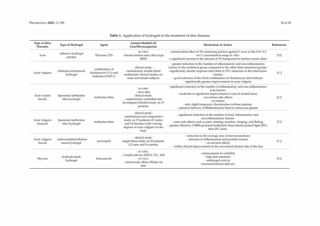
Pharmaceutics 2020, 12, 396 24 of 43
Table 1. Application of hydrogels in the treatment of skin diseases.
Type of SkinDisorder Type of Hydrogel Agent Animal Model/Cell
Line/Microorganism Mechanism of Action References
Acne adhesive hydrogelpatches Triclosan (TS)
in vitro:- female hairless mice skin (type
SKH)
- antimicrobial effect of TS containing patches against P. acnes in the 0.01–0.3wt.% concentration range in vitro
- a significant increase in the amount of TS transported in hairless mouse skins[38]
Acne vulgaris clindamycin/tretinoinhydrogel
combination ofclindamycin (1%) and
tretinoin (0.025%)
clinical study:-randomized, double-blind
multicenter clinical studies onmale and female subjects
- greater reduction in the number of inflammatory and non-inflammatorylesions in the combined group compared to the other three treatment groups- significantly shorter response time (time to 50% reduction in the total lesion
counts)- good tolerance of the fixed combination of clindamycin and tretinoin
- significantly greater improvements in acne vulgaris
[61]
Acne vularis(facial)
liposomal methyleneblue hydrogel methylene blue
in vitro:- mice skin
clinical study:- randomized, controlled and
investigator blinded study on 13patients
- significant reduction in the number of inflammatory and non-inflammatoryacne lesions
- moderate to significant improvement in acne in treated areas- no serious side effects
- no edema- only slight temporary discoloration in three patients
- selective delivery of BM(methylene blue) to sebaceous glands
[62]
Acne vulgaris(truncal)
liposomal methyleneblue hydrogel methylene blue
clinical study:- randomized and comparativestudy on 35 patients (21 malesand 14 females) with varying
degrees of acne vulgaris on theback
- significant reduction in the number of total, inflammatory andnon-inflammatory lesions
- some side effects such as pain, staining, pruritus, stinging, and flaking- greater efficiency LMB(Liposomal methylene blue)-intense pulsed light (IPL)
than IPL alone
[70]
Acne vulgaris(facial)
carboxymethylcellulose-based hydrogel resveratrol
clinical study:single-blind study on 20 patients
(12 men and 8 women)
- reduction in the average area of microcomedones- decrease in inflammation and pustular lesions
- no adverse effects- visible clinical improvement on the resveratrol-treated side of the face
[71]
Mycosis hydroalcoholichydrogel luliconazole
in vitro:- Candida albicans (MTCC No. 183)
ex vivo:- dorsal male albino Wistar rat
skin
- enhancement in solubility- high skin retention- antifungal activity
- increased dermal delivery
[80]

Pharmaceutics 2020, 12, 396 25 of 43
Table 1. Cont.
Type of SkinDisorder Type of Hydrogel Agent Animal Model/Cell
Line/Microorganism Mechanism of Action References
Mycosis microemulsion-basedhydrogel bifonazole
in vitro:- Candida albicans
ex vivo:- dorsal rat skin
in vivo:- rabbits
- good stability of hydrogel over the period of three months- antifungal activity
- sustained release of bifonazole- permeability enhancement of drug
- improvement of solubility
[88]
Mycosis dextran-basedhydrogel (amphogel) amphotericin B
in vitro:- Candida albicans
in vivo:- male SV129 mice
- quick killing of fungi- no survival of fungi with amphogels
- in vivo hydrogel biocompatibility- prevention of fungal infection
- mitigation fungal biofilm formation
[89]
Mycosis dextran-aldehydehydrogel amphotericin B
in vitro:- Candida albicansSC5314
in vivo:- SV129 mice
- effectiveness in killing fungi on contact- long-lasting antifungal activity
- biocompatibility[91]
Mycosis bolalipid hydrogel methylene blue in vitro:- Saccharomyces cerevisiae
- sustained drug release- antifungal activity
- excellent biocompatibility- high solidification capacity under body temperature conditions
[92]
Psoriasis
supramolecularbis-imidazolium
based amphiphilehydrogels
- gemcitabinehydrochloride
- methotrexate sodiumsalt,
- tacrolimus,- betamethasone
17-valerate- triamcinolone
acetonide
ex vivo:- human excised skin
in vivo:- male Swiss CD-1 mice with
induced inflammation andhyperplasia
- suitable viscoelastic properties of the obtained hydrogel for topical drugdelivery
- significant influence of contained drugs on gel morphology at themicroscopic level
- protection of the drug by the hydrogel against degradation,- induction of exponential drug release
- promotion of the drug penetration and retention in the skin- greater reduction of hyperplasia and macroscopic tissue damage in vivo
compared to drug solutions
[103]

Pharmaceutics 2020, 12, 396 26 of 43
Table 1. Cont.
Type of SkinDisorder Type of Hydrogel Agent Animal Model/Cell
Line/Microorganism Mechanism of Action References
Psoriasis
carbomer hydrogelbearing
nanostructured lipidcarriers
methotrexate
in vitro:- Wistar stain albino rat skin
in vivo:- rabbits
- effective incorporation of the drug into nanostructured lipid carrier (NLC)and solid lipid nanoparticle (SLN) hydrogel structures
- reduction of drug release during storage- significant reduction in skin irritation in rabbits
[112]
Psoriasishydrogel transformedfrom microemulsionusing Carbopol 934
- betamethasonedipropionate
- salicylic acid
in vitro:- rat abdominal skin
in vivo:- Wistar rats
- sustained and good anti-inflammatory activity- good stability, powerful permeation ability. and suitable viscosity of hydrogel
- sustained drug release for the desired period of time- reduced dosing frequency
[113]
Psoriasisnanostructured lipidcarrier based topical
hydrogel- mometasone furoate
in vitro:- cellulose membrane
ex vivo:- Wistar rats’ abdominal skin
in vivo:- BALB/c female mice
- sustained release of mometasone furoate- negligible skin irritation
- marked reduction in psoriatic lesions- lesser skin blackening
- significant reduction of hyperkeratosis, parakeratosis, and hyperplasia
[114]
Psoriasis hydrogel patch -clinical study:
- men and women withplaque-type psoriasislesions
- intrinsic adhesion of the hydrogel- cooling and soothing properties
- itching relief- safety and ease of use
- no serious adverse events
[115]
Psoriasis hydrogel micropatch -
clinical study:- 100 psoriatic patients (75 menand 25 women) and 100 healthy
volunteers
- the ability to analyze skin excretions using the hydrogel micropatchsampling approach combined with mass spectrometry
- the possibility of using hydrogels as a non-invasive diagnostic tool for skindiseases
- the ability to detect various skin biomarkers specific for psoriasis through theuse of hydrogel micropatch
[116]

Pharmaceutics 2020, 12, 396 27 of 43
Table 2. Hydrogels for wound healing applications.
Type of Hydrogel Analysis Animal Model/CellLine/Microorganism Biological Activity References
poly(vinyl alcohol)(PVA)/β-glucan
(β-1,6-branched-β-1,3-glucan)
-determination of cellular morphology- assessment of cell viability
- wound healing evaluation in vivo- assessment of skin regeneration (in vitro scratch
wound healing assay)- evaluation of granulation tissue formation
(hematoxylin and eosin staining)- epidermal thickness measurements
- assessment of localization of cytokeratin proteinsin the skin tissue
- examination of protein expression (Westernblotting
in vitro:- human dermal
fibroblasts- human keratinocytes
(HaCaT)in vivo:- mice
- wound healing acceleration- development of skin appendages in regenerated skin tissue
- formation of capillary vessel- better granulation and reepithelialization- hierarchical arrangement of dermal layers
- increase in expression level of transforming growth factor(TGF)-β3,cytokeratin 10 (K10), and cytokeratin 14 (K14) in the skin
tissue- no detectable changes in cell morphology
- migration and proliferation of keratinocytes and fibroblastsenhancement
- skin regeneration- significantly faster wound closure
- skin regeneration around the wound site- increase intranslational levels of K10, K14, and TGF-β3 proteins
[126]
dextran hydrogel
- evaluation of progress in wound healing- assessment of cell infiltration
- assessment of macrophages and neutrophilsaccumulation
- examination of aniogenic response- assessment of neovascularization
- immunohistochemical analysis (Masson’strichrome and alpha-smooth muscle actin staining)- assessment of blood flow surrounding the wound
(laser Doppler)- analysis of the regenerated skin structure
in vivo:- mice
- murine burn woundmodel
- promotion of remarkable neovascularization- promotion of dermal regeneration with complete skin appendages- facilitating early infiltration and degradation of inflammatory cells- promotion of the infiltration of angiogenic cells into healing wounds- mature epithelial structure with hair follicles and sebaceous glands
development- acceleration of the recruitment of endothelial and cell progenitors
into the wound area
[127]
self-crosslink abledextran-isocyanatoethylmethacrylate-ethylamine
hydrogel (DexIEME
- evaluation of new skin regeneration in murineand porcine models
- assessment of newly regenerated hair follicles- polarization of pro-inflammatory
(M1)/anti-inflammatory (M2) macrophages- assessment of regenerative capacity in burn scars
on mice- histological examination (hematoxylin and eosin
staining)- assessment of regenerative capacity in deep full
skin injury in pigs
in vitro:- human monocytic
leukemia (THP-1)cellline
in vivo:- murine and porcine
models
- low pro-inflammatory response in murine and porcine models- regeneration of full skin structures with appendages on both
pre-existing scars and acute wounds- promotion of complete skin regeneration with hair regrowth on
preexisting scars- promotion of M2 macrophage phenotype
- more mature wound- retention of the reticulated epithelial layer by new skin
- more adipose tissue within the newly regenerated skin in bothanimal models
- attenuation of scar formation
[128]

Pharmaceutics 2020, 12, 396 28 of 43
Table 2. Cont.
Type of Hydrogel Analysis Animal Model/CellLine/Microorganism Biological Activity References
hybrid dextran hydrogel withincorporated curcumin
encapsulatedPEG-PLA(poly(lactide)-block-poly(ethylene
glycol))
- evaluation of capacity to control curcumin release- histological study (hematoxylin and eosin
staining)- collagen staining using Masson’s trichrome
method- evaluation of neovascularization
- assessment of expression of CD31 and vimentinin wound tissue
in vivo:- male BALB/C mice
- murine full thicknesswound model
- sustained release of curcumin from dextran hydrogel- significantly augment the reepithelialization of epidermis; increase
in collagen deposition in the wound area- increase in fibroblast/mesenchymal cell density
- acceleration of angiogenesis and fibroblast accumulation- increase tissue granulation
- more mature epithelial structure with hair follicles and sebaceousglands
- acceleration of the wound healing process
[129]
PVA/dextran/chitosan- assessment of cell proliferation
- determination of cytotoxicity (WST-1 assay)- evaluation of antimicrobial effect
in vitro:- mouse NIH-3T3
fibroblasts- Escherichia coli
- Staphylococcus aureus
- antimicrobial ability to both Gram (+) and Gram (−) bacteria- no adverse effect on cell growth
- dextran concentration-dependent stimulation of NIH-3T3 cellproliferation
- improvement of cell adhesion to hydrogel with higher dextranconcentration
[130]
cyclodextrin/cellulose hydrogelwith gallic acid
- evaluation of antibacterial activity- assessment of cytotoxicity
in vitro:- 3T3 fibroblasts- Staphylococcus
epidermidis- Staphylococcusaureus- Klebsiellapneumoniae
- sustained gallic acid release- significant reduction in the growth of all three bacteria
- no cytotoxic effect on fibroblasts- no release of substances potentially toxic to fibroblasts
[125]
hydroxyethyl cellulose(HEC) supplemented hydrogel
loaded with tungsten oxide(WO3)
- assessment of cytotoxicity of hydroxyethylcellulose (HEC) with/without WO3- determination of healing capacity- evaluation of anti-inflammatory
activity(lipopolysaccharide-stimulatedinflammation)
- evaluation of antibacterial activity
in vitro:- human dermalfibroblast cells
- white blood cells- Shigella sp.
- Salmonella sp.- Pseudomonas aeruginosa
- Bacillus cereus-Staphylococcus aureus
- antibacterial activities against pathogenic Gram-negative andGram-positive strains
- no morphological changes in the examined cells- improving tungsten oxide safety toward normal human cells (white
blood cells and dermal fibroblast)- suppressed an abnormal immune response via normalization
proinflammatory cytokine
[131]

Pharmaceutics 2020, 12, 396 29 of 43
Table 2. Cont.
Type of Hydrogel Analysis Animal Model/CellLine/Microorganism Biological Activity References
bacterial cellulose/acrylic acidhydrogel containing
keratinocytes and fibroblasts
- assessment of wound closure in vivo- histological analysis
(hematoxylin-eosin and Masson’s trichromestaining)
- determination of COL-1, CK-14, involucrin, andα-SMA expression in the tissue
- assessment of regenerated skin ultrastructure(transmission electron microscopy)
in vitro:- human epidermal
keratinocytes- human dermal
fibroblastsin vivo:
- male athymicmice(CrTac:NCr-Foxn1nu)-skin samples from six
consenting patients
- acceleration on burn wound healing in vivo- confirmed by the presence of more mature keratinocytes
- increase in involucrin expression- complete reepitheliation of the wound area by day 13
- decrease in cytokeratin 14 antibody and α-SMA expression- increased expression of collagen type I
- more organized skin and collagen structures in the skin layer- a greater deposition of collagen in the mice
- promotion of skin regeneration- the appearance of regenerated skin similar to normal skin
[132]
chitosan–cellulose nanofiber(CS–CNF) compositeself-healing hydrogels
- investigation of cell proliferation rate by confocalmicroscopy and CCK-8 assay
- evaluation of the oxygen metabolism andmitochondrial function of the cells
- determination of nestin, glialfibrillary acidicprotein (GFAP), CNPase, β-tubulin and
microtubule-associated protein 2 (MAP2) geneexpression for cells in chitosan–cellulose nanofiber
(CS–CNF) hydrogels- assessment of nestin, GFAP, CNPase, β-tubulin
and MAP2 protein expression of NSCs in CS–CNFhydrogels by immunostaining
- evaluation of therapeutic function in vivo
in vitro:- neural stem cells (NSCs)from adult mouse brains
in vivo:- adult wild-type
zebrafish
- promotion of neural regeneration- the increase in the efficiency of mitochondrial electron transfer- strong relationship between oxygen metabolism and hydrogel
self-healing properties- the increase in nestin, GFAP, CNPase, and MAP2 gene expression
- lower level of β-tubulin (the early neural marker)- increase in the expression level of nestin, GFAP, CNPase, and MAP2
proteins- better regeneration in cerebellar injury of adult zebrafish
[133]
chitosan hydrogel containingfibroblast growth factor-2
- assessment of contraction and speed of woundclosure in mice
- evaluation of granulation tissue formation- assessment of capillary formation and
epithelialization- histological examination (hematoxylin-eosin
staining)
in vitro:- human umbilical vein
endothelial cells(HUVECs)
in vivo:- healing-impaired
diabetic (db/db)C57BL/6female mice and
their normal (db/+)littermates
- significant induction of wound contraction and accelerated woundclosure in mice
- accelerating wound closure by adding FGF-2 to the chitosanhydrogel in db/db mice, but not in db/+ mice
- advanced granulation tissue and capillary formation- replacement of almost all necrotic tissues with new granulation
tissue after injury on day 16- significant epithelialization
- stimulation of HUVEC growth, but loss of this ability after washingwith phosphate-buffered saline (PBS) hydrogels for more than 3 days
[134]

Pharmaceutics 2020, 12, 396 30 of 43
Table 2. Cont.
Type of Hydrogel Analysis Animal Model/CellLine/Microorganism Biological Activity References
rutin-conjugatedchitosan-poly(ethylene
glycol)-tyramine (RCPT)hydrogel
- evaluation of cell cytotoxicity- assessment of wound healing
- evaluation of neoepithelium and granulationtissue formation
- assessment of proteins formation
in vitro:- L929 mouse fibroblasts
in vivo:- male Sprague-Dawley
rats
- increase in fibroblasts proliferation at low concentration- very low cytotoxicity at high concentrations
- increase in fibroblasts proliferation by releasing rutin- enhancement of wound healing
- induction of better defined formation of neoepithelium and thickergranulation
- more new blood vessels in wound- increase in the formation of extracellular proteins, primarily collagen
[135]
gelatin/CM-chitosan hydrogel
- assessment of hydrogel cytotoxicity- evaluation of the ability of NIH 3T3 cells toproliferate on or into the three-dimensional
structure of hydrogel
in vitro:- L929 mouse fibroblasts
- NIH/3T3 fibroblasts
- promotion of cell attachment on the hydrogel surface- acceleration of NIH 3T fibroblasts growth on the hydrogel
- excellent cytocompatibility of the hydrogels[136]
alginate/chitin/chitosan/fucoidanhydrogel
- assessment of hydrogel cytotoxicity andstimulatory effects on dermal fibroblast cells
(DFCs) and dermal microvascular endothelial cells(DMVECs)
- histological examination of removed skin andwound tissue samples (hematoxylin-eosin staining)
- histological observations on repair ofhealing-impaired wounds
in vitro:- human dermal
fibroblast cells (DFCs)- dermal microvascular
endothelial cells(DMVECs)
in vivo:- male Sprague-Dawley
rats
- no cytotoxicity to DFCs and DMVECs- minor effect on healing of wounds not treated with mitomycin C
- significant stimulation of repair of mitomycin C-treatedhealing-impaired wounds in rats
- promotion of tissue granulation and capillary formation- positive effects on wound closure
- progress in wound contraction and reepithelialization
[137]
chitosan hydrogel/honey
- assessment of antimicrobial activity- evaluation of granulation and fibrotic tissue
formation- evaluation of inflammatory response- determination of reepithelialization
- evaluation of wound shrinkage effects
in vitro:- Staphylococcus aureus
- Bacillus cereus- Escherichia coli
- Pseudomonas aeruginosa- Candida albicans
in vivo:- male Wistar rats- rat full-thickness
wound model
- significant decrease in the minimum inhibitory concentrationagainst bacteria after the combination of a chitinase hydrogel with
honey- significant improvement in the inflammatory index on days 3 and 7
- increase in granulation tissue formation- significantly higher fibrotic tissue formation index
- increase in the level of reepithelialization
[138]

Pharmaceutics 2020, 12, 396 31 of 43
Table 2. Cont.
Type of Hydrogel Analysis Animal Model/CellLine/Microorganism Biological Activity References
water-soluble chitin hydrogel
- histological examination of the wounded skins(hematoxylin and eosin staining, Masson’s
trichrome staining)- determination of collagen-hydroxyproline in
wounded skin- assessment of reepithelialization
in vivo:- animal model: rats
- acceleration of wound healing- significant reepithelialization
- granulation tissue replacement in the wound by fibrosis- healing of hair follicles in rats
- high skin tensile strength and the formation of the correct collagenfiber system
[139]
alginate hydrogels coated withchitosan
- assessment of cell morphology- determination of cytotoxicity
- evaluation of antibacterial activity
in vitro:- human mesenchymal
stromal cells (MSCs)- Escherichia coli
- unmodified cell morphology- no inhibition of MSC growth and proliferation- bacterial inactivation higher than 99% after 3 h
- complete killing of bacteria after 24 h
[140]
gelatin-alginate hydrogel
- assessment of cytotoxicity(MTT assay)
- determination of cell adhesion- live/dead staining (calcein
acetoxymethyl–propidium iodide)
in vitro:- HFF-1 foreskinfibroblast cells
- time-dependent partial inhibition of cell viability- reduction of cell adhesion properties
- presence of only living cells, no dead cells[141]
collagen-hyaluronic acidhydrogel
- assessment of cytotoxicity (MTT assay)- evaluation of cells proliferation encapsulated into
hydrogels at different time using confocal laserscanning microscopy
- measurement of the cells’ attachment withscanning electron microscope
- measurement of vascular endothelial growthfactor secretion by human microvascular
endothelial cells (HMECs) with enzyme-linkedimmunosorbent assay (vascular endothelial
growth factor (VEGF)-ELISA)- assessment of antibacterial activity
- measurement of biocompatibility andinflammatory in vivo
- assessment of wound healing
in vitro:- human microvascular
endothelial cells(HMECs)
- fibroblasts (COS-7)- Escherichia coli
- Staphylococcus aureusin vivo:- mice
- mice full-thickness skinwound model
- good viabilities of HMECs and COS-7 cells- biocompatibility
- excellent differential behaviors of cells in hydrogel- increase in the amount of vascular endothelial growth factor (VEGF)
up to day 7 of culture- no foreign body reaction like immigration of giant cells into hydrogel
- only few inflammatory infiltrations in the interface of hydrogeland tissue
- gradual reduction in the number and infiltration ofinflammatory cells
- normal morphology with no signs of pathology for all organs,including heart, liver, spleen, lungs, and kidney
- good adhesion of the hydrogel to the skin- killing about 55% of E. coli and 47% of S. aureus after incubating for
3 h- less colony-forming units of E. coli or S. aureus
- disappearance of redness of wounds
[142]

Pharmaceutics 2020, 12, 396 32 of 43
Table 2. Cont.
Type of Hydrogel Analysis Animal Model/CellLine/Microorganism Biological Activity References
hyaluronic acid-basedhydrogels
- measurements of the white blood cell (WBC) level- histological staining with hematoxylin
- immunochemistry analyses- determination of α-SMA, VEGF, and TGF-β1
gene expression(fluorescence-based quantitative PCR)
in vivo:- New Zealand white
rabbits
- wound reduction- reduction of fluid secretion and thinner scab formation
- faster recovery of the animals- increase in the level of white blood cells after surgery, reaching a
maximum on day 7, followed by a decrease to a constant value- hair follicle renewal
- increase in α-SMA expressions, which alleviated woundinflammation in the first few days after surgery and alleviated scar
formation by reducing TGF-β1 levels.- faster self-healing compared to control, manifested by a faster
increase in TGF-β1 level and higher VEGF levels after 7 days andlower after 14 days compared to controls
- increase in α-SMA, TGF-β1, and VEGFmRNA levels from day 3,reaching the highest level on day 7 or 14,followed by a continuous
decrease- increase in VEGF expression, which promotes skin regeneration
through neovascularization
[143]
heparin-based hydrogel withloaded human epidermal
growth factor (hEGF)
- evaluation of human epidermal growth factor(hEGF) release in vitro
- histological examination (hematoxylin and eosinstaining)
- determination of the total amount of collagen inthe regenerated skin(hydroxyproline assay)
- immunochemistry staining with an anti-widespectrum cytokine antibody by using
LSAB+system-HRP kit
in vivo:- male BALB/c mice
- sustained release profile of hEGF in vitro- significantly accelerated closure of wounds
- closing the epidermal layer and regenerating the extracellular matrix- denser epithelialization, blood vessel, hair follicles, and sebaceous
gland formation- high similarity in the structure of the skin tissue of mice after injury
after applying the hydrogel to normal skin tissue- accelerating wound closing and facilitating wound remodeling into
normal skin tissue- increased proliferation and differentiation of keratinocytes- improvement of keratinocyte migration to the site of injury
- hair follicles formation at the recovered tissue
[144]
collagen hydrogel
- assessment of wound morphology- determination of scarring shape and physical
properties- histological examination (hematoxylin and eosin
staining)- assessment of angiogenesis changes
in vivo:- Sprague-Dawley
diabetic rat
- faster wound healing- increasing the speed and quality of full thickness wound healing in
diabetic rats- significantly higher quantity of fibroblasts- increase in the number of new capillaries
[157]

Pharmaceutics 2020, 12, 396 33 of 43
Table 2. Cont.
Type of Hydrogel Analysis Animal Model/CellLine/Microorganism Biological Activity References
collagen gel
- assessment of Mrc-1 (a reparative M2macrophage marker) gene expression
- assessment of anti-inflammatory cytokineinterleukin (IL)-10 and fibroblast growth
factor-basic (β-FGF)expression- immunohistochemical staining (with
anti-vimentin and)DAPI)(4′,6-diamidino-2-phenylindole)
- evaluation of mature collagen deposition- analyses of wound tissues
in vitro:- human THP-1
monocytesin vivo:
- Yorkshire pigs- swine model of chronic
ischemic wounds
- upregulation of Mrc-1 expression in vitro- induction of (IL)-10 and (β-FGF) expression in vitro
- increased expression of CCR2 (M2 macrophage marker)- upregulation of transforming growth factor-β, vascular endothelialgrowth factor, von Willebrand’s factor, and collagen type I expression
in ischemic wounds- increase in endothelial cells proliferation
- significant increase in fibroblasts in ischemic wound-edge tissue- higher abundance of mature collagen
[158]
injectable tannic acid-loadedgelatin-based hydrogels
blended with gellan gum
- evaluation of antibacterial activity using agardisc diffusion test
- assessment of wound healing efficacy- histological examination (hematoxylin and eosin
(H&E) and Masson’s trichrome staining)- measurements of epidermal thickness
- assessment of skin tissue formation- evaluation of collagen deposition
- examination of cell cytotoxicity and migration
in vitro:-murine L929 fibroblasts
- Escherichia coli- Staphylococcus aureus- methicillin-resistantStaphylococcus aureus
in vivo:- BALB/c mice
- long-term antibacterial efficacy- sustained release of tannic acid in vitro
- significant wound reduction- no scars
- skin fully covered with hair- 100% healing rate
- increase in the thickness of the epidermis- effective granulation tissue formation
- greater accumulation and collagen fiber content- effective skin regeneration and function restoration
- increase in L929 cell viability
[159]
gelatin/oxidizedalginate-tyramine hydrogels
- examination of tissue adhesive strength- assessment of residue H2O2 level- evaluation of antibacterial activity
- measurements of cytotoxicity in vitro(WST-1 assay)
in vitro:- human dermal
fibroblasts (hDFBs)- Escherichia coli
- Staphylococcus aureusin vivo:
- porcine skin
- high antibacterial ability due to the constant release of H2O2- low adhesive strength of hydrogel allowing for the easy dressing
removal- no cytotoxic effect on test cells
[160]

Pharmaceutics 2020, 12, 396 34 of 43
Table 2. Cont.
Type of Hydrogel Analysis Animal Model/CellLine/Microorganism Biological Activity References
mesenchymal stem cells (MSCs)spheroid-containing fibrin
hydrogels
- evaluation of cytokine bioactivity to stimulateendothelial cells and macrophages
- assessment of anti-inflammatory potential- capacity of entrappedMSC spheroids to promote
angiogenesis in a three-dimensional skin-likeenvironment assessment
in vitro:- human bone
marrow-derived MSCs- diabetic human
microvascular cells(HMVECs)
- raw 264.7 mousemacrophages
in vivo:- human skin equivalent
(HSE) model
- cytokine secretion with potential proangiogenic andanti-inflammatory effects
- enhancing angiogenesis in the equivalent of human skin- increased endothelial cell penetration
- stimulation of sprouts and greater invasion distance into the wound- dependence of VEGF and prostaglandin E2 (PGE2) secretion on the
mechanical properties of hydrogels
[161]
human hair keratin-basedhydrogel
- assessment of cell proliferation- immunocytochemical staining
- evaluation of the cellular interaction of keratinwith HaCaT cells
- assessment of cell migration- measurements of gene expression
in vitro:- human keratinocyte
cell line (HaCaT)in vivo:
-male C57BL/6J mice
- stronger expression of vimentin and fibroblastic spindle shape inHaCaT cells
- increasing the number of cells migrating into the space betweenconfluent cell colonies
- upregulation of the expressions of migration-related genes such asintegrin αV, integrin α5, integrin β1, and integrin β6
- increase in mRNA expressions of integrin αV, integrin β5, integrinβ6, fibronectin, Snail and vimentin
- induction of molecular expressions of integrin β1 and vimentin- fully recovering the skin aspect without any signs of scab and skin
contraction in animal test- reepithelialization of skin and the proliferation of dermal fibroblast
with adnexa in dermis in vivo- effective wound remodeling with skin adnexa in vivo
- high rate of the proliferation of dermal fibroblasts with skin adnexaand dense dermal fiber in vivo
- full wound repair with skin appendages- regeneration of muscle tissue
- increasing the number of hair follicles
[162]

Pharmaceutics 2020, 12, 396 35 of 43
Table 2. Cont.
Type of Hydrogel Analysis Animal Model/CellLine/Microorganism Biological Activity References
feather keratin hydrogel
- evaluation of wound healing in vivo- assessment of cellular responses and
vascularization(hematoxylin and eosin staining)
- determination of collagen deposition in thewound skin (trichrome staining)
- determination of the inflammatory cytokinesIL-1β, IL-6, and TNF-α levels in the serum of rats
(ELISA)
in vivo:- male Sprague-Dawley
rats
- significant acceleration of wound healing- approximately 90% wound closure within 10 days
- complete reepithelialization after 21 days- more hair follicles
- more new capillaries around the inflammatory cells- maturation of the epidermis over the wound and restoration of new
skin to normal after 28 days- acceleration of collagen deposition
- thicker collagen fibers- no adverse systemic toxicity in rats after hydrogel implantation
- no obvious organ damage and significant histopathologicaldifferences in the tissue organs in rats
- no or minimal inflammatory response- no significant elevations in cytokines levels
[163]
injectable silk fibroin hydrogel
- assessment of cell migration- evaluation of wound closure
- histological analysis- determination of collagen type I and III, TNF-α,
CD68, CD163, and glyceraldehyde-3-phosphate- dehydrogenase (GAPDH) expression
(qRT-PCR analysis)- determination of suprabasal keratin markercytokeratin 10 (CK10), basal keratin marker
cytokeratin 14 (CK14), and marker of terminallydifferentiated keratinocytes involucrin (INV)
expression
in vitro:- HaCaT cells
in vivo:- female Wistar albino
rats- full thickness
third-degree burnwounds
- complete wound closure after 21 days- two-fold accelerated healing rate in a regenerative manner
- rapid development of granulation tissue- enhanced, early reepithelialization
- mature epidermo-dermal regeneration- increase in HaCaT cells proliferation
- formation of small blood vessels on day 7- ten-fold increase in vessel density
- significant upregulation and downregulation of collagen type I andIII during different stages of wound healing
- increase in collagen type I secretion from day 7 to 21- higher secretion of collagen type III
- visible expression of cytokines CK10 and CK14
[164]
heparinized silk fibroinhydrogels loading FGF1
(fibroblast growth factor 1
- assessment of platelet-derived growth factor(PDGF) and growth factor TGF-β expression
(ELISA)- histological evaluation using hematoxylin and
eosin and Masson’s trichrome staining- assessment of fibroblast L929 cells proliferation
and migration
in vitro:- fibroblast L929 cells
in vivo:- Sprague-Dawley rats
- full-thickness excisionalwounds
- increase in expression level of PDGF on day 7- no statistically significant difference for expression of TGF-β1
- acceleration of dermis formation- acceleration of epidermal differentiation into hair follicles and
sebaceous glands- improvement in small scars healing
[165]

Pharmaceutics 2020, 12, 396 36 of 43
Author Contributions: M.Z.-D. and M.S. substantially contributed to conception, design, acquisition, analysisand interpretation of data and drafted the manuscript. M.S. critically improved the manuscript for importantintellectual content. M.Z.-D. and M.S. accepted the manuscript content. All authors have read and agreed to thepublished version of the manuscript.
Funding: This work was founded by the 503-07-01-35 Statutory Project of the University of Information Technologyand Management in Rzeszow, Poland.
Conflicts of Interest: The authors declare no conflicts of interest.
References
1. Hay, R.J.; Johns, N.E.; Williams, H.C.; Bolliger, I.W.; Dellavalle, R.P.; Margolis, D.J.; Marks, R.; Naldi, L.;Weinstock, M.A.; Wulf, S.K.; et al. The global burden of skin disease in 2010: An analysis of the prevalenceand impact of skin conditions. J. Investig. Dermatol. 2014, 134, 1527–1534. [CrossRef]
2. Dreno, B.; Araviiskaia, E.; Berardesca, E.; Bieber, T.; Hawk, J.; Sanchez-Viera, M.; Wolkenstein, P. The scienceof dermocosmetics and its role in dermatology. J. Eur. Acad. Dermatol. Venereol. 2014, 28, 1409–1417.[CrossRef]
3. Hay, R.; Bendeck, S.E.; Chen, S.; Estrada, R.; Haddix, A.; McLeod, T.; Mahé, A. Skin Diseases. In DiseaseControl Priorities in Developing Countries, 2nd ed.; Jamison, D.T., Breman, J.G., Measham, A.R., Alleyne, G.,Claeson, M., Evans, D.B., Jha, P., Mills, A., Musgrove, P., Eds.; The International Bank for Reconstruction andDevelopment/The World Bank: Washington, DC, USA; Oxford University Press: New York, NY, USA, 2006.
4. Andersen, L.K.; Davis, M.D. Prevalence of Skin and Skin-Related Diseases in the Rochester EpidemiologyProject and a Comparison with Other Published Prevalence Studies. Dermatology 2016, 232, 344–352.[CrossRef]
5. Karimkhani, C.; Dellavalle, R.P.; Coffeng, L.E.; Flohr, C.; Hay, R.J.; Langan, S.M.; Nsoesie, E.O.; Ferrari, A.J.;Erskine, H.E.; Silverberg, J.I.; et al. Global Skin Disease Morbidity and Mortality: An Update from the GlobalBurden of Disease Study 2013. JAMA Dermatol. 2017, 153, 406–412. [CrossRef]
6. Donnelly, R.F.; Singh, T.R.R.; Morrow, D.I.; Woolfson, A.D. Microneedle-Mediated Intradermal Delivery.In Microneedle-Mediated Transdermal and Intradermal Drug Delivery; Wiley: Hoboken, NJ, USA, 2012; pp. 113–151.[CrossRef]
7. Larrañeta, E.; Stewart, S.; Ervine, M.; Al-Kasasbeh, R.; Donnelly, R.F. Hydrogels for Hydrophobic DrugDelivery. Classification, Synthesis and Applications. J. Funct. Biomater. 2018, 9, 13. [CrossRef]
8. McKenzie, M.; Betts, D.; Suh, A.; Bui, K.; Kim, L.; Cho, H. Hydrogel-Based Drug Delivery Systems for PoorlyWater-Soluble Drugs. Molecules 2015, 20, 20397–20408. [CrossRef]
9. Fahr, A.; Liu, X. Drug delivery strategies for poorly water-soluble drugs. Expert Opin. Drug Deliv. 2007, 4,403–416. [CrossRef]
10. Gu, D.; O’Connor, A.J.; Qiao, G.G.H.; Ladewig, K. Hydrogels with smart systems for delivery of hydrophobicdrugs. Expert Opin. Drug Deliv. 2017, 14, 879–895. [CrossRef]
11. Nie, S.; Hsiao, W.L.; Pan, W.; Yang, Z. Thermoreversible Pluronic® F127-based hydrogel containing liposomesfor the controlled delivery of paclitaxel: In vitro drug release, cell cytotoxicity, and uptake studies. Int. J.Nanomed. 2011, 6, 151–166. [CrossRef]
12. Yang, Y.; Wang, J.; Zhang, X.; Lu, W.; Zhang, Q. A novel mixed micelle gel with thermo-sensitive property forthe local delivery of docetaxel. J. Control. Release 2009, 135, 175–182. [CrossRef]
13. Benson, H.A.E.; Grice, J.E.; Mohammed, Y.; Namjoshi, S.; Roberts, M.S. Topical and Transdermal DrugDelivery: From Simple Potions to Smart Technologies. Curr. Drug Deliv. 2019, 16, 444–460. [CrossRef]
14. Ruela, A.L.M.; Perissinato, A.G.; de Sousa Lino, M.E.; Mudrik, P.S.; Pereira, G.R. Evaluation of skin absorptionof drugs from topical and transdermal formulations. Braz. J. Pharm. Sci. 2016, 52, 527–544. [CrossRef]
15. Schoellhammer, C.M.; Blankschtein, D.; Langer, R. Skin permeabilization for transdermal drug delivery:Recent advances and future prospects. Expert Opin. Drug Deliv. 2014, 11, 393–407. [CrossRef]
16. Wollina, U.; Tirant, M.; Vojvodic, A.; Lotti, T. Treatment of Psoriasis: Novel Approaches to Topical Delivery.Open Access Maced. J. Med. Sci. 2019, 7, 3018–3025. [CrossRef]

Pharmaceutics 2020, 12, 396 37 of 43
17. Angerer, J.; Bernauer, U.; Chambers, C.; Chaudhry, Q.; Degen, G.; Eisenbrand, G.; Platzek, T.; ChandraRastogi, S.; Rogiers, V.; Rousselle, C.; et al. Basic Criteria for the In Vitro Assessment of Dermal Absorption ofCosmetic Ingredients; SCCS (Scientific Committee on Consumer Safety): Brussels, Belgium, 22 June 2010.[CrossRef]
18. Abd, E.; Yousef, S.A.; Pastore, M.N.; Telaprolu, K.; Mohammed, Y.H.; Namjoshi, S.; Grice, J.E.; Roberts, M.S.Skin models for the testing of transdermal drugs. Clin. Pharmacol. 2016, 8, 163–176. [CrossRef]
19. Trombino, S.; Servidio, C.; Curcio, F.; Cassano, R. Strategies for Hyaluronic Acid-Based Hydrogel Design inDrug Delivery. Pharmaceutics 2019, 11, 407. [CrossRef]
20. Highley, C.B.; Prestwich, G.D.; Burdick, J.A. Recent advances in hyaluronic acid hydrogels for biomedicalapplications. Curr. Opin. Biotechnol. 2016, 40, 35–40. [CrossRef]
21. Nguyen, M.K.; Alsberg, E. Bioactive factor delivery strategies from engineered polymer hydrogels fortherapeutic medicine. Prog. Polym. Sci. 2014, 39, 1235–1265. [CrossRef]
22. Li, J.; Wu, C.; Chu, P.K.; Gelinsky, M. 3D printing of hydrogels: Rational design strategies and emergingbiomedical applications. Mater. Sci. Eng. R. Rep. 2020, 140, 100543. [CrossRef]
23. Bahram, M.; Mohseni, N.; Moghtader, M. An Introduction to Hydrogels and Some Recent Applications, EmergingConcepts in Analysis and Applications of Hydrogels; Sutapa Biswas Majee; IntechOpen: London, UK, 2016;pp. 9–38. [CrossRef]
24. Indermun, S.; Choonara, Y.E.; Kumar, P.; du Toit, L.C.; Modi, G.; Luttge, R.; Pillay, V. An InterfaciallyPlasticized Electro-responsive Hydrogel for Transdermal Electro-activated and Modulated (TEAM) DrugDelivery. Int. J. Pharm. 2014, 462, 52–65. [CrossRef]
25. Feng, Z.; Iln, S.; McDonagh, A.; Yu, C. Natural Hydrogels Applied in Photodynamic Therapy. Curr. Med.Chem. 2019. [CrossRef]
26. Li, J.; Mooney, D.J. Designing hydrogels for controlled drug delivery. Nat. Rev. Mater. 2016, 1, 16071.[CrossRef]
27. Lee, Y.; Mooney, D.J. Hydrogels for Tissue Engineering. Chem. Rev. 2001, 101, 1869–1877. [CrossRef]28. Drury, J.L.; Mooney, D.J. Hydrogels for Tissue Engineering: Scaffold Design Variablesand Applications.
Biomaterials 2003, 24, 4337–4351. [CrossRef]29. Bryan, J. Moist wound healing: A concept that changed our practice. J Wound Care. 2004, 13, 227–228.
[CrossRef]30. Priya, S.G.; Jungvid, H.; Kumar, A. Skin tissue engineering for tissue repair and regeneration. Tissue Eng.
Part B Rev. 2008, 14, 105–118. [CrossRef]31. Chyzy, A.; Tomczykowa, M.; Plonska-Brzezinska, M.E. Hydrogels as potential nano-, micro- and macro-scale
systems for controlled drug delivery. Materials 2020, 13, 188. [CrossRef]32. Mantha, S.; Pillai, S.; Khayambashi, P.; Pham, H.M.; Tran, S.D. Smart hydrogels in tissue engineering and
regenerative medicine. Materials 2019, 12, 3323. [CrossRef]33. Palmese, L.L.; Thapa, R.K.; Sullivan, M.O.; Kiick, K.L. Hybrid hydrogels for biomedical applications.
Curr. Opin. Chem. Eng. 2019, 24, 143–157. [CrossRef]34. Steffens, D.; Braghirolli, D.I.; Maurmann, N.; Pranke, P. Update on the main use of biomaterials and techniques
associated with tissue engineering. Drug Discov. Today 2018, 23, 1474–1488. [CrossRef]35. Kim, D.Y.; Kwon, D.Y.; Kwon, J.S.; Min, B.H.; Kim, M.S. Stimuli-responsive injectable in situ-forming
hydrogels for regenerative medicines. Polym. Rev. 2015, 55, 407–452. [CrossRef]36. Kirschner, C.M.; Anseth, K.S. Hydrogels in Healthcare: From Static to Dynamic Material Microenvironments.
Acta Mater. 2013, 61, 931–944. [CrossRef]37. Parente, M.E.; Ochoa Andrade, A.; Ares, G.; Russo, F.; Jiménez-Kairuz, Á. Bioadhesive hydrogels for cosmetic
applications. Int. J. Cosmet. Sci. 2015, 37, 511–518. [CrossRef]38. Lee, T.W.; Kim, J.C.; Hwang, S.J. Hydrogel patches containing Triclosan for acne treatment. Eur. J. Pharm.
Biopharm. 2003, 56, 407–412. [CrossRef]39. Nilforoushzadeh, M.A.; Amirkhani, M.A.; Zarrintaj, P.; Salehi Moghaddam, A.; Mehrabi, T.; Alavi, S.;
Mollapour Sisakht, M. Skin care and rejuvenation by cosmeceutical facial mask. J. Cosmet Dermatol. 2018, 17,693–702. [CrossRef]
40. Aramwit, P.; Bang, N. The characteristics of bacterial nanocellulosegel releasing silk sericin for facial treatment.BMC Biotechnol. 2014, 14, 104. [CrossRef]

Pharmaceutics 2020, 12, 396 38 of 43
41. Gao, Z.; Yu, Z.; Huang, C.; Duan, L.; Gao, G.H. Carboxymethyl cellulosereinforced poly (vinyl alcohol) withtrimethylol melamine as a chem-ical crosslinker. J. Appl. Polym. Sci. 2016, 134, 1–5. [CrossRef]
42. Huang, D.; Sun, M.; Bu, Y.; Luo, F.; Lin, C.; Lin, Z.; Weng, Z.; Yang, F.; Wu, D. Microcapsule-embeddedhydrogel patches for ultrasound responsive and enhanced transdermal delivery of diclofenac sodium.J. Mater. Chem. B 2019, 7, 2330–2337. [CrossRef]
43. Koehler, J.; Brandl, F.P.; Goepferich, A.M. Hydrogel wound dressings for bioactive treatment of acute andchronic wounds. Eur. Polym. J. 2018, 100, 1–11. [CrossRef]
44. Monticelli, D.; Martina, V.; Mocchi, R.; Rauso, R.; Zerbinati, U.; Cipolla, G.; Zerbinati, N. ChemicalCharacterization of Hydrogels Crosslinked with Polyethylene Glycol for Soft Tissue Augmentation.Open Access Maced. J. Med. Sci. 2019, 7, 1077–1081. [CrossRef]
45. Vlaia, L.; Coneac, G.; Olariu, I.; Vlaia, V.; Lupuleasa, D. Cellulose-Derivatives-Based Hydrogels as Vehicles forDermal and Transdermal Drug Delivery, Emerging Concepts in Analysis and Applications of Hydrogels; SutapaBiswas Majee; IntechOpen: London, UK, 2016. [CrossRef]
46. Cen, L.; Liu, W.; Cui, L.; Zhang, W.; Cao, Y. Collagen tissue engineering: Development of novel biomaterialsand applications. Pediatr Res. 2008, 63, 492–496. [CrossRef]
47. Dimatteo, R.; Darling, N.J.; Segura, T. In situ forming injectable hydrogels for drug delivery and woundrepair. Adv. Drug Deliv. Rev. 2018, 127, 167–184. [CrossRef]
48. Cheng, L.; Ji, K.; Shih, T.Y.; Haddad, A.; Giatsidis, G.; Mooney, D.J.; Orgill, S.P.; Nabzdyk, C.S. InjectableShape-Memorizing Three-Dimensional Hyaluronic Acid Cryogels for Skin Sculpting and Soft TissueReconstruction. Tissue Eng. Part A 2017, 23, 243–251. [CrossRef]
49. Kutlubay, Z.; Kecici, A.S.; Engin, B.; Serdaroglu, S.; Tuzun, Y. Acne and Acneiform Eruptions; InTech: Rijeka,Croatia, 2017; pp. 7–39.
50. Webster, G.F. The pathophysiology of acne. Cutis 2005, 76, 4–7.51. Cordain, L. Implications for the role of diet in acne. Semin. Cutan. Med. Surg. 2005, 24, 84–91. [CrossRef]52. Jovic, A.; Marinovic, B.; Kostovic, K.; Ceovic, R.; Basta-Juzbašic, A.; BukvicMokos, Z. The Impact of
Pyschological Stress on Acne. Acta Dermatovenerol. Croat. 2017, 25, 1133–1141.53. Tan, A.U.; Schlosser, B.J.; Paller, A.S. A review of diagnosis and treatment of acne in adult female patients.
Int. J. Womens Dermatol. 2017, 4, 56–71. [CrossRef]54. Kraft, J.; Freiman, A. Management of acne. CMAJ 2011, 183, E430–E435. [CrossRef]55. William, D.; James, W.D. Clinical practice. Acne. N. Engl. J. Med. 2005, 352, 1463–1472. [CrossRef]56. Gollnick, H.; Cunliffe, W.; Berson, D.; Dreno, B.; Finlay, A.; Leyden, J.J.; Shalita, A.R.; Thiboutot, D.
Management of acne: A report from a Global Alliance to Improve Outcomes in Acne. JAAD 2003, 49, S1–S37.[CrossRef]
57. Zouboulis, C.C.; Derumeaux, L.; Decroix, J.; Maciejewska-Udziela, B.; Cambazard, F.; Stuhlert, A.A multicentre, single-blind, randomized comparison of a fixed clindamycin phosphate/tretinoin gelformulation (Velac) applied once daily and a clindamycin lotion formulation (Dalacin T) applied twice dailyin the topical treatment of acne vulgaris. Br. J. Dermatol. 2000, 143, 498–505. [CrossRef]
58. Magin, P.; Pond, D.; Smith, W.; Watson, A. A systematic review of the evidence for ‘myths and misconceptions’in acne management: Diet, face-washing and sunlight. Fam. Pract. 2005, 22, 62–70. [CrossRef]
59. Oprica, C.; Nord, C.E. ESCMID Study Group on Antimicrobial Resistance in Anaerobic Bacteria. Europeansurveillance study on the antibiotic susceptibility of Propionibacterium acnes. Clin. Microbiol. Infect. 2005,11, 204–213. [CrossRef]
60. van Vloten, W.A.; van Haselen, C.W.; van Zuuren, E.J.; Gerlinger, C.; Heithecker, R. The effect of 2 combinedoral Contraceptives containing either drospirenone or cyproterone acetate on acne and seborrhea. Cutis 2002,69, 2–15.
61. Leyden, J.J.; Krochmal, L.; Yaroshinsky, A. Two randomized, double-blind, controlled trials of 2219 subjects tocompare the combination clindamycin/tretinoin hydrogel with each agent alone and vehicle for the treatmentof acne vulgaris. JAAD 2006, 54, 73–81. [CrossRef]
62. Fadel, M.; Salah, M.; Samy, N.; Mona, S. Liposomal methylene blue hydrogel for selective photodynamictherapy of acne vulgaris. J. Drugs Dermatol. 2009, 8, 983–990.
63. Haider, A.; Shaw, J.C. Treatment of acne vulgaris. JAMA 2004, 292, 726–735. [CrossRef]64. Leyden, J.J. Open-label evaluation of topical antimicrobial andanti-acne preparations for effectiveness versus
Propionibacte-rium acnes in vivo. Cutis 1992, 49, 8–11.

Pharmaceutics 2020, 12, 396 39 of 43
65. Bergfeld, W.F. Topical retinoids in the management of acne vulgaris. J. Drug Dev. Clin. Pract. 1996, 8, 151–160.66. Dreno, B.; Poli, F. Epidemiology of acne. Dermatology 2003, 206, 7–10. [CrossRef]67. MacCormack, M.A. Photodynamic therapy in dermatology: An 4. update on applications and outcomes.
Semin. Cutan. Med. Surg. 2008, 27, 52–62. [CrossRef]68. Terrell, S.; Aires, D.; Schweiger, E.S. Treatment of acne vulgaris using blue light photodynamic therapy in an
African-American patient. J. Drugs Dermatol. 2009, 8, 669–671.69. Tardivo, J.P.; Del Giglio, A.; de Oliveira, C.S.; Gabrielli, D.S.; Junqueira, H.C.; Tada, D.B.; Severino, D.;
de Fátima Turchiello, R.; Baptista, M.S. Methylene blue in photodynamic therapy: From basic mechanisms toclinical applications. Photodiagnosis Photodyn. Ther. 2005, 2, 175–191. [CrossRef]
70. Moftah, N.H.; Ibrahim, S.M.; Wahba, N.H. Intense pulsed light versus photodynamic therapy using liposomalmethylene blue gel for the treatment of truncal acne vulgaris: A comparative randomized split body study.Arch. Dermatol. Res. 2016, 308, 263–268. [CrossRef]
71. Fabbrocini, G.; Staibano, S.; De Rosa, G.; Battimiello, V.; Fardella, N.; Ilardi, G.; La Rotonda, M.I.;Longobardi, A.; Mazzella, M.; Siano, M.; et al. Resveratrol-Containing Gel for the Treatment of AcneVulgaris. Am. J. Clin. Dermatol. 2011, 12, 133–141. [CrossRef]
72. Bongomin, F.; Gago, S.; Oladele, R.O.; Denning, D.W. Global and Multi-National Prevalence of FungalDiseases-Estimate Precision. J. Fungi 2017, 3, 57. [CrossRef]
73. Soares, L.A.; de CássiaOrlandiSardi, J.; Gullo, F.P.; de Souza Pitangui, N.; Scorzoni, L.; Leite, F.S.; Giannini, M.S.;Almeida, A.M. Anti dermatophytic therapy–prospects for the discovery of new drugs from natural products.Braz. J. Microbiol. 2014, 44, 1035–1041. [CrossRef]
74. Gupta, A.K.; Sauder, D.N.; Shear, N.H. Antifungal agents: An overview. Part I. JAAD 1994, 30, 677–700.[CrossRef]
75. Hay, R. Therapy of Skin, Hair and Nail Fungal Infections. J. Fungi 2018, 4, 99. [CrossRef]76. Li, C.; Wang, J.; Wang, Y.; Gao, H.; Wei, G.; Huang, Y.; Haijun, Y.; Gan, Y.; Wang, Y.; Mei, L.; et al. Recent
progress in drug delivery. Acta Pharm. Sin. B 2019, 9, 1145–1162. [CrossRef]77. Ellepola, A.N.; Samaranayake, L.P. Oral candidal infections and antimycotics. Crit. Rev. Oral. Biol. Med.
2000, 11, 172–198. [CrossRef] [PubMed]78. Naldi, L. Seborrhoeic dermatitis. BMJ Clin. Evid. 2010, 2010, 1713. [PubMed]79. Tey, H.L.; Tan, A.S.; Chan, Y.C. Meta-analysis of randomized, controlled trials comparing griseofulvin and
terbinafine in the treatment of tinea capitis. JAAD 2011, 64, 663–670. [CrossRef] [PubMed]80. Kumar, M.; Shanthi, N.; Mahato, A.K.; Soni, S.; Rajnikanth, P.S. Preparation of luliconazole nanocrystals
loaded hydrogel for improvement of dissolution and antifungal activity. Heliyon 2019, 5, e01688. [CrossRef]81. Malamatari, M.; Taylor, K.M.G.; Malamataris, S.; Douroumis, D.; Kachrimanis, K. Pharmaceutical nanocrystals:
Production by wet milling and applications. Drug Discov. Today. 2018, 23, 534–547. [CrossRef]82. Hatahet, T.; Morille, M.; Hommoss, A.; Dorandeu, C.; Müller, R.H.; Bégu, S. Dermal quercetin smartCrystals®:
Formulation development, antioxidant activity and cellular safety. Eur. J. Pharm. Biopharm. 2016, 102, 51–63.[CrossRef]
83. Arellano, A.; Santoyo, S.; Martín, C.; Ygartua, P. Influence of propylene glycol and isopropyl myristate onthe in vitro percutaneous penetration of diclofenac sodium from carbopol gels. Eur. J. Pharm. Sci. 1999, 7,129–135. [CrossRef]
84. Lackner, T.E.; Clissold, S.P. Bifonazole. A review of its antimicrobial activity and therapeutic use in superficialmycoses. Drugs 1989, 38, 204–225. [CrossRef]
85. Yamane, M.; Williams, A. Effect of terpenes and oleic acid as skin penetration enhancers. Int. J. Pharm. 1995,116, 237–251. [CrossRef]
86. Touitou, E.; Godin, B.; Karl, Y.; Bujanover, S.; Becker, Y. Oleic acid, a skin penetration enhancer, affectsLangerhans cells and corneocytes. J. Control. Release 2002, 80, 1–7. [CrossRef]
87. Kawakami, K.; Yoshikawa, T.; Hayashi, T.; Nishihara, Y.; Masuda, K. Microemulsion formulation for enhancedabsorption of poorly soluble drugs. II. In vivo study. J. Control Release 2002, 81, 75–82. [CrossRef]
88. Sabale, V.; Vora, S. Formulation and evaluation of microemulsion-based hydrogel for topical delivery. Int. J.Pharm. Investig. 2012, 2, 140–149. [CrossRef]
89. Zumbuehl, A.; Ferreira, L.; Kuhn, D.; Astashkina, A.; Long, L.; Yeo, Y.; Iaconis, T.; Ghannoum, M.; Fink, G.R.;Langer, R.; et al. Antifungal hydrogels. Proc. Natl. Acad. Sci. USA 2007, 104, 12994–12998. [CrossRef]

Pharmaceutics 2020, 12, 396 40 of 43
90. Pound, M.W.; Townsend, M.L.; Dimondi, V.; Wilson, D.; Drew, R.H. Overview of treatment options forinvasive fungal infections. Med. Mycol. J. 2011, 49, 561–580. [CrossRef]
91. Hudson, S.P.; Langer, R.; Fink, G.R.; Kohane, D.S. Injectable in situ cross-linking hydrogels for local antifungaltherapy. Biomaterials 2010, 31, 1444–1452. [CrossRef]
92. Goergen, N.; Wojcik, M.; Drescher, S.; Pinnapireddy, S.R.; Brüßler, J.; Bakowsky, U.; Jedelská, J. The Useof Artificial Gel Forming Bolalipids as Novel Formulations in Antimicrobial and Antifungal Therapy.Pharmaceutics 2019, 11, 307. [CrossRef]
93. Dai, T.; Huang, Y.Y.; Hamblin, M.R. Photodynamic therapy for localized infections–state of the art. Photodiagn.Photodyn. 2009, 6, 170–188. [CrossRef]
94. Rendon, A.; Schäkel, K. Psoriasis Pathogenesis and Treatment. Int. J. Mol. Sci. 2019, 20, 1475. [CrossRef]95. Kim, W.B.; Jerome, D.; Yeung, J. Diagnosis and management of psoriasis. Can. Fam. Physician 2017, 63,
278–285.96. O’Leary, C.J.; Creamer, D.; Higgins, E.; Weinman, J. Perceived stress, stress attributions and psychological
distress in psoriasis. J. Psychosom. Res. 2004, 57, 465–471. [CrossRef]97. Sathyanarayana Rao, T.S.; Basavaraj, K.H.; Das, K. Psychosomatic paradigms in psoriasis: Psoriasis, stress
and mental health. Indian J. Psychiatry 2013, 55, 313–315. [CrossRef] [PubMed]98. OliveiraMde, F.; Rocha Bde, O.; Duarte, G.V. Psoriasis: Classical and emerging comorbidities. An. Bras.
Dermatol. 2015, 90, 9–20. [CrossRef] [PubMed]99. Henseler, T.; Christophers, E. Disease concomitance in psoriasis. JAAD 1995, 32, 982–986. [CrossRef]100. Christophers, E. Comorbidities in psoriasis. Clin. Dermatol. 2007, 25, 529–534. [CrossRef] [PubMed]101. Ashcroft, D.M.; Po, A.L.; Williams, H.C.; Griffiths, C.E. Systematic review of comparative efficacy and
tolerability of calcipotriol in treating chronic plaque psoriasis. BMJ 2000, 320, 963–967. [CrossRef] [PubMed]102. Mason, A.R.; Mason, J.; Cork, M.; Dooley, G.; Hancock, H. Topical treatments for chronic plaque psoriasis.
Cochrane Database Syst. Rev. 2013, CD005028. [CrossRef]103. Limón, D.; Talló Domínguez, K.; Garduño-Ramírez, M.L.; Andrade, B.; Calpena, A.C.; Pérez-García, L.
Nanostructured supramolecular hydrogels: Towards the topical treatment of Psoriasis and other skindiseases. Colloids Surf. B Biointerfaces 2019, 181, 657–670. [CrossRef]
104. Scott, L.J.; Dunn, C.J.; Goa, K.L. Calcipotriol ointment. A review of its use in the management of psoriasis.Am. J. Clin. Dermatol. 2001, 2, 95–120. [CrossRef]
105. Menter, A.; Korman, N.J.; Elmets, C.A.; Feldman, S.R.; Gelfand, J.M.; Gordon, K.B.; Gottlieb, A.; Koo, J.Y.;Lebwohl, M.; Lim, H.W.; et al. Guidelines of care for the management of psoriasis and psoriatic arthritis:Section 5. Guidelines of care for the treatment of psoriasis with phototherapy and photochemotherapy.JAAD 2010, 62, 114–135. [CrossRef]
106. Lebwohl, M.; Drake, L.; Menter, A.; Koo, J.; Gottlieb, A.B.; Zanolli, M.; Young, M.; McClelland, P. Consensusconference: Acitretin in combination with UVB or PUVA in the treatment of psoriasis. JAAD 2001, 45,544–553. [CrossRef]
107. Menter, A.; Korman, N.J.; Elmets, C.A.; Feldman, S.R.; Gelfand, J.M.; Gordon, K.B.; Gottlieb, A.B.; Koo, J.Y.;Lebwohl, M.; Lim, H.W.; et al. Guidelines of care for the management of psoriasis and psoriatic arthritis:Section 4. Guidelines of care for the management and treatment of psoriasis with traditional systemic agents.JAAD 2009, 61, 451–485. [CrossRef] [PubMed]
108. Saurat, J.H.; Stingl, G.; Dubertret, L.; Papp, K.; Langley, R.G.; Ortonne, J.P.; Unnebrink, K.; Kaul, M.; Camez, A.;CHAMPION Study Investigators. Efficacy and safety results from the randomized controlled comparativestudy of adalimumab vs. methotrexate vs. placebo in patients with psoriasis (CHAMPION). Br. J. Dermatol.2008, 158, 558–566. [CrossRef] [PubMed]
109. American Academy of Dermatology Work Group; Menter, A.; Korman, N.J.; Elmets, C.A.; Feldman, S.R.;Gelfand, J.M.; Gordon, K.B.; Gottlieb, A.; Koo, J.Y.; Lebwohl, M.; et al. Guidelines of care for the managementof psoriasis and psoriatic arthritis: Section 6. Guidelines of care for the treatment of psoriasis and psoriaticarthritis: Case-based presentations and evidence-based conclusions. JAAD 2011, 65, 137–174. [CrossRef][PubMed]
110. Menter, A.; Gottlieb, A.; Feldman, S.R.; Van Voorhees, A.S.; Leonardi, C.L.; Gordon, K.B.; Lebwohl, M.;Koo, J.Y.; Elmets, C.A.; Korman, N.J.; et al. Guidelines of care for the management of psoriasis and psoriaticarthritis: Section 1. Overview of psoriasis and guidelines of care for the treatment of psoriasis with biologics.JAAD 2008, 58, 826–850. [CrossRef] [PubMed]

Pharmaceutics 2020, 12, 396 41 of 43
111. Dommasch, E.D.; Abuabara, K.; Shin, D.B.; Nguyen, J.; Troxel, A.B.; Gelfand, J.M. The risk of infection andmalignancy with tumor necrosis factor antagonists in adults with psoriatic disease: A systematic review andmeta-analysis of randomized controlled trials. JAAD 2011, 64, 1035–1050. [CrossRef]
112. Tripathi, P.; Kumar, A.; Jain, P.K.; Patel, J.R. Carbomer gel bearing methotrexate loaded lipid nanocontainersshows improved topical delivery intended for effective management of psoriasis. Int. J. Biol. Macromol. 2018,120, 1322–1334. [CrossRef]
113. Baboota, S.; Alam, M.S.; Sharma, S.; Sahni, J.K.; Kumar, A.; Ali, J. Nanocarrier-based hydrogel ofbetamethasone dipropionate and salicylic acid for treatment of psoriasis. Int. J. Pharm. Investig. 2011, 1,139–147. [CrossRef]
114. Kaur, N.; Sharma, K.; Bedi, N. Topical Nanostructured Lipid Carrier Based Hydrogel of Mometasone Furoatefor the Treatment of Psoriasis. Pharm. Nanotechnol. 2018, 6, 133–143. [CrossRef]
115. Koo, B.; Hong, J.; Colaco, S.; Cheung, L.C.C. Hydrogel Patch: A New Occlusive Device for the Treatment ofPsoriasis—Patient Feedback from a Bilaterally Controlled Study. Psoriasis Forum. 2007, 13a, 27–30. [CrossRef]
116. Dutkiewicz, E.P.; Hsieh, K.T.; Wang, Y.S.; Chiu, H.Y.; Urban, P.L. Hydrogel Micropatch and MassSpectrometry-Assisted Screening for Psoriasis-Related Skin Metabolites. Clin. Chem. 2016, 62, 1120–1128.[CrossRef]
117. Guo, S.; DiPietro, L.A. Factors Affecting Wound Healing. J. Dent. Res. 2010, 89, 219–229. [CrossRef] [PubMed]118. Han, G.; Ceilley, R. Chronic Wound Healing: A Review of Current Management and Treatments. Adv. Ther.
2017, 34, 599–610. [CrossRef] [PubMed]119. Gonzalez, A.C.; Costa, T.F.; Andrade, Z.A.; Medrado, A.R. Wound healing-A literature review. Ann. Bras.
Dermatol. 2016, 91, 614–620. [CrossRef] [PubMed]120. Field, C.K.; Kerstein, M.D. Overview of wound healing in a moist environment. Am. J. Surg. 1994, 167, S2–S6.
[CrossRef]121. Junker, J.P.E.; Kamel, R.A.; Caterson, E.J.; Eriksson, E. Clinical Impact upon Wound Healing and Inflammation
in Moist, Wet, and Dry Environments. Adv. Wound Care 2013, 2, 348–356. [CrossRef]122. Kamoun, E.A.; Kenawy, E.S.; Chen, X. A review on polymeric hydrogel membranes for wound dressing
applications: PVA-based hydrogel dressings. J. Adv. Res. 2017, 8, 217–233. [CrossRef]123. Mogosanua, G.D.; Grumezescu, A.M. Natural and synthetic polymers for wounds and burns dressing. Int. J.
Pharm. 2014, 463, 127–136. [CrossRef]124. ÜstündagOkur, N.; Hökenek, N.; Okur, M.E.; Ayla, S.; Yoltas, A.; Siafaka, P.I.; Cevher, E. An alternative
approach to wound healing field; new composite films from natural polymers for mupirocin dermal delivery.Saudi Pharm J. 2019, 27, 738–752. [CrossRef]
125. Pinho, E.; Henriques, M.; Soares, G. Cyclodextrin/cellulose hydrogel with gallic acid to prevent woundinfection. Cellulose 2014, 21, 4519–4530. [CrossRef]
126. Muthuramalingam, K.; Choi, S.I.; Hyun, C.; Kim, Y.M.; Cho, M. β-Glucan-Based Wet Dressing for CutaneousWound Healing. Adv. Wound Care 2019, 8, 125–135. [CrossRef]
127. Sun, G.; Zhang, X.; Shen, Y.I.; Sebastian, R.; Dickinson, L.E.; Fox-Talbot, K.; Reinblatt, M.; Steenbergen, C.;Harmon, J.W.; Gerecht, S. Dextran hydrogel scaffolds enhance angiogenic responses and promote completeskin regeneration during burn wound healing. Proc. Natl. Acad. Sci. USA 2011, 27, 20976–20981. [CrossRef][PubMed]
128. Sun, G. Pro-Regenerative Hydrogel Restores Scarless Skin during Cutaneous Wound Healing. Adv. Healthc.Mater. 2017, 6. [CrossRef] [PubMed]
129. Alibolandi, M.; Mohammadi, M.; Taghdisi, S.M.; Abnous, K.; Ramezani, M. Synthesis and preparation ofbiodegradable hybrid dextran hydrogel incorporated with biodegradable curcumin nanomicelles for fullthickness wound healing. Int. J. Pharm. 2017, 532, 466–477. [CrossRef] [PubMed]
130. Lin, S.P.; Lo, K.Y.; Tseng, T.N.; Liu, J.M.; Shih, T.Y.; Cheng, K.C. Evaluation of PVA/dextran/chitosan hydrogelfor wound dressing. Cell. Polym. 2019, 38, 1–16. [CrossRef]
131. El Fawal, G.F.; Abu-Serie, M.M.; Hassan, M.A.; Elnouby, M.S. Hydroxyethyl cellulose hydrogel for wounddressing: Fabrication, characterization and in vitro evaluation. Int. J. Biol. Macromol. 2018, 111, 649–659.[CrossRef]
132. Mohamad, N.; Loh, E.Y.X.; Fauzi, M.B.; Ng, M.H.; Mohd Amin, M.C.I. In vivo evaluation of bacterialcellulose/acrylic acid wound dressing hydrogel containing keratinocytes and fibroblasts for burn wounds.Drug Deliv. Transl. Res. 2019, 9, 444–452. [CrossRef] [PubMed]

Pharmaceutics 2020, 12, 396 42 of 43
133. Cheng, K.C.; Huang, C.F.; Wei, Y.; Hsu, S. Novel chitosan–cellulose nanofiber self-healing hydrogels tocorrelate self-healing properties of hydrogels with neural regeneration effects. NPG Asia Mater. 2019, 11, 25.[CrossRef]
134. Obara, K.; Ishihara, M.; Ishizuka, T.; Fujita, M.; Ozeki, Y.; Maehara, T.; Saito, Y.; Yura, H.; Matsui, T.;Hattori, H.; et al. Photocrosslinkable chitosan hydrogel containing fibroblast growth factor-2 stimulateswound healing in healing-impaired db/db mice. Biomaterials 2003, 24, 3437–3444. [CrossRef]
135. Tran, N.Q.; Joung, Y.K.; Lih, E.; Park, K.D. In Situ Forming and Rutin-Releasing Chitosan Hydrogels asInjectable Dressings for Dermal Wound Healing. Biomacromolecules 2011, 12, 2872–2880. [CrossRef]
136. Yang, C.; Xu, L.; Zhou, Y.; Zhang, X.; Huang, X.; Wang, M.; Li, J. A green fabrication approach ofgelatin/CM-chitosan hybrid hydrogel for wound healing. Carbohydr. Polym. 2010, 82, 1297–1305. [CrossRef]
137. Murakami, K.; Aoki, H.; Nakamura, S.; Nakamura, S.; Takikawa, M.; Hanzawa, M.; Ishihara, M. Hydrogelblends of chitin/chitosan, fucoidan and alginate as healing-impaired wound dressings. Biomaterials 2010, 31,83–90. [CrossRef] [PubMed]
138. Movaffagh, J.; Fazly Bazzaz, B.S.; Yazdi, A.T.; Sajadi-Tabassi, A.; Azizzadeh, M.; Najafi, E.; Amiri, N.;Taghanaki, H.B.; Ebrahimzadeh, M.H.; Moradi, A. Wound Healing and Antimicrobial Effects ofChitosan-hydrogel/Honey Compounds in a Rat Full-thickness Wound Model. Wounds 2019, 31, 228–235.[PubMed]
139. Cho, Y.W.; Cho, Y.N.; Chung, S.H.; Yoo, G.; Ko, S.W. Water-soluble chitin as a wound healing accelerator.Biomaterials 1999, 20, 2139–2145. [CrossRef]
140. Straccia, M.; d’Ayala, G.; Romano, I.; Oliva, A.; Laurienzo, P. Alginate Hydrogels Coated with Chitosan forWound Dressing. Mar. Drugs 2015, 13, 2890–2908. [CrossRef] [PubMed]
141. Stubbe, B.; Mignon, A.; Declercq, H.; Van Vlierberghe, S.; Dubruel, P. Development of Gelatin-AlginateHydrogels for Burn Wound Treatment. Macromol. Biosci. 2019, 19, e1900123. [CrossRef] [PubMed]
142. Ying, H.; Zhou, J.; Wang, M.; Su, D.; Ma, Q.; Lv, G.; Chen, J. In situ formed collagen-hyaluronic acid hydrogelas biomimetic dressing for promoting spontaneous wound healing. Mater. Sci. Eng. C 2019, 101, 487–498.[CrossRef]
143. Hong, L.; Shen, M.; Fang, J.; Wang, Y.; Bao, Z.; Bu, S.; Zhu, Y. Hyaluronic acid (HA)-based hydrogels forfull-thickness wound repairing and skin regeneration. J. Mater. Sci. Mater. Med. 2018, 29, 150. [CrossRef]
144. Goh, M.; Hwang, Y.; Tae, G. Epidermal growth factor loaded heparin-based hydrogel sheet for skin woundhealing. Carbohydr. Polym. 2016, 147, 251–260. [CrossRef]
145. Majtan, J.; Jesenak, M. β-Glucans: Multi-Functional Modulator of Wound Healing. Molecules 2018, 23, 806.[CrossRef]
146. Tejada, S.; Manayi, A.; Daglia, M.; Nabavi, S.F.; Sureda, A.; Hajheydari, Z.; Gortzi, O.; Pazoki-Toroudi, H.;Nabavi, S.M. Wound Healing Effects of Curcumin: A Short Review. Curr. Pharm. Biotechnol. 2016, 17,1002–1007. [CrossRef]
147. Orlando, I.; Roy, I. Cellulose-Based Hydrogels for Wound Healing. In Cellulose-Based Superabsorbent Hydrogels;Mondal, M.I.H., Ed.; Springer: Berlin/Heidelberg, Germany, 2018; pp. 1–18. [CrossRef]
148. Chang, C.; Zhang, L. Cellulose-based hydrogels: Present status and application prospects. Carbohydr. Polym.2011, 84, 40–53. [CrossRef]
149. Wei, Z.; Zhao, J.; Chen, Y.M.; Zhang, P.; Zhang, Q. Self-healing polysaccharide-based hydrogels as injectablecarriersfor neural stem cells. Sci. Rep. 2016, 6, 37841. [CrossRef] [PubMed]
150. Dong, R.N.; Zhao, X.; Guo, B.L.; Ma, P.X. Self-healing conductive injectablehydrogels with antibacterialactivity as cell delivery carrier for cardiac celltherapy. ACS Appl. Mater. Inter. 2016, 8, 17138–17150.[CrossRef] [PubMed]
151. Albu, M.G. Collagen Gels and Matrices for Biomedical Applications; Lambert Academic Publishing: Cambridge,MA, USA, 2011.
152. Chattopadhyay, S.; Raines, R.T. Review collagen-based biomaterials for wound healing. Biopolymers 2014,101, 821–833. [CrossRef] [PubMed]
153. Brett, D. A review of collagen and collagen-based wound dressings. Wounds 2008, 20, 347–356. [PubMed]154. Akturk, O.; Tezcaner, A.; Bilgili, H.; Deveci, M.S.; Gecit, M.R.; Keskin, D. Evaluation of sericin/collagen
membranes asprospective wound dressing biomaterial. J. Biosci. Bioeng. 2011, 112, 279–288.155. Huang, S.; Fu, X. Naturally derived materials-based celland drug delivery systems in skin regeneration.
J. Control Release 2010, 142, 149–159. [CrossRef] [PubMed]

Pharmaceutics 2020, 12, 396 43 of 43
156. Albu, M.G.; Ferdes, M.; Kayaetal, D.A. Collagen wound dressings with anti-inflammatory activity. Mol. Cryst.Liq. Cryst. 2012, 555, 271–279. [CrossRef]
157. Lei, J.; Chen, P.; Li, Y.; Wang, X.; Tang, S. Collagen hydrogel dressing for wound healing and angiogenesis indiabetic rat models. Int. J. Clin. Exp. Med. 2017, 10, 16319–16327.
158. Elgharably, H.; Ganesh, K.; Dickerson, J.; Khanna, S.; Abas, M.; Ghatak, P.D.; Dixit, S.; Bergdall, V.; Roy, S.;Sen, C.K. A modified collagen gel dressing promotes angiogenesis in a preclinical swine model of chronicischemic wounds. Wound Repair Regen. 2014, 22, 720–729. [CrossRef]
159. Zheng, Y.; Liang, Y.; Zhang, D.; Sun, X.; Liang, L.; Li, J.; Liu, Y.N. Gelatin-Based Hydrogels Blended withGellan as an Injectable Wound Dressing. ACS Omega 2018, 3, 4766–4775. [CrossRef]
160. Hoang, T.T.; Lee, Y.; Le Thi, P.; Park, K.D. Oxidized Alginate Supplemented Gelatin Hydrogels for the In SituFormation of Wound Dressing with High Antibacterial Activity. Macromol. Res. 2019, 27, 811–820. [CrossRef]
161. Murphy, K.C.; Whitehead, J.; Zhou, D.; Ho, S.S.; Leach, J.K. Engineering fibrin hydrogels to promote thewound healing potential of mesenchymal stem cell spheroids. Acta Biomater. 2017, 64, 176–186. [CrossRef][PubMed]
162. Kim, S.Y.; Park, B.J.; Lee, Y.; Park, N.J.; Park, K.M.; Hwang, Y.S.; Park, K.D. Human hair keratin-basedhydrogels as dynamic matrices for facilitating wound healing. J. Ind. Eng. Chem. 2019, 73, 142–151.[CrossRef]
163. Wang, J.; Hao, S.; Luo, T.; Cheng, Z.; Li, W.; Gao, F.; Guo, T.; Gong, Y.; Wang, B. Feather keratin hydrogel forwound repair: Preparation, healing effect and biocompatibility evaluation. Colloids Surf. B Biointerfaces. 2017,149, 341–350. [CrossRef] [PubMed]
164. Chouhan, D.; Lohe, T.U.; Samudrala, P.K.; Mandal, B.B. In Situ Forming Injectable Silk Fibroin HydrogelPromotes Skin Regeneration in Full Thickness Burn Wounds. Adv. Healthc. Mater. 2018, 7, e1801092.[CrossRef] [PubMed]
165. He, S.; Shi, D.; Han, Z.; Dong, Z.; Xie, Y.; Zhang, F.; Zeng, W.X.; Yi, Q. Heparinized silk fibroin hydrogelsloading FGF1 promote the wound healing in rats with full-thickness skin excision. Biomed. Eng. Online 2019,18, 97. [CrossRef]
166. Qi, C.; Xu, L.; Deng, Y.; Wang, G.; Wang, Z.; Wang, L. Sericin hydrogels promote skin wound healing witheffective regeneration of hair follicles and sebaceous glands after complete loss of epidermis and dermis.Biomater. Sci. 2018, 6, 2859–2870. [CrossRef]
167. Konieczynska, M.D.; Grinstaff, M.W. On-Demand Dissolution of Chemically Cross-Linked Hydrogels.Acc. Chem. Res. 2017, 50, 151–160. [CrossRef]
168. da Silva, L.P.; Reis, R.L.; Correlo, V.M.; Marques, A.P. Hydrogel-Based Strategies to Advance Therapies forChronic Skin Wounds. Annu. Rev. Biomed. Eng. 2019, 21, 145–169. [CrossRef]
169. Yahia, L.H.; Chirani, N.; Gritsch, L.; Motta, F.L.; Chirani, S.; Fare, S. History and Applications of Hydrogels.J. Biomed. Sci. 2015, 4, 2. [CrossRef]
170. Buwalda, S.J.; Boere, K.W.; Dijkstra, P.J.; Feijen, J.; Vermonden, T.; Hennink, W.E. Hydrogels in a historicalperspective: From simple networks to smart materials. J. Control. Release 2014, 190, 254–273. [CrossRef][PubMed]
171. Chung, H.J.; Lee, Y.; Park, T.G. Thermo-sensitive and biodegradable hydrogels based on stereocomplexedPluronic multi-block copolymers for controlled protein delivery. J. Control. Release 2008, 127, 22–30. [CrossRef][PubMed]
172. Caló, E.; Khutoryanskiy, V.V. Biomedical applications of hydrogels: A review of patents and commercialproducts. Eur. Polym. J. 2015, 65, 252–267. [CrossRef]
© 2020 by the authors. Licensee MDPI, Basel, Switzerland. This article is an open accessarticle distributed under the terms and conditions of the Creative Commons Attribution(CC BY) license (http://creativecommons.org/licenses/by/4.0/).
Copyright © 2022 FDOKUMEN