Novel Hydrogel Material with Tailored Internal ... - MDPI
23
Citation: Heger, R.; Kadlec, M.; Trudicova, M.; Zinkovska, N.; Hajzler, J.; Pekar, M.; Smilek, J. Novel Hydrogel Material with Tailored Internal Architecture Modified by “Bio” Amphiphilic Components— Design and Analysis by a Physico-Chemical Approach. Gels 2022, 8, 115. https://doi.org/ 10.3390/gels8020115 Academic Editor: Yang Liu Received: 12 January 2022 Accepted: 11 February 2022 Published: 13 February 2022 Publisher’s Note: MDPI stays neutral with regard to jurisdictional claims in published maps and institutional affil- iations. Copyright: © 2022 by the authors. Licensee MDPI, Basel, Switzerland. This article is an open access article distributed under the terms and conditions of the Creative Commons Attribution (CC BY) license (https:// creativecommons.org/licenses/by/ 4.0/). gels Article Novel Hydrogel Material with Tailored Internal Architecture Modified by “Bio” Amphiphilic Components—Design and Analysis by a Physico-Chemical Approach Richard Heger 1,2, * , Martin Kadlec 1,2 , Monika Trudicova 1,2 , Natalia Zinkovska 1,2 , Jan Hajzler 3 , Miloslav Pekar 2, * and Jiri Smilek 2, * 1 Materials Research Center, Faculty of Chemistry, Brno University of Technology, Purkynova 118, 61200 Brno, Czech Republic; [email protected] (M.K.); [email protected] (M.T.); [email protected] (N.Z.) 2 Institute of Physical and Applied Chemistry, Faculty of Chemistry, Brno University of Technology, Purkynova 118, 61200 Brno, Czech Republic 3 Institute of Materials Science, Faculty of Chemistry, Brno University of Technology, Purkynova 118, 61200 Brno, Czech Republic; [email protected] * Correspondence: [email protected] (R.H.); [email protected] (M.P.); [email protected] (J.S.) Abstract: Nowadays, hydrogels are found in many applications ranging from the industrial to the biological (e.g., tissue engineering, drug delivery systems, cosmetics, water treatment, and many more). According to the specific needs of individual applications, it is necessary to be able to modify the properties of hydrogel materials, particularly the transport and mechanical properties related to their structure, which are crucial for the potential use of the hydrogels in modern material engineering. Therefore, the possibility of preparing hydrogel materials with tunable properties is a very real topic and is still being researched. A simple way to modify these properties is to alter the internal structure by adding another component. The addition of natural substances is convenient due to their biocompatibility and the possibility of biodegradation. Therefore, this work focused on hydrogels modified by a substance that is naturally found in the tissues of our body, namely lecithin. Hydrogels were prepared by different types of crosslinking (physical, ionic, and chemical). Their mechanical properties were monitored and these investigations were supplemented by drying and rehydration measurements, and supported by the morphological characterization of xerogels. With the addition of natural lecithin, it is possible to modify crucial properties of hydrogels such as porosity and mechanical properties, which will play a role in the final applications. Keywords: lecithin; hydrogel; rheology; scanning electron microscopy; drying and swelling; extracellular matrix; mesh size 1. Introduction Hydrogels are hydrophilic polymers with a three-dimensional network structure that have the ability to absorb a large volume of water due to the presence of hydrophilic moieties, which makes them particularly suitable materials for biomedical applications (e.g., scaffolds) [1]. Selecting the pertinent components for the fabrication of the final hydro- gel allows for a functional and applicable material with unique properties (e.g., porosity, biocompatibility, biodegradability) to be obtained. This exact customizable functionality makes these materials appropriate and desirable for a wide range of application areas (tissue engineering, pharmacy, water treatment, material engineering, etc.). An equally important property of hydrogels is their ability to simulate and mimic biological systems such as the extracellular matrix (ECM), which is, in fact, a structural support network composed of diverse proteins, sugars, and other components. ECM regulates cellular processes including survival, growth, proliferation, migration, and dif- ferentiation [2]. Engineering a tailored in vitro environment mimicking the organized Gels 2022, 8, 115. https://doi.org/10.3390/gels8020115 https://www.mdpi.com/journal/gels
-
Upload
khangminh22 -
Category
Documents
-
view
5 -
download
0
Transcript of Novel Hydrogel Material with Tailored Internal ... - MDPI

�����������������
Citation: Heger, R.; Kadlec, M.;
Trudicova, M.; Zinkovska, N.;
Hajzler, J.; Pekar, M.; Smilek, J. Novel
Hydrogel Material with Tailored
Internal Architecture Modified by
“Bio” Amphiphilic Components—
Design and Analysis by a
Physico-Chemical Approach. Gels
2022, 8, 115. https://doi.org/
10.3390/gels8020115
Academic Editor: Yang Liu
Received: 12 January 2022
Accepted: 11 February 2022
Published: 13 February 2022
Publisher’s Note: MDPI stays neutral
with regard to jurisdictional claims in
published maps and institutional affil-
iations.
Copyright: © 2022 by the authors.
Licensee MDPI, Basel, Switzerland.
This article is an open access article
distributed under the terms and
conditions of the Creative Commons
Attribution (CC BY) license (https://
creativecommons.org/licenses/by/
4.0/).
gels
Article
Novel Hydrogel Material with Tailored Internal ArchitectureModified by “Bio” Amphiphilic Components—Design andAnalysis by a Physico-Chemical ApproachRichard Heger 1,2,* , Martin Kadlec 1,2, Monika Trudicova 1,2 , Natalia Zinkovska 1,2, Jan Hajzler 3,Miloslav Pekar 2,* and Jiri Smilek 2,*
1 Materials Research Center, Faculty of Chemistry, Brno University of Technology, Purkynova 118,61200 Brno, Czech Republic; [email protected] (M.K.); [email protected] (M.T.);[email protected] (N.Z.)
2 Institute of Physical and Applied Chemistry, Faculty of Chemistry, Brno University of Technology,Purkynova 118, 61200 Brno, Czech Republic
3 Institute of Materials Science, Faculty of Chemistry, Brno University of Technology,Purkynova 118, 61200 Brno, Czech Republic; [email protected]
* Correspondence: [email protected] (R.H.); [email protected] (M.P.); [email protected] (J.S.)
Abstract: Nowadays, hydrogels are found in many applications ranging from the industrial to thebiological (e.g., tissue engineering, drug delivery systems, cosmetics, water treatment, and manymore). According to the specific needs of individual applications, it is necessary to be able tomodify the properties of hydrogel materials, particularly the transport and mechanical propertiesrelated to their structure, which are crucial for the potential use of the hydrogels in modern materialengineering. Therefore, the possibility of preparing hydrogel materials with tunable properties is avery real topic and is still being researched. A simple way to modify these properties is to alter theinternal structure by adding another component. The addition of natural substances is convenientdue to their biocompatibility and the possibility of biodegradation. Therefore, this work focusedon hydrogels modified by a substance that is naturally found in the tissues of our body, namelylecithin. Hydrogels were prepared by different types of crosslinking (physical, ionic, and chemical).Their mechanical properties were monitored and these investigations were supplemented by dryingand rehydration measurements, and supported by the morphological characterization of xerogels.With the addition of natural lecithin, it is possible to modify crucial properties of hydrogels such asporosity and mechanical properties, which will play a role in the final applications.
Keywords: lecithin; hydrogel; rheology; scanning electron microscopy; drying and swelling; extracellularmatrix; mesh size
1. Introduction
Hydrogels are hydrophilic polymers with a three-dimensional network structure thathave the ability to absorb a large volume of water due to the presence of hydrophilicmoieties, which makes them particularly suitable materials for biomedical applications(e.g., scaffolds) [1]. Selecting the pertinent components for the fabrication of the final hydro-gel allows for a functional and applicable material with unique properties (e.g., porosity,biocompatibility, biodegradability) to be obtained. This exact customizable functionalitymakes these materials appropriate and desirable for a wide range of application areas(tissue engineering, pharmacy, water treatment, material engineering, etc.).
An equally important property of hydrogels is their ability to simulate and mimicbiological systems such as the extracellular matrix (ECM), which is, in fact, a structuralsupport network composed of diverse proteins, sugars, and other components. ECMregulates cellular processes including survival, growth, proliferation, migration, and dif-ferentiation [2]. Engineering a tailored in vitro environment mimicking the organized
Gels 2022, 8, 115. https://doi.org/10.3390/gels8020115 https://www.mdpi.com/journal/gels

Gels 2022, 8, 115 2 of 23
structure of ECM is a huge challenge and a desired goal. Since the scaffolds must offerrelevant properties sufficient for cellular function, hydrogels have an advantage as poten-tial materials due to their tunable physico-chemical (electrical charge and pore size) andmechanical (stiffness, tensile strength) properties [3]. The majority of hydrogels are alsobiocompatible, for example, naturally derived polymers such as agarose, alginate, chitosan,collagen, fibrin, gelatin, hyaluronic acid, and dextran as well as biocompatible synthetic gelsbased on poly(ethylene glycol) (PEG), poly(vinyl alcohol) (PVA), and poly(hydroxyethylmethacrylate) (PHEMA) [4].
Since the 3D network structure of hydrogels is mainly responsible for their mechanicalproperties and porous microstructure, one of the possibilities of how to modify, upgrade,or tailor properties of hydrogels is to incorporate hydrophobic or micellar domains into thegel structure [5].
Pure hydrophobic association (HA) hydrogels refer to physically crosslinked hydro-gels formed by hydrophobic interactions, which account for 5–20% of the total amount ofpolymer. The bulk of hydrophobic association hydrogels are produced by micellar copoly-merization [6]. For instance, Tuncaboylu et al. attempted to improve the low mechanicalstrength of self-healing hydrogels by creating hybrid hydrogels with strong hydrophobicinteractions between hydrophilic polymers mediated by the large hydrophobic moiety of aphysical crosslinker (stearyl methacrylate) [7]. The addition of NaCl to the reaction solutionduring the copolymerization of large hydrophobes (stearyl methacrylate (C18)) with thehydrophilic monomer acrylamide (AAm) in an aqueous solution of sodium dodecyl sulfate(SDS) led to micellar growth and the solubilization of the large hydrophobes within the SDSmicelles. Rheological measurements showed that the hydrophobic associations surroundedby surfactant micelles acted as reversible breakable crosslinks responsible for the rapidself-healing of the hydrogels [7].
An alternative approach to enhance the toughness of the hydrogel network is to in-troduce particles as additional crosslinking points (e.g., latex particles, nanoparticles) [6].Latex particles (LPs) that are usually prepared via emulsion polymerization ensure effectiveenergy dissipation and provide hydrogels with higher mechanical properties. Gu et al. [8]proposed a method that encompassed the adsorption of the hydrophobic alkyl chains ofhydrophobic monomers on the surface of the latex microspheres and their subsequentstabilization in the presence of surfactants, thus forming hydrophobic association centersas the first physical crosslinking points. Moreover, anionic sulfate radicals (originatingfrom the dissociation of the persulfate) were attracted toward the cationic chains of latexmicrospheres (obtained via surfactant-free emulsion copolymerization of styrene with avinylidene comonomer bearing a cationic side group) and formed secondary physicalcrosslinking centers. The incorporation of cationic latex microspheres led to an improve-ment in the tensile and compression strength of the modified hydrogel compared with purehydrophobic association hydrogel.
Since inorganic nanoparticles have a high specific surface area, their incorporationinto the hydrogel network could also improve its mechanical behavior relating to sur-face structure and charging [6]. At the same time, the introduction of calcium carbonatenanoparticles [9], hydroxyapatite [10], kaolin [11], and laponite particles [12] could alsoinduce hydrogel adhesion.
On the other hand, the embodiment of polymeric nanoparticles provides the abilityto encapsulate both hydrophobic and hydrophilic substances [6]. In addition, Arno et al.investigated how particle morphology (e.g., particle shape, size, and surface) affected theadhesion and mechanical properties of the resultant calcium-alginate hydrogels [13]. Theauthors demonstrated that 2D platelets substantially improved both the adhesion betweenhydrogel surfaces and the material’s mechanical strength when blended into the polymericnetwork compared to their 0D spherical or 1D cylindrical counterparts.
The properties of hydrogels, as mentioned previously, can be adapted not only throughthe appropriate choice of materials and crosslinking techniques, but also by modifyingthe internal structure of the gel by using a structure modifier such as lecithin during the

Gels 2022, 8, 115 3 of 23
preparation process. It should be remembered that lecithin is a typical amphiphilic phos-pholipid mixture primarily containing distearoylphosphatidylcholine, which possessesgood biocompatibility and capability to enhance the bioavailability of co-administereddrugs [14]. Lecithin in water systems can self-assembly into array of liquid-crystallinestructures depending on the amount of water and temperature. The most likely structuresformed under normal working laboratory conditions are lamellar liquid-crystalline struc-tures [15]. Moreover, varying the ratio of lecithin in the multi-component hydrogel systemmay further improve the applicability and functionality of designed gels. The transportand mechanical properties of materials are given by their internal structure and can begreatly affected by its rearrangement.
Among the different types of lecithin-based systems, the most common platformsin this area are liposomes and microemulsions [16]. Liposomes are an example of softphospholipid nanoparticles with typical diameters of around 100 nm [17]. Due to theirclosed vesicular structure, hydrophilic active compounds could be embedded into theirinternal water compartments, while hydrophobic compounds could be loaded into thebilayer of the liposome. In most cases, lecithin-based liposomal hydrogels are used ascarriers; nevertheless, such systems still have certain disadvantages such as a slow anduncontrolled process of drug release [18]. In contrast, lecithin microemulsion-based gels ororganogels have some advantages over liposomal hydrogels such as an easier preparationprocedure, an absence of organic solvents, and higher storage stability due to the thermo-dynamic stability of microemulsions [19]. The matrix of lecithin microemulsion-based gelsis composed of lecithin, which acts as a surfactant as well as a gelling agent in the presenceof a nonpolar organic solvent (external phase) or a polar agent, which is usually water.
Substantial research is focused on modifying the internal structures of hydrogels,however, to the best of our knowledge, there has previously been no systematic studyinvestigating the preparation and targeted modification of the internal structures of bio-compatible hydrogels that focused on the use of natural amphiphilic substances and theircrucial (e.g., mechanical) application properties.
Thus, this work focuses on the effect of the structure modifier lecithin (as stated before,the lecithin is able to self-organize into liquid-crystalline structures) and its concentrationon the resultant mechanical properties of differently crosslinked hydrogels. The resultsof this work could provide a deeper understanding of the interactions between lecithinand the hydrogel network, and, alternatively, between lecithin and model drugs. Lecithinaggregates in hydrogels can also be viewed as a model of phospholipid structures (like cellmembranes) occurring in real tissues, and thus as a model of their potential impact on therheological or transport properties of the extracellular matrix.
2. Results and Discussion
On the basis of the prior experience of our team and in an attempt to investigate theeffect of different crosslinking strategies on the final properties of hydrogels, the followingmaterials were selected: agarose as a physically crosslinked hydrogel, alginate crosslinkedby polyvalent ions as an ionically crosslinked hydrogel, and PVA-chitosan as a chemicallycrosslinked hydrogel.
As stated in Section 4, for each type of crosslinking, four different samples wereinvestigated. Three samples with lecithin additions at different concentrations (0.5, 1, and2 wt.%) were labeled according to their lecithin concentration (i.e., “0.5”, “1” and “2”).The fourth sample was a reference sample without lecithin, simply marked as “R”. Thelecithin concentrations were selected on the basis of preliminary experiments focusedmainly on estimating the maximum amount of lecithin that could be incorporated into thehydrogel matrix.
2.1. Physical Crosslinking
Agarose was a representative of the physically crosslinked hydrogel matrix, whoseproperties were affected by lecithin content. Hydrogel samples after preparation as well as

Gels 2022, 8, 115 4 of 23
samples after the drying and rehydration procedure were studied (schematic figure of thepreparation procedure can be seen in the Supplementary Materials Figure S1).
2.1.1. Rheology
Amplitude sweep results for physically crosslinked hydrogels obtained under anapplied oscillatory strain of 1 Hz suggest that differences in lecithin concentration have,from a viscoelastic property point of view, a minimal influence on the hydrogel structureafter preparation, especially with respect to the width of the linear viscoelastic region (ascan be seen in Figure 1a). The storage as well as the loss modulus gradually increased withincreasing lecithin concentration, which might be due to the overall higher dry content ofthe hydrogels. The effect of lecithin concentration on the viscoelastic properties of agarosehydrogels was also minimal in the linear viscoelastic region (LVR), which is the range ofthe values of storage modulus where the hydrogel is able to resist the applied oscillatorystrain and can thus indicate the strength of non-covalent hydrogel nodes. Probably, thestrength of the physically crosslinked hydrogel is provided mainly by non-covalent weakinteractions (H-bonding) between the chains of agarose. Lecithin only had a small effecton the viscoelastic properties of 1 wt.% aq. agarose. The obtained values marking the endof the LVR were very similar for all samples physically crosslinked (Table 1). The valuesreported in the tables were either obtained by rheology software (TRIOS TA Instruments)analyses (cross-over point, average moduli values in LVR) or calculated. The end of LVRwas obtained by comparing the average value of storage modulus in LVR with each point,where the deviation greater than 5% marked the end of the LVR. The mesh size calculationsare described in Section 4.2. The cross-over point (G′ = G′′), the point at which the hydrogelwas irreversibly damaged, was very similar for all samples.
Table 1. Values for physically crosslinked agarose hydrogels after preparation obtained from strainand frequency sweep tests before drying.
Cross-Over Point Average Moduli Values in LVR End of LVR Mesh Size
LecithinConcentration G′ Strain G′ G′′ Strain Mesh
(wt.%) (Pa) (%) (Pa) (Pa) (%) (nm)
0 (R) 157.5 ± 4.1 425.8 ± 2.2 3299 ± 277 366 ± 28 2.5 ± 1.0 13.3 ± 0.10.5 207.9 ± 2.1 414.1 ± 4.5 4576 ± 12 551 ± 15 1.8 ± 0.0 13.4 ± 0.41 194.9 ± 10.7 433.2 ± 10.2 4002 ± 81 461 ± 4 1.8 ± 0.0 12.7 ± 0.12 224.5 ± 0.0 468.0 ± 2.9 4880 ± 27 529 ± 8 1.8 ± 0.0 12.9 ± 0.3
The same amplitude sweep tests were performed on samples dried to the xerogelform and again rehydrated. The amount of absorbed water had a significant effect on thesesamples. As can be seen from Figure 1b as well as from the dry matter content experiments(Section 2.1.2), the samples with the highest lecithin content were able to reabsorb thelargest amount of water (twice as much water as the sample without lecithin). This wasalso reflected in the amplitude sweep results because the moduli values for these hydrogelsdecreased proportionately. The reference sample had the highest moduli values, whereasthe lowest values were observed for the samples with the greatest lecithin concentrations.The moduli values were somewhat larger than those for the samples studied after prepara-tion (Table 2), mainly due to the elevated values of the swelling degrees of the systems afterdrying and rehydration in comparison with those of the just prepared hydrogels. Lecithin,therefore, favored water absorption. For the physically crosslinked hydrogels, even thecross-over point was affected, and samples with higher lecithin concentrations shifted thecross-over point to higher strain values. This could be the effect of the attractive interac-tions between lecithin and the polysaccharide chains, leading to the reinforcement of thehydrogels obtained after their drying and rehydration. In the initially prepared hydrogels,lecithin was dispersed to a greater extent in a liquid medium without this (strong) effect.This could be explained by the H-bonding between polysaccharide chains and lecithin,

Gels 2022, 8, 115 5 of 23
which are more significant for the rehydrated hydrogels because of the absence of water (inxerogel), which could not interfere. The same could be observed for the cross-over point,which again gradually increased with lecithin concentration.
Gels 2022, 8, 115 5 of 23
the attractive interactions between lecithin and the polysaccharide chains, leading to the reinforcement of the hydrogels obtained after their drying and rehydration. In the initially prepared hydrogels, lecithin was dispersed to a greater extent in a liquid medium without this (strong) effect. This could be explained by the H-bonding between polysaccharide chains and lecithin, which are more significant for the rehydrated hydrogels because of the absence of water (in xerogel), which could not interfere. The same could be observed for the cross-over point, which again gradually increased with lecithin concentration.
Figure 1. (a) Strain sweep of agarose hydrogels with different lecithin concentrations (0, 0.5, 1, and 2 wt.%) after preparation; (b) strain sweep of agarose hydrogels with different lecithin concentra-tions (0, 0.5, 1, and 2 wt.%) after drying and rehydration of the xerogels; (c) frequency sweep of agarose hydrogels with different lecithin concentrations (0, 0.5, 1, and 2 wt.%) after preparation; (d) frequency sweep of agarose hydrogels with different lecithin concentrations (0, 0.5, 1, and 2 wt.%) after drying and rehydration of the xerogels.
Figure 1. (a) Strain sweep of agarose hydrogels with different lecithin concentrations (0, 0.5, 1, and2 wt.%) after preparation; (b) strain sweep of agarose hydrogels with different lecithin concentrations(0, 0.5, 1, and 2 wt.%) after drying and rehydration of the xerogels; (c) frequency sweep of agarosehydrogels with different lecithin concentrations (0, 0.5, 1, and 2 wt.%) after preparation; (d) frequencysweep of agarose hydrogels with different lecithin concentrations (0, 0.5, 1, and 2 wt.%) after dryingand rehydration of the xerogels.
Gels 2022, 8, 115 6 of 23
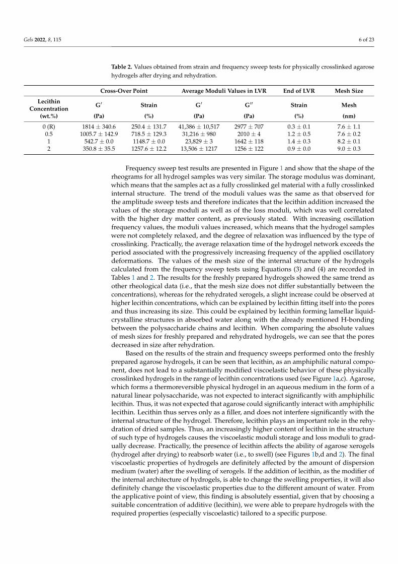
Table 2. Values obtained from strain and frequency sweep tests for physically crosslinked agarosehydrogels after drying and rehydration.
Cross-Over Point Average Moduli Values in LVR End of LVR Mesh Size
LecithinConcentration G′ Strain G′ G′′ Strain Mesh
(wt.%) (Pa) (%) (Pa) (Pa) (%) (nm)
0 (R) 1814 ± 340.6 250.4 ± 131.7 41,386 ± 10,517 2977 ± 707 0.3 ± 0.1 7.6 ± 1.10.5 1005.7 ± 142.9 718.5 ± 129.3 31,216 ± 980 2010 ± 4 1.2 ± 0.5 7.6 ± 0.21 542.7 ± 0.0 1148.7 ± 0.0 23,829 ± 3 1642 ± 118 1.4 ± 0.3 8.2 ± 0.12 350.8 ± 35.5 1257.6 ± 12.2 13,506 ± 1217 1256 ± 122 0.9 ± 0.0 9.0 ± 0.3
Frequency sweep test results are presented in Figure 1 and show that the shape of therheograms for all hydrogel samples was very similar. The storage modulus was dominant,which means that the samples act as a fully crosslinked gel material with a fully crosslinkedinternal structure. The trend of the moduli values was the same as that observed forthe amplitude sweep tests and therefore indicates that the lecithin addition increased thevalues of the storage moduli as well as of the loss moduli, which was well correlatedwith the higher dry matter content, as previously stated. With increasing oscillationfrequency values, the moduli values increased, which means that the hydrogel sampleswere not completely relaxed, and the degree of relaxation was influenced by the type ofcrosslinking. Practically, the average relaxation time of the hydrogel network exceeds theperiod associated with the progressively increasing frequency of the applied oscillatorydeformations. The values of the mesh size of the internal structure of the hydrogelscalculated from the frequency sweep tests using Equations (3) and (4) are recorded inTables 1 and 2. The results for the freshly prepared hydrogels showed the same trend asother rheological data (i.e., that the mesh size does not differ substantially between theconcentrations), whereas for the rehydrated xerogels, a slight increase could be observed athigher lecithin concentrations, which can be explained by lecithin fitting itself into the poresand thus increasing its size. This could be explained by lecithin forming lamellar liquid-crystalline structures in absorbed water along with the already mentioned H-bondingbetween the polysaccharide chains and lecithin. When comparing the absolute valuesof mesh sizes for freshly prepared and rehydrated hydrogels, we can see that the poresdecreased in size after rehydration.
Based on the results of the strain and frequency sweeps performed onto the freshlyprepared agarose hydrogels, it can be seen that lecithin, as an amphiphilic natural compo-nent, does not lead to a substantially modified viscoelastic behavior of these physicallycrosslinked hydrogels in the range of lecithin concentrations used (see Figure 1a,c). Agarose,which forms a thermoreversible physical hydrogel in an aqueous medium in the form of anatural linear polysaccharide, was not expected to interact significantly with amphiphiliclecithin. Thus, it was not expected that agarose could significantly interact with amphiphiliclecithin. Lecithin thus serves only as a filler, and does not interfere significantly with theinternal structure of the hydrogel. Therefore, lecithin plays an important role in the rehy-dration of dried samples. Thus, an increasingly higher content of lecithin in the structureof such type of hydrogels causes the viscoelastic moduli storage and loss moduli to grad-ually decrease. Practically, the presence of lecithin affects the ability of agarose xerogels(hydrogel after drying) to reabsorb water (i.e., to swell) (see Figures 1b,d and 2). The finalviscoelastic properties of hydrogels are definitely affected by the amount of dispersionmedium (water) after the swelling of xerogels. If the addition of lecithin, as the modifier ofthe internal architecture of hydrogels, is able to change the swelling properties, it will alsodefinitely change the viscoelastic properties due to the different amount of water. Fromthe applicative point of view, this finding is absolutely essential, given that by choosing asuitable concentration of additive (lecithin), we were able to prepare hydrogels with therequired properties (especially viscoelastic) tailored to a specific purpose.

Gels 2022, 8, 115 7 of 23
Gels 2022, 8, 115 7 of 23
able to prepare hydrogels with the required properties (especially viscoelastic) tailored to a specific purpose.
Figure 2. Drying (a) and rehydration (b) of the physically crosslinked agarose hydrogels with dif-ferent contents of lecithin.
2.1.2. Drying and Rehydration Measurements The amounts of water and dry matter associated with the studied gels are two of the
most important parameters for hydrogel characterization and future applicability. Dry matter affects the behavior of the final material. The same is true for the water inside the hydrogel, which significantly affects, for example, the transport properties. As stated in Section 1, these parameters predetermine the applicative nature of the final system.
The results of the drying kinetics of physically crosslinked hydrogels can be seen in Figure 2a. At the start of these experiments, all weights of the hydrogels (2 ± 0.2 g) and xerogels were comparably the same. It can be seen that the lecithin addition had no influ-ence on the drying kinetics. The most likely explanation is that water retained by lecithin is not bound as tightly as water hydrating agarose. Conversely, during the swelling pro-cess, hydrogel with lecithin easily draws water (more easily than the agarose hydrogel solely) and this resulted in the lecithin-agarose samples showing a higher swelling ability with corresponding lower moduli (Figure 1). The swelling experiments demonstrated the influence of lecithin on the swelling capacity. Therefore, the lecithin structures insert themselves into the hydrogel pores and support the water intake. The kinetics of the swell-ing process was very similar for all samples, with a peculiarity noted at the onset of the experiment, where the samples richer in lecithin (1 and 2 wt.%) revealed a greater rate of water absorption. Additionally, the same systems (agarose with 1 and 2 wt.% of lecithin) were able to absorb the largest amount of water.
2.1.3. Morphological Characterization of Xerogels Morphological characterization was performed on dried samples; therefore, the re-
sults may not correspond to the results obtained from methods where hydrogels are stud-ied in native form (specifically, rheology). From the results obtained by scanning electron microscopy (SEM), the effect of lecithin addition could be observed in sectional view. The surfaces of these xerogels were smooth and with no visible pores on the micrometer scale.
Figure 2. Drying (a) and rehydration (b) of the physically crosslinked agarose hydrogels with differentcontents of lecithin.
2.1.2. Drying and Rehydration Measurements
The amounts of water and dry matter associated with the studied gels are two of themost important parameters for hydrogel characterization and future applicability. Drymatter affects the behavior of the final material. The same is true for the water inside thehydrogel, which significantly affects, for example, the transport properties. As stated inSection 1, these parameters predetermine the applicative nature of the final system.
The results of the drying kinetics of physically crosslinked hydrogels can be seenin Figure 2a. At the start of these experiments, all weights of the hydrogels (2 ± 0.2 g)and xerogels were comparably the same. It can be seen that the lecithin addition had noinfluence on the drying kinetics. The most likely explanation is that water retained bylecithin is not bound as tightly as water hydrating agarose. Conversely, during the swellingprocess, hydrogel with lecithin easily draws water (more easily than the agarose hydrogelsolely) and this resulted in the lecithin-agarose samples showing a higher swelling abilitywith corresponding lower moduli (Figure 1). The swelling experiments demonstratedthe influence of lecithin on the swelling capacity. Therefore, the lecithin structures insertthemselves into the hydrogel pores and support the water intake. The kinetics of theswelling process was very similar for all samples, with a peculiarity noted at the onset ofthe experiment, where the samples richer in lecithin (1 and 2 wt.%) revealed a greater rateof water absorption. Additionally, the same systems (agarose with 1 and 2 wt.% of lecithin)were able to absorb the largest amount of water.
2.1.3. Morphological Characterization of Xerogels
Morphological characterization was performed on dried samples; therefore, the resultsmay not correspond to the results obtained from methods where hydrogels are studiedin native form (specifically, rheology). From the results obtained by scanning electronmicroscopy (SEM), the effect of lecithin addition could be observed in sectional view. Thesurfaces of these xerogels were smooth and with no visible pores on the micrometer scale. Insectional view, the lecithin-free xerogel exhibited a layered structure of polymer fibers withno visible interferences (see Figure 3). The same layered morphology was also observedfor xerogels of agarose with different contents of lecithin even though there were regionsof fusion of adjacent layers. Overall, the general morphology, practically devoid of pores

Gels 2022, 8, 115 8 of 23
as revealed by SEM, is most likely due to a compact structure resulting via the air dryingprocedure applied to hydrogels to finally obtain xerogels.
Gels 2022, 8, 115 8 of 23
In sectional view, the lecithin-free xerogel exhibited a layered structure of polymer fibers with no visible interferences (see Figure 3). The same layered morphology was also ob-served for xerogels of agarose with different contents of lecithin even though there were regions of fusion of adjacent layers. Overall, the general morphology, practically devoid of pores as revealed by SEM, is most likely due to a compact structure resulting via the air drying procedure applied to hydrogels to finally obtain xerogels.
Figure 3. Physically crosslinked agarose xerogels with different lecithin contents observed in sec-tional view by SEM. Magnification 5000×.
For these xerogels, gas sorption measurements were also performed (Table 3). The low values of the specific surface suggest a lack of the pore structure of xerogels, with a slight dependence on the compactness of layered morphology of these systems in dry state. Even if the results of gas sorption are in line with those of SEM investigation, the gas sorption method is not quite a suitable technique for determining the structure of these xerogels.
Table 3. Specific surface area for physically crosslinked agarose xerogels with the addition of leci-thin determined by gas sorption.
Concentration of Lecithin (wt.%) Specific Surface Area (m2/g) 0 (R) 3.4 0.5 1.0 1 1.9 2 2.1
Figure 3. Physically crosslinked agarose xerogels with different lecithin contents observed in sectionalview by SEM. Magnification 5000×.
For these xerogels, gas sorption measurements were also performed (Table 3). The lowvalues of the specific surface suggest a lack of the pore structure of xerogels, with a slightdependence on the compactness of layered morphology of these systems in dry state. Evenif the results of gas sorption are in line with those of SEM investigation, the gas sorptionmethod is not quite a suitable technique for determining the structure of these xerogels.
Table 3. Specific surface area for physically crosslinked agarose xerogels with the addition of lecithindetermined by gas sorption.
Concentration of Lecithin (wt.%) Specific Surface Area (m2/g)
0 (R) 3.40.5 1.01 1.92 2.1
2.2. Ionic Crosslinking
Sodium alginate crosslinked by the calcium chloride in the two to one weight ratiowas a representative of the ionically crosslinked hydrogel matrix, where the negativelycharged poly(guluronic) acid units of alginate (-COO−) interact with the polyvalent ions(Ca2+) to form a bond (schematic figure of the preparation procedure can be seen inSupplementary Materials, Figure S2). The final properties were also affected by lecithinaddition. Hydrogel samples, both after preparation and dried and rehydrated, were studiedby rheology, drying, and rehydration as well as morphological characterization.

Gels 2022, 8, 115 9 of 23
2.2.1. Rheology
Ionically crosslinked hydrogels also underwent amplitude sweep tests. What is im-mediately observable is the decreasing trend of moduli for the freshly prepared samplesas lecithin content increase (see Figure 4a). One of the reasons for this is the water in-take during gelling, which increases for samples with ascending lecithin concentration(Section 2.2.2), the amphiphilic component playing a major role in the preparation of ioni-cally crosslinked hydrogels. Larger lecithin addition also modified some characteristicsof the hydrogels (see Table 4). The average moduli values in LVR steadily decreased afterlecithin addition, thus making the gel softer. The most likely explanation is that after thecrosslinking of alginate by calcium ions, free calcium chloride is still present in the systemand is able to interact with the added lecithin micelles due to its dissociated form. Higherlecithin content causes a competitive interaction and as a result, lecithin displaces thecalcium ions in the crosslinked alginate. Further lecithin could interact with the alginate viaquaternary ammonia or with the calcium ions via negatively charged phosphate residues.For the moduli decrease, we could suggest that newly formed nodes are weaker and, ina lesser amount compared with the original alginate gel. Such competitive interactionswere observable even during sample preparation, where the precipitate was visible on thesurface of the solution. They were also confirmed by viscosity measurements, where thesolution of calcium chloride and lecithin had higher viscosity values than expected, basedon the viscosity of lecithin in water and of calcium chloride in water (figure is availablein Supplementary Materials Figure S3). Other rheological data were very similar for thesamples and, as stated earlier, the biggest differences were in the moduli values, thus in thehydrogel strength.
Table 4. Values for ionically crosslinked alginate hydrogels after preparation obtained from strainand frequency sweep tests before drying.
Cross-Over Point Average Moduli Values in LVR End of LVR Mesh Size
LecithinConcentration G′ Strain G′ G′′ Strain Mesh
(wt.%) (Pa) (%) (Pa) (Pa) (%) (nm)
0 (R) 150.4 ± 9.1 260.4 ± 18.6 1667 ± 192 165 ± 23 1.7 ± 0.0 10.9 ± 0.40.5 158.6 ± 12.5 275.3 ± 24.4 2138 ± 480 245 ± 66 1.3 ± 0.0 11.0 ± 0.71 110.8 ± 1.2 260.8 ± 5.4 1052 ± 1 104 ± 0 1.6 ± 0.3 13.8 ± 1.92 65.3 ± 17.9 278.2 ± 9.0 468 ± 15 41 ± 0 2.1 ± 0.4 17.3 ± 1.5
The rehydrated samples followed a similar trend with respect to the moduli values,where these values decreased with increasing lecithin concentration. Average modulivalues in LVR reported in the table below (Table 5) were higher than those presented inTable 4 because the rehydrated samples were not able to reabsorb the same amount of wateras the freshly prepared hydrogels. Such behavior could be due to a compact arrangementfavored by non-covalent interactions (mainly ionic interactions induced by Ca2+ ions ontoboth alginate and lecithin components) during the drying process.

Gels 2022, 8, 115 10 of 23Gels 2022, 8, 115 10 of 23
Figure 4. (a) Strain sweep of alginate hydrogels with the addition of different lecithin concentrations (0, 0.5, 1, and 2 wt.%) after preparation; (b) strain sweep of alginate hydrogels with different lecithin concentrations (0, 0.5, 1, and 2 wt.%) after drying and rehydration of the xerogels (frequency applied 1 Hz); (c) frequency sweep of alginate hydrogels with different lecithin concentrations (0, 0.5, 1, and 2 wt.%) after preparation; (d) frequency sweep of alginate hydrogels with different lecithin concen-trations (0, 0.5, 1, and 2 wt.%) after drying and rehydration of the xerogels.
The rheograms obtained during the frequency sweep tests (expressed as viscoelastic moduli on applied frequency) (Figure 4c) obeyed the same order as those that resulted from the amplitude sweep tests (storage and loss moduli as a function of oscillatory ap-plied strain of 1 Hz) (Figure 4a) for all the studied alginate and alginate-lecithin hydrogels. The calculated mesh size from the rheological (frequency sweep) measurement for freshly prepared ionically crosslinked alginate hydrogels indicated the effect of lecithin on the structural properties of these hydrogels. The higher addition of lecithin causes a higher
Figure 4. (a) Strain sweep of alginate hydrogels with the addition of different lecithin concentrations(0, 0.5, 1, and 2 wt.%) after preparation; (b) strain sweep of alginate hydrogels with different lecithinconcentrations (0, 0.5, 1, and 2 wt.%) after drying and rehydration of the xerogels (frequency applied1 Hz); (c) frequency sweep of alginate hydrogels with different lecithin concentrations (0, 0.5, 1,and 2 wt.%) after preparation; (d) frequency sweep of alginate hydrogels with different lecithinconcentrations (0, 0.5, 1, and 2 wt.%) after drying and rehydration of the xerogels.

Gels 2022, 8, 115 11 of 23
Table 5. Values for ionically crosslinked alginate hydrogels after drying and rehydration obtainedfrom the strain and frequency sweep tests.
Cross-Over Point Average Moduli Values in LVR End of LVR Mesh Size
LecithinConcentration G′ Strain G′ G′′ Strain Mesh
(wt.%) (Pa) (%) (Pa) (Pa) (%) (nm)
0 (R) 479.2 ± 129.7 210.8 ± 119.7 26,342 ± 13,355 3191 ± 1346 1.6 ± 0.4 4.6 ± 1.40.5 894.9 ± 612.4 522.6 ± 51.8 68,513 ± 17,434 9861 ± 1533 0.6 ± 0.6 12.3 ± 2.11 1179.5 ± 106.7 209.1 ± 37.3 25,386 ± 741 2912 ± 45 1.2 ± 0.2 8.3 ± 1.82 553.5 ± 24.3 189.5 ± 17.4 4599 ± 500 1842 ± 1447 2.4 ± 0.0 7.6 ± 0.5
The rheograms obtained during the frequency sweep tests (expressed as viscoelasticmoduli on applied frequency) (Figure 4c) obeyed the same order as those that resulted fromthe amplitude sweep tests (storage and loss moduli as a function of oscillatory appliedstrain of 1 Hz) (Figure 4a) for all the studied alginate and alginate-lecithin hydrogels. Thecalculated mesh size from the rheological (frequency sweep) measurement for freshlyprepared ionically crosslinked alginate hydrogels indicated the effect of lecithin on thestructural properties of these hydrogels. The higher addition of lecithin causes a highermesh size (more than 50% if the hydrogels without/with 2 wt.% of lecithin is compared).The effect of lecithin concentration was also not observed for dried and rehydrated hydro-gels. Although ionically crosslinked hydrogels have the ability to reabsorb the dispersionmedium and again create a network internal structure by water intake, the internal struc-ture of these hydrogels is probably damaged by the air-drying process. Moreover, swelledhydrogels differ in mesh size values in comparison with freshly prepared (e.g., hydrogelswith 2 wt.% of lecithin had a mesh size of 17.3 nm while the mesh size of the hydrogelswith the same concentration of lecithin after swelling was 7.6 nm). Therefore, the effect oflecithin on the mesh size of hydrogels repeatedly prepared by drying and swelling in watermedium was negligible.
2.2.2. Drying and Rehydration Measurements
The drying curves for the alginate-lecithin systems were very similar almost irrespec-tive of the lecithin content, in contrast to the drying dependence obtained for the freshlyprepared hydrogels of alginate solely (Figure 5a). The different kinetics regarding the rateof water loss during the drying step could be due to the way lecithin fills the hydrogelpores and holds water within, and also due to the favorable electrostatic Ca2+-lecithininteractions, which influence the hydrogel structure and thus enable it to better hold water.As for the swelling after drying, it can be observed that the samples with higher lecithinconcentrations were able to absorb water more rapidly and to a higher capacity, which isagain due to the modified hydrogel network due to the presence of lecithin.
2.2.3. Morphological Characterization of Xerogels
SEM images taken for xerogels prepared by ionic crosslinking show the effect oflecithin on the surface morphology of the samples (see Figure 6). Surface morphologyof lecithin-free samples and of those with 0.5 wt.% lecithin exhibited a roughness dueto the many micrometer-sized crystals of CaCl2 resulted after air-drying. Instead, thesurface of xerogels with 1 and 2 wt.% lecithin is practically devoid of crystalline aggregates,with some degree of roughness, which led to a more compact structure of these mixedsystems in their dry state. The morphological characteristics microscopically revealedare in accordance with the decreasing tendency of the specific surface values (from gassorption measurements, Table 6) as the lecithin content rose. On the other hand, the lack ofCaCl2 crystalline aggregates for the systems with a higher lecithin content (1 and 2 wt.%)could be related to Ca2+ consumption in favorable electrostatic interactions with lecithinanions, which means that the crystalline structures observed in the case of alginate xerogels

Gels 2022, 8, 115 12 of 23
without lecithin and for those with 0.5 wt.% lecithin could be due to the excess of CaCl2contained in these explored samples.
Gels 2022, 8, 115 11 of 23
mesh size (more than 50% if the hydrogels without/with 2 wt.% of lecithin is compared). The effect of lecithin concentration was also not observed for dried and rehydrated hy-drogels. Although ionically crosslinked hydrogels have the ability to reabsorb the disper-sion medium and again create a network internal structure by water intake, the internal structure of these hydrogels is probably damaged by the air-drying process. Moreover, swelled hydrogels differ in mesh size values in comparison with freshly prepared (e.g., hydrogels with 2 wt.% of lecithin had a mesh size of 17.3 nm while the mesh size of the hydrogels with the same concentration of lecithin after swelling was 7.6 nm). Therefore, the effect of lecithin on the mesh size of hydrogels repeatedly prepared by drying and swelling in water medium was negligible.
Table 5. Values for ionically crosslinked alginate hydrogels after drying and rehydration obtained from the strain and frequency sweep tests.
Cross-Over Point Average Moduli Values in LVR End of LVR Mesh Size Lecithin
Concentration G’ Strain G’ G’’ Strain Mesh
(wt.%) (Pa) (%) (Pa) (Pa) (%) (nm) 0 (R) 479.2 ± 129.7 210.8 ± 119.7 26,342 ± 13,355 3191 ± 1346 1.6 ± 0.4 4.6 ± 1.4 0.5 894.9 ± 612.4 522.6 ± 51.8 68,513 ± 17,434 9861 ± 1533 0.6 ± 0.6 12.3 ± 2.1 1 1179.5 ± 106.7 209.1 ± 37.3 25,386 ± 741 2912 ± 45 1.2 ± 0.2 8.3 ± 1.8 2 553.5 ± 24.3 189.5 ± 17.4 4599 ± 500 1842 ± 1447 2.4 ± 0.0 7.6 ± 0.5
2.2.2. Drying and Rehydration Measurements The drying curves for the alginate-lecithin systems were very similar almost irrespec-
tive of the lecithin content, in contrast to the drying dependence obtained for the freshly prepared hydrogels of alginate solely (Figure 5a). The different kinetics regarding the rate of water loss during the drying step could be due to the way lecithin fills the hydrogel pores and holds water within, and also due to the favorable electrostatic Ca2+-lecithin in-teractions, which influence the hydrogel structure and thus enable it to better hold water. As for the swelling after drying, it can be observed that the samples with higher lecithin concentrations were able to absorb water more rapidly and to a higher capacity, which is again due to the modified hydrogel network due to the presence of lecithin.
Figure 5. Drying (a) and rehydration (b) of ionically crosslinked alginate hydrogels with different lecithin content.
Figure 5. Drying (a) and rehydration (b) of ionically crosslinked alginate hydrogels with differentlecithin content.
Gels 2022, 8, 115 12 of 23
2.2.3. Morphological Characterization of Xerogels SEM images taken for xerogels prepared by ionic crosslinking show the effect of lec-
ithin on the surface morphology of the samples (see Figure 6). Surface morphology of lecithin-free samples and of those with 0.5 wt.% lecithin exhibited a roughness due to the many micrometer-sized crystals of CaCl2 resulted after air-drying. Instead, the surface of xerogels with 1 and 2 wt.% lecithin is practically devoid of crystalline aggregates, with some degree of roughness, which led to a more compact structure of these mixed systems in their dry state. The morphological characteristics microscopically revealed are in ac-cordance with the decreasing tendency of the specific surface values (from gas sorption measurements, Table 6) as the lecithin content rose. On the other hand, the lack of CaCl2 crystalline aggregates for the systems with a higher lecithin content (1 and 2 wt.%) could be related to Ca2+ consumption in favorable electrostatic interactions with lecithin anions, which means that the crystalline structures observed in the case of alginate xerogels with-out lecithin and for those with 0.5 wt.% lecithin could be due to the excess of CaCl2 con-tained in these explored samples.
Figure 6. Surface morphologies of ionically crosslinked alginate xerogels with the addition of leci-thin revealed by SEM. Magnification 5000×.
Table 6. Specific surface area for ionically crosslinked alginate xerogels with the addition of lecithin determined by gas sorption.
Concentration of Lecithin (wt.%) Specific Surface Area (m2/g) 0 (R) 9.1 0.5 6.3 1 5.9 2 4.7
Figure 6. Surface morphologies of ionically crosslinked alginate xerogels with the addition of lecithinrevealed by SEM. Magnification 5000×.

Gels 2022, 8, 115 13 of 23
Table 6. Specific surface area for ionically crosslinked alginate xerogels with the addition of lecithindetermined by gas sorption.
Concentration of Lecithin (wt.%) Specific Surface Area (m2/g)
0 (R) 9.10.5 6.31 5.92 4.7
2.3. Chemical Crosslinking
Poly(vinyl alcohol) and chitosan crosslinked by the epichlorohydrin was a representa-tive of the chemically crosslinked hydrogel matrix. Epichlorohydrin reacts with either thehydroxyl group of PVA or amino group of chitosan to form a highly reactive intermediate.This intermediate product reacts with another hydroxyl (PVA) or amino group (chitosan)to form the crosslinked structure. Study of these hydrogels, both in their freshly preparedstate, after air-drying at 40 ◦C and their subsequent rehydration and as xerogels, showedsome physico-mechanical properties altered by the lecithin content (schematic figure of thepreparation procedure can be seen in the Supplementary Materials Figure S4).
2.3.1. Rheology
For chemically crosslinked hydrogels, the amplitude sweep results showed that theaddition of lecithin modified the rheological properties of hydrogels (see Figure 7a). How-ever, the highest lecithin concentration did not lead to further changes in the mechanicalproperties. The same can be said after comparing the data points (see Table 7). At thesame time, a higher content of lecithin decreased the values marking the end of the LVRas well as the strength of the hydrogels and the cross- over point values. The results areacceptable after taking into account the preparation and final state of the hydrogel. Animportant step of the preparation procedure is drying of the liquid mixture, which leadsto crosslinking of the nodes and its subsequent rehydration. If lecithin is present, therehydration is improved.
Table 7. Values for chemically crosslinked PVA-chitosan hydrogels obtained from strain and fre-quency sweep tests before drying.
Cross-Over Point Average Moduli Values in LVR End of LVR Mesh Size
LecithinConcentration G′ Strain G′ G′′ Strain Mesh
(wt.%) (Pa) (%) (Pa) (Pa) (%) (nm)
0 (R) 1665.3 ± 43.2 53.8 ± 8.2 8629 ± 304 398 ± 4 1.6 ± 0.3 13.6 ± 0.70.5 1005.5 ± 32.4 49.4 ± 18.4 6644 ± 1503 307 ± 44 1.2 ± 0.9 13.8 ± 0.61 666.6 ± 5.4 40.2 ± 3.2 4545 ± 129 377 ± 68 0.6 ± 0.1 12.7 ± 0.12 631.6 ± 24.7 39.1 ± 4.7 4398 ± 195 421 ± 5 0.7 ± 0.1 12.9 ± 0.1
The same experiments were performed for hydrogel samples dried and rehydrated.The dried and rehydrated hydrogels with lecithin assembled into the pores ended upwith modified properties (see Figure 7b), specifically, an increase in moduli values and adecrease in the values marking the cross-over point, in contrast to the reference sample.As can be seen in Figure 7b and Table 8, the presence of lecithin makes the hydrogelsobtained after the drying–rehydration step much more deformation resistant, characterizedby much higher values of strain at the cross-over point. At the same time, for these mixedrehydrated hydrogels, lecithin, irrespective of its content, exerted a larger influence in theenhancement of the hydrogels’ strength (average moduli values in LVR) when comparedto the rehydrated systems physically and ionically crosslinked.

Gels 2022, 8, 115 14 of 23
Gels 2022, 8, 115 14 of 23
of the applied frequencies. The same trend was also observed for the dried and rehydrated samples. Again, for all samples, the storage modulus prevailed in comparison to the loss modulus. The mesh sizes of these samples (Tables 7 and 8) were not affected by the con-tent of lecithin, a result that can be explained by the character of covalent crosslinking, which is stronger than physical and ionic crosslinking. On the other hand, the same trend of decreasing mesh sizes after rehydration could be observed.
Figure 7. (a) Strain sweep of PVA-chitosan hydrogels with the different lecithin concentrations (0, 0.5, 1, and 2 wt.%) after preparation; (b) strain sweep of PVA-chitosan hydrogels with different lec-ithin concentrations (0, 0.5, 1, and 2 wt.%) after drying and rehydration of the xerogels (frequency applied–1 Hz); (c) frequency sweep of PVA-chitosan hydrogels with different lecithin concentra-tions (0, 0.5, 1, and 2 wt.%) after preparation; (d) frequency sweep of PVA-chitosan hydrogels with different lecithin concentrations (0, 0.5, 1, and 2 wt.%) after drying and rehydration of the xerogels.
Figure 7. (a) Strain sweep of PVA-chitosan hydrogels with the different lecithin concentrations (0,0.5, 1, and 2 wt.%) after preparation; (b) strain sweep of PVA-chitosan hydrogels with differentlecithin concentrations (0, 0.5, 1, and 2 wt.%) after drying and rehydration of the xerogels (frequencyapplied–1 Hz); (c) frequency sweep of PVA-chitosan hydrogels with different lecithin concentrations(0, 0.5, 1, and 2 wt.%) after preparation; (d) frequency sweep of PVA-chitosan hydrogels with differentlecithin concentrations (0, 0.5, 1, and 2 wt.%) after drying and rehydration of the xerogels.

Gels 2022, 8, 115 15 of 23
Table 8. Values for chemically crosslinked PVA-chitosan hydrogels after drying and rehydrationobtained from strain and frequency sweep tests.
Cross-Over Point Average Moduli Values in LVR End of LVR Mesh Size
LecithinConcentration G′ Strain G′ G′′ Strain Mesh
(wt.%) (Pa) (%) (Pa) (Pa) (%) (nm)
0 (R) 2470.0 ± 494.7 138.5 ± 13.5 14,514 ± 1413 532 ± 33 3.2 ± 0.0 11.6 ± 0.30.5 7122.4 ± 633.3 379.1 ± 233.0 62,099 ± 6505 1928 ± 65 5.0 ± 1.0 7.1 ± 0.11 4964.6 ± 275.8 502.2 ± 277.5 52,833 ± 10,153 2089 ± 246 3.0 ± 1.7 6.2 ± 1.32 4074.2 ± 182.3 900.1 ± 97.5 43,685 ± 3177 1761 ± 211 5.9 ± 2.3 8.1 ± 0.0
The frequency and amplitude sweep results indicated the same tendency discussedabove (see comparatively Figure 7). Thus, a critical lecithin concentration is necessary tomodify the properties of this type of chemically crosslinked hydrogels (according to theresults lying between 0.5 and 1 wt.%); also, there is a maximum concentration above whichfurther modifications do not occur (differences between 1 and 2 wt.% are negligible). Thesignificant difference in the chemically crosslinked hydrogels (comparing to the physicallyand ionically crosslinked) is the relaxation phenomenon characterized by much longerrelaxation times in contrast to covalently crosslinked systems. Covalently crosslinkedhydrogels exhibit almost constant values of storage moduli over the whole range of theapplied frequencies. The same trend was also observed for the dried and rehydratedsamples. Again, for all samples, the storage modulus prevailed in comparison to the lossmodulus. The mesh sizes of these samples (Tables 7 and 8) were not affected by the contentof lecithin, a result that can be explained by the character of covalent crosslinking, whichis stronger than physical and ionic crosslinking. On the other hand, the same trend ofdecreasing mesh sizes after rehydration could be observed.
2.3.2. Drying and Rehydration Measurements
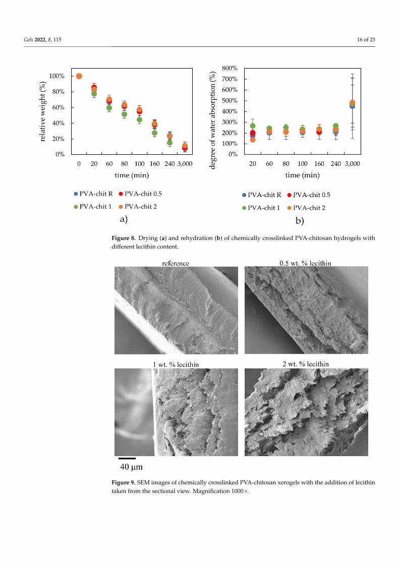
As can be seen from Figure 8, the drying and swelling kinetics were not significantlyaltered by the addition of lecithin. Only a marginal influence was observed for sampleswith the highest lecithin concentrations, which were able to absorb the most water. Thisgenerally smaller influence of lecithin can be explained by the structure of chemicallycrosslinked hydrogels, which are characterized by a high enough crosslinking density and,consequently, by a smaller pore size morphology. The structure is more organized due tothe stronger covalent bonds. The water absorption for this kind of hydrogel possessingstronger covalent cross linkages was very fast and occurred almost immediately during thefirst minutes of the swelling experiments.
2.3.3. Morphological Characterization of Xerogels
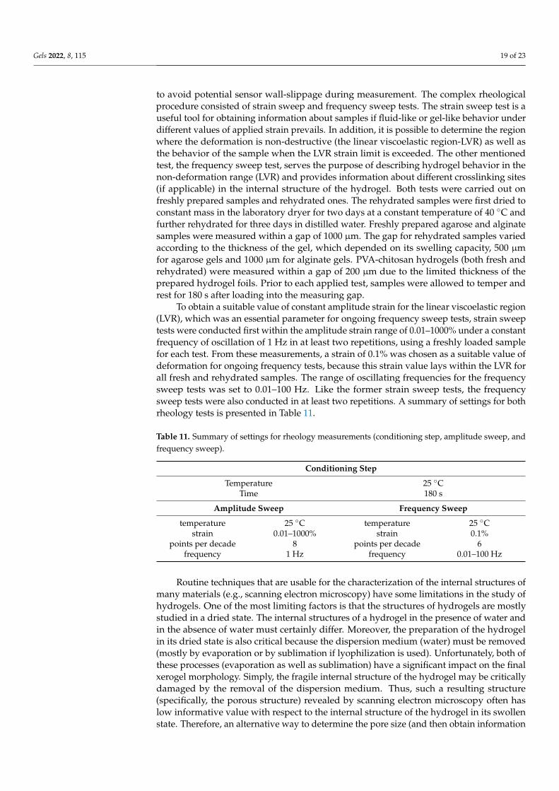
Results on the structural characterization of chemically crosslinked xerogels weresimilar to those for physically crosslinked hydrogels. The surface morphology of thesexerogels looked smooth with no visible pores. In sectional view, SEM images revealed clearlayered structures, with an interlayer roughness increasing with lecithin content (Figure 9),which in turn led to a gradual ascension of the value of specific surface (Table 9). Despitethis fact, an apparently less corrugated surface observed for lecithin-free hydrogels had ahigher specific surface area (Table 9), which might be explained by a greater compactnessassociated with the layered structure of the mixed xerogels.
Gels 2022, 8, 115 16 of 23
Gels 2022, 8, 115 15 of 23
Table 8. Values for chemically crosslinked PVA-chitosan hydrogels after drying and rehydration obtained from strain and frequency sweep tests.
Cross-Over Point Average moduli values in LVR End of LVR Mesh Size Lecithin
Concentration G’ Strain G’ G’’ Strain Mesh
(wt.%) (Pa) (%) (Pa) (Pa) (%) (nm) 0 (R) 2470.0 ± 494.7 138.5 ± 13.5 14514 ± 1413 532 ± 33 3.2 ± 0.0 11.6 ± 0.3 0.5 7122.4 ± 633.3 379.1 ± 233.0 62099 ± 6505 1928 ± 65 5.0 ± 1.0 7.1 ± 0.1 1 4964.6 ± 275.8 502.2 ± 277.5 52833 ± 10153 2089 ± 246 3.0 ± 1.7 6.2 ± 1.3 2 4074.2 ± 182.3 900.1 ± 97.5 43685 ± 3177 1761 ± 211 5.9 ± 2.3 8.1 ± 0.0
2.3.2. Drying and Rehydration Measurements As can be seen from Figure 8, the drying and swelling kinetics were not significantly
altered by the addition of lecithin. Only a marginal influence was observed for samples with the highest lecithin concentrations, which were able to absorb the most water. This generally smaller influence of lecithin can be explained by the structure of chemically crosslinked hydrogels, which are characterized by a high enough crosslinking density and, consequently, by a smaller pore size morphology. The structure is more organized due to the stronger covalent bonds. The water absorption for this kind of hydrogel pos-sessing stronger covalent cross linkages was very fast and occurred almost immediately during the first minutes of the swelling experiments.
Figure 8. Drying (a) and rehydration (b) of chemically crosslinked PVA-chitosan hydrogels with different lecithin content.
2.3.3. Morphological Characterization of Xerogels Results on the structural characterization of chemically crosslinked xerogels were
similar to those for physically crosslinked hydrogels. The surface morphology of these xerogels looked smooth with no visible pores. In sectional view, SEM images revealed clear layered structures, with an interlayer roughness increasing with lecithin content (Figure 9), which in turn led to a gradual ascension of the value of specific surface (Table 9). Despite this fact, an apparently less corrugated surface observed for lecithin-free
Figure 8. Drying (a) and rehydration (b) of chemically crosslinked PVA-chitosan hydrogels withdifferent lecithin content.
Gels 2022, 8, 115 16 of 23
hydrogels had a higher specific surface area (Table 9), which might be explained by a greater compactness associated with the layered structure of the mixed xerogels.
Figure 9. SEM images of chemically crosslinked PVA-chitosan xerogels with the addition of lecithin taken from the sectional view. Magnification 1000×.
Table 9. Specific surface area for chemically crosslinked PVA-chitosan xerogels with different leci-thin content determined by gas sorption.
Concentration of Lecithin (wt.%) Specific Surface Area (m2/g) 0 (R) 2.9 0.5 0.8 1 1.2 2 1.6
3. Conclusions This work studied the influence of lecithin (L-α-phosphatidylcholine) on three dif-
ferently crosslinked hydrogels (physically crosslinked agarose, alginate ionically cross-linked by calcium ions, and a mixture of PVA and chitosan chemically crosslinked by epichlorohydrin). The bulk of this work was to study differences between the gels inves-tigated immediately after preparation and the corresponding rehydrated xerogels (pre-pared by swelling). By choosing the lecithin content, we were able to modify some of the mechanical properties of the hydrogels with a modified internal structure, especially in the case of the rehydrated ones. In this regard, the addition of lecithin had the strongest influence in enhancing the strength of chemically crosslinked PVA-chitosan gels, which is partially consistent with the mesh size and by the amount of water absorbed into their structure after being previous air-dried. Apart from the rheological data and those ob-tained from the kinetics of water loss during hydrogel dehydration, these conclusions
Figure 9. SEM images of chemically crosslinked PVA-chitosan xerogels with the addition of lecithintaken from the sectional view. Magnification 1000×.

Gels 2022, 8, 115 17 of 23
Table 9. Specific surface area for chemically crosslinked PVA-chitosan xerogels with different lecithincontent determined by gas sorption.
Concentration of Lecithin (wt.%) Specific Surface Area (m2/g)
0 (R) 2.90.5 0.81 1.22 1.6
3. Conclusions
This work studied the influence of lecithin (L-α-phosphatidylcholine) on three differ-ently crosslinked hydrogels (physically crosslinked agarose, alginate ionically crosslinkedby calcium ions, and a mixture of PVA and chitosan chemically crosslinked by epichloro-hydrin). The bulk of this work was to study differences between the gels investigatedimmediately after preparation and the corresponding rehydrated xerogels (prepared byswelling). By choosing the lecithin content, we were able to modify some of the mechanicalproperties of the hydrogels with a modified internal structure, especially in the case ofthe rehydrated ones. In this regard, the addition of lecithin had the strongest influencein enhancing the strength of chemically crosslinked PVA-chitosan gels, which is partiallyconsistent with the mesh size and by the amount of water absorbed into their structureafter being previous air-dried. Apart from the rheological data and those obtained from thekinetics of water loss during hydrogel dehydration, these conclusions were supported bythe scanning electron microscopy and gas sorption experiments performed on the xerogels.For this type of material, even though gas sorption appears to be inappropriate, however, itserves to confirm the non-porous structure of the xerogels.
In this work, we determined that the addition of phospholipid lecithin into the hy-drogel matrix can alter their mechanical properties, which might be highly beneficialknowledge for the use of such hydrogels in particular applications. However, the transportproperties also need to be investigated. Therefore, further transport experiments are re-quired, which are absolutely crucial for a better understanding of such hydrogel materialsand how they can be used in final applications.
4. Materials and Methods
Hydrogels with distinct gelation mechanisms (physical, ionic, chemical crosslink-ing) [20] were studied. As an example of a physically crosslinked matrix, the linearthermoreversible polysaccharide agarose (Agarose E, Condalab, Madrid, Spain) at 1 wt.%,was used [21]. As an example of an ionically crosslinked matrix, sodium alginate (Sigma-Aldrich, Prague, Czech Republic) at 2 wt.% crosslinked by calcium chloride (Lach-Ner,Neratovice, Czech Republic) at a two to one weight ratio was chosen [22]. For chemi-cally crosslinked hydrogels, poly(vinyl alcohol) (Sigma-Aldrich, Prague, Czech Republic)mixed with chitosan (low molecular weight, Sigma-Aldrich, Prague, Czech Republic)and crosslinked by epichlorohydrin (Sigma-Aldrich, Prague, Czech Republic) was em-ployed [23]. L-α-Phosphatidylcholine (lecithin) was incorporated into all hydrogel samplesbefore gelation at three different weight percentage concentrations (Sigma-Aldrich, CzechRepublic, Prague).
The materials and their concentrations and ratios were selected on the basis of datapreviously reported [20–24] and can be seen in the table below (Table 10).

Gels 2022, 8, 115 18 of 23
Table 10. Concentrations of each individual component in the final hydrogel form (agarose, sodiumalginate, calcium chloride, PVA, chitosan, and lecithin).
Physically Crosslinked Hydrogels
Sample Agarose (wt.%) Lecithin (wt.%)
AG R 1 0AG 0.5 1 0.5AG 1 1 1AG 2 1 2
Ionically Crosslinked Hydrogels
Sample SodiumAlginate (wt.%)
Calcium Chloride(mol·dm3)
Lecithin(wt.%)
ALG R 2 0.1 0ALG 0.5 2 0.1 0.5ALG 1 2 0.1 1ALG 2 2 0.1 2
Chemically Crosslinked Hydrogels
Sample PVA (wt.%) Chitosan (wt.%) Lecithin(wt.%)
PVA R 7.8 2.5 0PVA 0.5 7.8 2.5 0.5PVA 1 7.8 2.5 1PVA 2 7.8 2.5 2
4.1. Water Loss during Drying and Rehydration Measurements
The ability to hold, release, and absorb water was tested by different approaches.Water loss was monitored by means of simple drying tests. All samples were dried eitherin the laboratory dryer at 40 ◦C and regularly weighed, or in a semi-automatic moistureanalyzer (IR-35, Denver Instrument, Denver, CO, USA), where the weight was recordedautomatically. The relative weight of the hydrogel (x) during drying was calculated usingthe following formula:
x =mt
m0· 100 (1)
where mt is the weight of the gel at time t, and m0 is the weight of the hydrogel in theswollen state.
Often very small weight losses of water from the hydrogel samples made usingdrying scales more difficult. For this reason, drying kinetics were mostly studied using thecombination of laboratory driers and analytical scales, upon which samples were weighedevery twenty minutes. After the samples were dried to the xerogel form, they were insertedinto a water bath, where they were kept until they reached their maximum water absorptioncapacity. The degree of water absorption (ma) was calculated by:
ma =mt
mx· 100 (2)
where mt is the weight of the hydrogel at time t, and mx is the weight of the xerogel. The hy-drogel samples were regularly weighed on analytical scales to study their swelling kinetics.
4.2. Rheology
Hydrogels are semi-solid materials that exhibit distinctive mechanical characteristicslying between those of solids and liquids. Therefore, rheology is indeed an appropriatetechnique for studying their behavior [25–29]. The mechanical properties of the preparedhydrogels were determined by rheological characterization using a rotational rheometer(Discovery HR-2, TA Instruments) employing cross-hatched 20 mm plate–plate geometry
Gels 2022, 8, 115 19 of 23
to avoid potential sensor wall-slippage during measurement. The complex rheologicalprocedure consisted of strain sweep and frequency sweep tests. The strain sweep test is auseful tool for obtaining information about samples if fluid-like or gel-like behavior underdifferent values of applied strain prevails. In addition, it is possible to determine the regionwhere the deformation is non-destructive (the linear viscoelastic region-LVR) as well asthe behavior of the sample when the LVR strain limit is exceeded. The other mentionedtest, the frequency sweep test, serves the purpose of describing hydrogel behavior in thenon-deformation range (LVR) and provides information about different crosslinking sites(if applicable) in the internal structure of the hydrogel. Both tests were carried out onfreshly prepared samples and rehydrated ones. The rehydrated samples were first dried toconstant mass in the laboratory dryer for two days at a constant temperature of 40 ◦C andfurther rehydrated for three days in distilled water. Freshly prepared agarose and alginatesamples were measured within a gap of 1000 µm. The gap for rehydrated samples variedaccording to the thickness of the gel, which depended on its swelling capacity, 500 µmfor agarose gels and 1000 µm for alginate gels. PVA-chitosan hydrogels (both fresh andrehydrated) were measured within a gap of 200 µm due to the limited thickness of theprepared hydrogel foils. Prior to each applied test, samples were allowed to temper andrest for 180 s after loading into the measuring gap.
To obtain a suitable value of constant amplitude strain for the linear viscoelastic region(LVR), which was an essential parameter for ongoing frequency sweep tests, strain sweeptests were conducted first within the amplitude strain range of 0.01–1000% under a constantfrequency of oscillation of 1 Hz in at least two repetitions, using a freshly loaded samplefor each test. From these measurements, a strain of 0.1% was chosen as a suitable value ofdeformation for ongoing frequency tests, because this strain value lays within the LVR forall fresh and rehydrated samples. The range of oscillating frequencies for the frequencysweep tests was set to 0.01–100 Hz. Like the former strain sweep tests, the frequencysweep tests were also conducted in at least two repetitions. A summary of settings for bothrheology tests is presented in Table 11.
Table 11. Summary of settings for rheology measurements (conditioning step, amplitude sweep, andfrequency sweep).
Conditioning Step
Temperature 25 ◦CTime 180 s
Amplitude Sweep Frequency Sweep
temperature 25 ◦C temperature 25 ◦Cstrain 0.01–1000% strain 0.1%
points per decade 8 points per decade 6frequency 1 Hz frequency 0.01–100 Hz
Routine techniques that are usable for the characterization of the internal structures ofmany materials (e.g., scanning electron microscopy) have some limitations in the study ofhydrogels. One of the most limiting factors is that the structures of hydrogels are mostlystudied in a dried state. The internal structures of a hydrogel in the presence of water andin the absence of water must certainly differ. Moreover, the preparation of the hydrogelin its dried state is also critical because the dispersion medium (water) must be removed(mostly by evaporation or by sublimation if lyophilization is used). Unfortunately, both ofthese processes (evaporation as well as sublimation) have a significant impact on the finalxerogel morphology. Simply, the fragile internal structure of the hydrogel may be criticallydamaged by the removal of the dispersion medium. Thus, such a resulting structure(specifically, the porous structure) revealed by scanning electron microscopy often haslow informative value with respect to the internal structure of the hydrogel in its swollenstate. Therefore, an alternative way to determine the pore size (and then obtain information

Gels 2022, 8, 115 20 of 23
about the internal structure of the hydrogel) must be found. An interesting solution to thisproblem is offered by the rheological characterization of the hydrogel, which involves thecalculation of the mesh size.
Mesh size, as one of the most critical parameters in hydrogel characterization, wascalculated by means of relaxation spectra (relaxation moduli G and relaxation time λ) fromthe frequency sweep oscillation measurements in accordance with the Maxwell model [30].The frequency sweep (viscoelastic moduli as a function of oscillation frequency) wasinterpolated by continuous relaxation spectra in TRIOS software (TA Instruments, NewCastle, DE, USA).
Typical relaxation spectra can be found in the Supplementary Materials (Figure S5).On the basis of previous rheological investigation [25], it was concluded that the optimalnumber of Maxwell elements was 4, in order to fit the frequency sweep measurements ofthe hydrogels. Four relaxation moduli were obtained from continuous relaxation spectraanalyses. The sum of relaxation moduli was calculated in order to determine the crosslink-ing density [31] (see Equation (3), where ρx represents the crosslinking density (mol·m−3))and provides information on the density of the junction in the swollen hydrogel form.G (Pa) is the sum of 4 relaxation moduli, R (J·mol−1·K−1) represents the universal gasconstant, and T is the thermodynamic temperature in Kelvins.
ρx =G
RT(3)
If all criteria are met (in particular, frequency sweep measurements are realized inthe linear viscoelastic region and the mechanical properties of hydrogels with differentcrosslinking are consistent with rubber elasticity theory [32]), finally the mesh size canbe calculated using Equation (4), where ξ is the mesh size (unit: m) and NA representsAvogadro’s number.
ξ = 3
√6
πρxNA(4)
4.3. Morphological Characterization of Xerogels
Since the structure affects properties that are crucial for hydrogel applications, deter-mining the hydrogel morphology is one of the most important characterizations. There aremany direct (microscopy) and indirect (scattering-based) methods to characterize hydrogelmorphology [33]. Several direct visualization techniques (light microscopy, laser scanningconfocal microscopy, and micro-computed tomography) that can handle swollen hydro-gels have considerable disadvantages (e.g., limited resolution) [34]. On the other hand,commonly used scanning electron microscopy includes a critical step (i.e., the inevitablesolidification of the sample using drying or freezing, during which the collapse of the struc-ture or the creation of artifacts can occur) [35,36]. Kaberova et al. [37] tested the usabilityof scanning electron microscopy and concluded that the results from this method shouldalways be confirmed by microscopy techniques applicable for gels in their swollen state.
For the characterization of dry samples, the specific surface area (the Brunauer–Emmett–Teller (BET) approach) is typically determined. The specific surface area is notsuitable for characterizing hydrogels because of the already mentioned artifacts that appearduring the preparation of dried samples. However, it can be used, for example, for the char-acterization of materials used in a dried state and that can form hydrogels (adsorbent) [38],or for the confirmation of reversible porosity [39].
The structure of the xerogels was studied in this work. Specifically, scanning electronmicroscopy and gas sorption were chosen as suitable techniques for determining the inter-nal architecture of xerogels. Since the mechanical properties were studied for hydrogelsright after preparation and also for swollen hydrogels after dehydration, it seemed con-venient to investigate the structural properties of the hydrogels in these forms. Since thisform is a dry form, it was possible to avoid deformation of the structure caused by thepreparation of hydrogels for scanning electron microscopy.

Gels 2022, 8, 115 21 of 23
4.3.1. Scanning Electron Microscopy
To determine changes in hydrogel structure, xerogels of all prepared samples weresubjected to direct visualization using scanning electron microscopy. The samples weredried in a laboratory dryer at 40 ◦C. A few small specimens were taken from each studiedsample to maintain objective observation. These specimens were subsequently gold-coatedin a sputtering device (POLARON) and investigated using a ZEISS EVO LS 10 scanningelectron microscope.
Both the surface morphologies and sectional images of samples were recorded. Obser-vations were realized in secondary electron (SE) mode and the accelerating voltage was setto 5 kV to avoid charging of the samples.
4.3.2. Gas Sorption
A NOVA 2200e high-speed gas sorption analyzer (Quantachrome Instruments) wasused to determine the specific surface area. The samples were weighed into a measuringcell (0.05–0.1 g). The measuring cell was placed in a degassing station, where the degassingprocess was carried out at 75 ◦C for 20 h. After cooling, the degassed sample was weighedto four decimal places. The samples were placed in a measuring station. The adsorptionand desorption isotherms were measured under liquid nitrogen (77 K) from 0.05–0.95 ofthe relative pressure P/P0. The obtained data were processed by NovaWin software andspecific surface area was calculated by the multi-point BET method.
Supplementary Materials: The following supporting information can be downloaded at: https://www.mdpi.com/article/10.3390/gels8020115/s1, Figure S1: Preparation procedure of physicallycrosslinked agarose hydrogels; Figure S2: Preparation procedure of ionically crosslinked alginate hy-drogels; Figure S3: Dynamic viscosity measurements for combinations of solutions of lecithin, CaCl2and alginate; Figure S4: Preparation procedure of chemically crosslinked PVA-chitosan hydrogels;Figure S5: Typical relaxation spectra for mesh size calculations, TRIOS software (TA Instruments).
Author Contributions: Conceptualization, R.H. and J.S.; Methodology, R.H., M.K., M.T. and J.H.;Validation, R.H., M.K. and M.T.; Formal analysis, R.H. and J.S.; Investigation, R.H., M.K., M.T.,J.H. and N.Z.; Data curation, R.H., M.T. and M.K.; Writing—original draft preparation, R.H., M.K.,M.T. and N.Z.; Writing—review and editing, J.S. and M.P.; Visualization, R.H. and N.Z.; Projectadministration, R.H.; Funding acquisition, R.H.; Supervision, J.S. All authors have read and agreed tothe published version of the manuscript.
Funding: This research project was supported by the project Quality Internal Grants of BUT (KInGBUT), Reg. No. CZ.02.2.69/0.0/0.0/19_073/0016948, which is financed from the Operational Program:Research, Development, and Education.
Institutional Review Board Statement: Not applicable.
Informed Consent Statement: Not applicable.
Data Availability Statement: The data used in this study are available on request from the corre-sponding author.
Conflicts of Interest: The authors declare no conflict of interest.
References1. Aswathy, S.; Narendrakumar, U.; Manjubala, I. Commercial hydrogels for biomedical applications. Heliyon 2020, 6, e03719.
[CrossRef] [PubMed]2. Kular, J.K.; Basu, S.; Sharma, R.I. The extracellular matrix: Structure, composition, age-related differences, tools for analysis and
applications for tissue engineering. J. Tissue Eng. 2014, 5, 2041731414557112. [CrossRef] [PubMed]3. Geckil, H.; Xu, F.; Zhang, X.; Moon, S.; Demirci, U. Engineering hydrogels as extracellular matrix mimics. Nanomedicine 2010, 5,
469–484. [CrossRef]4. Gadjanski, I. Recent advances on gradient hydrogels in biomimetic cartilage tissue engineering. F1000Research 2017, 6, 2158.
[CrossRef]5. Pekar, M. Hydrogels with Micellar Hydrophobic (Nano)Domains. Front. Mater. 2015, 1, 35. [CrossRef]

Gels 2022, 8, 115 22 of 23
6. Zhang, Y.; Chen, Q.; Dai, Z.; Dai, Y.; Xia, F.; Zhang, X. Nanocomposite adhesive hydrogels: From design to application. J. Mater.Chem. B 2021, 9, 585–593. [CrossRef]
7. Tuncaboylu, D.C.; Argun, A.; Algi, M.P.; Okay, O. Autonomic self-healing in covalently crosslinked hydrogels containinghydrophobic domains. Polymer 2013, 54, 6381–6388. [CrossRef]
8. Gu, S.; Duan, L.; Ren, X.; Gao, G.H. Robust, tough and anti-fatigue cationic latex composite hydrogels based on dual physicallycross-linked networks. J. Colloid Interface Sci. 2017, 492, 119–126. [CrossRef]
9. Li, A.; Jia, Y.; Sun, S.; Xu, Y.; Minsky, B.B.; Stuart, M.A.C.; Cölfen, H.; von Klitzing, R.; Guo, X. Mineral-Enhanced PolyacrylicAcid Hydrogel as an Oyster-Inspired Organic–Inorganic Hybrid Adhesive. ACS Appl. Mater. Interfaces 2018, 10, 10471–10479.[CrossRef]
10. Cui, C.; Wu, T.; Gao, F.; Fan, C.; Xu, Z.; Wang, H.; Liu, B.; Liu, W. An Autolytic High Strength Instant Adhesive Hydrogel forEmergency Self-Rescue. Adv. Funct. Mater. 2018, 28, 1804925. [CrossRef]
11. Fan, X.; Wang, S.; Fang, Y.; Li, P.; Zhou, W.; Wang, Z.; Chen, M.; Liu, H. Tough polyacrylamide-tannic acid-kaolin adhesivehydrogels for quick hemostatic application. Mater. Sci. Eng. C 2020, 109, 110649. [CrossRef] [PubMed]
12. Rajabi, N.; Kharaziha, M.; Emadi, R.; Zarrabi, A.; Mokhtari, H.; Salehi, S. An adhesive and injectable nanocomposite hydrogelof thiolated gelatin/gelatin methacrylate/Laponite® as a potential surgical sealant. J. Colloid Interface Sci. 2020, 564, 155–169.[CrossRef] [PubMed]
13. Arno, M.C.; Inam, M.; Weems, A.C.; Li, Z.; Binch, A.L.A.; Platt, C.I.; Richardson, S.M.; Hoyland, J.A.; Dove, A.P.; O’Reilly, R.K.Exploiting the role of nanoparticle shape in enhancing hydrogel adhesive and mechanical properties. Nat. Commun. 2020, 11, 1420.[CrossRef] [PubMed]
14. Zhang, H.; Hao, R.; Ren, X.; Yu, L.; Yang, H.; Yu, H. PEG/lecithin–liquid-crystalline composite hydrogels for quasi-zero-ordercombined release of hydrophilic and lipophilic drugs. RSC Adv. 2013, 3, 22927–22930. [CrossRef]
15. Shchipunov, Y.A. Lecithin. In Encyclopedia of Surface and Colloid Science, 3rd ed.; Somasundaran, P., Ed.; CRC Press: Boca Raton, FL,USA, 2015; Volume 3, pp. 3674–3693.
16. Elnaggar, Y.S.; El-Refaie, W.M.; El-Massik, M.A.; Abdallah, O.Y. Lecithin-based nanostructured gels for skin delivery: An updateon state of art and recent applications. J. Control. Release 2014, 180, 10–24. [CrossRef] [PubMed]
17. Thompson, B.R.; Zarket, B.C.; Lauten, E.H.; Amin, S.; Muthukrishnan, S.; Raghavan, S.R. Liposomes Entrapped in BiopolymerHydrogels Can Spontaneously Release into the External Solution. Langmuir 2020, 36, 7268–7276. [CrossRef]
18. Li, D.; An, X.; Mu, Y. A liposomal hydrogel with enzyme triggered release for infected wound. Chem. Phys. Lipids 2019, 223, 104783.[CrossRef]
19. Talaat, S.M.; Elnaggar, Y.S.R.; Abdalla, O.Y. Lecithin Microemulsion Lipogels Versus Conventional Gels for Skin Targeting ofTerconazole: In Vitro, Ex Vivo, and In Vivo Investigation. AAPS PharmSciTech 2019, 20, 161. [CrossRef]
20. Maitra, J.; Shukla, V.K. Cross-linking in Hydrogels—A Review. Am. J. Polym. Sci. 2014, 4, 25–31. [CrossRef]21. Trudicova, M.; Smilek, J.; Kalina, M.; Smilkova, M.; Adamkova, K.; Hrubanova, K.; Krzyzanek, V.; Sedlacek, P. Multiscale
Experimental Evaluation of Agarose-Based Semi-Interpenetrating Polymer Network Hydrogels as Materials with TunableRheological and Transport Performance. Polymers 2020, 12, 2561. [CrossRef]
22. Kuo, C.K.; Ma, P.X. Ionically crosslinked alginate hydrogels as scaffolds for tissue engineering: Part 1. Structure, gelation rate andmechanical properties. Biomaterials 2001, 22, 511–521. [CrossRef]
23. Garnica-Palafox, I.M.; Sánchez-Arévalo, F.M.; Velasquillo, C.; García-Carvajal, Z.; Garcia-Lopez, J.; Ortega-Sánchez, C.; Ibarra, C.;Luna-Barcenas, G.; Solís-Arrieta, L. Mechanical and structural response of a hybrid hydrogel based on chitosan and poly(vinylalcohol) cross-linked with epichlorohydrin for potential use in tissue engineering. J. Biomater. Sci. Polym. Ed. 2014, 25, 32–50.[CrossRef] [PubMed]
24. Mendes, A.C.L.; Shekarforoush, E.; Engwer, C.; Beeren, S.; Gorzelanny, C.; Goycoolea, F.M.; Chronakis, I.S. Co-assembly ofchitosan and phospholipids into hybrid hydrogels. Pure Appl. Chem. 2016, 88, 905–916. [CrossRef]
25. Smilek, J.; Jarábková, S.; Velcer, T.; Pekar, M. Compositional and Temperature Effects on the Rheological Properties ofPolyelectrolyte–Surfactant Hydrogels. Polymers 2019, 11, 927. [CrossRef]
26. Mourycová, J.; Datta, K.K.R.; Procházková, A.; Plotená, M.; Enev, V.; Smilek, J.; Másilko, J.; Pekar, M. Facile synthesis andrheological characterization of nanocomposite hyaluronan-organoclay hydrogels. Int. J. Biol. Macromol. 2018, 111, 680–684.[CrossRef]
27. Derkach, S.R.; Ilyin, S.O.; Maklakova, A.A.; Kulichikhin, V.G.; Malkin, A.Y. The rheology of gelatin hydrogels modified byκ-carrageenan. LWT-Food Sci. Technol. 2015, 63, 612–619. [CrossRef]
28. López-Marcial, G.R.; Zeng, A.Y.; Osuna, C.; Dennis, J.; García, J.M.; O’Connell, G.D. Agarose-Based Hydrogels as SuitableBioprinting Materials for Tissue Engineering. ACS Biomater. Sci. Eng. 2018, 4, 3610–3616. [CrossRef]
29. Gila-Vilchez, C.; Bonhome-Espinosa, A.B.; Kuzhir, P.; Zubarev, A.; Duran, J.D.G.; Lopez-Lopez, M.T. Rheology of magneticalginate hydrogels. J. Rheol. 2018, 62, 1083–1096. [CrossRef]
30. Gradzielski, M.; Hoffmann, I. Polyelectrolyte-surfactant complexes (PESCs) composed of oppositely charged components. Curr.Opin. Colloid Interface Sci. 2018, 35, 124–141. [CrossRef]
31. Pescosolido, L.; Feruglio, L.; Farra, R.; Fiorentino, S.; Colombo, I.; Coviello, T.; Matricardi, P.; Hennink, W.E.; Vermonden, T.;Grassi, M. Mesh size distribution determination of interpenetrating polymer network hydrogels. Soft Matter 2012, 8, 7708–7715.[CrossRef]

Gels 2022, 8, 115 23 of 23
32. Flory, P.J. Principles of Polymer Chemistry; Cornell University Press: New York, NY, USA, 1953.33. Raghuwanshi, V.S.; Garnier, G. Characterisation of hydrogels: Linking the nano to the microscale. Adv. Colloid Interface Sci. 2019,
274, 102044. [CrossRef] [PubMed]34. Suchý, T.; Šupová, M.; Bartoš, M.; Sedlácek, R.; Piola, M.; Soncini, M.; Fiore, G.B.; Sauerová, P.; Kalbácová, M.H. Dry versus
hydrated collagen scaffolds: Are dry states representative of hydrated states? J. Mater. Sci. Mater. Med. 2018, 29, 20. [CrossRef][PubMed]
35. Muthulakshmi, L.; Pavithra, U.; Sivaranjani, V.; Balasubramanian, N.; Sakthivel, K.M.; Pruncu, C.I. A novel Ag/carrageenan–gelatin hybrid hydrogel nanocomposite and its biological applications: Preparation and characterization. J. Mech. Behav. Biomed.Mater. 2021, 115, 104257. [CrossRef]
36. Marmorat, C.; Arinstein, A.; Koifman, N.; Talmon, Y.; Zussman, E.; Rafailovich, M. Cryo-Imaging of Hydrogels SupermolecularStructure. Sci. Rep. 2016, 6, 25495. [CrossRef] [PubMed]
37. Kaberova, Z.; Karpushkin, E.; Nevoralová, M.; Vetrík, M.; Šlouf, M.; Dušková-Smrcková, M. Microscopic Structure of SwollenHydrogels by Scanning Electron and Light Microscopies: Artifacts and Reality. Polymers 2020, 12, 578. [CrossRef]
38. Bhagat, S.D.; Kim, Y.-H.; Yi, G.; Ahn, Y.-S.; Yeo, J.-G.; Choi, Y.-T. Mesoporous SiO2 powders with high specific surface area bymicrowave drying of hydrogels: A facile synthesis. Microporous Mesoporous Mater. 2008, 108, 333–339. [CrossRef]
39. Weber, J.; Bergström, L. Mesoporous Hydrogels: Revealing Reversible Porosity by Cryoporometry, X-ray Scattering, and GasAdsorption. Langmuir 2010, 26, 10158–10164. [CrossRef]