Hydrogel Dressing with a Nano-Formula against Methicillin ...
13
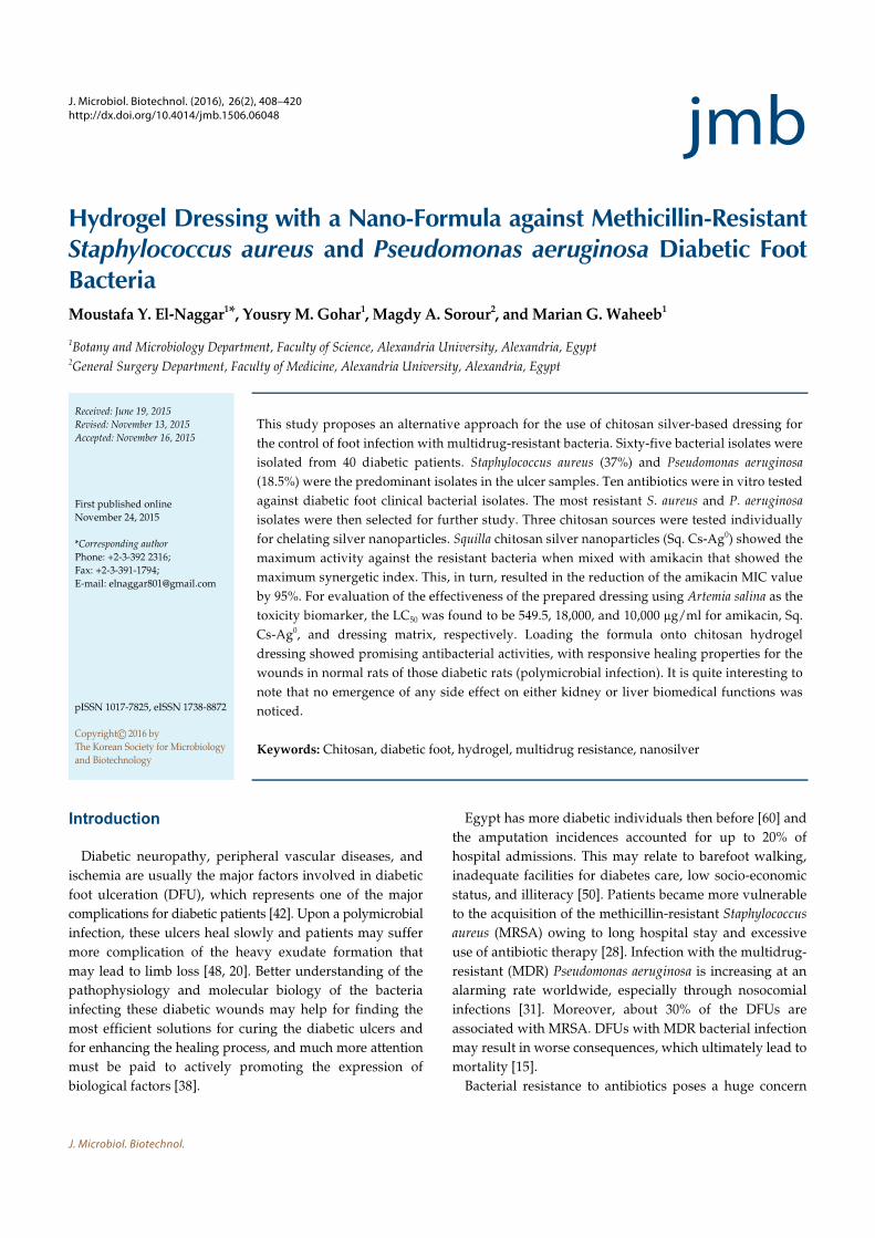
J. Microbiol. Biotechnol. J. Microbiol. Biotechnol. (2016), 26(2), 408–420 http://dx.doi.org/10.4014/jmb.1506.06048 Research Article jmb Hydrogel Dressing with a Nano-Formula against Methicillin-Resistant Staphylococcus aureus and Pseudomonas aeruginosa Diabetic Foot Bacteria Moustafa Y. El-Naggar 1 * , Yousry M. Gohar 1 , Magdy A. Sorour 2 , and Marian G. Waheeb 1 1 Botany and Microbiology Department, Faculty of Science, Alexandria University, Alexandria, Egypt 2 General Surgery Department, Faculty of Medicine, Alexandria University, Alexandria, Egypt Introduction Diabetic neuropathy, peripheral vascular diseases, and ischemia are usually the major factors involved in diabetic foot ulceration (DFU), which represents one of the major complications for diabetic patients [42]. Upon a polymicrobial infection, these ulcers heal slowly and patients may suffer more complication of the heavy exudate formation that may lead to limb loss [48, 20]. Better understanding of the pathophysiology and molecular biology of the bacteria infecting these diabetic wounds may help for finding the most efficient solutions for curing the diabetic ulcers and for enhancing the healing process, and much more attention must be paid to actively promoting the expression of biological factors [38]. Egypt has more diabetic individuals then before [60] and the amputation incidences accounted for up to 20% of hospital admissions. This may relate to barefoot walking, inadequate facilities for diabetes care, low socio-economic status, and illiteracy [50]. Patients became more vulnerable to the acquisition of the methicillin-resistant Staphylococcus aureus (MRSA) owing to long hospital stay and excessive use of antibiotic therapy [28]. Infection with the multidrug- resistant (MDR) Pseudomonas aeruginosa is increasing at an alarming rate worldwide, especially through nosocomial infections [31]. Moreover, about 30% of the DFUs are associated with MRSA. DFUs with MDR bacterial infection may result in worse consequences, which ultimately lead to mortality [15]. Bacterial resistance to antibiotics poses a huge concern Received: June 19, 2015 Revised: November 13, 2015 Accepted: November 16, 2015 First published online November 24, 2015 *Corresponding author Phone: +2-3-392 2316; Fax: +2-3-391-1794; E-mail: [email protected] pISSN 1017-7825, eISSN 1738-8872 Copyright © 2016 by The Korean Society for Microbiology and Biotechnology This study proposes an alternative approach for the use of chitosan silver-based dressing for the control of foot infection with multidrug-resistant bacteria. Sixty-five bacterial isolates were isolated from 40 diabetic patients. Staphylococcus aureus (37%) and Pseudomonas aeruginosa (18.5%) were the predominant isolates in the ulcer samples. Ten antibiotics were in vitro tested against diabetic foot clinical bacterial isolates. The most resistant S. aureus and P. aeruginosa isolates were then selected for further study. Three chitosan sources were tested individually for chelating silver nanoparticles. Squilla chitosan silver nanoparticles (Sq. Cs-Ag 0 ) showed the maximum activity against the resistant bacteria when mixed with amikacin that showed the maximum synergetic index. This, in turn, resulted in the reduction of the amikacin MIC value by 95%. For evaluation of the effectiveness of the prepared dressing using Artemia salina as the toxicity biomarker, the LC 50 was found to be 549.5, 18,000, and 10,000 μg/ml for amikacin, Sq. Cs-Ag 0 , and dressing matrix, respectively. Loading the formula onto chitosan hydrogel dressing showed promising antibacterial activities, with responsive healing properties for the wounds in normal rats of those diabetic rats (polymicrobial infection). It is quite interesting to note that no emergence of any side effect on either kidney or liver biomedical functions was noticed. Keywords: Chitosan, diabetic foot, hydrogel, multidrug resistance, nanosilver
-
Upload
khangminh22 -
Category
Documents
-
view
4 -
download
0
Transcript of Hydrogel Dressing with a Nano-Formula against Methicillin ...
J. Microbiol. Biotechnol.
J. Microbiol. Biotechnol. (2016), 26(2), 408–420http://dx.doi.org/10.4014/jmb.1506.06048 Research Article jmbReview
Hydrogel Dressing with a Nano-Formula against Methicillin-ResistantStaphylococcus aureus and Pseudomonas aeruginosa Diabetic FootBacteriaMoustafa Y. El-Naggar1*, Yousry M. Gohar1, Magdy A. Sorour2, and Marian G. Waheeb1
1Botany and Microbiology Department, Faculty of Science, Alexandria University, Alexandria, Egypt2General Surgery Department, Faculty of Medicine, Alexandria University, Alexandria, Egypt
Introduction
Diabetic neuropathy, peripheral vascular diseases, and
ischemia are usually the major factors involved in diabetic
foot ulceration (DFU), which represents one of the major
complications for diabetic patients [42]. Upon a polymicrobial
infection, these ulcers heal slowly and patients may suffer
more complication of the heavy exudate formation that
may lead to limb loss [48, 20]. Better understanding of the
pathophysiology and molecular biology of the bacteria
infecting these diabetic wounds may help for finding the
most efficient solutions for curing the diabetic ulcers and
for enhancing the healing process, and much more attention
must be paid to actively promoting the expression of
biological factors [38].
Egypt has more diabetic individuals then before [60] and
the amputation incidences accounted for up to 20% of
hospital admissions. This may relate to barefoot walking,
inadequate facilities for diabetes care, low socio-economic
status, and illiteracy [50]. Patients became more vulnerable
to the acquisition of the methicillin-resistant Staphylococcus
aureus (MRSA) owing to long hospital stay and excessive
use of antibiotic therapy [28]. Infection with the multidrug-
resistant (MDR) Pseudomonas aeruginosa is increasing at an
alarming rate worldwide, especially through nosocomial
infections [31]. Moreover, about 30% of the DFUs are
associated with MRSA. DFUs with MDR bacterial infection
may result in worse consequences, which ultimately lead to
mortality [15].
Bacterial resistance to antibiotics poses a huge concern
Received: June 19, 2015
Revised: November 13, 2015
Accepted: November 16, 2015
First published online
November 24, 2015
*Corresponding author
Phone: +2-3-392 2316;
Fax: +2-3-391-1794;
E-mail: [email protected]
pISSN 1017-7825, eISSN 1738-8872
Copyright© 2016 by
The Korean Society for Microbiology
and Biotechnology
This study proposes an alternative approach for the use of chitosan silver-based dressing for
the control of foot infection with multidrug-resistant bacteria. Sixty-five bacterial isolates were
isolated from 40 diabetic patients. Staphylococcus aureus (37%) and Pseudomonas aeruginosa
(18.5%) were the predominant isolates in the ulcer samples. Ten antibiotics were in vitro tested
against diabetic foot clinical bacterial isolates. The most resistant S. aureus and P. aeruginosa
isolates were then selected for further study. Three chitosan sources were tested individually
for chelating silver nanoparticles. Squilla chitosan silver nanoparticles (Sq. Cs-Ag0) showed the
maximum activity against the resistant bacteria when mixed with amikacin that showed the
maximum synergetic index. This, in turn, resulted in the reduction of the amikacin MIC value
by 95%. For evaluation of the effectiveness of the prepared dressing using Artemia salina as the
toxicity biomarker, the LC50 was found to be 549.5, 18,000, and 10,000 µg/ml for amikacin, Sq.
Cs-Ag0, and dressing matrix, respectively. Loading the formula onto chitosan hydrogel
dressing showed promising antibacterial activities, with responsive healing properties for the
wounds in normal rats of those diabetic rats (polymicrobial infection). It is quite interesting to
note that no emergence of any side effect on either kidney or liver biomedical functions was
noticed.
Keywords: Chitosan, diabetic foot, hydrogel, multidrug resistance, nanosilver

A Nano-Formula for MR S. aureus and P. aeruginosa 409
February 2016⎪Vol. 26⎪No. 2
and treatment with any new antibiotic will eventually
result in the emergence of resistant bacterial isolates [58].
Chitosan has been successfully included in a wide variety
of pharmaceutical applications, such as transdermal drug
delivery carriers [10] and especially those used for wound
dressing [4]. Chitosan films also can be used as a proper
candidate in healing DFUs, which are common in patients
with diabetes mellitus. It works efficiently and in a faster
manner for combating long-term complications [32].
Chitosan-containing metal nanoparticles can significantly
overcome multiple drug resistance mechanisms, such as
uptake decrease and efflux increase of drug from the
microbial cell, and inhibit biofilm formation. Moreover,
nanoparticles target antimicrobial agents to the infected site
where higher doses of drug are successfully delivered [41].
Treatment of DFUs represents a challenge in the
development of new and widely satisfying wound dressings
[36]. Polysaccharide hydrogels are very convenient for
producing effective dressings. Hydrogel wound dressings
are available in different forms or as a spreadable viscous
gel of three-dimensional polymeric networks. They are
semipermeable to gases and water vapor and can provide a
moist and hydrated environment for the wound area [45,
49, 17]. Hydrogels can be the most effective form of chitosan
for chronic wound dressings that can provide completion
of the healing process for wounds of all requirements [54].
The present study aimed at finding a suitable and
effective formula for a potent antibacterial activity with an
effective healing of the infected diabetic ulcers.
Materials and Methods
Isolation of Clinical Samples
Bacterial samples were collected over a period of 6 months (July
to December), 2012, from forty diabetic patients admitted to the
Vascular Surgery Unit and the Diabetic Foot Unit, Alexandria
Main University Hospital, Faculty of Medicine, Egypt. Bacterial
samples were isolated from heel, dorsum, and amputation sites.
Bacterial isolation from the samples was performed by swabbing
[12], aspiration, and tissue biopsy [39]. All samples were
immediately transferred within 1-2 h to the laboratory, incubated
at 37oC for 24 h in sterile nutrient broth tubes, vortexed, and then
inoculated onto sterile blood agar plates using sterile swabs. All
bacterial samples were initially identified by gram-staining and
the application of routine biochemical tests. Selected bacterial
isolates of S. aureus and P. aeruginosa were also identified with the
API NE system (bioMerieux, Paris, France). The results obtained
were interpreted using the APILAB PLUS software (ver. 3.3.3,
France).
Identification of Bacterial Isolates by Polymerase Chain Reaction
(PCR)
Bacterial isolates were finally identified by PCR technique using
specific primers purchased from Sigma Aldrich, Germany (Table 1).
Genomic DNA was extracted according to the procedure given in
the protocol of the Gene Jet Genomic DNA Purification Kit
(Thermo Scientific) (Sigma Aldrich, Germany).
The PCR amplification process was carried out (using Maxima
hot start green PCR master mix kit; Sigma Aldrich) for 40 cycles of
95°C for 4 min, annealing at 59°C for 30 sec, and extension at 72°C
for 15 sec. After PCR amplification, to estimate the sizes of the
Table 1. Specific primers used to identify certain genes in MRSA and P. aeruginosa.
Strain Primer Sequence Gene Amplicon size (bp) Reference
S. aureus MecA-F
MecA-R
FemB-F
FemB-R
GTA GAA ATG ACT GAA CGT CCG ATAA
CCA ATT CCA CAT TGT TTC GGTCTAA
TTA CAG AGT TAA CTG TTA CC
ATA CAA ATC CAGCAC GCT CT
mecA
femB
310
365
[9]
[13]
P. aeruginosa gyrB-F
gyrB-R
CCTGACCATCCGTCGCCACAAC
CGCAGCAGGATGCCGACGCC
gyrB 222 [23]
ETA-F
ETA-R
GACAACGCCCTCAGCATCACCA
CGCTGGCCCATTCGCTCCAGCG
ETA 397 [35]
oprL-F
oprL-R
Pa16S-F
Pa16S-R
ATGGAAATGCTGAAATTCGGC
CTTCTTCAGCTCGACGCGACG
GGGGGATCTTCGGACCTCA
TCCTTAGAGTGCCCACCCG
oprL
16S rDNA
504
618
[22]
[53]

410 El-Naggar et al.
J. Microbiol. Biotechnol.
amplification products, 5 µl was removed and subjected to agarose
gel electrophoresis (2% agarose, 1× Tris-buffered-EDTA, 100 V,
90 min) and compared with a 1 KB standard ladder (Qiagen,
Australia). The gel was stained with 10 µg/ml ethidium bromide,
and the amplicons were visualized using a UV illuminator. MRSA
ATCC 43300 was used as a positive control, whereas MSSA ATCC
25923 was used as a negative control for methicillin resistance.
P. aeruginosa ATCC 27853 was used as a positive control.
Antimicrobial Agents and Isolates Identification
The 10 antibiotics used in this study represent five antibiotic
groups (aminoglycosides, β-lactams, chloramphenicol, macrolides,
and quinolones). Antibiotics used were amikacin (30 µg/disc),
meropenem (10 µg/disc), cefepime (30 µg/disc), azetronam
(30 µg/disc), piperacillin (100 µg/disc), amoxicillin/clavulanic acid
(30 µg/disc), vancomycin (30 µg/disc), ciprofloxacin (5 µg/disc),
levofloxacin (30 µg/disc), and erythromycin (15 µg/disc). Antibiotics
were purchased from Novartis Pharma, Kahira Pharma, and Amria
Pharma Industrial Companies, Egypt. Antimicrobial activities of
the tested antibiotics were determined by Kirby-Bauer’s disc
diffusion method according to Clinical Laboratory Standards
Institute recommendations [7]. Inoculums sizes were adjusted to
0.5 McFarland turbidity standard for each bacterial isolate.
Chitosan Preparation
Chitosan was prepared by chitin deacetylation from crab
(Corystes cassivelaunus), shrimp (Palaemon serratus), and Squilla
(Squilla mantis). It was dissolved in 1% lactic acid. The degree of
deacetylation was the same for the three types of chitosan (85%).
The molecular mass amounted to 5,000 g/mol [16].
Antibacterial Activity of Chitosan
Antibacterial activity was assessed by application of the disc-
diffusion method [7]. Each disc received 10 µl of 6.9 mg/ml from
each chitosan source and was added to a plate inoculated with
tested bacteria, incubated at 37oC for 24 h, and the inhibition zone
diameter was measured in millimeter unit.
Preparation of Chitosan-Based Silver Nanoparticles
Nanoparticles were prepared from three crustaceans according
to Wie et al. [59] with some modifications. A solution of 52.0 mM
AgNO3 and chitosan (6.9 mg/ml) was prepared and stirred well
until homogeneity. Modification was performed by adding the
prepared chitosan solution in sterile test tubes (1 ml) and then
different volumes of AgNO3 solution were added (5, 10, 20, 30, 40,
and 50 µl). The previous step was repeated at a narrower scale by
adding 1, 2, 3, up to 10 µl of AgNO3 to the chitosan solution. The
above-mentioned mixtures were kept standing for 12 h in 95oC.
The color of the solutions changed from colorless to light yellow
overnight after the reaction was initiated. They contained chitosan-
based silver nanoparticles, which were tested for antibacterial
activity. A modification step was performed by increasing the
temperature and time of the deacetylation process.
Characterization of Silver Nanoparticles
Transmission electron microscopy (TEM). Silver nanoparticles
were characterized using a transmission electron microscope
(Joel-100 CX, Japan) at the Electron Microscope Unit, Faculty of
Science, Alexandria University, Alexandria, Egypt. The voltage
used was 25 KeV. The samples were dissolved in deionized water
and a drop was transferred onto copper grids pre-coated with
carbon for microscopic observation.
UV–visible spectroscopy (UV–Vis). For tracking silver nanoparticles
formation in the chitosan matrix, a UV–visible spectrophotometer
was used, using 2 cm quartz cuvettes with an optical path of 1 cm.
Determination of total silver concentration. Yellow solutions of
chitosan-silver nitrate solutions used in chitosan silver nanoparticles
synthesis were used for determination for total silver concentration.
Atomic absorption was used to determine the final concentration
of silver metal in the dressing preparation.
Combinational Antibacterial Effect of Antibiotics and Chitosan-
Based Silver Nanoparticles
Each antibiotic disc (6 mm inner dimension) received 10 µl of
each source of the chitosan-based silver nanoparticles. Discs were
then placed onto the inoculated plates (0.1 ml suspension containing
1 × 10⁶ CFU/ml) and incubated at 37oC for 24 h, and then inhibition
zone diameters if any were measured (in mm).
Synergetic/Antagonistic Indices
Synergetic/antagonistic indices were calculated according to
the following equation.
Synergetic/antagonistic index =
The combinatorial activity among the tested antibiotics and the
three sources of chitosan-based silver nanoparticles was determined.
Synergetic effect was concluded if the index <1, antagonistic effect
if the index >1, and additive effect if the index =1. Practical or
additive results are expressed by the inhibition zone diameter
(measured in mm) [11, 21].
Minimum Inhibitory Concentration (MIC) of Amikacin
MIC values were determined by the broth-dilution method [1].
Inoculum sizes of the bacterial isolates were adjusted to 0.5
McFarland turbidity standard.
Effect of Nano-Formula on Cell Morphology of S. aureus and
P. aeruginosa by Scanning Electron Microscopy (SEM)
A mixed culture of S. aureus and P. aeruginosa was incubated
together with a chitosan-based silver nanoparticle formula (less
than MIC dose) at 37oC for 24 h. Bacterial pellets were fixed (2.5%
formaldehyde and 0.1 M sucrose) and incubated at 37oC for 1.5 h.
Then, fixed cells were dehydrated through gradient concentrations
of ethanol (70%, 80%, 90%, and 95%). Bacterial samples were
passed through sequential steps of air-drying, mounted on SEM
stubs, and gold coated for 5 min, creating a 20 nm gold layer for
Practical results
Additive results------------------------------------------

A Nano-Formula for MR S. aureus and P. aeruginosa 411
February 2016⎪Vol. 26⎪No. 2
SEM examination (Joel-JSM-5300, Japan) at the Electron Microscope
Unit, Faculty of Science, Alexandria University, Alexandria, Egypt.
Preparation of Starch Chitosan Hydrogel Dressing
Dressing preparation was performed according to Baran et al.
[5] with some modifications. Soluble starch was dissolved in
distilled water (5 mg/ml) and then sodium iodate (0.1 v/v) was
added. Reaction was continued for 30 min at room temperature
with occasional shaking. Glycerol was added (0.2 ml) for each
milliliter of reaction medium and mix for 10 min. Oxidized starch
(6 ml) was mixed with chitosan (20 ml, 1% (w/v)). Mixtures were
cast in plastic Petri dishes (10 cm2) and allowed to dry for several
days at room temperature. After drying, carbonate buffer (0.5 M,
pH 8.5) was poured over the membrane in a Petri dish and
incubated for 3 h. A modification step was performed by
increasing glycerol and decreasing the starch volume added to the
dressing matrix. Formula was added (4 ml) before drying of the
membrane. Cubes of the dressing preparation (1.5 × 1.5 cm2) were
tested for their antibacterial activity.
Biotoxicity of the Prepared Dressing
The toxicity of the dressing components was tested against
Artemia salina as a toxicity biomarker. Different concentrations of
amikacin (2, 20, 200, and 2,000 µg/ml), chitosan silver nanoparticles
(6,900, 8,000, 15,000, and 18,000 µg/ml), and dressing matrix (starch
(5 mg/ml) + sodium iodate (10, 100, 1,000, and 10,000 µg/ml)) were
prepared. Ten milliliters of a sterile brackish water was added in
20 ml glass vials. Ten Artemia salina nauplii were transferred to
each vial. The number of the viable biomarker was counted after
24 h of application of tested matter. The control vial contained
only brackish water, with 10 nauplii. The LC50 of this combination
(the concentration at which 50% of the biomarker nauplii individuals
died) was determined from the best-fit line obtained by using the
probit analysis method [30].
SEM of Cross-Section of Chitosan Hydrogel
Morphologies of the surface and the cross-section of the hydrogel
samples were visualized using a Joel JSM-5300 scanning electron
microscope (JEOL Technics Ltd., Japan) at the Electron Microscope
Unit, Faculty of Science, Alexandria University, Alexandria, Egypt.
The microscope had an accelerating voltage of 25 KeV after gold
coating for SEM observations at 5,000× and 15,000× magnification
powers.
In Vivo Potency of the Prepared Dressing
Forty-eight male albino rats (Rattus norvegicus albinus) weighing
150-180 g were used. All rats were adults of 4 months of age, kept
at 23-25oC and 10-14 h light and dark cycles. This experiment
was performed in an approved animal care center in accordance
with the rules of the ethics committee at Alexandria University
and the corresponding animal care protocol. Twenty-four rats
were normal non-diabetic (group 1), eight replicates for negative
and positive controls and treatment. For induction of diabetes
mellitus, another set of 24 (group 2) were injected with alloxan
(40 mg per kg of body weight) (Sigma Aldrich, Germany) in their
penial vein [8]. After 48 h, blood sugar was analyzed with a blood
glucose monitoring system (On Call Plus, China). After confirmation
that group 2 was diabetic, rats were anesthetized with anesthetic
ether. For both groups, the dorsum surface was shaved of body
hair, the skin was disinfected with 70% isopropyl alcohol, and a
1 cm wound was made in the middle of the dorsum. Bacterial
suspensions of S. aureus (0.1 ml) and P. aeruginosa (0.1 ml) were
adjusted (0.5 McFarland turbidity standard) to correspond to
approximately 108 CFU/ml [6] and then vortexed and dropped on
the wounds. Negative controls from both groups were not
infected and not treated with dressing. Positive controls from both
groups were infected by the mixed culture of S. aureus and
P. aeruginosa and were not treated with dressing. Rats were
previously infected with mixed culture and treated with the
prepared dressing. It is worthy to mention that the change of the
dressing was on a daily basis. The wound healing rate (WHR) was
then calculated as follows: WHR (%) = Ao - An(100)/Ao, where Ao
is the wound area on day 0; n = day 2, 7, or 14 [29].
Effect of the Prepared Dressing on Some Parameters of Kidney
and Liver Functions
Blood samples were collected from the dorsal pedal vein into
clean tubes before and after 12 days of the dressing treatment [40].
Samples were stored in an icebox at -4oC until transferring to the
analysis laboratory. Samples were centrifuged for 3 min at 1,500 rpm
until clear sera were obtained. Analysis was performed within
24 h of blood collection using kits from Diasys Diagnostic Systems
for biomedical analysis (Star Dust FC, Germany) (n = eight for
each group).
Statistical Analyses of the Data
Data were analyzed using IBM (SPSS, 2012) software package
ver. 20.0. Qualitative data were described using number and
percent. Comparison between different groups regarding categorical
variables was tested using the Chi-square test. Statistical significance
was set at p < 0.05.
Results
Bacterial Isolates
Sixty-five clinical bacterial isolates were obtained from
40 patients. Thirty-seven isolates (56.9%) were proved to be
gram-positive bacteria and 28 isolates (43.1%) were gram-
negative bacteria. There were 24 isolates (37%) of S. aureus,
10 isolates (15%) of MRSA, 12 isolates (18%) of P. aeruginosa,
7 isolates (11%) of E. coli, 5 isolates (8%) of Klebsiella
pneumoniae, 4 isolates (6%) of Proteus mirabilis, and 3 isolates
(5%) of S. pyogenes. Of the cases, 62.5% were polymicrobial

412 El-Naggar et al.
J. Microbiol. Biotechnol.
infections and 37.5% were mono-microbial infections.
Antibiotic Sensitivity Pattern of the Bacterial Isolates
A sensitivity profile was determined for the most six
resistant isolates of S. aureus (MRSA) and MDR P. aeruginosa
(three isolates each). The most resistant S. aureus and
P. aeruginosa isolated were selected for further study.
Vancomycin recorded the maximum average inhibition zone
diameter (16 mm), whereas cefepime and erythromycin
failed to show any antibacterial activity. All S. aureus
isolates were methicillin resistant by testing sensitivity to
methicillin and oxacillin. For P. aeruginosa, imipenem
recorded the maximum average inhibition (15.67 mm), and
there was complete resistance to cefepime, azetronam, and
erythromycin as no inhibition was recorded. All P. aeruginosa
isolates were multidrug resistant. Vancomycin and amikacin
had a significant effect over the rest of the antibiotics
against S. aureus and P. aeruginosa (p = 0.0253 and 0.0251),
respectively.
Molecular Identification of the Bacterial Isolates
Selected bacterial isolates were further identified by PCR
using specific primers for each (Fig. 1). All Staphylococcus
isolates in lanes 1-5 were proven to be S. aureus and
methicillin resistant, showing amplicon sizes of 310 bp and
651 bp (for mecA and femB primers, respectively), except
MSSA ATCC 25923, which showed the absence of mecA
gene. For P. aeruginosa, four primers were used to amplify
four highly specific gene fragments of P. aeruginosa (16S
rDNA, gyrB, oprL, and ETA), giving amplicons with sizes of
618, 222, 504, and 397 bp, respectively in lanes 1-4.
Synthesis and Characterization of Chitosan-Based Silver
Nanoparticles
Silver nanoparticles were synthesized at 5 µl of silver
nitrate (golden color solution). Dark color solutions appeared
upon addition of 10 µl of AgNO3, which indicated the
formation of silver oxide in nanoparticles synthesis. When
silver nitrate was added in a narrower scale, silver oxide
was formed at 6 µl of silver nitrate, which indicated that
5 µl of silver nitrate was the optimum volume for silver
nanoparticle synthesis. The size of particles was determined
for 100 particles as shown in the histogram, and ranged
from 6 to 9 nm in the chitosan solution (Fig. 2A). They were
also characterized by UV- Vis absorption, showing maximum
peak λmax = 420 nm, indicating the presence of silver
nanoparticles with a, spherical structure. There were not
any detected impurities, as there were clear sharp peaks at
different time intervals, which confirmed the stability of
the synthesized nanoparticles (Fig. 2B).
Antibacterial Activity of Chitosan and Chitosan-Based
Silver Nanoparticles
The total average inhibition zone diameter of bulk
chitosan for S. aureus and P. aeruginosa was 11.88 and 14.11
mm, respectively. The total average inhibition of chitosan-
based silver nanoparticles against S. aureus was 14.67 mm,
and for P. aeruginosa was 15.67 mm. For both S. aureus and
P. aeruginosa, Squilla chitosan and Squilla chitosan-based
silver nanoparticles exhibited the maximum average
inhibition. Chitosan silver nanoparticles showed more activity
than bulk chitosan. S. aureus showed more resistance
towards chitosan and chitosan-based silver nanoparticles
than P. aeruginosa (Fig. 3).
Determination of the Total Concentration of Silver Metal
Yellow chitosan-silver nitrate solution (Cs Ag0) gave a
Fig. 1. Agarose gel electrophoresis patterns showing PCR
amplification products from different S. aureus and
P. aeruginosa isolates.
For S. aureus; M: (Marker) 1KB DNA ladder; Lane 1: MRSA 1; Lane 2:
MRSA 2; Lane 3: MRSA 3; Lane 4: MRSA ATCC 43300 (positive
control); Lane 5: MSSA ATCC 25923 (negative control). In the first
five lanes, the amplicon products of mecA are at 310 bp, which
disappeared in the fifth lane. The other five lanes; the amplicon
products of femB at 651 bp. In the case of P. aeruginosa; Lane 1:
P. aeruginosa 1; Lane 2: P. aeruginosa 2; Lane 3: P. aeruginosa 3; Lane 4:
P. aeruginosa ATCC 27853 (positive control). The first four lanes
represent the amplicon gene products of gyrB at 222 bp and the
second four lanes represent the amplicon gene products of ETA at
397 bp. The third and fourth four lanes are the amplicon gene
products of oprL at 504 bp and 16S rDNA at 618 bp, respectively.

A Nano-Formula for MR S. aureus and P. aeruginosa 413
February 2016⎪Vol. 26⎪No. 2
concentration of silver equal to 5 ppm to be the total
concentration of silver in prepared dressing. The silver
metal concentration was measured in the chitosan silver
nanoparticle solution where 5 µl of silver nitrate was added
to chitosan.
Synergetic/Antagonistic Indices
Synergetic/antagonistic indices for evaluation of the
combinatorial activity among antibiotics and the three
sources of chitosan-based silver nanoparticles were
determined. Upon combination of the tested antibiotics,
individually and with each source of chitosan based-silver
nanoparticles, amikacin had the maximum average
synergetic/antagonistic indices with all sources of chitosan
nanoparticles for six isolates of both S. aureus and
P. aeruginosa, which were 1.460 and 1.523, respectively.
Aztreonam was antagonistic with all sources of chitosan
silver nanoparticles, giving an average index of 0.414 for
S. aureus and 0.801 for P. aeruginosa isolates, forming
the worst combinations (Fig. 4A). Squilla chitosan silver
nanoparticles showed maximum average synergetic/
antagonistic indices when combined with tested antibiotics
for S. aureus (1.225) and P. aeruginosa (1.321), with highest
synergy, followed by shrimp and then crab chitosan-based
silver nanoparticles (Fig. 4B). Squilla chitosan nanosilver
showed maximum activity and synergy with amikacin
(1.57 and 1.6) for S. aureus and P. aeruginosa, respectively,
than shrimp and crab chitosan silver nanoparticles. This
combination was chosen as a potential formula (Fig. 4C).
MIC Values of Amikacin Against S. aureus and P. aeruginosa
The MIC values (Fig. 5) of amikacin for all bacterial
isolates decreased, after the combination of amikacin and
Squilla chitosan-based silver nanoparticles, from the total
average of 48 µg/ml to 2 µg/ml. The MIC for all bacterial
isolates decreased by 95% after the combination between
amikacin and Squilla chitosan-based silver nanoparticles,
Fig. 2. Characterization of chitosan-based silver nanoparticles.
(A) TEM of silver nanoparticles in chitosan matrix; the range of diameter range is illustrated in white boxes. (B) UV-visible absorption spectra at
different time intervals for chitosan-based silver nanoparticles for the detection of the maximum wavelength and stability.
Fig. 3. Comparison of chitosan-based silver nanoparticles
efficiency to the bulk chitosan against both S. aureus and
P. aeruginosa.
Cs: Chitosan. Av IZ: average inhibition zone diameter for most
resistant tested bacterial strains.

414 El-Naggar et al.
J. Microbiol. Biotechnol.
confirming that the antibacterial activity is synergetic.
Impact of the Suggested Formula on Bacterial Cell
Morphology
After formula addition in a concentration less than the
MIC dose, the bacterial cells (S. aureus and P. aeruginosa)
were completely lysed and lost their structural entity
(Fig. 6B). For S. aureus, the cell wall seemed rough and
shrinking (arrow) at 15,000× magnification (Fig. 6C). In the
case of P. aeruginosa, the cell wall was ruptured, forming
holes (arrow), with magnification of 35,000× (Fig. 6D),
compared with the intact control cells (Fig. 6A).
Evaluation of Hydrogel Dressing Preparation
The prepared hydrogel dressing (flexible and semi-
transparent) was assessed for its antibacterial activity
against the bacterial isolates. The inhibition zone diameter
(in mm) for P. aeruginosa 1 amounted to 65 mm, P. aeruginosa
2 was 56 mm, S. aureus 1 was 59 mm, and S. aureus 2 was
50 mm. Regarding the inhibitory activity of the hydrogel
dressing, the application of dressing treatment resulted in
an increase in the activity by 3.9-fold compared with the
Fig. 4. Synergetic/antagonistic indices of (A) tested antibiotics
upon combination with chitosan-based silver nanoparticles,
(B) three sources of chitosan silver nanoparticles upon
combination with chitosan, and (C) amikacin and chitosan-
based silver nanoparticles.
Amox/Clav: Amoxycillin/Clavulanic acid.
Fig. 5. MIC of amikacin before and after the combination with
Squilla silver nanoparticles against S. aureus and P. aeruginosa
(A) and the decrease of total average of MIC values after
formula treatment with formula (B).

A Nano-Formula for MR S. aureus and P. aeruginosa 415
February 2016⎪Vol. 26⎪No. 2
application of amikacin alone.
Toxicity Test on Artemia salina Biomarkers
The LC50 was found to be 2.74, 4.255, and 4 for amikacin,
Squilla chitosan-based silver nanoparticles + 5 ppm Ag0,
and dressing matrix (sodium iodate + starch), which were
equivalent to the concentration of 549.5, 18,000, and
10,000 µg/ml, respectively. All used concentrations were
quiet safe and much lower than the LC50.
Morphology of Hydrogel Dressing
Scanning electron micrographs of the hydrogel exhibited
silver nanoparticles embedded into a smooth porous
surface. A cross-section of the sheet showed large pores
and tubules (arrows) with spongy appearance, which
showed the porosity of the dressing and the cross-linking
between main composing polymers (chitosan and starch).
Response of Wound Healing Rates to the Prepared
Dressing Application
After bacterial infection, symptoms developed, such as
swelling in skin around the wound area, appearance of
green exudates, pus formation, and skin dehydration.
Prepared dressing had antibacterial and healing effects on
the infected wounds (with mixed bacterial culture) for both
non-diabetic and diabetic rats. There was no sign of skin
redness (flushing) and irritation. Dressing treatment showed
significant wound healing rates (p = 0.0042, 0.001) compared
with negative and positive control non-diabetic groups and
diabetic ones (p = 0.0021, 0.001), respectively. Diabetic rats
showed slower impaired wound healing than normal ones.
Treated, and negative and positive control diabetic rats had
lower wound healing rates compared with the normal
ones, by 26.8%, 32.5%, and 49.4%, respectively, on the third
day. However, on the sixth day of follow-up, the percentage
decreases were 16.7%, 22.8%, and 50.1%, and were 3.5%,
11%, and 10.2% on the eleventh day of follow-up (Figs. 7
and 8).
Effect of Dressing Application on Kidney and Liver
Functions
All parameter levels of blood urea nitrogen, ALT (alanine
aminotransferase), AST (aspartate aminotransferase), serum
creatinine, serum uric acid, and total bilirubin for the
experimental rats were found to be within the normal
range. There was no significant change in the tested
parameters before and after treatment for non-diabetic and
diabetic rats (p < 0.05) at 0.05 level and there was no sign of
nephrotoxicity, which confirmed the safety of the dressing
treatment on kidney functions. Moreover, the dressing
Fig. 6. Scanning electron micrographs of (A) mixed culture of S. aureus and P. aeruginosa (control), (B) after treatment for 24 h with
the suggested formula, (C) treated S. aureus cells, and (D) treated P. aeruginosa cells.

416 El-Naggar et al.
J. Microbiol. Biotechnol.
preparation had no adverse side effects on liver biomedical
functions (Table 2).
Discussion
Combinational therapy and nano-dressing are the most
safe alternative approaches than conventional antimicrobial
agents. This approach holds promise in that it seeks
complete infection recovery without selection pressure that
leads to the emergence of resistant strains and for not
forcing a patient to lose a part of the body. Both MRSA and
P. aeruginosa were the most distributed and the main
causative agents of the infected ulcers. Similarly, Sugandhi
and Prasanth [55] recorded that out of 51 bacterial isolates,
S. aureus (41%) and P. aeruginosa (35%) remained the
causative agents of diabetic foot ulceration compared with
the other species such as Enterococcus spp. (4%), E. coli (4%),
Salmonella spp. (4%), Bacillus spp. (4%), Micrococcus spp.
(2%), Listeria spp. (2%), Shigella spp. (2%), and Proteus spp.
(2%). The antibiotic sensitivity pattern was similar to the
present study in choosing amikacin as one of the most
effective antimicrobial agents against different bacterial
species.
The identification of S. aureus (MRSA) was carried out by
the PCR technique using specific primers to certain genes
for S. aureus species (femB) and for the identification of
methicillin resistance (mecA gene). As approved by Jonas et
al. [19], were confirmed reliable results for MRSA
identification within 18 h with the application of duplex
PCR of mecA and femB. Lin et al. [26] also developed a
duplex regime for the PCR assay for rapid microbial
detection of S. aureus and P. aeruginosa, using the femB gene
of S. aureus and the DNA gyrase subunit B gene of
P. aeruginosa. Moreover, P. aeruginosa isolates were identified
in this study via specific gene fragments; namely, 16S
rDNA, gyrB, oprL, and ETA. Anuj et al. [3] proved that the
Fig. 7. In vivo evaluation of the application of the topical hydrogel dressing on wound healing of normal and induced diabetic
experimental rats.
Wounds were about 1 cm in diameter and photographed with a digital camera.
Fig. 8. Response of wound healing rates to the topical dressing
treatments in the treated and control groups.
Results are presented as the mean ± SD (n = 8). Each point represents
the mean percentage of wound healing rates.

A Nano-Formula for MR S. aureus and P. aeruginosa 417
February 2016⎪Vol. 26⎪No. 2
gyrB gene coding a type II topoisomerase is a better
candidate for bacterial species identification. De Vos et al.
[10] concluded that the specificity of the oprL gene-based
PCR for P. aeruginosa eliminated the chances for the
emerging of false-positive results of closely related species.
Hussien et al. [18] evaluated the specificity of the ETA gene
in the identification of P. aeruginosa and the results yielded
96% to 100% specificity using specific primers to certain
ETA gene fragments.
Exhibition of chitosan antibacterial activity against six
resistant bacterial isolates is due to attraction to negative
cell-walled bacterial cells adsorbing bacteria on their
surface, as it is the only cationic natural polymer. This can
be further explained on the grounds proposed by Lee et al.
[24] who stated that the positive charge of chitosan allows
for electrostatic interactions between positively charged
polymer and carboxyl-negative charge on the bacterial cell.
The activity of chitosan molecules was enhanced by their
chelation to silver nanoparticles. Mock et al. [33] stated the
synergetic effect between chitosan acetate and nanoparticle
silver on MRSA, A. baumannii, P. mirabilis, and P. aeruginosa
by enhancing bacterial cell wall permeability. Silver
nanoparticles could also increase the activity of antibiotics
in a similar study by Mohammed [34], who studied
gentamicin (an aminoglycoside with a very similar
structure and same active groups as amikacin) with silver
nanoparticles against the MRSA isolate. The antimicrobial
activity of gentamicin increased because of its combination
with silver nanoparticles, which may be due to interaction
of the active groups in the antibiotic molecules such as
hydroxyl and amide groups present, which chelate silver
nanoparticles. In a similar study by Gohar et al. [14],
combination of Squilla chitosan as a capping agent to silver
nanoparticles showed the highest synergetic indices with
cephradin and piperacillin, separately against diabetic foot
bacteria. The reduction in the MIC value by 94% for gram-
positive bacteria and 88% for gram-negative bacteria was
attained. In this study, the synergy between the formula
components resulted in the decline of MIC value by 95%
against all clinical isolates.
As evidenced by the SEM study, bacterial cells from both
species lost their structural entity when exposed to the
proposed formula for 24 h, leading to cell wall roughness,
shrinkage, and disaggregation for S. aureus clusters, and
forming large holes in the outer wall layer for P. aeruginosa.
These results matched the observations reported by Tao et
al. [57] about bacterial cells rupturing after the exposure to
chitosan for 20 min. On the other hand, Regiel et al. [43]
observed an increased roughness of the cell surface,
appearance of filaments, presence of pits in the cell wall,
and complete lysis of cells for S. aureus and E. coli upon
exposure to chitosan films containing silver. Sondi and
Salopek-Sondi [52] have explained that the antimicrobial
inhibitory effect of silver nanoparticles resulted in the
Table 2. Kidney and liver function parameters for both non-diabetic and diabetic rats before and after dressing application.
-ve control +ve controlBefore
treatment-ve control +ve control
After
Treatment
Non-diabetic BUN (mg/dl) 15.6 ± 0.7 16.5 ± 1.0 15.8 ± 1.19 16.0 ± 1.0 16.9 ± 0.8 16.6 ± 1.18
Serum creatinine (mg/dl) 0.43 ± 0.05 0.48 ± .062 0.47 ± 0.05 0.44 ± 0.05 0.49 ± 0.06 0.49 ± 0.06
Serum uric acid (mg/dl) 1.63 ± 0.14 1.5 ± 0.1 1.56 ± 0.2 1.6 ± 0.1 1.65 ± .0.11 1.7 ± 0.1
Total bilirubin (mg/dl) 0.24 ± 0.02 0.25 ± .04 0.24 ± .03 0.24 ± 0.04 0.26 ± .04 0.25 ± .03
ALT (U/l) 18.76 ± 1.3 18.6 ± 1.2 19.48 ± 0.99 19.1 ± 1.34 19.6 ± 1.33 19.3 ± 1.3
AST (U/l) 49.6 ± 1.7 52 ± 1.0 46.7 ± 1.2 52 ± 2.0 50 ± 1.0 47.25 ± 1.8
Diabetic BUN 16 ± 1.0 15.3 ± 0.6 15.7 ± 0.57 15.7 ± 1.15 15.6 ± 1.15 17 ± 1.7
Serum creatinine 0.58 ± 0.06 0.61 ± 0.07 0.65 ± 0.04 0.59 ± 0.07 0.63 ± 0.8 0.66 ± .02
Serum uric acid 1.64 ± 0.11 1.57 ± 0.09 1.6 ± 0.12 1.75 ± 0.07 1.6 ± 1 1.63 ± 0.14
Total bilirubin 0.26 ± .02 0.26 ± 0.03 0.25 ± 0.04 0.24 ± 0.03 0.28 ± 0.03 0.26 ± 0.04
ALT 18.93 ± 1.2 19.1 ± 1.8 19.63 ± 1.3 19.5 ± 1.8 18.9 ± 1.2 19.5 ± 1.3
AST 50.2 ± 1.6 51.2 ± 0.5 53 ± 1.7 51.2 ± 1.16 50.7 ± 0.65 47.8 ± 2.4
BUN = blood urea nitrogen.
ALT = alanine aminotransferase.
AST = aspartate aminotransferase.
N.S.: Nonsignifican.
All values are presented as the mean ± SD (standard deviation).

418 El-Naggar et al.
J. Microbiol. Biotechnol.
formation of ‘‘pits’’ in the cell wall of bacteria and this is
concentration dependent.
Amikacin is considered the most potent aminoglycoside
with broad-spectrum activity, especially against gram-
negative rods. Amikacin is proved as an effective antibiotic
[44, 47, 56, 61] for the treatment of patients infected with
gram-negative bacteria, even for those suffering bacteremia
caused by gentamicin-resistant bacteria. Combinations of
amikacin with doripenem, colistin, or levofloxacin were
synergistic against 100 P. aeruginosa isolates [56]. Savov et
al. [47] demonstrated that there was a significant decrease
in MICs for amikacin and ampicillin/sulbactam compared
with the MIC values of each antibiotic alone against 52
strains of multi-resistant A. baumannii, showing a synergistic
effect against 98% of the isolated strains. Rey-Jurado et al.
[44] stated that the drug combination of amikacin,
levofloxacin, and ethambutol was the best choice over all
the tested combinations, with a mean decrease of log 5.84
CFU/ml against 12 isolates of Mycobacterium tuberculosis. In
vitro synergy of bactericidal activities upon combination of
amikacin with a Chinese drug was beneficial for the
combinatorial therapy of MRSA infection [61].
Based on the above-mentioned results, the proposed
formula (amikacin (4 µg/ml) + Squilla chitosan-based silver
nanoparticles (5 ppm Ag in 6.9 mg/ml chitosan)) is
recommended for the treatment of MRSA and P. aeruginosa
chronic wound infection. Sasikala and Durai [46] reported
that hydrogel sheets produced from chitosan promote
significant wound contraction and healing process in
tested Wister rats. Lee et al. [25] showed the effectiveness of
chitosan alginate hydrogels on wound healing progress in
a type 1 induced diabetic rat model, which exhibited a
higher wound healing rate than the conventional hydrogels.
Anisha et al. [2] showed the potential antimicrobial activity
of the wound dressing of chitosan–hyalurunic acid/nAg
composite sponges for DFU infected with resistant S. aureus,
MRSA, P. aeruginosa, and K. pneumonia. The amikacin dose
used in this dressing preparation (4 µg/ml) is much lower
than the prescribed dosage of the topical antibiotic
(125 mg/ml). It is reported that higher doses of amikacin
(as an aminoglycoside antibiotic) can lead to nephrotoxicity
[37, 51]. After therapy initiation, nephrotoxicity is termed
as an increase of the serum creatinine by 0.5 mg/dl or 50%,
whichever is greater, [27]. In this current study, treatment
by this proposed dressing preparation did not show any
sign of nephrotoxicity. It was also proved safe for liver
biochemical functions.
In conclusion, this study proposed a safe new formula of
Squilla chitosan silver-based nanoparticles in hydrogel
dressing form, which may have multi-functions. To the
best of our knowledge, this formula has not been reported
elsewhere, especially for the rare use of Squilla chitosan or
against polymicrobial infections. It may propose a promising
and successful healing regime by overcoming the resistance
of MRSA and P. aeruginosa inhabiting diabetic foot ulcers.
One more advantage to this study is the use of the lowest
concentration of amikacin compared with the concentrations
reported in other published studies. Although it could be
applied as a promising dressing, further studies are still
required to prove its clinical significance, and this dressing
can be envisioned to other different wound types.
Acknowledgments
We would like to thank the technicians’ team working in
both the Electron Microscope Unit, Faculty of Science, and
those in the Central Laboratory at the Higher Institute for
Graduate Studies and Research, Alexandria University,
Egypt for their technical support.
References
1. Andrews JM. 2001. Determination of minimum inhibitory
concentrations. J. Antimicrob. Chemother. 48: 5-16.
2. Anisha BS, Biswas R, Chennazhi KP, Jayakumar R. 2013.
Chitosan–hyaluronic acid/nano silver composite sponges
for drug resistant bacteria infected diabetic wounds. Int. J.
Biol. Macromol. 62: 310-320.
3. Anuj SN, Whiley DM, Kidd TJ, Bell SC, Wainwright CE,
Nissen MD, Sloots TP. 2009. Identification of Pseudomonas
aeruginosa by a duplex real-time polymerase chain reaction
assay targeting the ecfX and the gyrB genes. Diagn.
Microbiol. Infect. Dis. 63: 127-131.
4. Archana D, Dutta J, Dutta PK. 2013. Evaluation of chitosan
nano dressing for wound healing: characterization, in vitro
and in vivo studies. Int. J. Biol. Macromol. 57: 193-203.
5. Baran ET, Manao JF, Reis RL. 2004. Starch-chitosan hydrogels
prepared by reductive alkylation cross-linking. J. Mater. Sci.
Med. 15: 759-765.
6. Berg JO, Mossner BK, Skov MN, Lauridsen J, Gottrup F,
Kolmos HJ. 2006. Antibacterial properties of EMLAs and
lidocaine in wound tissue biopsies for culturing. Wound
Repair Regen. 14: 581-585.
7. Clinical and Laboratory Standards Institute. 2012. Performance
standards for antimicrobial susceptibility testing. Twenty-
Second Informational Supplement Document M100-S22, CLSI
31: 62-86.
8. De Carvalho EN, De Carvalho NAS, Ferreira LM. 2003.
Experimental model of induction of diabetes mellitus in
rats. Acta Cir. Bras. 18: 60-64.

A Nano-Formula for MR S. aureus and P. aeruginosa 419
February 2016⎪Vol. 26⎪No. 2
9. De Vos D, Lim AJ, Pirnay JP, Struelens M, Vandenvelde C,
Duinslaeger L, et al. 1997. Detection and identification of
Pseudomonas aeruginosa in clinical samples such as skin
biopsy specimens and expectorations by multiplex PCR
based on two outer membrane lipoprotein genes, oprI and
oprL. J. Clin. Microbiol. 35: 1295-1299.
10. Dhillon GS, Kaur S, Sarma SJ, Brar SK, Verma M, Surampalli
RY. 2013. Recent development in applications of important
biopolymer chitosan in biomedicine, pharmaceuticals and
personal care products. Curr. Tiss. Eng. 2: 20-40.
11. El-Naggar MYM. 1998. Synergistic effect of actinomycin X2
produced by Streptomyces nasri strain YG62 with other
antibiotics. Biomed. Lett. 58: 169-173.
12. Gardner SE, Frantz RA, Doebbeling BN. 2001.The validity of
the clinical signs and symptoms used to identify localized
chronic wound infection. Wound Repair Regen. 9: 178-186.
13. Geha DJ, Uhl JR, Gustaferro CA, Persing DH. 1994. Multiplex
PCR for identification of methicillin-resistant staphylococci
in the clinical laboratory. J. Clin. Microbiol. 32: 1768-1772.
14. Gohar YM, Barakat KM, Abu Yousef MA, Ghonaim HB.
2013. Marine chitosan-silver nano-particles as antibiotic
synergizers against diabetic foot bacteria. Arch. Clin.
Microbiol. 4: 1-7.
15. Grimble SA, Magee TR, Galland RB. 2001. Methicillin
resistant Staphylococcus aureus in patients undergoing major
amputation. Eur. J. Vasc. Endovasc. Surg. 22: 215-218.
16. Harikrishnan R, Kim J, Balasundaram C, Heo M. 2012.
Dietary supplementation with chitin and chitosan on
hematology and innate immune response in Epinephelus
bruneus against Philasterides dicentrarchi. Exp. Parasitol. 131:
116-124.
17. Himly N, Darwis D, Hardiningsih L. 1993. Poly (N-
vinylpyrrolidone) hydrogels: hydrogel composites as wound
dressing for tropical environment. Radiat. Phys. Chem. 42:
911-914.
18. Hussien IA, Habeb KA, Jassim KA. 2012. Identification of
Pseudomonas aeruginosa from burn wounds isolates by using
PCR exatoxin-A specific primers. Iraqi J. Biotechnol. 11: 282-
291.
19. Jonas D, Grundmann H, Hartung D, Daschner FD, Towner
KJ. 1999. Evaluation of the mecA and femB duplex
polymerase chain reaction for detection of methicillin-
resistant Staphylococcus aureus. Eur. J. Clin. Microbiol. Infect.
Dis. 18: 643-647.
20. Jude EB, Apelqvist J, Spraul M, Martini J. 2007. Prospective
randomized controlled study of Hydrofiber® dressing
containing ionic silver or calcium alginate dressings in non-
ischemic diabetic foot ulcers. Diabet. Med. 24: 280-288.
21. Keith CT, Borisy AA, Stockwell BR. 2005. Multicomponent
therapeutics for networked systems. Nat. Rev. Drug Discov.
4: 71-78.
22. Khan AA, Cerniglia CE. 1994. Detection of Pseudomonas
aeruginosa from clinical and environmental samples by
amplification of the exotoxin A gene using PCR. Appl.
Environ. Microbiol. 60: 3739-3745.
23. Kobayashi N, Wu H, Kojima K, Taniguchi K, Urasawa S,
Uehara N, et al. 1994. Detection of mecA, femA, and femB
genes in clinical strains of staphylococci using polymerase
chain reaction. Epidemiol. Infect. 113: 259-266.
24. Lee DW, Lim H, Chong HN, Shim WS. 2009. Advances in
chitosan material and its hybrid derivatives: a review. Open
Biomater. J. 1: 10-20.
25. Lee YH, Chang JJ, Yang MC, Chien CT, Lai WF. 2012.
Acceleration of wound healing in diabetic rats by layered
hydrogel dressing. Carbhydr. Polym. 88: 809-819.
26. Lin T, Lin L, Zeng P. 2014. Development of a duplex real-
time PCR method for the pharmaceutical rapid microbial
detection of Staphylococcus aureus and Pseudomonas aeruginosa.
J. Biosci. Med. 2: 12-19.
27. Lodise TP, Lomaestro Ben GJ, Drusano GL. 2008. Larger
vancomycin doses (at least 4 grams per day) are associated
with an increased incidence of nephrotoxicity. Antimicrob.
Agents Chemother. 52: 1330-1336.
28. Maguire GP, Arthur AD, Boustead PJ, Dwyer B, Currie BJ.
1998. Clinical experience and outcomes of community
acquired and nosocomial methicillin-resistant Staphylococcus
aureus in a northern Australian hospital. J. Hosp. Infect. 38:
273-281.
29. Masson-Meyers DS, Enwemeka CS, Bumah VV, Andrade
TAM, Cashin SE, Frade MAC. 2013. Antimicrobial effects of
Copaifera langsdorffii oleoresin infected rat wounds. Int. J.
Appl. Microbiol. Sci. 2: 9-20.
30. Mayer BN, Ferrigni NR, Putnam JE, Jacobsen LB, Nichols
DE, Melaughhlin JL. 1982. Brine shrimp: a convenient
general bioassay for active plant constituents. Planta Med.
45: 31-34.
31. Mesaros N, Nordmann P, Plésiat P, Roussel-Delvallez M,
Van Eldere J, Glupczynski Y, et al. 2007. Pseudomonas
aeruginosa: resistance and therapeutic options at the turn of
the new millennium. Clin. Microbiol. Infect. 13: 560-578.
32. Mizuno K, Yamamura K, Yano K, Osada T, Saeki S,
Takimoto N, et al. 2002. Effect of chitosan film containing
basic fibroblast growth factor on wound healing in
genetically diabetic mice. J. Biomed. Mater. Res. 64: 177-181.
33. Mock JJ, Barbic M, Smith DR, Schultz DA, Schultz S. 2002.
Shape effects in plasmon resonance of individual colloidal
silver nanoparticles. J. Chem. Phys. 116: 6755-6760.
34. Mohammed FA. 2010. Biogenic synthesis of silver nanoparticles
and their synergistic effect with antibiotics: a study against
gram-positive and gram-negative bacteria. J. Nanomed. 6:
103-109.
35. Motoshima M, Yanagihara K, Fukushima K, Matsuda J,
Sugahara K, Hirakata Y, et al. 2007. Rapid and accurate
detection of Pseudomonas aeruginosa by real-time polymerase
chain reaction with melting curve analysis targeting gyrB
gene. Diagn. Microbiol. Infect. Dis. 58: 53-58
420 El-Naggar et al.
J. Microbiol. Biotechnol.
36. Moura LIF, Dias AMA, Carvalho E, De Sousa HC. 2013.
Recent advances on the development of wound dressings
for diabetic foot ulcer treatment. Acta Biomater. 9: 7093-7114.
37. Nagai G, Takano M. 2014. Entry of aminoglycosides into
renal tubular epithelial cells via endocytosis-dependent and
endocytosis-independent pathways. Biochem. Pharmacol. 90:
331-337.
38. O’Loughlin A, O’Brien T. 2011. Topical stem and progenitor
cell therapy for diabetic foot ulcers. Stem Cells Clin. Res. 978:
579-604.
39. O'Meara S, Nelson EA, Golder S, Dalton JE, Craig D,
Iglesias C. 2006. Systematic review of methods to diagnose
infection in foot ulcers in diabetes. Diabet. Med. 23: 341-347.
40. Parasuraman S, Raveendran R, Kesavan R. 2010. Blood
sample collection in small laboratory animals. J. Pharmacol.
Pharmacother. 1: 87-93.
41. Pelgrift RY, Friedman A.J. 2013. Nanotechnology as a
therapeutic tool to combat microbial resistance. Adv. Drug
Deliv. Rev. 65: 1803-1815.
42. Rathur HM, Bloulton AJM. 2005. Recent advances in the
diagnosis and management of diabetic neuropathy. J. Bone
Joint Surg. 87: 1605-1610.
43. Regiel A, Irusta S, Kyzio A, Arruebo M, Santamaria J. 2013.
Preparation and characterization of chitosan–silver nanocomposite
films and their antibacterial activity against Staphylococcus
aureus. Nanotechnology 24: 015101.
44. Rey-Juradoa E, Tudób G, Soyc D, González-Martína J. 2013.
Activity and interactions of levofloxacin, linezolid, ethambutol
and amikacin in three-drug combinations against Mycobacterium
tuberculosis isolates in a human macrophage model. Int. J.
Antimicrob. Agents 42: 524-530.
45. Saha N, Saarai A, Roy N, Kitano T, Saha P. 2011. Polymeric
biomaterial based hydrogels for biomedical applications. J.
Biomater. Nanobiotechnol. 2: 85-90.
46. Sasikala L, Durai B. 2015. Development and evaluation of
chitosan honey hydrogel sheets as wound dressing. Int. J.
Pharma Bio. Sci. 6: 26-37.
47. Savov E, Chankova D, Vatcheva R, Dinev N. 2002. In vitro
investigation of the susceptibility of Acinetobacter baumannii
strains isolated from clinical specimens to ampicillin/
sulbactam alone and in combination with amikacin. Int. J.
Antimicrob. Agents 20: 390-392
48. Schwartz JA, Lantis JC, Gendics C, Fuller AM, Payne W,
Ochs DA. 2013. Prospective, non-comparative, multicenter
study to investigate the effect of cadexomer iodine on
bioburden load and other wound characteristics in diabetic
foot ulcers. Int. Wound J. 10: 193-199.
49. Shaheen SM, Yamaura K. 2002. Preparation of theophylline
hydrogels of tactic poly (vinyl alcohol)/NaCl/H2O system
for drug delivery system. J. Control. Release 81: 367-377.
50. Shankar EM, Mohan V, Premalatha G, Srinivasan RS, Usha
AR. 2005. Bacterial etiology of diabetic foot infections in
South India. Eur. J. Intern. Med. 16: 567-570.
51. Smith CR, Maxwell RR, Edwards CQ, Rogers JF, Lietman
PS, Hopkins J. 1978. Nephrotoxicity induced by gentamicin
and amikacin. Med. J. 142: 85-90.
52. Sondi I, Salopek-Sondi B. 2007. Silver nanoparticles as
antimicrobial agent: case study on E. coli as a model for
gram-negative bacteria. J. Colloid Interface Sci. 275: 177-182.
53. Spilker T, Coenye T, Vandamme P, LiPuma JJ. 2004. PCR-
based assay for differentiation of Pseudomonas aeruginosa
from other Pseudomonas species recovered from cystic
fibrosis patients. J. Clin. Microbiol. 42: 2074-2079.
54. Stashak TS, Farstvedt E, Othic A. 2004. Update on wound
dressings: indications and best use. Clinic. Tech. Equine
Pract. 3: 148-163.
55. Sugandhi P, Prasanth D. 2014. Microbiological profile of
bacterial pathogens from diabetic foot infections in tertiary
care hospitals, salem diabetes & metabolic syndrome.
Diabetes Metab. Syndr. 8: 129-132.
56. Tally FP, Gorbach LS. 1977. Amikacin therapy of gram-
negative bacteremia. Am. J. Med. 62: 936-939.
57. Tao Y, Qian LH, Xie J. 2011. Effect of chitosan on membrane
permeability and cell morphology of Pseudomonas aeruginosa
and Staphylococcus aureus. Carbohydr. Polym. 86: 969-974.
58. Ternent L, Dyson RJ, Krachler AM, Jabbari S. 2015. Bacterial
fitness shapes the population dynamics of antibiotic-resistant
and -susceptible bacteria in a model of combined antibiotic
and anti-virulence treatment. J. Theor. Biol. 372: 1-11.
59. Wei D, Sun W, Qian W, Ye Y, Ma X. 2009. The synthesis of
chitosan-based silver nanoparticles and their antibacterial
activity. Carbohydr. Res. 344: 2375-2382.
60. Wild S, Roglic G, Green A, Sicree R, King H. 2004. Global
prevalence of diabetes: estimates for the year 2000 and
projections for 2030. Diabetes Care 27: 1047-1053.
61. Zuo GY, Han ZQ, Hao XY, Han J, Li ZS, Wang GC. 2014.
Synergy of aminoglycoside antibiotics by 3-benzylchroman
derivatives from the Chinese drug Caesalpinia sappan against
clinical methicillin-resistant Staphylococcus aureus (MRSA).
Phytomedicine 21: 936-941.
l