_tt/S2 _o241 - NASA Technical Reports Server
160
I_,_ASA-C&-1719_6) BED_EST IN 5_AL_._ _OM_N: £}_LC_5 OF M_IISIRUAL _UNCIICI_ AND OaA£ £CNT_AC_:ETIVES Final _eport |Jchr_s Hopkins Ur.iv.) 160 p CSCL 06.P '_ 7- i56£ I Unclas _tt/S2 _o241 FINAL REPORT : CONTRACT #S: NAS9-16703, NAS9-17199 BEDREST IN HEALTHY WOMEN: EFFECTS OF MENSTRUAL FUNCTION AND ORAL CONTRACEPTIVES PRINCIPLE INVESTIGATOR : Dr. Suzanne M. Fortney, PHD Assistant Professor Johns Hopkins School of Public Health 615 N. Wolfe Street, Room 7032 Baltimore, Maryland 21205 Phone, (301-955-3612)
-
Upload
khangminh22 -
Category
Documents
-
view
1 -
download
0
Transcript of _tt/S2 _o241 - NASA Technical Reports Server

I_,_ASA-C&-1719_6) BED_EST IN 5_AL_._ _OM_N:£}_LC_5 OF M_IISIRUAL _UNCIICI_ AND OaA£
£CNT_AC_:ETIVES Final _eport |Jchr_s Hopkins
Ur.iv.) 160 p CSCL 06.P
'_ 7- i56£ I
Unclas
_tt/S2 _o241
FINAL REPORT : CONTRACT #S: NAS9-16703, NAS9-17199
BEDREST IN HEALTHY WOMEN: EFFECTS OF MENSTRUAL FUNCTION AND ORAL
CONTRACEPTIVES
PRINCIPLE INVESTIGATOR : Dr. Suzanne M. Fortney, PHDAssistant Professor
Johns Hopkins School of Public Health615 N. Wolfe Street, Room 7032
Baltimore, Maryland 21205
Phone, (301-955-3612)
OTHER INVESTIGATORS :
Dr. William B. Beckett
Assistant Professor, Dept. of 0ccupational Medicine
Yale School of Medicine
Dr. Neil B. Vroman
Assistant Professor, Dept. of Physical Education
University of New Hampshire
Dr. John Davis
Assistant Professor, Dept. of Exercise Science
Alma College, Michigan
Dr. John Rock
Division of Reproductive Endocrinology
Johns Hopkins Hospital
Dr. Allyn Kimbell
Professor, Dept of Biostatistics
Johns Hopkins School of Public Health
Dr. Norman LaFrance
Dir. In Vitro Laboratory
Dept. of Nuclear Medicine, Johns Hopkins Hospital
Ms. Helen Drew
Medical Technologist
Dept. Nuclear Medicine, Johns Hopkins Hospital
DATES OF STUDY : JUNE 1982-DECEMBER 1985
TYPE OF CONTRACT : COST REIMBURSEMENT NON-PROFIT
CONTRACT NEGOTIATOR : CHERYL HARRISON, MAIL CODE BB63 (51)
TECHNICAL MONITOR : J. LOGAN, MAIL CODE SD24
PHONE: A/C 713, 483-4021B

BEDREST IN HEALTHY WOMEN: EFFECTS OF MENSTRUAL FUNCTION AND ORAL
CONTRACEPTIVES
TABLE OF CONTENTS
I) IDENTIFICATION OF THE TASK
II) PURPOSE AND SCOPE OF THE STUDY
III) MATERIALS AND METHODS
IV) RESULTS AND COMMENTS
V) DISCUSSION OF RESULTS
VI) CONCLUSIONS
VII) RECOMMENDATIONS, DISCUSSION OF UNRESOLVED
PROBLEMS, AND PROPOSED COURSES OF ACTION
Vlll) REFERENCES
page 3
page 3
page 4
page 17
page 70
page 80
page 81
page 84
APPENDICES :
kl-I APPENDIX I: COMMITTEE ON HUMAN VOLUNTEERS FORMS
A2-1 APPENDIX 2: PULMONARY FUNCTION RESULTS
A3-1AOOENDIX 3: PUBLICATIONS FROM THIS CONTRACT
I) IDENTIFICATION OF THE TASK
This work originated as an unsolicited proposal No. JSC6-82-7994,submitted July 23, 1982, to study the physiological responses of
women to bedrest. This proposal was funded during years I and 2
(1982-83) under contract # NAS9-16703. At this point the contract was
renegotiated because of a NASA budget reallocation. The work during
year 3 was performed under contract number NAS9-17199. In June 1985an extension was granted without further funding.
II) PURPOSE AND SCOPE OF THE STUDY
With the development of the space shuttle program, space flight forthe first time is available to individuals who have not be_n
specially selected and trained to be astronauts. In addition, women
are being actively recruited into the space program, both as mission
specialists and as career astronauts. One purpose of this project
was to examine some of the physiological responses of women to a
simulated weightlessness program (12-day horizontal bedrest), to
compare their responses to those reported in men during similar
programs, and to test whether menstrual function might alter some of
the physiological changes which occur during bedrest, specifically
changes in the plasma volume, exercise tolerance, and venous
compliance before and after bedrest. Specific hypotheses testedinclude:
I) that an elevation in blood estrogens might be associated with a
retention of body fluids, and thus reduce the hypovolemia seen during
bedrest. It was predicted that a smaller reduction of plasma volume
would occur during bedrest when a woman is in a stage of her
menstrual cycle when estrogens are elevated. Further, the use of
estrogen-containing oral contraceptives may prevent or reduce the
decrease in plasma volume seen during the first several days of
bedrest. The body fluid-retaining effect of estrogens was postulated
to occur through an increase in sodium reabsorption in the distil and
collecting tubules. Therefore urine output would be predicted to be
lower during bedrest when estrogens are elevated, than when estrogensare lower.
2) that the reduction in plasma volume which occurs during bedrest
contributes significantly to the reduced exercise tolerance reported
following bedrest. Therefore, if the loss of plasma volume during
bedrest could be prevented (through an effect of elevated estrogens),
then exercise tolerance following bedrest should be significantly
improved over results seen when plasma volume is decreased. Specific
exercise responses postulated to be influenced by the decrease in
plasma volume during bedrest included exercise heart rate, stroke
volume, body temperature regulation, sweat rate, and venous
compliance.
Specific questions which arose during the study include:
I) Is there a significant danger of lower leg edema occurring in
women (not previously reported in men) following bedrest .

2) Based on the changes in plasma volume seen in women with normal
menstrual cycles (not on oral contraceptives) and in two women who
took oral contraceptives, it appeared that elevation in estrogenconcentration in the presence of high progesterone, did not
consistently result in retention of plasma volume during bedrest.
However when blood estrogens were elevated in the presence of low
progesterone, the data suggested that there was at least a temporary
maintenance of plasma volume. Therefore the original hypothesis thatelevated estrogens would retain plasma volume was revised to now
state that, women administered natural estrogens (premarln) during
bedrest without progesterone supplement, may maintain their plasmavolume during bedrest.
Physiological questions which have not been previously addressedinclude:
1) What specific changes occur in exercise thermoregulatory
responses following bedrest? It was hypothesized that there would bea decrease in sweating sensitivity (slope of the sweat rate/core
temperature (Tes) relationship), and possibly an upward shift in the
Tes sweating threshold.
2) Would the exercise venoconstrictor reflex be attenuated (by a
loss of sympathetic nervous system responsiveness) or potentiated (bythe decrease in plasma volume) followlng bedrest? A loss of
venoconstrictor tone could contribute significantly to the"
orthostatlc intolerance reported in astronauts followlng spaceflight.
3) It has often been hypothesized that estrogens are involved inthe lower sweating responses seen in women than in men. The effects
of elevated estrogens to alter the sweating sensitivity (the slope of
the sweat rate/core temperature relationship) and sweating core
temperature threshold was examined in 7 women with and without
premarin administration (1.25 mg daily) for 7-10 days. It was
hypothesized that estrogen administration would reduce sweating
responses, resulting in significantly higher body temperatures for agiven exercise task.
111") MATERIALS AND METHODS
A) The overall protocol
The study was performed during three years, in which all data
collection was performed in the summer months (late May to early
September) because of the increased likelihood of recruiting subjects
in these months. A total of 22 women between 21 and 39 years of age
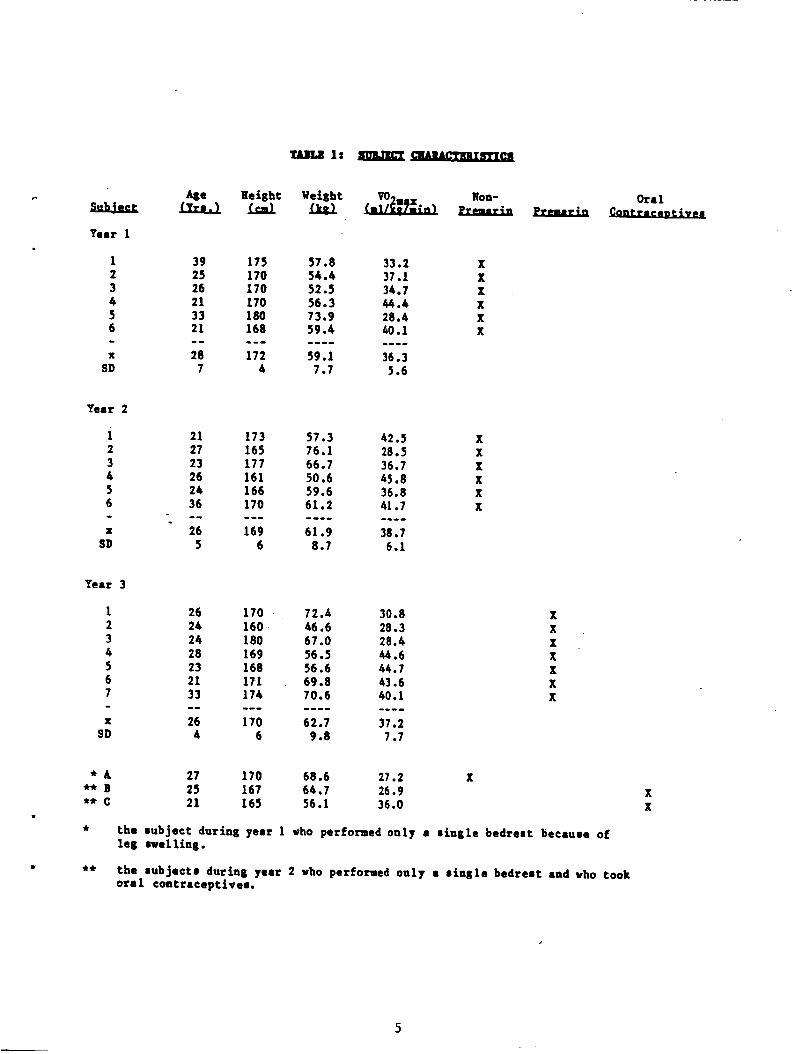
participated (see Table 1 for their physlcal characteristics).
i) Year I
During the first summer (1982), six "normally cycllng" women
completed the protocol shown in Figure 1. The overall protocol
consisted of a control month with testing at two week intervals (CI
and C2), two 12-day periods of bedrest, and a recovery month after
4
_USLS ls E _AJiAl_tlll_._
Year I
123436
X
$D
Age Heisht qeisht VOo_.. Non-
39 175 57.8 33.225 170 54.4 37.126 170 52.5 34.721 170 36.3 44.433 180 73.9 28.421 168 59.4 /001
28 172 59.1 36.37 4 7.7 3.6
Oral
Year
i234
56
X
SD
21 173 57.3 42.527 165 76.1 28.523 177 66.7 36.726 161 50.6 45.824 166 59.6 36.836 170 61.2 41.7
26 169 61.9 38.75 6 8.7 6.1
xxxXxx
Year
1234567
X
SD
26 170 72.4 30.8 X24 160 46.6 28.3 X24 180 67.0 28.4 X28 169 56.5 44.6 X
23 168 56.6 44.7 X21 171 69.8 43.6 X33 174 70.6 40.1 X
26 170 62.7 37.24 6 9.8 7.7
*A** B** C
27 170 68.6 27.225 167 64.7 26.921 165 56.1 36.0
X
the subject durins year 1 who performed only a 8inzle bedreot because ofle8 ewellina.
the subjects durin8 year 2 who perforued only a s£nsle bedreot and vho tookoral contraceptives.
XX
5
i--I
OUC]
O
(3_
tO
C_
O3
C.w-I
C_
E
C_EL
COZ
e_
%
X
(.urn
X
x
c_jQ20
X
X
oJ a21:3
x(0
t-_ eno
%
=l _>
_+'+o,.,+,mm.__m>c,,,j C_(::}
_+7_e_,' _
"vT_vL "o'X
c%1
each bedrest, with testing performed after 2 and 4 weeks of recovery.Before beginning the study, there were several training sessions inwhich each woman was taught the Farhi C02 rebreathing technique, howto position the esophageal thermocouple, and how to pedal a low-sitcycle ergometer while keeping the left arm steady enough for forearmblood flow measurements. Once these basic techniques were mastered,"practice" submaximal exercise tests were performed to furtherfamiliarize the subjects with the protocols, and to insure that theywere sufficiently trained to produce reproducible data.
Two 12-day horizontal bedrest periods were performed with anambulatory interval of from 3-4 weeks between bedrests. This timingwas chosen so that each woman began the second bedrest 5-6 weeksafter the start of the first bedrest. Since all of the women were
expected to have menstrual cycles of from 25-32 days, the bedrestprocedures were designed so that each woman would begin bedrest indifferent phases of the menstrual cycle, and thus should have a
differing hormonal millieu. No attempt was made to begin the bedrestat a particular day or phase of the menstrual cycle. Table 2 presentsthe menstrual cycle stage (see methods) for each of the women on the
first day of bedrest. During the recovery and control months,subjects resumed their normal lifestyle.
One additional subject began the bedrest protocol during the firstyear of this study. However, within 2-4 days after the end of bedrest
I, she developed severe edema in both lower legs. She was tested
with the venous impedance technique for venous obstruction (blood
clotting), and the results were negative. A report of this incident
was immediately sent to the Committee of Human Volunteers of the
School of Public Health, and all further testing was stopped until an
inquest could be held to determine whether this event was likely to
occur again or in other subjects. During this interval, 2 week
recovery data collection was not able to be obtained. The inquest
resolved that this was an unusual event, possibly provoked by the
inactivity of this particular subject after bedrest, which may have
delayed the recovery of venous tone in her lower legs, resulting in
blood pooling and edema. Although the committee thought it unlikely
that a similar incident would occur again to this subject, it wasrecommended that she should not undergo the second scheduled bedrest.
Permission to continue the study with the other subjects was obtained
in time for the 4 week recovery collections and the start of bedrest2. (see appendix I for copies of correspondence with the Committee of
Human Volunteers about this incident).
ii) Year 2
Six additional "normally cycling" women were recruited during the
summer of 1983. Each woman performed the protocol illustrated in
Figure I except that, I) in order to randomize the presentation of
the control tests and to evaluate possible seasonal fluctuations, the
control month was perfomed before the start of bedrest I, rather than
after the end of the second recovery month, and 2) since no
significant differences were seen between the duplicate maximum
oxygen consumptions (VO2max) during the control month, C1 and C2 V02max tests were omitted.

Non-Premarin Group
I I 3
2 I 2
3 3 3 (NO)
4 I 3
5 3 2
6 4 3
7 3 38 2 3 (LD)
9 I 3
10 1 311 4 112 2 (LD) 3 (I/))
+ where stage I = early follicular (cycle days 2 - 6, prior toestimated ovulation)
stage 2 = periovular (5 days prior to estimated ovulation to Iday after estimated ovulation)
stage 3 = early luteal (2-9 days post estimated ovulation)
stage 4 = late luteal (I0 days past estimated ovulation tocycle day 1 of next cycle).
LD = luteal phase deficiencysuspected from homonalresults.
NO = No ovulation suspected based on hormonal results.

Two women were recruited who were taking estrogen-contalnlng oralcontraceptives (Ovulen 21 or 0rthonovum). Both of these women
performed the protocols from the control month, a single bedrest, andthe recovery month.
lii) Year 3
During the last year of the study, 7 women performed the protocol
f_om the control month, a single bedrest, and the recovery month.Similar to year 2, the control month occurred before the bedrest
procedure rather than after the recovery month. Each woman ingested
1.25 mg of an estrogen supplement (premarin), starting on the thirdcycle day of the month in which she would undergo the bedrest
protocol (7-10 days before bedrest day 1). The premarin was
continued during the bedrest, and for five days after bedrest.
Progesterone supplements (10 mg /day Provera) were taken £or 5 days,starting on the second day after bedrest in order to allow normal
endometrlal sloughing. A single blood volume determination (with
technetium radioisotope labelling) was performed during the control
month, since no significant differences were seen in any of the
previous results of the duplicate measurements during the controlmonth. This was done to prevent unnecessary exposure of these
subjects to repeated radioisotope testing.
B) The Bedrest Protocol
i) The environment
Bedrests were performed at the Sheraton Johns Hopkins Inn locatedat 400 North Broadway. Each summer, 4 rooms were reserved for
subjects (2 subjects per room), and one room was reserved for the
"nurses" (persons trained and hired to look after the subjects duringthe bedrests), and one room was the impromptue blood chemistrylaboratory. During the second and third summers, a sixth room was
reserved and tranformed into a pulmonary function laboratory_ The
hotel provided maid service and most of the meals. Occasionally food
was obtained from local restaurants chosen by the subjects. No
restriction was placed on the amount or type of food eaten, althoughalcoholic beverages were restricted.
Each room consisted of 2 single beds and a bathroom. The beds were
normal hotel beds, except that a small pressure-sensitive platform
was placed under the castors at the foot of the bed. These pressure
sensors were connected to automatic timers to record the time spentout-of-bed by each subject. Each morning the accumulative time forthe previous 24 hours was recorded by the nurse. The installation of
the platforms resulted in a 4 degree head-down tilt of the beds.
However, since each bed contained one pillow, when the subject lay
with her head on the pillow, her head was positioned approximatelyhorizontal to her feet.
ii) Daily activities
On the first day of bedrest, a subject ate a light breakfast and
reported to the nurse at the hotel at 9 am. She unpacked hersuitcase, emptied her bladder and immediately began the bedrest. One
hour later, the first venous blood sample was obtained. Subjects
maintained the horizontal position for the remainder of the bedrest,
except for time allowed for bathroom activities. Subjects moved toand from the bathroom in wheelchairs, without supporting their weight
on their feet. Baths, not showers were taken, and each subject was
instructed to spend no more than 15-20 minutes in each 24-hour period
in the sitting position, and no time standing. Sitting in bed was
not permitted, although they were allowed to rest on one elbow to
eat. The amount of time spent out-of-bed during each bedrest is
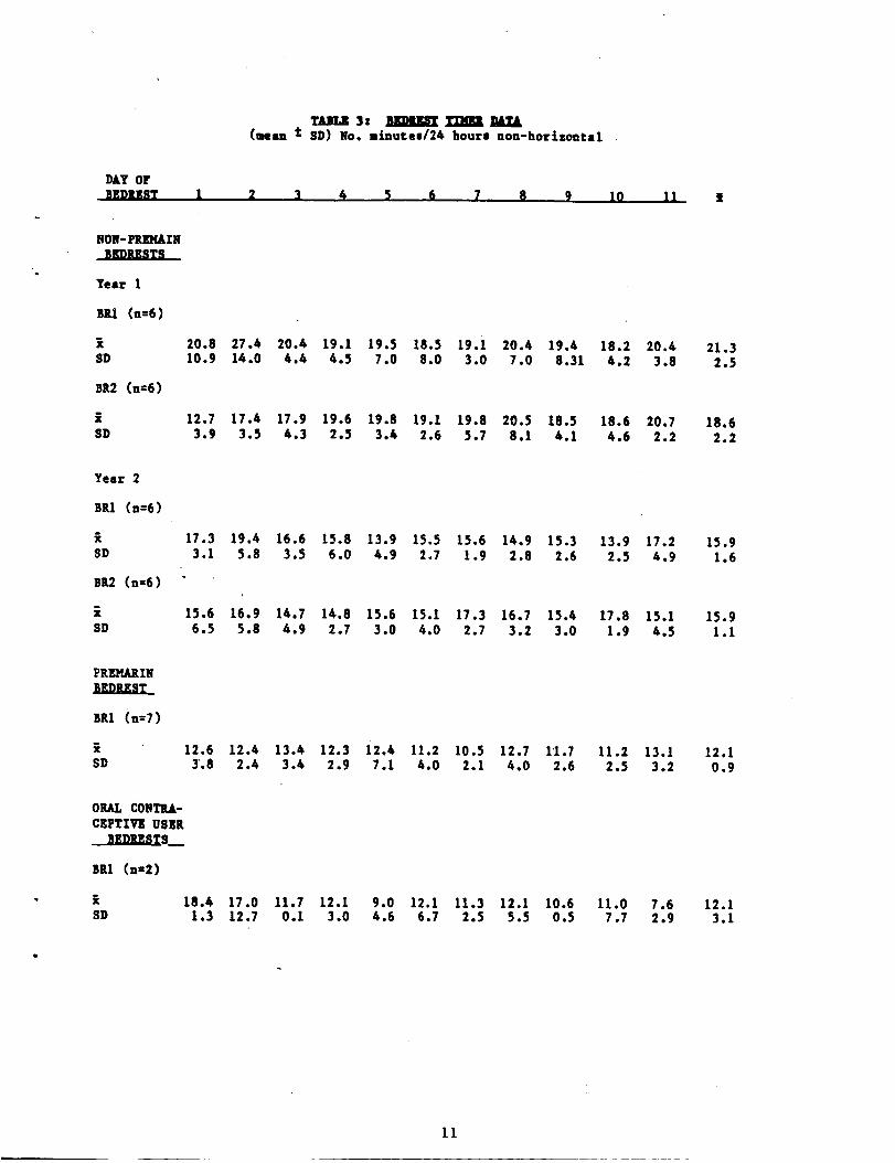
listed in Table 3. No significant differences occured in the time
out-of-bed between bedrest days (ANOVA, one-way, repeated measures
design), between duplicate bedrests during years I and 2, (ANOVA,
2-way, repeated measures design) or between the 3 years.
For each succeeding day of bedrest, the subject was awoken at 7 am.
She then took her oral temperature, emptied her bladder into a
24-hour urine container (second and third years), and ate a light
breakfast. Two hours later, the morning blood sample was obtained,
making sure that the subject had been horizontal during the previous
60 minutes. In this way, blood samples were controlled for posture,
time since last food intake, and circadian variation.
During the second and third years of the study, pulmonary function
tests were added to the bedrest protocol. Each subject performed
these tests on at least 3 days before bedrest, on each day of
bedrest, and on the day after bedrest. Subjects were transported to
the pulmonary function room on a gurney, in order to insur_ that the
subjects did not alter their posture from the horizontal position.
iii) Measurements obtained during bedrests
From each daily blood sample the following measurements were
obtained: hematocrit (microhematocrit technique), hemoglobin
concentration (cyanomethemoglobln), total solids (refractometry), and
plasma osmolality (freezing point depression). Plasma estrogen and
progesterone concentrations were determined from the blood samples on
each day of bedrest during year 2, and on every third day of bedrest
during year 3. Throughout the bedrests, urine osmolallty was
determined each day of bedrest. Twenty-four hour urine volumes were
recorded durln8 years 2 and 3.
Daily diaries were kept during the entire study. Each subject
recorded her fluld and food intake during bedrest, her morning oral
temperature, and the time at which she took estrogen medication (the
oral contraceptive users and premarln subjects). Other medication
and medical or menstrual symptoms also were noted.
Pulmonary function tests consisted of lung volume measurements,
total lung capacity (helium dilution), forced expiratory volumes,
lung diffusing capacity for carbon monoxide, the slope of phase 3
(Nitrogen washout), and maximal inspiratory and expiratory pressures.
An exploratory study was performed on one subject during the third
year to determine whether brain caudate D2 dopamine receptors would
be altered during the bedrest procedure (this was a special study to
10
DAY OFBEDREST
NON-PREMAINnD_STS
Year 1
Bll (n=6)
SD
BR2 (n=6)
iSD
Year 2
BRI (n=6)
$D
BR2 (n=6)
iSD
PREMARINaI&JUm_
Sal (u=7)
i$D
ORAL CONTRA-CEPTIVE USE!
SED_ST8__
BR1 (n=2)
SD
TUI.Z3: szmz_'rnaml_.l,(mean t SD) No. minutes/24 hours non-horizontal
1 2 3 !t _ 6 7 8 9 10 11 ]t
20.8 27.4 20.6 19.1 19.5 18.5 19.i 20.4 19.4 18.2 20.610.9 14.0 4.4 4.5 7.0 8.0 3.0 7.0 8.31 4.2 3.8
12.7 17.4 17.9 19.6 19.8 19.I 19.8 20.5 18.53.9 3.5 4.3 2.5 3.t 2.6 5.7 8.1 4.1
17.3 19.4 16.6 15.8 13.9 15.5 13.6 14.9 13.33.1 3.8 3.5 6.0 4.9 2.7 1.9 2.8 2.6
13.9 17.22.5 4.9
15.6 16.9 14.7 14.8 15.6 15.1 17.3 16.7 15.46.5 5.8 4.9 2.7 3.0 4.0 2.7 3.2 3.0
12.6 12.4 13.4 12.3 i2.4 11.2 10.5 12.7 11.73.8 2.4 3.4 2.9 7.1 4.0 2.1 4.0 2.6
I1.2 13.12.5 3.2
18.4 17.0 11.7 12.1 9.0 12.1 11.3 12.1 10.6 11.0 7.61.3 12.7 0.I 3.0 4.6 6.7 2.5 5.5 0.5 7.7 2.9
21.32.5
18.62.2
15.91.6
15.91.1
12.10.9
12.13.1
11

test whether acute changes in fitness alters the binding of caudate
dopamine receptors). Dopamine receptor binding was assessed by
positron emission tomography (Wong et al, 1984), vlth imaging
performed after intravenous injection of 11C-labelled3-N-methylsplperone• The prebedrest imaging was performed 2 days
prior to the start of bedrest in subject 19, and repeated on the last
day of bedrest. During imaging the subject did not change from thehorizontal position. She was transported by ambulance to the Nuclear
Medicine Department in the Johns Hopkins Hospital, and immediately
afterwards, performed the postbedrest submaximal exercise test.
C) Maximum Oxygen Consumption (V02 max) Protocol
Maxlmal Oxygen Consumption determination was performed
approximately every two weeks during the study (C2 tests were not
performed during years 2 and 3).Prebedrest tests (PREBR) were performed within 48 hours of the start
of bedrest, and postbedrest tests (POSTBR) were performed within 48
hours, but usually within 24 hours, after bedrest.
Oxygen consumption was determined at progressively increasingexercise intensities for each woman pedalling an upright cycleergometer. Ventilation was measured in either a tissot tank (year 1)or with a dry gas meter (years 2 and 3). The oxygen concentration of
the expired air was measured with a Beckman OM 11 gas analyzer, andC02 concentration with either a Beckman LB2 (year 1) or an Applied
Electrochemistry C02 Analyzer (years 2 and 3). Each subject pedalledat a rate of 50 revolutions per minute while the exercise intensitywas increased (in 25 Watt steps) every 2 minutes. For the first V02
max determination, tests were performed at least in duplicate (onseparate days) in an effort to obtain a plateau of the oxygenconsumption curve during the final two exercise intensities. Control
tests were repeated until this criteria was met in order toaccurately establish the fitness level of each subject beforebedrest. Following bedrest however, the plateau criteria couldseldom be met, as most women seemed limited by leg fatigue before
attaining the plateau. Non-plateau values are therefore referred toas "peak VO2" values. Heart rates were also measured during thesetests with either a 12-lead E[G system (for the first test on each
subject), or with a respironics heart rate monitor (for all furthertests).
D Submaximal Exercise Protocol
Submaxlmal exercise tests were performed in approximately 2 week
intervals during this study (except when the 2 week recovery data was
missed during year I). The postbedrest tests were performed
immediately following the bedrest procedure. Subjects were
transported in wheelchairs and by car (less than 5 minute ride) from
their bed in the hotel to the Stress Physiology Laboratory in the
School of Public Health. The ambient temperature for these tests wasmaintained at 30 °C + 1°C and 50-60 Z rh Be£ore the submaximal
exercise test, subjects supported their weight only briefly, during
the pre-exercise body weighing procedure. Next, they sat in the seat
of the low-sit cycle ergometer for at least 40 minutes before the
start of the exercise test to control posture before blood sampling,
12

and to allow time for attachment of measurement devices.
Each subject pedalled for 30 minutes at an exercise intensity that
was 70Z of her V02 max determined just prior to the bedrest.
However, exercise was stopped earlier if a subject's core temperatureexceeded 39@C, or if her heart rate approached 95X of her maximal
heart rate value, which was determined during the prebedrest VO2 max
test. For any given subject, all exercise tests were conducted at the
same time of day (_ 2 hours), not more often than once a week (to
avoid training or accllmation effects), at least 2 hours after a
light meal, and about I hour after drinking 200 ml of water to assure
adequate hydration.
During the first two years of the study, subjects were instructed
to stop pedalllng immediately at the end of the submaxlmal exercisetest in order to obtain resting recovery data. Following the
postbedrest tests (but none of the other tests), 7 out of 15 of thewomen felt faint and had to be moved to a horizontal position (two
actually fainted). Thereafter, to avoid this traumatic experience for
the subjects and investigators, subjects were instructed to
free-pedal during recovery in order to assist cardiac return. During
the third year of the study, all subjects free-pedalled afterexercise and none experienced fainting episodes.
During each submaximal exercise test the following measurements
were obtained:
- exercise time, in minutes, was recorded for each test.
- body weight, before and immediately after exercise, was measuredwith the subject wearing a dry scrub suit. After exercise, the
subject dried herself and removed all wet clothing. Weights wereobtained on a Homs scale (accuracy _ 50 gms) and were adjusted for
any water drunk while placing the esophageal thermocouple.
- heart rate (HR) was measured each minute with a respironics heart
rate monitor
- body core temperature (Tes) was monitored continuously with a
thermocouple positioned in the esophagus at the level of the right
atrium (Nenger, 1975).
- skin temperatures (Tsk) were recorded every 5 minutes withuncovered thermistors (Yellow Springs) postioned over the chest (TI),
lateral upper arm (T2), lateral thigh (T3), and lateral calf (T4).
Mean skin temperature (Tsk) was calculated by the formula:
Tsk _ 0.3 TI + 0.3 T2 + 0.2 T3 + 0.2 T4.
- forearm venous compliance (FVC) was measured every 2-3 minutes
using the technique of venous occlusion plethysmography (Wenger,
1980). Changes in forearm circumference were measured using a Whitney
mercury-in-silastic strain gauge. The forearm was suspended from thewrist with a cloth sling so that the elbow was level with the
shoulder.
13

- cardiac output was determined after 10, 15, 20,and 25 minutes of
exercise, using the Fahri C02 rebreathing technique (Farhi, 1976).The rebreathing bag was filled with 100% 02, and the breathing ratewas maintained at 60 breaths per minute during the 15-17 seconds of
the rebreathing maneuver. Calculations of cardiac output wereperformed using the CO2 dissociation curve adjusted for the
pre-rebreathing hemoglobin concentration. Heart rates weredetermined during the rebreathing maneuver and used to calculatestroke volume.
-local sweating responses (SR) were measured continuously before and
during exercise, using a resistance hygrometry sweating system(Bullard, 1962). For determination of sweating sensitivity (slope ofthe SR/Tes relationship) and the Tes threshold for the onset of
sweating, the local sweating data (mg/cm2/min) was paired with
corresponding Tes values. A linear regression equation was" used todetermine the slope of the relationship, and the Tes valu_ at whichthe line intersected the x axis (Tea sweating threshold).
- blood samples were obtained immediately before the start ofexercise, after 2, 6, 15, 20, and 30 minutes of exercise, and 5minutes after the end of exercise. Blood was drawn after at least 40
minutes of controlled posture, with the arm in the same position for
all samples, and in a free-flowing manner (without tourniquet or
vacutainer). Between sampling, the butterfly needle was kept patent
with a heparin/saline lock.
From all blood samples, determinations of hematocrit
(microhematocrit technique), hemoglobin concentration
(cyanomethemoglobin), total solids (refractometry), and osmolality
(freezing point depression) were obtained. From the resting and 30
minute exercise samples, blood lactate (Sigma enzymatic reaction),
and arginine vasopressin concentration (third year only) weredetermined. The arginine vasopressin assay was used as an index of
antidiuretic activity, and was performed using an immunonuclear
radioimmunoassay kit. This is a delayed tracer technique with the
plasma extraction done on octadecasilyl-silica columns. The assay is
sensitive from 3-80 pg/ml. From the resting samples, plasma estrogen
and progesterone concentrations were also determined withradioimmunoassay techniques (Diagnostic Products).
E) Red Cell and Plasma Volume Determinations
I) Radioisotope dilution methods
To determine Red Cell Volume (RCV), 40 microcurries of 99M
technetium (Tc) pertechnitate (Brookhaven National Laboratory) was
bound to a sample of red blood cells after first determining the
background radiation of the sample. Exactly 5 ml of the labelledblood was then reinjected intravenously and allowed to mix in the
vascular compartment for 20 and 40 minutes. Then 10 ml samples of
heparinized blood were drawn to measure the 99MTc activity of themixed blood.
Plasma volume was determined in a similar manner. Twenty
microcurries of 99M Technetium pertechnitate was used to label a
14

sample of human serum albumin and saline. This protein solution was
then injected through an arm vein and 20 and 40 minutes later, blood
samples were drawn for counting. Technetium was chosen as the
radioisotope in both of the above tests because of its short
half-life (6 hours), and the need for multiple blood volume
determinations in this study. During each RCV determination,
hematocrits were measured from one of the blood samples in the exact
manner that hematocrits were determined from bedrest and exercise
blood samples.
2) Plasma Volume (PV) calculations during bedrest
The Absolute Plasma Volume on the first day of bedrest was
calculated by multiplying the ratio of the hematocrits from the blood
sample drawn during the plasma volume determination (performed on the
day prior to bedrest) and the blood sample drawn during day I of the
bedrest, by the PV determined on the day prior to bedrest (Van
Beaumont, 1972). The assumptions made using this formula are that,
within the 24-hour interval between the two blood samples, there was
no significant change in red cell volume or red cell size.
Relative changes in plasma volume during bedrest (Z change from
day i) were calculated from the changes in hematocrit and hemoglobin
(Dill and Costi11, 1974). The assumption with this calculation is
that in the interval between blood sampling, there was no significant
change _n red cell volume (see later in results section). PV on each
day of bedrest was calculated by multiplying the relative changes in
plasma volume by the PV on day I.
3) Relative Changes in Plasma Volume During the SubmaximalExercise Tests
Relative changes in plasma volume during exercise tests (% change
between the resting and each succeeding exercise sample), were
calculated (Dill and Costill, 1974) from the changes in hematocrit
and hemoglobin concentration during the exercise.
F) Menstrual Cycle Determinations
Menstrual cycle data consisted of daily records of morning
temperature and comments written in diaries kept by each subject. In
addition, blood samples were drawn at various intervals during the
study to assess hormonal function (estrogen, progesterone, LH, and
FSH). Each year of the study, samples were drawn before each
submaximal exercise test. Hormonal determinations were also performed
during either each day of bedrest (year 2) or every third day of
bedrest (year 3). From the frequency of the blood sampling (and thus
hormonal determinations), it was not always possible to define the
exact day of ovulation. Therefore menstrual cycle comparisons were
made by comparing different stages of the cycle rather than on
specific cycle days. Each woman's menstrual cycle was divided into 4
stages, in which a differing hormonal milieu would be expected (and
in each case verified by the results from at least one blood sample).
Stage 1 : Early follicular stage - from cycle day 2 (day 1 t the
15

first day of bleeding) until 6 days prior to the day estimated as
ovulatlon. (Only in a few subjects could the exact day of ovulation
be identified by an LH peak). In subjects in whom an LH peak was not
obtained, the day of ovulation was estimated by counting back 14 days
from the start of the next cycle, from the morning temperature
records, and from the hormonal data available. During this cycle
stage low estrogen and progesterone concentrations were seen.
Stage 2: Periovular stage - from 5 days prior to the estimated day of
ovulation until one day after the ovulatory day. During this stage,
elevated estrogens and low progesterone concentrations occurred.
Stage 3: Early luteal phase - from 2-9 days after the estimated day
of ovulation. During this stage, estrogen and progesterone
concentrations were increasing.
Stage 4 : Late luteal phase - from 10 days after the estimated day of
ovulation until the first day of the next cycle. During this stage
estrogen and progesterone concentrations were decreasing.
16
IV) RESULTS AND COMMENTS
A) Body Fluid Responses Durin R Bedrest
I) Changes in Red Cell Volume
The red cell volume data are presented in Table 4. Since there.
were no significant changes in body weights during the bedrest, thesame patterns of change in red cell responses were found whether thedata was represented as in Table 4 (absolute volumes in ml), or as
the RCV corrected for body weight (ml/kg). The F value from a one-wayanalysis of variance test for repeated measures (ANOVA 1-W-RM) wassignificant (P < 0.01) when comparing the RCV values from thenon-premarin group, but non-significant for the premarin group (P <0.20).
i) Non-premarin group, Control I vs Control 2
There was no significant difference in RCV between the 2 Control
tests. (The post hoc comparisions were performed using the Duncan
Multiple Range Test, with the level of significance accepted at P <o.o5).
ii) Non-premarin group, Bedrest I
Red cell volume (RCV) was measured with the technetium labellingtechnique (Methods) within 48 hours before bedrest i and within 48
hours after the end of bedrest I. The decrease in RCV averaged only85 ml (5.8Z) after the 12 days of bedrest. This difference was notsignificant (P < 0.05).
iii) Non-premarin group, Bedrest 2
The decrease in RCV during the second bedrest averaged 63 ml, oronly 4.4Z. Again this change in RCV during the 12-day bedrest was not
significant. However the decrease in RCV since the beginning of thestudy appeared to be accumulative, so that the decrease in RCV
between the beginning of the first bedrest and the end of the second
bedrest was significant (p < 0.05) and averaged 6.2Z. The differencein RCV between the start of the first bedrest and the start of the
second bedrest was not significant.
iv) Premarin Group
Although there was a tendency for the RCV to decrease during the
bedrest (6.5Z decrease), the difference between the beginning and end
of bedrest was not significant. A comparison of the changes in RCV
between the non-premarin and the premarin groups in response to
bedrest and during recovery can be seen in Figure 2. Estrogen
supplementation did not appear to alter this overall pattern ofchanges in RCV.
v) Changes in RCV during recovery, non-premarin and premarin
groups
The significant decrease in RCV seen in the non-premarin group
17
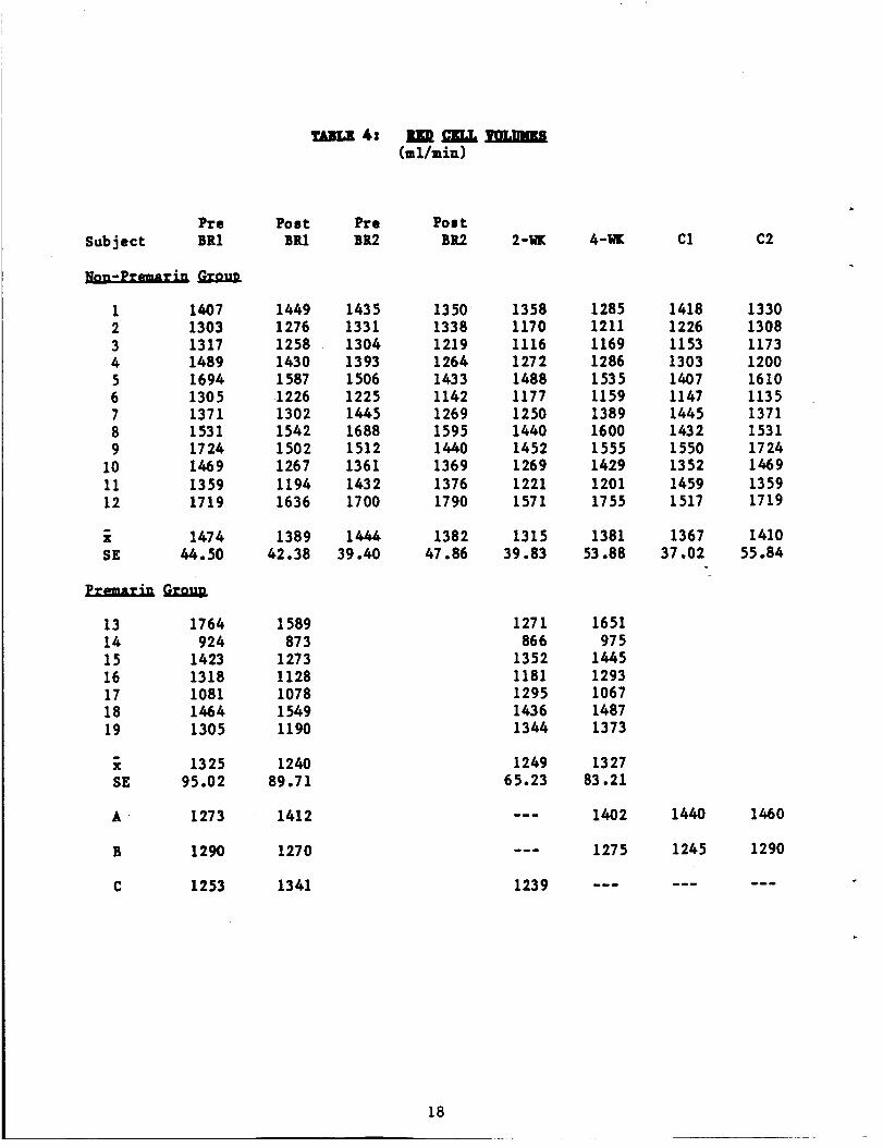
4: nugDc'Jgu, _mammCml/min)
Pre Poet Pre Poet
Subject BR1 BR1 BR2 BR2 2-I_ 4-WK Cl C2
1 1407 1449 1435 1350 1358 1285 1418 13302 1303 1276 1331 1338 1170 1211 1226 13083 1317 1258 1304 1219 1116 1169 1153 11734 1489 1430 1393 1264 1272 1286 1303 12005 1694 1587 1506 1433 1488 1535 1407 16106 1305 .1226 1225 1142 1177 1159 1147 11357 1371 1302 1445 1269 1250 1389 1445 13718 1531 1542 1688 1595 1440 1600 1432 15319 1724 1502 1512 1440 1452 1555 1550 1724
10 1469 1267 1361 1369 1269 1429 1352 146911 1359 1194 1432 1376 1221 1201 1459 135912 1719 1636 1700 1790 1571 1755 1517 1719
i 1474 1389
SE 44.50 42.38
13 1764 158914 924 87315 1423 127316 1318 112817 1081 107818 1464 1549
19 1305 1190
i 1325 1240SE 95.02 89.71
1444 1382 1315 1381 1367 141039.40 47.86 39.83 53.88 37.02 55.84
1271866
13521181129514361344
1249
65.23
1239
A 1273 1412
1651975
14451293106714871373
132783.21
1402
1275B 1290 1270
1440
1245
C 1253 1341
1460
1290
18

Red Cell Volumes (Tc-RBC)
t600 -
a No Premarln
E 1500
2 14oo
1300
2oo
II00
Pre Post 2 wk 4 wk
BR BR RC RC
FIGURE 2: The absolute red cell volumes (mean + SE) determined24-48 hours before bedrest 1 (PRE BR)| 24-48 ho'_rs after bedrest
1 (POST BR); and after 2 and 4 weeks of recovery from bedrest (2wk RC and 4 wk RC) for non-premarin (n = 12) and premarin ( n =7) subjects.
19
over the course of the duplicate bedrests, was still continuing after2 weeks of recovery (RCV was now IO.8Z lower than at the start ofbedrest 1). After 4 weeks of recovery however, the mean RCV wasbeginning to return toward the prebedrest value. These resultssuggest that there is about a 2-week delay in the effect of bedresttO alter RCV, and that only after at least 4 weeks of recovery was,
the RCV no longer significantly different from the prebedrest value.
Although there were were no significant changes in RCV in the
premarin group between any of the measurement conditions, the pattern
of response was similar to the changes seen in the non-premarin group(see Figure 2).
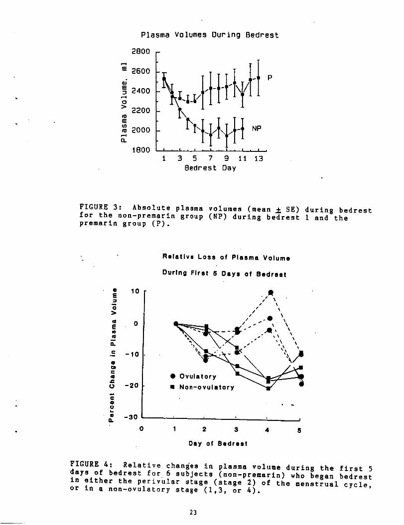
2) CHANGES IN PLASMA VOLUME DURING BEDREST
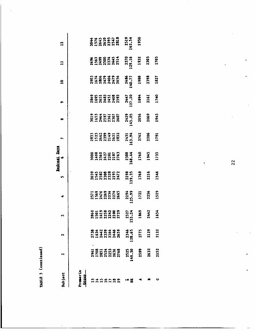
The absolute plasma volume values (PV) for all subjects duringbedrest are presented in Table 5. These absolute values were
calculated from the prebedrest technetium results and the ratio of
changes in hematocrlt and hemoglobin concentration (see Methods).
i) Non-premarin group, Bedrest 1.
Plasma volume decreased significantly (P< 0.01) during the first5-7 days of bedrest and then became relatively stable for the
remainder of the bedrest (see Figure 3). The average decrease in PV
was 505 ml (19.9X) between the first and the last day of bedrest.
li) Non-premarln group, Bedrest 2
The PV at the start of the second bedrest did not differ
significantly from the PV at the start of the first bedrest (Table
5). There was a tendency towards a smaller decrease in PV during the
second bedrest (averaged 12.3Z between day 1 to the last day of
bedrest 2) than during the first bedrest (19.9Z). However, the
difference in plasma volumes between the duplicate bedrests was not
significant (P < 0.05, determined using a 2-way ANOVA, repeatedmeasures, split plot design, ANOVA-2W-RM-SP).
iii) Effect of menstrual cycle stage on the loss of PV duringbedrest
For the non-premarln group, the change in PV was examined in each
woman as a function of the stage of the menstrual cycle (see Methods)
during the first 5 days of bedrest. There were no significant
differences between the decreases in PV during cycle stages I, 3, and
4. However, if a woman began bedrest in stage 2 of her menstrual
cycle, the periovular stage, the decrease in PV was postponed. There
was also a tendency for women in the late luteal phase of the cycle
(stage 4) to maintain or expand PV during the first 5 days of
bedrest, however this was a less consistent finding and was not
present in every subject. Shown in Figure 4 are the changes in PV for
the six women who began bedrest during year I. The results shownillustrate PV changes in 3 women who began bedrest in the second
menstrual stage (dotted lines), and 3 women who began bedrest in one
of the other stages (1,3, or 4). The women who began bedrest in
stage 2 showed no significant drop in PV during the first 4 days of
2O

0
,,-q ,.-* ¢1_ _ *',.. <r _r_ ('q r.,. c._ o e,_ cO .,qp
,,,_ ,,-o _,-o N ¢',1 _"4 ,,,,,q _"ql L"_I _,,,4 ,,-_ N 4"_1 0
g-I
0
0
m_
O'i
p_
I
o_o_o_ _ _ _
__o_ _ _ _
Plasma Volumes Ouring Bedrest
2800 -
e 2600
G• 2400
o
> 2200
E
m 2000
i800
NpI • I . ! I • I A P I •
I 3 5 7 9 II 13
8edrest Day
FIGURE 3: Absolute plasma volumes (mean _ SE) during bedrestfor the non-premarin group (NP) during bedrest I and the
premarin group (P).
• 10E-I
o>
0E
a-
_= -lo
e
cq
o -2o
I=,
• -30Q.
Relative Loss of Plasma Volume
During First 5 Days of Bedreat
1 _ // ,.'"" _ " _'_
__._IC -" .,e, _ ..
.....-,,,,•.,\ %" ",I , _
@ Ovula_--
• Non-ovulatory "11"
t J |
0 1 2 3 4 5
Day of Bedrest
FIGURE 4: Relative changes in plasma volume during the first 5
days of bedrest for 6 subjects (non-premarin) who began bedrest
in either the perivular stage (stage 2) of the menstrual cycle,or in a non-ovulatory staze (I,3, or 4).
23
the bedrest, and in 2 cases, PV even increased above the day 1PVlevel. These results led us to revise our initial hypothesis thatelevated estrogen levels would be associated with a retention of PV
during bedrest. (Blood estrogens would be elevated during stages 3and 4 as well as during stage 2). Our new hypothesis now statedthat, when blood estrogens are elevated in the presence of lowprogesterone concentrations, the water-retaining effects of theestrogens may, at least temporarily, prevent the hypovolemia whichoccurs at the beginning of bedrest.
iv) Effect of estrogen-containing oral contraceptives on the PV
changes during bedrest.
During the second year of the study, 2 women who were taking oral
contraceptives containing synthetic estrogen (mestranol, 80 and i00
mcg/tab) and a progestin (norethindrone or ethynodiol diacetate),performed a slngle bedrest procedure. The decreases in P_ seen in
these 2 women (Table 5, subjects B and C), did not differ
significantly from the responses seen in the non-premarin group(years 1 and 2). The decrease in PV between the first and last day ofbedrest was 20.8Z and 12.5% for these two women respectively, with a
significant decrease in ?V during the first 4-5 days of bedrest.These data suggest that elevations of blood estrogens in the presence
of elevated progesterone, may not be associated with a maintenance of
PV during bedrest.
v) Effect of Premarin on the change in PV during bedrest
Figure 3 illustrates the mean change in PV from the 12
non-premarin subjects (bedrest I data) and the mean change in PV from
7 women who performed an identical bedrest protocol, except that they
ingested 1.25 mg/per day of premarin for 7-10 days before bedrest and
throughout the bedrest protocol. During the first 2 days of bedrest,
the PV decreased in the premarin group in a pattern similar to that
seen in the non-premarin group. During the 3rd to 5th day of
bedrest, PV remained relatively stable. Then between bedrest days
7-13 (except for day 11), the PV recovered and was not significantlydifferent from the PV of each woman on the first day of bedrest (P <
0.05, ANOVA-IW-RM, with the Duncan Multiple Range test to compare for
differences between bedrest days).
vi) Changes in PV during recovery from bedrest
Plasma volume recovered to prebedrest levels within 24-48 hours in
most subjects. Figure 5 illustrates the mean PV _ SE, measured with
the technetium-labelling technique. Plasma volumes were measuredwithin 48 hours before and 48 hours after bedrest (PREBR and POSTBR)
and after 2 and 4 weeks recovery from bedrest. Results are shown for
the 12 non-premarin subjects and the 7 premarin subjects.
For the non-premarin group, the F ratio determined from anANOVA-1W-RM was non-significant (P< 0.20), suggesting that thedecrease in PV which occurred during bedrest was completely restoredwithin 24-48 hours.
In the premarin group, the F ratio was significant (P <0.02).
24

Plasma Volumes (Tc-HSA).
2700 r I_ No Premartn
2500 L [] Premsrin '
3 2500
240o
2300
o.
2200
Pre Post 2 wk 4 wk8A 8R "" --m_ HI.;
FIGURE 5: Absolute plasma volumes (mean _ SE) determined with
the technetium labelling technique 24-48 hours before bedrest I
(PRE BR); 24-48 hours after bedrest I (POST BR); and two andfour weeks after the end of bedrest (2 wk RC and 4 wk RC,
respectively) for subjects in the non-premarin' group (n ffi12)
and in the premarin group (n _ 7).
25
Further tests (Duncan Multiple Range), concluded that the PV 24-48hours after bedrest and 2 weeks after bedrest, were slgnificantlygreater than the PV before bedrest (P< 0.05).
3) CHANGES IN BLOOD PROTEINS AND ELECTROLYTES DURING BEDREST
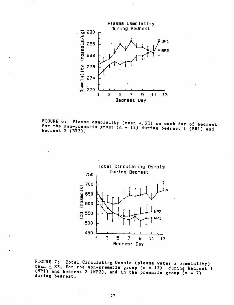
i) Non-premarin group, bedrest 1
Plasma osmolality increased slgniflcantly (P< 0.01) during bedrest
(see Figure 6) from an average bedrest day 1 value of 279 mosmol/kg,
to a highest average value of 287 mosmol/kg, which was measured on
the 7th day of bedrest. This increase in plasma osmolality occurred
in the presence of an average 18_ net loss of total circulating
osmotically active particles (TCO) from the plasma in this same
interval (see Figure 7). TCO values were significantly lower (P <
0.05) than the first day of bedrest from the 3rd to the last day ofbedrest.
The plasma total protein concentration increased significantly (P <
0.01) during bedrest (Figure 8). At the same time, there was an
average 13Z net loss of total circulating protein content (TCP), as
seen in Figure 9. The above results suggest that during bedrest there
was a net loss of hypotonic, protein-poor fluid from the vascular
compartment.
ii) Non-premarin group, bedrest 2
A similar increase in plasma osmolallty (P < 0.01) occurred
during the second bedrest (Figure 6) and this increase was
accompanied by a significant (P <0.01) decrease in TCO, as seen in
Figure 7. The TCO were signiflcantly reduced (Duncan Multlple Range
Test) below the values obtained on the first day of bedrest from the
second to the last bedrest day.
The plasma total protein concentration did not increase
significantly (P < 0.50) during the second bedrest (Figure 8), and
TCP decreased (average 11.1Z decrease between day i and the last day
of bedrest) in a manner similar to that seen during the first bedrest
(Figure 9).
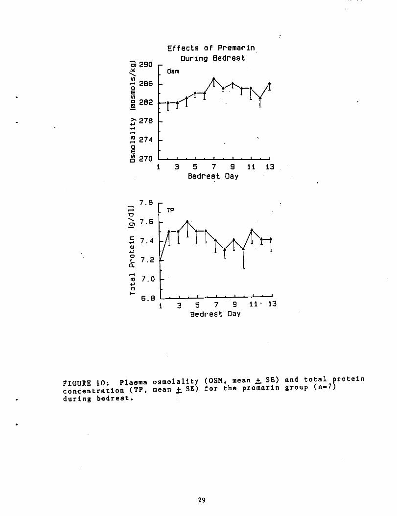
ill) Premarin Group
The mean plasma osmolality increased during bedrest in the
premarin group (Figure 10), but the difference was not significant (P< 0.20). During bedrest days 2-5, there was a significant (P <0.01)decrease in TC0, but by the end of the bedrest, the TCO value did not
differ significantly from the day 1 value (Figure 7).
The increase in plasma protein concentration during bedrest was not
significant (P < 0.50) in the premarin group (Figure I0), and a
transient decrease in the TCP occurred, with average values
significantly lower than the first day of bedrest on only the 4th and
5th days of bedrest (Figure 9). Thereafter, the TCP values recovered
to eventually exceed the day I mean value on the last bedrest day.
4) FLUID INTAKE AND OUTPUT DURING BEDREST
26
290
o
o 282G
w
278.4J
,"4
m 274oE
2700
t
Plasma Osm01alityDuring 8edrest
BR1
BR2
/
! , ! , I I , I " J
3 5 7 g 1t 13
8edrest Day
FIGURE 6: Plasma osmolality (mean f_SE) on each day of bedrest
for the non-premarin group (n - 12) during bedrest 1 (BRI) and
bedrest 2 (BR2).
o 650
o 600
o 550
500
Total Circulating Osmols
During Bedrest
_ "i\__-!LI//I/I-I-I_" NpIXk'[)i I-II_I_.LLI.
45O
P
, I , I , I , I , I , I
3 5 7 g ii 13
Bedrest Day
FIGURE 7: Total Circulating Osmols (plasma water x osmolality)
mean + SE, for the non-premarin group (n = 12) during bedrest 1
(NP1)--snd bedrest 2 (NP2), and in the premarin group (n - 7)
during bedrest.
27

Total Protein
7 6 - During Bedrest
x7d
¢-•_ 72
oL 7 0
n
r--i
m68.iJo
I..-6 6 • I m I , I I • I m I
3 5 7 g t1 t3
Bedrest Day
FIGURE 8: Total protein concentration (mean + SE) during
bedrest for the non-premarin group (n = 12) du"ring bedrest 1(BRI) and bedrest 2 (BR2).
Total Circulating Proteins
2OO
190 r
t80
t70 _\nu 160
I50
I40
130
During Bedrest
• I • I • I • I • I I I
i 3 5 7 g 1t t3
P
Bedrest Day
FIGURE 9: Total circulating proteins (plasma volume x total
protein concentration), mean _ SE, for the non-premarin group (n= 12) during bedrest 1 (BRI) and bedrest 2 (BR2), and for the
premarln group (n = 7) during bedrest.
28
o_ 290v
t/]"_ 286C)
o 282
2784J-PI
m 2740
m 2700
Effects of Premarin
During Bedrest
Osm
l-t [ " [
• " I • I ° I • I * I • I
3 5 7 g tt t3
Bedrest Day
78 -r-4
_76
r-._ 74
4J0L 72O.
m 70
oI.,-
6 8
TP
/k
I • I0 . I , I I I I
3 5 7 g II" 13
Bedrest Day
FIGURE I0: Plasma osmolality (OSM, mean _ SE) and total proteinconcentration (TP, mean _ SE) for the premarin group (n=7)
during bedrest.
29

i) intake
Fluid intake during bedrest was crudely calculated based on theinformation recorded in each subject's diary. The amount of fluiddrunk each day was measured and the water content in the food wasestimated. When an ANOVA-1W-RM was performed on the data from each of
the bedrests, the differences across bedrest days were notsignificant during either bedrest 1 of the non-premarin group (P<0.10) or during bedrest in the premarin group (P < 0.20). There was
a significant difference between fluid intake on various days ofbedrest for the non-premarin group during the second bedrest (P <0.05). There was no consistent trend however (Figure 11), with thelargest difference seen between bedrest days 6 and 9.
Although there may appear to be a greater fluid intake in thenon-premarin group during the second bedrest than during the firstbedrest, when an ANOVA-2W-RM-SP was performed to test whether thesedifferences were significant, the differences between bedrest 1 and
bedrest 2 were not significant (P< 0.50).
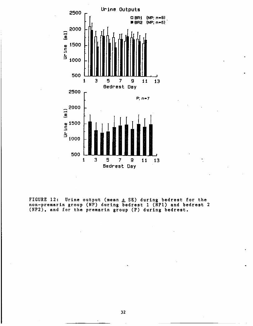
ii) urine output
Figure 12 illustrates the changes in 24 hour urine volume during
bedrests (the first value does not represent a full 24-hour sample
since the early morning urine collection was not included)_ None of
the differences in urine output between bedrest days were
significant; for the non-premarin group, bedrest I (P < 0.30) orbedrest 2 ( P< 0.20), or for the premarin group (P < 0.70). These
results represent the data only from years 2 and 3 of this study,since 24-hour urine volumes were not recorded during year i.
iii) fluid intake - fluid output
The difference between the fluid intake and fluid output was
calculated for each woman during bedrest (only data from the second
year were used in the non-premarin calculations). This is not a truewater-balance calculation, since the output is not corrected for
insenslble water losses or fluid loss in the stool (both would have
to be estimated, and large errors would occur). As seen in Figure
13, during the first 4-5 days of a bedrest, a negative fluid balancewas seen in each bedrest condition, with the difference between fluid
in and urine out closer to zero during the later days of thebedrests. Differences between bedrest conditions were not
significant.
4) URINE OSMOLALITY DURING BEDREST
The osmolallties (mean value _ SE) determined from the 24-hoururine collections are shown in Table 6. The urine osmolality
decreased, especially during the first few days of the bedrests.
This difference was significant during bedrest 1 in the non-premarin
group (P < 0.05), but not significant during the second bedrest (P <
0.20). The changes in urine osmolallty during bedrest in the
premarin group were not significant (P < 0.70).
30

Fluld Intakeo eml
2000 (N
t500
2 iooo.=" 500
0
2000
-- i500
.E
z_ iO00-#-4
h
500
0
l 3 5 7 9 1t t3
Bedrest Day
P: n-7
i 3 5 7 9 II 13
Bedrest Day
(NP: n=6)
(NP: n=B)
_,_iGi:,_ALPAGE F5
OF POOR QUALITY
FIGURE II: Fluid intake (mean ± SE) during bedrest for the
non-premarin group (NP) during bedrest I (BRI) and bedrest 2(BR2), and for the premarin group (P), Fluid inteke was
estimated from the fluid and food entries in each subject'sdiary.
31
Urine Outputs
2500 - [ o eRt
2000
15oo.r4
1ooo
5OO
i 3 5 7 g li
BeOrest Day2500 -
2000
E
® 1500C
L
iO00
P: n-7
(NP: n-6)o
(NP: n=6)
i3
500 m
i 3 5 7 g ii i3
Bedrest Day
FIGURE 12: Urine output (mean _ SE) during bedrest for the
non-premarin group (NP) during bedrest 1 (NPI) and bedrest 2
(NP2), and for the premarin group (P) during bedrest.
32

A
$==4
Ew
400
20O
3o 0
c -200f_:D
-400.I
-600
o -800
b_
-iO00
Fluid In - Urine Out
During Bedrest
F '_?,,I_L_I _.,'_
'_/" V-
: tt/o
\f • BR2: NP• BR_ P
• I . I • | • ! I .
1 3 5 7 g 11
Bedrest Day
FIGURE 13: Fluid in - fluid out (mean _ SE) for the
non-premarin group (NP, n = 6) and the premarln group (P, n = 7)during bedrest.
33

iI
I
*dD
'4
J
%
1'4 _d) r'_ ¢_i 0 0
'4' U_ U'_
O _ ,IP _-o _ ff_
,4P o,,,,, m ,,,,o 0,4' _
,41'
I',,,, 4P_ 0 N ,8,
O qd) w.q I_ _) *4)Cl_ '41' irl ir_ q_ Io
'41' .41'
.4" oo_ u'_
,4r ,41' o_ _o
_o _ N i_ o oD,.o f,,,, irl Id) r_ tl_'41' '4' '4'
gO V'_ m0 I",. C_, "4'q_ ,4r 41.
'41' _ _r
or, ,4' ,41'
!. 1o
B) MAXIMAL EXERCISE RESPONSES
i) Effect of menstrual cycle
For the non-premarin subjects, there were at least 5 determinations
of maximal oxygen consumption (V02 max) which were performed during
varying stages of each woman's menstrual cycle, and which would nothave been affected by bedrest. These tests included control months
(CI and C2), prebedrest tests (PREBR1 and PREBR2), and the 4 wkrecovery from bedrest. The data from these tests were compared for
each woman, averaging the V02 max data from tests done in the luteal
and follicular phases. The results are shown in Table 7. There wasno significant difference in V02 max in these women between these 2
stages of the menstrual cycle (P <0.70).
ii) Non-premarin group, Bedrest I
The V02 peak value obtained following the first bedrest was
significantly reduced from the prebedrest value, whether the resultswere expressed as llters of 02 consumed per minute (average decrease
of 11.2Z), or corrected for body mass by dividing the peak 02
consumption by body weight (IO.4Z), as shown in Table 8.
iii) Non-premarin group, Bedrest 2
Following the second bedrest however, the reduction in V02 peak was
not significant either when expressed as 1/min (average decrease of
4.1Z) or as ml/min/kg body weight (average decrease of 4.3_).
iv) Premarin group
The decrease in V02 peak following bedrest in the premarin group
was significant (P< 0.01) and averaged 21.6Z (I/min) and 24.5Z
(ml/min/kg body weight). There was considerable variability in this
decrease in V02 peak however between subjects, and thus the decrease
in V02 peak between the premarin group and the decrease seen in the
non-premarin group after the first bedrest was not significant
(t-test comparison).
v) Recovery Data
V02 max was almost completely recovered to the prebedrest level by
2 weeks of recovery from bedrest (see Table 8) and this difference
was not significant (P< O.01) for either the non-premarin or the
premarin groups. Also no significant differences were seen between 2
and 4 week recovery comparisons, prebedrest and 4 wk recovery
comparisions, or control and recovery tests.
vi) Correlation between the Pre-Bedrest VO2 max and the %
decrease in V02 peak following bedrest I.
Linear regression analysis was performed to determine the slope and
correlation for the relationship between the prebedrest fitness level
of each subject (prebedrest V02 max), and the reduction in VO2 peak.
The calculations were performed on the non-premarin group for both
the first and the second bedrests. The regression equations and
35

TABLE 7 :
CYCLE J_[]t___BJ.,.S_B OF .V._O2BA_. COMBINED DATA FOR
ALL PRE-BEDREST, AND 4, 6, AND 8 WEEKS AFIER BEDREST DATA.
EOLLICU./ . LUTEAL VOzMAX
1 37.8 (N=2) 36.6 (N=3)
2 44.6 (N=4) 47.0 (N=I)
3 39.8 (N=4) 44.0 (N=I)
4 43.7 (N=2) 42.0 (N=2)
5 31.3 (N=3) 31.6 (N=2)
6 40.2 (N=3) 39.9 (N=2)
7 26.4 (N=I) 27.4 (N=2)
8 40.8 (N=I) 42.9 (Nf2)
9 36.4 (N=2) 35.2 (N=I)
10 45.8 (N=I) 41.2 (N=2)
11 38.9 (N=2) 33.5 (N=I)
12 35.7 (N=I) 35.0 (N=2)
X 38.5 38.0
STANDARD DEVIATION 5.5 5.7
STANDARD ERROR 1.5 1.6
NS (P < 0.70) PAIRING DESIGN TEST
MENSTRUAL CYCLE STAGE WAS VERIFIED FOR EACH WOMAN FROM
RECORDS OF MORNING TEMPERATURE, FROM PERSONAL DIARIES,
AND FROM BLOOD ESTROGEN/PROGESTERONE CONCENTRATIONS
36

'e_J.lg_8: _R_bwc md _R)2 P_W IILS_.,T8
Subject
Pre Post Pre PosC
BR1 BR2 BR2 BR2 2-_ 4-k'E C1 C2
Non-Premarin
I 33.2 33.52 37.1 38.73 34.7 31.14 44.4 41.15 28.4 26.26 40.1 38.07 28.5 24.48 42.5 36.79 36.8 32.1
I0 45.8 37.611 41.7 28.912 36.7 34.7
i 37.4 33.5SD 5.46 4.99
34.3 34.6 35.8 36.840.3 39.7 41.0 47.235.5 31.7 38.3 44.042.9 46.2 42.7 40.832.1 31.4 28.8 32.142.0 37.9 38.8 39.126.3 26.0 26.3 26.44302 36.8 41.2 40.836.0 32.3 35.4 35.244.9 37.4 39.5 37.433.5 34.9 33.6 36.133.2 35.4 33.9 35.7
42.247.046.943.133.438.4
38.751.942.047.031.040.7
37.0 35.3 36.2 37.6 41.8 41.85.39 4.79 4.81 5.19 4.79 6.53
13 30.8 26.314 28.3 22.215 28.4 16.316 44.6 35.517 44.7 36.418 43.6 31.019 40.1 28.7
• 37.2 28.0SD 7.15 6.65
A 27.2 27.3
B 26.9 27.3
C 36.0 29.0
26.4 30.2 29.3 27.2
28.4 27.3 26.8 26.9
39.3 40.5 39.4 36.0
37

correlations are shown below:
Bedrest 1, VO2 values expressed as 1/minute.
y - -17.8x + 29.1. r - - 0.48.
Bedrest 1, V02 values expressed as ml/min/kg
y = -4.6x + 6.0 r - -0.12
Bedrest 2, VO2 expressed as 1/minute
y - -0.49 + 8.3 r - -0.31
Bedrest 2, V02 expressed as ml/mln/kg
y - -0.20 = 2.9 r ffi-0.27
C) SUBMAXIMAL EXERCISE RESULTS
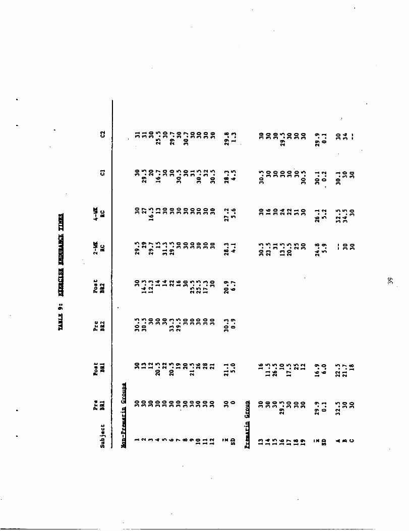
i) Exercise duration
The total exercise times during the submaximal tests are listed in
Table 9. Durlng control or prebedrest tests, the exercise duration
was about 30 minutes. Occasionally, there was difficulty obtaining
the final 30 minute blood sample, and the exercise time wa_ prolonged
by one or two minutes. For tests in which the exercise was stopped
earlier, the subject's heart rate exceeded 95Z of her prebedrest
maximal heart rate, which was measured during VO2 max determination.
The only exception was subject 17, who was stopped during her
postbedrest and recovery tests because of the core temperature
limitatlon. (An exercise test is immediately terminated if core
temperature exceeds 39 degrees C).
The average exercise times in the prebedrest and control tests were
all between 28 and 30 minutes. Following bedrest however most women
could not finish the entire protocol without reaching the heart rate
or core temperature criteria. The average exercise duration for the
non-premarln group was 21 minutes for both the postbedrest I and
postbedrest 2 tests. A similar decrease in exercise duration
occurred followlng bedrest in the premarin group (the postbedrest
duration averaged 16.9 minutes).
Most of the women in the non-premarln group completed the 30-minute
exercise test after both 2 and 4 weeks of recovery from bedrest. For
the premarin group, 4 of the women were told to stop exercise early
during the 2 week recovery tests, and 3 stopped early during the 4
week recovery test.
li) Changes in plasma volume during exercise tests
Plasma volume decreased significantly (P < 0.01) in all tests
during exercise. Figure 14 illustrates the PV changes which occurred
in the non-premarln group during the pre and postbedrest tests.
Plasma volume decreased rapidly with the onset of exercise, with most
38
U
Us-4
6.1
U41
*e-i
:l_J
"*"O_O P'-Or',.OO O O e0 _j
O u'_ O r*,. O Otn O,-,t_ ¢'_ u_ (e.),_
¢r_ _ O O O a0 .4'
o_oooooooo _
_ooo_ooooo _
oo _m oo
__@ _ IN
mo
O O Oo O
__ .0 e*_
__ _o __
OOO_OOO m_ _OO

2600
24O0PLASMAVOLUME
(ml.)
2000
18OO
16OOO
Plasma Volume
During ExerciseB
R
• L ]_ B_ ¸ _..°
"_\" " in
_4_ j J;
10 20 30 FC
, preBR 1
o post BR 1
I + pre BR 2mpost BR 2
EXERCISE TIME
(min.)
FIGURE 14: Absolute plasma volume (mean i SE) during thesubmaximal exercise tests for the non-premarin group (n = 12)during the pre and postbedrest tests before or after bedrest 1and bedrest 2.
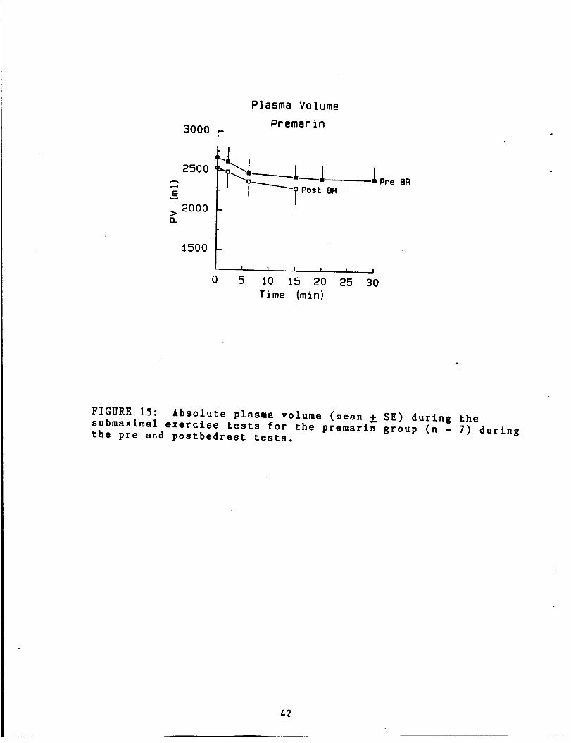
40
of the changes occurring within the first 10-15 minutes. Thereafter,
the decrease in PV was attenuated, and even in some subjectspartially recovered. Significantly smaller PV were seen during
postbedrest tests than during prebedrest tests in the non-premaringroup (P < 0.01). Absolute PV did not differ slgnificantly (P <
0.50) in the premarln group between the pre an postbedrest tests
(Figure 15).
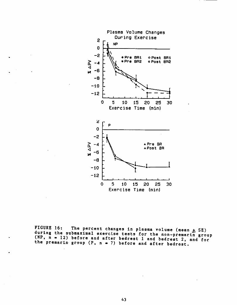
A slmilar pattern of plasma volume decrease during exercise
occurred during all exercise conditions despite the significant
differences in absolute PV. Figure 16 illustrates the changes in
plasma volume during the pre and postbedrest exercise tests, where
the data are presented as the Z change in PV (X difference from the
resting sample). The decrease in PV during bedrest was significant(P < 0.01) for each exercise test condition (PREBR, POSTBR, CI, C2,
2wk RC, 4wk RC). However a comparision of the Z changes in PV between
the pre and postbedrest results (ANOVA-2W-RM-SP) was non-significant(P < 0.i0) for non-premarin bedrests 1 and 2 as well as for the
premarin results (P < 0.I0).
lii) Changes in blood electrolytes and proteins during exercisetests
Plasma osmolality increased during all exercise tests (P < 0.01).
Figure 17 (upper panel) illustrates the increase in plasma osmolality
during exercise for the non-premarin group, comparing pre and
postbedrest results. The bottom panel of this same figure
illustrates the changes for the premarin group. An ANOVA-2W-RM-SP
was used to compare the differences in osmolalities between pre and
postbedrest conditions. Non-significant differences between the pre
and postbedrest tests occurred in the non-premarin group during both
the first (P < 0.80) and second (P < 0.90) bedrests, and also between
pre and postbedrest tests in the premarin group ( P < 0.70).
Total protein concentration increased significantly (P < 0.01)
during each exercise test. Figure 18 (upper panel) illustrates these
changes in total protein concentration for the non-premarin group
during the pre and postbedrest tests (bedrests 1 and 2). During the
first bedrest, the total protein concentrations were significantly
higher (P <0.05) during the postbedrest test than during the
prebedrest test. During the second bedrest, though the average total
protein concentration was higher at each exercise interval, this
difference was not significant (P < 0.30).
In the premarln group, an increase in total protein concentration
occurred during the exercise tests. There were no significant (P <
0.10) differences in these protein values between the pre and
postbedrest tests.
iv) Cardiovascular responses during exercise
Exercise heart rates
Figure 19 illustrates the exercise heart rates during the pre and
postbedrest exercise. The exercise heart rate following bedrest was
significantly higher than the prebedrest value for the non-premarin
group following bedrest I (P < 0.05), but not quite significant
following bedrest 2 (P < 0.I0). During the 2 and 4 week recovery
41
r-4
E
O.
Plasma Volume
Premarin
3000 F
2000
1500
J,
0
.i Pre BR
I | i | | I
5 10 15 20 25 30
Time (min)
FIGURE 15: Absolute plasma volume (mean _ SE) during thesubmaximal exercise tests for the premarin group (n - 7) duringthe pre and postbedrest tests.
42
2
0
-2
o. -4
-6
-8
-iO
-t2
Plasma Volume Changes
Ouring Exercise
NP
0 5 I0 15 20 25 30
Exercise Time (rain)
;J
0
-2
>-4<]a< -6
-8
-t0
-t2
' _i_ aPre BR
"_ APost BR
0
• I t . ! , t I |
5 10 t5 20 25 30
Exercise Time (rain)
FIGURE 16: The percent changes in plasma volume (mean _ SE)
during the submaxlmal exercise tests for the non-premarln group(NP, n - 12) before and after bedrest I and bedrest 2, and for
the premarin-group (P, n = 7) before and after bedrest,
43

Plasma 0smolaltty .
During Exercise
F295
,_ 2go
2s5._1
275o 0 5 l0 t5 20 25 30
Exercise Time (min)
-_ 300 fm 295oE
m 290oE
>. 2854.1
m 280
OEm 275o 0
P
A Pre BRAPost BR
! , I I m I _ 1• I I
5 10 i5 20 25 30
Exercise Time (rain}
FIGURE 17: Plasma osmolality (mean _ SE) during the submaxlmal
exercise tests for the non-premarin group (NP, n - 12) before
and after bedrest I and bedrest 2, and in the premarin group (P,n - 7) before and after bedrest.
44

• Total Protein
During ExerciseB.5
8.2 _ &
_7.9
_7.6 /
67.3
+7.0 _._I .Pre snl oPo.t SRi+
6.7_
0 5 tO i5 20 25 30
Exercise Time (rain}
6.70
I * I , I I , I . |
5 iO 15 20 25 30
Exercise Time (rain)
FIGURE 18: Total protein concentration (seen _ SE) during thesubmaxtmal exercise tests for the non-premartn group (NP, n =12) before and after bedrest 1 and bedrest 2, and for the
premarin group (P, n = 7) before and after bedrest.
45

185
165
._t45125
t05
85
65
Heart Rate
During Exercise
- NP
[ *Pre BRI oPostBR[//"
f • Pre BR2 o Post BR2
0
, I , I , I • I • I • I
5 I0 15 20 25 30
Exercise Time Imin}
185
165
145Cl
125
rri 105
85
65
A Post BR
0
, I , I , I , I • i . J
5 lO 25 20 25 30.
Exercise Time (mln)
FIGURE 19: Heart rate (mean _ SE) during the submaxlmaI
exercise tests for the non-premarln group (NP, n - 12) before
and after bedrest 1 and bedrest 2, and for the premarin group(P, n - 7) before and after bedrest.
46

exercise testa, the exercise heart rates were not significantly
different from the prebedrest teats ( P < 0.10) for the 2 week
recovery test, and P <0.50 for the 4 week recovery comparison. Theheart rate responses between the two prebedrest testa did not differ
significantly (P < 0.50).
For the premarin group, heart rate changes similar to those sees
in the non-premarin group occurred. Following bedrest, exercise heartrates were slgnificantly higher than during the prebedrest test (P <
0.05). The exercise responses seen during the 2 and 4 week recoverytests did not differ significantly from the prebedrest test (P < 0.30
for the 2 week recovery test, and P < 0.80 for the 4 week recovery
test).
In both the non-premarin and the premarin groups, the postbedrest
heart rate was higher (paired t-test comparision, P < 0.05) than the
prebedrest value even before the start of the exercise.
Exercise Cardiac Outputs
Cardiac outputs were performed at 4 exercise intervals; after I0,
15, 20, and 25 minutes of exercise. As there were no significantdifferences between the sampling times (F < 0.05), aii cardiac
outputs from a given test were averaged and these mean values are
presented for each subject in Table I0.
No significant differences occurred for either the non-premarin, or
the premarin group between experimental conditions. An ANOVA-IW-RM
was performed on each set of data, where the F ratio for thenon-premarin group was 1.83 ( P < 0.i0, n - 77), and for the premarin
group was 1.75 (P < 0.20, n - 30). Thus cardiac output did not differ
significantly during exercise after bedrest or during any of the
recovery tests.
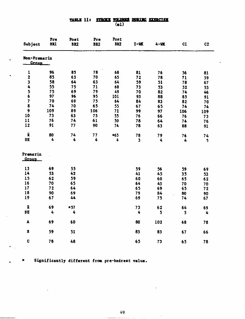
Exercise stroke volumes
Stroke volumes were calculated by dividing the cardiac output by
the heart rate during the rebreathing technique. The values shown in
Table II are the average stroke volumes, calculated from the cardiac
outputs performed during the submaximal exercise tests.
Significant differences in exercise stroke volumes occured in the
non-premarin group (ANOVA-IW-RM), where the F ratio was 3.05,
significant at the P < 0.02 level. Post hoc analysis (Duncan Multiple
Range Test) found that the stroke volumes during the postbedrest 2
test were significantly lower than stroke volumes during either
prebedrest i or 2 tests. Stroke volumes were lower during
postbedrest I tests than during the prebedrest I condition for I0 outof 12 subjects. However this difference was not significant at the P
< 0.05 level.
For the premarin group, the F ratio was 7.63, significant at the P< 0.O1 level. A post hoc analysis (Duncan Multiple Range Comparison)
determined that stroke volumes were significantly lower during
postbedrest tests than during prebedrest tests (P < 0.05). No
significant 4ifferences occured between prebedrest and recovery or
47

TAm_ lOz llr_J[ c_m_c _3__(ul/u£n)
Pre
Subject BR1
I 13548
2 11254
3 98044 9017
5 11840
6 14261
7 103148 12374
9 1580 3
10 10298
11 11050
12 13205
i 11897
SZ 555
Pr_ariu f_r._m
Post Pre PostBR1 BR2 BR2 2-_ 4-WK Cl C2
13041 10592 10157 12583 10512 8517 11083
8965 10021 10367 11852 12647 11684 9160
11517 10663 11126 10171 9211 13985 12081
12459 10380 12023 12448 9023 8953 930512145 14644 8738 12506 14023 12112 7573
15305 13110 16982 14955 13681 13195 1339211005 11791 10345 12475 11833 12120 10314
12799 9838 9194 11673 10630. 12988 12374
14207 14630 11630 14124 14165 15289 15803
10730 11726 9130 12223 10519 11709 10298
12239 9539 8699 12771 10527 10671 11050
12322 10457 11360 12154 9302 13595 13205
12227 11449 10812 12494 11339 12068 11303
451 490 621 327 521 546 615
13 10521 9571 9077 8360
14 8997 7673 7142 7386
15 9438 9771 9662 9426
16 11673 11902 11354 7156
17 12437 11728 11335 12003
18 13920 12327 13634 12903
19 9970 7584 10420 10934
i 10993 10079 10374 9738
SE 470 297 547 605
A 11866 11456 13082 6316
B 9340 8841 13082 10560
9219
5934
10235
11495
11891
14793
10951
10645
723
11093
10696
9570C 11171 7951 10365 9570
1052189979438
1167312437
139209970
10993470
11866
10911
48
'EOLIKllz me_n_ VOLmg_ /i_I,IJE_Cml)
Pre Post Pre Post
Subject BR1 BR2 BR2 BR2 2-_ 4-W[ Cl C2
Non-Premarin
1 96 85 78 68 81 76 56 812 85 63 70 65 72 78 71 593 58 64 63 64 59 51 78 674 55 75 71 68 73 53 52 535 75 69 79 49 70 82 74 466 97 96 95 101 93 88 85 917 70 69 75 64 84 83 82 708 74 70 6.5 55 67 6.5 74 749 109 89 106 72 99 97 106 109
10 73 63 75 55 76 66 76 7311 76 74 61 50 78 64 74 7612 91 77 90 74 78 63 88 91
i 80 74 77 *65 78 79 76 74SE 4 4 4 4 3 4 4 5
Premarin
13 69 55 5914 53 42 4115 62 59 6016 70 65 6417 72 64 6518 90 69 7919 67 44 69
i 69 *57 73SE 4 4 4
A 69 60 80
B 59 51 83
C 78 48 65
56456043698475
625
102
83
73
59356570658074
645
68
67
65
69
53
62
7072
9067
694
78
66
78
Sisni£icantly dif£erent £rom pre-bedrest value.
49

control tests.
v) Thermoregulatory responses
a) Thermoregulatory responses as a function of menstrual
cycle phase
Data from the control and prebedrest tests from this study werecombined with other data collected in a separate study to analyze the
effect of menstrual cycle phase on thermoregulatory responses. Usingeach woman as her own control, 8 normally cycling women were studied
in the folllcular and luteal phases of their menstrual cycles. We
reported no significant differences in body temperature responsesduring exercise (except for the expected increase in body core
temperature in the 1urea1 phase), and no differences in either totalbody sweat losses or local sweating responses (see abstract entitled
" Thermoregulatory response to exercise at different phases of the
menstrual cycle" in Appendix 3.
b) Effect of Premarin on thermoregulatory responses
The control and prebedrest data from the women in this study were
used to test the effect of premarin on thermoregulatory responses.Five women in this study performed the control exercise tests without
taking either oral contraceptives or premarln. Each woman also
performed premarin exercise tests, after ingesting 1.25 mg_remarin
for 7-10 days. We reported no significant alteration in body
temperatures, skin conductances, total body sweat loss, sweating
sensitivity or sweating threshold with the use of premarin (see
abstract entitled "The lack of an affect of elevated estrogens on
exercise thermoregulation" in Appendix 3).
c) The effect of bedrest on esophageal temperature durin R
exercise - non-premarin group
The esophageal temperatures (Tes, mean _ SE) during the submaximal
exercise tests are illustrated in Figure 20. During each submaximal
test there was a significant increase in the Tes during exercise.
During the first bedrest, the Tes values during the postbedrest
exercise were significantly higher (P < 0.05) than the temperatures
during the prebedrest condition (ANOVA-2W-RM-SP). No significant
differences were seen between the prebedrest and the 2 week (P <
0.70) or the 4 week (P < 0.90) recovery tests. Even before the start
of exercise, the resting Tes in the postbedrest test was
significantly higher (paired t comparison, P < 0.05) than the
comparable prebedrest value.
Although Tes values were consistently higher during all postbedrest
tests, these differences were not significant (P < 0.90 for the preto postbedrest I comparison, and P < 0.20 for the pre to postbedrest
2 comparison). The difference between the pre and postbedrest Tesvalues after 15 minutes of exercise (the last value obtained in most
postbedrest tests) was significantly higher than the prebedrest Tes
at the same time interval (paired T comparislon, P < 0.01). Thus
during the postbedrest tests, Tes values were not significantly
50
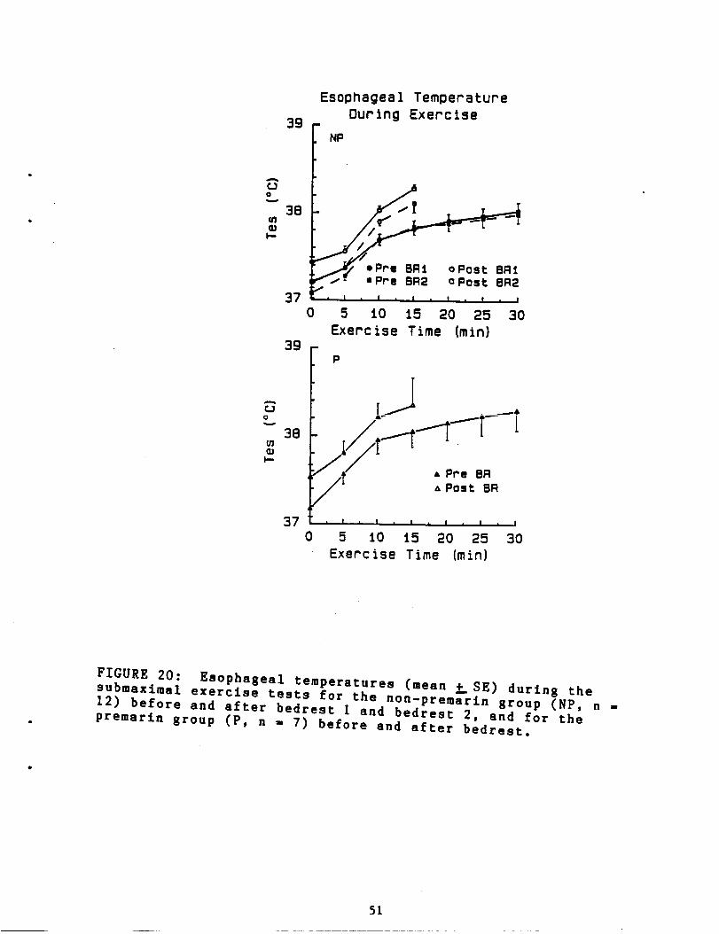
Esophageal TemDeratureOur ing Exercise
39 f NP
38
?-QG.Pro °post
0 5 tO i5 20 25 30
Exercise Time (rain)
38
370
0
I,-
P
/_/ APrs BR
A Post BR
I I i | I _ I • I
5 iO i5 20 25 30
Exercise Time {rain)
FIGURE 20: Esophaseal temperatures (mean _ SE) durtn 8 thesubmaximel exercise tests for the non-premarin 8roup (NP, n =
12) before and after bedrest I and bedrest 2, and for the
premarin 8roup (P, n - 7) before and after bedrest.
5L
higher than the prebedrest values during the beginning minutes of theexercise test, but by the end of the test they were significantly
higher.
Esophageal temperatures during submaximal exercise_ premarin
_rouv
No significant differences occurred between the Tes responses ofthe prebedrest and the 2 or 4 week recovery tests. Although theresting postbedrest Tes was significantly higher than the prebedrestresting value (P < 0.05), as the exercise continued the differencesbetween the pre and postbedrest values were no longer significant ( P
0.09, ANOVA-2W-RM-SP). Although the mean Tes at each exerciseinterval was consistently higher in the postbedrest tests (Figure
20), even a comparision of the 15 minute values (paired t comparison,P < 0.09) showed a non-significant difference between the pre and
postbedrest Tes values.
d) Mean Skin Temperatures During the Submaximal exercise tests_
non-premarin Rroup
The mean skin temperature was calculated from 4 skin sites
(Methods) and are illustrated in Figure 21. Although the mean skin
temperatures during the prebedrest tests (both first and second
bedrest) decreased during the exercise tests, while the postbedrest
values either did not change or increased slightly, there _as
considerable variability among the individual responses. For any
given exercise condition, the changes in mean skin temperature during
exercise were not significant; prebedrest I, P < 0.80; prebedrest 2,
P < 0.50; postbedrest i, P < 0.70; postbedrest 2, P < 0.70. No
significant differences occurred in mean skin temperature responsesbetween any of the exercise conditions (ANOVA-2W-RM-SP), P < 0.20).
Mean Skin Temperatures during Submaximal Exercise Tests_ premarin
KEEEE
The pattern of change in mean skin temperature during the exercise
tests appeared different from the pattern seen for the non-premarin
group. Again, there was considerable variability among subjects, and
the increase in mean skin temperature during exercise (Figure 21,
bottom panel) was not significant during either the prebedrest (P <
0.20) or the postbedrest (P < 0.50) condition. No significant
differences occurred in mean skin temperature response between any of
the exercise conditions (ANOVA-2W-RM-SP), P < 0.50).
e) Total body sweat losses during submaximal exercise tests
Total body weight loss was used as an estimate of the total sweat
loss during the submaximal exercise tests. The change in body weight
(in grams) during exercise was divided by the number of minutes of
exercise and by the surface area of each woman (from Dubois nomogram
based on height and weight). The results presented in Table 12 are
the sweat rate results (g/min/m2 body surface area) obtained during
each exercise test.
52
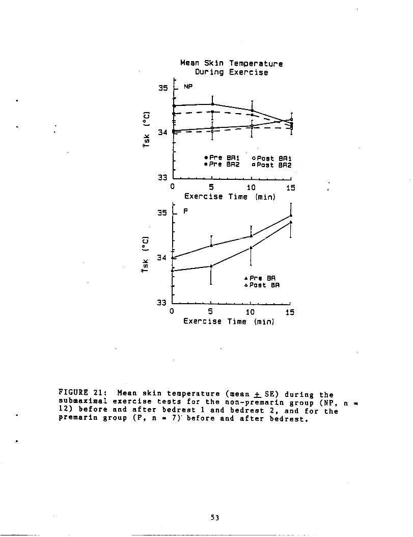
35
u_,t-
33
35
CJOw
34ul
it-
33
Hean Skln Temperature
Our ing Exercise
NP
opre BRI oPost BRI• Pre BR2 -Post BR2
• • | i | | • , . I m | i |
0 5 10 15
Exercise Time (min)
0
_I' 'P • Pre BRA Post BR
• • ' I | * , . . l I
5 10 i5
Exercise Time (minl
FIGURE 21: Mean skin temperature (mean __SE) during thesubmaximal exercise tests for the non-premarin group (NP, n =12) before and after bedrest 1 and bedrest 2, and for thepremarin group (P, n = 7)' before and after bedrest.
53

12= "f'emu._mrsmutTx,ou
Cglmi.nlu 2)
Pre Post
Subject BR1 BR1
Pre PostBE2 BR2 2-,me 4-WK C1 C2
1 11.33 8.97 11.63 9.92 11.68 10.00 9.56 11.332 12.66 17.79 13.28 17.92 12.24 10.97 11.31 12.663 9.79 19.22 11.46 14.99 11.15 14.47 15.22 9.794 13.35 14.72 16.28 16.65 16.89 19.54 17.08 13.355 8.58 8.48 7.74 12.14 16.62 9.36 8.65 8.586 14.94 13.71 7.82 5.22 8.99 8.50 11.87 14.947 10.36 13.24 15.93 16.09 15.17 13.05 12.17 10.368 7.57 10.19 8.99 9.54 9.78 8.77 8.82 7.579 10.31 12.59 9.58 14.30 9,80 8.73 10.52 10.31
10 9.44 12.79 10.67 10.98 12.00 11.78 8.74 9.4411 9.18 11.50 9.38 13.50 10.40 11.18 6.74 9.1812 10.33 14.48 17.54 13.45 10.33 12.28 14.51 10.33
i 10.65 .13.14Sg 0.58 0.89
1 10.45 19.12
2 7.48 18.723 8.06 9.044 11.71 18.795 10.37 16.386 8.85 11.507 8.88 10.81
i 9.40 14.91
SE 0.52 1.51
11.69 -12.89 12.09 11.55 11.27 10.65
0.93 0.98 0.75 0.87 0.85 0.58
10.588.928.24
13.0212.20
9.709.80
10.350.60
9.20
7.58
9.46
A 7.36 10.20
10.3910.68
7.3711.36
8.878.819.30
9.540.47
11.84
6.71
12.42
B 10.23 12.57
11.637.416.59
11.4511.598.52
8.47
9,380.75
9.20
6.82
C 11.90 15.53
10.457.488.06
11.7110.37
8.858.60
9.36
0.53
10.05
10.23
11.90
* Significantly different from pre/bedrest 1. 4-
54
There were no significant differences in the total sweat losses
between the non-premarin and the premarin group, which averaged 10.85
g/mln/m2 for the non-premarin group and 9.40 g/mln/m2 for the
premarin group.
For the non-premarln group, significant differences (P < 0.05)occurred in the total sweating results between exercise conditions
(ANOVA-IW-RM). The total sweat loss was significantly greater (P <0.05) than the prebedrest condition during postbedrest I and
postbedrest 2 (Duncan Multiple Range Comparislons). No significantdifferences occurred between other exercise conditions.
For the premarln group, the F test ratio comparing all conditions
was not significant (P < 0.10). However for every subject, the total
body sweat losses were greater during the postbedrest test thanduring the prebedrest test (see Table 12).
f) Sweat sensitivity and Tea threshold determinations
Local sweat rates (SR) were obtained froa sweat capsules placed
over a skin site on the chest and plotted as a function of the
corresponding Tes (see Methods), and the slopes of these SR/Tes
relationships are shown in Table 13.
During the first year of the study, the sweat system was not yetbuilt, and so local sweat records for subjects 1-6 could not be
obtained. Also any subject in whom the data was "missing" from more
than one exercise test was not included in any of the data analysis.
(This included subjects 7, 9, and A). Data became "missing" if the
sweat capsule leaked during the test.
The statistical analysis was affected by the small number of
subjects in each group (4 non-premarin, 7 premarin, and 2 oral
contraceptive users) and by the fact that, since sweating slopes andthreshold values are not normally distributed, non-parametric
statistical analysis were required. In order to obtain the largest
number of comparisions, all pre and postbedrest data (from the
non-premarin, the premarln, and the oral contraceptive users) were
combined to test whether bedrest reduces the slope of the sweating
response, and the significance of this comparison was tested using aHann-Whltney U test. The Z statistic was 1.79, which is significant
for a one-tailed design at P < 0.04. Decreases in the sweating slope
followlng bedrest occurred in 6 out of the 8 bedrests in the
non-premarln group, in 6 out of 7 of the premarin group, and in both
of the oral contraceptive users.
g) Sweating thresholds
The same limitations as above applied to the calculations of the
sweating thresholds. These results are presented in Table 14, and
the pre and postbedrest data were also analysed by combining the
results from all groups and performing a Mann-Whltney U test. The Zvalue was 0.82, which was non- significant for either a one-tailed (P
< 0.21) or a two-tailed (P < 0.41) analysis.
vi) Venous lactate values during exercise
55

mglm_l I
F_e
Subject Bll
Post Pre PostBR1 BR2 BR2 2-WI[ 4-WI[ C1 C2
Non-Premarin
8 0.83 0.8810 0.88 0.7211 0.69 0.6612 0.9 0.62
i 0.82 0.72SE 0.04 0.05
0.86 0.28 0.26 0.4 0.94 0.831.14 2.5 1.13 1.6 0.69 0.880.74 0.39 0.76 0.47 0.55 0.691.35 1.05 0.79 0.81 0.82 0.90
1.02 1.05 0.73 0.82 0.75 0.830.04 0.44 0.15 0.23 0.07 0.04
Premar£n
13 3.88 0.5614 0.19 0.2915 1.07 0.4916 0.44 0.37
17 0.5 0.318 1.87 0.61
19 0.9 0.62
1.26 0.46SE 0.45 0.17
B 0.98 0.51
C 0.46 0.29
2.81 I .74 4.49 3.880.3 0.35 0.21 0.190.84 0.75 0.93 1.070.47 0.67 0.98 0.440.31 0.54 0.53 - 0.500.68 2.23 1.43 1.871.32 1.28 0.99 0.90
0.96 1.08 1.36 1.26
0.31 0.24 0.45 0.45
0.86 0.56 0.78
0.67 0.6 0.59
56

_e
Subject BR1
Post Pre PostBR1 BR2 BR2 2-T,_ 4-_ Cl C2
Non-Fremarin
8 37.4310 37.1511 37.112 37.15
i 37.21Sg 0.07
37.49 37.43 37.15 37.32 37.43 37.59 37.3537.46 37.25 37.47 37.18 37.15 37.11 37.3737.41 37.44 37.58 37.62 37.1 37.44 37.4837.43 37.46 37.59 37.36 37.15 37.24 37.33
37.44 37.39 37.44 37.37 37.20 37.34 37.380.01 0.04 0.02 0.08 0.04 0.09 0.03
Premarin
_£¢OmL--
13 37.0214 36.7815 37.2816 37.5417 38.2118 37.919 37.45
36.84 36.9 37.i4
37.19 37.07 37.0137.47 37.17 37.0537.02 37.45 37.1938.11 37.81 38.26
37.77 37.06 37.7638.26 37.4 37.73
i 37.45 37.52 37.26 37.44SE 0.17 0.19 0.11 0.16
B 37.59
C 36.56
37.76 37.66 37.29
36.72 37.41 37.56
a_ A
.II,Z
36.7537.437.4937.73
37.3537.37
37.320.11
37.25
37.00
37.0336.7837.2837.5438.2137.9037.45
37.450.17
37.47
36.56
57

The individual blood lactate values obtained from each restingand final exercise sample are shown in Table 15. Blood lactateincreased significantly during each exercise condition (P < 0.01).
For the non-premarin group, venous lactate increased by an average29.4 and 29.0 mg Z during the prebedrest 1 and prebedrest 2 exercise
respectively. Following bedrest, the increase in venous lactate wassignificantly greater (P < 0.01); 47.4 and 45.7 mgZ during the
postbedrest 1 and 2 tests, respectively. Although not as high as thepostbedrest tests, venous lactate values were still significantlyhigher than the prebedrest tests during the 2 week (34.8 mgZ) and the4 week recovery (34.0 ms Z) tests.
For the premsrin group, venous lactates also increased
significantly more (P < 0.01) during the postbedrest test than during
the prebedrest condition (Table 15). However the rise in venouslactate was not significantly different (P <0.05) from the prebedrest
condition during the 2 and the 4 week recovery tests.
vii) Change in forearm venous compliance during exercise
The change in forearm venous compliance (FVV) which occured
during the pre and post bedrest I tests in the non-premarin group isillustrated in the upper panel of Figure 22. During exercise therewas an increase in venous tone (decreased FVV), which persisted
throughout the exercise test, despite" the marked increases in body
temperatures. This venoconstriction during exercise was also seen
during the postbedrest tests. Although all mean postbedrest FVVvalues were smaller than the prebedrest values, thus suggesting a
potentiated venoconstriction, the differences were not significant
(ANOVA-2W-RM-SP, P < 0.20). No significant differences occurred
between the prebedrest 2 and postbedrest 2 tests (P < 0.20), the
prebedrest 1 vs 2 week recovery ( P < 0.70) tests, or the 4 week
recovery tests (P < 0.02). Figure 23 illustrates that the apparentdifferences in the FVV response between pre and postbedrest tests
were due to a shift in the resting values and not the result of a
change in the rate of venoconstriction during the exercise. This isevident from the fact that the shape of the relative changes in the
venoconstrictor response in the pre and postbedrest tests was almost
identical.
Similar FVV responses occurred in the premarin group (see the lower
panels of Figures 22 and 23). The change in FVV during pre and
postbedrest exercise was not significantly different (P < 0.I0).There also were no significant differences between prebedrest and 2
week (P < 0.I0) or 4 week ( P< 0.10) recovery tests.
D) 17-bets Estradiol and Progesterone Results
I) During Bedrest
The 17-beta estradiol and progesterone values obtained from blood
samples drawn at the same time of day from each woman during bedrest
is shown in Tables 16 and 17. Normal cycllc fluctuations in blood
estrogens occurred in the non-premarin group, with peak 17-beta
estradiol concentrations of about 200-400 pg/ml near ovulation. The
58

I
iS U
-i.III ill
Sill
!llI
i.im
Ill
)4ill
IIill
IlU
illiIi.iel
nil!ill1,4
llllII
i.itiill
"Iol
eo • .0 oe • •
el 0* • • •
_ _ _ 0oo Ioeooo, I0 . .
ilill|*illil II IIIIlil II I I I
__ _-.=_ _o , , , , , , , , , , , ,lilililillil _ Ilillii li i
--oe--eee-- --eo• ** iillill I| i ! I
O_Ol_lli_ lO Illllll II I I i___0_ _0 I I I I I I I I I I I I
_0__ _
• oe, ee • • ,
e,_ emJ p"4 am# _4
ti
II.s.iiJI.*li)l
i-I
liil
.p4
._,,4I:1
.r4t_
- Forearm Venous Volume
No Premarin
3.32.9
m=4
2.5
> i 7b_
<_ 1.3
.9
0
I I I I l I
5 iO i5 20 25 30
Time (min)
Pro OR
3.7
_- 3.3utJo 2.90
_. 2.5u
_ 2.i
> 1.7b.
<3.9
Premarin
\ \i-i4-LLLLLLLLLi
I I I I I
0 5
Pre BR
iO i5 20 25 30
Time (min)
FIGURE 22: The change in forearm venous volume (compliance)between 0 and 30 Torr arm cuff congesting pressures (mean_.+ SE)
during the submaximal exercise tests for the non-premarin group
(n - 12) before and after bedrest I, and for the premarln group
(n = 7) before and after bedrest.
60
_.f
iO0
'-' 900L.
a.ac 80OU
70
0
60
50
U.
Forearm Venous Volume
Pre-Bedrest
_x ,N0 Premarin
k o Premarin
• G- - --G--G--G,_.
- --0_. 0
0
I I I I I I
5 iO 15 2O 25 3O
Time (min)
0C.4J
C0U
0
>>U.
Post-BedrestiO0
80 emarin
60
50 -
0 5
I I t I I ,I
10 15 2O 25 30
Time (min}
FIGURE 23: The relative change in forearm venous volume between
the resting value and sucessive exercise time intervals during
the submaximal prebedrest and postbedrest exercise tests for the
non-premarin (n = 12) and the premarin (n = 7) groups.
61
O_
i+a+++
i-4"
¢,,I
CJ
IP0pl
O"_ I'_ I",4
• 1_ I I • I•41' _D I I ,-o I
i,-4
mm _
• ego ee_ooe oe
*_ _ **
_ _ ..
*ueoee eo
111 ollOl*
O0
+. <. _.
i-.I
sO
001-4
i,+l l_.l
O0
¢+4 +-4
I_+-4 *<lr I'_ ¢_1
• cell oeelee el
I,,'10_
• eeoc el el
o04",-I
011"1 co
¢h +.1
Ill
• * * II
•,t" oDOOl_ i_ I_
41
eeeeee o_ooeo *e eel
OsO 0_-* _4D
P, iO_ .-*OD I%
¢_1 ,41' ff) p.+
0s00 _'*
P"I .-*
O'_OD O0
0 P_I ¢*,4
ee e*l oo el *
cecile eoeooe 11
',,,C
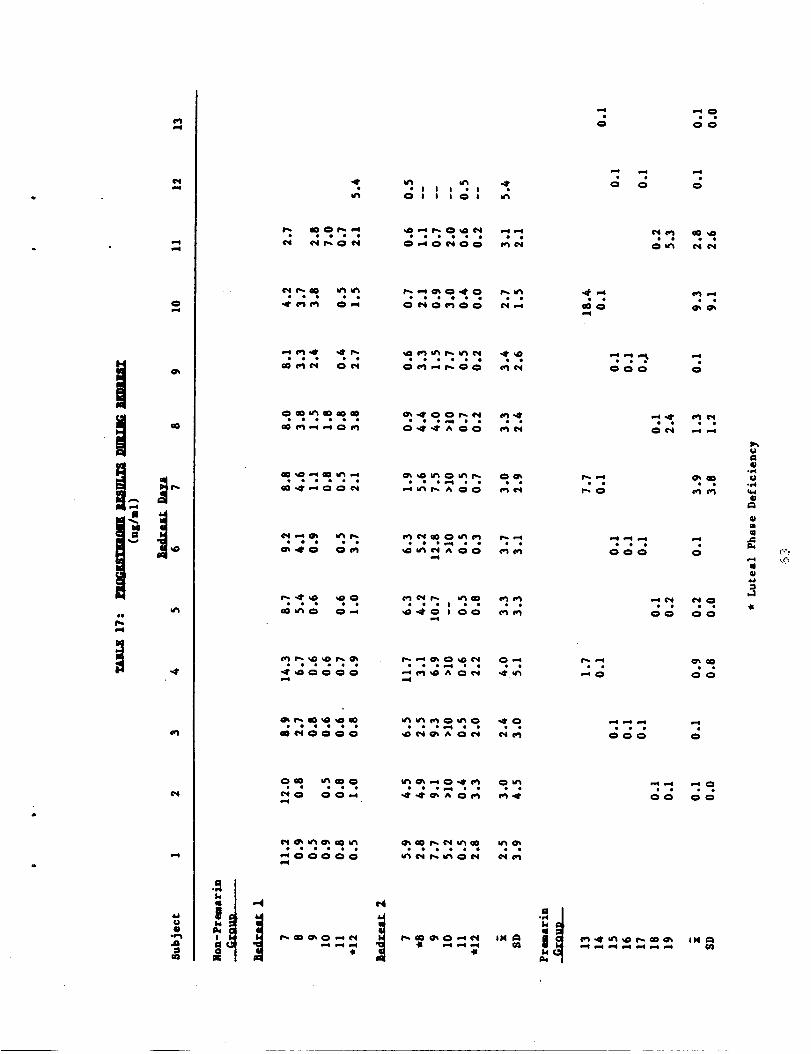
tin0
e-q
0pue
aO
64(JmA
=ua
0 O0
,4" _ *_t ,.4'• • I I I • I •
,..: .-, ..:
eo oeooeo .o oe e*
eoo_oe *o oe go
ooo eo oo*_*e *o
... -_ _:_ ". O0 O0
ee oee e •
oo*o*e *o
CJ
el
.p¢
III
IUn
GI
progesterone levels in the non-premarin group also showed cyclicfluctuations as would be expected from normally cycling women.
However in two of the subjects, subject 8 during bedrest 2 andsubject 12 during both bedrests 1 and 2, the progesteroneconcentrations during the luteal phase were abnormally low, and thecycles were defined as a luteal phase deficiency using the definitionof Abraham et al (1974). See Figure 24 for an illustration of thehormonal responses from one of these subjects. Also during the first
year of the study, subject 3 had an extremely short (18-day) cycleduring the first bedrest, and blood hormonal values obtained duringthe post bedrest exercise test and morning temperature data suggestthat this subject did not ovulate during this menstrual cycle.
The 17-beta estradiol and progesterone values obtained from bloodsamples in the premarin group are also presented in Tables 16 and 17.The 17-beta estradiol values averaged from 184 to 365 pg/ml during
bedrest, with consistent elevations seen throughout the bedrestprotocol - except for subject 17 who forgot to take her pill on themorning of bedrest day 9. Progesterone levels for 5 of the subjectsremained less than 0.1 ng/ml, suggesting that ovulation had beeninhibited for most subjects. Subjects 13 and 19 appeared to haveovulated despite the premarin. Subject 19 reported "breakthroughbleeding" and increased the dosage of premarin to 2 mg/day
2) Before the submaximal exercise tests
The 17-beta estradiol and progesterone values obtained_rom the
resting blood sample drawn immediately before the start of thesubmaximal exercise tests are shown in Tables 18 and 19,
respectively. The 17-beta estradlol concentration ranged from 35.5
to 336.7 in the non-premarin group during the prebedrest tests, and
from 105.4 to 337.9 pg/ml in the premarin group. Again, the
progesterone data are representative of either a preovulatory stage
or a non-ovulatory cycle for all premarin subjects except subjects 13
and 19 in the postbedrest test.
E) Arglnine Vasopressin Concentrations during the SubmaximalExercise Tests
To test our hypothesis that plasma ADH (arginine vasopressin) may
be involved in the alteration of thermoregulatory function, plasma
arginine vasopressin concentrations were measured from the resting
and final exercise blood sample during submaximal exercise tests.
Samples were obtained only during the last year of the study
(premarin group), and the resting and final exercise values (pg/ml)
are shown in Table 20. No significant differences (P < 0.30) were
found (ANOVA-IW-RM) between any of the arglnine vasopressin values,
including rest and exercise, and pre and postbedrest values.
F) Pulmonary Function Test Results
Forced vital capacity (FVC) increased in each subject (n=13)
during bedrest. Total lung capacity increased in each subject, while
residual volume and resting volume of the lung did not change. No
change in FVC was found in an ambulatory control group using
identical measurement techniques. (see the manuscript in Appendix 2
64

Progesterone 3, /
(ng/mi) .2
Estrogen(pg/ml)
I .
•
Bedrest Doy ,4
2 3 4 5 6 7 B 9 I0 II 12
200, Estroqen
I00O,
Toral 98
(°F) 97
24 2.52627 I 2 3 4 5 6 7 B
Cycle Ooy
FIGURE 24: Endocrine measurements of one subject during
bedrest. An insufficiency of progesterone secretion was
documented according to the method of Abraham.
65
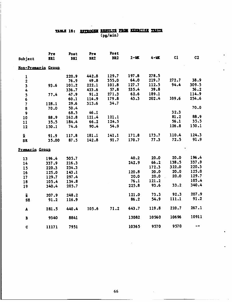
m 18- I3HII. ]gIIMIUIgL IIILll(pg/min)
PTe
Subject BR1
llzu=,l =Lc f,.co..un
Poac Pre PostBR1 BR2 BB.2 2-_ 4-W£
1 220.9 442.8 129.72 76.9 49.8 355.03 93.6 101.2 222.1 101.84 336.7 433.6 57.85 77.4 47.9 91.2 271.36 60.1 114.9 179.87 128.1 29.6 313.6 34.78 70.0 50.49 68.5 /46.1
10 88.9 162.8 121.4 121.111 35.5 184.4 66.2 124.512 150.1 74.6 90.4 54.9
C1 C2
197.8 278.564.0 219.7 272.7 38.9
127.7 112.5 94.4 309.5535.4 39.8 56.262.6 189.1 114.943.3 202.4 109.6 254.6
32.381.256.1
126.8
70.0
88.935.5
150.1
91.9 117.8 181.1 141.1 171.8 173.7 110.4 124.3SE 35.00 87.5 142.8 91.7 170.7 77.3 72.5 91.9
13 196.4 505.714 337.9 216.3
15 220.3 234.316 125.0 143.117 129.7 297.418 105.4 134.819 3/40.4 205.7
i 207.9 248.2SE 91.2 116.9
/t0.2 20.0 20.0 196.4242.9 66.2 138.5 337.9
172.3 322.0 220.3120.8 20.0 20.0 125.0
20.0 20.0 20.0 129.776.1 121.2 105.4
225.8 93.6 33.2 3/40.4
121.0 73.3 92.3 207.986.2 54.9 111.1 91.2
A 281.5 /4./-40.4 105.6 71.2 643.7 119.8 210.7 267.1
B 9340 8841 13082 10560 106% 10911
10365 9570 9570C 11171 7951
66
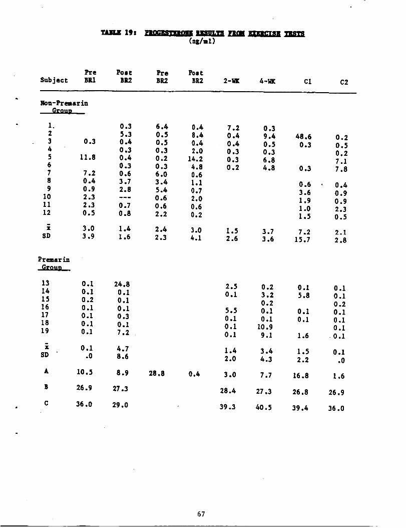
SubjectPre Post _e _stB_ B_ B_ B_ 2-_ 4-_ C1 C2
Nou-Premarin
1. 0.3 6.4 0.42 5.3 0.5 8.43 0.3 0.4 0.5 0.44 0.3 0.3 2.05 11.8 0.4 0.2 14.26 0.3 0.3 4.87 7.2 0.6 6.0 0.68 0.4 3.7 3.4 1.19 0.9 2.8 5.4 0.7
10 2.3 --- 0.6 2.011 2.3 0.7 0.6 0.612 0.5 0.8 2.2 0.2
7.2 0.30.4 9.4 48.6 0.20.4 0.5 0.3 0.50.3 0.3 0.20.3 6.8 7.10.2 4.8 0.3 7.8
0.63.61.91.01.5
0.40.90.92.30.5
i 3.0 1.4 2.4 3.0 1.5 3.7 7.2 2.!SD 3.9 1.6 2.3 4.1 2.6 3.6 15.7 2.8
Pre=arin
13 0.I 24.814 0.1 0.115 0.2 0.116 0.1 0.117 0.I 0.318 0.1 0.119 0.1 7.2
i 0.1 4.7SD .0 8.6
A 10..5 8.9 28.8 0.4
B 26.9 27.3
C 36.0 29.0
2.5 0.2 0.1 0.10.1 3.2 5.8 0.1
0.2 0.25.5 0.1 0.1 0.10.1 0.1 0.1 0.10.1 10.9 0.1
0.1 9.1 1.6 0.1
1.4 3.4 1.5 0.I2.0 4.3 2.2 .0
3.0 7.7 16.8 1.6
28.4 27.3 26.8 26.9
39.3 40.5 39.4 36.0
67
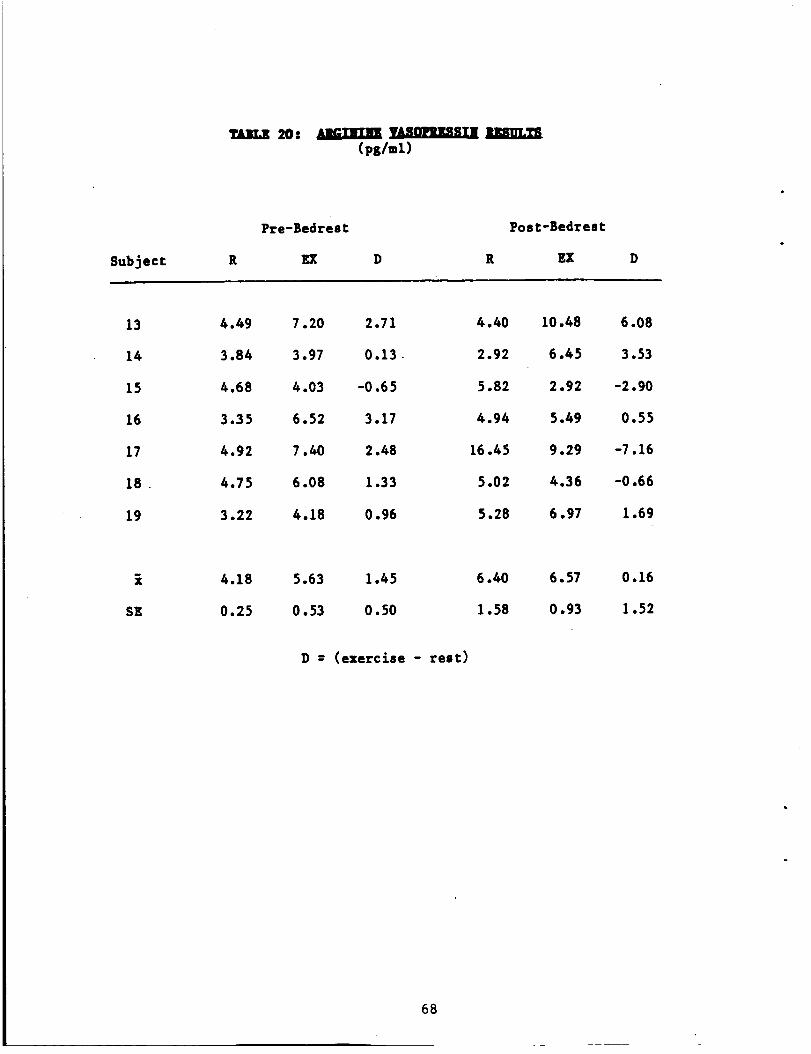
20: 1___
Cps/ml)
Pre-BedresC
Subject R _ D
Post-BedresC
R EX
13
14
15
16
17
18
19
4.49 7.20 2.71
3.84 3.97 0.13
4.68 4.03 -0.65
3.35 6.52 3.17
4.92 7.40 2.48
4.75 6.08 1.33
3.22 4.18 0.96
4.40
2.92
5.82
4.94
16.45
.5.02
.5.28
10.48
6.45
2.92
5.49
9.29
4.36
6.97
6.08
3.53
-2.90
0.55
-7.16
-0.66
1.69
SE
4.18 5.63 1.45
0.25 0.53 0.50
D = (exercise - rest:)
6.57
0.93
68
for a more complete description of these results).
G) Positron Emmision Tomo ra h Result
No change was found in the D2 receptor binding of the caudatenucleus following 5edrest. See Figure 25 for the slopes of the
caudate/cerebellum binding ratios obtained durin$ the prebedrest
(7/11/84) and postbedrest (7/25/84) tests.
&
5RATI 40
FIGURE 25: The ratio of llC-labelled 3-N-methlyspiperone
binding in the caudate/ cerebellum as an index of dopamine D2
receptor binding in one subject before (7/11/84) and after(7/25/84) bedrest. The similarity of the two relationships
suggest no change in the number of central dopamine D2 receptorsin the caudate nucleus during bedrest.
69
V) DISCUSSION OF RESULTS
A) Body Fluid Responses During Bedrest
1) Comparison to previous studies
A fairly consistent finding is that plasma volume decreases during
spaceflight (Johnson, 1979) and during programs of bedrest.
Greenleaf and coworkers (1977) utilized the data from several bedrest
studies to characterize a hyperbolic relationship between the
decrease in plasma volume as a function of the number of days of
bedrest. According to their calculations, plasma volume decreases to
an asymptote of about 22.5Z below prebedrest level after about 30
days of horizontal bedrest. After 11 days of bedrest, a 13Z decrease
in plasma volume would be expected. Most of the data used in these
calculations were obtained from studies of the responses of male
subjects. In the present study, the hypothesis was tested that women
may have a smaller reduction in plasma volume during bedrest,
especially during the pre-ovulatory and the luteal phases of the
menstrual cycle, through a direct or indirect action of estrogens to
retain body water. This hypothesis was based on early observations
of hemodilution during estrogen therapy (Whitten et al, 1951: Preedy
et al 1956), possibly through a sodium conserving action in the
kidneys (Dignam, 1956). In addition, Gaebelein and Senay (1982)
observed that the decrease in plasma volume during exercise was less
during the luteal phase, when estrogens would be higher, than during
the early follicular phase. Our results however did not stfickly
support this hypothesis. The decrease in plasma volume seen in the
women without estrogen supplementation was similar to that predicted
by Greenleaf's formula; it averaged 19.9Z following bedrest I and
12.3Z following bedrest 2 (Table 5). If anything, the women in our
study lost more plasma volume than that reported for men undersimilar conditions.
No attempt was made in this study to control either fluid or food
intake. Therefore some of the day-to-day variations in blood and
urine constituents were undoubtedly influenced by changes in the
diet. Nevertheless, while there was no consistent change in the
amount of fluid or food intake, as assessed from the diary records,
consistent changes in plasma volume, electrolytes and proteins
occured during bedrest. The blood and urine responses seen in this
study were similar to those found in a previous study (Greenleaf et
al, 1977) which studied the body fluid responses of 7 men during a
similar bedrast protocol (non-exercise condition). In both studies
plasma osmolality and protein concentration increased significantly
during bedrest, although there was a net loss in the plasma content
of total protein (TCP) and osmotically active particles (TCO). The
mechanism responsible for this loss of a hypotonic fluid from the
vascular compartment during bedrest is most likely multifactorial.
In another paper by Greenleaf and Kozlowski, the body fluid shifts
during bedrest were divided into 3 separate stages. During the first
day of bedrest, fluid moves from the lower regions of the body into
the thoracic region, stimulating volume receptors, and producing a
diuresis. Then during the next 2 weeks of bedrest, plasma volume
reduction continues, and the extracellular volume is restored and
possibly even exceeds the prebedrest level. After 2 weeks of bedrest,
70

the plasma volume is still below the prebedrest level, but it remains
fairly stable; extracellular water is normal, and intracellular wateris reduced. Thus during the early hours of bedrest, the initial
reduction in plasma volume may be due to either a Henry-Gauer typereflex (Gauer, 1983) or to the stimulation of selective natriuretic
peptides (Greenleaf, 1985). Later reductions in plasma volume couldresult from a redistribution of water between various body fluid
compartments, or from potential alterations in protein, electrolyteor hormonal regulations. The data in this study support a
multifactorial mechanism rather than a simple Henry Gauer reflex.
The diuresis in this study occurred during only the first I or 2 days
of bedrest (Figure 12), yet plasma volume continued to decrease
significantly for another 4-5 days before the plasma volume remained
relatively stable (Figure 4). This lack of correlation between the
changes in plasma volume and urine output under conditions of a
stable fluid intake, suggest that the later decreases in plasma
volume may have occurred at the expense of some other fluid
compartment. The higher urine osmolality on the first day of bedrest
supports the suggeston of the stimulation of a natriuretic factor
during the first days of a bedrest.
Whatever the mechanism by which plasma volume is gradually reduced
during bedrest, it is very rapidly recovered f^11^_., bedrest
Within 24-48 hours after the end of the bedrest protocol, the plasma
volume of the non-premarin subjects averaged only about 3Z less than
their prebedrest plasma volume (Figure 5). Thus studies in which
post-flight plasma volume was estimated from blood samples drawn
several hours after return to normogravic conditions, may have
underestimated the plasma volume changes during weightlessness.
2) Effect of menstrual cycle on plasma volume changes duringbedrest
The effect of menstrual cycle stage on plasma volume changes
during bedrest was minimal. As seen in Figure 3, if a woman began
bedrest in the pre-ovulatory stage (stage 2) of her menstrual cycle,
the decrease in plasma volume at the onset of bedrest was postponed
for one or two days. Thereafter, plasma volume decreased rapidly to
the level of plasma volume seen in the women who began bedrest in
other stages of the menstrual cycle• If a woman reached the
pre-ovulatory stage of her cycle in the latter stages (days 7-12) of
a bedrest protocol, a temporary hemodilution occured, which againdisappeared within one or two days (see abstract entitled "Plasma
volume responses during bedrest in healthy women" Appendix 3). The
predicted retention of plasma volume in the luteal phase of the cycle
was sometimes, but not consistently seen in these subjects. The
plasma volume responses of the two subjects who took
estrogen-containing oral contraceptives were not different from the
plasma volume responses of the non-premarin group. Thus only in
conditions in which blood estrogens were elevated in the presence of
low concentrations of progesterone did consistant plasma volumeretention occur•
3) Effect of bedrest on menstrual function
Bedrest presents both a physiological as well as a psychological
71

stress to a woman, and stress is a well-known modifier of menstrual
function. Therefore it might be expected that menstrual cycles would
be altered during bedrest. The potential consequences of disruption
of normal menstrual function during weightless conditions are poorly
understood. Indeed, the consequences and the choice whether to treat
menstrual dysfunction during other stressful conditions (athlete's
ammenorrhea) are currently under debate (Loucks and Horvath, 1985).
During exposure to long-term weightlessnss, a cessation of menstrual
function potentially could have serious consequences. A decrease in
blbod estrogen levels could potentially accelerate the bone mineral
loss in women astronauts. Although this study did not directly
address the problem of menstrual function regulation, it was noted
that for the 6 "normally cycling women" in whom daily hormonal
samples were obtained during bedrest, two of these women had evidence
of luteal phase deficiency. However, the conclusion that the bedrest
protocol specifically produced this menstrual irregularity cannot be
drawn, since control endocrinological data were not obtained from
these two women during non-bedrest conditions.
4) The potential for lower leg edema following bedrest
The potential for lower leg edema following exposure to
weightlessness was emphasized in a review article by A.R. Hargens
(1982). He demonstrated that the interstitial fluid pressure in the
lower legs of male subjects decreased during head-down tilt as
interstitial fluid was shifted from lower body to upper body regions.
Hargens suggested that "countermeasures may be necessary t_maintain
precapillary-muscle tone during long space flight in order to prevent
swelling of lower leg tissues upon readjustment to Earth's gravity".
To date however, there have been no published accounts of severe
lower leg edema following bedrest or spaceflight. In this study,
subject "A" developed edema in both lower legs which probably was a
consequence of her participation in the bedrest study. Upon
finishing the bedrest protocol and the postbedrest exercise tests,
this subject remained in the sitting position for a prolonged
interval (typing a term paper all night). It is possible that this
period of inactivity prevented the restoration of a normal venous
tone in her lower legs, resulting in blood pooling and excessive
fluid extravascularization. Similar conditions of inactivity may need
to be avoided following spaceflight to prevent this painful and
disabilitating experience.
5) Effect of premarin to restore plasma volume during bedrest
In the original hypothesis, elevated blood estrogens were proposed
to be associated with body fluid retention. However from the results
of the two oral contraceptive users (who took pills containing
synthetic estrogens and progestins), and because of the greater
hemodilution seen during cycle stage 2 (pre-ovulatory) rather than
during cycle stage 3 (early luteal), this initial hypothesis was
modified to now predict fluid retention in conditions of elevated
blood estrogens only in the presence of low blood progesterone. The
estrogen supplement used to test the hypothesis was premarin, a
natural estrogen supplement which has been used for estrogen
replacement therapy. Premarin tablets administer about 48Z estrone
sulfate, 26Z equilin sulfate, and 26Z other hormones. One hour after
72

ingestion of a 1.25 mg tablet, total estrogen concentrations should
be elevated in the following proportions, 87Z equilin, 8% estrone,
and 5Z 17-beta estradiol (Whittaker, 1980). One disadvantage of
premarin administration is that, although total estrogens are
significantly elevated, the most biologically active human estrogen
(17-beta estradiol) is only moderately elevated (in our study,
17-beta estradiol was elevated in the plasma to 200-400 pg/ml).However this moderate elevation in 17-beta estradiol is also an
adyantage, since moderate levels may prevent many of the estrogenic
side effects, such as blood clotting and migrane headaches, reported
with estrogen treatment. In future studies however, a better
estrogen supplement may be low doses of estrace, which is pure17-beta estradiol.
It is clear from the results shown in Figure 4, that the premarin
administration was effective in restoring plasma volume duping
bedrest. During the first 2 days of bedrest, the plasma volume
response of the premarin group was similar to that of the
non-premarin subjects. Thereafter, the loss of plasma volume was
attenuated and then reversed. The mechanism by which premarin
restored plasma volume during bedrest is uncertain. Although the
urine losses in the premarin group tended to be less than in the
non-premarin group, there was much variability in subject responses
and no significant differences were seen between the two groups forurine loss, fluid intake, or the difference between fluid in and
fluid out. The concentration of blood electrolytes and total protein
in the two groups during bedrest were similar, thus not only was
water retained by the premarin action, but also electrolytes and
proteins.
6) Changes in red cell volume during and after bedrest
A decrease in red cell mass has been reported following both US and
Soviet space flights. The Skylab data indicated teat once a loss of
red cell volume is triggered, it may continue for as long as two
weeks after the cessation of the flight (Nicogossian, 1982). The
precise mechanism for the decrease in red blood cells has been
variously postulated to involve an inhibition of new red cell
production through decreased erythropoietin release and new red cell
formation, increased intravascular hemolysis, or sequestration of the
red blood cells (Nicogossian, 1982). Red cell mass was determined in
this study with a Technetium radioisotope labelling technique. One
advantage of this method is that the half-life of the label is short
enough that eight repeated measurements could be obtained at 2-week
intervals during this study. During a single bedrest, the red cell
mass decreased 4-7% in these women although this difference was not
significant (Table 4). Prior to the start of the second bedrest
(with about a 4-week recovery), the red cell mass was not completely
restored to the prebedrest I level. A further 4.4Z decrease in red
cell mass occured during bedrest 2, resulting in significant
accumulative reduction from the prebedrest I value. This reduction
in red cell mass persisted and increased during the next 2 weeks of
ambulatory recovery. Although these results shed no new light into
the possible mechanism of the decrease in red cell loss, it is clear
that this response is not rapidly reversible (as the plasma volume
response), and that accumulative effects are seen with repeated
73
bedrests, which are separated by 4 weeks of recovery.
7) The effect of repeated bedrest
One unique aspect of this study was that repeated identicalbedrests were performed, with the duplicate bedrests separated by a3-4 week period of ambulatory recovery. (In previous studies in which
repeated bedrests were performed, some alteration in the bedrestprotocol, ie. exercise intervention, precluded direct comparision ofthe results from the two bedrests). Several small differences
between responses measured during the first and second bedrest wereseen in this study. First the decrease in plasma volume during thesecond bedrest was only about 60Z of the decrease in plasma volumeseen during the first bedrest. Although the starting plasma volumewas slightly smaller during the second bedrest, the average plasmavolume remained higher than the bedrest 1 values througout.the last 8
days of bedrest (Table 5). Also, smaller increases in plasmaosmolality and total protein concentration occurred during the secondbedrest (Figures 6 and 8). These results suggest the interestingpossiblility that there is an adaptation of the body fluid responseswith repeated bedrest. Possibly, during the initial bedrestexposure, a redistribution of fluid or proteins occurred between bodyfluid compartments, such that there was an increase in the volume orcapacity of the interstitial compartment even in the presence of areduction in the plasma compartment (Greenleaf, 1982). The mechanismwhich altered the distribution of fluid between the interstitial,
plasma and possibly intracellular compartments, might invol:vealteration in the local tissue metabolism of these fluid
compartments, or an alteration in the protein composition or content(reminesent of Senay's theory (1972) of how heat acclimation alters
the regulation of plasma volume). Following the first bedrest, thesebody fluid alterations may have been retained until the start of asecond bedrest and been responsible for the smaller disturbance of
body fluid balance during the second bedrest, ie an increase inprebedrest interstitial volume could function to maintain plasmavolume. Thus a peripheral tissue body fluid "adaptation" may occurwith repeated weightless exposures. Alternately, a "central"modification may occur with repeated bedrests. The redistribution ofblood volume into the thoracic regions during the first bedrest couldsomehow result in a decrease in the sensitivity of blood volume
receptors, resulting in an attenuation of the bedrest-induced
diuresis during the second bedrest.
B) MAXIMAL EXERCISE RESPONSES FOLLOWING BEDREST
I) Difficulty in obtaining V02 max following bedrest
Measurements of maximum oxygen consumption (VO2 max) are often
interpreted as a measure of aerobic capacity, and when corrected for
body mass, is an indication of aerobic fitness. In order to
accurately determine an individual's V02 max however, conditions mustexist in which the subject is able to exercise to an intensity at
which oxygen transport is the limiting factor. If the active musclemass is too small, local muscle fatigue occurs before the maximal
aerobic capacity is reached. Evidence of the attainment of a true
aerobic capacity is the presence of a plateau in oxygen consumption
74

with further increase in exercise intensity.
Prior to bedrest in this study, the subjects were able to exercise
to their maximal aerobic capacity. Following bedrest however, the
criteria for VO2 max was seldom obtained, and thus the postbedrest
values are referred to as the "peak VO2." In this study it is
unclear what is the primary factor which limits peak VO2 after
bedrest. Potentially, the loss of muscle tone during the bedrest
might limit the working capacity of the leg muscles. Thus during the
V02 max testing after bedrest, the exercise performance was limited
by this local muscle fatigue rather than by a true aerobic capacity
llmitation. Thus following bedrest without muscle training, it might
be impossible to test the decrease in maximal aerobic capacity.
2) Comparison with data in the literature
Previous studies in the literature which claim to have measured V02
max or peak VO2 changes before and after bedrest report decreases in
the range from 17 to 31Z (Miller, 1965; Saltin, 1968; Taylor, 1949).
The responses of 8 women following bedrest averaged 9.7Z following 17
days of bedrest (Convertino, 1977). The decreases in V02 peak
(ml/kg/min) which were measured 24-48 hours after the end of bedrest
"- the present study averaged in_ ..A _ _^11^.J_.. _drests I _.._--_2.L LA |
for the non-premarin group, and 25Z in the premarin group. The range
of decrease in peak V02 following bedrest however was great; all the
way from no decrease following bedrest to a 31Z reduction. Some of
this variability may have been related to the experiences of the
women during the submaximal exercise tests on the previous day.
Subjects who fainted following the submaximal exercise tests appeared
to be more cautious and refrain from "going all out" during the post
bedrest VO2max test.
3) Lack of influence of premarin treatment
Premarin did not significantly alter V02 max during the control
tests or the decrease in V02 peak seen following bedrest (Table 8).
This is not an altogether unexpected finding as the decrease in
plasma volume which occurred in the non-premarin group was almost
completely restored by the time the maximal exercise test was
performed. For most subjects, postbedrest VO2 peak was determined on
the same day that the postbedrest plasma and red cell volume was
measured, which was 24-48 hours after the end of the bedrest. Figure
5 illustrates that at this time, the plasma volumes in both the
premarin and non-premarin groups were not significantly different
from the prebedrest values. Thus if the absolute decrease in plasma
volume during bedrest has any effect on the decrease in V02 peak,
this effect would no longer be present at the time of testing in this
study. In addition, it would not be expected that alterations in
plasma volume alone, without significant alteration in red cell
volume, would alter significantly V02 peak (Kanstrup and Ekblom,
1982).
4) Lack of correlation between V02 max and the Z decrease in V02
peak
One hypothesis which has been recently tested (Greenleaf, 1982b)
75

is that a greater reduction in V02 peak occurs in fit subjects than
in unfit subjects following bedrest. Positive correlations between
prebedrest V02 max and the Z decrease in V02 peak have been reportedin several studies which examined the responses of male subjects
(Chase, 1966; Saltin, 1968; Convertino, 1977). Very little data isavailable from women (Convertlno, 1977), and the correlation found in
one study was very low (-0.38). In the present study similar lowcorrelations for this relationship (-0.12 to -0.48) occurred in the
non-premarin bedrests. This result may have been due to the narrow
range of prebedrest fitness values (28-45 ml/kg/min). Anothercontributing factor may have been that the exercise was performed in
the upright posture, which according to Greenleaf et al (1982b)
presents an additional complicating factor in the determination of
V02 peak following bedrest as there are hypothesized differences inblood pressure regulation with differing fitness level. The postitive
correlations between prebedrest V02 max and Z change in V02 peak have
been reported from studies in which postbedrest V02 peak tests were
performed in the supine position.
C) SUBMAXIMAL EXERCISE RESPONSES FOLLOWING BEDREST
I) What limits exercise tolerance following bedrest?
The submaximal exercise responses following bedrest would be
expected to be reduced because of several concurrent effects. First,
deconditioning would be expected to alter the cardiovascular and
thermoregulatory responses that were studied. The average_prebedrest
V02 max results for the non-premarin and premarin groups were 2.24
and 2.41 liters/min, respectively (a non-significant difference).
Exercise intensities for the submaximal tests were calculated to
require an oxygen consumption of about 70% of each subject's
prebedrest VO2 max; The actual oxygen consumption of each woman
during the submaximal exercise was determined during the control
tests, and found to average 68 _ 1.3% VO2 max for the non-premarin
group, and 69 _ 2.7% for the premarin group. Since the average
decrease in V02 peak was 11% in the non-premarin group, then the
postbedrest exercise test was performed at an exercise intensity of
approximately 76.5g V02 peak. For the premarin group, the average
decrease in V02 peak was 22g (non-signlficant from the non-premarin
group because of the great variability in responses), and would have
resulted in an exercise intensity of about 88.0Z V02 peak during the
post bedrest test. Although the decrease in aerobic capacity was
probably not as great as suggested by the decrease in V02 peak
following bedrest, the postbedrest exercise was likely performed at a
significantly higher relative exercise intensity.
Secondly, in order to assist in subject recruitment, this study was
performed during the summer months. Thus during bedrest, it is
possible that changes in the state of heat acclimation occurred,
which would affect the postbedrest thermoregulatory responses. The
magnitude of this effect would be expected to be very small in this
study, as all subjects were employed in jobs in which they worked in
air conditioned buildings, and none were athletes or consistently
exercised out-of-doors. No significant difference was seen in the
thermoreguatory data between control and prebedrest responses where
the control tests were performed at the end of the study (late August
76
and early September) in year I, and at the beginning of the study
(end of May and early June) during years 2 and 3.
Third, the decrease in plasma volume which normally occurs during
bedrest would be expected to significantly alter cardiovascular,venoconstrictor (Fortney, 1983) and thermoregulatory (Fortney 1981)
responses during exercise.
Finally, a loss of venoconstrlctor tone during bedrest might result
id greater blood pooling in lower body regions, decreasing cardiac
return and thus exerting effects similar to the responses seen with
hypovolemia.
All of the above effects would be expected to influence the
submaximal exercise responses in this study. However, the magnitude
of the effects due to losses of training, heat acclimation and venous
tone would be expected to be comparable between the raring bedrest
conditions. Any change in the cardiovascular or thermoregulatory
responses caused by the decrease in plasma volume during bedrest
should be evident from comparisons of the responses of thenon-premarin (who had a 19.9 g decrease in plasma volume during
bedrest) and the premarin subjects (plasma volume decreased only 0.2Z
during bedrest).
2) Body Fluid Responses during Exercise
In 1969, Hyatt et al proposed that at least part of the postbedrest.orthostatic intolerance during tilt was due to a greater loss of
plasma volume during the tilt. They speculated that during bedrest
the extravascular compartment become dehydrated, and during
post-recumbancy tilt, a large transudation of plasma water occurred
into the leg tissue spaces. The resultant decrease in plasma volumeduring tilt would then account for some of the decrement in stroke
volume and cardiac output seen after bedrest. It is now known that
the extracellular compartment is not dehydrated during bedrest, and
thus this mechanism for a greater plasma water filtration after
bedrest seems unlikely.
Moderate cycle exercise in a warm environment is also a stress in
which decreases in plasma volume occur (Harrison, 1985). Thus if
Hyatt's theory is correct, perhaps a greater loss of plasma volume
would occur during exercise following bedrest than during the
prebedrest tests. In this study, plasma volume decreased during all
of the submaximal exercise tests, but the Z loss of plasma volume was
similar in pre and postbedrest tests (Figure 16). There was no
evidence of an increased extravascular fluid loss which may have
contributed to the greater cardiovascular and thermoregulatory strain
seen in the postbedrest tests. Indeed in the non-premarin group, the
same relative decrease in plasma volume occurred in the pre and
postbedrest tests, despite the different absolute plasma volumes at
the start of the exercise. This suggests that in the hypovolemic
condition (postbedrest), a smaller rather than a greater, absolute
loss of plasma volume occurred.
3) Cardiovascular responses following bedrest
77
The cardiovascular responses were altered significantly in all
postbedrest exercise tests. Heart rates were higher at all exercisetimes and stroke volumes were lower during the postbedrest exercise.
The increase in heart rate was able to compensate for the lower
stroke volume in these tests, and cardiac outputs were not
significantly lower postbedrest. For all but one subject, the
criteria for shortening a postbedrest exercise test was the
attainment of a heart rate of 95_ of the prebedrest maximal heart
rate value (one subject was stopped early because of a high core
temperature). In the non-premarin group, we first speculated that at
least part of the elevation in exercise heart rate and reduction in
stroke volume was due to the significantly lower plasma volume during
the postbedrest test, which presumably would limit cardiac filling.
This expectation was based on the similar changes in cardiovascular
responses seen in a previous study (Fortney et al, 1983) in which an
identical exercise protocol was employed to compare the
cardiovascular responses of men during normovolemic exercise, and
exercise in which plasma volume was reduced 15_ with diuretics prior
to the start of exercise. Therefore we were surprised to find that
the cardiovascular responses of the premarin group were similar to
those seen in the non-premarin group following bedrest. The increase
in heart rate, and reduction in stroke volume and endurance time were
of a similar magnitude as the non-premarin group, despite significant
difference in plasma volume responses during bedrest. Thus a similar
increase in cardiovascular strain was seen in these women following
bedrest, and it appeared to be independent of the level of
pre-exercise hydration.
4) Thermoregulatory Responses following Bedrest
Esophageal temperatures were higher at each exercise time in the
postbedrest condition for each subject group, while skin temperatures
were not different. Higher core temperatures would be predicted
during postbedrest tests, since the subjects exercised at a higher
relative exercise intensity (Saltin and Hermanson, 1966). Since core
temperature is the primary input for the control of sweating,
significantly greater total sweat loss might also be expected during
the postbedrest tests, when correction were made for the shorter
exercise endurance. However, following a decrease in fitness
(Roberts, 1977) or a decrease in plasma volume (Fortney, 1984),
sweating sensitivity is reduced. Thus for a given change in core
temperature, a reduced sweat production is ellicited. It has alsobeen shown that the threshold for the onset of sweating is influenced
by the degree of acclimation of the subject (Roberts et al, 1977).Thus if a significant loss of heat acclimation occurred in these
subjects, the sweating threshold would have been shifted so that
sweating would to be initiated at a higher core temperature following
bedrest. Very few studies have examined thermoregulatory responses
following simulated weightlessness and a decrease in heat tolerance
during spaceflight could have important consequences for men
attempting to work in space.
Greenleaf and Reese (1980) reported signlficantly higher increases
in rectal temperatures in men durin 8 submaximal exercise following 2
week bedrests. They suggested that this greater heat intolerance
could be due to combined effects of deconditloning and inhibition of
78

sweating. The results of the present study confirm that core
temperature increases to a higher level during exercise following
bedrest. Part of the greater increase in core temperature
postbedrest, was probably due to the significant decrease in the
slope of the sweat rate/core temperature relationship (Table 13).
Such a change in sweating sensitivity would be expected because of
the bedrest decondltioning. However, the contribution of the decrease
in plasma volume to this decreased sweating sensitivity is difficult
to identify in this study, because of the variability and small
number of non-premarln subjects in whom local sweating records were
obtained. Three out of four of the non-premarin subjects, both oral
contraceptive subjects, and 6 out of 7 of the premarin subjects hadreductions in sweating sensitivity.
In a previous study (Fortney et al, 1981), we postulated that in
conditions of hypovolemia, increased titers of anti-dluretic hormone
may be released which act either at the sweat gland to reduce sweat
output, or centrally in the hypothalmus to inhibit the output of the
heat loss thermoregulatory neurons. In this study, we found that
following bedrest the sweating sensitivity was significantly reduced
despite non-significant changes in blood ADH concentration. These
results would tend to present evidence against a role of blood ADH to
inhibit sweat gland function. However because of the small number of
subjects in whom local sweating records and plasma ADH were obtained
it is difficult to draw any firm conclusions.
5) Forearm Venous Compliance Changes after Bedrest
The decrease in orthostatic tolerance following actual or simulated
weightlessness as been variously ascribed to the reduction in blood
volume, reduced cardiac fillin$ or muscle pumping, reduced arterial
mechanoreceptor activity, decreased responses of vascular muscle to
adrenergic neural activity, decreased skeletal muscle tone, decreased
vascular tone, or increased distensibility of the lower limb veins
(Levy and Talbot, 1983). That at least part of the orthostatic
intolerance is probably due to factors other than the decrease in
blood volume, is evident from studies in which the decrease in
orthostatic tolerance persisted following bedrests where plasma
volume was restored prior to orthostatic testing, by either oral
rehydration (Hyatt, 1977)or administration of a mineralocorticoid
(Bohnn et al, 1970).
One hypothesis in the present study was that following bedrest,
there might be an attenuation of the sympathetic venoconstrictor
response to exercise. The prolonged inactivity combined with a
reduction in the lower body vascular volume was predicted to result
in a relaxation of the vascular smooth muscle tone in the leg veins
and/or a decreased responsiveness to hormonal or sympathetic nervous
stimulation. In addition, the increased thoracic blood volume could
result in an attenuation of the low-pressure baroreceptor
responsiveness, resulting in delayed or unresponsive sympathetic
venoconstrictor reflex response. Bevegard and Shepherd (1966) first
demonstrated that during exercise, there is a sympathetic
venoconstrictor response which is proportional to the intensity of
the exercise challenge. Peripheral venous constriction during
exercise helps to prevent blood pooling and enhance cardiac filling.
79

An impairment of these venous reflexes following bedrest would
reduce, not only orthostatic, but also exercise tolerance.
In the present study, followlng bedrest the forearm venoconstrictor
response to exercise was not significantly altered from the
prebedrest condition. There was no evidence of a delayed or
attenuated reflex venoconstriction, either at the beginning or at any
time during the exercise test. Instead, following each bedrest there
was a greater decrease in forearm venous compliance during exercise.
This increased postbedrest venoconstrictlon might have occured in
response to the increased relative exercise intensity (Bevegard and
Shepherd, 1965) or in response to the decreased plasma volume
(Fortney et al, 1983), at least in the non-premarin group. The
similar forearm venoconstriction between the non-premarin and the
premarin subjects suggests that either the decrease in plasma volume
during bedrest does not alter the venoconstrictor response, or that
significant blood poollng may have occurred in both groups following
bedrest to negate any beneficial affects of the relatively bigger
plasma volume in the premarin group.
These findings of a "normal" exercise venoconstrictor reflex
following bedrest, agree with several other studies in which normal
sympathetic nervous responses were seen after simulated
weightlessness. Chobenian et al (1974) reported no change in
postbedrest pressor responses to norepinephrine and angiotension
infusions. The plasma catecholamlne response to tilt also was
unaffected following bedrest and the apparent turnover rate of
norepinephrine was normal (Blomquist, 1983). Thus the superficial
venoconstrlctor responses appear to be maintained following periods
of simulated weightlessness. However it is still possible that
significant pooling of blood may occur in splanchnlc or in dependent
deep venous regions, such as in the lower legs. The deeper veins
have less smooth muscle and are not as richly innervated as the
superficial veins (Ludbrook, 1966). Thus the technique of measuring
forearm venous constriction may resolve only that the superficial
venoconstrictor reflex is maintained following bedrest, and may even
attempt to compensate for the increased blood pooling which may
simultaneously occur in dependent deeper veins. Therefore in the
present study, as long as the women continued to perform rhythmic leg
exercise, there was no problem of orthostatic intolerance. Only after
they stopped pedalling, and the muscle pump action was no longer
operative, was hypotension and fainting a common occurrence in the
postbedrest test.
VI) CONCLUSIONS
The following conclusions may be drawn from this study:
i) Young healthy women have a similar loss of plasma volume during
12-day horizontal bedrest as that reported in the literature from
men. In this study the decrease in plasma volume averaged 19.9Z after
bedrest 1, and 12.4Z following the second bedrest in the same women.
2) Menstrual cycle stage has very little effect of the plasma volume
changes during bedrest. Just prior to ovulation there is often a
water-retaining effect, such that if a bedrest program begins at this
80

stage of the cycle, the hypovolemlc response may be delayed for oneor two days. Thereafter, plasma volume is decreased to a levelsimilar to that seen in other women who began bedrest in other stagesof the menstrual cycle.
3) Oral contraceptive usage (Ovulin 21 or Orthonovum) in two women
did not significantly alter their body fluid responses duringbedrest.
4) There was no correlation between the time-course of the decrease
in plasma volume and the increase in urine output in this study
during the first 5 days of bedrest, suggesting that the decrease in
plasma volume is not a simple Henry-Gauer reflex response.
5) Premarin, an estrogen supplement, resulted in a recovery of plasmavolume during bedrest and may therefore be used to establish a model
whereby the effect of the decrease in plasma volume during bedrest
may be separated from other bedrest effects. The premarin ingestion
alone did not significantly alter any of the exercise responses
studied (as determined from the control studies in the premaringroup).
6) Maximal exercise responses following bedrest were not altered by........ L -- __
plasma volume retention during the bedrest, w_ _imil_ Lu L,u_
reported in men following bedrest, and were not correlated topre-bedrest fitness.
7) Submaximal exercise responses were significantly altered following
bedrest. Exercise heart rates were higher, stroke volumes were
reduced, core temperatures were higher, blood lactates were higher,
and sweating sensitivity was reduced. Cardiac outputs, sweatingthresholds, and mean skin temperatures were not different from
pre-bedrest tests.
8) No change in the % loss of plasma volume during exercise occurred
between pre and postbedrest exercise tests, suggesting no change in
capillary permeability following bedrest.
9) Surprisingly, no significant differences were seen between the
postbedrest exercise responses of the non-premarln and the premarin
groups. Alterations of plasma volume of less than 19.9% before
normal ambulatory exercise tests, would significantly alter heart
rate, stroke volume, sweating, and body temperature responses in
normal ambulatory subjects. In the postbedrest condition, the
varying levels of hydration had no influence on these exercise
responses.
10) The sympathetic venoconstrictor response to exercise is not
significantly altered following bedrest.
VII) RECOMMENDATIONS, DISCUSSION OF UNRESOLVED PROBLEMSf AND
PROPOSED COURSES OF ACTION
I) Lack of an increased risk to women in space because of differing
8t

body fluid responses.function.
Need for further studies on menstrual
From the results in this study, women do not appear to be atgreater risk than men during spaceflight because of differences inbody fluid responses to weightlessness. Although women generallytend to have a smaller absolute blood volume, the decreases in plasmaand red cell volume found in this study were of a similar relative
magnitude as those reported in men. Thus the absolute decrease in
plasma volume in these women resulted in a Z change in plasma volumesimilar to the Z changes which are reported to occur in men.
Menstrual function and oral contraceptive usage had very little
affect on the body fluid changes during bedrest. However further
studies are needed on the potential effects of weightlessness on
menstrual function. It is possible that some aspect of
weightlessness (changes in blood volume, protein population, blood
flow distribution pattern or organ perfusion, hormonal responses,
etc.) might alter normal follicular development and decrease estrogen
production. Further work is needed on the potential effects of
menstrual dysfuncion with decreases in blood estrogens, on bone
mineral losses during long-term exposure to weightlessness.
2) Further study of "fluid loading" countermeasures.
Several recent reports have been published about the success offluid replacement through the use of salt tablets Just prior to
re-entry. While this treatment may be useful to prevent oFthostaticintolerance in resting subjects, its usefullness in preventing anincreased cardiovascular or thermoregulatory strain in men who mustperform physical tasks shortly after re-entry is unclear. From theresults of this study, following bedrest there was a severe
impairment in cardiovascular and thermoregulatory responses toexercise, and these responses were not improved by preventing the
decrease in plasma volume during bedrest. The additional factors
(other than the decrease in plasma volume) responsible for the
decreased postbedrest exercise tolerance (deconditioning, blood
pooling, myocardial deconditlonlng) need to be further identified andmethods for their compensation developed. To do these studies, models
need to be developed in which weightless responses can be studied
both with and without the decrease in plasma volume. It could proveuseful to study effects of fluld replacement "acutely", just prior to
re-entry vs. "slowly", by maintenance of plasma volume throughout a
weightless procedure. Such a protocol might be useful to identifycertain mechanisms of orthostatic or exercise intolerance which occur
during spaceflight and are not rapldly reversible (le. alteration of
baroreceptor sensitivity, or redistribution of fluid between body
fluid compartments). One could also evaluate potential negative
effects of preventing the welghtless hypovolemia (le. if plasma
volume was maintained at pre-spacefllght levels would faclalswe11Ing, headache, increased central venous pressure and thus
cardiac prelosd occur) to determine whether plasma volume maintenance
should be a goal during spaceflight.
Fluid loading "treatments" prior to spaceflight might also be
explored. Is it possible to alter the plasma, interstitial, or
intracellular composition prior to launch and minimize the
82

alteratlons in body fluids which occur during spaceflight? Wouldminimizing such fluid shifts reduce or prolong the initial
readjustments to welghtlessness? Would it allevlate the problemsduring re-entry?
3) Potential adaptation to repeated weightless simulations
During the second bedres.t, the changes in plasma volume and the
submaxlmal exercise responses were not as severely affected as duringthe first bedrest, suggesting that some sort of adaption may haveoccurred. Further studies could be conducted to confirm whether
brief programs of simulated welghtlessness prior to spaceflight mightnot decrease some ofthe discomfort experienced during anindivldual's actual weightless exposure.
83
VIII) REFERENCES
Wong,DF, HN Wagner, FR Dannals, JM Links, JJ Frost, et al. Effects
of age on dopamine and seratonin recepters measured by positrontomography in the living human brain. Science 226: 1393-1396, 1984.
Wenger,CB, MF Roberts, JAJ Stolwijk, and ER Nadel. Forearm bloodflow during body temperature transients produced by leg exercise. J
Appl Physiol 38:58-63, 1975.
Wenger,CB, and MF Roberts. Control of forearm venous volume duringexercise and body heating. J Appl Physiol 48:114-119, 1980.
Farhi,LE, MS Nesarajah, AJ 01szowka, LA Metildi and AK Ellis.
Cardiac output determination by simple one-step rebreathing
technique. Resp Physiol 28:141-159, 1976.
Bullard,RW. Continuous recording of sweating rate by resistance
hygromentry. J Appl Physiol 17:735-737, 1962.
Van Beaumont, W., JE Greenleaf, and L Juhos. Disproportional changes
in hematocrit, plasma volume, and proteins during exercise and bed
rest. J Appl Physiol 33: 55-61, 1972.
Dill, DB, and DL Costill. Calculation of percentage changes involume of blood, plasma, and red cells in dehydration. J Appl
Physiol 37: 247-248, 1974.
Abraham, GE, GB Maroulis, and JR Marshall. Evaluation of ovulation
and corpus luteum function using measurements of plasma progesterone.
Obstet Gynecol. 44: 522, 1974.
Johnson, PC. Fluid volumes changes induced by spaceflight. Acta
Astronautica 6: 1335-1341, 1979.
Greenleaf,JE, EM Bernauer, HL Young, JT Morse, RW Staley, LT Juhos,
and W Van Beaumont. Fluid and electrolyte shifts during bed rest
with isometric and isotonic exercise. J Appl Physiol 42: 59-66,
1977.
Witten, CL and JT Bradbury. Hemodilution as a result of estrogen
therapy.Estrogenic effects in the human female. Proc Soc Exp Biol and Med.
73: 626-629, 1951.
Preedy, JRK, and EH Aitken. The effect of estrogen on water and
electrolyte metabolism. J Clin Invest 35: 423-429, 1956.
Dignam, WS, J. Voskian and NS Assali. Effect of estrogens on renal
hemodynamics and excretion of electrolytes in human subjects. J ClinEndocrinol 16: 1032, 1956.
Gaebelein, CJ and LC Senay, Jr. Vascular volume dynamics during
ergometer exercise at different menstrual phases. Eur J Appl Physiol
50: 1-11, 1982.
84
Greenleaf,JE, and S Kozlowski. Physiologlcal Consequences of reduced
physlcal activity during bed rest. Exercise and Sport Sciences
Reviews, 10: 84-119, 1982.
Gauer, OH and JP Henry. Circulatory basis of fluid volume control.
Physlol Rev. 43: 423-481, 1963.
Greenleaf, JE. Mechanisms for negative water balance during
weightlessness: immersion or bed rest? Physiologist 28:S-38 - S-39,1985.
Loucks AB, and SM Horvath. Athletic amenorrhea: a review.
Sports Exerc 17:56-72, 1985.
Med Sci
Hargens, AR. Fluid shifts in vascular and extravascular spaces during
and after simulated weightlessness. Med Sci Sports Exerc. 15:
421-427, 1983.
Whittaker, PG, MRA Morgan, PDB Dean et al. Serum equilin, oestrone,
and oestradiol levels in postmenopausal women receiving conjugated
equine oestrogens. (Premarin). Lancet I: 14-16, 1980.
Nicogossian, AE, and JF Parker. Space Physiology and Medicine .
Chapter 13, NASA publication SP _7,-_ _, _ .......
Senay, LC Jr. Changes in plasma volume and protein content during
exposures of working men to various temperatures before and afteracclimatization to heat: separation of the roles of cutaneous and
skeletal muscle circulation. J Physiol 224: 61-81, 1972.
Miller PB, RL Johnson, and LE Lamb. Effects of moderate physical
exercise during four weeks of bed rest on circulatory functions in
man. Aerosp Med 36: 1007-1082. 1965.
Saltin B, G Blomqvist, JH Mitchell, RL Johnson Jr. K Wildenthal, and
CB Chapman. Responses to exercise after bed rest and after training.
Circ 38 (Suppl 7), 1-78, 1968.
Taylor, HL, A Henschel., J Brozek, and A Keys.
on cardiovascular function and work performance.
223-239, 1949.
Effects of bed rest
J Appl Physiol 2:
Convertino, VA, RW Stremel, EM Bernauer, and JE Greenleaf.
Cardiorespiratory responses to exercise after bed rest in men andwomen. Acta Astronautica 4: 895-905, 1977.
Kanstrup, I and B Ekblom. Acute hypervolemia, cardiac performance,
and aerobic power during exercise. J Appl Physiol 52: 1186-1191,1982.
Greenleaf, JE and S Kozlowski. Reduction in peak oxygen uptake after
prolonged bed rest. Med Sci Sports Exerc. 14: 477-480, 1982b.
Chase GA, C Grave, and LB Rowell. Independence of changes in
functional and performance capacities attending prolonged bed rest.
Aerosp Med. 37: 1232-1238, 1966.
85

Fortney, SM, CB Wenger, JR Bore, and ER Nadel. Effect of bloodvolume on forearm venous and cardiac stroke volume during exercise. J
Appl Physiol 55: 884-890, 1983.
Fortney, SM, ER Nadel, CB Wenger, and JR Bore. Effect of blood volume
on sweating rate and body fluids in exercising humans. J Appl
Physiol 51: 1594-1600, 1981.
Hyatt, KH, LG Kamenetsky and WM Smith. Extravascular dehydration asan etiologic factor in post-recumbency orthostatism. Aerosp Med. 40:
644-650, 1969.
Harrison, MH. Effects of thermal stress and exercise on blood volume
in humans. Physiol Rev. 65: 149-209, 1985.
Saltin B and L Hermansen. Esophageal, rectal and muscle temperature
during exercise. J Appl Physiol 21: 1757, 1966.
Roberts, MF, CB Wenger, JAJ Stolwijk, and ER Nadel. Skin blood flow
and sweating changes following exercise training and heat
acclimation. J Appl Physiol 43: 133-137, 1977.
Greenleaf JE and RD Reese. Exercise thermoregulation after 14 days
of bed rest. J Appl Physiol 48: 72-78, 1980.
Levy MN and JM Talbot. Cardiovascular deconditioning of space
flight. Physiologist 26: 297-303, 1983.
Hyatt, KH and DA West. Reversal of bedrest-induced orthostaticintolerance by lower body negative pressure and saline. Aviat. Space
Environ. Med 48: 120-124, 1977.
Bonn, BJ, KH Hyatt, LG Kamenetsky, BE Calder, and WM Smith.Prevention of bed-rest induced orthostatism by
9-alpha-fluorohydrocortisone. Aerosp Med 41:495-499, 1970.
Bevegard BS and JT Shepherd. Reaction in man of resistance and
capacity vessels in forearm and hand to leg exercise. J Appl Physiol
21: 123-132, 1966.
Chobanian AV, RD Lille, A Tercyak and P Blevins. The metabolic and
hemodynamic effects of prolonged bed rest in normal subjects. Circ
49: 551-559, 1974.
Blomqvist, CG. Cardiovascular adaptation to weightlessness. Med Sci
Sports Exerc 15: 428-431, 1983.
Ludbrook J. Venous function in the lower limbs .
Comp. Springfield, Ill, 1966, pgs 3-87.
Charles C Thomas
86

ACKNOWLEDGEMENTS
Other individuals whose help was greatly appreciated in the work
done in this contract included the following:
Claudia Turner. Graduate student in the School of Public Health
Mat Frledetzky. German exchange student.
The management of the Johns Hopkins Sheraton Hotel.
Clarke Tankersley. Head technician and student, School of PublicHealth
Kirsti Levine. Technician and student. Johns Hopkins UniverrsityBethli Melscher. Post doctoral fellow, School of Public Health
Ajay Carpenter. Visiting research scientist.
Larry Molson. Graduate student, Dept. of Biostatistics.
Em Shivoder. Instrument designer, School of Public Health
John Howell. Instructor, School of Public Health
Howard Zacur. Director, Reproductive Endocrinology Laborato'ryDaniel Hanley. Assistant Professor, Dept. NeurologyDean Wongo Depts. or Radiation Health Sciences and Nuclear Medicine
The subjects, without whose help the study could not have been done.
87
Appendix 1 : Committee on HumanVolunteer Forms
AI-I
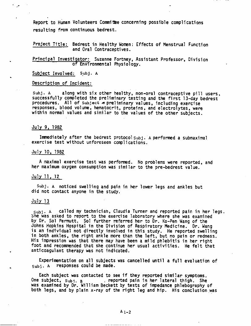
Report to Human Volunteers Commit lee concerning possible complications
resulting from continuous bedrest.
Project Title: Bedrest in Healthy Women: Effects of Menstrual Function
and Oral Contraceptives.
Principal Investigator: Suzanne Fortney, Assistant Professor, Division
of Environmental Physiology.
Subject Involved: Subj. A
Description of Incident:
Subj. A along with six other healthy, non-oral contraceptive pill users,
successfully completed the preliminary testing and the first 13-day bedrest
procedures. All of Subject ,_preliminary values, including exercise
responses, blood volume, hematocrit, proteins, and electrolytes, werewithin normal values and similar to the values of the other subjects.
July 9_ 1982
Immediately after the bedrest protocol Subj. A performed a submaximal
exercise test without unforeseen complications.
July I0_ 1982
A maximal exercise test was performed. No problems were reported, and
her maximum oxygen consumption was similar to the pre-bedrest value.
July II _ 12
Subj. A noticed swelling and pain in her lower legs and ankles butdid not contact anyone in the study.
July 13
Subj. A called my technician, Claudia Turner and reported pain in her legs.
She was asked to report to the exercise laboratory where she was examined
by Dr. Sol Permutt. Sol further referred her to Dr. Ko-Pen Wang of the
Johns Hopkins Hospital in the Division of Respiratory Medicine. Dr. Wang
is an individual not directly involved in this study. He reported swellingin both ankles, the right ankle more than the left, but no pain or redness.
His impression was that there may have been a mild phlebitis in her rightfoot and recommended that she continue her usual activities. He felt that
anticoagulant therapy was not indicated.
Experimentation on all subjects was cancelled until a full evaluation of
Subj. A responses could be made.
Each subject was contacted to see if they reported similar symptoms.
One subject, Subi. B . reported pain in her lateral thigh. She
was examined by Dr. William Beckett by tests of impedance phlebography of
both legs, and by plain x-ray of the right leg and hip. His conclusionwas
A I-2
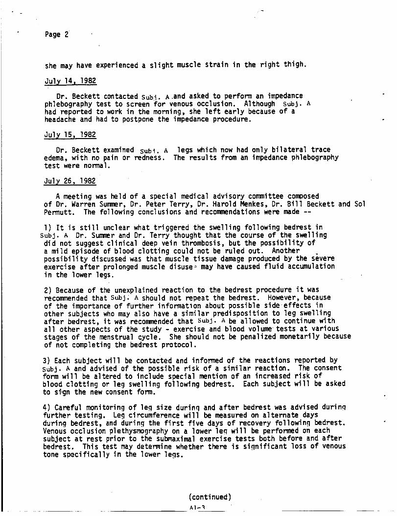
Page 2
she may have experienced a slight muscle Strain in the right thigh.
July 14_ 1982
Dr. Beckett contacted sub_. A.and asked to perform an impedance
phlebography test to screen for venous occlusion. Although Subj. Ahad reported to work in the morning, she left early because of a
headache and had to postpone the impedance procedure.
July 15, 1982
Dr. Beckett examined Subi. A
edema, with no pain or redness.test were normal.
legs which now had only bilateral trace
The results from an impedance phlebography
July, 26, 1982
A meeting was held of a special medical advisory committee composedof Dr. Warren Sumner, Dr. Peter Terry, Dr. Harold Menkes, Dr. Bill Beckett and Sol
Permutt. The following conclusions and recommendations were made --
l) It is still unclear what triggered the swelling following bedrest in
Subj. A Dr. Summer and Dr. Terry thought that the course of the swelling
did not suggest clinical deep vein thrombosis, but the possibility of
a mild episode of blood clotting could not be ruled out. Anotherpossibility discussed was that muscle tissue damage produced by the severe
exercise after prolonged muscle disuse-" may have caused fluid accumulation
in the lower legs.
2) Because of the unexplained reaction to the bedrest procedure it was
recommended that Subj: A should not repeat the bedrest. However, because
of the importance of further information about possible sideeffects in
other subjects who may also have a similar predisposition to leg swellingafter bedrest, it was recommended that Subj. A be allowed to continue with
all other aspects of the study - exercise and blood volume tests at various
stages of the menstrual cycle. She should not be penalized monetarily because
of not completing the bedrest protocol.
3) Each subject will be contacted and informed of the reactions reported by
Subj. A and advised of the possible risk of a similar reaction. The consent
form will be altered to include special mention of an increased risk of
blood clotting or leg swelling following bedrest. Each subject will be asked
to si,gn the new consent form.
4) Careful monitoring of leg size during and after bedrest was advised durinq
further testing. Leg circumference will be measured on alternate days
during bedrest, and during the first five days of recovery following bedrest.
Venous occlusion plethysmography on a lower leg will be performed on each
subject at rest prior to the submaximal exercise tests both before and after
bedrest. This test may determine whether there is significant loss of venous
tone specifically in the lower legs.
(continued)
A1 -_

Page 3
5) If in the future a subject complains of leg swelling following bedrest,a lOcc venous blood sample will be obtained to determine plasma creatine
phosphokinase levels. Thus we can test for the presence of possible
muscle damage which may account for leg swelling. Impedance phleboqraphywill also be performed to test for venous occlusion.
6) The initial medical history of all future studies will include specific
questions addressing the occurance of blood clotting disorders in the
potential subject or her immediate family.
7) Anticoagulant therapy in future studies was discussed and discouraged.
This option was also considered prior to the start of this study and
rejected because there is no information present about the efficiacy ofadministration of heparin to normal subjects during prolonged bedrest,
and because of the increased discomfort and added risk of frequent
injections (twice a day). A NASA consultant (Dr. Peter Bungo) who has
participated in bedrest studies agreed that anticoagulant therapy is
inadvisable during bedrest in healthy individuals. Elastic stockings
and leg exercise during bedrest were ruled out because these procedures
may alter the blood volume responses being studied in the protocol.
Leg massage was recommended if subjects complain of leq crampinq or tingling
during bedrest.
8) The consent form should also point out the additive risk of blood clotting
in subjects who use the pill and perform bedrest. Subjects who take oral
contraceptives have been shown (Am. J. Epidemiol. 90, 365) to have a
4.4 fold greater risk of pulmonary embolism than non-pill users. Thisadditional risk must be added to the risk of blood clottinq induced in the
bedrest protocol. (No data is available to evalute the risk of blood
clotting in normal healthy individuals confined to bed). Presumably, the
risk lies somewhere between the statistics of risk in normal ambulatory
women of child bearing age who suffer from blood clots in the legs, about
.22 percent/yr {jAMA 219:583); and women who suffer thrombosis in un-
Complicated but major surgery, 2% for deep vein thrombosis.
9) In summary, this particular subject was considered to be at increased
risk to repeat the bedrest portion of this study and will be asked not to
repeat bedrest. All other subjects will be advised of the possibility of
lower extremity swelling following bedrest. Although critical data has
already been lost pending the decision of the medical advisory committee,
it is recommended that the study continue on schedule as determined by
the menstrual cycle of each subject and the scheduling difficulties of the
bedrest procedure.
AI-4
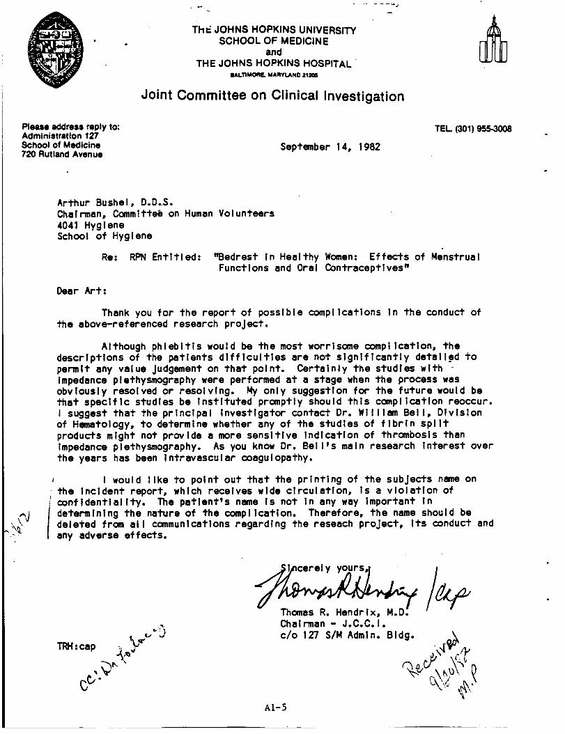
...... j
ThE JOHNS HOPKINS UNIVERSITYSCHOOL OF MEDICINE
andTHE JOHNS HOPKINS HOSPITAL
BALTIMORE, MARYLAND 21_
Joint Committee on Clinical Investigation
Please address reply to:Administration 127School of Medicine720 Rutland Avenue
September 14, 1982
TEl (301) 95_3008
Arthur Bushel, D.D.S.Chairman, Committee on Human Volunteers4041 HygieneSchool of Hyglene
Re: RPN Entitled: "Bedfast In Healthy Women: Effects of MenstrualFunctions and Oral Contraceptives"
Deer Art:
Thank you for the report of possible ¢ompllcatlons In the conduct ofthe above-referenced research proJect.
Although phlebitis would be the most worrisome ¢ompllcatlon, thedescriptions of the patlents dlfflcultles are not slgnlficantly detalled topermit any value Judgement on that point. Certainly the studies withImpedance plethysmography were performed at a stage when the process wasobviously resolved or resolving. My only suggestion for the future would bethat specific studies be Instltuted promptly should thls complication reoccur.I suggest that the prlnclpal Investigator contact Dr. William Bell, Divisionof Hematology, to determine whether any of the studies of fibrin splitproducts might not provide a more sensitive Indication of thrombosis thanImpedance plethysmography. As you know Dr. Bell's main research Interest overthe years has been Intravascular coagulopathy.
I would like to point out that the printing of the subjects name onthe Incldent report, which recelves wide circulation, Is a violation ofconfidentiality. The petlent's name is not In any way Important Indetermlnlng the nature of the complication. Therefore, the name should bedeleted from all communlcatlons regarding the reseach proJect, Its conduct andany adverse effects.
TRH =cap _ j_
Chalrman - J.C.C.I.
c/o 127 S/N Admln. Bldg. n_
AI-5

Appendix 2: Pulmonary Function Results
A2-1
\ _'_ /_
EFFECT OF PROLONGED BEDREST ON LUNG VOLUME
IN NORMAL INDIVIDUALS
W.S. Beckett, N.B. Vroman, D. Nigro, S. Thompson-Gorman,
J.E. Wilkerson and S.M. Fortney
Division of Environmental Physiology
School of Hygiene and Public Health
The Johns Hopkins Medical Institutions
Baltimore, Maryland 21205
Running Head: Lung function and bedrest
Address corres'>ondence to:
William S. Beckett, M.D.
Division of Environmental Physiology
The Johns Hopkins University
School of Hygiene and Public Health615 North Wolfe Street
Baltimore, Maryland 21205
(301) 955-3515
A2-2
ABSTRACT
Pulmonary function vas assessed in supine subjects before, during and after
three separate bedrest studies of 11 and 12 days duration. Forced vital capa-
city (FVC) increased during bedrest in each subject. Total lung capacity by
helium dilution was measured in one bedrest study and increased in each subject.
while residual volume and resting volume of the lung did not change. No change
in FVC was found in an ambulatory control group using identical measurement
techniques. Ha_ntalning baseline plasma volume during one bedrest by the use of
exogenous estrogen did not prevent an increase in FVC. and decreasing plasma
volume with diuretics in ambulatory subjects to the same degree as seen in the
bedrests Lid not cause an increase in FVC. We conclude that prolonged bedrest
results in a small, significant increase in maximum lung volume, and that this
change is not dependent on alterations in plasma volume.
INDEX TERMS: Pulmonary function, lung volume, bedrest
A2-3
mechanical function of the normal lung may be altered by changes in the
gravitational environment or by altering the body's position within the gravita-
tional field (3,4,12,14.15). Within minutes of going from the upright to the
supine position, lung volumes in normal subjects decrease, the largest change
being in the residual volume of the lungs at the end of a maximal expiration
(17). These postural changes in residual volume (RV) and to a smaller degree in
the forced vital capacity (FVC) can be prevented by maneuvers designed to ob-
struct the transfer of blood from the legs to the upper body -- such as placing
tourniquets around the thighs (16). Such i_nediate changes in lung volume are
caused in part by flow of blood from the lower body into the chest; they are
considered static changes in lung mechanics, and have been assumed to persis_
while the supine position is maintained.
Prolonged bedrest places the lungs at 90 ° to the usual upright relation to
the gravitational field for an extended period of time. It has been used-as a
model of a zero gravity environment to induce decrements in intravascular vol-
0
umes. orthostatic tolerance, and cardiovascular and skeletal muscular condi-
tioning such as those seen during zero-gravity spaceflight. Previous studies of
lung mechanics before and after prolonged bedrest have not detected significant
changes other than those seen i_mediately on lying down (9.19). The purpose of
the present studies was to re-examine whether lung function changes occur in
normal subjects during eleven to twelve-day bedrests.
Hethods and
Eighteen healthy adults (Table 1) gave informed consent to participate in
these studies; twelve in the bedrest studies, and seven in the ambulatory con-
trol and diuresis studies. 0nly one subject in the bedrest studies (number 8)

also participated in the ambulatory control and diuresis studies (number 14).
Informed consent was obtained from each subject, and the protocols were approved
by the Hu_,an Volunteers c_ittee of the Johns Hopkins Hedical Institutions.
Before and after each bedrest study and before the ambulatory control
studies absolute plasma volume and absolute red cell volume were determined by
radionuclide dilution techniques (technetium labeled human serum albumin for
plasma volume and technetium labeled autologous red blood cells for red cell
volume). Subsequent changes in blood volume were calculated from alterations in
hemoglobin and hematocrit (10). In each study, subjects received one t_aining
session £or familiarization with the techniques of pulmonary function measure-
ment begore the first control session.
I. Bedrest Studies
Subjects remained supine in bed for eleven days in the first two bedrests
(BR1 and BR2) and for 12 days in the third bedrest (BE3). Subjects did not
exercise while in bed, and were limited to no more than IS minutes in the seated
0
position per 24 hour period.
The first two bedrests, using the same subjects (No. 1-6, table 1), were
conducted consecutively, separated by a four week recovery period. In BR3 a
comparable group of six subjects (No. 7-12, table 1) completed the identical
protocol except that each received daily oral estrogen supplementation
(Premarin, 1.25 mg) to maintain plasma volume under conditions where a spontan o
eous diuresis o£ plasma volume would otherwise occur during the first few days
of bedrest, resulting in a persistent lowering of plasma volume for the duration
of bedrest.
In each bedrest study, two sets of control pulmonary function measurements
2
A2-5
were made on two days prior to the start of bedrest. Tests were repeated daily
during bedrest (at approximately the same time of day), one day after the end of
bedrese, and after two weeks ambulatory recovery. All pulmonary function mea-
surements, including pre-bedrest control and post-bedrese recovery measuremenes,
were made with subjects in the supine position.
Daily 4 cc blood samples for calculation of plasma volume were taken with
free flowing technique at the same time each morning within one hour of pulmon-
ary function tests. Each sample was analyzed for whole blood hemoglobin con-
centration (cyanmethemog l ob in technique in triplicate), hematocrit (microhe-
maCocrit Cechnqiue in triplicate), total plasma protein concentration (refrac-
tive index in quadruplicate), and plasma osmolality (£reezins point depression
in duplicate).
Forced expiratory volume in the first second o£ expiration (FEVl) and FVC
were tested using a Stead-Wells survey spirometer and technique meeting the ATS
Snowbird Criteria (1) each day during all bedrests. In BR1 and BR2, the two
largest values of three acceptable spirograms were averaged to determine that
subjectts FEV 1 and FVC for a given day. In BR3, the single largest values from
three acceptable spirograms were used as the subjectts FEV 1 and FVC £or a given
day. All spirograms including pre- and post-bedrest studies were performed with
subjects in the supine position and were interpreted by the same reader. In BR1
and BR2, single breath di£gusing capacity o£ the lungs £or carbon monoxide
CDLco ) was measured by the method of Yorster, et al. (I1), and slope of phase
IIZ of the single breath nitrogen washout test was measured by the method of
Craig ee al. (7), with subjects exhaling at a rate of approximately 500 ml • sec
-1 into a direct-reading nitrogen analyzer. Records were traced on an X-Y
3
A2-6
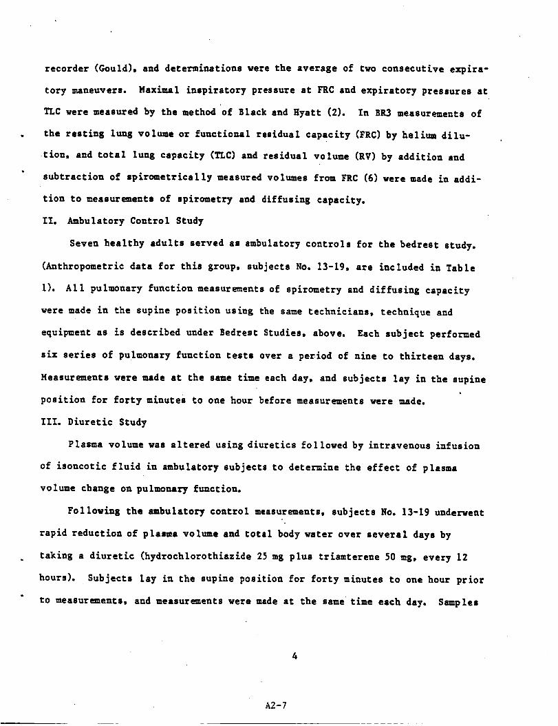
recorder (Gould). and determinations were the average of two consecutive expira-
tory maneuvers. Maximal inspiratory pressure at FRC and expiratory pressures at
TLC were measured by the method of Black and Hyatt (2). In BR3 measurements of
• the resting lung volume or functional residual capacity (FRC) by helium dilu-
tion. and total lung capacity (TLC) and residual volume (RV) by addition and
subtraction o£ spirometrically measured volumes from FRC (6) were made in addi-
tion to measurements o£ spirometry and dif£using capacity.
II. Ambulatory Control Study
Seven healthy adults served as ambulatory controls for the bedrest study.
(Anthropometric data for this group, subjects No. 13-19, are included in Table
1). All pulmonary gunction measurements o£ spirometry and di££using capacity
were made in the supine position using the same technicians, technique and
equipment as is described under Bedrest Studies, above. Each subject per£ormed
six series o£ pulmonary gunction tests over a period og nine to thirteen days.
Measurements were made at the same time each day, and subjects lay in the supine
position for £orty minutes to one hour be£ore measurements were made.
III. Diuretic Study
Plasma volume was altered using diuretics followed by intravenous infusion
of isoncotic fluid in ambulatory subjects to determine the effect o£ plasma
volume change on pulmonary gunction.
Following the ambulatory control measurements, subjects No. 13-19 underwent
rapid reduction of plasma volume and total body water over several days by
. taking a diuretic (hydrochlorothiazide 25 mg plus triamterene 50 mg, every 12
hours). Subjects lay in the supine position for forty minutes to one hour prior
to measurements, and measurements were made at the same time each day. Samples
4
A2-7
of venous blood (5 ml.) were drawn each day with care to maintaining horizontal
position of the arm. Analyses of the blood samples were performed as described
above under Bedrest Studies. Diuretic treatment was continued until plasma
volume loss of 15Z or more had occurred. (1-3 days on diuretic). On the last
day o£ diuresis, blood and pulmonary function tests were performed before and
i--_ediately after restoration of plasma volume by intravenous infusion of a
solution o£ 5 per cent human serum albumin suspended in isotonic saline and
warmed to 37°C. Diuretic administration was then stopped and measurements
repeated the following day.
For the bedresC, ambulatory control and diuretic studies, each dependent
variable was tested by a one factor factorial analysis of variance with repeated
measures across time Co determine ig bedrest or diuresis caused significant
changes (alpha = .05) from control measure=ents. If the o=nibus F indicated
significance then post-hot testing was performed on the ordered means by the
0
Neuman-Kuels technique. Student's t-test was used to compare inspiratory
pressures and expiratory pressures before, at the end of, and after bedrest.
For the diuretic study the six ambulatory control determinations for each
lung function test and for plasma volume were averaged for use as the control
value for each subject. I_mediate pre-infusion and one day recovery values were
tested together with the average control value for changes across time and daily
measurements of each lung function test were compared with that day's plasma
volume by least squares regression techniques. Pearson product-moment correla-
tion coefficients were calculated.
A2-8
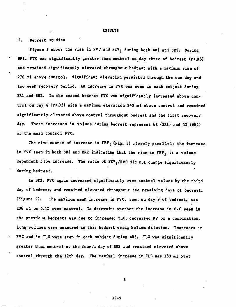
RESULTS
I. Bedrest Studies
Figure 1 shows the rise in FVC and FEV 1 during both BR1 and BR2. During
• BR1, FVC was significantly greater than control on day three of bedrest (P,.05)
and remained significantly elevated throughout bedrest with a maximum rise of
270 ml above control. Significant elevation persisted through the one day and
two week recovery period. An increase in FVC was seen in each subject during
BR1 and BR2. In the second bedrest FVC was significantly increased above con-
trol on day 4 (P<.05) with a maximum elevation 240 ml above control and remained
significantly elevated above control throughout bedfast and the first recovery
day. These increases in volume during 5edrest represent 6Z (BR1) and 3Z (BR2)
of the mean control FVC.
The time course of increase in FEV 1 (Fig. I) closely parallels the increase
in FVC seen in both BR1 and BR2 indicating that the rise in FEV 1 is a volume
dependent flow increase. The ratio of FEVI/FVC did not change significantly
D
during bedrest.
In BR3, FVC again increased significantly over control values by the third
day of bedrest, and remained elevated throughout the remaining days of bedrest,
(Figure 2). The maximum mean increase in FVC, seen on day 9 of bedrest, was
206 ml or 5.4Z over control. To determine whether the increase in FVC seen in
the previous bedrests was due to increased TLC, decreased RV or a combination,
lung volumes were measured in this bedrest using helium dilution. Increases in
- FVC and in TLC were seen in each subject during BP.3. TLC was significantly
greater than control at the fourth day of BR3 and remained elevated above
control through the 12th day. The maximal increase in TLC was 180 ml over
6
A2-9
control. One day and two-week recovery values were not significantly greater
than controls. The maximal inspiratory and expiratory pressures, an index of
respiratory muscle isometric strength measured at FRC for inspiration and TLC
for expiration, did not change significantly in BR1 and BR2 comparing pre-
bedrest controls to the last day o£ bedrest and day 1 recovery.
Changes in DLCO did not follow a consistent pattern_ _(hen all bedrest
values for diffusing capacity were compared with all control values by analysis
of variance, they were found to be significantly lower. The mean decrease in
DLCO during BR1 was -1.7 cc_nin-l_nHg -1 (P<.05) and during BR2 -1.55 cc'min-
lunnHg-1 (P<.002). However, day-by-day analysis of DLCO changes during bedrest
and recovery showed DLCO was significantly (P<.05) lower than controls only on
day 1 recovery in BR1, and on bedrest days 5 and 6 during BR2.
Figure 3 shows mean changes in plasma volume, total protein concentration
and osmolality during BR1 and BR2, demonstrating the plasma volume diuresis
which regularly accompanies prolonged bedrests. During the first bedrest the
mean plasma volume for the six subjects decreased a maximum o£ -20X (-487 ml) in
the first six days, and remained decreased from control values during the fol-
lowing days of bedrest. Similarly in the second bedrest, plasma volume de-
creased by -14Z (-317 ml) during the first six days and remained in this range
during the remaining days. Plasma volume was signi£icantly lower than control
(P<.05) on days 2-9 during BR1 and days 2-10 during BR2. At 24 hours after the
end of BR1 and BR2, plasma volume had returned to control levels. Total red
cell mass decreased by a mean o£ -5.9Z or -121 ml (not significant) during the
eleven days of BR1, and by -2.1Z or -50 ml (not significant) during BR2. Com-
parison o£ the increase in FVC in Figure 1 with the concurrent fall in plasma
A2-10
volume in Figure 2 suggested an association between changes in total plasma
volume and FVC.
These observations motivated the measurements of FVC in studies using
• slightly diZferent conditions. The first was a repeat of the bedresC studies
(BR3) under conditions identical to BRI and BR2 except that diuresis was inhib-
• iced by the use of exogenous estrogen. The second used controlled diuresis of
plasma volume in ambulatory subjects, (diuretic study below). No significant
overall change in plasma volume occurred in the BL3 subjects taking estrogen,
while FVC and TLC increased significantly as noted above.
II. Diuretic Study
Oral diuretics used in ambulatory subjects produced decrements in plasma
volume in each of the seven subjects comparable Co those measured during bed-
rest. The mean decrease in plasma volume in the seven subjects was -20.4Z,
significantly lower than control (Figure 4). During this induced diuresis there
was no significant change in FVC compared with control measurements (Figure 4).
The maximal decrease in plasma osmolality was -1Z (-3 m0sm • 1 "1) and maximal
increase in plasma protein concentration was 19Z (+1.3 EmZ). Infusion of the
saline and albumin solution after diuresis was completed increased plasma volume
by a mean of 832ml Co 3Z above control plasma volume. After reinfusion, mean
plasma osmolality was at 1Z below control and plasma protein concentration aC
10% above control. No significant relationship between plasma volume changes
and FVC, FEV1, or DLCO was detected by linear regression analysis of all con-
. trol, diuresis, re-infusion, and one day post-infusion values. FVC and plasma
volume during diuresis are plotted against time in Figure 3. The lack of change
" in FVC during diuresis is contrasted with the increase in FVC found during all
8
A2-11

bedrests (Figures 1 and 2).
DISCUSSION
The mechanical relationships of the static lung volumes requires that the
significant increase in FVC found in the three bedrest studies must result
either from increased TLC, decreased RV, or a combination of the two. The
finding of increased TLC in BR3 with no significant change in FRC indicates that
all or most of the increased FVC is due to increased TLC.
Explanations for the increase in FVC and TLC can be divided into three
groups: increased neuromuscular force to inflate the lungs, increased chest or
lung compliance, or a change in the mechanical relationship of inspiratory
muscles to chest wall allowing more negative pleural pressures and greater
inflation of the lungs per unit of tension produced by the inspiratory muscles.
Increased neuromuscular force may occur either through increased maximum
neural output to the inspiratory muscles or by augmentation of the maximum
musclar force of contraction to maximum neural stimulation. Some studies of
I
spirometry in normal subjects have shown small increments in FVC on serial
testing which are attributed to a training effect, with better performance
through familiarity with the procedure or conditioning of the inspiratorymus-
cles. Three findings of the present studies suggest that neither a learned
response in the performance of spirometry nor a muscle conditioning effect can
account for all of the observed increase in FVC. First, a significant increase
in FVC was found in BR2 in the same subjects already familiarized with the
procedure from each completing over 50 apirograms during the BR1 measurements
several weeks before. Second, an increase in FVC due to conditioning of inspir-
atory muscles would be expected to be accompanied by increased isometric muscu-
9
A2-12

lar force, which would be reflected in increased maximal inspiratory pressure
when breathing against an occluded mouthpiece from FRC. No change in maximal
inspirator 7 pressure was seen during either BR1 or BR2 in these subjects.
, Third. when the same techniques of measuring supine FVC were followed in a
similar group of ambulatory control subjects, no increase in FVC was found.
" indicating that the measured increase in lung volume in the present studies was
related specifically to bedrest and not to a training process.
Increased compliance of the chest wall or the lungs in the presence of
unchanged neuromuscular force would also result in increased FVC and TLC; Al-
teration in airway tone. interstitial water, mechanical properties of the inter-
stitium, or elastic properties of the chest wall as a result o£ bedrest alone
appears unlikely. Hpwever, changes in lung volumes due to alterations of cen-
tral vascular engorgement with blood, leading to alteration in pulmonary com-
pliance or a displacement of air spaces, may occur. Buono (5) has demonstrated
that increasing central vascular engorgement either by head-out water immersion
or by C-suit inflation to 80 torr about the calves and thighs of normal subjects
results in decreases in FVC o£ -S to -6Z below control. Conversely, Potanin et
al. (18) have demonstrated increased vital capacity by applying -30 Torr lower
body negative pressure. Because a spontaneous diuresis of plasma volume during
bedrest was associated with increased FVC. it was suggested that a decrease in
central vascular volume associated with diuresis might account for the increased
lung volumes. However. induction of a comparable plasma volume loss by diure-
tics in ambulatory subjects did not aiter lung volumes, indicating that a 20Z
decrease in plasma volume is not alone suf£icient to produce the changes in lung
- volume seen in bedrest.
10
A2-13
Demonstration'of these changes repeatedly in the same subjects of BRI and
BR2, with a return toward control levels in the interval between bedrests, sug-
gests a gradual process independent o£ muscular force or strength. Without
change in either neuromuscular output or chest compliance, a shift in the ana-
tomic relationship of the inspiratory muscles to the chest wall that improves
their mechanical advantage in generating lower pleural pressure without in-
creasing their maximum tension may have this result. Such anatomical adjust-
ments, occurring gradually over several days after the acute decrease in lung
volume on going £romthe upright to supine position, would represent an opposite
and approximately equal compensatory change to the initial postural change in
lung volume. The present results would support such a hypothesis, but do not
indicate the mechanism for such a gradual compensation.
These changes were not seen _n one previous study of pulmonax7 function in
four subjects during bedrest (8). although small non-significant increases in
FVC and TLC were found in another five healthy subjects after 20 days bedrest
(19). Our study of a larger number of subjects or the use of more consistent
measurement with subjects always supine may explain why these changes were seen
in the present but not the previous studies.
_n conclusion, a small increase in maximum lung volume (TLC) demonstrated
during prolonged bedrest in normal subjects appears to be a true alteration in
lung mechanics and not the result of decreased plasma volume alone.
ACKNOWLEDGEMENTS
The authors gratefully acknowledge the technical assistance of Louis
Wilkinson, Michael Soaper. Thomas Cross. Debble Gerke. and Clarke Tankersley.
11
A2-14
This research was partially supported by NASA. N_H grants HL 10342 and HL
07534. and NIOSH.
12
A2-15

REFERENCES
I.
.
ATS Snowbird Workshop:
Dis., 199: 831, 1979.
Standardization of Spirometr 7. Am. Rev. Resptr.
Black, LF, and RE Hyatt. Maximal respiratory pressure: normal values and
relationship to age and sex. Am. Rev. Eespir. Dis. 99: 696-702, 1969.
. Bryan, AC, WD MacNamara, J Simpson and HN Wagner. Effect of acceleration
on the distribution of pulmonary blood flow. 5. Appl. Physiol. 20: 1129-
1132, 1965.
4' Bryan, AC, J Millc-Emili and D Pengelly. Effect of gravity on the die-
tribution of pulmonary ventilation. J. Appl. Physiol. 21: 778-784, 1966.
.
o
Buono, MJ. Effect of central vascular engorgement and immersion on various
lung volumes. J. Appl. Physiol. 54: 1094-1096, 1984.
. Meneely, AR, and NLgaltreider. The volume of the lung determined by
helium dilution. Description of the method and comparison with other
procedures. J. Clin. Invest. 28: 129, 1949.
. Craig, D, W _ahra, H Don, J Couture, and M Becklake. Closing volume and
its relation to gas exchange in seated and supine positions. J. Appl.
Physiol. 31: 717, 1971.
13
A2-16

. Dietleiu, L and W 2udy. Exper_nental M-2 cardiovascular conditioning. In:
Manned Space Flight Experiments, Interim Report, Gemini V Mission. Nation-
al Aeronautics and Space Administration. Washington, D.C. January 6,
1966.
. Dietrick, JE, GD _hedou, E Short, V Toscani and VB Davis. Effect of
immobilization upon various metabolic and physiologic functions of normal
men. Am. 2. Med. 4: 3-35, 1948.
i0. DxlI, DB, and DL Costill. Calculation of changes in volumes of blood
plasma and red cells in dehyration. 2. Appl. Physiol. 37: 247-248, 1974.
11. Forster, RE, WS Fowler, DV Bates and B Van Lingen.
carbon monoxide by the lungs during breathholding.
1135-1145, 1954.
The absorption of
J. Clin. Invest. 33:
12. Glazier, 2B, J_4B Hughes, 2E Maloney, and JB West. Effect of acceleration
on alveolar size in the lungs of dogs. Aerosp. Med. 39: 282-288, 1968.
13. Michels, DB and JB West. Distribution of pulmonary ventilation and per-
fusion during short periods of weightlessness. J. Appl. Physiol. 19: 212-
216, 1964.
14
A2-17

14.
15.
Milic-Emili J, JA Henderson, MB Dolovich, D Trop and K Kaneko.
distribution of inspired gas in the lung. J. Appl. Physiol.
1966.
Milic-Emili, J, J Mead, and JM Turner. Topography of esophageal pressure
as a function of posture in man. J. Appl. Physiol. 19: 212-216, 1964.
16. Mills J. The influence upon the vital capacity of procedures calculated to
alter the volume of blood in the lungs. J. Physiol. London. 110: "207-216,
1949.
17. Moreno, B, and H Lyons. Effect of body posture on lung volumes. J. Appl.
Physiol. 16: 27-29, 1961.
18. Potanin, C, S Morehead, WWNeblett, and BC Sinclair-Smith. Respiratoryi
mechanics vitb lover body nesative pressure (LBNP) a simulation of the
erect posture. Aerosp. Med. 40z 1101-1104, 1969.
19. Saltin, B, C. Blomqulst, JR Mitchell, RL Johnson, K Wildenthal, and C.
Crapman. Response to exercise after bedrest and after training. Circula-
tion 38 Suppl VII: VII-1 through VII-78, 1968.
20. Savin, CF, AE Nicogossian, JA Rummel, and EL Michel. Pulmonary function
evaluation during the Skylab and Apollo-Soyuz Missions. Aviat. Space and
Env. Med. 47: 168-172, 1976.
15
A2-18
21.
22.
West, JB and FL Matthews. Stresses, strains and surface pressures in the
lung caused by its weight. J. Appl. Physiol. 32: 332-345, 1972.
West, JB and CT Dollery. Distribution of blood flow and ventilation-
perfusion ratio in the lung, measured with radioactive C02. J. Appl.
Physiol. 15: 405-410. 1960.
o
16
A2-19

Figure 1:
Figure 2:
Figure 3:
Figure 4:
Mean forced vital capacity of the lungs (FVC) and mean forced
expiratory volume in one second (FEV 1) for six subjects as a
function of time measured daily during pre-bedrest control (C),
bedrest days 1-11, one day post-bedrest recovery (R1), and two
weeks post-bedrest recovery (R2). Circles indicate days on which
values were significantly (P<.05) greater than control during the
first bedrest (BR1), and squares days on which values were greater
than controls during the second bedrest (BR2) in the same six
subjects.
Mean total lung capacity (TLC) forced vital capacity (FVC) and
residual volume (RV) og the lungs of the six subjects in the third
bedrest study (BR3). Circles indicate days on which volmne w_s
significantly greater than control (P<.05). There was no signifi-
cant change in RV.
Mean values for absolute plasma volume (ml), plasma oemolality,
and plasma protein concentretion (Em%) for the same six subjects
as a function of time during bedrest 1 and bedrest 2. Circles
indicate days on which values were significantly (P<.05) different
from controls during bedrest 1, and squares during bedrest 2.
Forced vital capacity (FVC, mean ± S.E.M.) and absolute plasma
volume (PV, mean % S.E.M.) for seven subjects during pre-diuresis
control measurements, after diuresis (prior to re-infusion of
17
A2-20

plasma volume), izmed_ately after re-infusion of plasma volume,
and one day after re-infusion. PV, but not FVC, vas signifi-
cantly d£_ferent from control after diuresis.
18
A2-2l
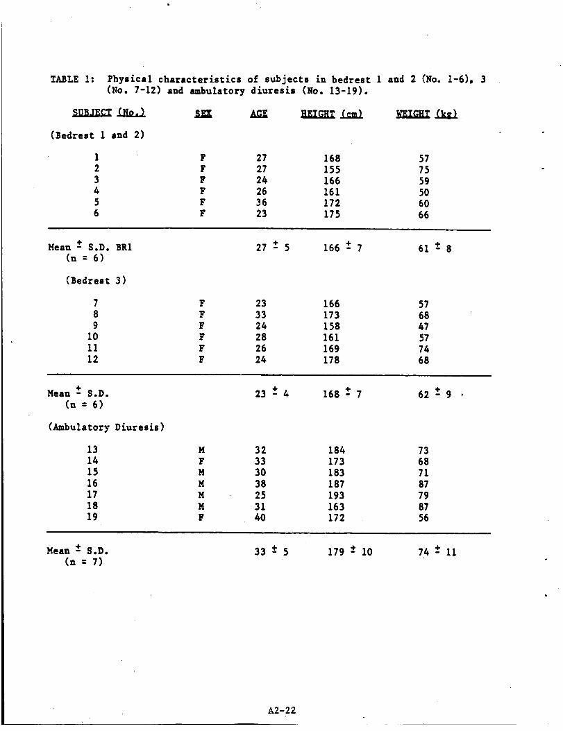
TABLE 1: Physical characteristics of subjects in bedrest 1 and 2 (No. 1-6). 3(No. 7-12) and ambulatory diuresis (No. 13-19).
(Bedrest 1 and 2)
1 F 27 168 57
2 F 27 155 753 F 24 166 594 F 26 161 505 F 36 172 606 F 23 175 66
Mean ± S.D. BR1
(n = 6)
(Bedrest 3)
789
101112
27 ± 5 166 ± 7
F 23 166 57F 33 173 68F 24 158 47F 28 161 57F 26 169 74F 24 178 68
61+8
Mean + S.D.
(n = 6)
(Ambulatory Diuresis)
13141516171819
M
F
M
M
M
M
F
23 +4
32
33
30
38
25
31
40
168 + 7
184173183187193163172
62+9 .
736871
87798756
Mean -+ S.D.
(n = 7)33-+5 179 -+ 10 74 ± 11
A2-22

•--'---- BR I
......... BR2
Oay-of Bedrest
A2-23

.= _= _o-
_. 3.7--
1.7-£
._=1.6
U 1.5
1,1.1 '_• a
1.4-b,Ia. ¢,¢
°i 0.11-
o p< -05
: BEDREST
0 0 0 0 0 0 0 0
p
J I I I I I I I I I _'_'_I =_C I 2 3 4 5 6 7 8 9 I0 II 12 RI RZweel
DAY OF BEDREST
A2-24

2500 -
uJ2400
..J0> 2300 -
<C-_Em 2200 -
.J0.
2100 -Z
uJ2000 -
1900
290
280
0 _ 270< 0_ E_ v
..1Q,.
Z0i
<E
ZuJ(JZQ
*Zii1p.O
-n-o.
<{
co<{._1
-----. Bedrest 1
_ ...... "'" Bedrest 2 a
O, 0 0 0 O 0, ...............
o P<.05 BR1
p<.05 BR2
Plasma Volume
o o o o o o o o o
_. o o • • o o
_eoeoa
0 0 0 0 0 0 0 0
Osmolality
o o o o o o o o o7.ar I _..R::..._ z>....._ ......(_....
" "" Protein
_oe. °e
7.6 ." ""7.4
7.2
7.0
6.8
Control 1 2- 3 4 5 6 7 8 9 10
Day of Bedrest
g2-25
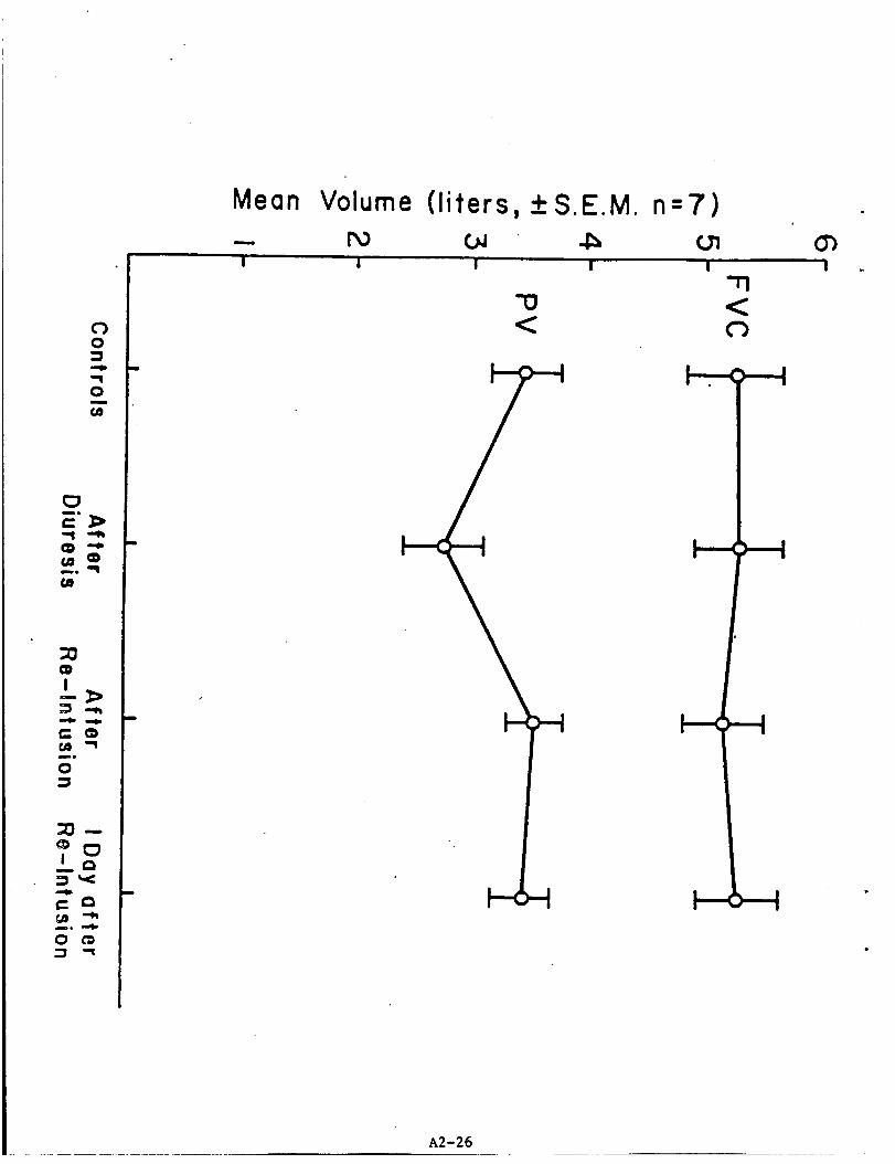
C")0
q m
"1
0
{3elllo
{e
(DI
q ee
t,A "Imo
0
i
illlll ill
mo
0;_ -'1
Mean
!
Volume
P_I
(liters, _+S.E.M. n=7)
I I I
-I"1<0
I _-_ II
I
A2-26

Appendix 3: Publications from
this Contract
A3-1
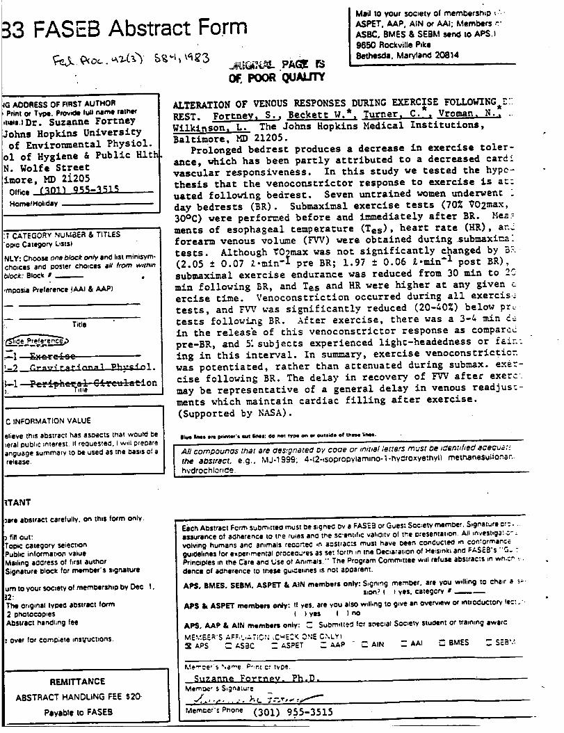
93 FASEB Abstract Form
PAre rsPope QUAIJTY
Mail to your society of meml:,ersr, p _'.-
ASPET. AAP, AIN or/_.1; Members .'."
ASBC, BMES & SEBM send tO APS.)
9650 Rockville Pike
bthe ,a. Ua l=.a20814, =m •
IG ADDRESS OF FIRST AUTHORPdnt or Tyl_e. Prov,_ fuU name rather
,t..S.) DE. Suzanne Fort:ney
Johns Hopkins University
of Envlronmental Physiol.
ol of Hygiene & Public Hlth
N. Wolfe SCreet
imore, HI) 21205
Office (301) 955-3515
Home/Holiday
:T CATEGORY NUMBER & TITLES"opt¢ Category Usts!
NLY: Choose one block onh,,lind list minisym-choices an0 poster cho=ces a# /ram wom,nOlock: Block #
,mDos,a Preference _AAI & A.AP)
_ ) Title
.8-1 ---E_.:refco
C INFORMATION VALUE
elieve this abstract has aspects that would be_eral pubhc =merest. If reauestecl. I wtil prepareanguage Summary to De usecI as the Das_sOfarelease.
ALTERATION OF VENOUS RESPONSES DURING EXERCISE FOLLOWING E;_
REST. Portney, S., Beckett _.*_ Turner_ C.*_ Vroman. N._
_ilkinson t L. The Johns Hopkins Medical InsCicutlons,
Baltimore, MD 21205.
Prolonged bedrest produces a decrease in exercise toler-
ance, which has been partly attributed to a decreased card±
vascular responsiveness. In =hls study we _ested the hypc-
=hesls that the venoconstric_or response _o exercise is a::
uated following bedrest. Seven untrained women underwen= _
day bedrests (BR). Submaxlmal exercise _ests (70_ VO2ma_,
30°C) vere perforT.ed before and immediately after BE. _le_
ments of esophageal temperature (Yes) , heart _a_e (HR), a_3
forearm venous volume (FVV) were obEained during submaxi=a;
=es_s. Al_houBh TO,max was not significantly changed by 5;:
(2.05 ± 0.07 Z.min -¢ pre BR; 1.97 = 0.06 _.mln -I pos= BR),
submaximal exercise endurance was reduced from 30 min to 25
min following _R, and Yes and HR were higher a= any given
ercise time. Venoconstric_ion occurred during all exercis,_
=es_s, and FW was significantly reduced (20-40_) below p_
Ces_s follo_in_ B_. A_er exercise, _here was a 3-4 min d_
in the releas_ of chis venoconstric_or response as compar¢c
pre-BR, and _ subjects experienced light-headedness or _a±_
log in _his in=er'val. In summary, exercise venoconstrlc_ic_
was potentiated, ra_her _han a_tenuated during submax, e_e:-
cise follo_ing BR. The delay in recovery of _rV after exe:c
may be representative of a general delay in venous readjusc-
men_s which maintain cardiac filling af=er exercise.
(Supported by. NASA).
Blue II_es m pfmtM s _ ires: (Io _t _ on or outside of those tim
All com_ouna$ [nat are aes:gna£e# by cage or tort/at letters m_..sr De _Cemff_e_ acecua::|
the abstract, e.g., MJ-1999; 4-(2-_sopropylamlno-l-hy,roxyetnyl) metnanesulion6n.hvdrochlortCe.
TANT
)are abstract carefully, on thnS form only.
fill out:
ITol:_ category seiect,onPublic informat_op veiueMailing a0(:ress of l,rst authorSignature 0lock for member's s_gnature
um to your socnetv of rnemOershipby Dec 1,]2:The orig_nel tyDecl eostriK:t form2 photocop,esN=strect henClhng fee "
._over for comb;ere 0ns_ruct=ons.
REMITTANCE
ABSTRACT HANDLING FEE S20
Payable to FASEB
E,_chAbstract Form su0m=tted must be s;g,_e_ ov a FASE_ or Guest Sac:ely member. S,gnature c::.el_;urance of a0herence tO tl_e rules ana ¢_e sc:ent,f,c vaho=tv of t_e presenter,on All _n'vest,ga: c- _-voIwng humans anO annmals reo_.":eC ,n aDstrac:s must have been conOucteO on conformancegu=0elines for experimental proceo_;res as se'. forth ,n me Deciarat_on of Heis=nk=and FASEB's "'5. ":Principles in the C_re ano Use at AnamalS.'" The Program Carom,tree w,|l refuse abstracts =n wn,c, ÷.dence of aclrterenceto thesegu=_,ehnes_snot aDDarent.
APS, BMES. SEBM. ASPET & AIN members only:S_gn,_g memDer, are you willingtO Cheera _--smart?L I yes. category • ____
APS & ASPET member= only: If yes. are you also wdhng to give an overwew or _nttO=uctory leo'_--( I yes i ) no
APS. AAP & AIN members only: _ Subm,tte"J for s_ec0a! Sac;ely student or traon,ng aware
MEMEER'S A;,=,:.:,T;C'; iCWESE ONE C,%LY)_E' APS _ AS@C _-- ASPET .- AAP - _ AIN _. AAI [_ BMES -- SEB'..'
Me".-'e,s %a'_e _:_nt Cr type.
Suzanne ForttlCy. Ph.D,Memoe, S S,gnatu-e
Memccf's Phone (3011 9.55-3515

!r " ' / / ASPEToAAP AINorAAI Mern0ecsof
u L.ic u form . ,ASBC, 8MES & SEBM send to APS.!
; " i. '_ # # 9650 Rockville Pike ;
L_SOFF_RST AUTNOn PLASMA VOLUME RESPONSES DURING EXZRCZSE FOLLO_,qNG BEDREST.
._,_ _o_fulln=,_,at,w W. Beckett*. 5. For_nev, C. Turner*. and N. Vroman*. The
-|/l£am S. Beckett Johns Hopkins"Medical Inscl_ucions,'Baltimore, _[D-"-'----'_1205.
_viron=en_al Phyciol. Al_hough decreased exercise tolerance fo_lowing prolonged
:_ Hopkins Unlversic7 bedresc has been a_rlbu_ed to translen_ dluresis; resul_inc
• | Rygiene & 9ubli¢ Hl_ in loss of plasma volume (PV), i_ is unclear whether factors
_lfe St:. • o_her than hypovolen_La, such as changes in plasma proteins or_ .L_ 21205 capillary permeability, affect t:he ¢ou=rol of PV during exe=-
bedrest, a change occurs not only in [:he resting plasma vol_=
but also _n the ra_e of £ntravascular fluid loss during exer-
else. Exercise studies (70Z _02_mx, 30oc, 40Z rh) were per-
:Y _UMaE% & TITLES formed on 7 untrained women be£ore and after duplica_e 12-de,:I.lstsl
bedrests separated by I mon_h recovery. Absolute red cell vol-:e one :10¢_ _ and list m,n_svm-_:=:s_ cno_es ,///fore,,,,_ ume (ROY) and plasma volume (PV) were determined using Tech-:,,____ • netlum-labelled red cells and albumin before and after bedres"
_,_,ence,_41a AA_) Relative changes _n plasma volume during bedrest and exercise
were det:ermined from changes in hemat:ocrlt: and hemoglobin con-
cen_raClon. PV siEnlf_cant:17 decreased 18.5 z 5.8Z during
bedrest 1 (BKI), a_d 9.5 ± 4.6?. during _he second bedrest (E.'<_Llle
wi_hout: significan_ changes in RCV or plasma osmolallt:y. Our-£ng pre-bedrest: exercise, PV decreased 13.3 __ 1.3 and
_rcise 12.6 ± 1.3X and 9.3 _ 1.0 and 10.4 Z 4.3X following bedres¢.
Al_hough t:he Y. loss of PV was similar during exercise, the e_-
solute loss was less post:-BR. _e conclude that: al_hough BR re-
sults in hypovolem/a, specific effects of BR on _he control o-
PV dur_n E exercise are minimal. (Supported by NASA).
'-_vi_a_ional Physiolo
ood volume regulauior
T, lie
i
i i
|-" ? 3.N '.'ALU Ei
I:%:'_-."_as asoects _,_t woul_ be
"'_.'es: _f ,eoueste_. I w_ll ;:regare
,''4"v :o :e us_ &s lr_e Oos_s o| a
J me aOstrect, MJ-1999:e g,
I_yc_,rocntoncle.
• ; . .II/ue _o o_e i_ow, em Ib_ou de _ h,pe e_ o_ o_ne0do ef tilth lien
i
All compounds met are ciemgna_ec/ ov cooe ot mmal /et_e_ must t_e _cient_fiec' aOeOuete:/ ._"
4-[2-,soorooylarmno-l-hyOroxvetnyl) met_anesul|onan,.=- i
P
r. :ItSfullv. on th=S form only.
Se -_.'t:cn
IP'4: :, " _alue
_.;1 :' !,rS: au[,0r
_':':¢ 'or memoer S s=gnaturo
I •'_,: .,'v ol _eml=ersmO 0v Dec. 1,
"':-"_J d=slract form
It • -_ •.
REMITTANCE
':'_ _:,;.bLING FEE S20
.... e to _ASE8
ORIGINAL PAGE I_
Poor QUAUTV
Each Al_strect Form suom_tte_ must I:e s,gned o,_ a FASE_ Or Guest $ocetv memoer. S_gnoture orov,oe •.
asl_ro_:e O| a4necence to "he ru_eS an(: me sc_ent,hc va:,(:,tv of tl_e cresentet_n All _nvesbgat;t:ns ,,'-
v(]iwng .umans and emma,s re=erie4 _n a0strac'.s must f_ave t_een c0n(:_ucte(: _n conformance v..:.-
(_,l_del_$ |of exoer.mental or0(:eaures as set latin _n one 0eclarit_on of he_s_nk_ and FASE_'s "Gu,C ,.:
PfmCiOles =n (,e Care an(: Use at An,maiS '" ,"he P_=gram Comm,:'ee wdl refuse aOstracts ,n wmcn e_,-
dence Of aonerence to mess gu,cel,nes ,s not a0oarent.
APS. |MES. SEBM, ASPET & AIN mer_bers only: 5 on,no mem0er, are you _.lling to ¢na,r I ses-
SJOn_ ( I yes, Category • __
lP$ l ASPET mlmbltl on,v: If yes. Ire you ltSO w,lhng tO g,ve an Overview or _ntr(:_uc_ rot:rule;( ) yes ( I no
AP$. AAP 8, AIN members only: -- 5uDm_:eO for SOeC,al Sot)err SluOent or tratn,n 9 eward.
MEMBER'S AFF:LI'_T]C', ,CHEC_ O,_E O_LVl
APS - AS3C -- ASPET - AAi = =AIN _ AAI _ 8MES _ SEB.%I
_'e_=e: s Nan-e Pr,e_ .-r ,'_-'.e
Suzanne Fortne?_ Ph.D.
MemOer's 5 _na_u,e
-- ;Mem_er's/Phone (_01) 055--_61_ 9i
i •
A3-3
ORIGINAL PAGE
OF POOR QuALrrY "
PL_SMA, VOLL._OE RESPONSES DURI_ 8EDRZST IN _EALT_ ;_.M_
S. Fortney, H. Drew, end _. LeFrancs
The Johns Ropktns Medical InStitutions
Decreases in plam no1--- {PV) occur during
spaceflight which could potentially compromise an_cronaucs' blood pressure maintenance during re-
entry. Such losses of PV are ui,_,l_y attributed
Co • cephalic shift of body pacer during Icy
gravt_/ exposure, and subsequent stimulation of
Icy-pressure bar•receptors which initiate 8 di-
uretic response (3). The pu_-poso of ch£s study
was to critically examine PV responses in healthy
women during simulated weightlessness (bedrest).
Ve tested the hypothesis that the loss of PV
during bedrest (BR) is influenced by menstrual
function, specifically by the water-retaining
properties of plss'_e estrogens.
Six women 23 to 37 years of age who did not
use oral cont_aceptives gave cheer Informed con-
gents and underwent cvo il-day periods of on-
end on the hematocrits obtained on the f£rst dayof BR.
The technettu_ test results ere shown in the
following table. Red cell =ass did not signific-
antly cha_ge du=ing either B_, e_chough there was
s progressive tendency towards docseas£ng levels
during the eight-week program. Plasma voices,
which significantly decreased an average
17._ + 2.1% during BR1 and 9.3 + 1.7% during BRi,
were not significantly different from pre-gR
levels 24-48 hours after BR. Such findings agree
with the rapid recover7 of PV reported in earlier
n studies (5).
Table 1
Plasma and Re__d Cell Vo!L_es Before and Within
_8 hours after Bedresc (mesn:S.L, n=6)
forced BR, where each BR period was separated by _t¢_.e.1.)four weeks of --bu_acor'y. recovery. A seventh
woman participated in the first bedrest (BRI),
but was prevented from parclcipaclng _n the sec-
ond bedrest (3R2) because she unexniainably de-
veloped pronounced lo_.'er lee. ede_ a couple days
after B_. The possio_itty of such c_--ticacions
foll_ing spaceflight was predic:ed recently by
Eargens ec el. (i)
Bedrest procedures were timed co occur at
opposin 8 stages of the :menstrual cycle for each
vo_n. Menst=-.tal cycles were divided into fot_
separate stages v'hers -
Stage 1 - cycle day 2 (day l=firsc day of
bleeding) until 6 days prior co ov_lecion
(ovulation was est£mnced from -_oru£ng cenpers-
Cures end by counting beck 14 nays fro= day 1).
This is • stage of low estrogens and progester-ones.
Stage 2 - five days prier to ovulation until
one day after ovulation. _urin_ thls stage there
is 8 large estrogen peak while plasma progestsr-ones remain low.
Stage 3 = _o to 9 days after ovulation.
FJtrogen and prozescerone concentrations both
gradually increase.
Stage _ = con days after ovulation _ncil
cycle day 1. Estrogens and progescerones g_ed-
uslly decreese.
Each morning of 3R, a 3=1 venous blood
sample was drw,-n :o deteru£ne relative changes in
PV during BR (from he=ecocrlt and hemoglobin con-
centrations), total pro:sin concentration (re-
fractomeCry), end plasma csmo_ality (_reezing
point depression). Periodic 1Oct blood samples
were drawn.to _onicor blood estrogen and prog-
esterone (rsdioi---_-unosssey), and morning tem-
peratures and persona_ diaries yore ob=ained tomonitor mensc_'_'1 function.
Prior to each BR, and v_thin 48 hours afterB_, absolute Red Cell ','olu=e (_CV) and PV were
detez--'-ined usln_ rsdioac:i':: d_luc_on _echnlques
wlth Technetiu: (Tc)-labeile_ red blood cells
end Tc-label}ed Human Seru._ Alb,._..inas :_,e labels
respeccivel.v. Absolu'-e ?',' _u:tn; e_¢h Ca:: of "=R
was calcula:ed b_sed ".'_ :he T_-_e:e.-_.._:ed ._'; and
her.scurries •0rained ourir._ the ?.'e-_ TC :es:,
Pre-_R1 Posc-BR1 ?re-BR2 Post-BR2
1419:57 1371:52 1366:37 1301:_2
PVp,;;;&_) 2305_143 2247_107 2257_121 232&:72
During BR, blood hemacccrit increased sig-
niiicantly, plasma osmolal:t_" remained u:,altered,
and total protein concentre:ton increased sig-
nificantly during BR1 buc not during BR2 (see
Fig. 1) "_'
tO*_}
_Y_t. _KR|m (O**eI4)
" _'l'" -['i-_Trr-r'_ ':to
Ime_me qj4}
! II :_ 4 • • ? • e 10_5 r_
Plasma volume decreased re?idly during the
first _-5 days of BR, then remained fairly stable
•during the lest 7 days of B_ (see -ig.2). Also
during BR, there was a net loss of Total Circul-
atory Proteins (TCP-PV tL'_es the :otai pro:sin
concentration), and Tctal Circuiatory Os=o!s
(TOO-plasma water times p_esma osmolality).
o- :_,
.,_ _.._ _.,_._.z .._.._,, .,_ "'_'l_'*'z'6.'_''_ I
.:[
_lty IP I_O/'ebt_
A3-4

The 1on (pez_auc decrease) of ?V during thef£ra¢ five days of BR v_8 analyzed In tens oi
_r._uaA cycle stage (sea FAg.3). The loss of -_/
duZ'lJ_g gg days 1-5 yes progressive and shoved a
s_ar pattern for each VOmln _t_o began S& vhila£u mm_ct'ual cycle stage" i, 3, o¢ 4. H:vever, in
t_:ee *,mmnvho began BK _'hlle An cycle stage
2, there vlus a :tansies: aCteu,_,cicm in :ha loss
o£ IV. ZlluJcracsd An FIKu=e 3 are r_e relative
--losues'ot PY In the sLz umm= d_- S BR2. The
dotted Zines illuecraca ?V responses for :he 3
mn in scare 2, chile the solid llnes re_:esenc
respo_aas for _he ocher three vo_tn.
D_-lng SPA none ot the rouen vere ;n _cage 2,
earl a 1_ had 1Yf responses sllAJ.ar to che _¢l_.d
_,, Ft4u:e 3.
Pm'_:em
0
• _'#o,%
OAY OF BF.D_EST
Nmc_,l_ J_tta fro= Sg days 5-12 vere analyzed
in terms of menstrual stage. The e_fecc o_ BR on
FV at this par_.od yes =in_l (see Fi&.2). Dur-
ing _J_e last 7 days of B&, ovulation occurred "
durf_ sLx B_ periods. Figure 4 illustrates the
FV responses in these uomen, _ne_e days 0-6 re--
present the seven days =rlor co ovuiac_on vi=h
day 0 calculated as ovulation. Fluctuations in
PV vere calculated by subtracting :he :san PV for
a given subject durir.g earn 7-day period, _rom
her cLIAly PV. The d-ca vere centered in :hAs
manner in order co adjust _or di_erlnx absolute
levels of PV aeons sub._eccs, thus enabling visual
comparison of changes In PV durlng the days pre-
reading ov_lacion_ For each sub,etc, a transientincrease in PV occurred £_ this interval before
ovulation. These changes _.n PV _ere analyzed
through a _ao-_ey snal_'sis of variance, vhere ob-
servacicms rare ¢lsssi_led by su=ject arm byday
1_rior t= ovulaclon. _nsp_,:clon of _ha dace aug-
seated testing for a quadraclc response (i.e., a
ce_orar_ fluc=_ation f_o_ baseline) in _his ia-carrel before ovulaclon. ,'hl calculated F-value
of 4.36, based on 1 and 30 degrees of freedou,
si_uAflcanc _Ath P < 0.05.
om,
e S • ; I ' :
ORIGINALPAGE
OF POOR qUALm'
Ve began this study v£ch the hypothesis chat
elevated blood estrogens, _usc prior to, and after
ovulation vou4d atten_utte or reverse the decreases
£n PV seen during 3_. _e postulated chac as long
as blood estrogen concent_a_Lons mere elevated,
the£r vacet-recalnAc_ proper¢£es vould oppose thed_Lureclc e_feccs of BR. We fa£1ed hovever to
accotmc for _he crans_anc nature of cha vacer-
reca£_tn$ properties of estrogens. Several au-thors (3, 4 ) have studied r.he effects of esc=o-
ge_ on body flu£d responses. E_CrOgen ad_Ln-
_stratAon consistently resu.lts in a reduction In
_£uat'y excretion of sod£,,- and chloride a_d re-
ducts urinary volm=e. Preede and Aickan (_) scud-
Lad urinary excretion during e_ght days of dailyL_Je_t£ous of estradiol in vO=en, b_lle there yes
an increased vacsr retention during chs first five
da_s of treat=eat, on the sixth day the _Ldney
"escaped" fro_ the e_¢ccs of estradtal, and a
dl_zns£a occurred. Such an escape pheno=ena =ay
help to explain vhy only traosleuc changes in PV
occurred in ch£s study durln$ periods o_ expected
prolonged elevation _o blood es:rogens, Thus cha
vater-reca£nln_ effects of estrogens vere only
e_dant during _ens_rual cycle stake 2. _hers an
inAtlal large, transient rise in estrogens =tour
_,, most _o=en. During s_ages 3 and _, the small-
er, and slower rise fn estrogens was much less
e_fect£va ia prevenclo4 the decrease _n PV assoc-l_cedvich S_.
_n conclusion, :he overall PV responses seen
in chase co=an during _R vere of a similar =ag-
nAcude as that repor=_d for =nn during _o-_eek
bedfast procedures (2). Only small ::ansienc
flu:cuaCions in PV occurred _ust prior co ovula-
tion vh£ch :ay have been due co the influence of
_ncreaslng blood estrogen clears. Therefore, the
procec:ive (vacer-reca_n!ng) effects of estrogens
on PV losses duri=; velgncless conditions are
probably only small a_d =tam, lent.
*The authors gracefully acknovledge the stacis-ci:al asslscance o_ Lavrence _oulcon.
Su=porced by ._ABA.Re_erences "
1. _argens, A._., B.J. Tucker, and C.H. TlpCon.
1982. Fluid shifts in vascular and extra-
vascular compartments of _u=ans durln_ and
after simulated veighclessness. Physiologist_:S63-S64, Suppl.
2. Greenleaf, J.£., E.H. Bar, suet, H.L. Young,
J.T. Horse, R._. Scaley, L.T. J_hos, and
_. Va_ea_onc. 1977. Fluid and elec_roly¢e
shifts during bedfast vlch _somecric and
isotonic exercise. _. Appl. Physiol.:_espl_
"Environ. Exercise Physiol. _2:_9-66.
3. Pi,_-e, T.E. 1968. Body fluid volu=e and
renal relacinnsh£ps co gravity. _n: _v___-d_-n--,,_cs _nd Hvoo=ravics, ed. _. HcCally,
Acad. _ress, pp. 133-ibi.
4. Preed}, J.A.K. and E.H. Aicken. 19_6..'he
effect of estrogen on racer and electrolyte
mecabolis=. 2. Ciln Invest. 35: _23-A29.
5. Taylor, H.L., L. £rlckaon, A. Renschelo and
A. _eTs. 19_5. The e_ecc of bedresc on the
blood volum,.e oi _o.-....al Foun$ =an. A=. J.Physiol. 1A_:227-;32.
A3-5
rtAL;| MUbt hr. .:lv¢
:RIL 1£'34 F/:,SEB ABSTRACT i'tJ _'i IVI ASCBe APAOiv._ ,,:_,_ send to ASPEt.BMES. SEAM, SOP E. SM5 send to APS. SOT
|elegy, Pharmacology, Pathology Et Nutrition) . ,._C_, member= send to AiR.• g650 Rocky.fie Pike
oFPOORQUALm # i q ='
ING ADDRESS OF FIRST AUTHOR=e Prmt or Type. Prowde furl name ratine
,Indlals.I • . •.tam S. Beckett, M.D.ronment al Physiology
LS Hopkins University School
[y giene and Public Health
:_u_ Wolfe St., Balto. Md.Of(ice301-955-3515
e: HomelHohday 301-235-6956
!CT CATEGORY NUMBER & TITLES, Topic Category Ustsl326-1 Gravitnr_onal
_25-I _J_chaniis of Breathing__ [_enera±)
330-I Pulmonary Ven=ilatlon
No. Title
-.'SENT PREFERENCE (CHECK ONEI
Poster session _ Slide session
nisyml:)Osium I-1 Indifferent
_aldecision regarding presentation format is atdiscretion of the progrlming society.
3LIC INFORMATION VALUE
If requested, I wiit pre'l_a_ a lay language_rnary to be used is the basis of a press release.
EFFECTS OF ALTERATION OF PLAS_tA VOLUME ON Pt_MONARY FUNCTION.
W. Beckett*_ N. Vroman* S Fortn_v, and J Wilkeraon,, P • • •
The Johns Hopkins Hedical Insti=u=ions, Baltimore, Hd. 21205
In a previous study of prolonged bedrest we demons=rated
a 15Z decrease in plasma volume due to spontaneous diuresis,
¢oncomltant wich a sta_Istically significant increase in
forced vital capacity (FVC) of about 200 ml and decrease in
diffusing capacity of the lun 8 (DLCO) of 2 cc/min/mmHg during
the firs_ 3 to 4 days of bedfast. Both changes resolved
rapidly after bedrest. These findings suggested that de-
creased pulmonary vascular volume migh_ account for these
changes-by increasing lung compliance and decreasing capil-
lary perfusion. In the present s_udy we cested _he hypoth-
esis Ehat changes in plasma volume can cause hl=era_ions in
lung function including FVC and DLCO. Control to_al red cell"
and plasma volumes were measured by radionuclide dilution in
seven heal_hy adults. Control measuremenEs of hema_ocri=,
hemoglobin, protein concen_ra=ion and osmolality, FVC and
DLCO were taken after one hour supine res_ on six normovol-
emic control days, following plasma reduc¢ion wi_h diure=ic,
•and after plasma volume restoration by infusion of an iso-
"Eonic saline and albumin solution. Plasma volumes calculated
from hemoglobin and hematocri= decreased by a mean of 20%
during _he dluresis, while protein concentration and osmol-
allty were no_ changed by dluresis or infusion. FVC and DLCC
did no_ change significantly from controls fol!_ing either
diuresis or re-infusion of plasma volume. Thus changes in FVC
and DLCO wl;h bedrest are no_ due to decreased plasma volume.
_ Mtl I_mtM'S Out lines: _10 not type _m _ mltskle of th4mo ifmes.
I AII compounc/s mat are designated Oy cocle or _nma/letters must be i<len:ihecl aClocuate/,//,_ tthe abstract, e.g., MJ-1999; 4-(2-iSOl_rOl:)vlamino-l-hyClroxyethyl) metnanesulfonanihce_
hydrochloricle. , , . I
ORTANT ..
repare abstract carefully, on this form only.
_,o fill out:
Topic category selectionPubhc reformat=on valueMailing adclress of first author
it. Signature block for member's signature
return to your sc_:ety of membersh=D by r,,_c. 1.1_3 :_. The or;o;nal tyoeO aDs!rat! fo:m
:.. A_stract har,c_:ng fee
over for complete mstructions.
REMITTANCE
ABSTRACT HANDLING FEE $20
• Payable to FASE8
E41chAbstract Form submitted must De s,gneo by a FASEB or Guest Society member. S,gnature prey=desalsurance of adherence to me rules anO the sc:entihc vakd=tv el the presenter,on. All ,nvest,gat=ons _n-voivmg humans and anlma!s re!_orte0 ,n abstracts must have been conductecl ,n conformance w,t."guidelines for ex_e,menta! moceouros as set forth =n the Declaration of Helsmk=and FASEB's "'Gu*o;ngI%ri_ci_es in the Care and Use of An,mass.'" The Program Committee wdl refuse abstracts =nwh,Cn ev,-¢lence of adherence to these guidelines =snot apparent.
AP_, BMES, SEBM, APA Div. 28, SGP. SMB, ASPET Et AIN membe_ only:.
Signing member, are you willing tO chair a mann? ( ) yes. category #__
AP_ & ASPET members only: I _v.._ ,re you also w,!iing to give an overview or int':.dactory lecture _I , yes ( I no
APS, AAP/,, AIN members only: C'_ Submitted for s_ec:al See:ely stu,lent or. _rain,ng award.
MEMBER'S A;F!L;,:,T=ON :CHECK 0_;- ONLY):APS [:3 AS.=C _ ASPET [_ ,:,AP _ AIN I-I AAI r'l ASCB [3 BME_. [_ SEBM
!"1 APA Div. 28 _. SGP _ SMB _ SOTSubmiss:on of signed form :nd=c=tes acceptance of rules including final w'it;'_drewal date c'
December 14. 1_.P_3.
Suzanne Fortney. Ph.D. . ._embt:r's Name. Pr:nt or tyGe.
301-955_3515
_,lemne,'sP.on_ A3-6
t,
JT
.m
.i
I
1984 ABSTRACT SUBMISSION FORMAerospace Medical Association
Topic No. (see reverse sic_e): Ist 5 2nd 2
Mode preference (check one): Slide Poster x Either
Name and _M_ for_m_s_de,=:
The Johns Hopkins Universit7
School of Hyg. & Public Health
At=n: S.M. Fortney, Room 7032
615 N. Wolfe Street
Baltimore, MD 21205
Td_: (301) 955-3612 or 3515i
I. Follow roles---includingformat--in Call ,or Papers onrever_ side.
2. Subm/ssions not meeting requirementswillbe rejected.
3. _ abstractwith care and submit on _hisform. Donot erase; use correctionfluid when needed.
4. Type only withinblue box.
5. Use elecu'ictypewriterwith elitetype or largerand only
clear,black ribbon.
ORIGINAL AND5 COPIES OF
THIS PAGE MUST
BE RECEIVED BYNOVEMBER 4, 1983
DO NOT FOLDH_
MAIL FLAT!!!!!
A. Nicege_tanPrqram Chairmen
Aerespeca Medical As-
uw_tionWashington National
• AirportWashington, DC 200QIATlrN: Abstracts
I aflh'm thatthisisoriginalwork which will nm
appear elsewhere before the meeting. I hereby grant
permissionfor the Aerospace Medical Associationto
taperecordthe verbalpresentation(ifa slidesession).
6. Use the followingformat:a) Title in CAPITAL LETTERS using significant,de-
scriptivewords.b) Authors (initials then last name) underlined.c) Institutionwhere work was done.
d) Indent first line of text by 3 spaces.e) Rest of text is a single para m'aph, single spaced, with
no photos, and no type smaller than elite. (Printingreduction size will be 60% of original.)
DEVELOPM_ OF TOLEI_NCE TO REPEATED BEDKKb£5. S.M. _ortney, W.S.
Beckett, and N.B. Vrom_n. Johns Hopkins Univ., Baltimore, >_ 21205.
Intro: Deconditioning, as measured by reduction in maximum oxyge_
uptake (VO2MAX), usually occursfollowlng exposure to welghtless
conditions. We examined this deconditionlng response following pro-
grams of si=mlated weightlessness (bedrest) to test for l)an effect
of a previous bedrest exposure on the deconditioning response to a
second bedrest, and 2)for a relationship between initial fitness
level and the degree of deconditioning followin_ bedrest. Methods:
Twelve women, with VO2MAX from 26.3 to 45.g ml/min/k_, performed du-
plicate ll-day programs of horizontal bedrest. Each woman began the
second bedrest (BR2) about 6 weeks after the start of the first bed-
rest (BRI), and between bedrests resumed normal daily activity, vo2
MAX was measured before each BR, and after I day, 2 weeks, and 4
weeks of recovery from BR. VO2MAX was determined as no further in=
crease in VO 2 (42 ml/_in/kg) with increase in exercise intensity.Results: One day after bedrest VO2_L%X (ml/min/kg) decreased I0.0 r
2.5 per cent followin_ BRI, and 4.0 _ 2.3 per cent following BR2.
The decrease in VO2MAX was significant (P4D.002) following BRI bu_
not following BR2 (P_O.12), despite almost comple:e recovery (to
within -0.9 ± 2.4 per cent) of pre BRI VO2MAX level prior to the
start of BR2. The relationship between Pre-BR fitness (VO2MAX in
ml/min/kg), and the decrease in VO2MAX (per cent change between Pre-
BR and Post-BR VO2MAX) following BR, was not siEniflcant; where the
regression for this relationship was Y= -O._9X + 8.3, K= -_).31. _on-clusions: The observation tha: there was a smaller reduction in
=
following the second bedrest ,_i.e., less decondltioning) indicates
che development of tolerance.
COMMITTEE USE ONLY:
Abswac_ No. AccepVReject Slide/PosterQuA .r,ry.
A3-7

',
1985 ABSTRACT SUBMISSION FOOM,,._ Aerospace Medical Association '. !/
_F pOGR _':" Topic No. (see reverse side): lit ,5 2nd 2
Mode preference (check one): Slide _ Poster __ Either
Name and address for concspondence:
Suzanne Forcney, Ph.D.
School of Hygiene and Publlc Health
of the Johns Hopkins University
615 North Wolfe Street -7032
wBal_imore, Maryland 21205ork Telephone: 301-955-3515
1. Follow rules---including format--in Call for Papers onreverse side.
2. Submissions not meeting requirements will be rejected.
3. Prepare absu'act with care and submit ON THIS FORM.Do not erase; use correction fluid when needed.
4. Type o_.v within blue box.
$. Use electric type',writer with elite type or larger and only_I.o._i°_ ribbon.
I afFum thaE this is original work which will notappear elsewhere before the meeting. I hereby grantperm/ssion for the Aerospace Medical Association totape record the verbal presentation (if a slide session).
6, Use the following format:a) Title in CAPITAL LErTF.RS using significant, de.
scriptive words.b) Authors (initials then last name) u_rtlned.¢) Institution where work was done.d) Indent f'LrStline of text by 3 spaces.e) Rest of text is a single paragraph, single spaced, with
no photos, and no type smaller than elite. (Printingredu_ion size willbe 60% oforiginal.)
THIS PAGE AND5 PHOTOCOPIES OF
THIS PAGE MUSTBE RECEIVED BY
NOVEMBER 2, 1984
DO NOT FOLD!H
MAIL FLAT!!:!!
Send twJ. Ro_ DIIJe
Program Chairmen
aoraspoce Medical As-_clation
Washington NationalAJrperB
Washington, DC 20001ATTNs Abstracts
PLASMA VOLUME MAINTENANCE DURING BEDREST ZN I_OMEN RECEIVING
Pt EMA IS. £./G. Rock. S.S. Lco.mm L.Johns Ropkins Medical ZnsCituCes, Baltimore. Earyland
21205.
_,lJZr_Lq_J,.QR: Decreases in plasma volume have been reportedin both men and women during simulated weight:lessness (bedresc),and are believed associat:ed witch t:he decrease in exercise and
ore:host:stir tolerance follm,-ing spaceflight. _n a previous st:udyve reported an attenuat:ion in plasma volume loss during bedfast
during the _diat:e pre-ovulat:ory phase, which we suggested may beaC.Cr£but:ed Co t:he vat:st-retraining properties of est:rogen, in t:his
phase of t:he meuse:fuel cycle. In the present st:udy, we lure:herst:udied t:he effects of female sex ,t:eroide t:o prevent bedresC
hypovolemLa_ METRODS: Nineteen healt:hF women (21 Co 39 7rs)
underwent a 12-day horizontal bedrest:. Plasma and Red Cell Volumeswere determined using t:echnet:iun-labels before and after bedrest Co
verify red cell mass stability during bedrest. Daily plasma
volumes were calculated from hemat:ocrit and hemoglobin ratios.Twelve women did - *.o. receive eat:rosen supplement, vn£ie seven
addiCiona.l v_n received 1.2_ Praarin daily. _ Plasmavo_u_e s_uificant:ly decreased by an averse - 20.1 z 3.9Z between
the firsC and last da_+ of bedrest: in Che non-premarin group, andwas not: reduced (+0.3 - 2.8Z) in the premarin users. J_._I_L_:Premarin, an estrogen-containing preparation, e_fect:ively preventedthe decrease in plasma v_lume associated witch bedresC, and may
serve as a useful nodal Co study the cole of t:he decreasing plasmavolume on post-bedrest responses. Support:ed by NAS9-16703
COMMITTEE USE ONLY:
AbstractNo. . Accept/Reject Slide,/Posler
A3-9 !

_BSTRACT MUST BE RECEIVED AT SOCIETY OFFICE BY
VlONDAY, DECEMBER 3, 1984 IAPRIL 1985 FASEB ABSTRACT FORM I
I
DO NOT FOLD THIS FORM
Moil to your Imcmy ol __ (APS. ASBC,ASPET, AAP, AIN or AAI.) ASCE mem_o,jto ASPET; BMES. SEEM 6 SMB mom_o_ lendto APS.
9O6O Rockv_e P_ISet,es_. Mar_ 20814
MAILINGADDRESSOF FIRSTAUTHOR TOTAL LUItG CAPACITY INCILEASES DURING PIOLOHGED "BEDKES(Please Print or TyI_ Provide full name rathet Z][_]_]_][TDE_' 0F [_JLBHJL VOLUI{E CRJIJ[;ES. S. _ _ f,_n in_w..) _ and S. Z£X/JI£_. Johns Hopkins SCress.Physiolog:Susan Thompson Gorman Laboratory. Baltimore. liD 21205.Johns Hopkins School of We have previou.sl7 =horn that Jigu.if£cant increases i_Rygiene and Public Health forced vztal capaczt7 (FVC) of approxzmaCely 6.Z occur vhelRoom 7032, .615 North Wolfe St. using prolonged bedfast as a model for spacefl_ht. Zn th,
present study, seven healthy adult females we.re =tudie,Baltimore, MD 21205 during 12 days of bedfast to cl.ar£fy the mechanism of th£zPhoneoff_e (;301) 955-35]:5 increase..FVC, funct;-onal resxdual capacill:y (.FRC), total
:Nome/Ho,c_y (;301)823-3917 . lung capaczCy ('_C), resxdmL1 vol_ (:RV), and szoEle breaC:d£ffusinS capacity for carbon monoxide (Dv.C0) were measure,on eva ambulatory control days, during e[ch day of bedrest
PRESENTATIONPREFERENCE(Checkone) one day post-bedre=C, and after two reeks recovery. AI_Ce=t.s were done with the subject _n the supine position
Poster " _ Slide r-1 Minisyml::)ol_um Subjects received exogenous e=Crogen (1.25 rag/day). Plan=;
O Indi,am_ volume (measured £mmediace17 before bedre,t by technetim
Fin,I _ec_n _ing bmsemat_n _ _ label, and calculated daily fro_ henatocri.t and hemoglob_at me discretion of the !orograming Socmy. values) dzd not decrease significa=Cl7 durzng bedfast as £
had in the previous stud£es without estrogen. A significan
(p < .05) increase in FVC occurred by the third day oSELECTCATEGORYNUMBERS_TITLES bedfast which persisted throughout the rest of the study(SeeTol:icCetagoryListsl with a maximal mean increase of 206 ml (5.6¢). TLC ale
£ncreased slgn£ficantly by the fourth day of bedfast an10-MIN.ORALOR POSTERPREFERENCE remained elevated through day 12, vith a maximal mea
• _-_ _ech_nics of Breachins increase of 180 ml. Post-bedrest TLC values were not signl__ urav_cac_ona_ ficanC17 d£fferent from control at one day and two week
3. _ recovery. There was. no slgn£ficant change in DtC0, _V anNo. Tit_ 1_C. These results _ud_cate that the _ncrea=e in'YVC £s tb
result of an increase in TLC and usa a decrease £n _V, anMINISYMPOSIUMPREFERENCE that Chess mechanical changes are not dependent on changes i_. _ plasma volume. (Supported by NASA).
No. Title ..............................................................
ff AAI Topio List is U_:I. Choose one D/oct on/y
8n_ list minisvmbosm c_oices and I:_ostar _oces _ _ m ram,', _ _,.; m _ _ m _ _ _ _ _ ORIGINAL PAGE I_
,,_ fro,',,_m,,_ _,,,,, _w.*: e_:_ # OF POOR qUALITY
PUBLIC INFORMATION VALUE
7_ If rl_lUeSleCl, I will breDare a fly langulge sum-
ma_ to be use_ as the bern of a Dress mlesse.
A# coml_oun_s rher are designated by code or/n/r/a/lertem must be idemified adeOuareiy/n the at_tmc_, e.g., MJ-1999; 4-(2-isoproloylamino-l-hyclroxyethyl) methanesulfonaniliclehydrochloride.
MPORTANT
P.mloare abstract carefully, on this form only.
Also fill out:
a. Topic category selectionb. Public information value
c. Mailing aclOrus of first author
d. Signature block for member's signature
Return tO your SoCiety of mernberehil0 by
Dec. 3, 1984:
a. Tl_e original tyDecl abs_recl form
h. 1 Dhoto¢oDy
AI0strect handling fee
See OVer for complete instruct,ons.
REMITTANCE (NONREFUNDABLE)
ABSTRACT HANDLING FEE $20
Paya01eto FASEB
All in_oattSlttom m_ adhm to the guidelines o8 listod on the reverse of the abstract form.
AIDS, BMES, SESM. SMB, AsPrr It AlN member_ only:
Signing member, are you willing to chair a sossion_ ( ) y_, category I
AIDS, AAP It AlN membem only: _ Submitted for sm_cml Society student or training 8ward. "
MEMBER'S AFFlUATION (Check one only):
:_APS [] ASBC _ASPET ,_.AAP r't AIN [] AAI _ASCB _ BMES 1"1 SEBM _ SME
Submission of signeci form indtatm icceptance of rules including final withdrilwll dill of December 19. 1984
St,4.Z,_nnc 19|_ _/--'O_lP..'/_.,.f
Name. (Print or tYPe.) J
-M ber,= s,gna,,,re / -(301_ o,;.-.- __'_
MemDer's Phone .......
i
1985 ABSTRACT 5UBMISSION.JFORMAerospace Medical Association
and address for coa_ I affirm that this is original work which will notJohn E. Davis appear elsewhere before the meetin8. I berry Irruptton of Environmental Phystolo87 pamission for the Aerospace Medical Association to
johns Hopkins Untversit7 tape record _e verbal presentation (if a slide session).
jc electric typewriu_ with elite type or larger and only_r. black ribbon.
_ae _de. a) Tide in CAPITAL LETTERS using silnificant, de-bauuious not meedng n_luiremenu will be m]ecuKl. ¢ripdve words.
abstract with care and submit ON THIS FORM. b) Authors (initials then last name) ur_lerliaed.no( erase;use correction fluid when needed, c) Institution wher_ work wus done.
o_y within blue box. d) Indent fuss line of text by 3 spaces.e) Rest of tzxt is a single paragral_, single spiced, with
no p_xos, and ao _pe smaller _sn eliw. (Prin_ngreducuon size will be 60_ of originaL)
FOLE£1M VENOCONSTRZCTOR ILESPONSES FOLLOWII_G BEDREST, 3.E.N.B. ____ __0 _S Johns Kopkl.ntMedical Inst:iCuCions, SalCinore, lid 21205.
We have previously observed tha¢ 8inulaced weight-lessness (Sedrest) augmncs resting and exercise venoconaCricCLou,u measured by decreases in fortaru venous compliance (FVC). This
vls aCCrilmCed Co a lov pressure beroreceptor reflex response cobedrest (Bi0oinduced hypovoleuia, The purpose of the present in-vestigation v88 to determine if maintaining plasma volume (?V)during Bi vould rtsulC in an attenuation of resting and exercise
vanocoustrictiou, _RODS, FVC yes meMured (venous occlusion ple-
t:hygmography) in 19 healthy yemen !21-39 yr8) at rest and during 30
minutes of cycling (70% VO2maz) _n a varm environment (30"C, 40gILK) before and ¢fter Bit, Seven subjects vere given hurin (]P) Co
prevent Bit-induced hypovolemia, sad 12 subjects received noPreuarin (NP). PV yes calculated from hemg¢ocrit ratios and tech-
netium dilution determinations. _LS_T_F._. PV, prior Co exercise,decreased crier Sl in 1_ (2389 to 1957 t 317 el) but not ia ? (2567Co 2537 ¢497 hi). letting FVC yes reduced after Bm in lit (3.72 to2.97 :!:o8 cc/100cc) and in P (3030 co 2029 _06 cc/100cc). Veaocou--
eCr_ction during the first I0 minutes of exercise vas greater afterki_m,K=O001 lSl for I_P (2o18 ¢o 1.64 +..7 cc/lOOcc) and P (2.14 to 1021 +_.06__ cc/100cc). £1Chough _V van maintained in P after BR, there was a
ffreater veaoconscr_cCioa in P than lrP at rest (30.6Z to 20.2% +_6Z)and during exercise (_% Co 25% .+6%)0 q__._. VenoconsCrictioafolloving Sl is independent of l_V euggescinl tl_c ocher variables,e.g, peripheral pooling, ms7 contribute to the greater venocon-strictiou. Supported h_ lq_q._-167030
4MITIT_ USE ONLY:
?
.i
iS PAGE ANDOTOCOPIES OFS PAGE MUSTRECEIVED BYEMBER 2, 1984
NOT FOLD!!!
_L FLAT_!!!
ue_
ki_eten National
rot No. AccepUReject Slide/Poster
A3-11
m ram..--_____

ABSTRACT MUST BE RECEIVED AT SOCIETY OFFICE BY
MONDAY, DECEMBER 2, 1985
APRIL 1986 FASEB ABSTRACT FORM
DO NOT FOLD THIS. FORMm
Mail tO your _ of membetlh_ lAPS,,
ASPET, AAP, AIN or AAtL ASBC metal=era send
to AIN; ASCB members send to ASPET; 8MEg
end SEaM membe_ _ to APS.
Rockville Pike
Bethe_la, Maryland 2(NM
MAILING ADDRESS OF FIRST AUTHOR
(Plane Pnm or TyI_. Pfovicle full frame tither
than ,nitials.)
Suzanne Fortney
Johns Hopkins School of Hyg.615 N. Wolfe Street, I_=. 7032
3alclmore, Maryland 21205
Phone:Om=e (301) 955-3515
Home/HOJidly (301) 574-0793l
PRESENTATION PREFERENCE (CheCk one)
MinisymDosium _ Sii0e _ Poster
r'l Indifferent
Final 0tristan regarding presentation format s at
the dcscrst=on of the grogreming society.
SELECT CATEGORY NUMBERS _ TITLES
(See Too=c Category Lira)
MINISYMPOSlUM PREFERENCE
i( No-- Tit,,
IIO-MIN. SLIDE OR POSTER PREFERENCE
I 72_..-I T-Id=_t- & ,_er4=_2 727-I _p_=p3. 735-I Temperature/Regulation
No. Title
IfAAI To_i¢ I._ is Use¢l.Chooseone Z_ockon/),an¢l hat mmtsymDo$1a ¢ho_es an_ pOSter ¢hOlCei
all from w/rh/n ,_me blOCk: Block O __
or ,I, ylcT or .zv zn r..m xams as ]=znzca
3A ]_.¢k. Johns Ropkin8 Stress Physiolol_y Lab, Baltiamre, -MD.
One proposed ez_lauation for the lover 8veati=q_ response
of women than of sen, is that estrogens inh£bit sweating. Te
test this hypothesis and to explore other potential estro-
gen£c effects on exercise responses, 5 women cycled with and
without 7-10 d=ys pretreatnent with a _tcural estrogen sup-
plment, 1.25 mg pre==rin per die_ Etch subject exercised(70: V02max) for 30 minutes in a vat'= (30°C, 60: _) envi-
ro=nent, l;on-pre=arin tests (1_) occured in the follicular
phases of 2 subjects, the luteal phases of 2 subjects, and
in both phases of the menstrual cycle of 1 subject. Esopha-
geal te=pera_ures (Tea) did not differ siEnifimantly between
NP and pre_uarin (P) tests durin_ exercise, nor was there a
siEnif_cant difference in the rise in Tel; 1_=1_5 +_ 0J_l°C,and P=l.07 ± 0_39°C, mean +_ S_ No significant differences
were seen in mean skin re=pars=urea, skin conductanres,
total body =west losses, Tee matizq_ thresholds, the slopes
of the Tee/sweat rate relationship, heart rates, strokevolune8, or blood lactates. Plam=a volume did not differ
siEnificantly between _P and P at rest or during exercise.
We conclude that estrogen suppl_=entation with _re=arin at
this do=age does not alter ther=ore_ulatory re=pauses during
moderate exercise in • war= enyiro=nent either by J_hiblting
=wetting, or by altering plasma volume. (Suppo=ted by 1_$9-
17199 .)
abe _m me g_mm_s ¢_ _: _e _ _me on m mm_e _ _e88 hu_
PUBLIC INFORMATION VALUE
If reouested, I will ampere a _ Mlngulge aura-
mary to De -,sea as the Oasis of a Dress steles.
!VlPORTANTI
; Prt_are ao=rac_ carefully, on th_ foffn only.
Also fill out:
a. Topic category selectionb. Puioi=c reformat,on value
c. Mailing a0orHs of hint author
d. S=gnature Dlock for member's s_gnatum •
Return tc your society of memoe,Jh|o .byDec. 2, 1_5:
a. The Orrjmal tyoe,, a0strac form
10nO!O_ODv
AOstrac: nan0hn,; fee
See ave, foo coma=eta ;nS_ruCtlO_S.
i
! REMITTANCE (NONREFUNDABLE)
ABSTRACT {..,IANDLtNG =_; S_=
I
I
A// compounds that are o_ignated by code or initial iarrers mus_ Oe iOentifia_ aOeouate/in the abstr#ct, e.g., MJ-1999; 4-(2-isopropylsmino-l-hyclroxymnyl) metl_anesulfonaniliclhyclrochiori0e.
All immstill_tors must 8dbo_ to the guicieiinas as listed on them of the abstract form._
AIDS, BMES, $EBM, AsPrr bAIN members only:
Signing member, are you wilkng to che:r a session= (X) _. c=mWry # _Z23.-1v
APS, AAP b AIN mMnberl only: _ SuDmitte¢l for specml Society StUdef_ or tra,ning awa_l.
MEMBER'S AFFILIATION (Check one only):
_P APS ._ ASBC _ ASPET ._ AAP ,'I AIN _ AAI _ ASCB ':..."BMES _ SEBM
Sumimien of _Ined form _ eaoel_nnee of i _ _1 _1 _ of _ 111.11_.No _0_8 will be
Suzanne For_nev
Meml_ s Name. (Pnn_ or tyDe.)

RACT MUST BE RECEIVED AT SOCIETY OFFICE BY
)AY, DECEMBER 2, 1985
'RIL 1986 FASEB ABSTRACT FORM
DO NOT FOLD THIS FORM
Mail to your society of memOwllh_ (APS,ASPET.AAP,AIN or AAJ).ASBC _ sendto AIN; ASCB memberssend to ASPET;BMESim¢l SEBM memberssend to AP_.
RockviilePike
LING ADDRESS OF FIRST AUTHOR
Pnnt or Type. Provide full name ritl'_rmmals.)
f-cabet h H:[.escher
_=us Hopkins SchooZ of HyEien
5 N.. Wol£e Screet, R=. 7032
L_Cmore, Har71and 21205
of{ce (301) 955-3515
_: Home/Hoiic_ (301) 574-8464
i
IENTATION PREFERENCE (Check one)
linisvmDosium _ SliG_e [_ Po_"
_liffemnt
deci_on regsrt:ling Dresentition format is it
_¢rmmn of the ptograming sooity.
ECT CATEGORY NUMBERS 6' TITLES
SYMPOSIUM PREFERENCE
No. TiUe
IN, SLIDE OR POSTERPREFERENCE'29-1 _eac & Stress
" 1'5_-I =""""t qPT_m.nerature/Re__ula_::Lo_
No. Title
,I Tolmc List i= U_¢I. Choose one Oloc/c omy
,S_m,n=symoos=o choices an0 oomer choices
_m within same Dlock: Block # __
IT RI_JGII_ TO EI_.SK I.T DI3TKtnT FKd.SKS (Nr
[. Levite. S.M. Z£T_.Z. Johns Hopkins MedicalInstitutions, Stress Phys£olo&y Laboratory. Baltimore, MD
It has been suElested that thermoresulatory responsesdiffer with the phase of the uenst_ual cycle. ThermoreEula-tory responses to exercise were =easured £n the early fol li-cular (prolesterone <l.0ng/al, 17-B-estradiol <i00 p$/=l)and mldluteal (progesterone >6n&/ml, 17-B-estradiol 70-150pg/ml) phases £n 8 women with regular (28 ±4 daFs) andendocr£nolo&ical ly normal cycles. Each subject exercised for30 minute= at 70Z VO2max in a warm (30uC, 60Z LH) environ-sent,. Lxerc£se tests were performed be_een 8:00 and 14:00h_at the sims time of day for each woman- EJophageal tempera =
ture_ [Tes) at rest were greater in the luteal phase (37.25_+0.16_C) than in the _ollicula._ phase (36.91 _+0=15_C). Teewas approximately 0.3"C higher _n luteal than _ fol licularphase at all times during the exercise test. Thus, heatstorage du_inK exercise was similar in either phase of themenstrual cycle. Neither evaporative heat loss estimatedfro_ total body _eat loss, nor akin conductance in responseto exercise differed s_u_£fcantly as a function of themenstrual cycie. Zn ao_tzon, piaxea voit_es, exercise car-diac outputs, heart rates, stroke volt_es and metabolic
rates did not differ sig_i£icantly. Thus, although there isa cons£stant upward shift in bod_ temperature during theluteal phase o£ the menstrual cycle both at rest and inresponse to moderate exercise= no difference in the heatloll mechanisms measured was detected in th_ study.Supported by ere _ant HI_0342-18, and NAS9-17199.
BluoImm em mm_e ¢m _: ,*-nm t_e cm_ oumm of tMm _
ORIGINAl. P/_GE t5OF POOR QUALITY
.IC INFORMATION VALUE
roQuesteO I will oreDam a 1By language sum-
to oe useo e$ the ha=Ms of a Drl_ reNllse.
:n'ANT
_ilm aOstract carefully, on this form only
fill Out:
Tomc category selectionPUDi,¢ informat=on value
Moihng aclclress of fir_'t author
"S=gnature bio¢lc for member's r, gnitum
jrn to your society of memOet_hip:. 2. 1985:The ong_na_ tyl_KI aOsTract form
1 _P_O!CCODy
A_s:ra:" nanOling fee
over _. complete =flstruc_ons
•.'MITTA N CE (NONREFUNDABLE)
A_S-_ACT HANDLING FEE $25
_avab,e to FASEB
All com_oun_ mar ere o_es/gnamdby code or initis/ lermPJmust De io_nrifie¢l e<_eOua_tyin the eDstr_cr, e.g., MJ-1999; 4-(2-isopropvIsmino-l-hyOroxyethyl) met_anesulfonaniliOehyclro¢hloriOe.
All iwtigetom mu__ __1.k_e._ m the_ guide!!.n._=__ =,_ !braid on the mwrle of the obt__.'l__, fo..'m..
APS, BMES, SEBM, ASpLrr &AIN membem only:.
Signing mem0er, are you willing to _r a Sus_on? {X) YeS, c4Regow I 727'-].
AIDS, AAP b AIN membem only:. _ Submitted for sm_,ml Socmw student or treming _vo_.
MEMBER'S AFFlUATION (Check one onh/):
APS _ ASBC _ ASPET _, AAP _ AIN t"l AAI r- ASCB _] BMES _ SEBM
Subm_m of _ed form k_V_m_ ee_m=m=e ef role= b_ludi_ tinct wtmdm_l ck_ of Dee_mber 11. 1_SNo _ wt. be mod_
Suzan_e For_nevMemDer S Name (Pnnt or tVDe,]
(301) 955-3515 _/
Memoer s Phone A3-13

i
oo29-7_'s/s_'_,o_-o_25/S2.oo/o
i
Val._.Nc_ aPrk,_ _ U.S.A
ORIG|NAL PAGE _
POOR QUALITYi
_ U-;LLLGE IL• o
.." o .
, _._- L..,;i-._._ v I I i tiJ | II.
A. ROCX 1 and SUZANN-- M. FORTNF_Y"z
Divi=-.:,_ c; F,'._.'odu_ivc End::ri.c=,':17 , Thc Jch,,'=.Hc_,':ln._ H_.p,'t_l, _ an,d De_"tme_. cf Envircnmentz! H;=.,':h Science=, The Jo.h,'_
Hc;.':i,*tc Un_v¢';_y $c._oc: ¢L [" ._Ec H_lth and Hygic,*re,_.=_a/:/m, ore, Ma.?land
With th 5 c.;sve:cpment of the spz_ shut'tie pro-gr_m, bc_n men end women will serve as astronauts.Space. 5i_.hts wi:l be. ma._e avai',z.ble to sp÷:iaiists inscience &n.d technc',c_'=/to conduct expe_i,,'nents inp!ann=_.5c_!:z_ laboratories. R_ent;y, Sally K. RideWaS tr,_ =--',, =: womzn astronaut to enter _'ot,,. weight-less er,-'ircnment aboard the space shut+Je Ch_l-_nge: (Fig. 1). Consideration should now be given
• to tn_ i.,-n.7_.ctof wei_htiessness on the repro_uc_ve-_._.
This r¢.';c,'t P.Jsoreviews the cun',ent methods for
simulzt!:n of we_gh_essness _.nd hypcdynamicspace,qi;ht. With these investi_-tive mode!s inmind,the _."_'v""'-";",=._,._._._ adaotive, processes both predicL,s<land observed atzero-grayly,arepresented.Results
of curr_nt in.'esti'zatioms into th_se r-deprive proc-esses ;'.'!:h r"=_=Ct tO the "' =-" _"':",-., - _:_,..O,...-.,.,, ER_oorine
syste'r, v':: bs d:,s:'-'_ss_ _r,.d re:c,,mm_.n_-z.tions forfuture ...........,.'..,.,,....,,,._,'"'"'_investig---:i:n pre._-cnt÷d.
pt "' :_1.._'._1.. (. ¢¢.Sim:_::.;.-:'- ..._.. _,= ...... he__ -"n_ H:,'p:z'_'r;zmL__..: zc:f!i_,%t
Tn_. n_.C to ascertain v,h_,,,.r a human c_n .ur-rive e_ps'iences of long-term "-:': '-"spa.=,,@, with_L:tharmfu; efi_c, ts has prompted the use of methods_at prc'::_,P, a simub_tion of ce,_a!n aspects ofwe:_';t;._.sness (Table I). Current methods may pro-vide re-z.::.n_._le complete n_--ticn of ce,_a!n _-s-pscts c' Crz.vi_' tissue _n,_, ,=.... n. Ho,, .._.,, tn._ arefrequent,;, cf no use in approxi,mzting other aspe.tsof the v.'-:.!c.ht;ess state.
True a',,-:a.ions in cra Aty cannot be _.chNz.vs5onthis p:z.n;..',but the simulation of one c: m_,'e cf the
Reprint req.,es:._ to: John A. Rc_.k, I/,.D., D;_'io_n cf Rc;,-ed_c-_ve Endcc_r,c::_.;', Th_ Johns Hop!dns H:sp::z:, E.% :.'c-._ V,'c:.=.Street, Bz::in'.c.*e, I.'a 21205.
predictable effects of zero-gravity is possible. Im-mobilizatJon (i.e., casts, spiints, prolong.-=_:lbed r_t)has been us-_d to simulate effects on the cardic';r-s-
• oular system. Other simulat_ons incluoe tumbling,immersion, direct acceIsr_t_on, end opposing a._et-eraticn [i.e., suborb_t_-!and _iic flight).
Bed rest has been _ c_mmon method for sirr_.t-
ing zero---ravity. This is a valuable technique, be-cause 1) less work is requir.--_Jfor pos:ural supportwhile a diminished load is supplied to the bodystructure, end 2) there is a reduction in the effective
length of the major fluid columns. There are generP.l;-yfour general physiologic re_sponses to bed rest: bone
demineralization,circuI_torydeconditioning,fluid
and e!ectroi)_e aiterations, a_ndmuscular atrophy.Disadvant_css include prolonged studies (minimum12-do;, be:l rest) ancl the psycho',og!cal stress favorassociated with this slmulation.
Parabc:ictr_sctcries have _so been used to si,m-utats ccnd,itions of we!ghti_ssness. The use of iner-
tial prope_b.'s of a mz.ss in producing a we[ght;essstate can be extenc;sd to gre_ter tLme pedods ff onet_kes advantage of the physical characteristics oftrajectile mot!on. The duration of fall can be extends-dand the problems with air frictional resistance over-come using an aircraft. The fright pettem duringwhich weightlessness occurs is called a Kep!eri_ntrajectory. The parabolic form of such a trajectory isthat of an et',!pse with one focus at the center of thegravitation or traction (Fig. 2). In this maneuver theeJrcraft dives to gain speed end then pu!Is up ontoone leg of the appropriate parabola with the powerof the engines used to negate air friction. The maxi-mum durz.tion cf exposure possible with this tech-nique is _bo';t 1 minute. The disadvantages of thissimu!_tion include an ex;_sure of intense G-fis_dsjusL before the period of brief weightlessness. This
525
A3-14
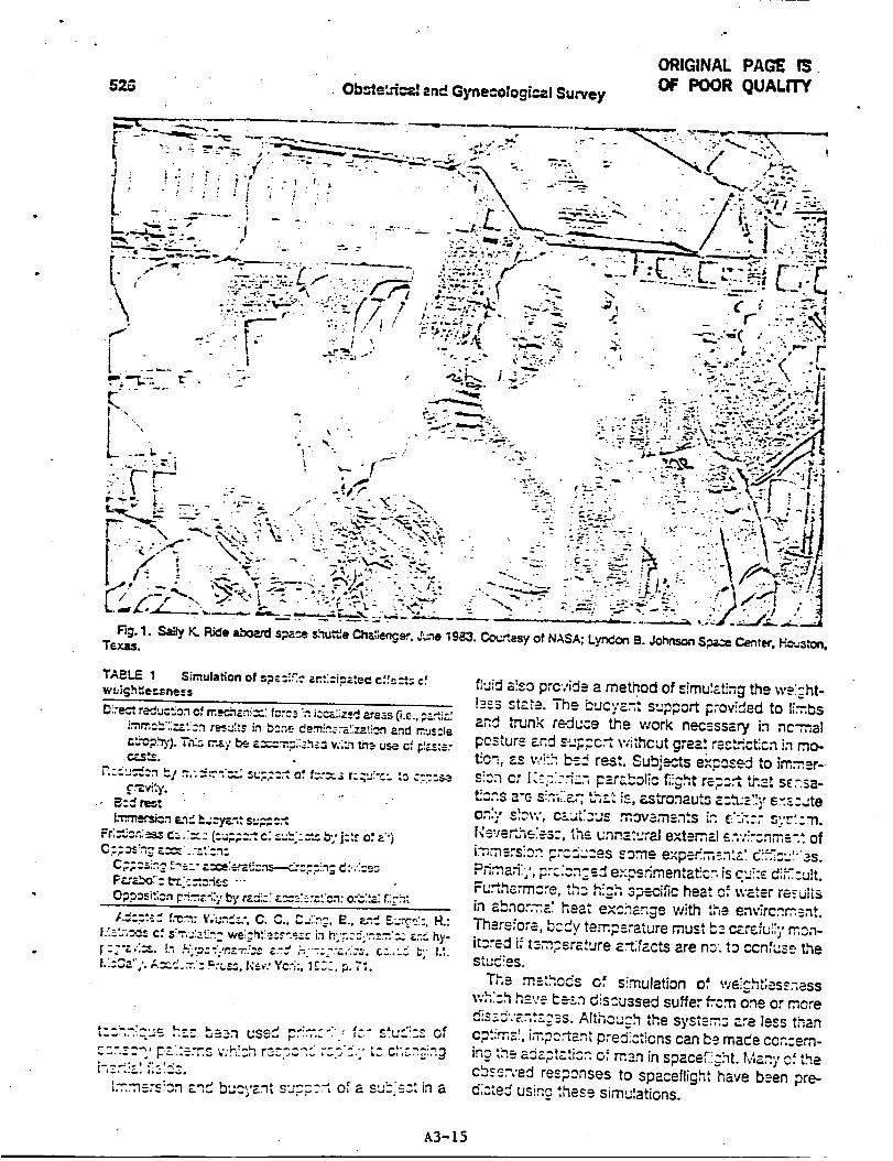
526 Ob=1-.Lri_! 2n_ Gynecologir_! Survey
ORIGINAL PAG_ I_.
OF POOR QUALITY
J _ | : ; r • ,, . ", + .. \ _ _-': ..,,..-..I ' : • _ ! : ." : -. : ' - ' ' " _. \ " - -_ • .-_--J:'-=':' --,
L.... _ ....... , . " Y \ --...---_ . / 1, ,'_. .... "!.I-- '_" "I ' -" -- - "; " "_-'-"__ " I _ ! '""[_[_---,.,--_.. ---.. , • .....,._.-_--, . . .._ _-----._ / _ _ .,.._...8,6,,__ _.._.... • ." ...... "'_'-'--_.._'.'"; "r" G." - -- .. _ _ __. . , . •. _'_• "._. _ _...._..--_,._... ' ... • . ;,'..- r_--.-" _ _ • - I.- _- ." .- *._'. . ...-r _- .... ='_"-- -.... ..... --"-----,--- i.- .-.; .,I " r ot. ;_._ ,-_ - _-j F • r"• .. .... -,. "-- ¢ -_.." .L. ".... .'l I"i . ...-,,, .,,,,+,..,,... ......... L._
..... _ "': . ,, . ' ," _,,,...i r . " "........ "',¢ ; -" ":,'-"I• .- . -- " ; ' ;" • "_':-_" i .,_. _'",';. .. " _-_° _ ." ", "'"
• f- _-.-._.,_- -,..- .- _.¢-,.-..-. • t - -_. ,. ..... -., ./
c._...-- . ..... , _,_.:., . :A• ,, .+" . • .'.."- -. oo- ":t, ..&. " "..,;.'-,--_ :.
- c_- "-_., • .", " _:_: _-::" ';,-':-._.'..,_.,',,-.'_'_
, .. ... _[[:_ :: ._...'-.._ .,,.,_,_,,. _,'-'" ,,=_.-,,.--_-'_:.,..
,, ?-2 =.
,, ,_. . . ,,,,, "-_g.y>'/_'. q" '-" • _-_"..:-_k',,"._2;-'/-_-.
• ' " --c-_-.__-r- ....:-- '":':t !.
' i _' ,b,:+ ,_-=....... .::':...;-_ -:_F ..... ""-_.-, ;:." -: - :%--_ '- .. :.
_._," ._.= -:= ._ _ +" -.__,.:__..::.:... _. E...._4_: : -.-.._.._.,,_ >r--- ''_E .- ,_, . - .':'_:."- . ,,, -- ......... - ..... I .... -_, I• . -. _, -_- +'."_ . i_ • ; -. -_._.:_. . ...._._; -_ ._ "+" -.:t . ".- "-" t.'.:_'t +_,.l \ _-_'_--._ S_ .' ;_- "_'_c-"" "_.'._.P.+ " - "::-:: --_ " . ..','t- .!_'--Jt-.-": .', : ".:',_" \"=':= L_- .-.:.*: :-='- :;:_.. :, ! : :'- _: r .... _..--: _ . . ":1 " • t -_-"_'."l "1"" _ \_- _ -,'_:'C,Gq.'- - • ".-- _.:.: _ _ • -:-i "t"': _"'_I_! / '_ " ,., .... ,,... ,,_. ,."_. ...... _.: -" # r- _ • . I //"_-.,..-
7I J "+" . " - "*.+ " + _. i" . - " .... ," "... -" "_ "... - _=, : | I ,'. ". " • .,. I,.-,'-_`
_,_: .--_, ,_'?._ _. /-- - ; .- ,--,_,_. ..... %2...... . _.,___- , ..=::: . _e.f.:... . : t j..,,,_._r / / • ", :_" " L _ " .:".r ""L: "-"_' .... """. " " +-'='_" ='_,_-", --='*" "/ .... "P 'L.._-.Z.___'.. " . ...-.= .--..__.=_...:_.-_.." =_.Z__- " ".:" ___ .z_ ,./X.-..A./_Y
Fig. I.._h I I_ Ride _.%oattlspace shu,_Je Ch_leng._,,, June I_. C_--,'1,esyof NASA; LyrW.+on m. Joht_o_ $t_ _tet, Ho,_to_,
TABLE 1 Simulation of s.=c:'_." zr.*L:i=ztea, c,,..._-",,-+-c,'w_i_h_e=sne.¢s
D:-e_t reduc_onofm._nzni=', far:- ;n i:c._;;z=.d8re-s (i.e.,p=_i--:!mm=_;::=at!:n r.=suX; inbun=_,de."n!r.:-a::zztion and mus:le
c2"op_'). _n:_.m%' be._.._,=m_;;_h-=-_v:;:n_ use cf l:Izs'_,."c,_,._.
£_vitl'. .:_:... m_: '
FK_,L.a,_;_=s,_r;: .!._: (:u;_.:.': c: _"b_:: -_-b;' j:L" of z")
O;_sitlcn i:'.'i.m_-;;yby rzd::,Tz_-_:_:a:::n: o::-.::z;E:."ht
l,_:-t_.: f,-c_: V,un"-=.', C. C., C-::_2., E., _.._ 5ur_-: ", R.:
!."..'-':,_= cf s!'_',;:_.:_Zwe;;hf.-_---s_.--,c in h;'F.:-.'r.zm'_ _,% hy*
I,;:32:';'. A_._..-:.: F::ss, l{_v: fc.-i:, 1E._-, p. T1.
t==Xr.;zue, h£: ba2n ussd pr:m."--.., f:- stuC!:s of
lmmers!gn -."",. buo:,'_nt su=st,'t,, oT .- s.,_ :.,,, in a
f_uid a!so prc.-ide a method of simulating _hsw.=:.=ht-I_=ssstz_. The buc,/z.t support prov!dedto limbsand trunk reduce the work necessary in nc ,'realpostur_ and s-::ppc:'t withcut greatrsst,fict:,cn inmo-tion, as v,,[:h b-=__ rest. Subjects e_.posed to immsr--S!cn or 1_:_;72.ff_.i.... _",_:," +_-'p:, _-..... flight re;:."t ,,,.=: s.=,-,sa-tJz,_s ate s!m::ar; '"'",.,--, is, astronauts a=,%'-.'Y,;ex-c:JteOn:'-' _l., ,.....x,, c&_t:3us .m.ov_m-_nts i_ _,:;.:: -.'":rn.
i_ve_.h._:es:, th =. un_.tural e×temal =,,--::.......nm= .t of
imme:s:,a_ ?rcgu:es some expedm-n'..--', d:.-i:u!-as.
., ....a, i.,, r ...... ._.., experimentat!cn is q.'.'T:_d:,h'sutt.Fu."th_.rmcre, th_ h::n .... :_'- ."-. or,--,.,,,,, heat .,f water re._ultsin abnor.,-._: heat exchange with tha envircnm_mt.Therefore, b:dy temperature must b3 carefu:;y m_n-icored if t._m_srature art:factsare n_. to ccnfuse thestud!es.
The m_thods of s!mulation of v,'ei_ht;_ssnessv'h::h h=ve b_--:n dis:ussed suffer from one or more
d!_2.d','_.=t-=='_s.Although the syst-_m; are less thanc_tlm.---i,imp_."tant predi:tions can be made een:ern-ino the -'+-_ ""'-". +.,_.,_t,.u.,., of man in spacef::;ht. Many of thecb._s_'ed responses to spaceflight have been pre-d;:t.ed usmu ,he,_ simulations.
A3-15
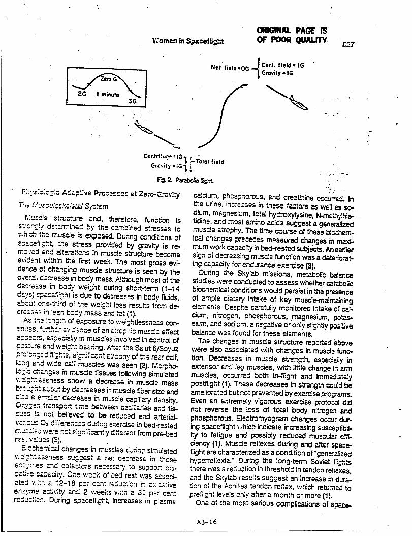
V;omen in S;_cefiight
ORIGINAL PA(]E
OF POORQWu.rrY .r_7
"_ ,o
Centrifuae =IG-_ _TotQ! field
Grc_iiy =IG_ LFig. 2. PP.tabolic fi_ht.
Fh":::;:;i: ACz_ve Proc_ss_c at Zero-G:=vh'7
T,_s[/UZCL':Cs.':e'.._al Syctem
MUCC!e St,-u..--'ture and, therefore, function is
stronglyds'.erminedby _e combined stressesto
which the musc!e isexpose. During conditionsof
spacefi!_'ht, the s_ess provided by gravity is re-.moved and _teraticns in musc]e st,'uc,_Jreb_cme
evident wi'.hin the ft,*st week. The most gross evi-dence of changing muscle structure is seen by theovsrE; dscreese in body mass. AJthough most of theC_rease in body wei;ht during short-term (i-14d."-ys) spa_fi!ght is due to decreases in body fluids,_bc_ut on.third of the we!.ght I_ss results ftcm de-cre---_.ssinleanbody mass and fat (1).
As th._ lzn:th of e×p:sure to wei:htlessness con-tin' 'ss, f_:._h_.: evi."_nce of an ztrcr.hi: muscle effe_aFQs--'rs, espscizIiy in muscles invo;vs-zJ in control ofFosture _nd weight b_._'ing. Ah,er Cn_.Salut 6/Soyuzl::C:cr,;-: :I ,fi_;h:s, s!_r,!_,:znt r.trcphy of the rear c_.Jf,l_n_ _.",dw_e c_f mus_es was seen (2). Mc.-pho-Ic_',c. chzzgss in muscle tissues following simulated_:'s_;ht]sssnsssshow a decrease in tousle messbrC;:;'h: &_,Out by decreases inmuscle fiber size anda;soa sm-z.;isrdecrease inmuscie capillari density.
O;:_'gsn transpo_ time beb,veen c_-piI:afies an_ tis-::s- is_' _ not betieved to be. redL::ed and F.J'teriel-
v.cnccsO= di_erencssdu.'ingexercise inbed-rested
r:,,_:'.=-'_ v-e'e n_t s:.gni,icantiyd!fferentfrom pre-bedr_s" values (,3).
F.,!cchsmicP.Jchanges in muscles during simulatedV,'3!_._ItiSSSneSS SUC._E_t a =_. .. n., decrease in thoseen:l'm,,s.s z.nd cofzc.tors ns:essz.rC, to supp_,-t o×i-
v.eeK of bed rest was _-ssoci-"_-",'= cz._zci_,.One ' 'P..tsd":'_ " '';.... _ a _2-18 p_r cent reduction in c,:;._b;'eenzyme activity and 2 weeks v:i_h a ,3,3p_.r centresuctien. During spaceflight, increases in plasma
• INet field,OG _ Ce_t. field • IG
Grovity • IG
o o
- .:- ." ;,,,.,.
." = j- . •
calcJum, phcspho'ous, and cre_=t_nineoccu._d. In_e udne, in¢'e-:.ses in these fa_ots as we:l as so-
dium, magnes!um, totaJ hydroxylysine, N-meLhyIhJs-tidine, end most amino acids suggest a generalized
musc!e a_ophy. The time course of these bloc,hem-
iceJ chang_ precedes measured changes in maxi-mum work capacity inbed-rested subjects. An earliersign of de,:J'eas_ngmuscle function was a dete_orat-ine capacity for endurance exercise (3).
Dudng the SKylab missions, metabolic baI_ncestudies were conducted to assess whether c_tabolicbiochemical conditions would persist in {he presence
of ample dietary intake of key muscle-maJntP.Jningelements. Despite cerefuiiy monitored intake of cal-cium, nitrogen, phosphorous, magnesium, potas-sium, and sodiL;m, a n=.gative or onIy slightly posiSvebalance was found for these elements.
The changes in muscle structure reported abovewere also ass._cia'.ed with c_hangesin muscle func-tion. Decreases in mus_e strength, espedzi.:y inextensor and leg muse!as, with li_Ischange inarmmuscles, occurre_ both i,_Ii]ght and immedizte',ypostflight (1). These decreases in strength cou:d beameiiorate_ but not prevented by exercise programs.Even ._n er.tremely vigorous exercise proto:ot didnot reverse the loss of total body nitzcgsn and
phosphorous. Elsctromyogram changes occur dur-ing spaceflight which indicate increasing susceptibil-ity to fatigue and possibly reduced muscular e,_-ciency (1). Muscle reflexes during and after space-flight are characterized as a condition of "gener_izedhypsrref;e×ia." During the long-term Soviet f',_htsthere was a re_.Jction in thresho!_ in tendon reflexes,=nd the Skylab results suggest an increase in dura-tion cf the Achi!ies tendon reflex, which return,ed to
prefiigh: levels only after a month or more (1).One of the most serious compIications of space-
A3-16
£2_ Obste%dcal an{[ Gynecological Survey
ORIGINAL 7".%_ ""
OF POOR _U,_LF:_'f
C!.3ht is the continuing loss of bone mineral. Uketousle, bone is a dynamic tissue whi_ maintains amess depende,_ upon the level of ove_i daily
-¢_'esse.s. The greatest stresses for bone result fromf_rces caused by we',ght and inertia. Passive stand-in=_exerts a fo,ce pmpo,rtlo,-_Jto an individuaYsbodywe:ght ¢.'_dthe surrounding gravitational fle_d,sucht,h-=t==112-pound mat_ wi]_generate a force equiva-lent to 66 pounds on the. tib_a./flbu,_t,bones. Thisf.:ce, howev_,', is small compared to the forces
caused by musde counte,"fo.'ces during exe:cise.Jogging or runn!ng may p:o'.ide forces exc_=_d!ng1000 pounds of t_ansient force, on the bones o," thef¢ot (4). Conc;iticns of spa:eOight or s!rnu]atedweightlessness remove most of the major forcesfrom the musculoskeletal system so that only aP,z_on of the ine,'_al force-:,rein,a!n. The maj._r med-i-.._.Jh_.e.rds of pro!onged spao_J;,:ghton bone inclL:dea leng_y re.very cf lost bone mass followingspaceflight, possible irreversible bone loss, bonefractures, and toxic effects on soft tissues such as
the kidney due to increased re_ease of calcium andphosphorous (1).
Loss of skeletal mass becomes an important ccn-side,orion, however, only in Iong-te,,'rn spacefiights.Changes in bone density were found even during_ho,rt-term spaceflights, but the rate of loss of de-m!n_aJizatJon was gradual. Evidence from the bngerApoTlo and Skylab flights support the hypothesis thatbone mineral loss occurs only in weight-bearingbones dudng spacef!i_ht (i). The mechan!sm cf boned.%_eneration is still unknown but may involveche.nges in eTecttochemical properties, and in hor-monal and r',-_ra! factors. '-,r,.,-,_.-,.:_.,. animal studiesc:;;cest that Lh-_loss of bon._ mineral is due to ani_h_bition of n_'.,'" bone fc..-mz.t!3.:. In tar.n, h_;'._.;'er,• . U_,,4:._.;s .... ar wh2"J-ter there is reduced bone f_:m_'tion
cr increas:-d bone reso,-_ti:n du_ng space_i_ht. Au-',:psi_s of C_ree Russie.n ccsm._nauts who die.."after21 days of s;zce.';i=ht revealed evid_nceof anatom-i:-=! bon-3 c,h-=n_=.._sugge,_t!ng increased bone re-¢_:pt!cn (1).
Studies on the reve.'s!b!I_' cl bona loss f3:iawing_.-.z:ef,;=_.ht ate in:c==:usive. If the decrease in bonemass is sirr,._:7 d,._ t_ a t_m.=c:r-_' imbz!_.n:e in bonet.:'.-,3ve:, th-_n bc:$ loss v.'cu'.=:b3 rep',aced at t later
s_,a.=,,.g,,t. On the othert!.me, perhaps following " "_"; "h-"n._, an alterat!on in th-:. functi:n: of individua! bonac::'._ could resu'.t in in'evers!b:z bone Io._s (5}.
r==.-t of the d....... ; in theo: _=n_ Ices with v-e_.;ht[-s:%zs is that, so far,'..:':,'c'.ztT::', te:hn;qu2s c3 nsL n-.!.T,!3th5 resu::s durin$
spaceflight. During bed rests, a negative ¢_dumbaJance appears within a few days. Udna,'y calciumincreases and plateaus after about 5 weeks, whilefecal ca_c/umincreases until about 10-.12 we-.:-s and
then plateaus. Dudng spa_flight, urinary ca!alump!ateaus after about 30 days, but fe_ calcium lossdoes not oppear to plateau. Extt-zpolaLed c,_c_umlosses from the Skylab spaceflights suggest that t,
within I year an astronaut would lose about 25 per.cent of the total body cal_um poe|. Extrapo'.atedbed rest data give va!ues of only 6 p-- cent (6).
The results reported from long-term Soviet space..flights suggest that the calcium loss does not contin-u_',y increase during long-term we'.gh_essness, butslows considerably between the third and sixthmonth when vigorous exercise progr'ams a.-e cornducted (2). The effectiveness of exe.-dse as a cou_termeasure is still uncertain, however, as duringSk'ylab 3 and 4, weight-loading exercise red=codbone loss in only bhree of the six astronauts. Soviet_nd!ngs reg_d_ng the effects of exercise as a cour_te,_easure are more hopeful (Ni_gossian and Par-ker, 1982; 1). Ot,_er countermeasures which havebeen proposed include dietary suppIements andphaJ'rnaco!crg_icalag_ts. One drug, dodronate diso-dium, effective!y prevented negative calcium ba!ancein human bed-rested subjects but was discontinuedfrom use because of evidence of possible c_rcino=genie properties (6).
Comparisons of bone mineral losses in men andwomen suegest that women had significantly lesscolt!urn loss than simifP..rly stressed men. Furtherstu.d_es are needed to verify th!s finding a,',d toestab:i_h the mechanism for such d!fferences CI).
R.z:,_frc;oS" S:,,ctem
The c_.-_.-n_esthat occur in the resp!r.--'to'y syc:-=mduringactual or simulated v =',,,-+,--- _-_.-.-;_;,-._,-
lel:_p_.--,"to b_ ..,._;ht and of littie ore:aT: p,_',_.s;&,.;..cPJ""-"s!gn;ficznc=.. Pulmo,=a._/functbn m-_as,re,T.en:_ c_
ta!.=e_ from five men dudng a 20-da_, b_.cEzest pro-tc,.=o_re"_a',_-d nonsignificant chang.us in tote: I,ngcapacib/, forced vital capacib,, 1-second forced ex-p!zzto,3, vo!ume, residuaJ vo!ume, a_d th_ d2_'-s_ngcap-=c_ty of the lung for carbon m_noxide. It wasunc'.=.ar in this study, however, wheth-=r a'.imea=-ure--m_..':ts were controlled for posture (8). In a recentsilo: 7 in wh!:h six women und._r_vent duplicat-_ 11-d_" bad rests, smaIl but signific_.nt increases in v!talcapa:ity a_d 1-second forced expir_to_, Volume cc-c-::..d d::r:,,_ the first 5 days of the b_.drest protocol,csntinued to be elevated throughout the remainder
&3- ].7

c: th: b:_ rc:ts, _'_d r¢_.'_-:..t tow--,"d sup_n_ pre-t-_ r_t l-ve'.s gu_.;n_2 w_'.:s of ='nbu1-:.:o_-re:or-cry. 1_v:zs po._tu_t¢:! by these aut.ho."s that the
,._ .JC'_'_e.': in _ _.:_-._._" _n,d 1-sc:ond fo,,,e., explr-_tor 3, vo:ums mr.y h_ve bc-s..naes,_ated wiC't theI5-,33 pz." _nt dc='e=ses in plasma vo!ums v:hi_c::'_:':zd in _-o sam: tlm._ inte.'v=_LPree,_'m-:b:-'de..cre_s.%."p!zsrn= vo:ums wcu_:; b3 re_._._ed in a sirn-i_3r prc-'-c,'_r'.eg de._'e-=sc in t,4ctz_= b:c:. _ v=:ume¢.',d _.,_in_e_. -_- in lung ca,'np:_z,'::eduffng the fl.'stfew d_-ys cf b_d rest (S). A f=:low_p study, in v.'hi:h_. 23 .::: cznt d-==zc.s_ in p6:.-:m= vc!-m¢ w_s in.
cf d;u.'a::._-, fa::-=g t: =...--._.._,_":_"=-c.3rT_.:a_cnb?.'_¢:een
ch_.n;-_s in p]zsm=- volume and changes in vitr-! ca-p_:L"/cr 1-s-c.ond forced exp_r=tory volume (I0).
f_ ccns!=t=..nt_'_'_es in pulmonary lunch, on haveb.:_ re_:.-t=-_ during spacefi;ghts of 28-84 d=_.ys'Curz.t:c.-t. D'.:._ng one spacefii;ht, vitzJ c_'.pac_ onmcct days av-_,_ged ab:ut 10 pe,r cent lower th_np.-_._:_-htc=ntrc!s in Cnree astronauts, but retu.med topre._!:nt levels ra.:_dly a_er I,,.nding (11).
E cdr' F,uids
With the remov_J of hydrostatic pressure gradientdudng conditions of weightlessness, body flu!ds arere.d':r.t_buted such that fluid r÷dis='-"ibutes from theI_v,.'sre×tremities in e ceF..hr_d d:re:tion. The result-
ing increase in t:':orz_c b_ood volume is be;ieved toin;::,=t_a G_u-_r-Henry reflex, whereby s_etch recep-to-s in the IoN at_um inhibit the re_.ase of antidiureti¢
hcrms,ne. As a resu;t, a sF.cntznecus diuresis oc-cur:, resj:tingina decreese inF_.'.a_.mavolume and
el.: :::::_es. _.,n',=...-,,,_ 2-wee;: b_d rest studies in sevenm_n, th".s d:ures_s was see_nCuffn_ :hefL-st 4-5 daysand r:s_::_ed in a 13 per cent re_',.'ctJ'_n in p!zsma'.'cO=mr (i 2). A s!m;ia- re:at_v_ loss of p_&smsvo:umev.'zsreTc,_ed in be.d-rested women dudng l 1-claybe_ re_.;s. However, it was obsewed that, when bedrest was Ic=--'Junin the immed{_.te "-" , '-' ,. _, eovu,=.o_ stageof a v.'cm_'n's menst,-ua] cycle, e!ther a,nincre_e orcnty z s.'.,a'.ldecrease in pl_ma volume w_s seenduring the first 4 days of bed rest. These resultssu;;_st that the he,,-'non._s asscciate-'J with the preo-Vu',-:.:C_'st&ae may " c:= +. t_an....n.Jy prevent the d!uresis-"ss: c;--ted with bed rest (13).Dud,_ -"" "•--.,us, spacefiichts, however, this early-
cns_', c_:uresis h_s not been seen probzb',y becauseof prcb'.._ms in cbt_.!n!n= urine s&m_',es during flight,an_ b-=:_use of a reduced fluid intake due to motionsis',:ness _-nd enti-mot_:n sickness dru.=s. [.'-;&sure-mer, ts cf the net v:&ter b&:znce during Skv;=_.b' -"*-
indicate, however, that although fluid in_.3= is re-duced, udne output is not reduced to a c_-.,.=:-a::elevel resuWng in a negative water baJ_: d::._ngthe first 6 d£ys of flight, in-flight increases L"zt.,:L"P.rysodium, pot_._sium, and c.h[oride, and d.e,'J_-.._esL'_antJdiuretic hormone lend support for the d_u.-esistheo._, of _ p_sma vo:ume decrease durirq,v.'3:_ht-lessness (1).
Thzse f,u_dlosses P-"ebelievedtO conb'ib_a to the
po_.ht c.'_os_-_c intolerance seen in most a.stro-nauts _nd ¢osm:nauts. Countermeasures to preventt;n_diures£sin_ude vigorousexercise programs and
wzt_.r and e!ectro'.,,te supp]em_nts during sp_.=efi_ghtor just before re_.ntty. During long-term Soviet
• flights, the water and electrolyte supplem_'=tationwas combined with Iower._..,ody negative p,'_,_sureadm_nist_tion to ensurethat the ingested fiuT_werereta!ned with!n the vascul=_rspace (2). Mi_,_:cor-ticoid (gq=-f;_orchydroco,"Jsone) ingest_an dudngbed rest studies has su_ssfui;y prevented thelossof p!asma volume but the effete on orthostati¢to_erzncefc:lowingbed rest have been in_,.sive04, 15).
Ee:_des C_¢_oss cf p!_ma volume, red ce: vc:umealso decre-=ses d',','ing v/e!ch'_essness. The. effect,however, now appears to be. se;f4imiting, where witht_ng-term spaceflight, the red cell mass decreaseduntil about 60 days into the mission and the.n grad-ua!ly be-gan to recover (1). Along with the loss in cellmass, there is a change in the shape of the red cells.Preflight, 80-90 per cent of the red ce!!s had 8b!concave (discccyte) shape. During the Soyu% 6Eight, the.re was an increase in number of e_:!psoidalend spherical shzped ce:!s. These chang_ were
rap!d:y revers!b!e p:stf;!ght (1).The most &cc_pted theo,_' to explain the decre.=-,se
L7red Ce!lmass is that" "'_'"re-,I,=, ,, there is a suppressionof nev: redcell production by a direct inhibitoryeffectcn bone marrow. The reduction in prcpc.-f.cn of
reti_Jlc='_es during ez:iy spz_?,ight woui_ _.-d,-rnth!s hypothesis. Find_n;s from the Apo._o-Soyuzflight dispute other theories that increased red celldestruction or seL=ctive sequestration of red cellsc_use the decreased red cell mass (1).
C_'.rdiovascular System
It is especlatiy difficult to simulate the e_'_ts ofwe!ghtlessness cn the cardiovascular system sincesimu[atlon procedures, such aS bed rest znd waterimmersion, produce physi:al deconditioning in addi-t;on to the hypo_ravic resp3nses.Most ofthis re'Hewv:[:;, therefore, focus on cardiovascular responses
A3-18
53O Obstetdca|=ndGynece|ogica|Survey OF PO0 UAUq"f
seen during or after spaceflight. Three majorchanges in cardiovascular function fol_owing weight-lessness include decreased orthostatio tolerance,
changes in ca,-di--: e!ectmmechanics, and changesin exetdse c_pacity postflighL In addition, somemorphological changes have been reported in theheart fo,owing prolonged space_ight.
One ez_y ccnttove.-sy centered P.'ound how the¢zrdiovas=JlZ=" system would de_ with the cep,h_adshift in body fluids. It was f-c._'e_ thzt a ¢_hronice_evatJonin cenLc-aJvenous end L=.P,ztdaJ p:essur,as
would persist du_ng we!ghtiess e_.posums, p:'.en-t_ally compromising cardiac f',:nct;on. Recent evi-dence of only _'_siant increases in central venouspressure during head-down tilt s!mu!zt_ons and nor-m_ cardL_: function curves duM,ng the Skylab mis-sions argue ag_nst this theoq, (16, 17). Recently, it
c_._ to examine thehas become possible to co_-_ "- --ef':ect of long-term spa_f,',ght cn cardiac structureand func_on. Electroc_dic-g_.phic studies affor the
Skylab missions found decreases in left ven_cutardimensions without significant change in c_n_acti]epropeJ'_es (17). Fo_Iowing SaJyut 7, e:ecJ¢o:_'diog-raphy studies revealed a smut but reversible de-crease in left ven_cular mus:ie mass whHJ_ was
restored wit,bin 7 days p.._.h,. The rapidity ofmuscJe mass restoration sugg.ests that the changesv'ere due to aJteration in myocardial interceliularhydration rather than to myocardial muscle degen-eration (18).
Decreases in orthostatic tolerance are consistently
reported during en_ a_er spa:afi:ght. At least partof the into_e.-ance to tilt, lower _""" nP.gative Fes-sure, or qu[et standing pos_';;;h: is due to the blood
_,_,'__.==h_ .... he.s. Thisvo_,Jme losses 8$son!ate_ '"" "-" "_=¢" =?_50-500 ml Cu!d loss, howe.us', is too sin--'4 to
a::ount f:." ,_,_ c_-;ree of cz::::,:;'-s :u:-"r dy._,'u.-.ct!:nre7 :._e_. A[s=', repla:em=.nt of tnc vcTum_ Ices b_fc,'e orthost_¢ to',erance t_st_n_ C;,dnot ¢omp:-:t_:yr-:store c,%'_os'_=tJ:to!e,'anca (:i 5}.
_her me:_,--'_isms that may c:.:_bute to t.h- d=--c,'sased c,-thc-:_tic tc',erznc5 m-:-' inc!u_e an in-cTease in V_.'I.,CUS r'n_'= "co_p,_.,....es_-'__z:a:.yin_.he!0_.','erh._y or =.,.....-_"_--'"-',,,,,.:. re;'!cns, or cha_ges in the neu-;C3en!3 O: hc.'m,c,na! co,ntrc! 1";333_.,'lis,"T1,sthst re-' _-
l:'.e b_::d p-cssure.Venous ccm.:::-:nce m.-:zsur_
msnts du-:n_ S1:_,!ab flights e'.-:--:_.-_an increase incomp',!zn=3 during the first 10 d_ys of flight, then zs:.zv,decrease withcont:.nuin2s;.-zefl!cht.The tim=.
cnurS-_ Of th-_se comp:ian:e c;-,'---_;_;sd:,d not concur,., ,. =.=n_.. to lowerwith ao.omp-:nying chances ;_ '- ....
_--' ' ne:_.t;ve pre-_-sure(!).
Data describing changes in autonomic fun_onafterspaoe,'_ightor bed rest are in,,_nc:_ive. Uponb'Itat lower body negative pressure c_:h_ce afterspaceflight, the cardiovascular responses are exag-gerated with greater increases in h¢_-t rate and agreater f_ in mean arterial pressure. However, thenorm_ vasocons_ctor responses appe-:.J"int_:L Imjections of nor,epinephrine or eng!o_nsin p:c_.ucesimilar pmssor responses during 2--3 wee!; bedrests. Although plasma c_t_holam.7..._ are re_/uced.r..;uringbed rest, their response du:_,g t_;_ is un=c,hangsd (19). The venoconslziCtOr rec.".onseof fore-.ann b:ood vessels during exercise was found to bepotent;atsd rather than reduced fo"ovdno bed rest,suggesting normal sympathetic v=_no,'..onst.dctorfunction. The increased venocons_;_on was pro-portioned to the reduction in plasma volume duringbed rest (20).
E/_'o_-d:ogm;hic and vect._¢,-c-zrd;o;_phicdata, at rest end during lower body negative pres-sure challenge during Skylab missions, showed aninc_ase in the QRS maximum vecto:, which may bere_ated to in.eases in thoracic blood volume _nd a
pro!ong;,tion of the P-R interval, poss_b[ydue to an.a_oven_cula: conduc_on slowdo;_ caused by anin.eased v£g_ tone. Frequent arrhythmias duringspaceflight may aJso resu=t from sympathetic-para-sympathetic imbP.Jance or may be due to fl,u!d orelectrolyte imbaJences induced by the weightlessstate (1). Dudng Sa]yut 6 flights, kinetoc_rdiographicmeasurements _so revealed a sign_nt shorteningof isometric contraction and relaxa_on of ejectionan_ pos_:Z':_ phases (2).
Exercise capa:{b/ is severely re_.u:ed fc:::,vings._-,__ce[;_.h:.Af_.: t.,e 3-11 day AT._::o flights, de.-craases in exe:c!se intensi_, oxy_:n uptake, and_rS:.-:.ccut;utv,,_rec_se,'vedatr-nexercising,".cart
rat_ of 163 t:-:.-:.:sp :: m!nute. Oxy_-=_ ccnsu,T,.-_t_on
{:: a given e':_rcis=_ int-=ns[%' (musa'.;.e.'-T,:i_-:).) wasn?.t a__red pzst{;:._ht. Followin_ tY._ ton_-_rS'-:y_abE.-h._,. _!m:,:s.:dncrean_.s in exe-c!s5 ,.-z"-z!._. _' werefcuz_ bu_ ,:,:.'e restored with:.n 3 v.=_::s c" re::ve:'_
% I"
One unexpected finding, howev_ r, was t-e ira.
p:avement in exercise to!erance d'-,r_ng spac=._:!ght.C_,:e th_ mechanical problems c,' exe.-c!si_g inv/s_'htiess condi:ions were nego:i=ted, astronautsduring S!:ylab flights were able to a:complish h!gherexercise intensi:i_s than were pc:s!bte in a cn_cjrav;t.y environment. An increased maximum oxygenuptake in zero-gravity conditions is p.;obah:_' notL'-,_,.=,,v_';"-"= c[ ,--train!n_ respor, se as in normz._condi.
k3-19
_:'cmen in SF.--'e,':_;_t r_l
tJons, but may have _..n due to the effe_ of aninc,oe-"s._dvenous ret_:m _nd maximum c_'-_:= out-
put resu!_n;] from, t,he incre-:.sed cen::_ blood vol-ume. As a line-"." reL_t]=nsh:p exists be,%,:eznabso',utework output and _'dla¢ output, a higher exerciseintens.:ty wou_d be. a._.Jmzb!e. Ox-'ygon consumptionmat,, aJ._o incre-cse because of an in=r¢-=s--_d_='Jvemus=1-_mass in zero.<j'.av;_,l as the arms p'.zy a moreactive role. c_u.'_..gexercJs¢. Rxed subm_L,'n: exer-cise intensL'ies were a_:=mptished wi_-_lower he_,'trates, and b_ood pressures than p,"mfi:.;ht.A morer_p_d recovery of heart rate after ex=_;c!s._in thewe;;htiessness environment was prob-"L_l,., due tome grs--te" venc'..,s re.'-,_..m.Thers'cre, in c-_,zr toma!nt_Jn a stab!e flmess level while in-flicht, trainingexercise intensives must be adjusted in an upwarddirection (21).
Eng.:trine Sy_:em
T;-.,=sr,_::_ne resp._nse during so-_,fiicht.. re._tsthe _mb.!ne:l effe_s of stresses in.i;ced throughthe removal of gre.vibl, effects from motion s!ckness,ch_n,_es in d}et, and the psychological stresses as-scc_-:.,_.i with space travel. B.cause so manysb'esses are simultaneously combin.=='J,the hormonal
d,,,,cu,, to interpret.responses are o,"[en :"= _"He-_-dc;','n be_ rest s:udies (22) have b_n con.
duc'.a:l in an a_empt to iden:i;-y some of the tr_.ns_snthc,'_,.:na_ fluc,'ua_ons associated ",'"_,,,n changes in
the hydros'.at:,cgradientexertedon the body.Within
the firs',2 hours abet lyingd:};vn,thereisan increase
in c_.ntr_-;venous pressure wh!ch prob_b!j,tri_.=rs
the dez.r_._seinp'.asmz,c_ncentrationsofantidiuretic
horm.,=nc, ang!:_sns!n, and a!_osterone. In this sametlme int-c,'v_J,sym:athetio a=tJvity is de,eased asreS_cte_ b_,deers_so3 in concentrations of ep_nep,h-fins _'.nd nore:oinephrine. Giomerular filtration rate isa!so trr-nsisntly d÷creas_=-'J.A,%r 4 hours of bed rest,cent.';.J venous pressure retLfms to prc--b_.d restleve:_ _--d-,,then continues to de.ease. The g!omer-u:ar fOr:ration rate re;ums to baseiine only after about, '
•F.,hours, at which t_me p!_sma ._.dos._, on. and an-
g ........ n lsve!s have _Iso recovered _.n_ may evene;:css5 the pr'c--be_ rest level
Dztc fm.,m z..strc,_e.u_ in the space shut't,Ie programreve,'=-':s5increased p',asma and urinary, a!dosterone,ang!o:snsin,and urinary antidiuretic hormone post-ft,=h,, s_m_i_.r:o the r==.,,_ repo,led after 8 h. _,. ofheF.:;-_own bed rest. The plasma leve!s c: these,_ormones =n-,:,=h_, however, have a!s3 bs--__shov,'nto _e in'._snce.1 by sa',t and wr-ter in;;::-:.. U:;ns
anEdiuretic hormone concentration during lon_termSkylab flights was reduced during the first 40-53days of flight, then began to return toward pr_F,_;htleve!s (23). These hormonal fluctuations undoubtedlyoccur in response to the fluid shifts with exp_ureto weightJessness.
During Sl,'ylab spacefligh_s, parathyroid h:._rn,onewas s,._n,,y e:avated (6.4 per cent) above pre?,':;htlevels (23). Parathyroid hormone normally func'Jcnsto in=e=se p!_sma calcium by first inc.'easing bonereso.-ption, second by increasing resorption ofcium in the kidney and decreasing excretion, =ndthird b,' increasing caJciumresorption from the intes-tine. Increz.sed pa._thyroid hormone may fun_onduring spaceflight to oppose the effects of !r_creasingcortisoi which, exerts a negative calcium bEz.nceeffe=t. The action of para_yroid hormone requiresthe presence o: v_tEnin D. It is not yet known whathap;-=.':s to vitamin D levis during spacefli.ght, al-though in bed rest studies urinary concentraE:ns of1,25 dihydroxycholec_cfferol, and vitamin D metab-o!P,es were decreased.
Inc.-eased co,sol leve!s in spaceflight may have as!gnif,cant role in the muscle degeneration. Thymx.ine and t_hyro!d-stimuIzting hon'none were both foundto be incre_-sed during spaceflight and may alsocontribute to an increased metabolism of proteins,carbohydrates, and fats (23).
Cateoholamine excretion decreased during theSky:ab flights, which is within the observation of"reduced sympathetic responsiveness. During bedrest studies, the reduction in norepinephrine ex=e-tion could be prevented by exercise (23).
Both American and Soviet spaceflights haveshown evidence cf a change in energy utilization andproduction. Fasting brood g:ucose levels increaseinitially, then c_ecrease beiow preflight levels. Plasmainsulin concentrations decreased duffng Sky.labflights (23) but were reportediy increased in theSaiyut 6 cosmonauts (3).
Fundamental changes in _rbohydrate metabolismare evident after only 2 days o,"bed rest. DoIkas andGreenleaf (24) found that the degree of g!ucoseintolerance during bed rests is proportional to thedegree of immobility as assessed by the total energyexpenditure each day, and is probably not due to ahydrostatic effect. Exercise during bed rests waseffective in reducing glucose intolerance. A poss',b]emechanism of this decreased g!ucose tolera,",:e isthat, during deconditioning or bed rest, there is adecreased sensitivity of the tissues to insulin actionthrough modification of the insulin rece_.;ors.
&3-20
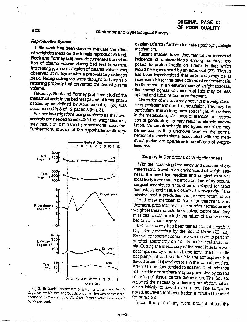
5_ Obstetrical and Gynecological Survey
Reproductive System
Little work has been done to evaluate the effect
of weightlessness on the female reproductive tract.Rock and Forthey (25) have domJmented the reduc-tion of p_asma volume dudng bed rest in women.Interestingly, a norm=',_.ation of plasma volume wasobserved _t m_dcycle with a preovulatory estrogenpeak, Ris+ng estrogens were thou cht to have salt-
- retaining prol_ty that prevented the loss of plasmavorume.
Recently, Rock, and Fortney (25) have studis_ themenstru_ cyr..Jein the bed rest pat:=nt. A luteaJphasedefidency as defined by Abmha.m et aJ. (26) wasdocumented in 3 of 12 patients (Fig. 3).
Further investigations using subjects as their'owncontrols are needed to establish th-z.tweightiessnessmay result in diminished pregesterone sc--cretion.Furthermore, studies of the hypothalamic-pitui_.n/-
I,,,H .IO0
(nglml) 0 I"
FSHtnelml} 200
I00
6
5
4
Prceest_oae_nglml ) 3
2
I
0
- m_ed.,es! 0oy
2 _t 4 ,5 6 7 8 9 IO Ii ;Z
LH
FSH
Toro I
{';') ;7 •;
ZIZ?.Z_242_ZC27 t Z 3 4
C,_cle Dolt
Fi_'. 3. Endccdno pa,'amete:s of z v.'cm_n _.t bed rest for 12
da_'s. An i_sa:_.ci-_n_' Qf p og ........ .- .... e,_on was do,um.nt.d
_.:¢o.'c;n; to _he method, of [,b'z.h;::. P;_s.,-nz volume decregsed
b_; 22 pe,r cent.
,e
ORIGINAL PA_aE IS
OF POOR QUALITY
ovarian axis may further elucidate a p_thophys_ologicmechanism.
Recent studies have document:-,J an increased
in_dence of endometriosis among monkeys ex-posed to proton irradiation simiI_o to that whichwould be experienced by an astronz'..,t (27). Thus, ithas been hypothesized that astm,-'_u_s may be atincreased risk for the development of endometriosis.Furthermore, in an environment of weightlessness,the no,'maJ egress of menstrual fluid may be lessop,ffmaland tubal reflux more frequent.
Abe,'r_tion of menses may occur in _e we!ghtJess-hess environment due to anovula_on. This ma r, beparticularly true in long-term space_ght. Alterationsin the metabolism, clearance of stero!ds, and secre-tion of gonadotropins may result in chronic anovu-lation. Menomevor_nagia and hyp_.=rmenorrheamaybe serious as it is unknown whe_er the normalhemostatic mechanisms associated with the men-
strual period are o._..'-atJve in conditions of we!._ht-lessness.
Surgery in Candit/ons of Weig_Je¢sness
With the increasing frequency and duration of ex-_aterrestdaJ travel in an environment of weight[assoness, the need for medical and surgical care willmost likely increase. In particular, if an injury occurs,surgical t.,_,hniques should be developed for rapidhemostasis and tissue closure at zero-gravity if themlssion profile precludes the prompt return of aninjured crew member to earth for treatment. Fur-thermore, prob!ems related to surgi_t t_hnique andweightlessness shou!d be resolved before planetarym!sr.isn_, v,h_ch preclude the return of = crew mere-be, to ea.-th for surgery.
In-_,ZghtS_rge_' has been tested z_cz:d a;rc:.:..=.inl(epi_.::an pzra_._Iz.s by the Soviet Union (2S, 29).Spec_ transparent c_ntainers were used to ps.ffc,-msur,_L:_.IIap--.ro:cmy on rab_ts unde-tcc."-i ane_:he-sia. C :_:n; the mesente,_, of _e sm._;; intestine wasaccompanied by v!gorous b_cod flow. The blood didnot pump out and scatter into the a,'nosphere butflowed around injured vesse!s in the f_rm of pu_dies.Arterial blood f;ow tended to scatter. Ccntamination
of the cabin atmosphere may be prevented by carefulclamping of tissue before the incisTon. The Sovietsrepo,-te_ the necessity of limiting tn_ aUdominal in-cision initially to avoid eventration. The surgeonsnoted, however, that eventration eliminated the neec_for retractors.
Thus, this p:e:!,,-ninary work brc::ght about the
A3-21

, . , ; ¥:omen in
rea:izati..n th.-t s-,r_-.'y todd be pa.'/orrned in the
we!ght_:.=cn-_.s envirc, nm=nt. Although our under-standing ¢f _r: £i;c_ of we!ghttessness have rapidlyadv_'_:_, the av_]zb'._, literature on surg_czdtech-nique_ z: zero%ravk'y is sparse. Studies are primarilyin the Russi-'t I!tem_.'-e a.-,d obse,wz_or, FJin n_,tum
(2o, _3)...UR.cn ents._ng zero-Gray: _, thee is a slowing
down cf speed a._d a:,cur_ fc,r a pu.'poseful move-menL In pzr'd_la. °, thare are errors in trying to hitthe cznt_: o f the ta."_et (deviation of hits upv:zrd).There is, however, ra_:d r.._ap_t;cn _s motor ccor-dinaL':_n habi.s '= -"'u.u=,, deve!op q'J!t-3 -':"s--;,q (31-33).Spec:_c;.;:y d-s;';n_ i_$'="uments may "be rsqu:redto peKorm comp;_x motor reactions required forde!::ate s'_,rg!czJmaneuvers.
Stz.zhar::-_ et _. (_0) s.==.s_e., th..t th._ limitationsof the s_ze cf th_ _smi¢ cra_ _nd the redu_._,d
im,.'nunc.-ezctivi:-] of man =n space requires a soft
surg!c._Jant!bz_e,",_ chamber ma_e of ttansp--rentfluo,.plast_c f,im _._dh_ving two to thre-e p_rs of bui_t-in sleeves with, sur_!cz! Gloves. The authors stressedthe need for deve:c=!ng a compact set of IighbNeightsure,col inst:_.,msnts meeting the r._u!rements forsurgical int._rven*Jon at zero-gravi_.
Rock (34) has suc_ested an expandable chamberfo; m_n_r surce,'y in ¢onditSons of we!;htlessness.Maior consi_e:aScns in developing such a systemfor use in a sp._cec:a_ were to provide a sterileenvironment _-s we!l as to prevent contamination ofthe cab!n v/_th bleed ar:d other debris while perform-ing the surcery.
The "_'"m,._,,,_e is a se;f-contained system with a cuff
tourniquet and i_suf_ato'. The stadia g!oves are builtinto the system so that the environment within theca_.su;=_,is st-=rile, c=.'=tr-"nin§ l:=th c'.oves and instru-ments, v,h:.:h P.rea_.,_ne_ with Ve_=o straps to theclear viny; wa[!s of _e cz.psule (Figs. 4 and 5). In theevent of an arm inju_, the a,,'m would be inse,"ted rthro";h the cuff tourniquet; the tourniquet wouldthen be infiate_ to a._n;eve hemcstasis. The capsulethan may be insufflatedand the surgeon's handsinse,'ted into the gloves, the instruments grasped,an_ th-_ surgery paffc.'med. The surg!cal area maybe preT._ad and '---',,,,._anesthesia a,_ministered withinth!s s=:._!ca! env!ronmsnt. The b!eedsrs may eitherbe cilp_a& or suture I_ated. As normal hand tyingmovemeats may b8 di,icult under conditions of
v,,ei_'i'X:.essness, c;!p systems may be more appro-priate to a..hieve vessel ligetion and hemostasis.Contamination o: the spacecraft with b'.cod an_ tis-sue c;abris is thus avoided, maintaining a clean en-vironm=.nt.
Sp--:eflic.ht .,r_.3ollmm Is
i,eoll mcnry
,-.: . .....• _I " -_ -_"'-' :_ " -.,-'_. ." .:,._,. _; o t,&'_E_ .-_,-:-...............--:F_-- ,-, "-;-_--=..-_'-;_;--."-:_, --'; ..
\\ i,-.:_ _ _.._ .,..... :£..-:::-'.:.; ,_ _-'_- ":,-,j_; -.-.---.-._e_.-- _<:L_._.-:.,"t'-.._. "_,..--.'__,;_;.&,-: ,_ ",-"
." _ ;: ,% _ _" . .-..._. -'%;. _._.ll_ .-; ' t- :',.*"" _ . .. _'_"¢_.,._,_. ;4 :[_ , . ,-.
.=- _ ._--.-=.--..C*_:._-4.-.- _.- _.-_,_" ", " --:-_ ,-_.""_-. ,_"*_ _/-'z";.--,4," 4-."I . _.._." _ • _'-.__:_.- .- , ._._.,: :.-.i.) .... .,l.,. ',. .... __. __....
,_'- . -_'_'. . .,-_'_---_ -;-_ , ,.. .....In,,_.,'y._,i_,. : ', ,_-,.. ,._;':-._'_p,._.:'_,_.-_._.;, " .-_---..: !
/ " -'"_ " : -- "-" 4.--'"' .':_ '_"'" "":"'- ''_• _it..• .. ' -. : ,_. ',"k"" •:_ . .,_,.... ,=
: --i- !!;-I " .... "
Rg. 4. An expandab!e chamber for surgery in conditions ofwe!l,_=essness. Instruments and s_¢_;re matedal are stored in
I;x:_<=.ts withLn the chamber. From Rock, J. A.: An expar¢l_ble,surgi:_J c;'_m_: for use in concr_cns of weightles._ess. Aviat
S_.ce =,.n_.%cn.Med. 55: 43,3, 1984. Reproduced with pe:n'_ssionof _,e Aerospa:e Medical Asso_io_, Washington, DC.
)! __-'_- .:_,,,,_.._, L_'- -_--,'.:.--:.. _I'._---'_-,_
_.: . .._....,_, .,,'-'_--._-.-._• ." "_,a" _ ..... _ - .'i "- &._'_C"_'-'-'_
/. -- \. - . :-:.. .,:..%_._ -. . ._.,_., ,,, • ..• ".,'-_' : _ .."" ," L .__-::_,', ..
I" .'.,.-'.. .'; .,_ " ...... ...-_ ,.T_ -- ,_ ._ ,_... , _ • • . . . _... .... .y "_ _ • ... /... _ . .._.:.... ,.., ,. .... %._,
-...: .... : ._.._.. .... _ ___ • ._ .\ /_!- _.':. _-, <:_,_ -'.v,
__,# - • ; • .? , .4,." "_¢'-: ;PfC :*-N-]_¢-'-_ ;_'__ _. ._"_._" _-._._* - .,--_ ..--._. ,_- "_'___,-E.r- - -._l
....--'_- {,_ - -, ..... =-. -- " _-_-:'._ .-'_-. ".:_,_,__]. ,4 ._• --' ..-:".-._-_-- • • _ " . • I_" . -"
._- .,..::.'.-. _,..=.... -;--_:_--.ir_ _.. _-:;._, _ t -. ._ ,. " _ -;_" ;- -- "'. _." • /_j- ; 'J
"" '..6-. l'v'". ",..._,-.i'_' /_:_._ i_..-._",J,-._,, _- "" L d_ _.', -_'-- " :#_.,,'," :,; ,:. :- . . , _...,_$_._: _,- . . _f
•-_ ..,. - . -_,,_,_-.- _.;....-_-,_. _-,___,_ I
Fig. 5. The injure:l limb is inserted through the cuff toum!quet,the toumLsuet in,_;te_, the surgeon's hands inserted in',o the
c:sves, instruments g;_sped, and surgery performed. Frcm Rock,J. A.: An expt.-,d_b!e surgical chamber for use in ¢on,_i_ons ofwei;htL=ssness. Aviat. Sp--ce Environ. Med. 55: 403, 1£54. Re-
p:o:luc.ed wi_h permission o' the Aerospaca Medica! Assodation,Washington, DC.
A3-22
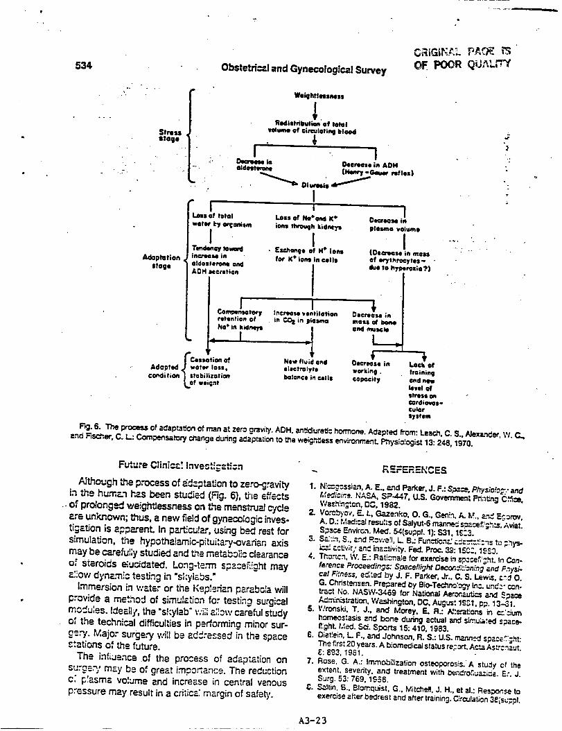
534 Obstetric! and Gynecolog|cal SurveyOF. POOR QuA rr '
Adaptation
|logo
G
.
Fk3. 6. The process of adaptat_c_ of man at zero gravitT. ADH, an_d, Jret_c hormone. Adapted from: Leach, C. S., AlexP.,_d_, W. C..
and Fcsc_,er, C. I_: Compensatory change during adaptation to the weigh=ass e_vironmertL Physiologist 13: 246, 1970.
Future Clinic=!Invest_;z_i:_
A_though the process of _daptat_on to zero-gravityin the human has been studic-d (Fig. 6), _=e eff_tso; prolonged weightlessness on t,_e menstrual cycle_-e unk,nown; thus, a new field of gynecologic inves-tigation is appP_.renL in pa_cu!P..r, using bed rest forsimulation, the hypoth_amic-pitui:a_-ovarian axismay be c_reful;y studied and tlne mstab_iic cl=.aranceo," steroids _.'u_dated. Lo_g-te,"rn s?z:eF',!_ht may
arrow dynzrni_, testing in "sLylabs."immersion in water or the Kep_edan parabda will
provide a n',e'.hod of si,-nuIati_n for testing surgicalmoCu:es. Ideatly, the "shylab" v:ii:a;iow careful studyoJ the technical difficulties in performing minor sur-
get 7. Major surgery will be addressed in the space_tations of the future.
The influenceof the process of adaptation onsvrge.',/may be of great impo_ance. The reductionc; p'asma volume and increase in central venouspressure may result in a critica: margin of safety.
I.
REFERENCES
N!,'_g¢ssizn, A. E., a,nd P_rker, J. F.: Sp_ce, Physio!_E.." and
h:ed_c:,_e, tqASA, SP-447, U.S. Govemme_ Printing C_ce,
Wash!n_:on, _C, 1982.
2. Vo._byov, E. I.,Ga.zen.t',o, O. G., Gee!n, A. M., arts E_:rov,
A. D.: Medic_ resu:_s of Salyuto6 rn_n,,w_ s.:_i_._,L_. AriaS.
Sp._ce Environ. Meal. 54{suppl. 1): $31,1£:3.
3. Sa:Jn, S., _nd Ro,,';e'l, L. B.: Fun_n_ r._=.-'t..--:'.:ns t_ ;,hys-
ic_ _.ct_vit;' and ins:SNip. Fed. Pro¢. 39.°: 15C3, 19E3.
4. Tho._cn, W. E.: Rationale for exercise i_ sp_r_ht. In Con-
ference Proceedings: Spaceflight Decond;:_Jning and Fnys_-
ca/Fitness, e¢;:ed by J. F. Parker, Jr., C. $. Lewis, _._d O.
G. Christensen. Pre_,,ared by Bio-Tedv_:ogy L_:. und. _- _n-
tract t;o. NASWo3469 for National Aer.on_u*3,_ e,'_d Space
Ad,'_n;strat_on, Washington, DC, Augur: le,'31, pp. 13-31.
S. V/ronski, T. J., and Moray, E. R.: N'._r'4t_oP,s _ cc':;,um
homeostasis and bone dunng actual and sLmu;_ted space-
flight, t,'ed. Sci. Sp_rts 15: 410, 1983.
£. Distrain, L F., 8nd Johnson, R. S.: U.S. rr_a_n.,_J space_ght:
The firs: 20 years. A biomedical status reran. A¢*,a AsL-cnz,.Jt.
£: E93, 1961.
7. Rose, G. A.: Im,-'no_ilization osteoporosis. A study of the
extent, seventy, an8 treatment with be,'_roS_zi¢e. Er. J.
Surg. 53: 769, lC_$6.
_,. S,31tin, _., Blomquist. G., Mitch_l, J. H.. et aL: Response to
exercise _her bedrest and after training. Circulation 38{su_.pl.
J__
":":. :"" :+ :i: Y.'o,*n_n in Spacefl+_ht ..... "e
VII):. VII-l, 1958. NASA, SP377, U.S. Government _ Office, Was_gtott,9. E_v.'_, W., Vromcn, N., N$_.--0,D., et -=I.: In_ezse:! vit_t DC, 1977.
c:_,_: • =._1 d_inis.'_cd d;:-f',_ng r_pcdty d_ng pm._jng-=d 22. Lea_, C. S., Johnson, P. C., end Sukll W. N." C_.-rentI:P.,,'_'.L Am. Rev. R_p_.'. Di=. 129: A25_, 12S4. ¢oncept_ of sp_ceE;ht induced changes in t'Kxmon_ ¢=n_l
10. E_e",ks_ W., Vron_n, N., Fa_m_/, S., et _ Etfec_ of alter- of fluid and elecC_yte metabo_sm. Phys_=iog_t 2_=up;_.):r_on of pL_1_-rtP,votume on pu_oncty ft,m¢_gn. Fed. ProP, $24. 1983.43: 2S7, 19_4. 23. Laa.-h, C. S., AIt..-huler, S. L, and C_lzon-Tmvino, N. M- The
11. So;win, C. F.. _,t, A. E., Ru.'rn_, J. A., et aL: en¢_x:ir_ and metaborx: respones to spaceflight. Med. SoLPu_.-nonz.'y _ mre:',-t_on d_"ing _ S._J'-_ =.'_dApo,",o., Spots Exercise 15: 432, 19_3.S_._. _ _ Ariel Sp¢_ E.,wic_n. t_c:L 47:1£_, 1976. 24. Oo::_._, C. B., and G_f, G. E.: Insu_ and g._._ose
12. G.'-.ee_e:.=,J. E., E_'J_cu_.', E. M., Y_..n_, FL L, e*, r3.: Ru_ res.pons¢_ during bedmst v_tb iso,'on_cand isome_ exer-z.-d e_.,c'J_'_t _ d_-'ing bedmst wi_ i_,,.nme_: and iso. rise. J. Appl. Pl_ysiot. Resp_. Environ. Exerdse Pt_-,_. 43:t:._'= ex_rc=se d. A,."_. P_. Re¢_-. Env;:c.'z. F_xerc_e 1033, 1977.F,'P/s_:. 42: 59, lgT/'. 25. RocP., J. A., end Fom_y, S. M- Unpub_s,%,d data, 1984.
13. F= _'*_%', S., Drew, H., =,-_d I.=.=r-=nce, N.: i=_s.'na vo_u,,ne 26. _arn, G. E., MP.rou[is, G. B., _ Marsh_, J. FI_ EwJu-response_ du,'ing bcdn_ L'_he_'_:ty women. Aerosp. k:.-,d. I_.,,_nof ovulation P-'_dco.-pus iuL-_m func_on using=measum-/-,ss_=. PR_p'=-t_ lg-33, p. 245. ments of ptz.srrt_ p,-og_terone. Obstet. Gyne¢o_. 44: 522,
14. E chr_, B. J., Hy.-tt, K. H., K_.'r_ncLs.._7,L C., e*. --'.: Pmve_ 1974.C::_ c' i._.=u¢_d__,m by S_-'p_'_--_uor_y_-¢,._c:_na. 27. Woe.d, H. D., Yoct_mowP_ M. G., S_Jmon, Y. L, et alJ ProtonA_o_..p. MF.J. 41: 495, 1970. irradi¢l_ snd endome1_iosis. AviaL Space Environ. Meal. 54:
15. S;eve.".s, P. M., Lynd% T. N., Johnson, R. L. et al- Eff¢_s of 718, 1933..c-_,_._--,=,_n:_ty__ and venoJs oc_.,Js_on cuffs on 28. Berry, C A.: Med_-...._care of space crews in flight. In r-oun-c--_._:._._: d_"3oP.i"_ of F'o:on_e:! be_.,_st. Ae:osp. d_,,ior,s _f Spzce, Bio/ogy and Medicine, edged by k'. C_Jvinh;_. 37: 10¢g, 1..¢,-35. e.-,d O. G. G_¢n;:o. NASA, U.S. Government Printing O_ce,
1E. E _'_:'.._.L, C. G., -G,z,;'_ne,j,,F. A., =.'x:l Nixo,% J. V.: Cz:d_ovas- Wa._hin_cn, DC, 1975, p. 3425.t;,: :." r_:;_:_r,._ _ l',e_:_-down t_ in y'_._._g_.,_dm_C_e-aged 29. Yz:cshenko, G. L, Terent'ev, V. G., and Mokov, M. D.:men. Pr_sio_:__ 25(s_._p!.): S81, 1983. C,_._¢_-istics of su;gi_J intecven1_on under _ of
17. H-=_'_", W. L, E.,_..#-'_, S. E., _, J. M., st £.: Effect of wc-;g_h_,essness.Voenno-Med. Z.11.10: 69, 1967.p:c':.._:_spa=e_.-;t o,'_..-dizcf,:nc*3o_ _._l_sio_s. In 30. S'-.z_adze, L L, Gonch_ov, I. B., NeumyvakJn, I. P., et al.:_:,3_'= _c_: _=..zu,_ from Sk'vtzJ._,ed__,adby R. S. J(:hnson P..nd ,P-,=_e.'ns of anes,_s_a, surg_,_'y and resusdlatJon dud,'tgL F. D;e._,e.'n.NASA, SP-377, U.S. Government Prin_ng Of- ma."_-d sp_cefii_,hts. Kc_rn. Bk=L Aviakosrn. Me,d. 1(}: 9,_._, ;','_._hin;',,_'=, DC, 1977. 1_$2.
18. t;i::_'3_-ssizn, A. =.., Po_, S. L, Ind R_'_beut, P. C.: Ca'clio- 31. _.,e_,j, C. A.: The med_PJ _ o._ Apollo. Presented at thev=..sc_a_- response to spa=e_;;ht. Phys_o_g_t 2E:suppL): 21_ C,c,'_;;ess on A_aCon anO Space Madicine at Munich,$78. 1983. Gen'n_-_f, September 1973.
19. C_c_,_nian, A. V, l._:e, R. D., TercyP.._ A., et aL: The meta- 32. P_s:.ov, I. D., end Ge_ewo_, S. J.: Weight_essne,,_1,.InL_'!= e.'.d __c e,"mc_ of _o_onge"d bed:est in nor- Fcu,",da_ions of Spa.-e, _o_c_'y end MedirJne, edited by M.-mz: subject. CL,_._.-'n, XLI_ 551, 1974. Ce._n and O. G. Gazenko. NASA, U.S. Government Print_
20. Fu..":hey, S., Bec_e_ W., Tt_-ner, C., et al- Alter-_tion of C_ce, Washihg_on, DC, 1975, voL 2, book 1, p. 305.vs_ous respcr.ses c'unng exercise fol;owing bed, est. "Fed. 33. Terent'ev, V. G., zn:l Kreysina, T. N.: Medical and propt_ac.P-_:. 42: 5S4, 1_3. t_: s_fe_, zt sT,t._e ¢_ews. Ak_l. Nauk SSSR, Moscow, 1970.
21. E-_'_._,_._. S. A., Johnson. K. L, _.'_dHo_er. G. W.: Eva',uation NASA TIF.13.1949.c: t:'_-: e;ecL'c_c--_'.ar,_.J prope_es vf t_e c_'diovascular 34. Rock, J. A.: An expandP...,Ne surgi-,.,al chamber for use in=)'s:-_m _her p..'oionged we_g_:_essness. In Bi_'ned.cal Re- con_;tions of we_htlessness. Aviat. Space Environ. Med. 55:¢: ,.':: !;,_m Sky/ab, e,_t÷:l by R. S. J:._ns,._'_and L..F. D;etI_n. 403, 1984.
ORIGINAL PAGE IS
OF POOR QUALITY
A3-24

Fortney. S.M. 1
TB_qDREGULATORT ADAFEATIONS TO _CTI_TY
t
Suzanne M. Fortney, Ph.D.
Johns Hopkins School of Hygiene and Pubtic Health
Division of Enviro_ental Ph7siology
615 North Wolfe Street
Baltimore, Maryland 21205
Addresa correspondence to:
Dr. Suzanne M. Yortney
Johns Hopkins School of Hygiene and Public Health
615 North Wolfe Street, Room 7032
Baltimore, Maryland 21205
A3-25

YorCney, S.M. 2
ABSTRACT
Exercise conditioning is usually accompanied by increases in blood volume and
improvements in heat tolerance. Deconditioning on the other hand, especially whenJ
p_oduced by extended periods of bedresc, is accompanied by a decrease of blood
volume. In this study, the e££ects o£ bedrest deconditioning on exercise thet'aoregu- •
lator 7 responses were studied, both rich and without an accompany_ reduction in
plasma volume. Two groups o£ women underwent identical 12-day bedrest protocols.
Group 1 (n=12) was ada£nistered no hormonal Creament. and their plasma volumes vere
s£gnificanCly reduced (19.9Z) at the end o£ the bedrest. Group 2 (n=7) received
daily estrogen supplementation (1.25 _ premarin) during bedresc, and they did not
sugger a significant reduction in plasma volume during the last days of bedrest.
Both groups however had _npaired thermoregulaCory responses agter bedrest. Therefore
deconditioning, even i£ unaccompanied by a reduction in plasma volume, results in an
impairaent o£ thermoregulatory £unction.
INTRODUCTION
Although many studies have examined the e££ect o£ increasing £itness level on
heat tolerance 1-5, vet7 few 6 have looked at the effect o£ deconditioning. There are
many reasons to suspect Chat heaC tolerance vould be impaired following periods of
inactivity, especial ly i£ that inactivity involved physical confinement, £or example,
as ogten occurs in patients con_£ned to bed £or several days or weeks. Bedrest
results not only • reduction in £itness level, but also is known to result in pro-
nounced reduction in body £1uid compartments. 7,8 Hypovolemia both with and vithout
electrolyte losses, has been shown to alter signigicantly both the vasodilatory and
the sweating heat loss responses in exercising humans. 9-11
The e££ect og increasing £itness level on local sweating and skin blood £1ov
responses was studied by Roberts and co-workers. 12 The local sweat response was
measured with sweat capsules placed on the chest and connected to a resistance
A3-26
Yortney, $.M. 3
hygrometry _t system. The local sweat rate was then plotted as a function of the
internal body temperature, which was measured with a thermocouple placed in the
esophagus at heart level. A plot of the chest sweat rate (el) as a function of the
r_se in deep body temperature (Tes) characterized the effect o£ increased gitness.
An increase in the slope o5 this $1/Tes relationship indicated that the sensitivity
og the sweating response increased during the training prosr_, which produced an 1I_
increase in the maximum oxygen uptake (V02max) in these subjects. The Tes threshold
at which sweatins began was reduced signigicantly. The skin blood glow (SkBF)
response was measured by electrocapacitance plethysmography. Again this thermoregu-
$atoz7 response was characterized by plotting the skin blood glow values as a gunc-
tion o£ the rise in Tes. The slope o£ the SkBY/Tes relationship was only slightly
altered gollowing trainins. However the Tes threshold at which vasodilation occur-
red. was sign££icantly reduced during the training program,
Thus changes in gitness level may be expected to alter both the sweatin S and the
skin blood glow responses, We would hypothesize further, that after deconditionins,
the internal temperature durins an exercise challense would be elevated to a greatez
extent than before deconditionin S. The hisher core temperature would be due to a
reduction in the slope of the sweatins response, and an elevation in the thresholds
of the sweatinS and skin blood flow responses. Deconditioning as a result o5 bed
confinement would be expected to result in even further _pairment of heat tolerance°
The combined eggects of hypovolemia and deconditioning during bedrest would be pre-
dicted to act in an additive unneT to impair thermal exercise responses.
EXPEIIHEI_TAL METHODS AND RvSULTS
To test our hypothesis that thermoresulatory responses would be i_npaired follow _
ins deconditioning, we studied the exercise responses of twelve young healthy women
begore and agter 12-day prosrems o£ horizontal bedrest. Then to further test the
contribution of hypovolemia on the impsirment of exercise thermal responses, a second
A3-2 7
Fortney, $.M. 4
group of 7 wcaen performed an identical 12-day bedrest protocol, except they were
siren a daily estroaen suppleBent (prmarin) several days before and during the
bedrest. The estrosen suppleaent resulted in an attenuation of the loss of body
fhaids during bedrest so that deconditioniug responses miEht be separated from hypo-
vol emic effects.
Table 1 illustrates the physical characteristics of the two Eroups of women.
All the women in this study reported a normal uenstrual cycle history, with a total
cycle lenEth consistently between 25 and 32 days. Group 1 was composed of 12 women
who did not regularly take medication of any kind. including oral contraceptives.
They were between 21 and 39 years of age, with a group mean age of 27. They were of
average fitness as determined by VO2max testing. Group 2 had similar physical char-
acteristics, but had at some point in their lives used an estrogen-containing oral
contraceptive without suffering siEnificant side effects. These women were in-
structed to ingest one tablet (1.25 mE) of premarin each morning starting on the
third cycle day (where day 1 is the first day of bleeding) immediately before the
bedrest, and to continue this treatment until two days after the bedrest.
The bedrest procedure consisted of 12 days of horizontal bedrest. Each woman
maintained a horizontal position during al 1 but 18 minutes in each 24 hour interval.
These 18 minutes were spent in the sitting position and this was the total accumula-
tive time in which the subjects traveled by wheelchair to and from the bathro_ At
no time did a subject support her weight by standing or walking. At the nae time
each morninE during the bedrest, a blood sample was drawn without stasis to determine
the heaatocrit and hemoglobin concentratio_ These values were used to calculate 13
the relative chanEes in plasma volume between the first and each succeeding day of
the bedrest.
The absolute red cell volume and plasma volume of each woman was determined by
radioisotope dilution. 14 99M technetium pertechnetate was used to label a sample of
A3-28
Yortney, S.M. 5
each subject's red blood cells to determine absolute red cell volume, and 991/ techne_
tium-labelled human serum albuman was used to determine absolute plasua volume.
&bsolute red cell and plasma volume determinations were per£ormed one day begore and
wil:hin 48 hours a£ter each bedrest°
Although signigicant reductions in red cell volume have been reported during
longer bedrest procedures 15. in this study there was a tendency to reduce red cell
volume during bedrest, but these di£gerences were not signi£icant (P<0.05 £rom t-test
determinations). Red cell volume averaged 1474 + 44 and 1326 + 95 be£ore bedrest for
Groups 1 and 2 respectively. The red cel 1 volumes got the two groups averaged 1389
42 and 1240 + 90 ml agter bedrest.
_'_e 1
Figure 1 illustrates the changes in plasma volume during the two bedrest proce-
dures. Plasma volume decreased significantly during the £irst 4-5 days o£ bedrest in
the women who did not receive estrogen supplement (Group 1). A narkedly di£ferent
pattern however was seen in Group 2. Plasma volume decreased significantly during
the £irst two days o£ bedrest. Then the decrease in plasma volume was attenuated and
even reversed, such that. the plasma volume in the group o£ women who received
estrogen supplement was not significautly di££erent (P<0.05. Duncan Multiple Range
comparisons) from their £irst day o£ bedrest during 7 of the last 8 days o£ the
bedrest.
Submaxiaal exercise tests were performed in a warm (30 degrees C, 50-60Z rela-
tive humidity) tsmperature-control, led roou. one da_ before and i_ediately after
bedrest. The exercise intensity on the low-sit cycle ergoueter was adjusted to 70:[
of each subjects pre-bedrest VO2nax, which was determined previously and defined by a
plateau in the oxygen consumption/exercise intensity relationship measured during
progressive cycle exercise tests. The submaximal tests continued for 30 minutes, o_
until a subject's heart rate exceeded 95Z of her pre-bedrest maxi_ heart rate
A3-29

rortney, S.M. 6
value.
Subaaximal exercise tolerance was reduced significantly in both groups of rouen
golloving bedfast, despite the marked differences in plasma volume decrease during
bedrest between Croups 1 and 2. Whereas, before bedrest each roman vas able to
comfortably complete the 30-minute exercise task, golloving bedrest, only one woman
in Group 1, and none of the roman in Group 2 could complete the thirty minutes of
exercise vithout being instructed to stop because theft heart rate had reached 95
percent of their maximal heart rate.
• _p=e 2
The heart rate responses during these submaximal tests are illustrated in Figure
2. The average -+ standard error values for the first group of women are shown in the
top panel of this £_ure, for both pre-bedrest and post-bedrest exercise. Since the
submaximal exercise tests vere performed immediately after bedfast, a pastural stress
vas induced by the assumption of the sitting position in the lay-sit cycle for 30
minutes before the start of exercise. Thus even at rest, heart rates vere sign£fi-
cantly h_gher than pre-bedrest values in the post-bedrest tests. Initially, we
attributed at least part of the increase in heart rate after bedrest to a hypovolemic
effect. The average plasma volume at the start of exercise vas 432 ml lover during
the post-bedfast teat than during the pre-bedresc test.
However, the results in the bottom half of Figure 2 illustrate the heart rate
responses in the second group of vomen. The average difference in plasma volume at
the start of exercise between the pre-bedrest and the post-bedrest tests was only 130
ml (non-significant). Yet an examination of the heart rate responses folloving
bedfast, shove a similar pa_Cern as seen in Group I, that is a similar increase in
heart rate post-bedrest.
Yiswre 3
Continuous measurement of body core temperature was obtained during each exer-
A3-30

Yortney, $.M. 7
rise test frma a theraocouple placed in the esophagus aC the level of the heart and
these results are illustrated in Figure 3. Asaln the responses from Group I are
shown in the top panel of this figure. Like the heart race responses, the esophageal
tehperaCure (Tes) already was elevated sisuif£cantly above pre-bedrest levels before
the start of the post-bedresc test. Tea remained higher than pre-bedresC time-
matched values throughout exercise following bedrest. A similar pattern of Tea
response occurred in the pre- and post- bedrest tests in Group 2 (bottom panel).
Hoverer, the rise in Tee during posC-bedrest exercise was significantly greater than
before bedrest in group 1 (p • 0.02), buC noC in the group 2 (p • 0.67).
The total sweat output was est_ted in these exercise tests, by accurately
veighing each subject in dry cotton clothing before and after exercise. The changes
in body weight are shown in Table 2.
Table 2
The total change in body weight (g) was less in the posc-bedrest tests, in part
due to the shorter exercise period. When the sweat rate _s expressed as a weight
loss per minute of exercise, (g/sin), the total sweat rate vas siguificantly higher
in the post-bedrest tests for both groups 1 and 2. It appears chac the sweating
response was not inhibited by deconditioning fol lowing bedrest, and that the higher
post-bedrest sweat rate vould be expected to accompany the higher Tea values. We had
predicted an inhibition of mfeating in the hypovolemic exercise condition (Group 1,
post-bedrest), as compared to the normovolemic post-bedrest conditions (Group 2).
Although there vas a tendency for the sveat races Co be lower in Croup ! than in
Group 2 following bedrest, these differences just barely were not significant
(P•O.06).
DISCUSSION
In these studies we have developed a model to examine the effects of bedrest,
both with and without plasma volome reduction, on submaximal exercise responses. We
A3-31

Yortney, $°M. 8
predicted that exercise tolerance would be reduced following bedrest for two reasons.
First, a certain degree of physical deconditioning would be expected following 2 days
of bedfast; then, since the same absolute exercise intensity (70Z of the pre-bedrest
V(_2max) was used in the pre- and post-bedfast tests, post-bedfast exercise was per-
formed at a h£gher relative exercise intensity. Second, it has been reported in men
during bedrestlS, that a spontaneous diuresis occurs in the first few days of a
bedfast procedure. PresuBably a Henry-Gauer type of reflex is illicited 16 by an
increase in central blood volume caused by the cephalad movezent of body fluids with
the assumption of the horizontal position. Cardiopulmonary stretch receptors are
then stimulated resulting in an increased urine output and a decrease in total body
fluids. Tn this study the decrease in plasma volume averaged about 20Z in Croup 1.
In previous studies 10"11"17 in which plasma volume was reduced to a si1ilar degree
through the use of diuretics or by blood withdrawal before submaximal exercise, the
thermoregulatory responses were i_rpaired significantly. The surprising finding in
this study was that exercise responses following bedrest were equally impaired in
these subjects, whether the plasma volume was reduced (by about 20Z), or restored to
the pre-bedrest level before exercise testing. An attempt was made to measure the
decrease in VO2max within 48 hours after the end of the bedfast procedure. However,
following bedfast, we were not able to attain consistently our criteria for
determination of V02max; that is, a plateau of the oxygen constzaption/exercise inten-
sity curve. Only peak oxygen uptake values could be obtained, because most subjects
stopped the progressive exercise test due to leg fatigue rather than a cardiovascu-
lar limitation- The peak VO2 value obtained following bedfast in group 1 averaged an
II.SZ ..... "-.=d,_t,vn fr_ th= pre-bedrest values. Croup 2 showed a 2i.5Z average reduc-
tion. There was tremendous variation in the responses of the individual subjects in
each group, and so the decrease in peak VO2 seen following bedrest was not statist-
ically different (p • 0.05) between Group 1 and Croup 2. Thus there is no reason to
A3-32

Fortney, S.M. 9
believe that the decondit£onins effect was different between the _ Woupe.
An alteration in themoresulatory responses vas evident after bedrest even
before the start of exercise. The higher restin$ core temperature and heart rate in
thb post-bedrest tests occurred before the initiat£on of the sweating reglex. This
gact, together with the lack og a si_£icant reduction in the sweat rate gollowing
bedrest, argues against the idea that deconditionins may have i_hib£Ced the sweat£ng
response, and thus been responsible for the higher body temperatures a£ter bedreet.
There are t_o other possible explanations got the impaired heat tolerance a£te_
bedrest. The £irst fs that there was a change in the resting metabolic rate agter
bedrest, such that the total body heat product£on increased both at rest and during
exercise. This explanation seems unlikely, as oue would expect that i£ any change
occurred in the resting orygen consumption during bedrest. VO2 would decrease, not
increase.
The second more likely explanation is that there was an alteration in vasomotor
tone £ollowing bedrest, producing a greater vasoconstriction which decreased heat
exchange bet_een the skin sur£ace and the environment. Indirect evidence to support
this hypothesis is the greater venoconstr£ction we reported in a _oup og women at
rest and durins exercise gol lowing bedrest. 18 At the time o£ this report, ve sug-
gested that gollowing bedrest, there vas an increase £n peripheral venocoustr£ctor
tone which helped to compensate for the reduction in plasma voleme, and which gunc-
tioned to uaintain central blood volume. Although in this previous study ve measured
an increase in gorearm venous cone. ve mi4|hC also expect a greater cutaneous arterio-
lar Cone, since both venous and arteriolar constrictor responses are produced by the
increased s_pathetic drive vhich occurs with the onset o£ exercise. Although the
cutaneous vessels _ay show an increase in Cone gollowin8 bedrest, a decrease in the
tone may occur in the deeper blood vessels og the lower extremities or o£ the
splanchnic region. Thus following bedrest when the subjects in this study sat up-
A3-33

ForCney, S.M. 10
right just prior to the ezercise tests, blood may have pooled in dependent body
re$ions, illicltinS a vasoconstrictor response and £mpairin$ cutaneous heat ezchange.
The potential for blood poolinS followins bedrest may be so great, that even Id,en the
plasma volume vas restored durinS bedrest (Group 2), blood still pooled in the lower
extremities, produc£us a vasoconstrictor response and i_pairins heat loss. Thus even
an expansion of plasma volume following bedrest may not improve ezercise tolerance,
but may simply lead to greater blood poolins, and in eztreme cases, may produce lower
body edema.
CONCLUSIONS
Bedrest in healthy youns women resulted in impairment of thermores_latory res-
ponses both at rest and during exercise in a vaz_ environment. The higher body
temperature following bedrest may have been due to effects caused by deconditioains
or to effects caused by the hTpovolemia vhich normally accompanies bedrest. In this
study the sweating responses vere not significantly altered following bedrest. It is
most likely that the higher body temperatures were caused by a cutaneous vasocon-
strictor response vhich functioned to oppose blood pooling in the lower extremities.
Restoring blood volume to pre-bedrested values was ineffective in reversing the
impairment of Chermoregulatory responses.
ACKN_LEDGEMENTS
This york was supported by NASA Contract NAS9-16703. Thanks to Clarke
Tankersley, Kirstin Levine, and Kay Chadvell for their help in the preparation of
this manuscript.
A3-34

Yortney, S.M. 11
HFE_gCZS
1. Strydom, N.B.o Wyndhma, C.L, Williams C.G., Morris,n, J.7., Bredell, G.A,G.,_enade, A.J.S. and Vou Lshden, 14. Acclimatization to humid heat and the role of
physical conditioning. J. Appl. ?hTsiol. 21" 636, 1966.
2. +' Pivonka. B.W.. Robinson. S.. Gay. V.I. and Manalis, LS. Preaccliaatizatiou
of men to hut by training. J. Appl. Physiol. 20: 379. 1965.
3. Pandolf. LB. Effects of physical training and cardiorespiratory physicalfitness on exercise-heat tolerance: recent observations. }fed. Sci. Sports 11:60, 1979.
4. Gisolfi, C. and Robinson, S. Relations between physical training,acclimatization, and heat tolerance. J. Appl. Physiol. 26: 530, 1969.
5. Henane. g., Flandrois, B.L and Chazbounier, J.?. Increase in sweating
sensitivity by endurance conditioning in man. J. Appl. Physiol. 43: 822, 1977.
6.. Greenleaf, J.Y., and Reese, R.D. Exercise thermoregulation after 14 daysof bedrest. J. Appl. Physiol. 48: 72, 1980.
7. Taylor, ILl., Ericksou, I., Henschell, A. and Kaye, A. The effect of bedrest
on the blood volume of normal young men. Am. J. Physiol. 144: 227, 1945.
8. Hargens, A.L Fluid shifts in vascular and extravascular spaces during andafter simulated weightlessness.
9. Horetnnan, D.IL, and Horvath, S.IL Cardiovascular and temperatureregulator T changes during progressive dehydration and euhydration. J. Appl.Physiol. 33: 446, 1972.
10. Nadel. F.R., Fortney. S.IL, and Wenger, C._ Effect of hydration state on
circulatory and thermal regulations. J. Appl. Physiol. 49: 715, 1980.
11. Fortney. S.M., Nadel, E.R.. Wenger, C.B., and Bore, J.R.volume on sweating rate and body fluids in exercising humans.51: 1594, 1981.
Effect of blood
J. Appl. Physiol.
12. loberts, M.7., WenKer, C.B., Stolvijk, J.A.J., and Nadel, F.R. Skin bloodflov and reseating changes foil,rinK exercise training and heat acclimation. J.
Appl. Physiol. 43: 133, 1977.
13. Dill, D.B. and Costill, D.I. Calculation of percentage changes in volumes
of blood, plasla, and red cells in dehydration. J. Appl. Physiol. 37: 247,1974.
14. Glass, ILl. Standard techniques for the measurement of red-cell and plasmavolume. Br. J. of Haematology 25: 801, 1973.
15. Greenleaf, J.E. Physiological responses to prolonged bedrest and fluidi_mersion in humans. J. Appl. Physiol. 57: 619, 1984.
16. Gauer, O.IL, Henry, J.P. and Sieker, ILIL Cardiac receptors and fluid volumecontrol. Prof. in Cardiovas. Des. 4: 1, 1961.
A3-35
Fortney, S.M. 12
17. Yortney, S.M., N&del, F.B., Wenger, C.B., and Bore, J.R. Effect o£ acute
alterations of blood volme on circulatory performance in humans. J. Appl.Physiol. 50: 292, 1981.
18. Yortney, S.X., Beckett, W., Turner, C., Vroman, N._ and Wilkinson, I.ALteration o£ venous responses during exercise £ollov£ng bedrest. Fed. Proc.42: 584, 1983.
A3-36
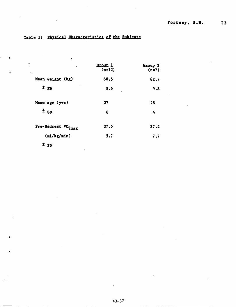
Fortney, $.M. 13
Table 1: _ CharacCer*nClcs of che_b_
(n=12)
60.5
8.0
.Ccau_ Z(.=7)
62.7
9.8
27
6
26
4
Pre-BedresC VO2max
-* SD
37.2
7.7
A3-37

Yortney, S.M. 14
Table 2: Total Body Sveat Losses Dorlnf Zxerc£se
Grou_ I (Non-esero_en) Grou_ 2 {Esero_en3
Change of body wt. 512 *436 486 409
-+ S.E. 20 19 33 47
Cg)
Minutes of exercise 30 * 21 30 * 16
-+ S.E. 0 2 0 2
Cmtn)
Total Sveat B_tte 17.31 * 22.19 16.19 * 26.84
-+ S.V.. 2..56 4..59 2.73 5.00
0.62 * 0.78 0.89 0.83
0.06 0.07 0.16 0.09
Increase i.=Tes
* Post-Bedrest value significantly different from Pre-Bedrest value
(P__0.05). Paired t/test comparison.
A3-38
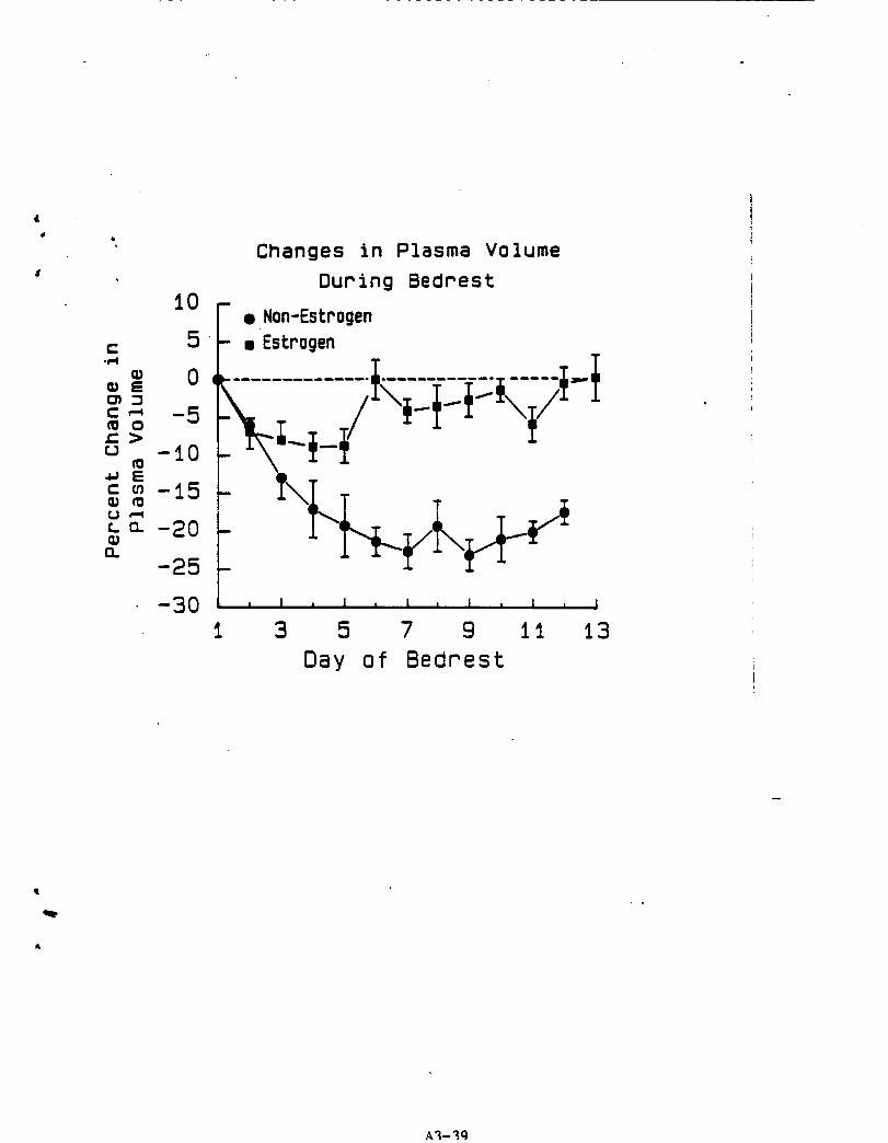
' Changes in Plasma Volume
During BedrestI0
• Non-Estrogen
.c 5 _" • Estrogen
c_ _ -50 _--................................/___.___/_ _-
E -2o-25
-30 " ' ' ' ' , ' , ' , ' , i
1 3 5 7 9 11 13
Day of Bedrest
I
A_-3q

Heart Rates
During Exercise
18_ F Non Estrogen170 Srou=
155
140
i25
ilO
95 _/ -- _ = in.12).
) 80 _ o Post= (nia)B5
185 r- Estrogen Grou_
"_o _-
:z:ii0i25i4015595i//I�Ill /__----_--_, Pre BR in=7)
oo65
0 5 10 15 20 25 30Exercise Time (rain)
_b
A3-40

4_
Esophageal Temperatures
During Exercise
3B. 5 r/ Non-Estrogen
38.3 I- s mrou.
37.9 -
_ 37.737.5
37.3 _ PPeBR (n.,la
o PoSt BR {n-12}m 37.1L
36.9 -
E
385-
_ 38 3 -
C_m 38 I -
{=.379 _0tm,,, 377
37 5
37 3
37 1
36.90
/
/
Tj Estrogen GrouD
/
• Pee BR (n-7)
. Post: BR (n-7)
I I I I I I
5 10 15 20 25 30Exercise Time {mln)
dl i ..
r
A3-41