Tsg101 Is Essential for Cell Growth, Proliferation, and Cell Survival of Embryonic and Adult Tissues
14
10.1128/MCB.23.1.150-162.2003. 2003, 23(1):150. DOI: Mol. Cell. Biol. Riedlinger, Edmund B. Rucker III and Lothar Hennighausen Park, MaLinda D. Henry, Aleata A. Triplett, Gregory Kay-Uwe Wagner, Andrea Krempler, Yongyue Qi, KyungRan Embryonic and Adult Tissues Proliferation, and Cell Survival of Tsg101 Is Essential for Cell Growth, http://mcb.asm.org/content/23/1/150 Updated information and services can be found at: These include: REFERENCES http://mcb.asm.org/content/23/1/150#ref-list-1 at: This article cites 37 articles, 12 of which can be accessed free CONTENT ALERTS more» articles cite this article), Receive: RSS Feeds, eTOCs, free email alerts (when new http://journals.asm.org/site/misc/reprints.xhtml Information about commercial reprint orders: http://journals.asm.org/site/subscriptions/ To subscribe to to another ASM Journal go to: on January 24, 2014 by guest http://mcb.asm.org/ Downloaded from on January 24, 2014 by guest http://mcb.asm.org/ Downloaded from
Transcript of Tsg101 Is Essential for Cell Growth, Proliferation, and Cell Survival of Embryonic and Adult Tissues
10.1128/MCB.23.1.150-162.2003.
2003, 23(1):150. DOI:Mol. Cell. Biol. Riedlinger, Edmund B. Rucker III and Lothar HennighausenPark, MaLinda D. Henry, Aleata A. Triplett, Gregory Kay-Uwe Wagner, Andrea Krempler, Yongyue Qi, KyungRan Embryonic and Adult TissuesProliferation, and Cell Survival of Tsg101 Is Essential for Cell Growth,
http://mcb.asm.org/content/23/1/150Updated information and services can be found at:
These include:
REFERENCEShttp://mcb.asm.org/content/23/1/150#ref-list-1at:
This article cites 37 articles, 12 of which can be accessed free
CONTENT ALERTS more»articles cite this article),
Receive: RSS Feeds, eTOCs, free email alerts (when new
http://journals.asm.org/site/misc/reprints.xhtmlInformation about commercial reprint orders: http://journals.asm.org/site/subscriptions/To subscribe to to another ASM Journal go to:
on January 24, 2014 by guesthttp://m
cb.asm.org/
Dow
nloaded from
on January 24, 2014 by guesthttp://m
cb.asm.org/
Dow
nloaded from
MOLECULAR AND CELLULAR BIOLOGY, Jan. 2003, p. 150–162 Vol. 23, No. 10270-7306/03/$08.00�0 DOI: 10.1128/MCB.23.1.150–162.2003Copyright © 2003, American Society for Microbiology. All Rights Reserved.
Tsg101 Is Essential for Cell Growth, Proliferation, and Cell Survival ofEmbryonic and Adult Tissues
Kay-Uwe Wagner,1* Andrea Krempler,1 Yongyue Qi,1 KyungRan Park,1 MaLinda D. Henry,1Aleata A. Triplett,1 Gregory Riedlinger,2 Edmund B. Rucker III,2†
and Lothar Hennighausen2
Eppley Institute for Research in Cancer and Allied Diseases, University of Nebraska Medical Center,Omaha, Nebraska 68198-6805,1 and Laboratory of Genetics and Physiology,
National Institute of Diabetes, Digestive, and Kidney Diseases, NationalInstitutes of Health, Bethesda, Maryland 20892-08222
Received 31 July 2002/Returned for modification 9 September 2002/Accepted 25 September 2002
Tumor susceptibility gene 101 (Tsg101) was identified in a random mutagenesis screen for potential tumorsuppressors in NIH 3T3 cells. Altered transcripts of this gene have been detected in sporadic breast cancersand many other human malignancies. However, the involvement of this gene in neoplastic transformation andtumorigenesis is still elusive. Using gene targeting, we generated genetically engineered mice with a floxed alleleof Tsg101. We investigated essential functions of this gene in vivo and examined whether the loss of functionof Tsg101 results in tumorigenesis. Conventional knockout mice were generated through Cre-mediated excisionof the first coding exon in the germ line of mouse mammary tumor virus (MMTV)-Cre transgenic mice. Thecomplete ablation of Tsg101 in the developing embryo resulted in death around implantation. In contrast,mammary gland-specific knockout mice developed normally but were unable to nurse their young as a resultof impaired mammogenesis during late pregnancy. Neither heterozygous null mutants nor somatic knockoutmice developed mammary tumors after a latency of 2 years. The Cre-mediated deletion of Tsg101 in primarycells demonstrated that this gene is essential for the growth, proliferation, and survival of mammary epithelialcells. In summary, our results suggest that Tsg101 is required for normal cell function of embryonic and adulttissues but that this gene is not a tumor suppressor for sporadic forms of breast cancer.
The tumor susceptibility gene 101 (Tsg101) was first discov-ered in murine fibroblasts in a screen for potential tumorsuppressors by using retroviral gene trap vectors in combina-tion with an antisense strategy (13). A ligand-inducible orconstitutive expression of the partial and full-length Tsg101cDNA in antisense orientation led to instant transformation ofNIH 3T3 cells. The transformed cells were capable of formingmetastasizing tumors in nude mice. Neoplastic transformationof the NIH 3T3 cells was reported to be reversible when thefunction of Tsg101 was restored (13). Cloning and sequencingof the human TSG101 cDNA revealed that the mouse andhuman genes encode proteins with more than 94% similarity atthe amino acid level (14). The human TSG101 gene wasmapped to chromosome 11 p15.1-p15.2, a site that shows lossof heterozygozity in a subset of sporadic breast cancers andother human malignancies (14). Although somatic mutationsor deletions within the TSG101 gene are rare events, aberrantsplice forms have been observed frequently in a variety ofhuman cancers (7, 12, 27, 28). After cloning, sequencing, andrevising the TSG101 gene structure in both the human and themouse, we showed that many of the previously described ab-
errant transcripts in human malignancies were in fact alterna-tive splice forms generated solely by exon skipping (31, 32).
Various biological functions of Tsg101 have been postulatedfrom the predicted protein structure, the intracellular localiza-tion of Tsg101, the identification of Tsg101 binding proteins,and the deletion of the putative homologous gene Vsp23p inyeast. These functions include a role in ubiquitination (10, 23),transcriptional regulation (8, 36), endosomal trafficking (1, 6),and cell proliferation (37, 40). Although some of these func-tions have been verified experimentally in cell culture systems,their relevance in vivo during normal development and neo-plastic transformation is still elusive. In addition, it has neverbeen confirmed, using a site-directed targeting approach, thatthe deletion of both endogenous Tsg101 alleles leads to accel-erated growth and neoplastic transformation of nonimmortal-ized cells and to tumorigenesis in vivo. Based on our earlierfindings that (i) Tsg101 is expressed in all tissues of the mousethroughout development and (ii) the 5� sequence of this genehas common features of housekeeping gene promoters, wehypothesized that a conventional knockout of this gene wouldresult in early embryonic death (32). Therefore, we have cho-sen the Cre-loxP technology, which allows the conditional de-letion of Tsg101 from any given cell type in genetically engi-neered mice. In this article, we report that Tsg101 is essentialfor cell growth, cell survival, and normal function of embryonicand adult tissues. Tsg101-deficient cells exhibited a defect incell cycle regulation and underwent increased cell death. Incontrast to an earlier report (13), a Tsg101 knockout did notresult in accelerated cell growth and instant neoplastic trans-
* Corresponding author. Mailing address: Eppley Institute for Re-search in Cancer and Allied Diseases, University of Nebraska MedicalCenter, 986805 Nebraska Medical Center, Rm. 8009, Omaha, NE68198-6805. Phone: (402) 559-3288. Fax: (402) 559-4651. E-mail:[email protected].
† Present address: Animal Science Research Center, University ofMissouri, Columbia, MO 65211.
150
on January 24, 2014 by guesthttp://m
cb.asm.org/
Dow
nloaded from
formation. Our findings suggest that a null mutation of Tsg101is not an initiating event for tumorigenesis.
MATERIALS AND METHODS
Gene targeting. Overlapping BAC clones encompassing the Tsg101 locus wereisolated from a mouse 129SvJ genomic library and mapped (31, 32). A 11.5-kbEcoRI fragment was cloned into pZero (Invitrogen, Inc.), sequenced, and usedas a template to generate the floxed targeting vector (see Fig. 1A). Three
consecutive fragments (2.9, 3.1, and 3.3 kb) approximately 950 bp downstream ofthe 5� EcoRI site were amplified with Pfx polymerase (Gibco BRL) and clonedinto pZero. The 2.9-kb 5� fragment was released by EcoRI and subcloned intopLoxpNeo (39), resulting in an additional EcoRI site 5� of the PGK-neomycin(PGK-neo) expression cassette, which was used to verify the targeting event bySouthern blotting. The 3� homology region of the targeting vector was generatedby subcloning the 3.1- and 3.3-kb PCR fragments into pBS64, thereby insertinga loxP sequence and an additional EcoRI site into intron 1. After inserting anoligonucleotide containing an XhoI recognition site into the 5� end of the ho-
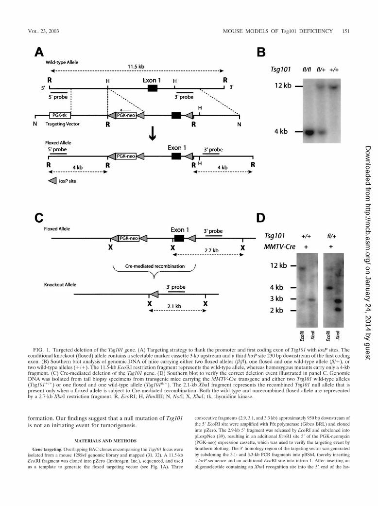
FIG. 1. Targeted deletion of the Tsg101 gene. (A) Targeting strategy to flank the promoter and first coding exon of Tsg101 with loxP sites. Theconditional knockout (floxed) allele contains a selectable marker cassette 3 kb upstream and a third loxP site 230 bp downstream of the first codingexon. (B) Southern blot analysis of genomic DNA of mice carrying either two floxed alleles (fl/fl), one floxed and one wild-type allele (fl/�), ortwo wild-type alleles (�/�). The 11.5-kb EcoRI restriction fragment represents the wild-type allele, whereas homozygous mutants carry only a 4-kbfragment. (C) Cre-mediated deletion of the Tsg101 gene. (D) Southern blot to verify the correct deletion event illustrated in panel C. GenomicDNA was isolated from tail biopsy specimens from transgenic mice carrying the MMTV-Cre transgene and either two Tsg101 wild-type alleles(Tsg101�/�) or one floxed and one wild-type allele (Tsg101fl/�). The 2.1-kb XbaI fragment represents the recombined Tsg101 null allele that ispresent only when a floxed allele is subject to Cre-mediated recombination. Both the wild-type and unrecombined floxed allele are representedby a 2.7-kb XbaI restriction fragment. R, EcoRI; H, HindIII; N, NotI; X, XbaI; tk, thymidine kinase.
VOL. 23, 2003 MOUSE MODELS OF Tsg101 DEFICIENCY 151
on January 24, 2014 by guesthttp://m
cb.asm.org/
Dow
nloaded from

mologous region, the entire 6.4-kb fragment was released from the pBS64 vectorby using XhoI and NotI and subcloned into the pLoxpNeo vector containing the2.9-kb 5� homology region. The final targeting construct, designatedpFloxedTSG101, contained a floxed PGK-neo selection marker approximately 3kb upstream and a third loxP site 230 bp downstream of the first coding exon. ThepFloxedTSG101 plasmid was linearized using NotI and electroporated into iso-genic RW4 embryonic stem (ES) cells (GenomeSystems, Inc.). Analysis of G418-resistant clones by Southern blotting using 5� and 3� diagnostic probes (Fig. 1)indicated that about 7% of the ES cell clones (10 of 150) had undergone correcthomologous recombination both in the 5� region and at the 3� end downstreamof the third loxP site. Of the 10 correctly targeted ES cell clones, 2 (clones 7 and36) were expanded and injected into C57BL/C blastocysts. Germ line transmis-sion of the Tsg101 floxed allele was achieved from male chimeras of both clones.
PCR genotyping protocols. The PCR protocols for genotyping mouse mam-mary tumor virus (MMTV)-Cre and WAP-Cre mice have been described previ-ously (34). MMTV-Cre and WAP-Cre mice are available from the JacksonLaboratory (stock 003551, 003553, and 003552) and the National Cancer Insti-tute repository in Frederick, Md. For identification of the various Tsg101 allelesby PCR, we used the following primer sets: wild-type allele (5�-GTT CGC TGAAGT AGA GCA GCC AG-3� and 5�-CAT TTC TGG AGT CCG ATG CGCAG-3�), floxed allele (5�-AGA GGC TAT TCG GCT ATG ACT G-3� and5�-TTC GTC CAG ATC ATC CTG ATC-3�), and recombined/null allele (5�-GAT GGT CAT ACC TGG TTA GAA AGC-3� and 5�-CAT TTC TGG AGTCCG ATG CGC AG-3�). Note that the primer set for the wild-type allele isspecific; i.e., it will not recognize the targeted floxed allele or the null allele. Allanimals used in the described studies were treated humanely and in accordancewith federal guidelines and institutional policies.
Southern and Northern hybridization. Genomic DNA from tissues or celllines was prepared using standard phenol-chloroform extraction, and 15 �g wasdigested with EcoRI or XbaI at 37°C overnight. The DNA was separated on a0.8% agarose gel, blotted onto a nylon membrane (GeneScreen Plus; NEN), andhybridized with a 32P-labeled probe. The 5� diagnostic probe, which was used toscreen targeted ES cells, was generated by PCR using the primer set 5�-TTGCTT GTA TGA GTG CAG GTG CC-3� and 5�-TAT GAC CTG CTT CTTGCA AAA GCA G-3�. The 3� internal probe for identification of the variousTsg101 alleles was amplified by PCR using the forward and reverse primers5�-ATC TTA TCT CCC ATC CTA AGC AGA C-3� and 5�-TCT CTC ACATCA CTA AAG CTC AAT G-3�, respectively. Membranes were washed in 0.1�SSC buffer (1� SSC is 0.15 M NaCl plus 0.015 M sodium citrate) containingsodium dodecyl sulfate (SDS) and exposed for 6 h to a Kodak X-Omat-AR film.
The isolation of total RNA from mammary tissue has been described previ-ously (32). A 20-�g portion of total RNA was separated on a 1.5% formaldehydegel and transferred to a GeneScreen Plus membrane. Tsg101 transcripts weredetected by probing the membranes with 32P-labeled full-length mouse Tsg101cDNA.
Whole-mount analysis of the mammary gland and other histological tech-niques. The preparation and staining of mammary gland whole mounts anddetailed protocols for immunohistochemistry and bromodeoxyuridine (BrdU)labeling of proliferating mammary epithelial cells were described previously (35).For immunohistochemistry, mammary gland tissue was fixed for 5 h in 4%(vol/vol) paraformaldehyde or overnight at 4°C in 10% buffered formalin (FisherScientific Co.). The specimens were embedded in paraffin by standard methods.Sections were deparaffinized, rehydrated, and treated with antimasking solutionfrom Vector Laboratories, Inc., as specified by the manufacturer. We used a 1:10dilution of the M30 CytoDEATH stock solution (see detailed protocol by RocheMolecular Biochemicals) to label apoptotic mammary epithelial cells. The Vecta-stain Elite ABC and peroxidase substrate kit from Vector Laboratories, Inc., wasused to visualize labeled cells. The slides were counterstained with hematoxylin.Immunohistochemical detection of BrdU-labeled nuclei was performed by fol-lowing the Amersham-Pharmacia-Biotech (RPN202) protocol.
Construction of retroviral expression vectors. The Cre coding sequence fromvector pBS185 (a kind gift of B. Sauer, National Institute of Diabetes, Digestive,and Kidney Disease [NIDDK], to L. Hennighausen, NIDDK) was cloned as anXhoI-MluI(blunt) fragment into the EcoRV-XhoI sites of pZero (Invitrogen,Inc.). The pBabe-Cre-puro retroviral vector was constructed by subcloning theCre recombinase cDNA as an XhoI(blunt)-EcoRI fragment into the Bam-HI(blunt) and EcoRI sites of pBabe-puro. We have used 293T cells to generatereplication-deficient viral particles of pBabe and its Cre-expressing derivative(pBabe-Cre).
Cell culture and immunocytochemistry. Primary mammary epithelial cell cul-tures were prepared similarly to methods described previously (16, 20). Theepithelial cells were maintained in Dulbecco’s minimal essential medium(DMEM)/F12 supplemented with 2% fetal calf serum, 10 �g of insulin per ml,
10 ng of epidermal growth factor per ml, 10 �g of gentamicin per ml, 100 U ofpenicillin per ml, and 100 �g of streptomycin per ml. For retroviral infection,cells at passages 2 and 3 were plated at a density of 3 � 105 to 4 � 105 cells per10-cm culture dish. Infection with pBabe-Cre or control constructs lacking theCre coding sequence (pBabe) was performed in the presence of 10 �g of poly-brene (Sigma) per ml. Forty-eight hours later, the cells were selected in completemedium containing 3 to 7 �g of puromycin (Sigma) per ml.
For immunocytochemistry, cells were fixed in 70% ethanol, washed twice in1� phosphate-buffered saline (PBS) and incubated for 20 min in blocking solu-tion (1� PBS, 1% bovine serum albumin, 0.1% Tween 20). The slides weretreated for several hours with a 1:500 to 1:1,000 dilution of the primary antibodyin blocking solution (anti-proliferating cell nuclear antigen [PCNA] [PC-10],anti-cyclin A [C-19] [both from Santa Cruz], or anti-pan-cytokeratin [Sigma]).The primary PCNA and anti-pan-cytokeratin antibodies were fluorescein iso-thiocyanate (FITC)-conjugated. After an additional washing step, cyclin A wasvisualized with an FITC-conjugated secondary antibody (sc-2012 [Santa Cruz];1:1,000 dilution). The slides were washed repeatedly in 1� PBS and mountedwith Vectashield containing 1.5 �g of 4�,6-diamidino-2-phenylindole (DAPI)(Vector Laboratories, Inc.).
Western blot analysis. Cell pellets were lysed on wet ice for 30 min in 1�PBS–1 % NP-40–0.5% sodium deoxycholate–0.1% SDS, 1 mM phenylmethyl-sulfonyl fluoride–0.4 U of aprotinin per ml–1 mM NaF–0.1 mM sodium or-thovandate. Protein was quantified using a Bradford assay (Pierce). Protein (20to 50 �g per lane) was resolved by SDS-polyacrylamide gel electrophoresis andblotted onto polyvinylidene difluoride membranes (Invitrogen). The membraneswere blocked for 1 h in 1� TBS–0.1% Tween 20–5% dry milk. They were thenincubated with primary antibodies in blocking buffer at 4°C overnight, washedthree times for 15 min in washing buffer (1� TBS–0.1% Tween 20), and incu-bated for 1 h at room temperature with horseradish peroxidase-conjugatedsecondary antibodies in blocking buffer. The membranes were washed againthree times in washing buffer and once for 15 min in 1� TBS without Tween 20.Protein bands were detected using the ECL chemiluminescence kit for Westernblot analysis (Amersham) as specified by the manufacturer. Membranes werestripped using 0.2 M NaOH for consecutive detection of various proteins. Theanti-ActB (I-19) and anti-Tsg101 (C-2) antibodies from Santa Cruz Biotechnol-ogy were used at a 1:1,000 dilution. Horseradish peroxidase-conjugated second-ary antibodies were purchased from Santa Cruz Biotechnology and used at a1:1,000 dilution.
RESULTS
Generation of genetically engineered mice with a condi-tional knockout allele of Tsg101. A conditional knockout(floxed) allele of the Tsg101 gene was generated through ho-mologous recombination in mouse ES cells (Fig. 1A). ThePGK-neo selectable marker was placed 3 kb upstream of exon1 to avoid interference with transcriptional regulation of theTsg101 locus. A third loxP site was introduced into the firstintron immediately following exon 1. Homozygous mutantmice that carry two floxed Tsg101 alleles (Fig. 1B) developednormally until adulthood. Both males and females were fertile,and they exhibited no obvious phenotypic abnormalities.Therefore, the insertion of the selectable marker upstream ofTsg101 had no effect on the transcriptional regulation ofTsg101 and the floxed allele was phenotypically indistinguish-able from its wild-type counterpart. Excision of the promoterand the first coding exon was achieved through Cre-mediatedrecombination (Fig. 1C). The presence of the recombined(null) Tsg101 allele was verified by Southern blotting usingXbaI restriction digestion of genomic DNA and an internalgenomic probe that recognized all alleles (the wild-type,floxed, and knockout alleles) simultaneously. To verify theaccuracy of the predicted recombination event, the Tsg101floxed mice were crossed with MMTV-Cre transgenic animalsthat exhibit high expression of Cre recombinase in many or-gans including the female germ line (33, 34). In cells expressingCre recombinase, the floxed allele in MMTV-Cre Tsg101fl/�
152 WAGNER ET AL. MOL. CELL. BIOL.
on January 24, 2014 by guesthttp://m
cb.asm.org/
Dow
nloaded from
mice was converted into a null allele with the expected 2.1-kblength of the XbaI restriction fragment, demonstrating that theloxP sites were functional and the knockout strategy was tech-nically successful (Fig. 1D).
A complete knockout of Tsg101 is deleterious at a very earlystage during embryonic development. To test the hypothesisthat Tsg101 is indispensable for embryonic development (32),we generated mice with a complete null mutation of Tsg101 byconverting the floxed gene into a complete null allele in thefemale germ line of MMTV-Cre Tsg101fl/� mice. Line A,which is one of several MMTV-Cre transgenic strains de-scribed previously (33, 34), exhibits Cre expression in develop-ing oocytes. This feature makes this particular line useful togenerate a complete null allele from any floxed locus. MMTV-Cre (line A) Tsg101fl/� females were mated with wild-typemales to generate Tsg101 heterozygous null mice (Tsg101�/�)and to segregate out the MMTV-Cre transgene. Tsg101�/�
mice of both genders were selected from the resulting off-spring. These heterozygous knockouts were maintained as aseparate population and crossed among each other to generateTsg101 homozygous null mutants (Tsg101�/�). Initially, 50mice from six independent litters of heterozygous knockoutintercrosses were genotyped. We were unable to identify asingle Tsg101 knockout mouse. Surviving pups were eitherTsg101�/� (18 of 50) or Tsg101�/� (32 of 50). These resultsrepresented the Mendelian ratio of mutant and wild-type alleleswhen Tsg101 deficiency results in embryonic death. This ob-servation further suggested that the wild-type Tsg101 locus wasnot imprinted.
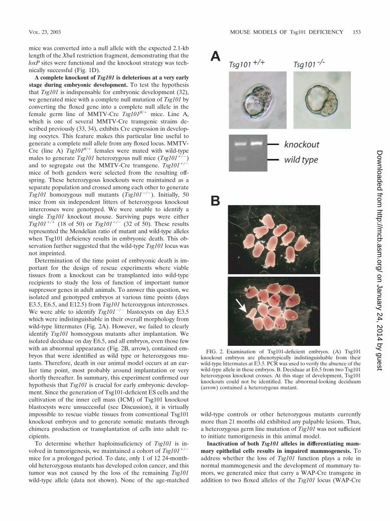
Determination of the time point of embryonic death is im-portant for the design of rescue experiments where viabletissues from a knockout can be transplanted into wild-typerecipients to study the loss of function of important tumorsuppressor genes in adult animals. To answer this question, weisolated and genotyped embryos at various time points (daysE3.5, E6.5, and E12.5) from Tsg101 heterozygous intercrosses.We were able to identify Tsg101�/� blastocysts on day E3.5which were indistinguishable in their overall morphology fromwild-type littermates (Fig. 2A). However, we failed to clearlyidentify Tsg101 homozygous mutants after implantation. Weisolated deciduae on day E6.5, and all embryos, even those fewwith an abnormal appearance (Fig. 2B, arrow), contained em-bryos that were identified as wild type or heterozygous mu-tants. Therefore, death in our animal model occurs at an ear-lier time point, most probably around implantation or veryshortly thereafter. In summary, this experiment confirmed ourhypothesis that Tsg101 is crucial for early embryonic develop-ment. Since the generation of Tsg101-deficient ES cells and thecultivation of the inner cell mass (ICM) of Tsg101 knockoutblastocysts were unsuccessful (see Discussion), it is virtuallyimpossible to rescue viable tissues from conventional Tsg101knockout embryos and to generate somatic mutants throughchimera production or transplantation of cells into adult re-cipients.
To determine whether haploinsuficiency of Tsg101 is in-volved in tumorigenesis, we maintained a cohort of Tsg101�/�
mice for a prolonged period. To date, only 1 of 12 24-month-old heterozygous mutants has developed colon cancer, and thistumor was not caused by the loss of the remaining Tsg101wild-type allele (data not shown). None of the age-matched
wild-type controls or other heterozygous mutants currentlymore than 21 months old exhibited any palpable lesions. Thus,a heterozygous germ line mutation of Tsg101 was not sufficientto initiate tumorigenesis in this animal model.
Inactivation of both Tsg101 alleles in differentiating mam-mary epithelial cells results in impaired mammogenesis. Toaddress whether the loss of Tsg101 function plays a role innormal mammogenesis and the development of mammary tu-mors, we generated mice that carry a WAP-Cre transgene inaddition to two floxed alleles of the Tsg101 locus (WAP-Cre
FIG. 2. Examination of Tsg101-deficient embryos. (A) Tsg101knockout embryos are phenotypically indistinguishable from theirwild-type littermates at E3.5. PCR was used to verify the absence of thewild-type allele in these embryos. B. Deciduae at E6.5 from two Tsg101heterozygous knockout crosses. At this stage of development, Tsg101knockouts could not be identified. The abnormal-looking deciduum(arrow) contained a heterozygous mutant.
VOL. 23, 2003 MOUSE MODELS OF Tsg101 DEFICIENCY 153
on January 24, 2014 by guesthttp://m
cb.asm.org/
Dow
nloaded from
Tsg101fl/fl). The WAP-Cre transgene is specifically active indifferentiating alveolar cells during late pregnancy and lacta-tion. However, a significant number of recombined cells bypassapoptotic signals, survive remodeling during involution, andserve as progenitor cells for the alveolar compartment in sub-sequent pregnancies (30, 34). This feature makes the WAP-Cre strain an important tool to study tumor suppressor func-tion in the mammary gland (39).
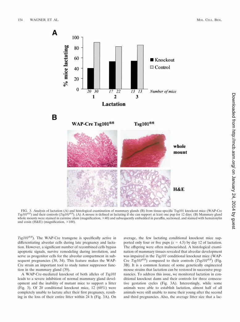
A WAP-Cre-mediated knockout of both alleles of Tsg101leads to a severe inhibition of normal mammary gland devel-opment and the inability of mutant mice to support a litter(Fig. 3). Of 20 conditional knockout mice, 12 (60%) werecompletely unable to lactate after their first pregnancy, result-ing in the loss of their entire litter within 24 h (Fig. 3A). On
average, the few lactating conditional knockout mice sup-ported only four or five pups (x � 4.5) by day 12 of lactation.The offspring were often malnourished. A histological exami-nation of mammary tissues revealed that alveolar developmentwas impaired in the Tsg101 conditional knockout mice (WAP-Cre Tsg101fl/fl) compared to their controls (Tsg101fl/fl) (Fig.3B). It is a common feature of some genetically engineeredmouse strains that lactation can be restored in successive preg-nancies. To address this issue, we monitored lactation in con-ditional knockout dams and their controls for three consecu-tive gestation cycles (Fig. 3A). Interestingly, while someanimals were able to establish lactation, almost half of allanimals were still unable to nurse their young after the secondand third pregnancies. Also, the average litter size that a lac-
FIG. 3. Analysis of lactation (A) and histological examination of mammary glands (B) from tissue-specific Tsg101 knockout mice (WAP-CreTsg101fl/fl) and their controls (Tsg101fl/fl). (A) A mouse is defined as lactating if she can support at least one pup for 12 days. (B) Mammary glandwhole mounts were stained in carmine alum (magnification, �40) and subsequently embedded in paraffin, sectioned, and stained with hematoxylinand eosin (H&E) (magnification, �100).
154 WAGNER ET AL. MOL. CELL. BIOL.
on January 24, 2014 by guesthttp://m
cb.asm.org/
Dow
nloaded from
tating conditional knockout dam was able to support did notincrease (x � 4.5 and 4.0, respectively). Therefore, a completereversal of the phenotypic abnormalities of the mutant micewas not achieved. This suggested that either normal mammaryepithelial cells have only a very limited capacity to bypassTsg101 gene function or there is selective amplification ofalveolar cells that maintain unrecombined alleles of Tsg101due to WAP-Cre transgene silencing.
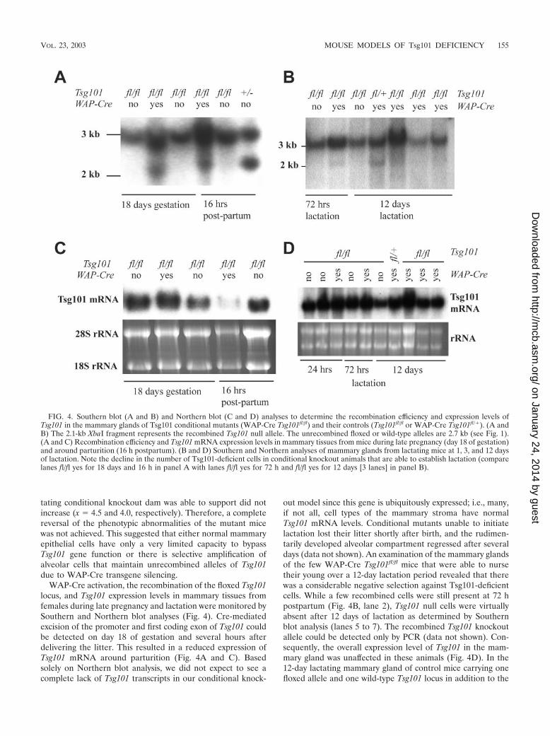
WAP-Cre activation, the recombination of the floxed Tsg101locus, and Tsg101 expression levels in mammary tissues fromfemales during late pregnancy and lactation were monitored bySouthern and Northern blot analyses (Fig. 4). Cre-mediatedexcision of the promoter and first coding exon of Tsg101 couldbe detected on day 18 of gestation and several hours afterdelivering the litter. This resulted in a reduced expression ofTsg101 mRNA around parturition (Fig. 4A and C). Basedsolely on Northern blot analysis, we did not expect to see acomplete lack of Tsg101 transcripts in our conditional knock-
out model since this gene is ubiquitously expressed; i.e., many,if not all, cell types of the mammary stroma have normalTsg101 mRNA levels. Conditional mutants unable to initiatelactation lost their litter shortly after birth, and the rudimen-tarily developed alveolar compartment regressed after severaldays (data not shown). An examination of the mammary glandsof the few WAP-Cre Tsg101fl/fl mice that were able to nursetheir young over a 12-day lactation period revealed that therewas a considerable negative selection against Tsg101-deficientcells. While a few recombined cells were still present at 72 hpostpartum (Fig. 4B, lane 2), Tsg101 null cells were virtuallyabsent after 12 days of lactation as determined by Southernblot analysis (lanes 5 to 7). The recombined Tsg101 knockoutallele could be detected only by PCR (data not shown). Con-sequently, the overall expression level of Tsg101 in the mam-mary gland was unaffected in these animals (Fig. 4D). In the12-day lactating mammary gland of control mice carrying onefloxed allele and one wild-type Tsg101 locus in addition to the
FIG. 4. Southern blot (A and B) and Northern blot (C and D) analyses to determine the recombination efficiency and expression levels ofTsg101 in the mammary glands of Tsg101 conditional mutants (WAP-Cre Tsg101fl/fl) and their controls (Tsg101fl/fl or WAP-Cre Tsg101fl/�). (A andB) The 2.1-kb XbaI fragment represents the recombined Tsg101 null allele. The unrecombined floxed or wild-type alleles are 2.7 kb (see Fig. 1).(A and C) Recombination efficiency and Tsg101 mRNA expression levels in mammary tissues from mice during late pregnancy (day 18 of gestation)and around parturition (16 h postpartum). (B and D) Southern and Northern analyses of mammary glands from lactating mice at 1, 3, and 12 daysof lactation. Note the decline in the number of Tsg101-deficient cells in conditional knockout animals that are able to establish lactation (comparelanes fl/fl yes for 18 days and 16 h in panel A with lanes fl/fl yes for 72 h and fl/fl yes for 12 days [3 lanes] in panel B).
VOL. 23, 2003 MOUSE MODELS OF Tsg101 DEFICIENCY 155
on January 24, 2014 by guesthttp://m
cb.asm.org/
Dow
nloaded from
WAP-Cre transgene (WAP-Cre Tsg101fl/�), the recombined(null) allele could be detected by Southern blotting (Fig. 4B,lane 4), suggesting that there was no general selection againstCre-expressing or Tsg101 haploinsufficient cells. This resultwas expected since neither Tsg101 heterozygous knockouts(Tsg101�/�) nor single-transgenic WAP-Cre mice exhibitedabnormal mammary phenotypes. In summary, our data sug-gested that mammary epithelial cells were unable to compen-sate for the loss of Tsg101. Lactation could only be initiated inanimals that exhibited a nonuniform expression of WAP-Creand a selected amplification of alveolar cells with unrecom-bined Tsg101 alleles (i.e., negative selection against Tsg101-deficient cells).
To investigate a possible involvement of a Tsg101 null mu-tation in mammary tumorigenesis, WAP-Cre Tsg101fl/fl miceand their controls were maintained over a period of 2 years andmonitored for mammary tumor formation. These animals werecontinuously mated to (i) repeatedly activate WAP-Cre in thenewly developed alveolar compartment and subsequently in-crease the number of Tsg101-deficient cells temporarily, (ii)increase the overall proliferation rate of mammary epithelialcells in response to higher estrogen and progesterone levelsduring pregnancy, and (iii) introduce and select for genomicmutations that might cooperate with the lack of Tsg101 towardneoplastic transformation. Thus far, none of the mice, whichare currently up to 24 months old, have developed mammarygland lesions. These data suggest that in contrast to a previousreport (14), Tsg101 is not a tumor suppressor gene for theinitiation of sporadic forms of breast cancer.
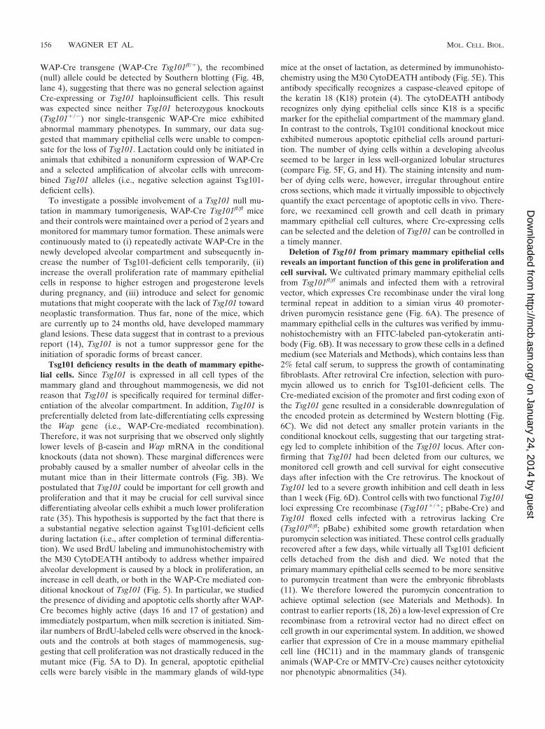
Tsg101 deficiency results in the death of mammary epithe-lial cells. Since Tsg101 is expressed in all cell types of themammary gland and throughout mammogenesis, we did notreason that Tsg101 is specifically required for terminal differ-entiation of the alveolar compartment. In addition, Tsg101 ispreferentially deleted from late-differentiating cells expressingthe Wap gene (i.e., WAP-Cre-mediated recombination).Therefore, it was not surprising that we observed only slightlylower levels of �-casein and Wap mRNA in the conditionalknockouts (data not shown). These marginal differences wereprobably caused by a smaller number of alveolar cells in themutant mice than in their littermate controls (Fig. 3B). Wepostulated that Tsg101 could be important for cell growth andproliferation and that it may be crucial for cell survival sincedifferentiating alveolar cells exhibit a much lower proliferationrate (35). This hypothesis is supported by the fact that there isa substantial negative selection against Tsg101-deficient cellsduring lactation (i.e., after completion of terminal differentia-tion). We used BrdU labeling and immunohistochemistry withthe M30 CytoDEATH antibody to address whether impairedalveolar development is caused by a block in proliferation, anincrease in cell death, or both in the WAP-Cre mediated con-ditional knockout of Tsg101 (Fig. 5). In particular, we studiedthe presence of dividing and apoptotic cells shortly after WAP-Cre becomes highly active (days 16 and 17 of gestation) andimmediately postpartum, when milk secretion is initiated. Sim-ilar numbers of BrdU-labeled cells were observed in the knock-outs and the controls at both stages of mammogenesis, sug-gesting that cell proliferation was not drastically reduced in themutant mice (Fig. 5A to D). In general, apoptotic epithelialcells were barely visible in the mammary glands of wild-type
mice at the onset of lactation, as determined by immunohisto-chemistry using the M30 CytoDEATH antibody (Fig. 5E). Thisantibody specifically recognizes a caspase-cleaved epitope ofthe keratin 18 (K18) protein (4). The cytoDEATH antibodyrecognizes only dying epithelial cells since K18 is a specificmarker for the epithelial compartment of the mammary gland.In contrast to the controls, Tsg101 conditional knockout miceexhibited numerous apoptotic epithelial cells around parturi-tion. The number of dying cells within a developing alveolusseemed to be larger in less well-organized lobular structures(compare Fig. 5F, G, and H). The staining intensity and num-ber of dying cells were, however, irregular throughout entirecross sections, which made it virtually impossible to objectivelyquantify the exact percentage of apoptotic cells in vivo. There-fore, we reexamined cell growth and cell death in primarymammary epithelial cell cultures, where Cre-expressing cellscan be selected and the deletion of Tsg101 can be controlled ina timely manner.
Deletion of Tsg101 from primary mammary epithelial cellsreveals an important function of this gene in proliferation andcell survival. We cultivated primary mammary epithelial cellsfrom Tsg101fl/fl animals and infected them with a retroviralvector, which expresses Cre recombinase under the viral longterminal repeat in addition to a simian virus 40 promoter-driven puromycin resistance gene (Fig. 6A). The presence ofmammary epithelial cells in the cultures was verified by immu-nohistochemistry with an FITC-labeled pan-cytokeratin anti-body (Fig. 6B). It was necessary to grow these cells in a definedmedium (see Materials and Methods), which contains less than2% fetal calf serum, to suppress the growth of contaminatingfibroblasts. After retroviral Cre infection, selection with puro-mycin allowed us to enrich for Tsg101-deficient cells. TheCre-mediated excision of the promoter and first coding exon ofthe Tsg101 gene resulted in a considerable downregulation ofthe encoded protein as determined by Western blotting (Fig.6C). We did not detect any smaller protein variants in theconditional knockout cells, suggesting that our targeting strat-egy led to complete inhibition of the Tsg101 locus. After con-firming that Tsg101 had been deleted from our cultures, wemonitored cell growth and cell survival for eight consecutivedays after infection with the Cre retrovirus. The knockout ofTsg101 led to a severe growth inhibition and cell death in lessthan 1 week (Fig. 6D). Control cells with two functional Tsg101loci expressing Cre recombinase (Tsg101�/�; pBabe-Cre) andTsg101 floxed cells infected with a retrovirus lacking Cre(Tsg101fl/fl; pBabe) exhibited some growth retardation whenpuromycin selection was initiated. These control cells graduallyrecovered after a few days, while virtually all Tsg101 deficientcells detached from the dish and died. We noted that theprimary mammary epithelial cells seemed to be more sensitiveto puromycin treatment than were the embryonic fibroblasts(11). We therefore lowered the puromycin concentration toachieve optimal selection (see Materials and Methods). Incontrast to earlier reports (18, 26) a low-level expression of Crerecombinase from a retroviral vector had no direct effect oncell growth in our experimental system. In addition, we showedearlier that expression of Cre in a mouse mammary epithelialcell line (HC11) and in the mammary glands of transgenicanimals (WAP-Cre or MMTV-Cre) causes neither cytotoxicitynor phenotypic abnormalities (34).
156 WAGNER ET AL. MOL. CELL. BIOL.
on January 24, 2014 by guesthttp://m
cb.asm.org/
Dow
nloaded from
The deletion of Tsg101 from primary cells in culture con-firmed our in vivo observations that the protein encoded bythis gene is indispensable for normal cell growth and cell sur-vival. It was suggested previously that Tsg101 may play a role inproliferation (24, 40). However, we did not observe a consid-erable reduction of proliferating cells in the mammary glands
of WAP-Cre Tsg101fl/fl females. Since Wap is expressed mainlyin differentiating cells exhibiting reduced proliferation, the an-imal model might not entirely recapitulate a phenotype thatsuggests an additional role for Tsg101 in cell proliferation.Therefore, we studied primary cells in vitro to determinewhether Tsg101 deficiency results in perturbation of the cell
FIG. 5. Detection of proliferating (arrows in panels B to D) and apoptotic (arrows in panels E to H) cells at various stages of mammarydevelopment in Tsg101 mammary-specific knockout mice (WAP-Cre Tsg101fl/fl B, D, and F to H) and their controls (Tsg101fl/fl [A, C, and E]). (Aand B) 17 days of gestation (magnification, �400). (C to H) Shortly after parturition (magnification, �400). Proliferating cells were detected usingBrdU labeling, whereas apoptotic cells were identified by immunohistochemistry using the M30 CytoDEATH antibody. Slides were counterstainedwith hematoxylin. Note the larger number of dying cells in less organized or collapsed alveolar structures (F to H).
VOL. 23, 2003 MOUSE MODELS OF Tsg101 DEFICIENCY 157
on January 24, 2014 by guesthttp://m
cb.asm.org/
Dow
nloaded from
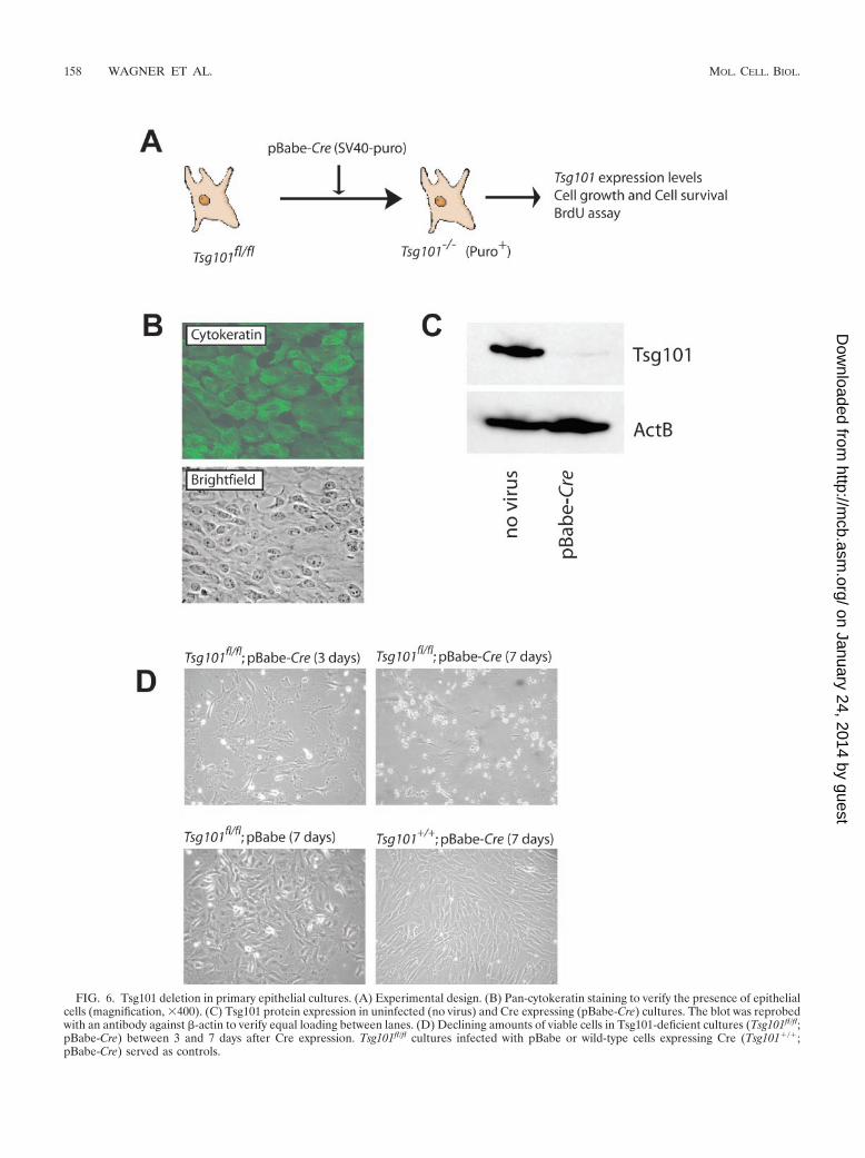
FIG. 6. Tsg101 deletion in primary epithelial cultures. (A) Experimental design. (B) Pan-cytokeratin staining to verify the presence of epithelialcells (magnification, �400). (C) Tsg101 protein expression in uninfected (no virus) and Cre expressing (pBabe-Cre) cultures. The blot was reprobedwith an antibody against �-actin to verify equal loading between lanes. (D) Declining amounts of viable cells in Tsg101-deficient cultures (Tsg101fl/fl;pBabe-Cre) between 3 and 7 days after Cre expression. Tsg101fl/fl cultures infected with pBabe or wild-type cells expressing Cre (Tsg101�/�;pBabe-Cre) served as controls.
158 WAGNER ET AL. MOL. CELL. BIOL.
on January 24, 2014 by guesthttp://m
cb.asm.org/
Dow
nloaded from
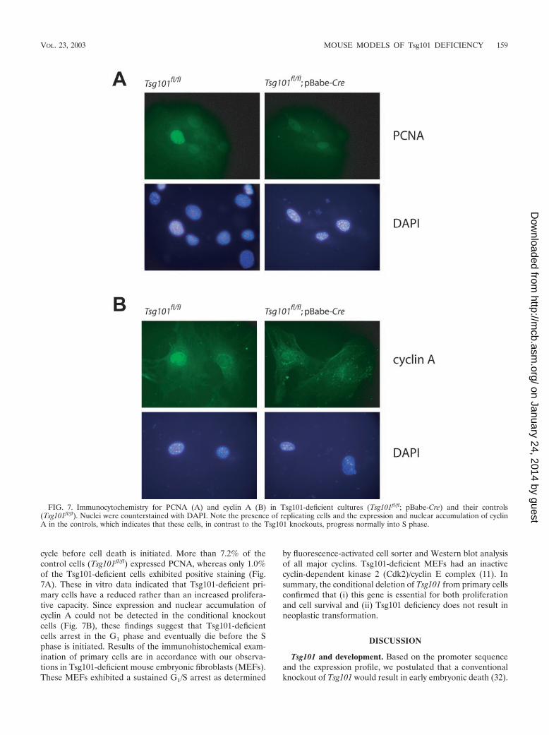
cycle before cell death is initiated. More than 7.2% of thecontrol cells (Tsg101fl/fl) expressed PCNA, whereas only 1.0%of the Tsg101-deficient cells exhibited positive staining (Fig.7A). These in vitro data indicated that Tsg101-deficient pri-mary cells have a reduced rather than an increased prolifera-tive capacity. Since expression and nuclear accumulation ofcyclin A could not be detected in the conditional knockoutcells (Fig. 7B), these findings suggest that Tsg101-deficientcells arrest in the G1 phase and eventually die before the Sphase is initiated. Results of the immunohistochemical exam-ination of primary cells are in accordance with our observa-tions in Tsg101-deficient mouse embryonic fibroblasts (MEFs).These MEFs exhibited a sustained G1/S arrest as determined
by fluorescence-activated cell sorter and Western blot analysisof all major cyclins. Tsg101-deficient MEFs had an inactivecyclin-dependent kinase 2 (Cdk2)/cyclin E complex (11). Insummary, the conditional deletion of Tsg101 from primary cellsconfirmed that (i) this gene is essential for both proliferationand cell survival and (ii) Tsg101 deficiency does not result inneoplastic transformation.
DISCUSSION
Tsg101 and development. Based on the promoter sequenceand the expression profile, we postulated that a conventionalknockout of Tsg101 would result in early embryonic death (32).
FIG. 7. Immunocytochemistry for PCNA (A) and cyclin A (B) in Tsg101-deficient cultures (Tsg101fl/fl; pBabe-Cre) and their controls(Tsg101fl/fl). Nuclei were counterstained with DAPI. Note the presence of replicating cells and the expression and nuclear accumulation of cyclinA in the controls, which indicates that these cells, in contrast to the Tsg101 knockouts, progress normally into S phase.
VOL. 23, 2003 MOUSE MODELS OF Tsg101 DEFICIENCY 159
on January 24, 2014 by guesthttp://m
cb.asm.org/
Dow
nloaded from
The generation and analysis of embryos lacking Tsg101 com-pletely confirmed this hypothesis. In our Tsg101� exon 1�/�
model, deficiency of this gene resulted in embryonic deatharound the time of implantation. This assumption was alsoverified recently in a different mouse model carrying a deletionof exons 8 and 9, which encode the coiled-coil domain near theC-terminal end of Tsg101 (24). These mice die between E5.5and E6.5 due to a defect in cell proliferation and mesodermformation. An increase in cell death was not observed in thesemutants. In the Tsg101� exon 1�/� model, we were unable toidentify homozygous mutant embryos at day E6.5. This sug-gests that there might be marginal differences in the pheno-types of the two mouse models depending on which functionaldomains were deleted from the Tsg101 locus. Since neither thegeneration of Tsg101-deficient ES cells nor the cultivation ofthe ICM from Tsg101� exon 8/9�/� blastocycts was successful(24), it is virtually impossible to rescue Tsg101-deficient cellsby transplantation.
To investigate the biological effects of Tsg101 deficiency onthe proliferation and differentiation of breast epithelial cellsand to verify the proposed function of this gene as a tumorsuppressor for sporadic forms of breast cancer, we developed aknockout mouse with a mammary cell-specific deletion ofTsg101. Deficiency of Tsg101 in differentiating mammary epi-thelial cells resulted in impaired mammary development. Oneconsequence was the inability of the mutant mice to lactatenormally. This phenotype was caused mainly by apoptosis ofdifferentiated cells, which normally exhibit a low proliferationrate (35). This phenomenon seems not to be a unique featureof mammary epithelial cells or of differentiating cells in gen-eral. We observed cell death as a consequence of Tsg101 de-ficiency in a variety of proliferating cell types including MEFs(11). In agreement with our findings, Garrus et al. (6) reporteda slight growth inhibition of 293 cells when Tsg101 was tran-siently knocked down for 72 h by using the small interferingRNA approach. In addition, the introduction of Tsg101 siR-NAs into human breast cancer cells (MCF-7) was highly toxicwhen these cells were treated repeatedly over time. WhereasTsg101-deficient MCF-7 cells died, transfection of othersiRNA control vectors had little or no effect on the growth ofthese cells (X. Lin and W. Nelson, John-Hopkins University,personal communication). In summary, these observations sug-gest that Tsg101 is important for the survival of nonimmortal-ized (e.g., mammary epithelial cells and MEFs) as well asimmortalized and transformed (e.g., 293T and MCF-7 cells)cell lines. Our further studies of transformed Tsg101fl/fl MEFs,which were able to form tumors in nude mice, seemed tosupport this assumption. The Cre-mediated deletion of Tsg101in explanted tumor cells resulted in instant cell death (M. D.Henry, A. Krempler, and K.-U. Wagner, unpublished data).
Tsg101 and cell cycle regulation. Our studies of Tsg101-deficient primary cells revealed that in addition to cell survival,Tsg101 is important for cell proliferation. The retroviral Cre-mediated conditional knockout of Tsg101 resulted in cell cyclearrest and death before the cells entered the S phase. Theseobservations were consistent with our findings about the role ofTsg101 in proliferating embryonic fibroblasts (11). Tsg101-de-ficient MEFs did not incorporate BrdU and lacked expressionof cyclins A and B. In addition, their cyclin E/Cdk2 complexwas inactive, which clearly suggested a sustained G1/S arrest
before Tsg101-deficient cells died. The mechanism responsiblefor the inactivation of Cdk2 in the conditional knockout cellsneeds to be identified. In a recent report, Oh et al. (22) showedthat exogenous levels of Tsg101 in differentiating keratinocytescan positively regulate p21WAF-1/C1P-1, a suggested tumor sup-pressor protein that mediates G1 arrest in a p53-dependentmanner (2, 5, 19). In proliferating keratinocytes, which exhibitstable levels of p21, overexpression of Tsg101 exerts growthsuppression through modulation of p21 binding to the cyclinE/Cdk2 complex. It is possible that the mechanisms, whichmediate growth arrest in Tsg101-deficient cells and the Tsg101overexpression model, are the same. However, we did notobserve a significant change in p27Kip1 and p21WAF-1/CIP-1 pro-tein levels in the conditional knockouts, suggesting that Tsg101is not essential for the stability of these major Cdk2 inhibitorsin MEFs (11). Preliminary studies of Tsg101�/� MEFs showeda larger amount of phosphorylated Cdk2 (A. Krempler andK.-U. Wagner, unpublished data), and we are currently inves-tigating whether inhibiting phosphorylations mediate inactiva-tion of the cyclin E/Cdk2 complex in Tsg101-deficient cells.
The role of Tsg101 as a positive regulator of p21 and/orCdk2 activity would provide a mechanism for the suggestedfunction of this protein as an inhibitor of the cell cycle and apotential tumor suppressor. In contrast, Li et al. (15) reporteda role for Tsg101 as a positive regulator of Mdm2, which is anoncogene that mediates the degradation of p53. The samegroup of investigators was able to verify their findings by usinga Tsg101-deficient mouse model, which exhibited a cell cyclearrest mediated by upregulation of p53 and p21 (24). Throughintroduction of a p53-null mutation into the Tsg101� exon 8/9�/�
background, Ruland et al. (24) were able to prolong the sur-vival of mutant embryos for about 2 to 3 days. Given thesuggested importance of Tsg101 as a key regulator of Mdm2,this rescue seemed to be marginal compared to that in Mdm2/p53 double-mutant mice, where a knockout of p53 completelyrescued embryonic death caused by Mdm2 deficiency (9, 21).In addition, the suggested function for Tsg101 as a stabilizer ofMdm2 (i.e., an oncogene) is inconsistent with various reportsthat Tsg101 overexpression leads to cell cycle arrest and celldeath (22, 32, 37, 40). The findings that Tsg101 deficiencyresulted in cell cycle arrest through upregulation of p53 andp21 therefore contradict the mechanism proposed by Oh et al.(22), in which Tsg101 is a crucial mediator of p21 proteinstability and of p21 function as a cell cycle inhibitor on thecyclin E/Cdk2 complex (see above). Our conditional knockoutmodel did not reveal any involvement of p53 and its maineffectors in the deleterious phenotype caused by Tsg101 defi-ciency (11). Various upstream (p19ARF, p16Ink4a, and Mdm2)and downstream (p21WAF-1/CIP-1 and p27Kip1) targets of p53exhibited no significant change in their protein levels inTsg101�/� MEFs. Furthermore, neither the functional inhibi-tion of p53 nor the deletion of the p53 gene had a noticeableeffect on the deleterious phenotype. Therefore, our findings donot support a biologically relevant function of Tsg101 in reg-ulating the Mdm2-p53 feedback loop, as previously suggested(15, 24). On the other hand, Tsg101 deficiency causes a cellcycle arrest in addition to cell death. While p53 is not essentialfor mediating cell death in Tsg101�/� MEFs (11), more de-tailed analysis of whether p53 plays a role in cell cycle arrestobserved in the Tsg101 conditional knockout model is needed.
160 WAGNER ET AL. MOL. CELL. BIOL.
on January 24, 2014 by guesthttp://m
cb.asm.org/
Dow
nloaded from
Tsg101 and neoplastic transformation. Li and Cohen (13)reported that a functional knockout of Tsg101 using a conven-tional antisense approach resulted in instant neoplastic trans-formation of immortalized fibroblasts. However, the role of theTsg101 gene as a tumor suppressor in human malignancies isstill controversial. Genomic deletions and aberrant splice vari-ants have been implicated in sporadic forms of breast cancerand a variety of other human malignancies (see, for example,references 7, 12, 14, 28, and 29). However, there are alsonumerous contradictory reports (see, for example, references3, 17, 27, and 38). To verify whether the loss of function ofTsg101 is involved in neoplastic transformation in vivo, wemaintained a large cohort of Tsg101 mutant mice (heterozy-gous complete and tissue-specific knockouts) for 2 years. Thesemice were closely monitored for tumor formation. Neitherhaploinsufficiency of Tsg101 (see also the report by Ruland etal. [24]) nor the deletion of both Tsg101 alleles in mammaryepithelial cells resulted in tumor formation. Our data suggestthat a loss of function of Tsg101 is insufficient to trigger neo-plastic transformation; therefore, our observations contradictearlier reports by Li et al. (13, 14). Generally, in vitro modelssuch as NIH 3T3 cells, which were used to identify Tsg101 andits tumor suppressive properties, are less reliable since thesecells are immortal and carry numerous mutations in othertumor suppressor loci such as p53, p19Arf, or p16Ink4a (25).Specifically, the original NIH 3T3 cell line and its tumorigenicderivative (SL6 cells) described by Li and Cohen (13) aredeficient in p19Arf and p16Ink4a (Krempler and Wagner, un-published). More importantly, we showed recently that theantisense strategy used by Li et al. did not completely inhibitgene expression. SL6 cells still express a significant amount ofthe Tsg101 protein (11), and therefore it needs to be estab-lished (i) whether a knockdown triggers a different phenotypefrom a null mutation and (ii) whether antisense constructstarget other genes with partial sequence similarity to Tsg101. Insummary, the results of our studies on Tsg101 conditionalknockout mice demonstrate that it is very important to gener-ate genetically engineered mouse models with defined targetedmutations to verify a proposed tumor-suppressive function ofgenes identified in random mutagenesis screens in immortal-ized cell lines.
ACKNOWLEDGMENTS
We thank Stanley Cohen (Stanford) for providing the Tsg101 cDNAand SL6 cells. We are grateful to Alan Diehl (University of Pennsyl-vania) for his valuable suggestions and for providing the pBabe retro-viral vector and 293T cells.
This work was supported by Public Health Service grant CA-93797from the National Cancer Institute to K.U.W. The embryonic stem cellwork was funded in part by the NIH Breast Cancer Think Tank. A.K.receives a stipend from the Deutsche Forschungsgemeinschaft (DFG,KR 2107/1-1).
REFERENCES
1. Babst, M., G. Odorizzi, E. J. Estepa, and S. D. Emr. 2000. Mammalian tumorsusceptibility gene 101 (TSG101) and the yeast homologue, Vps23p, bothfunction in late endosomal trafficking. Traffic 1:248–258.
2. Brugarolas, J., C. Chandrasekaran, J. I. Gordon, D. Beach, T. Jacks, andG. J. Hannon. 1995. Radiation-induced cell cycle arrest compromised by p21deficiency. Nature 377:552–557.
3. Carney, M. E., G. L. Maxwell, J. M. Lancaster, C. Gumbs, J. Marks, A.Berchuck, and P. A. Futreal. 1998. Aberrant splicing of the TSG101 tumorsuppressor gene in human breast and ovarian cancers. J. Soc. Gynecol.Investig. 5:281–285.
4. Caulin, C., G. S. Salvesen, and R. G. Oshima. 1997. Caspase cleavage ofkeratin 18 and reorganization of intermediate filaments during epithelial cellapoptosis. J. Cell Biol. 138:1379–1394.
5. Deng, C., P. Zhang, J. W. Harper, S. J. Elledge, and P. Leder. 1995. Micelacking p21CIP1/WAF1 undergo normal development, but are defective inG1 checkpoint control. Cell 82:675–684.
6. Garrus, J. E., U. K. von Schwedler, O. W. Pornillos, S. G. Morham, K. H.Zavitz, H. E. Wang, D. A. Wettstein, K. M. Stray, M. Cote, R. L. Rich, D. G.Myszka, and W. I. Sundquist. 2001. Tsg101 and the vacuolar protein sortingpathway are essential for HIV-1 budding. Cell 107:55–65.
7. Gayther, S. A., P. Barski, S. J. Batley, L. Li, K. A. de Foy, S. N. Cohen, B. A.Ponder, and C. Caldas. 1997. Aberrant splicing of the TSG101 and FHITgenes occurs frequently in multiple malignancies and in normal tissues andmimics alterations previously described in tumours. Oncogene 15:2119–2126.
8. Hittelman, A. B., D. Burakov, J. A. Iniguez-Lluhi, L. P. Freedman, and M. J.Garabedian. 1999. Differential regulation of glucocorticoid receptor tran-scriptional activation via AF-1-associated proteins. EMBO J. 18:5380–5388.
9. Jones, S. N., A. E. Roe, L. A. Donehower, and A. Bradley. 1995. Rescue ofembryonic lethality in Mdm2-deficient mice by absence of p53. Nature 378:206–208.
10. Koonin, E. V., and R. A. Abagyan. 1997. TSG101 may be the prototype of aclass of dominant negative ubiquitin regulators. Nat. Genet. 16:330–331.
11. Krempler, A., M. D. Henry, A. A. Triplett, and K. U. Wagner. 2002. Targeteddeletion of the Tsg101 gene results in cell cycle arrest at G1/S and p53independent cell death. J. Biol. Chem. 277:43216–43223.
12. Lee, M. P., and A. P. Feinberg. 1997. Aberrant splicing but not mutations ofTSG101 in human breast cancer. Cancer Res. 57:3131–3134.
13. Li, L., and S. N. Cohen. 1996. Tsg101: a novel tumor susceptibility geneisolated by controlled homozygous functional knockout of allelic loci inmammalian cells. Cell 85:319–329.
14. Li, L., X. Li, U. Francke, and S. N. Cohen. 1997. The TSG101 tumorsusceptibility gene is located in chromosome 11 band p15 and is mutated inhuman breast cancer. Cell 88:143–154.
15. Li, L., J. Liao, J. Ruland, T. W. Mak, and S. N. Cohen. 2001. A TSG101/MDM2 regulatory loop modulates MDM2 degradation and MDM2/p53feedback control. Proc. Natl. Acad. Sci. USA 98:1619–1624.
16. Li, M., K. U. Wagner, and P. A. Furth. 2000. Transfection of primarymammary epithelial cells by viral and nonviral methods, p. 233–244. In M. M.Ip and B. B. Ash, (ed.), Methods in mammary gland biology and breastcancer. Kluwer Academic/Plenum Publishers, New York, N.Y.
17. Lin, S. F., P. M. Lin, T. C. Liu, J. G. Chang, Y. C. Sue, and T. P. Chen. 2000.Clinical implications of aberrant TSG101 transcripts in acute myeloblasticleukemia. Leuk. Lymphoma 36:463–466.
18. Loonstra, A., M. Vooijs, H. B. Beverloo, B. A. Allak, E. van Drunen, R.Kanaar, A. Berns, and J. Jonkers. 2001. Growth inhibition and DNA dam-age induced by Cre recombinase in mammalian cells. Proc. Natl. Acad. Sci.USA 98:9209–9214.
19. Martin-Caballero, J., J. M. Flores, P. Garcia-Palencia, and M. Serrano.2001. Tumor susceptibility of p21 (Waf1/Cip1)-deficient mice. Cancer Res.61:6234–6238.
20. Medina, D. and F. S. Kittrell. 2000. Establishment of mouse mammary celllines, p. 137–145. In M. M. Ip and B. B. Ash (ed.), Methods in mammarygland biology and breast cancer. Kluwer Academic/Plenum Publishers, NewYork, N.Y.
21. Montes de Oca, L. R., D. S. Wagner, and G. Lozano. 1995. Rescue of earlyembryonic lethality in mdm2-deficient mice by deletion of p53. Nature 378:203–206.
22. Oh, H., C. Mammucari, A. Nenci, S. Cabodi, S. N. Cohen, and G. P. Dotto.2002. Negative regulation of cell growth and differentiation by TSG101through association with p21Cip1/WAF1. Proc. Natl. Acad. Sci. USA 99:5430–5435.
23. Ponting, C. P., Y. D. Cai, and P. Bork. 1997. The breast cancer gene productTSG101: a regulator of ubiquitination? J. Mol. Med. 75:467–469.
24. Ruland, J., C. Sirard, A. Elia, D. MacPherson, A. Wakeham, L. Li, D. L. P.Luis, S. N. Cohen, and T. W. Mak. 2001. p53 Accumulation, defective cellproliferation, and early embryonic lethality in mice lacking tsg101. Proc.Natl. Acad. Sci. USA 98:1859–1864.
25. Sherr, C. J. 1998. Tumor surveillance via the ARF-p53 pathway. Genes Dev.12:2984–2991.
26. Silver, D. P., and D. M. Livingston. 2001. Self-excising retroviral vectorsencoding the Cre recombinase overcome Cre-mediated cellular toxicity.Mol. Cell 8:233–243.
27. Steiner, P., D. M. Barnes, W. H. Harris, and R. A. Weinberg. 1997. Absenceof rearrangements in the tumour susceptibility gene TSG101 in humanbreast cancer. Nat. Genet. 16:332–333.
28. Sun, Z., J. Pan, G. Bubley, and S. P. Balk. 1997. Frequent abnormalities ofTSG101 transcripts in human prostate cancer. Oncogene 15:3121–3125.
29. Turpin, E., B. Dalle, A. de Roquancourt, L. F. Plassa, M. Marty, A. Janin, Y.Beuzard, and H. de The. 1999. Stress-induced aberrant splicing of TSG101:association to high tumor grade and p53 status in breast cancers. Oncogene18:7834–7837.
30. Wagner, K. U., C. A. Boulanger, M. D. Henry, M. Sgagias, L. Hennighausen,
VOL. 23, 2003 MOUSE MODELS OF Tsg101 DEFICIENCY 161
on January 24, 2014 by guesthttp://m
cb.asm.org/
Dow
nloaded from
and G. H. Smith. 2002. An adjunct mammary epithelial cell population inparous females: its role in functional adaptation and tissue renewal. Devel-opment 129:1377–1386.
31. Wagner, K. U., P. Dierisseau, and L. Hennighausen. 1999. Assignment of themurine tumor susceptibility gene 101 (tsg101) and a processed tsg101 pseu-dogene (tsg101-ps1) to mouse chromosome 7 band B5 and chromosome 15band D1 by in situ hybridization. Cytogenet. Cell Genet. 84:87–88.
32. Wagner, K. U., P. Dierisseau, E. B. Rucker, G. W. Robinson, and L. Hen-nighausen. 1998. Genomic architecture and transcriptional activation of themouse and human tumor susceptibility gene TSG101: common types ofshorter transcripts are true alternative splice variants. Oncogene 17:2761–2770.
33. Wagner, K. U., K. McAllister, T. Ward, B. Davis, R. Wiseman, and L.Hennighausen. 2001. Spatial and temporal expression of the Cre gene underthe control of the MMTV-LTR in different lines of transgenic mice. Trans-genic Res. 10:545–553.
34. Wagner, K. U., R. J. Wall, L. St-Onge, P. Gruss, A. Wynshaw-Boris, L.Garrett, M. Li, P. A. Furth, and L. Hennighausen. 1997. Cre-mediated genedeletion in the mammary gland. Nucleic Acids Res. 25:4323–4330.
35. Wagner, K. U., W. S. Young, X. Liu, E. I. Ginns, M. Li, P. A. Furth, and L.
Hennighausen. 1997. Oxytocin and milk removal are required for post-partum mammary-gland development. Genes Funct. 1:233–244.
36. Watanabe, M., Y. Yanagi, Y. Masuhiro, T. Yano, H. Yoshikawa, J. Yanagi-sawa, and S. Kato. 1998. A putative tumor suppressor, TSG101, acts as atranscriptional suppressor through its coiled-coil domain. Biochem. Biophys.Res. Commun. 245:900–905.
37. Xie, W., L. Li, and S. N. Cohen. 1998. Cell cycle-dependent subcellularlocalization of the TSG101 protein and mitotic and nuclear abnormalitiesassociated with TSG101 deficiency. Proc. Natl. Acad. Sci. USA 95:1595–1600.
38. Xu, C. F., J. Greenman, and E. Solomon. 1998. Truncated TSG101 tran-scripts are present in peripheral blood from both familial breast cancerpatients and controls. Eur. J. Cancer 34:1077–1080.
39. Xu, X., K. U. Wagner, D. Larson, Z. Weaver, C. Li, T. Ried, L. Hen-nighausen, A. Wynshaw-Boris, and C. X. Deng. 1999. Conditional mutationof Brca1 in mammary epithelial cells results in blunted ductal morphogenesisand tumour formation. Nat. Genet. 22:37–43.
40. Zhong, Q., Y. Chen, D. Jones, and W. H. Lee. 1998. Perturbation of TSG101protein affects cell cycle progression. Cancer Res. 58:2699–2702.
162 WAGNER ET AL. MOL. CELL. BIOL.
on January 24, 2014 by guesthttp://m
cb.asm.org/
Dow
nloaded from