Hematopoietic Stem Cell Gene-Addition/Editing Therapy in ...
Upload
independentCategory
view
3download
0
Cell Stem Cell
Article
Human Embryonic Stem Cell-DerivedGABA Neurons Correct Locomotion Deficitsin Quinolinic Acid-Lesioned MiceLixiang Ma,1 Baoyang Hu,3 Yan Liu,1,4 Scott Christopher Vermilyea,4 Huisheng Liu,4 Lu Gao,1 Yan Sun,1 Xiaoqing Zhang,6
and Su-Chun Zhang1,2,4,5,*1Department of Anatomy, Histology & Embryology, Shanghai Medical College2Institute of Stem Cell and Regenerative Medicine, Institutes of Biomedical SciencesFudan University, Shanghai 200032, China3Institute of Zoology, Chinese Academy of Sciences. Beijing 100101, China4Waisman Center5Department of Neuroscience and Department of Neurology, School of Medicine and Public HealthUniversity of Wisconsin, Madison, WI 53705, USA6Tongji University Medical School, Shanghai 200092, China
*Correspondence: [email protected]
DOI 10.1016/j.stem.2012.01.021
SUMMARY
Degeneration of medium spiny GABA neurons inthe basal ganglia underlies motor dysfunction inHuntington’s disease (HD), which presently lackseffective therapy. In this study, we have successfullydirected human embryonic stem cells (hESCs) toenriched populations of DARPP32-expressingforebrain GABA neurons. Transplantation of thesehuman forebrain GABA neurons and their progeni-tors, but not spinal GABA cells, into the striatum ofquinolinic acid-lesioned mice results in generationof large populations of DARPP32+ GABA neurons,which project to the substantia nigra as well asreceiving glutamatergic and dopaminergic inputs,corresponding to correction of motor deficits. Thisfinding raises hopes for cell therapy for HD.
INTRODUCTION
Huntington’s disease (HD) is an inherited degenerative disorder
with cognitive and psychiatric impairments as well as chorea
movements. The motor dysfunction is primarily associated with
degeneration of g-aminobutyric acid (GABA) medium spiny
neurons (MSN) in the basal ganglia resulting from abnormal
expansion of cytosine-adenine-guanine (CAG) repeats within
the genome leading to a malfunctioning huntingtin protein.
Significant efforts have been placed in reducing the numbers
of CAG repeats through genetic inhibition of the pathologic
CAG repeats, though balancing the removal of pathologic and
maintenance of normal CAG is probably not trivial (Johnson
and Davidson, 2010). Presently, no disease-modifying therapy
is on the horizon for this debilitating disorder.
An alternative strategy is to replace the degenerated GABA
neurons in the basal ganglia (Dunnett and Rosser, 2007). Unlike
in Parkinson’s disease in which tonic supplementation of
dopamine by dopamine-producing cells may provide beneficial
effects, in HD reformation of circuitry is necessary (Campbell
et al., 1993; Goto et al., 1997; Mazzocchi-Jones et al., 2011;
Nakao and Itakura, 2000). Transplanted GABA neurons must
project to and receive from their target tissues, consisting of
the globus pallidus and substantia nigra, as well as inputs from
the cerebral cortex (Wictorin, 1992), in order to achieve functional
benefits. This is a relatively long distance for grafted neuronal
axons to travel in the adult brain environment, which becomes
a major obstacle of cell therapy (Bonner et al., 2010; Pfeifer
et al., 2004). Nevertheless, fetal striatal tissue transplantation
into the quinolinic acid (QA)-lesioned rodent or nonhuman
primate brain has yielded improvements in motor deficits
(Dobrossy and Dunnett, 2006; Palfi et al., 1998; Pearlman et al.,
1993; Wictorin et al., 1990). Such results prompted clinical trials
using fetal tissue transplantation (Bachoud-Levi et al., 2000;
Hauser et al., 2002; Philpott et al., 1997; Rosser et al., 2002)
with clinical benefits in some patients receiving bilateral striatal
allografts (Bachoud-Levi et al., 2006; Gallina et al., 2010; Reuter
et al., 2008). However, clinical application of transplant therapy
is plagued by lack of a consistent source of human MSN, which
presents yet another roadblock for therapeutic application.
Human pluripotent stem cells (hPSCs), including human
embryonic (hESCs) and human induced pluripotent stem cells
(hiPSCs) (Takahashi et al., 2007; Thomson et al., 1998), are
potential sources of MSNs. Differentiation of GABA neurons
from hESCs or hiPSCs was explored but the differentiation
efficiency of striatal GABA neurons was low (Aubry et al.,
2008; Zhang et al., 2010) and transplantation of these cells
into QA-lesioned rats led to tumor formation or overgrowth
(Aubry et al., 2008), suggesting a critical need of efficiently
restricting stem cells to the MSN fate. In the present study,
we have successfully directed hESCs to enriched populations
of GABA MSNs. Importantly, these human GABA neurons, after
transplantation into the striatum of QA-lesioned mice, project
to the substantia nigra as well as receiving glutamatergic and
dopaminergic inputs, corresponding to correction of motor
dysfunction.
Cell Stem Cell 10, 455–464, April 6, 2012 ª2012 Elsevier Inc. 455

Figure 1. Specification of LGE-like Progeni-
tors from hESCs
(A) A schematic procedure for differentiating LGE-
like progenitors from hESCs.
(B) Pax6-expressing dorsal progenitors decrease
while Nkx2.1-expressing ventral progenitors in-
crease in response to increasing concentrations of
SHH.
(C–E) Quantification of Pax6- and Nkx2.1- (C) and of
Meis2- (D) expressing cell population among total
cells and Gsx2 mRNA expression (E) in response to
SHH (H9 and H1 cell lines, day 26).
(F) Representatives of Meis2-, Mash1-, Nkx2.1-, and
Pax6-expressing cells in cultures with medium
concentration of SHH (200 ng/ml) or purmorphamine
(0.65 mM).
(G) Quantification of Meis2-, Otx2-, Mash1-ex-
pressing cells among total cells in the presence of
SHH (200 ng/ml) or purmorphamine (0.65 mM).
Data are presented as mean ± SEM (*p < 0.05. one-
way ANOVA test). Blue, Hoechst-stained nuclei.
Scale bars represent 50 mm.
Cell Stem Cell
Stem Cell-Derived GABA Neurons Correct Model Mice
RESULTS
LGE-like Progenitors Are Efficiently Induced fromhESCs by SHHMedium spiny GABA neurons originate from lateral ganglionic
eminence (LGE) (Campbell, 2003; Olsson et al., 1995; Wichterle
456 Cell Stem Cell 10, 455–464, April 6, 2012 ª2012 Elsevier Inc.
et al., 2001; Wilson and Rubenstein, 2000).
LGE progenitors express Gsx2, Ctip2, and
Meis2 and to a lesser degree Pax6 but not
Nkx2.1 (Arlotta et al., 2008; Fode et al.,
2000; Puelles et al., 2000; Toresson et al.,
2000). We hypothesized that LGE pro-
genitors are specified from hESCs under
a specific level of SHH. hESCs were differ-
entiated for 10–12 days to generate primi-
tive neuroepithelial cells (Pankratz et al.,
2007), which were then exposed to SHH
(0, 100, 200, 500, and 1,000 ng/ml) from
day 12 to 26 (Figure 1A). Immunostaining
indicated that SHH reduced both the level
and proportion of Pax6, while it increased
those of Nkx2.1 and Meis2 in a dose-
dependent manner (Figures 1B–1D and
not shown) but did not affect the levels of
Otx2 and FoxG1, transcription factors of
the anterior neuroectoderm (Figures 1G,
and not shown). At 200 ng/ml, SHH opti-
mally reduced the Pax6-expressing cells
to 40.1% ± 1.9% and increased Mash1-
positive cells to 56.4% ± 1.3%, yet mini-
mally elevated the Nkx2.1 population
(30.2% ± 0.9%) (Figures 1B, 1C, 1F, and
1G). These results suggest that the majority
of progenitors are characteristic of LGE
cells with the application of 200 ng/ml of
SHH. It should be noted that Pax6 is also
expressed in the LGE although the level of expression is lower
than that in the cortex (Flames et al., 2007; Yun et al., 2001).
Therefore, the low-Pax6-expressing cells under 200 ng/ml
SHH (Figure 1B) are probably LGE-like progenitors. Indeed,
Meis2, a transcription factor that is enriched in striatal GABA
progenitors (Toresson et al., 2000), was highly induced
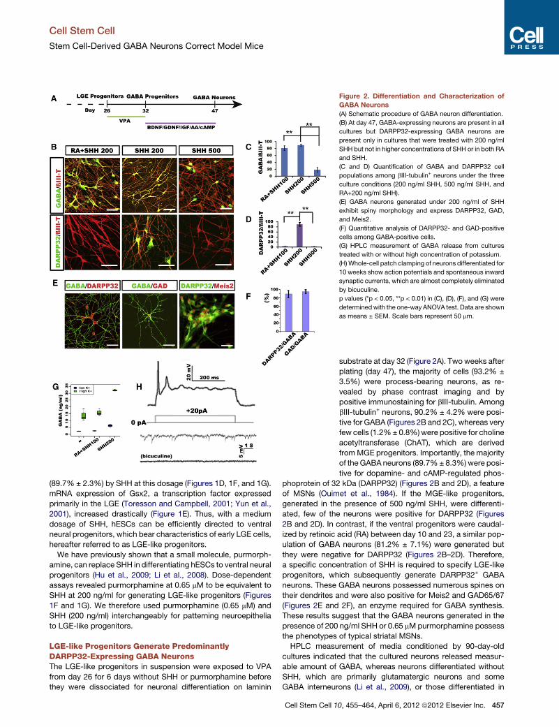
Figure 2. Differentiation and Characterization of
GABA Neurons
(A) Schematic procedure of GABA neuron differentiation.
(B) At day 47, GABA-expressing neurons are present in all
cultures but DARPP32-expressing GABA neurons are
present only in cultures that were treated with 200 ng/ml
SHH but not in higher concentrations of SHH or in both RA
and SHH.
(C and D) Quantification of GABA and DARPP32 cell
populations among bIII-tubulin+ neurons under the three
culture conditions (200 ng/ml SHH, 500 ng/ml SHH, and
RA+200 ng/ml SHH).
(E) GABA neurons generated under 200 ng/ml of SHH
exhibit spiny morphology and express DARPP32, GAD,
and Meis2.
(F) Quantitative analysis of DARPP32- and GAD-positive
cells among GABA-positive cells.
(G) HPLC measurement of GABA release from cultures
treated with or without high concentration of potassium.
(H)Whole-cell patch clamping of neurons differentiated for
10 weeks show action potentials and spontaneous inward
synaptic currents, which are almost completely eliminated
by bicuculine.
p values (*p < 0.05, **p < 0.01) in (C), (D), (F), and (G) were
determinedwith the one-way ANOVA test. Data are shown
as means ± SEM. Scale bars represent 50 mm.
Cell Stem Cell
Stem Cell-Derived GABA Neurons Correct Model Mice
(89.7% ± 2.3%) by SHH at this dosage (Figures 1D, 1F, and 1G).
mRNA expression of Gsx2, a transcription factor expressed
primarily in the LGE (Toresson and Campbell, 2001; Yun et al.,
2001), increased drastically (Figure 1E). Thus, with a medium
dosage of SHH, hESCs can be efficiently directed to ventral
neural progenitors, which bear characteristics of early LGE cells,
hereafter referred to as LGE-like progenitors.
We have previously shown that a small molecule, purmorph-
amine, can replace SHH in differentiating hESCs to ventral neural
progenitors (Hu et al., 2009; Li et al., 2008). Dose-dependent
assays revealed purmorphamine at 0.65 mM to be equivalent to
SHH at 200 ng/ml for generating LGE-like progenitors (Figures
1F and 1G). We therefore used purmorphamine (0.65 mM) and
SHH (200 ng/ml) interchangeably for patterning neuroepithelia
to LGE-like progenitors.
LGE-like Progenitors Generate PredominantlyDARPP32-Expressing GABA NeuronsThe LGE-like progenitors in suspension were exposed to VPA
from day 26 for 6 days without SHH or purmorphamine before
they were dissociated for neuronal differentiation on laminin
Cell Stem Cell 1
substrate at day 32 (Figure 2A). Two weeks after
plating (day 47), the majority of cells (93.2% ±
3.5%) were process-bearing neurons, as re-
vealed by phase contrast imaging and by
positive immunostaining for bIII-tubulin. Among
bIII-tubulin+ neurons, 90.2% ± 4.2% were posi-
tive for GABA (Figures 2B and 2C), whereas very
few cells (1.2%± 0.8%)were positive for choline
acetyltransferase (ChAT), which are derived
fromMGE progenitors. Importantly, the majority
of theGABA neurons (89.7%± 8.3%)were posi-
tive for dopamine- and cAMP-regulated phos-
phoprotein of 32 kDa (DARPP32) (Figures 2B and 2D), a feature
of MSNs (Ouimet et al., 1984). If the MGE-like progenitors,
generated in the presence of 500 ng/ml SHH, were differenti-
ated, few of the neurons were positive for DARPP32 (Figures
2B and 2D). In contrast, if the ventral progenitors were caudal-
ized by retinoic acid (RA) between day 10 and 23, a similar pop-
ulation of GABA neurons (81.2% ± 7.1%) were generated but
they were negative for DARPP32 (Figures 2B–2D). Therefore,
a specific concentration of SHH is required to specify LGE-like
progenitors, which subsequently generate DARPP32+ GABA
neurons. These GABA neurons possessed numerous spines on
their dendrites and were also positive for Meis2 and GAD65/67
(Figures 2E and 2F), an enzyme required for GABA synthesis.
These results suggest that the GABA neurons generated in the
presence of 200 ng/ml SHH or 0.65 mMpurmorphamine possess
the phenotypes of typical striatal MSNs.
HPLC measurement of media conditioned by 90-day-old
cultures indicated that the cultured neurons released measur-
able amount of GABA, whereas neurons differentiated without
SHH, which are primarily glutamatergic neurons and some
GABA interneurons (Li et al., 2009), or those differentiated in
0, 455–464, April 6, 2012 ª2012 Elsevier Inc. 457
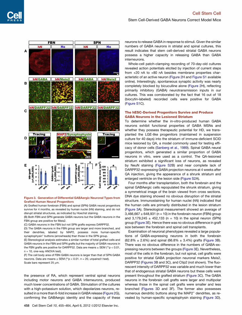
Figure 3. Generation of Differential GABAergic Neuronal Types from
Grafted Human Neural Progenitors
(A) Grafted human forebrain (FBN) and spinal (SPN) GABA neural progenitors
survive for 4 months, as revealed by human nuclei (hN) staining, and do not
disrupt striatal structures, as indicated by Hoechst staining.
(B) Both FBN and SPN generate GABA neurons but the GABA neurons in the
FBN group are positive for Meis2.
(C) GABA neurons in the FBN but not SPN grafts express DARPP32.
(D) The GABA neurons in the FBN group are larger and more branched, and
their dendrites, labeled by MAP2, possess more human-specific
synaptophysin+ buttons (arrowheads) than those in the SPN group.
(E) Stereological analysis estimates a similar number of total grafted cells and
GABA neurons in the FBN and SPN grafts but the majority of GABA neurons in
the FBN grafts are positive for DARPP32. Data are means ± SEM (**p < 0.01,
n = 10, one-way ANOVA test).
(F) The cell body area of FBN GABA neurons is larger than that of SPN GABA
neurons. Data are means ± SEM (**p < 0.01. n = 20, unpaired t test).
Scale bars represent 25 mm.
Cell Stem Cell
Stem Cell-Derived GABA Neurons Correct Model Mice
the presence of RA, which represent ventral spinal neurons
including motor neurons and GABA interneurons, produced
much lower concentrations of GABA. Stimulation of the cultures
with a high-potassium solution, which depolarizes neurons, re-
sulted in amore than 6-fold increase inGABA release (Figure 2G),
confirming the GABAergic identity and the capacity of these
458 Cell Stem Cell 10, 455–464, April 6, 2012 ª2012 Elsevier Inc.
neurons to release GABA in response to stimuli. Given the similar
numbers of GABA neurons in striatal and spinal cultures, this
result indicates that stem cell-derived striatal GABA neurons
possess a higher capacity in releasing GABA than GABA
interneurons.
Whole-cell patch-clamping recording of 70-day-old cultures
revealed action potentials elicited by injection of current steps
from +20 nA to +80 nA besides membrane properties char-
acteristic of an active neuron (Figure 2H and Figure S1 available
online). Interestingly, spontaneous synaptic activity was nearly
completely blocked by bicuculline alone (Figure 2H), reflecting
primarily inhibitory (GABA) neurotransmission inputs in our
cultures. This was corroborated by the fact that 16 out of 18
(biocytin-labeled) recorded cells were positive for GABA
(Figure S1C).
The hESC-Derived Progenitors Survive and ProduceGABA Neurons in the Lesioned StriatumTo determine whether the in-vitro-produced human GABA
neurons exhibit functional properties of GABA MSNs and
whether they possess therapeutic potential for HD, we trans-
planted the LGE-like progenitors (maintained in suspension
culture for 40 days) into the striatum of immune-deficient SCID
mice lesioned by QA, a model commonly used for testing effi-
cacy of donor cells (Sanberg et al., 1989). Spinal GABA neural
progenitors, which generated a similar proportion of GABA
neurons in vitro, were used as a control. The QA-lesioned
striatum exhibited a significant loss of neurons, as revealed
by NeuN staining (Figure S2B) and near complete lack of
DARPP32-expressing GABA projection neurons at 4 weeks after
QA injection, giving the appearance of a shrunk striatum and
enlarged ventricle on the lesion side (Figure S2A).
Four months after transplantation, both the forebrain and the
spinal GABAergic cells repopulated the shrunk striatum, giving
a symmetrical image of the brain viewed from cross sections.
DNA dye staining showed no obvious disruption of the striatal
structure. Immunostaining for human nuclei (hN) indicated that
the human cells are primarily distributed in the lesion striatum
(Figure 3A). Stereological measurement showed an average of
3,466,667 ± 648,931 (n = 10) in the forebrain neuron (FBN) group
and 3,179,245 ± 452,133 (n = 10) in the spinal neuron (SPN)
group (Figure 3E). Hence there was no obvious difference in graft
size between the forebrain and spinal cell transplants.
Examination of neuronal phenotypes revealed a large popula-
tions of GABA-expressing neurons in both the forebrain
(62.8% ± 2.6%) and spinal (66.8% ± 3.4%) grafts (Figure 3B).
There was no obvious difference in the numbers of GABA-ex-
pressing neurons between the groups (Figure 3E). Nevertheless,
most of the cells in the forebrain, but not spinal, cell grafts were
positive for striatal GABA projection neuronal markers Meis2,
DARPP32 (Figures 3B and 3C), and Ctip2 (not shown). The fluo-
rescent intensity of DARPP32 was variable and much lower than
that of endogenous striatal GABA neurons but these cells were
present throughout the grafted striatum (Figure 3C). The GABA
neurons in the forebrain cell grafts were larger and multipolar
whereas those in the spinal cell grafts were smaller and less
branched (Figures 3D and 3F). The former also possesses
numerous dendritic buttons along the MAP2+ dendrites, as re-
vealed by human-specific synaptophysin staining (Figure 3D).

Figure 4. Projection and Connections by Grafted Human GABA
Neurons
(A) In the striatum, GABA neurons of the FBN but not the SPN grafts possess
dense TH+ terminals on cell bodies. GABA neurons of both FBN and SPN
grafts have vGLUT1-positive terminals but GABA neurons in the FBN grafts
have more (upper row). Most of the TH+ and vGlu+ fibers are negative for
human-specific STEM121 (low row).
(B) In anterior midbrain (STN/SN), human neuronal fibers, indicated by human-
specific synaptophysin, are present and surround TH+ dopamine neurons in
the brain grafted with FBN but not SPN cells. The DARPP32+ fibers in the FBN-
grafted nigra are also positive for substance P but not enkephalin (lower row).
Scale bars represent 25 mm.
Cell Stem Cell
Stem Cell-Derived GABA Neurons Correct Model Mice
Stereological measurement estimated that DARPP32-express-
ing GABA neurons constitute 58.6% ± 3.0% of total grafted cells
in the FBN group, in comparison to none in the SPN group (Fig-
ure 3E). Like endogenous striatal GABA neurons (Durieux et al.,
2011), the grafted human striatal GABA neurons also expressed
enkephalin or substance P (Figure S3). Therefore, although both
forebrain and spinal progenitors generate similar numbers of
GABAergic neurons, only the forebrain progenitors differentiate
to GABA neurons with the striatal projection phenotypes.
Although the vast majority of the grafted cells become bIII-
tubulin+ neurons (86%± 4.04%), some (8.43%±1.42%) became
astrocytes (Figures S4A and S4B). Besides GABA neurons being
the predominant population, small populations of neurons were
present in the forebrain neuron (FBN) grafts that expressedChAT
(1.57% ± 0.41%), vGLUT1 (0.92% ± 0.15%), 5-HT (0.86% ±
0.27%), and TH (0.97% ± 0.29%) (Figures S4C and S4D).
Although the majority of neurons were DARPP32-expressing
projection neurons, 6.0% ± 0.52% of total cells were positive
for calbindin (Figures S4E and S4F), a calcium binding protein
(Kawaguchi et al., 1995). Interestingly, two-thirds of them, or
3.89% ± 0.57% of the total cells, did not colabel with GABA
(Figures S4G and S4H). In addition, 1.05% ± 0.22% of the
grafted cells were positive for calretinin, 1.02% ± 0.16% were
positive for parvalbumin, and 0.50% ± 0.07% were positive for
somatostatin and they were all positive for GABA (Figures S4E
and S4F). Immunostaining for neural progenitor marker nestin
and mitotic marker KI67 showed a lack of mitotic progenitors
in the grafts 4 months after transplantation, although at 1 week
after transplantation, KI67-positive cells were present (Fig-
ure S4I). At 1 week, the majority of the cells were nestin-positive
but Pax6-negative progenitors with few (2.4% ± 0.72%)
DARPP32-positive GABA neurons (Figure S4J), suggesting
that the majority of the GABA neurons are generated from
grafted progenitors.
Human GABA Neurons Connect with Endogenous CellsStriatal GABA projection neurons reciprocally synapse with
neurons in the globus pallidus, thalamus, and substantia nigra,
thus regulating locomotion (Wictorin, 1992). Confocal analyses
showed that grafted GABA neurons in both groups colabeled
with PSD95 (Figure 4A), suggesting that the grafted GABA
neurons receive potential inputs. Further labeling showed robust
TH staining on GABA+ cell bodies and processes in the forebrain
cell graft but not in the spinal cell transplant (Figure 4A), sugges-
tive of dopaminergic inputs into the grafted MSN, possibly from
the midbrain. Double staining also revealed dense vGlu signals
on forebrain GABA neurons but substantially less in spinal
GABA neurons (Figure 4A), suggesting glutamatergic inputs,
possibly from cortex, globus pallidus, and/or thalamus. Because
GABA neurons are almost completely destroyed in the lesion site
and because there are few TH+ dopamine neurons and vGlu+
glutamate neurons in the graft (Figures S4C and S4D), colabeling
of TH or vGlu on GABA neurons suggests inputs from endoge-
nous neurons. This is confirmed by lack of labeling of (human-
specific) STEM121 on TH- or vGlu-expressing fibers (lower row
in Figure 4A).
In the anterior substantia nigra, a discrete bundle of (human-
specific) synaptophysin-expressing fibers were observed in the
FBN group but not the SPN group. These fibers overlapped
with TH-expressing neurons (Figure 4B), indicating that the
grafted human GABA neurons project to the anterior substantia
nigra and potentially form connections with dopaminergic
neurons. In this area, the DARPP32 fibers also expressed
substance P but none of them expressed enkephalin (Figure 4B),
suggesting specific projection by subtype-specific DARPP32-
expresing GABA neurons. These results confirm the projection
nature of the grafted human forebrain GABA neurons and their
ability to project to appropriate targets.
Transplantation of Forebrain but Not Spinal GABAProgenitors Corrects Motor DeficitsBesides rotarod and open field tests, we employed an unbiased
motor tests using Treadscan, which not only reveals limb
movements detected by skilled paw use tests but also shows
characteristic chorea movement behaviors that are features of
Huntington patients (Menalled et al., 2009). Unilateral QA-
induced striatal lesion resulted in motor deficits typical of HD,
including decreased latency in rotarod test, decreased center
ratio/crossings in open field test, and changes in a number of
Cell Stem Cell 10, 455–464, April 6, 2012 ª2012 Elsevier Inc. 459

Figure 5. Transplantation of Human GABA MSN Corrects Locomotive Deficits
(A) Schematic presentation of temporal course of transplantation and behavioral analysis.
(B) Rotarod tests show increased latency in animals transplanted with FBN but not SPN.
(C) Open field tests indicate increased center ratio, crossing times, and distances in animals grafted with FBN but not SPN.
(D) Treadscan analysis reveals increased stride length, as well as decreasedmaximum longitudinal deviation, minimum lateral deviation, and the foot base of their
rear limbs in animals receiving FBN but SPN grafts.
(B) and (D) were analyzed with the repeat measures of general linear model (GLM) analysis, and open field behaviors (E) were analyzed by one-way ANOVA
(*p < 0.05, n = 10).
Cell Stem Cell
Stem Cell-Derived GABA Neurons Correct Model Mice
parameters of fine gait movements revealed by Treadscan such
as smaller stride length (the distance between two successive
initiations of stances) but a larger foot base (distances between
front and rear foot pair), increased minimum lateral deviation
(the closest distance from foot to the body axis), and increased
maximum longitudinal deviation (the farthest distance of foot
away from the body axis, equivalent to the ataxic gait of a HD
patient). The small stride length and lagging and wobbling of
rear limbs resemble the gait abnormalities seen in typical HD
patients (Koller and Trimble, 1985; Vandeputte et al., 2010).
Monthly behavioral analysis posttransplantation (Figure 5A)
revealed that mice receiving the forebrain but not spinal GABA
progenitor transplantation exhibited increased latency on ro-
tarod (Figure 5B) and increased center ratio, crossings, and total
distance in open field tests (Figure 5C). Gait analysis with
Treadscan indicated that mice receiving forebrain GABA
progenitors exhibited a significantly greater stride length. The
maximum longitudinal deviation, minimum lateral deviation,
and the foot base of their rear limbs, especially on the right
(lesion) side, became smaller over time (Figure 5D). These results
indicate that the forebrain, but not spinal, GABA neurons correct
the locomotion deficits in the QA-lesioned mice.
460 Cell Stem Cell 10, 455–464, April 6, 2012 ª2012 Elsevier Inc.
DISCUSSION
By following the developmental principles and by tuning a partic-
ular dose of SHH or purmorphamine on primitive neuroepithelia,
we have directed hESCs to an enriched population of LGE-like
progenitors, which differentiate to predominantly DARPP32-
expressing MSNs in vitro and in vivo. Importantly, the human
stem cell-derived MSNs, but not the DARPP32-negative GABA
interneurons, project to the substantia nigra and possibly also
receive from neurons in the cerebral cortex and substantia nigra,
which corresponds to correction of locomotion deficits in the
QA-lesionedmice. These results raise the prospect of cell-based
therapy as a potential treatment for HD, given the complete lack
of effective treatment for HD at the present.
Differentiation of MSNs from hESCs was explored by Perrier
and colleagues (Aubry et al., 2008). They generated approxi-
mately 8% GABA+ and 12% DARPP32+ neurons among the
differentiated progenies in the presence of both SHH and
a Wnt inhibitor DKK1. Transplantation of these cells, either at
day 45 or 56, invariably leads to overgrowth 13–15 weeks post-
graft (Aubry et al., 2008). A number of factors may explain the
contrasting outcomes between our results, ranging from culture
Cell Stem Cell
Stem Cell-Derived GABA Neurons Correct Model Mice
modes to use of morphogens (timing, concentration, combina-
tions, etc.). As we have demonstrated in the present study,
a specific concentration of SHH is necessary to induce neuroe-
pithelia at a particular window of development in order to induce
a synchronized population of LGE-like progenitors, which give
rise to DARPP32-expressing MSNs. Less or over ventralization
of primitive NSCs will lead to generation of cortical or MGE
neuronal types, respectively (Danjo et al., 2011; Li et al., 2009),
demonstrating the precise regulation of cell fate specification
by extracellular signals. Exposure of monolayer neuroepithelial
colonies to morphogens under our chemically defined culture,
instead of neurospheres derived from coculture with PA6
stromal cells (Aubry et al., 2008), may further account for the
synchronized population of LGE-like progenitors and MSNs. It
may seem surprising to produce a large population of DARPP32
MSNs given the presence of a substantial proportion of Pax6-
and Nkx2.1-expressing progenitors. It should be noted that
Pax6 is also expressed in the LGE although the expression level
is lower than that in the cortex (Flames et al., 2007; Yun et al.,
2001). Indeed, under 200 ng/ml SHH, the Pax6 fluorescent inten-
sity is much lower (Figure 1B); hence they are probably LGE-like
progenitors. This is supported by a complete lack of Pax6-
expressing cells at 1 week posttransplantation and few glutama-
tergic neurons in the grafts 4 months postgraft. The Nkx2.1
progenitors can produce cholinergic neurons and GABA inter-
neurons (Sussel et al., 1999), but our differentiation culture
(without NGF) does not favor generation/survival of cholinergic
neurons. It is also suggested by the observation that only about
5% of the total grafted cells express GABA and other interneu-
ronal phenotypes, parvalbumin, calretinin, and somatostatin.
Together, this progenitor pool produces themost robust popula-
tion of DARPP32-expressing GABA neurons in vitro and in vivo,
as evidenced by electrophysiological and transplant results. Our
method was also effective in differentiating MSNs from iPSCs
(not shown), thus facilitating studies of MSN-degenerative
processes, especially with those from HD iPSCs (Zhang et al.,
2010).
HD remains a devastating condition because of a near
complete lack of effective therapy. Clinical trials with fetal cell
transplantation suggest that cell therapy is a potential promising
option for HD if a consistent donor source is available (Clelland
et al., 2008; Gallina et al., 2010). Although the QA-lesioned
mice do not completely mimic the pathology of HD, they offer
a simple model to examine the efficacy of cell therapy. It is
encouraging that grafted human neural progenitors repopulated
the atrophied striatum, differentiated to GABAergic neurons, and
contributed to correction in movement impairments. Our parallel
comparison of two different populations of human GABA
progenitors provides insights into the biology and potential use
of GABA neurons in HD treatment. First, GABA MSNs are
required for correcting the motor deficits. A simple provision of
GABA or GABA-producing cells, such as the GABA interneurons
in the present study, is not effective for correcting the motor
deficits. Second, therapeutic outcome may be dependent
upon the reformation of circuitry by grafted GABA neurons.
Although the GABA interneurons can form connections with
each other and potentially also with host neurons, they do not
project out of the striatum. In contrast, the human GABA MSNs
not only receive inputs but also project outside of the graft site.
More importantly, these GABA neurons appear to receive
specific inputs, including dopaminergic and glutamatergic
inputs, and project to specific brain regions—substantia nigra.
And the projection appears specific to the substance P- but
not enkephalin-expressing GABA neuronal subtype, resembling
the endogenous striatal GABA neurons (Gerfen, 1988). These
phenomena strongly suggest the intrinsic capacity of appropri-
ately patterned developing neurons to find their target and the
reformation of neural circuitry, which may underlie the correction
of motor deficits. Third, the adult CNS environment is generally
inhibitory to axonal regeneration. However, our present obser-
vations indicate that the human stem cell-derived neurons can
project for quite a distance. Indeed, human fetal striatal cells
transplanted into a similar QA-lesioned rat striatum could project
to the substantia nigra (Wictorin et al., 1990). The striking
similarity between fetal human striatal cells and our in-vitro-
produced striatal cells validates the identity and utility of our
stem cell-produced GABA neurons. It would be interesting to
replicate these cellular behaviors in other models such as the
newly established transgenic HD model that has clear striatal
GABA neuron degeneration (Wilburn et al., 2011).
One of the main obstacles to stem cell therapy is tumor forma-
tion or overgrowth (Aubry et al., 2008; Roy et al., 2006). The grafts
from both the forebrain and the spinal GABA neuronal progeni-
tors do not appear disruptive to the striatal structures nor do
they remain proliferating progenitors. Multiple factors may
contribute to such an outcome. Our initial adherent colony neural
induction culture mode allows obtaining a nearly pure population
of neuroepithelia (Hu et al., 2010; Zhang et al., 2001), and subse-
quent patterning with SHH effectively restricts the cells to
a ventral progenitor fate. Expansion of the progenitors for an
additional 2 weeks also significantly decreases cell cycle
numbers needed for the grafted human progenitors to produce
neurons. This is evidenced by the fact that at least 94% of the
grafted cells are neurons and astrocytes and the majority of the
neurons are of GABA phenotypes. It is no doubt that rigorous
testing in more animals for a longer period is necessary to
move to clinic trials; however, the present finding, particularly
the ability of hESC-derived GABA MSNs to receive and project
to specific targets, raises hopes for cell-based therapy for HD.
EXPERIMENTAL PROCEDURES
Differentiation of Forebrain and Spinal GABA Neurons from hESCs
hESCs (line H9, passages 21–42; line H1, passages 24–34) weremaintained on
a feeder layer of irradiated mouse embryonic fibroblasts (MEFs) as described
(Thomson et al., 1998). hESCs were differentiated to Pax6-expressing
primitive neuroepithelia (NE) for 10–12 days in a neural induction medium
consisting of DMEM/F12, N2 supplement, and nonessential amino acids (Pan-
kratz et al., 2007; Zhang et al., 2001). Sonic hedgehog (SHH, 50–500 ng/ml)
or its small molecular agonist purmorphamine (0.1–1.5 mM; Calbiochem, San
Diego, CA) was added at days 12–26 to induce ventral progenitors. For gener-
ating spinal GABAneurons, retinoic acid (RA, 0.1mM)was added fromday10 to
23 (Li et al., 2008). For neuronal differentiation, neural progenitor clusters were
dissociated with Accutase (1 unit/ml, Invitrogen) at 37�C for 5 min and placed
onto polyornithine/laminin-coated coverslips at day 26 in Neurobasal medium
in the presence of valproic acid (VPA, 10 mM, Sigma) for 1 week, followed by
a set of trophic factors, including brain-derived neurotrophic factor (BDNF,
20 ng/ml), glial-derived neurotrophic factor (GDNF, 10 ng/ml), insulin-like
growth factor 1 (IGF1, 10 ng/ml), and cAMP (1 mM) (all from R&D Systems).
Detailed differentiation procedure is presented in Supplemental Information.
Cell Stem Cell 10, 455–464, April 6, 2012 ª2012 Elsevier Inc. 461
Cell Stem Cell
Stem Cell-Derived GABA Neurons Correct Model Mice
Immunocytochemistry and Quantification
Coverslip cultures were fixed in 4% paraformaldehyde for 15 min at 4�C,washed with PBS, and incubated in a blocking buffer (10% donkey serum
and 0.2% Triton X-100 in PBS) for 60 min at room temperature before being
incubated in the following primary antibodies (Table S1) overnight at 4�C.Fluorescently conjugated secondary antibodies were used to reveal the
binding of primary antibodies (1:1000, Jackson, West Grove, PA) and nuclei
were stained with Hoechst 33258. Images were collected with a Nikon
TE600 fluorescence microscope (Nikon Instruments, Melville, NY) or a Nikon
C1 laser-scanning confocal microscope (Nikon, Tokyo, Japan). The population
of Otx2-, Pax6-, Nkx2.1-, and Mash1-expressing cells among total differenti-
ated cells (Hoechst-labeled) was counted with ImageJ software. At least five
fields of each coverslip were chosen randomly and three coverslips in each
group were counted. Data were replicated three times in two different cell lines
(H9 and H1) and were expressed as mean ± SEM.
RNA Isolation and Quantitative PCR
The total RNA was isolated from cultured cells with RNA STAT-60 (Tel-Test,
Friendswood, TX). cDNAwas generated from 1 mg of total RNAwith the Super-
Script III First-Strand Synthesis System (Invitrogen, Carlsbad, CA) and was
used as a template for the quantitative PCR (qPCR).
Measurement of GABA Release by HPLC
Cultured cells were washed four times with Krebs’-Ringer’s solution contain-
ing 130 mM NaCl, 3 mM KCl, 2 mM CaCl2, 0.8 mM MgSO4, 10 mM glucose,
and 20mMHEPES (pH 7.4) before themediumwas collectedwith depolarizing
Krebs’-Ringer’s solution containing 83 mM NaCl, 50 mM KCl, 2 mM CaCl2,
0.8 mM MgSO4,10 mM glucose, and 20 mM HEPES (pH 7.4). The collected
media were thenmixedwith o-phthalaldehyde (0.8 g/l) and 2-mercaptoethanol
(2 ml/l) for 5 min at 15�C. Samples were then injected into the HPLC system
and analyzed with a fluorescence monitor (excitation at 350 nm, emission at
450 nm) (CMA/Microdialysis, Stockholm, Sweden). The mobile (60 ml/min)
solution consisted of (in mM) 100 KH2PO4, 100 Na2HPO4, and 0.1 EDTA
(pH 6.0), containing 10% acetonitrile and 3% tetrahydrofuran. The peak area
at the predicted position was calibrated against the standard curves for
quantification with CMA200 software (CMA/Microdialysis).
Whole-Cell Patch-Clamp Recording
Whole-cell patch-clamp recordings were performed on hESC-derived neurons
that were differentiated for 8–10 weeks as described (Johnson et al., 2007).
The identity of recorded cells was revealed by injecting biocytin (1%, Sigma)
followed by immunostaining.
Striatal Lesions and Cell Transplantation
All animal experiments were conducted according to a protocol approved by
the animal care and use committee at University of Wisconsin-Madison. To
create the unilateral lesion, adult male SCID mice (10 weeks of age) were
anesthetized with 1%–2% isoflurane mixed in oxygen and received a stereo-
taxic injection of 2 ml of 0.1 M quinolinic acid (QA, P63204; Sigma, in saline
with 0.2 mg/ml ascorbic acid) into the right striatum on a coordinator
(anterior-posterior [AP] = �0.7 mm, lateral [L] = +1.7 mm, vertical [V] =
�3.2 mm) (Hansson et al., 1999).
Differentiated forebrain or spinal GABA neuron progenitors (40 days of
differentiation from hESCs in suspension) were dissociated with Accutase
and prepared at approximately 50,000 cells/ml in artificial cerebrospinal fluid
(aCSF) containing B27, 200 mM ascorbic acid, 1 mM cAMP, 20 ng/ml BDNF,
and 10 ng/ml GDNF. Cell suspension (2 ml) was injected into the lesioned stria-
tum (AP = �0.8 mm, L = +2.0 mm, V = �3.2 mm) of anesthetized animals
4 weeks after QA lesion with a glass pipette (0.3–0.5 mm in diameter) over
a period of 5 min. The QA-lesioned animals receiving the same surgery and
injection of 2 ml of the same aCSF solution (without cells) served as controls.
Behavioral Tests
Behavioral tests were conducted before and after transplantationmonthly until
the animals were sacrificed.
Treadscan Analysis
We utilized an unbiased treadmill device, TreadScan (Columbus Instruments,
Columbus, OH), to detect gait alterations that are reminiscent of locomotion
462 Cell Stem Cell 10, 455–464, April 6, 2012 ª2012 Elsevier Inc.
changes seen in Huntington patients. All mice were allowed to walk on the
motor-driven treadmill belt at a speed of 15 cm/s for a period of 20 s. The
footprints and body movement were recorded with a high-speed digital video
camera from the ventral view of the treadmill belt reflected off the mirror.
TreadScan software (CleverSys) was used to identify initial foot contact,
stance duration, stride duration, foot liftoff, swing duration, stride length, track
width, and toe spread for each foot. The digital data were analyzed to compare
each of the parameters between ipsilateral and contralateral sides and
between the transplant and nontransplant groups.
Rotarod Test
An accelerating Rotarod (Columbus Instruments) was used to test motor
coordination. The mouse was placed on a rotating rod that accelerated from
4 rotations per minute (rpm) to 40 rpm in a period of 300 s. The period of
time the mouse stayed on the rod was monitored and the three of a total of
five runs in which the mouse performed the best were recorded.
Open Field Test
Mice were placed in the center of activity chambers equipped with infrared
beams (Med ASSOCIATES, St. Albans, VT; 27 3 27 3 20.3 cm). Activities
were recorded for 30 min under normal conditions of lighting. Quantitative
analysis was done on total distance, center ratio, and crossings.
Stereological Analysis of Grafts
Animals were perfused with 4% paraformaldehyde, brains dissected out, and
coronal cryo-sections made at 30 mm in thickness for free-floating immunos-
taining (Yang et al., 2008). The number of grafted human cells (hN+) and
GABA neurons (GABA+/DARPP32+/hN+) were counted with a StereoInvestiga-
tor software (MicroBrightField, Inc) on every six sections as described (Yang
et al., 2008). The area of the graft was outlined according to the presence of
hN-positive cells under a 103 objective of a Zeiss fluorescent scope. Cell
counting was performed with a 403 objective in fields chosen by the software.
The number of cells on each section and within the whole graft were estimated
by the Stereo Investigator software (Peterson, 1999). Data are presented as
mean ± SEM (n = 10). For measuring GABA neuronal body and processes,
we employed NeuonJ (from ImageJ) software to trace fluorescent images of
neurons that expressed GABA. Tracings were color coded: red for primary
neurite (emanating directly from the soma), blue for secondary neurite (branch-
ing from a primary), yellow for tertiary neurite (branching from a secondary),
and purple for quaternary neurite (branching from a tertiary).
Statistical Analyses
SPSS software was used for statistical analysis. In all studies, comparison of
mean values was conducted with unpaired t test, one-way ANOVO, or repeat
measures of general linear model (GLM) analysis. In all analyses, statistical
significance was determined at the 5% level (p < 0.05).
SUPPLEMENTAL INFORMATION
Supplemental Information includes Supplemental Experimental Procedures,
four figures, and one table and can be found with this article online at
doi:10.1016/j.stem.2012.01.021.
ACKNOWLEDGMENTS
This study was supported in part by the NIH-NINDS (NS045926), the NICHD
(P30 HD03352), Ministry of Science and Technology, China (2006CB94700,
2006AA02A101), and Shanghai Municipality (06dj14001).
Received: September 12, 2011
Revised: December 7, 2011
Accepted: January 27, 2012
Published online: March 15, 2012
REFERENCES
Arlotta, P., Molyneaux, B.J., Jabaudon, D., Yoshida, Y., and Macklis, J.D.
(2008). Ctip2 controls the differentiation of medium spiny neurons and the
establishment of the cellular architecture of the striatum. J. Neurosci. 28,
622–632.
Cell Stem Cell
Stem Cell-Derived GABA Neurons Correct Model Mice
Aubry, L., Bugi, A., Lefort, N., Rousseau, F., Peschanski, M., and Perrier, A.L.
(2008). Striatal progenitors derived from human ES cells mature into DARPP32
neurons in vitro and in quinolinic acid-lesioned rats. Proc. Natl. Acad. Sci. USA
105, 16707–16712.
Bachoud-Levi, A., Bourdet, C., Brugieres, P., Nguyen, J.P., Grandmougin, T.,
Haddad, B., Jeny, R., Bartolomeo, P., Boisse, M.F., Barba, G.D., et al. (2000).
Safety and tolerability assessment of intrastriatal neural allografts in five
patients with Huntington’s disease. Exp. Neurol. 161, 194–202.
Bachoud-Levi, A.C., Gaura, V., Brugieres, P., Lefaucheur, J.P., Boisse, M.F.,
Maison, P., Baudic, S., Ribeiro, M.J., Bourdet, C., Remy, P., et al. (2006).
Effect of fetal neural transplants in patients with Huntington’s disease 6 years
after surgery: a long-term follow-up study. Lancet Neurol. 5, 303–309.
Bonner, J.F., Blesch, A., Neuhuber, B., and Fischer, I. (2010). Promoting direc-
tional axon growth from neural progenitors grafted into the injured spinal cord.
J. Neurosci. Res. 88, 1182–1192.
Campbell, K. (2003). Dorsal-ventral patterning in the mammalian telenceph-
alon. Curr. Opin. Neurobiol. 13, 50–56.
Campbell, K., Kalen, P., Wictorin, K., Lundberg, C., Mandel, R.J., and
Bjorklund, A. (1993). Characterization of GABA release from intrastriatal striatal
transplants: dependence on host-derived afferents. Neuroscience 53,
403–415.
Clelland, C.D., Barker, R.A., and Watts, C. (2008). Cell therapy in Huntington
disease. Neurosurg. Focus 24, E9.
Danjo, T., Eiraku, M., Muguruma, K., Watanabe, K., Kawada, M., Yanagawa,
Y., Rubenstein, J.L., and Sasai, Y. (2011). Subregional specification of embry-
onic stem cell-derived ventral telencephalic tissues by timed and combinatory
treatment with extrinsic signals. J. Neurosci. 31, 1919–1933.
Dobrossy, M.D., and Dunnett, S.B. (2006). The effects of lateralized training
on spontaneous forelimb preference, lesion deficits, and graft-mediated
functional recovery after unilateral striatal lesions in rats. Exp. Neurol. 199,
373–383.
Dunnett, S.B., and Rosser, A.E. (2007). Cell transplantation for Huntington’s
disease: Should we continue? Brain Res. Bull. 72, 132–147.
Durieux, P.F., Schiffmann, S.N., and de Kerchove d’Exaerde, A. (2011).
Targeting neuronal populations of the striatum. Front Neuroanat 5, 40.
Flames, N., Pla, R., Gelman, D.M., Rubenstein, J.L., Puelles, L., and Marın, O.
(2007). Delineation of multiple subpallial progenitor domains by the combina-
torial expression of transcriptional codes. J. Neurosci. 27, 9682–9695.
Fode, C., Ma, Q., Casarosa, S., Ang, S.L., Anderson, D.J., and Guillemot, F.
(2000). A role for neural determination genes in specifying the dorsoventral
identity of telencephalic neurons. Genes Dev. 14, 67–80.
Gallina, P., Paganini, M., Lombardini, L., Mascalchi, M., Porfirio, B., Gadda, D.,
Marini, M., Pinzani, P., Salvianti, F., Crescioli, C., et al. (2010). Human striatal
neuroblasts develop and build a striatal-like structure into the brain of
Huntington’s disease patients after transplantation. Exp. Neurol. 222, 30–41.
Gerfen, C.R. (1988). Synaptic organization of the striatum. J. Electron Microsc.
Tech. 10, 265–281.
Goto, S., Yamada, K., Yoshikawa, M., Okamura, A., and Ushio, Y. (1997).
GABA receptor agonist promotes reformation of the striatonigral pathway by
transplant derived from fetal striatal primordia in the lesioned striatum. Exp.
Neurol. 147, 503–509.
Hansson, O., Petersen, A., Leist, M., Nicotera, P., Castilho, R.F., and Brundin,
P. (1999). Transgenic mice expressing a Huntington’s disease mutation are
resistant to quinolinic acid-induced striatal excitotoxicity. Proc. Natl. Acad.
Sci. USA 96, 8727–8732.
Hauser, R.A., Furtado, S., Cimino, C.R., Delgado, H., Eichler, S., Schwartz, S.,
Scott, D., Nauert, G.M., Soety, E., Sossi, V., et al. (2002). Bilateral human fetal
striatal transplantation in Huntington’s disease. Neurology 58, 687–695.
Hu, B.Y., Du, Z.W., Li, X.J., Ayala, M., and Zhang, S.C. (2009). Human oligo-
dendrocytes from embryonic stem cells: conserved SHH signaling networks
and divergent FGF effects. Development 136, 1443–1452.
Hu, B.Y., Weick, J.P., Yu, J., Ma, L.X., Zhang, X.Q., Thomson, J.A., and Zhang,
S.C. (2010). Neural differentiation of human induced pluripotent stem cells
follows developmental principles but with variable potency. Proc. Natl.
Acad. Sci. USA 107, 4335–4340.
Johnson, C.D., and Davidson, B.L. (2010). Huntington’s disease: progress
toward effective disease-modifying treatments and a cure. Hum. Mol.
Genet. 19 (R1), R98–R102.
Johnson, M.A., Weick, J.P., Pearce, R.A., and Zhang, S.C. (2007). Functional
neural development from human embryonic stem cells: accelerated synaptic
activity via astrocyte coculture. J. Neurosci. 27, 3069–3077.
Kawaguchi, Y., Wilson, C.J., Augood, S.J., and Emson, P.C. (1995). Striatal
interneurones: chemical, physiological and morphological characterization.
Trends Neurosci. 18, 527–535.
Koller, W.C., and Trimble, J. (1985). The gait abnormality of Huntington’s
disease. Neurology 35, 1450–1454.
Li, X.J., Hu, B.Y., Jones, S.A., Zhang, Y.S., Lavaute, T., Du, Z.W., and Zhang,
S.C. (2008). Directed differentiation of ventral spinal progenitors and motor
neurons from human embryonic stem cells by small molecules. Stem Cells
26, 886–893.
Li, X.J., Zhang, X., Johnson, M.A., Wang, Z.B., Lavaute, T., and Zhang, S.C.
(2009). Coordination of sonic hedgehog and Wnt signaling determines ventral
and dorsal telencephalic neuron types from human embryonic stem cells.
Development 136, 4055–4063.
Mazzocchi-Jones, D., Dobrossy, M., and Dunnett, S.B. (2011). Environmental
enrichment facilitates long-term potentiation in embryonic striatal grafts.
Neurorehabil. Neural Repair 25, 548–557.
Menalled, L., El-Khodor, B.F., Patry, M., Suarez-Farinas, M., Orenstein, S.J.,
Zahasky, B., Leahy, C., Wheeler, V., Yang, X.W., MacDonald, M., et al.
(2009). Systematic behavioral evaluation of Huntington’s disease transgenic
and knock-in mouse models. Neurobiol. Dis. 35, 319–336.
Nakao, N., and Itakura, T. (2000). Fetal tissue transplants in animal models
of Huntington’s disease: the effects on damaged neuronal circuitry and
behavioral deficits. Prog. Neurobiol. 61, 313–338.
Olsson, M., Campbell, K., Wictorin, K., and Bjorklund, A. (1995). Projection
neurons in fetal striatal transplants are predominantly derived from the lateral
ganglionic eminence. Neuroscience 69, 1169–1182.
Ouimet, C.C., Miller, P.E., Hemmings, H.C., Jr., Walaas, S.I., and Greengard,
P. (1984). DARPP-32, a dopamine- and adenosine 30:50-monophosphate-
regulated phosphoprotein enriched in dopamine-innervated brain regions.
III. Immunocytochemical localization. J. Neurosci. 4, 111–124.
Palfi, S., Conde, F., Riche, D., Brouillet, E., Dautry, C., Mittoux, V., Chibois, A.,
Peschanski, M., and Hantraye, P. (1998). Fetal striatal allografts reverse cogni-
tive deficits in a primate model of Huntington disease. Nat. Med. 4, 963–966.
Pankratz, M.T., Li, X.J., Lavaute, T.M., Lyons, E.A., Chen, X., and Zhang, S.C.
(2007). Directed neural differentiation of human embryonic stem cells via an
obligated primitive anterior stage. Stem Cells 25, 1511–1520.
Pearlman, S., Levivier, M., and Gash, D.M. (1993). Striatal implants of fetal
striatum or gelfoam protect against quinolinic acid lesions of the striatum.
Brain Res. 613, 203–211.
Peterson, D.A. (1999). Quantitative histology using confocal microscopy:
implementation of unbiased stereology procedures. Methods 18, 493–507.
Pfeifer, K., Vroemen, M., Blesch, A., and Weidner, N. (2004). Adult neural
progenitor cells provide a permissive guiding substrate for corticospinal
axon growth following spinal cord injury. Eur. J. Neurosci. 20, 1695–1704.
Philpott, L.M., Kopyov, O.V., Lee, A.J., Jacques, S., Duma, C.M., Caine, S.,
Yang, M., and Eagle, K.S. (1997). Neuropsychological functioning following
fetal striatal transplantation in Huntington’s chorea: three case presentations.
Cell Transplant. 6, 203–212.
Puelles, L., Kuwana, E., Puelles, E., Bulfone, A., Shimamura, K., Keleher, J.,
Smiga, S., and Rubenstein, J.L. (2000). Pallial and subpallial derivatives in
the embryonic chick and mouse telencephalon, traced by the expression of
the genes Dlx-2, Emx-1, Nkx-2.1, Pax-6, and Tbr-1. J. Comp. Neurol. 424,
409–438.
Reuter, I., Tai, Y.F., Pavese, N., Chaudhuri, K.R., Mason, S., Polkey, C.E.,
Clough, C., Brooks, D.J., Barker, R.A., and Piccini, P. (2008). Long-term
Cell Stem Cell 10, 455–464, April 6, 2012 ª2012 Elsevier Inc. 463

Cell Stem Cell
Stem Cell-Derived GABA Neurons Correct Model Mice
clinical and positron emission tomography outcome of fetal striatal transplan-
tation in Huntington’s disease. J. Neurol. Neurosurg. Psychiatry 79, 948–951.
Rosser, A.E., Barker, R.A., Harrower, T., Watts, C., Farrington, M., Ho, A.K.,
Burnstein, R.M., Menon, D.K., Gillard, J.H., Pickard, J., and Dunnett, S.B.;
NEST-UK. (2002). Unilateral transplantation of human primary fetal tissue in
four patients with Huntington’s disease: NEST-UK safety report ISRCTN no
36485475. J. Neurol. Neurosurg. Psychiatry 73, 678–685.
Roy, N.S., Cleren, C., Singh, S.K., Yang, L., Beal, M.F., and Goldman, S.A.
(2006). Functional engraftment of human ES cell-derived dopaminergic
neurons enriched by coculture with telomerase-immortalized midbrain astro-
cytes. Nat. Med. 12, 1259–1268.
Sanberg, P.R., Giordano, M., Henault, M.A., Nash, D.R., Ragozzino, M.E., and
Hagenmeyer-Houser, S.H. (1989). Intraparenchymal striatal transplants
required for maintenance of behavioral recovery in an animal model of
Huntington’s disease. J. Neural Transplant. 1, 23–31.
Sussel, L., Marin, O., Kimura, S., and Rubenstein, J.L. (1999). Loss of Nkx2.1
homeobox gene function results in a ventral to dorsal molecular respecification
within the basal telencephalon: evidence for a transformation of the pallidum
into the striatum. Development 126, 3359–3370.
Takahashi, K., Tanabe, K., Ohnuki, M., Narita, M., Ichisaka, T., Tomoda, K.,
and Yamanaka, S. (2007). Induction of pluripotent stem cells from adult human
fibroblasts by defined factors. Cell 131, 861–872.
Thomson, J.A., Itskovitz-Eldor, J., Shapiro, S.S., Waknitz, M.A., Swiergiel, J.J.,
Marshall, V.S., and Jones, J.M. (1998). Embryonic stem cell lines derived from
human blastocysts. Science 282, 1145–1147.
Toresson, H., and Campbell, K. (2001). A role for Gsh1 in the developing stria-
tum and olfactory bulb of Gsh2 mutant mice. Development 128, 4769–4780.
Toresson, H., Parmar, M., and Campbell, K. (2000). Expression of Meis and
Pbx genes and their protein products in the developing telencephalon: impli-
cations for regional differentiation. Mech. Dev. 94, 183–187.
464 Cell Stem Cell 10, 455–464, April 6, 2012 ª2012 Elsevier Inc.
Vandeputte, C., Taymans, J.M., Casteels, C., Coun, F., Ni, Y., Van Laere, K.,
and Baekelandt, V. (2010). Automated quantitative gait analysis in animal
models of movement disorders. BMC Neurosci. 11, 92.
Wichterle, H., Turnbull, D.H., Nery, S., Fishell, G., and Alvarez-Buylla, A. (2001).
In utero fate mapping reveals distinct migratory pathways and fates of neurons
born in the mammalian basal forebrain. Development 128, 3759–3771.
Wictorin, K. (1992). Anatomy and connectivity of intrastriatal striatal trans-
plants. Prog. Neurobiol. 38, 611–639.
Wictorin, K., Brundin, P., Gustavii, B., Lindvall, O., and Bjorklund, A. (1990).
Reformation of long axon pathways in adult rat central nervous system by
human forebrain neuroblasts. Nature 347, 556–558.
Wilburn, B., Rudnicki, D.D., Zhao, J., Weitz, T.M., Cheng, Y., Gu, X., Greiner,
E., Park, C.S., Wang, N., Sopher, B.L., et al. (2011). An antisense CAG repeat
transcript at JPH3 locus mediates expanded polyglutamine protein toxicity in
Huntington’s disease-like 2 mice. Neuron 70, 427–440.
Wilson, S.W., and Rubenstein, J.L. (2000). Induction and dorsoventral
patterning of the telencephalon. Neuron 28, 641–651.
Yang, D., Zhang, Z.J., Oldenburg, M., Ayala, M., and Zhang, S.C. (2008).
Human embryonic stem cell-derived dopaminergic neurons reverse functional
deficit in parkinsonian rats. Stem Cells 26, 55–63.
Yun, K., Potter, S., and Rubenstein, J.L. (2001). Gsh2 and Pax6 play comple-
mentary roles in dorsoventral patterning of the mammalian telencephalon.
Development 128, 193–205.
Zhang, S.C., Wernig, M., Duncan, I.D., Brustle, O., and Thomson, J.A. (2001).
In vitro differentiation of transplantable neural precursors from human embry-
onic stem cells. Nat. Biotechnol. 19, 1129–1133.
Zhang, N., An, M.C., Montoro, D., and Ellerby, L.M. (2010). Characterization of
human Huntington’s disease cell model from induced pluripotent stem cells.
PLoS Curr 2, RRN1193.
Copyright © 2022 FDOKUMEN