Manipulation of spermatogonial stem cells in livestock species
Upload
independentCategory
view
4download
0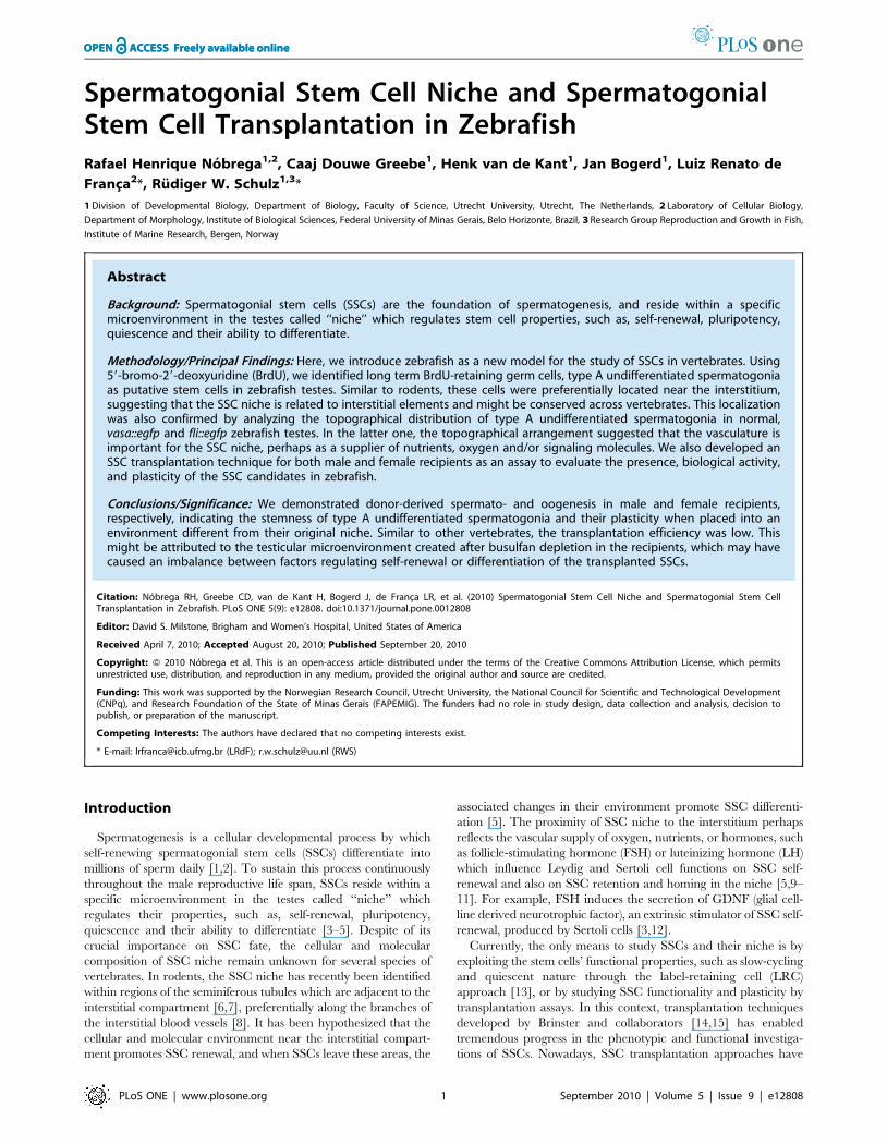
Spermatogonial Stem Cell Niche and SpermatogonialStem Cell Transplantation in ZebrafishRafael Henrique Nobrega1,2, Caaj Douwe Greebe1, Henk van de Kant1, Jan Bogerd1, Luiz Renato de
Franca2*, Rudiger W. Schulz1,3*
1 Division of Developmental Biology, Department of Biology, Faculty of Science, Utrecht University, Utrecht, The Netherlands, 2 Laboratory of Cellular Biology,
Department of Morphology, Institute of Biological Sciences, Federal University of Minas Gerais, Belo Horizonte, Brazil, 3 Research Group Reproduction and Growth in Fish,
Institute of Marine Research, Bergen, Norway
Abstract
Background: Spermatogonial stem cells (SSCs) are the foundation of spermatogenesis, and reside within a specificmicroenvironment in the testes called ‘‘niche’’ which regulates stem cell properties, such as, self-renewal, pluripotency,quiescence and their ability to differentiate.
Methodology/Principal Findings: Here, we introduce zebrafish as a new model for the study of SSCs in vertebrates. Using59-bromo-29-deoxyuridine (BrdU), we identified long term BrdU-retaining germ cells, type A undifferentiated spermatogoniaas putative stem cells in zebrafish testes. Similar to rodents, these cells were preferentially located near the interstitium,suggesting that the SSC niche is related to interstitial elements and might be conserved across vertebrates. This localizationwas also confirmed by analyzing the topographical distribution of type A undifferentiated spermatogonia in normal,vasa::egfp and fli::egfp zebrafish testes. In the latter one, the topographical arrangement suggested that the vasculature isimportant for the SSC niche, perhaps as a supplier of nutrients, oxygen and/or signaling molecules. We also developed anSSC transplantation technique for both male and female recipients as an assay to evaluate the presence, biological activity,and plasticity of the SSC candidates in zebrafish.
Conclusions/Significance: We demonstrated donor-derived spermato- and oogenesis in male and female recipients,respectively, indicating the stemness of type A undifferentiated spermatogonia and their plasticity when placed into anenvironment different from their original niche. Similar to other vertebrates, the transplantation efficiency was low. Thismight be attributed to the testicular microenvironment created after busulfan depletion in the recipients, which may havecaused an imbalance between factors regulating self-renewal or differentiation of the transplanted SSCs.
Citation: Nobrega RH, Greebe CD, van de Kant H, Bogerd J, de Franca LR, et al. (2010) Spermatogonial Stem Cell Niche and Spermatogonial Stem CellTransplantation in Zebrafish. PLoS ONE 5(9): e12808. doi:10.1371/journal.pone.0012808
Editor: David S. Milstone, Brigham and Women’s Hospital, United States of America
Received April 7, 2010; Accepted August 20, 2010; Published September 20, 2010
Copyright: � 2010 Nobrega et al. This is an open-access article distributed under the terms of the Creative Commons Attribution License, which permitsunrestricted use, distribution, and reproduction in any medium, provided the original author and source are credited.
Funding: This work was supported by the Norwegian Research Council, Utrecht University, the National Council for Scientific and Technological Development(CNPq), and Research Foundation of the State of Minas Gerais (FAPEMIG). The funders had no role in study design, data collection and analysis, decision topublish, or preparation of the manuscript.
Competing Interests: The authors have declared that no competing interests exist.
* E-mail: [email protected] (LRdF); [email protected] (RWS)
Introduction
Spermatogenesis is a cellular developmental process by which
self-renewing spermatogonial stem cells (SSCs) differentiate into
millions of sperm daily [1,2]. To sustain this process continuously
throughout the male reproductive life span, SSCs reside within a
specific microenvironment in the testes called ‘‘niche’’ which
regulates their properties, such as, self-renewal, pluripotency,
quiescence and their ability to differentiate [3–5]. Despite of its
crucial importance on SSC fate, the cellular and molecular
composition of SSC niche remain unknown for several species of
vertebrates. In rodents, the SSC niche has recently been identified
within regions of the seminiferous tubules which are adjacent to the
interstitial compartment [6,7], preferentially along the branches of
the interstitial blood vessels [8]. It has been hypothesized that the
cellular and molecular environment near the interstitial compart-
ment promotes SSC renewal, and when SSCs leave these areas, the
associated changes in their environment promote SSC differenti-
ation [5]. The proximity of SSC niche to the interstitium perhaps
reflects the vascular supply of oxygen, nutrients, or hormones, such
as follicle-stimulating hormone (FSH) or luteinizing hormone (LH)
which influence Leydig and Sertoli cell functions on SSC self-
renewal and also on SSC retention and homing in the niche [5,9–
11]. For example, FSH induces the secretion of GDNF (glial cell-
line derived neurotrophic factor), an extrinsic stimulator of SSC self-
renewal, produced by Sertoli cells [3,12].
Currently, the only means to study SSCs and their niche is by
exploiting the stem cells’ functional properties, such as slow-cycling
and quiescent nature through the label-retaining cell (LRC)
approach [13], or by studying SSC functionality and plasticity by
transplantation assays. In this context, transplantation techniques
developed by Brinster and collaborators [14,15] has enabled
tremendous progress in the phenotypic and functional investiga-
tions of SSCs. Nowadays, SSC transplantation approaches have
PLoS ONE | www.plosone.org 1 September 2010 | Volume 5 | Issue 9 | e12808

been developed for a number of species including also teleost fish
[16,17]. These data have broad implications for understanding the
regulation of spermatogenesis, stem cell biology, etiology of male
infertility [18–22], and also for advancing biotechnologies such as
conservation of valuable genetic stocks, preservation of endan-
gered species, and also as new option for transgenesis [16,17,19].
In anamniote vertebrates (fishes and amphibians), we find the
cystic type of spermatogenesis [2]. There are two main differences
compared to higher vertebrates. First, within the spermatogenic
tubules, cytoplasmic extensions of Sertoli cells form cysts that
envelope a single, clonally and hence synchronously developing
group of germ cells deriving from a single spermatogonium.
Second, the cyst-forming Sertoli cells retain their capacity to
proliferate also in adult fish [23,24]. Hence, the basic functional
unit of the spermatogenic epithelium in fish is a spermatogenic cyst
formed by a dynamic group of Sertoli cells surrounding and
nursing one synchronously developing germ cell clone. Different
clones being in different stages of development generate the typical
histological picture of fish testes, where the tubular compartment
contains cysts of different sizes with groups of germ cells in
different stages of spermatogenesis (see Figures 1, 2 and 3).
Detailed histological studies in zebrafish (Danio rerio) have described
two subtypes of type A undifferentiated spermatogonia in the
testes, designated as Aund* and Aund [23]. It is not known if these
two subtypes are separated by mitosis, or represent different stages
of the same cell cycle, and moreover, there is no information on
the spermatogonial stem cell niche in the zebrafish testis, or on the
stemness of Aund* and Aund.
In the current study, we identified the putative SSCs and their
niche by identifying the LRCs in zebrafish testes and using a
transgenic zebrafish expressing enhanced green fluorescent protein
under the control of the germ cell specific vasa promoter (vasa::egfp)
[25]. Furthermore, we evaluated the spatial relationship between
blood vessels and the SSC niche, using testes expressing enhanced
green fluorescent protein under the control of the endothelial cell
specific fli promoter (fli::egfp) [26]. To confirm the biological
activity (re-establishment of function and plasticity) of the potential
SSCs, we developed a transplantation assay in zebrafish. Finally,
we studied the recipient’s testicular microenvironment prior to
transplantation as it might influence the behavior of transplanted
SSCs.
Materials and Methods
AnimalsSexually mature zebrafish males and females, and sexually
mature transgenic zebrafish males expressing enhanced green
fluorescent protein under the control of the germ cell-specific vasa
promoter (vasa::egfp) [25] or the endothelial cell-specific fli
promoter (fli::egfp) [26] were used. Animal housing and experi-
mentation were consistent with Dutch and Brazilian national
regulations and were approved by the Utrecht University and
Federal University of Minas Gerais animal use and care
committees, respectively.
Topographical distribution of type A undifferentiatedspermatogonia in zebrafish seminiferous tubules
Testes from males (n = 5) were fixed in 4% buffered glutaral-
dehyde at 4uC overnight, dehydrated, and embedded in Technovit
7100 (Heraeus Kulzer, Wehrheim, Germany, http://www.kulzer-
technik.de), sectioned and stained according to conventional
histological procedures [23,27]. The topographical distribution
of A spermatogonia was recorded by examining if type A
undifferentiated spermatogonia (Aund* or Aund) were adjacent to
the interstitial compartment, or contacted one or more tubules
(intertubule). The position of 500 type A undifferentiated
spermatogonia was counted per animal and expressed as
percentage of the total number evaluated; to determine if the
distribution of these cells follows a random pattern, the tubular
perimeters adjacent to the interstitium, or intertubule were
measured using Image J software (National Institutes of Health,
Bethesda, Maryland, USA, http://rsbweb.nih.gov/ij), and the
values were expressed as percentage of the total tubular perimeter
(n = 50 tubules/animal).
Identification and quantification of label retaining cells(LRCs) in zebrafish testes
To estimate the cell cycle duration of type A undifferentiated
spermatogonia (Aund* and Aund), 10 males were exposed to BrdU
(Sigma-Aldrich, St. Louis, MO, USA, http://www.sigmaaldrich.
com) dissolved in water (4 mg/ml) for approximately 4 and 10 h.
To identify the LRC population, 25 males were pulsed with BrdU
dissolved in water (4 mg/ml) for 10 h/day during 3 consecutive
days. Animals (n = 5) were sacrificed immediately after the third
BrdU pulse, and after 4, 11, 18 and 25 days of chase. Testes were
fixed, embedded, and sectioned as described above. BrdU
incorporation was detected by immunohistochemistry as described
previously [28]. The labeling index of both types of A
undifferentiated spermatogonia (Aund* or Aund) was determined
by counting the total number of labeled cells out of 300 cells
(labeled and non-labeled) during the different periods after chase.
After 18 and 25 days of chase, the spatial distribution of labeled
Aund* or Aund (LRCs) was determined by counting the number of
labeled cells situated near the interstitium, or in the intertubule, or
near the testicular capsule. The values were expressed as
percentage of positive-BrdU cells in the mentioned regions.
Whole-mount analysis of vasa::egfp testes under confocallaser scanning microscopy (CLSM) and examination offli::egfp testes
Testes from transgenic vasa::egfp [25] or fli::egfp [26] zebrafish
were fixed in 2% buffered paraformaldehyde for 2 h. For whole-
mount examination, vasa::egfp testes were permeabilized with 0.2%
PBT (0.2% Triton X-100 in PBS) for 10 min, and subsequently
stained in DAPI (Invitrogen Molecular Probes, Carlsbad, CA,
USA, www.invitrogen.com) for 5 min, followed by two rinses in
PBS for 10 min. vasa::egfp testes (n = 5) were analyzed by CLSM
510 Meta (Zeiss, Jena, Germany, www.zeiss.de/lsm) using 358 nm
and 488 nm as excitation wavelengths for DAPI and GFP,
respectively. fli::egfp testes were frozen in Tissue-Tek (Sakura
Finetek Europe B.V., Leiden, Netherlands, http://www.sakura.eu)
and cryosectioned at 10 mm, permeabilized in 0.2% PBT for
10 min, stained with DAPI (Invitrogen) for 5 min, and mounted
with a coverslip using an anti-fading Vectashield mounting
medium (Vector Laboratories, Burlingame, CA, USA, http://
www.vectorlabs.com). Sections were examined under fluorescence
microscopy using filters for DAPI and FITC visualization.
Depletion of endogenous spermatogenesis in zebrafishmale recipients for SSC transplantation
To deplete endogenous spermatogenesis, we first examined the
effect of different temperatures. Overall, the speed of spermato-
genesis in teleost fish is significantly influenced by temperature
[29]. Hence, males were kept in water at 20uC (n = 15), 27uC(n = 12), 30uC (n = 7), or 35uC (n = 12) for at least one week. Then,
animals received one single intraperitoneal injection of 3H-
thymidine (Amersham/GE Healthcare, Piscataway, NJ, USA,
SSC Niche and SSC In Zebrafish
PLoS ONE | www.plosone.org 2 September 2010 | Volume 5 | Issue 9 | e12808

http://www.gehealthcare.com) (2 mCi/g/BW), and were sacri-
ficed at 2 h, 12 h, 1, 2, 3, 4, 5 and 6 days after thymidine injection.
Testes were weighed for calculating the gonadosomatic index
(GSI = testes weight/body weight 6100), fixed, embedded and
sectioned as above, and prepared for autoradiographic analysis to
estimate the duration of meiosis and spermiogenesis as described
previously [16,29]. Based on these results (Figure S1), 35uC was
chosen as optimal temperature to deplete endogenous zebrafish
Figure 1. Topographical distribution of type A undifferentiated spermatogonia in zebrafish testes. A. Quantification of type Aundifferentiated spermatogonia as located near the interstitium or in the intertubular area. Note that ,76% of type A undifferentiatedspermatogonia are preferentially located near the interstitium. B. Tubular perimeter of the regions contacting the interstitium or intertubular areas.A,B. Bars represent the mean 6 SE which are expressed as percentage. Different letters mean significant differences among the groups. C,D.Histological sections of the zebrafish seminiferous tubules. Note that most of type A undifferentiated spermatogonia (arrowheads) are distributednear the (yellow) interstitium. Staining: PAS (Periodic Acid Schiff)/Ferric Hematoxylin/Metanil Yellow. Scale bar = 10 mm. E. Relative number of typeAund* and Aund in normal (n = 7) and busulfan-depleted testes (n = 10). Bars represent the percentage mean 6 SE of 100–200 type A spermatogonia,significant differences among the groups (p,0.05) are indicated by an asterisk.doi:10.1371/journal.pone.0012808.g001
SSC Niche and SSC In Zebrafish
PLoS ONE | www.plosone.org 3 September 2010 | Volume 5 | Issue 9 | e12808

SSC Niche and SSC In Zebrafish
PLoS ONE | www.plosone.org 4 September 2010 | Volume 5 | Issue 9 | e12808

spermatogenesis. Then, 60 zebrafish males were kept at 35uC for
one week, and received a single intraperitoneal dose of 30 or
40 mg/Kg/BW of busulfan (Sigma). Testes from males (n = 6)
were sampled 2, 4, 6, 10 and 12 days after the injection, weighed,
fixed, embedded, sectioned and stained as above. As control
group, males (n = 30) received a single intraperitoneal injection of
dimethyl sulfoxide and sampled at the same reported periods. To
evaluate the optimal dose and the best window of depletion,
frequency of spermatogenic cysts, germ cell apoptosis, and Sertoli
cell only phenotype were determined for each sampled period.
Results were expressed as percentage of the total number of
counted structures. To address the composition of type A
undifferentiated spermatogonia (Aund* or Aund) in busulfan-
depleted testes (n = 10), the percentage of both subtypes was
determined by counting the number of Aund* and Aund out of 100–
200 type A undifferentiated spermatogonia in each male. As
control, the percentage of both subtypes was also determined in
normal testes (n = 7).
Preparation of zebrafish female recipients for SSCtransplantation
Ovoposition was induced according to usual procedures
(http://zfin.org). To evaluate if females after ovoposition may be
suitable recipients for SSC transplantation, ovaries (n = 5) were
fixed, embedded, and sectioned as above, and stained with PAS to
determine the number of postovulatory follicles (POFs)/mm2 of
tissue. POFs consisting for a great part of granulosa cells remaining
after ovulation were considered as a space potentially available for
transplanted SSCs. The number of POFs/mm2 from preovulatory
females was also evaluated as a control.
Donor cell preparation and SSC transplantation into maleand female zebrafish recipients
Testes from males (n = 10) were digested with 0.2% collagenase
and 0.12% dispase [30]. The obtained cell suspension was
immediately submitted to FACS (Fluorescence Activated Cell
Sorting) using an inFlux cell sorter (BD Bioscience, San Jose, CA,
USA, www.bdbiosciences.com). Vasa is highly expressed in type A
undifferentiated spermatogonia, but expression decreases during
meiosis and spermiogenesis [23]. Since type A undifferentiated
spermatogonia are the largest germ cell type in zebrafish testes
(,10 mm nuclear diameter [23]), FACS settings were adjusted to
sort a cell population displaying large size and high intensity of
fluorescence, which should enrich type A undifferentiated
spermatogonia. To validate the enrichment, the unsorted and
sorted cell fractions obtained by FACS was analyzed under
fluorescence microscopy, or fixed, embedded, sectioned, and
stained as above to determine the percentage of germ cell type. For
SSC transplantation, the FACS-enriched cell fraction was
suspended in L-15 medium (Sigma), 5% trypan blue and 10%
calf serum. SSCs were transplanted into testes or ovaries through
the genital pores, using a glass capillary needle coupled to a
peristaltic pump (Figure S2). To optimize this procedure, zebrafish
male and female genital pores were analyzed morphologically
(diameter and angle) to adjust the settings for the glass capillary
needle (Figure S2). The transplantation route was standardized
and tested by injecting trypan blue (Figure S2).
SSC transplantation analysisRecipients were sacrificed 2 and 3 weeks (males) or 3 and 4
weeks (females) after transplantation. The gonads were fixed in 2%
buffered paraformaldehyde for 2 h, permeabilized, and stained
with DAPI before analysis by CLSM, as described above. As
positive control, Vasa protein expression was examined by GFP
immunodection in vasa::egfp gonad sections or by Vasa immuno-
cytochemistry in wild-type males [31]. The PCR detection of gfp
DNA from donor-derived germ cells in male and female recipients
was carried out as described previously [32].
11-Ketotestosterone (11-KT) plasma levels in busulfan-depleted male zebrafish and 11-KT release by depletedtestes in vitro
Males (n = 19) were sampled 10 days after busulfan (40 mg/kg)
treatment. A blood sample was collected for quantification of 11-
ketotestosterone (11-KT) plasma levels, as described previously
[33]. As controls, 11-KT plasma levels were also quantified in
zebrafish kept at 27uC (n = 7), or at 35uC (n = 11). In other
experiments, carried out at the same time, but published
separately [33], 11-KT plasma levels were measured 2 h after a
single injection of recombinant zebrafish Fsh or hCG. Results are
expressed as ng 11-KT/ml of plasma. To evaluate testicular 11-
KT release in tissue culture, testes were collected from adult
zebrafish kept at 27uC (control) (n = 7), or from busulfan-treated
zebrafish (n = 7). The two testes of a given fish were incubated in
parallel, such that one of them (randomly chosen left or right)
served as control (basal) for the contralateral one, which was
incubated in the presence of 1 mM of the adenylate cyclase
activator forskolin [33]. After incubation the medium was
processed for the quantification of 11-KT [34]. Results were
expressed as ng 11-KT/mg of tissue.
Gene expression in spermatogenesis-depleted testes inbusulfan-treated zebrafish
Testes from males kept at 27uC (control) (n = 7), or at 35uC(n = 5), or treated with busulfan (n = 12) were snap frozen in liquid
nitrogen and stored at 280uC until RNA extraction. Total RNA
was extracted from testes using the RNAqueousH-Micro Kit
(Ambion, Austin, TX, USA, http://www.ambion.com). Further
processing to determine the threshold cycle (Cq) values of the
reference endogenous control gene elongation factor 1-alpha (ef1a) and
b-actin1, as well as of insulin-like 3 (insl3) [35], steroidogenic acute
regulatory protein (star), and cytochrome P450, family 17, subfamily A,
polypeptide 1 (cyp17a1), androgen receptor (ar), anti-Mullerian hormone
(amh), gonadal soma-derived growth factor (gsdf), insulin growth factor 1a
(igf1a) and 1b (igf1b) [36], and germ cell genes piwil1 (spermato-
gonia), and synaptonemal complex protein 3 (sycp3l) (spermatocytes) by
qPCR analysis was performed as reported [33,37,38]. No
significant differences (P.0.05) were found among the mean b-
Figure 2. Label retaining cell (LRC) approach in zebrafish testes. A. BrdU labeling index of type Aund* and Aund during BrdU exposure (4, 10,and 30 h) and after 4, 11, 18 and 25 days of chase. Bars represent the percentage mean 6 SE (n = 5); different letters denote significant differences(p,0.05) in time. B. Distribution of BrdU-positive Aund* and Aund spermatogonia after 18 and 25 days of chase. Bars represent the mean 6 SE whichare expressed as percentage, and different letters indicate significant differences (p,0.05) among the groups. C–H. BrdU immunodetection with PASstaining after 4 (C–E), 11 (F), 18 (G), and 25 (H) days of chase. Note that BrdU immunostaining is diluted as a consequence of the progression ofspermatogenesis. Only the slow-cycling cells (stem cells candidates) are able to retain the BrdU label for long periods of time. LRCs are indicated byarrows. Some of LRC are near to blood vessels (arrowhead) (E). Insets are high magnification of the LRCs (type A undifferentiated spermatogonia).The interstitium is delimited by spotted lines. Scale bars = 10 mm.doi:10.1371/journal.pone.0012808.g002
SSC Niche and SSC In Zebrafish
PLoS ONE | www.plosone.org 5 September 2010 | Volume 5 | Issue 9 | e12808

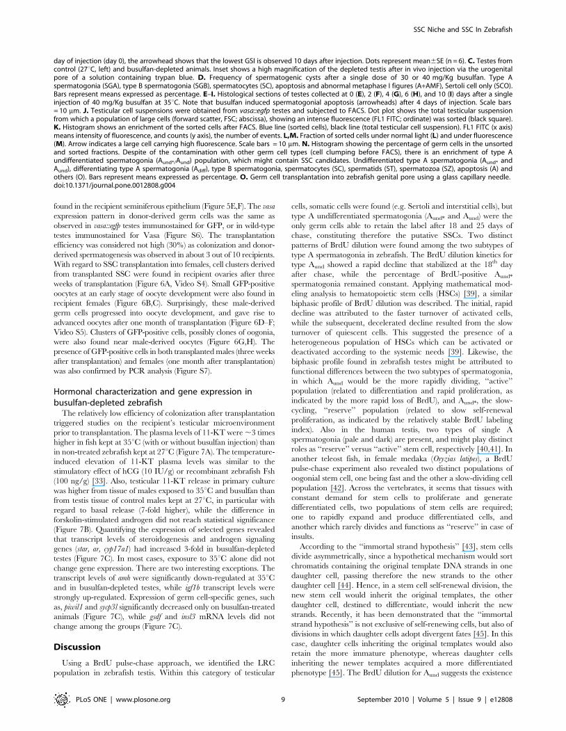
Figure 3. SSC niche in zebrafish testes. Whole-mount of vasa::egfp testes analyzed under fluorescence (A,B) and CLSM (C–E). A,B. Most of thebrightest and small spots (type A undifferentiated spermatogonia) (arrowheads) are adjacent the triangle/lozenge dark areas (interstitium). Scale bars= 0.1 mm and 0.5 mm, respectively. C–E. Vasa is highly expressed in type A undifferentiated spermatogonia, and is gradually decreased during thespermatogenesis. Aund*/Aund (arrowheads), type B spermatogonia (SGB), primary spermatocytes (SCI), secondary spermatocytes (SCII), spermatids (ST),spermatids and spermatozoa (ST/SZ). Note that most of Aund*/Aund (arrowheads) are situated near the interstitium (D, dark areas; E, delimited by
SSC Niche and SSC In Zebrafish
PLoS ONE | www.plosone.org 6 September 2010 | Volume 5 | Issue 9 | e12808

actin1 and ef1a Cq values in the different groups (Figure S3) thus
validating b-actin1 and ef1a as suitable references for the current
experiments. Then, relative mRNA levels of the selected genes
were normalized to b-actin1 and ef1a, and expressed as fold of
relative control (27uC) mRNA levels. Nomenclature of zebrafish
proteins, mRNAs and genes are according to ZFIN (http://zfin.
org) rules.
Statistical analysisSignificant differences between two groups were identified using
Student test (paired and unpaired) (P,0.05). Comparisons of more
than two groups were performed with one-way ANOVA followed
by Student-Newman-Keuls test (P,0.05). Graph Pad Prism 4.0
(Graph Pad Software, Inc., San Diego, CA, USA, http://www.
graphpad.com) was used for all statistical analysis.
Results
Characterization of SSC candidates and their niche inzebrafish testes
Analyzing the topographical distribution of type A spermatogo-
nia showed that 75% of these cells are situated adjacent to the
interstitial compartment (Figure 1A,C,D) although this compart-
ment represents only 1/3rd of the total perimeter of the
spermatogenic tubules (Figure 1B). Quantification of the mitotic
index of these cells was achieved by determining the BrdU labeling
index during three successive 10 h long exposures to BrdU during
three consecutive days. The results suggested that both subtypes of
type A undifferentiated spermatogonia (Aund* and Aund) have a long
cell cycle lasting at least 10 h but less than 30 h (Figure 2A). To
confirm the existence of slow-cycling cells (stem cell candidates),
testes were examined for BrdU-positive type Aund* and Aund
spermatogonia at different periods (up to 25 days) after the final
BrdU exposure; putative stem cells are considered to be part of the
LRC population. During this chase period, two distinct patterns of
BrdU staining were found among the two subtypes of type A
spermatogonia (Figure 2A). Aund rapidly lost the label until the 18th
day of chase, when the labeling index stabilized at ,10% for this
cell type (Figure 2A). On the other hand, the percentage of BrdU-
positive type Aund* spermatogonia varied in a statistically not
significant manner around 30% during the complete chase period
(Figure 2A). Apart from some somatic elements, type A undiffer-
entiated spermatogonia (Aund* and Aund) were the only BrdU
retaining germ cells after 18 and 25 days of chase. Intriguingly, most
of the labeled spermatogonia (71% of Aund* and 80% of Aund) were
situated adjacent to the interstitium (Figure 2B,C–H). A similar
pattern was found in vasa::egfp testes examined under CLSM where
the strongest expression of vasa was observed adjacent to the
interstitium (Figure 3 and Video S1). To further study the spatial
relation between the vasculature and undifferentiated spermatogo-
nia, testes of transgenic zebrafish expressing GFP in endothelial cells
(fli::egfp) were analyzed. Capillaries surround the seminiferous
tubules (Figure 3F), and type A undifferentiated spermatogonia
were often found in close association with endothelial cells
(Figures 2E-inset; 3G–I). A combination of the above results is
illustrated schematically in Figure S4, showing a hypothetical
spermatogonial stem cell niche in zebrafish.
Preparation of male and female recipients for SSCtransplantation
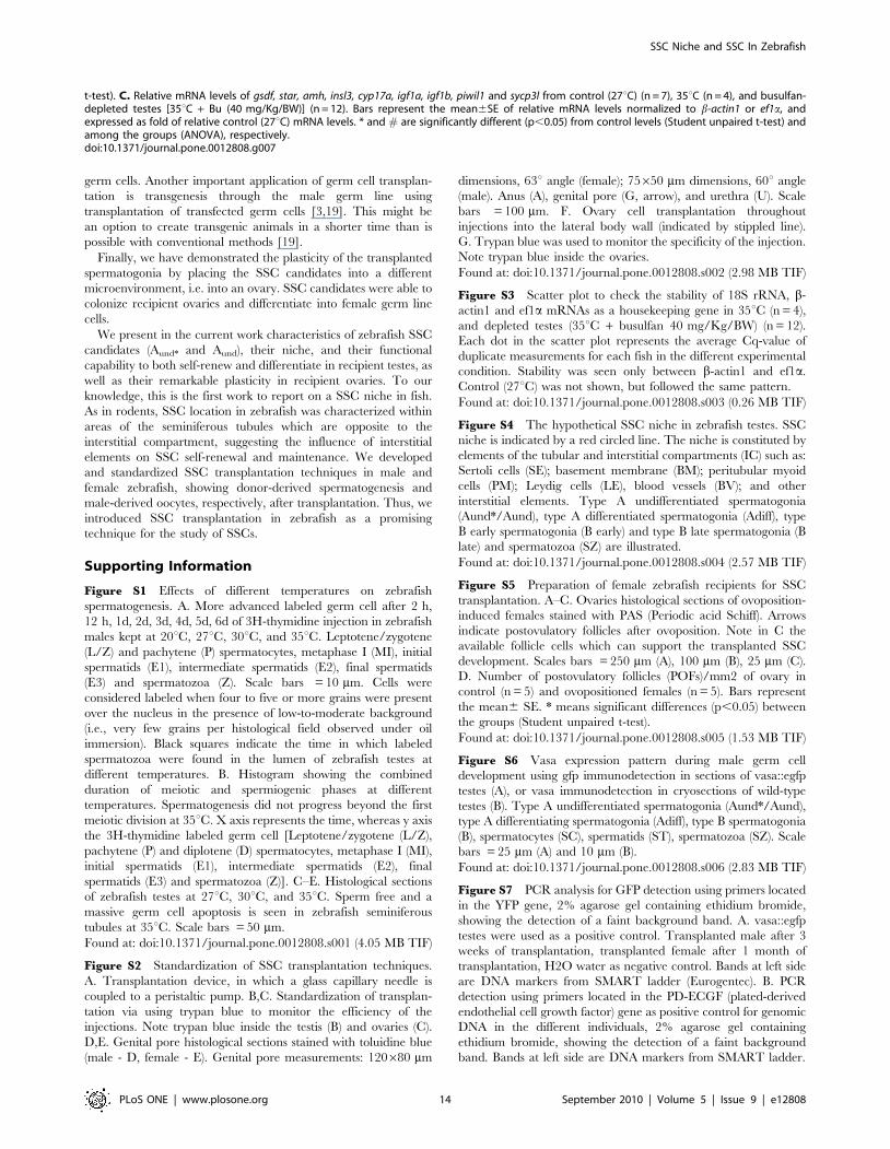
Zebrafish were exposed to different temperatures to optimize
the treatment with busulfan (Figure 4A, Figure S1). Higher
temperatures accelerated spermatogenesis (Figure S1), whereas at
35uC, spermatogenesis did not progress beyond metaphase I and
showed abnormalities, such as massive apoptosis in particular
amongst spermatocytes, absence of sperm, and a significant
decrease in the GSI (Figure 4A,E; Figure S1). Both doses of
busulfan (30 or 40 mg/Kg/BW) tested at 35uC induced further
and a more general germ cell apoptosis (Figure 4D–H) and a
progressive GSI decrease, which reached its lowest value 10 days
after injection (Figure 4B,C). However, only the higher dose
suppressed efficiently endogenous spermatogenesis, resulting in
88% of the spermatogenic tubules showing a Sertoli cell only
appearance, i.e. all germ cells were missing (Figure 4D,I).
Analyzing the composition of type A spermatogonia in the 12%
of tubules where some germ cells remained and comparing this to
normal testes demonstrated that the ratio between Aund* and Aund
shifted from 1:1 to 1:3 in busulfan-depleted testes (Figure 1E). The
preponderance of Aund might be associated with the observation
that 12 days after injection, most tubules showed the first type B
spermatogonia and/or spermatocytes again, i.e. showed a fast
recovery of endogenous spermatogenesis after treatment with
40 mg/Kg/BW of busulfan at 35uC (Figure 4D).
To prepare female zebrafish recipients for SSC transplantation,
ovoposition was induced; a pilot study indicated a high mortality
among females when applying the combination of high temperature
and busulfan. The increased number of POFs showed that
ovoposition created spaces and ‘‘free’’ somatic cells (follicle cells from
POFs), potentially suitable for receiving injected germ cells, and to
support transplanted SSCs development, respectively (Figure S5).
Donor cell isolation and male and female transplantationUsing vasa::egfp transgenic zebrafish as donors, a testicular cell
suspension was obtained and subsequently submitted to FACS, in
order to enrich transplantable SSC (Figure 4J,K) by sorting for big
cells carrying high fluorescence (Figure 4L,M). Indeed, after
sorting, we observed an enrichment of type A undifferentiated
spermatogonia (Figure 4K–N). The sorted cells (8.10324.104
cells/ml) were injected through the genital pore (Figure 4O, Figure
S2). Since females have a prominent belly, direct injections into
ovaries (via lateral body wall) were also successfully performed
(Figure S2). After transplantation, zebrafish recipient males and
females were placed in water of 27uC.
Male and female transplantation analysisAfter two weeks of transplantation, donor cells colonized the
recipient’s seminiferous epithelium, and formed clusters, which were
situated near the interstitial compartment (Figure 5A,B). Using
CLSM, we found that these clusters were composed of ,8 cells/cyst
(Figure 5C,D; Video S2). Three weeks after transplantation, donor-
derived cysts had increased in number and size, and were found at
different stages of spermatogenesis (e.g. differentiating type A
spermatogonia, type B spermatogonia and spermatocytes) along the
recipient’s seminiferous epithelium (Figure 5E,F; Video S3). Donor
type A undifferentiated spermatogonia (Aund* and Aund) were also
spotted lines). E is a high magnification of the square in D. Scale bars = 10 mm. F–I: Cryosections of fli::egfp testes stained with DAPI (nuclear staining)and analyzed under fluorescence microscopy. The arrow in F shows a blood vessel (green) surrounding the circumference of a seminiferous tubule(ST). Interstitium (delimited with red dotted lines), Leydig cells (LE) and group of type A spermatogonia (asterisks) are shown in G. H,I. Aund*/Aund arenear the endothelial cell (EC) nuclei (n). Note a metaphase figure (M) in H. Nucleolus (nu) of type A undifferentiated spermatogonia is shown in I.Compare the similar morphology of Aund* (inset) with the cells found near the endothelial cell. Scale bars = 10 mm.doi:10.1371/journal.pone.0012808.g003
SSC Niche and SSC In Zebrafish
PLoS ONE | www.plosone.org 7 September 2010 | Volume 5 | Issue 9 | e12808

Figure 4. Depletion of endogenous spermatogenesis in male recipients for SSC transplantation. A. Effects of different temperatures onzebrafish GSI (gonadosomatic index). Bars represent mean 6 SE (n = 15, 20uC), (n = 12, 27uC), (n = 7, 30uC), and (n = 12, 35uC). Different letters indicatesignificant differences (p,0.05) among groups. B. Effects of two single doses of busulfan (30 or 40 mg/Kg) on zebrafish GSI. The arrow indicates the
SSC Niche and SSC In Zebrafish
PLoS ONE | www.plosone.org 8 September 2010 | Volume 5 | Issue 9 | e12808
found in the recipient seminiferous epithelium (Figure 5E,F). The vasa
expression pattern in donor-derived germ cells was the same as
observed in vasa::egfp testes immunostained for GFP, or in wild-type
testes immunostained for Vasa (Figure S6). The transplantation
efficiency was considered not high (30%) as colonization and donor-
derived spermatogenesis was observed in about 3 out of 10 recipients.
With regard to SSC transplantation into females, cell clusters derived
from transplanted SSC were found in recipient ovaries after three
weeks of transplantation (Figure 6A, Video S4). Small GFP-positive
oocytes at an early stage of oocyte development were also found in
recipient females (Figure 6B,C). Surprisingly, these male-derived
germ cells progressed into oocyte development, and gave rise to
advanced oocytes after one month of transplantation (Figure 6D–F;
Video S5). Clusters of GFP-positive cells, possibly clones of oogonia,
were also found near male-derived oocytes (Figure 6G,H). The
presence of GFP-positive cells in both transplanted males (three weeks
after transplantation) and females (one month after transplantation)
was also confirmed by PCR analysis (Figure S7).
Hormonal characterization and gene expression inbusulfan-depleted zebrafish
The relatively low efficiency of colonization after transplantation
triggered studies on the recipient’s testicular microenvironment
prior to transplantation. The plasma levels of 11-KT were ,3 times
higher in fish kept at 35uC (with or without busulfan injection) than
in non-treated zebrafish kept at 27uC (Figure 7A). The temperature-
induced elevation of 11-KT plasma levels was similar to the
stimulatory effect of hCG (10 IU/g) or recombinant zebrafish Fsh
(100 ng/g) [33]. Also, testicular 11-KT release in primary culture
was higher from tissue of males exposed to 35uC and busulfan than
from testis tissue of control males kept at 27uC, in particular with
regard to basal release (7-fold higher), while the difference in
forskolin-stimulated androgen did not reach statistical significance
(Figure 7B). Quantifying the expression of selected genes revealed
that transcript levels of steroidogenesis and androgen signaling
genes (star, ar, cyp17a1) had increased 3-fold in busulfan-depleted
testes (Figure 7C). In most cases, exposure to 35uC alone did not
change gene expression. There are two interesting exceptions. The
transcript levels of amh were significantly down-regulated at 35uCand in busulfan-depleted testes, while igf1b transcript levels were
strongly up-regulated. Expression of germ cell-specific genes, such
as, piwil1 and sycp3l significantly decreased only on busulfan-treated
animals (Figure 7C), while gsdf and insl3 mRNA levels did not
change among the groups (Figure 7C).
Discussion
Using a BrdU pulse-chase approach, we identified the LRC
population in zebrafish testis. Within this category of testicular
cells, somatic cells were found (e.g. Sertoli and interstitial cells), but
type A undifferentiated spermatogonia (Aund* and Aund) were the
only germ cells able to retain the label after 18 and 25 days of
chase, constituting therefore the putative SSCs. Two distinct
patterns of BrdU dilution were found among the two subtypes of
type A spermatogonia in zebrafish. The BrdU dilution kinetics for
type Aund showed a rapid decline that stabilized at the 18th day
after chase, while the percentage of BrdU-positive Aund*
spermatogonia remained constant. Applying mathematical mod-
eling analysis to hematopoietic stem cells (HSCs) [39], a similar
biphasic profile of BrdU dilution was described. The initial, rapid
decline was attributed to the faster turnover of activated cells,
while the subsequent, decelerated decline resulted from the slow
turnover of quiescent cells. This suggested the presence of a
heterogeneous population of HSCs which can be activated or
deactivated according to the systemic needs [39]. Likewise, the
biphasic profile found in zebrafish testes might be attributed to
functional differences between the two subtypes of spermatogonia,
in which Aund would be the more rapidly dividing, ‘‘active’’
population (related to differentiation and rapid proliferation, as
indicated by the more rapid loss of BrdU), and Aund*, the slow-
cycling, ‘‘reserve’’ population (related to slow self-renewal
proliferation, as indicated by the relatively stable BrdU labeling
index). Also in the human testis, two types of single A
spermatogonia (pale and dark) are present, and might play distinct
roles as ‘‘reserve’’ versus ‘‘active’’ stem cell, respectively [40,41]. In
another teleost fish, in female medaka (Oryzias latipes), a BrdU
pulse-chase experiment also revealed two distinct populations of
oogonial stem cell, one being fast and the other a slow-dividing cell
population [42]. Across the vertebrates, it seems that tissues with
constant demand for stem cells to proliferate and generate
differentiated cells, two populations of stem cells are required;
one to rapidly expand and produce differentiated cells, and
another which rarely divides and functions as ‘‘reserve’’ in case of
insults.
According to the ‘‘immortal strand hypothesis’’ [43], stem cells
divide asymmetrically, since a hypothetical mechanism would sort
chromatids containing the original template DNA strands in one
daughter cell, passing therefore the new strands to the other
daughter cell [44]. Hence, in a stem cell self-renewal division, the
new stem cell would inherit the original templates, the other
daughter cell, destined to differentiate, would inherit the new
strands. Recently, it has been demonstrated that the ‘‘immortal
strand hypothesis’’ is not exclusive of self-renewing cells, but also of
divisions in which daughter cells adopt divergent fates [45]. In this
case, daughter cells inheriting the original templates would also
retain the more immature phenotype, whereas daughter cells
inheriting the newer templates acquired a more differentiated
phenotype [45]. The BrdU dilution for Aund suggests the existence
day of injection (day 0), the arrowhead shows that the lowest GSI is observed 10 days after injection. Dots represent mean6SE (n = 6). C. Testes fromcontrol (27uC, left) and busulfan-depleted animals. Inset shows a high magnification of the depleted testis after in vivo injection via the urogenitalpore of a solution containing trypan blue. D. Frequency of spermatogenic cysts after a single dose of 30 or 40 mg/Kg busulfan. Type Aspermatogonia (SGA), type B spermatogonia (SGB), spermatocytes (SC), apoptosis and abnormal metaphase I figures (A+AMF), Sertoli cell only (SCO).Bars represent means expressed as percentage. E–I. Histological sections of testes collected at 0 (E), 2 (F), 4 (G), 6 (H), and 10 (I) days after a singleinjection of 40 mg/Kg busulfan at 35uC. Note that busulfan induced spermatogonial apoptosis (arrowheads) after 4 days of injection. Scale bars= 10 mm. J. Testicular cell suspensions were obtained from vasa::egfp testes and subjected to FACS. Dot plot shows the total testicular suspensionfrom which a population of large cells (forward scatter, FSC; abscissa), showing an intense fluorescence (FL1 FITC; ordinate) was sorted (black square).K. Histogram shows an enrichment of the sorted cells after FACS. Blue line (sorted cells), black line (total testicular cell suspension). FL1 FITC (x axis)means intensity of fluorescence, and counts (y axis), the number of events. L,M. Fraction of sorted cells under normal light (L) and under fluorescence(M). Arrow indicates a large cell carrying high fluorescence. Scale bars = 10 mm. N. Histogram showing the percentage of germ cells in the unsortedand sorted fractions. Despite of the contamination with other germ cell types (cell clumping before FACS), there is an enrichment of type Aundifferentiated spermatogonia (Aund*/Aund) population, which might contain SSC candidates. Undifferentiated type A spermatogonia (Aund* andAund), differentiating type A spermatogonia (Adiff), type B spermatogonia, spermatocytes (SC), spermatids (ST), spermatozoa (SZ), apoptosis (A) andothers (O). Bars represent means expressed as percentage. O. Germ cell transplantation into zebrafish genital pore using a glass capillary needle.doi:10.1371/journal.pone.0012808.g004
SSC Niche and SSC In Zebrafish
PLoS ONE | www.plosone.org 9 September 2010 | Volume 5 | Issue 9 | e12808

of two types of template DNA strand segregation, one random
type, possibly reflecting the rapid loss of the label up to the 18th
day of chase, and one non-random type (asymmetrical division),
which could explain the retention of BrdU after 18 and 25 days of
chase. We speculate that the asymmetrical division of Aund might
prevent its exhaustion during spermatogenesis, maintaining,
therefore, a subset of immature Aund. An asymmetrical division
of type A undifferentiated spermatogonia was suggested previously
in zebrafish based on morphological evidence [2].
The LRC assay also revealed the precise location of stem cells,
and consequently the characterization of the neighboring cells that
form the stem cell niche [46–48]. As in rodents [6,7], we found
that slow-cycling BrdU-labeled type A undifferentiated spermato-
gonia (Aund* and Aund) were preferentially situated in regions of the
seminiferous tubules adjacent to the interstitial compartment. This
observation suggests that elements of the SSC niche are probably
conserved across vertebrates. The preferential location of SSC
close to the interstitial compartment in rodents may be related to a
locally high concentration of androgens which inhibit spermato-
gonial differentiation [49]. 11-KT is the main androgen in
zebrafish [37] and other teleost fish, and is able to support full
spermatogenesis in testis tissue culture of juvenile Japanese eel
(Anguilla japonica) [50] or adult zebrafish [38]. Moreover, induction
of androgen insufficiency in adult male zebrafish inhibited the
differentiation of type A to type B spermatogonia [51]. Although
the possible role(s) of androgen signaling in regulating zebrafish
Figure 5. SSC transplantation into male zebrafish recipients. A,B. Recipient testes after two weeks (2w) of transplantation analyzed by light(A) and fluorescence (B) microscopies. Seminiferous tubules are delimited by stippled lines. Arrows indicate the same area in A and B. Donor cellsformed clusters situated near the interstitium in a similar way as observed in vasa::egfp testes (see arrowheads in the inset). Nuclei (blue) are stainedwith DAPI. Scale bars = 50 mm. C,D. CLSM analysis of recipient testes after two weeks (2w) of transplantation. Arrowheads indicate a donor-derivedcyst composed of ,8 cells. Inset. High magnification of donor-derived cyst. Nuclei (blue) are stained with DAPI. E,F. Recipient testes after 3 weeks(3w) of transplantation analyzed under CLSM. Donor-derived cysts increased their number and size, being found at different stages ofspermatogenesis. Type A undifferentiated spermatogonia (Aund*/Aund), type A differentiating spermatogonia (Adiff), type B spermatogonia (B), andspermatocytes (SC). Inset shows a high magnification of type A undifferentiated spermatogonia. Nuclei (blue) are stained with DAPI.doi:10.1371/journal.pone.0012808.g005
SSC Niche and SSC In Zebrafish
PLoS ONE | www.plosone.org 10 September 2010 | Volume 5 | Issue 9 | e12808

Figure 6. SSC transplantation into female zebrafish recipients analyzed under CLSM. A. A GFP cell cluster-derived from transplanted SSCafter three weeks (3w) of transplantation. Nuclei (blue) are stained with DAPI. Scale bar = 10 mm. B,C. Arrowhead indicates an early donor-derivedoocytes surrounded by follicle cells (FC) after three weeks (3w) of transplantation. Nuclei (blue) are stained with DAPI. D–F. Arrowhead indicates anadvanced GFP oocyte, which was originated from transplanted SSC into zebrafish ovaries after one month (1mo) of transplantation. Note a small GFPcell cluster (asterisk) near the donor-derived oocyte. Light (D) and fluorescence (E) microscopies, and overlay of both (F). Scale bars = 25 mm. G. Thesame oocyte in D–F examined under CLSM. A green donor-derived perinucleolar oocyte (PO), endogenous perinucleolar oocyte (PO), small GFP cellcluster (asterisk), and nucleus (n) are shown. Inset. High magnification of advanced donor-derived oocyte. Nuclei (blue) are stained with DAPI. H.
SSC Niche and SSC In Zebrafish
PLoS ONE | www.plosone.org 11 September 2010 | Volume 5 | Issue 9 | e12808

SSC activity still have to be clarified, the available evidence in fish
strongly suggests that, different from mammals, androgens
stimulate spermatogonial differentiation [2]. On the other hand,
estrogens stimulated SSC self-renewal in Japanese eel [52], so that
Leydig cells may be relevant as a source for aromatizeable
androgens, thereby contributing directly and/or indirectly to the
zebrafish SSC niche. Moreover, Leydig cell paracrine signaling
might influence SSC behavior, since colony stimulating factor 1
(Csf1) produced by Leydig and myoid cells is an extrinsic
stimulator of SSC self-renewal in mice [10].
Endothelial cells support the expansion of normal and
malignant stem cells not only by delivering oxygen and nutrients,
but also by the paracrine release of endothelial cell growth factors
and trophogens, which are referred to as ‘‘angiocrine factors’’ [53].
Thus, endothelial cells have been pointed out as an important
element of the stem cell niche in several systems, including the
rodent SSC niche [8,54]. In the zebrafish model, the distribution
of SSC candidates (Aund* and Aund) close to blood vessels might
indicate an involvement of endothelial cells in regulating SSC
function. While there is no information on angiocrine signaling in
the zebrafish testis, it has been shown that platelet-derived
endothelial cell growth factor (PD-ECGF) induced SSC self-
renewal in Japanese eel [55,56].
The functional capacity of the identified SSC candidates in
zebrafish was investigated by transplantation assays. In this assay,
SSC candidates were transplanted into zebrafish in which
endogenous spermatogenesis had been depleted by busulfan,
similar to the technique developed for rodents and others species
[14–17,19]. It seems that similar to tilapia [16], zebrafish
spermatogenesis is more sensitive to busulfan at elevated
temperatures (35uC), resulting in 88% of spermatogenic tubules
showing Sertoli cells only after 10 days of treatment. Interestingly,
spermatogenesis recovered quickly after having passed the nadir of
busulfan-induced depletion. It has been shown previously that
proliferation of surviving, undifferentiated spermatogonia is
greatly enhanced when the number of differentiating spermato-
gonia is reduced following busulfan exposure [57,58]. In addition,
differentiated spermatogonia can act as ‘‘potential stem cells’’,
shifting their nature of transit-amplifying cells to self-renewal in
order to rapidly recover spermatogenesis in depleted testes [59].
Analyzing the composition of type A spermatogonia in normal
versus busulfan-depleted testes in zebrafish, we have demonstrated
a relative increase in the population of Aund, and a relative
decrease in the population of Aund* spermatogonia in busulfan-
depleted testes. Then, considering Aund as the ‘‘active’’ stem cell,
our results suggest that the spermatogenic recovery after busulfan
treatment might be associated with a preponderance of the active
stem cell population Aund.
The treatment with busulfan induced an increase in androgen
plasma levels as well as an increase in testicular androgen release.
This might reflect a suppression of the negative feedback on
gonadotropin release. In rat, the negative feedback on FSH release
exerted by germ cells via Sertoli cell-derived inhibin is transiently
eliminated after busulfan treatment [60], explaining the increase in
circulating FSH levels between 6 and 10 weeks after busulfan
injection [61,62]. In mammals, androgen plasma levels were not
elevated in busulfan-treated animals [60–62]. However, in fish,
also Leydig cells express the receptor for Fsh, which is a strongly
steroidogenic gonadotropin in fish (e.g. [33,63,64]). Hence, we
attribute the increased androgen levels after busulfan treatment to
higher Fsh plasma levels. Consistent with the elevated androgen
production, busulfan treatment increased the mRNA levels of
transcripts encoding proteins involved in steroidogenesis, such as
star and cyp17a1. The down-regulation of amh mRNA expression in
busulfan-depleted testes also seems coherent with higher androgen
levels, since androgens down-regulated amh expression in juvenile
Japanese eel testis and stimulated spermatogonial differentiation
towards meiosis [65]. Moreover, since Sertoli cells also proliferate
in the adult fish testis, in particular those associated with
expanding spermatogonial cysts [23,24], the busulfan treatment
may have depleted the proliferating Sertoli cell population as well,
thereby contributing to lower levels of amh mRNA, and possibly
also to the arrest in spermatogenesis. With regard to the expression
of igf gene family members, a strong up-regulation in particular of
igf1b mRNA levels has been observed. This is interesting in the
context of ongoing work showing that recombinant zebrafish Fsh
increases igf1b levels in adult testis tissue culture (unpublished
data). Spermatogonial proliferation was stimulated in newt testis in
primary tissue culture by exposure to Igf1 [66]. Since Sertoli cells
also express a factor (similar to platelet-derived endothelial cell
growth factor) that stimulates SSC self-renewal in Japanese eel
[55], busulfan-induced changes in the number and/or physiolog-
ical state of Sertoli cells might have resulted in a more generalized
imbalance of factors released by Sertoli cells and relevant for SSC
self-renewal and differentiation. Taken together, our data suggest
that busulfan treatment might create a microenvironment in
zebrafish testes where the balance of somatic (mainly Sertoli) cell-
derived factors has shifted to a reduced level of factors supporting
self-renewal and/or an elevated level of factors stimulating
differentiation. While this might facilitate the fast recovery after
the severe loss of germ cells induced by busulfan, it may also
influence the behavior of transplanted cells. Thus, transplanted
SSCs may differentiate rather than self-renew, possibly limiting the
efficiency of donor-colonization (30%) in the recipient seminifer-
ous tubules; however, a relatively low efficiency of donor-derived
colonization is not exceptional for zebrafish and has been observed
in other vertebrates as well [67]. Future studies will aim at
weakening the pro-differentiation or strengthening the self-renewal
environment, for example by an estrogen treatment [51] of the
recipients.
Despite of the limited efficiency, we have taken important steps
towards the development and standardization of SSC transplan-
tation assay to confirm the ‘‘stemness’’ of SSC candidates in
zebrafish. Transplantation of a FACS-enriched fraction of Aund*
and Aund into busulfan-depleted testes showed that stem cell
candidates were able to colonize, self-renew, and to differentiate in
the recipient seminiferous tubules. This confirms the presence of
stem cells in the transplanted pool of type A undifferentiated
spermatogonia. Moreover, spermatogonial colonization started
from regions within the recipient’s seminiferous tubules which are
adjacent to the interstitial compartment, the putative SSC niche in
zebrafish testes. This might indicate that the SSC niche creates a
cellular and molecular environment which is optimal for
colonization and development of transplanted SSCs. Since
zebrafish have been used as a model for understanding the
mechanisms of testicular germ cell tumors [68], and most of these
tumors have an unknown etiology, a standard experimental design
including reciprocal transplantation of germ cells from affected
donors to wild-type testes and vice versa could be applied to
evaluate whether the defect is on Sertoli cells or intrinsic to the
Perinucleolar oocyte from vasa::egfp ovaries immunostained for gfp. Compare similar vasa expression pattern between donor-derived oocyte andperinucleolar oocyte from vasa::egfp ovaries. Scale bar = 25 mm.doi:10.1371/journal.pone.0012808.g006
SSC Niche and SSC In Zebrafish
PLoS ONE | www.plosone.org 12 September 2010 | Volume 5 | Issue 9 | e12808

Figure 7. Characterization of male recipients prior SSC transplantation. A. 11-KT plasma levels (ng/ml plasma) from zebrafish kept at 27uC(control) (n = 7), 35uC (n = 11), and busulfan-depleted animals (n = 19). Bars represent the mean6SE. Different letters mean significant differences(p,0.05) among groups. B. In vitro 11-KT (ng/mg testis) release from control (27uC) (n = 7) and busulfan-depleted testes [35uC + Bu (40 mg/Kg/BW)](n = 7) in basal and 1 mM forskolin-induced. Bars represent the mean6SE. * means significant differences (p,0.05) between control and depleted inthe same experimental condition (Student unpaired t-test). # indicates significantly higher (p,0.05) than the respective basal release (Student paired
SSC Niche and SSC In Zebrafish
PLoS ONE | www.plosone.org 13 September 2010 | Volume 5 | Issue 9 | e12808
germ cells. Another important application of germ cell transplan-
tation is transgenesis through the male germ line using
transplantation of transfected germ cells [3,19]. This might be
an option to create transgenic animals in a shorter time than is
possible with conventional methods [19].
Finally, we have demonstrated the plasticity of the transplanted
spermatogonia by placing the SSC candidates into a different
microenvironment, i.e. into an ovary. SSC candidates were able to
colonize recipient ovaries and differentiate into female germ line
cells.
We present in the current work characteristics of zebrafish SSC
candidates (Aund* and Aund), their niche, and their functional
capability to both self-renew and differentiate in recipient testes, as
well as their remarkable plasticity in recipient ovaries. To our
knowledge, this is the first work to report on a SSC niche in fish.
As in rodents, SSC location in zebrafish was characterized within
areas of the seminiferous tubules which are opposite to the
interstitial compartment, suggesting the influence of interstitial
elements on SSC self-renewal and maintenance. We developed
and standardized SSC transplantation techniques in male and
female zebrafish, showing donor-derived spermatogenesis and
male-derived oocytes, respectively, after transplantation. Thus, we
introduced SSC transplantation in zebrafish as a promising
technique for the study of SSCs.
Supporting Information
Figure S1 Effects of different temperatures on zebrafish
spermatogenesis. A. More advanced labeled germ cell after 2 h,
12 h, 1d, 2d, 3d, 4d, 5d, 6d of 3H-thymidine injection in zebrafish
males kept at 20uC, 27uC, 30uC, and 35uC. Leptotene/zygotene
(L/Z) and pachytene (P) spermatocytes, metaphase I (MI), initial
spermatids (E1), intermediate spermatids (E2), final spermatids
(E3) and spermatozoa (Z). Scale bars = 10 mm. Cells were
considered labeled when four to five or more grains were present
over the nucleus in the presence of low-to-moderate background
(i.e., very few grains per histological field observed under oil
immersion). Black squares indicate the time in which labeled
spermatozoa were found in the lumen of zebrafish testes at
different temperatures. B. Histogram showing the combined
duration of meiotic and spermiogenic phases at different
temperatures. Spermatogenesis did not progress beyond the first
meiotic division at 35uC. X axis represents the time, whereas y axis
the 3H-thymidine labeled germ cell [Leptotene/zygotene (L/Z),
pachytene (P) and diplotene (D) spermatocytes, metaphase I (MI),
initial spermatids (E1), intermediate spermatids (E2), final
spermatids (E3) and spermatozoa (Z)]. C–E. Histological sections
of zebrafish testes at 27uC, 30uC, and 35uC. Sperm free and a
massive germ cell apoptosis is seen in zebrafish seminiferous
tubules at 35uC. Scale bars = 50 mm.
Found at: doi:10.1371/journal.pone.0012808.s001 (4.05 MB TIF)
Figure S2 Standardization of SSC transplantation techniques.
A. Transplantation device, in which a glass capillary needle is
coupled to a peristaltic pump. B,C. Standardization of transplan-
tation via using trypan blue to monitor the efficiency of the
injections. Note trypan blue inside the testis (B) and ovaries (C).
D,E. Genital pore histological sections stained with toluidine blue
(male - D, female - E). Genital pore measurements: 120680 mm
dimensions, 63u angle (female); 75650 mm dimensions, 60u angle
(male). Anus (A), genital pore (G, arrow), and urethra (U). Scale
bars = 100 mm. F. Ovary cell transplantation throughout
injections into the lateral body wall (indicated by stippled line).
G. Trypan blue was used to monitor the specificity of the injection.
Note trypan blue inside the ovaries.
Found at: doi:10.1371/journal.pone.0012808.s002 (2.98 MB TIF)
Figure S3 Scatter plot to check the stability of 18S rRNA, b-
actin1 and ef1a mRNAs as a housekeeping gene in 35uC (n = 4),
and depleted testes (35uC + busulfan 40 mg/Kg/BW) (n = 12).
Each dot in the scatter plot represents the average Cq-value of
duplicate measurements for each fish in the different experimental
condition. Stability was seen only between b-actin1 and ef1a.
Control (27uC) was not shown, but followed the same pattern.
Found at: doi:10.1371/journal.pone.0012808.s003 (0.26 MB TIF)
Figure S4 The hypothetical SSC niche in zebrafish testes. SSC
niche is indicated by a red circled line. The niche is constituted by
elements of the tubular and interstitial compartments (IC) such as:
Sertoli cells (SE); basement membrane (BM); peritubular myoid
cells (PM); Leydig cells (LE), blood vessels (BV); and other
interstitial elements. Type A undifferentiated spermatogonia
(Aund*/Aund), type A differentiated spermatogonia (Adiff), type
B early spermatogonia (B early) and type B late spermatogonia (B
late) and spermatozoa (SZ) are illustrated.
Found at: doi:10.1371/journal.pone.0012808.s004 (2.57 MB TIF)
Figure S5 Preparation of female zebrafish recipients for SSC
transplantation. A–C. Ovaries histological sections of ovoposition-
induced females stained with PAS (Periodic acid Schiff). Arrows
indicate postovulatory follicles after ovoposition. Note in C the
available follicle cells which can support the transplanted SSC
development. Scales bars = 250 mm (A), 100 mm (B), 25 mm (C).
D. Number of postovulatory follicles (POFs)/mm2 of ovary in
control (n = 5) and ovopositioned females (n = 5). Bars represent
the mean6 SE. * means significant differences (p,0.05) between
the groups (Student unpaired t-test).
Found at: doi:10.1371/journal.pone.0012808.s005 (1.53 MB TIF)
Figure S6 Vasa expression pattern during male germ cell
development using gfp immunodetection in sections of vasa::egfp
testes (A), or vasa immunodetection in cryosections of wild-type
testes (B). Type A undifferentiated spermatogonia (Aund*/Aund),
type A differentiating spermatogonia (Adiff), type B spermatogonia
(B), spermatocytes (SC), spermatids (ST), spermatozoa (SZ). Scale
bars = 25 mm (A) and 10 mm (B).
Found at: doi:10.1371/journal.pone.0012808.s006 (2.83 MB TIF)
Figure S7 PCR analysis for GFP detection using primers located
in the YFP gene, 2% agarose gel containing ethidium bromide,
showing the detection of a faint background band. A. vasa::egfp
testes were used as a positive control. Transplanted male after 3
weeks of transplantation, transplanted female after 1 month of
transplantation, H2O water as negative control. Bands at left side
are DNA markers from SMART ladder (Eurogentec). B. PCR
detection using primers located in the PD-ECGF (plated-derived
endothelial cell growth factor) gene as positive control for genomic
DNA in the different individuals, 2% agarose gel containing
ethidium bromide, showing the detection of a faint background
band. Bands at left side are DNA markers from SMART ladder.
t-test). C. Relative mRNA levels of gsdf, star, amh, insl3, cyp17a, igf1a, igf1b, piwil1 and sycp3l from control (27uC) (n = 7), 35uC (n = 4), and busulfan-depleted testes [35uC + Bu (40 mg/Kg/BW)] (n = 12). Bars represent the mean6SE of relative mRNA levels normalized to b-actin1 or ef1a, andexpressed as fold of relative control (27uC) mRNA levels. * and # are significantly different (p,0.05) from control levels (Student unpaired t-test) andamong the groups (ANOVA), respectively.doi:10.1371/journal.pone.0012808.g007
SSC Niche and SSC In Zebrafish
PLoS ONE | www.plosone.org 14 September 2010 | Volume 5 | Issue 9 | e12808

Found at: doi:10.1371/journal.pone.0012808.s007 (0.82 MB TIF)
Video S1 Z stack from vasa::egfp testis showing the distribution
of vasa positive cells in the zebrafish seminiferous tubules.
Found at: doi:10.1371/journal.pone.0012808.s008 (1.63 MB
WMV)
Video S2 Z stack from wild-type zebrafish testis 2 weeks after
SSC transplantation. Note a donor-derived cyst composed of ,8
cells in the seminiferous epithelium.
Found at: doi:10.1371/journal.pone.0012808.s009 (0.83 MB
WMV)
Video S3 Z stack from wild-type zebrafish testis 3 weeks after
SSC transplantation. SSCs were able to colonize recipient testis
and differentiate into daughter cells committed with the
spermatogenic process. Donor-derived cysts at different stages of
zebrafish spermatogenesis were found in recipient seminiferous
epithelium.
Found at: doi:10.1371/journal.pone.0012808.s010 (0.81 MB
WMV)
Video S4 Z stack from ovaries fragments 3 weeks after SSC
transplantation into female zebrafish ovaries. Small cell clusters-
derived from SSC were seen in female ovaries.
Found at: doi:10.1371/journal.pone.0012808.s011 (1.10 MB
WMV)
Video S5 Z stack from ovaries fragments 1 month after SSC
transplantation. SSCs were able to colonize female ovaries and
differentiate into female germ cell line. Note a male-derived oocyte
in the female recipient ovary.
Found at: doi:10.1371/journal.pone.0012808.s012 (0.60 MB
WMV)
Acknowledgments
The authors would like to acknowledge Wytske van Dijk for technical
assistance during radio immuno assays (RIAs); Ger Arkesteijn for technical
assistance with FACS; Henk Schriek and Co Rootselaar for assistance
during the in vivo experiments and maintaining the zebrafish stocks; and
Teresa Bowman for the donation of fli::egfp transgenic zebrafish.
Author Contributions
Conceived and designed the experiments: RHN CDG JB LRdF RWS.
Performed the experiments: RHN CDG HvdK. Analyzed the data: RHN
CDG HvdK JB LRdF RWS. Contributed reagents/materials/analysis
tools: RHN LRdF RWS. Wrote the paper: RHN JB LRdF RWS.
Molecular biology, primers design, qPCR, PCR: JB.
References
1. Hess RA, Franca LR (2008) Spermatogenesis and cycle of the seminiferous
epithelium. Adv Exp Med Biol 636: 1–15. 10.1007/978-0-387-09597-4_1.
2. Schulz RW, Franca LR, Lareyre JJ, LeGac F, Chiarini-Garcia H, et al. (2010)
Spermatogenesis in fish. Gen Comp Endocrinol 165(3): 390–411. 10.1016/
j.ygcen.2009.02.013.
3. Oatley JM, Brinster RL (2008) Regulation of spermatogonial stem cell self-
renewal in mammals. Annu Rev Cell Dev Biol 24: 263–286. 10.1146/
annurev.cellbio.24.110707.175355.
4. de Rooij DG (2006) Regulation of spermatogonial stem cell behavior in vivo and
in vitro. Anim Reprod v.3(n.2): 130–134.
5. de Rooij DG (2009) The spermatogonial stem cell niche. Microsc Res Tech
72(8): 580–585. 10.1002/jemt.20699.
6. Chiarini-Garcia H, Hornick JR, Griswold MD, Russell LD (2001) Distribution
of type A spermatogonia in the mouse is not random. Biol Reprod 65(4):
1179–1185.
7. Chiarini-Garcia H, Raymer AM, Russell LD (2003) Non-random distribution of
spermatogonia in rats: Evidence of niches in the seminiferous tubules.
Reproduction 126(5): 669–680.
8. Yoshida S, Sukeno M, Nabeshima Y (2007) A vasculature-associated niche for
undifferentiated spermatogonia in the mouse testis. Science 317(5845):
1722–1726. 10.1126/science.1144885.
9. Meistrich ML, Shetty G (2003) Suppression of testosterone stimulates recovery
of spermatogenesis after cancer treatment. Int J Androl 26(3): 141–146.
10. Oatley JM, Oatley MJ, Avarbock MR, Tobias JW, Brinster RL (2009) Colony
stimulating factor 1 is an extrinsic stimulator of mouse spermatogonial stem cell
self-renewal. Development 136(7): 1191–1199. 10.1242/dev.032243.
11. Hofmann MC (2008) Gdnf signaling pathways within the mammalian
spermatogonial stem cell niche. Mol Cell Endocrinol 288(1-2): 95–103.
10.1016/j.mce.2008.04.012.
12. Simon L, Ekman GC, Tyagi G, Hess RA, Murphy KM, et al. (2007) Common
and distinct factors regulate expression of mRNA for ETV5 and GDNF, Sertoli
cell proteins essential for spermatogonial stem cell maintenance. Exp Cell Res
313(14): 3090–3099. 10.1016/j.yexcr.2007.05.002.
13. Braun KM, Niemann C, Jensen UB, Sundberg JP, Silva-Vargas V, et al. (2003)
Manipulation of stem cell proliferation and lineage commitment: Visualisation of
label-retaining cells in wholemounts of mouse epidermis. Development 130(21):
5241–5255. 10.1242/dev.00703.
14. Brinster RL, Zimmermann JW (1994) Spermatogenesis following male germ-cell
transplantation. Proc Natl Acad Sci U S A 91(24): 11298–11302.
15. Brinster RL, Avarbock MR (1994) Germline transmission of donor haplotype
following spermatogonial transplantation. Proc Natl Acad Sci U S A 91(24):
11303–11307.
16. Lacerda SM, Batlouni SR, Costa GM, Segatelli TM, Quirino BR, et al. (2010) A
new and fast technique to generate offspring after germ cells transplantation in
adult fish: The nile tilapia (Oreochromis niloticus) model. PLoS One 5(5): e10740.
10.1371/journal.pone.0010740.
17. Majhi SK, Hattori RS, Yokota M, Watanabe S, Strussmann CA (2009) Germ
cell transplantation using sexually competent fish: An approach for rapid
propagation of endangered and valuable germlines. PLoS One 4(7): e6132.
10.1371/journal.pone.0006132.
18. McLean DJ (2005) Spermatogonial stem cell transplantation and testicularfunction. Cell Tissue Res 322(1): 21–31. 10.1007/s00441-005-0009-z.
19. Dobrinski I (2006) Transplantation of germ cells and testis tissue to study
mammalian spermatogenesis. Anim Reprod v.3(n.2): 135–145.
20. Krawetz SA, de Rooij DG, Hedger MP (2009) Molecular aspects of malefertility. international workshop on molecular andrology. EMBO Rep 10(10):
1087–1092. 10.1038/embor.2009.211.
21. Wu X, Schmidt JA, Avarbock MR, Tobias JW, Carlson CA, et al. (2009)Prepubertal human spermatogonia and mouse gonocytes share conserved gene
expression of germline stem cell regulatory molecules. Proc Natl Acad Sci U S A
106(51): 21672–21677. 10.1073/pnas.0912432106.
22. Sadri-Ardekani H, Mizrak SC, van Daalen SK, Korver CM, Roepers-
Gajadien HL, et al. (2009) Propagation of human spermatogonial stem cells in
vitro. JAMA 302(19): 2127–2134. 10.1001/jama.2009.1689.
23. Leal MC, Cardoso ER, Nobrega RH, Batlouni SR, Bogerd J, et al. (2009)
Histological and stereological evaluation of zebrafish (Danio rerio) spermatogenesis
with an emphasis on spermatogonial generations. Biol Reprod 81(1): 177–187.10.1095/biolreprod.109.076299.
24. Schulz RW, Menting S, Bogerd J, Franca LR, Vilela DA, et al. (2005) Sertoli cell
proliferation in the adult testis—evidence from two fish species belonging todifferent orders. Biol Reprod 73(5): 891–898. 10.1095/biolreprod.105.039891.
25. Krovel AV, Olsen LC (2002) Expression of a vas::EGFP transgene in primordial
germ cells of the zebrafish. Mech Dev 116(1-2): 141–150.
26. Lawson ND, Weinstein BM (2002) In vivo imaging of embryonic vasculardevelopment using transgenic zebrafish. Dev Biol 248(2): 307–318.
27. Quintero-Hunter I, Grier H, Muscato M (1991) Enhancement of histological
detail using metanil yellow as counterstain in periodic acid schiff’s hematoxylinstaining of glycol methacrylate tissue sections. Biotech Histochem 66(4):
169–172.
28. van de Kant HJ, de Rooij DG (1992) Periodic acid incubation can replacehydrochloric acid hydrolysis and trypsin digestion in immunogold—silver
staining of bromodeoxyuridine incorporation in plastic sections and allows thePAS reaction. Histochem J 24(3): 170–175.
29. Nobrega RH, Batlouni SR, Franca LR (2009) An overview of functional and
stereological evaluation of spermatogenesis and germ cell transplantation in fish.Fish Physiol Biochem 35(1): 197–206. 10.1007/s10695-008-9252-z.
30. Sakai N (2006) In vitro male germ cell cultures of zebrafish. Methods 39(3):
239–245. 10.1016/j.ymeth.2005.12.008.
31. Braat AK, van de Water S, Goos H, Bogerd J, Zivkovic D (2000) Vasa proteinexpression and localization in the zebrafish. Mech Dev 95(1-2): 271–274.
32. Rehbein H, Bogerd J (2007) Identification of genetically modified zebrafish
(Danio rerio) by protein- and DNA-analysis. Journal fur Verbraucherschutz undLebensmittelsicherheit 2(2): 122–125. Available: http://dx.doi.org/10.1007/
s00003-007-0179-6 via the Internet..
33. Garcıa-Lopez A, de Jonge H, Nobrega RH, de Waal PP, van Dijk W, et al.(2010) Studies in zebrafish reveal unusual cellular expression patterns of
gonadotropin receptor messenger ribonucleic acids in the testis and unexpectedfunctional differentiation of the gonadotropins. Endocrinology 151(5):
2349–2360. 10.1210/en.2009-1227.
34. Schulz RW, van der Sanden MC, Bosma PT, Goos HJ (1994) Effects ofgonadotrophin-releasing hormone during the pubertal development of the male
SSC Niche and SSC In Zebrafish
PLoS ONE | www.plosone.org 15 September 2010 | Volume 5 | Issue 9 | e12808

African catfish (Clarias gariepinus): Gonadotrophin and androgen levels in plasma.
J Endocrinol 140(2): 265–273.
35. Good-Avila SV, Yegorov S, Harron S, Bogerd J, Glen P, et al. (2009) Relaxin
gene family in teleosts: Phylogeny, syntenic mapping, selective constraint, and
expression analysis. BMC Evol Biol 9: 293. 10.1186/1471-2148-9-293.
36. Zou S, Kamei H, Modi Z, Duan C (2009) Zebrafish IGF genes: Gene
duplication, conservation and divergence, and novel roles in midline and
notochord development. PLoS One 4(9): e7026. 10.1371/journal.pone.
0007026.
37. de Waal PP, Wang DS, Nijenhuis WA, Schulz RW, Bogerd J (2008) Functional
characterization and expression analysis of the androgen receptor in zebrafish
(Danio rerio) testis. Reproduction 136(2): 225–234. 10.1530/REP-08-0055.
38. Leal MC, de Waal PP, Garcıa-Lopez A, Chen SX, Bogerd J, et al. (2009)
Zebrafish primary testis tissue culture: An approach to study testis function ex
vivo. Gen Comp Endocrinol 162(2): 134–138. 10.1016/j.ygcen.2009.03.003.
39. Glauche I, Moore K, Thielecke L, Horn K, Loeffler M, et al. (2009) Stem cell
proliferation and quiescence—two sides of the same coin. PLoS Comput Biol
5(7): e1000447. 10.1371/journal.pcbi.1000447.
40. Schulze C (1979) Morphological characteristics of the spermatogonial stem cells
in man. Cell Tissue Res 198(2): 191–199.
41. Schulze C (1988) Response of the human testis to long-term estrogen treatment:
Morphology of sertoli cells, leydig cells and spermatogonial stem cells. Cell
Tissue Res 251(1): 31–43.
42. Nakamura S, Kobayashi K, Nishimura T, Higashijima S, Tanaka M (2010)
Identification of germline stem cells in the ovary of the teleost medaka. Science
328(5985): 1561–1563. 10.1126/science.1185473.
43. Cairns J (1975) Mutation selection and the natural history of cancer. Nature
255(5505): 197–200.
44. Rando TA (2007) The immortal strand hypothesis: Segregation and recon-
struction. Cell 129(7): 1239–1243. 10.1016/j.cell.2007.06.019.
45. Conboy MJ, Karasov AO, Rando TA (2007) High incidence of non-random
template strand segregation and asymmetric fate determination in dividing stem
cells and their progeny. PLoS Biol 5(5): e102. 10.1371/journal.pbio.0050102.
46. Szotek PP, Chang HL, Brennand K, Fujino A, Pieretti-Vanmarcke R, et al.
(2008) Normal ovarian surface epithelial label-retaining cells exhibit stem/
progenitor cell characteristics. Proc Natl Acad Sci U S A 105(34): 12469–12473.
10.1073/pnas.0805012105.
47. Chan RW, Gargett CE (2006) Identification of label-retaining cells in mouse
endometrium. Stem Cells 24(6): 1529–1538. 10.1634/stemcells.2005-0411.
48. Kuwahara R, Kofman AV, Landis CS, Swenson ES, Barendswaard E, et al.
(2008) The hepatic stem cell niche: Identification by label-retaining cell assay.
Hepatology 47(6): 1994–2002. 10.1002/hep.22218.
49. Zhang Z, Shao S, Meistrich ML (2007) The radiation-induced block in
spermatogonial differentiation is due to damage to the somatic environment, not
the germ cells. J Cell Physiol 211(1): 149–158. 10.1002/jcp.20910.
50. Miura T, Yamauchi K, Takahashi H, Nagahama Y (1991) Hormonal induction
of all stages of spermatogenesis in vitro in the male Japanese eel (Anguilla japonica).
Proc Natl Acad Sci U S A 88(13): 5774–5778.
51. de Waal PP, Leal MC, Garcıa-Lopez A, Liarte S, de Jonge H, et al. (2009)
Oestrogen-induced androgen insufficiency results in a reduction of proliferation
and differentiation of spermatogonia in the zebrafish testis. J Endocrinol 202(2):
287–297. 10.1677/JOE-09-0050.
52. Miura T, Miura C, Ohta T, Nader MR, Todo T, et al. (1999) Estradiol-17beta
stimulates the renewal of spermatogonial stem cells in males. Biochem BiophysRes Commun 264(1): 230–234. 10.1006/bbrc.1999.1494.
53. Butler JM, Kobayashi H, Rafii S (2010) Instructive role of the vascular niche in
promoting tumour growth and tissue repair by angiocrine factors. Nat RevCancer 10(2): 138–146. 10.1038/nrc2791.
54. Nikolova G, Strilic B, Lammert E (2007) The vascular niche and its basementmembrane. Trends Cell Biol 17(1): 19–25. 10.1016/j.tcb.2006.11.005.
55. Miura T, Ohta T, Miura CI, Yamauchi K (2003) Complementary deoxyr-
ibonucleic acid cloning of spermatogonial stem cell renewal factor. Endocrinol-ogy 144(12): 5504–5510. 10.1210/en.2003-0800.
56. Miura C, Kuwahara R, Miura T (2007) Transfer of spermatogenesis-relatedcDNAs into eel testis germ-somatic cell coculture pellets by electroporation:
Methods for analysis of gene function. Mol Reprod Dev 74(4): 420–427.10.1002/mrd.20653.
57. van Keulen CJ, de Rooij DG (1974) The recovery from various gradations of cell
loss in the mouse seminiferous epithelium and its implications for thespermatogonial stem cell renewal theory. Cell Tissue Kinet 7(6): 549–558.
58. Kanatsu-Shinohara M, Ogonuki N, Miki H, Inoue K, Morimoto H, et al. (2009)Genetic influences in mouse spermatogonial stem cell self-renewal. J Reprod
Dev;10.1262/jrd.09-153N.
59. Nakagawa T, Nabeshima Y, Yoshida S (2007) Functional identification of theactual and potential stem cell compartments in mouse spermatogenesis. Dev Cell
12(2): 195–206. 10.1016/j.devcel.2007.01.002.60. O’Shaughnessy PJ, Hu L, Baker PJ (2008) Effect of germ cell depletion on levels
of specific mRNA transcripts in mouse Sertoli cells and Leydig cells.Reproduction (135): 839–850.
61. Gomes WR, Hall RW, Jain SK, Boots LR (1973) Serum gonadotropin and
testosterone levels during loss and recovery of spermatogenesis in rats.Endocrinology 93(4): 800–809.
62. Morris ID, Bardin CW, Musto NA, Thau RB, Gunsalus GL (1987) Evidencesuggesting that germ cells influence the bidirectional secretion of androgen
binding protein by the seminiferous epithelium demonstrated by selective
impairment of spermatogenesis with busulphan. Int J Androl 10(5): 691–700.63. Ohta T, Miyake H, Miura C, Kamei H, Aida K, et al. (2007) Follicle-stimulating
hormone induces spermatogenesis mediated by androgen production inJapanese eel, Anguilla japonica. Biol Reprod 77(6): 970–977. 10.1095/biolre-
prod.107.062299.64. Garcıa-Lopez A, Bogerd J, Granneman JC, van Dijk W, Trant JM, et al. (2009)
Leydig cells express follicle-stimulating hormone receptors in African catfish.
Endocrinology 150(1): 357–365. 10.1210/en.2008-0447.65. Miura T, Miura C, Konda Y, Yamauchi K (2002) Spermatogenesis-preventing
substance in Japanese eel. Development 129(11): 2689–2697.66. Li Y, Oral O, Abe K, Eto K, Abe S (2008) The roles of pericystic cells and
Sertoli cells in spermatogonial proliferation stimulated by some growth factors in
organ culture of newt (Cynops pyrrhogaster) testis. Gen Comp Endocrinol 159(1):80–87. 10.1016/j.ygcen.2008.07.023.
67. Honaramooz A, Behboodi E, Megee SO, Overton SA, Galantino-Homer H,et al. (2003) Fertility and germline transmission of donor haplotype following
germ cell transplantation in immunocompetent goats. Biol Reprod 69(4):1260–1264. 10.1095/biolreprod.103.018788.
68. Neumann JC, Dovey JS, Chandler GL, Carbajal L, Amatruda JF (2009)
Identification of a heritable model of testicular germ cell tumor in the zebrafish.Zebrafish 6(4): 319–327. 10.1089/zeb.2009.0613.
SSC Niche and SSC In Zebrafish
PLoS ONE | www.plosone.org 16 September 2010 | Volume 5 | Issue 9 | e12808
Copyright © 2022 FDOKUMEN