Advances in Induced Pluripotent Stem Cell Technologies
154
Stem Cells International Guest Editors: Rajarshi Pal, Mahendra Rao, Mohan C. Vemuri, Paul Verma, and Andras Dinnyes Advances in Induced Pluripotent Stem Cell Technologies
-
Upload
khangminh22 -
Category
Documents
-
view
0 -
download
0
Transcript of Advances in Induced Pluripotent Stem Cell Technologies

Stem Cells International
Guest Editors: Rajarshi Pal, Mahendra Rao, Mohan C. Vemuri, Paul Verma, and Andras Dinnyes
Advances in Induced Pluripotent Stem Cell Technologies

Advances in Induced Pluripotent StemCell Technologies

Stem Cells International
Advances in Induced Pluripotent StemCell Technologies
Guest Editors: Rajarshi Pal, Mahendra Rao,Mohan C. Vemuri, Paul Verma, and Andras Dinnyes

Copyright © 2012 Hindawi Publishing Corporation. All rights reserved.
This is a special issue published in “Stem Cells International.” All articles are open access articles distributed under the Creative Com-mons Attribution License, which permits unrestricted use, distribution, and reproduction in any medium, provided the original workis properly cited.

Editorial Board
Nadire N. Ali, UKAnthony Atala, USANissim Benvenisty, IsraelKenneth R. Boheler, USADominique Bonnet, UKB. Bunnell, USAKevin D. Bunting, USARichard K. Burt, USAGerald A. Colvin, USAStephen Dalton, USALeonard M. Eisenberg, USAMarina E. Emborg, USAJosef Fulka, Czech RepublicJ. M. Gimble, USA
Joel C. Glover, NorwayJoseph Itskovitz-Eldor, IsraelPavla Jendelova, Czech RepublicArne Jensen, GermanySue Kimber, UKMark D. Kirk, USAGary E. Lyons, USAAthanasios Mantalaris, UKEva Mezey, USAKarim Nayernia, UKK. Sue O’Shea, USAJ. Parent, USABruno Peault, USAStefan Przyborski, UK
Amin Rahemtulla, UKPranela Rameshwar, USAHannele T. Ruohola-Baker, USAD. S. Sakaguchi, USAPaul R. Sanberg, USAPaul T. Sharpe, UKAshok K. Shetty, USAIgor Slukvin, USAAnn Steele, USAAlexander Storch, GermanyMarc Turner, UKCatherine Verfaillie, BelgiumSu-Chun Zhang, USAWeian Zhao, USA

Contents
Advances in Induced Pluripotent Stem Cell Technologies, Rajarshi Pal, Mahendra Rao, Mohan C. Vemuri,Paul Verma, and Andras DinnyesVolume 2012, Article ID 850201, 1 page
Laser-Based Propagation of Human iPS and ES Cells Generates Reproducible Cultures with EnhancedDifferentiation Potential, Kristi A. Hohenstein Elliott, Cory Peterson, Anuradha Soundararajan,Natalia Kan, Brandon Nelson, Sean Spiering, Mark Mercola, and Gary R. BrightVolume 2012, Article ID 926463, 13 pages
Cellular Reprogramming Employing Recombinant Sox2 Protein, Marc Thier, Bernhard Munst,Stephanie Mielke, and Frank EdenhoferVolume 2012, Article ID 549846, 10 pages
Cell Reprogramming, IPS Limitations, and Overcoming Strategies in Dental Bioengineering,Gaskon Ibarretxe, Antonia Alvarez, Maria-Luz Canavate, Enrique Hilario, Maitane Aurrekoetxea,and Fernando UndaVolume 2012, Article ID 365932, 8 pages
The Promise of Human Induced Pluripotent Stem Cells in Dental Research,Thekkeparambil Chandrabose Srijaya, Padmaja Jayaprasad Pradeep, Rosnah Binti Zain, Sabri Musa,Noor Hayaty Abu Kasim, and Vijayendran GovindasamyVolume 2012, Article ID 423868, 10 pages
Drug Discovery Models and Toxicity Testing Using Embryonic and Induced PluripotentStem-Cell-Derived Cardiac and Neuronal Cells, Rahul S. Deshmukh, Krisztian A Kovacs,and Andras DinnyesVolume 2012, Article ID 379569, 9 pages
Temporal Requirements of cMyc Protein for Reprogramming Mouse Fibroblasts, Corey Heffernan,Huseyin Sumer, Luis F. Malaver-Ortega, and Paul J. VermaVolume 2012, Article ID 541014, 12 pages
Nonhuman Primate Induced Pluripotent Stem Cells in Regenerative Medicine, Yuehong Wu,Anuja Mishra, Zhifang Qiu, Steven Farnsworth, Suzette D. Tardif, and Peter J. HornsbyVolume 2012, Article ID 767195, 7 pages
Small Molecules Greatly Improve Conversion of Human-Induced Pluripotent Stem Cells to the NeuronalLineage, Sally K. Mak, Y. Anne Huang, Shifteh Iranmanesh, Malini Vangipuram, Ramya Sundararajan,Loan Nguyen, J. William Langston, and Birgitt SchuleVolume 2012, Article ID 140427, 12 pages
High-Resolution Genomic Profiling of Chromosomal Abnormalities in Human Stem Cells Using the135 K StemArray, Aaron M. Elliott, Kristi A. Hohenstein Elliott, and Anja KammesheidtVolume 2012, Article ID 431534, 6 pages
Advances in MicroRNA-Mediated Reprogramming Technology, Chih-Hao Kuo and Shao-Yao YingVolume 2012, Article ID 823709, 4 pages

Long-Term Cultured Human Term Placenta-Derived Mesenchymal Stem Cells of Maternal OriginDisplays Plasticity, Vikram Sabapathy, Saranya Ravi, Vivi Srivastava, Alok Srivastava, and Sanjay KumarVolume 2012, Article ID 174328, 11 pages
Generation of Human-Induced Pluripotent Stem Cells by a Nonintegrating RNA Sendai Virus Vector inFeeder-Free or Xeno-Free Conditions, Chad C. MacArthur, Andrew Fontes, Namritha Ravinder,David Kuninger, Jasmeet Kaur, Matthew Bailey, Antje Taliana, Mohan C. Vemuri, and Pauline T. LieuVolume 2012, Article ID 564612, 9 pages
Induction of Pluripotency in Adult Equine Fibroblasts without c-MYC, Khodadad Khodadadi,Huseyin Sumer, Maryam Pashaiasl, Susan Lim, Mark Williamson, and Paul J. VermaVolume 2012, Article ID 429160, 9 pages
Histone Deacetylase Inhibitors in Cell Pluripotency, Differentiation, and Reprogramming,Androniki Kretsovali, Christiana Hadjimichael, and Nikolaos CharmpilasVolume 2012, Article ID 184154, 10 pages
Analysis of Embryoid Bodies Derived from Human Induced Pluripotent Stem Cells as a Means to AssessPluripotency, Steven D. Sheridan, Vasudha Surampudi, and Raj R. RaoVolume 2012, Article ID 738910, 9 pages
Prospect of Induced Pluripotent Stem Cell Genetic Repair to Cure Genetic Diseases,Jeanne Adiwinata PawitanVolume 2012, Article ID 498197, 7 pages

Hindawi Publishing CorporationStem Cells InternationalVolume 2012, Article ID 850201, 1 pagedoi:10.1155/2012/850201
Editorial
Advances in Induced Pluripotent Stem Cell Technologies
Rajarshi Pal,1 Mahendra Rao,2 Mohan C. Vemuri,3 Paul Verma,4 and Andras Dinnyes5
1 Manipal Institute of Regenerative Medicine, Manipal University Branch Campus, Bangalore 560 071, India2 NIH Center for Regenerative Medicine, Bethesda, MD 20892, USA3 Life Technologies, Frederick, MD 21704, USA4 Centre for Reproduction and Development, Monash Institute of Medical Research, Monash University, Clayton, VIC 33168, Australia5 BioTalentum Ltd., Godollo 2100, Hungary
Correspondence should be addressed to Rajarshi Pal, [email protected]
Received 5 April 2012; Accepted 5 April 2012
Copyright © 2012 Rajarshi Pal et al. This is an open access article distributed under the Creative Commons Attribution License,which permits unrestricted use, distribution, and reproduction in any medium, provided the original work is properly cited.
Progress in studying induced pluripotent stem cell (iPSC)has been extremely rapid. Ever since the remarkable dis-covery of iPSCs by Takahashi and Yamanaka, the field hascontinued to evolve with exciting discoveries furthering ourunderstanding of early development, the process of cellularreprogramming, acquisition and maintenance of pluripo-tency, determination of cell fate, and enhancing our ability tomodel diseases in vitro. These advances and the possibility ofgenerating patient or disease specific pluripotent cells placedthe field on a trajectory that may lead to personalized celltherapy in the near future.
Although there have been significant advances in iPSC-based research, significant challenges remain and these willneed to be addressed before tangible outcomes can berealized. Such challenges include developing robust strate-gies to reprogram cells free of a viral/genetic foot print,resolving the immune and potential tumorigenicity issues,understanding better the genome and epigenome status ofiPSC, and developing techniques to predict the quality ofiPSC clones. Understanding the basic biology of iPSCs wellenough to allow for successful transition of iPSCs into theclinic someday will also require additional studies.
To further discussion of the barriers in the field ofiPSC biology, the guest editors of this special issue havecompiled select original research findings and review articlesdescribing important issues in iPSC research. These includemanuscripts that describe the role of recombinant Sox-2(M. Thier et al.) and cMyc (C. Heffernan et al.) protines inreprogramming of fibroblasts. Other manuscripts describethe development of better methods of iPSC derivation suchas the use of small molecules (S. K. Mak et al.), HDAC
inhibitors (A. Kretsovali et al.), nonintegrating sendai virus-based generation (C. C. MacArthur et al.), microRNA medi-ated reprogramming (C.-H. Kuo and S.-Y. Ying). Additionarticles on developing iPSC lines from nonhuman primates(Y. Wu et al.), and other large animal models such asequines (K. Khodadadi et al.) highlight the importance ofthis aspect in translational efforts. Yet other papers deal withthe prospects of iPSC-based genetic repair (J. A. Pawitan),techniques for high-resolution genomic profiling (K. H.Elliott et al.), the promise of iPSC in dental problems (T.C. Srijaya et al.), using iPSCs for drug discovery and fordeveloping models of toxicity (R. S. Deshmukh et al.). Elliotet al. describe resolution genome profiling that will facilitateassessment of the quality of iPSC.
We are enthused to see the rapidity with which the fieldhas advanced and how international in scope the field hasbecome. It was gratifying for us to see papers from Asia,Europe, Australia, and America and it is perhaps appropriategiven the title of the journal Stem Cells International.
We hope these latest review articles and original researcharticles in the field of iPSC research will promote and enablebetter understanding of the basic biology of iPSC that iscritical for successful translation of iPSC for patient specificcell therapies in future.
Rajarshi PalMahendra Rao
Mohan C. VemuriPaul Verma
Andras Dinnyes

Hindawi Publishing CorporationStem Cells InternationalVolume 2012, Article ID 926463, 13 pagesdoi:10.1155/2012/926463
Research Article
Laser-Based Propagation of Human iPS and ES Cells GeneratesReproducible Cultures with Enhanced Differentiation Potential
Kristi A. Hohenstein Elliott,1 Cory Peterson,1 Anuradha Soundararajan,2 Natalia Kan,3
Brandon Nelson,3 Sean Spiering,3 Mark Mercola,3 and Gary R. Bright2
1 Intrexon Corporation, Cell Engineering Unit, 6620 Mesa Ridge Road, San Diego, CA 92121, USA2 Cyntellect, Inc., 6620 Mesa Ridge Road, San Diego, CA 92121, USA3 Sanford-Burnham Medical Research Institute, 10901 Torrey Pines Road, La Jolla, CA 92037, USA
Correspondence should be addressed to Kristi A. Hohenstein Elliott, [email protected]
Received 15 September 2011; Revised 3 February 2012; Accepted 3 February 2012
Academic Editor: Mohan C. Vemuri
Copyright © 2012 Kristi A. Hohenstein Elliott et al. This is an open access article distributed under the Creative CommonsAttribution License, which permits unrestricted use, distribution, and reproduction in any medium, provided the original work isproperly cited.
Proper maintenance of stem cells is essential for successful utilization of ESCs/iPSCs as tools in developmental and drug discoverystudies and in regenerative medicine. Standardization is critical for all future applications of stem cells and necessary to fullyunderstand their potential. This study reports a novel approach for the efficient, consistent expansion of human ESCs and iPSCsusing laser sectioning, instead of mechanical devices or enzymes, to divide cultures into defined size clumps for propagation. Laser-mediated propagation maintained the pluripotency, quality, and genetic stability of ESCs/iPSCs and led to enhanced differentiationpotential. This approach removes the variability associated with ESC/iPSC propagation, significantly reduces the expertise, labor,and time associated with manual passaging techniques and provides the basis for scalable delivery of standardized ESC/iPSC lines.Adoption of standardized protocols would allow researchers to understand the role of genetics, environment, and/or proceduraleffects on stem cells and would ensure reproducible production of stem cell cultures for use in clinical/therapeutic applications.
1. Introduction
Human embryonic and induced pluripotent stem cell (ESC,iPSC) lines have been derived and maintained in a varietyof ways, creating extensive variability and inconsistency fromlaboratory to laboratory. Currently, ESC and iPSC linesare cultured under diverse conditions, involving numerousmethods of expansion, both feeder-dependent and feeder-independent matrices, and a variety of medium formulations[1–8]. Large-scale practical utilization of human ESCs andiPSCs for drug discovery applications, developmental anddisease models, and regenerative therapeutic applications,will require more consistent and scalable culturing methods.Likewise, generation of GMP-quality stem cell lines will re-quire standardized, traceable methods for stem cell deriva-tion and expansion [9].
Manual passage, using specialized stem cell knives, ra-zors, or pipettes to physically section stem cell colonies, iswidely accepted as the best method for propagation of
human ESC and iPSC lines. Manual propagation of stem celllines does not involve the use of enzymes and therefore isthought to better maintain genetic stability of human ESCsand iPSCs in long-term culture [10–14]. Other benefits ofmanual expansion of stem cell cultures include passage ofsimilar sized cell clumps, low cellular trauma, and selectivetransfer of specific undifferentiated colonies [7, 15].However, scale-up of multiple stem cell lines using thesemethods is unattractive because of the high labor cost, incon-sistency of output associated with varying expertise, risk ofcontamination, and the inability to effectively automate. Dueto these technical demands associated with manual passage,routine propagation of most human ESC and iPSC lines isoften performed using enzymatic passage [5, 6]. Enzymaticmethods (e.g., using accutase, collagenase, dispase, trypsin,or TrypLE) are usually used for large-scale expansion andare well suited for automated platforms [16]. However, thesemethods are highly problematic as enzymatic dissociationresults in variable-sized colonies leading to significant

2 Stem Cells International
inconsistency among cultures [7]. Also, human ESCs/iPSCsdo not survive well as single cells which limit the utility ofenzymatic propagation [17]. While chemical compounds(e.g., ROCK inhibitor) may be used to promote survivalof dissociated stem cells, these compounds do not alleviatethe heterogeneity associated with enzymatically passagedhuman ESC and iPSC cultures, nor is the full impact ofroutine usage of such compounds known [18]. The largeheterogeneity in colony size also limits the usefulness of highdensity plates, such as 96-well and 384-well plates, for higherthroughput applications.
This study reports a novel approach for the expansion ofhuman stem cell lines using laser-based propagation. Laser-mediated passaging was performed by precise cutting ofhuman stem cell cultures by a laser into specific sized cellsections. These cell sections were transferred by simple pipet-ting to new culture dishes for propagation. The cell sectionswere of uniform size leading to greater uniformity of theresulting colonies. Additionally, enzyme-free conditions weremaintained throughout and all processing occurred withina sterile, closed environment. Operating within standardformat multiwell plates enable incorporation of automatedrobotic systems for scalable delivery of standardized stem cellcultures.
Laser-mediated propagation maintained the quality andpluripotency of ESC/iPSCs and led to enhanced differentia-tion potential. This approach removes the variability associ-ated with passaging stem cells, which should greatly improvethe evaluation of gene expression signatures, genetic/epi-genetic profiles, and differentiation capabilities/efficienciesof stem cell lines. Laser-mediated ESC/iPSC passage signifi-cantly reduces the expertise, labor, and time associated withmanual passaging techniques and provides the basis for re-producible propagation of GMP-quality human stem celllines.
2. Materials and Methods
2.1. Human ESC and iPSC Culture. Four human iPSC lines(BIMR 6, P15–40, and BIMR 14, P25–40 generated fromadult fibroblasts, BIMR A, P20–50 and BIMR L, P30–45generated from fetal fibroblasts, all transduced with retro-viruses containing Oct4, Sox2, Klf4, and c-Myc genes, Burn-ham Institute for Medical Research (BIMR)) were culturedin KODMEM supplemented with 20% KnockOut SerumReplacement, 1% GlutaMax, 1% nonessential amino acids,0.1 mM 2-mercaptoethanol, and 8 ng/mL bFGF (all fromInvitrogen). iPSCs were expanded on PMEF-CFs (Millipore)or Matrigel (BD Biosciences, in medium conditioned usingPMEF-CFs), medium was changed every day and cells werepassaged at 1 : 2–1 : 8 every 5–7 days. H9 human ESCs (P35–65) were cultured using the same medium on PMEF-CFsand were passaged 1 : 2–1 : 4 every 5–7 days. Experimentsinvolving H9 human ESCs were performed at the BIMR StemCell Core.
iPSCs were passaged by several methods including man-ual passage using a pipette tip or using StemPro EZPassageDisposable Stem Cell Passaging Tool (Invitrogen), enzymatic
passage using collagenase IV (Invitrogen) or 0.05% trypsin(Invitrogen), and laser-mediated passage using the LEAP CellProcessing Workstation (Intrexon Corp).
Stem cell colony size was determined by measuring thelongest diameter of colonies from brightfield images andby manual counting of Hoechst (Invitrogen) stained nucleifrom fluorescent images. Whole well brightfield images ofstem cell cultures were acquired on LEAP (Intrexon Corp)and Celıgo (Brooks Automation, Inc). To generate ESC/iPSCgrowth curves, 0.25% trypsin (Invitrogen) was used to pro-duce a single-cell suspension of ESC/iPSCs which were thencounted using a hemocytometer on days 0–5 after passage.
Transfer efficiency after laser-mediated passage was de-termined by manual counting of the number of ESC/iPSCsections per well after sectioning (prior to section removalby pipetting), after removal of sections from the processedwell, and after transfer of sections to new culture platesusing whole well brightfield images. Two days after passage,passage efficiency was determined by manual counting of thenumber of alkaline phosphatase positive colonies in culturescontaining transferred sections.
2.2. Laser-Mediated Passage. Optimal laser processing con-ditions were established assessing laser power, laser spot size,and density of laser spots for cutting stem cell colonies intosections with minimal loss of cells. Assessment was per-formed empirically by testing the ability of a given conditionto consistently cut typical stem cell cultures across 96-,12-, and 6-well plates. Photothermal laser processing waschosen to minimize cell loss during processing [19]. Laserpulse powers from 3–10 μJ and laser spot sizes from 10–25 um were systematically evaluated for sectioning throughstem cell cultures of varying thickness. It was determinedthat ∼8 μJ laser power delivered in a 10 um spot size wassufficient to section cultures of all thicknesses. To create acontinuous sectioning line, laser pulses were positioned ∼16 μm apart in a line to effectively cut stem cell colonies. Afterprocessing, samples were washed, sections were dislodged bypipetting using normal iPSC/ESC medium, and all sectionswere transferred to fresh culture plates containing PMEF-CFsor Matrigel. Cells were passaged at 1 : 2–1 : 8 every 5–7 days.
2.3. Human ESC/iPSC Differentiation. Embryoid bodies(EBs) were generated using collagenase IV treatment of day5 human iPSC cultures for 0.5–1.0 hour to remove coloniesfrom culture dishes. Colonies were grown in differentiationmedium in suspension culture using Ultra Low Attachmentplates (Corning). The longest diameter of resulting EBs wasmanually measured using brightfield images acquired onLEAP (Intrexon Corp) on day 4 or 5 of suspension culture.
Human iPSCs were induced to spontaneously differ-entiate in medium composed of KODMEM supplementedwith 20% KnockOut Serum Replacement, 1% GlutaMax, 1%nonessential amino acids, and 0.1 mM 2-mercaptoethanol(Invitrogen). EBs were grown in suspension culture for 8days and then plated onto gelatin-coated plates and allowedto differentiate for an additional 8 days. Medium was

Stem Cells International 3
changed every other day. Cultures were fixed on day 16 forimmunocytochemical analyses.
Human iPSCs were induced to form cardiomyocytes byculturing EBs for 4 days in suspension culture in mediumcomposed of KODMEM (Invitrogen) supplemented with20% fetal bovine serum (FBS, Hyclone), 1% Glutamax, 1%nonessential amino acids, and 0.1 nM 2-mercaptoethanol(Invitrogen). On the 4th day, EBs were plated onto gelatin-coated plates and allowed to differentiate for an additional18 days (22 total days). Medium was changed every otherday. RNA was collected for QRT-PCR analyses on day 16, thenumber of contracting EBs was counted on days 16 and 22,and cultures were fixed for immunocytochemical analyses onday 22.
Human iPSCs were induced to form neural rosettes byculturing EBs for 7 days in suspension culture in mediumcomposed of 50% DMEM/F12, 50% Neurobasal mediumsupplemented with glutamax, 0.5x N2 supplement, 0.5xB27 supplement (Invitrogen), 0.5 mM ascorbic acid, 0.1%albumin, 4.5× 10⁻⁴ M MTG (Sigma), and bFGF (20 ng/mL,Peprotech). On the 7th day, EBs were plated onto gelatin-coated plates in the medium described above supplementedwith EGF (20 ng/mL, Peprotech) and allowed to differentiatefor an additional 4 days. On day 11, the number of EBscontaining ≥1 neural rosette was manually counted.
2.4. Immunocytochemistry. Cells were fixed in 4% parafor-maldehyde in PBS for 15 min, permeabilized in 0.1% Triton-X100 in PBS for 5 min, and then blocked for 1 hour inblocking buffer (10% serum of the same species as thesecondary antibody, 0.05% Triton X-100 in PBS). Cells werewashed and incubated with primary antibodies in 1% serum(same species as the secondary antibody) in PBS for 2 hoursat room temperature or overnight at 4◦C. Human iPSC/ESCswere characterized using the following antibodies: Oct4,Sox2, and Nanog (R & D Systems), SSEA4 (DevelopmentalStudies Hybridoma Bank), and TRA1-60 and TRA1-81(Santa Cruz Biotechnology). Apoptosis was analyzed usingthe following antibodies: caspase-3 and cleaved PARP (BDBiosciences) with staurosporine treatment of iPSC culturesused as a control (10 μM staurosporine, 4 hour treatment).Differentiated iPSCs were characterized using the followingantibodies: Nestin, Map2, ANP (NPPA), and Troponin I(Millipore), Brachyury and Sox17 (R & D Systems), AFPand α-actinin (Sigma), and α-MHC (Developmental StudiesHybridoma Bank). Cells were then washed and incubatedwith Alexa Fluor-conjugated secondary antibodies (Invitro-gen) for 2 hours. All antibodies were diluted according tomanufacturer’s instructions. Cell nuclei were stained withHoechst (Invitrogen). All images were acquired using theLEAP system (Intrexon Corp).
2.5. Alkaline Phosphatase Staining. Cells were fixed in 4%paraformaldehyde in PBS for 15 min and stained with FastRed TR hemi (zinc chloride) salt (Sigma) and Naphthol, AS-MX phosphate alkaline solution (Sigma) in H2O for 15–30 min. All images were acquired using the LEAP system(Intrexon Corp).
2.6. Quantitative RT-PCR Analysis. RNA was prepared usingthe RNeasy Micro/Mini Kit (Qiagen) and cDNA synthesiswas performed using the ABI High Capacity cDNA ReverseTranscription Kit. QRT-PCR was performed in triplicate foreach primer set and in each cell sample using an ABI7900HT Sequence Detection System. Amplification was per-formed using the Taqman Univeral PCR Mastermix (ABI).Specific primers and probes for stem-cell-associated genes,differentiation-associated genes, and cardiomyocyte geneswere obtained from ABI. Stem-cell-associated genesincluded Pou5f1 (Hs00999632 g1), Sox2 (Hs01053049 s1),Nanog (Hs02387400 g1), Tert (Hs99999022 m1), Zfp42(Hs00399279 m1), Dppa2 (Hs00414515 m1), and Esg1(Hs00988349 g1). Cardiomyocte-associated genes includ-ed Nkx2.5 (Hs00231763 m1), TnnI3 (Hs00165957 m1),Actn1 (Hs00998100 m1), Mef2C (Hs01554599 m1), Myh6(Hs00411908 m1), and Nppa (Hs00383230 g1). Expressionlevels of all genes were normalized to Eukaryotic 18s rRNA(Hs99999901 s1), and then analyzed using the 2ΔΔCt method[20].
2.7. Karyotype Analysis. Live cell cultures were analyzed byCell Line Genetics. Cytogenetic analysis was performed ontwenty G-banded metaphase cells.
2.8. aCGH Analysis. Genomic DNA was collected and puri-fied using the Gentra Puregene Cell Kit (Qiagen). Hybridiza-tion was conducted with the 44 K Human StemArray (AmbryGenetics), with a resolution of ∼24 kb over the entiregenome and high resolution exonic coverage in known stem-cell-associated genes, tumor suppressors, and oncogenes.Samples were hybridized to a sex-matched pooled normalreference DNA (Promega). Data was analyzed by AmbryGenetics using DNA Analytics (Agilent) and reported usinggenome build HG18.
2.9. Statistical Analysis. Statistical analyses were performedusing GraphPad Prism with a P value of ≤0.05 consideredto be significant. One way analysis of variance (ANOVA)with Bartlett’s test for equal variances was performed toevaluate resulting colony sizes generated after passage by fivetechniques (n = 20 colonies/sample, Figure 2(c)) and result-ing EB sizes generated from laser-mediated, collagenase, andtrypsin-passaged cells (n = 30 EB/sample, Figure 5(b)).Statistical analysis of QRT-PCR data (n = 3, Figures 4(c) and5(d)) was performed using a two-tailed t-test.
3. Results
3.1. Optimization of Laser-Mediated Passage. Laser-mediatedpassaging conditions were optimized using four human iPSClines and one human ESC line. Human iPSC/ESC cultureswere initially passaged by standard methodology (i.e., col-lagenase treatment plus manual scraping of cultures) intoplates containing mitomycin c-treated murine embryonicfibroblasts and cultured for 5 days in iPSC/ESC medium.Laser-mediated cutting of stem cell colonies into clumps orsections of cells was facilitated by addition of a reagent to

4 Stem Cells International
increase photothermal absorption of the laser’s energy bythe culture medium [19]. Laser processing conditions wereoptimized with respect to laser power, laser spot size, andnumber of laser shots required to effectively cut cultures withminimal loss of cells.
To determine the impact of section size on resultingcolony size, stem cell cultures were cut into square cellsections ranging from 75 to 300 μm in size and transferredto new culture dishes by gentle pipetting (Figure 1(a),hiPSCs (top, middle), hESCs (bottom)). Sections below75 μm contained very few cells (<8 cells/section), whereas300 μm sections were too large to easily remove from theplate by gentle pipetting alone. Stem cell colony size andnumber of cells per colony were assessed by brightfieldimaging and fluorescent staining of nuclei, respectively, afterprocessing cultures into 100–250 μm sections (Figure 1(b)).Human iPSC cultures sectioned into 100, 150, 200, and250 μm sizes resulted in sections containing 12, 25, 47, and 68cells, respectively (Figure 1(b), top). Three days after passagehuman iPSC colonies measured 306, 367, 493, and 693 μmin diameter with 62, 119, 184, and 283 cells per colony,respectively. Similar results were obtained with all iPSC celllines (data not shown) and with human ESCs (Figure 1(b),bottom) using the same laser processing conditions.
For routine propagation of iPSC and ESC cultures,section sizes of 200 μm were used, which allowed consistentsplitting every 7 days. Notably, other groups have identified200 μm (50–100 cells) as the optimal clump size for passage[1, 5, 15, 21]. Propagation of human iPSCs using 200 μm sec-tions was highly efficient with an average transfer efficiencyof 91± 2% with 93± 5% of the transferred sections formingviable colonies for an overall passage efficiency of 85 ± 3%.Passage of human ESCs resulted in similar data (overallpassage efficiency of∼82%, data not shown). Laser-mediatedpassage with the current system required a total of ∼50–90 min to process an entire plate of stem cells (dependingon plate type using 200 μm sections; with the majority oftime (>90%) spent for laser processing and only a fewminutes spent by the user). iPSCs and ESCs cultured underfeeder-free conditions were also successfully propagatedusing the same laser-mediated passage conditions. These datademonstrate that multiple stem cell lines can be propagatedby laser-based passage and that the size of resulting coloniescan be easily controlled by varying the input section size.
3.2. Improved Consistency of Stem Cell Cultures. Laser-mediated passage was compared with traditional passagingtechniques, both manual and enzymatic. iPSC cultures(BIMR 6) were passaged by (1) laser-mediated passage using200 μm sections, (2) manual passage using the StemProEZPassage Disposable Stem Cell Passaging Tool (Invitrogen),(3) manual passage using a pipette tip (performed by anindividual with 6-years experience), (4) enzymatic passageusing collagenase, and (5) enzymatic passage using trypsin.Manual passage approaches generated significantly moreuniform colonies than the enzymatic methods. Compar-ing laser-mediated passage with collagenase-based passage,image analysis of laser-passaged cultures revealed more
homogeneous colony formation than collagenase passagedcultures (Figures 2(a) and 2(b). Stem cell cultures passagedby all methods were analyzed with respect to colony diameterand cells per colony (Figure 2(c)). Laser-mediated passageresulted in the most uniform colonies measuring 240 ±43 μm (18% CV) in diameter containing 45 ± 7 (16% CV)cells per colony one day after passage. Enzymatic passageby collagenase or trypsin resulted in significantly variablesized colonies measuring 365± 177 μm (48% CV) and 172±97 μm (56% CV) in diameter containing 90 ± 42 (47% CV)and 25 ± 19 (76% CV) cells per colony, respectively. Manualpassage techniques using a pipette tip or the EZPassage toolresulted in more similar sized colonies measuring 214 ±9 μm (37% CV) with 34 ± 3 (37% CV) cells per colony and226 ±65 μm (29% CV) in diameter with 37 ± 9 (25% CV)cells per colony, respectively. However, the EZPassage tooldoes not allow colonies growing at the edge of each wellto be propagated, leaving >25% of the culture unsectioned(data not shown). Statistical analysis of variance showed thatthe stem cell cultures propagated by laser-mediated passagevaried significantly less than (P value < 0.0001) culturespassaged manually or by enzymatic methods, demonstratingthat laser-mediated passage results in more consistent stemcell cultures than all other methods. Comparable results werealso obtained using the BIMR A iPSC line (data not shown).
3.3. Pluripotency, Quality, and Stability of Stem Cells afterLaser-Mediated Passage. The effect of the laser on humaniPSC and ESC quality and pluripotency was examinedimmediately following laser-mediated sectioning of stem cellcultures into 200 μm sections. As shown in Figure 3(a),pluripotency markers such as Oct4, Sox2, Nanog, SSEA4,TRA1-60, and TRA1-81 were highly expressed in sectionediPSC cultures. Image analysis demonstrated that all markerswere expressed homogeneously across sections, even in cellsright next to the laser sectioning lines. In addition, cells rightnext to the laser cutting lines did not show any significantincrease in apoptosis, as measured by immunocytochemicalanalysis of activated caspase-3 and cleaved PARP four hoursafter laser-mediated sectioning (Figure 3(b)). Incubation ofreplicate cultures overnight (i.e., cultures were sectionedinto 200 μm sizes and then given fresh medium) resulted insignificant growth of cells into the areas previously sectionedusing the laser. Morphological and immunocytochemicalanalysis of these cultures (using the same pluripotency andapoptosis markers above) indicated that cells regrown intothe laser sectioning area were indeed undifferentiated humaniPSCs. The laser was then used to section a wider area (∼1000 μm) into cultures for analysis of growth over severaldays. Again, these cultures showed no change in morphology,apoptosis, and pluripotency marker expression, indicatingthat laser processing did not affect stem cell self-renewal orpluripotency (Supplemental Figure 1 of the supplementarymaterial available online at doi:10.1155/2012/926463).
In addition, laser-mediated passage of human iPSCs andESCs did not alter cell growth as the cells exhibited equivalentgrowth rates as compared with collagenase passaged cellsafter multiple rounds of expansion (Figure 3(c)).

Stem Cells International 5
100 μm 150 μm 200 μm 250 μm
(a)
350
300
250
200
150
100
50
0100 150 200 250
Section size (μm)
iPSC
s/co
lony
(#)
350
300
250
200
150
100
50
0100 150 200 250
Section size (μm) Section size (μm)
Section size (μm)
hE
SCs/
colo
ny (
#)
Day 0Day 1Day3
Day 0Day 1Day3
800
700
600
500
400
300
200
100
0100 150 200 250
iPSC
col
dia
met
er (μ
m)
800
700
600
500
400
300
200
100
0100 150 200 250
hE
SCs
col d
iam
eter
(μ
m)
(b)
Figure 1: Stem cell colony size was controlled by section size using laser-mediated passage. (a) Brightfield images of human iPSC (top,middle) and ESC (bottom) cultures cut into 100–250 μm sections. Scale bar, 1 mm. (b) Colony size over time following propagation of 100–250 μm iPSC sections (top) or ESC sections (bottom) by laser-mediated passage. Number of cells per colony were manually counted usingHoechst stained cultures (left, n = 15 colonies per data point). Longest diameter of each colony was manually measured using brightfieldimages (right, n = 15 colonies per data point). Data are shown as mean + s.d.
To further assess the potential laser effects on humaniPSC and ESC stability and pluripotency, stem cell cultureswere propagated using laser-mediated passage over long-term culture (two iPSC lines were maintained for >5 passages
(5 weeks), one iPSC line was maintained for >10 passages(2.5 months), and BIMR 6 iPSCs and H9 ESCs weremaintained for >24 passages (>6 months)) and comparedwith replicate cultures passaged using collagenase. Image

6 Stem Cells International
Laser Collagenase
(a)
Laser Collagenase
(b)
1200
1000
800
600
400
200
0
Lase
r
EZ
pas
sage
Man
ual
Col
lage
nas
e
Tryp
sin
Passage method
Col
ony
diam
eter
(μ
m)-
day
1
18 2937
48
56
1200
1000
800
600
400
200
0
Lase
r
EZ
pas
sage
Man
ual
Col
lage
nas
e
Tryp
sin
Passage method
Col
ony
diam
eter
(μ
m)-
day
3
Lase
r
EZ
pas
sage
Man
ual
Col
lage
nas
e
Tryp
sin
Passage method
Lase
r
EZ
pas
sage
Man
ual
Col
lage
nas
e
Tryp
sin
Passage method
Cel
ls/c
olon
y (#
)-da
y 1
600
500
400
300
200
100
0
Cel
ls/c
olon
y (#
)-da
y 3
600
500
400
300
200
100
0
150
100
50
0
Lase
r
EZ
pas
sage
Man
ual
Col
lage
nas
e
Tryp
sin
1729
35
42
52
16 25 3747
76
14
29
38
49
68
(c)
Figure 2: Improved uniformity of stem cell cultures by laser-mediated passage. (a) Brightfield images of iPSC cultures 2 days after laser-mediated passage (200 μm sections, left) or collagenase passage (right). Colonies are shown by red outline. Scale bar, 1 mm. (b) Alkalinephosphatase (AP) staining of iPSC colonies 1 day after laser-mediated passage (200 μm sections, left) or collagenase passage (right). Scalebar, 1 mm. (c) Colony size of iPSC cultures on days 1 and 3 after laser-mediated passage (200 μm sections), StemPro EZPassage DisposableStem Cell Passaging Tool (EZ Passage), manual passage using a pipette tip, collagenase treatment, or trypsin dissociation of cells. Longestdiameter of each colony was manually measured using brightfield images (top, n = 20 colonies per data point). Number of cells per colonywas manually counted using Hoechst stained cultures (bottom, n = 15 colonies per data point). Data are shown as scatter plot with redline indicating mean. The CV is shown in red text above each sample. Asterisks (∗) indicate variances that are statistically significant whencompared to laser-mediated passage using ANOVA, with a P ≤ 0.05 considered significant.
analyses of cultures propagated by laser-mediated passageshowed no change in morphology with all cells exhibitinga high nuclear to cytoplasmic ratio typical of pluripotentstem cells (Figure 4(a)). Immunocytochemical analyses ofthese cultures demonstrated iPSCs and ESCs continued toexpress characteristic pluripotency markers including alka-line phosphatase (AP), Oct4, Sox2, Nanog, SSEA4, TRA1-60,and TRA1-81 (Figure 4(a)).
Visual comparison showed that human BIMR 6 iPSC cul-tures propagated using laser-mediated passage were of higherquality over time than those propagated using collagenase.Cultures propagated by laser-mediated passage containedmany more undifferentiated, compact stem cell colonieswith clear discernible colony borders compared to culturespropagated using collagenase. Collagenase passaged cultureshad a greater tendency for spontaneous differentiation

Stem Cells International 7
Hoechst Hoechst
Nanog
Hoechst
NanogSox2TRA1-60 TRA1-81
Oct4 TRA1-60 TRA1-81Sox2SSEA4
SSEA4Oct4
(a)
CaspaseHoechst
PARPHoechst
PARPHoechst
PARPHoechst
CaspaseHoechst
CaspaseHoechst
Staurosporine Control Sectioned iPSC culture
(b)
7
6
5
4
3
2
1
0Day 0 Day 1 Day 2 Day 3 Day 4
Time
Fold
ch
ange
iPSC growth after passage
Laser (P1)
Laser (P24)Collagenase (P1)Collagenase (P24)
7
6
5
4
3
2
1
0
Day 0 Day 1 Day 2 Day 3 Day 4
Time
Fold
ch
ange
Laser (P1)
Laser (P24)Collagenase (P1)Collagenase (P24)
hESC growth after passage
(c)
Figure 3: Quality of iPSC cultures after laser-mediated passage. (a) Immunocytochemical analysis of Oct4, Sox2, Nanog, SSEA4, TRA1-60,and TRA1-81 expression immediately following laser-mediated sectioning of iPSC cultures (BIMR L) into 200 μm sections. Hoechst was usedas a nuclear counterstain. Note that all markers are expressed homogeneously across iPSC clumps. Scale bar, 1 mm. (b) Immunocytochemicalanalysis of apoptosis markers, caspase-3, and cleaved PARP, following laser-mediated sectioning of iPSC cultures. Hoechst was used as anuclear counterstain. Scale bar, 1 mm. (c) Analysis of iPSC (BIMR 6, left) and ESC (H9, right) growth following propagation using laser-mediated passage or collagenase passage. P indicates passage number. Data are shown as mean + s.d. (n = 3).

8 Stem Cells International
APSSEA4Hoechst Hoechst Hoechst
Oct4Hoechst
Sox2Hoechst
NanogHoechstBF
TRA1-60 TRA1-81
(a)
Laser Collagenase
(b)
2
1.5
1
0.5
0Sarting population Laser Collagenase
iPSC passage method
Fold
ch
ange
Oct4Sox2Nanog
TertZfp42Dppa2
Esg1
(c)
1 2 3 4 5
6 7 8 9 10 11 12
13 14 15 16 17 18
19 20 21 22 X Y
(d)
1 2 3 4 5 6 7 8 9 10 11 12
13 14 15 16 17 18 19 20 21 22 X
(e)
Figure 4: Pluripotency and stability of stem cells after laser-mediated passage. (a) Colony morphology (brightfield, BF) andimmunocytochemical analysis of Oct4, Sox2, Nanog, alkaline phosphatase (AP), SSEA4, TRA1-60, and TRA1-81 expression in human ESCs(H9) after 24 consecutive laser-mediated passages. Hoechst was used as a nuclear counterstain. Scale bar, 250 μm. (b) Whole well brightfieldimages of human iPSC (BIMR 6) cultures after 10 consecutive laser-mediated passages or collagenase passages. Scale bar, 5 mm. (c) QRT-PCR analysis of stem-cell-associated gene expression in iPSCs (BIMR 6) after 10 consecutive laser-mediated passages or collagenase passages.The asterisks (∗) indicate values that are statistically significant compared with the starting population of iPSCs. The data are presented asmean± s.d. (n = 3). Statistical analysis was performed using t-test, with a P value≤ 0.05 considered to be significant. (d) Normal karyotypeof H9 human ESCs after 24 laser-mediated passages (6 months). (e) Schematic depicting genomic abnormalities of H9 ESCs at P35 (startingpopulation) and at P59, after 24 laser-mediated passages (6 months), as determined by aCGH. No new subkaryotypic abnormalities weredetected after 24 passages. Red bars indicate a deletion. Green bars indicate an amplification. See Table 1 for complete list of aberrationsfound in these cells.

Stem Cells International 9
(Figure 4(b)). To quantify these phenotypic observations,QRT-PCR analyses were performed using known markersfor undifferentiated stem cells. As shown in Figure 4(c),iPSCs propagated using laser-mediated passage continued toexpress high levels of stem cell-associated genes, includingOct4 (Pou5f1), Sox2, Nanog, Tert, Zfp42 (Rex1), Dppa2,and Esg1 (Dppa5), similar to the starting population ofiPSCs (no statistical difference, P value ≥ 0.1). In contrast,iPSCs propagated using collagenase resulted in a significantdecrease in expression of Sox2, Nanog, Tert, Zfp42, and Esg1when compared to the starting population of iPSCs (P value< 0.05, Figure 4(c)). Comparison of human ESC culturespassaged by both methods did not show any significantmorphological changes or differences in gene expression(Supplemental Figure 2), although human ESC cultures weremore established, later passage cultures than human iPSCcultures used in these experiments.
Data from multiple groups have shown that H9 humanESCs maintain a stable karyotype over long-term culture[1, 17, 22]. These cells were therefore used to examine thegenetic stability of stem cells after long-term propagationusing laser-mediated passage. Karyotype analysis of H9 ESCsafter 24 consecutive laser-mediated passages showed nochange in karyotype with all cells having a normal diploidkaryotype (6 months, Figure 4(d)). To detect subkary-otypic alterations, array comparative genomic hybridiza-tion (aCGH) was also performed using the Stemarray(Figure 4(e), Table 1). No subkaryotypic alterations weredetected in human ESCs propagated for 24 consecutivepassages (P59) by laser-based passaging relative to thestarting population (H9, P35), suggesting that the genomeof laser-mediated passaged cells is both normal and stable. Itis important to note that both subkaryotypic and karyotypicalterations were observed in H9 ESCs after consecutivepassaging by collagenase for 4 and 6 months, respectively(data not shown). In addition, no subkaryotypic changeswere detected in human iPSCs (BIMR 6) propagated for10 consecutive passages by laser-mediated passage relativeto the starting population (data not shown). Althoughgenetic stability of iPSCs was only analyzed after 2.5 months,taken together with 6-month human ESC results, these datasuggest that the genome of laser-mediated passaged stemcells is and stable.
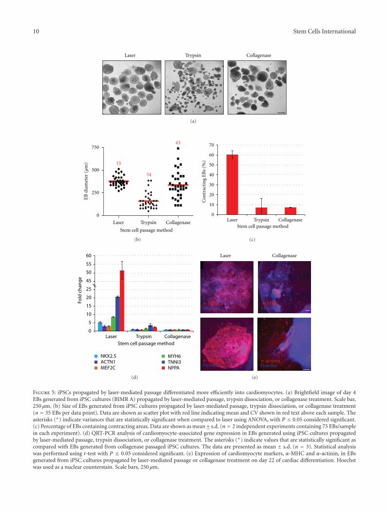
3.4. Improved Differentiation Potential of EBs Generated afterLaser-Mediated Passage. To test the differentiation potentialof these cells, in vitro differentiation assays of human iPSCswere performed after propagation using laser-mediatedpassage (160 μm sections) or enzymatic passage. iPSCspassaged by either methodology efficiently formed well-defined embryoid bodies in suspension culture which coulddifferentiate into derivatives of all three primary germ layersincluding endodermal cells (Sox17, Afp), mesodermal cells/cardiac muscle cells (brachyury, α-MHC), and ectodermalcells/neurons (Nestin, Map2, Supplemental Figure 3). Mor-phological analysis of the resulting EB populations showedthat EBs generated from laser-passaged iPSCs were moreuniform in size than those generated from enzymaticallypassaged iPSCs (Figure 5(a)). To quantify these observations,
the diameter of resulting EB populations was measuredmanually using images acquired on day 4 of suspensionculture. As shown in Figure 5(b), laser-mediated passageresulted in significantly more uniform EBs (374± 56 μm;15% CV) than enzymatic passage by either collagenase (336± 145 μm; 43% CV) or trypsin (158 ± 85 μm; 54% CV).Statistical analysis of variance showed that EBs generatedusing stem cell cultures propagated by laser-mediated pas-sage were significantly more uniform (P value < 0.0001)than EBs generated using enzymatically passaged cultures,demonstrating that laser-mediated passage results in moreconsistent EB cultures than other methods.
Several studies have shown that heterogeneity in humanESC colony size and resulting EB aggregate size resultsin variability in differentiation experiments and significantdecreases in differentiation yields [23–26]. The effect of EBhomogeneity on differentiation potential of human iPSCsinto cardiomyocytes was examined. EBs were generatedusing iPSC colonies formed 5 days after laser-mediated pas-sage (160 μm sections) or enzymatic passage. All EBs weredifferentiated using a standard multistage protocol, growingEBs in suspension culture for 4 days followed by adherent cellculture for an additional 18 days [27]. Cardiomyocyte differ-entiation potential was analyzed on day 22 of differentiationby manual counting of contracting EBs. EBs produced usingenzymatically passaged iPSCs yielded a small proportionof beating EBs (∼7%), whereas laser-mediated passagediPSCs resulted in a significantly higher proportion (∼60%)of contracting EBs (Figure 5(c)). QRT-PCR analyses con-firmed these results with EBs generated from laser-mediatedpassaged cultures showing 3- to 51-fold higher expressionof cardiomyocyte genes, Nkx2.5, Actn1 (α-actinin), Mef2C,Myh6 (α-Mhc), TnnI3, and NppA (Anp), than EBs gen-erated from collagenase passaged cultures (Figure 5(d)).Similarly, immunocytochemical analyses demonstrated thatEBs produced from laser-mediated passaged iPSCs containedsubstantially more cardiac cells within each EB (i.e., EBs con-tained more cells staining positive for known cardiomyocytemarkers) than EBs produced from collagenase passaged cells(α-MHC and α-actinin shown in Figure 5(e)). EBs generatedfrom all populations stained positive for all markers testedincluding α-MHC, α-actinin, cardiac troponin, and NPPA(data not shown).
To further analyze the effect of EB homogeneity ondifferentiation potential, EBs generated from iPSC coloniesformed 5 days after laser-mediated passage (160 μm sections)or enzymatic passage were differentiated into neural rosettesusing a modified multistep protocol. Ability to differentiateinto neural rosettes was analyzed on day 11 of differentiationby manual counting of EBs containing≥1 neural rosette. EBsproduced using laser-mediated passaged iPSCs resulted in95% of EBs containing neural rosettes, while EBs generatedby trypsin or collagenase passaged iPSCs yielded only 29%and 32% of EBs containing neural rosettes, respectively (datanot shown). These data indicate increased homogeneity inhuman iPSC colonies and resultant EBs result in significantincreases in differentiation yield of iPSCs.
To investigate the effect of EB size on differentia-tion potential of human iPSCs were examined. iPSCs were
10 Stem Cells International
Laser CollagenaseTrypsin
(a)
54
43
15
54
15
750
500
250
0
Laser Trypsin Collagenase
EB
dia
met
er (μ
m)
Stem cell passage method
(b)
0
10
20
30
40
50
60
70
Laser Trypsin Collagenase
Con
trac
tin
g E
Bs
(%)
Stem cell passage method
(c)
60
55
50
45
25
20
15
10
5
0Laser Trypsin Collagenase
Fold
ch
an
ge
Stem cell passage method
NKX2.5ACTN1MEF2C
MYH6TNNI3NPPA
(d)
Laser Collagenase
α-MHCHoechst
α-MHCHoechst
Hoechstα-actinin
Hoechstα-actinin
(e)
Figure 5: iPSCs propagated by laser-mediated passage differentiated more efficiently into cardiomyocytes. (a) Brightfield image of day 4EBs generated from iPSC cultures (BIMR A) propagated by laser-mediated passage, trypsin dissociation, or collagenase treatment. Scale bar,250 μm. (b) Size of EBs generated from iPSC cultures propagated by laser-mediated passage, trypsin dissociation, or collagenase treatment(n = 35 EBs per data point). Data are shown as scatter plot with red line indicating mean and CV shown in red text above each sample. Theasterisks (∗) indicate variances that are statistically significant when compared to laser using ANOVA, with P ≤ 0.05 considered significant.(c) Percentage of EBs containing contracting areas. Data are shown as mean + s.d. (n = 2 independent experiments containing 75 EBs/samplein each experiment). (d) QRT-PCR analysis of cardiomyocyte-associated gene expression in EBs generated using iPSC cultures propagatedby laser-mediated passage, trypsin dissociation, or collagenase treatment. The asterisks (∗) indicate values that are statistically significant ascompared with EBs generated from collagenase passaged iPSC cultures. The data are presented as mean ± s.d. (n = 3). Statistical analysiswas performed using t-test with P ≤ 0.05 considered significant. (e) Expression of cardiomyocyte markers, α-MHC and α-actinin, in EBsgenerated from iPSC cultures propagated by laser-mediated passage or collagenase treatment on day 22 of cardiac differentiation. Hoechstwas used as a nuclear counterstain. Scale bars, 250 μm.

Stem Cells International 11
Table 1: Regions in H9 human ESCs with genomic abberations as determined by aCGH (corresponding schematic is shown in Figure 4(e)).No subkaryotypic alterations were detected in hESCs propagated for 24 consecutive passages (H9 P59) relative to the starting hESCpopulation (H9 P35), suggesting that the genome of laser-mediated passaged cells is both normal and stable. Data is reported using genomebuilt HG18. log2 ratios ≥ 0.6 are amplifications (amp) or ≤ −1.0 are deletions (del) found in all cells. log2 ratios < 0.6 or > −1.0 representmosaicism within the culture.
Chromosome: region CytobandSize
(Mb)# Probes Amp/Del
H9 P35log2 ratio
H9 P59log2 ratio
Annotations
Chr1: 224141493-224195678 q42.12 0.054 14 Amp 0.528 0.632 LEFTY1, PYCR2, LEFTY2
Chr6: 31663619-31691605 p21.33 0.028 3 Amp 0.686 1.220 LST1, NCR3, AIF1
Chr12: 21580165-22105263 q12.1 0.282 12 Del −0.495 −0.480 GYS2, LDHB, KCNJ8, ABCC9, CMAS
Chr14: 62486603-62852257 q23.2 0.366 7 Amp 0.560 0.432 KCNH5, RHOJ, GPHB5
Chr17: 35097815-35153082 q12 0.055 15 Amp 0.469 0.485 PERLD1, ERBB2, C17orf37, GRB7
propagated by laser-mediated passage at varying section sizes(80, 160, and 240 μm sections). Five days after passage,EBs were generated and differentiated into cardiomyocytesas described above. Homogeneous EB populations (≤15%CVs) of varying sizes, 278, 418, and 528 μm in diameter, wereproduced from 80, 160, and 240 μm section sizes, respectively(Supplemental Figure 4). Analysis of cardiomyocyte differ-entiation potential showed that 55% of EBs generated from160 μm sections were contracting, while only 38% and 21%of EBs generated from 240 μm and 80 μm sections were con-tracting. Taken together, these data indicate that increasedhomogeneity in human iPSC colonies and resultant EBs,as well as EB size, significantly increase the differentiationyield of iPSCs. The ability to reproducibly generate uniform,size-specific colonies which subsequently result in moreuniform, size-specific EB populations decreases variabilityin differentiation experiments and enhances differentiationyields of both ESC and iPSCs into specialized cell types.
4. Discussion
The lack of standardization in passage techniques for stemcell derivation and propagation is a major limitation withinthe stem cell field. Because universal protocols for humanstem cell cultures have not been adopted, it is currentlydifficult to compare and interpret scientific data from cellscultured in different conditions. Passage method differenceshave significantly confounded the understanding of intra-and interline differences in gene expression data, expressionof stem cell- and lineage-associated markers, miRNA sig-natures, and epigenetic profiles [28–31]. Although humanESC lines have distinct genotypes, it is unlikely that reporteddifferences in cell lines (e.g., up to 65% variation in geneexpression data across two ESC lines) can be attributed togenetic variation alone, as <2% variation in gene expressionhas been found in adult human tissues of different individu-als [28, 32, 33]. Likewise, discrepancies associated with differ-entiation protocols and reported differentiation capabilitiesand efficiencies of stem cells into specialized cell types maybe due to the lack of standardization [34]. Adoption of stan-dardized protocols should greatly improve determination ofthe role of inherent genetic variation, environmental niche,and/or procedural effects on stem cell quality, self-renewal,pluripotency, and differentiation potential.
Laser-mediated passage provides a novel method forexpansion of human ESCs/iPSCs which can be used tocreate standardized, traceable procedures for the produc-tion of GMP-quality stem cell lines without requirementfor enzymes. This method combines the benefits of bothmanual and enzymatic passage techniques, allowing efficient,automated passaging of undifferentiated stem cell culturesinto uniform-sized stem cell sections within a sterile closedenvironment. Laser-mediated passage is compatible with avariety of culture methods including animal-free, feeder-free-based conditions, and serum-free defined media con-ditions. Notably, this approach is not susceptible to inter-individual variation reducing the need for skilled techniciansto create high-quality stem cell cultures.
Laser-mediated passage does not involve the use of en-zymes and therefore should better maintain the geneticstability of human ESCs and iPSCs in long-term culture(3–12 months, [10–14]). The results show that H9 ESCsmaintained a stable karyotype over six months (>24 pas-sages). More importantly, laser-mediated passage did notinduce subkaryotypic alterations over time in H9 ESCs (6months, and iPSCs (2.5 months)) as monitored by aCGH.The more sensitive aCGH data suggests that laser-mediatedpassage maintains genetic integrity of human ESCs/iPSCs.Importantly, genetic abnormalities were detected in H9 ESCsafter consecutive passaging by collagenase during the sametime period. Results also showed that human iPSCs andESCs propagated using laser-mediated passage maintained anormal stem cell morphology and continued to express highlevels of stem-cell-associated genes and proteins. Althoughteratoma analyses were not performed on these cells, in vitrodifferentiation analyses of laser-mediated passaged iPSCsdemonstrated the cells could spontaneously differentiateinto derivatives of all three primary germ layers and coulddifferentiate into cardiomyocytes and neural rosettes. Inaddition, iPSCs propagated by laser-mediated passage havebeen differentiated into motor neurons, RPE cells, endodermprogenitors, and hepatocytes-like cells (data not shown);taken together these data indicate that laser-mediated pas-sage does not affect stem cell pluripotency. Likewise, laser-mediated passage did not alter the growth rate of stem cellsor increase expression of apoptotic markers, all supportingthat the laser sectioning did not affect stem cell quality, self-renewal, or pluripotency.

12 Stem Cells International
Laser-mediated passage provides control of stem cellcolony size. Regular passage schedules can be established byselection of section size. A section size of ∼200 μm has ena-bled routine splitting of all ESC/iPSC lines every 7 days,allowing for more efficient planning of experiments. An over-all passage efficiency of 85% combined with more uniformsection sizes (20% CV), enables a larger proportion of ESC/iPSC colonies to contribute to culture expansion reducingthe number of plates required for culture maintenance. Com-patibility with conventional robotic systems enables scalabil-ity of culture needs. Additionally, the ability to control inputsection size, particularly smaller sizes, allows more effectivecreation of stem cell colonies in multi-well plates for large-scale experimentation and screening purposes. It is also likelythat stem cell section size will affect cryopreservation andgenetic modification efficiency of ESCs and iPSCs [35–38].
Laser-mediated passage involves sectioning the entirewell systematically without respect for the boundaries of thecolonies. Well-established undifferentiated stem cell culturesare easily propagated using this technique. For newly derivedESC/iPSC cultures, early passage ESC/iPSC lines, or lessstable lines, which tend to have more spontaneous differenti-ation, a combination of manual selection or laser purificationof colonies followed by laser-mediated passage would berecommended. One of the more important results of thisapproach showed that over time, stem cell cultures (in partic-ular, early passage iPSCs which tend to be more susceptibleto differentiation than later passage, more established ESCs)are of higher quality than those maintained by collagenasetreatment. It is likely that passage of homogeneous sectionsis important for maintaining undifferentiated stem cells andlimiting the differentiation of colonies. Therefore, potentiallyearly passage ESCs/iPSCs will require less colony isolationbefore expansion using laser-mediated passage.
One of the more important outcomes of this studyshowed that uniform human iPSC colonies produced afterlaser-mediated passage resulted in a more homogeneouspopulation of EBs, with respect to size and shape, withgreater differentiation efficiency as compared with typicalEB cultures derived from enzyme passaged cultures. EBsgenerated from laser-mediated passaged iPSCs resulted ina significant increase in cardiomyocyte yield, with up to8.5-fold greater beating incidence than EBs generated fromcollagenase passaged iPSCs. The ability to reproduciblygenerate uniform colonies using laser-mediated passageresulting in EBs that are more uniform in size and shapewill decrease variability in differentiation experiments andenhance differentiation yields of both ESC and iPSCs intospecialized cell types. These yield enhancements couldsignificantly reduce the cost of stem cell experimentationboth in terms of labor and materials. Potentially, uniformcolony formation will also augment differentiation yields ofstem cells when performing direct differentiation procedures(i.e., without an EB intermediate).
5. Conclusions
In conclusion, proper maintenance of human stem cellsis essential for successful utilization of ESCs and iPSCs
as tools in developmental and drug discovery studies andin regenerative medicine. Standardization is critical for allfuture applications of stem cells and necessary in order tofully understand the potential of these cells and the differ-ences observed among varying stem cell lines and betweenESCs and iPSCs. Laser-mediated passage is an innovativemethod for maintenance and expansion of stem cell lines,without introducing genetic instability, which is genericallyapplicable to all cell lines and to all technicians regardlessof skill. This approach provides an efficient, standardizedprotocol for the propagation of human ESCs and iPSCs,which should significantly reduce the inconsistency andvariability within the stem cell field. Laser-mediated passageallows for traceability and ensures reproducible productionof stem cell lines according to standard operating procedures,all of which are necessary to manufacture stem cells for usein clinical/therapeutic applications.
Author’s Contribution
C. Peterson and A. Soundararajan contributed equally to thepaper.
Acknowledgments
The authors would like to thank A. Elliott for assistance withaCGH experiments, and M. Koller, F. Kamme, and A. Pyle forsuggestions and comments. The α-MHC (developed by Fis-chman, D.A.) and SSEA4 (developed by Solter, D/Knowles,B.B.) antibodies were obtained from the DevelopmentalStudies Hybridoma Bank developed under the auspices ofthe NICHD and maintained by The University of Iowa,Department of Biology, Iowa City, IA USA.
References
[1] J. A. Thomson, “Embryonic stem cell lines derived from hu-man blastocysts,” Science, vol. 282, no. 5391, pp. 1145–1147,1998.
[2] C. Xu, M. S. Inokuma, J. Denham et al., “Feeder-free growthof undifferentiated human embryonic stem cells,” Nature Bio-technology, vol. 19, no. 10, pp. 971–974, 2001.
[3] M. Richards, C. Y. Fong, W. K. Chan, P. C. Wong, and A.Bongso, “Human feeders support prolonged undifferentiatedgrowth of human inner cell masses and embryonic stem cells,”Nature Biotechnology, vol. 20, no. 9, pp. 933–936, 2002.
[4] M. Amit, C. Shariki, V. Margulets, and J. Itskovitz-Eldor,“Feeder layer- and serum-free culture of human embryonicstem cells,” Biology of Reproduction, vol. 70, no. 3, pp. 837–845,2004.
[5] B. E. Reubinoff, M. F. Pera, C. Y. Fong, A. Trounson, and A.Bongso, “Embryonic stem cell lines from human blastocysts:somatic differentiation in vitro,” Nature Biotechnology, vol. 18,no. 4, pp. 399–404, 2000.
[6] C. A. Cowan, I. Klimanskaya, J. McMahon et al., “Derivationof embryonic stem-cell lines from human blastocysts,” TheNew England Journal of Medicine, vol. 350, no. 13, pp. 1353–1356, 2004.
[7] K. O. Sun, S. K. Hee, B. P. Yong et al., “Methods for expansionof human embryonic stem cells,” Stem Cells, vol. 23, no. 5, pp.605–609, 2005.

Stem Cells International 13
[8] L. M. Hoffman and M. K. Carpenter, “Characterization andculture of human embryonic stem cells,” Nature Biotechnology,vol. 23, no. 6, pp. 699–708, 2005.
[9] C. Unger, H. Skottman, P. Blomberg, M. Sirac dilber, andO. Hovatta, “Good manufacturing practice and clinical-gradehuman embryonic stem cell lines,” Human Molecular Genetics,vol. 17, no. 1, pp. R48–R53, 2008.
[10] J. J. Buzzard, N. M. Gough, J. M. Crook, and A. Colman, “Kar-yotype of human ES cells during extended culture,” NatureBiotechnology, vol. 22, no. 4, pp. 381–382, 2004.
[11] M. M. Mitalipova, R. R. Rao, D. M. Hoyer et al., “Preservingthe genetic integrity of human embryonic stem cells,” NatureBiotechnology, vol. 23, no. 1, pp. 19–20, 2005.
[12] J. S. Draper, K. Smith, P. Gokhale et al., “Recurrent gain ofchromosomes 17q and 12 in cultured human embryonic stemcells,” Nature Biotechnology, vol. 22, no. 1, pp. 53–54, 2004.
[13] G. Caisander, H. Park, K. Frej et al., “Chromosomal integritymaintained in five human embryonic stem cell lines after pro-longed in vitro culture,” Chromosome Research, vol. 14, no. 2,pp. 131–137, 2006.
[14] S. N. Brimble, X. Zeng, D. A. Weiler et al., “Karyotypicstability, genotyping, differentiation, feeder-free maintenance,and gene expression sampling in three human embryonic stemcell lines derived prior to August 9, 2001,” Stem Cells and De-velopment, vol. 13, no. 6, pp. 585–597, 2004.
[15] A. Joannides, C. Fiore-Heriche, K. Westmore et al., “Auto-mated mechanical passaging: a novel and efficient method forhuman embryonic stem cell expansion,” Stem Cells, vol. 24, no.2, pp. 230–235, 2006.
[16] S. Terstegge, I. Laufenberg, J. Pochert et al., “Automatedmaintenance of embryonic stem cell cultures,” Biotechnologyand Bioengineering, vol. 96, no. 1, pp. 195–201, 2007.
[17] M. Amit, M. K. Carpenter, M. S. Inokuma et al., “Clonallyderived human embryonic stem cell lines maintain pluripo-tency and proliferative potential for prolonged periods ofculture,” Developmental Biology, vol. 227, no. 2, pp. 271–278,2000.
[18] K. Watanabe, M. Ueno, D. Kamiya et al., “A ROCK inhibitorpermits survival of dissociated human embryonic stem cells,”Nature Biotechnology, vol. 25, no. 6, pp. 681–686, 2007.
[19] M. R. Koller, E. G. Hanania, J. Stevens et al., “High-throughputlaser-mediated in situ cell purification with high purity andyield,” Cytometry Part A, vol. 61, no. 2, pp. 153–161, 2004.
[20] K. J. Livak and T. D. Schmittgen, “Analysis of relative geneexpression data using real-time quantitative PCR and the 2-ΔΔCT method,” Methods, vol. 25, no. 4, pp. 402–408, 2001.
[21] N. Heins, M. C. O. Englund, C. Sjoblom et al., “Derivation,characterization, and differentiation of human embryonicstem cells,” Stem Cells, vol. 22, no. 3, pp. 367–376, 2004.
[22] E. S. Rosler, G. J. Fisk, X. Ares et al., “Long-term culture ofhuman embryonic stem cells in feeder-free conditions,” De-velopmental Dynamics, vol. 229, no. 2, pp. 259–274, 2004.
[23] E. S. Ng, R. P. Davis, L. Azzola, E. G. Stanley, and A. G.Elefanty, “Forced aggregation of defined numbers of humanembryonic stem cells into embryoid bodies fosters robust, re-producible hematopoietic differentiation,” Blood, vol. 106, no.5, pp. 1601–1603, 2005.
[24] P. W. Burridge, D. Anderson, H. Priddle et al., “Improvedhuman embryonic stem cell embryoid body homogeneity andcardiomyocyte differentiation from a novel V-96 plate aggre-gation system highlights interline variability,” Stem Cells, vol.25, no. 4, pp. 929–938, 2007.
[25] C. L. Bauwens, R. Peerani, S. Niebruegge et al., “Con-trol of human embryonic stem cell colony and aggregate size
heterogeneity influences differentiation trajectories,” StemCells, vol. 26, no. 9, pp. 2300–2310, 2008.
[26] B. Valamehr, S. J. Jonas, J. Polleux et al., “Hydrophobic surfacesfor enhanced differentiation of embryonic stem cell-derivedembryoid bodies,” Proceedings of the National Academy of Sci-ences of the United States of America, vol. 105, no. 38, pp.14459–14464, 2008.
[27] C. Xu, S. Police, N. Rao, and M. K. Carpenter, “Characteriza-tion and enrichment of cardiomyocytes derived from humanembryonic stem cells,” Circulation Research, vol. 91, no. 6, pp.501–508, 2002.
[28] C. Allegrucci and L. E. Young, “Differences between humanembryonic stem cell lines,” Human Reproduction Update, vol.13, no. 2, pp. 103–120, 2007.
[29] C. Allegrucci, Y. Z. Wu, A. Thurston et al., “Restrictionlandmark genome scanning identifies culture-induced DNAmethylation instability in the human embryonic stem cell epi-genome,” Human Molecular Genetics, vol. 16, no. 10, pp. 1253–1268, 2007.
[30] M. J. Abeyta, A. T. Clark, R. T. Rodriguez, M. S. Bodnar,R. A. Reijo Pera, and M. T. Firpo, “Unique gene expressionsignatures of independently-derived human embryonic stemcell lines,” Human Molecular Genetics, vol. 13, no. 6, pp. 601–608, 2004.
[31] H. Skottman, M. Mikkola, K. Lundin et al., “Gene expressionsignatures of seven individual human embryonic stem celllines,” Stem Cells, vol. 23, no. 9, pp. 1343–1356, 2005.
[32] X. Zeng, T. Miura, Y. Luo et al., “Properties of pluripotenthuman embryonic stem cells BG01 and BG02,” Stem Cells, vol.22, no. 3, pp. 292–312, 2004.
[33] L. L. Hsiao, F. Dangond, T. Yoshida et al., “A compendium ofgene expression in normal human tissues,” Physiol Genomics,vol. 7, no. 2, pp. 97–104, 2001.
[34] K. Osafune, L. Caron, M. Borowiak et al., “Marked differencesin differentiation propensity among human embryonic stemcell lines,” Nature Biotechnology, vol. 26, no. 3, pp. 313–315,2008.
[35] X. Li, G. Meng, R. Krawetz, S. Liu, and D. E. Rancourt, “TheROCK inhibitor Y-27632 enhances the survival rate of humanembryonic stem cells following cryopreservation,” Stem Cellsand Development, vol. 17, no. 6, pp. 1079–1085, 2008.
[36] R. Eiges, “Genetic manipulation of human embryonic stemcells by transfection,” Methods in Molecular Biology, vol. 331,pp. 221–239, 2006.
[37] K. A. Hohenstein, A. D. Pyle, Y. I. C. Jing, L. F. Lock, and P. J.Donovan, “Nucleofection mediates high-efficiency stable geneknockdown and transgene expression in human embryonicstem cells,” Stem Cells, vol. 26, no. 6, pp. 1436–1443, 2008.
[38] B. Y. Zhou, Z. Ye, G. Chen, Z. P. Gao, Y. A. Zhang, andL. Cheng, “Inducible and reversible transgene expression inhuman stem cells after efficient and stable gene transfer,” StemCells, vol. 25, no. 3, pp. 779–789, 2007.

Hindawi Publishing CorporationStem Cells InternationalVolume 2012, Article ID 549846, 10 pagesdoi:10.1155/2012/549846
Research Article
Cellular Reprogramming Employing Recombinant Sox2 Protein
Marc Thier, Bernhard Munst, Stephanie Mielke, and Frank Edenhofer
Stem Cell Engineering Group, Institute of Reconstructive Neurobiology, University of Bonn-Life &Brain Center and Hertie Foundation, Sigmund-Freud Straße 25, D-53105 Bonn, Germany
Correspondence should be addressed to Frank Edenhofer, [email protected]
Received 1 December 2011; Accepted 23 January 2012
Academic Editor: Rajarshi Pal
Copyright © 2012 Marc Thier et al. This is an open access article distributed under the Creative Commons Attribution License,which permits unrestricted use, distribution, and reproduction in any medium, provided the original work is properly cited.
Induced pluripotent stem (iPS) cells represent an attractive option for the derivation of patient-specific pluripotent cells for cellreplacement therapies as well as disease modeling. To become clinically meaningful, safe iPS cells need to be generated exhibiting nopermanent genetic modifications that are caused by viral integrations of the reprogramming transgenes. Recently, various experi-mental strategies have been applied to accomplish transgene-free derivation of iPS cells, including the use of nonintegrating viruses,episomal expression, or excision of transgenes after reprogramming by site-specific recombinases or transposases. A straightfor-ward approach to induce reprogramming factors is the direct delivery of either synthetic mRNA or biologically active proteins. Wepreviously reported the generation of cell-permeant versions of Oct4 (Oct4-TAT) and Sox2 (Sox2-TAT) proteins and showedthat Oct4-TAT is reprogramming-competent, that is, it can substitute for Oct4-encoding virus. Here, we explore conditions forenhanced Sox2-TAT protein stabilization and functional delivery into somatic cells. We show that cell-permeant Sox2 protein canbe stabilized by lipid-rich albumin supplements in serum replacement or low-serum-supplemented media. Employing optimizedconditions for protein delivery, we demonstrate that Sox2-TAT protein is able to substitute for viral Sox2. Sox2-piPS cells expresspluripotency-associated markers and differentiate into all three germ layers.
1. Introduction
Pluripotent cells represent a most attractive source for bothcell repair in regenerative medicine and disease modeling inbasic biomedical research since they are able to differentiateinto every cell type of an adult organism. Until recently, earlyembryonic stages of development represented the mainsource of pluripotent cells, and thus, those cells were desig-nated as embryonic stem (ES) cells. Nowadays, the artificialderivation of pluripotent stem cells from somatic cellsbecomes increasingly important. Induced pluripotent stem(iPS) cells were first generated by retrovirally induced ectopicexpression of four transcription factors Oct4, Sox2, Klf-4,and c-Myc in somatic cells [1]. Human iPS cells representan attractive option for the derivation of pluripotent patient-specific cells as no embryos are required for their generation.However, crucial safety issues have to be addressed in order togenerate human iPS cells that are clinically useful. Soon afteridentification of the viral reprogramming protocol in mousecells [1] and its adaptation to human cells [2, 3], unwantedside effects such as tumorigenesis [4] became apparent.
Since the cause of tumor formation was ascribed torandom integration of the retroviral vectors and sustainedexpression of transgenes after reprogramming, optimizedprotocols were explored to circumvent the permanent inte-gration of foreign DNA into the genome. One strategyinvolves the excision of reprogramming transgenes employ-ing DNA recombinases [5, 6] or transposases [7–10]. AfteriPS derivation, transgenes can be deleted by a second roundof recombinase/transposase activation. However, furtherlaborious and cumbersome genetic methods are needed toidentify and confirm transgene-free iPS clones. An alterna-tive strategy is to utilize less-invasive genetic vectors thatdo not integrate into the host genome. Repeated plasmidtransfection has been used for iPS induction albeit with avery low efficiency [11]. Minicircle vectors lacking bacterialDNA and thus enabling high transfection efficiency andlong ectopic expression were reported to reprogram aswell [12]. Moreover, transduction employing viruses thatdo not integrate their genome into host cells such asadenovirus [13] or Sendai virus [14] were applied. Smallmolecules that are able to translocate into cells and interfere

2 Stem Cells International
with key signaling pathways have been identified to eitherenhance the process of reprogramming [15, 16] or replace[15, 17] single viral factors (for review, see [18]). Therepeated transfection of synthetic mRNA [19–21] or thedirect delivery of reprogramming proteins [22, 23] representsa straightforward but technically challenging approach toachieve nongenetic iPS derivation.
Protein transduction technology has been used to directlydeliver numerous biologically active proteins into mam-malian cells by modifying them with so-called cell-pene-trating peptides (CPPs) or protein transduction domains(PTDs). These relatively small peptides confer cell perme-ability when linked to cargo molecules (for review, see[24–26]). A highly basic CPP derived from the humanimmunodeficiency virus type 1 (HIV-1) Tat (transactivator oftranscription) protein is often applied for cellular delivery(TAT) [25–28]. PTDs have been used to generate cell-penetrating versions of various transcription factors that playmajor roles in cell differentiation including HoxB4 [27],Pdx1 [28], Scl [29], Nkx2.2 [30], and Notch-ICD [31]. Wepreviously reported the derivation of cell-permeant versionsof reprogramming factors Oct4 and Sox2 [22]. Oct4-TAT andSox2-TAT were shown to specifically bind to DNA such asthe Oct4/Sox2 combined element in the Nanog promoter,and both proteins compensate the RNAi-induced loss offunction after direct delivery into ES cells [22]. Moreover,employing Sox2-TAT, it has been demonstrated that Sox2has an essential function in the preimplantation mouseembryo by facilitating establishment of the trophectodermlineage [32]. Zhou et al. used fusion protein derivatives ofreprogramming factors from E. coli for the derivation ofmouse ES-like cells, albeit with very low efficiency [23].Kim et al. reported the use of cell extracts from transfectedHEK293 cells for the reprogramming of human newbornfibroblasts [33]. The recently reported use of ES cell extractsto induce pluripotency in murine fibroblasts [34] needs to beexplored whether it can be adapted to human cells. In con-clusion, a robust, standardized, and efficient protocol for thegeneration of protein-induced iPS cells from human adultcells still needs to be developed.
Further optimization of protein transduction for cellularreprogramming greatly depends on overcoming a majorbottleneck associated with protein transduction: stabilityof recombinant factors under cell culture conditions. Werecently established optimized stabilization conditions forOct4-TAT and demonstrated the efficient substitution forOct4-encoding virus by recombinant Oct4-TAT [36]. Here,we explore conditions for enhanced Sox2-TAT proteinstabilization and delivery into somatic cells. We show thatcell-permeant Sox2 protein can be stabilized by lipid-richalbumin supplements in serum replacement or low-serum-supplemented media. Employing these conditions for pro-tein delivery, we demonstrate that Sox2-TAT protein is ableto substitute for viral Sox2.
2. Materials and Methods
2.1. Protein Expression and Purification. The pSESAME-Sox2NTH expression plasmid [22] was transformed into
E. coli BL21 (DE3) gold strain (Stratagene, La Jolla, USA) byheat shock at 30◦C and incubated for 1 h in SOC mediumat 30◦C. Transformed bacteria were inoculated overnight at30◦C with shaking at 140 rpm in LB medium containing50 mg/mL carbenicillin. For protein expression, the over-night culture was pelletized and resuspended in fresh TBmedium (terrific broth)/50 mg/mL ampicillin, 0.5% glucoseand incubated at 37◦C with shaking at 110 rpm until anOD600 of 1.5 was achieved. Protein expression was inducedby IPTG at a final concentration of 0.5 mM. Cells wereharvested by centrifugation, and cell pellets were stored at−20◦C.
For purification of His-tagged proteins, cell pellets werethawed and resuspended in 20 mL lysis buffer (50 mMNa2HPO4, 5 mM Tris, pH 7.8, 500 mM NaCl, and 10 mMimidazole) per 1 L of expression culture. Cells were lysed byapplication of 1 mg/mL lysozyme (Sigma, Deisenhofen, Ger-many), 10–15 U/mL Benzonase (Novagen, Darmstadt, Ger-many), and sonication. After centrifugation (17 200 g,20 min), the cleared lysate was incubated with Ni-NTAagarose beads (Qiagen, Hilden, Germany) (1 mL of slurry for1 L of bacterial expression culture) for 1 h with rotationat 4◦C. The slurry was packed into a column and washedwith 8 column volumes of wash buffer (50 mM Na2HPO4,5 mM Tris, pH 7.8, 500 mM NaCl, and 80 mM imidazole).The Sox2-TAT protein was eluted with 3 column volumesof elution buffer (50 mM Na2HPO4, 5 mM Tris, pH 7.8,500 mM NaCl, and 250 mM imidazole).
2.2. Preparation of Transduction Media. Sox2-TAT eluatefraction was supplemented with 7.5% serum replacementand dialyzed against DMEM F12 over night at 4◦C. Thenext day cell culture supplements were added to the dialyzedfraction to a final concentration of 2% FCS, 2.5% AlbumaxII (200 mg/mL), 7.5% serum replacement, 1% ITS, 0.1 mMnonessential amino acids, 1 mM sodium pyruvate, 2 mM L-glutamine, 0.5 mM β-mercaptoethanol, and 1000 U/mL LIF.The mixture was preconditioned in a water bath for 1 h at37◦C and cleared by centrifugation (5 min at 2500 g) andsterile filtration.
2.3. Cell Culture. Oct4-GiP MEFs [38] were cultured inhigh-glucose DMEM (Invitrogen) with 10% FCS, 0.1 mMnonessential amino acids, 1 mM sodium pyruvate, and 2 mML-glutamine. MEFs were trypsinized at 70–80% confluenceand reseeded on tissues culture dishes coated with gelatin.For reprogramming assays, MEFs were used to a maximumof passage 4.
mESCs were cultured in DMEM F12 (Invitrogen) supple-mented with 2% FCS, 2.5% Albumax II (200 mg/mL), 7.5%serum replacement, 1% ITS, 0.1 mM nonessential aminoacids, 1 mM sodium pyruvate, 2 mM L-glutamine, 0.5 mMβ-mercaptoethanol, and 1000 U/mL LIF. Cells were splitevery 3 days and cultured on inactivated feeder cells.
2.4. Retroviral Infection and iPS Induction. Plasmids ofpMXs-Oct3/4, pMXs-Sox2 (positive control), pMXs-c-Mycand pMXs-Klf4 were obtained from ADDGENE. The retro-viruses were generated by the plat E packaging cell line as

Stem Cells International 3
previously described [39]. Target cells were seeded at 10×104
cells per well in six-well plates. 24 hours after transfection,the supernatant comprising the viruses was collected andfiltered through a 0.45 μm cellulose acetate filter. For substi-tution experiments and for negative controls, Oct4, Klf4, andc-Myc were mixed in equal shares and supplemented withpolybrene (Millipore) to a final concentration of 4 μg/mL.Positive controls additionally contained pMXs-Sox2 virusand were treated alike. Oct4-GiP MEF cells were incubatedwith viruses for 16 hours. Protein transduction experimentsbegan after the virus-containing supernatant had beenremoved. After 5 days, cells were split onto irradiated feedercells. 11 days later, cells were fixed with 4% PFA and analyzedby fluorescence microscopy. For the purpose of generatingstable iPS cell lines, reprogramming assays were cultured for21 days under designated conditions. Subsequently, colonieswere picked and expanded monoclonally.
2.5. In Vitro Differentiation. Cells were harvested bytrypsinization and transferred to bacterial culture dishes.Next, cells were grown in ES medium lacking LIF for 3 days.The derived embryoid bodies were transferred to gelatine-coated tissue dishes afterwards and incubated for another 3days. In order to detect key marker expression specific for allthree germ layers, immunostainings with antibodies againstβ-3-tubulin (TUJ1), smooth muscle actin (SMA), and α-fetoprotein (AFP) were conducted.
2.6. RT-PCR. Total RNA was extracted using the RNAeasykit (Qiagen, Hilden, Germany) following the manufacturer’sinstructions. Subsequently, reverse transcription of 1 μg RNAper sample was performed using the iScript cDNA SynthesisKit (Bio-Rad). In order to detect viral transgene expression,the following primer pairs were used:
Oct4TGforw: CCCCACTTCACCACACTCTAC,
Oct4TGrev: TTTATCGTCGACCACTGTGC,
Klf4TGforw: AGGCACTACCGCAAACACAC,
Klf4TGrev: TTTATCGTCGACCACTGTGC,
Sox2TGforw: GCCCAGTAGACTGCACATGG,
Sox2TGrev: CCCCCTTTTTCTGGAGACTA,
c-MycTGforw: CAGAGGAGGAACGAGCTGAAG-CGC,
c-MycTGrev: TTTGTACAAGAAAGCTGGGT.
For the detection of endogenous Oct4, Sox2, and Nanog, thefollowing primer pairs were used:
Oct4forw: TCTTTCCACCAGGCCCCCGGCTC,
Oct4rev: TGCGGGCGGACATGGGGAGATCC,
Sox2for: TAGAGCTAGACTCCGGGCGATGA,
Sox2rev: TTGCCTTAAACAAGACCACGAAA,
Nanogfor: CAGGTGTTTGAGGGTAGCTC,
Nanogrev: CGGTTCATCATGGTACAGTC.
PCR-program: 95◦C 2 min, 95◦C 30 sec, 60◦C 30 sec, 72◦C1 min, 72◦C 10 min. Steps 2–4 are repeated 35 times.
3. Results
3.1. Purification of Reprogramming-Competent Sox2 FusionProtein from Bacteria. We have previously shown thatrecombinant Sox2 can be purified from E. coli as a TAT-modified cell-permeant version, designated Sox2-TAT [22].In particular, this fusion protein comprises an additionalexogenous nuclear localization sequence (NLS), a cell-penetrating peptide TAT, and a carboxy-terminal Histidine-tag for single-step purification (Figure 1(a)). Sox2-TAT wasshown to specifically bind to DNA and to compensate for theRNAi-induced loss of activity in ES cells [22] and preimplan-tation embryos [32]; however, its capability to reprogramsomatic cells has not been assessed. In order to study thereprogramming activity of Sox2-TAT, we decided to combinethe purified recombinant Sox2-TAT together with retro-viruses encoding for Oct4, Klf4, and c-Myc to convert mouseembryonic fibroblasts (MEFs) into iPS cells (Figure 1(b)).Sox2-TAT-transformed bacteria were lysed and subjected toNi-affinity chromatography. Immunoblotting of purificationfractions employing a His-specific antibody revealed thatSox2-TAT is highly expressed in bacteria although the major-ity of the recombinant protein remains in the insoluble frac-tion (Figure 1(d)). However, the estimated 20% of proteinsolubilized and detectable in the supernatant turned out tobe sufficient for further purification. The elution from theNi-affinity chromatography column yielded a Sox2-TAT-containing fraction of about 70% purity (Figure 1(c)).
3.2. Defining Optimal Conditions to Stabilize Sox2-TATProtein. Poor solubility and limited stability of recombinantproteins under cell culture conditions represent a significanthurdle to the application of protein transduction technology.On the one hand, serum components stabilize recombinantproteins in cell culture media, but on the other hand, theyare known to inhibit interaction of transducible proteinswith cells and by this decrease the cellular uptake. In someexperimental settings, this can be overcome by applying thetransducible protein in serum-free media. We assessed thestability of Sox2-TAT employing various cell culture con-ditions. In serum-free media, Sox2-TAT precipitates almostcompletely within 1 hour (Figure 2(a)). Serum componentshave been shown to execute a positive effect on the stabilityof recombinant proteins [36, 40]. Therefore, we aimedat stabilizing the protein by supplements like FCS, serumreplacement [35], and lipid-rich albumin fractions (Albu-max). Supplementation with either 5% FCS or 2.5% Albu-max showed a strong stabilizing effect on Sox2-TAT inculture media, while Sox2-TAT exhibited major decreasein the presence of 7.5% SR. A combination of low FCS(2%) together with 7.5% SR resulted in a stabilizationthat was comparable to high FCS supplementation (5%)(Figure 2(a)).
Next, we set out to analyze to which extent the stabilizingsupplements interfere with the protein transduction process.To that aim, we used a well-established transduction read-out system based on a cell-permeant version of the DNArecombinase Cre [40]. A major feature of this protein trans-duction system is that the efficiency of intracellular protein

4 Stem Cells International
Sox2 NLS TAT H6
(a)
F
Sox2
Sox2 Sox2
Sox2
Sox2
Sox2So
x2
Sox2
Sox2
Sox2
Sox2Sox2
Sox2
Sox2
Sox2
Sox2
Sox2
+ Oct4+ Klf4+ c-MycReprogramming
(b)
CL M P SN FT W E
(c)
CL M P SN FT W E
(d)
Figure 1: Purification of recombinant Sox2-TAT fusion protein and reprogramming setup. (a) The recombinant cell-permeant Sox2 fusionprotein [22] consists of the full-length Sox2 protein and a carboxy-terminally fused sequence of tags consisting of a nuclear localizationsequence (NLS), cell-penetrating peptide TAT, and a histidine-tag (H6) for single-step purification. (b) Schematic representation of theexpression and purification procedure and the reprogramming setup used in this study. After expression in E. coli, Sox2-TAT-containing celllysates are subjected to affinity column chromatography employing Ni-NTA resin. Purified recombinant Sox2-TAT protein is eluted from thecolumn and its reprogramming competency assessed in combination with retroviruses encoding Oct4, Klf-4, and c-Myc. (c, d) Biochemicalanalysis of Sox2-TAT purification from E. coli. The following fractions were subjected to SDS-PAGE analysis: crude lysate (CL), marker (M),pellet (P), supernatant (SN), flow-through (FT), washing buffer (W), and elution fraction (E). SDS gels were either stained using Coomassie(c) or used for preparation of an immunoblot using anti-Sox2-specific antibody (d).
delivery can reliably be quantified by a Cre recombinasereporter assay. We used the CV1-5B Cre reporter cell line [37]that specifically expresses β-galactosidase after Cre-mediatedrecombination. Using this read-out system, we tested theinfluence of FCS and SR on the transduction efficiency(Figure 2(b)). 2 μM of TAT-Cre in 5% FCS-supplementedmedia induced recombination in approximately 35% of cells,whereas more than 90% of cellular targets were recombinedin medium containing 15% SR. Application of 1 μM of TAT-Cre revealed a similar correlation: about 20% recombinationin the presence of FCS and 70% with SR (Figure 2(b)). Thesedata demonstrate that FCS in contrast to SR exhibits a stronginhibition on protein transduction. Based on these results,we established an optimized transduction protocol for iPS
derivation employing Sox2-TAT. We decided to apply a two-step protocol to optimize both, protein stability and trans-duction capacity. In a first step, the eluate fraction was sup-plemented with 7.5% SR and dialyzed against DMEM/F12.Afterwards, the dialysis fraction was supplemented with FCS(2%) and Albumax (2.5%). The optimized medium showedprotein stabilizing capacity during the dialysis and under cellculture conditions in the same range as compared to SR andFCS, respectively.
3.3. Reprogramming OKC-Infected Cells with Cell-PermeantSox2 Fusion Protein. We then assessed the reprogrammingactivity of Sox2-TAT employing the optimized media con-ditions. For that, we used a modification of the classical

Stem Cells International 5
No supplement
5% FCS
2.5% Albumax
7.5% SR
Albumax +2% FCS +7.5% SR
D 1 h 6 hEluate
(a)
100
90
80
70
60
50
40
30
20
10
0
Rec
ombi
nat
ion
eci
ency
(%
)
0.25 0.5 1 2
c (TATCre) ( μM)
5% FCS7.5% SR+ 2% FCS
15% SR
(b)
Figure 2: Effect of media supplements on the stability of cell-permeant Sox2-TAT fusion protein and efficiency of protein delivery. (a) Abilityof media supplements to stabilize Sox2-TAT. Fetal calf serum (FCS), Albumax, and serum replacement (SR) were added to the eluate fractionand subsequently dialyzed against DMEM-F12 media. Depicted are anti-Sox2-immunoblots of the dialysis fraction (D) and a stability testof samples being taken after 1 hour (1 h) and 6 hours (6 h), respectively. (b) Influence of FCS, SR, and a combination of both on thetransduction efficiency. Protein transduction efficiencies were analyzed by quantifying the recombined cells after delivery of cell permeantCre-protein (TAT-Cre) into the CV1-5B Cre reporter cell line. Cells were treated with different concentrations of TAT-Cre (0.25 μM–2 μM)in transduction media supplemented with either 15% serum replacement, 5% FCS, or mixture of 2% FCS and 7.5% SR. To determine therecombination activity, cells were fixed and stained for β-galactosidase activity after 48 hours. Cre protein transduction and quantificationof recombination in Cre reporter cells was performed as described previously [35].
four-factor viral reprogramming paradigm and aimed atsubstituting the Sox2-encoding virus by Sox2-TAT protein(Figure 3(a)). We transfected Oct4-GiP-transgenic MEFs[38], which enable GFP-based monitoring of reprogram-ming by reactivation of the Oct4 promoter, with virusesencoding for Oct-4, Klf4, and c-Myc (OKC). OKC-MEFswere initially cultivated for five days in medium containingeither 200 nM or 400 nM Sox2-TAT. During the whole repro-gramming procedure, we changed the protein-supplementedmedia every day to ensure a continuous delivery of therecombinant reprogramming factor. At day five, cells weresplit onto freshly plated feeder cells and either cultured innormal media or further exposed to Sox2-TAT-containingmedia for five more days (Figure 3(a)). First, iPS-like struc-tures appeared after 9 days and formed well-defined GFP-positive colonies (Figure 3(b)). The viral transduction ofthe three factors Oct4, Klf4, and c-Myc without Sox2-TATprotein did not yield any GFP-positive colony (Figure 3(b)).GFP+ colonies were quantified at day 16. We counted eightGFP+ colonies in wells containing cells treated with 400 nMSox2-TAT for five days. Prolonged incubation with Sox2-TATuntil day 10 did not result in a marked increase of colonynumbers. Instead, the number of colonies slightly decreasedeventually due a strict time window required for Sox2 appli-cation. The application of 200 nM Sox2-TAT for five and tendays yielded no and just one GFP+ colony, respectively(Figure 3(c)), indicating that the Sox2-TAT concentration is
a limiting factor. We aimed at further increasing theSox2-TAT concentration by either dialysis against glycerol-containing concentration buffer or ultrafiltration; however,beyond 400 nM, the protein strongly precipitated in culturemedia and interfering with cellular growth (data not shown).
3.4. Sox2-piPS Cells Exhibit Pluripotency. Two Sox2-proteiniPS colonies were isolated and expanded for further charac-terization, yielding Sox2-piPS-1 and Sox2-piPS-2 cell lines,respectively. Both could be proliferated for at least 20passages, and they maintained their Oct4 promoter-driven GFP activity. Moreover, they stained positively forthe pluripotency-associated cell surface marker SSEA-1(Figure 4(a)). Sox2-piPS-1 and Sox2-piPS-2 were subjectedto PCR analysis in order to assess transgenic integrations.This analysis revealed that both lines carry integrated viraltransgenes, but no exogenous Sox2 (Figure 4(b)). Transgenesilencing represents a major hallmark of successful iPSderivation. Thus, we applied RT-PCR analysis to detectthe transcripts of the transgenic reprogramming factors aswell as endogenous stemness factors. Both clones analyzedexhibit no detectable transgenic Oct4, Sox2, Klf4, or c-Myc(Figure 4(c)). The mRNA of endogenous Oct4, Sox2 andNanog, in contrast, was found as abundant as in the ES con-trol cells, indicating complete silencing of exogenous factorsand reactivation of the endogenous grid of stemness. Finally,we set out to confirm the pluripotent status of Sox2-piPS-2

6 Stem Cells International
5 15
OKC virusSox2 protein transduction
Analysis
Reseeding on feeder cells
10d = 0
(a)
OKC OKC + Sox2-TAT
(b)
0
1
2
3
4
5
6
7
8
9
10
ctrl
8090
Nu
mbe
r of
GFP
+co
lon
ies
− +
Sox2-TATSox2-TAT
(days)5 10 5 10200 nM 400 nM
(c)
Figure 3: Reprogramming of MEFs using cell-permeant Sox2-TAT protein. (a) Schematic presentation showing the timeline ofreprogramming setup. Oct4-GiP MEFs were infected with viruses encoding Oct4, Klf4, and c-Myc (OKC) at day 0. Starting at day 1 postinfection (p.i.), cells were incubated with Sox2-TAT for 5 and 10 days, respectively, changing the Sox2-TAT-supplemented media daily. (b)Representative pictures of cells transduced with 200 nM Sox2-TAT protein (right panel) displaying phase contrast and GFP channel (inset) 14days p.i. OKC-infected cells treated with medium only served as controls (left panel). Scale bar = 100 μm. (c) Quantification of GFP-positivecolonies at day 16 p.i. OKC-infected cells (−) as well as SOKC-infected cells (+) served as controls.
by spontaneous differentiation into embryoid bodies (EBs).5-day old EBs were plated and analyzed for the appearance
of specific germ layer marker by staining against β-3-tubulin
(TUJ1), smooth muscle actin (SMA), and α-feto-protein(AFP) (Figure 5). According to this analysis, Sox2-piPS-2cells differentiated into all three germ layers, demonstratingan unrestricted in vitro differentiation potential.
4. Discussion
In this study, we elaborated an optimized protocol for thedelivery of cell-permeant Sox2-TAT protein into mammaliancells. Poor stability of Sox2-TAT under cell culture conditionsrepresents a major bottleneck of Sox2 protein transduction.Media supplements Albumax, SR, and FCS were analyzedfor their stabilizing effect on Sox2-TAT. FCS was found

Stem Cells International 7
BF SSEA-1 Oct4-GFP
BF SSEA-1 Oct4-GFP
(a)
Sox2
-piP
S-1
Sox2
-piP
S-2
Pos. co
ntrol
Control
Oct4
Sox2
Klf4
(b)
Tg Oct4
Tg Sox2
Tg Klf4
Tg c-Myc
EndoOct 4
EndoSox2
Nanog
Mar
ker
Inf. M
EFs
MEFs
ESCs
Sox2
-piP
S-1
Sox2
-piP
S-2
(c)
Figure 4: Cellular and molecular characterization of iPS clones derived by Sox2 protein transduction into OKC-MEFs. (a) Pictures ofisolated cell lines Sox2-piPS-1 (upper row) and Sox2-piPS-2 (lower row) exhibiting brightfield (BF), staining against pluripotency-associatedmarker SSEA-1 and native GFP fluorescence. Sox2-piPS-1 cell line was clonally isolated from 400 nM Sox2-TAT treatment from day 1 to 5,and Sox2-piPS-2 was derived from 200 nM condition (day 5 to 10) Scale bar = 100 μm. (b) PCR analysis of genomic DNA demonstratinggenomic integration of Oct4 and Klf4 transgenes. As expected, no transgenic Sox2 was detected in Sox2-piPS clones excluding possibility ofcontamination. (c) RT-PCR analysis demonstrating transgene silencing in Sox2-piPS cells. Primers specific for transgenic Oct4, Sox2, Klf4,and c-Myc were used. Additionally, we analyzed endogenous Oct4, Sox2, and Nanog transcripts. RNA preparations from infected (Inf.) anduninfected MEFs as well as ES cells served as controls.

8 Stem Cells International
SMA
AFP
DAPI
SMADAPI
AFPDAPI
TUJ1 TUJ1
Figure 5: In vitro differentiation of Sox2-piPS cells. Spontaneously differentiated Sox2-piPS cells were stained for β-3-tubulin (TUJ1,ectoderm) smooth muscle actin (SMA, Mesoderm), and α-fetoprotein (AFP, endoderm) as indicated. DAPI co-staining was performedin every condition.
to stabilize Sox2-TAT, whereas the recombinant proteinrapidly precipitates in protein-free media. We used theCre protein transduction system [40] to assess the effectsof media supplements SR, FCS, and Albumax on proteintransduction in a quantitative manner. It turned out that SRhas no deleterious effect on cellular delivery, whereas FCSstrongly reduces the cellular uptake. Thus, in terms ofprotein delivery, supplementation of SR is preferred overFCS. However, since in contrast to ES and iPS cells fibroblastcells do not tolerate SR we employed a mixture of 7.5% SRand 2% FCS. These media conditions represent an optimalcompromise for cultivation during protein-induced repro-gramming of fibroblasts.
Employing these optimized conditions, we demonstratethat Sox2-TAT is able to substitute for a Sox2-encodingvirus during the OKC-viral induction of pluripotency infibroblast cells. Stable Sox2-piPS cell lines could be generated
exhibiting major hallmarks of ES cells such as pluripotency-associated marker expression and full differentiation poten-tial in vitro. We found that proliferation of Sox2-piPS cellsdoes not depend on the continuous expression of the OKCtransgenes as judged by RT-PCR analysis. Notably, the trans-duction of 200 nM Sox2-TAT from day 5 to day 10 resultedin the induction of one GFP+ colony (data not shown),whereas treatment from day 1 to 5 did not give rise to anycolony, albeit the same concentration of protein was used.Whether this observation hints at a specific time dependenceof Sox2 during the reprogramming process or is a result fromstochastic variation remains to be investigated. In general,the efficiency of Sox2-piPS derivation is at least one orderof magnitude lower as compared to our previously reportedgeneration of Oct4-piPS cells [36]. From this observation, weconclude that although we provide proof-of-principle data,that recombinant Sox2-TAT is reprogramming-competent,

Stem Cells International 9
sufficient delivery of biologically active Sox2 protein intothe right cellular compartment represents a bottleneck forprotein-induced iPS derivation.
Further investigations are needed to accomplish robustreprogramming of human adult cells such as fibroblasts orkeratinocytes employing recombinant proteins. Optimizedexpression and purification protocols are needed to beestablished that, for example, exploit the insoluble fractionof recombinant Sox2-TAT by purification under denaturingconditions. Moreover, alternative expression hosts includingeukaryotic cells might enhance the derivation of solublenative Sox2-TAT protein. Sox2 protein transduction mightnot only be instrumental for the derivation of factor-freeiPS cells but also for the analysis of the reprogrammingmechanism by providing a tool to precisely determine theduration and dose of Sox2 induction.
Acknowledgments
The authors thank all members of the Stem Cell EngineeringGroup for support and valuable discussions and criticalreading of the paper. They are indebted to Nicole Russ andKathrin Vogt for excellent technical assistance. This work wassupported by Grants from the Deutsche Forschungsgemein-schaft DFG (ED79/1-2), the German Ministry of Educationand Research, BMBF (01 GN 0813), and Merck KGaA,Darmstadt, Germany.
References
[1] K. Takahashi and S. Yamanaka, “Induction of pluripotent stemcells from mouse embryonic and adult fibroblast cultures bydefined factors,” Cell, vol. 126, no. 4, pp. 663–676, 2006.
[2] K. Takahashi, K. Tanabe, M. Ohnuki et al., “Induction ofpluripotent stem cells from adult human fibroblasts by definedfactors,” Cell, vol. 131, no. 5, pp. 861–872, 2007.
[3] J. Yu, M. A. Vodyanik, K. Smuga-Otto et al., “Induced pluri-potent stem cell lines derived from human somatic cells,”Science, vol. 318, no. 5858, pp. 1917–1920, 2007.
[4] K. Okita, T. Ichisaka, and S. Yamanaka, “Generation ofgermline-competent induced pluripotent stem cells,” Nature,vol. 448, no. 7151, pp. 313–317, 2007.
[5] F. Soldner, D. Hockemeyer, C. Beard et al., “Parkinson’s diseasepatient-derived induced pluripotent stem cells free of viralreprogramming factors,” Cell, vol. 136, no. 5, pp. 964–977,2009.
[6] C. A. Sommer, A. G. Sommer, T. A. Longmire et al., “Excisionof reprogramming transgenes improves the differentiationpotential of iPS cells generated with a single excisable vector,”Stem Cells, vol. 28, no. 1, pp. 64–74, 2010.
[7] K. Kaji, K. Norrby, A. Paca, M. Mileikovsky, P. Mohseni, andK. Woltjen, “Virus-free induction of pluripotency and subse-quent excision of reprogramming factors,” Nature, vol. 458,no. 7239, pp. 771–775, 2009.
[8] K. Yusa, R. Rad, J. Takeda, and A. Bradley, “Generation oftransgene-free induced pluripotent mouse stem cells by thepiggyBac transposon,” Nature Methods, vol. 6, no. 5, pp. 363–369, 2009.
[9] K. Woltjen, I. P. Michael, P. Mohseni et al., “PiggyBac trans-position reprograms fibroblasts to induced pluripotent stemcells,” Nature, vol. 458, no. 7239, pp. 766–770, 2009.
[10] T. VandenDriessche, Z. Ivics, Z. Izsvak, and M. K. Chuah,“Emerging potential of transposons for gene therapy andgeneration of induced pluripotent stem cells,” Blood, vol. 114,no. 8, pp. 1461–1468, 2009.
[11] K. Okita, M. Nakagawa, H. Hyenjong, T. Ichisaka, and S.Yamanaka, “Generation of mouse induced pluripotent stemcells without viral vectors,” Science, vol. 322, no. 5903, pp. 949–953, 2008.
[12] F. Jia, K. D. Wilson, N. Sun et al., “A nonviral minicircle vectorfor deriving human iPS cells,” Nature Methods, vol. 7, no. 3,pp. 197–199, 2010.
[13] M. Stadtfeld, M. Nagaya, J. Utikal, G. Weir, and K. Hoched-linger, “Induced pluripotent stem cells generated without viralintegration,” Science, vol. 322, no. 5903, pp. 945–949, 2008.
[14] N. Fusaki, H. Ban, A. Nishiyama, K. Saeki, and M. Hasegawa,“Efficient induction of transgene-free human pluripotent stemcells using a vector based on Sendai virus, an RNA virus thatdoes not integrate into the host genome,” Proceedings of theJapan Academy B, vol. 85, no. 8, pp. 348–362, 2009.
[15] D. Huangfu, K. Osafune, R. Maehr et al., “Induction of pluri-potent stem cells from primary human fibroblasts with onlyOct4 and Sox2,” Nature Biotechnology, vol. 26, no. 11, pp.1269–1275, 2008.
[16] T. S. Mikkelsen, J. Hanna, X. Zhang et al., “Dissectingdirect reprogramming through integrative genomic analysis,”Nature, vol. 454, no. 7200, pp. 49–55, 2008.
[17] Y. Shi, C. Desponts, J. T. Do, H. S. Hahm, H. R. Scholer,and S. Ding, “Induction of pluripotent stem cells from mouseembryonic fibroblasts by Oct4 and Klf4 with small-moleculecompounds,” Cell Stem Cell, vol. 3, no. 5, pp. 568–574, 2008.
[18] B. Feng, J. H. Ng, J. C. D. Heng, and H. H. Ng, “Moleculesthat promote or enhance reprogramming of somatic cells toinduced pluripotent stem cells,” Cell Stem Cell, vol. 4, no. 4,pp. 301–312, 2009.
[19] E. Yakubov, G. Rechavi, S. Rozenblatt, and D. Givol, “Repro-gramming of human fibroblasts to pluripotent stem cellsusing mRNA of four transcription factors,” Biochemical andBiophysical Research Communications, vol. 394, no. 1, pp. 189–193, 2010.
[20] M. Angel and M. F. Yanik, “Innate immune suppressionenables frequent transfection with RNA encoding reprogram-ming proteins,” Plos ONE, vol. 5, no. 7, Article ID e11756,2010.
[21] L. Warren, P. D. Manos, T. Ahfeldt et al., “Highly efficientreprogramming to pluripotency and directed differentiationof human cells with synthetic modified mRNA,” Cell Stem Cell,vol. 7, no. 5, pp. 618–630, 2010.
[22] M. Bosnali and F. Edenhofer, “Generation of transducibleversions of transcription factors Oct4 and Sox2,” BiologicalChemistry, vol. 389, no. 7, pp. 851–861, 2008.
[23] H. Zhou, S. Wu, J. Y. Joo et al., “Generation of inducedpluripotent stem cells using recombinant proteins,” Cell StemCell, vol. 4, no. 6, p. 581, 2009.
[24] F. Edenhofer, “Protein transduction revisited: Novel insightsinto the mechanism underlying intracellular delivery ofproteins,” Current Pharmaceutical Design, vol. 14, no. 34, pp.3628–3636, 2008.
[25] J. M. Gump and S. F. Dowdy, “TAT transduction: themolecular mechanism and therapeutic prospects,” Trends inMolecular Medicine, vol. 13, no. 10, pp. 443–448, 2007.
[26] H. Brooks, B. Lebleu, and E. Vives, “Tat peptide-mediatedcellular delivery: back to basics,” Advanced Drug DeliveryReviews, vol. 57, no. 4, pp. 559–577, 2005.
[27] J. Krosl, P. Austin, N. Beslu, E. Kroon, R. K. Humphries, andG. Sauvageau, “In vitro expansion of hematopoietic stem cells

10 Stem Cells International
by recombinant TAT-HOXB4 protein,” Nature Medicine, vol.9, no. 11, pp. 1428–1432, 2003.
[28] Y. D. Kwon, S. K. Oh, H. S. Kim et al., “Cellular manipulationof human embryonic stem cells by TAT-PDX1 protein trans-duction,” Molecular Therapy, vol. 12, no. 1, pp. 28–32, 2005.
[29] J. R. Landry, S. Kinston, K. Knezevic et al., “Runx genes aredirect targets of Scl/Tall in the yolk sac and fetal liver,” Blood,vol. 111, no. 6, pp. 3005–3014, 2008.
[30] K. Stock, L. Nolden, F. Edenhofer, T. Quandel, and O. Brustle,“Transcription factor-based modulation of neural stem celldifferentiation using direct protein transduction,” Cellular andMolecular Life Sciences, vol. 67, no. 14, pp. 2439–2449, 2010.
[31] S. Haupt, L. Borghese, O. Brustle, and F. Edenhofer, “Non-genetic modulation ofnotch activity by artificial delivery ofnotch intracellular domain into neural stem cells,” Stem CellReviews and Reports. In press.
[32] M. Keramari, J. Razavi, K. A. Ingman et al., “Sox2 is essentialfor formation of trophectoderm in the preimplantationembryo,” Plos ONE, vol. 5, no. 11, Article ID e13952, 2010.
[33] D. Kim, C. H. Kim, J. I. Moon et al., “Generation of humaninduced pluripotent stem cells by direct delivery of repro-gramming proteins,” Cell Stem Cell, vol. 4, no. 6, pp. 472–476,2009.
[34] H. J. Cho, C. S. Lee, Y. W. Kwon et al., “Induction of pluripo-tent stem cells from adult somatic cells by protein-basedreprogramming without genetic manipulation,” Blood, vol.116, no. 3, pp. 386–395, 2010.
[35] J. H. Mansfield, B. D. Harfe, R. Nissen et al., “MicroRNA-responsive “sensor” transgenes uncover Hox-like and otherdevelopmentally regulated patterns of vertebrate microRNAexpression,” Nature Genetics, vol. 36, no. 10, pp. 1079–1083,2004.
[36] M. Thier, B. Munst, and F. Edenhofer, “Exploring refinedconditions for reprogramming cells by recombinant Oct4 pro-tein,” International Journal of Developmental Biology, vol. 54,no. 11-12, pp. 1713–1721, 2011.
[37] C. Kellendonk, F. Tronche, A. P. Monaghan, P. O. Angrand,F. Stewart, and G. Schutz, “Regulation of Cre recombinaseactivity by the synthetic steroid RU 486,” Nucleic AcidsResearch, vol. 24, no. 8, pp. 1404–1411, 1996.
[38] Q. L. Ying, J. Nichols, E. P. Evans, and A. G. Smith, “Changingpotency by spontaneous fusion,” Nature, vol. 416, no. 6880,pp. 545–548, 2002.
[39] K. Takahashi, K. Okita, M. Nakagawa, and S. Yamanaka,“Induction of pluripotent stem cells from fibroblast cultures,”Nature Protocols, vol. 2, no. 12, pp. 3081–3089, 2007.
[40] M. Peitz, K. Pfannkuche, K. Rajewsky, and F. Edenhofer,“Ability of the hydrophobic FGF and basic TAT peptides topromote cellular uptake of recombinant Cre recombinase: atool for efficient genetic engineering of mammalian genomes,”Proceedings of the National Academy of Sciences of the UnitedStates of America, vol. 99, no. 7, pp. 4489–4494, 2002.
[41] A. Ho, S. R. Schwarze, S. J. Mermelstein, G. Waksman, and S.F. Dowdy, “Synthetic protein transduction domains: enhancedtransduction potential in vitro and in vivo,” Cancer Research,vol. 61, no. 2, pp. 474–477, 2001.

Hindawi Publishing CorporationStem Cells InternationalVolume 2012, Article ID 365932, 8 pagesdoi:10.1155/2012/365932
Review Article
Cell Reprogramming, IPS Limitations, and OvercomingStrategies in Dental Bioengineering
Gaskon Ibarretxe, Antonia Alvarez, Maria-Luz Canavate, Enrique Hilario,Maitane Aurrekoetxea, and Fernando Unda
Department of Cell Biology and Histology, Faculty of Medicine and Dentistry, University of the Basque Country (UPV/EHU), 48940Leioa, Spain
Correspondence should be addressed to Fernando Unda, [email protected]
Received 5 January 2012; Accepted 29 March 2012
Academic Editor: Paul Verma
Copyright © 2012 Gaskon Ibarretxe et al. This is an open access article distributed under the Creative Commons AttributionLicense, which permits unrestricted use, distribution, and reproduction in any medium, provided the original work is properlycited.
The procurement of induced pluripotent stem cells, or IPS cells, from adult differentiated animal cells has the potential torevolutionize future medicine, where reprogrammed IPS cells may be used to repair disease-affected tissues on demand. Thepotential of IPS cell technology is tremendous, but it will be essential to improve the methodologies for IPS cell generation and toprecisely evaluate each clone and subclone of IPS cells for their safety and efficacy. Additionally, the current state of knowledge onIPS cells advises that research on their regenerative properties is carried out in appropriate tissue and organ systems that permita safe assessment of the long-term behavior of these reprogrammed cells. In the present paper, we discuss the mechanisms of cellreprogramming, current technical limitations of IPS cells for their use in human tissue engineering, and possibilities to overcomethem in the particular case of dental regeneration.
1. Cell Dedifferentiation and Pluripotency
Cellular dedifferentiation underlies important issues in biol-ogy, such as tissue regeneration and cloning, and signifies thewithdrawal of cells from a given differentiated state into astem-cell-like state that confers pluripotency. Pluripotency invivo pertains to the cells of early embryos that can generateall of the tissues in the organism. Embryonic stem cells(ESC) are preimplantation embryo-derived cells having threeproperties: self-renewal, pluripotency, and primary chimeraformation [1]. ES cells represent invaluable tools for researchinto the mechanism of tissue formation. In vitro pluripotencymay be maintained in ES cells, which are harvested fromthe inner cell mass of the blastocyst stage embryo. ES cellshave demonstrated longevity in culture by maintaining theirundifferentiated state for at least 80 passages. Moreover,if ES cells are cultured with the appropriate nutrients attheir disposal, these cells can potentially give rise to all celltypes of the body, including pluripotent germinal cells, andtheir offspring can become integrated in a tissue, adoptingthe character and behavior of the cells in this new tissue
environment. However, there are also significant problemsassociated with the use of human ES cells.
(i) Their obtention involves manipulation of humanembryos and therefore serious legal and ethical issues[2].
(ii) If the transplanted cells differ genetically from thecells of the patient, the immune system of the lattermay reject and destroy these cells and the patientswould be on life-long immunosuppressants.
(iii) Pluripotent stem cells (SCs) present a safety concernbecause of their potential to form tumours. Whenthese cells are transplanted in the undifferentiatedstate, they form teratomas, tumours derived from allthree germ layers. Currently, the only way to ensurethat teratomas do not form is to differentiate the EScells, enrich for the desired cell type, and screen forthe presence of undifferentiated cells [3].
The first two of these problems could be avoided usingdedifferentiation of somatic cells as a means to obtain

2 Stem Cells International
autologous “patient specific” pluripotent stem cell lines.The capacity for dedifferentiation is retained in mammaliansomatic cells, and the reprogramming technology has pro-vided two strategies for the generation of pluripotent SCsfrom adult differentiated cells.
(1) The denominated somatic cell nuclear transfer(SCNT) also called “therapeutic cloning” or nuclearcloning (NC): one hallmark in this field of researchtook place in 1996 with the birth of Dolly, the clonedsheep conceived by transfer of an adult differentiatedcell nucleus to an enucleated unfertilized oocyte [4].This groundbreaking discovery made evident thateven somatic highly differentiated cells retain theintrinsic ability to revert to a zygote state and thusprovide a potentially inexhaustible source of ES cells.
(2) The generation of pluripotent cells from differenti-ated reprogrammed animal cells, known as inducedpluripotent SCs, or IPS cells: one decade after thebirth of Dolly, another decisive discovery brought theadvent of IPS cells by transgenic expression of merelyfour transcription factors in adult somatic mousecells, namely, Oct3/4, Sox2, Klf4, and c-myc [5]. Thisprotocol also worked with adult human cells, usingOCT3/4, SOX2, KLF4, and c-MYC [6] or OCT-4,SOX-2, LIN28, and NANOG [7].
Somatic cell nuclear transfer (SCNT) entails the removalof an oocyte nucleus followed by its replacement witha nucleus derived from an adult somatic cell [8]. SCNThas limitations; in addition to the serious ethical issuessurrounding the cloning of human embryos created forresearch [9], the scarcity of fresh donated mature humanmetaphase-II oocytes of high quality available for researchis a significant obstacle [10]. Currently, the efficiency ofthe overall cloning process is quite low as the majorityof embryos derived from animal cloning do not surviveafter implantation [11]. At present, the medical applicationsof SCNT have been halted on account of the inefficacyof the process, the lack of knowledge of the underlyingmechanism, and ethical concerns [12]. Nevertheless, nucleartransfer has shown that all genes required to create an entireorganism are present in the nucleus of the differentiatedcell and can be activated on exposure to reprogrammingfactors present in the oocyte [13]. In addition, SCNT isa powerful tool to probe the developmental potential ofa cell, and the major conclusion from these findings wasthat development imposes reversible epigenetic rather thanirreversible genetics changes on the genome during cellulardifferentiation [14]. However, generation of embryos directlyfrom embryonic stem cells by tetraploid embryo comple-mentation has become a popular means as an alternative toSCNT.
Somatic cells can be reprogrammed by fusion withES cells, and Takahashi and Yamanaka concluded that EScells contain factors that induce pluripotency, and thesefactors were also likely involved in the maintenance ofpluripotency in ES cells. Based on this hypothesis, theyshowed that ectopic expression of defined transcription
factors was sufficient to reprogram mouse embryonic fibrob-lasts and adult fibroblasts to pluripotent ES-like cells afterretroviral-mediated transduction of the four transcriptionfactors Oct3/4, Sox2, c-myc, and Klf-4, under ES cultureconditions [5]. These cells, designated IPS cells, exhibitthe morphology and growth properties of ES cells andexpress ES marker genes. Subcutaneous transplantation ofIPS cells in nude mice resulted in teratomas. Injection ofiPS cells into blastocyst contributes to chimaeras. IPS cellsshowed unlimited proliferation in vitro maintaining theirpluripotency. Overexpression of the four factors generatedcells capable of forming adult chimaeras and generatingfunctional germ cells [15–18]. Human IPS cells, produced byexpression of either Oct-4, Sox-2, c-myc, and Klf-4 or Oct-4,Sox-2, Nanog, and Lin-28, are remarkably similar to humanES cells [19]. However, controversy exists with regard to thedifferential gene expression profiles (genetic signatures) in ESand IPS cells [20, 21]. Consistent with this, IPS cells showattenuated potential differentiation in comparison to ES cells[22, 23].
A major limitation of reprogramming strategies is theuse of potentially harmful genome integrating viruses todeliver reprogramming factor transgenes. Most IPS cellsare prepared by viral vectors, such as retrovirus [5] andlentivirus [24], that integrate the reprogramming factorsinto host genomes, increasing the risk of tumor formation.The residual presence of integrated transgenes followingthe derivation of IPS cells is highly undesirable. The fourfactors used to induce reprogramming are strictly speakingoncogenes, thus implying a risk of transformation to a cancerphenotype. There are substantial grounds to state that theprocess of nuclear reprogramming by virus-assisted factorinsertion in the cell genome increases the risk of carcinogen-esis [25]. Supporting this, the efficiency of reprogrammingincreases largely in cells where the p53 tumor suppressorgene is knocked-out [26, 27]. This high risk of carcinogenesisis largely, but not exclusively, related to the integration of c-MYC transgenes [28, 29].
Alternative gene factor delivery systems include non-integrating adenoviruses [30], plasmid transfection [31],doxycycline-inducible excisable piggyBac (PB) transposonsystem [32], and nonintegrating episomal vectors [33].Another different strategy consists of delivery of recombinantproteins rather than genes into the cells to be reprogrammed[34, 35]. Others have explored the induction of repro-gramming by chemical stimulation and screening/selectionof effective small molecules, thus reducing the amount offactors delivered to cells [36]. Using the latter approach,there have been successful trials to generate IPS cells withthe introduction of only one reprogramming factor (OCT-4) in multipotent neural SC [37] and dermal papilla cellsfrom hair follicles [38]. The four factors that were initiallyidentified can now be substituted with different factors orwith certain small molecules, but the original finding—thata set of factors is required—holds true, and certain key genefactors such as OCT-4 cannot be omitted.
The possibility to obtain patient-specific IPS cells hasbrought big hope on the prospect of future tissue engineeringregenerative therapies by cell transplant since these new

Stem Cells International 3
pluripotent cells circumvent two of the main problems tra-ditionally associated with the sources of human pluripotentSCs: the serious ethical issues arising from the need tomanipulate human embryos, and the possibility of rejectionof the transplanted cells by the host immune system.However, the induction of carcinogenesis remains a pendingthreat. The potential of IPS cell technology is tremendous,but it will be essential to improve the methodologies forIPS cell generation and to precisely evaluate each clone andsubclone of IPS cells for their safety and efficacy. It will benecessary to perform a detailed cellular and molecular studyof somatic cells during their progression to pluripotent state[39]. Many aspects still remain to be clarified, given that theefficiency of the cell reprogramming process is certainly verylow [40].
2. Tissue Engineering and IPS Cells: Definitionand General Concepts
Tissue engineering aims to generate living tissues that couldbe used for the restoration of the function of several organs[41]. Tissue engineering has progressed from the use of bio-materials, which may repair or replace diseased or damagedtissue, to the use of controlled three-dimensional scaffolds inwhich cells can be seeded before implantation [42]. Organshortage and suboptimal prosthetic or biological materialsfor repair or replacement of diseased or destroyed humanorgans and tissues are the main motivation for increasingresearch in the emerging field of tissue engineering inregenerative medicine [43–45]. While tissues such as boneor skin can effectively repair a small injury given sufficienttime, many tissues such as myocardium, cartilage, and neuraltissues do not regenerate properly without intervention [46].
The availability of sources of pluripotent SC hasincreased immensely the potential of cell therapy in medicineand opens up new perspectives in the treatment of dis-eases [47, 48]. IPS cells can proliferate and be inducedto differentiate to a particular cell type, and the selectedcells can be seeded in a specific mould or scaffold andcultured in vitro. Scaffolds (natural or synthetic) may becomposed of polymers, metals, ceramics, or composites [49,50]. Bioreactors are used to grow the cells on the scaffoldsuntil the tissue or the organ is fully developed [41]. Thecells can be expanded in culture and then reimplanted inthe patient [51, 52] (Figure 1). The cells can come from thesame individual (autologous) or the same species but from adifferent individual (allogeneic) or even can originate fromdifferent species (heterologous).
Nevertheless, because of the limitations inherent to thecell reprogramming process, it is advised that research on IPScells in the field of tissue engineering is carried out in appro-priate tissue and organ systems that permit a safe assessmentof the long-term behavior of these reprogrammed cells. Aswe will discuss in the last part of the paper, the dental systemmay constitute a very good choice as a testing ground for IPScells applied to tissue engineering, owing to several specificfeatures of dental cells and tissues. We will proceed by brieflydescribing endogenous sources of dental SC, the process of
IPS cells
Multipotentstem cells
In vitroculture
Seeding in sca olds
Figure 1: Basic scheme of tissue engineering. A biopsy is carriedout to extract cells from the patient. These can be endogenousorgan-specific multipotent SCs, or alternatively they can be adultdifferentiated somatic cells, reprogrammed to IPS cells. SCs areisolated, expanded and differentiated to the cell type of interest inan appropriate culture medium, seeded in a scaffold, and culturedin vitro. At this point, the new tissue is implanted in the patient.
normal tooth development and its associated structures, andfinally we will discuss how these features may constitute adecisive advantage to investigate future applications of IPScells in full dental regeneration.
3. Are Teeth the New Golden Mine of SC?
Teeth are nonvital organs that, remarkably, have proven tobe a surprisingly rich source of multipotent ectomesenchy-mal SC (EMSC). The majority of live adult tooth tissuesderive from the neural crest, and therefore all dental SCsconsidered here are collectively termed EMSCs. Teeth areeasily amenable for extraction in the dental clinic, precludingthe need of complex chirurgical care and invasive isolationmethods. Owing to their amount and accessibility, dentaltissues constitute one of the most consistent sources ofhuman SCs that can be found nowadays. Human teeth areextracted and disposed of by thousands in dental clinicsworldwide, the majority of them corresponding to third-molars (wisdom teeth) of young patients, which are usuallyremoved for orthodontic reasons.
There are five different types of dental EMSCs thathave been isolated and characterized: dental pulp SC, orDPSC [53], SC from human exfoliated deciduous teeth,or SHED [54], periodontal ligament SC, or PDLSC [55],SC from the apical papilla, or SCAP [56], and SC fromdental follicle [57]. All these display stem cell featuressuch as multilineage differentiation potential to variouscell types including odontoblasts, cementoblasts, osteoblasts,chondroblasts, adipocytes, muscle cells, and neurons [58].Notably, due to their neural crest origin, dental SCs areconsidered to be a good stem cell choice to generate neuraland glial cell derivatives. Some of these cells, such as SHED,express early immature glial and neuronal cell markers

4 Stem Cells International
in basal conditions, even in the absence of neurogenicstimulation [54].
Very recent research has described a population of OCT-4+, NANOG+, LIN28+, and SOX-2+ cells in the dental pulpthat is argued to constitute an endogenous dental sourceof pluripotent SCs [59]. These cells can be induced todifferentiate to endoderm and mesoderm cell derivatives. Ifthis finding got confirmed by other research teams in the nearfuture, the field of dental stem cell research will no doubtreceive a definite boosting. Other tested strategies to obtainpluripotent cells out of dental cells consist of reprogrammingdental multipotent EMSC or adult gingival and periodontalfibroblasts to IPS. This has been successfully carried out bydifferent research groups [60–62].
4. Potential of the Dental System for IPSTechnology in Full Organ Bioengineering
Dental SCs have been successfully tested in tissue engineeringresearch, where full generation of dentin pulp complexesand even whole teeth out of isolated cells (complete organrestoration) has proven to be possible [63]. The two celltypes that take part in the generation of teeth come from dif-ferent embryonic origins: surface epithelial (ectoderm) andectomesenchymal (neural crest). Those tissues are precursorsof the enamel organ and dental papilla, which will generatetooth enamel and the dentin-pulp complex, respectively.Tooth development takes place over different morphogeneticstages (placode, bud, cap, bell, appositional) and is governedthrough a complex series of epithelial-mesenchymal cellularinductions. As a consequence of continuous reciprocal sig-naling, the precursors of ameloblasts and odontoblasts, thetwo key mineralizing adult dental cell types, will elongate,polarize, and differentiate at the epithelium-mesenchymeinterface. These ameloblastic (enamel producing) and odon-toblastic (dentin producing) cells will terminally differentiateat the late bell-early appositional stage transition, and thiswill mark the beginning of secretion and deposition of hardenamel and dentin tissues, starting by the tooth cusps [64].
Importantly, once the deposition and maturation oftooth enamel is complete, ameloblastic cells will undergo adrastic regression, losing their elongated size and polarizedstate and mingling with adjacent epithelial cells to form theso-called “reduced enamel epithelium,” a transient coatingstructure that will end up disappearing at the moment oftooth eruption. The only epithelial cells that will remain inadult tooth structures are the epithelial cell rests of Malassez(ECRM), deriving from Hertwig’s epithelial root sheath(HERS), another transient structure involved in dental rootformation. ECRMs play no known role in the adult toothand appear as little cell clusters in the periodontal ligament.On the contrary, ectomesenchyme-derived odontoblasts anddental pulp tissues will persist throughout the tooth life wellinto adulthood (Figure 2).
De novo generation of fully functional dentin-pulpcomplexes and periodontal tissues has been successfullyaccomplished by transplantation of endogenous EMSC toexperimental animals, in combination with mineralized
hydroxyapatite/tricalcium phosphate scaffold carriers [65].Transplanted cells eventually assimilate and remodel the scaf-fold to create completely biological structures. Under theseconditions, it is also possible to design a biological toothroot that supports the placement of a synthetic tooth crown,as shown by elegant studies [66]. With improvements andadaptations of this technique, in the future it may be possibleto replace synthetic implants by biocompatible engineeredtooth root tissues in humans. Therefore, endogenous EMSCshold a big potential for their use in regenerative dentistry.
Even more striking was the reported generation of func-tional mouse teeth generated exclusively from dissociateddental SCs. In a sound study, Tsuji et al. [67] isolateddental epithelial cells from E14.5 cap stage mouse teeth andrecombined them together with ectomesenchymal DPSC in acollagen gel, thus creating a bioengineered molar tooth germ.Remarkably, the bioengineered tooth proceeded normallythrough all the different morphogenetic stages and could beeventually transplanted into the jawbone of a host mouse,to create a fully functional adult tooth that integrated wellinto surrounding tissues, presented a correct occlusion,supported masticatory forces, could perform orthodonticmovements, was normally innervated, and responded ade-quately to pain stimuli. This positive experience holds greatpromise in the field of full dental organ regeneration, whichwould no doubt revolutionize future dentistry.
However, obviously much experimentation is requiredand major issues need yet to be solved before an approachlike tooth germ engineering by dental stem cell recombina-tion can be translated to the dental clinic. Probably the mostimportant limiting factor is the absence of consistent sourcesof epithelial SC with odontogenic potential in the adulthuman individual, to be recombined with endogenous dentalmesenchymal SCs. There has been substantial progress in theidentification of possible epithelial substitutes, using PDL-derived ECRM [68], and postnatal oral mucosal epithelialcells [69]. Both these cell types can be cultured in vitroand induced to differentiate to ameloblastic cell lineages.Another realistic possibility, exclusively for research pur-poses, would constitute the rodent incisor, which containsan epithelial stem cell niche [70]. However, although thesources of endogenous dental epithelial SC seem to bescarce, an appealing alternative would be to obtain themfrom autogenic IPS cells, properly differentiated in vitro.Once this step is accomplished, the remaining processof recombination into collagen scaffold matrices, in vitroorgan culture, and in vivo transplantation should not poseextreme technical difficulties. The final outcome would be afully developed bioengineered human tooth obtained fromdissociated autogenic EMSC and IPS cells, in which the latterwould almost completely disappear after tooth eruption(Figure 3).
Thus, there are several arguments that point to teeth asa very attractive system to test IPS cells in a context of fullorgan restoration therapy.
(i) First, tooth development by epithelial (IPS derived)and ectomesenchymal (endogenous) autogenic orallogeneic cell recombination can be performed and

Stem Cells International 5
oe
dm dpdm
de
ApA
pOsekpek
O
oe A E
D
A
O
E
D
placode bud cap
AppositionalAdult pre-erupting
Bell
Figure 2: Stages and events of molar tooth development. Tooth morphogenesis is carried out by complex epithelium-ectomesenchymeinteractions. Epithelial cells are depicted in gray and ectomesenchymal cells in red. As a consequence of sequential induction events,ameloblast (A) and odontoblast (O) cells start to differentiate at the interface between dental epithelium (de) and dental mesenchyme(dm) at the end of bell stage. Enamel (E) and dentin (D) tissues are secreted during the appositional stage, when the developing dentalorgan appears separated from the oral epithelium (oe). When enamel mineralization is completed, ameloblasts undergo regression, whereasodontoblasts will be maintained during the whole life of the tooth. The areas covered by squares are represented magnified below. Signalingcenters during tooth morphogenesis are drawn as red circles: dental placode (dp), primary enamel knot (pek), and secondary enamel knot(sek). pA: preameloblasts; pO: preodontoblasts.
In vitro recombination
Dental pulpectomesenchyme
stem cells
Adult tooth
Di erentiation toodontogenicepithelium
(1) In vitro culture(2) Transplantation
IPS cells
Figure 3: Theoretical design of a dental engineering process involv-ing IPS cells. Tooth tissues already present well-characterized pop-ulations of ectomesenchymal SCs, that can generate de novoa complete dentin-pulp complex and periodontium. The hardenamel tissue constituting the remaining part of the tooth mustbe formed by dental epithelial cells. In this context, autogenicIPS cells could be used as a source of new dental epithelium,to be recombined with ectomesenchymal cells, thus creating abioengineered tooth germ that can be cultured in vitro andtransplanted to the jawbone/maxillary bone of a recipient host toform a fully functional tooth. Almost all IPS-derived epithelial cellswill disappear after tooth eruption, as a consequence of normaldental development.
followed up in vitro during early developmentalstages up to two weeks, therefore permitting selectionof the most appropriate or best-looking bioengi-neered teeth before transplant.
(ii) Second, IPS cells in the context of tooth engineeringare mostly needed as a source of dental epithelialcells and eventually enamel producing ameloblasts.These epithelial cell derivatives will be present onlytransiently and disappear after tooth eruption occurs,with the sole exception of ECRM. Therefore, therisk of IPS-induced tumorigenesis should be greatlyreduced in the dental system.
(iii) Third, it is possible to generate autogenic (patient-specific) IPS cells from dental cells and tissues tominimize the chance of immune rejection.
(iv) Fourth, long-term outcomes of biological teeth canbe readily followed during routine dental and peri-odontal check-ups, even by visual exploration.
(v) Finally, should complications arise because of the useof SC or IPS cells (tumorigenic or other), extractionof the tooth piece can be performed with relativesimplicity, by noninvasive procedures and with nolife-threatening risk to the patient.
At the present time, we have no means to predictwhat will be the future of IPS technology to treat humandiseases by cell therapy, but the recently discovered processof adult cell reprogramming still continues to fascinate theresearch community. No doubt that development of safe

6 Stem Cells International
nontumorigenic IPS cell lines will have a great deal to do withtheir eventual success [71, 72]. Time will tell whether teethwill become an important testing ground for applications ofIPS cells in a complex context of tissue regeneration, withmultiple cells of different lineages, IPS and host, that willneed to coordinate and communicate with each other. Itseems clear that we will learn a lot about IPS cell integrationon tissues over the next decades.
Acknowledgments
This work was supported by grants from the Universityof the Basque Country (UPV/EHU: GIU09/70, UFI11/44),Fondo de Investigacion Sanitaria (FIS) of Spanish Ministryof Health (PS09/02326), and the Basque Government (IT-287-07, BFI07.288 and SPE11UNO51). The authors thankProfessor David Hallett for the review of this paper. M.Aurrekoetxea holds a fellowship from the University of theBasque Country (UPV/EHU).
References
[1] J. Silva and A. Smith, “Capturing pluripotency,” Cell, vol. 132,no. 4, pp. 532–536, 2008.
[2] S. Camporesi, “The context of embryonic development andits ethical relevance,” Biotechnology Journal, vol. 2, no. 9, pp.1147–1153, 2007.
[3] W. Z. Zhu, B. Van Biber, and M. A. Laflamme, “Methodsfor the derivation and use of cardiomyocytes from humanpluripotent stem cells,” Methods in Molecular Biology, vol. 767,pp. 419–431, 2011.
[4] K. H. S. Campbell, J. McWhir, W. A. Ritchie, and I. Wilmut,“Sheep cloned by nuclear transfer from a cultured cell line,”Nature, vol. 380, no. 6569, pp. 64–66, 1996.
[5] K. Takahashi and S. Yamanaka, “Induction of pluripotent stemcells from mouse embryonic and adult fibroblast cultures bydefined factors,” Cell, vol. 126, no. 4, pp. 663–676, 2006.
[6] K. Takahashi, K. Tanabe, M. Ohnuki et al., “Induction ofpluripotent stem cells from adult human fibroblasts by definedfactors,” Cell, vol. 131, no. 5, pp. 861–872, 2007.
[7] J. Yu, M. A. Vodyanik, K. Smuga-Otto et al., “Induced pluripo-tent stem cell lines derived from human somatic cells,” Science,vol. 318, no. 5858, pp. 1917–1920, 2007.
[8] A. Atala, “Engineering organs,” Current Opinion in Biotechnol-ogy, vol. 20, no. 5, pp. 575–592, 2009.
[9] K. Hug, “Sources of human embryos for stem cell research:ethical problems and their possible solutions,” Medicina, vol.41, no. 12, pp. 1002–1010, 2005.
[10] V. J. Hall, D. Compton, P. Stojkovic et al., “Developmentalcompetence of human in vitro aged oocytes as host cells fornuclear transfer,” Human Reproduction, vol. 22, no. 1, pp. 52–62, 2007.
[11] D. Solter, “Mammalian cloning: advances and limitations,”Nature Reviews Genetics, vol. 1, no. 3, pp. 199–207, 2000.
[12] L. Armstrong and M. Lako, “The future of human nucleartransfer?” Stem Cell Reviews, vol. 2, no. 4, pp. 351–358, 2006.
[13] S. Yamanaka and H. M. Blau, “Nuclear reprogramming to apluripotent state by three approaches,” Nature, vol. 465, no.7299, pp. 704–712, 2010.
[14] M. Stadtfeld and K. Hochedlinger, “Induced pluripotency: his-tory, mechanisms, and applications,” Genes and Development,vol. 24, no. 20, pp. 2239–2263, 2010.
[15] K. Okita and S. Yamanaka, “Induction of pluripotency bydefined factors,” Experimental Cell Research, vol. 316, no. 16,pp. 2565–2570, 2010.
[16] M. Wernig, A. Meissner, R. Foreman et al., “In vitro repro-gramming of fibroblasts into a pluripotent ES-cell-like state,”Nature, vol. 448, no. 7151, pp. 318–324, 2007.
[17] K. Okita, T. Ichisaka, and S. Yamanaka, “Generation ofgermline-competent induced pluripotent stem cells,” Nature,vol. 448, no. 7151, pp. 313–317, 2007.
[18] K. Plath and W. E. Lowry, “Progress in understanding repro-gramming to the induced pluripotent state,” Nature ReviewsGenetics, vol. 12, no. 4, pp. 253–265, 2011.
[19] J. Yu and J. A. Thomson, “Pluripotent stem cell lines,” Genesand Development, vol. 22, no. 15, pp. 1987–1997, 2008.
[20] M. H. Chin, M. J. Mason, W. Xie et al., “Induced pluripotentstem cells and embryonic stem cells are distinguished by geneexpression signatures,” Cell Stem Cell, vol. 5, no. 1, pp. 111–123, 2009.
[21] A. M. Newman and J. B. Cooper, “Lab-specific gene expressionsignatures in pluripotent stem cells,” Cell Stem Cell, vol. 7, no.2, pp. 258–262, 2010.
[22] B. Y. Hu, J. P. Weick, J. Yu et al., “Neural differentiation ofhuman induced pluripotent stem cells follows developmentalprinciples but with variable potency,” Proceedings of theNational Academy of Sciences of the United States of America,vol. 107, no. 9, pp. 4335–4340, 2010.
[23] Q. Feng, S. J. Lu, I. Klimanskaya et al., “Hemangioblastic de-rivatives from human induced pluripotent stem cells exhibitlimited expansion and early senescence,” Stem Cells, vol. 28,no. 4, pp. 704–712, 2010.
[24] T. Brambrink, R. Foreman, G. G. Welstead et al., “Sequentialexpression of pluripotency markers during direct reprogram-ming of mouse somatic cells,” Cell Stem Cell, vol. 2, no. 2, pp.151–159, 2008.
[25] K. Miura, Y. Okada, T. Aoi et al., “Variation in the safety ofinduced pluripotent stem cell lines,” Nature Biotechnology, vol.27, no. 8, pp. 743–745, 2009.
[26] J. Utikal, J. M. Polo, M. Stadtfeld et al., “Immortalization elim-inates a roadblock during cellular reprogramming into iPScells,” Nature, vol. 460, no. 7259, pp. 1145–1148, 2009.
[27] V. Krizhanovsky and S. W. Lowe, “Stem cells: the promises andperils of p53,” Nature, vol. 460, no. 7259, pp. 1085–1086, 2009.
[28] M. Nakagawa, M. Koyanagi, K. Tanabe et al., “Generation ofinduced pluripotent stem cells without Myc from mouse andhuman fibroblasts,” Nature Biotechnology, vol. 26, no. 1, pp.101–106, 2008.
[29] A. Martinez-Fernandez, T. J. Nelson, Y. Ikeda, and A. Terzic,“C-MYC-independent nuclear reprogramming favors cardio-genic potential of induced pluripotent stem cells,” Journal ofCardiovascular Translational Research, vol. 3, no. 1, pp. 13–23,2010.
[30] M. Stadtfeld, M. Nagaya, J. Utikal, G. Weir, and K.Hochedlinger, “Induced pluripotent stem cells generatedwithout viral integration,” Science, vol. 322, no. 5903, pp. 945–949, 2008.
[31] K. Okita, M. Nakagawa, H. Hyenjong, T. Ichisaka, and S.Yamanaka, “Generation of mouse induced pluripotent stemcells without viral vectors,” Science, vol. 322, no. 5903, pp. 949–953, 2008.
[32] K. Woltjen, I. P. Michael, P. Mohseni et al., “PiggyBactransposition reprograms fibroblasts to induced pluripotentstem cells,” Nature, vol. 458, no. 7239, pp. 766–770, 2009.
[33] K. Kaji, K. Norrby, A. Paca, M. Mileikovsky, P. Mohseni,and K. Woltjen, “Virus-free induction of pluripotency and

Stem Cells International 7
subsequent excision of reprogramming factors,” Nature, vol.458, no. 7239, pp. 771–775, 2009.
[34] H. Zhou, S. Wu, J. Y. Joo et al., “Generation of inducedpluripotent stem cells using recombinant proteins,” Cell StemCell, vol. 4, no. 5, pp. 381–384, 2009.
[35] D. Kim, C. H. Kim, J. I. Moon et al., “Generation ofhuman induced pluripotent stem cells by direct delivery ofreprogramming proteins,” Cell Stem Cell, vol. 4, no. 6, pp. 472–476, 2009.
[36] C. A. Lyssiotis, R. K. Foreman, J. Staerk et al., “Reprogram-ming of murine fibroblasts to induced pluripotent stem cellswith chemical complementation of Klf4,” Proceedings of theNational Academy of Sciences of the United States of America,vol. 106, no. 22, pp. 8912–8917, 2009.
[37] J. B. Kim, B. Greber, M. J. Arazo-Bravo et al., “Directreprogramming of human neural stem cells by OCT4,” Nature,vol. 461, no. 7264, pp. 649–653, 2009.
[38] S. Y. Tsai, B. A. Bouwman, Y. S. Ang et al., “Single transcriptionfactor reprogramming of hair follicle dermal papilla cells toinduced pluripotent stem cells,” Stem Cells, vol. 29, no. 6, pp.964–971, 2011.
[39] G. Grafi, “The complexity of cellular dedifferentiation: impli-cations for regenerative medicine,” Trends in Biotechnology,vol. 27, no. 6, pp. 329–332, 2009.
[40] L. Ou, X. Wang, and F. Zou, “Is iPS cell the panacea?” IUBMBLife, vol. 62, no. 3, pp. 170–175, 2010.
[41] R. Cortesini, “Stem cells, tissue engineering and organogenesisin transplantation,” Transplant Immunology, vol. 15, no. 2, pp.81–89, 2005.
[42] F. Baino and C. Vitale-Brovarone, “Three-dimensional glass-derived scaffolds for bone tissue engineering: current trendsand forecasts for the future,” Journal of Biomedical MaterialsResearch Part A, vol. 97, no. 4, pp. 514–535, 2011.
[43] U. A. Stock and J. P. Vacanti, “Tissue engineering: current stateand prospects,” Annual Review of Medicine, vol. 52, pp. 443–451, 2001.
[44] Y. W. Chun and T. J. Webster, “The role of nanomedicine ingrowing tissues,” Annals of Biomedical Engineering, vol. 37, no.10, pp. 2034–2047, 2009.
[45] W. M. M. T. van Hout, A. B. M. van der Molen, C. C. Breugem,R. Koole, and E. M. van Cann, “Reconstruction of the alveolarcleft: can growth factor-aided tissue engineering replaceautologous bone grafting? A literature review and systematicreview of results obtained with bone morphogenetic protein-2,” Clinical Oral Investigations, vol. 15, no. 3, pp. 297–303,2011.
[46] J. W. Nichol and A. Khademhosseini, “Modular tissue engi-neering: engineering biological tissues from the bottom up,”Soft Matter, vol. 5, no. 7, pp. 1312–1319, 2009.
[47] N. S. Hwang, S. Varghese, and J. Elisseeff, “Controlleddifferentiation of stem cells,” Advanced Drug Delivery Reviews,vol. 60, no. 2, pp. 199–214, 2008.
[48] D. E. Discher, D. J. Mooney, and P. W. Zandstra, “Growthfactors, matrices, and forces combine and control stem cells,”Science, vol. 324, no. 5935, pp. 1673–1677, 2009.
[49] J. Zhu, “Bioactive modification of poly(ethylene glycol) hydro-gels for tissue engineering,” Biomaterials, vol. 31, no. 17, pp.4639–4656, 2010.
[50] N. Zippel, M. Schulze, and E. Tobiasch, “Biomaterials andmesenchymal stem cells for regenerative medicine,” RecentPatents on Biotechnology, vol. 4, no. 1, pp. 1–22, 2010.
[51] A. Atala, S. B. Bauer, S. Soker, J. J. Yoo, and A. B. Retik,“Tissue-engineered autologous bladders for patients needingcystoplasty,” Lancet, vol. 367, no. 9518, pp. 1241–1246, 2006.
[52] F. D. Pagani, H. DerSimonian, A. Zawadzka et al., “Autol-ogous skeletal myoblasts transplanted to ischemia-damagedmyocardium in humans: histological analysis of cell survivaland differentiation,” Journal of the American College of Cardi-ology, vol. 41, no. 5, pp. 879–888, 2003.
[53] S. Gronthos, M. Mankani, J. Brahim, P. G. Robey, and S. Shi,“Postnatal human dental pulp stem cells (DPSCs) in vitro andin vivo,” Proceedings of the National Academy of Sciences of theUnited States of America, vol. 97, no. 25, pp. 13625–13630,2000.
[54] M. Miura, S. Gronthos, M. Zhao et al., “SHED: stem cells fromhuman exfoliated deciduous teeth,” Proceedings of the NationalAcademy of Sciences of the United States of America, vol. 100,no. 10, pp. 5807–5812, 2003.
[55] B. M. Seo, M. Miura, S. Gronthos et al., “Investigation ofmultipotent postnatal stem cells from human periodontalligament,” Lancet, vol. 364, no. 9429, pp. 149–155, 2004.
[56] G. T. J. Huang, W. Sonoyama, Y. Liu, H. Liu, S. Wang, and S.Shi, “The hidden treasure in apical papilla: the potential rolein pulp/dentin regeneration and bioroot engineering,” Journalof Endodontics, vol. 34, no. 6, pp. 645–651, 2008.
[57] C. Morsczeck, W. Gotz, J. Schierholz et al., “Isolation ofprecursor cells (PCs) from human dental follicle of wisdomteeth,” Matrix Biology, vol. 24, no. 2, pp. 155–165, 2005.
[58] G. T. J. Huang, S. Gronthos, and S. Shi, “Mesenchymal stemcells derived from dental tissues versus those from othersources: their biology and role in regenerative medicine,”Journal of Dental Research, vol. 88, no. 9, pp. 792–806, 2009.
[59] M. Atari, M. Barajas, F. Hernandez-Alfaro, C. Gil, M. Fabregat,and E. Ferres Padro, “Isolation of pluripotent stem cells fromhuman third molar dental pulp,” Histology and Histopathology,vol. 26, no. 8, pp. 1057–1070, 2011.
[60] N. Wada, B. Wang, N. H. Lin, A. L. Laslett, S. Gronthos, andP. M. Bartold, “Induced pluripotent stem cell lines derivedfrom human gingival fibroblasts and periodontal ligamentfibroblasts,” Journal of Periodontal Research, vol. 46, no. 4, pp.438–447, 2011.
[61] X. Yan, H. Qin, C. Qu, R. S. Tuan, S. Shi, and G. T. J. Huang,“iPS cells reprogrammed from human mesenchymal-likestem/progenitor cells of dental tissue origin,” Stem Cells andDevelopment, vol. 19, no. 4, pp. 469–480, 2010.
[62] H. Egusa, K. Okita, H. Kayashima et al., “Gingival fibroblastsas a promising source of induced pluripotent stem cells,” PloSONE, vol. 5, no. 9, article e12743, 2010.
[63] A. A. Volponi, Y. Pang, and P. T. Sharpe, “Stem cell-basedbiological tooth repair and regeneration,” Trends in CellBiology, vol. 20, no. 12, pp. 715–722, 2010.
[64] A. Nanci, Ten Cate’s Oral Histology: Development, Structure,and Function, Mosby, London, UK, 7th edition, 2008.
[65] G. T. J. Huang, T. Yamaza, L. D. Shea et al., “Stem/Progenitorcell-mediated de novo regeneration of dental pulp with newlydeposited continuous layer of dentin in an in vivo model,”Tissue Engineering Part A, vol. 16, no. 2, pp. 605–615, 2010.
[66] W. Sonoyama, Y. Liu, D. Fang et al., “Mesenchymal stem cell-mediated functional tooth regeneration in Swine,” PLoS ONE,vol. 1, no. 1, article e79, 2006.
[67] E. Ikeda, R. Morita, K. Nakao et al., “Fully functionalbioengineered tooth replacement as an organ replacementtherapy,” Proceedings of the National Academy of Sciences of theUnited States of America, vol. 106, no. 32, pp. 13475–13480,2009.
[68] Y. Shinmura, S. Tsuchiya, K. I. Hata, and M. J. Honda,“Quiescent epithelial cell rests of malassez can differentiate

8 Stem Cells International
into ameloblast-like cells,” Journal of Cellular Physiology, vol.217, no. 3, pp. 728–738, 2008.
[69] E. Nakagawa, T. Itoh, H. Yoshie, and I. Satokata, “Odontogenicpotential of post-natal oral mucosal epithelium,” Journal ofDental Research, vol. 88, no. 3, pp. 219–223, 2009.
[70] T. A. Mitsiadis, O. Barrandon, A. Rochat, Y. Barrandon, andC. De Bari, “Stem cell niches in mammals,” Experimental CellResearch, vol. 313, no. 16, pp. 3377–3385, 2007.
[71] A. Seifinejad, M. Tabebordbar, H. Baharvand, L. A. Boyer, andG. H. Salekdeh, “Progress and promise towards safe inducedpluripotent stem cells for therapy,” Stem Cell Reviews andReports, vol. 6, no. 2, pp. 297–306, 2010.
[72] O. Tsuji, K. Miura, Y. Okada et al., “Therapeutic potential ofappropriately evaluated safe-induced pluripotent stem cellsfor spinal cord injury,” Proceedings of the National Academy ofSciences of the United States of America, vol. 107, no. 28, pp.12704–12709, 2010.

Hindawi Publishing CorporationStem Cells InternationalVolume 2012, Article ID 423868, 10 pagesdoi:10.1155/2012/423868
Review Article
The Promise of Human Induced Pluripotent Stem Cells inDental Research
Thekkeparambil Chandrabose Srijaya,1, 2 Padmaja Jayaprasad Pradeep,3
Rosnah Binti Zain,3, 4 Sabri Musa,5 Noor Hayaty Abu Kasim,2 and Vijayendran Govindasamy5
1 Research and Development Department, Hygieia Innovation Sdn. Bhd, Lot 1G-2G, Komplex Lanai, No.2, Persiaran Seri Perdana,Persint 10, Territory of Putrajaya, 62250 Federal, Malaysia
2 Department of Conservative Dentistry, Faculty of Dentistry, University of Malaya, 50603 Kuala Lumpur, Malaysia3 Oral Cancer Research and Coordinating Centre (OCRCC), Faculty of Dentistry, University of Malaya, 50603 Kuala Lumpur, Malaysia4 Department of Oral Pathology, Oral Medicine and Periodontology, Faculty of Dentistry, University of Malaya,50603 Kuala Lumpur, Malaysia
5 Department of Children’s Dentistry and Orthodontics, Faculty of Dentistry, University of Malaya, 50603 Kuala Lumpur, Malaysia
Correspondence should be addressed to Thekkeparambil Chandrabose Srijaya, [email protected]
Received 21 November 2011; Revised 20 February 2012; Accepted 22 February 2012
Academic Editor: Rajarshi Pal
Copyright © 2012 Thekkeparambil Chandrabose Srijaya et al. This is an open access article distributed under the CreativeCommons Attribution License, which permits unrestricted use, distribution, and reproduction in any medium, provided theoriginal work is properly cited.
Induced pluripotent stem cell-based therapy for treating genetic disorders has become an interesting field of research in recentyears. However, there is a paucity of information regarding the applicability of induced pluripotent stem cells in dental research.Recent advances in the use of induced pluripotent stem cells have the potential for developing disease-specific iPSC lines in vitrofrom patients. Indeed, this has provided a perfect cell source for disease modeling and a better understanding of genetic aberrations,pathogenicity, and drug screening. In this paper, we will summarize the recent progress of the disease-specific iPSC developmentfor various human diseases and try to evaluate the possibility of application of iPS technology in dentistry, including its capacityfor reprogramming some genetic orodental diseases. In addition to the easy availability and suitability of dental stem cells, theapproach of generating patient-specific pluripotent stem cells will undoubtedly benefit patients suffering from orodental disorders.
1. Introduction
Human embryonic stem cells (hESCs) are pluripotent cells,which have remarkable proliferation ability to differentiateinto any cell types of all three germ layers in a defined culturecondition. Hence embryonic stem cells have been regarded asthe most potent tool for experimental studies, drug screen-ing, and regenerative medicine [1]. However, the ethicaldilemmas regarding the donation or destruction of humanembryos and the immunoincompatibility of hESCs haveimpeded its application in cell-based therapy [1]. In order toovercome these problems, reprogramming techniques havebeen introduced where somatic cells can be reversed into apluripotent stem cell-like state. It is generally believed thatinduced pluripotent stem (iPSC) cells might demonstrate thepotential for alleviating incurable diseases and aiding organtransplantation [2].
It has been shown that iPSCs can be derived efficientlyfrom various human cell types [3–8]. An interesting obser-vation is that the transcriptional and epigenetic features ofiPSCs are reported to be similar to hESCs [9–11]. Never-theless, further insights into the inherent similarities anddifferences between hESCs and iPSCs would be advantageousin understanding the reasons why the use of hESCs in clinicaland translational applications has been held back [12, 13].
2. Generation of Induced Pluripotent Stem Cells
Induced pluripotent stem cells can be produced by forcedexpression of certain genes by reversing them to a pluripotentstate similar to that of embryonic stem cells (ESCs). However,the generation of iPSC requires extremely safe and efficientapproaches or strategies to decrease the risk of tumors thatmay result from the introduction of undifferentiated iPSCs

2 Stem Cells International
into patients. Though such constraints prevail, the approachof generating patient-specific pluripotent cells will undoubt-edly benefit regenerative medicine in many ways [14].The first iPSCs generation was reported by Takahashi andYamanaka [15] in 2006. They generated the iPSCs throughsimultaneous overexpression of a group of transcriptionfactors using cell lines derived from mice. A similar geneticmanipulation approach was used to generate pluripotentstem cells from human fibroblast cells [11, 13, 16]. In addi-tion to this approach, other modes have also been devisedfor iPSCs generation including; single polycistronic vector[17], nonintegrating adenoviral APS approaches [18, 19], thePiggyBac transposon system, which removes the transgenesfrom established iPSC lines after inducing pluripotency [20,21], the Cre/lox Precombination system [22], and noninte-grating “episomal” vectors that create iPSCs free of vectorand transgene DNA [23]. As these methodologies dependedsolely on foreign DNA transfer into target cells, protein-based methods have been introduced to address safetyissues. In these methods various reprogramming proteinsare delivered into cells by conjugating them with a shortpeptide that mediates protein transduction, such as HIVtat and polyarginine [24, 25]. In addition, an alternativeapproach has been described which uses synthetic mRNAto induce pluripotency and differentiation [26]. This newapproach showed superior conversion efficiency and kineticsthan the earlier described protocols. This mode of cellularreprogramming is a holistic approach as this transfers all theregulatory components from a target cell to a donor cell.Moreover, the cellular reprogramming is achieved by manip-ulating the whole genome system rather than a small set ofmaster genes.
Therefore finding a safe and efficient mode of iPSCs gen-eration requires a better understanding of the biology of cel-lular reprogramming. Even though live cells are the pheno-typic representations of their genomic state (gene-regulation,epigenetic modifications, and cellular physiology), they donot have a steady molecular state [27]. For this reason, it ispossible for the cells to be switched or reprogrammed into apluripotent state, even in their differentiated form.
3. Characterization of iPSC Lines
Generation of iPSC lines were always followed up by sub-sequent characterizations to ensure the purity and qualityof the generated cells and their pluripotency potential. Oneof the most convenient and direct methods developed fordetecting and isolating iPSC was by live immunocytochem-istry [28]. Using this technique the characterization of pluri-potency can be achieved using intracellular and cell-surfacebiomarkers such as SSEA-3, SSEA-4, Tra-1-60, and Tra-1-81[29]. In addition, flow cytometry analyses helps to quantifythe expression of these markers at the individual cell level.
In addition to live staining, auxiliary identification wasdemonstrated using alkaline phosphatase (AP) staining forthe reprogramming factors, as AP is a universal marker inthe identification of iPSCs [29]. Further evaluation of plu-ripotency is performed through semiquantitative and quan-titative polymerase chain reactions (PCRs) through the
expression of both endogenous genes and transgenes [30].This is followed up by the analysis of methylation status ofthe promoter region of pluripotent genes by bisulfite sequen-cing of the CpG islands [29]. Karyotyping analysis is alsocarried out using standard G-banding chromosome analysisto determine chromosome stability of iPS cell lines [29].Further, in vitro differentiation of pluripotent stem cells ischaracterized by the formation of embryoid body followedby teratoma assays [30]. This assay is used to confirm forma-tion of all three embryonic germ layers [30].
4. Advances in Disease-Specific iPSCs andTheir Applications
Although most of the human-related disease studies areundertaken using rodent models, a genetic defect or disorderproduced in human does not necessarily cause the samesymptoms in rodents. Therefore, cell cultures from humantissues are considered to be the most suitable complementto animal models. The iPSC technology has made the pro-duction of disease-specific stem cells that carry the genomeof the donor possible and it mimics the human diseasesmore reliably than animal models. Apart from generating anin vitro model, disease-specific iPS cell lines from differentindividuals also allow better understanding of the natureand complexity of a disease. At present the most immediaterequirement of such a human disease model is to explorethe progression of a disease in different tissues of the humanbody and also to compare the variability among patients [2].
5. Existing Types of Disease-Specific iPSC Lines
A number of studies have been conducted on disease-specificiPSC lines and some of them have provided understanding ofthe disease mechanisms. Table 1 summarizes the up-to-dateliterature in which human disease-specific iPSC lines havebeen generated. The most convincing fact for commencingthese studies using iPSC technology was that disease-specificpluripotent cell lines could be generated successfully frompatients with a variety of genetic disorders where the iPSClines had similar characteristic capacity, equivalent to thosefrom a normal individual [11]. Moreover, these iPSC lineswere able to differentiate into required cell types of relevantdiseases and recapitulate disease-specific effects in vitrowhich may not be detectable in animal models [31, 32].
6. Perspective of iPS Technology inDental Research
Initially the concept of utilizing iPSCs technology to modeldisease was mostly emphasized in neural degenerative dis-eases, which was then extended to other genetic disordersincluding immune system, muscular, blood, pancreas, skin,bone marrow, liver, lung, retinal, premature ageing, as wellas other physical and intellectual disorders. However, theconcept of utilizing iPSCs technology is still in its infancyfor orodental disorders and diseases. Chronic degenerativedental diseases are widespread in human populations andrepresent a significant problem for public health. The iPS

Stem Cells International 3
Table 1: Disease-specific-induced pluripotent stem cells (iPSCs) lines from various human genetic disorders.
Disease category Disease References
Neural
Amyotrophic lateral sclerosis [31]
Parkinson’s disease [11, 22, 33, 34]
Huntington’s disease [11, 35]
Lesch-Nyhan syndrome [11]
Rett syndrome [36]
Familial dysautonomia [37]
Angelman syndrome [38]
Prader-Willi syndrome [38, 39]
Friedreich’s ataxia [40]
Rett syndrome [41]
Schizophrenia [42]
Immune systemADA-SCID [11]
Scleroderma [43]
Primary immunodeficiency [44]
Muscular
Duchenne muscular dystrophy [11]
Becker muscular dystrophy [11]
Spinal muscular atrophy [25]
Duchenne muscular dystrophy [45]
BloodThalassemia [46, 47]
Sickle cell anemia [43, 47]
Chronic myeloid leukemia [48]
Heart Long QT syndrome [49, 50]
PancreasJuvenile diabetes mellitus [11]
Shwachman-Bodian-Diamond syndrome [11]
Type I diabetes [51]
Skin Leopard syndrome [52]
Recessive dystrophic, Epidermolysis bullosa [53]
Bone marrow Fanconi anemia [54]
Myeloproliferative diseases [54]
LiverLiver diseases: a1-antitrypsin deficiency, familialhypercholesterolemia, glycogen storagedisease type 1a, Crigler-Najjar,tyrosinemia type 1
[55]
Lung Lung diseases: cystic fibrosis, a-1 antitrypsin deficiency-relatedemphysema
[43]
Others (physical and intellectual limitations)
Down syndrome [11]
Hurler syndrome [44]
Gaucher disease [11]
Fragile X syndrome [56]
Premature ageing Dyskeratosis congenital [57]
Eye Retinitis pigmentosa [58]
gyrate atrophy [59]
Dental ?
technology and its application in treating orodental diseasescould be a powerful therapeutic tool in dentistry.
Most of the diseases and disorders have a major geneticcomponent. Human diseases and disorders may result fromsingle-gene mutations, but more commonly they are com-plex as a consequence of multiple gene-gene or gene-en-vironment interactions [60]. The cause of the majority of
orodental diseases could be genetically related if infectionand traumatic effects are not taken into account. The charac-teristic signs and symptoms of these diseases indicate geneticorigin [61–63], although not all have been clearly identified.
Globally, every year an average of 7% of infants havesome mental or physical defect. Among these, 75% are re-lated to craniofacial defects or malformations [64]. Again

4 Stem Cells International
it is the dental anomalies that form an integral aspect ofsuch genetic disorders, often representing important clinicalclues to the true underlying disorders. Specific examplesthat are well documented include (1) ectodermal dysplasia[65] with dental manifestations of oligodontia and conicalshaped teeth and (2) cleidocranial dysplasia with multiplesupernumerary and unerupted teeth [66, 67]. Therefore,it is necessary for dentists to be aware of the clinicalcharacteristics and the possible alterations that are part ofthe genetic syndromes, so that they can offer patients mul-tidisciplinary and the best possible treatments. Some of thedocumented examples of these types of direct or indirectgenetic alterations causing dental defects are listed in Table 2.
Possibly, iPSC possess the potential for treating suchgenetic orodental disorders, confining the availability ofsuitable disease-specific iPSCs from the diseased personwhich are able to multiply, cooperate and reform the missingor diseased part. Though, multiple types of stem/progenitorcells have been identified based on their ability to repair/regenerate and partially restore organ function in the humanbody, growing evidence illustrates that stem cells are primar-ily found in niches and that certain tissues contain more stemcells than others [94].
7. Dental Stem Cell Niches as a Potential Sourcefor Human iPSCs Generation
The foundation of personalized medicine profoundly lies onprocuring the most suitable cell sources. In the human body,various cell sources have been shown to be reprogrammedinto iPSC. Among these are dermal fibroblasts, the first of thecell types to be reprogrammed into iPSC, followed by othersources like amniotic fluid-derived cells, skin keratinocytes,embryonic stem cell-derived fibroblasts (ESFs), CD34 bloodcells, mesenchymal stem cells (MSCs), and dental pulp [7].However, studies are showing that it is easier to reprogrammore immature cells than somatic cells. Hence immenseresearch was carried out to refine the methodology of iPStechnology in terms of techniques, efficiency, and cell typechoice. It has been reported that reprogramming efficiencyfor human fibroblasts is relatively low, while the repro-gramming process for keratinocytes generates iPSC colonies100-fold more efficiently and 2-fold faster as compared tohuman fibroblasts [95]. The probable cause for such efficacydifference is that keratinocytes have expression levels of stemcell-related genes more similar to ESC than fibroblasts [95].
A similar comparable study reported that dental tissue-derived mesenchymal-like stem cells can be reprogrammedinto iPSCs more efficiently, when compared to other maturesomatic cells from human body such as neonatal foreskinfibroblasts, adult MSCs, and adult dermal fibroblasts [7].This is probably because of the timing and other factorsrequired for reprogramming a somatic cell to iPS variesgreatly depending on cellular context. For example, thereprogramming of MSCs from somatic cell sources men-tioned above requires the addition of hTERT (telomerasereverse transferase) and SV40 large-T to turn into iPSCs,whereas dental tissue-derived cells are not confined the same
way [7]. Perhaps this emphasizes the use of dental pulp asthe most feasible and rich source of mesenchymal stem cellsto be used in regenerative therapy, as they are easily availablewhen compared to the tedious collection procedure of othersomatic cells.
Dental stem cells can be easily obtained from the pulp ofexfoliated primary teeth (SHED) or extracted primary (SCD)and permanent (DPSC), apical papilla (SCAP), tooth germs,and human periodontal ligament. In fact, all these cells canbe successfully reprogrammed into iPS cells [94]. A recentreport further strengthens the potential of dental-derivedstem cells, where reprogramming of human immature dentalpulp stem cells (hIDPSCs) was successful within a short-timeframe as compared to human fibroblasts, SHED, and DPSC.Furthermore, primary hIDPSC-iPSC colonies were readilyobtained even under feeder-free conditions eliminating thepossibility of contamination from xenoenvironment [95].The physiologically intact dental pulp stem cells could besuccessfully differentiated to advanced derivatives of allthree primary germ layers (odontoblast, osteoblast, chondro-cyte, myocyte, neurocyte, adipocyte, corneal epithelial cell,melanoma cell, iPSC) (refer review, [94]). Collectively, itsmultipotency, high proliferation rates, and accessibility makethe dental stem cell an attractive source of mesenchymalstem cells for iPS generation. Hence dental-derived stem cellsshould be considered as a strategy in future regenerativetherapies. A schematic representation of the human iPSCsgeneration from dental stem cells and its applications invarious therapeutic approaches is shown in Figure 1.
8. Therapeutic Potentials ofDisease-Specific iPSCs for Genetic OrodentalDiseases/Disorders
Mutations have been shown to play a dominant part inmost orodental diseases as tabulated in Table 2 and thesegenetically caused diseases are the ones that could benefitthe most from iPS technology. One of the main focuses ofthe present stem cell therapy is genetic correction, whichwould be a permanent solution. For example the iPSCshas shown its therapeutic capability to treat diseases bycorrecting the underlying genetic defects, which was success-fully demonstrated in mouse models of sickle cell anemia[96]. The defective gene was replaced by wild type β-globinby homologous recombination. Surprisingly, the geneticallycorrected iPSC-derived hematopoietic progenitor was effec-tive in improving and restoring the physiological functionof the diseased animal. This proof of principle was alsointroduced in human individuals with Fanconi anemia,a disease characterized by severe genetic instability [54].Hence, this approach can be applied to any genetic diseaseunderlying the human body. Recent studies have also shownthe possibilities of developing human endoderm tissue-derived iPSC lines. This, along with other established humaniPSCs lines, has provided a base to elucidate the mechanismsof cellular reprogramming and also to study the safety aswell as efficiency of differentially originated human iPSCs[27]. Studies on liver pathogenesis using iPSCs technology

Stem Cells International 5
Table 2: Human genetic oral diseases/disorders causing dental defects.
Dental disease/disorder Symptoms Genetic cause References
Orofaciodigital syndrome 1(OFD1)
Malformations of the face, oral cavity, oralclefts, underdeveloped nose flaps, fingerabnormalities, hydronephrosis, and variableinvolvement of the central nervous system.
Mutations in OFD1 gene; mutations inthe Cxorf5 gene, located in the Xp22
[68–70]
Oculofaciocardiodental(OFCD)
Canine radiculomegaly; oligodontia, delayederuption of the dentition, malocclusion, rootdilacerations, macrodontia, and enameldefects; microphthalmia and, congenitalcataracts with secondary glaucoma
Mutations in the BCOR gene located inthe chromosome Xp11.4
[71, 72]
Amelogenesis imperfecta(AI)
Developmental abnormalities in the quantityand/or quality of tooth enamel, occasionally inconjunction with other dental, oral, andextraoral tissues
Mutations in any of the six genesAMELX, ENAM, MMP20, KLK4,FAM83H, and WDR72
[73]
CherubismBilateral bone enlargement of the jaws inchildhood; displacement or aplasia of teeth andtooth-germs
Mutations in the gene encoding thebinding protein SH3BP2 on chromosome4p16.3
[74]
Disorders of human dentin:(a) dentinogenesisimperfectas (DI, typesI–III)(b) dentin dysplasias (DD,types I and II)
Discoloured teeth (brown-blue or opalescentbrown) and structural defects such as bulbouscrowns and small pulp chambers
Mutation in dentin sialophosphoproteingene (DSPP, 4q21.3)
[75]
Periodontal diseaseInflammatory as well as recessive alterations ofthe gingiva and periodontium
Mutation in interleukin-1 (IL-1) gene [76, 77]
HypodontiaMissing one to six teeth (excluding the thirdmolars)
Mutations in transcriptions factors ofMSX1 gene in chromosome 4 or anothertranscription factor gene PAX9 inchromosome 14
[78, 79]
Cleidocranial dysplasia(CCD)
Affects the bones of the face causing a wideskull, a prominent forehead, a flat nose and asmall upper jaw; delayed resorption andshedding of primary teeth, delayed maturation,and partial or absent eruption of thepermanent teeth combined with ectopicposition and development of cysts around thenonerupted molar
Mutation in the RUNX2 (CBFA1) genefound on chromosome six, 6p21.1
[80–82]
Some dermatological syndrome causing oral and dental manifestation
Congenital erythropoieticporphyria
Hemolytic anemia, photosensitivity(manifested as blistering of the skin), skinfragility, mutilating scarring, hypertrichosisand hyperpigmentation, and deposition ofred-brown pigment in the bones and teeth;oral mucosa is pale and the teeth have a red tomaroon color
Mutations in the UROS gene which islocated in the locus 10q25.2–q26.3
[83]
Ectodermal dysplasias
Characterized by the observation of anodontiaand hypodontia of the temporal andpermanent dentition, impacted teeth, pin-typedental malformations, enamel hypoplasia,multiples diastemas, and underdevelopedalveolar ridges
Mutation of Xq12–q13.1 (XLHED-gene)and also mutations in the TP63 gene
[84, 85]
Epidermolysis bullosa
Repeated blistering, the formation of scars,limitation of oral aperture, ankyloglossia,disappearance of the oral and vestibular sulci,perioral stenosis, severe periodontal diseaseand bone reabsorption, atrophy of the uppermaxilla with mandibular prognathism, anincreased mandibular angle, and apredisposition to oral carcinoma
Mutations in either the keratin 5 (KRT5)or keratin 14 (KRT14) gene
[86, 87]

6 Stem Cells International
Table 2: Continued.
Dental disease/disorder Symptoms Genetic cause References
Gardner syndrome
Epidermoid cysts, desmoid tumors, and otherbenign tumors; supernumerary teeth,compound odontomas, hypodontia, abnormaltooth morphology, and impacted or uneruptedteeth
Mutation in the APC gene located inchromosome 5q21. High-resolutionbanding analysis showed an interstitialdeletion of the long arm of chromosome5 (q22.1→q31.1)
[88]
Incontinentia pigmenti
Distinctive swirling pattern of the skin; defectsof teeth, hair, and nails; ophthalmic, centralnervous system, and musculoskeletalabnormalities
Mutations in the NEMO gene thatcompletely abolishes expression ofNF-kappaB essential modulator
[89]
Naegeli-Franceschetti-Jadassohn syndrome
Affects the sweat glands, skin, nails, and teeth;reticulated hyperpigmentation, hypohidrosis,palmoplantar hyperkeratosis, abnormal teeth,and nail dysplasia; abnormally shaped teeth,polydontia, yellow spotted enamel, caries, andearly total loss
Mutations in the keratin 14 (KRT14)gene, located on chromosome17q11.2–q21
[90]
Papillon-Lefevre syndromePalmoplantar hyperkeratosis and rapidperiodontal destruction
Mutations of a gene that regulatesproduction of an enzyme known ascathepsin C, located on the long arm (q)of chromosome 11 (11q14–q21)
[91]
Sjogren-Larsson syndrome
Congenital ichthyosis, spastic diplegia orquadriplegia, and mental retardation; whitedots in the fundus, speech defects, epilepsy,dental problems, and skeletal abnormalities
Mutations in the FALDH (ALDH3A2)gene on chromosome 17p11.2
[92, 93]
have provided a more amenable system to generate liverdisease-specific cell lines. The ability to develop such disease-specific stem cell models can be utilized for disease modelingwhich helps in the study of the complicated pathogenesisand drug screening purposes [27]. Similarly, studies havealso been undertaken for neural degenerative diseases likeParkinson’s disease and retinal disease (Retinitis pigmentosa;gyrate atrophy) (Table 1).
Most clinical therapies and treatments on disorders ofneural, retinal, hepatic, diabetic, bone, and tissue aberrationsare mostly focused on only particular tissue aspects of humanbody. However, some of these disorders have orodentalmanifestations. Moreover, most of the identified genetic oro-dental diseases are also encountered with similar problemsas those of other disorders. In this context, the use of iPSCtherapy for treating such disorders that were applied earliercan also be considered for orodental diseases. The promiseof regenerative medicine in orodental disease is reinforcedwith the potential applicability of stem cell therapy indentistry, which could provide an ideal solution to certainprevailing problems. For example, an immature tooth withextensive coronal and pulp damage could be reversiblethrough regeneration of tooth tissues. Similarly, regenerationof resorbed root, cervical, or apical dentin, periodontal re-generation, whole-tooth regeneration, repair and replace-ment of bone in craniofacial defects can facilitate restoringthe physiologic structural integrity [97]. For instance, thesuccessful regeneration of periodontal tissue, alveolar bone,cementum, and periodontal ligament has been achievedusing autologous periodontal ligament mesenchymal stemcells (PDL-MSCs), with no adverse effect when transplanted[98]. Considering the success of such attempts using tissue
engineering techniques, by applying the advanced iPS celltechnology, more fruitful advantage can be expected for theiruse in cell transplantation therapies and gene corrections inorodental disorders.
The regeneration of orodental tissues is dependent onfour basic components. The appropriate signals, cells, bloodsupply, and scaffold that are needed to target the tissue at thesite of defect [99]. These four elements play a fundamentalrole in the reconstruction and healing of lost tissues. Thecells provide the machinery for new tissue growth and differ-entiation, whereas the growth factors modulate the cellularactivity and stimulate the cells to differentiate as well asproduce tissue matrix [99]. The new vascular tissues providethe nutritional base for tissue growth and the scaffoldsguide and create a template structure in three-dimensionsto facilitate the tissue regeneration process [99]. Tissueengineering strategies using this basic cell transplantationapproach can be successfully applied for a wide variety oforal structures such as bone, periodontal ligament, oralmucosa, skin, and teeth. In addition, such cells can alsobe genetically modified ex vivo by using iPS technologyand thereby merging stem cell technology and precisiongene therapy, a new therapeutic approach for oral geneticdisorders is possible. This impulses the possibility of theiruse in iPS technology, since they can be utilized not only fordental associated problems, but may facilitate the repair ofnondental tissues such as bones and nerves [61, 100, 101].
Hence, if we could attempt the real possibility of exvivo genetic manipulations, iPSCs will be the most powerfultherapeutic tools for a variety of dental pathologies whichhave yet to be investigated. In this regard, it is valuable toestablish disease-specific iPSC lines, preferably for the genetic

Stem Cells International 7
Reprogrammingfactors
Oct3/4Sox2
Nanog
LIN28
Klf4C-MycN-Myc
Patient specific iPSCs
Drug screening/toxicity testing
Purified cell lineages for degenerativediseases/disease modeling
Gene corrections/disease modelingDev
elop
men
t of
th
erap
ies
and
cure
s
iPS cells
Patient/donor
Figure 1: A schematic representation of the human iPSCs generation.
dental disorders to comprehensively evaluate their diseasemodeling potentials. Therefore fundamental research pro-gram is needed to ascertain the application of iPS technologyin genetic orodental disorders, which requires extensiveprograms that can be directed to each aspect of dentaldiseases and its genetic cause.
9. Conclusion
Though studies have reported the successful generation ofdisease-specific iPSC lines from individuals with differentdiseases, effective disease modeling has been demonstratedonly by a few studies. The development of iPSC modelsfor orodental diseases is still a new concept. The availabilityof such iPSC models will lead to better understanding ofthe nature and behavior of orodental diseases. Possibly theopportunities for the exploration of iPS technology in treat-ing orodental diseases will lead to a significant benefit for thepopulation at large.
Acknowledgment
The work is part of an ongoing project supported by Uni-versity of Malaya, High Impact Research-Ministry of HigherEducation, Malaysia (UM.C/HIR/MOHE/DENT/01).
References
[1] X. Chen and F. Zeng, “Directed hepatic differentiation fromembryonic stem cells,” Protein and Cell, vol. 2, no. 3, pp. 180–188, 2011.
[2] W. Deng, “Induced pluripotent stem cells: paths to new med-icines,” EMBO Reports, vol. 11, no. 3, pp. 161–165, 2010.
[3] T. Aasen, A. Raya, M. J. Barrero et al., “Efficient and rapidgeneration of induced pluripotent stem cells from humankeratinocytes,” Nature Biotechnology, vol. 26, no. 11, pp.1276–1284, 2008.
[4] W. E. Lowry, L. Richter, R. Yachechko et al., “Generation ofhuman induced pluripotent stem cells from dermal fibrob-lasts,” Proceedings of the National Academy of Sciences of theUnited States of America, vol. 105, no. 8, pp. 2883–2888, 2008.
[5] Y. H. Loh, S. Agarwal, I. H. Park et al., “Generation ofinduced pluripotent stem cells from human blood,” Blood,vol. 113, no. 22, pp. 5476–5479, 2009.
[6] A. Haase, R. Olmer, K. Schwanke et al., “Generation ofinduced pluripotent stem cells from human cord blood,” CellStem Cell, vol. 5, no. 4, pp. 434–441, 2009.
[7] X. Yan, H. Qin, C. Qu, R. S. Tuan, S. Shi, and G. T. J.Huang, “iPs cells reprogrammed from human mesenchymal-like stem/progenitor cells of dental tissue origin,” Stem Cellsand Development, vol. 19, no. 4, pp. 469–480, 2010.
[8] A. Kunisato, M. Wakatsuki, H. Shinba, T. Ota, I. Ishida, andK. Nagao, “Direct generation of induced pluripotent stemcells from human nonmobilized blood,” Stem Cells and De-velopment, vol. 20, no. 1, pp. 159–168, 2011.
[9] N. Maherali, R. Sridharan, W. Xie et al., “Directly repro-grammed fibroblasts show global epigenetic remodeling andwidespread tissue contribution,” Cell Stem Cell, vol. 1, no. 1,pp. 55–70, 2007.
[10] K. Okita, T. Ichisaka, and S. Yamanaka, “Generation ofgermline-competent induced pluripotent stem cells,” Nature,vol. 448, no. 7151, pp. 313–317, 2007.
8 Stem Cells International
[11] I. H. Park, N. Arora, H. Huo et al., “Disease-specific inducedpluripotent stem cells,” Cell, vol. 134, no. 5, pp. 877–886,2008.
[12] S. M. Wu and K. Hochedlinger, “Harnessing the potentialof induced pluripotent stem cells for regenerative medicine,”Nature Cell Biology, vol. 13, no. 5, pp. 497–505, 2011.
[13] K. Takahashi, K. Tanabe, M. Ohnuki et al., “Inductionof pluripotent stem cells from adult human fibroblasts bydefined factors,” Cell, vol. 131, no. 5, pp. 861–872, 2007.
[14] G. Amabile and A. Meissner, “Induced pluripotent stem cells:current progress and potential for regenerative medicine,”Trends in Molecular Medicine, vol. 15, no. 2, pp. 59–68, 2009.
[15] K. Takahashi and S. Yamanaka, “Induction of pluripotentstem cells from mouse embryonic and adult fibroblast cul-tures by defined factors,” Cell, vol. 126, no. 4, pp. 663–676,2006.
[16] J. Yu, M. A. Vodyanik, K. S. Otto et al., “Induced pluripotentstem cell lines derived from human somatic cells,” Science,vol. 318, no. 5858, pp. 1917–1920, 2007.
[17] B. W. Carey, S. Markoulaki, J. Hanna et al., “Reprogram-ming of murine and human somatic cells using a singlepolycistronic vector,” Proceedings of the National Academy ofSciences of the United States of America, vol. 106, no. 1, pp.157–162, 2009.
[18] K. Okita, M. Nakagawa, H. Hyenjong, T. Ichisaka, and S.Yamanaka, “Generation of mouse induced pluripotent stemcells without viral vectors,” Science, vol. 322, no. 5903, pp.949–953, 2008.
[19] M. Stadtfeld, M. Nagaya, J. Utikal, G. Weir, and K.Hochedlinger, “Induced pluripotent stem cells generatedwithout viral integration,” Science, vol. 322, no. 5903, pp.945–949, 2008.
[20] K. Kaji, K. Norrby, A. Paca, M. Mileikovsky, P. Mohseni,and K. Woltjen, “Virus-free induction of pluripotency andsubsequent excision of reprogramming factors,” Nature, vol.458, no. 7239, pp. 771–775, 2009.
[21] K. Woltjen, I. P. Michael, P. Mohseni et al., “PiggyBactransposition reprograms fibroblasts to induced pluripotentstem cells,” Nature, vol. 458, no. 7239, pp. 766–770, 2009.
[22] F. Soldner, D. Hockemeyer, C. Beard et al., “Parkinson’sdisease patient-derived induced pluripotent stem cells free ofviral reprogramming factors,” Cell, vol. 136, no. 5, pp. 964–977, 2009.
[23] J. Yu, K. Hu, K. S. Otto et al., “Human induced pluripotentstem cells free of vector and transgene sequences,” Science,vol. 324, no. 5928, pp. 797–801, 2009.
[24] D. Kim, J. I. Moon, Y. G. Chung et al., “Generation ofhuman induced pluripotent stem cells by direct delivery ofreprogramming proteins,” Cell Stem Cell, vol. 4, no. 6, pp.472–476, 2009.
[25] H. Zhou, S. Wu, J. Y. Joo et al., “Generation of inducedpluripotent stem cells using recombinant proteins,” Cell StemCell, vol. 4, no. 5, pp. 381–384, 2009.
[26] L. Warren, P. D. Manos, T. Ahfeldt et al., “Highly efficientreprogramming to pluripotency and directed differentiationof human cells with synthetic modified mRNA,” Cell StemCell, vol. 7, no. 5, pp. 618–630, 2010.
[27] Y. S. Chun, P. Chaudhari, and Y. Y. Jang, “Applications ofpatient-specific induced pluripotent stem cells; focused ondisease modeling, drug screening and therapeutic potentialsfor liver disease,” International Journal of Biological Sciences,vol. 6, no. 7, pp. 796–805, 2010.
[28] N. Y. Kang, S. W. Yun, H. H. Ha, S. J. Park, and Y. T. Chang,“Embryonic and induced pluripotent stem cell staining andsorting with the live-cell fluorescence imaging probe CDy1,”Nature Protocols, vol. 6, no. 7, pp. 1044–1052, 2011.
[29] J. Yu, K. F. Chau, M. A. Vodyanik, J. Jiang, and Y. Jiang, “Ef-ficient feeder-free episomal reprogramming with small mol-ecules,” Plos one, vol. 6, no. 3, Article ID e17557, 2011.
[30] M. Ohnuki, K. Takahashi, and S. Yamanaka, “Generation andcharacterization of human induced pluripotent stem cells,”Current Protocols in Stem Cell Biology, vol. 4, pp. 4A.2.1–4A.2.25, 2009.
[31] J. T. Dimos, K. T. Rodolfa, K. K. Niakan et al., “Inducedpluripotent stem cells generated from patients with ALS canbe differentiated into motor neurons,” Science, vol. 321, no.5893, pp. 1218–1221, 2008.
[32] A. D. Ebert, J. Yu Jr., F. F. Rose et al., “Induced pluripotentstem cells from a spinal muscular atrophy patient,” Nature,vol. 457, no. 7227, pp. 277–280, 2009.
[33] O. Cooper, G. Hargus, M. Deleidi et al., “Differentiationof human ES and parkinson’s disease iPS cells into ventralmidbrain dopaminergic neurons requires a high activityform of SHH, FGF8a and specific regionalization by retinoicacid,” Molecular and Cellular Neuroscience, vol. 45, no. 3, pp.258–266, 2010.
[34] G. Hargus, O. Cooper, M. Deleidi et al., “Differentiatedparkinson patient-derived induced pluripotent stem cellsgrow in the adult rodent brain and reduce motor asymmetryin parkinsonian rats,” Proceedings of the National Academy ofSciences of the United States of America, vol. 107, no. 36, pp.15921–15926, 2010.
[35] N. Zhang,, M. C. An, D. Montoro, and L. M. Ellerby,“Characterization of human Huntington’s disease cell modelfrom induced pluripotent stem cells,” PLoS Currents, vol. 2,Article ID RRN1193, 2010.
[36] A. Hotta, A. Y. L. Cheung, N. Farra et al., “Isolation ofhuman iPS cells using EOS lentiviral vectors to select forpluripotency,” Nature Methods, vol. 6, no. 5, pp. 370–376,2009.
[37] G. Lee, E. P. Papapetrou, H. Kim et al., “Modelling pathogen-esis and treatment of familial dysautonomia using patient-specific iPSCs,” Nature, vol. 461, no. 7262, pp. 402–406, 2009.
[38] S. J. Chamberlain, P. F. Chen, K. Y. Ng et al., “Inducedpluripotent stem cell models of the genomic imprintingdisorders angelman and prader-willi syndromes,” Proceedingsof the National Academy of Sciences of the United States ofAmerica, vol. 107, no. 41, pp. 17668–17673, 2010.
[39] J. Yang, J. Cai, Y. Zhang et al., “Induced pluripotent stemcells can be used to model the genomic imprinting disorderPrader-Willi syndrome,” The Journal of Biological Chemistry,vol. 285, no. 51, pp. 40303–40311, 2010.
[40] S. Ku, E. Soragni, E. Campau et al., “Friedreich’s ataxiainduced pluripotent stem cells model intergenerationalGAATTC triplet repeat instability,” Cell Stem Cell, vol. 7, no.5, pp. 631–637, 2010.
[41] M. C. N. Marchetto, C. Carromeu, A. Acab et al., “A modelfor neural development and treatment of Rett syndromeusing human induced pluripotent stem cells,” Cell, vol. 143,no. 4, pp. 527–539, 2010.
[42] K. J. Brennand, A. Simone, J. Jou et al., “Modelling schiz-ophrenia using human induced pluripotent stem cells,”Nature, vol. 473, no. 7346, pp. 221–225, 2011.

Stem Cells International 9
[43] A. Somers, J. C. Jean, C. A. Sommer et al., “Generation oftransgene-free lung disease-specific human induced pluripo-tent stem cells using a single excisable lentiviral stem cellcassette,” Stem Cells, vol. 28, no. 10, pp. 1728–1740, 2010.
[44] I. M. Pessach, J. Ordovas-Montanes, S. Y. Zhang et al.,“Induced pluripotent stem cells: a novel frontier in the studyof human primary immunodeficiencies,” Journal of Allergyand Clinical Immunology, vol. 127, no. 6, pp. 1400–1407,2011.
[45] Y. Kazuki, M. Hiratsuka, M. Takiguchi et al., “Complete ge-netic correction of ips cells from duchenne muscular dystro-phy,” Molecular Therapy, vol. 18, no. 2, pp. 386–393, 2010.
[46] Y. Wang, Y. Jiang, S. Liu, X. Sun, and S. Gao, “Generationof induced pluripotent stem cells from human β-thalassemiafibroblast cells,” Cell Research, vol. 19, no. 9, pp. 1120–1123,2009.
[47] L. Ye, J. C. Chang, C. Lin, X. Sun, J. Yu, and Y. W. Kan,“Induced pluripotent stem cells offer new approach totherapy in thalassemia and sickle cell anemia and option inprenatal diagnosis in genetic diseases,” Proceedings of theNational Academy of Sciences of the United States of America,vol. 106, no. 24, pp. 9826–9830, 2009.
[48] J. E. Carette, J. Pruszak, M. Varadarajan et al., “Generation ofiPSCs from cultured human malignant cells,” Blood, vol. 115,no. 20, pp. 4039–4042, 2010.
[49] A. Moretti, M. Bellin, A. Welling et al., “Patient-specificinduced pluripotent stem-cell models for long-QT syn-drome,” New England Journal of Medicine, vol. 363, no. 15,pp. 1397–1409, 2010.
[50] I. Itzhaki, L. Maizels, I. Huber et al., “Modelling the long QTsyndrome with induced pluripotent stem cells,” Nature, vol.471, no. 7337, pp. 225–239, 2011.
[51] R. Maehr, S. Chen, M. Snitow et al., “Generation ofpluripotent stem cells from patients with type 1 diabetes,”Proceedings of the National Academy of Sciences of the UnitedStates of America, vol. 106, no. 37, pp. 15768–15773, 2009.
[52] X. Carvajal-Vergara, A. Sevilla, S. L. D’Souza et al., “Patient-specific induced pluripotent stem-cell-derived models ofLEOPARD syndrome,” Nature, vol. 465, no. 7299, pp. 808–812, 2010.
[53] J. Tolar, I. H. Park, L. Xia et al., “Hematopoietic differenti-ation of induced pluripotent stem cells from patients withmucopolysaccharidosis type I (Hurler syndrome),” Blood,vol. 117, no. 3, pp. 839–847, 2011.
[54] A. Raya, I. Rodriguez-Piz, G. Guenechea et al., “Disease-corrected haematopoietic progenitors from Fanconi anaemiainduced pluripotent stem cells,” Nature, vol. 460, no. 7251,pp. 53–59, 2009.
[55] S. T. Rashid, S. Corbineau, N. Hannan et al., “Modelinginherited metabolic disorders of the liver using humaninduced pluripotent stem cells,” Journal of Clinical Investiga-tion, vol. 120, no. 9, pp. 3127–3136, 2010.
[56] A. Urbach, O. Bar-Nur, G. Q. Daley, and N. Benvenisty,“Differential modeling of fragile X syndrome by humanembryonic stem cells and induced pluripotent stem cells,”Cell Stem Cell, vol. 6, no. 5, pp. 407–411, 2010.
[57] S. Agarwal, Y. H. Loh, E. M. McLoughlin et al., “Telomereelongation in induced pluripotent stem cells from dyskerato-sis congenita patients,” Nature, vol. 464, no. 7286, pp. 292–296, 2010.
[58] Z. B. Jin, S. Okamoto, F. Osakada et al., “Modeling retinaldegeneration using patient-specific induced pluripotent stemcells,” PloS One, vol. 6, no. 2, Article ID e17084, 2011.
[59] J. S. Meyer, S. E. Howden, K. A. Wallace et al., “Optic vesicle-like structures derived from human pluripotent stem cellsfacilitate a customized approach to retinal disease treatment,”Stem Cells, vol. 29, no. 8, pp. 1206–1218, 2011.
[60] H. C. Slavkin, “The human genome, implications for oralhealth and diseases, and dental education,” Journal of DentalEducation, vol. 65, no. 5, pp. 463–479, 2001.
[61] H. O. Sedano, C. Freyre, M. L. Garza de la Garza et al.,“Clinical orodental abnormalities in Mexican children,” OralSurgery Oral Medicine and Oral Pathology, vol. 68, no. 3, pp.300–311, 1989.
[62] A. Mostowska, A. Kobielak, and W. H. Trzeciak, “Molecularbasis of non-syndromic tooth agenesis: mutations of MSX1and PAX9 reflect their role in patterning human dentition,”European Journal of Oral Sciences, vol. 111, no. 5, pp. 365–370, 2003.
[63] B. L. Pihlstrom, B. S. Michalowicz, and N. W. Johnson,“Periodontal diseases,” The Lancet, vol. 366, no. 9499, pp.1809–1820, 2005.
[64] P. E. Petersen, D. Bourgeois, H. Ogawa, S. Estupinan-Day,and C. Ndiaye, “The global burden of oral diseases and risksto oral health,” Bulletin of the World Health Organization, vol.83, no. 9, pp. 661–669, 2005.
[65] P. J. M. Crawford, M. J. Aldred, and A. Clarke, “Clinicaland radiographic dental findings in X linked hypohidroticectodermal dysplasia,” Journal of Medical Genetics, vol. 28, no.3, pp. 181–185, 1991.
[66] B. S. Gonzalez Lepez, C. Ortiz Solalinde, T. I. Kubodera,E. Lara Carrillo, and E. Ortiz Solalinde, “Cleido cranialdysplasia: report of a family,” Journal of Oral Science, vol. 46,no. 4, pp. 259–266, 2004.
[67] M. M. Cohen Jr. and S. Kreiborg, “Perspectives on craniofa-cial syndromes,” Acta Odontologica Scandinavica, vol. 56, no.6, pp. 315–320, 1998.
[68] H. V. Toriello, “Oral-facial-digital syndromes,” Clinical Dys-morphology, vol. 2, no. 2, pp. 95–105, 1992.
[69] S. A. Feather, A. S. Woolf, D. Donnai, S. Malcolm, andR. M. Winter, “The oral-facial-digital syndrome type 1(OFD1), a cause of polycystic kidney disease and associatedmalformations, maps to Xp22.2-Xp22.3,” Human MolecularGenetics, vol. 6, no. 7, pp. 1163–1167, 1997.
[70] M. I. Ferrante, G. Giorgio, S. A. Feather et al., “Identificationof the gene for oral-facial-digital type 1 syndrome,” AmericanJournal of Human Genetics, vol. 68, no. 3, pp. 569–576, 2001.
[71] P. Hedera and J. L. Gorski, “Oculo-facio-cardio-dental syn-drome: skewed X chromosome inactivation in mother anddaughter suggest X-linked dominant inheritance,” AmericanJournal of Medical Genetics, vol. 123, no. 3, pp. 261–266, 2003.
[72] E. McGovern, M. AL-Mudaffer, C. McMahon, D. Brosnahan,P. Fleming, and W. Reardon, “Oculo-facio-cardio-dentalsyndrome in a mother and daughter,” International Journalof Oral and Maxillofacial Surgery, vol. 35, no. 11, pp. 1060–1062, 2006.
[73] J. T. Wright, M. J. Aldred, K. Seow et al., “Relationship of phe-notype and genotype in X-linked amelogenesis imperfecta,”Connective Tissue Research, vol. 44, no. 1, pp. 72–78, 2003.
[74] J. Mangion, N. Rahman, S. Edkins et al., “The gene forcherubism maps to chromosome 4p16.3,” American Journalof Human Genetics, vol. 65, no. 1, pp. 151–157, 1999.
[75] M. L. Beattie, J. W. Kim, S. G. Gong, C. A. Murdoch-Kinch,J. P. Simmer, and J. C. C. Hu, “Phenotypic variation indentinogenesis imperfecta/dentin dysplasia linked to 4q21,”Journal of Dental Research, vol. 85, no. 4, pp. 329–333, 2006.

10 Stem Cells International
[76] K. S. Kornman, A. Crane, H. Y. Wang et al., “The interleukin-1 genotype as a severity factor in adult periodontal disease,”Journal of Clinical Periodontology, vol. 24, no. 1, pp. 72–77,1997.
[77] C. C. Pontes, J. R. Gonzales, A. B. Novaes et al., “Interleukin-4 gene polymorphism and its relation to periodontal diseasein a Brazilian population of African heritage,” Journal ofDentistry, vol. 32, no. 3, pp. 241–246, 2004.
[78] P. Das, D. W. Stockton, C. Bauer et al., “Haploinsufficiencyof PAX9 is associated with autosomal dominant hypodontia,”Human Genetics, vol. 110, no. 4, pp. 371–376, 2002.
[79] L. Lammi, K. Halonen, S. Pirinen, I. Thesleff, S. Arte, andP. Nieminen, “A missense mutation in PAX9 in a familywith distinct phenotype of oligodontia,” European Journal ofHuman Genetics, vol. 11, no. 11, pp. 866–871, 2003.
[80] T. Yoshida, H. Kanegane, M. Osato et al., “Functional analysisof RUNX2 mutations in Japanese patients with cleidocranialdysplasia demonstrates novel genotype-phenotype correla-tions,” American Journal of Human Genetics, vol. 71, no. 4,pp. 724–738, 2002.
[81] R. Mendoza-Londono and R. Lee, “Cleidocranial dysplasia,”Gene Reviews, Bookshelf ID: NBK1513, 2009, http://www.genetests.org.
[82] J. K. Northup, R. Matalon, L. H. Lockhart, J. C. Hawkins,and G. V. N. Velagaleti, “A complex chromosome rearrange-ment, der(6)ins(6)(p21.1q25.3q27)inv(6)(p25.3q27), in achild with cleidocranial dysplasia,” European Journal ofMedical Genetics, vol. 54, no. 4, pp. e394–e398, 2011.
[83] C. Ged, H. Megarbane, E. Chouery, M. Lalanne, A. Megar-bane, and H. de Verneuil, “Congenital erythropoietic por-phyria: report of a novel mutation with absence of clinicalmanifestations in a homozygous mutant sibling,” Journal ofInvestigative Dermatology, vol. 123, no. 3, pp. 589–591, 2004.
[84] J. Kere, A. K. Srivastava, O. Montonen et al., “X-linkedanhidrotic (hypohidrotic) ectodermal dysplasia is causedby mutation in a novel transmembrane protein,” NatureGenetics, vol. 13, no. 4, pp. 409–416, 1996.
[85] J. Amiel, G. Bougeard, C. Francannet et al., “TP63 genemutation in ADULT syndrome,” European Journal of HumanGenetics, vol. 9, no. 8, pp. 642–645, 2001.
[86] J. D. Fine, R. A. J. Eady, E. A. Bauer et al., “Revised classifi-cation system for inherited epidermolysis bullosa: report ofthe Second International Consensus Meeting on diagnosisand classification of epidermolysis bullosa,” Journal of theAmerican Academy of Dermatology, vol. 42, no. 6, pp. 1051–1066, 2000.
[87] R. E. Cummins, S. Klingberg, J. Wesley, M. Rogers, Y. Zhao,and D. F. Murrell, “Keratin 14 point mutations at codon119 of helix 1a resulting in different epidermolysis bullosasimplex phenotypes,” Journal of Investigative Dermatology,vol. 117, no. 5, pp. 1103–1107, 2001.
[88] T. Kobayashi, K. Narahara, Y. Yokoyama et al., “Gardnersyndrome in a boy with interstitial deletion of the long armof chromosome 5,” American Journal of Medical Genetics, vol.41, no. 4, pp. 460–463, 1991.
[89] M. Ehrenreich, M. M. Tarlow, E. Godlewska-Janusz, andR. A. Schwartz, “Incontinentia pigmenti (Bloch-Sulzbergersyndrome): a systemic disorder,” Cutis, vol. 79, no. 5, pp. 355–362, 2007.
[90] N. V. Whittock, C. M. Coleman, W. H. I. Mclean et al., “Thegene for Naegeli-Franceschetti-Jadassohn syndrome maps to17q21,” Journal of Investigative Dermatology, vol. 115, no. 4,pp. 694–698, 2000.
[91] T. C. Hart, P. S. Hart, D. W. Bowden et al., “Mutations ofthe cathepsin C gene are responsible for Papillon-Lefevresyndrome,” Journal of Medical Genetics, vol. 36, no. 12, pp.881–887, 1999.
[92] M. Pigg, S. Jagell, A. Sillen, J. Weissenbach, K. H. Gustavson,and C. Wadelius, “The Sjogren-Larsson syndrome gene isclose to D17S805 as determined by linkage analysis and allelicassociation,” Nature Genetics, vol. 8, no. 4, pp. 361–364, 1994.
[93] V. de Laurenzi, G. R. Rogers, D. J. Hamrock et al., “Sjogren-Larsson syndrome is caused by mutations in the fattyaldehyde dehydrogenase gene,” Nature Genetics, vol. 12, no.1, pp. 52–57, 1996.
[94] C. Estrela, A. H. G. de Alencar, G. T. Kitten, E. F. Vencio, andE. Gava, “Mesenchymal stem cells in the dental tissues: per-spectives for tissue regeneration,” Brazilian Dental Journal,vol. 22, no. 2, pp. 91–98, 2011.
[95] P. C. B. Beltrao-Braga, G. C. Pignatari, P. C. Maiorka etal., “Feeder-free derivation of induced pluripotent stemcells from human immature dental pulp stem cells,” CellTransplantation, vol. 20, no. 11-12, pp. 1707–1719, 2011.
[96] J. Hanna, M. Wernig, S. L. Markoulaki et al., “Treatment ofsickle cell anemia mouse model with iPS cells generated fromautologous skin,” Science, vol. 318, no. 5858, pp. 1920–1923,2007.
[97] R. R. Nadig, “Stem cell therapy—hype or hope? A review,”Journal of Conservative Dentistry, vol. 12, no. 4, pp. 131–138,2009.
[98] T. Yoshida, K. Washio, T. Iwata, T. Okano, and I. Ishikawa,“Current status and future development of cell transplan-tation therapy for periodontal tissue regeneration,” Interna-tional Journal of Dentistry, vol. 2012, Article ID 307024, 8pages, 2012.
[99] S. Sood, S. Gupta, and A. Mahendra, “Gene therapy withgrowth factors for periodontal tissue engineering—a review,”Medicina Oral, Patologıa Oral y Cirugıa Bucal, vol. 12, no. 2,pp. e301–e310, 2012.
[100] J. E. Nor, “Tooth regeneration in operative dentistry,” Opera-tive Dentistry, vol. 31, no. 6, pp. 633–642, 2006.
[101] F. F. Demarco, M. C. M. Conde, B. N. Cavalcanti,L. Casagrande, V. T. Sakai, and J. E. Nor, “Dental pulp tissueengineering,” Brazilian Dental Journal, vol. 22, no. 1, pp. 3–14, 2011.

Hindawi Publishing CorporationStem Cells InternationalVolume 2012, Article ID 379569, 9 pagesdoi:10.1155/2012/379569
Review Article
Drug Discovery Models and Toxicity Testing UsingEmbryonic and Induced Pluripotent Stem-Cell-DerivedCardiac and Neuronal Cells
Rahul S. Deshmukh,1 Krisztian A Kovacs,1 and Andras Dinnyes1, 2, 3
1 BioTalentum Ltd., 2100 Godollo, Hungary2 Molecular Animal Biotechnology Laboratory, Szent Istvan University, 2100 Godollo, Hungary3 Department of Farm Animal Health, Faculty of Veterinary Medicine, Utrecht University 3584 CL Utrecht, The Netherlands
Correspondence should be addressed to Andras Dinnyes, [email protected]
Received 25 October 2011; Revised 7 February 2012; Accepted 16 February 2012
Academic Editor: Mohan C. Vemuri
Copyright © 2012 Rahul S. Deshmukh et al. This is an open access article distributed under the Creative Commons AttributionLicense, which permits unrestricted use, distribution, and reproduction in any medium, provided the original work is properlycited.
Development of induced pluripotent stem cells (iPSCs) using forced expression of specific sets of transcription factors has changedthe field of stem cell research extensively. Two important limitations for research application of embryonic stem cells (ESCs),namely, ethical and immunological issues, can be circumvented using iPSCs. Since the development of first iPSCs, tremendouseffort has been directed to the development of methods to increase the efficiency of the process and to reduce the extent of genomicmodifications associated with the reprogramming procedure. The established lineage-specific differentiation protocols developedfor ESCs are being applied to iPSCs, as they have great potential in regenerative medicine for cell therapy, disease modeling eitherfor drug development or for fundamental science, and, last but not least, toxicity testing. This paper reviews efforts aimed atpractical development of iPSC differentiation to neural/cardiac lineages and further the use of these iPSCs-derived cells for drugdevelopment and toxicity testing.
1. Introduction
The history of induced pluripotency/cellular reprogrammingdates back to the 1950s when Briggs and King developed atechnique of nuclear transfer (NT) to test the developmentalpotential of late-stage blastula cells following transfer intoenucleated Xenopus oocytes [1]. Later experiments [2, 3]with the transfer of nuclei of differentiated cells into theamphibian oocyte lead to the production of live offspringsuggesting genetic/epigenetic reprogramming of the differ-entiated cells to the totipotent/pluripotent state. In mam-malians, the first adult-cell-derived animal, Dolly the sheep,was produced using SCNT only in 1997 [4], strengtheningthe concept of epigenetic reprogramming of the differ-entiated mammalian cells to totipotent/pluripotent statemediated by the ooplasm. Several reports later proved thatthe oocyte contains certain factors which are responsible forthe reprogramming of the transferred genome [5, 6].
Moreover the fusion of the somatic cells with pluripotentstem cells such as embryonal carcinoma cells (ECCs) derivedfrom teratocarcinoma [7, 8] was able to induce pluripotencyin the somatic cells [9]. This suggests that the pluripotentcells contain certain factors which are responsible for theconversion of the somatic cells into the pluripotent state.Embryonic stem cells (ESCs) derived from the inner cell massof mouse [10] and subsequently from human blastocysts [11]are considered pluripotent. Mouse ESCs are able to con-tribute to all kinds of tissues of the live offspring after injec-tion into the blastocysts. The pluripotency of human andmouse ESCs have been shown in vivo by teratoma formationand in vitro by embryoid body differentiation assays to formtissue of the three germ layers (endoderm, ectoderm, andmesoderm) [12]. The expression of key transcription factorssuch as OCT4, SOX2, and NANOG is mandatory to keepthese cells self-renewing and pluripotent. Fusion of ESCs

2 Stem Cells International
with somatic cells has led to the conversion of somatic cellsfrom differentiated to pluripotent state.
The conclusion from the above observations is that thepluripotent cells have certain regulatory pathways involv-ing powerful transcription factors which are sufficient torevert the somatic cells to a pluripotent state. Screening of24 different transcription factors by the Yamanaka group[13] surprisingly demonstrated that generation of inducedpluripotent stem cells (iPSCs) required a combination ofonly four transcription factors: OCT4, SOX2, KLF4, and c-Myc (OSKM) [13]. These iPSCs were almost identical toESCs at the molecular as well as morphological level. Genera-tion of iPSCs has fundamentally changed stem cell researchconsiderably. In the case of iPSCs derived from somatic cells,the ethical issues and immunocompatibility problems arisingfrom use of ESCs for cell therapy can be avoided. Mouse andhuman iPSCs can therefore be used to study the early devel-opmental process, disease mechanisms, cell therapy, drugdiscovery, and toxicity testing assays. Methods developed toproduce the iPSCs with no/less genetic modifications areunder examination for robustness and ease of use.
2. Methods to Produce iPSCs
Since the description of the first iPSCs from mouse [13] andhuman [14], several optimized methods have been developedto produce iPSCs from various tissues and species [15–20].The c-Myc from the original OSKM factors was thoughtbeing responsible for tumorigenicity [21] and thus affectingthe potential clinical use of the cells. Consequently, effortswere made to screen more transcription factors, and theiPSCs were produced using a set of transcription factorsOCT4, SOX2, NANOG, and LIN28 suggesting alternativefactors affecting the pathways needed for reprogramming[22–24]. iPSCs can also be generated with fewer transcrip-tion factors [25]. Initially the technology involved the useof retroviral or lentiviral vectors for the transduction of thereprogramming factors. This technique leads to the integra-tion of the viral vectors in the genome causing insertionalmutations, and these cells are not likely to be acceptable forthe clinical purposes. This problem was partially overcomeby the use of a single polycistronic lentiviral vector carryingall OSKM factors reducing the number of insertions in thegenomes [26, 27]. Furthermore, the use of nonintegrativeviral vectors has also been suggested [28].
Options towards delivering the reprogramming factorswith less or no genetic modification include the use of LoxPsites and Cre-induced excision of the transgenes—this hasbeen achieved successfully—while in case of transposon-mediated gene transfer the suggested excision by transposaseof the integrated vector sequences has never been published[29–31].
Protein transduction can completely replace the need forgene delivery for the generation of iPSCs. The conjugation ofproteins with the short peptides responsible for cell penetra-tion can be used for delivery of the proteins into the cells.Mouse and human iPSCs were generated with this approachusing purified polyarginine-tagged OCT4, SOX2, KLF4, andc-MYC proteins [32–34]. Yamanaka factors have also been
introduced by an mRNA-transfection method, Although thisapproach is reported to be more efficient and is not basedon integration into the host genome, there is still a very lowbut not negligible risk of genetic alteration given that theexogenous substance introduced into the cells is nucleic acid[35].
Though until recently the protein iPSCs was consideredas the best approach, the use of microRNAs (miRNAs) withor without transcription factors has been shown to be an effi-cient method to generate these cells [36, 37]. The generationof iPSCs using only miRNAs without any transcription fac-tors has created an interesting puzzle concerning the mech-anisms of reprogramming [37]. Notwithstanding the enig-matic mechanism behind the miRNA-mediated reprogram-ming, presently this method offers the highest publishedyield of reprogrammed cells and does not modify the genomeof the cells—qualifying miRNA-reprogrammed cells forpotential clinical applications. Nevertheless, further evalua-tion and confirmations by independent teams will be neededto demonstrate the superiority of any given method—although all of them become commercially available (andpatent protected) within months of their first publication,often without independent validation.
3. ESCs and iPSCs in Drug Discovery andToxicity Testing
Developing reliable systems to study drug toxicity is a majorchallenge for developing new and safe drugs for the treat-ment of humans. Currently, toxicological testing is based onthe established immortal cancer cells lines containing chro-mosomal abnormalities, primary explanted somatic cells,and laboratory animals. Immortalized cell lines, showingseveral features reminiscent of cancer, mimic neither the nor-mal physiological status nor the diseased state of the organ-ism in vivo. The heterogeneity of primary explant culturesleads to inconsistent results and low reproducibility in toxic-ity testing. Using live animal models for toxicity testing maynot mimic the human physiology, can raise ethical/animalwelfare concerns, and is rather expensive. Research on ESCsand iPSCs promises to enhance drug discovery and develop-ment by providing simple, reproducible, and cost-effectivetools for toxicity testing of drugs under development and,on the other hand, for studying the disease mechanisms andpathways [38–40]. Modeling human disease in standardisedcell culture and the opportunity for high throughput drugscreening are potential advantages of using iPSCs [38].Patient-specific iPSCs could improve the efficiency of drugdiscovery by helping the identification of drugs effective inspecific patient populations.
The ESCs, and to some extent iPSCs, differentiated eitherinto cardiac or into neural cell types have been used widelyfor drug discovery and toxicity testing. This application isthe most advanced and practical use of pluripotent cells;however, the acceptance of major pharmaceutical companiesto adopt new approaches and replace the well-establishedand FDA-approved test methods is a rather slow process [40–43]. Consequently, disease models developed from iPSCshave not been used for the development of drugs that have

Stem Cells International 3
reached clinical trials. Some experiments and ongoing effortswill be the focus of the remainder of this paper.
4. ESC- and iPSC-Derived Neurons inDrug Discovery
Neurodegenerative diseases including Parkinson’s disease(PD), Alzheimer’s disease (AD), Huntington’s disease (HD),and amyotrophic lateral sclerosis (ALS) constitute an increas-ing burden for society. The mechanisms of neurologicaldisorders like AD and PD is not well known due to thelimited accessibility of the diseased tissue. Several iPSC lineswere derived from spinal muscular atrophy (SMA) [44],schizophrenia [45], familial dysautonomia (FD) [46], andFriedreichs ataxia (FA) patients [47] to study the diseaseprogression. The differentiation potential of ESCs and iPSCsinto functional neurons and glia is known, and the specificculture conditions needed for the differentiation of ESCs andiPSCs into neural lineage for optimization of drug discoverymodels are more or less established even though need furtherrefinement.
The establishment of in vitro models of ALS by dif-ferentiation of ESCs into motor neurons was one of thepioneering examples [48–50] of neurological disease model-ing. Transgenic mice carrying human superoxide dismutasemutation (G93A) responsible for ALS were used for ESCisolation. The major observation made by this study wasthe more rapid death of G93A-positive motor neurons thantheir wild-type counterparts. Moreover, the astrocytes inthe G93A-positive cultures were involved in the secretionof toxic factors causing selective and increased death ofmotor neurons without having an effect on interneurons.Human ESC-derived motor neurons were also killed by themutant mouse astrocyte-conditioned medium [49, 51]. Thiswork has demonstrated the potential for successful diseasemodeling using ESC-derived neuronal and astroglial cells.
A group of neurodevelopmental defect related autismspectrum disorders (ASD) have also been studied usingiPSCs [52], including the Rett syndrome caused by the X-chromosome linked MeCP-2 mutation. This mutation causesimpaired neural development after one year of age andthe affected individual shows symptoms similar to otherASDs. iPSCs generated from the affected individuals weredifferentiated into GABAergic inhibitory and glutamatergicexcitatory neurons. Interestingly, erased X-inactivation ofthe MeCP-2 during reprogramming was reestablished in thedifferentiated neurons, an observation that highlights thegreat potential that exists in disease modeling by pluripotentcells, and also the need of producing the congruent cell-typeby differentiation from pluripotent cells for the purpose ofdisease modeling. After 2 months—in culture no significanteffect on survival of the MeCP-2 neurons was observed.However, the number of glutamatergic synapses decreasedsignificantly as observed in the Rett syndrome. Interestingly,treatment of these neuronal cultures with the insulin-like growth factor 1 (IGF-1) has shown an increase inthe number of synapses, pinpointing the fact that rescueexperiments can successfully be designed based on culturesof neurons harboring a disease-specific mutation. Overall,
this demonstrates the ability of patient specific iPSCs inhelping understand complex neurodegenerative diseases andto serve as a model for drug discovery.
Another major neurological disorder PD, that affects asubset of midbrain dopaminergic neurons, is a sporadic andlate-onset disease. Out of several identified mutations themost common PD associated mutation is in Leucine-richrepeat kinase-2 (LRKK2). iPSCs generated from patients car-rying a LRKK2 mutation were differentiated down the neu-ronal lineage and physiologically active dopaminergic neu-rons were produced [53]. The comparative analysis of geneexpression profile by microarray has shown that neuronsproduced from the patient iPSCs had higher expression ofoxidative stress genes, compared to the control patient iPSCs.Moreover, the protein α-synuclein, responsible for formationof characteristic aggregates in PD was highly expressed [54].These neurons were found to be susceptible to hydrogenperoxide and 6-hydroxydopamine like stressors. This exper-iment demonstrated that the patient specific iPSC-derivedneurons can be used for neurological disease modeling.
Recently, FD, a genetic disease, caused by mutation inIκB kinase complex-associated protein (IKBKAP) was stud-ied by Lee et al., [46]. The disease leads to the death of neuralcrest derived neurons in sensory and autonomic ganglia.iPSCs were generated from the affected individual andwere subjected to neuronal differentiation. The neural crestprecursors derived from these iPSCs showed low level ofIKBKAP protein as well as defective migration and neuronaldifferentiation. The treatment of these cultures with kinetin,a plant hormone has resulted in relatively strong correctiveeffects on the FD neuronal cells, demonstrating that theiPSC-based drug discovery approach has good predictivevalue—although the full clinical proof-of-concept is still faraway.
Thus, the patient-specific iPSCs offer a unique opportu-nity for studying and modeling the effects of specific genedefects on human neuronal development in vitro and fortesting small molecules or other potential therapies for therelevant genetic disorders of the nervous system.
5. ESC and iPSC-Derived Cardiomyocytes inDrug Discovery
Cardiovascular disease is considered as one of the major andleading cause of death. Since adult cardiomyocytes have alimited regenerative capacity, their loss permanently com-promises myocardial contractile function leading to loss ofcardiac function and heart failure. Efforts are being made todevelop different ways of treating cardiovascular diseases thatinvolves not only perfecting the production of immunocom-patible cardiomyocytes but also the establishment of moresophisticated cellular drug discovery and test systems.
Rodent models fail to mimic the basic physiological func-tions of heart due to their having a much faster heart beatthan human; thus they make unreliable animal models to testthe drugs for arrhythmias in human. Generation of iPSCsfrom patients suffering congenital heart disease and theirdifferentiation into cardiomyocytes has been predicted toserve as a model system to study disease pathogenesis and

4 Stem Cells International
for drug discovery [55]. ESC- and iPSC-derived functionalcardiomycytes can potentially improve the efficiency of thedrug discovery and screening process [56–58]. Despite theimmature sarcomeric and myofibrillar organization, theultrastructural and morphological similarities between the invitro derived cardiomyocytes and adult heart cardiomyocytesmake them the better choice model for drug discovery [58–61]. Given that pluripotent cells can be differentiated intocardiac cells that form a functional syncytium in vitro withinwhich action potential propagation is synchronized, previ-ously unavailable models can be engineered for studying theeffect of potential cardioactive drugs in the electric conduc-tion in human cardiac tissue [62]. To build such models,which might substitute for whole animal experiments, differ-entiated cells have to be grown on the surface of multielec-trode arrays (MEAs) [63]. The advantages of using in vitrodifferentiated cardiomycytes include their ability to keepthe contractile function thereby providing the homogenouscell culture for screening which ultimately contributes toimproved high-throughput drug discovery process. ESCs andiPSCs differentiated in vitro into cardiomyocytes are beingused by pharmaceutical companies to screen compoundsinvolved in several biological processes.
6. ESCs and iPSC-Derived Neurons inToxicity Testing
Toxic effects of chemical compounds, environmental chan-ges, and naturally occurring substances can lead to neuro-toxicity which, in turn, leads to temporary or permanentharm to the central or peripheral nervous system. In caseof excitotoxicity, a specific form of neurotoxicity, excessivestimulation of the neurons occurs due to spinal cord injury,stroke, or traumatic brain injury during which neurotrans-mitters like glutamate and similar substances are responsiblefor the damage and death of nerve cells. Environmentaltoxicants or pharmaceutical agents can influence such excito-toxic processes and can exaggerate their deleterious effect.Therefore, it is of utmost importance to develop more pre-dictive cell-based models and powerful screening tools forassessing the neurotoxicity of chemical compounds, drugcandidates, and environmental agents. Human neuronsderived from ESCs and iPSCs can be attractive models tostudy the neurotoxicity. The ESC- and iPSC-derived neuronsexhibit functionality and behavior of mature neurons andare available in large quantities. It is possible to developlive cell assays that allow characterization of neurons andneuronal networks for health and extent of the connectivity.The neurotoxicity test models will allow for studying on onehand the adverse effect of drug candidates on neuronal cellsand on the other hand the general neurotoxicity in assaysthat are well suited for screening of lead compounds andpotentially important for reducing animal experimentationand the cost of preclinical development.
One variable that can be used in neurotoxicity tests andcan be easily turned into an assay endpoint is the amountand changes of intracellular calcium. Several toxicants havealready been shown to have an effect on intracellular calciumhomeostasis, which is a reliable indicator of neuronal health
and undisturbed function. Developing a system to visualizeintracellular calcium levels may shed light on similar toxiceffects of previously uncharacterized substances. Currently,cell lines such as PC12 are typically used for the analysis ofcalcium signaling with the purpose of determining the com-plex cellular changes triggered by environmental and phar-macologic neurotoxicants [64]. However, scientific consen-sus exists that this immortalized cell line does not recapit-ulate the phenotypic features of neurons. Murine primaryneuronal cultures could be more adequate to address suchquestions, but unfortunately the lifespan of such cultures istypically 2-3 weeks, and their establishment requires sacri-ficing pregnant mice and dissecting their embryos. Severalstudies have demonstrated the usefulness of calcium mea-surements in assessing the toxicity of various compounds.For instance, observing a decrease in the depolarization-elicited calcium elevations that accompanies the release ofthe specific neurotransmitter synthesized by a given neuroncan provide valuable information about the toxicity of anuncharacterized compound. A similar approach has beenused to reveal the toxic effects of hexabromocyclododecane(HBCD), a substance that is not biodegradable under aerobicconditions and is very toxic to aquatic organisms [64]. Cal-cium signals can also uncover excitotoxic effects that previ-ously uncharacterized substances might have. Excitotoxicityis accompanied by an abnormally high level of neuronalactivation and a concomitant increase in intracellular cal-cium, for instance, has been found to underlie the neurotoxiceffects tributyltin, a substance used as a heat stabilizer, agri-cultural pesticide, and component of antifouling paints [65].The relevance of calcium measurements for environmentaltoxicity is exquisitely exemplified by a study revealing thatneurotoxicants such as manganese, lead, and benzo(a)pyrene(a product of incomplete combustion) alter the properties ofintracellular calcium waves [66].
Neurotoxicity screens using test systems (TSs) such astwo-dimensional neuronal culture with a defined percentageof neuronal subtypes or three-dimensional culture [67, 68]differentiated from pluripotent cells are under development.The TSs are studied by test methods (TMs) defining a setof variables (such as neurite length for example) to measurethe changes occurring in response to the tested substance.Reference compounds are typically selected from a list of rec-ommended compounds for developmental neurotoxicity testvalidation [69]. Comparing cytotoxic or apoptosis-triggeringeffects of a given compound at different stages of neuronaldifferentiation can also provide useful information aboutspecific developmental neurotoxicity [70].
7. ESC and iPSC-Derived Cardiomyocytes inToxicity Testing
Pluripotent stem cells can be spontaneously differentiatedinto beating cardiomyocyte-like cells. The current cardiaclineage differentiation system from pluripotent stem cellsincludes use of spontaneous embryoid body (EB) formationin suspension culture, coculture of pluripotent stem cellswith mouse endoderm-like cells (END-2 cells), and directeddifferentiation towards cardiac lineage using defined growth

Stem Cells International 5
factors (such as BMP-4 or activin) either in suspension or inmonolayer culture (REFs).
Cardiotoxicity can lead to the formation of reactive oxy-gen species (ROS), apoptosis, altered contractibility, changein cardiac rhythm, and altered cardiac gene expression,which can be life threatening or may lead to long-term altera-tions of cardiovascular functions. Of the 40% of drug failuresduring the clinical trials [71, 72], 19% drug withdrawal hasbeen observed due to cardiotoxicities—thus it is extremelyimportant to test the safety and efficiency of the drugs usingan appropriate model system [73]. Development of high-throughput technologies (where more than, 100 moleculescan be tested) for drug screening, evaluation and toxicitytesting can help to identify the best compound, and improvethe efficiency of drug discovery, and save economic loss [71].
In many cardiotoxicity cases, a direct interaction of drugswith specific ion channels expressed by the cardiomyocytesleads to alteration in ion conduction through these specificchannels. Drug effects on potassium currents could lead toQT-prolongation, potentially fatal arrhythmias and some-times cardiomuscular damage without affecting ion channels[42, 74]. Cancer chemotherapies might cause cardiomyocyteapoptosis and dysfunction; however, different chemothera-peutics might have different toxicity mechanisms [75, 76].In pharmaceutical industries, the cardiotoxicity test modelsare based on cell lines, animal cardiomyocytes, and small/large animal models [40, 41]. Failure of animal tissue modelsystems to respond to the drugs in a similar manner as hu-man tissue responds has motivated some of the pharma-ceutical companies to use human ESCs and iPSCs derivedcardiomyocytes for the cardiotoxicity testing [40–42]; it isexpected that with time other companies will follow thisexample.
Several protocols and strategies have been reported forin vitro differentiation of cardiomyocytes from ESCs andiPSCs. These cardiomyocytes are functional in vitro and haveresponded to the drugs in similar way as fetal cardiomyocytes[77]. Usually, a subset of drugs with known and acceptedarrhythmogenic properties can be used to treat the ESC-and iPSC-derived cardiomyocytes for functional character-ization. Encouraging results have been obtained with theuse of electrophysiology for studying the response of theESC- and iPSC-derived cardiomyocytes to drug treatment.However, these results have limitations due to the differentexperimental setup and number of drugs in each study.
Human pluripotent stem-cell-derived cardiomyocyteshave been used to study drug-induced QT interval pro-longation [41, 78]. The drugs including quinidine D, L-sotalol, cisapride, and terfenadine used in this study wereassociated with QT prolongation and/or torsade de pointesin humans. Furthermore, drugs such as ketoconazole andverapamil were included as negative controls to demonstratespecificity [41]. A detailed dose-response analysis in whichexpected effects on QT interval overlapped with prolongedfield potential duration demonstrated a good starting point,but to obtain a higher level of confidence in this systemand to gain industry acceptance, a larger collection of drugsneeds to be evaluated, and the cells have to be producedunder standard operating procedures (SOPs) governed by
an industry standard quality assurance and quality controlsystem. Alternatively, for the comparison of the drug effects,two recent studies [79, 80] have demonstrated concordancebetween hiPSC-derived cardiomyocytes and conventional,well-validated, rabbit and canine ex vivo Purkinje fiber mod-els, which are commonly used as follow-up assays. The trans-membrane action potential of the ESC- and iPSC-derivedcardiomyocytes has been studied using microelectrodes [80].The drug-induced arrhythmic events were assessed by usingreverse use dependence, triangulation of the action potential,and short-term variability of repolarization parameters. Theresults suggest that the rabbit Purkinje fibers and ventricular-like ESC- and iPSC-derived cardiomyocytes responded ina similar way with regard to the incidence of early after-depolarization, increased triangulation, and short-term vari-ability of repolarization in response to the human ether-a-go-go–related gene (hERG) channel blocking compound E-4031.
Another study demonstrated that ESC- and iPSC-derivedcardiomyocytes provided exceptional pharmacological sensi-tivity in an action potential assay when compared with rabbitand canine Purkinje fibers [79]. Additionally, these cardi-omyocytes resulted in reduced compound consumption, costand time savings compared to the conventional Purkinjefiber assays. The study also reports that these cardiomyocytesare good detectors of proarrhythmic events and that soonafter depolarization were induced by the reference com-pounds terfenadine, sotalol, cisapride, and E-4031. It isknown that the compounds that do not interfere with ionchannel functionality can also cause cardiotoxic insults. TheESC- and iPSC-derived cardiomyocytes are considered tobe well suited to study the effects of compounds which donot interfere with the ion channel functions but still causecardiotoxicity, an effect that cannot be revealed by usingthe conventional cell line and receptor overexpression-basedapproaches [41].
The ESC- and iPSC-derived cardiomyocytes have recent-ly been used to study doxorubicin-triggered toxicity [81].Two clinically decisive biomarkers of cardiac damage that aresensitive indicators for doxorubicin-induced toxicity werestudied. The ESC- and iPSC-derived cardiomyocytes releaseddetectable levels of cardiac troponin T and fatty acid-bindingprotein 3 in a dose-dependent manner after doxorubicininduction [82]. Based on the availability of very sensitive andrapid analytical tools for these biomarkers, the assay lendsitself well to miniaturization and high-throughput formats.This strategy demonstrates that molecular mechanisms ofcardiotoxicity after drug treatment can be studied.
8. Future Challenges and Perspectives
One of the important tasks in using ESCs and iPSCs for drugdiscovery and toxicity testing is producing large amount ofcells with low heterogeneity that behave in a consistent way.The robotic and suspension culture methods for ESCs [83,84] provide an extensively evaluated and standardized systemfor ESC culture. Although derivation of iPSCs is consideredas interesting and superior model system for the develop-ment of drug discovery and toxicity testing model system,

6 Stem Cells International
several obstacles related to the reprogramming procedurehave to be overcome. To increase the yield of reprogrammedcells, the choice of cells to be reprogrammed is of particularimportance, given the variable reprogrammability of thedifferent cell types [24, 85–87] (Tat et al. [20]). Furthermorethe choice of reprogramming factors and gene delivery meth-ods will constitute a cornerstone for each and every drug dis-covery or toxicity testing application.
The next challenge would be to optimize the protocolsfor the differentiation of ESCs and iPSCs into neuronal andcardiac lineages, though several protocols have been used toproduce functional neurons and cardiomyocytes from theESCs and iPSCs, albeit with varying efficiency. Understand-ing the different signaling pathways and factors responsiblefor cardiac and neuronal differentiation of these cells wouldhelp to improve the differentiation protocols. Developmentof more homogenous cell phenotypes after differentiationis a necessity for the development of reliable and well-standardized model system for drug discovery and toxicitytesting. Moreover, production of more mature cell typesfrom pluripotent stem cells can be beneficial for use as amodel system for adult human organs in pharmacologicaland toxicological studies. Culture of the differentiated neu-rons and cardiomyocytes may help to produce more appro-priate cell types for testing pharmacological compounds.
In conclusion, the use of ESC- and iPSC-derived neuronsand cardiomyocytes for drug discovery and toxicity testingconstitute an extremely promising tool when more sophisti-cated, efficient, and reproducible methods of differentiationand for scaling up are be achieved. The early “proof-of-concept” examples presented in this paper highlights thetrend and the potential of the approach; however the currentindustrial and medical practice is still based on the properlyvalidated “old” technologies. Further major efforts and com-mitment will be necessary to fulfill the expectations towardspluripotent stem cell-based systems—international effortsincluding the recent European Union 7th Framework Pro-gramme “Innovative Medicine Initiative” calls for humaniPSC-based drug and toxicity testing systems hold goodpromise to finance and stimulate such developments.
Acknowledgments
Supporting grants are EU FP6 (“CLONET” MRTN-CT-2006-035468), EU FP7 (PluriSys, HEALTH-2007-B-223485;Partners, PIAP-GA-2008-218205; Industem, PIAP-GA-2008-230675; EpiHealth, FP7-HEALTH-2011-278418; Resolve,FP7-Health-F4-2008-202047), and NKFP 07 1-ES2HEART-HU, BONUS HU 08/2-2009-0008, BONUS HU 08/2-2009-0002.
References
[1] R. Briggs and T. J. King, “Transplantation of living nuclei fromblastula cells into enucleated frogs’ eggs,” Proceedings of theNational Academy of Sciences of the United States of America,vol. 38, pp. 455–463, 1952.
[2] J. B. Gurdon, “The developmental capacity of nuclei takenfrom intestinal epithelium cells of feeding tadpoles,” Journal
of Embryology and Experimental Morphology, vol. 10, pp. 622–640, 1962.
[3] J. B. Gurdon, R. A. Laskey, and O. R. Reeves, “The develop-mental capacity of nuclei transplanted from keratinized skincells of adult frogs,” Journal of Embryology and ExperimentalMorphology, vol. 34, no. 1, pp. 93–112, 1975.
[4] I. Wilmut, A. E. Schnieke, J. McWhir, A. J. Kind, and K. H.S. Campbell, “Viable offspring derived from fetal and adultmammalian cells,” Nature, vol. 385, no. 6619, pp. 810–813,1997.
[5] O. Østrup, I. Petrovicova, F. Strejcek et al., “Nuclear andnucleolar reprogramming during the first cell cycle in bovinenuclear transfer embryos,” Cloning and Stem Cells, vol. 11, no.3, pp. 367–375, 2009.
[6] O. Østrup, P. Hyttel, D. A. Klærke, and P. Collas, “Remodelingof ribosomal genes in somatic cells by Xenopus egg extract,”Biochemical and Biophysical Research Communications, vol.412, no. 3, pp. 487–493, 2011.
[7] B. W. Finch and B. Ephrussi, “Retention of multiple develop-mental potentialities by cells of a mouse testicular teratocarci-noma during prolonged culture in vitro and their extinctionupon hybridization with cells of permanent lines,” Proceedingsof the National Academy of Sciences of the United States ofAmerica, vol. 57, pp. 615–621, 1967.
[8] L. C. Stevens and C. C. Little, “Spontaneous testicular terato-mas in an inbred strain of mice,” Proceedings of the NationalAcademy of Sciences of the United States of America, vol. 40, pp.1080–1087, 1954.
[9] R. A. Miller and F. H. Ruddle, “Pluripotent teratocarcinomathymus somatic cell hybrids,” Cell, vol. 9, no. 1, pp. 45–55,1976.
[10] M. J. Evans and M. H. Kaufman, “Establishment in cultureof pluripotential cells from mouse embryos,” Nature, vol. 292,no. 5819, pp. 154–156, 1981.
[11] J. A. Thomson, “Embryonic stem cell lines derived from hu-man blastocysts,” Science, vol. 282, no. 5391, pp. 1145–1147,1998.
[12] K. Gertow, S. Przyborski, J. F. Loring et al., “Isolation ofhuman embryonic stem cell-derived teratomas for the assess-ment of pluripotency,” Current Protocols in Stem Cell Biology,2007.
[13] K. Takahashi and S. Yamanaka, “Induction of pluripotent stemcells from mouse embryonic and adult fibroblast cultures bydefined factors,” Cell, vol. 126, no. 4, pp. 663–676, 2006.
[14] K. Takahashi, K. Tanabe, M. Ohnuki et al., “Induction ofpluripotent stem cells from adult human fibroblasts by definedfactors,” Cell, vol. 131, no. 5, pp. 861–872, 2007.
[15] T. Ezashi, B. P. V. L. Telugu, A. P. Alexenko, S. Sachdev, S.Sinha, and R. M. Roberts, “Derivation of induced pluripotentstem cells from pig somatic cells,” Proceedings of the NationalAcademy of Sciences of the United States of America, vol. 106,no. 27, pp. 10993–10998, 2009.
[16] S. Hamanaka, T. Yamaguchi, T. Kobayashi et al., “Generationof Germline-Competent rat induced pluripotent stem cells,”PLoS One, vol. 6, no. 7, Article ID e22008, 2011.
[17] X. Han, J. Han, F. Ding et al., “Generation of induced pluripo-tent stem cells from bovine embryonic fibroblast cells,” CellResearch, vol. 21, no. 10, pp. 1509–1512, 2011.
[18] J. Liu, D. Balehosur, B. Murray, J. M. Kelly, H. Sumer, and P.J. Verma, “Generation and characterization of reprogrammedsheep induced pluripotent stem cells,” Theriogenology, vol. 77,no. 2, pp. 338–346.e1, 2012.
[19] H. Sumer, J. Liu, L. F. Malaver-Ortega, M. L. Lim, K.Khodadadi, and P. J. Verma, “NANOG is a key factor for

Stem Cells International 7
induction of pluripotency in bovine adult fibroblasts,” Journalof Animal Science, vol. 89, no. 9, pp. 2708–2716, 2011.
[20] P. A. Tat, H. Sumer, K. L. Jones, K. Upton, and P. J. Verma,“The efficient generation of induced pluripotent stem (iPS)cells from adult mouse adipose tissue-derived and neural stemcells,” Cell Transplantation, vol. 19, no. 5, pp. 525–536, 2010.
[21] S. Pelengaris, M. Khan, and G. Evan, “c-MYC: more than justa matter of life and death,” Nature Reviews Cancer, vol. 2, no.10, pp. 764–776, 2002.
[22] R. Blelloch, M. Venere, J. Yen, and M. Ramalho-Santos, “Gen-eration of induced pluripotent stem cells in the absence ofdrug selection,” Cell Stem Cell, vol. 1, no. 3, pp. 245–247, 2007.
[23] M. Nakagawa, M. Koyanagi, K. Tanabe et al., “Generation ofinduced pluripotent stem cells without Myc from mouse andhuman fibroblasts,” Nature Biotechnology, vol. 26, no. 1, pp.101–106, 2008.
[24] J. Yu, M. A. Vodyanik, K. Smuga-Otto et al., “Induced pluripo-tent stem cell lines derived from human somatic cells,” Science,vol. 318, no. 5858, pp. 1917–1920, 2007.
[25] H.-Y. Li, Y. Chien, Y.-J. Chen et al., “Reprogramming inducedpluripotent stem cells in the absence of c-Myc for differenti-ation into hepatocyte-like cells,” Biomaterials, vol. 32, no. 26,pp. 5994–6005, 2011.
[26] C. W. Chang, Y. S. Lai, K. M. Pawlik et al., “Polycistroniclentiviral vector for “hit and run” reprogramming of adult skinfibroblasts to induced pluripotent stem cells,” Stem Cells, vol.27, no. 5, pp. 1042–1049, 2009.
[27] E. P. Papapetrou and M. Sadelain, “Generation of transgene-free human induced pluripotent stem cells with an excisablesingle polycistronic vector,” Nature Protocols, vol. 6, no. 9, pp.1251–1273, 2011.
[28] C. Sarkis, S. Philippe, J. Mallet, and C. Serguera, “Non-inte-grating lentiviral vectors,” Current Gene Therapy, vol. 8, no. 6,pp. 430–437, 2008.
[29] K. Kaji, K. Norrby, A. Paca, M. Mileikovsky, P. Mohseni, andK. Woltjen, “Virus-free induction of pluripotency and sub-sequent excision of reprogramming factors,” Nature, vol. 458,no. 7239, pp. 771–775, 2009.
[30] F. Soldner, D. Hockemeyer, C. Beard et al., “Parkinson’s diseasepatient-derived induced pluripotent stem cells free of viralreprogramming factors,” Cell, vol. 136, no. 5, pp. 964–977,2009.
[31] K. Woltjen, I. P. Michael, P. Mohseni et al., “PiggyBac trans-position reprograms fibroblasts to induced pluripotent stemcells,” Nature, vol. 458, no. 7239, pp. 766–770, 2009.
[32] H. Zhou, S. Wu, J. Y. Joo et al., “Generation of induced plu-ripotent stem cells using recombinant proteins,” Cell Stem Cell,vol. 4, no. 5, pp. 381–384, 2009.
[33] H. J. Cho, C. S. Lee, Y. W. Kwon et al., “Induction of pluripo-tent stem cells from adult somatic cells by protein-based re-programming without genetic manipulation,” Blood, vol. 116,no. 3, pp. 386–395, 2010.
[34] D. Kim, C. H. Kim, J. I. Moon et al., “Generation of humaninduced pluripotent stem cells by direct delivery of repro-gramming proteins,” Cell Stem Cell, vol. 4, no. 6, pp. 472–476,2009.
[35] L. Warren, P. D. Manos, T. Ahfeldt et al., “Highly efficientreprogramming to pluripotency and directed differentiationof human cells with synthetic modified mRNA,” Cell Stem Cell,vol. 7, no. 5, pp. 618–630, 2010.
[36] R. Ambasudhan, M. Talantova, R. Coleman et al., “Direct re-programming of adult human fibroblasts to functional neu-rons under defined conditions,” Cell Stem Cell, vol. 9, no. 2,pp. 113–118, 2011.
[37] F. Anokye-Danso, C. M. Trivedi, D. Juhr et al., “Highly effi-cient miRNA-mediated reprogramming of mouse and humansomatic cells to pluripotency,” Cell Stem Cell, vol. 8, no. 4, pp.376–388, 2011.
[38] Q. Lian, Y. Chow, M. A. Esteban, D. Pei, and H. F. Tse, “Futureperspective of induced pluripotent stem cells for diagnosis,drug screening and treatment of human diseases,” Thrombosisand Haemostasis, vol. 104, no. 1, pp. 39–44, 2010.
[39] P. Menendez, C. Bueno, and L. Wang, “Human embryonicstem cells: a journey beyond cell replacement therapies,” Cyto-therapy, vol. 8, no. 6, pp. 530–541, 2006.
[40] C. W. Pouton and J. M. Haynes, “Embryonic stem cells as asource of models for drug discovery,” Nature Reviews DrugDiscovery, vol. 6, no. 8, pp. 605–616, 2007.
[41] S. R. Braam, L. Tertoolen, A. van de Stolpe, T. Meyer, R. Passier,and C. L. Mummery, “Prediction of drug-induced cardiotox-icity using human embryonic stem cell-derived cardiomy-ocytes,” Stem Cell Research, vol. 4, no. 2, pp. 107–116, 2010.
[42] E. Dick, D. Rajamohan, J. Ronksley, and C. Denning, “Evalu-ating the utility of cardiomyocytes from human pluripotentstem cells for drug screening,” Biochemical Society Transac-tions, vol. 38, no. 4, pp. 1037–1045, 2010.
[43] H. T. Hogberg, A. Kinsner-Ovaskainen, S. Coecke, T. Hartung,and A. K. Bal-Price, “mRNA expression is a relevant tool toidentify developmental neurotoxicants using an in vitro ap-proach,” Toxicological Sciences, vol. 113, no. 1, pp. 95–115,2009.
[44] A. D. Ebert, J. Yu, F. F. Rose et al., “Induced pluripotent stemcells from a spinal muscular atrophy patient,” Nature, vol. 457,no. 7227, pp. 277–280, 2009.
[45] K. J. Brennand, A. Simone, J. Jou et al., “Modelling schizophre-nia using human induced pluripotent stem cells,” Nature, vol.473, no. 7346, pp. 221–225, 2011.
[46] G. Lee, E. P. Papapetrou, H. Kim et al., “Modelling patho-genesis and treatment of familial dysautonomia using patient-specific iPSCs,” Nature, vol. 461, no. 7262, pp. 402–406, 2009.
[47] J. Liu, P. J. Verma, M. V. Evans-Galea et al., “Generation ofinduced pluripotent stem cell lines from Friedreich ataxia pa-tients,” Stem Cell Reviews and Reports, vol. 7, no. 3, pp. 703–713, 2011.
[48] F. P. Di Giorgio, M. A. Carrasco, M. C. Siao, T. Maniatis,and K. Eggan, “Non-cell autonomous effect of glia on motorneurons in an embryonic stem cell-based ALS model,” NatureNeuroscience, vol. 10, no. 5, pp. 608–614, 2007.
[49] F. P. Di Giorgio, G. L. Boulting, S. Bobrowicz, and K. C.Eggan, “Human embryonic stem cell-derived motor neuronsare sensitive to the toxic effect of glial cells carrying an ALS-causing mutation,” Cell Stem Cell, vol. 3, no. 6, pp. 637–648,2008.
[50] M. Nagai, D. B. Re, T. Nagata et al., “Astrocytes expressingALS-linked mutated SOD1 release factors selectively toxic tomotor neurons,” Nature Neuroscience, vol. 10, no. 5, pp. 615–622, 2007.
[51] M. C. N. Marchetto, A. R. Muotri, Y. Mu, A. M. Smith, G. G.Cezar, and F. H. Gage, “Non-cell-autonomous effect of humanSOD1 G37R astrocytes on motor neurons derived from hu-man embryonic stem cells,” Cell Stem Cell, vol. 3, no. 6, pp.649–657, 2008.
[52] M. C. N. Marchetto, C. Carromeu, A. Acab et al., “A model forneural development and treatment of rett syndrome using hu-man induced pluripotent stem cells,” Cell, vol. 143, no. 4, pp.527–539, 2010.
[53] H. N. Nguyen, B. Byers, B. Cord et al., “LRRK2 mutant iPSC-derived da neurons demonstrate increased susceptibility to

8 Stem Cells International
oxidative stress,” Cell Stem Cell, vol. 8, no. 3, pp. 267–280,2011.
[54] M. J. Devine, M. Ryten, P. Vodicka et al., “Parkinson’s diseaseinduced pluripotent stem cells with triplication of the α-synuclein locus,” Nature Communications, vol. 2, no. 1, article440, 2011.
[55] Y. Yoshida and S. Yamanaka, “Recent stem cell advances:induced pluripotent stem cells for disease modeling and stemcell-based regeneration,” Circulation, vol. 122, no. 1, pp. 80–87, 2010.
[56] S. R. Braam, R. Passier, and C. L. Mummery, “Cardiomyocytesfrom human pluripotent stem cells in regenerative medicineand drug discovery,” Trends in Pharmacological Sciences, vol.30, no. 10, pp. 536–545, 2009.
[57] S. J. Kattman, C. H. Koonce, B. J. Swanson, and B. D. Anson,“Stem cells and their derivatives: a renaissance in cardiovas-cular translational research,” Journal of Cardiovascular Trans-lational Research, vol. 4, no. 1, pp. 66–72, 2011.
[58] N. Yokoo, S. Baba, S. Kaichi et al., “The effects of cardioac-tive drugs on cardiomyocytes derived from human inducedpluripotent stem cells,” Biochemical and Biophysical ResearchCommunications, vol. 387, no. 3, pp. 482–488, 2009.
[59] M. Gherghiceanu, L. Barad, A. Novak et al., “Cardiomyocytesderived from human embryonic and induced pluripotentstem cells: comparative ultrastructure,” Journal of Cellular andMolecular Medicine, vol. 15, no. 11, pp. 2539–2551, 2011.
[60] E. N. Olson, “A decade of discoveries in cardiac biology,” Na-ture Medicine, vol. 10, no. 5, pp. 467–474, 2004.
[61] M. Snir, I. Kehat, A. Gepstein et al., “Assessment of the ultra-structural and proliferative properties of human embryonicstem cell-derived cardiomyocytes,” American Journal of Physi-ology, vol. 285, no. 6, pp. H2355–H2363, 2003.
[62] P. W. Burridge, S. Thompson, M. A. Millrod et al., “A uni-versal system for highly efficient cardiac differentiation ofhuman induced pluripotent stem cells that eliminates interlinevariability,” PLoS One, vol. 6, no. 4, Article ID e18293, 2011.
[63] P. Igelmund, B. K. Fleischmann, I. R. Fischer et al., “Actionpotential propagation failures in long-term recordings fromembryonic stem cell-derived cardiomyocytes in tissue culture,”Pflugers Archiv European Journal of Physiology, vol. 437, no. 5,pp. 669–679, 1999.
[64] M. M. L. Dingemans, H. J. Heusinkveld, A. De Groot, A.Bergman, M. Van den Berg, and R. H. S. Westerink, “Hexabro-mocyclododecane inhibits depolarization-induced increase inintracellular calcium levels and neurotransmitter release inPC12 cells,” Toxicological Sciences, vol. 107, no. 2, pp. 490–497,2009.
[65] Y. Nakatsu, Y. Kotake, K. Komasaka et al., “Glutamate ex-citotoxicity is involved in cell death caused by tributyltin incultured rat cortical neurons,” Toxicological Sciences, vol. 89,no. 1, pp. 235–242, 2006.
[66] R. Barhoumi, Y. Mouneimne, I. Awooda, S. H. Safe, K. C.Donnelly, and R. C. Burghardt, “Characterization of calciumoscillations in normal and benzo[a]pyrene-treated clone 9cells,” Toxicological Sciences, vol. 68, no. 2, pp. 444–450, 2002.
[67] M. L. Dubois-Dauphin, N. Toni, S. D. Julien, I. Charvet, L. E.Sundstrom, and L. Stoppini, “The long-term survival of invitro engineered nervous tissue derived from the specificneural differentiation of mouse embryonic stem cells,” Bioma-terials, vol. 31, no. 27, pp. 7032–7042, 2010.
[68] O. Preynat-Seauve, D. M. Suter, D. Tirefort et al., “Develop-ment of human nervous tissue upon differentiation of embry-onic stem cells in three-dimensional culture,” Stem Cells, vol.27, no. 3, pp. 509–520, 2009.
[69] K. M. Crofton, W. R. Mundy, P. J. Lein et al., “Developmen-tal neurotoxicity testing: recommendations for developingalternative methods for the screening and prioritization ofchemicals,” Altex, vol. 28, no. 1, pp. 9–15, 2011.
[70] L. Buzanska, J. Sypecka, S. Nerini-Molteni et al., “A humanstem cell-based model for identifying adverse effects of organicand inorganic chemicals on the developing nervous system,”Stem Cells, vol. 27, no. 10, pp. 2591–2601, 2009.
[71] I. Kola and J. Landis, “Can the pharmaceutical industry reduceattrition rates?” Nature Reviews Drug Discovery, vol. 3, no. 8,pp. 711–715, 2004.
[72] K. E. Lasser, P. D. Allen, S. J. Woolhandler, D. U. Himmelstein,S. M. Wolfe, and D. H. Bor, “Timing of new black box warn-ings and withdrawals for prescription medications,” Journal ofthe American Medical Association, vol. 287, no. 17, pp. 2215–2220, 2002.
[73] P. D. Kessler and B. J. Byrne, “Myoblast cell grafting into heartmuscle: cellular biology and potential applications,” AnnualReview of Physiology, vol. 61, pp. 219–242, 1999.
[74] T. Meyer, C. Leisgen, B. Gonser, and E. Gunther, “QT-screen:High-throughput cardiac safety pharmacology by extracellu-lar electrophysiology on primary cardiac myocytes,” Assay andDrug Development Technologies, vol. 2, no. 5, pp. 507–514,2004.
[75] Y. M. Li, Y. P. Guo, and Y. Liu, “Cancer chemotherapy in-duces cardiotoxicity by targeting cardiac stem cells,” Journalof Cellular and Molecular Medicine, vol. 14, no. 11, pp. 2630–2632, 2010.
[76] E. Raschi and F. De Ponti, “Cardiovascular toxicity of anti-cancer-targeted therapy: emerging issues in the era of cardio-oncology,” Internal and Emergency Medicine, vol. 7, no. 2, pp.113–131, 2012.
[77] C. W. Kong, F. G. Akar, and R. A. Li, “Translational potentialof human embryonic and induced pluripotent stem cellsfor myocardial repair: insights from experimental models,”Thrombosis and Haemostasis, vol. 104, no. 1, pp. 30–38, 2010.
[78] A. Moretti, M. Bellin, A. Welling et al., “Patient-specific in-duced pluripotent stem-cell models for long-QT syndrome,”New England Journal of Medicine, vol. 363, no. 15, pp. 1397–1409, 2010.
[79] S. Peng, A. E. Lacerda, G. E. Kirsch, A. M. Brown, and A.Bruening-Wright, “The action potential and comparativepharmacology of stem cell-derived human cardiomyocytes,”Journal of Pharmacological and Toxicological Methods, vol. 61,no. 3, pp. 277–286, 2010.
[80] M. K. B. Jonsson, G. Duker, C. Tropp et al., “Quantified proar-rhythmic potential of selected human embryonic stem cell-derived cardiomyocytes,” Stem Cell Research, vol. 4, no. 3, pp.189–200, 2010.
[81] H. Andersson, D. Steel, J. Asp et al., “Assaying cardiac bio-markers for toxicity testing using biosensing and cardiomy-ocytes derived from human embryonic stem cells,” Journal ofBiotechnology, vol. 150, no. 1, pp. 175–181, 2010.
[82] H. Andersson, B. Kagedal, and C. F. Mandenius, “Monitoringof troponin release from cardiomyocytes during exposure totoxic substances using surface plasmon resonance biosensing,”Analytical and Bioanalytical Chemistry, vol. 398, no. 3, pp.1395–1402, 2010.
[83] A. M. Fernandes, T. G. Fernandes, M. M. Diogo, C. L. da Silva,D. Henrique, and J. M. S. Cabral, “Mouse embryonic stemcell expansion in a microcarrier-based stirred culture system,”Journal of Biotechnology, vol. 132, no. 2, pp. 227–236, 2007.
[84] S. Terstegge, I. Laufenberg, J. Pochert et al., “Automated main-tenance of embryonic stem cell cultures,” Biotechnology andBioengineering, vol. 96, no. 1, pp. 195–201, 2007.

Stem Cells International 9
[85] J. Kim, C. J. Lengner, O. Kirak et al., “Reprogramming of post-natal neurons into induced pluripotent stem cells by definedfactors,” Stem Cells, vol. 29, no. 6, pp. 992–1000, 2011.
[86] H. Liu, Z. Ye, Y. Kim, S. Sharkis, and Y. Y. Jang, “Generationof endoderm-derived human induced pluripotent stem cellsfrom primary hepatocytes,” Hepatology, vol. 51, no. 5, pp.1810–1819, 2010.
[87] N. Sun, N. J. Panetta, D. M. Gupta et al., “Feeder-free deri-vation of induced pluripotent stem cells from adult humanadipose stem cells,” Proceedings of the National Academy ofSciences of the United States of America, vol. 106, no. 37, pp.15720–15725, 2009.

Hindawi Publishing CorporationStem Cells InternationalVolume 2012, Article ID 541014, 12 pagesdoi:10.1155/2012/541014
Research Article
Temporal Requirements of cMyc Protein forReprogramming Mouse Fibroblasts
Corey Heffernan,1 Huseyin Sumer,1 Luis F. Malaver-Ortega,1 and Paul J. Verma1, 2
1 Cell Reprogramming and Stem Cells Laboratory, Centre for Reproduction and Development, Monash Institute of Medical Research,Monash University, 27-31 Wright Street, Clayton, VIC 3168, Australia
2 South Australian Research and Development Institute, Turretfield Research Centre, Rosedale, SA 5350, Australia
Correspondence should be addressed to Paul J. Verma, [email protected]
Received 9 November 2011; Revised 9 January 2012; Accepted 9 January 2012
Academic Editor: Rajarshi Pal
Copyright © 2012 Corey Heffernan et al. This is an open access article distributed under the Creative Commons AttributionLicense, which permits unrestricted use, distribution, and reproduction in any medium, provided the original work is properlycited.
Exogenous expression of Oct4, Sox2, Klf4, and cMyc forces mammalian somatic cells to adopt molecular and phenotypic char-acteristics of embryonic stem cells, commencing with the required suppression of lineage-associated genes (e.g., Thy1 in mouse).Although omitting cMyc from the reprogramming cocktail minimizes risks of uncontrolled proliferation, its exclusion results infold reductions in reprogramming efficiency. Thus, the feasibility of substituting cMyc transgene with (non-integrative) recom-binant “pTAT-mcMyc” protein delivery was assessed, without compromising reprogramming efficiency or the pluripotent phe-notype. Purification and delivery of semisoluble/particulate pTAT-mcMyc maintained Oct4-GFP+ colony formation (i.e., repro-gramming efficiency) whilst supporting pluripotency by various criteria. Differential repression of Thy1 by pTAT-mcMyc±Oct4,Sox2, and Klf4 (OSK) suggested differential (and non-additive) mechanisms of repression. Extending these findings, attemptsto enhance reprogramming efficiency through a staggered approach (prerepression of Thy1) failed to improve reprogrammingefficiency. We consider protein delivery a useful tool to decipher temporal/molecular events characterizing somatic cellreprogramming.
1. Introduction
Forced expression of four key transcriptional regulators,Oct4, Sox2, Klf4, and cMyc, converts mammalian somaticcells to “induced pluripotent stem cells” (iPSCs), that satisfyall pluripotent criteria of embryonic stem (ES) cells [1–3].The reprogramming of fibroblasts occurs sequentially, com-mencing with requisite suppression of lineage-associatedgenes [4, 5]; Thy1 (CD90) is a glycosylphosphatidylinositol-anchored plasma membrane glycoprotein expressed in mu-rine fibroblasts and commonly used as a lineage gene markerin reprogramming literature [5–7]. Although required forendogenous lineage gene suppression [8], exogenous cMycexpression is dispensable for the induction of pluripoten-cy,and its omission from the reprogramming cocktail favour-able given its link to oncogenesis. However, fold-reductionsin reprogramming efficiency commonly result (potentiallydue to maintenance of endoderm gene regulators and failure
to activate microRNA clusters beneficial to reprogramming;[9–11]). Thus, application of nonintegrative cMyc con-ceptually circumvents risks of oncogenesis whilst utilisingbeneficial effects in regards to lineage gene suppression.
Application of fusion protein incorporating (i) a cationicpolyarginine tag (for transduction across plasma mem-branes) and (ii) cMyc sequence, in combination with theother reprogramming proteins, to target cells in vitro success-fully reprograms murine and human cells to pluripotency[12, 13]. These studies purified denatured protein frombacterial inclusion bodies before refolding and applica-tion to target cells (micromolar concentrations) [12], orapplied unknown concentrations of whole protein extractfrom induced human cells without purification [13]. Initialattempts to purify recombinant proteins incorporating (i)reprogramming factor domains fused to (ii) a similarlyarginine-rich basic domain (49RKKRRQRRR57) of HIVtransactivating transcriptional-activator (Tat) protein from
2 Stem Cells International
bacterial inclusion bodies under denaturing conditions en-countered problems with restriction to endosomes in tar-get cells following transduction [14]. Binding of the Tattransduction domain to plasma membrane-bound heparansulfate proteoglycans initiates transduction through caveolar(“lipid raft”) endocytosis; translocation to the nuclear com-partment soon follows via an importin protein-independentmechanism [15–20]. However, subsequent studies havedemonstrated cytoplasmic release of active recombinantprotein [21].
Here, we implemented a similar strategy to dissect theearly molecular mechanisms of iPSC derivation, namely, thecontribution of each reprogramming factor in suppressionof the murine fibroblast lineage gene Thy1 that characterizesreprogramming of transgenic (Oct4-GFP) mouse embryonicfibroblasts to iPSC [22] and exploited the ease of manip-ulation of protein delivery to attempt to maximize repro-gramming efficiency through a staggered approach of repro-gramming factor expression/exposure. Conceptually, advan-tages associated with utilizing protein delivery to dissectmolecular mechanisms of reprogramming include its readyreversibility, allowing transient treatment of known and/orbolus quantities of protein, and the ability to circumvent thelag in transcription and translation inherent in constitutiveand inducible proviral strategies. We further describe analternative (fusion) protein purification approach, purifyingand concentrating soluble (nondenatured) pTAT-mcMycprotein from induced bacterial cells, and deliver particulate/semisoluble protein of known concentrations.
2. Materials and Methods
2.1. Materials. All reagents were purchased from Sigma-Aldrich (Castle Hill, NSW, Australia) unless otherwise stated.All sequencing was performed at the Gandel SequencingFacility, Monash Institute of Medical Research, Australia.
2.2. Methods
2.2.1. Mice and Animal Ethics. Experiments were approvedby the Monash University Animal Ethics Committee andsatisfied Australian National Health and Medical ResearchCouncil (NH&MRC) guidelines for animal experimentation.MEF were harvested from 13.5 dpc OG2 × OG2 mice har-boring a GFP reporter expressed from Oct4 proximal anddistal enhancers and the Oct4 promoter proper [23]. Allexperiments were conducted using MEF passage 3.
2.2.2. Construction of Tat Expression Vectors. PCR productswere amplified (High Fidelity PCR protocol; Roche,Australia) from pMXs plasmid template encoding cDNA formouse cMyc (mcMyc; Addgene, USA). Primers contributedrestriction enzyme digest sites with adjacent linker DNA andstop sequences (where applicable) to PCR product (outlinedby supplemental Figure 1C of the supplementary materialavailable online at doi: 10.1155/2012/541014). AmplifiedPCR products and pTAT expression vector (generously pro-vided by Dr Stephen F. Dowdy, University of California/Howard Hughes Medical Institute, USA) underwent
overnight digestion (4◦C) with restriction enzymes EcoR1and Xho1 (Biolabs, Australia) before agarose gel purificationand isolation (QIAgen Australia). Digested PCR productsand pTAT plasmid were ligated with T4 DNA ligase (Pro-mega, Australia) via manufacturer’s conditions (overnight,4◦C), before electro-transformation to DH10B competentcells (BioRAD, Australia). Transformation preparations werespread to agar plates under 50 μg/mL kanamycin selectionand clones screened for successful ligation by sequencing(T7 sequencing primer).
2.2.3. Expression, Purification, and Concentration of 6XHis-Tagged, pTAT-mcMyc Fusion Protein. Ligated pTAT-mcMycplasmid, and native pTAT plasmid (control) were electro-transformed to BL21 (DE3) competent cells (Stratagene,USA) and spread on agar under kanamycin selection. Cloneswere expanded in LB Broth (made in house) containingkanamycin selection and sequenced again. Protein expres-sion was performed by 0.1 mM isopropyl β-D-thiogalactosi-dase-(IPTG-)induced protein expression (230 rpm agitationovernight, 37◦C). Initially, we purified soluble and insoluble(requiring purification of denatured protein) fractions ofpTAT-mcMyc and control pTAT protein before analysis byWestern Blot (purified Ni-NTA columns; QIAgen, Australia).We confirmed pTAT-mcMyc protein predominantly presentin the soluble fraction by Western Blot (outlined below).
To purify soluble pTAT-mcMyc and pTAT control pro-teins, bacteria were lysed (1% Triton X-100, 0.1 mg/mLLysozyme in 50 mM NaH2PO4, 300 mM NaCl, 10 mM imid-azole, pH 8.0) and incubated at 37◦C for 1 hour, and on icefor 30 mins. RNase (5 μg/mL; Invitrogen, Australia), DNase(2 units/mL; Promega, Australia) in 1 mM MgCl2 and phe-nylmethylsulfonyl fluoride (1 mM) were added before son-ication on ice. Cells were further homogenized througha 23-gauge needle before centrifugation (9000 xg, 30 mins,4◦C). The supernatant was collected for purification/elutionthrough Ni-NTA columns via manufacturer’s instructions.
To concentrate protein preparations, four-part ice-coldacetone was added to one part purified protein (v/v) andincubated at −20◦C for 30–45 minutes. Following centrifu-gation at 6800 xg for 10 minutes (4◦C), proteins pellets wereresuspended in 50 μL sterile H2O or PBS. Molar concen-trations of proteins were calculated against known proteinstandards with the colorimetric BioRAD D
C Protein Assay(for pTAT control protein; BioRAD, Australia), or by proteinspectrophotometry (for pTAT-mcMyc).
2.2.4. Western Blot. Reduced and denatured pTAT-mcMycand control pTAT protein was electrophorated (90 V, ∼2hour, 4◦C) through 12–15% denaturing polyacrilymide geland either (a) fixed in 10% methanol /7% acetic acid beforestaining with SYPRO Ruby whole protein stain (BioRAD,Australia) and visualization under UV light, or (b) blotted toPVDF membrane (Millipore, Australia). Blotted wet mem-brane was blocked with Odyssey blocking Buffer (Odyssey,Australia) and probed with anti-6xHis-tag primary antibody(1 : 2500; Sapphire/Abcam, Australia) or anti-mcMyc (tar-get sequence CSTSSLYLQDLSAAASEC) primary antibody(1 : 50, Sapphire/Abcam, Australia). Primary antibodies were

Stem Cells International 3
detected with the anti-mouse Alexa fluor-680 secondaryantibody (Molecular Probes; Invitrogen, Australia) for 1 hat room temperature. Test and negative control membranes(secondary antibody only) were visualized on an OdysseyInfraRed Imager (LI-COR Biosciences, Lincoln, NE, USA;intensity: 3–10, quality: medium, resolution: 169). Visual-ized protein bands were compared to predicted molecularweights for each protein calculated with EXpasy Software(http://web.expasy.org/compute pi/).
2.2.5. Immunocytochemistry. To confirm translocation ofpTAT-mcMyc and pTAT control protein to the nuclearcompartment of treated cells, we performed fluorescentimmunocytochemistry according to standard methods. 2 ×10⁴ MEF were plated to coverslips before 100 nM pTAT-mcMyc or pTAT control protein was applied and incubatedat 37◦C for 1 hour [24]. Cells were extensively washed inPBS, fixed in 4% paraformaldehyde (prepared inhouse) andblocked in 2.5% skim milk/2.5% goat serum/PBS beforelabeling with 6xHis (1 : 1500; Sapphire/Abcam, Australia).For confirmation of expression of pluripotency markers,iPSCs were labeled with SSEA1 (dilution 1 : 100; Chemi-con/Millipore, Australia). Bound primary antibody wasdetected with Alexa Fluor 488 or 555 (1 : 1500; MolecularProbes/Invitrogen, VIC, Australia). Cell nuclei were detectedwith 1 mg/mL Bisbenzimide Hoechst 33342. Images weretaken using confocal microscope with FluorView software(version 1.5 or 4.5).
2.2.6. Induction of Pluripotency in MEF OSK ± pTAT-cMycProtein Treatment. iPSCs were derived from OG2 MEF viaestablished protocols, using pMXs retroviral vectors [1, 22].Briefly, 3.8 × 10⁴ MEF (12-well plates) were infected withretrovirus harboring mOct4, mSox2, mKlf4 (denoted OSKforthwith) ± mcMyc packaged in Platinum-E cell [25].Parallel infection of MEF with a GFP-reporter transgeneconfirmed ≥80% infection efficiency. One-part FP media(10% (v/v) fetal bovine serum (FBS; JRH, Australia), 0.5%(v/v) penicillin/streptomycin in DMEM) replaced mcMycretrovirus where applicable. After 24-hour incubation, viruscontaining media was replaced with standard ES cell culturemedia (designated day 0, see Figure 2(a); 15% FBS, 1% (v/v)Non-essential amino acids (Invitrogen, Australia), 1% (v/v)glutamax (Gibco, Australia), 0.1% (v/v) β-mercaptoethanol(Gibco, Australia), and mouse LIF (Millipore/Chemicon,Australia). ES media was changed daily for 12 days. At day 12,suitable clones of each condition were picked and expandedfor future analysis.
To avoid pH-related protein denaturing events, ES mediawas equilibrated at 37◦C/5% CO2 for≥1 hour before 100 nMsemisoluble pTAT-cMyc or control protein was added toapplicable conditions. On days 5, 7, 9, and 12 after infection(PI), Oct4-GFP+ colonies were counted or cells collected forflow cytometry (outlined below).
2.2.7. Alkaline Phosphatase Expression Analysis. Alkalinephosphatase expression was confirmed in 4% paraformalde-hyde fixed iPSCs colonies via standard protocols (Millipore,Australia).
2.2.8. Teratoma Formation Assay: Hind Leg Injection. Ap-proximately 1 × 10⁶ iPSCs (clone TATc1) were injected intothe hind leg of 2x SCID mice. Teratomas were harvested6–8 weeks after injection and sectioned for haemotox-ylin/eosin staining (Histology Facility, MIMR) and visualiza-tion.
2.2.9. Flow Cytometry. Detached cells were blocked in block-ing buffer (1-2% bovine serum albumin/PBS) for 15 minutesat room temperature before primary antibody was added(4.8 μg/mL Thy1-PE; eBioscience, Australia). Following 30–45 minute incubation, cells were washed 3 times in blockingbuffer and resuspended in PBS (without Ca2+/Mg2+). Flowcytometry and analysis was performed on Becton DickinsonBDCanto II Flow cytometer (Becton Dickinson, Australia).
2.2.10. Embryo Aggregation. Zygotes (0.5 dpc) were isolatedfrom ampullae of mated female F1 mice and cultured indroplets of KSOM media (Chemicon/Millipore, Australia)until they developed to the compacted morula stage at2.5 dpc. Zona pellucidae were digested from embryos with ashort incubation in Acid Tyrodes solution (pH 2.5) beforeaggregation with 10–15 TATc1 iPSC in depressions in cul-ture dishes formed with darning needles [26]. Embryo/cellaggregates were cultured until blastocyst stage (4.5 dpc) andassessed for contribution of GFP+ cells to inner cell mass ofaggregated embryos.
2.2.11. Mycoplasma Testing. Culture media was supple-mented with 100 nM pTAT-mcMyc or pTAT-control andincubated for 24 hours at 37◦C. Media was collected andtested for the presence of mycoplasma. Mycoplasma testingwas performed with MycoAlert Mycoplasma Detection kit(Lonza Rockland Inc, ME USA) at MIMR Histology Corefacility.
2.2.12. Statistical Analysis. Where variances of OSKM andother treatment groups were sufficiently different (Tukey’stest), one-way ANOVA was performed on log-transformeddata. Means of duplicate wells for experimental repeats ofOSK and OSK + pTAT-mcMyc (Figure 2(d)) indicated with-in-day variability. One-way ANOVA was performed on rawdata (equal variances) or log-transformed data (unequal var-iances) for normalized Thy1+ cells of each treatment group(http://faculty.vassar.edu/lowry/t ind stats.html).
3. Results
3.1. Individual, and Combinations, of Reprogramming iPSCFactors Downregulate Thy1 to Varying Degrees. To highlightthe effect of each combinations of reprogramming factor/sto Thy1 repression over 12 days, MEFs were infected withretrovirus harboring transgenes for individual or combi-nations of reprogramming factors before assessment forThy1 expression by flow cytometry at day 12 (n = 3–5).Unsurprisingly, OSKM-expressing cells almost totally down-regulated Thy1 over 12 days. All individual reprogrammingfactors, and combinations of ≤3 factors, suppressed Thy1between approximately 50 and 75% of MEF over 12 days

4 Stem Cells International
(Figure 1), although insignificantly different from each other.Although OSKM expression almost entirely extinguishedThy1 expression over 12 days, the capacity of individualKlf4 or cMyc factors to suppress Thy1 was not significantlyenhanced when both factors were combined with either Oct4or Sox2. Similarly, no additive Thy1 repression was observedwhen Oct4 and Sox2 factors were combined, or when cMycand Klf4 were expressed in the same MEF (Figure 1). Thissuggests that maximal Thy1 repression can only be achievedin the presence of all four reprogramming factors, and not≤3 factors (Figure 1).
3.2. Construction of pTAT-mcMyc Expression Vector and Sub-sequent Protein Expression. Control over expression levels,as well as timing of expression, is limited using retroviralstrategies. Therefore we adopted a recombinant proteinstrategy to dissect the molecular and temporal mechanismsof reprogramming. We amplified cDNA for mouse cMyc(mcMyc) flanked by an N-terminal EcoRI restriction enzymedigest site and a C-terminal XhoI site (linked by a singleguanine to maintain in-frame mcMyc sequence; primersoutlined in supplemental Figure 1(C)). Following directrestriction digestion of PCR products and subsequent DNAligation, sequencing confirmed successful, in-frame ligationof mcMyc cDNA insert to digested pET28b TAT plas-mid; denoted pTAT-mcMyc forthwith (Figure 2(a)). pTAT-mcMyc and empty vector (control) plasmid were trans-formed to BL21(DE3) cells for IPTG-induced proteinexpression. We initially purified pTAT-mcMyc protein undereither soluble or insoluble (denaturing) conditions andemployed SDS-PAGE to confirm which fraction the recom-binant proteins (of predicted molecular weight) was presentin (predicted pTAT-mcMyc 53.05 kDa; EXpasy software;http://web.expasy.org/compute pi/). Protein concentrationwas equated by colorimetric protein assay or spectrophotom-etry before freezing aliquoted protein. Reduced and dena-tured pTAT-mcMyc protein (of both purifying preparations)was electroporated through 12–15% polyacrilymide gel andblots probed with antibodies recognizing either (i) the 6xHis-tidine leader sequence (Figure 2(b) and supplemental Figure1(A)), or (ii) amino acids 186–203 of mouse/human mcMyc(Figure 2(b)). Contrary to previous reports, we detect-eda primary band of purified, histidine-tagged pTAT-mcMycprotein close to predicted molecular weight primarily in thesoluble fraction using antibody detecting the leader sequence(Figure 2(b)), with little detectable protein in denaturingconditions (supplemental Figure 1(A)). To confirm specific-ity of our anti-Histidine antibody, western blots were repeat-ed using antibody directed against mouse/human cMyc;again, protein was detected at the same MW (Figure 2(b)).Expression of control protein of predicted molecular weight(6.099 kDa) was confirmed by His6 leader sequence detection(data not shown). These results suggest successful construc-tion, expression, and purification of semisolubilized, partic-ulate pTAT-mcMyc and pTAT-control protein close to pre-dicted molecular weight in our bacterial expression system.
Initial experiments confirmed the pH of culture mediareduced in alkalinity to near neutral after 1-hour incuba-tion at 37◦C/5% CO2. Therefore, to avoid denaturation
of recombinant protein upon application to acidic culturemedia, neutrality was established prior to application ofpTAT-mcMyc application by 1 hour incubation at 37◦C/5%CO2. To ascertain whether eluted pTAT-mcMyc protein (andpTAT control protein) can transduce to the nucleus of MEF,100 nM of pTAT-mcMyc (or 100 nM control protein) wasadded to cultures of MEF grown on coverslips and incu-bated for 1 hour. A concentration of 100 nM was adoptedfrom a survey of previous reports for TAT fusion proteindelivery [27–30]. Fixed and permeabilized cells were labeledwith antibody recognizing the 6xHistidine sequence. pTAT-mcMyc and pTAT-control proteins were detected primarilyconfined to the nuclear compartment of treated MEF, (com-pare “Nuclei” and “6xHis Detection” panels; Figure 2(c)).Minimal protein was observed in cytoplasm/cytoplasmicvesicles, perhaps reflective of recombinant TAT protein with-in cytoplasmic endosomes or in transit to the nucleus (redarrows, Figure 2(c); [15]). Detection of antibody binding innontreated cells was minimal, suggesting minimal nonspe-cific binding of antibody to alternative histidine-rich proteins(data not shown). These results confirm rapid (≤1 hour) andefficient transduction of pTAT-mcMyc (and control pTATprotein) to nuclear compartment of MEF.
3.3. Addition of pTAT-mcMyc Protein to OSK Provirus-Expressing Cells Accelerates Oct4-GFP+ Colony Formation over12 Days (after Infection). A delayed and reduced efficiency ofiPSC colony formation is observed when cMyc is omittedfrom the reprogramming factor cocktail [9]. We hypoth-esized that the addition of the pTAT-mcMyc recombinantprotein to OSK-expressing MEF would result in a significantincrease in Oct4-GFP+ colony formation. In our hands,Oct4-GFP+ iPSC colony formation in OSKM provirus ex-pressing MEF was comparable to OSK-expressing cells up to12 days, only surpassing OSK colony formation after 12 days(data not shown). Therefore, we infected Oct4-GFP trans-genic MEF with retrovirus harboring transgenes for eitherOSK or mcMyc alone and examined OSK ± pTAT-mcMycover 12 days. In four separate experiments, we added either100 nM pTAT-mcMyc or pTAT control protein to duplicatewells of OSK-expressing MEF daily for 12 days and comparedOct4-GFP+ colony counts to OSK-expressing MEF at days 5,7, 9, and 12 after infection (n = 4; Figure 2(d)). The addi-tion of pTAT control protein to OSK-infected cells did notelicit significant improvements in Oct4-GFP+ colony forma-tion over OSK-infected cells at any time point, suggesting theTat and linker protein sequences had little effect on Oct4-GFP+ colony formation (see green line, Figure 2(d)). MEFtreated exclusively with 100 nM pTAT-mcMyc protein alone(Figure 2(d)) and mcMyc transgene-expressing MEF (datanot shown) failed to generate colonies at any time points.Statistically significant improvements in Oct4-GFP+ colonyformation were observed at day 7 and 9 after infection (P <0.05 and P < 0.005, resp.) in OSK-expressing cells treatedwith 100 nM pTAT-mcMyc protein. This suggests that nu-clear-localized pTAT-mcMyc protein is biologically func-tional and can accelerate iPSC colony formation in OSKMEF.

Stem Cells International 5
100
80
60
40
20
0
ME
F
OSK
M
mcM
yc
mK
lf4
mcM
yc a
nd
mK
lf4
mO
ct4
mSo
x2
mO
ct4
and
mSo
x2
mO
ct4/
mcM
yc/m
Klf
4
mSo
x2/m
cMyc
/mK
lf4
P < 0.05
Proviral expression
(% r
elat
ive
to u
ntr
eate
d M
EF)
aa
a
a
a aa a
Thy
1+ce
lls
Figure 1: Contribution of individual reprogramming factors torepression of Thy1. MEFs were infected with either all fourreprogramming factors (OSMK), individual reprogramming fac-tors (mOct4 or mSox2 or mcMyc, or mKlf4), or combinations ofreprogramming factors (mcMyc & mKlf4, OS, OMK, SMK). On day12 after infection, the percentage of cells expressing the fibroblastmarker Thy1 was assessed by flow cytometry in all treatmentgroups. Mean percentage Thy1+ cells, normalized to untreated MEFcontrols (black bar) of n = 5 experiments ± SEM are shown.Statistical significance established by one-way ANOVA, with “a”denoting groups insignificantly different from each other.
3.4. Confirmation of Pluripotency of OSK+ pTAT-mcMyc-Treated Cells. We expanded 3 × Oct4-GFP+ clones fromexperiment outlined in Figure 2(d), of which one cell line ischosen on proliferative and morphological characteristics forfurther analysis (denoted TATc1 forthwith; Figure 3(B). Weassessed the pluripotency of the TATc1 cell line by standardpluripotency criteria. We confirmed that colonies expressalkaline phosphatase (Figure 3(C)). We confirmed this clonewas not contaminated with mcMyc transgene by PCR usinggenomic DNA template (“T”gene” in Figure 3(D)) and con-firmed expression of endogenous Oct4, Sox2, Klf4, cMyc,Rex1, and Nanog (“Endo” in Figure 3(D)). Histological in-spection of teratomas formed from intramuscular injectionof TATc1 into the hind leg of SCID mice highlighted regionsof differentiation characteristic of all three cell lineages,cartilage characteristic of mesoderm, glandular tissue rem-iniscent of endoderm, and neural rosettes characteristic ofectoderm (Figure 3(E)). Immunocytochemistry confirmedthat compact TATc1 (Oct4-GFP+) colonies coexpress stage-specific embryonic antigen-1 SSEA1 (Figure 3(F)). TATc1cells retained normal karyotype during the reprogrammingperiod (passage 5, 20/20 counts 40XY; Figure 3(G)) andcontributed to the inner cell mass of developing blasto-cysts when aggregated with diploid F1 4–8 cell embryos(Figure 3(H)). Collectively, these results confirm thatOSK + pTAT-mcMyc-treated cells share the hallmarks of
pluripotency observed in OSKM- and OSK-induced cells[1, 9].
3.5. Addition of pTAT-mcMyc to OSK Provirus-ExpressingCells Augments Downregulation of Thy1 over 12 Days afterInfection. All individual reprogramming factors can represslineage gene expression (i.e., Thy1) over a 12 days period,with ≥5 days of initial cMyc expression required for efficientAP+ colony formation (Figure 1) [8]. Hence, we adopted arecombinant protein delivery approach to dissect the tem-poral and molecular mechanisms of somatic cell reprogram-ming, primarily repression of Thy1 [12, 31]. In an indepen-dent experiment, we collected experimental groups on days5, 7, 9, and 12 (n = 3) to assess the percentage of cells express-ing the Thy1 by flow cytometry, normalizing results tountreated MEF controls (39.2 ± 4.7%, mean ± SEM in ourhands; Figure 4). A gradual downregulation of Thy1 wasobserved in OSK expressing cells over 12 days post infection(Figure 4). Application of 100 nM pTAT-mcMyc proteinalone to MEF elicited a significant downregulation of Thy1over the initial 5-day treatment period, which was main-tained, from 5 to 12 days (Figure 4; P < 0.05). Improved(although not significant) Thy1 repression continued to 7days in MEF expressing OSK and treated with 100 nM pTAT-mcMyc protein, with Thy1 repression maintained to 12 daysafter infection (Figure 4). Mycoplasma testing of protein-treated culture media confirmed Thy1 repression was notdue to contaminating mycoplasma from protein purification(data not shown).
3.6. Five-Day Pretreatment of OG2 MEF with pTAT-mcMyc ±Exogenous mKlf4 Expression for Subsequent mOct4/mSox2-Mediated Reprogramming. Lineage gene repression earlyin reprogramming is a prerequisite for pluripotency geneactivation late in reprogramming [4, 5]. The dispensability ofcMyc from the initial 5 days of reprogramming suggests a rolein lineage gene repression for this reprogramming factor [8].Since significant Thy1 repression results from pTAT-mcMycrecombinant protein application (in presence or absence ofKlf4 expression; Figure 4), we proposed a staggered approachto initiating reprogramming; a 5-day pretreatment (andtherefore “prerepression” of Thy1) of MEF with Klf4 ± 100nM pTAT-mcMyc recombinant protein could facilitate sub-sequent accelerated Oct4 + Sox2-mediated reprogramming(assessed by Thy1 downregulation, Figure 5(a); Oct4-GFPcolony formation, Figure 5(b)). Therefore, we infected OG2MEF with retrovirus harboring Klf4 transgene before incu-bation in the presence or absence of 100 nM pTAT-mcMycfor 5 pretreatment days (designated: 5 days; Figure 5(a)).Alternatively, MEF remained uninfected and untreated forthe pretreatment period. After 5 days of Klf4 expression ±recombinant protein treatment (designated day 0), treatmentgroups and untreated MEFs were (i) collected and analysedby FACS for proportion of Thy1+ cells and (ii) replatedfor a second infection of retrovirus for mOct4 and mSox2.As controls, nontreated MEF were plated for infection withretrovirus for (i) mOct4 and Sox2 only, (ii) mOct4, mSox2and mKlf4 only, or (iii) mOct4, mSox2, mKlf4, and mcMyc(Figure 5). FACS analysis for Thy1+ cells was performed

6 Stem Cells International
Tat (aa49–57)6Xhistidine mcMyc
(a)
mcMycdetection
6Xhistidinedetection
pTAT-mcMyc
100
72
55
35
35
130
100
72
55
(b) (c)
40
30
20
10
0 12975
Days (after infection)
(mea
n c
olon
ies
+/−
SEM
)
OSK + 100 nM pTAT-mcMyc
OSK + 100 nM pTAT control
100 nM pTAT-mcMyc alone
OSK
Oct
4-G
FP+
colo
nie
s
(d)
Figure 2: Construction and transduction of of pTAT-mcMyc recombinant protein to MEF in vitro. (a) Schematic representation of thepTAT-mcMyc construct. (b) Reduced and denatured pTAT-mcMyc recombinant protein was resolved on 12–15% SDS-polyacrylamide geland transferred to PVDF membrane by electrophoresis (90 V, 2 hours, 4◦C). Wet membrane was blocked for 1 hour in Odyssey blockingbuffer (1 : 1 Tris-HCl buffer) and probed with either mouse anti-6xHistidine or mouse anti-mouse cMyc primary antibody before detectionwith goat anti-mouse Alexa Fluor-680 secondary antibody. Recombinant pTAT-mcMyc was detected at predicted molecular weight (57–60 kDa; arrow). (c) MEFs were plated to coverslips before 100 nM pTAT-mcMyc recombinant protein was applied to equilibrated culturemedia. Following a 1-hour incubation, MEFs were washed, fixed, blocked, and probed with antibody recognizing 6xHistidine (bottompanel). Cell nuclei were detected with Hoechst dye (middle panel). Scale bar: 200 μM. Red arrows shown detection of pTAT-mcMyc proteinoutside of detectable nuclei. (d) MEFs were infected with retrovirus harboring OSK or left uninfected. OSK-expressing MEFs were treatedwith 100 nM pTAT-mcMyc protein (red square) or pTAT-control protein (green triangle) daily for 12 days. Uninfected MEFs were left as acontrol, or treated with 100 nM pTAT-mcMyc protein (brown diamond) daily for 12 days. On days 5, 7, 9, and 12 after infection, Oct4-GFP+
colonies were counted in duplicate wells. Mean ± SEM of n = 4 independent experiments shown; P < 0.05.

Stem Cells International 7
Endo.
Oct4
Sox2
Endo.
cMyc
Endo.
Endo.
Klf4
Rex-1
Nanog
B-actin
250
250
250
250
250
250
500
500
500
500
500
P P P P P
− 1 0 1 2 3 12
Days after infection
Mesoderm Endoderm Ectoderm
40X
Y
ME
F
Pla
smid
4F 3F
IW
T’gene
T’gene
T’gene
T’gene
3F+
pTA
T-c
ontr
olco
ntr
ol
3F+
pTA
T-m
cMyc
cMyc
Figure 3: OSK + pTAT-mcMyc treated MEF display the hallmarks of pluripotency. (A) Schedule for pTAT-mcMyc treatment of somaticcells. MEF were infected for 24 hours (“I”) before retrovirus was washed from cells (“W”) and pTAT-mcMyc and control recombinantprotein applied (day 0; “P”). Protein was applied daily for 12 days. (B) Oct4-GFP+ clone Tatc1 from Oct4/Sox2/Klf4 + pTAT-mcMycs treatedcells. (C) Confirmation of alkaline phosphatase expression in clone Tatc1. (D) RT-PCR confirms Tatc1 expresses endogenous markers Oct4,Sox2, cMyc, Klf4, Rex-1, and Nanog (“Endo”). We further confirmed this clone was not contaminated with mcMyc transgene by PCRusing genomic DNA template (“T’gene”; all oligonucleotides outlined elsewhere) [1]. (E) Following injection into SCID mice, Tatc1 formteratomas with differentiated cells characteristic of the three germ layers (cartilage characteristic of endoderm, glandular tissue reminiscentof mesoderm, and neural rosettes characteristic of ectoderm). (F) Immunocytochemistry confirms Tatc1 expresses pluripotency markerstage-specific embryonic antigen-1 (SSEA1). (G) Reprogramming of MEF to Tatc1 maintained normal karyotype (40XY). (H) Followingaggregation with 2.5 dpc embryos, Oct-GFP+ Tatc1 cells contribute to the inner cell mass (labeled “ICM”) of subsequent 4.5 dpc blastocysts(arrow).

8 Stem Cells International
0 125 7 90
20
40
60
80
100
(% r
elat
ive
to u
ntr
eate
d M
EF)
Days (after infection)
OSK + 100 nM pTAT-mcMyc
100 nM pTAT-mcMyc alone
OSK
b
b
a
c
c
c
d
d
d
e
e
e
P < 0.01
Thy
1+ce
lls
Figure 4: Thy1 downregulation by OSK, pTAT-mcMyc alone orOSK + pTAT-mcMyc at 5, 7, 9, and 12 days after infection. MEFswere infected with retrovirus harboring transgenes for mOct4,mSox2, and mKlf4 (OSK; orange line). 100 nM pTAT-mcMycrecombinant protein was applied to OSK expressing cells (red line),or to non-transduced MEFs (brown line). On days 5, 7, 9, and 12after infection, percentage of cells expressing the fibroblast markerThy1 was assessed in all treatment groups by flow cytometry withresults normalized to untreated MEF controls (black line). Mean ±SEM of n = 2 experiments shown.
on days 0, 7, and 14 after mOct4/mSox2 infection withpercentage Thy1+ cells in each treatment group normalizedto untreated control MEF. Oct4-GFP colony counts were alsocounted at same time points to assess effect of pre/post-treatment on reporter gene+ colony formation (Figure 5(b)).
As highlighted in Figure 5, five days pretreatment/ex-pression of (i) exogenous Klf4 and (ii) exogenous Klf4 + 100nM pTAT-mcMyc resulted in repression of Thy1 in 20–70%of MEF, respectively (designated day 0, Figure 5(a)). Applica-tion of 100 nM pTAT-mcMyc significantly enhanced Thy1repression in Klf4-expressing MEF (P < 0.01; Figure 5(a)).Untreated MEF were infected with OS, OSK, or OSKM (pink,orange, and green lines, resp.) and pretreated MEF were in-fected with OS only, with pTAT-mcMyc pretreatment contin-uing in pretreated MEF (red line).
After 7-day expression, Thy1 repression in OS- and OSK-infected (only) MEF was modest (in 35–40% of MEF). Thy1expression was almost completely abolished in MEF infectedwith OSKM (i.e., not prereated) at day 0. Continued 100 nMpTAT-mcMyc treatment in Klf4 preinfected/Oct4/Sox2postinfected MEF elicited continued Thy1 repression to day7 to a level insignificantly different from MEF expressing allfour reprogramming factors. Interestingly, preinfection withKlf4 (thus prerepressing Thy1), with subsequent Oct4/Sox2infection, was equally effective at repressing Thy1 at 14 daysthan infecting cells concurrently with OSK (compare blueand orange lines; Figure 5(a)).
100
80
60
40
20
− 5 0 7 14
(% r
elat
ive
to u
ntr
eate
d M
EF)
Days (after infection)
e
eee
e
c
c
c
d
d
b
a
OSOSK
OSKM
pTAT-mcMyc
vKlf4
Thy
1+ce
lls
vKlf4+P < 0.01
P < 0.05
(a)
Pre-treat Pre-treatnilnil
Klf4
OSKMOSK
pTAT-mcMycpTAT-mcMyc
0 5 7 12 14
5
10
15
20
25
30
35
(mea
n c
olon
ies
+/−
SEM
)
Days (after Oct4/Sox2-infection)
Oct
4-G
FP+
colo
nie
s
Klf4+ pTAT-mcMyc
(b)
Figure 5: A staggered approach to application of reprogrammingfactors. (a) MEF were pre-treated with mKlf4 (provirus) ± pTAT-mcMyc protein (or untreated) for 5 days and infected with mOct4and mSox2 at day 0. Alternatively, MEF were not pre-treated andinfected with mOct4+mSox2, mOct4+mSox2+mKlf4 or all fourreprogramming factors (OSKM). The percentage of Thy1+ MEFwas ascertained relative to untreated MEF at days 0, day 7 andday 14 post-infection. Statistical significance in percentage of Thy1+
MEF was assessed by one-way ANOVA. Mean ± SEM of n = 3experiments shown. (b) Oct4-GFP+ colony counts for experimentsoutlined in 6A were counted at days 0, 5, 7, 12, and 14 post-infection. Mean ± SEM of n = 3 experiments shown.
Concurrent OSKM (significant) and concurrent OSK(insignificant) infection still yielded more colonies than thestaggered approach adopted above (Figure 5(b)). In fact, fewOct4-GFP+ colonies were observed for any of the Klf4 ±100 nM pTAT-mcMyc pretreated/Oct4 + Sox2 postinfectedgroups. These results suggest that although increased Thy1repression is achieved through pretreatment with Klf4 ±100 nM pTAT-mcMyc, concurrent infection with all fourreprogramming factors still yields most efficient reportergene+ colonies at day 14. At 14 days, pretreatment ofMEF with exogenous Klf4 + 100 nM pTAT-mcMyc before

Stem Cells International 9
Oct4/Sox2 infection at day 0, significantly repressed Thy1than cells infected with Oct4/Sox2 alone. However, this effectis not reflected in GFP+ colony counts at day 14.
4. Discussion
To highlight the contribution of each/combinations of repro-gramming factors, and possible suppressive effects of eachcapability to repress Thy1, we infected MEF with individualreprogramming factors or combinations of factors andassessed Thy1 at day 12 after infection (Figure 1). Twelve-dayexpression of each individual reprogramming factors effec-tively downregulates Thy1 (Figure 1). We failed to observe anadditive effect of expressing both Oct4 and Sox2 comparedwith expressing either factor alone, or expressing bothcMyc and Klf4 rather than expressing either factor alone.The capacity of Oct4 or Sox2 to individually downregulateThy1 was not significantly enhanced when (i) expressedconcurrently, or when (ii) cMyc and Klf4 were also expressedin the same MEF (Figure 1).
We adopted a recombinant protein delivery approachto dissect the molecular mechanisms of somatic cellreprogramming. We describe a method for generatingsemisoluble, particulate recombinant mcMyc protein withan N-terminal linked 11-amino-acid, arginine-rich motif(49RKKRRQRRR57) of transactivating transcriptional-activator (Tat) of HIV (Figure 2(a)) [21, 32]. The TAT andmcMyc functional domains were coupled through a peptidelinker sequence, reducing interference between these do-mains [14]. Detection of pTAT-mcMyc protein with anti-bodies recognizing either the 6xHistidine leader sequence orcMyc protein by western blot analysis confirmed expressionand purification of in-frame recombinant protein of pre-dicted molecular weight (Figure 2(b)). Initial experimentssuggested pTAT-mcMyc protein was primarily soluble, andparticulate when resuspended after acetone precipitation(data not shown). Binding of the Tat domain to heparansulfate proteoglycan of cells in vitro initiates rapid (in theorder of minutes) translocation of recombinant protein tothe nuclear compartment (Figure 2(c)) [15–20, 33].
Notable differences in protein purification and concen-tration distinguish published reports from the present study,with recombinant proteins commonly purified from bacte-rial inclusion bodies before refolding [13, 14]. Kim et al. [12],expressed proteins in transfected human cells and appliedunknown quantities of unpurified reprogramming factorsin whole protein extracts to targets cells, with neither studyconcentrating recombinant protein fractions/preparations.Purification of soluble, un-denatured protein in the presentstudy circumvents potential problems associated with mis-folding of recombinant protein to numerous alternative (andpotentially inactive) conformations, with acetone concen-tration removing nonprotein bacterial contaminants [14,34, 35]. Previous attempts to utilize denatured Tat-fusionprotein for reprogramming of human fibroblasts werehampered by restriction of protein to endocytotic vesicles[14]. Treatment of target cells in serum-free conditions mayalso restricted cytoplasmic vesicular release. Purification ofsemisoluble, particulate pTAT-mcMyc recombinant protein,
and/or equilibration of culture media before recombinantprotein application, may contribute to escape and/or evasionof our Tat-fusion proteins from such endocytotic vesicles[14].
We applied 100 nM pTAT-mcMyc protein to uninfectedMEF or OSK-expressing MEF daily for 12 days and mon-itored Oct4-GFP+ colony formation (Figure 2(d)) [32, 36,37]. Contrary to previous reports demonstrating toxicity of80 nM TAT-DsRED-Klf4 application in MEF [14], we failedto observe adverse effects in MEF cultured in 100 nM pTAT-mcMyc. This may be attributable to cytotoxicity observedin a number of red fluorescent protein (RFP) variants, orunexplained toxicity of Klf4 protein itself at these concentra-tions. Nonetheless, up to day 5 after infection, ES-like Oct4-GFP+colonies were not observed in any treatment group(Figure 2(d)) [9]. Significantly accelerated Oct4-GFP+ col-ony formation was observed in OSK-expressing cells treatedwith pTAT-mcMyc at days 7 and 9 after infection. CloneTatc1 expressed (Oct4)-GFP and alkaline phosphatase, dif-ferentiated to all germ layers in teratomas, expressed pluripo-tency markers (as assessed by RT-PCR and immunocyto-chemistry), maintained normal karyotype and was capableof contributing to the inner cell mass of aggregated chimericembryos (Figures 3(A)–3(H)). This confirms conversion ofOSK + pTAT-mcMyc recombinant protein-treated MEF to afully reprogramming phenotype.
We combined transgene expression ± pTAT-mcMyc re-combinant protein delivery to highlight mechanisms oflineage gene repression. Thy1 (CD90) is glycosylphosphati-dylinositol-anchored plasma membrane glycoprotein ex-pressed in a variety of cell types (including fibroblast pop-ulations) implicated in cell proliferation and apoptosis,cytoskeletal organization, cell-cell/matrix adhesion, and anumber of cytoplasmic signaling cascades [6, 7, 38]. Sinceconstitutive expression of OSKM transgenes represses Thy1in the majority of murine fibroblasts, we exploit the abilityto apply controlled concentrations of proteins over definedperiods to highlight mechanisms of Thy1 repression [5]. Fivedays of 100 nM pTAT-mcMyc recombinant protein treat-ment (±OSK expression) initiates considerable repression ofThy1 expression (Figure 4; as assessed by flow cytometry).OSK transgene expression also elicits downregulation ofThy1, but at markedly reduced efficiency. Surprisingly,combining OSK expression and pTAT-mcMyc delivery failedto elicit additive Thy1 repression on OSK alone. This resultsuggests that mcMyc primarily mediates Thy1 repression<5 days in the presence or absence of OSK, overriding or“saturating” the moderate OSK-mediated repression or isutilized preferentially when “expressed” in the same cell, thussuggesting Thy1 repression in the initial stages of repro-gramming may be rate limiting. Perhaps OSK-mediatedThy1 repression is the default mechanism for early repro-gramming, when sufficient concentrations of mcMyc areabsent. It is unclear, yet possible, that cMyc directly or indi-rectly promotes recruitment of histone methyltranferase/s tothe Thy1 promoter to initiate transcriptional repression, ordisrupt cytoskeletal actin bundles to permit cellular mor-phological changes and adoption of pluripotent phenotype[38–40]. The transition from Thy1+ to a Thy1− phenotype

10 Stem Cells International
in nasopharyngeal mucosa is a feature of carcinogenesis, andthus suggests that Thy1 represents a candidate as a tumorsuppressor [41]. Given the similarities in genetic profile be-tween embryonic stem cells and cancer stem cells, it is unsur-prising that repression of tumour-repressor gene function/immortalization markedly increases the efficiency of somaticcell reprogramming [42–44]. Lung fibroblast populationslacking Thy1 have considerably reduced methyltransferaselevels ascertained by real-time PCR, with chemically induceddemethylation of Thy1− fibroblasts reinitiating Thy1 expres-sion [39].
A gradual trend (although statistically insignificant) ofThy1 downregulation is observed in OSK expressing cellsbetween 5 and 12 days (Figure 4). By contrast, pTAT-mcMycprotein alone fails to mediate significant improvements inThy1 downregulation over the same period and is not a sig-nificant improvement on OSK-mediated Thy1 suppression.However, addition of 100 nM pTAT-mcMyc protein to OSKprovirus-expressing MEF further promotes Thy1 suppres-sion to 7 days after infection, an improvement approachingstatistical significance on OSK alone (P = 0.07; Figure 4).A cooperative or additive mechanism between 100 nM pTAT-mcMyc and either O± S±K may accelerate Thy1 repressionfrom 5 to 7 days.
The reversibility of recombinant protein delivery, aswell as ability to control the temporal and concentrationaspects of its application, permits the manipulation of theiPS reprogramming methodology with a view to improvingreprogramming efficiency. cMyc is required for the initialdays of reprogramming only (for AP+ colonies), yet Oct4is required throughout reprogramming [8]. Since lineagegene repression is an early prerequisite for reprogramming,we hypothesized a staggered approach, that is, prerepressingThy1 through application of Klf4 ± pTAT-mcMyc with sub-sequent proviral expression of Oct4 and Sox2, could improvereprogramming efficiency, as assessed by Oct4-GFP+ colonyformation. This may also reduce the time required to exposetarget cells with reprograming factors, in particular Oct4and Sox2. Although 5-day application of Klf4 ± pTAT-mcMyc significantly repressed Thy1 at the time of Oct4/Sox2application (day 0), total Thy1 repression was comparableto consecutive 4-factor expression at 14 days (Figure 5(a)).Importantly, pre-repression of Thy1 did not improve Oct4-GFP+ colony formation (Figure 5(b)). Although Thy1 ex-pression is repressed with equal effectiveness over 7 and 14days by (i) consecutive OSKM expression or (ii) staggeredexpression Klf4/pTAT-mcMyc and Oct4/Sox2 (Figure 5(a)),it does not translate to Oct4-GFP+ colony formation at anytime point (Figure 5(b)). In fact, Oct4-GFP+ colony forma-tion was also delayed in pretreated MEF (Figure 5(b)). Theseresults suggest although Thy1 repression is a pre-requistefor subsequent pluripotency gene activation and successfulreprogramming, with cMyc and Klf4 primarily responsiblefor mediating this early event (Figures 4 and 5) [4, 5, 8],early coexpression of Oct4 and Sox2 is still required fortimely and efficient reporter gene+ colony formation later inreprogramming (i.e., 14 days).
To conclude, we have exploited the endocytotic prop-erties of Tat transduction peptide to rapidly transport key
transcription factors to the nuclei of Oct4-GFP MEF. Exper-imental results outlined here suggest mcMyc regulates theinitial (<5 days) suppression of Thy1 in MEF, followed bya cooperative mechanism driven by mcMyc/Klf4 from 5–9days after infection (Figure 4). Protein delivery can be trans-duced to mitotically active or inactive cells, or cells that aredifficult to infect with cDNAs (e.g., mesenchymal stem cells),does not permanently disrupt the host genome and can betransiently applied to drive reprogramming and is rapidlyreversible. Therefore, we suggest application of additionalTat fusion constructs at varying concentrations and timeswill further highlight the events that characterize somaticcell reprogramming. As heparan sulfate proteoglycan dis-play considerable sequence homology between mammalianspecies and are ubiquitously expressed throughout many celltypes, the same Tat fusion constructs could potentially beused in many cells types within and between mammalianspecies and be used to generate protein-iPS cells in a con-trolled manner. We anticipate more studies dedicated tovarying concentrations of recombinant proteins to highlightthreshold levels of reprogramming factors to maximizesomatic cell reprogramming.
Conflict of Interests
The authors report no conflict of interests.
Author Contributions
C. Heffernan contributed in performing experiments, intel-lectual input, and writing of manuscript, H. Sumer tookpart in intellectual input, writing the paper, L. Malavercontributed in performing experiments, and P. J. Vermaparticipated in intellectual input, writing the paper, andfunding.
Acknowledgments
The present study was supported by the Victorian Gov-ernment’s Operational Infrastructure Support Program. Theauthors thank Dr. Stephen F. Dowdy (University of Cali-fornia/Howard Hughes Medical Institute, USA) for supply-ing the pTAT expression vector and Oct4-GFP transgenicmice for MEF isolation. They gratefully acknowledge thefollowing Monash Institute of Medical Research groupsand personnel; Dr. Nicky De Weerd and the Recombinantprotein group, Vivien Vasic at the Gandel Sequencing Facilityfor all sequencing, MIMR Histology Facility for teratomasectioning and staining, James Ngui and Dale Baum forhelpful flow cytometry advice. They thank Visiting ProfessorChristian Schroder (University of Applied Sciences Lausitz;Senftenberg, Germany) for critical appraisal of the paper.C. Heffernan gratefully acknowledges Australian NationalHealth and Medical Research Council for postgraduatefunding. The present study was supported by the VictorianGovernment’s Operational Infrastructure Support Programand funded by Australian Stem Cell Centre iPS programgrant. Personal postgraduate funding for C. Heffernan

Stem Cells International 11
was provided by National (Australian) Health and MedicalResearch Council (NH&MRC).
References
[1] K. Takahashi and S. Yamanaka, “Induction of pluripotent stemcells from mouse embryonic and adult fibroblast cultures bydefined factors,” Cell, vol. 126, no. 4, pp. 663–676, 2006.
[2] K. Okita, M. Nakagawa, H. Hyenjong, T. Ichisaka, and S.Yamanaka, “Generation of mouse induced pluripotent stemcells without viral vectors,” Science, vol. 322, no. 5903, pp. 949–953, 2008.
[3] C. Heffernan, H. Sumer, and P. J. Verma, “Generation of clini-cally relevant induced pluripotent stem (iPS) cells,” in Pluripo-tent Stem Cells, D. W. Rosales and Q. N. Mullen, Eds., pp.81–112, Nova Publishing, New York, NY, USA, 1st edition,2009.
[4] T. Brambrink, R. Foreman, G. G. Welstead et al., “Sequentialexpression of pluripotency markers during direct reprogram-ming of mouse somatic cells,” Cell Stem Cell, vol. 2, no. 2, pp.151–159, 2008.
[5] M. Stadtfeld, N. Maherali, D. T. Breault, and K. Hochedlinger,“Defining molecular cornerstones during fibroblast to iPS cellreprogramming in mouse,” Cell Stem Cell, vol. 2, no. 3, pp.230–240, 2008.
[6] L. Koumas, T. J. Smith, S. Feldon, N. Blumberg, and R. P.Phipps, “Thy-1 expression in human fibroblast subsets definesmyofibroblastic or lipofibroblastic phenotypes,” The AmericanJournal of Pathology, vol. 163, no. 4, pp. 1291–1300, 2003.
[7] T. A. Rege and J. S. Hagood, “Thy-1, a versatile modulatorof signaling affecting cellular adhesion, proliferation, survival,and cytokine/growth factor responses,” Biochimica et Biophys-ica Acta, vol. 1763, no. 10, pp. 991–999, 2006.
[8] R. Sridharan, J. Tchieu, M. J. Mason et al., “Role of the murinereprogramming factors in the induction of pluripotency,” Cell,vol. 136, no. 2, pp. 364–377, 2009.
[9] M. Nakagawa, M. Koyanagi, K. Tanabe et al., “Generation ofinduced pluripotent stem cells without Myc from mouse andhuman fibroblasts,” Nature Biotechnology, vol. 26, no. 1, pp.101–106, 2008.
[10] R. L. Judson, J. E. Babiarz, M. Venere, and R. Blelloch, “Embry-onic stem cell-specific microRNAs promote induced pluripo-tency,” Nature Biotechnology, vol. 27, no. 5, pp. 459–461,2009.
[11] K. N. Smith, A. M. Singh, and S. Dalton, “Myc represses prim-itive endoderm differentiation in pluripotent stem cells,” CellStem Cell, vol. 7, no. 3, pp. 343–354, 2010.
[12] D. Kim, C. H. Kim, J. I. Moon et al., “Generation of humaninduced pluripotent stem cells by direct delivery of repro-gramming proteins,” Cell Stem Cell, vol. 4, no. 6, pp. 472–476,2009.
[13] H. Zhou, S. Wu, J. Y. Joo et al., “Generation of inducedpluripotent stem cells using recombinant proteins,” Cell StemCell, vol. 4, no. 5, pp. 381–384, 2009.
[14] C. Pan, B. Lu, H. Chen, and C. E. Bishop, “Reprogramminghuman fibroblasts using HIV-1 TAT recombinant proteinsOCT4, SOX2, KLF4 and c-MYC,” Molecular Biology Reports,vol. 37, no. 4, pp. 2117–2124, 2010.
[15] A. D. Frankel and C. O. Pabo, “Cellular uptake of the tat pro-tein from human immunodeficiency virus,” Cell, vol. 55, no. 6,pp. 1189–1193, 1988.
[16] M. Green and P. M. Loewenstein, “Autonomous functionaldomains of chemically synthesized human immunodeficiency
virus tat trans-activator protein,” Cell, vol. 55, no. 6, pp. 1179–1188, 1988.
[17] A. Efthymiadis, L. J. Briggs, and D. A. Jans, “The HIV-1 tatnuclear localization sequence confers novel nuclear importproperties,” Journal of Biological Chemistry, vol. 273, no. 3, pp.1623–1628, 1998.
[18] M. Rusnati, G. Tulipano, D. Spillmann et al., “Multiple inter-actions of HIV-I Tat protein with size-defined heparin oligo-saccharides,” Journal of Biological Chemistry, vol. 274, no. 40,pp. 28198–28205, 1999.
[19] M. Tyagi, M. Rusnati, M. Presta, and M. Giacca, “Internal-ization of HIV-1 Tat requires cell surface heparan sulfateproteoglycans,” Journal of Biological Chemistry, vol. 276, no.5, pp. 3254–3261, 2001.
[20] A. Fittipaldi, A. Ferrari, M. Zoppe et al., “Cell membrane lipidrafts mediate caveolar endocytosis of HIV-1 Tat fusion pro-teins,” Journal of Biological Chemistry, vol. 278, no. 36, pp.34141–34149, 2003.
[21] Y. Tang, C.-J. Lin, and X. C. Tian, “Functionality and transduc-tion condition evaluation of recombinant klf4 for improvedreprogramming of iPS cells,” Cellular Reprogramming, vol. 13,no. 2, pp. 99–112, 2011.
[22] P. A. Tat, H. Sumer, K. L. Jones, K. Upton, and P. J. Verma,“The efficient generation of induced pluripotent stem (iPS)cells from adult mouse adipose tissue-derived and neural stemcells,” Cell Transplantation, vol. 19, no. 5, pp. 525–536, 2010.
[23] P. E. Szabo, K. Hubner, H. Scholer, and J. R. Mann, “Allele-specific expression of imprinted genes in mouse migratoryprimordial germ cells,” Mechanisms of Development, vol. 115,no. 1-2, pp. 157–160, 2002.
[24] L. Jiang, Y. Ma, J. Wang, X. Tao, and D. Wei, “The transductionof His-TAT-p53 fusion protein into the human osteogenicsarcoma cell line (Saos-2) and its influence on cell cycle arrestand apoptosis,” Molecular Biology Reports, vol. 35, no. 1, pp.1–8, 2008.
[25] S. Morita, T. Kojima, and T. Kitamura, “Plat-E: an efficient andstable system for transient packaging of retroviruses,” GeneTherapy, vol. 7, no. 12, pp. 1063–1066, 2000.
[26] G. S. Eakin and A. K. Hadjantonakis, “Production of chimerasby aggregation of embryonic stem cells with diploid ortetraploid mouse embryos,” Nature Protocols, vol. 1, no. 3, pp.1145–1153, 2006.
[27] M. Mayne, C. P. Holden, A. Nath, and J. D. Geiger, “Release ofcalcium from inositol 1,4,5-trisphosphate receptor-regulatedstores by HIV-1 Tat regulates TNF-α production in humanmacrophages,” Journal of Immunology, vol. 164, no. 12, pp.6538–6542, 2000.
[28] R. L. Self, P. J. Mulholland, A. Nath, B. R. Harris, and M.A. Prendergast, “The human immunodeficiency virus type-1 transcription factor Tat produces elevations in intracellularCa2+ that require function of an N-methyl-D-aspartate recep-tor polyamine-sensitive site,” Brain Research, vol. 995, no. 1,pp. 39–45, 2004.
[29] Y. Zhong, E. J. Smart, B. Weksler, P. O. Couraud, B. Hennig,and M. Toborek, “Caveolin-1 regulates human immunod-eficiency virus-1 Tat-induced alterations of tight junctionprotein expression via modulation of the ras signaling,”Journal of Neuroscience, vol. 28, no. 31, pp. 7788–7796, 2008.
[30] M. Thier, B. Munst, and F. Edenhofer, “Exploring refinedconditions for reprogramming cells by recombinant Oct4protein,” International Journal of Developmental Biology, vol.54, no. 11-12, pp. 1713–1721, 2010.
[31] H. D. Herce, A. E. Garcia, J. Litt et al., “Arginine-rich peptidesdestabilize the plasma membrane, consistent with a pore

12 Stem Cells International
formation translocation mechanism of cell-penetrating pep-tides,” Biophysical Journal, vol. 97, no. 7, pp. 1917–1925, 2009.
[32] H. Nagahara, A. M. Vocero-Akbani, E. L. Snyder et al., “Trans-duction of full-length TAT fusion proteins into mammaliancells: TAT-p27(Kip1) induces cell migration,” Nature Medicine,vol. 4, no. 12, pp. 1449–1452, 1998.
[33] C. O. Yun, H. C. Shin, T. D. Kim et al., “Transduction of artif-icial transcriptional regulatory proteins into human cells,”Nucleic Acids Research, vol. 36, no. 16, article e103, 2008.
[34] C. M. Dobson, “Protein folding and misfolding,” Nature, vol.426, no. 6968, pp. 884–890, 2003.
[35] L. Jiang, L. He, and M. Fountoulakis, “Comparison of proteinprecipitation methods for sample preparation prior to pro-teomic analysis,” Journal of Chromatography A, vol. 1023, no. 2,pp. 317–320, 2004.
[36] S. Dolgilevich, N. Zaidi, J. Song, E. Abe, B. S. Moonga, andL. Sun, “Transduction of TAT fusion proteins into osteoclastsand osteoblasts,” Biochemical and Biophysical Research Com-munications, vol. 299, no. 3, pp. 505–509, 2002.
[37] J. C. Clohisy, T. Hirayama, E. Frazier, S. K. Han, and Y. Abu-Amer, “NF-kB signaling blockade abolishes implant particle-induced osteoclastogenesis,” Journal of Orthopaedic Research,vol. 22, no. 1, pp. 13–20, 2004.
[38] T. H. Barker, H. E. Grenett, M. W. MacEwen et al., “Thy-1regulates fibroblast focal adhesions, cytoskeletal organizationand migration through modulation of p190 RhoGAP and RhoGTPase activity,” Experimental Cell Research, vol. 295, no. 2,pp. 488–496, 2004.
[39] Y. Y. Sanders, A. Pardo, M. Selman et al., “Thy-1 promoterhypermethylation: a novel epigenetic pathogenic mechanismin pulmonary fibrosis,” American Journal of Respiratory Celland Molecular Biology, vol. 39, no. 5, pp. 610–618, 2008.
[40] F. Martinato, M. Cesaroni, B. Amati, and E. Guccione,“Analysis of myc-induced histone modifications on targetchromatin,” PLoS ONE, vol. 3, no. 11, Article ID e3650, 2008.
[41] L. L. Hong, D. K. Bangarusamy, D. Xie et al., “THY1 is a can-didate tumour suppressor gene with decreased expression inmetastatic nasopharyngeal carcinoma,” Oncogene, vol. 24, no.43, pp. 6525–6532, 2005.
[42] D. J. Wong, H. Liu, T. W. Ridky, D. Cassarino, E. Segal, and H.Y. Chang, “Module map of stem cell genes guides creation ofepithelial cancer stem cells,” Cell Stem Cell, vol. 2, no. 4, pp.333–344, 2008.
[43] J. Utikal, J. M. Polo, M. Stadtfeld et al., “Immortalization elim-inates a roadblock during cellular reprogramming into iPScells,” Nature, vol. 460, no. 7259, pp. 1145–1148, 2009.
[44] R. C. Zhao, Y. S. Zhu, and Y. Shi, “New hope for cancer treat-ment: exploring the distinction between normal adult stemcells and cancer stem cells,” Pharmacology and Therapeutics,vol. 119, no. 1, pp. 74–82, 2008.

Hindawi Publishing CorporationStem Cells InternationalVolume 2012, Article ID 767195, 7 pagesdoi:10.1155/2012/767195
Review Article
Nonhuman Primate Induced Pluripotent Stem Cells inRegenerative Medicine
Yuehong Wu,1, 2, 3 Anuja Mishra,1, 2 Zhifang Qiu,1, 2 Steven Farnsworth,1, 2
Suzette D. Tardif,1, 2 and Peter J. Hornsby1, 2
1 Department of Physiology and Barshop Institute for Longevity and Aging Studies, University of Texas Health Science Center,15355 Lambda Drive, San Antonio, TX 78245, USA
2 Geriatric Research Education and Clinical Center, South Texas Veterans Healthcare System, San Antonio, TX 78229, USA3 Key Laboratory of Ministry of Education for Protection and Utilization of Special Biological Resources in Western China,and College of Life Science, Ningxia University, Yinchuan, Ningxia 750021, China
Correspondence should be addressed to Peter J. Hornsby, [email protected]
Received 16 September 2011; Accepted 27 January 2012
Academic Editor: Rajarshi Pal
Copyright © 2012 Yuehong Wu et al. This is an open access article distributed under the Creative Commons Attribution License,which permits unrestricted use, distribution, and reproduction in any medium, provided the original work is properly cited.
Among the various species from which induced pluripotent stem cells have been derived, nonhuman primates (NHPs) have aunique role as preclinical models. Their relatedness to humans and similar physiology, including central nervous system, makethem ideal for translational studies. We review here the progress made in deriving and characterizing iPS cell lines from differentNHP species. We focus on iPS cell lines from the marmoset, a small NHP in which several human disease states can be modeled.The marmoset can serve as a model for the implementation of patient-specific autologous cell therapy in regenerative medicine.
1. Induced Pluripotent Stem Cells inRegenerative Medicine
The aims of regenerative medicine are to restore healthyfunction to organs damaged by disease or aging. A majorissue is the source of cells to be used in regenerative medicine.It is often thought to be desirable to use cells derived from thepatient himself/herself, because this is hypothesized to avoidthe need to administer drugs to suppress immune rejectionof the transplanted cells. The possibility of using patient-specific cells in regenerative medicine was greatly expandedby the discovery of induced pluripotent stem cells (iPS cells)[1, 2]. iPS cells can be derived from any somatic cell, buthave the properties of embryonic stem cells. Like embryoniccells, they can be used to generate any cell of the body thatmay be needed in regenerative medicine. It is widely thoughtthat a form of autologous cell therapy will be possible, inwhich iPS cells would be derived from the patient’s cells, inorder to provide a source for cells that could be transplantedback to the patient to restore function to the heart, centralnervous system, hematopoietic system, or other organs that
are affected by disease or aging. The present experimentsconcern the development of nonhuman primate models forautologous cell therapy based on iPS cells.
2. Autologous versus AllogeneicCells in Cell-Based Therapies
Any consideration of the implementation of regenerativemedicine for human subjects must assess the source of thecells used in the therapy [3, 4]. Following the discoveryof iPS cells, it was almost immediately realized that thisdiscovery opened the way to autologous cell therapy. Areview in 2007 stated: “If this method can be translatedto humans, patient-specific stem cells could be madewithout the use of donated eggs or embryos” [5]. It isassumed that if the cells are accepted as “self” then theywould represent the best possible functional outcome ofa transplant: cells that function in their natural environ-ment, without eliciting chronic immune or inflammatoryreactions, and without the problems that would result

2 Stem Cells International
Isolate and culture skin fibroblasts
Reprogram fibroblaststo induced pluripotent
stem cells (iPS cells)
Verifypluripotencyof iPS cells
Differentiate iPS cellsto neural progenitor
cells (NPCs)
Transplant NPCsto donor of skin
fibroblasts
Figure 1: The marmoset as a preclinical model for patient-specificiPS cells in regenerative medicine. This scheme outlines progressto date and future studies of autologous cell transplantation usingreprogramming and redifferentiation to a specific cell lineage. Askin biopsy is taken from an individual marmoset, and fibroblastsfrom the biopsy are grown in culture. Reprogramming factors areexpressed in the cells; over a period of several weeks, clones ofcells arise that may be iPS cells. Clones are isolated and screenedto determine whether they are properly reprogrammed iPS cells; ifso, they are expanded and cryopreserved. Neuronal progenitor cells(NPCs) are derived from these iPS cells via protocols described inthe text. If the NPCs pass stringent tests of differentiation potentialand safety, in the future they may be implanted into the CNS of thesame individual from which the cells were originally derived.
from the use of immunosuppressive drugs. They would, inother words, be the “gold standard” for the best possibleresults of therapy based on cell transplantation. Whileallogeneic cells might produce an acceptable result forthe patient, autologous cell transplants would provide thestandard by which the results of allogeneic cells could bejudged.
Shortly after the discovery of iPS cells, the technologywas used in a tour-de-force study in which iPS cells werederived from a strain of mice that model human sickle cellanemia. The genetic defect was corrected in the iPS cellsand they were transplanted back into mice of the samestrain following differentiation to hematopoietic stem cells[6]. The symptoms in the treated mice were substantiallyameliorated. This was the first demonstration of the potentialpower of iPS cell-based therapy. As these cells were derivedfrom, and reintroduced into, mice of the same strain, theyare an example of the use of syngeneic cells, rather thantruly autologous cells. Subsequently, another study suggestedthat syngeneic iPS cells and their cell progeny may, in fact,elicit an immune response [7]. This unexpected findinghas not yet received a satisfactory explanation. At the dateof writing, the question of the immunogenicity of iPScells and derivatives has only been addressed in mice, and
not yet in more translationally relevant species, includingprimates.
Would therapeutic approaches based on the use of autol-ogous cells be worth the considerable efforts of developmentand implementation that would be required? The answerat the moment is quite unknown. First, in the absence ofsuitable translational models, or actual clinical trials of iPScell-based therapy, the advantages must remain theoretical.We do not know how much better, or not, therapy basedon autologous cells would be in comparison to therapybased on allogeneic cells. Possibly, autologous cells will proveto be superior, but perhaps there will be little differencefrom allogeneic cells. In some therapies, the need for avery rapid treatment would preclude the use of autologouscells. For example, in stroke, due to the need for immediatetreatment, “off-the-shelf” cells would be needed and iPScells are unlikely to be useful. However, understandingwhether immune-matched versus mismatched cells wouldhave an advantage in a stroke model would be of greatsignificance.
Second, it is extremely hard to predict how easily-implemented iPS cell-based therapy would eventuallybecome. When iPS cells were first made from skin fibroblastsin 2006-2007, reprogramming was highly inefficient andlaborious. Over the last 4 years, there has been astoundingprogress in terms of better, simpler protocols and increasesin efficiency [8–11]. Given that there are no reasons tothink that the process should not continue to undergosuch improvement in efficiency, it is quite possible that thecreation of iPS cells from a patient’s cells would becomequite routine and inexpensive at some time in the future.Similar dramatic improvements in efficiency and cost havebeen seen in other biomedical technologies, for example,DNA sequencing.
3. Importance of Nonhuman Primate Researchin Regenerative Medicine
Before it would be possible to consider applying autologouscell therapy to human patients, the properties of iPS cellsmust be thoroughly explored in suitable animal models, inorder to make sure that autologous cell therapy is both safeand effective. It has been generally recognized that clinicallyrelevant experiments should be performed in a nonhumanprimate (NHP) rather than a rodent. NHPs are thought beideal for such preclinical trials because of their relatednessto humans and their similar physiology, particularly withrespect to the central nervous system. Long-term studiesof transplanted cell function (>3 years) will be possible inNHPs, but are impossible in rodents.
Thus there is a clear path from basic to translationalstudies in iPS cell-based regenerative medicine in NHPs. Ofthe various NHPs that could be used, the marmoset hasseveral key advantages. The common marmoset (Callithrixjacchus) has the advantage of smaller size, more rapidbreeding, and defined housing conditions. In contrast tohumans, where uncontrolled environment and many comor-bidities are confounding factors, marmosets can be housed

Stem Cells International 3
Table 1: Publications on nonhuman primate iPS cells.
Species Title of publication cDNAs used for reprogramming Origin of cDNAs
Rhesus macaque (Macacamulatta)
Generation of induced pluripotent stemcells from adult rhesus monkeyfibroblasts [24]
POU5F1, SOX2 KLF4 and MYC Rhesus
Common marmoset (Callithrixjacchus)
Generation of induced pluripotent stemcells from newborn marmoset skinfibroblasts [25]
POU5F1, SOX2 KLF4 and MYC Human
Common marmoset (Callithrixjacchus)
Generating induced pluripotent stemcells from common marmoset (Callithrixjacchus) fetal liver cells using definedfactors, including Lin28 [26]
POU5F1, SOX2, KLF4, MYC,NANOG and LIN28
Human
Rhesus macaque (Macacamulatta)
Reprogramming Huntington monkeyskin cells into pluripotent stem cells [27]
POU5F1, SOX2 and KLF4 Rhesus
Pigtailed macaque (Macacanemestrina)
Efficient generation of nonhumanprimate induced pluripotent stem cells[28]
POU5F1, SOX2 KLF4 and MYC Human
Cynomolgus monkey (Macacafascicularis)
Development of histocompatible primateinduced pluripotent stem cells for neuraltransplantation [29]
POU5F1, SOX2 KLF4 and MYC Human
Rhesus macaque (Macacamulatta)
Generation of pancreaticinsulin-producing cells from rhesusmonkey induced pluripotent stem cells[30]
POU5F1, SOX2 KLF4 and MYC Rhesus
Pigtailed macaque (Macacanemestrina)
Safeguarding nonhuman primate iPScells with suicide genes [31]
POU5F1, SOX2 KLF4 and MYC Human
Drill (Mandrillus leucophaeus)Induced pluripotent stem cells fromhighly endangered species [32]
POU5F1, SOX2 KLF4 and MYC Human
Cynomolgus monkey (Macacafascicularis)
Induction of retinal pigment epithelialcells from monkey iPS cells [33]
POU5F1, SOX2 KLF4 and MYC Human
The table lists the publications (in order of publication, up to September 2011) that have reported the derivation and characterization of nonhuman primateiPS cells. All used mixtures of retroviruses, carrying the indicated cDNAs.
in a defined environment and have few known comorbidities[12]. A variety of human diseases can potentially be modeledin marmosets [13–15]. A chemical-induced model of Parkin-son’s disease has also been developed in this species [16]and a stroke model [17] has been developed. Histologicaland MRI brain atlases are available [18]. The marmosetgenome has been completed [19], and the marmoset is thefirst and so far only primate to have transgenic models thatshow germline transmission [20]. Although transgenics havealso been created in the rhesus macaque, they have notpassed the transgene to their offspring [21]. A genetic modelof Parkinson’s disease by overexpression of α-synuclein hasbeen developed in the marmoset [20]. Finally, a spinalcord injury model in the marmoset has been used intests of transplanted human neural stem cells for potentialtherapeutic effect [22, 23]. Our long-term goal is illustratedin Figure 1.
4. Progress in NHP iPS Cell Research
Despite the importance of NHPs in regenerative medicine,there has yet been relatively little work on iPS cells derivedfrom NHPs, in comparison to the extent of work on iPScells derived from mice and humans. The first NHP iPS cells
were derived from the rhesus macaque [24]. At the presenttime (September 2011), iPS cells have been derived fromfive NHP species (Table 1); three species of macaque (rhesusmacaque, pigtailed macaque, and cynomolgus monkey),the common marmoset, and an endangered primate, thedrill [24–33]. Common features of all reports on NHP iPScells are: derivation by mixtures of retroviruses carryingtranscription factor cDNAs, principally POU5F1, SOX2,KLF4, and MYC; maintenance of pluripotent characteristicsover long-term growth in culture; ability to differentiate intocells and tissues of the three germ layers; a lack of malignantproperties, despite the ability to form benign teratomas inimmunodeficient mice [24–33].
5. Marmoset iPS Cells: A Model for AutologousCell Therapy
The eventual goal of our studies is to derive iPS cells fromindividual marmosets and implant the cells into the donoranimal, following the directed differentiation of the iPScells to specific cell lineages (Figure 1). Before such studiesare possible, extensive in vitro investigations and studies inimmunodeficient mice are needed.

4 Stem Cells International
Oct4 Sox2 Klf4 c-MycloxP loxPP2A T2A E2A
P Eukaryoticselectionmarker
Prokaryoticselectionmarker
Ψ+
5 LTR
3 LTR
Figure 2: Retroviral reprogramming vector designed to deliver fourreprogramming factors (Oct4, Sox2, Klf4, and c-Myc; OSKM) in asingle virus using “self-cleaving” peptides, which support efficientpolycistronic expression from a single promoter [8]. In this version,expression is driven by the 5 LTR. Additionally, loxP sites arepresent just before and just after the OSKM coding region, enablingexcision of the vector from the genome of the reprogrammed cells.This vector was constructed by replacing the internal promoter (P)and eukaryotic selection marker of retroviral vector pLXSN by theOSKM sequence from FUW-OSKM [8].
We chose to derive marmoset iPS cells from skinfibroblasts because the fibroblast has been the most widelystudied cell type for iPS cell generation, and because theuse of small skin biopsies as a source of starting materialis relevant to future clinical application of iPS cells andtheir derivatives. In initial experiments, we used fibroblastsderived from newborn marmoset skin [25]. Retrovirusesencoding the human cDNAs for Oct4, Sox2, Klf4, and c-Myc [2] were prepared in Plat-A cells and were concentratedby Polybrene flocculation [34]. Following the infection ofthe cells with concentrated viruses, cultures were maintainedin normal fibroblast growth conditions with the additionof valproic acid [35]. After 14–21 days, small colonies ofaltered morphology were noted in the confluent fibroblastcultures. These colonies comprised small rapidly dividingcells with high nuclear/cytoplasmic ratio and prominentnucleoli. When cultures containing such colonies were fixedand stained for alkaline phosphatase activity, most of thesmall colonies of altered morphology were found to bepositive for alkaline phosphatase, a marker of pluripotency[36]. These colonies expanded rapidly, producing very densepatches of small cells. These cells have the morphologicalcharacteristics previously reported for human iPS cells [2].
Starting with a population of 4 × 10⁵ marmoset fibrob-lasts, we obtained 100 colonies of cells with iPS cell-likemorphology. Colonies were isolated and expanded on feederlayers. Of those colonies that were isolated from the fibroblastcultures, 30 showed sustained growth and were able to beexpanded to the point where they could be cryopreserved.Of these, 8 were selected for further study. Karyotypes wereinvestigated by G banding and were found to be normal
(a)
(b)
Figure 3: Marmoset iPS cells growing in feeder-free culture. (a) AniPS cell line derived by coinfection with four retroviruses (B8 cellline [25]). Cells are growing in defined xeno-free medium (Pluriton,Stemgent). (b) An iPS cell line derived by infection with a singleretrovirus, encoding the OSKM reprogramming factors, illustratedin Figure 2.
[25]. Following the initial expansion of marmoset iPS cellclones on feeder layers, we investigated if the cells couldbe grown under feeder-free conditions. Cells were replatedon Matrigel-coated dishes in medium containing 20% fetalbovine serum and 40% MEF-conditioned medium andcontinued to grow rapidly. Cell populations were expandedunder these conditions for further studies.
Marmoset iPS cell clones expressed pluripotency markersat levels that were comparable to that in a human embryonicstem cell line (I6) or exceeded that level [25]. In all 8marmoset iPS cell clones, NANOG and SOX2 mRNA levelswere higher than those in I6 cells, and levels of OCT4 werecomparable to that of I6 cells. Levels of OCT4 mRNA were>100-fold higher in iPS cell clones than in the fibroblastsused for reprogramming, and levels of NANOG and SOX2were >50-fold higher. We assessed the relative levels of vectorand total mRNAs for OCT4 and SOX2, two of the factorsused for reprogramming. We used primer pairs specificfor reprogramming vectors (vector sequence 5 primer andcoding region 3 primer). Vector OCT4 mRNA was presentat 0.01% to 0.1% of that of total OCT4 mRNA, while vectorSOX2 mRNA was present at 0.1% to 1% of the total SOX2

Stem Cells International 5
(a) (b) (c)
(d) (e) (f)
Figure 4: Derivation of neural progenitor cells (NPCs) from marmoset iPS cells and differentiation of NPCs to mature neurons. The series(a)–(c) shows the transition from undifferentiated iPS cells (a), to a line of NPCs (b), to mature neurons (c) (100x phase-contrast images).NPCs placed on a polylysine/laminin-coated glass surface stop dividing and form extensive axons and dendrites. Details of this furthermaturation are shown in series (d)–(f) (400x differential interference contrast images). Note particularly the varicosities of different sizesindicated by arrows in (f). These are sites of accumulation of cellular organelles and are precursors to the formation of synapses [37]. Theirpresence indicates the degree of maturity of these neurons.
mRNA. These findings indicate that the viral genomes areappropriately silenced [38].
In order to assess the potential of marmoset iPS cellclones to differentiate to cells of all three germ layers, cellswere transplanted into immunodeficient mice (subcuta-neous injection in 50% Matrigel: [39, 40]). Teratomas frommarmoset iPS cells contained a variety of tissue structuresrepresenting derivatives of all three germ layers. Because ithas been reported that teratomas derived from incompletelyreprogrammed cells formed tissues of ectodermal andmesodermal origin but not of endodermal origin [38] weperformed histological studies of the development of maturestructures of endodermal origin; we observed endodermaltissues, including simple columnar and pseudostratifiedepithelia, epithelia with goblet cells, and exocrine glandularstructures [25]. Immunohistochemical studies were alsoperformed; ectodermal tissue (developing neural tissue) wasdemonstrated by presence of βIII tubulin; mesodermal tissueby smooth muscle actin; endodermal tissue by α-fetoprotein.
Subsequently, we investigated the potential of a poly-cistronic vector for reprogramming (Figure 2). This retrovi-ral vector has the features that (a) because expression of thereprogramming factors is driven by the 5 LTR, expression is
silenced during reprogramming, if cells have been properlyreprogrammed [38]; (b) all factors are in one vector, thusavoiding the need for very high efficiency infection; (c) asa retroviral vector, only dividing cells are infected (this doesnot detract from the value of this type of vector, as iPS cellsmust arise from cells capable of cell division); (d) loxP sitesenable future excision of the coding region when required.Marmoset iPS cells derived using this polycistronic retroviralvector exhibited the same characteristics of iPS cell clonesderived by coinfection of the four factors. Therefore, cellsderived by a 1 : 1 : 1 : 1 expression of the four reprogrammingfactors have properties that are basically the same as thosederived by coinfection, in which the ratio of expression ofthe four factors is not necessarily equal and almost certainlyvaries from clone to clone.
Despite the advantages of such retroviral vectors, it islikely that the use of integrating forms of viral vectorsfor reprogramming will be made obsolete by nonviralreprogramming methods using modified mRNA or modifiedproteins [9]. These methods avoid any genetic modificationof the target cells during the reprogramming process.
Successful long-term expansion of marmoset iPS cellsis critical for any extensive studies of the properties of

6 Stem Cells International
the cells. Although we determined feeder-free conditions forgrowth of the cells, these conditions require fetal bovineserum and medium conditioned by a suitable cell type,such as mouse embryo fibroblasts. More recently, we haveestablished that marmoset iPS cells can grow continuouslyand over long periods in defined medium without theaddition of serum or of medium conditioned by anothercell type. Several types of defined media support long-termmarmoset iPS cell growth without loss of expression ofpluripotency genes such as NANOG and OCT4/POU5F1.Both clones derived by coinfection and clones derived byinfection with a polycistronic vector may be grown in definedmedium (Figure 3).
In summary, by the criteria of morphology, growthrequirements, expression of pluripotency factors, retroviralsilencing, and the ability to generate teratomas with tissuesof all three germ layers, we conclude that these lines of cellsrepresent bona fide induced pluripotent stem cells.
6. Differentiation of Marmoset iPS Cells toNeural Progenitor Cells
In subsequent work, we investigated the potential of mar-moset iPS cell lines to differentiate in vitro to cells ofthe neural lineage. Differentiation of iPS cells to neuralprogenitor cells (NPCs) has been extensively employed asa test of proper pluripotency; for example, this form ofdirected differentiation was used in a recent set of testson a panel of well characterized human iPS cells [10, 11].Protocols for NPC generation are of three general types:stromal cell-derived inducing activity (SDIA), a relativelypoorly characterized mix of factors secreted by certainmesenchymal cells, such as the PA6 cell line [2, 41, 42];embryoid body (EB) formation, followed by plating of theEBs on suitable surfaces in the presence of Neurobasalmedium [43, 44]; and induction using small molecules,such as chemical inhibition of BMP/activin/nodal signalingvia SMADs [45]. We have used each of these methods inmarmoset iPS cells, and all of them produce NPC lines(Figure 4).
7. Summary
In summary, iPS cells from NHPs have a unique importancein preclinical research leading to the implementation ofregenerative medicine in human patients. We have derivedand characterized iPS cells from the marmoset, a small NHPthat can serve as a suitable model for autologous cell therapyinvolving iPS cells. Future studies will test the principles ofautologous cell therapy in individual marmosets.
Acknowledgments
Work from the authors’ laboratory was funded by the USNational Institutes of Health (R21AG033286), by the USDepartment of Veterans Affairs (I01BX001454), by the TedNash Long Life Foundation, and by the Owens MedicalResearch Foundation. S. Farnsworth was supported by Grant
T32DE014318 (Craniofacial Oral-biology Student Trainingin Academic Research).
References
[1] K. Takahashi and S. Yamanaka, “Induction of pluripotent stemcells from mouse embryonic and adult fibroblast cultures bydefined factors,” Cell, vol. 126, no. 4, pp. 663–676, 2006.
[2] K. Takahashi, K. Tanabe, M. Ohnuki et al., “Induction ofpluripotent stem cells from adult human fibroblasts by definedfactors,” Cell, vol. 131, no. 5, pp. 861–872, 2007.
[3] S. Agarwal, M. W. Lensch, and G. Q. Daley, “Current prospectsfor the generation of patient-specific pluripotent cells fromadult tissues,” Regenerative Medicine, vol. 2, no. 5, pp. 743–752,2007.
[4] C. Mason and P. Dunnill, “Assessing the value of autologousand allogeneic cells for regenerative medicine,” RegenerativeMedicine, vol. 4, no. 6, pp. 835–853, 2009.
[5] J. Rossant, “Stem cells: the magic brew,” Nature, vol. 448, no.7151, pp. 260–261, 2007.
[6] J. Hanna, M. Wernig, S. Markoulaki et al., “Treatment ofsickle cell anemia mouse model with iPS cells generated fromautologous skin,” Science, vol. 318, no. 5858, pp. 1920–1923,2007.
[7] T. Zhao, Z.-N. Zhang, Z. Rong, and Y. Xu, “Immunogenicityof induced pluripotent stem cells,” Nature, vol. 474, no. 7350,pp. 212–216, 2011.
[8] B. W. Carey, S. Markoulaki, J. Hanna et al., “Reprogrammingof murine and human somatic cells using a single polycistronicvector,” Proceedings of the National Academy of Sciences of theUnited States of America, vol. 106, no. 1, pp. 157–162, 2009.
[9] L. Warren, P. D. Manos, T. Ahfeldt et al., “Highly efficientreprogramming to pluripotency and directed differentiationof human cells with synthetic modified mRNA,” Cell Stem Cell,vol. 7, no. 5, pp. 618–630, 2010.
[10] G. L. Boulting, E. Kiskinis, G. F. Croft et al., “A functionallycharacterized test set of human induced pluripotent stemcells,” Nature Biotechnology, vol. 29, no. 3, pp. 279–287, 2011.
[11] C. Bock, E. Kiskinis, G. Verstappen et al., “Reference mapsof human ES and iPS cell variation enable high-throughputcharacterization of pluripotent cell lines,” Cell, vol. 144, no. 3,pp. 439–452, 2011.
[12] D. H. Abbott, D. K. Barnett, R. J. Colman, M. E. Yamamoto,and N. J. Schultz-Darken, “Aspects of common marmosetbasic biology and life history important for biomedicalresearch,” Comparative Medicine, vol. 53, no. 4, pp. 339–350,2003.
[13] K. Mansfield, “Marmoset models commonly used in biomed-ical research,” Comparative Medicine, vol. 53, no. 4, pp. 383–392, 2003.
[14] S. Tardif, K. Bales, L. Williams et al., “Preparing New Worldmonkeys for laboratory research,” ILAR Journal, vol. 47, no. 4,pp. 307–315, 2006.
[15] D. Cyranoski, “Marmoset model takes centre stage,” Nature,vol. 459, no. 7246, p. 492, 2009.
[16] I. H. C. H. M. Philippens, B. A. ’t Hart, and G. Torres, “TheMPTP marmoset model of Parkinsonism: a multi-purposenon-human primate model for neurodegenerative diseases,”Drug Discovery Today, vol. 15, no. 23-24, pp. 985–990, 2010.
[17] E. Bihel, P. Pro-Sistiaga, A. Letourneur et al., “Permanent ortransient chronic ischemic stroke in the non-human primate:
Stem Cells International 7
behavioral, neuroimaging, histological, and immunohisto-chemical investigations,” Journal of Cerebral Blood Flow andMetabolism, vol. 30, no. 2, pp. 273–285, 2010.
[18] J. D. Newman, W. M. Kenkel, E. C. Aronoff, N. A. Bock, M.R. Zametkin, and A. C. Silva, “A combined histological andMRI brain atlas of the common marmoset monkey, Callithrixjacchus,” Brain Research Reviews, vol. 62, no. 1, pp. 1–18, 2009.
[19] R. M. Kuhn, D. Karolchik, A. S. Zweig et al., “TheUCSC genome browser database: update 2009,” Nucleic AcidsResearch, vol. 37, no. 1, pp. D755–D761, 2009.
[20] E. Sasaki, H. Suemizu, A. Shimada et al., “Generation oftransgenic non-human primates with germline transmission,”Nature, vol. 459, no. 7246, pp. 523–527, 2009.
[21] G. Schatten and S. Mitalipov, “Developmental biology: trans-genic primate offspring,” Nature, vol. 459, no. 7246, pp. 515–516, 2009.
[22] A. Iwanami, S. Kaneko, M. Nakamura et al., “Transplantationof human neural stem cells for spinal cord injury in primates,”Journal of Neuroscience Research, vol. 80, no. 2, pp. 182–190,2005.
[23] J. Yamane, M. Nakamura, A. Iwanami et al., “Transplantationof galectin-1-expressing human neural stem cells into theinjured spinal cord of adult common marmosets,” Journal ofNeuroscience Research, vol. 88, no. 7, pp. 1394–1405, 2010.
[24] H. Liu, F. Zhu, J. Yong et al., “Generation of inducedpluripotent stem cells from adult rhesus monkey fibroblasts,”Cell Stem Cell, vol. 3, no. 6, pp. 587–590, 2008.
[25] Y. Wu, Y. Zhang, A. Mishra, S. D. Tardif, and P. J. Hornsby,“Generation of induced pluripotent stem cells from newbornmarmoset skin fibroblasts,” Stem Cell Research, vol. 4, no. 3,pp. 180–188, 2010.
[26] I. Tomioka, T. Maeda, H. Shimada et al., “Generating inducedpluripotent stem cells from common marmoset (Callithrixjacchus) fetal liver cells using defined factors, including Lin28,”Genes to Cells, vol. 15, no. 9, pp. 959–969, 2010.
[27] A. W. S. Chan, P. H. Cheng, A. Neumann, and J. J. Yang,“Reprogramming Huntington monkey skin cells into pluripo-tent stem cells,” Cellular Reprogramming, vol. 12, no. 5, pp.509–517, 2010.
[28] B. Zhong, G. D. Trobridge, X. Zhang et al., “Efficient gene-ration of nonhuman primate induced pluripotent stem cells,”Stem Cells and Development, vol. 20, no. 5, pp. 795–807, 2011.
[29] M. Deleidi, G. Hargus, P. Hallett, T. Osborn, and O. Isacson,“Development of histocompatible primate-induced pluripo-tent stem cells for neural transplantation,” Stem Cells, vol. 29,no. 7, pp. 1052–1063, 2011.
[30] F. F. Zhu, P. B. Zhang, D. H. Zhang et al., “Generationof pancreatic insulin-producing cells from rhesus monkeyinduced pluripotent stem cells,” Diabetologia, vol. 54, no. 9,pp. 2325–2336, 2011.
[31] B. Zhong, K. L. Watts, J. L. Gori et al., “Safeguardingnonhuman primate iPS cells with suicide genes,” MolecularTherapy, vol. 19, no. 9, pp. 1667–1675, 2011.
[32] I. Friedrich Ben-Nun, S. C. Montague, M. L. Houck etal., “Induced pluripotent stem cells from highly endangeredspecies,” Nature Methods, vol. 8, no. 10, pp. 829–831, 2011.
[33] S. Okamoto and M. Takahashi, “Induction of retinal pigmentepithelial cells from monkey iPS cells,” Investigative Ophthal-mology & Visual Science, vol. 52, no. 12, pp. 8785–8790, 2011.
[34] Y. Wu, D. W. Melton, Y. Zhang, and P. J. Hornsby, “Improvedcoinfection with amphotropic pseudotyped retroviral vec-tors,” Journal of Biomedicine and Biotechnology, vol. 2009,Article ID 901079, 7 pages, 2009.
[35] D. Huangfu, K. Osafune, R. Maehr et al., “Induction ofpluripotent stem cells from primary human fibroblasts withonly Oct4 and Sox2,” Nature Biotechnology, vol. 26, no. 11, pp.1269–1275, 2008.
[36] M. D. O’Connor, M. D. Kardel, I. Iosfina et al., “Alkalinephosphatase-positive colony formation is a sensitive, specific,and quantitative indicator of undifferentiated human embry-onic stem cells,” Stem Cells, vol. 26, no. 5, pp. 1109–1116, 2008.
[37] G. Malkinson and M. E. Spira, “Clustering of excess growthresources within leading growth cones underlies the recurrent“deposition” of varicosities along developing neurites,” Exper-imental Neurology, vol. 225, no. 1, pp. 140–153, 2010.
[38] E. M. Chan, S. Ratanasirintrawoot, I. H. Park et al., “Live cellimaging distinguishes bona fide human iPS cells from partiallyreprogrammed cells,” Nature Biotechnology, vol. 27, no. 11, pp.1033–1037, 2009.
[39] T. A. Prokhorova, L. M. Harkness, U. Frandsen et al.,“Teratoma formation by human embryonic stem cells is sitedependent and enhanced by the presence of Matrigel,” StemCells and Development, vol. 18, no. 1, pp. 47–54, 2009.
[40] H. Hentze, P. L. Soong, S. T. Wang, B. W. Phillips, T. C. Putti,and N. R. Dunn, “Teratoma formation by human embryonicstem cells: evaluation of essential parameters for future safetystudies,” Stem Cell Research, vol. 2, no. 3, pp. 198–210, 2009.
[41] V. Tabar, G. Panagiotakos, E. D. Greenberg et al., “Migrationand differentiation of neural precursors derived from humanembryonic stem cells in the rat brain,” Nature Biotechnology,vol. 23, no. 5, pp. 601–606, 2005.
[42] O. Pomp, I. Brokhman, L. Ziegler et al., “PA6-induced humanembryonic stem cell-derived neurospheres: a new source ofhuman peripheral sensory neurons and neural crest cells,”Brain Research, vol. 1230, pp. 50–60, 2008.
[43] S. C. Zhang, M. Wernig, I. D. Duncan, O. Brustle, andJ. A. Thomson, “In vitro differentiation of transplantableneural precursors from human embryonic stem cells,” NatureBiotechnology, vol. 19, no. 12, pp. 1129–1133, 2001.
[44] X. J. Li and S. C. Zhang, “In vitro differentiation of neuralprecursors from human embryonic stem cells,” Methods inMolecular Biology, vol. 331, pp. 169–177, 2006.
[45] S. M. Chambers, C. A. Fasano, E. P. Papapetrou, M.Tomishima, M. Sadelain, and L. Studer, “Highly efficientneural conversion of human ES and iPS cells by dual inhibitionof SMAD signaling,” Nature Biotechnology, vol. 27, no. 3, pp.275–280, 2009.

Hindawi Publishing CorporationStem Cells InternationalVolume 2012, Article ID 140427, 12 pagesdoi:10.1155/2012/140427
Research Article
Small Molecules Greatly Improve Conversion of Human-InducedPluripotent Stem Cells to the Neuronal Lineage
Sally K. Mak, Y. Anne Huang, Shifteh Iranmanesh, Malini Vangipuram,Ramya Sundararajan, Loan Nguyen, J. William Langston, and Birgitt Schule
Basic Research Department, The Parkinson’s Institute, 675 Almanor Ave, Sunnyvale, CA 94085, USA
Correspondence should be addressed to Birgitt Schule, [email protected]
Received 15 October 2011; Accepted 16 January 2012
Academic Editor: Mahendra Rao
Copyright © 2012 Sally K. Mak et al. This is an open access article distributed under the Creative Commons Attribution License,which permits unrestricted use, distribution, and reproduction in any medium, provided the original work is properly cited.
Efficient in vitro differentiation into specific cell types is more important than ever after the breakthrough in nuclearreprogramming of somatic cells and its potential for disease modeling and drug screening. Key success factors for neuronaldifferentiation are the yield of desired neuronal marker expression, reproducibility, length, and cost. Three main neuronaldifferentiation approaches are stromal-induced neuronal differentiation, embryoid body (EB) differentiation, and direct neuronaldifferentiation. Here, we describe our neurodifferentiation protocol using small molecules that very efficiently promote neuralinduction in a 5-stage EB protocol from six induced pluripotent stem cells (iPSC) lines from patients with Parkinson’s disease andcontrols. This protocol generates neural precursors using Dorsomorphin and SB431542 and further maturation into dopaminergicneurons by replacing sonic hedgehog with purmorphamine or smoothened agonist. The advantage of this approach is that allpatient-specific iPSC lines tested in this study were successfully and consistently coaxed into the neural lineage.
1. Introduction
The advent of nuclear reprogramming of somatic cells intoinduced pluripotent stem cells (iPSCs) for in vitro diseasemodeling also accelerated the field of differentiation into spe-cialized cell types. Differentiation into specific lineages hadits primary place to provide a resource for cell replacementtherapies [1]. These specialized differentiated cells were ingeneral derived from a small number of “approved” humanembryonic stem cell lines [2, 3].
Patient-specific iPSC-derived differentiated cells havenow become an attractive tool to study disease mechanismson a human background and are a vanguard into a newera of science and potentially personalized medicine. Inparticular for monogenic forms of disease, patient-derivediPSCs have already been shown to recapitulate known diseasemechanisms, as shown in spinal muscular atrophy [4], fragileX syndrome [5], progeria syndrome [6], and several geneticforms of Parkinson disease (PD) like LRRK2 [7], PINK1 [8],SNCA [9], and GBA [10]. This novel approach of diseasemodeling becomes very attractive for drug screening anddiscovery [11, 12].
One of the challenges is to differentiate these patient-derived iPSCs into the desired specialized cell type ofinterest. For neuronal differentiation, there were three mainapproaches developed in the last decade to derive dopamin-ergic neurons [13–15]. The first method is stromal-inducedneuronal differentiation, termed stromal cell-derived induc-ing activity (SDIA) [16, 17]. The concept is that mousestromal cells such as PA6 or MS5 or midbrain astrocyteswere used to coax the regionalization of stem cells. Thedisadvantage of this method is the variability of stromal cellsand unknown factors; furthermore, this protocol is overalllengthy and takes about 40–60 days in vitro. The second mainapproach is embryoid body (EB)/neurosphere-mediateddifferentiation [18, 19], caveats are clonal expansion of sub-group of cells and potential forebrain specification. However,the usefulness of neuronal precursors (NPCs) is that theycan be expanded, cryopreserved, and be a starting poolfor final maturation. NPCs are also important for scientificquestions of developmental phenotypes related to disease.The third approach of direct neuronal differentiation utilizeshigh-density monolayer ESC/iPSC cultures via floor-plateformation which gives the promise of shortening the time for

2 Stem Cells International
neuronal development while reaching high differentiationefficiency of midbrain dopaminergic neurons [15] and showexcellent survival and functional benefit which gives hope forregenerative therapies in Parkinson’s disease [20].
Here, we describe a 5-stage EB differentiation approachusing small molecules to enhance neural induction con-sistently in patient and control iPSC lines. In addition,we substituted sonic hedgehog (Shh) with purmorphamine(Pur) and/or smoothened agonist (SAG) to reduce cost forthe final maturation and have shown comparable resultsbetween Shh and these small molecules. We have madeconsiderable progress in consistency and reproducibility ofthis process; however, there is still a challenge of improvingthe overall yield of region-specific dopaminergic neurons.
2. Materials and Methods
2.1. Skin Biopsies of Patient and Control Subjects. Skin punchbiopsies (4-mm circular) were taken from all individualsemploying a standard punch biopsy [21]. All biopsies weretaken from the upper inner arm, an area that is mostlyunexposed to direct sunlight. We used a standard skinexplant culture technique by cutting the biopsy tissue into12–15 pieces and placed 2-3 pieces into one well of agelatinized 6-well plate in 1 mL of high-glucose DMEM, 20%fetal bovine serum, 1x nonessential aminoacids (NEAA), 1xpenicillin streptomycin (P/S), 1x L-glutamine (Glu) (all werepurchased from Invitrogen, Carlsbad, CA). Outgrowth ofkeratinocytes was first noted 2–5 days after plating. Cells wereexpanded using standard tissue culture techniques, cells werepassaged upon confluency using trypsin/EDTA (Invitrogen)and 15–20 million cells. Miocells were cryopreserved forbanking. This study and protocol had Institutional ReviewBoard approval and all subjects gave written informedconsent for this study. Clinical information on the patientsis provided in Supplementary Table 1 (see supplementarymaterials available at doi:10.1155/2012/140427).
2.2. Generation of iPSC. All iPSCs were derived using aretroviral system to deliver four genes encoding OCT4, KLF4,SOX2 and cMYC (from Addgene plasmids 17217, 17218,17219, 17220, http://www.addgene.org/) using publishedprotocols [22, 23]. All lines have been characterized forpluripotency, differentiation potential into three germ layers,are karyotypically normal and genotype-match the parentalfibroblasts, (see Supplementary Table 2). IPSC line 1761 waspreviously described and characterized in Nguyen et al. 2011[7].
2.3. iPSC Maintenance and Propagation. iPSCs were culturedand maintained on mitomycin C inactivated mouse embry-onic fibroblasts (iMEF) (EMD Millipore Cat. No. PMEF-CF) in hESC media containing DMEM/F12, 20% knockoutserum replacement (Invitrogen, Cat. No. 10828028), 1xNEAA, 1x P/S, 0.1% β-Mercaptoethanol (Invitrogen, Cat.No. 21985023), 0.5x L-Glu and 6 ng/mL of basic fibroblastgrowth factor (FGF2) (Cat. No. 233-FB, R&D Systems,
Minneapolis, MN). Cells were split every week manuallywithout enzymatic treatment.
2.4. Generation and Maintenance of Neural Progenitor Cells(NPCs). To derive NPCs, iPSC colonies were harvestedusing 1 mg/mL of collagenase IV (Invitrogen, Cat. No.174104019). After about 1 hr, when all colonies lifted upcompletely from culture dish, colonies were transferred to10 cm bacterial petri dishes (BD Bioscience, Bedford, MA).Forming embryoid bodies (EB) was cultured in suspensionwith agitation on rocker (Rocker II Model 260350, BoekelScientific, Feasterville, PA) for 4 days in EB media, whichconsisted of hESC media minus FGF2 with or without 5 μMdorsomorphin (Dor) (Sigma, St Louis, MO, Cat. No. P5499)and 10 μM SB431542 (SB) (Tocris Bioscience, Ellisville, MO,Cat. No. 1614).
Next, EBs were cultured for additional 2-3 days withagitation in neural induction media (NIM) consisting ofDMEM/F12 (Invitrogen, Cat. No. 12500, powder form), 1xNEAA, 0.5x L-Glu, and freshly made and sterile filtered N2,which contained 1.55 g/L glucose (Sigma, Cat. No. G7021),2 g/L sodium bicarbonate (Sigma, Cat. No. S5761), 100 μMputrescine (Sigma, Cat. No. P5780), 30 nM sodium selenite(Sigma, Cat. No. S9133), 20 nM progesterone (Sigma, Cat.No. P8783), 0.1 mg/mL transferrin (Sigma, Cat. No. T0665),0.025 mg/mL insulin (Sigma, Cat. No. I6634) and FGF2(20 ng/mL). Cell culture plates were coated with Geltrex(Invitrogen, Cat. No. 12760021), media changed every day.Neural rosettes were formed in 2–5 days in adherent culture.
To obtain a pure population of NPCs, rosettes weremanually isolated using No. 15 scalpel cutting in squares withdistance to edges of colonies (Figure 3(F)). Dissected piecesof rosettes were lifted using a pipette, replated onto Geltrex-coated culture dishes and maintained in neural progenitorcell media (NPC media) containing neurobasal media (Invit-rogen, Cat. No. 21103049), 1x NEAA, 1x L-Glu, 1x P/S, 1xB27 supplement (Invitrogen, Cat. No. 17504044), and FGF2(20 ng/mL). Manual isolation of rosettes as described abovewas repeated once to obtain more pure population of NPCs.Approximately 5–10 pieces of rosettes were dissociated intosingle cells using Accutase (MP Biomedicals, Solon, OH,Cat. No. 0910004). Cells were treated with Accutase for 2–5 minutes until cells became round in shape, then the cellswere collected, centrifuged, resuspended in NPC media, andplated onto one 96-well coated with Geltrex and cultured at37◦C and 5% CO2. When confluent, NPCs were split at aratio of 1 : 2 in single wells with larger surface area such as48-well, 24-well, 12-well, and so forth in NPC media.
2.5. NPC Enrichment Using Anti PSA-NCAM Microbeads.For magnetic bead sorting, NPCs were treated with Accutase,collected, and passed through 30 μm nylon mesh (pre-separation filters, 30 μm, Miltenyi Biotec Auburn, CA, Cat.No. 130-041-407). The total cell number was approximately107. Cell suspension was centrifuged at 300×g for 10minutes. Supernatant was aspirated completely and cellpellet was resuspended in 60 μL of buffer (1x PBS, 2 mMEDTA (Ambion Inc, Austin, TX, Cat. No. AM9260G) and
Stem Cells International 3
Stage I
Expansion of iPSCs
Stage II
Embryoid body
formation
Stage III
Neural rosette
formation
Stage IV
Expansion of neural
progenitor cells
Stage V
Dopaminergic maturation
Dorsomorphine
SB431524
Media supplements
Basalmedia
Dishcoating
DMEM/F12 DMEM/F12 Neurobasal medium
Neurobasal medium
orpolyornithine/
laminin
Test components
β-mercaptoethanol
Putrescinesodium selenite
progesteronetransferrin
insulinFGF2
B27FGF2
B27FGF8
B27BDNFGDNF
PurmorphamineSAG
Sonic hedgehog
Media name
EB NIM NPC DA1 DA2
GeltrexTM
GeltrexTMGeltrexTM
(a)
0 4 10Days in culture
EB NIM NPC DA1 DA2
qPCRIF
Rosettecounting
IFStereology
Flow cytometry
(b)
Figure 1: Schematic summary of the differentiation conditions used in the generation of dopaminergic neurons (a) Schematic diagramillustrating the different stages of NPC generation and dopaminergic neuronal differentiation. The abbreviations are. Dor/SB: Dorsomorphinand SB431542, EB: EB media, NIM: neural induction media, NPC: neural progenitor cell media, and DA1/DA2: medium for dopaminergicdifferentiation. (b) Timeline shows the medium used at different stages of NPCs and dopaminergic maturation and displays the samplingdates for performing gene expression studies, such as qPCR, marker characterization (immunofluorescence: IF), to analyze efficiency ofrosette formation and for quantitative studies of TUJ1 and TH immunoreactivity using stereology and flow cytometry.
(a)
+Dor/SB
Control
PD
−Dor/SB
(b)
Figure 2: (a) Representative image of the quality of iPSC colonies used to produce EBs: distinct border with little to no differentiation. Therecommended size for EB formation should be double the size as the depicted colony, approximately 2 mm in diameter. Scale bar 100 μm (b)In-control and PD-specific cell lines (Control and PD), EBs were lacking that compact structure and round borders when cultured withoutDor/SB; EBs were found to be round, uniform in the presence of Dor/SB.

4 Stem Cells International
I
1761 1815 1827 1353 1679 1828
0
25
50
75
100
+Dor/SB−
Ros
ette
form
atio
n/t
otal
EB
sat
tach
ed o
n G
eltr
ex t
rade
mar
kco
ated
pla
tes
(%)
Dor/SB
Figure 3: Representative images of neural rosettes at different stages (A–D). Neural rosettes after Dor/SB treatment at days 7–12, stage III ofthe differentiation protocol (5x magnification). Arrows indicate boundary lines in the early and late stages of rosettes. (E) and (F) Manualdissection of rosettes using scalpel are illustrated before (E) and after (F) cutting. (G and H) Arrows indicate the positions of neural rosettesthat were immunoreactive with Pax6 (green) (E) and Sox1 (red) (F). Scale bar represents 20 μm. (i) There is a significant difference in neuralrosette formation in all control and PD-specific EBs treated with Dor/SM compared to those with no Dor/SB treatment during stage II(∗P < 0.01). Data are presented as mean + standard error of the mean (SEM) compared to the controls (n = 3, except 1679, n = 2). P valueof each study was assessed by one-way ANOVA along with Newman-Keuls post-hoc analysis.
0.5% albumin from bovine serum (BSA) (Sigma, Cat.No. A3294). Cells were mixed well and incubated for 10minutes in the refrigerator (2−8◦C). Then, 20 μL of anti-PSA-NCAM microbeads (Miltenyi Biotech, Auburn, CA,Cat. No. 130-092-966) were added to the mixture, mixed wellwith pipetting up and down, and incubated for 15 minutesin the refrigerator (2−8◦C). Cells were washed by adding2 mL of buffer and centrifuged at 300×g for 10 minutes.Supernatant was aspirated completely and cell pellet wasresuspended up to 108 cells in 500 μL of buffer. A MS column(Miltenyi Biotec, Cat. No. 130-042-201) was placed in themagnetic field of a miniMACS separator (Miltenyi Biotec,Cat. No. 130-042-102), rinsed with 500 μL of buffer threetimes. Cell suspension was applied onto the column. Thecolumn was washed with 500 μL of buffer three times again.New buffer was added when the column reservoir wasempty. The column was removed from the separator and
placed on a 15 mL BD Falcon conical tube (BD Bioscience,352097). One mL of buffer was added onto the column andmagnetically labeled cells were flushed out by firmly pushingthe plunger into the column. The eluted fraction was directlyenriched over a second column and the magnetic separationprocedure was repeated once by using a new MS column.One mL of NPC media was added onto the column to flushout the magnetically labeled cells. Then another 1 mL of NPCmedia was added and cell suspension was transferred to a35 mm Geltrex-coated culture dish.
2.6. Dopaminergic Differentiation of NPCs. Dopaminergicdifferentiation was initiated by culturing NPCs on Geltrex-coated culture dishes or glass coverslips (Fisher Scien-tific, Pittsburg, PA, Cat. No. 12-545-80-12CIR-1) coatedwith poly-L-ornithine (20 μg/mL) (Sigma, Cat. No. P4957)

Stem Cells International 5
600 1353
400
200
1
0
Sox1 Nestin Brachyury GATA4 Oct4
600 1679
400
200
1
0
Sox1 Nestin Brachyury GATA4 Oct4
600 1815
400
200
1
0
Sox1 Nestin Brachyury GATA4 Oct4
6001761
400
200
1
0Rel
ativ
e ge
ne
expr
essi
on (
fold
dier
ence
)
Rel
ativ
e ge
ne
expr
essi
on (
fold
dier
ence
)
Rel
ativ
e ge
ne
expr
essi
on (
fold
dier
ence
)
Rel
ativ
e ge
ne
expr
essi
on (
fold
dier
ence
)
Rel
ativ
e ge
ne
expr
essi
on (
fold
dier
ence
)
Rel
ativ
e ge
ne
expr
essi
on (
fold
dier
ence
)
Sox1 Nestin Brachyury GATA4 Oct4
600 1827
400
200
1
0
Sox1 Nestin Brachyury GATA4 Oct4
+Dor/SB−Dor/SB
+Dor/SB−Dor/SB
600 1828
400
200
1
0
Sox1 Nestin Brachyury GATA4 Oct4
Figure 4: Quantitative gene expression analysis of PD and control lines with and without Dor/SB. Expression levels of neuroectoderm (Sox1and Nestin), mesoderm (Brachyury), endoderm (GATA4), and pluripotent markers (Oct4) were assessed by quantitative PCR. The y-axisrepresents means + SEM of relative expression levels of each gene in the EBs with Dor/SB relative to no Dor/SB treatment (∗P < 0.01). Leftpanel depicts lines from healthy donors; right panels depicts cell lines derived from patients with PD. Data are presented as mean + SEMcompared to the controls (n = 3, except 1679, n = 2). P-value of each study was assessed by one-way ANOVA along with Newman-Keulspost-hoc analysis.

6 Stem Cells International
Figure 5: Characterization of NPCs. (A) and (B) NPC morphology was observed under phase-contrast microscopy with 5x (A and 10x (B)magnification. (C and D) NPCs expressed Nestin (green) (C) and Sox1 (red). (D) Nuclei were counterstained with DAPI (blue). Scale barrepresents 20 μm.
at 37◦C for 4 hrs and laminin (Sigma Cat. No. L2020)(20 μg/mL) at 4◦C overnight.
Dopaminergic differentiation in defined media was initi-ated by culturing ∼0.5 × 10⁶ NPCs (in 35 mm culture dish)in DA1 media for 10 days in Neurobasal media supplementedwith 1x NEAA, 1x L-Glu, 1x P/S, 1x B27, FGF8b (100 ng/mL)(R&D Systems, Cat. No. 423-F8) and tested with either 2 μMPurmorphamine (Pur) (EMD Chemicals, Cat. No. 540220),Gibbstown, NJ), 0.4 mM SAG (Enzo Life Sciences, Farming-dale, NY, Cat. No. ALX-270-426-M001) or 200 ng/mL SonicHedgehog, C24II (Shh) (R&D Systems, Cat. No. 1845-SH).During these 10 days, when cells grew confluent, they werepassaged with Accutase as described above and replated ontoa Geltrex-coated plates at cell density of∼80%. Lower densityled to cell death.
Final maturation into dopaminergic neurons was carriedout in DA2 media containing neurobasal media supple-mented with 1x NEAA, 1x L-Glutamine, 1x B27 supplement,1x Penicillin-Streptomycin, 20 ng/mL BDNF (R&D Systems,Cat. No. 248-BD), 20 ng/mL GDNF (R&D Systems, Cat.No. 212-GD), and 1 mM dibutyryl cAMP (Sigma, Cat.No. D0627) for 20–30 days. Cells were not continued topassage in DA2 media when the processes formed. Cells wereanalyzed on day 30 of the maturation process.
2.7. Quantitative PCR (qPCR). EBs were collected on day 6of EB formation before plating down for rosette formation.Total RNA was extracted using a RNeasy Micro kit (Qiagen,Valencia, CA) and 500 ng RNA was used for reverse-transcription into cDNA using the iScript cDNA SynthesisKit (BioRad, Hercules, CA). Total reaction volume was 20 μL;
the resulting cDNA sample was diluted with 80 μL of ultra-pure water, and 5 μL of the diluted cDNA sample was usedas template for qPCR amplification. qPCR was performedusing ABI PRISM 7000 Sequence Detection System (AppliedBiosystems, Foster City, CA). All reactions were run in20 μL reactions volume using SYBR Green PCR Master Mix(Applied Biosystems) and 30 pmol of each primer. qPCRparameters were as follows: 2 min at 50◦C; 10 min at 95◦C;40 cycles at 95◦C for 15 sec, and at 60◦C for 1 min. Datawere collected at 60◦C. All data were normalized to β-actinexpression and plotted as fold changes over samples fromEBs without Dor/SB. Primers of genes used in this study:Sox1 (5′-GAGATTCATCTCAGGATTGAGATTCTA-3′ and5′-GGCCTACTGTAATCTTTTCTCCAC-3′); Nestin (5′-TGCGGGCTACTGAAAAGTTC-3′ and 5′-AGGCTGAGG-GACATCTTGAG-3′); Brachyury (5′-AGGTACCCAACC-CTGAGGA-3′ and 5′-GCAGGTGAGTTGTCAGAATAG-GT-3′); GATA4 (5′-GTCATCTCACTACGGGCACA-3′ and5′-CTTCAGGGCCGAGAGGAC-3′); Oct4 (5′-TGGGCT-CGAGAAGGATGTG-3′ and 5′-GCATAGTCGCTGCTT-GATCG-3′) and β-actin (5′-CTGAACCCCAAGGCCAAC-3′ and 5′-TAGCACAGCCTGGATAGCAA-3′).
2.8. Immunocytochemistry. EBs and NPCs were fixed with4% paraformaldehyde (PFA) (Electron Microscopy Sciences,Hatfield, PA) at room temperature for 10 minutes. Neuronswere fixed carefully by adding equal volume of 8% PFA intowells to equal volume of medium and incubated at roomtemperature for 10 minutes. Fixed cells were permeabilizedwith 0.3% Triton X-100 (Sigma, Cat. No. X100) for 5minutes and were blocked in blocking buffer (5% normal

Stem Cells International 7
105
104
103
102
0
0 0
0100
1051041031020
G
TU
J1
TH
105
104
103
102
0
0 0
0100
1051041031020
H
TU
J1
TH
105
104
103
102
0
0.0156 0
0.0313100
1051041031020
I
TU
J1
TH
105
104
103
102
0
86.8 0.03
0.046710.1
1051041031020
J
TU
J1
TH
Stereology Flow cytometry
TUJ1/
total cells TH/TUJ1 TH/total
TUJ1/
total cellsTH/TUJ 1 TH/total
Pur 80.42 2.21 8.58 0.66 6.44 1.12 71.30 8.72 5.94 1.55 4.47 0.08
SAG 85.84 3.10 7.00 0.75 6.31 0.63 80.52 6.17 3.93 0.88 3.28 0.23
Shh 68.41 12.49 7.58 0.36 5.44 0.55 68.89 11.22 2.73 1.32 1.81 0.42
±
±
±
±
±
±
±
±
±
±
±
±
±
±
±
±
±
±
K
Figure 6: Analysis of number of TH+ and TUJ+ neurons (A)–(C). Image under phase-contrast microscopy showed plating density of NPCsat day 1 in DA1 media (5x magnification) (A), cell morphology at day 12 (arrows indicate the formation of processes) (B) 10x magnification)and neurons at day 30 during dopaminergic maturation (C) 10x magnification). (D)–(F), Neurons treated with Pur (D), SAG (E) and Shh(F) were characterized by immunostaining with TUJ1 (green) and TH (red) and counterstained with DAPI (blue). Scale bar represents200 μm. (G)–(J) Flow cytometry: No TUJ1 and TH immunoreactive NPCs were detected (H), which is comparable to unstained NPCs (G).In contrast to the population of unstained differentiated neurons (I), differentiated cells showed positive immunoreactivity to TUJ1 and TH(j). (k) Table illustrates percentage of TUJ1 relative to total cells, TH immunoreactive neurons relative to TUJ1 immunoreactive neurons,and also relative to total cells quantified using stereology and flow cytometry in control NPCs after dopaminergic maturation.
goat serum, Vector Labs, Burlingame, CA) in 1x Phosphatebuffered saline (PBS) (Sigma, Cat. No. P5493) and followedby incubation with the primary antibody at 4◦C overnightin 5% normal goat serum and PBS. The following primaryantibodies were used: Nestin (EMD Millipore, Billerica, MA,Cat. No. MAB5326), 1 : 200; Sox1 (EMD Millipore, Cat.No. AB15766), 1 : 100; Tyrosine Hydroxylase (TH) (PelFreezBiologicals, Rogers, AR, Cat. No. P40101-0), 1 : 300; Pax6(Developmental Studies Hybridoma Bank, Iowa City, IA,Cat. No. Pax6), 1 : 20; neuronal class III β-Tubulin (TUJ1)
(Covance, Princeton, NJ, Cat. No. MMS-435P), 1 : 500, andsecondary antibodies were Alexa Fluor 488 Goat Anti-Mouse, Alexa Fluor 555 Goat Anti-Mouse, Alexa Fluor 488Goat Anti-Rabbit, Alexa Fluor 555 Goat Anti-Rabbit (Invit-rogen) at 1 : 300. Coverslips were mounted with VectashieldMounting Medium with DAPI (Vector Labs). Fluorescentimages were captured on an Eclipse Ti inverted fluorescencemicroscope (Nikon Instruments Inc, Melville, NY). Phasecontrast images were taken with a Zeiss Axiovert 25 InvertedMicroscope (Carl Zeiss AG, Oberkochen, Germany).

8 Stem Cells International
2.9. Stereological Analysis. Stereological analysis was per-formed using an Olympus BH2 microscope (Olympus,Center Valley, PA) with a motorized X-Y stage linked to acomputer-assisted stereological system (Olympus AmericaInc.). This comprises a color video camera (CCD-Iris, Sony),a PC with a high-resolution SVGA monitor, a microcator(VRZ 401, Heidenhain), and Stereo Investigator (MBFBioscience, Williston, VT). Immunostained coverslips weredelineated at 4x magnification. From a random start posi-tion, a counting frame was superimposed on the image, andcells were systematically sampled using a 40x objective lens(Olympus), with DAPI stained nucleus used as the samplingunit. A minimum of 200 cells was sampled according to therules of the optical dissector [24], and the coefficient of errorfor each stereological estimate was between 0.07 and 0.1 [25].
2.10. Flow Cytometry. After dopaminergic differentiation,cells were dissociated with TrypLE Express (Invitrogen, Cat.No. 12605-028) at 37◦C for 5 minutes, washed with PBS,centrifuged, resuspended in PBS, and strained through a70 μm cell strainer (BD Biosciences, Cat No. 352350), cen-trifuged, resuspended, and fixed in 4% PFA in PBS at roomtemperature for 10 minutes. Then they were centrifuged,resuspended, and permeabilized with 0.3% saponin (Sigma,Cat. No. 47036), incubated with TUJ1 (Covance, 1 : 100) andTH (Pel Freez Biologicals, 1 : 100) on ice for 30 minutes andwashed with washing buffer (PBS and 0.03% saponin) once.Then cells were incubated with APC-conjugated anti mouseIgG antibody (BD Biosciences, Cat. No. 550826) and PE-conjugated anti rabbit IgG antibody (BD Biosciences, Cat.No. 558416) on ice for 30 minutes, washed with washingbuffer, and resuspended in PBS. All sorting procedures werecarried out using BD Digital Vantage (BD Biosciences) with a80 μm nozzle. Data were analyzed by FlowJo flow cytometrysoftware (Version 7.6.4, Tree Star Inc, Ashland, OR). Wecompared cell suspension of unstained NPCs and unstaineddifferentiated cells as negative control to determine thethreshold for detection of immunofluorescence.
2.11. Statistical Analysis. Statistical analysis was performedusing GraphPad Prism (Version 4, GraphPad Software, SanDiego, CA). Data were analyzed by one-way analysis ofvariance (ANOVA). Newman-Keuls post hoc analysis wasemployed when differences were observed in ANOVA testing(P < 0.05). Data were presented as the means + standarderror of the mean (SEM). All results were derived from atleast three independent experiments, except results of cellline 1679 in Figure 1 and flow cytometry data in Figure 6were derived from two independent experiments.
3. Results and Discussion
3.1. Neuronal Differentiation Using a 5-Stage Embryoid BodyApproach. The majority of published neuronal differenti-ation methods describe selected human embryonic stemcell (hESC) lines such as H9 or I6 and these protocolswere optimized around these cell lines. Despite general
reproducibility across multiple hESC lines, in patient-specific human iPSCs consistent reproducibility has notbeen demonstrated, posing a challenge for disease modelingand drug screening. [26–29]. One recent publication pointstowards specific markers such as miR-371-3 and FoxA2 thatcould predict a priori the differentiation potential of iPSCsor ESCs into the neuronal lineage, which can be relevant fordownstream applications [30].
Our goal was to develop a reliable protocol reproducibleacross various patient-specific iPSC lines. We tested a 5-stageprotocol for neuronal dopaminergic differentiation that wasoriginally introduced by Lee and Studer in mouse embryonicstem cells [18] and subsequently further developed [28, 29].This protocol involves EB formation for four days, neuralrosette formation, isolation of neural rosettes, and expansionand PSA-NCAM enrichment using magnetic bead sortingof neuroprogenitors. A final maturation stage utilizes FGF8and sonic hedgehog (Shh) for the first ten days followed byBDNF, GDNF, B27, and dcAMP in Neurobasal media foranother 20–50 days (Figure 1(a)). We reason that this 5-stageprotocol generating EBs has several advantages in generatingneural precursors that can be easily expanded without loss ofdifferentiation potential [31]. Thus, this protocol is suitablefor studying disease-mechanisms at the neuroprogenitorstage and maintaining potential for derivation of other CNScell types [28].
In a control iPSC line, EBs incubated for 4 days in EBmedia showed a similar result of neural rosette formation asdescribed by Swistowski et al., 2009 [28] (data not shown).When we attempted to derive NPCs from additional iPSCsderived from controls and patients affected with PD weobserved very little neural rosette formation. Furthermore,these rosettes were not expandable as NPCs.
Small molecules have been reported to improve directingESC/iPSCs into neural lineage [32, 33]. We tested a com-bination of small molecules: Dor and SB, both of whichhave been described for SMAD inhibition. The synergisticmode of action of inhibitors of SMAD signaling, Nogginand SB431542, has been reported to rapidly induce neuralconversion of hESCs [15, 34]. Noggin, a bone morphogeneticprotein (BMP) antagonist, and the small molecule Dor havesimilar activities which selectively inhibit the BMP type Ireceptors: ALK2, ALK3, and ALK6 and block SMAD1/5/8phosphorylation [35]. SB has been shown to be a selectiveinhibitor of activin receptor-like kinase receptors ALK4,ALK5, and ALK7 [36].
For successful generation of NPCs, it is crucial to startwith pristine, undifferentiated iPSC cultures. IPSC coloniesshould be densely packed show low nucleus to cytoplasmaratios and have discrete borders and no differentiation alongthe peripheries and/or in the centers of the colonies. In thisprotocol, we found that 2 mm diameter sized colonies yieldthe best results for neural rosette formation (Figure 2(a)).
3.2. Combination of Dorsomorphin and SB431542 ImprovedNeural Induction. IPSC colonies were enzymatically treatedwith collagenase. After detachment, half of the colonies ina dish were exposed to 5 μM Dor/10 μM SB. The other

Stem Cells International 9
half was left untreated. The colonies were then culturedfor 4 days in EB media with or without Dor/SB. EBscultured in EB media alone showed loose, less compact,and irregular shapes (Figure 2(b)) while the majority of EBstreated with Dor/SB demonstrated compact, solid and roundshaped aggregates and had an average size of 350 μm indiameter (Figure 2(b)). On day 4, media was changed toNIM media containing N2 media which was freshly made ofdifferent individual components. None of the commerciallyavailable N2 supplements showed consistent results (datanot shown). EBs were then plated onto Geltrex-coatedculture dishes on day 6. During days 6–10, neural rosetteswere detected by their characteristic morphology of radiallyarranged cells (Figures 3(A)–3(D)). In the early stages ofNIM incubation (approximately days 8–10), neural rosettesshowed darker centers of “flower-shaped” structures withindiscrete boundary lines (Figures 3(A) and 3(B)). In thelatter incubation with NIM, “flower-shaped” morphologieswere more distinct and edges more clearly defined, shown inFigures 3(C) and 3(D). Dissected rosettes (Figures 3(E) and3(F)) that are replated and manually isolated a second timegenerate NPC populations of higher purity.
We evaluated neural differentiation of EBs on day 10(Figure 1(b)) via immunocytochemistry. Neural markersPax6 and Sox1 were used as well as the pluripotent cellmarker Oct4. Pax6 and Sox1 showed positive staining inattached EB (Figures 3(G) and 3(H)), however, Oct4 showedno immunoreactivity (data not shown). At the same timepoint, the percentage of neural rosettes formed with andwithout addition of Dor/SB was quantified by manuallycounting the colonies containing neural rosettes divided bytotal colonies attached on the culture dish (Figures 1(b) and3(I)). Without Dor/SB, we observed low rosette formationbetween 0% and 31.9%, and we were not able to deriveexpandable NPCs. The combination of Dor/SB, on the otherhand, increased the neural rosette formation substantially to48% to 97.5% of EBs in both control and PD-specific celllines. Overall, we did not notice a difference in the efficiencyof rosette formation between PD lines and control lines.
At day 6, we performed gene expression analysis ofmultiple markers in attached EBs. In all six lines we studiedneuroectodermal markers Sox1 and Nestin, mesodermalmarker Brachyury, endodermal marker GATA4 and pluripo-tent marker Oct4. Surprisingly, there was a striking differenceof >150-fold in the gene expressions of neuroectodermalmarkers Sox1 and Nestin in Dor-/SB-treated EBs comparedto EBs without small molecules (Figure 4). This suggeststhat the two small molecules very efficiently modulatethe SMAD signaling pathway leading to this enormousincrease in neuroectodermal markers. This increase wasconsistent in all six iPSC lines tested, and differences inneuronal differentiation were not observed between patientand control lines. Endo and mesodermal markers GATA4and Brachyury as well as pluripotency marker Oct 4 wereall lower compared to the untreated, normalized NPClines.
Neural rosettes were manually cut and replated as piecesto produce a population of NPCs of higher purity. Rosetteswere manually isolated once again, collected, enzymatically
treated with Accutase, and plated and expanded in NPCmedia. Manual passaging and expansion of NPCs still yieldedapproximately 10% undifferentiated Oct4-positive cells inNPC cultures, which upon further expansion showed iPSCmorphology (data not shown). Therefore, we used magneticbead sorting with a neural cell adhesion molecule antibodyagainst polysialic acid neural cell adhesion molecule (PSA-NCAM or CD56) (Figures 5(A) and 5(B)). We observed anapproximately 20% cell loss after magnetic bead sorting. Wecharacterized NPCs after sorting by immunocytochemistrywith defined markers Nestin and Sox1. We detected >90%Nestin and Sox1 immunoreactive NPCs in all iPSC cell linestaken through this protocol (Figures 5(C) and 5(D)). NPCswere readily expandable at a passaging ratio of 1 : 2 to 1 : 3with Accutase. Cultures grew well when media was preparedfreshly every 2 to 3 days and B27 added freshly to NPC media,before media changes. We expanded NPC cultures for >15passages after derivation and did not observe any changes inmorphology or expression of Nestin and Sox1.
With this new approach for neural induction using smallmolecules, we have dramatically increased reproducibilityand efficiency of neural rosette stage/NPC generation. Thisis invaluable when using patient-derived iPSCs for diseasemodeling, which may have an intrinsic disadvantage inculture when carrying potential disease-related deficiencies.Since NPCs can be easily expanded, this could become asuitable cell type for high throughput screening where a verylarge number of starting material is needed.
3.3. Substitution of Small Molecules Purmorphamine orSmoothened Agonist for Sonic Hedgehog Had Similar Effectson Neuronal Maturation. We investigated the substitution ofsonic hedgehog (Shh) for small molecules purmorphamine(Pur) or smoothened agonist (SAG) during dopaminergicmaturation. These chemicals that are considerably lessexpensive, have minimal lot-to-lot variabilities, and havelonger shelf-life compared to recombinant proteins.
For final dopaminergic maturation, we used a 2-stepapproach. For the first ten days, we cultured NPCs inFGF8 and tested two small molecules SAG and Pur assubstitutes for Shh in control and patient cell lines. At day1 in DA1 media, the plating density of the NPCs shouldbe approximately 60% to 70% (Figure 6(A)). During this10-day protocol, cells were split at 100% confluency usingAccutase and replated at a cell density of approximately80%. When cells were plated at a lower cell density (<50%),we observed remarkable cell death and low rates of cellattachment. After ten days, we switched to Neurobasal mediasupplemented with BDNF, GDNF, dcAMP, and B27 everysecond day, but added B27 daily preventing cell death.Cells were split until they began growing out processes(Figure 6(B)). After day 30 of dopaminergic maturation,cells were fixed, immunostained with TUJ1 and TH, andcounterstained with DAPI (Figures 6(C)–6(F)).
To measure the efficiency of the neuronal differentiation,we evaluated the percentage of TH and TUJ1 expressing neu-rons relative to total cells using two approaches: stereologywith systematic random sampling and flow cytometry. Flowcytometry was employed to minimize bias. The challenges of

10 Stem Cells International
accurate counting of these cultures are the dense “patches”of neurons and the majority of TH immunoreactive neuronslocalized in these “patches” [37].
In the scatter plots for flow cytometry, (Figures 6(G)–6(J)), undifferentiated NPCs did not show immunoreactivityfor TUJ1 and TH (Figure 6(H)) and had a similar patternin the scatter plot to unstained NPCs (Figure 6(G)). Differ-entiated neurons were immunoreactive for TUJ1 and TH(Figure 6(I)) and were compared to the total number ofunstained differentiated neurons (Figure 6(J)).
Both approaches, stereology and flow cytometry, showedno significant differences among the three different compo-nents Shh, SAG, or Pur used in dopaminergic differentiationin terms of the ratio of TUJ1/total, TH/TuJ1, and TH/totalcells (Figure 6(K)). Data from flow cytometry was slightlylower than those from the stereological approach. We suspectthat we lost neurons during the handling process such asdissociation and passaging through a cell strainer to filterclumps from cell suspension before flow cytometry wasperformed.
Some studies have shown that with an extension ofculturing time by up to 60 days, more neurons convertto TH-positive as well as become electrophysiologicallymature [37, 38]. Other studies showed a higher percentageof TH-positive neurons, however, different quantificationapproaches may have introduced bias toward a higherpercentage of neuronal yields.
4. Discussion: Small Molecules for EfficientNeuronal Differentiation
Over the last few years, there has been an enormouspush to optimize differentiation protocols with differentsmall molecules and screens to identify new factors thatwould modulate and improve neuronal differentiation andmaturation.
Other small molecules and compounds have beenidentified for the enhancement of neuronal differentiation.Glycogen synthase kinase-3 (GSK-3) inhibitors such askenpaullone or SB-216763 have been shown to positivelyimpact the neuronal differentiation of neural progenitorcells without changing cell cycle exit or cell survival [39].Furthermore, GSK-3 inhibitors showed protection againstexcitotoxicity, mediated by NMDA and non-NMDA receptoragonists, in cultured rat primary cerebellar granule neuronalcultures from the cerebellum and hippocampus [40].
(+)-Cholesten-3-one but not cholesterol has been shownto effectively promote the activity of the TH promoter.(+)-Cholesten-3-one has also been shown to induce differ-entiation of neuroprogenitors into dopaminergic neuronsmonitored by expression of TH, dopamine transporter,dopa decarboxylase, and higher levels of dopamine secretion[41].
Neurosteroids are thought to affect neuronal survival,neurite outgrowth, and neurogenesis both in vivo and invitro [42], that is, progesterone [43] and estradiol [44]. Pro-gesterone added at the neural proliferation stage increasedthe number of dopaminergic neurons, whereas progesterone
added during final differentiation did not induce significantchanges in the number of dopaminergic neurons generated.Interestingly, this effect was not mediated by the activationof progesterone receptors because RU 486 did not blockthe effects of progesterone on dopaminergic differentiation[43]. It has also been shown that estradiol can increase thegeneration of dopaminergic precursors expressing Lmx1aand can induce formation of a higher percentage of maturedopaminergic neurons [44].
In addition, polyunsaturated fatty acids such as arachi-donic acid (ARA) and docosahexaenoic acid (DHA) havebeen shown to have critical roles in brain development andfunction and can promote neurogenesis [45]. Specifically,DHA, a ligand for the RXR/Nurr1 heterodimer, can activatethe Nurr1 gene in iPSCs. It has been shown that DHAfacilitates iPSC differentiation into TH-positive neurons invitro as well as in vivo [46].
Through a peptide library screen a novel small syn-thetic peptide Cripto BP was discovered to block Cripto,a glycosylphosphatidylinositol-anchored coreceptor. It hasbeen shown that this receptor binds Nodal and the ALK-4receptors and promotes cardiac differentiation. The deletionor inhibition of Cripto leads to a promotion of neuronal andmidbrain differentiation of mouse embryonic stem cells. Thesynthetic peptide Cripto BP can mimic this effect [47].
5. Conclusion
Small molecules can enhance various steps of neuronaldifferentiation into dopaminergic neurons and can replaceexpensive recombinant proteins that were initially used inthe pioneering protocols. However, there is still a need forimprovement of differentiation protocols that increase thenumber and region-specificity of mature region specificdopaminergic neurons, but selective inhibitors and othersmall molecules might change the field and reduce thecost.
Conflict of Interests
The authors declare they have no conflict of interests.
Acknowledgments
The authors are indebted to all patients, family members,spouses, and healthy volunteers participating in this study for“putting their skin in the game”. This study was sponsored bythe California Institute for Regenerative Medicine, ParkinsonAlliance, Blume Foundation, Brin Wojcicki Foundation. Theauthor thank Drs. Andrezj Swistowski and Xianmin Zengfor technical training and advice on their original 5-stageneuronal differentiation protocol. We were delighted toreceive the HUF5/1761 iPSC line from Dr. Renee Reijo Pera’slab as a control iPSC line. The author thank Drs. Theo Palmerand Alexandre de la Cruz for thoughtful discussions aboutin vitro and in vivo neuronal differentiation. The author alsothank Martha Isla for technical advice on stereology and all
Stem Cells International 11
staffs in the Stanford University FACS facility for technicalsupport on flow cytometry analysis.
References
[1] L. E. Allan, G. H. Petit, and P. Brundin, “Cell transplantationin Parkinson’s disease: problems and perspectives,” CurrentOpinion in Neurology, vol. 23, no. 4, pp. 426–432, 2010.
[2] R. SoRelle, “Two-thirds of Bush-approved stem-cell lines tooimmature for research, Thompson says; NIH access to someassured,” Circulation, vol. 104, no. 12, pp. E9027–9028, 2001.
[3] E. A. Zerhouni and J. F. Battey, “National Institutes of Health(NIH)—progress on stem cell research,” Stem Cell Reviews,vol. 1, no. 2, pp. 83–85, 2005.
[4] A. D. Ebert, J. Yu, F. F. Rose et al., “Induced pluripotent stemcells from a spinal muscular atrophy patient,” Nature, vol. 457,no. 7227, pp. 277–280, 2009.
[5] A. Urbach, O. Bar-Nur, G. Q. Daley, and N. Benvenisty,“Differential modeling of fragile X syndrome by humanembryonic stem cells and induced pluripotent stem cells,” CellStem Cell, vol. 6, no. 5, pp. 407–411, 2010.
[6] J. Zhang, Q. Lian, G. Zhu et al., “A Human iPSC model ofHutchinson Gilford progeria reveals vascular smooth muscleand mesenchymal stem cell defects,” Cell Stem Cell, 2010.
[7] H. N. Nguyen, B. Byers, B. Cord et al., “LRRK2 mutant iPSC-derived da neurons demonstrate increased susceptibility tooxidative stress,” Cell Stem Cell, vol. 8, no. 3, pp. 267–280,2011.
[8] P. Seibler, J. Graziotto, H. Jeong, F. Simunovic, C. Klein, andD. Krainc, “Mitochondrial parkin recruitment is impairedin neurons derived from mutant PINK1 induced pluripotentstem cells,” Journal of Neuroscience, vol. 31, no. 16, pp. 5970–5976, 2011.
[9] M. J. Devine, M. Ryten, P. Vodicka et al., “Parkinson’s diseaseinduced pluripotent stem cells with triplication of the α-synuclein locus,” Nature Communications, vol. 2, no. 1, article440, 2011.
[10] J. R. Mazzulli, Y. -H. Xu, Y. Sun et al., “Gaucher disease gluco-cerebrosidase and α-synuclein form a bidirectional pathogenicloop in synucleinopathies,” Cell, vol. 146, no. 1, pp. 37–52,2011.
[11] A. D. Ebert and C. N. Svendsen, “Human stem cells and drugscreening: opportunities and challenges,” Nature Reviews DrugDiscovery, vol. 9, no. 5, pp. 367–372, 2010.
[12] B. Schule, R. A. R. Pera, and J. W. Langston, “Can cellularmodels revolutionize drug discovery in Parkinson’s disease?”Biochimica et Biophysica Acta, vol. 1792, no. 11, pp. 1043–1051,2009.
[13] A. L. Perrier and L. Studer, “Making and repairing themammalian brain—in vitro production of dopaminergicneurons,” Seminars in Cell and Developmental Biology, vol. 14,no. 3, pp. 181–189, 2003.
[14] S. Kriks and L. Studer, “Protocols for generating EScell-derived dopamine neurons,” Advances in ExperimentalMedicine and Biology, vol. 651, pp. 101–111, 2009.
[15] S. M. Chambers, C. A. Fasano, E. P. Papapetrou, M.Tomishima, M. Sadelain, and L. Studer, “Highly efficientneural conversion of human ES and iPS cells by dual inhibitionof SMAD signaling,” Nature Biotechnology, vol. 27, no. 3, pp.275–280, 2009.
[16] H. Kawasaki, K. Mizuseki, S. Nishikawa et al., “Induction ofmidbrain dopaminergic neurons from ES cells by stromal cell-derived inducing activity,” Neuron, vol. 28, no. 1, pp. 31–40,2000.
[17] T. Vazin, J. Chen, C. T. Lee, R. Amable, and W. J. Freed,“Assessment of stromal-derived inducing activity in the gener-ation of dopaminergic neurons from human embryonic stemcells,” Stem Cells, vol. 26, no. 6, pp. 1517–1525, 2008.
[18] S. H. Lee, N. Lumelsky, L. Studer, J. M. Auerbach, and R. D.McKay, “Efficient generation of midbrain and hindbrain neu-rons from mouse embryonic stem cells,” Nature Biotechnology,vol. 18, no. 6, pp. 675–679, 2000.
[19] S. C. Zhang, M. Wernig, I. D. Duncan, O. Brustle, andJ. A. Thomson, “In vitro differentiation of transplantableneural precursors from human embryonic stem cells,” NatureBiotechnology, vol. 19, no. 12, pp. 1129–1133, 2001.
[20] S. Kriks, J. -W. Shim, J. Piao et al., “Dopamine neurons derivedfrom human ES cells efficiently engraft in animal models ofParkinson’s disease,” Nature, vol. 480, no. 7378, pp. 547–551,2011.
[21] T. J. Zuber, “Punch biopsy of the skin,” American FamilyPhysician, vol. 65, no. 6, pp. 1155–1167, 2002.
[22] K. Takahashi, K. Tanabe, M. Ohnuki et al., “Induction ofpluripotent stem cells from adult human fibroblasts by definedfactors,” Cell, vol. 131, no. 5, pp. 861–872, 2007.
[23] I. H. Park, P. H. Lerou, R. Zhao, H. Huo, and G. Q.Daley, “Generation of human-induced pluripotent stem cells,”Nature Protocols, vol. 3, no. 7, pp. 1180–1186, 2008.
[24] M. J. West, L. Slomianka, and H. J. Gundersen, “Unbiasedstereological estimation of the total number of neurons inthe subdivisions of the rat hippocampus using the opticalfractionator,” The Anatomical Record, vol. 231, pp. 482–497,1991.
[25] H. J. Gundersen and E. B. Jensen, “The efficiency of sys-tematic sampling in stereology and its prediction,” Journal ofMicroscopy, vol. 147, pp. 229–263, 1987.
[26] X. Zeng, J. Cai, J. Chen et al., “Dopaminergic differentiationof human embryonic stem cells,” Stem Cells, vol. 22, no. 6, pp.925–940, 2004.
[27] A. Swistowski, J. Peng, Q. Liu et al., “Efficient generationof functional dopaminergic neurons from human inducedpluripotent stem cells under defined conditions,” Stem Cells,vol. 28, no. 10, pp. 1893–1904, 2010.
[28] A. Swistowski, J. Peng, Y. Han, A. M. Swistowska, M. S.Rao, and X. Zeng, “Xeno-free defined conditions for cultureof human embryonic stem cells, neural stem cells anddopaminergic neurons derived from them,” PLoS One, vol. 4,no. 7, Article ID e6233, 2009.
[29] S. Colleoni, C. Galli, S. G. Giannelli et al., “Long-term cultureand differentiation of CNS precursors derived from anteriorhuman neural rosettes following exposure to ventralizingfactors,” Experimental Cell Research, vol. 316, no. 7, pp. 1148–1158, 2010.
[30] H. Kim, G. Lee, Y. Ganat et al., “miR-371-3 expression predictsneural differentiation propensity in human pluripotent stemcells,” Cell Stem Cell, vol. 8, no. 6, pp. 695–706, 2011.
[31] S. Chung, B. S. Shin, M. Hwang et al., “Neural precursorsderived from embryonic stem cells, but not those from fetalventral mesencephalon, maintain the potential to differentiateinto dopaminergic neurons after expansion in vitro,” StemCells, vol. 24, no. 6, pp. 1583–1593, 2006.

12 Stem Cells International
[32] D. S. Kim, J. S. Lee, J. W. Leem et al., “Robust enhancement ofneural differentiation from human ES and iPS cells regardlessof their innate difference in differentiation propensity,” StemCell Reviews and Reports, vol. 6, no. 2, pp. 270–281, 2010.
[33] A. Morizane, D. Doi, T. Kikuchi, K. Nishimura, and J.Takahashi, “Small-molecule inhibitors of bone morphogenicprotein and activin/nodal signals promote highly efficientneural induction from human pluripotent stem cells,” Journalof Neuroscience Research, vol. 89, no. 2, pp. 117–126, 2011.
[34] S. M. Chambers, Y. Mica, L. Studer, and M. J. Tomishima,“Converting human pluripotent stem cells to neural tissue andneurons to model neurodegeneration,” Methods in MolecularBiology, vol. 793, pp. 87–97, 2011.
[35] P. B. Yu, C. C. Hong, C. Sachidanandan et al., “Dorsomorphininhibits BMP signals required for embryogenesis and ironmetabolism,” Nature Chemical Biology, vol. 4, no. 1, pp. 33–41, 2008.
[36] G. J. Inman, F. J. Nicolas, J. F. Callahan et al., “SB-431542 is apotent and specific inhibitor of transforming growth factor-βsuperfamily type I activin receptor-like kinase (ALK) receptorsALK4, ALK5, and ALK7,” Molecular Pharmacology, vol. 62, no.1, pp. 65–74, 2002.
[37] G. S. Belinsky, A. R. Moore, S. M. Short, M. T. Rich, and S.D. Antic, “Physiological properties of neurons derived fromhuman embryonic stem cells using a dibutyryl cyclic AMP-based protocol,” Stem Cells and Development, vol. 20, no. 10,pp. 1733–1746, 2011.
[38] G. Lepski, J. Maciaczyk, C. E. Jannes, D. Maciaczyk, J. Bischof-berger, and G. Nikkhah, “Delayed functional maturation ofhuman neuronal progenitor cells in vitro,” Molecular andCellular Neuroscience, vol. 47, no. 1, pp. 36–44, 2011.
[39] C. Lange, E. Mix, J. Frahm et al., “Small molecule GSK-3inhibitors increase neurogenesis of human neural progenitorcells,” Neuroscience Letters, vol. 488, no. 1, pp. 36–40, 2011.
[40] L. Facci, D. A. Stevens, and S. D. Skaper, “Glycogen synthasekinase-3 inhibitors protect central neurons against excitotoxi-city,” NeuroReport, vol. 14, no. 11, pp. 1467–1470, 2003.
[41] D. F. Chen, L. J. Meng, S. H. Du et al., “(+)-Cholesten-3-oneinduces differentiation of neural stem cells into dopaminergicneurons through BMP signaling,” Neuroscience Research, vol.68, no. 3, pp. 176–184, 2010.
[42] I. Charalampopoulos, E. Remboutsika, A. N. Margioris, andA. Gravanis, “Neurosteroids as modulators of neurogen-esis and neuronal survival,” Trends in Endocrinology andMetabolism, vol. 19, no. 8, pp. 300–307, 2008.
[43] N. F. Dıaz, N. E. Dıaz-Martınez, I. Velasco, and I. Camacho-Arroyo, “Progesterone increases dopamine neurone numberin differentiating mouse embryonic stem cells,” Journal ofNeuroendocrinology, vol. 21, no. 8, pp. 730–736, 2009.
[44] N. F. Dıaz, N. E. Dıaz-Martınez, I. Camacho-Arroyo, and I.Velasco, “Estradiol promotes proliferation of dopaminergicprecursors resulting in a higher proportion of dopamine neu-rons derived from mouse embryonic stem cells,” InternationalJournal of Developmental Neuroscience, vol. 27, no. 5, pp. 493–500, 2009.
[45] N. Sakayori, M. Maekawa, K. Numayama-Tsuruta, T. Katura,T. Moriya, and N. Osumi, “Distinctive effects of arachidonicacid and docosahexaenoic acid on neural stem/progenitorcells,” Genes to Cells, vol. 16, no. 7, pp. 778–790, 2011.
[46] Y. L. Chang et al., “Docosahexaenoic acid promotes dopamin-ergic differentiation in induced pluripotent stem cells and
inhibits teratoma formation in rats with Parkinson-likePathology,” Cell Transplantation, vol. 21, no. 1, pp. 313–32,2012.
[47] E. Lonardo, C. L. Parish, S. Ponticelli et al., “A smallsynthetic cripto blocking peptide improves neural induction,dopaminergic differentiation, and functional integration ofmouse embryonic stem cells in a rat model of Parkinson’sdisease,” Stem Cells, vol. 28, no. 8, pp. 1326–1337, 2010.

Hindawi Publishing CorporationStem Cells InternationalVolume 2012, Article ID 431534, 6 pagesdoi:10.1155/2012/431534
Research Article
High-Resolution Genomic Profiling ofChromosomal Abnormalities in Human StemCells Using the 135 K StemArray
Aaron M. Elliott, Kristi A. Hohenstein Elliott, and Anja Kammesheidt
Ambry Genetics, Departments of Genomic Services and R&D, 15 Argonaut, Aliso Viejo, CA 92656, USA
Correspondence should be addressed to Aaron M. Elliott, [email protected]
Received 16 September 2011; Revised 6 February 2012; Accepted 6 February 2012
Academic Editor: Mohan C. Vemuri
Copyright © 2012 Aaron M. Elliott et al. This is an open access article distributed under the Creative Commons AttributionLicense, which permits unrestricted use, distribution, and reproduction in any medium, provided the original work is properlycited.
Culturing stem cells for an extended period of time can lead to acquired chromosomal aberrations. Determining the copy numbervariant (CNV) profile of stem cell lines is critical since CNVs can have dramatic effects on gene expression and tumorigenicpotential. Here, we describe an improved version of our StemArray, a stem-cell-focused comparative genomic hybridization(aCGH) microarray, which contains 135,000 probes and covers over 270 stem cell and cancer related genes at the exon level. Wehave dramatically increased the median probe spacing throughout the genome in order to obtain a higher resolution genetic profileof the cell lines. To illustrate the importance of using the StemArray, we describe a karyotypically normal iPSC line in which wedetected acquired chromosomal variations that could affect the cellular phenotype of the cells. Identifying adaptive chromosomalaberrations in stem cell lines is essential if they are to be used in regenerative medicine.
1. Introduction
Several studies have demonstrated that human embryonicand induced pluripotent stem cells (ESCs, iPSCs) acquiregenomic abnormalities during prolonged culture [1–3].These chromosomal aberrations can have dramatic effects onthe survival, proliferative ability, and differentiation potentialof the cells, which can result in unreliable experimentalresults and jeopardize their potential use in regenerativemedicine. The most common method used by stem cellresearchers to monitor the genomic stability of the cell linesis G-banded karyotype analysis. However, this method canonly detect large variations over 5 megabases (Mb), andtherefore, the majority of smaller chromosomal changes aremissed. Recently, numerous groups have been employingother methods for stem cell characterization, including geneexpression profiling and array-based comparative genomichybridization (aCGH) microarray analysis [4–6]. Althoughgene expression profiling is beneficial to illustrate the truetranscriptional state of the cells, the resolution of this tech-nique is over 10 Mb in size [4]. aCGH is a technique which
can detect unbalanced structural abnormalities at a typicalresolution under 100 Kb. Studies using aCGH microarraysto detect copy number variations in stem cells have iden-tified numerous subkaryotypic alterations acquired duringcultural adaptation [2, 6]. However, the arrays used in thesestudies were nontargeted whole genome tiling arrays, whichgenerally have low coverage of single genes and are relativelyexpensive for routine analysis.
We previously developed a stem-cell-targeted aCGHmicroarray which contains 44 K probes with increased probecoverage in targeted regions [7]. Here, we describe anupdated and improved version of the StemArray that iscurrently used by a wide variety of stem cell laboratories tocharacterize the genomic integrity of their stem cell lines. Thearray contains 135 K probes to cover the entire genome at anaverage resolution of 15 Kb. In addition, the custom-targetedmicroarray has exon level resolution in over 270 stem cell andcancer-related genes. The use of the 12 × 135 K array plat-form, which allows 12 samples to be run per slide, signifi-cantly reduces the costs of the array and makes it competitivein pricing with karyotype analysis.

2 Stem Cells International
2. Materials and Methods
2.1. iPSC Line Culture. iPSC lines used in the study were gen-erated from fibroblasts using standard retroviral transduc-tion of OCT4, SOX2, KLF4, and c-MYC. Resulting iPSC lineswere cultured on Matrigel (BD Biosciences) substrates inconditions described previously [7]. Genomic DNA was iso-lated using the Puregene DNA purification Kit (Qiagen) andthe quality determined using an ND-1000 spectrophotome-ter (NanoDrop).
2.2. aCGH. The stem-cell-focused microarray was devel-oped by Ambry Genetics (Aliso Viejo, CA) using Roche Nim-bleGen probe sets. The microarray contains 135,000 probesannotated against the human genome assembly build 37(UCSC hg 19). Probe density was increased in over 270 stemcell and cancer-associated genes, with an average of in theseregions (gene list available upon request). The remainingprobes were tiled throughout the genomic backbone at amedian probe spacing of 15 Kb. Following validation runs,only those probes with optimal performance were selectedfor the final array design. aCGH was performed accordingto the Roche NimbleGen protocol (V.8.0). Briefly, 500 ng ofhuman stem cell DNA and 500 ng of pooled sex-matchedreference DNA (Promega) were heat denatured at 98◦C for10 minutes and then labeled with Cyanine 3 Random Non-amers and Cyanine 5 Random Nonamers by Exo-Klenowfragment. The labeled DNA was then purified by isopropanolprecipitation and the labeling efficiency determined using anND-1000 spectrophotometer. Based on the concentration,20 μg of the labeled sample and reference DNA along with2X hybridization buffer, hybridization component A, andalignment oligo were added together and placed on the 135 KStemArray (Ambry Genetics). Microarrays were hybridizedon the Maui Hybridization System (Roche NimbleGen) at42◦C for 72 hours. Slides were washed according to theprotocol and scanned at 2 μM resolution on a NimbleGenMS200 high resolution scanner.
2.3. Data Analysis. Data was extracted and normalized usingNimbleScan 2.6 software package (Roche NimbleGen). Foraberration calling, normalized data sets were imported intoNexus Copy Number version 6.0 (BioDiscovery). To correctfor GC content, a noise reducing systematic correction filewas developed based on the genomic locations of the probesin the design. Aberrant regions were determined using theFASST2 segmentation algorithm with a significance thresh-old of 1.0E-6. The aberration filter was selected with the fol-lowing parameters: minimum number of probes in the re-gion 4, minimum absolute average log2ratio for one copyamplification was .3 and for a heterozygous deletion−.3, anda mean log2 ratio ≥1.0 represents a high copy gain and ≤1.1a homozygous copy loss.
3. Results and Discussion
In an effort to identify smaller intragenic variations in genesimportant for stem cell maintenance, we have made sig-nificant improvements to the StemArray design published
previously [7]. The probe content has increased from 44 Kprobes to 135 K probes, resulting in an overall increase inbackbone resolution from 43 Kb to 15 Kb, respectively. Theupdated design also includes on average of 5 probes perexon in over 270 stem cell and cancer-related genes, enablingsingle exon resolution in these functionally importantregions. For example, the 28 exons of the kinase BUB1, a key“stemness” gene essential for maintaining genomic stability,are covered at the exon level by 141 probes (Figures 1(a)and 1(b)) [8, 9]. In contrast, the standard catalog 135 K non-targeted array contains only one probe in the BUB1 gene(Figure 1(c)).
iPSC lines are generally derived by transforming fibrob-last cells with retroviral vectors containing OCT4, SOX2,KLF4, and c-MYC [10]. These genes provide good positivecontrols for iPSC aCGH data since multiple copies of thesetransgenes integrate into the DNA. Using our previous 44 Kdesign, we were not able to detect transgene integration ofOCT4 due to lack of quality probes in the exons. However,with the new 135 K design, we are able to identify high copyamplifications of all four pluripotency genes (Figures 2(a)–2(d)).
To illustrate the importance of characterizing stem celllines with a stem-cell-focused microarray, we monitored thegenomic stability of a late passage iPSC line by both G-banded karyotyping and the custom focused 135 K StemAr-ray. Karyotype analysis revealed no aberrations in this iPSCline (Figure 3(a)). Following this result, most stem cell re-searchers would consider this cell line normal and suitablefor further research. However, high-resolution aCGH anal-ysis revealed 9 subkaryotypic variations in the iPSC lineranging in size from 1.5 Kb to 595 Kb (Figure 3(b)).
When conducting aCGH on an iPSC line, it is recom-mended to first determine the genomic profile of the parentalfibroblast line from which the cells were derived. Since allindividuals genomic DNA contain copy number variations,performing this initial test will allow one to separate thechromosomal variations acquired during culture from thoseinherent to the parental fibroblasts. Moreover, it is not un-common for fibroblast lines to acquire genomic alterations inculture similar to stem cells. Performing aCGH on these cellsbefore one derives iPSC lines is good practice as one wouldnot want to waste time and money developing stem cell linesfrom fibroblasts which already contain detrimental aberra-tions. By doing this, we could classify 7 of the identified varia-tions as derived from either the integration of the 4 trans-genes, or as copy number variations present in the parentalfibroblast population. Therefore, the remaining 2 aberrationshad been acquired by the iPSC line during reprogramming orprolonged culture.
The 595 Kb amplification at 3q13.13 contains 8 genesincluding the stem-cell-associated DPPA2 and DPPA4 genes(Figure 4(a)). Several studies have identified these tightlylinked genes as specific markers for pluripotent cells [11–13]. The function of DPPA2 and DPPA4 in stem cells hasbeen controversial. Madan et al. [14] created DPPA2/DPPA4double deficient mouse ES cells and concluded that thesegenes were dispensable to the ES cell phenotype, since they

Stem Cells International 3
10.5
0
− 0.5− 1
111.4 Mb 111.44 Mb
BUB1 StemArray 135 K
(a)
10.5
0
− 0.5− 1
16 15 14 13
111.4150 Mb 111.4160 Mb
(b)
10.5
0
− 0.5− 1
111.4 Mb 111.44 Mb
BUB1 catalog 135 K
(c)
Figure 1: The 135 K StemArray has increased probe coverage in stem cell and cancer-associated genes. (a) Stemness gene BUB1 contains 141total probes resulting in single exon resolution. (b) Exons are covered with 5 probes/exon enabling the detection of a single exon deletion oramplification. (c) In comparison, the standard NimbleGen catalog 135 K array only contains a single probe in the BUB1 gene and thereforedoes not have the resolution to detect a variation in this gene.
maintained their ability to self-renew and differentiate simi-lar to wild-type ES lines [14]. However, a recent study by Duet al. [15] demonstrated that siRNA knockdown of DPPA2in mouse ES cells resulted in downregulation of markergenes OCT4 and NANOG, accelerated differentiation, anddecreased proliferation [15]. In support of these findings,several other knockdown screens have identified these genesas critical in mouse ES cell self-renewal, differentiation, andpossible targets of OCT4 and SOX2 [16, 17]. It would beinteresting to determine the function of DPPA2 and DPPA4in human stem cells, as these stem cell markers have also beenshown to be highly expressed in different types of humancancers [18]. Overall, the data suggests stem cells lines har-boring amplifications of DPPA2 and DPPA4 may have aselective advantage, and one should be cautious using suchlines in their studies.
The other abnormality acquired in the iPSC line duringextended culture was a 285 Kb deletion at 16q23.3 spanningexons 4-5 of the CDH13 gene (Figure 4(b)). CDH13, alsoknown as H-cadherin, has been implicated in cell growth,survival, and proliferation [19]. Downregulation of CDH13has been observed in numerous cancer types and hasbeen associated with increased tumor cell aggressiveness[19, 20]. Likewise, overexpression of CDH13 in cancer cellsresults in reduced proliferation, increased susceptibility toapoptosis, and a reduction of tumor growth in vivo [21, 22].Moreover, recurrent deletions encompassing CDH13 havebeen observed in various cancers including lung cancer,ovarian cancer, and retinoblastoma [20, 23, 24]. This findingis in agreement with Baker et al. [25] who suggest there isa link between cultural adaptation and tumorigenic events
that occur in vivo [25]. Chromosomal abnormalities such asthese found in human stem cells during long-term cultureraise obvious concerns about the safety of particular lines.
Although we could provide further examples of the utilityof using our updated 135 K StemArray to monitor genomicstability, we believe the example provided here clearly dem-onstrates the benefits for such testing. Although karyotypeanalysis is still a popular technique to monitor the geneticintegrity of stem cells, many stem cell researchers are begin-ning to realize the importance of using higher resolutionmethods to detect submicroscopic alterations. Using anAffymetrix 115,000 single-nucleotide polymorphism (SNP)microarray, Maitra et al. were able to identify an amplifica-tion of ∼2 Mb on chromosome 8 encompassing the c-MYConcogene in a high passage ESC line [1].
Several groups have identified the acquired duplication at20q11.21 using a wide variety of microarray platforms fromlow-resolution bacterial artificial chromosome/P1-plasmidartificial chromosome (BAC/PAC) arrays to high-resolution244,000 probe aCGH arrays [2, 5]. This alteration has beenobserved in both human ESC and iPSC lines and typicallyincludes the stemness gene DNMT3B. Cell lines containingthis duplication tend to grow better, have increased survival,and differentiate slower than wildtype lines. Interestingly,our group and others have also detected amplificationsspanning this region which do not contain the DNMT3Bgene but do include ID1 [7]. ID1 encodes a helix-loop-helixprotein which interacts with the HLH transcription factors,altering their DNA-binding ability [26]. Recently, a studyby Martins-Taylor et al. used 135,000 and 385,000 probemicroarrays to identify recurrent copy number variations

4 Stem Cells International
OCT4
27.5 30 3532.5
(Mb)
1
2
0.5
0
− 1
− 0.5
(a)
SOX2
177.5 180 182.5 185 187.5
(Mb)
1
2
0.5
0
− 1
− 0.5
(b)
KLF4
(Mb)
105 107.5 110 112.5 115
1
2
0.5
0
− 1
− 0.5
(c)
c-MYC
(Mb)
125 130 135
1
2
0.5
0
− 1
− 0.5
(d)
Figure 2: iPSC lines are typically derived by transforming fibroblast cells with retroviral vectors containing OCT4, SOX2, KLF4, and c-MYC.These reprogramming genes provide excellent positive controls for the microarray as multiple duplications for each transgene should beobserved. The updated 135 K StemArray can detect multiple copy integrations of the iPSC transforming factors (a) OCT4, (b) SOX2, (c)KLF4, and (d) c-MYC.
in iPSC lines. Although several small regions commonlyacquired in iPSC lines were discovered including 1q31.3,2p11.2, and 17q21.1, there were no evident candidate genesin these segments with associated functions in stem cells [6].
Although these studies utilizing aCGH technology tocharacterize stem cell lines were informative, they were allconducted using nontargeted standard catalog microarrays.These microarray platforms are generally designed to tile theentire genome with the resolution dependent on the totalnumber of probes used. Therefore, the majority of stem-cell-related genes in catalog microarrays have little to no
coverage, and, as a result, small aberrations spanning theseregions are typically missed. In accordance, we routinelydetect causative aberrations when testing stem cell samplesin our laboratory that had previously appeared normalwith karyotype or catalog aCGH microarray analysis. Itis important to note that karyotype analysis should notbe disregarded completely, because it allows detection ofbalanced translocations, which is not possible with aCGH.For that reason, we believe both methods should be usedin order to obtain a complete genetic profile of a stem cellline. As more data is generated with the 135 K StemArray,

Stem Cells International 5
1 2 3 4 5
6 7 8 9 10 11 12
13 14 15 16 17 18
19 20 21 22 X
(a)
1 2 3 4 5 6 7 8 9 10 11 12
13 14 15 16 17 18 19 20 21 22 X
(b)
Figure 3: Genomic stability profiling of a human iPSC line by karyotype analysis and the StemArray. (a) The majority of stem cell researchersstill characterize their cells by G-banding metaphase karyotyping which has a resolution of only 5 Mb. Testing our iPSC line with this methoddid not detect any aberrations. (b) aCGH with the custom 135 K microarray identified 4 deletions and 5 amplifications in the iPSC sampleranging in size from 1.5 Kb to 595 Kb.
1
0.5
0
− 1
− 0.5
106 108 110 112(Mb)
DPPA2/DPPA4
(a)
1
0.5
0
− 1
− 0.5
Exons 1–3 4 5 6 7–11 12 13 14
CDH13
82.6 82.8 83 83.2 83.4 83.6
(Mb)
(b)
Figure 4: The stem-cell-targeted 135 K StemArray can detect causative aberrations in iPSC lines known to influence cell survival andproliferation. Detection of two acquired chromosomal abnormalities in stem-cell-associated and cancer-related genes in an iPSC line. (a) A595 Kb amplification spanning the stem-cell-related DPPA2 and DPPA4 genes, and (b) a 285 Kb deletion covering exons 4-5 of the cancerassociated CDH13 gene.
we expect to gain new insights into those regions importantin stem cell maintenance. In addition, since microarraydesigns vary wildly in probe placement and gene coverage,it is important for stem cell researchers to agree on specificdesign parameters to monitor their cell lines if data is to becompared.
4. Conclusions
Human stem cell lines that are cultured for an extendedperiod of time are susceptible to chromosomal aberrations.Obtaining a comprehensive genomic profile of these linesis essential, because the acquired structural variations caninfluence the proliferative ability of the cells. By using astem-cell-focused microarray such as StemArray, researcherscan identify causative aberrations that would otherwise bemissed by karyotype analysis and standard catalog arrays. As
ES and iPSC lines begin to be used for therapeutic purposesit will be necessary to assess the cells genomic stabilitywith a high-resolution focused array to ensure safety andusefulness.
References
[1] A. Maitra, D. E. Arking, N. Shivapurkar et al., “Genomic alter-ations in cultured human embryonic stem cells,” Nature Ge-netics, vol. 37, no. 10, pp. 1099–1103, 2005.
[2] C. Spits, I. Mateizel, M. Geens et al., “Recurrent chromosomalabnormalities in human embryonic stem cells,” Nature Bio-technology, vol. 26, no. 12, pp. 1361–1363, 2008.
[3] S. M. Taapken, B. S. Nisler, M. A. Newton et al., “Karotypicabnormalities in human induced pluripotent stem cells andembryonic stem cells,” Nature Biotechnology, vol. 29, no. 4, pp.313–314, 2011.

6 Stem Cells International
[4] Y. Mayshar, U. Ben-David, N. Lavon et al., “Identification andclassification of chromosomal aberrations in human inducedpluripotent stem cells,” Cell Stem Cell, vol. 7, no. 4, pp. 521–531, 2010.
[5] H. Wu, K. J. Kim, K. Mehta et al., “Copy number variant anal-ysis of human embryonic stem cells,” Stem Cells, vol. 26, no. 6,pp. 1484–1489, 2008.
[6] K. Martins-Taylor, B. S. Nisler, S. M. Taapken et al., “Recurrentcopy number variations in human induced pluripotent stemcells,” Nature Biotechnology, vol. 29, no. 6, pp. 488–491, 2011.
[7] A. M. Elliott, K. A. Hohenstein Elliott, and A. Kammesheidt,“High resolution array-CGH characterization of human stemcells using a stem cell focused microarray,” Molecular Biotech-nology, vol. 46, no. 3, pp. 234–242, 2010.
[8] R. R. Rao, J. D. Calhoun, X. Qin, R. Rekaya, J. K. Clark, and S.L. Stice, “Comparative transcriptional profiling of two humanembryonic stem cell lines,” Biotechnology and Bioengineering,vol. 88, no. 3, pp. 273–286, 2004.
[9] S. S. L. Li, Y. H. Liu, C. N. Tseng, T. L. Chung, T. Y. Lee, and S.Singh, “Characterization and gene expression profiling of fivenew human embryonic stem cell lines derived in Taiwan,” StemCells and Development, vol. 15, no. 4, pp. 532–555, 2006.
[10] K. Takahashi, K. Tanabe, M. Ohnuki et al., “Induction of plu-ripotent stem cells from adult human fibroblasts by definedfactors,” Cell, vol. 131, no. 5, pp. 861–872, 2007.
[11] M. Ramalho-Santos, S. Yoon, Y. Matsuzaki, R. C. Mulligan,and D. A. Melton, “‘Stemness’: transcriptional profiling ofembryonic and adult stem cells,” Science, vol. 298, no. 5593,pp. 597–600, 2002.
[12] A. Bortvin, K. Eggan, H. Skaletsky et al., “Incomplete reacti-vation of Oct4-related genes in mouse embryos cloned fromsomatic nuclei,” Development, vol. 130, no. 8, pp. 1673–1680,2003.
[13] J. Maldonado-Saldivia, J. Van Den Bergen, M. Krouskos etal., “Dppa2 and Dppa4 are closely linked SAP motif genesrestricted to pluripotent cells and the germ line,” Stem Cells,vol. 25, no. 1, pp. 19–28, 2007.
[14] B. Madan, V. Madan, O. Weber et al., “The pluripotency-associated gene dppa4 is dispensable for embryonic stemcell identity and germ cell development but essential forembryogenesis,” Molecular and Cellular Biology, vol. 29, no. 11,pp. 3186–3203, 2009.
[15] J. Du, T. Chen, X. Zou, B. Xiong, and G. Lu, “Dppa2 knock-down-induced differentiation and repressed proliferation ofmouse embryonic stem cells,” Journal of Biochemistry, vol. 147,no. 2, pp. 265–271, 2010.
[16] N. Ivanova, R. Dobrin, R. Lu et al., “Dissecting self-renewal instem cells with RNA interference,” Nature, vol. 442, no. 7102,pp. 533–538, 2006.
[17] H. Chakravarthy, B. Boer, M. Desler, S. K. Mallanna, T. W.Mckeithan, and A. Rizzino, “Identification of DPPA4 andother genes as putative Sox2:Oct-3/4 target genes using a com-bination of in silico analysis and transcription-based assays,”Journal of Cellular Physiology, vol. 216, no. 3, pp. 651–662,2008.
[18] T. John, O. L. Caballero, S. J. Svobodova et al., “ECSA/DPPA2is an embryo-cancer antigen that Is coexpressed with cancer-testis antigens in non-small cell lung cancer,” Clinical CancerResearch, vol. 14, no. 11, pp. 3291–3298, 2008.
[19] A. V. Andreeva and M. A. Kutuzov, “Cadherin 13 in cancer,”Genes Chromosomes and Cancer, vol. 49, no. 9, pp. 775–790,2010.
[20] M. Sato, Y. Mori, A. Sakurada, S. Fujimura, and A. Horii, “TheH-cadherin (CDH13) gene is inactivated in human lung can-cer,” Human Genetics, vol. 103, no. 1, pp. 96–101, 1998.
[21] Z. Y. Huang, Y. Wu, N. Hedrick, and D. H. Gutmann, “T-cadherin-mediated cell growth regulation involves G2 phasearrest and requires p21CIP1/WAF1 expression,” Molecular andCellular Biology, vol. 23, no. 2, pp. 566–578, 2003.
[22] D. W. Chan, J. M. F. Lee, P. C. Y. Chan, and I. O. L. Ng,“Genetic and epigenetic inactivation of T-cadherin in humanhepatocellular carcinoma cells,” International Journal of Can-cer, vol. 123, no. 5, pp. 1043–1052, 2008.
[23] M. Kawakami, J. Staub, W. Cliby, L. Hartmann, D. I. Smith,and V. Shridhar, “Involvement of H-cadherin (CDH13) on16q in the region of frequent deletion in ovarian cancer,”International Journal of Oncology, vol. 15, no. 4, pp. 715–720,1999.
[24] S. Gustmann, L. Klein-Hitpass, H. Stephan et al., “Loss atchromosome arm 16q in retinoblastoma: confirmation of theassociation with diffuse vitreous seeding and refinement of therecurrently deleted region,” Genes Chromosomes and Cancer,vol. 50, no. 5, pp. 327–337, 2011.
[25] D. E. C. Baker, N. J. Harrison, E. Maltby et al., “Adaptationto culture of human embryonic stem cells and oncogenesis invivo,” Nature Biotechnology, vol. 25, no. 2, pp. 207–215, 2007.
[26] R. Benezra, R. L. Davis, D. Lockshon, D. L. Turner, and H.Weintraub, “The protein Id: a negative regulator of helix-loop-helix DNA binding proteins,” Cell, vol. 61, no. 1, pp. 49–59,1990.

Hindawi Publishing CorporationStem Cells InternationalVolume 2012, Article ID 823709, 4 pagesdoi:10.1155/2012/823709
Review Article
Advances in MicroRNA-Mediated Reprogramming Technology
Chih-Hao Kuo and Shao-Yao Ying
Department of Cell and Neurobiology, Keck School of Medicine, University of Southern California, BMT-403,Los Angeles, CA 90033, USA
Correspondence should be addressed to Shao-Yao Ying, [email protected]
Received 30 November 2011; Revised 26 January 2012; Accepted 27 January 2012
Academic Editor: Rajarshi Pal
Copyright © 2012 C.-H. Kuo and S.-Y. Ying. This is an open access article distributed under the Creative Commons AttributionLicense, which permits unrestricted use, distribution, and reproduction in any medium, provided the original work is properlycited.
The use of somatic cells to generate induced-pluripotent stem cells (iPSCs), which have gene characteristic resembling those ofhuman embryonic stem cells (hESCs), has opened up a new avenue to produce patient-specific stem cells for regenerative medicine.MicroRNAs (miRNAs) have gained much attention over the past few years due to their pivotal role in many biological activites,including metabolism, host immunity, and cancer. Soon after the discovery of embryonic-stem-cell- (ESC-) specific miRNAs,researchers began to investigate their functions in embryonic development and differentiation, as well as their potential roles insomatic cell reprogramming (SCR). Several approaches for ESC-specific miRNA-mediated reprogramming have been developedusing cancer and somatic cells to generate ESC-like cells with similarity to iPSCs and/or hESCs. However, the use of virus-integration to introduce reprogramming factors limits future clinical applications. This paper discusses the possible underlyingmechanism for miRNA-mediated somatic cell reprogramming and the approaches used by different groups to induce iPSCs withmiRNAs.
1. Introduction
In the 1980s, the first mouse embryonic stem cell line, whichdisplayed both the capability for unlimited proliferation andthe pluripotency and capacity to differentiate into three germlayers, was established, and since then, embryonic stem cellshave shown great promise for advancing the fields of drugdiscovery, disease modeling, and regenerative medicine.It was believed that cells differentiated in a unidirectionalmanner until the invention of somatic cell nuclear transfer(SCNT) [1]. This technique replaces the nuclei of oocyteswith those of somatic cells, resulting in the reversal of thefused cells from a differentiated to pluripotent status. Subse-quently, many species of mammalian cells have been success-fully cloned, including the famous cloned sheep Dolly [2].However, this approach raises problems, such as the ethicalconcerns regarding the use of embryos and the technicalchallenges and low success rate associated with this method.
In 2006, Takahashi and Yamanaka successfully generatedinduced-pluripotent stem cells (iPSCs) with a novel ap-proach using the viral integration of the four transcriptionfactors, Oct4, Sox2, Kl4, and c-Myc [3]. This revolutionary
approach has demonstrated the feasibility of using somaticcells to generate iPSCs. Patient-specific iPSCs can be derivedfrom cells of the same patient, thereby avoiding the immunerejection that occurs with SCNT or human embryonic stemcells (hESCs). More importantly, use of somatic cells asthe starting material circumvents the ethical issues associat-ed with the use of human embryos. Subsequently, manyiPSCs have been generated from various types of somaticcells, including keratinocytes, neuronal stem cells, and fibro-blasts, among others [3–6]. These iPSCs display gene expres-sion patterns, cell morphology, and the capacity of formingteratoma in vivo similar to those of hESCs [3, 6]. Neverthe-less, the use of retroviral delivery hinders its application inclinical therapy because integration of viral genes into theiPSC genome may cause instability that leads to undesiredmutations [7]. In addition, studies have shown that theectopic expression of c-Myc, one of the original four tran-scription factors, correlates with increased tumorigenicityand further raises questions about the therapeutic potentialof iPSC generated in this manner [8]. Although successfulreprogramming has been reported using only 2 Yamanakafactors in the absence of c-Myc, this approach results in

2 Stem Cells International
greatly decreased efficiency [9]. In addition, synthetic mod-ified mRNAs have been used to replace viral delivery for ele-vating expression of the four Yamanaka factors, and whilethese studies demonstrated the efficient reprogramming ofcells in the absence of retrovirus, ectopic c-Myc was still pre-sent [10, 11].
In 2004, Suh and colleagues identified a set of embryonicstem cell-specific microRNAs (miRNAs), [12] which havesubsequently been found to contribute to embryo develop-ment [13–15]. In fact, deficiency in theses miRNAs can causedetrimental defects in cell proliferation and differentiation[16, 17]. Among the highly expressed ESC-specific miRNAs,miR-302/367 is highly expressed in early embryonic devel-opment and then rapidly declines after differentiation [12,13]. This information prompted several laboratories to in-vestigate the role of miR-302/367 in reprogramming [18–20].Numerous miRNA-mediated iPSC lines have been subse-quently developed with either miR-302/367 or a combinationof miR-302 and other miRNAs from mouse fibroblast, hu-man dermal fibroblast, and human skin cancer cells [21, 22].As this approach avoids the use of oncogene c-Myc, thepossibility exists that miRNA-reprogrammed iPSCs would bemore suitable for human use.
2. Proposed Mechanism ofmiR-302/367-Mediated Reprogramming
Currently, the mechanism by which somatic cells generateiPSCs remains unclear. Maternal materials rather than down-stream transcription factors regulate the maintenance andrenewal of fertilized oocytes [23]. A large portion of RNAsin mouse oocytes are transcribed from the maternal gen-ome, and maternal miRNAs present in the oocyte rapidlydecline during the oocyte-zygote transition. Therefore, it isreasonable to assume that these maternal miRNAs inhibitthe developmental signal for maintaining pluripotency in theearly embryonic stage through an RNAi effect. This, in com-bination with the observation of high miR-302/367 expres-sion in the early embryo followed by a rapid decrease upondifferentiation, strongly suggests that miR-302/367 serves asupstream pluripotency regulator to modulate the expressionof Oct4, Sox2, Nanog, and other embryonic transcriptionfactors. Given that miRNA can target several to hundreds ofgenes, the inhibition of multiple factors and pathways likelyinitiates miR-302/367 reprogramming.
miR-302/367 targets multiple epigenetic factors, leadingto global demethylation. Global DNA demethylation occursat the promoter binding site of several ESC-specific trans-cription factors during the 1–8 cell stages of early zygotes,resulting in preservation of imprinting. The same mecha-nism very likely happens during somatic cell reprogramming(SCR). MiR-302 silences lysine-specific histone demethylases1 and 2 (AOF1 and AOF2) and methyl-CpG-binding pro-teins 1 and 2 (MECP1-p66 and MECP2). DNA methyltrans-ferase 1 (DNMT1), an essential regulator in DNA methy-lation, is then silenced in response to the downregulationof AOF2, leading to genomewide demethylation and conse-quently coactivation of pluripotency-promoting genes [20,24]. Indeed, silencing AOF2 enhanced global demethylation
during reprogramming of human hair follicle cells by miR-302s [20].
miR-302/367 also directly targets NR2F2, a member ofthe nuclear orphan receptor family of transcriptional factorsand a negative regulator of Oct4 [25]. In hESCs, NR2F2expression begins with differentiation and conversely corre-lates with the expression of Oct4 and miR-302/367. Studieshave also shown that Oct4, Nanog, and Sox2 bind to the pro-moter regions of miR-302/367 and increase its expressionlevel [26]. Taken together, miR-302/367 expression inducesglobal demethylation and suppresses NR2F2, two events thatindirectly activate Oct4 expression, which in turn elevatesmiR-302/367 levels. This reciprocal cycle increases cellularlevels of miR-302/367 and Oct4, which leads to the co-activa-tion of other transcription regulators, such as Sox2 andNanog. Studies from Lin and colleagues showed that over-expression of mir-302/367 (approximately 1.1- to 1.3-fold ascompared with normal hESCs) leads to global demethylationand coexpression of Oct4, Sox-2, and Nanog in humaniPSCs [20, 27]. A similar study using RUES2 cells confirmedthat transfection with miR-302 elevates Oct4 and Nanog ex-pression [14].
Additional targets may include the transforming growthfactor beta receptor II (TGFBR2) and ras homolog gene fam-ily member C (RHOC) genes. A recent study has reportedthat a combination of miR-302b and miR-372 downregulatesTGFBR2 and RHOC gene expression [19]. Furthermore, theinhibition of these two molecules correlates with increasedefficiency of iPSC induction [19]. Evidence has shown thatmesenchymal-to-epithelial transition (MET) occurs duringthe early reprogramming process in mouse fibroblasts as a re-sult of blocking pro-epithelial-to-mesenchymal signals, suchas TGF-beta [28]. The same process may also occur duringreprogramming in human cells. We found that the miR-302 may also target TGFBR2 and RHOC, supporting thepossibility that miR-302/367 has the same effect on MET asmiR-302b/327. However, confirmation of this hypothesis re-quires further study.
miRNA-reprogrammed iPSCs display a decrease intumorigenecity as compared with the iPSCs generated byconventional approaches [27], which is likely due to multiplefactors. For example, miRNA-mediated somatic cell repro-gramming does not require enhanced expression of c-Myc;therefore, no oncogene is involved in the process. Also, miR-302/367 targets several cell cycle regulators. During miRNA-mediated SCR, both cyclin E-CDK and cyclin D-CDK4/6undergo downregulation. Consequently, this attenuates theG1 to S phase transition, resulting in decreased iPSCs tumor-igenicity. In addition, two tumor suppressor genes, p16Ink4aand p14/19Arf, undergo upregulation through the silencingof BMI1 (B lymphoma Mo-MLV insertion region 1 ho-molog). Taken together, the cell cycle of microRNA-repro-grammed iPSCs is highly regulated and resembles the earlymammalian zygote (20 to 24 hours) [27].
3. Different MicroRNA Approaches for SCR
In order to study the role of miRNAs in reprogramming aswell as in other physiological events, it is crucial to develop an

Stem Cells International 3
artificial expression vector that can both generate functionalmature miRNAs and maintain their expression in vitro andin vivo. Previously, several vectors have been established thatuse RNA polymerase III transcription activity to generatestable miRNA expression [29–32]. These vectors, however,have several drawbacks. First, the ubiquity of pol III makesit is difficult to target a specific population of cell types. Sec-ond, transcription by RNA pol III requires U6 and H1 pro-moters. Pol III activity could potentially generate the accu-mulation of large RNA transcripts (>25 bps), thereby pro-ducing interferon cytotoxicity [33, 34]. A recombinant geneexpression system, mediated by RNA polymerase II andbased on the mechanism of miRNA biogenesis, has also beendeveloped [35–37]. The precise regulation of RNA splic-ing and nonsense-mediated decay of pol-II-directed RNAbiogenesis ensures the degradation of excessive RNA accu-mulation and alleviates potential toxicity caused by highlevels of long double-stranded RNAs [38, 39]. miRNAstranscripts are frequently located in an intron in proximity tothe protein coding region; thus, this expression system usesan artificial intron (SpRNAi) placed between two exons that,together, encode a reporter, such as green or red fluorescentprotein. Because the intronic miRNA of interest disrupts thefunctional structure of the reporter gene, expression will notoccur without proper RNA splicing. Detection of the fluo-rescent signal provides a means to monitor the productionof this intronic miRNA. The artificial intron (SpRNAi) con-sists of the following components: a 5′-splice site, a branch-point domain, a polypyrimidine tract, and a 3′-splice site,and the pre-miRNA insert is placed between the 5′-splice siteand branch-point domain. This system has been shown toinduce the RNAi effect in LNCaP, HeLa, and HCN-A94-2cells as well as in mice [40, 41].
To date, viral transfection serves as the primary methodto introduce reprogramming factors, either Yamanaka fac-tors or miRNAs, into cells due to its high efficiency of deli-very. However, as mentioned previously, this may result inthe integration of exogenous genes into the host genome andas such is not ideal for clinical trials. Many studies have beenconducted to find alternatives with higher transduction effi-ciency. A recent study examined mature miRNAs ratherthan vector-based delivery as a potential approach for SCR[18]. The use of mature miRNA bypasses the DNA-basedplasmid and thus avoids any possible insertion of genes intogenome of targeted cells. Further, higher efficiency (0.1%)has been achieved as compared with the retroviral delivery ofthe Yamanaka factors (0.01∼0.04%). Although this methodseems promising, several problems remain. Depending onthe cell type and experimental conditions, SRC requires suf-ficient amounts of cellular mature miRNAs over the course ofdays to weeks. Thus, repeated transfections may be necessaryto maintain the appropriate levels over time. The need forample amounts of synthetic nucleotides may greatly increasethe cost for the large-scale production of iPSCs in a clinicalsetting. Moreover, instead of the miR-302 family alone, ithas been argued that successful reprogramming by maturemiRNAs always requires the combination of miR-302 s, miR-200c, and miR-369, while several miRNA-mediated iPSC
lines have been generated with only miR-302 or miR-302/367[20, 22].
4. Conclusions
Based on evidence of the successful establishment of iPSClines using a miRNA-mediated strategy, it seems that ESC-specific miRNA, especially the miRNA-302/367 family, caninduce reprogramming events similar to those of Yamanakafactors. The use of a miRNA expression vector, such as theintronic miRNA expression system, provides a simple andsafe way to generate iPSCs due to the fact that no oncogeneis required for successful reprogramming and, in the case ofmiR-302/367, only a single transcript is transfected ratherthan the simultaneous transfection of multiple genes, whoseexpression would be difficult to consistently maintain iniPSCs. In general, delivery of premade miRNAs provides afast and direct way to recapitulate miRNA-mediated RNAias compared to a vector-based approach. While its practi-cality for large-scale production of patient-specific iPSCs re-mains to be determined, the potential underlying mechanismof miRNA-mediated reprogramming suggests that it mayrepresent an improved means to generate patient-specificiPSCs, with better quality and safety for regenerative medi-cine and transplantation therapy.
Acknowledgment
This research is supported by an Idea Development Awardfrom the Prostate Cancer Research Program, CDMRP(W81XWH-10-1-0242).
References
[1] R. Briggs and T. J. King, “Transplantation of living nuclei fromblastula cells into enucleated frogs’ eggs,” Proceedings of theNational Academy of Sciences of the United States of America,vol. 38, pp. 455–463, 1952.
[2] K. H. S. Campbell, J. McWhir, W. A. Ritchie, and I. Wilmut,“Sheep cloned by nuclear transfer from a cultured cell line,”Nature, vol. 380, no. 6569, pp. 64–66, 1996.
[3] K. Takahashi and S. Yamanaka, “Induction of pluripotent stemcells from mouse embryonic and adult fibroblast cultures bydefined factors,” Cell, vol. 126, no. 4, pp. 663–676, 2006.
[4] T. Aasen, A. Raya, M. J. Barrero et al., “Efficient and rapid gen-eration of induced pluripotent stem cells from human ker-atinocytes,” Nature Biotechnology, vol. 26, no. 11, pp. 1276–1284, 2008.
[5] J. B. Kim, B. Greber, M. J. Arazo-Bravo et al., “Direct repro-gramming of human neural stem cells by OCT4,” Nature, vol.461, no. 7264, pp. 649–653, 2009.
[6] K. Takahashi, K. Tanabe, M. Ohnuki et al., “Induction ofpluripotent stem cells from adult human fibroblasts by definedfactors,” Cell, vol. 131, no. 5, pp. 861–872, 2007.
[7] N. M. Kane, S. McRae, C. Denning, and A. H. Baker, “Viraland non-viral gene delivery and its role in pluripotent stemcell engineering,” Drug Discovery Today, vol. 5, no. 4, pp. e107–e115, 2008.
[8] G. Amabile and A. Meissner, “Induced pluripotent stem cells:current progress and potential for regenerative medicine,”Trends in Molecular Medicine, vol. 15, no. 2, pp. 59–68, 2009.

4 Stem Cells International
[9] J. B. Kim, H. Zaehres, G. Wu et al., “Pluripotent stem cellsinduced from adult neural stem cells by reprogramming withtwo factors,” Nature, vol. 454, no. 7204, pp. 646–650, 2008.
[10] L. Warren, P. D. Manos, T. Ahfeldt et al., “Highly efficientreprogramming to pluripotency and directed differentiationof human cells with synthetic modified mRNA,” Cell Stem Cell,vol. 7, no. 5, pp. 618–630, 2010.
[11] G. Tavernier, K. Wolfrum, J. Demeester, S. C. De Smedt, J.Adjaye, and J. Rejman, “Activation of pluripotency-associatedgenes in mouse embryonic fibroblasts by non-viral transfec-tion with in vitro-derived mRNAs encoding Oct4, Sox2, Klf4and cMyc,” Biomaterials, vol. 33, no. 2, pp. 412–417, 2012.
[12] M. R. Suh, Y. Lee, J. Y. Kim et al., “Human embryonic stemcells express a unique set of microRNAs,” Developmental Bio-logy, vol. 270, no. 2, pp. 488–498, 2004.
[13] J. Ren, P. Jin, E. Wang, F. M. Marincola, and D. F. Stroncek,“MicroRNA and gene expression patterns in the differentia-tion of human embryonic stem cells,” Journal of TranslationalMedicine, vol. 7, article no. 20, 2009.
[14] A. Rosa, F. M. Spagnoli, and A. H. Brivanlou, “The miR-430/427/302 family controls mesendodermal fate specificationvia species-specific target selection,” Developmental Cell, vol.16, no. 4, pp. 517–527, 2009.
[15] H. Hohjoh and T. Fukushima, “Marked change in microRNAexpression during neuronal differentiation of human terato-carcinoma NTera2D1 and mouse embryonal carcinoma P19cells,” Biochemical and Biophysical Research Communications,vol. 362, no. 2, pp. 360–367, 2007.
[16] C. Kanellopoulou, S. A. Muljo, A. L. Kung et al., “Dicer-deficient mouse embryonic stem cells are defective in differ-entiation and centromeric silencing,” Genes and Development,vol. 19, no. 4, pp. 489–501, 2005.
[17] E. P. Murchison, J. F. Partridge, O. H. Tam, S. Cheloufi, andG. J. Hannon, “Characterization of Dicer-deficient murineembryonic stem cells,” Proceedings of the National Academy ofSciences of the United States of America, vol. 102, no. 34, pp.12135–12140, 2005.
[18] N. Miyoshi, H. Ishii, H. Nagano et al., “Reprogrammingof mouse and human cells to pluripotency using maturemicroRNAs,” Cell Stem Cell, vol. 8, no. 6, pp. 633–638, 2011.
[19] D. Subramanyam, S. Lamouille, R. L. Judson et al., “Multipletargets of miR-302 and miR-372 promote reprogramming ofhuman fibroblasts to induced pluripotent stem cells,” NatureBiotechnology, vol. 29, no. 5, pp. 443–448, 2011.
[20] S.-L. Lin, D. C. Chang, C.-H. Lin, S.-Y. Ying, D. Leu, and D.T. S. Wu, “Regulation of somatic cell reprogramming throughinducible mir-302 expression,” Nucleic Acids Research, vol. 39,no. 3, pp. 1054–1065, 2011.
[21] F. Anokye-Danso, C. M. Trivedi, D. Juhr et al., “Highly effici-ent miRNA-mediated reprogramming of mouse and humansomatic cells to pluripotency,” Cell Stem Cell, vol. 8, no. 4, pp.376–388, 2011.
[22] S. L. Lin, D. C. Chang, S. Chang-Lin et al., “Mir-302 repro-grams human skin cancer cells into a pluripotent ES-cell-likestate,” RNA, vol. 14, no. 10, pp. 2115–2124, 2008.
[23] M. L. Stitzel and G. Seydoux, “Regulation of the oocyte-to-zygote transition,” Science, vol. 316, no. 5823, pp. 407–408,2007.
[24] W. Reik, W. Dean, and J. Walter, “Epigenetic reprogrammingin mammalian development,” Science, vol. 293, no. 5532, pp.1089–1093, 2001.
[25] A. Rosa and A. H. Brivanlou, “A regulatory circuitry com-prised of miR-302 and the transcription factors OCT4 andNR2F2 regulates human embryonic stem cell differentiation,”EMBO Journal, vol. 30, no. 2, pp. 237–248, 2011.
[26] A. Marson, S. S. Levine, M. F. Cole et al., “ConnectingmicroRNA genes to the core transcriptional regulatory cir-cuitry of embryonic stem cells,” Cell, vol. 134, no. 3, pp. 521–533, 2008.
[27] S. L. Lin, D. C. Chang, S. Y. Ying, D. Leu, and D. T. S. Wu,“MicroRNA miR-302 inhibits the tumorigenecity of humanpluripotent stem cells by coordinate suppression of the CDK2and CDK4/6 cell cycle pathways,” Cancer Research, vol. 70, no.22, pp. 9473–9482, 2010.
[28] R. Li, J. Liang, S. Ni et al., “A mesenchymal-to-epithelial tran-sition initiates and is required for the nuclear reprogrammingof mouse fibroblasts.,” Cell stem cell, vol. 7, no. 1, pp. 51–63,2010.
[29] M. Miyagishi and K. Taira, “U6 promoter-driven siRNAs withfour uridine 3′ overhangs efficiently suppress targeted gene ex-pression in mammalian cells,” Nature Biotechnology, vol. 20,no. 5, pp. 497–500, 2002.
[30] N. S. Lee, T. Dohjima, G. Bauer et al., “Expression of smallinterfering RNAs targeted against HIV-1 rev transcripts inhuman cells,” Nature Biotechnology, vol. 20, no. 5, pp. 500–505,2002.
[31] C. P. Paul, P. D. Good, I. Winer, and D. R. Engelke, “Effectiveexpression of small interfering RNA in human cells,” NatureBiotechnology, vol. 20, no. 5, pp. 505–508, 2002.
[32] H. Xia, Q. Mao, H. L. Paulson, and B. L. Davidson, “siRNA-mediated gene silencing in vitro and in vivo,” Nature Biotech-nology, vol. 20, no. 10, pp. 1006–1010, 2002.
[33] S. Gunnery, Y. Ma, and M. B. Mathews, “Terminationsequence requirements vary among genes transcribed by RNApolymerase III,” Journal of Molecular Biology, vol. 286, no. 3,pp. 745–757, 1999.
[34] L. Schramm and N. Hernandez, “Recruitment of RNA poly-merase III to its target promoters,” Genes and Development,vol. 16, no. 20, pp. 2593–2620, 2002.
[35] S. L. Lin, D. Chang, D. Y. Wu, and S. Y. Ying, “A novelRNA splicing-mediated gene silencing mechanism potentialfor genome evolution,” Biochemical and Biophysical ResearchCommunications, vol. 310, no. 3, pp. 754–760, 2003.
[36] S. L. Lin, H. Kim, and S. Y. Ying, “Intron-mediated RNAinterference and microRNA (miRNA),” Frontiers in Bioscience,vol. 13, no. 6, pp. 2216–2230, 2008.
[37] S. L. Lin and S. Y. Ying, “Gene silencing in vitro and in vivousing intronic microRNAs,” Methods in Molecular Biology, vol.342, pp. 295–312, 2006.
[38] H. Zhou, X. G. Xia, and Z. Xu, “An RNA polymerase II con-struct synthesizes short-hairpin RNA with a quantitative in-dicator and mediates highly efficient RNAi,” Nucleic Acids Re-search, vol. 33, no. 6, article e62, 2005.
[39] K. H. Chung, C. C. Hart, S. Al-Bassam et al., “PolycistronicRNA polymerase II expression vectors for RNA interferencebased on BIC/miR-155,” Nucleic Acids Research, vol. 34, no. 7,article no. e53, 2006.
[40] S. L. Lin, S. J. E. Chang, and S. Y. Ying, “First in vivo evi-dence of microRNA-induced fragile X mental retardation syn-drome,” Molecular Psychiatry, vol. 11, no. 7, pp. 616–617, 2006.
[41] S. L. Lin, S. J. Chang, and S. Y. Ying, “Transgene-like animalmodels using intronic microRNAs,” Methods in MolecularBiology, vol. 342, pp. 321–334, 2006.

Hindawi Publishing CorporationStem Cells InternationalVolume 2012, Article ID 174328, 11 pagesdoi:10.1155/2012/174328
Research Article
Long-Term Cultured Human Term Placenta-DerivedMesenchymal Stem Cells of Maternal Origin Displays Plasticity
Vikram Sabapathy,1 Saranya Ravi,1 Vivi Srivastava,2 Alok Srivastava,1, 3
and Sanjay Kumar1
1 Center for Stem Cell Research, Christian Medical College, Bagayam, Vellore 632002, India2 Department of Cytogenetics, Christian Medical College, Bagayam, Vellore 632002, India3 Department of Hematology, Christian Medical College, Bagayam, Vellore 632002, India
Correspondence should be addressed to Sanjay Kumar, [email protected]
Received 8 December 2011; Accepted 19 January 2012
Academic Editor: Rajarshi Pal
Copyright © 2012 Vikram Sabapathy et al. This is an open access article distributed under the Creative Commons AttributionLicense, which permits unrestricted use, distribution, and reproduction in any medium, provided the original work is properlycited.
Mesenchymal stem cells (MSCs) are an alluring therapeutic resource because of their plasticity, immunoregulatory capacity andease of availability. Human BM-derived MSCs have limited proliferative capability, consequently, it is challenging to use in tissueengineering and regenerative medicine applications. Hence, placental MSCs of maternal origin, which is one of richest sourcesof MSCs were chosen to establish long-term culture from the cotyledons of full-term human placenta. Flow analysis establishedbonafied MSCs phenotypic characteristics, staining positively for CD29, CD73, CD90, CD105 and negatively for CD14, CD34,CD45 markers. Pluripotency of the cultured MSCs was assessed by in vitro differentiation towards not only intralineage cellslike adipocytes, osteocytes, chondrocytes, and myotubules cells but also translineage differentiated towards pancreatic progenitorcells, neural cells, and retinal cells displaying plasticity. These cells did not significantly alter cell cycle or apoptosis pattern whilemaintaining the normal karyotype; they also have limited expression of MHC-II antigens and are Naive for stimulatory factorsCD80 and CD 86. Further soft agar assays revealed that placental MSCs do not have the ability to form invasive colonies.Taking together all these characteristics into consideration, it indicates that placental MSCs could serve as good candidates fordevelopment and progress of stem-cell based therapeutics.
1. Introduction
The term Mesenchymal stem cells (MSCs) was coinedby Caplan in 1991 [1]. MSCs are defined as the classof stem cells that has the potential to self-renew anddifferentiate into multiple cell lineages [2, 3]. The pres-ence of mesenchymal stem cells in the bone marrow washypothesized by Cohnheim in 1860s [4]. In 1920s, Maximowpostulated the importance of the marrow stromal tissue insupporting the development and maintenance of blood andhematopoietic organs [5]. In 1960s, Friedenstein was the firstto demonstrate stromal cells could be isolated from wholebone marrow aspirate based on differentiation adhesion totissue culture plastic dishes [6]. In addition, MSCs secreteproangiogenic [7] and antiapoptotic cytokines and possessimmunosuppressive properties [8]. Bone marrow MSCs are
most commonly used and primary source of MSCs [9].However, due to invasive nature of bone marrow aspirationand limited proliferative capacity, efforts are underway toidentify abundant and reliable sources of MSCs for clinicalapplications [9]. Mesenchymal stem cells can be broadlygrouped into two different subgroups adult MSCs andfetal MSCs. Adult MSCs are isolated from bone marrow,peripheral blood. Fetal MSCs are isolated from Placenta,amniotic fluid, umbilical cord and umbilical cord blood [10].Placenta provides one of the most reliable and abundantsource of MSCs [11]. Term placental tissues are discardedafter birth, hence these tissues can be effectively utilized forresearch as well as clinical application without much ethicalconcern. In this paper, we systematically characterize theterm placental MSCs isolated from cotyledons and validatedthat the isolated MSCs fulfill the genotypic and functional

2 Stem Cells International
criteria laid out for a proper MSC [11, 12]. We havedemonstrated that these MSCs have the ability to rapidlyexpand up to even 25–30 passages without compromisingthe chromosomal number, cell cycle or apoptosis pattern,phenotypic characteristics, pluripotency-associated endoge-nous gene expression profile, and differentiation capacity.Placental MSCs were able to transdifferentiate into other celllineages thus exhibiting their inherent plasticity.
2. Materials and Methods
2.1. Collection of the Human Placenta Samples. The ethicalcommittee of Christian Medical College (CMC), Vellore,approved the study. Following the written consent termplacental samples were collected from donors after electivecaesarean.
2.2. Cell Isolation. Term human placental MSCs were iso-lated from cotyledons present towards the maternal side ofthe placenta. The placental membrane from the maternalside of the placenta was cut open and about 80 g ofcotyledons was exercised. The cotyledons was thoroughlywashed with PBS and cut into small pieces. The blood clotspresent in the cotyledons were mechanically removed. Theminced placental was once again washed with physiologicalsaline and subjected to sequential digestion with trypsinand collagenase I. The tissues were incubated with 0.25%trypsin for 1 hour at 37◦C. After trypsin digestion, the samplewas filtered through 250 μm metal sieve. The retentate wascollected and subjected to second digestion with12.5 U/mLcollagenase I for 1 hour at 37◦C. Collagenase I digested tissuesample was passed first through 250 μm metal sieve andfiltrate collected was passed through 100 μm cell strainer. Thefiltrate containing cell suspension after dual filtration stageswere subjected to centrifugation at 300 g for 10 minutes.The cell pellet was resuspended in RBC lysis buffer andcentrifuged at 300 g for 10 minutes. Finally, the cell pellet wasresuspended in Mesenchymal expansion medium (αMEM +10% FBS + 50 u/mL penicillin + 50 μg/mL streptomycin +1 mM L-glutamine) and plated into two 75 cm2 flasks.
2.3. Antibodies. Information on primary and secondaryantibodies used for flow-cytometry and immunostainingexperiments is provided in Supplementary Table 1 is avail-able online at doi: 10.1155/2012/174328.
2.4. Flow Cytometry. Cells after trypsinization was equallyaliquoted (1 × 10⁵ cells per reaction) into FACS tubes andstained on live cells with respective antibody. Unstainedantibody and cells stained with isotype antibody acted ascontrols. Antibodies were added to the cells in dark to avoidbleaching. After addition of the antibody, the sample wasincubated at room temperature in dark for 20 minutes.Cells were washed with 1 mL of DPBS without calciumand magnesium and centrifuged at 300 g for 5 min. Thepelleted cells were resuspended in 300 μL DPBS w/o calcium
and magnesium and analyzed with a flow cytometer (FACSCalibur; Becton Dickinson). A minimum of 104 gated eventswas acquired from each sample for analysis using cellquest.
2.5. Cytogenetic Analysis. Karyotyping of human placentalMSCs was carried at Passages 5 and 25 to verify the chro-mosomal integrity. Metaphase chromosomal preparationswere performed according to standard procedures at a 400–550 GTG band level. Zeiss axioplan microscope was used toidentify and analyse the chromosomes. Images were analyzedwith a photometrics charged coupled device camera andcontrolled with smart capture imaging software.
2.6. Immunostaining. The cells cultured in 6-well plates wereblocked with PBS (without Ca2+ and Mg2+) containing 0.1%BSA, fixed with 4% paraformaldehyde and permeabilizedusing 0.2% Triton X-100. If using unconjugated antibody,samples were first incubated with primary antibody, blockedwith PBS containing 0.1% BSA and subsequently incubatedwith fluorescent dye conjugated secondary antibody. Allcell samples were additionally counterstained with Hoechst33342. Images were taken using leica DMI6000B (Leica)equipped with DFC360FX digital camera and analyzed withLecia AF imaging software (Leica).
2.7. Total RNA Isolation and Reverse Transcription PolymeraseChain Reaction (RT-PCR). Total RNA isolation was carriedout using Trizol (Invitrogen). cDNA was prepared withsuperscript III reverse transcriptase enzyme. The primersequences and their respective annealing temperature arepresented in supplementary entary Table 2. PCR conditionswere initial denaturation at 94◦C for 2 min, followed bydenaturation at 94◦C for 1 min, annealing for 1 min, exten-sion at 72◦C for 2 min for 35 cycles, and final extension wascarried out at 72◦C for 5 min. Glyceraldehyde 3 phosphatedehydrogenase (GAPDH) RNA was used as a control fornormalization of RNAs. PCR products were analyzed usingethidium bromide stained 2% agarose gels. Analysis of thegel images was carried out (Supplementary Table 2).
2.8. QPCR. Total RNA was extracted with Trizol (Invitrogen)according to the manufacturer’s protocol. cDNA synthesiswas carried out using Superscript III First-Strand synthesissystem (Invitrogen). qRT-PCRs were carried out with SYBRGreen master mix and AB real-time thermocycler (AB 7500).Primer sequences for the analysis of endogenous pluripo-tency gene expression are mentioned in the table below. Theexpression levels of individual genes were normalized againstβ-Actin (Supplementary Table 2).
2.9. Cell Cycle Analysis. For cell cycle analysis [13], cells werefixed with cold methanol, treated with RNase A 10 μg/mL,stained with Propidium Iodide 50 μg/mL, and analyzed byflowcytometer.

Stem Cells International 3
2.10. Apoptosis Analysis. Apoptosis analysis was carried byfollowing the manufacturer’s instructions (BD PharmingenAnnexin V). The cells were subjected to live staining withAnnexin V and 7-AAD and analyzed the cells throughflowcytometer.
2.11. Oligo-Lineage Differentiation Analysis. Placental MSCsat various passages were subjected intra- and translineagedifferentiation protocol to analyze the plasticity of the cells.After differentiation, cells were stained with appropriatestains and examined microscopically under Leica micro-scope.
2.12. Adipogenic Differentiation. Placental MSCs at 5 × 10⁴
cells were seeded onto 24-well plate (corning) containingadipogenic differentiation medium (Invitrogen) for 30 days,fresh medium added every 48 hours. Oil red O staining wascarried out to visualize the presence of fat droplets. Cells werefixed with 4% paraformaldehyde, washed with sterile water,and incubated with 60% isopropanol at room temperature.Fixed cells were stained with 0.5% oil red O in isopropanolfor 20 minutes at room temperature. After staining, cells werefirst washed with 60% isopropanol later rinsed with sterilewater before observing under the microscope for imaging.
2.13. Chondrogenic Differentiation. Chondrogenic differen-tiation was carried out using falcon25 static cell culturesystem (specially fabricated in our lab for chondrocytedifferentiation). Cells were subjected to micromass cellculture conditions to induce the chondrocyte differentiationunder chondrocyte differentiation medium (Invitrogen) for30 days. One million MSCs were pelleted at 300 g andchondrocyte differentiation medium was added withoutdisturbing the pellet. Media was changed every 48 hrs. Afterdifferentiation, cells were fixed with 10% formalin, stainedwith merchrome, and embedded in paraffin. Staining ondeparaffinized 5 μm sections staining for proteoglycans wascarried out using saffranin O and 3% alcian blue. Afterstaining, sections were rinsed with distilled water, air dried atroom temperature, immersed in xylene, and mounted usingDPX before observing under microscopy.
2.14. Osteogenic Differentiation. For osteogenic differentia-tion, 5 × 10⁴ cells were seeded per well in 24-well platecontaining osteogenic induction medium (Invitrogen) for 30days, with media change every 48 hrs. After differentiation,presence of extracellular calcium was confirmed by VonKossastaining. For vonkossa staining, the cells were fixed in pre-cooled methanol. After fixing, the cells were washed withDPBS (W/O Ca2+ and Mg2+), treated with 5% silver nitratesolution in water, and exposed to UV light for 1 hour underthe laminar hood. Stained cells were washed with water andincubated with 5% sodium thiosulphate in water for 2 minat room temperature. Finally, sample was rinsed with sterilewater and observed under the microscope for imaging.
2.15. Myotubule Differentiation. For myotubule differentia-tion [14], 5× 10⁴ placental MSCs were seeded in 25 cm2 flask
containing mesenchymal expansion medium with 3 μM 5-azacytidine. The cells were cultured for 21 days with mediachanges every 7 days. The cells were stained with Hoechst33342 (5 μg/mL), incubated at 37◦C for 30 minutes beforeobserving under the microscope for imaging.
2.16. Tubular Assay. Matrigel (BD) was thawed at 4◦C forovernight. 50 μL of matrigel was aliquoted per well of 96 wellplate using precooled tips. The plate was centrifuged at 300 gfor 5 min, 4◦C. Allowed to polymerize at 37◦C for 30 min.MSCs at 1 × 10⁵ cells/well were seeded in mesenchymalexpansion medium. Cells were incubated at 37◦C underhypoxic condition for 6 hours before observing under themicroscope for imaging [15].
2.17. Neural Differentiation. To induced neuronal differen-tiation [16], 5 × 10⁵ placental MSCs were seeded ontoserum-free α-MEM containing 5 mM β-mercaptoethanoland cultured for 6–9 hrs. The cells after induction were fixedfor immunostaining analysis.
2.18. Retinal Cell Differentiation. For Retinal differentiation[17], 1 × 10⁵ cells were seeded into media containingMesenchymal expansion medium supplemented with 50 μMTaurine with 1 mM Beta-mercaptoethanol. The cells, werecultured for 4 days with media changes every 4 days. Afterretinal induction cells were collected in trizol for RT-PCRanalysis or fixed for immunostaining.
2.19. Pancreatic Progenitor Cell Differentiation. For pancre-atic differentiation [12, 18, 19], 25 cm2 flasks were treatedwith gelatin and 5 × 10⁵ cells were seeded onto gela-tinized dish containing mesenchymal expansion mediumwith 10 mM nicotinamide and 1 mM β-Mercaptoethanolfor 24 hrs. Following preinduction, cells were treated withMesenchymal expansion medium without FBS but contain-ing 10 mM nicotinamide and 1 mM β-Mercaptoethanol for6 hours, and for following 18 hrs cells were treated withinduction media containing FBS. After differentiation, cellswere collected in trizol and subjected to RT-PCR analysis orfixed for immunostaining.
2.20. Soft Agar Assay. For Soft agar assay [20], 0.6% agarcontaining MEM was layered on the surface of 35 mm dish(corning) and incubated in laminar hood for 30 min. Later, 2× 10⁴ MSCs were mixed with 0.3% agar containing MEMand overlayed on the top of 0.6% agar layer. Plate wasincubated in hood for 20 minutes. Following incubation,500 μL of Mesenchymal expansion medium was added andincubated for 21 days. To the dish, 500 μL of fresh media wasadded every 7 days once. HeLa cells were used as a positivecontrol.
2.21. Dithizone Staining. For Dithizone (STZ) Staining, thecells were incubated with DTZ solution 100 μg/mL in α-MEM media for 20 minutes at 37◦C. After staining, the

4 Stem Cells International
cells were rinsed with twice with PBS and examined undermicroscope [21].
2.22. Cell Population Doubling Time (Gt). Population dou-bling time indicates the growth rate of the placental MSCs[22], population doubling (PD)
PD = ln(Nf/Ni)ln 2
, (1)
where ln equals natural logarithm, Nf equals final cell count,Ni equals initial cell count
Gt = t
PD, (2)
t = Time in hours after cell seeding.Average Gt value was attained by adding the obtained
Gt values for different experiments divided by number ofexperiments.
3. Results
3.1. Derivation of Adherent Fibroblast Like MesenchymalStem Cells (MSCs) from Maternal Side of Human Placentaand Immunophenotypic Characterization of Human PlacentalMSCs. Enzyme-mediated fractionation of human termedplacenta resulted in derivation of fibroblast-like cells, whichare generally term placenta-derived multipotent mesenchy-mal stem cells (PD-MSCs). Selection for MSCs rested on theclassic adhesion method on tissue culture plastic. PlacentalMSCs from 8 term placental samples have been establishedfrom maternal side lobules of human placenta followingtrypsin digestion and collagenase-I treatment followingwhich samples were passed through the 100 μ filter and wereseeded in α-MEM containing 10% FBS, and adherent cellpopulation was then characterized for their proliferationcapabilities, cell cycle, apoptosis pattern, immunophenotypicfeatures, and differentiation capabilities. The isolated MSCsformed a homogenous monolayer of adherent spindle-shaped fibroblast-like cells. The protocol proved successful in8 of 8 placental tissues collection. Plating of cell suspensionsfrom the first digest with trypsin did not produce anycolonies, but cell suspensions produced from final collage-nase I digest of placental tissue fragments typically producedMSC colonies of variable sizes that contained outgrowingfibroblast-like cells. After initial plating of the cells, thecolonies became visible after 7 days. These MSC colonies inturn started to proliferate steadily, the flask was almost 60–70% confluent and ready for splitting by day 14. Typically,approximately 5–6 × 10⁴ cells were obtained within 12–14 days after plating. Following the process of initiationthe flasks were subjected to trypsinization in 1 : 2 or 1 : 3ratio. The 75 cm2 at 1 : 2 splitting was subconfluent by day3, indicating these isolated cells had very rapid proliferatingcapacity. Outgrowing cells when harvested and replated inhigher dilutions rapidly formed secondary colonies from sin-gle cells (Figures 1(a) and 1(b)). PD-MSCs were expandableup to passage 25–30 (as far as we cultured) without any
changes in the morphological characteristics (Figures 1(c)and 1(d)) and were amenable to routine cryopreservation,thawing and differentiation protocols. The MSCs werecharacterized using flow-cytometry-based positive reactionfor mesenchymal lineage surface markers CD29+, CD73+,CD90+, CD105+; and negative for hematopoietic markerCD34−, CD45−, also negative expression of CD14−, HLADR−; was used to define MSCs (Figures 1(c), 1(d), and 1(e)).Flow cytometry revealed very little scatter in the phenotypicmarker profile of placenta-derived isolates between all 8cases, also population doubling time calculated were notsignificantly altered. The expression profile confirmed to thecriteria generally defined for multipotent mesenchymal stemcells [23].
3.2. Plasticity of MSCs. Specific induction of differentiationwas investigated with PD-MSCs, one early, one mid, and onelate passage from all 8 subjects. This confirmed that the mes-enchymal stemness profile by PD-MSC populations indeedassociated with the ability to generate different mesodermallineage cell types on their exposure to soluble growth anddifferentiation factors in vitro. At the same time, when sub-jected to translineage differentiation MSC shows remarkableplasticity to differentiate into ectodermal (neuronal cells,retinal cells) and endodermal lineage (pancreatic beta cells).Subconfluent culture was found critically important to main-tain the stemness phenotype of PD-MSCs during expansion.The phenotypic profile of PD-MSCs when subcultured at50–70% cell density remained unaffected, also maintainedtheir initial marker profile and their ability to differentiateas well. MSCs can be differentiated into cells from all thethree germ layers under suitable supplementary conditions invitro. The figures display representative results of adipogenic(Figure 2 (a)), osteogenic (Figure 2 (b)), and chondrogenic(Figures 2 (c), and 2 (d)) differentiation assays, visualizinglarge lipid vacuoles, mineralized bone with calcium depositsand saffranin O positive collagen matrix respectively. Theseadipogenesis, osteogenesis, and chondrogenesis along withmyotubule formation (Figure 2 (e)) and endothelial cellstubular assay (Figure 2 (f)) indicates the ability of the MSCto differentiate into mesodermal cell lineage. Moreover,reports are available on MSC culture in presence of theangiogenic growth factor VEGF induced expression of CD34,which is a marker of hematopoietic, as well as endothelial,precursors [35]. Figure also shows neurogenesis (Figures 2(g), 2 (h), 2 (i), 2 (j), and 2 (k)) and retinal cell (Figure 2 (l))differentiation which exhibits the ectodermal differentiationcapacity of MSCs. Further, differentiation in pancreatic betacells indicates (Figure 2 (m)) the endodermal differentiationcapacity of placental MSCs. Also, RT-PCR amplification ofcalbindin2 and recoverin genes shows (Figure 2 (n)) retinal(ectodermal lineage) differentiation, and pancreatic amylasegene (Figure 2 (n)) was also amplified after pancreatic betacell induction.
3.3. Extensively Passaged Placenta-Derived MSC Does NotSignificantly Alter the Cell Cycle or Apoptotic Pattern WhileMaintaining the Normal Karyotype. In the next set of

Stem Cells International 5
PhMSCs016P5
(a)
PhMSCs016P25
(b)
CD14
0
20
40
60
80
100
Cou
nts
Mouse IgG1 FITC
100 101 102 103 104
CD29
0
20
40
60
80
100
Cou
nts
Mouse IgG1 FITC
100 101 102 103 104
CD73
0
20
40
60
80
100
Cou
nts
Mouse IgG1 FITC
100 101 102 103 104
CD90
0
20
40
60
80
100
Cou
nts
FL2-height
100 101 102 103 104
CD105
020406080
100120
Cou
nts
IgG APC
100 101 102 103 104
CD34
0
20
40
60
80
100
Cou
nts
Mouse IgG1 FITC
100 101 102 103 104
CD45
0
20
40
60
80
100
Cou
nts
Mouse IgG1 FITC
100 101 102 103 104
(c)
Mouse IgG1 FITC
CD14
Cou
nts
100 101 102 103 104
IgG APC
01020304050607080
Mouse IgG1 FITC
CD29
Cou
nts
100 101 102 103 1040
1020304050607080
Mouse IgG1 FITC
CD73
Cou
nts
100 101 102 103 1040
1020304050607080
Mouse IgG1 PE
CD90
Cou
nts
100 101 102 103 1040
1020304050607080
CD105
Cou
nts
100 101 102 103 1040
1020304050607080
Mouse IgG1 FITC
CD34
Cou
nts
100 101 102 103 1040
1020304050607080
Mouse IgG1 FITC
CD45
Cou
nts
100 101 102 103 1040
1020304050607080
(d)
1 kb
NE
B la
dder
HLA
DRα
HLA
DRβ
1
GA
PD
H
(e)
Figure 1: Morphology and characteristics of placental MSCs. (a) morphology of the placental MSCs at passage 5; (b) morphology of theplacental MSCs at passage 25; (c) flowcytometric analysis of Placental MSCs at passage 5; (d) flowcytometric analysis of Placental MSCs atpassage 25; (e) RT-PCR analysis of placental MSCs (PhMSCs 020P3) for MHC class II antigens.

6 Stem Cells International
Calbindin 2GAPDH
RecoverinGAPDH
Panc amylaseGAPDH
Pancreatic
Photoreceptor
Di Control
(a) (b)
(c) (d)
(e)
(f)
(g)
(h)
(i)
(j)
(k)
(l)
(m)
(n)
(o)
Figure 2: Pluripotency property of placental MSCs. (a) Oil red O staining (PhMSCsP5); (b) Von Kossa staining (PhMSCsP10); (c) “Falcon25” static micromass cell culture system for chondrocyte differentiation; (d) saffranin O staining (PhMSCsP5); (e) hoechst 33342 stainingof myotubules; (f) tubular assay; (g) neural differentiation of placental MSCs (PhMSCsP20); (h) map2 staining (PhMSCs021P15); (i) NeuNstaining (PhMSCsP15); (j) GFAP staining (PhMSCs021P15); (k) Neural filament staining (PhMSCsP15); (l) Retinal cell differentiation ofplacental MSCs (PhMSCsP9); (m) Pancreatic progenitor cell differentiation of placental MSCs (PhMSCsP9) (n) dithizone (DTZ) positivepancreatic progenitor cells; (o) PCR analysis of ectodermal lineage (photoreceptor genes calbindin2 and recoverin) and endodermal lineage(pancreatic amylase gene).
experiments after propidium iodide staining, we tested MSCcell cycle status; Figure 3(a) shows during early and latepassaging there was not significant change in the cell cyclingprocess. As detailed in Figure 3(b), karyotypes were normal46, XX in all test samples. Chromosome number was foundnormal in all analyzed PD-MSC isolates (n = 8). Lookingat maternal origin, we found that PD-MSC isolates obtainedwith our isolation procedure were always of maternal origin.Also, it was important to document the apoptosis pattern ofthe each passage proliferating MSC; Annexin-V and 7AADstainning did not show (Figure 3(c)) significant change in thepercentage apoptotic cells (∼5–7% cells).
3.4. Placental MSCs Displays Higher Endogenous Gene Expres-sion of Oct4, Sox2 and Nanog Compared to BM-Derived MSC.FACS analysis by Oct3/4, Stro-1 antibodies did show positivereaction. Next, we wanted to analyze the pluripotency-associated endogenous gene expression profiles of PD MSCsand bone-marrow-derived MSC (BM-MSC). Figure 4 showsdata from comparative real-time qPCR, which revealedhigher expression levels of Oct4, Sox2, and nanog comparedto BM-MSC. Reports are also available of flow cytometryand immunocytochemistry, which revealed that PD-MSCswere positive for stage-specific embryonic antigen SSEA-3but negative for SSEA-4 [11].
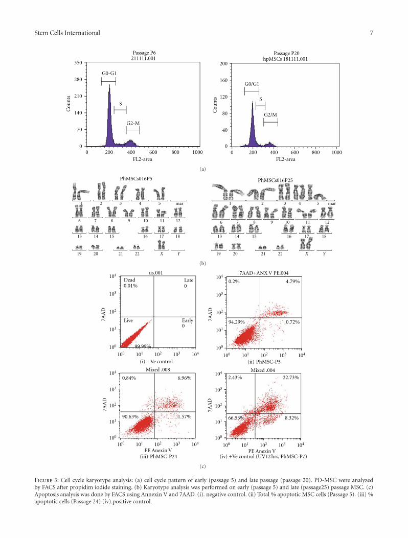
Stem Cells International 7
0
70
140
210
280
350C
oun
ts
0 200 400 600 800 1000FL2-area
211111.001Passage P6
G0-G1
S
G2-M
0
40
80
120
160
200
0 200 400 600 800 1000
Cou
nts
FL2-area
hpMSCs 181111.001Passage P20
G0/G1
S
G2/M
(a)
1 2 3 4 5
6 7 8 9 10 11 12
13 14 15 16 17 18
19 20 21 22 X Y
mar 1 2 3 4 5
6 7 8 9 10 11 12
13 14 15 16 17 18
19 20 21 22 X Y
mar
PhMSCs016P5 PhMSCs016P25
(b)
(i) −Ve control
us.001Dead0.01%
Late0
Live
99.99%
Early0
7AA
D
100
101
102
103
104
100 101 102 103 104
(ii) PhMSC-P5
0.2% 4.79%
0.72%94.29%7AA
D
100
101
102
103
104
100 101 102 103 104
7AAD+ANX V PE.004
(iii) PhMSC-P24
0.84% 6.96%
1.57%90.63%
7AA
D
100
101
102
103
104
100 101 102 103 104
PE Anexin V
Mixed .008
2.43% 22.73%
8.32%66.53%
7AA
D
100
101
102
103
104
100 101 102 103 104
PE Anexin V
Mixed .004
(iv) +Ve control (UV12 hrs, PhMSC-P7)
(c)
Figure 3: Cell cycle karyotype analysis: (a) cell cycle pattern of early (passage 5) and late passage (passage 20). PD-MSC were analyzedby FACS after propidim iodide staining. (b) Karyotype analysis was performed on early (passage 5) and late (passage25) passage MSC. (c)Apoptosis analysis was done by FACS using Annexin V and 7AAD. (i). negative control. (ii) Total % apoptotic MSC cells (Passage 5). (iii) %apoptotic cells (Passage 24) (iv).positive control.

8 Stem Cells International
QPCR analysis
Rel
ativ
e ge
ne
expr
essi
on le
vels
0
1
2
3
4
5
6
7
P < 0.001
P < 0.07
P < 0.01
BM OCT3/4
P OCT3/4
BM SOX2
P SOX2
BM NANOG
P NANOG
Endogenous gene expression between humanBM-MSCs and P-MSCs (N = 3, Passage = 3)
1 2 3 4 5 6
Figure 4: Comparative analysis of pluripotency associated endoge-nous gene expression between human marrow derived MSCand human placenta-derived MSC. Oct4. Sox2, and nanog geneexpression profiles of bone-marrow-derived MSC and PDMSCanalyzed by real-time qPCR analysis; error bars represent SE in fiveseparate experiments.
3.5. In Vitro Tumor-Genesis Detection Assay. Placental MSCswhen subjected to soft agar assay did not yield tumoroidseven after 4 weeks of in vitro culture in soft agar assay(Figure 5). However, HeLa cells began to form aggregateswithin 7 days, and many bigger colonies were formed at theend of day 21 (Figure 5).
4. Discussion
The human embryonic stem cells (ESCs) have the potentialto differentiate into all the three cell lineages [24]. However,some of the practical and ethical concerns render themin usable for day-to-day clinical applications. Nonetheless,extra embryonic tissues can be effectively used to isolatepluripotent stem cells. Placenta is one of the extra embryonicorgans that has rich source of progenitor or stem cells[25]. Placenta has two sides; one is foetal side consisting ofamnion and chorion and other is the maternal side consistingof deciduas [24]. Mesenchymal stem cells (MSCs) isolatedfrom maternal side of human term-placenta represent animportant cell type for stem cell research and clinicaltherapy not only because of their ability to differentiateinto mesodermal lineage cells, such as osteocytes, chon-drocytes, muscle, or endothelial cells [2], but also fortheir remarkable translineage differentiation capabilities likeneuronal cells, retinal cells (ectodermal), and pancreatic betacells (endodermal lineage). In addition, they secrete largeamounts of proangiogenic and antiapoptotic cytokines [26]and possess remarkable immunosuppressive properties [27].MSCs have been derived from many different organs andtissues [28]. Evidence has emerged that different parts ofhuman placenta, umbilical cords and amniotic membrane,
as well as umbilical cord blood harbor MSC [29–32]. Thesetissues are normally discarded after birth, avoiding ethicalconcerns [23] Mechanical, as well as enzymatic, methodsfor MSC isolation from different regions of human placentaof different gestational ages have been reported in literature[29, 33–47]. Knowledge about vitality, average populationdoubling time, stable karyotype, cell cycle and apoptosispattern, phenotype, and expandability of such placenta-derived MSC isolates is a prerequisite for therapeutic appli-cation; however, systematic investigations into reliability ofthis MSC source and phenotypic stability did not get thatmuch attention. Furthermore, reports on placenta-derivedMSCs often lack information about the cell cycle, apopto-sis pattern, progenitor-specific endogenous gene-expressionprofile, and karyotype of the cell isolates. In this paper, wedescribe enzymatic fractionation of term-human placentathat facilitates recovery of oligo-lineage, fibroblast-like cells,which generally are termed as placenta-derived mesenchymalstem cells (PDMSCs) with high fidelity. As demonstratedby cell cycle or apoptosis analysis of cells from early aswell as late passages; with average unaltered populationdoubling time, PD-MSC did not shows significant variationsin either cell cycle or apoptosis pattern. Also, genotypicanalyses of cell isolates from most of placental tissues were ofmaternal, not fetal, origin. Our systematic characterizationof cell isolates from multiple cases showed that these cellisolates reproducibly fulfill the general definition of MSCsby both phenotypic and differentiation capabilities criteria.[24]. We demonstrate that maternally derived PD-MSCscan be greatly expanded, do not alter significantly changetheir cell cycle or apoptosis pattern, show pluripotency-associated endogenous gene expression, and maintain theirdifferentiation capacity and stable phenotype displayingunaltered kayotype up to passage 25–30 passages. In theseexperiments, the placental MSCs were isolated from thecotyledons present in the maternal side of the placenta.Our method of cell isolation by way of sequential digestionof the trophoblast cell layer with trypsin and followingdigestion of remaining placental tissue with collagenase Iproved very effective for obtaining PD MSCs. Outgrowthof PD-MSCs from collagenase I digests was successful in8 of 8 placental tissues and resulted in populations withremarkably little scatter in their MSC profiles, betweensubjects. As for propagation, we found out that PD-MSCsmust be propagated in subconfluent culture to maintain theirMSC profile, because confluent culture led to gradual loss ofMSC identity. With proper subconfluent passage, PDMSCsmaintained their phenotypic MSC profile up to 30 passages.The flow cytometry studies indicate there is significantsimilarity in surface marker characteristics from passage1 till passage 30. Microscopic observations revealed thatplacental MSCs proliferate rapidly till passage 30 withoutcompromising on the morphological features and qualityof the mesenchymal stem cell properties like cell cycle andapoptosis pattern, pluripotency-associated endogenous geneexpression, and normal karyotype.
The characteristic data beyond passage 30 has not beentested in this study. The MSCs had spindle shaped fibroblastmorphology. The absence of HLA DRα and HLA DRβ1

Stem Cells International 9
PhMSCs021P17
Day
7
(a)
HeLa
Day
7
(b)
Day
21
(c)
Day
21
(d)
Figure 5: Soft agar assay. (a)Placental MSCs day 7; (b) HeLa cells day 7; (c) Placental MSCs day 21; (d) HeLa cells ay21.
expression, analyzed from RT-PCR, results indicate thatplacental MSCs could be effectively used for both autologousand allogenic transplantations. The rate of differentiation ofMSCs is much quicker, efficient and scalable when comparedto ES cells. The soft agar assay indicates that isolated placentalMSCs do not possess any malignant property. Several animalas well as human trials have indicated that use of MSCs unlikeES cells does not lead to the formation of teratomas in vivo[24]. In addition, usage of term placental MSCs has fewerethical concerns since they are isolated from foetal tissuesthat anyway would have been discarded.
5. Conclusion
The human term-placenta is relatively easily available andattracts less ethical concerns. Placental tissue constitutesa robust source of MSC. In this study, we investigatedseveral parameters, namely, (1) chromosome number, (2)pluripotency associated gene expression, (3) maternal origin,(4) sequential enzymatic digestion (trypsin followed bycollagenase) as methods of isolation, (5) cell propagation,cell cycle, and apoptosis pattern, that are important for theirprincipal utility for cell-based therapy and could influencetheir proliferative, as well as differentiation, capacities. Basedon the results, we conclude that the abundance of pluripotentcells, rapid proliferation, stable karyotype, plasticity andimmunomodulatory property make placental MSCs idealchoice for clinical and tissue engineering applications. Nev-ertheless, the main drawback of using MSCs is that, a panel ofsurface markers are required for characterization of isolatedMSCs for their homogeneity. Further, unlike the adultMSCs, where significant numbers of human clinical trials are
underway, use of placental MSCs in clinical applications isrelatively new. Additional studies are required to substantiatethe use of placental MSCs in medical applications.
Conflict of Interests
The authors declare no conflict of interests.
Acknowledgments
The authors appreciate encouragement and support extend-ed by all students and staffs of CSCR and CMC in carryingout the research work successfully. they express their grat-itude to Department of Biotechnology for Ramalingaswamifellowship to Sanjay Kumar and research support grant (DBTGrant no. BT/PR15420/MED/31/122/2011), Government ofIndia.
References
[1] P. Bianco, P. G. Robey, and P. J. Simmons, “Mesenchymal stemcells: revisiting history, concepts, and assays,” Cell Stem Cell,vol. 2, no. 4, pp. 313–319, 2008.
[2] S. Kumar, D. Chanda, and S. Ponnazhagan, “Therapeuticpotential of genetically modified mesenchymal stem cells,”Gene Therapy, vol. 15, no. 10, pp. 711–715, 2008.
[3] D. C. Ding, W. C. Shyu, and S. Z. Lin, “Mesenchymal stemcells,” Cell Transplantation, vol. 20, pp. 5–14, 2011.
[4] J. Cohnheim, “Uber entzundung und Eiterung,” VirchowsArchiv fur Pathologische Anatomie und Physiologie und furklinische Medizin, vol. 40, pp. 1–79, 1867.
[5] A. Maximow, “Relation of blood cells to connective tissues andendothelium,” Physiological Reviews, vol. 4, pp. 533–563, 1924.

10 Stem Cells International
[6] A. J. Friedenstein, U. F. Gorskaja, and N. N. Kulagina, “Fibrob-last precursors in normal and irradiated mouse hematopoieticorgans,” Experimental Hematology, vol. 4, no. 5, pp. 267–274,1976.
[7] Y. Miyahara, N. Nagaya, M. Kataoka et al., “Monolayeredmesenchymal stem cells repair scarred myocardium aftermyocardial infarction,” Nature Medicine, vol. 12, no. 4, pp.459–465, 2006.
[8] A. Uccelli, L. Moretta, and V. Pistoia, “Mesenchymal stem cellsin health and disease,” Nature Reviews Immunology, vol. 8, no.9, pp. 726–736, 2008.
[9] B. Parekkadan and J. M. Milwid, “Mesenchymal stem cells astherapeutics,” Annual Review of Biomedical Engineering, vol.12, pp. 87–117, 2010.
[10] R. Hass, C. kasper, S. Bohm, and R. Jacobs, “Differentpopulations and scources of human Mesenchymal stem cells(MSC): a comparison of adult and neonatal tissue-derivedMSC,” Cell Communication and Signaling, vol. 9, p. 12, 2011.
[11] O. V. Semenov, S. Koestenbauer, M. Riegel et al., “Multipo-tent mesenchymal stem cells from human placenta: criticalparameters for isolation and maintenance of stemness afterisolation,” American Journal of Obstetrics and Gynecology, vol.202, no. 2, pp. 193.e1–193.e13, 2010.
[12] S. Kadam, S. Muthyala, P. Nair, and R. Bhonde, “Humanplacenta-derived mesenchymal stem cells and islet-like cellclusters generated from these cells as a novel source for stemcell therapy in diabetes,” Review of Diabetic Studies, vol. 7, no.2, pp. 168–182, 2010.
[13] S. Kumar and S. Ponnazhagan, “Bone homing of mesenchymalstem cells by ectopic α4 integrin expression,” FASEB Journal,vol. 21, no. 14, pp. 3917–3927, 2007.
[14] Y. Zhang, Y. Chu, W. Shen, and Z. Dou, “Effect of 5-azacytidine induction duration on differentiation ofhuman first-trimester fetal mesenchymal stem cells towardscardiomyocyte-like cells,” Interactive Cardiovascular andThoracic Surgery, vol. 9, no. 6, pp. 943–946, 2009.
[15] X.-Y. Hu, W.-X. Wang, M.-J. Yu et al., “Tongxinluo promotesmesenchymal stem cell tube formation in vitro,” Journal ofZhejiang University, vol. 12, no. 8, pp. 644–651, 2011.
[16] D. Woodbury, E. J. Schwarz, D. J. Prockop, and I. B. Black,“Adult rat and human bone marrow stromal cells differentiateinto neurons,” Journal of Neuroscience Research, vol. 61, no. 4,pp. 364–370, 2000.
[17] A. Kicic, W. Y. Shen, A. S. Wilson, I. J. Constable, T. Robertson,and P. E. Rakoczy, “Differentiation of marrow stromal cellsinto photoreceptors in the rat eye,” Journal of Neuroscience, vol.23, no. 21, pp. 7742–7749, 2003.
[18] L. B. Chen, X. B. Jiang, and L. Yang, “Differentiation of ratmarrow mesenchymal stem cells into pancreatic islet beta-cells,” World Journal of Gastroenterology, vol. 10, no. 20, pp.3016–3020, 2004.
[19] S. Susman, O. Soritau, D. Rus-Ciuca, C. Tomuleasa, V. I.Pop, and C. M. Mihu, “Placental stem cell differentiation intoislets of Langerhans-like glucagon-secreting cells,” RomanianJournal of Morphology and Embryology, vol. 51, no. 4, pp. 733–738, 2010.
[20] P. Shetty, K. Cooper, and C. Viswanathan, “Comparisionof proliferative and multilineage differentiation potentials ofcord matix, cord blood and bone marrow Mesenchymal stemcells,” Asian Journal of Transfusion Science, vol. 4, pp. 14–24,2010.
[21] R. Q. Feng, L. Y. Du, and Z. Q. Guo, “In vitro cultivationand differentiation of fetal liver stem cells from mice,” CellResearch, vol. 15, no. 5, pp. 401–405, 2005.
[22] W. Zhao, Z. X. Lin, and Z. Q. Zhang, “Cisplatin-inducedpremature senescence with concomitant reduction of gapjunctions in human fibroblasts,” Cell Research, vol. 14, no. 1,pp. 60–66, 2004.
[23] O. Parolini, F. Alviano, G. P. Bagnara et al., “Concise review:isolation and characterization of cells from human termplacenta: outcome of the first international workshop onplacenta derived stem cells,” Stem Cells, vol. 26, no. 2, pp. 300–311, 2008.
[24] H. Abdulrazzak, D. Moschidou, G. Jones, and P. V. Guillot,“Biological characteristics of stem cells from foetal, cordblood and extraembryonic tissues,” Journal of the Royal SocietyInterface, vol. 7, no. 6, pp. S689–S706, 2010.
[25] O. Parolini, F. Alviano, I. Bergwerf et al., “Toward cell therapyusing placenta-derived cells: disease mechanisms, cell biology,preclinical studies, and regulatory aspects at the round table,”Stem Cells and Development, vol. 19, no. 2, pp. 143–154, 2010.
[26] Y. Miyahara, N. Nagaya, M. Kataoka et al., “Monolayeredmesenchymal stem cells repair scarred myocardium aftermyocardial infarction,” Nature Medicine, vol. 12, no. 4, pp.459–465, 2006.
[27] A. Uccelli, L. Moretta, and V. Pistoia, “Mesenchymal stem cellsin health and disease,” Nature Reviews Immunology, vol. 8, no.9, pp. 726–736, 2008.
[28] G. Chamberlain, J. Fox, B. Ashton, and J. Middleton, “Concisereview: mesenchymal stem cells: their phenotype, differen-tiation capacity, immunological features, and potential forhoming,” Stem Cells, vol. 25, no. 11, pp. 2739–2749, 2007.
[29] C. B. Portmann-Lanz, A. Schoeberlein, A. Huber et al.,“Placental mesenchymal stem cells as potential autologousgraft for pre- and perinatal neuroregeneration,” AmericanJournal of Obstetrics and Gynecology, vol. 194, no. 3, pp. 664–673, 2006.
[30] S. Ilancheran, Y. Moodley, and U. Manuelpillai, “Human fetalmembranes: a source of stem cells for tissue regeneration andrepair?” Placenta, vol. 30, no. 1, pp. 2–10, 2009.
[31] C. H. Jo, O. S. Kim, E. Y. Park et al., “Fetal mesenchymal stemcells derived from human umbilical cord sustain primitivecharacteristics during extensive expansion,” Cell and TissueResearch, vol. 334, no. 3, pp. 423–433, 2008.
[32] K. Bieback, S. Kern, H. Kluter, and H. Eichler, “Criticalparameters for the isolation of mesenchymal stem cells fromumbilical cord blood,” Stem Cells, vol. 22, no. 4, pp. 625–634,2004.
[33] A. J. Marcus and D. Woodbury, “Fetal stem cells from extra-embryonic tissues: do not discard: stem cells review series,”Journal of Cellular and Molecular Medicine, vol. 12, no. 3, pp.730–742, 2008.
[34] V. L. Battula, S. Treml, H. Abele, and H. J. Buhring, “Prospec-tive isolation and characterization of mesenchymal stem cellsfrom human placenta using a frizzled-9-specific monoclonalantibody,” Differentiation, vol. 76, no. 4, pp. 326–336, 2008.
[35] C. M. Chang, C. L. Kao, Y. L. Chang et al., “Placenta-derived multipotent stem cells induced to differentiate intoinsulin-positive cells,” Biochemical and Biophysical ResearchCommunications, vol. 357, no. 2, pp. 414–420, 2007.
[36] C. C. Chien, B. L. Yen, F. K. Lee et al., “In vitro differentiationof human placenta-derived multipotent cells into hepatocyte-like cells,” Stem Cells, vol. 24, no. 7, pp. 1759–1768, 2006.
[37] Y. Fukuchi, H. Nakajima, D. Sugiyama, I. Hirose, T. Kitamura,and K. Tsuji, “Human placenta-derived cells have mesenchy-mal stem/progenitor cell potential,” Stem Cells, vol. 22, no. 5,pp. 649–658, 2004.

Stem Cells International 11
[38] O. Genbacev, A. Krtolica, T. Zdravkovic et al., “Serum-freederivation of human embryonic stem cell lines on humanplacental fibroblast feeders,” Fertility and Sterility, vol. 83, no.5, pp. 1517–1529, 2005.
[39] K. Igura, X. Zhang, K. Takahashi, A. Mitsuru, S. Yamaguchi,and T. A. Takahashi, “Isolation and characterization ofmesenchymal progenitor cells from chorionic villi of humanplacenta,” Cytotherapy, vol. 6, no. 6, pp. 543–553, 2004.
[40] P. S. In’t Anker, S. A. Scherjon, C. Kleijburg-Van Der Keur etal., “Isolation of mesenchymal stem cells of fetal or maternalorigin from human placenta,” Stem Cells, vol. 22, no. 7, pp.1338–1345, 2004.
[41] Z. Miao, J. Jin, L. Chen et al., “Isolation of mesenchymal stemcells from human placenta: comparison with human bonemarrow mesenchymal stem cells,” Cell Biology International,vol. 30, no. 9, pp. 681–687, 2006.
[42] A. Poloni, V. Rosini, E. Mondini et al., “Characterizationand expansion of mesenchymal progenitor cells from first-trimester chorionic villi of human placenta,” Cytotherapy, vol.10, no. 7, pp. 690–697, 2008.
[43] Z. Strakova, M. Livak, M. Krezalek, and I. Ihnatovych,“Multipotent properties of myofibroblast cells derived fromhuman placenta,” Cell and Tissue Research, vol. 332, no. 3, pp.479–488, 2008.
[44] B. L. Yen, H. I. Huang, C. C. Chien et al., “Isolation ofmultipotent cells from human term placenta,” Stem Cells, vol.23, no. 1, pp. 3–9, 2005.
[45] X. Zhang, A. Mitsuru, K. Igura et al., “Mesenchymal pro-genitor cells derived from chorionic villi of +human placentafor cartilage tissue engineering,” Biochemical and BiophysicalResearch Communications, vol. 340, no. 3, pp. 944–952, 2006.
[46] X. Zhang, Y. Soda, K. Takahashi et al., “Successful immortal-ization of mesenchymal progenitor cells derived from humanplacenta and the differentiation abilities of immortalizedcells,” Biochemical and Biophysical Research Communications,vol. 351, no. 4, pp. 853–859, 2006.
[47] S. Barlow, G. Brooke, K. Chatterjee et al., “Comparisonof human placenta- and bone marrow-derived multipotentmesenchymal stem cells,” Stem Cells and Development, vol. 17,no. 6, pp. 1095–1107, 2008.

Hindawi Publishing CorporationStem Cells InternationalVolume 2012, Article ID 564612, 9 pagesdoi:10.1155/2012/564612
Research Article
Generation of Human-Induced Pluripotent Stem Cells bya Nonintegrating RNA Sendai Virus Vector in Feeder-Free orXeno-Free Conditions
Chad C. MacArthur,1 Andrew Fontes,1 Namritha Ravinder,2 David Kuninger,1
Jasmeet Kaur,1 Matthew Bailey,1 Antje Taliana,2 Mohan C. Vemuri,3 and Pauline T. Lieu1
1 Primary and Stem Cell Systems, Life Technologies Corporation, 5781 Van Allen Way, Carlsbad, CA 92008, USA2 Molecular and Cell Biology, Life Technologies Corporation, 5781 Van Allen Way, Carlsbad, CA 92008, USA3 Primary and Stem Cell Systems, Life Technologies Corporation, 7335 Executive Way, Frederick, MD 21704, USA
Correspondence should be addressed to Pauline T. Lieu, [email protected]
Received 15 September 2011; Accepted 4 January 2012
Academic Editor: Paul Verma
Copyright © 2012 Chad C. MacArthur et al. This is an open access article distributed under the Creative Commons AttributionLicense, which permits unrestricted use, distribution, and reproduction in any medium, provided the original work is properlycited.
The generation of induced pluripotent stem cells (iPSCs) from somatic cells has enabled the possibility of providing unprecedentedaccess to patient-specific iPSC cells for drug screening, disease modeling, and cell therapy applications. However, a major obstacleto the use of iPSC for therapeutic applications is the potential of genomic modifications caused by insertion of viral transgenesin the cellular genome. A second concern is that reprogramming often requires the use of animal feeder layers and reagents thatcontain animal origin products, which hinder the generation of clinical-grade iPSCs. Here, we report the generation of iPSCsby an RNA Sendai virus vector that does not integrate into the cells genome, providing transgene-free iPSC line. In addition,reprogramming can be performed in feeder-free condition with StemPro hESC SFM medium and in xeno-free (XF) conditions.Generation of an integrant-free iPSCs generated in xeno-free media should facilitate the safe downstream applications of iPSC-based cell therapies.
1. Introduction
Takahashi and Yamanaka first demonstrated that inducedpluripotent stem cells (iPSCs) can be generated from somaticcells by transducing four transcription factors using retro-viral vectors [1, 2]. This breakthrough in creating inducedpluripotent stem cells opened the door for personalized med-icine using patient-derived iPSCs [3–6]. The major limita-tion for potential clinical application is the integration ofviral transgenes into the host genome that can result inmultiple insertions and risk of tumorigenicity [7, 8]. Anothermajor disadvantage of reprogramming cells with integratingvectors is that silencing and activation of transgenes areunpredictable, which may affect terminal differentiation po-tential and increase the risk of using iPSC-derived cells. Mul-tiple methods have been developed to address these issues,including reprogramming with episomal vectors, mRNAs,miRNAs, protein transduction, or treatment with chemical
compounds [9]. The majority of these methods has one ormore limitations, such as low reprogramming efficiency orrequiring multiple rounds of transfections, or is effective onlywith specific cell types, such as skin fibroblasts. We have pre-viously shown that sustained expression of reprogrammingfactors is required for at least 10–20 days [10], and often thesereprogramming methods fail to sustain expression and aredifficult to repeat. Further, due to poor efficiency of existingmethods, reprogramming often has been performed in thepresence of animal feeders (inactivated mouse embryonicfibroblasts) to maximize colony formation [11] along withthe use of serum and xeno-containing products, which arenot ideal for clinical applications [11, 12].
Here, we investigate the use of Sendai virus vector togenerate transgene-free iPSCs in different conditions. Sendaivirus vector is a negative-strand RNA virus that belongsto the paramyxoviridae family [13–15]. Unlike other RNAviruses, it replicates in the cytoplasm of infected cells and

2 Stem Cells International
does not go through a DNA phase that can integrate into thehost genome [13]. In addition, Sendai virus vector can infecta broad host range and is nonpathogenic to humans. Sendaivirus vector has been considered for clinical studies of genetherapy for cystic fibrosis [16, 17] and vaccine delivery [18].Recent papers also demonstrated that Sendai virus vector canreprogram somatic cells with higher efficiency than otherreprogramming methods [19]. Thus, the nature of Sendaivirus vector makes it an ideal tool for cell reprogrammingand stem cell research.
The current methods of reprogramming often requirethe use of inactivated feeders and animal-derived productsduring several steps to maximize reprogramming efficiency.However, exposure of human cells to animal origin productsmay increase the risk of nonhuman pathogen transmissionand immune rejection [20], hence limiting their use in clin-ical settings. Given the reported high efficiency of Sendaivirus vector in generating pluripotent cells, we investigatedthe generation of transgene-free iPSCs with Sendai virusvector in feeder-free conditions using StemPro hESC SFMmedium and in xeno-free conditions. Several reports havedemonstrated the generation of iPSCs in animal product-freeculture media but their performance tends to be lower thanknockout serum (KO-SR) based media [21]. Here, we reporta robust and an efficient system to generate transgene-freeiPSCs in different conditions, and its ease of use can beapplied to a wide range of different cell types. Generation oftransgene-free iPSCs under these conditions will be impor-tant to facilitate the safe clinical translation of iPSC-basedtherapies.
2. Materials and Methods
2.1. Cell Culture. Human neonatal fibroblasts (ATCC Cat.no. CRL-2252) were maintained in fibroblast growth mediaconsisting of DMEM + GlutaMAX (Cat. no. 10569-010),supplemented with 10% ES cell-qualified fetal bovine serum(Cat. no. 16141-079), and 1% nonessential amino acids(Cat. no. 11140-050). Feeder-free iPSC cultures were main-tained on hESC-qualified GelTrex (Cat. no. A10480-02) withchemically defined StemPro hESC SFM (Cat. no. A1000701)supplemented with 100 μM 2-mercaptoethanol (Cat. no.21985-023) and 10 ng/mL recombinant human basic FGF(Cat. no. PHG0264). For xeno-free culture, human neonatalfibroblasts were maintained in StemPro MSC SFM Xeno-Free medium (Cat. no. A10675-01). Xeno-free iPSC cul-tures were maintained on KnockOut-DMEM/F-12 (Cat. no.12660012) supplemented with 15% KnockOut-SR Xeno-Free CTS (Cat. no. A1099202), 1X KnockOut SR Xeno-Free Growth Factor Cocktail, 2 mM GlutaMAX (Cat. no.35050-061), 1% nonessential amino acids, 100 μM 2-mer-captoethanol, and 20 ng/mL of bFGF. For feeder condition,human iPS cells were maintained on irradiated mouseembryonic fibroblasts (Cat. no. S1520-100) in DMEM/F-12 + GlutaMAX (Cat. no. 10565-018) supplemented with20% KnockOut SR (Cat. no. 10828-028), 1% nonessentialamino acids, 100 μM 2-mercaptoethanol, and 4 ng/mL ofbFGF. iPSCs were passaged manually using a 26-gauge needleor StemPro EZPassage Disposable Stem Cell Passaging Tool
(Cat. no. 23181-010). All reagents are from Life Technologies.Colonies were passaged every 3-4 days at a splitting ratio of1 : 3. The Gibco Episomal hiPSC line was purchased fromLife Technologies (Cat. no. A13777) and maintained onirradiated mouse embryonic fibroblasts in DMEM/F-12 +GlutaMAX supplemented with 20% KnockOut SR, 1%nonessential amino acids, 100 μM 2-mercaptoethanol, and4 ng/mL bFGF.
2.2. Generation of iPSCs in Feeder-Free and Xeno-Free Condi-tions. For the generation of iPSCs in feeder-free condition,approximately 100,000 human neonatal fibroblasts wereseeded per well in a 6-well plate and incubated at 37◦Cand 5% CO2. Two days later, cells were transduced with theCytoTune iPS Reprogramming Kit containing Sendai virusvectors using the F gene-defective vector as described in[19, 22] (Life Technologies Cat. no. A1378001) in fibroblastgrowth medium at an MOI of 3, as described in themanufacturer’s protocols. One day after transduction, themedium was replaced with fresh fibroblast growth mediumand the cells were cultured in fibroblast growth media for 6-7days. On day eight after transduction, cells were transferredto 10 cm Geltrex-coated tissue culture dishes at 5 × 10⁵ cellsper dish. After culturing overnight in fibroblast growth me-dia, the medium was replaced daily with StemPro hESC SFMmedium. The iPSC colonies were picked for expansion andcharacterization from days 17–21 of reprogramming.
For the generation of iPSCs in xeno-free condition, ap-proximately 500,000 human neonatal fibroblasts were seededin StemPro MSC SFM Xeno-Free medium per well in a 6-wellplate and incubated at 37◦C and 5% CO2. The next day, cellswere transduced with the CytoTune iPS Reprogramming Kitin StemPro MSC SFM Xeno-free medium at an MOI of3. One day after transduction, the medium was replacedwith fresh StemPro MSC SFM Xeno-Free medium and cellswere cultured for 6-7 days. On day eight after transduction,cells were transferred to 10 cm tissue culture dishes platedwith inactivated human feeders (NuFF, GlobalStem Cat. no.GSC-3001G) at a density of 2× 10⁵ cells per dish. Afterculturing overnight, the medium was replaced daily withxeno-free iPSC medium. The xeno-free medium is composedof KnockOut D-MEM CTS containing 15% KnockOutSR Xeno-Free CTS, 2 mM GlutaMAX, KnockOut SR GFCocktail CTS (used at 1 : 50), and 8 ng/mL bFGF. Just priorto use, medium was equilibrated at 37◦C and 5% CO2 inhumidified air, 100 μM 2-mercaptoethanol was added to theKnockOut SR Xeno-Free Complete Medium. The iPSC col-onies were picked for expansion and characterization fromdays 21–25 of reprogramming.
2.3. Immunofluorescence and Alkaline Phosphatase Staining.For immunofluorescence staining, cells were fixed in 2% PFAfor 30 minutes at room temperature, blocked for 30 minutesat room temperature in blocking buffer (5% normal goatserum, 1% BSA, and 0.1% Triton X100 in D-PBS), incu-bated with primary antibodies (diluted in blocking buffer)overnight at 4◦C, washed 3X with D-PBS, incubated withsecondary antibodies (diluted in D-PBS) for 30 minutesat room temperature, and washed 3X with D-PBS. For

Stem Cells International 3
live staining, cells were washed 1X with DMEM/F-12 andincubated with primary antibody at 1 : 100 dilution (inDMEM/F-12) for 60 min at 37◦C. Cells were washed 3Xwith DMEM/F12 and incubated with secondary antibody at1 : 500 dilution (in DMEM/F-12) for 60 minutes at 37◦C andwashed 3X with DMEM/F-12. Characterization was carriedout using the following antibodies: Mouse anti-Tra1-60antibody (Cat. no. 41–1000), Mouse anti-Tra1-81 antibody(Cat. no. 41–1100), Mouse anti-SSEA4 (Cat. no. 41–4000),Alexa Fluor 488 goat anti-mouse IgG (H + L) antibody (Cat.no. A11029), Alexa Fluor 594 goat anti-mouse IgG (H + L)antibody (Cat. no. A11032), Alexa Fluor 488 goat anti-rabbitIgG (H + L) antibody (Cat no. A11034), and Alexa Fluor 594goat anti-rabbit IgG (H + L) antibody (Cat no. A11037). Allreagents are from Life Technologies. Clones were screenedfor residual virus using rabbit anti-SeV antibody (MBL Inter-national Corporation, Woburn, MA; Cat. no PD029). Imageswere captured using a Zeiss Axiovision microscope andprocessed using AdobePhotoshop CS. Where applicable, onday 25 of reprogramming, cells were stained using VECTORRed Alkaline Phosphatase Substrate Kit (Vector Labs Cat. no.SK 5100) and positive colonies were counted.
2.4. RNA Isolation, qRT-PCR, and End Point PCR for GeneExpression Analysis. Total RNA was extracted from cellsusing TRIzol LS reagent (Cat. no. 10296-010) with DNaseI(Cat. no. AM2222). RT-PCR reactions were carried out using20x Reverse Transcriptase enzyme mix and 2x RT Bufferprovided in the TaqMan Gene Expression Cells-to-CT Kit(Cat. no. 4399002) with 1 ug of total RNA per reaction. Geneexpression assays were performed according to the man-ufacturer’s protocols. Primers for gene expression can befound at http://www.invitrogen.com/site/us/en/home.html.All reactions were performed in triplicates and normalizedto endogenous B-actin. End point PCR was performed with10 uL of cDNA in 50 uL of AccuPrime Pfx Supermix (Cat. no.12344040) with the following primers to detect for the pres-ence of Sendai virus vector (SeV: Frw: GGATCACTAGGT-GATATCGAGC, Rev: ACCAGACAAGAGTTTAAGAGATAT-GTATC) as described in [19]. The expected size of the PCRproduct is 181 bp. All reagents are from Life Technologies.PCR was performed with the following conditions: denatura-tion 95◦C, 30 sec, annealing: 55◦C, 30 sec, elongation: 72◦C,30 sec, for 30–35 cycles. PCR products were analyzed on 2%agarose gel electrophoresis.
2.5. MicroRNA Profiling. Total RNA was extracted fromcells using TRIzol LS reagent (Cat. no. 10296-010) withDNASE 1 (Cat. no. AM2222). 500 ng of total RNA was sub-jected to cDNA synthesis using TaqMan MicroRNA ReverseTranscription Kit (Cat. no. 4366597) and TaqMan HumanMicroRNA Array Card A (Cat. no. 4398965) to quantifymicroRNA expression levels, according to the manufacturer’sprotocol. All reagents are from Life Technologies. Real-timePCR was performed on an AB 7900HT Sequence DetectionSystem with cycling conditions of 95◦C for 10 min followedby 95◦C for 15 sec and 60◦C for 60 sec for a total of 40 cycles.The Ct values for all miRNAs were normalized to mam-malian U6 snRNA levels by calculating their respective delta
Ct values. Relative changes in miRNA expression levels be-tween samples were compared using the delta Ct values.
2.6. TaqMan Protein Assays and Reagents. All TaqMan Pro-tein Assays and associated reagents kits were obtained fromLife Technologies. Cells were harvested, counted, and 50 Kcells were incubated in 100 uL of resuspension and 100 uL oflysis buffer. TaqMan Protein Assays were carried out with celllysate dilutions of 500 or 250 cells per reaction using AppliedBiosystems StepOnePlus real-time PCR system. In parallel,the same cell inputs were also subjected to 18S genomicribosomal DNA assays to obtain endogenous control Cq,described in [10].
2.7. In Vitro Differentiation of iPSCs. Undifferentiated iPSCswere harvested using collagenase to generate embryoid bod-ies (EBs) and were cultured for 4 days in suspension in differ-entiation medium containing DMEM-F12 with GlutaMAX,20% Knockout Serum Replacement, 1% nonessential aminoacid, and 55 μM 2-mercaptoethanol (Life Technologies). Onday 5, EBs were seeded on Geltrex coated plates for an addi-tional 17 days of differentiation in the same medium, thenthe cells were used for immunocytochemistry. Induction toneural lineages was performed as described in [23].
3. Results and Discussion
3.1. Generation of iPSCs with Sendai Virus Vector in Feeder-Free or Xeno-Free Condition. Generation of human iPSCswith Sendai virus vector encoding OCT3/4, KLF4, SOX2,and cMYC was performed on feeder-free conditions withStemPro hESC SFM and Geltrex coated plates, or xeno-freeconditions on KnockOut-DMEM/F-12 supplemented with15% KnockOut-SR Xeno-Free CTS, and 1X KnockOut SRGF Cocktail CTS on inactivated human feeders. Human ne-onatal foreskin fibroblast cells were plated onto a 6-well platetwo days before transduction, and approximately 5× 10⁵
cells were transduced with OCT3/4, KLF4, SOX2, and cMYCSendai virus vector at an MOI of 3. About 6-7 days aftertransduction, cells were collected and transferred to Geltrex-coated matrix plate (feeder-free condition) or onto inac-tivated human feeders (xeno-free conditions) at the densityof 500,000 cells per 10 cm dish for feeder-free condition and200,000 cells per 10 cm dish for xeno-free conditions. Theworkflow is outlined in Figure 1, and viable colonies werevisible as early as day 10 after transduction, and these col-onies could be readily picked by 21–25 days.
A majority of colonies generated with Sendai virus vectorhave ES-like morphology, and very few transformed colonieswere observed. Clones from human fibroblasts were stainedwith pluripotent markers, SSEA4, Sox2, Nanog, and Tra1-81 or Tra1-60 shown on Figure 2(A) (feeder-free condition)and Figure 3(A) (xeno-free condition). Alkaline phosphatasestaining demonstrated efficiency on feeder-free condition tobe between 0.01 and 0.04%, and very few transformed ornon-iPSC colonies were detected, shown on Figure 2(C).In addition, these cells were able to give rise to embryoidbodies (EBs) that could differentiate to all the three lineages:ectoderm, endoderm, and mesoderm (see Figure 2(B) for
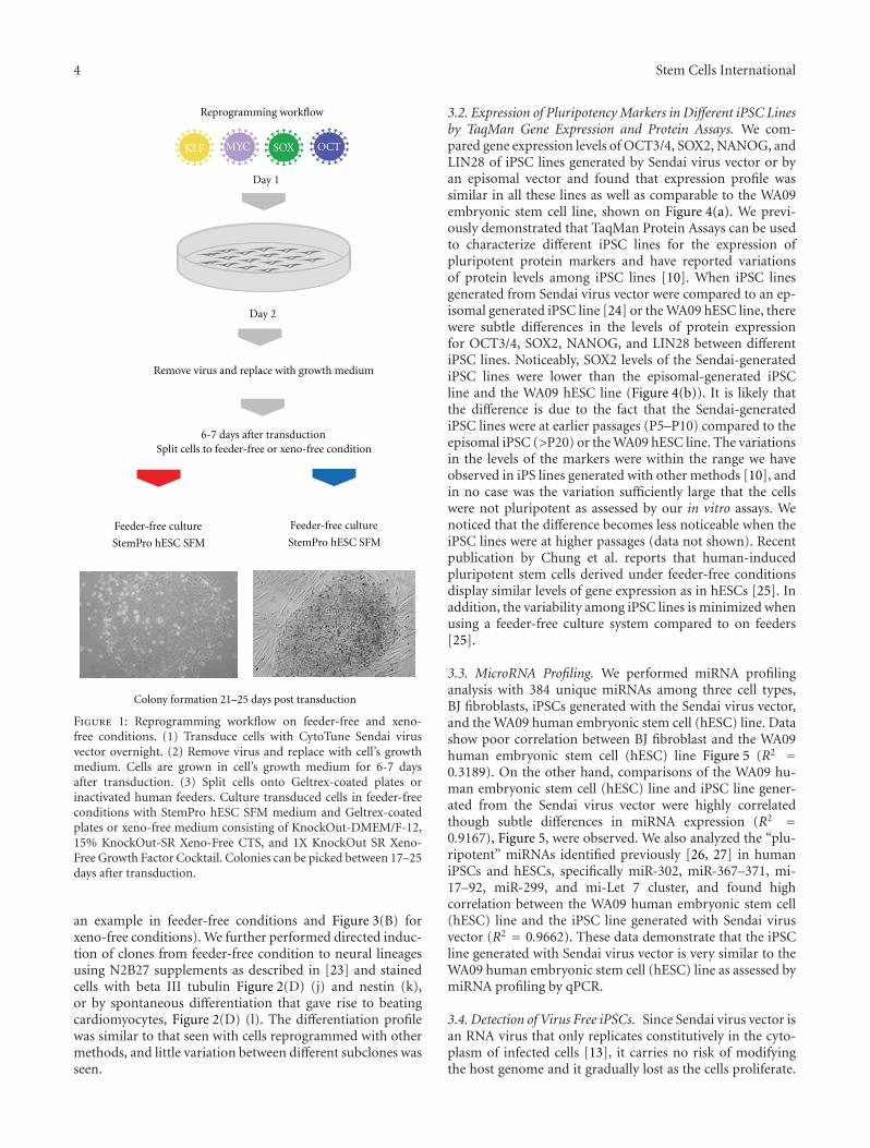
4 Stem Cells International
Reprogramming workflow
OCTMYC SOXKLF
Day 1
Day 2
Split cells to feeder-free or xeno-free condition
Feeder-free culture
StemPro hESC SFM
Feeder-free culture
StemPro hESC SFM
Colony formation 21–25 days post transduction
6-7 days after transduction
Remove virus and replace with growth medium
Figure 1: Reprogramming workflow on feeder-free and xeno-free conditions. (1) Transduce cells with CytoTune Sendai virusvector overnight. (2) Remove virus and replace with cell’s growthmedium. Cells are grown in cell’s growth medium for 6-7 daysafter transduction. (3) Split cells onto Geltrex-coated plates orinactivated human feeders. Culture transduced cells in feeder-freeconditions with StemPro hESC SFM medium and Geltrex-coatedplates or xeno-free medium consisting of KnockOut-DMEM/F-12,15% KnockOut-SR Xeno-Free CTS, and 1X KnockOut SR Xeno-Free Growth Factor Cocktail. Colonies can be picked between 17–25days after transduction.
an example in feeder-free conditions and Figure 3(B) forxeno-free conditions). We further performed directed induc-tion of clones from feeder-free condition to neural lineagesusing N2B27 supplements as described in [23] and stainedcells with beta III tubulin Figure 2(D) (j) and nestin (k),or by spontaneous differentiation that gave rise to beatingcardiomyocytes, Figure 2(D) (l). The differentiation profilewas similar to that seen with cells reprogrammed with othermethods, and little variation between different subclones wasseen.
3.2. Expression of Pluripotency Markers in Different iPSC Linesby TaqMan Gene Expression and Protein Assays. We com-pared gene expression levels of OCT3/4, SOX2, NANOG, andLIN28 of iPSC lines generated by Sendai virus vector or byan episomal vector and found that expression profile wassimilar in all these lines as well as comparable to the WA09embryonic stem cell line, shown on Figure 4(a). We previ-ously demonstrated that TaqMan Protein Assays can be usedto characterize different iPSC lines for the expression ofpluripotent protein markers and have reported variationsof protein levels among iPSC lines [10]. When iPSC linesgenerated from Sendai virus vector were compared to an ep-isomal generated iPSC line [24] or the WA09 hESC line, therewere subtle differences in the levels of protein expressionfor OCT3/4, SOX2, NANOG, and LIN28 between differentiPSC lines. Noticeably, SOX2 levels of the Sendai-generatediPSC lines were lower than the episomal-generated iPSCline and the WA09 hESC line (Figure 4(b)). It is likely thatthe difference is due to the fact that the Sendai-generatediPSC lines were at earlier passages (P5–P10) compared to theepisomal iPSC (>P20) or the WA09 hESC line. The variationsin the levels of the markers were within the range we haveobserved in iPS lines generated with other methods [10], andin no case was the variation sufficiently large that the cellswere not pluripotent as assessed by our in vitro assays. Wenoticed that the difference becomes less noticeable when theiPSC lines were at higher passages (data not shown). Recentpublication by Chung et al. reports that human-inducedpluripotent stem cells derived under feeder-free conditionsdisplay similar levels of gene expression as in hESCs [25]. Inaddition, the variability among iPSC lines is minimized whenusing a feeder-free culture system compared to on feeders[25].
3.3. MicroRNA Profiling. We performed miRNA profilinganalysis with 384 unique miRNAs among three cell types,BJ fibroblasts, iPSCs generated with the Sendai virus vector,and the WA09 human embryonic stem cell (hESC) line. Datashow poor correlation between BJ fibroblast and the WA09human embryonic stem cell (hESC) line Figure 5 (R2 =0.3189). On the other hand, comparisons of the WA09 hu-man embryonic stem cell (hESC) line and iPSC line gener-ated from the Sendai virus vector were highly correlatedthough subtle differences in miRNA expression (R2 =0.9167), Figure 5, were observed. We also analyzed the “plu-ripotent” miRNAs identified previously [26, 27] in humaniPSCs and hESCs, specifically miR-302, miR-367–371, mi-17–92, miR-299, and mi-Let 7 cluster, and found highcorrelation between the WA09 human embryonic stem cell(hESC) line and the iPSC line generated with Sendai virusvector (R2 = 0.9662). These data demonstrate that the iPSCline generated with Sendai virus vector is very similar to theWA09 human embryonic stem cell (hESC) line as assessed bymiRNA profiling by qPCR.
3.4. Detection of Virus Free iPSCs. Since Sendai virus vector isan RNA virus that only replicates constitutively in the cyto-plasm of infected cells [13], it carries no risk of modifyingthe host genome and it gradually lost as the cells proliferate.

Stem Cells International 5
10x
10x
(a) (b) (c)
(d) (e) (f)
(A)
(B)
(C)
(D)
(i)(h)
AFP SMA Beta 3
(j)(k) (l)
Nestin Beating cardiomyocytesBeta III tubulin
Alkaline phosphatase staining on feeder-free culture
Figure 2: (A) Characterization and differentiation of iPSC colonies generated on feeder-free conditions. Colonies were stained forpluripotent markers: SSEA4 (b), SOX2 (c), Tra1-81 (e), NANOG (f), and phase ((a) and (d)). (B) These colonies were differentiated in vitrointo embryoid bodies (EBs) comprising of the three embryonic germ layers: endoderm marker α fetoprotein (AFP) (g), mesodermal markersmooth muscle actin (SMA) (h), and ectoderm marker Beta 3-tubulin (Beta III) (i). (C) Colonies were stained with alkaline phosphatasestaining 25 days after transduction. (D) Colonies were also subjected to directed differentiation to neural stem cells, stained with Beta 3-tubulin (Beta III) (j), Nestin (k), and random differentiation to beating cardiomyocytes (l).

6 Stem Cells International
10x
10x
(a) (b) (c)
(d) (e) (f)
(g) (h) (i)
(A)
(B)
Figure 3: (A) Characterization and differentiation of iPSC colonies generated on xeno-free conditions. Colonies were stained for pluripotentmarkers: NANOG (b), SSEA4 (c), SOX2 (e), Tra1-60 (f), and phase ((a) and (d)). (B) These colonies were differentiated in vitro intoembryoid bodies (EBs) comprising of the three embryonic germ layers: endoderm marker α fetoprotein (AFP) (g), mesodermal markersmooth muscle actin (SMA) (h), and ectoderm marker Beta 3-tubulin (Beta III) (i).
We used two different methods to detect loss of Sendai virusvector in cells. Using RT-PCR with Sendai specific primers,we can detect transgene-RNAs as early as 24 hours aftertransduction, Figure 6(a). The rate at which the transgenesdisappeared varies between each clone, and transgene-freeiPSC colonies were obtained by passage 10 using end pointPCR method, Figure 6(b). The second method used to detectthe presence of Sendai virus vector is by immunochemistrystaining with anti-Sendai antibody. As early as passage 5, wecan detect some colonies staining negative for Sendai virusvector, shown on Figure 7. Sendai virus vectors tend to diluteout and disappear as the clones are being carried out tofurther passages. By passage 10, residual Sendai transcriptswere completely eliminated in three out of four clones tested.In addition, the Sendai backbone vector carries temperature-sensitive mutations that enable complete removal of theresidual vector by shifting to nonpermissive temperature of38-39◦C [22]. Thus, removal of Sendai virus vector is lesslabor intensive compared to alternative methods that require
subsequent excision of transgenes from the host genome[6, 28].
4. Conclusions
Sendai virus vector has been used as vectors for gene therapysince it has no risk of being integrated into the host genomeand is nonpathogenic to humans. In addition, Sendai virusvector has been shown to generate transgene-free humaniPSCs with high efficiency from fibroblasts and CD34+ cordblood cells [22]. In the present study, we demonstrated thatthe Sendai virus vector can be used to generate transgene-free human iPSCs in feeder-free and xeno-free conditions.Comparing the feeder-based and feeder-free systems, wenoticed a decrease in reprogramming efficiency in feeder-free conditions, independent of which media we used. SinceSendai virus vector can reprogram with high efficiency, weare able to obtain sufficient number of colonies for furtherexpansion in either condition. Another advantage of using

Stem Cells International 7
Gene expression profile
OCT SOX LIN NANOG
Rel
ativ
e ex
pres
sion
FibroblastsESC-H9Epi-line1
Sendai-1Sendai-3Sendai-6
(a)
FibroblastsESC-H9Epi-line1
Sendai-1Sendai-3Sendai-6
Rel
ativ
e ex
pres
sion
TaqMan protein assays
OCT SOX LIN NANOG
(b)
Figure 4: (a) TaqMan Gene Expression Assays of OCT4/POU5F1, SOX2, LIN28, and NANOG were compared among the WA09 hESC line,an episomally derived iPSC line (Epi-line 1), and three Sendai-derived iPSC lines (Sendai 1, 3, and 6). Samples were normalized against theendogenous reference B-actin. (b) TaqMan Protein Assays of OCT4/POU5F1, SOX2, LIN28, and NANOG are compared among the WA09hESC line, an episomally derived iPSC line (Epi-line 1), and three Sendai-derived iPSC lines (Sendai 1, 3, and 6). Samples were normalizedagainst the endogenous reference 18S ribosomal genomic DNA and plotted relative fold change to the expression level of untransducedhuman dermal fibroblasts.
BJ
fibr
obla
sts
WA09 ESC
R2 = 0.3189
(a)
R2 = 0.9167
WA09 ESC
Sen
-iP
SC
(b)
R2 = 0.0167
BJ
fibr
obla
sts
WA09 ESC
“Pluripotent miRNAs”
(c)
“Pluripotent miRNAs”
R2 = 0.9662
WA09 ESC
Sen
-iP
SC
(d)
Figure 5: ((a) and (b)) Scatter plots comparing microRNA expression profiles using the TaqMan Human MicroRNA Array of BJ fibroblasts,iPSCs generated with the Sendai virus vector (Sen-iPSC), and the WA09 human embryonic stem cell (hESC) line. ((c) and (d)) Scatter plotscomparing “pluripotent” microRNA expression profiles.
Sendai virus vector is the ease of use with single transduc-tion comparing to other methods that require multiple trans-fections [29]. Finally, iPSCs depleted of virus from the cyto-plasm can be easily obtained without further manipulation.Thus, the high efficiency and ease of use of the Sendai virus
vector is ideal for reprogramming with different cell types onfeeder-based or feeder-free conditions.
Gene expression of pluripotent markers and miRNA pro-filing data show that iPSCs derived from Sendai virus vectoron feeder-free condition are similar to hESC line (the WA09

8 Stem Cells International
OCT SOX
Rel
ativ
e ex
pres
sion
Fibroblasts
KLF4 cMYC
Gene expression 24 h post transduction
24 h Sendai
(a)
Sendai-specific
Sen
dai-
24 h
Passage 10
Sen
d-iP
SC #
1
Sen
d-iP
SC #
2
Sen
d-iP
SC #
3
B-actin
(b)
Figure 6: (a) Detection of viral gene expression at 24 hours after transduction with Sendai virus vector by TaqMan Gene Expression Assays.(b) Detection of virus-free iPSCs after 10 passages in culture by end-point PCR.
DA
PI
Mer
ge
Positive Negative
An
ti-S
enda
ivi
rus
Passage 5
Figure 7: Immunofluorescence staining with anti-Sendai virusvector antibodies to detect virus-free iPSCs. iPSC clones werestained at passage 5.
embryonic stem cell line) and to another episomally derivediPSC line. Our data is consistent with the recent publicationby Chung et al., which reports that feeder-free iPSCs havesimilar transcriptomic profiles and differentiation capacityto hESCs [25]. In addition, increasing evidence demonstratesthe importance of mircoRNAs (miRNAs) for human embry-onic stem cells self-renewal, pluripotency, and differentiation[27]. Previous publications reported a signature group ofmiRNAs that is upregulated in both iPSCs and hESCs, suchas the miR-17–92 clusters, miR-290 clusters, miR-302, 367cluster, miR-371/372/373, and let-7 family [26]. We furthercompared these “pluripotent” miRNAs and found high
correlation between the human iPSCs generated by Sendaivirus vector and the WA09 human embryonic stem cell(hESC) line. These data suggest that the nature of iPSCs gen-erated with Sendai virus vector is similar to that of hESC line.
In summary, reports have shown that the use of integrat-ing viruses to generate iPSCs is not ideal due to the potentialof tumorigenicity and the exposure of iPSCs in animalcontaining products impedes downstream applications. Itis important to develop a system that can generate iPSCsthat are free of transgenes and in conditions that are free ofanimal-derived products. Exposure of human cells to animalorigin products may increase immune rejection of graftedcells and make them unsuitable for the generation of clinical-grade iPSCs [11]. Here we have demonstrated that iPSCs canbe generated in feeder-free or xeno-free conditions. Further,the iPSCs generated here are free of transgenes and behavesimilar to those generated on feeders or animal-derivedproducts. The generation of a footprint-free iPSCs underthese conditions should facilitate the safe clinical translationof iPSC-based therapies.
Acknowledgments
This work was supported by Life Technologies Corporation.The authors thank Dr. Ying Liu and Dr. Mahendra Rao forcritical reading of the paper.
References
[1] K. Takahashi and S. Yamanaka, “Induction of pluripotent stemcells from mouse embryonic and adult fibroblast cultures bydefined factors,” Cell, vol. 126, no. 4, pp. 663–676, 2006.
[2] K. Takahashi, K. Tanabe, M. Ohnuki et al., “Induction ofpluripotent stem cells from adult human fibroblasts by definedfactors,” Cell, vol. 131, no. 5, pp. 861–872, 2007.
[3] I. H. Park, P. H. Lerou, R. Zhao, H. Huo, and G. Q. Daley,“Generation of human-induced pluripotent stem cells,” Na-ture Protocols, vol. 3, no. 7, pp. 1180–1186, 2008.
[4] J. T. Dimos, K. T. Rodolfa, K. K. Niakan et al., “Inducedpluripotent stem cells generated from patients with ALS can

Stem Cells International 9
be differentiated into motor neurons,” Science, vol. 321, no.5893, pp. 1218–1221, 2008.
[5] A. D. Ebert, J. Yu, F. F. Rose et al., “Induced pluripotent stemcells from a spinal muscular atrophy patient,” Nature, vol. 457,no. 7227, pp. 277–280, 2009.
[6] F. Soldner, D. Hockemeyer, C. Beard et al., “Parkinson’s diseasepatient-derived induced pluripotent stem cells free of viralreprogramming factors,” Cell, vol. 136, no. 5, pp. 964–977,2009.
[7] K. Okita, T. Ichisaka, and S. Yamanaka, “Generation of germ-line-competent induced pluripotent stem cells,” Nature, vol.448, no. 7151, pp. 313–317, 2007.
[8] K. Okita, Y. Matsumura, Y. Sato et al., “A more efficientmethod to generate integration-free human iPS cells,” NatureMethods, vol. 8, no. 5, pp. 409–412, 2011.
[9] L. U. W. Muller, G. Q. Daley, and D. A. Williams, “Uppingthe ante: recent advances in direct reprogramming,” MolecularTherapy, vol. 17, no. 6, pp. 947–953, 2009.
[10] D. Ruff, C. Macarthur, H. Tran, J. Bergseid, J. Tian et al.,“Applications of quantitative polymerase chain reaction pro-tein assays during reprogramming,” Stem Cells and Develop-ment, vol. 21, no. 4, pp. 530–538, 2012.
[11] L. Ahrlund-Richter, M. De Luca, D. R. Marshak, M. Munsie, A.Veiga, and M. Rao, “Isolation and production of cells suitablefor human therapy: challenges ahead,” Cell Stem Cell, vol. 4,no. 1, pp. 20–26, 2009.
[12] P. A. De Sousa, G. Galea, and M. Turner, “The road to pro-viding human embryo stem cells for therapeutic use: the UKexperience,” Reproduction, vol. 132, no. 5, pp. 681–689, 2006.
[13] H. O. Li, Y. F. Zhu, A. Makoto et al., “A cytoplasmic RNA vectorderived from nontransmissible Sendai virus with efficient genetransfer and expression,” Journal of Virology, vol. 74, no. 14, pp.6564–6569, 2000.
[14] Y. Ikeda, Y. Yonemitsu, T. Sakamoto et al., “RecombinantSendai virus-mediated gene transfer into adult rat retinal tis-sue: efficient gene transfer by brief exposure,” ExperimentalEye Research, vol. 75, no. 1, pp. 39–48, 2002.
[15] I. Masaki, Y. Yonemitsu, K. Komori et al., “RecombinantSendai virus-mediated gene transfer to vasculature: a new classof efficient gene transfer vector to the vascular system,” TheFASEB Journal, vol. 15, no. 7, pp. 1294–1296, 2001.
[16] Y. Yonemitsu, C. Kitson, S. Ferrari et al., “Efficient gene trans-fer to airway epithelium using recombinant Sendai virus,” Na-ture Biotechnology, vol. 18, no. 9, pp. 970–973, 2000.
[17] S. Ferrari, U. Griesenbach, A. Iida et al., “Sendai virus-medi-ated CFTR gene transfer to the airway epithelium,” Gene Ther-apy, vol. 14, no. 19, pp. 1371–1379, 2007.
[18] A. Takeda, H. Igarashi, M. Kawada et al., “Evaluation of theimmunogenicity of replication-competent V-knocked-outand replication-defective F-deleted Sendai virus vector-basedvaccines in macaques,” Vaccine, vol. 26, no. 52, pp. 6839–6843,2008.
[19] N. Fusaki, H. Ban, A. Nishiyama, K. Saeki, and M. Hasegawa,“Efficient induction of transgene-free human pluripotent stemcells using a vector based on Sendai virus, an RNA virus thatdoes not integrate into the host genome,” Proceedings of theJapan Academy Series B, vol. 85, no. 8, pp. 348–362, 2009.
[20] M. J. Martin, A. Muotri, F. Gage, and A. Varki, “Human em-bryonic stem cells express an immunogenic nonhuman sialicacid,” Nature Medicine, vol. 11, no. 2, pp. 228–232, 2005.
[21] K. Rajala, H. Hakala, S. Panula et al., “Testing of nine differentxeno-free culture media for human embryonic stem cell cul-tures,” Human Reproduction, vol. 22, no. 5, pp. 1231–1238,2007.
[22] H. Ban, N. Nishishita, N. Fusaki et al., “Efficient generation oftransgene-free human induced pluripotent stem cells (iPSCs)by temperature-sensitive Sendai virus vectors,” Proceedingsof the National Academy of Sciences of the United States ofAmerica, vol. 108, no. 34, pp. 14234–14239, 2011.
[23] Y. Liu, B. Thyagarajan, U. Lakshmipathy et al., “Generation ofplatform human embryonic stem cell lines that allow efficienttargeting at a predetermined genomic location,” Stem Cells andDevelopment, vol. 18, no. 10, pp. 1459–1472, 2009.
[24] P. W. Burridge, S. Thompson, M. A. Millrod et al., “A universalsystem for highly efficient cardiac differentiation of humaninduced pluripotent stem cells that eliminates interline vari-ability,” PLoS ONE, vol. 6, no. 4, Article ID 18293, 2011.
[25] H. C. Chung, R. C. Lin, G. J. Logan, I. E. Alexander, P. S.Sachdev et al., “Human induced pluripotent stem cells derivedunder feeder-free conditions display unique cell cycle andDNA replication gene profiles,” Stem Cells and Development,vol. 21, no. 2, 2012.
[26] U. Lakshmipathy, J. Davila, and R. P. Hart, “MiRNA inpluripotent stem cells,” Regenerative Medicine, vol. 5, no. 4, pp.545–555, 2010.
[27] C. -S. Yang, Z. Li, and T. M. Rana, “microRNAs modulate iPScell generation,” RNA, vol. 17, no. 8, pp. 1451–1460, 2011.
[28] K. Woltjen, I. P. Michael, P. Mohseni et al., “PiggyBac trans-position reprograms fibroblasts to induced pluripotent stemcells,” Nature, vol. 458, no. 7239, pp. 766–770, 2009.
[29] L. Warren, P. D. Manos, T. Ahfeldt et al., “Highly efficientreprogramming to pluripotency and directed differentiationof human cells with synthetic modified mRNA,” Cell Stem Cell,vol. 7, no. 5, pp. 618–630, 2010.

Hindawi Publishing CorporationStem Cells InternationalVolume 2012, Article ID 429160, 9 pagesdoi:10.1155/2012/429160
Research Article
Induction of Pluripotency in Adult EquineFibroblasts without c-MYC
Khodadad Khodadadi,1 Huseyin Sumer,1 Maryam Pashaiasl,1, 2 Susan Lim,3
Mark Williamson,4 and Paul J. Verma1, 5
1 Centre for Reproduction and Development, Monash Institute of Medical Research, Monash University, Clayton, VIC 3800, Australia2 Faculty of Medicine, Tabriz University of Medical Sciences, Tabriz 51666-14766, Iran3 Stem Cell Technologies i (SCTi), Gleneagles Medical Centre, Singapore 2584994 Gribbles Veterinary, Clayton, VIC 3168, Australia5 South Australian Research Institute (SARDI), Turretfield Research Centre, Rosedale, SA 5350, Australia
Correspondence should be addressed to Huseyin Sumer, [email protected]
Received 9 November 2011; Revised 28 December 2011; Accepted 3 January 2012
Academic Editor: Rajarshi Pal
Copyright © 2012 Khodadad Khodadadi et al. This is an open access article distributed under the Creative Commons AttributionLicense, which permits unrestricted use, distribution, and reproduction in any medium, provided the original work is properlycited.
Despite tremendous efforts on isolation of pluripotent equine embryonic stem (ES) cells, to date there are few reports aboutsuccessful isolation of ESCs and no report of in vivo differentiation of this important companion species. We report the inductionof pluripotency in adult equine fibroblasts via retroviral transduction with three transcription factors using OCT4, SOX2, andKLF4 in the absence of c-MYC. The cell lines were maintained beyond 27 passages (more than 11 months) and characterized.The equine iPS (EiPS) cells stained positive for alkaline phosphatase by histochemical staining and expressed OCT4, NANOG,SSEA1, and SSEA4. Gene expression analysis of the cells showed the expression of OCT4, SOX2 NANOG, and STAT3. The celllines retained a euploid chromosome count of 64 after long-term culture cryopreservation. The EiPS demonstrated differentiationcapacity for the three embryonic germ layers both in vitro by embryoid bodies (EBs) formation and in vivo by teratoma formation.In conclusion, we report the derivation of iPS cells from equine adult fibroblasts and long-term maintenance using either of thethree reprogramming factors.
1. Introduction
Cartilage and tendon injuries are common features of tissuedamage in both humans and horses. These two tissues have apoor vascular system with low mitotic ability and therefore alimited ability for self-repair. The reduced performance andreinjury create considerable attention for treatments [1].
Adult mesenchymal stem cells (MSCs), embryonic stemcells (ESCs), and reprogrammed somatic cells such as in-duced pluripotent stem (iPS) cells can provide potentialsources of cells for treatment of cartilage and tendon injuries.
MSCs can be isolated from different sources such as bonemarrow aspirates [2], umbilical cord [3], and adipose tissue[4] and have the ability to differentiate into different celltypes such as muscle, cartilage, and bone [5–7]. MSCs havebeen used for treatment of cartilage injuries in equines andhumans. Although there were the early improvements in
cartilage injuries, no significant or long-term recovery couldbe observed [8, 9]. In addition, MSCs are limited in bonemarrow aspirates and need to be cultured after isolationfor at least 4 weeks and have limited in vitro differentiationpotential compared with ESCs [1, 10].
ES cells can overcome this limitation, as they can pro-vide an inexhaustible supply of cell derivatives of all threegerm layers. Despite tremendous efforts on isolation of ESCs,to date there are a few reports on isolation of equine ESCs,which had limited success and no investigation of in vivo dif-ferentiation of the isolated cells [11, 12]. Isolation of equineESCs is difficult due to the shortage of oocytes and embryos,as well as complexity associated with oocyte collection,maturation, IVF, and in vitro culture in this species [13]. Evenif ES cells can be successfully derived, a subsequent problemis the anticipated immune rejection of the derivatives of

2 Stem Cells International
ES cells by the recipient due to incompatibility of the ma-jor histocompatibility complex (MHC) antigens because ofdifferences in genomic DNA compared with that of therecipient [14].
There are alternative methods to produce autologouscells lines via reprogramming of adult somatic cells to thepluripotent states such as somatic cell nuclear transfer(SCNT) [15, 16] and induced pluripotent stem (iPS) cells[17]; however, limitation with derivation of equine ESCsextend to SCNET-ESC isolation as well.
Takahashi and Yamanaka [17] reported the generationof pluripotent cells from adult mouse fibroblast followingretroviral-mediated transduction of four transcription fac-tors, OCT4, SOX2, c-MYC, and KLF4. A number of stud-ies have shown that iPS cells are similar to ESCs in mor-phology and epigenetic status, expression of pluripotentmarkers, and ability to differentiate into derivatives of allthree embryonic germ layers both in vivo and in vitro andcontribute to the germ-line in chimeric mice confirmingtheir true pluripotency [17–19]. Therefore, these cells couldhave therapeutic application in both human and animals.
Pluripotency has been induced in somatic cells fromhuman [20], primate [21], rat [22, 23] pigs [24–27], sheep[21], and cattle [28].
More recently, the generation of equine iPS cell linesfrom fetal fibroblasts using transposon-based delivery of fourfactors has been reported [29]. In this study, we reportthe generations of equine-induced pluripotent stem (EiPS)cells by retroviral-mediated transduction of adult equinefibroblasts using three transcription factors: OCT4, SOX2,and KLF4, (OSK) without the protooncogene c-MYC, andthe pluripotent characteristics of the resulting EiPS cells havebeen demonstrated both in vitro and in vivo.
2. Materials and Methods
Experimental procedures were carried out under the guide-lines of the Monash University, Animal Ethics Committee,and conducted according to the International guidelines forBiomedical Research Involving Animals. All chemicals weresourced from Sigma (Castle Hill, Australia) unless otherwisestated.
2.1. Generation of Induced Pluripotent Stem (iPS) Cells from
Adult Equine Fibroblasts
2.1.1. Transfection, Isolation, and Culture of iPS Cells. EquineiPS cells were generated as previously reported [28]. Briefly,for VSVG pseudotyped retroviral production 3 × 10⁶ GP2293 cells (Clontech; Scientifix, Cheltenham, Australia) wereseeded in a 100 mm culture dish one day before transfectionand incubated overnight at 37◦C, 5% CO2. pMX-basedretrovirus vectors encoding human DNA sequence of OCT4,SOX2, and KLF4 were transfected into packaging cells (GP2293) by FuGENE 6 transfection reagent (Roche, Castel Hill,Australia), and the media were replaced by fresh media onthe following day. Viral supernatant was collected 48 and 72hours later and filtered through a 0.45 μm cellulose acetate
filter. Viral supernatants were then mixed with polybrene to afinal concentration of 8 ng/mL. Adult equine fibroblasts wereplated one day prior to transduction at a density of 1 × 10⁵
cells per 100 mm dish. The cells were incubated overnightwith the viral supernatant including equal contributionsof the factors and 8 ng/mL polybrene. The following day,transduction process was performed similar to the first day.A pMX-GFP and no-vector dishes were provided as a positiveand negative control, respectively. Transduced cells were thencultured in conventional medium containing α-minimumessential medium (α-MEM) with deoxyribonucleosides andribonucleoside (Invitrogen, Mulgrave, Australia), supple-mented with 2 mmol/mL glutamax (Gibco, Invitrogen, Mul-grave, Australia), 0.1% (v/v) Mercaptoethanol (Gibco), 1%(v/v) nonessential amino acid (NEAA) (Gibco), 1% (v/v)ITS (10 μg/mL insulin, 5.5 μg/mL 125 transferrin, 6.7 ng/mLselenium; Gibco), 5 ng/mL human LIF (Millipore, NorthRyde, Australia), 10 ng/mL βFGF (Millipore), 10 ng/mL EGF(Invitrogen), 0.5% (v/v) penicillin-streptomycin (Gibco),and 20% (v/v) FBS. The medium was changed every otherday to maintain cell proliferation. After 12 to 16 days ofiPS induction, the best colonies based on equine ES cell-like colony’s morphology were picked and manually passagedonto mouse embryonic fibroblasts (MEFs) inactivated with4 μg/mL of mitomycin C and plated in an organ culture dish.Colonies were manually cut into small clumps by insulinsyringe needles and expanded on the freshly inactivatedfeeder layers to maintain the EiPS cell line. Seven cell lineswere initially produced and maintained in culture, and onecell line was characterised in detail. The transduction effi-ciency of adult equine fibroblast was evaluated by expressionof the pMX-GFP vector control, which was conducted inparallel with the iPS induction experiments. Seventy-twohours after pMX-GFP induction, cells were photographedunder a fluorescence microscope, and the percentage ofcells expressing GFP was quantified by flow cytometry.Reprogramming efficiency evaluated by correlation of pMX-GFP transduction efficiency with iPS cell colony numberswas established [17].
2.1.2. FACS Analysis. Cells were incubated in incubator(37◦C, 5% Co2) using 0.25% trypsin-EDTA (Invitrogen) forfive min and dissociated through pipetting. After spinningat 400 g for 3 min, the pellet was resuspended and filteredthrough a 40 μm cell strainer (BD Falcon) and analyzed bya BD FACSCanto Flow Cytometer (BD).
2.2. Characterization of Equine iPS Cell Lines
2.2.1. Alkaline Phosphatase and Immunofluorescence Staining.Cells were fixed for 15 min in 4% (w/v) paraformalde-hyde at room temperature and then stained. For alkalinephosphatase (ALP) activity, the cells were stained by his-tochemistry according to manufacturer’s instructions usingAlkaline Phosphatase Detection kit (Millipore). For OCT4and NANOG staining, the cells were permeabilized in 0.2%Triton X-100 in 3%(v/v) goat serum in DPBS for 15 min. Thecells were incubated with 3%(v/v) goat serum in DPBS at RT

Stem Cells International 3
Table 1: List of primers used for RT-PCR.
Markers Primer F Primer R References
GAPDH GATTCCACCCATGGCAAGTTCCATGGCAC GCATCGAAGGTGGAAGAGTGGGTGTCACT
OCT4 TCTTTCCACCAGGCCCCCGGCTC TGCGGGCGGACATGGGGAGATCC
NANOG TCAAGGACAGGTTTCAGAAGCA GCTGGGATACTCCACTGGTG
SOX2 GGTTACCTCTTCCTCCCACTCCAG TTGCCTTAAACAAGACCACGAAA
STAT-3 TCTGGCTAGACAATATCATCGACCTT TTATTTCCAAACTGCATCAATGAATCT Li et al. [12]
β-Tubulin III CAGAGCAAGAACAGCAGCTACTT GTGAACTCCATCTCGTCCATGCCCTC Li et al. [12]
GATA-4 CTCTGGAGGCGAGATGGGACGGG GAGCGGTCATGTAGAGGCCGGCAGGCATT Li et al. [12]
α-Fetoprotein CTTACACAAAGAAAGCCCCTCAAC AAACTCCCAAAGCAGCACGAG Li et al. [12]
BMP4 TCGTTACCTCAAGGGAGTGG GGCTTTGGGGATACTGGAATPashaiasl et
al. [30]
OCT4 and SOX2 primers were based on primers specific for Homo sapiens primers. The sequences of these genes were blasted against horse nucleotidesequences that have 95% and 94% coverage with the coding sequence of Equus caballus. BMP4 primers were designed on a bovine sequence that has 91%coverage with the coding sequence of Equallus equa. STAT3, GATA4, β-tubulin III, and α-fetoprotein primers have been applied by Li et al. [12].
for 1 hr to block nonspecific binding of the primary anti-bodies and then incubated with primary antibodies raisedagainst mouse anti-human SSEA1 (Millipore, MAB4301),mouse anti-human SSEA-4 (Millipore, MAB4304), mouseanti-human OCT4 (Santa Cruz, sc-5279) and rabbit anti-human Nanog (Abcam-ab21603) diluted at 1 : 100 in DPBScontaining 3% (v/v) goat serum overnight at 4◦C. The nextday the dishes were washed with DPBS three times and incu-bated with secondary antibodies (diluted in DPBS 1 : 1000,Alexa Flour 594 or 488, Invitrogen) for 1 hr at RT. Afterthree washes with DPBS, the cells were counterstained with1 μg/mL Hoechst 33342 in DPBS for 10 min at RT. Controlcell lines were treated mouse ESD3 and human ES cells aswell as negative control by omitting the primary antibodies(Supplemental Figures 1 and 2) (In Supplementary Materialavailable on line at doi: 10.1155/2012/429160). Images werecaptured on an Olympus Ix71 microscope.
2.2.2. RNA Extraction and RT-PCR Analysis of Gene Expres-sion. Gene expression was analyzed by RT-PCR. Total RNAwas extracted from harvested cell samples using Dyn-abeads mRNA DIRECT Micro Kit (Invitrogen) or usingthe RNeasy kit (Qiagen, Doncaster, Australia) accordingto the manufacturer’s instructions. RNA concentrationswere determined using the nanoDrop ND-1000 (NanoDropTechnology, Australia). The extracted RNA was treated byRQ1 DNase (Promega, South Sydney, Australia) to removeany contaminating genomic DNA. cDNA was generatedusing the superscript III enzyme as described before [30].The first strand cDNA was further amplified by PCR usingforward and reverse primers for specific genes. All sampleswere checked for GAPDH to verify the success of the RTreaction and then for other specific genes with individualprimers. PCR amplification was performed in 50 μL reactioncontaining 5 μL DNA polymerase 10x reaction buffer, 3 μLMgCl2 (25 mM), 1 μL dNTP mixture (10 mM), 0.4 μL GoTaqDNA Polymerase, 1 μL (10 μM) from each forward andreverse primer, 1 μL sample and μL Milli-Q water (Promega).The PCR was processed in a MyCycler Thermal Cycler and
run for 35 cycles: denaturation (95◦C, 45 s), annealing (55–56◦C), and extension (72◦C, 45 s) steps.
All PCR samples were analyzed by electrophoresis on a2% (w/v) agarose gel. The sequence of primers used for PCRand the product size are listed in the Table 1.
2.2.3. Chromosome Counts of Equine iPS Cell Lines. Chromo-some counts were performed at P15 and P22. To estimatechromosome number, the cells were treated with 5-bromo-2-deoxyuridine (BrdU) overnight and then with Colcemid(Gibco) for a further 4 hours to suppress mitosis. Aftertreating with TrypLE Express (Invitrogen) and hydrating inhypotonic KCL for 15 min, they were washed and fixed inmethanol and acetic acid in a ratio of 3 : 1 and centrifuged.The fixation and centrifuge process were repeated threetimes. The fixed cell pellet was resuspended in 50 uL fixativeand was dropped onto clean slides at RT. The slides werestained with a freshly made staining solution containing3 mL of Leishman stain in 17 mL Gurrapostrophes buffer(Invitrogen) for 8 min. The Leishman stain was preparedby dissolving 2 g Leishman powder in 1 liter methanol. Acoverslip was mounted on the slides with Histomount andslides viewed using a light microscope under oil immersionoptics (Nikon C1) at 1000x magnification.
2.3. Differentiation Potential of Equine iPS Cell Line
2.3.1. Embryoid Body Formation. Equine iPS cells colonieswere mechanically dissociated into clumps with needles andcultured on Petri dishes in medium containing α-MEM withdeoxyribonucleosides and ribonucleoside supplementedwith glutamax (Gibco), mercaptoethanol (Gibco), nonessen-tial amino acid (NEAA, Gibco), ITS (insulin, transferrin,selenium; Gibco), penicillin-streptomycin (Gibco), and FBS[30] at 39◦C in a humidified gas environment of 5% CO2 inair. Culture medium was changed every 3 days. Samples fromattached and nonattached EBs were collected at two weeksto check gene expression of ectodermal markers (β-tubulin

4 Stem Cells International
(ii)(i)
(a)
4 K
3 K
2 K
1 K
0
SS L
in::
SS
FS Lin:: FS
104
102
103
101
100
75.56%
GFP
4 K3 K2 K1 K0 104102 103101100
FL1 log:: GFP
FL2
log:
: FL
2
(b)
0%
GFP
4 K
3 K
2 K
1 K
0
SS L
in::
SS
FS Lin:: FS
4 K3 K2 K1 K0
104
102
103
101
100
104102 103101100
FL1 log:: GFP
FL2
log:
: FL
2
(c)
Figure 1: FACs analysis showing GFP fluorescence in adult equine fibroblasts following pMX-GFP viral transduction. (a) GFP fluorescence inAEFs following GP2293 mediated retroviral transduction, scale bar 200 μM. (i) Bright filed. (ii) Green filter. FACs profile of GFP fluorescence,(b) Retroviral transduction using GP2 293 packaging cell. (c) Control EAFs.
III), endodermal markers (Gata4), and mesodermal markers(BMP4) (Table 1) by RT-PCR as described before.
2.3.2. Teratoma Formation. Equine iPS colonies were dis-sociated into single cells and left on ice until preparationof mice for injection. Five-week-old male SCID mice wereused for hind leg muscle injection of 2 × 10⁶ EiPS cells.All procedures were performed with sterile materials in abiological safety cabinet. They were then monitored for
well-being and teratoma formation. A growth in the hind legwas visible after approximately 8–10 weeks after injection.Mice were humanely sacrificed; the tumor was dissectedout, washed in DPBS, fixed in HistoChoic,e and embeddedin paraffin for histological analysis. The samples weresectioned at 4 μm thickness onto superfrost slides andallowed to dry overnight. After staining with hematoxylinand eosin, sections were observed using an Olympus Ix71microscope.

Stem Cells International 5
(a) (b)
(c) (d)
(e) (f)
Figure 2: Generation of EiPS cells. (a) Morphology of EiPS single colony growing from individual fibroblast. (b) Typical colony of EiPScells on the MEF and colony selection. (c) Cutting individual colony for manual passaging. (d) Passaged pieces on the MEF. (e) Alkalinephosphatase activity of EiPS cells scale bar 200 μM. (f) Chromosome spread of EiPS cells.
3. Freezing and Thawing
One hour before freezing the cells, a cryofreezing containercontaining isopropanol was equilibrated at 4◦C. The colonieswere dissociated into small clumps about 100 to 200 cellsand collected into 15 mL falcon tube and washed by iPScells medium and centrifuged for 3 min at 400 g. Supernatantwas discarded, and clumps were resuspended in appropriateamount of EiPS cells medium. Freezing medium whichconsists of 80% FBS (JRH Bioscience, Australia) supple-mented with 20% dimethyl sulphoxide (DMSO) was addedto prepared 500 μL suspension including 80–100 clumpsof putative EiPS cells in iPS medium in a cryovial (Nunc,Thermo Fisher, Scoresby, Australia). Then the vials wereinitially frozen to −80◦C overnight and then transferred to a
LN2 tank at minus 196◦C for long-term storage. The thawingprocess involved the placing of the cryovials containing theclumps of EiPS cells in a water bath at 37◦C to be thawed,and cells were transferred to a 15 mL falcon tube, and then10 mL iPS cells medium was slowly added. The cells werecentrifuged for 3 min at 400 g, and then supernatant wasdiscarded, the pallet was resuspended with EiPS medium,and clumps were implanted on fresh MEF in a culture dishusing insulin syringe needle.
4. Results
4.1. Generation of Induced Pluripotent Stem (iPS) Cellsfrom Adult Equine Fibroblasts. After two rounds of repeatedtransduction with the three factors (OCT4, SOX2, and Klf4)

6 Stem Cells International
Exo OCT4
Exo SOX2
Exo KLF4
GAPDH
− ve
(a)
Exo OCT4
Exo SOX2
Exo KLF4
GAPDH
RT-
(b)
NANOG
STAT3
EiPS Fib MEF
OCT4
SOX2
GAPDH
RT-
(c)
Figure 3: Gene expressions profile of EiPS cells and genomic DNA analysis. (a) Genomic PCR confirming the integration of the fourtransgenes. (b) Gene expression of exogenous reprogramming factors. (c) Gene expression profile of the EiPS cells compared to the parentalEAFs and MEF as feeder cells.
into adult equine fibroblast, we achieved a transductionefficiency of greater than 60% on day two postinfection usingpMX-GFP control plasmid, while negative control showed noGFP-marked cells (Figures 1(b) and 1(c)).
iPS cell colonies first appeared on the day 8–10 postin-fection with the dome-like and tightly packed structure.They became large enough at around day 16 to be pickedand expanded. Colonies were isolated mechanically andtransferred onto prepared culture dishes containing MEFlayer and equine ES cell medium (Figure 2).
4.2. Characterization of Equine iPS Cell Line. EiPS cells had alow cytoplasm to nuclear ratio and formed colonies to thoseobserved in cattle [28]. The cell line was characterized bymolecular analysis. The integration of reprogramming trans-genes into the genome of the cells was confirmed by gDNAPCR analysis and expression of exogenous factor examined atpassage 24 (Figures 3(a) and 3(b)). RT-PCR analysis showedmRNA expression of key pluripotent markers includingOCT4, SOX2, NANOG, and STAT3 (Figure 3(c)). Someexpression of Nanog was detected in equine fibroblasts, andSTAT3 was also detected in the mouse embryonic feedercells using the primer pairs. The cell line expressed a highlevel of alkaline phosphatase activity (Figure 2(e)). Theywere positive for protein expression of OCT4, NANOG,SSEA1, and SSEA4 as determined by immunofluorescentstaining (Figure 4). Moreover, chromosome spreads revealeda normal diploid chromosome count of 64 in metaphasespreads at passage 15 (data not shown) and 22 (Figure 2(f)).More than 90% of frozen EiPS cells clumps were recoveredafter thawing and formed colonies after implanting on freshMEF feeder layer. Thawed cell lines survived and weremaintained for more than four passages without losing iPScell morphology.
4.2.1. Differentiation Potential of Equine iPS Cells. The EiPScells formed embryoid bodies after 5 days in suspensionculture, after which they were transferred to gelatin-coateddishes to attach and develop outgrowths (Figures 5(a) and5(b)). RT-PCR results demonstrated mRNA expression ofgenes representative of the three embryonic germ layers[11, 12], endoderm (α-fetoprotein), mesoderm (Gata4 andBMP4), and ectoderm (β-tubulinIII) (Figure 5(c)). EquineiPS cells formed teratomas 8 to 10 weeks after injectioncontaining cells of the three embryonic germ layers: endo-derm (vessels), mesodermal cells (muscle), and ectoderm(epidermal cells) (Figure 5(d)).
5. Discussion
Due to similarity in size, physiology, and immunology, largeanimals are better models for human genetic or acquireddiseases compared with rodents. In addition, they have alonger life span and have a heterogeneous genetic back-ground which is similar to humans and unlike rodents;therefore, they can provide a good model for long-termexperiments. Also about 95 equine genetic diseases sharea high homology with human genetic defects [13]. Fur-thermore, equine can be an appropriate model for humandiseases such as osteoarthritis as well as a model formusculoskeletal injuries as there are common features of theathletic injuries in human and equine. Limited capability forfull functional repair of musculoskeletal injuries has limitedtreatments outcomes [1]. Joint injuries and related illnessescost an estimated US$6.5 billion annually for the equine raceindustry [13].
MSCs, ESCs, and iPS cells are options for research andtherapeutic applications regarding musculoskeletal injuries.Compared with MSCs and ESCs, iPS cells are better as they

Stem Cells International 7
(i)
(ii)
(iii)
(a)
(i)
(ii)
(iii)
(b)
(i)
(ii)
(iii)
(c)
(i)
(ii)
(iii)
(d)
Figure 4: Immunoflourescence staining of pluripotent markers in EiPS cells. Immunostaining of EiPS cells for (a) OCT4, (b) NANOG, (c)SSEA1, and (d) SSEA4, counterstained with DAPI, scale bar 200 μM.
(a) (b)
EiP
S
Fib
GAPDH
RT-
β-tubulin
BMP4
GATA4
α-FP
EB
(c)
(i) (ii) (iii)
(d)
Figure 5: Differentiation potential of EiPS cells. (a) Embryoid body formation of EiPS cells grown in suspension medium in the absence ofLIF. (b) Single EB, scale bar 500 μM. (c) Gene expression profile of EiPS cells following differentiation of embryoid body. (d) Histology ofdifferentiated tissues found in the hind leg muscle of SCID mice following injection of EiPS cells included (i) endodermal differentiation,(ii) mesodermal differentiation, (iii) ectodermal (neuroblastic) differentiation, scale bar 100 μM.

8 Stem Cells International
provide pluripotent cells that can be immunocompatibleto the recipient. There is one report on induction of plu-ripotency in equine [29], using the Yamanaka cocktail(OSKM) to generate iPS cells from fetal cells. In this studywe report the generation of equine iPS cells from adult cellsand without the use of the protooncogene c-MYC whichopens the door for autologous transplantation in cartilageand tendon injury models. Similar to the finding of Nagy andcolleagues the equine iPS cells generated required continuousexpression of the transgenes to maintain pluripotency. Apartfrom one report in sheep [21], iPS cells generated inother domestic species have shown similar traits [12, 18,24, 25, 28], suggesting that maintenance of pluripotencylargely depends on the expression of the reprogrammingtransgenes.
We established the equine iPS cell line which proliferatedin culture beyond 27 passages. The cells maintained ESCcharacteristics and expressed pluripotent markers includingalkaline phosphatase activity and expression of pluripotencymarkers OCT4 and NANOG. Furthermore, the cells stainedpositively for SSEA1 similar to mouse pluripotent cells; aswell as SSEA4 which is expressed on human pluripotent cells,similar findings have been reported in equine ES [11, 12] andiPS cells [29]. The EiPS cells expressed pluripotency genesOCT4, SOX2, NANOG, and STAT3 by RT-PCR. The EiPScells showed differentiation potential in vitro by EB forma-tion and expressing genes indicative of the three embryonicgerm layers. Some of the discrepancies in the markers aredue to the difficulties in characterizing pluripotency in thehorse as there is a lack of reliable pluripotency markers [1]and lack of suitable antibodies raised against equine cellsfor immunocytochemical analyses [29]. Therefore, in vivodifferentiation by teratoma formation was used as furtherevidence of pluripotential of the cells as has been routinelyconducted for iPS cells from most domestic species.
In summary, our findings indicate that adult equinefibroblast can be reprogrammed into pluripotent state viathe retroviral delivery of transcription factors, OCT4, SOX2,and KLF4. The generated iPS cells are pluripotent as shownby expression of pluripotent markers and have capability todifferentiate into cell types indicative of the three embryonicgerm layers both in vitro and in vivo.
Acknowledgment
This project was supported by the Victorian Government’sOperational Infrastructure Support Program. K. Khodadadiand H. Sumer contributed equally to the work.
References
[1] D. B. B. P. Paris and T. A. E. Stout, “Equine embryos andembryonic stem cells: defining reliable markers of pluripo-tency,” Theriogenology, vol. 74, no. 4, pp. 516–524, 2010.
[2] K. C. Kemp, J. Hows, and C. Donaldson, “Bone marrow-derived mesenchymal stem cells,” Leukemia and Lymphoma,vol. 46, no. 11, pp. 1531–1544, 2005.
[3] M. L. Weiss and D. L. Troyer, “Stem cells in the umbilical cord,”Stem Cell Reviews, vol. 2, no. 2, pp. 155–162, 2006.
[4] M. N. Helder, M. Knippenberg, J. Klein-Nulend, and P. I. J. M.Wuisman, “Stem cells from adipose tissue allow challengingnew concepts for regenerative medicine,” Tissue Engineering,vol. 13, no. 8, pp. 1799–1808, 2007.
[5] Y. S. Yoon, N. Lee, and H. Scadova, “Myocardial regenerationwith bone-marrow-derived stem cells,” Biology of the Cell, vol.97, no. 4, pp. 253–263, 2005.
[6] H. L. Holtorf, T. L. Sheffield, C. G. Ambrose, J. A. Jansen, andA. G. Mikos, “Flow perfusion culture of marrow stromal cellsseeded on porous biphasic calcium phosphate ceramics,”Annals of Biomedical Engineering, vol. 33, no. 9, pp. 1238–1248,2005.
[7] M. E. Bernardo, J. A. M. Emons, M. Karperien et al., “Humanmesenchymal stem cells derived from bone marrow displaya better chondrogenic differentiation compared with othersources,” Connective Tissue Research, vol. 48, no. 3, pp. 132–140, 2007.
[8] R. K. W. Smith, M. Korda, G. W. Blunn, and A. E. Good-ship, “Isolation and implantation of autologous equine mes-enchymal stem cells from bone marrow into the superficialdigital flexor tendon as a potential novel treatment,” EquineVeterinary Journal, vol. 35, no. 1, pp. 99–102, 2003.
[9] S. Wakitani, K. Imoto, T. Yamamoto, M. Saito, N. Murata, andM. Yoneda, “Human autologous culture expanded bone mar-row-mesenchymal cell transplantation for repair of cartilagedefects in osteoarthritic knees,” Osteoarthritis and Cartilage,vol. 10, no. 3, pp. 199–206, 2002.
[10] S. Violini, P. Ramelli, L. F. Pisani, C. Gorni, and P. Mariani,“Horse bone marrow mesenchymal stem cells express embryostem cell markers and show the ability for tenogenic differenti-ation by in vitro exposure to BMP-12,” BMC Cell Biology, vol.10, article no. 29, 2009.
[11] S. Saito, H. Ugai, K. Sawai et al., “Isolation of embryonic stem-like cells from equine blastocysts and their differentiation invitro,” FEBS Letters, vol. 531, no. 3, pp. 389–396, 2002.
[12] X. Li, S. G. Zhou, M. P. Imreh, L. Ahrlund-Richter, and W. R.Allen, “Horse embryonic stem cell lines from the proliferationof inner cell mass cells,” Stem Cells and Development, vol. 15,no. 4, pp. 523–531, 2006.
[13] R. T. Tecirlioglu and A. O. Trounson, “Embryonic stem cells incompanion animals (horses, dogs and cats): present status andfuture prospects,” Reproduction, Fertility and Development,vol. 19, no. 6, pp. 740–747, 2007.
[14] H. Lin, J. Lei, D. Wininger et al., “Multilineage potential ofhomozygous stem cells derived from metaphase II oocytes,”Stem Cells, vol. 21, no. 2, pp. 152–161, 2003.
[15] I. Wilmut, N. Beaujean, P. A. De Sousa et al., “Somatic cellnuclear transfer,” Nature, vol. 419, no. 6907, pp. 583–586,2002.
[16] T. Wakayama, V. Tabar, I. Rodriguez, A. C. F. Perry, L. Studer,and P. Mombaerts, “Differentiation of embryonic stem celllines generated from adult somatic cells by nuclear transfer,”Science, vol. 292, no. 5517, pp. 740–743, 2001.
[17] K. Takahashi and S. Yamanaka, “Induction of pluripotent stemcells from mouse embryonic and adult fibroblast cultures bydefined factors,” Cell, vol. 126, no. 4, pp. 663–676, 2006.
[18] K. Okita, T. Ichisaka, and S. Yamanaka, “Generation of germ-line-competent induced pluripotent stem cells,” Nature, vol.448, no. 7151, pp. 313–317, 2007.
[19] M. Wernig, A. Meissner, R. Foreman et al., “In vitro repro-gramming of fibroblasts into a pluripotent ES-cell-like state,”Nature, vol. 448, no. 7151, pp. 318–324, 2007.

Stem Cells International 9
[20] J. Yu, M. A. Vodyanik, K. Smuga-Otto et al., “Induced pluripo-tent stem cell lines derived from human somatic cells,” Science,vol. 318, no. 5858, pp. 1917–1920, 2007.
[21] J. Liu, D. Balehosur, B. Murray, J. M. Kelly, H. Sumer, and P.J. Verma, “Generation and characterization of reprogrammedsheep induced pluripotent stem cells,” Theriogenology, vol. 77,no. 2, pp. 338–346.e1, 2012.
[22] W. Li, W. Wei, S. Zhu et al., “Generation of rat and hu-man induced pluripotent stem cells by combining geneticreprogramming and chemical inhibitors,” Cell Stem Cell, vol.4, no. 1, pp. 16–19, 2009.
[23] J. Liao, C. Cui, S. Chen et al., “Generation of inducedpluripotent stem cell lines from adult rat cells,” Cell Stem Cell,vol. 4, no. 1, pp. 11–15, 2009.
[24] M. A. Esteban, J. Xu, J. Yang et al., “Generation of inducedpluripotent stem cell lines from Tibetan miniature pig,”Journal of Biological Chemistry, vol. 284, no. 26, pp. 17634–17640, 2009.
[25] T. Ezashi, B. P. V. L. Telugu, A. P. Alexenko, S. Sachdev, S.Sinha, and R. M. Roberts, “Derivation of induced pluripotentstem cells from pig somatic cells,” Proceedings of the NationalAcademy of Sciences of the United States of America, vol. 106,no. 27, pp. 10993–10998, 2009.
[26] F. D. West, S. L. Terlouw, D. J. Kwon et al., “Porcine inducedpluripotent stem cells produce chimeric offspring,” Stem Cellsand Development, vol. 19, no. 8, pp. 1211–1220, 2010.
[27] Z. Wu, J. Chen, J. Ren et al., “Generation of pig inducedpluripotent stem cells with a drug-inducible system,” Journalof molecular cell biology, vol. 1, no. 1, pp. 46–54, 2009.
[28] H. Sumer, J. Liu, L. F. Malaver-Ortega, M. L. Lim, K. Khoda-dadi, and P. J. Verma, “NANOG is a key factor for inductionof pluripotency in bovine adult fibroblasts,” Journal of AnimalScience, vol. 89, no. 9, pp. 2708–2716, 2011.
[29] K. Nagy, H. -K. Sung, P. Zhang et al., “Induced pluripotentstem cell lines derived from equine fibroblasts,” Stem CellReviews and Reports, vol. 7, no. 3, pp. 693–702, 2011.
[30] M. Pashaiasl, K. Khodadadi, M. K. Holland, and P. J. Ver-ma, “The efficient generation of cell lines from bovine parthe-notes,” Cellular Reprogramming, vol. 12, no. 5, pp. 571–579,2010.

Hindawi Publishing CorporationStem Cells InternationalVolume 2012, Article ID 184154, 10 pagesdoi:10.1155/2012/184154
Review Article
Histone Deacetylase Inhibitors in Cell Pluripotency,Differentiation, and Reprogramming
Androniki Kretsovali,1 Christiana Hadjimichael,1, 2 and Nikolaos Charmpilas2
1 Institute of Molecular Biology and Biotechnology, FORTH, Heraklion, 70013 Crete, Greece2 Department of Biology, University of Crete, Heraklion, 71409 Crete, Greece
Correspondence should be addressed to Androniki Kretsovali, [email protected]
Received 12 October 2011; Accepted 19 December 2011
Academic Editor: Andras Dinnyes
Copyright © 2012 Androniki Kretsovali et al. This is an open access article distributed under the Creative Commons AttributionLicense, which permits unrestricted use, distribution, and reproduction in any medium, provided the original work is properlycited.
Histone deacetylase inhibitors (HDACi) are small molecules that have important and pleiotropic effects on cell homeostasis. Underdistinct developmental conditions, they can promote either self-renewal or differentiation of embryonic stem cells. In addition,they can promote directed differentiation of embryonic and tissue-specific stem cells along the neuronal, cardiomyocytic, andhepatic lineages. They have been used to facilitate embryo development following somatic cell nuclear transfer and induced pluri-potent stem cell derivation by ectopic expression of pluripotency factors. In the latter method, these molecules not only increaseeffectiveness, but can also render the induction independent of the oncogenes c-Myc and Klf4. Here we review the molecularpathways that are involved in the functions of HDAC inhibitors on stem cell differentiation and reprogramming of somatic cellsinto pluripotency. Deciphering the mechanisms of HDAC inhibitor actions is very important to enable their exploitation forefficient and simple tissue regeneration therapies.
1. Introduction
Stem cells are distinguished from other cell types by theirunique properties to self-renew and differentiate along mul-tiple lineages [1]. These processes are regulated by extrinsicand intrinsic determinants that affect gene expression pro-files, signal transduction pathways, and epigenetic mecha-nisms.
DNA methylation and histone modifications constitutemajor mechanisms that are responsible for epigenetic regu-lation of gene expression during development and differenti-ation [2–4]. Among other histone modifications, acetylationis very important in nucleosome assembly and chromatinfolding. Acetylation favors an open chromatin structure byinterfering with the interactions between nucleosomes andreleasing the histone tails from the linker DNA. Chromatinregions that are marked by lysine acetylation catalyzed byHistone Acetyl-transferase (HATs) are generally activelytranscribed, whereas regions that are bound by HistoneDeacetylases (HDACs) bear deacetylated lysines and are
inactive [5]. Accordingly, HATs and HDACs reside in multi-protein coactivatory or corepressory complexes, respectively.HATs and HDACs may act either in a site-specific manner,when they are recruited through binding to sequence-specificDNA binding activators or repressors, or in a broad mannerwhereby they function across large genomic areas.
There are up to date 18 genes coding for histone (or ep-silon lysine) deacetylases in the mammalian genomes. Theyare grouped in four families. Group I (comprising HDACs 1,2, 3, and 8). IIa (HDAC 4, 5, 7, 9), IIb (6, 10), III (SIRT 1–7),and IV (HDAC 11) [6]. In spite of their name, histonedeacetylases have also nonhistone target proteins especiallythose belonging to group II which do not have histones assubstrates. Class I HDACs participate in diverse repressorycomplexes via interaction with different cofactors such as theSin3A, Nurd, and CoRest [7]. Contrary to their considerationas repressors, HDACs may act as coactivators of transcriptionas was reported in the interferon stimulated genes [8]. Ge-nome-wide detection of HATs and HDACs of higher eukary-otic organism has revealed a highly complex situation, active

2 Stem Cells International
genes are bound by both enzyme types, whereas inactivegenes are not bound by HDACs [9]. Inactive genes that wereprimed for activation by H3K4 methylation were transientlybound by both HATs and HDACs [9].
HDAC inhibitors (HDACis) are natural or syntheticsmall molecules that can inhibit the activities of HDACs. Inspite of similarities in their enzymatic activities, loss of func-tion experiments have attributed highly specific roles to indi-vidual members of HDAC proteins in the course of devel-opment and differentiation. In addition, HDAC inhibitorsthat have broad specificity towards their HDAC targets haveshown highly specific effects depending on the target cell type[10].
The profound events that govern stem cell differentiationand somatic cell reprogramming to pluripotency are mainlyepigenetic [11]. HDACis are epigenetic modifiers that canpromote efficient and temporally regulated control of geneexpression. This paper will discuss the role of HDACi in stemcell pluripotency and differentiation as well as in the repro-gramming of somatic cells into pluripotency.
2. The Role of HDAC Class I andII Members in Mammalian Developmentand Differentiation
Analysis of knockout mice lacking HDAC genes has revealedtheir functions during mammalian development and differ-entiation [10]. HDAC1 gene deletion is embryonic lethaldue to cell proliferation and growth defects [12]. The sameproliferation defects were reported in HDAC1-null embry-onic stem (ES) cells which overexpress the cell cycle inhi-bitors p21 and p27 [13]. This analysis has revealed a dualrole for HDAC1 in both repression and activation of genetranscription. Tissue-specific deletion of HDAC1 in mice didnot have significant effect due to functional redundancy withHDAC2 [14]. However, deletion of HDAC2 was reported tocause perinatal lethality in one publication [12], whereas itresulted in a failure to reactivate fetal gene expression prog-ramme under cardiac hypertrophic stress in another study[15]. Regarding cardiac growth and development, one alleleof either HDAC1 or 2 is sufficient, whereas conditionaldeletion of both HDAC1 and 2 is lethal due to heart develop-ment failure [12].
Similar to the cardiac differentiation, HDAC1 and 2 haveessential but redundant roles in the differentiation of neu-ronal precursors into neurons [12]. Deletion of both en-zymes results in severe brain abnormalities and lethality atpostnatal day 7 [12]. The roles of individual HDACs 1, 2, and3 have been assessed in the differentiation of cortical stemcells using dominant negative mutants [16]. Specifically,all three of them inhibit oligodentrocytic differentiation,HDAC2 inhibits astrocytic, whereas HDAC1 is required forneuronal differentiation. On the other hand, specific deletionof both HDAC1 and 2 in oligodendrocyte lineage cellsresulted in Wnt pathway activation, which in turn inhibitedoligodendrocyte development by repressing Olig2 expression[17]. In agreement with these data, ablation of both HDAC1and 2 in Schwann cells caused severe myelination deficiencydue to NFkB deacetylation [18].
Finally, HDAC1 and 2 have important functions in he-mopoiesis [19]. HDAC1 activity is required for erythroid,whereas it blocks myeloid differentiation [20].
HDAC3 deletion is embryonic lethal due to deficient gas-trulation [21–23] that is connected to failure in DNA damagerepair mechanisms [23]. Conditional tissue-specific dele-tions of HDAC3 have pointed to an involvement in liver [22]and heart [21] function.
Although class I HDACs are widely expressed, membersof the IIa group show tissue-restricted expression. HDAC4regulates skeletogenesis and knockout mice die in the firstweek after birth due to excessive ossification of endochondralcartilage which interferes with breathing [24]. This effectis due to unrestricted function of MEF2 and RUNX2, twotranscription factors that activate bone formation [25, 26].RUNX2 is activated by MEF2 and both MEF2 and RUNX2are targeted by HDAC4 [26]. HDACs 5 and 9 control, inredundant manner, cardiovascular development since singleknockout mice are viable, whereas double disruption leads tolethality caused by defective cardiac development resultingfrom unrestricted activation of MEF2- [27], SRF-, myo-cardin- and Calmodulin-binding transcriptional activator 2[28]. In addition, HDAC 4, 5, and 9 control skeletal muscledifferentiation through negative regulation of MEF2, PGC1a,and NFAT in response to calcium signals [29] and motorneuron activation [30]. HDAC7 is specifically expressedin endothelial cells of the cardiovascular system [31] andHDAC7 gene deletion results in embryonic lethality due tovascular rupture and excessive hemorrhages [31]. These ef-fects are caused by extreme activation of matrix metallopro-teinase (MMP) 10 which is normaly inhibited by HDAC7[31]. Members of the HDAC class IIb group (HDAC 6, 10)regulate cytoskeletal dynamics by controlling the acetylationof cytoskeletal proteins such as tubulin [32].
HDAC expression and activity are intimately associatedwith the emergence of neoplasias. In Acute PromyelocyticLeukemia (APL), fusions between Promyelocytic Leukemia(PML) and Retinoic Acid Receptor (RAR) recruit HDACsresulting in the repression of differentiation-related genes[33, 34]. In solid tumors, mutations in HATs [35] and over-expression of HDAC-associated proteins lead to relativehyperactivity of HDAC. Consequently, HDAC inhibitors arelong established antitumor agents that were known beforethe identification of their target HDAC molecules [34, 36].
3. Inhibitors of HDACs
HDAC class I and II inhibitors (HDACi) fall into discretestructural categories such as hydroxamic acids, cyclic pep-tides, benzamides, benzofuranone, and sulfonamide con-taining molecules [37, 38]. The biological effects of HDACiresult from positive or negative regulation of gene expressionby induced acetylation of histones, transcription factors orother proteins. Genome-wide analyses of gene expressionchanges upon HDACi administration have revealed thatapproximately equal numbers of genes are induced andrepressed [39]. The genes affected are highly dependent onthe cell type and transformed cells are extremely sensitive asopposed to normal cells. Most studies have been performed

Stem Cells International 3
with transformed cells. The antitumor activity of HDACiresults from a combination of many processes involving cellcycle arrest, apoptosis, activation of mitotic cell death, andinhibition of angiogenesis. In addition, but not unrelated tothe aforementioned effects on cell functions, HDACis werereported to induce differentiation of certain cancer cell types[36]. This property gains extreme importance in light of therecently established discovery of “cancer stem cells” [40], asmall population of cells that are able to reproduce the tumorand possess self-renewal and pluripotency activities.
4. HDAC Inhibitors in Stem CellSelf-Renewal and Differentiation
Due to their activity in epigenetic regulation, HDACis havebeen widely used in order to alter the differentiation state ofstem and somatic cells as shown in Table 1.
4.1. Embryonic Stem Cell Pluripotency. Differentiation is aprocess of gradual loss of potency that ends up to the pointwhere specific cell fate is acquired. Embryonic stem (ES) cellsare isolated from the inner cell mass of blastocysts [1, 41, 42]and are characterized by indefinite self-renewal and pluri-potency, the capability to follow all potential differentia-tion pathways [43, 44]. Both mouse and human ES cellsexpress networks of pluripotency transcriptional regulatorsexemplified by Oct4, Sox2, and Nanog [45]. They differin the requirements for externally provided cytokines andgrowth factors. For instance, mouse ES cell culture requiresLeukemia Inhibiting Factor (LIF) [46], whereas human EScell culture depends on Activin/Nodal and FGF [47]. Thisdifference is due to the developmental stages from whichthese two cell types were isolated. Human ES cells are derivedfrom later stage of embryonic development compared to themouse and are highly similar to mouse EpiSC (epiblast stemcells) [47–49]. The differentiation of stem cells is very sensi-tive to epigenetic changes. Therefore, application of epige-netic regulators such as inhibitors of DNA methylation (5Azacytidine) and HDAC inhibitors may be valuable tools forstem cell interventions [50].
In accordance with their effects on the differentiation ofcancer cells, HDACis are able to promote the differentiationof ES cells. Treatment with Trichostatin A (TSA) promotesmorphological and gene expression changes reminiscent ofdifferentiation even in the presence of LIF [51, 52]. Inhibitionof HDAC activity accelerated the early differentiation steps ofES cells without being sufficient for commitment to a specificlineage. Genome-wide analysis revealed two gene groups thatare targeted by TSA: the first one contains genes related topluripotency that are suppressed by TSA (Sall4, Nanog, Klf4,Oct4, and Sox2), the second is required for lineage-specificdifferentiation and its expression is upregulated by TSA [52].
In contrast to these studies, other studies have shown thatHDACis increase self-renewal and interfere with differentia-tion. Low doses of TSA (10 nM) reverted mouse embryoidbodies towards the undifferentiated state [53] and employ-ment of sodium butyrate (NaBu) was reported to supporthuman and mouse ES cells self-renewal when administered
within a narrow range of concentrations [54]. In the latterstudy, low doses of butyrate (and TSA) were able to substitutefor FGF2 (human ES) and LIF (mouse ES). However, higherdoses led to differentiation. Surprisingly, nonoverlappingtranscriptional expression profile changes were observed inbutyrate-treated human and mouse ES cells [54]. These find-ings have shown the ability of butyrate to modulate the stemcell stage pushing mouse ES forward and pulling human ESbackward [54]. In agreement with these data, treatment ofmouse ES with TSA was able to shift a population of epi-blast-like ESC towards an ICM-like state [54, 55]. A conclu-sion of all these studies might be that HDACis exert an anti-differentiation effect when low doses are applied on cellsthat have already exited from self-renewal either as embryoidbodies [53] or epiblast-like [54, 55] cells, whereas higherdoses applied on undifferentiated cells provoke differentia-tions [51, 52]. The same effect was observed upon HDACitreatment of two embryonic carcinoma (EC) cell lines F9 andP19. In F9 cells which belong to a less differentiated state,the expression of the pluripotency factor Fgf4 decreased aftertreatment with Valproic acid (VPA) and TSA. In contrast, thesame treatment of P19 cells, which are more differentiated,caused the elevation of Fgf4 expression [56]. In agreementwith this data, reactivation of pluripotency genes such asOct4, Nanog, and Klf4 was observed in neurosphere cellstreated with TSA and azacytidine, AzaC [57]. Hence, changesin the acetylation levels of stem cells result in alterations ofthe differentiation status in correlation with the developmen-tal stage.
Directed differentiation of ES cells is not easy to control.Differentiation protocols generally rely either on the gen-eration of ES cell aggregates (embryoid bodies) or on cul-turing on stromal cells. Effectiveness and selectivity need tobe significantly improved in order for ES cell to be used astools for cell-based therapies.
HDACi treatment was used for directed differentiationof mouse ES cells towards the cardiomyocytic lineage. TSAadded on embryoid bodies between days 7 and 8 potentiatedcardiac differentiation due to hyperacetylation of GATA4[58] a master regulator of cardiogenesis. In addition, TSAinduced, whereas HDAC4 overexpression inhibited, car-diomyogenesis of embryonic carcinoma P19 cells [59]. TSAwas also able to facilitate the myocardial differentiation ofinduced pluripotent stem cells [60]. Interestingly, TSA andNaBu were reported to induce HDAC4 proteasomal degra-dation which in turn results in MEF2 activation and cardiaclineage commitment [61]. On the other hand, NaBu wasproven effective in the induction of pancreatic and hepaticdifferentiation from mouse and human ES cells [62–64].
4.2. Tissue-Specific Stem Cells
4.2.1. Neural Stem Cell Differentiation. As indicated previ-ously ablation of HDAC1 and 2 is postnatal lethal due todisorganization of brain structures [12]. However, adminis-tration of HDAC inhibitors led to the induction of neuronaland suppression of glial differentiation [65]. In additionHDAC activity is required for timing of oligodendrocytedifferentiation [66].

4 Stem Cells International
Table 1: Functions of HDAC inhibitors in stem cell self-renewal or differentiation and somatic cell reprogramming to pluripotency.
Name Chemical structure Self-renewal Differentiation Reprogramming References
Apicidin
HO
O
O
O
O
O
H
NH
HN
HN
N
N
CH3
CH3
H3 C
H3 C
— — + [67]
m-Carboxycinnamic acidbishydroxamide (CBHA)
O O
HO OH
NH
NH — — + [68]
Chlamydocin
HNH
N
N
OO
NHO
O
O
O
↑HSCs [69]
Entinostat (MS-275)
N
OHN
NH
O
O
NH2 —↑ Neuronal
↓ Oligodentrocytic+
[70][67]
M344
O
N
NH
O
NH
OH —↑ Neuronal
↓ Oligodendrocyte— [70]
Oxamflatin OHNH
NH
S
OO
O
— — + [71]
Scriptaid O
OHNH
O
O
N— — + [72]
Sodium butyrate(NaBu)
H3 C
CH3
O
O− Na+
↑ hESC↑mESC
[52][52]
↓ Adipogenic,chondrogenic
neurogenic↑ Osteogenic↑ Ductal
↑ Pancreatic andhepatic
[73][73][74]
[60–62]
+ [67, 75, 76]
Suberoylanilidehydroxamic acid(SAHA)
O
OHHN
O
NH
—
↑ Neuronal↓ Oligodendrocyte
[70]
+ [71]

Stem Cells International 5
Table 1: Continued.
Name Chemical structure Self-renewal Differentiation Reprogramming References
Trichostatin A(TSA)
OH
NHO
O
CH3
CH3
CH3
N
H3 C
↑mESC↑ ECCs (P19)↑ Neurosphere
cells
[51][54][55]
↑ ESCs↑ NSCsDuctal
Cardiomyocytic↑ ECCs (F9)↑Myocardial
[49, 50][77][74][78][54][58]
+ [67, 79–81]
Valproic acid(VPA)
H3 C CH3
O OH
↑ ECCs (P19) [54]
↓ Adipogenic,chondrogenic
neurogenic↑ Osteogenic↑ Ductal
↑ ECCs (F9)↓ Astrocyte,
oligodendrocyte↑ Neuronal
[73][73][74][54][65]
[63, 66, 82]
+ [81, 83]
Embryonic Stem Cells (ESCs), embryonic carcinoma Cells (ECCs), hemopoietic stem cells (HSCs), and neural stem cells (NSCs).
Specifically, VPA was reported to increase neuronal dif-ferentiation of adult neural progenitor cells and inhibit astro-cyte and oligodendrocyte differentiation [65]. Moreover,VPA administration inhibited the differentiation of oligo-dendrocyte progenitor cells in the developing rat brain [66].
The molecular mechanism of VPA function was induc-tion of NeuroD, a neurogenic bHLH transcription factor[65]. Derepression of NeuroD and neuronal fate activationwas also caused by HDAC5 nuclear exclusion [77]. In anotherstudy, VPA promoted neuronal fate commitment via acti-vation of the ERK pathway [70]. TSA was able to increasedifferentiation of neural stem cells at the expense of astrocyteproduction [82]. Importantly, the TSA-produced nerve cellsbear normal electrophysiological properties and morpholog-ical characteristics such as the extension of long dendriteswith branching points. Treatment of Adult SubventricularZone (SVZ) precursor cells with MS-275, M344, and sub-eroylanilide hydroxamic acid (SAHA) increased neuronaldifferentiation and inhibited oligodendrocyte production viainduction of NeuroD cyclinD2 and B-lymphocyte translo-cation gene 3 [84]. VPA was reported to promote neuronaldifferentiation of hippocampal neural progenitor cells byinduction of proneural factors Ngn1, Mash1, and p15 andhistone H4 acetylation [85]. Combination of TSA with Shh,Fgf8, and Wnt1 promotes differentiation of nonmecencepha-lic neural stem cells to dopaminergic neurons [69]. Interest-ingly, the regulatory role of histone acetylation in the nervoussystem is evolutionarily conserved between vertebrates andinvertebrates. High levels of acetylation are required forneuronal, whereas low levels are connected to the glial dif-ferentiation of Drosophila neural stem cells [86].
4.2.2. Hemopoietic Stem Cell Self-Renewal and Differentiation.Mouse and human hemopoietic stem cells (HSC) self-renewal was potentiated by chlamydocin [87]. In anotherstudy, the application of TSA with 5-AzaC increased 12.5-fold the proliferation of HSC isolated from umbilical cordHDACi [73, 74].
Mesenchymal stem cells (MSCs) from adipose tissue andumbilical cord blood were treated with two HDAC inhi-bitors, VPA, and NaBu [88]. Posttreatment controlled dif-ferentiation was conducted into bone, fat, cartilage, and ner-vous tissue. Different results were obtained depending on thecell types which were examined. VPA and NaBu attenuatedthe efficiency of adipogenic, chondrogenic, and neurogenicderivation. On the other hand, osteogenic differentiation waselevated after HDACi treatment. An interesting new prospecthas arisen following a publication which supports thatHDAC inhibitors can be used to direct pancreatic cells to aspecific lineage. It was shown that NaBu and TSA promoteductal differentiation at the expense of the acinar fate [78].Thus, cells with exocrine function are converted to endocrinecells, capable of producing hormones such as insulin andsomatostatin [89].
4.2.3. Cardiomyocytic Differentiation. Cardiac side popula-tion cells isolated from rat hearts were coaxed in cardiomy-ocytic differentiation by TSA treatment [90]. TSA inducedthe expression of transcription factors Nkx2.5, GATA4, andMEF2C that play important roles in the orchestration ofthe events that lead to the production of cardiomyocytes,endothelial, and smooth muscle cells [90]. In another study,TSA and azacytidine treatment promoted cardiomyocytic

6 Stem Cells International
differentiation of mesenchymal stem cells via induction ofthe same transcription factors GATA-4, NKx2.5, and MEF2c[91].
5. HDAC Inhibitors in Cell Reprogrammingto Pluripotency
Reprogramming differentiated somatic cells to pluripotentstem cells has emerged as a way of producing patient-specificstem cells. These cells can be possible candidates for regen-erative medicine after their differentiation to a specific cellfate.
A strategy used to reverse the differentiated state of cellswas somatic cell nucleus transfer (SCNT) to enucleated eggsor oocytes [92, 93]. This proved in an emphatic way the factthat cell differentiation is not an irreversible process and thatthe nucleus of a differentiated cell can be reprogrammed tofollow a dedifferentiation program. Additionally, it is a com-mon belief that the more ancestral a cell is, the easier it isto be reprogrammed using the method of nuclear transfer.There are several reports showing that HDAC inhibitors canin fact be very helpful tools in the attempt to increase theefficiency of nuclear transfer experiments (Table 1).
Early reports have applied TSA to donor cells [94] orto the embryos following SCNT [68, 72] and shown that itimproves both the in vivo and in vitro developmental rate.TSA was effective as cloning facilitating reagent for manyspecies embryos, bovine ([95], mouse ([71, 96]), and porcine([97]). TSA treatment caused chromatin rearrangementssuch as elevated histone acetylation and chromosome decon-densation as well as increased rate of RNA synthesis [98]).Treatment of SCNT-generated mouse embryos with scriptaidimproved the cloning efficiency for various inbred strains[96]. Moreover, scriptaid treatment resulted in higher levelsof nascent mRNA transcription at the two-cell stage and thisincrease depended on the genotype of the mouse strain used.The cloned mice were both viable and fertile and there wasa positive correlation between the increase in nascent mRNAsynthesis and full-term development of cloned mice [96]. Adifferent HDACi, CBHA, was reported to augment the devel-opmental potential of cloned mouse embryos at both thepre- and postimplantation stages. Furthermore, CBHA treat-ment resulted in a statistically significant increase in thetotal ICM cell number, simultaneously reducing the ratioof apoptotic cells. In addition, it was shown that Oct4 ex-pression was more abundant in the population of cells iso-lated from blastocysts of treated animals than untreated ones.Hence, those cells resembled ES cells as was confirmed bystaining for pluripotency markers (Sox2, SSEA1, alkalinephosphatase) [71]. Finally two other HDAC I and IIa/binhibitors suberoylanilide hydroxamic acid (SAHA) and ox-amflatin could improve the development of cloned mice byreducing the apoptosis in blastocysts [99].
In a pioneer work, the group of Yamanaka [100] repro-grammed fetal and adult mouse fibroblasts to induced Plu-ripotent Stem (iPS) cells using four key transcription factors,namely Oct4, Sox2, c-Myc, and Klf4. A year later human fi-broblasts were reprogrammed by the group of Takahasi et al.[101] and Park et al. [79], whereas the group of Thomson
substituted the oncogenic factors Klf4 and c-Myc with Nanogand Lin28 [102]. The aforementioned iPS cells possess iden-tical characteristics with ES cells, such as expression of pluri-potency markers, ES cell morphology, self-renewal, andcapability of teratoma formation [79, 101].
In order to improve the efficiency of reprogramming,several strategies were developed [83] using small moleculessuch as DNA methyltransferase inhibitors (5 AzaC, [75]),histone methylotransferase inhibitors (BIX, [76]), andHDAC inhibitors (Table 1). Important steps have been madetowards the direction of replacing the oncogenic factors withchemical compounds. In particular, Valproic acid (VPA)was used to substitute for c-myc [80]. VPA and the pluri-potency factors Oct4, Sox2, and Klf4 were able to reprogramprimary human fibroblasts. The presence of VPA increasedthe number of iPS colonies by 50-fold. Produced iPS cellsresemble ES cells in pluripotency and gene expressionprofiles [80]. In another study, Klf4 was fully dispensable[67]. The combination of Oct4, Sox2, and VPA was sufficientto reprogram somatic cells with a similar efficiency comparedto three-factor reprogramming (Oct4, Sox2, and Klf4). TheseiPS cells exhibited several desired characteristics, such asincreased levels of pluripotency markers and alkaline phos-phatase activity. In addition, they seemed morphologicallysimilar to human ES cells and were karyotypically normal.Finally, the two factor-induced human iPS cells were able toform teratomas derived from all three lineages. It is possiblethat VPA treatment sets somatic cells in a transition statebefore their complete dedifferentiation [67]. These resultsoffer great possibilities in attaining full reprogramming withchemical reagents, a procedure both safe and practical to beused in human therapies.
In a recent study [75], human fetal fibroblasts were re-programmed to pluripotency using human ES cell extractswith the addition of 5-azacytidine, TSA, and retinoic acid.This proves that the epigenetic state of cells has a great impacton the efficiency of reprogramming by this method. Duringthe process, upregulation of pluripotency markers (Oct4,Sox2) and morphological changes were observed. In parallel,markers of differentiation (LAMIN A/C) were downregu-lated, showing a positive correlation between dedifferentia-tion, and increase in acetylation status of cells.
Another HDAC inhibitor NaBu used at low doses im-proved the generation of iPS cells by 50-fold by using retro-viral or “piggyback” vectors for reprogramming human fi-broblasts even in the absence of Klf4 and c-myc [81]. Inanother study, butyrate was reported to potentiate iPS cellgeneration from mouse embryonic fibroblasts in the pres-ence of c-myc [103]. This difference might be due to dif-ferences in the endogenous c-myc levels between the humanand mouse cells.
In addition to the typical iPS cells, reversion of differen-tiation was assisted by the addition of HDACi in other celltypes. Dedifferentiation of primordial germ cells (PGC) intopluripotent embryonic germ (EG) cells was achieved usingTSA to replace FGF-2 [104]. A high-throughput screen hasrevealed the ability of four HDAC inhibitors (NaBu, TSA,MS-275 and Apicidin) to reprogram oligodendrocyte pro-genitors (OPC) into multipotent neural stem-like cells that

Stem Cells International 7
can generate both neurons and glia [105]. Finally, an intrigu-ing new possibility emerged from a recent publication usingthe nematode C. elegans as model [106]. The researchersemployed two common HDAC inhibitors (VPA and TSA)to mimic the removal of histone chaperone LIN-53 andmanaged to reprogram germ cells into specific neuron types.It would be interesting to examine the effect of HDAC inhi-bition in efforts of direct reprogramming from one type tothe other in the more complex context of mammalian cells.
6. Conclusions and Perspectives
Stem cell methodologies have revolutionized modern thera-peutic strategies that aim to replace damaged cells or tissues.Controlling the pluripotent stem cell fate [95] is dependenton important transcription, signaling, and epigenetic factors.Among other epigenetic regulators, Histone deacetylaseshave important roles in cell physiology, differentiation,developmental decisions, and tumor formation [10]. Com-pared to HDAC genes deletions, HDAC inhibitors elicit cellrestricted, albeit pleiotropic effects. A vast collection of nat-ural and synthetic HDAC inhibitors has shown very potenteffects in embryonic stem cell differentiation pathways. Theymay promote either self-renewal [54, 55] or differentiation[51, 52] depending on the stem cell status and the doseemployed. These effects might result from reorganization ofthe embryonic stem cell chromatin that is remarkably dyna-mic and decondensed [107]. Therefore, HDACi can reversethe repressive or activating epigenetic traits that characterizegenes involved in the regulation of self-renewal or differenti-ation.
Most importantly, HDACis have shown considerableactivity in directing the neuronal, cardiomyocytic, and hep-atic lineages differentiations. In most cases where the molec-ular mechanism was examined, it involved the inductionof differentiation-regulating transcription factors. Moreover,HDACis were used in somatic cell reprogramming processes.Treatment of donor cells before transfer or embryos fol-lowing transfer resulted in facilitation of embryo cloningand improvement of embryo developmental potential. Theseeffects were due to enhanced histone acetylation, chromatindecompaction, increase of RNA synthesis, and inhibition ofapoptosis. Due to the ethical issues raised by embryo cloning,these techniques are not yet applicable to humans. Therefore,the recent achievement of iPS generation has offered greatexpectations in custom-specific stem cells for human health.In that field, there is increasing effort in omitting retroviralvectors, oncogenes, and—if possible—all kinds of exogenousgenetic material. Substituting transcription or signaling fac-tors with simple small molecule reagents can render the ther-apies both safer and simpler. For that purpose, HDAC inhi-bitors have shown activity to enhance reprogramming andsubstitute for the presence of transcription factors, impor-tantly the oncogenes c-myc and Klf4 [67]. However, theexact molecular mechanism whereby VPA, TSA, and otherHDACi function needs to be elucidated. Future researchesare expected to elucidate the mechanism of HDACi actionin order to design novel reagents with increased effectivenessand specificity. On the other hand, genome-wide analyses
have shown that acetylation is a modification as frequent asphosphorylation. Considering that nonhistone proteins arealso targets for acetylation, it is expected that analysis of the“acetylome” [108, 109] changes in the course of stem cell dif-ferentiation will shed light on the functions and applicationsof HDAC inhibitors. In addition to mRNA profiling, analysisof miRNA expression changes that follow HDACi may revealmechanisms whereby these reagents have so specific effectson different cell differentiation backgrounds. HDACis areable to potentiate both stem cell differentiation and somaticcell reprogramming to pluripotency. This may suggest thatcommon mechanisms are involved in opposite changes ofthe differentiation status. Elucidation of these mechanisms isexpected to open new opportunities in the interface betweenchemistry and stem cell biology. Combining HDAC inhi-bitors with other small molecule effectors and miRNAs [110]can provide valuable tools to overcome challenges due togenetic interventions and improve stem cell applications fortissue regeneration therapies.
References
[1] A. G. Smith, “Embryo-derived stem cells: of mice and men,”Annual Review of Cell and Developmental Biology, vol. 17, pp.435–462, 2001.
[2] E. Li, “Chromatin modification and epigenetic reprogram-ming in mammalian development,” Nature Reviews Genetics,vol. 3, no. 9, pp. 662–673, 2002.
[3] B. Li, M. Carey, and J. L. Workman, “The role of chromatinduring transcription,” Cell, vol. 128, no. 4, pp. 707–719, 2007.
[4] H. Cedar and Y. Bergman, “Linking DNA methylation andhistone modification: patterns and paradigms,” Nature Re-views Genetics, vol. 10, no. 5, pp. 295–304, 2009.
[5] M. D. Shahbazian and M. Grunstein, “Functions of site-specific histone acetylation and deacetylation,” Annual Re-view of Biochemistry, vol. 76, pp. 75–100, 2007.
[6] X. J. Yang and E. Seto, “The Rpd3/Hda1 family of lysinedeacetylases: from bacteria and yeast to mice and men,” Na-ture Reviews Molecular Cell Biology, vol. 9, no. 3, pp. 206–218,2008.
[7] V. T. Cunliffe, “Eloquent silence: developmental functions ofClass I histone deacetylases,” Current Opinion in Genetics andDevelopment, vol. 18, no. 5, pp. 404–410, 2008.
[8] S. Sakamoto, R. Potla, and A. C. Larner, “Histone deacetylaseactivity is required to recruit RNA polymerase II to thepromoters of selected interferon-stimulated early responsegenes,” Journal of Biological Chemistry, vol. 279, no. 39, pp.40362–40367, 2004.
[9] H. Wang and B. W. Dymock, “New patented histone deacety-lase inhibitors,” Expert Opinion on Therapeutic Patents, vol.19, no. 12, pp. 1727–1757, 2009.
[10] M. Haberland, R. L. Montgomery, and E. N. Olson, “Themany roles of histone deacetylases in development and phys-iology: implications for disease and therapy,” Nature ReviewsGenetics, vol. 10, no. 1, pp. 32–42, 2009.
[11] K. Hochedlinger and K. Plath, “Epigenetic reprogrammingand induced pluripotency,” Development, vol. 136, no. 4, pp.509–523, 2009.
[12] R. L. Montgomery, J. Hsieh, A. C. Barbosa, J. A. Richardson,and E. N. Olson, “Histone deacetylases 1 and 2 control theprogression of neural precursors to neurons during brain

8 Stem Cells International
development,” Proceedings of the National Academy of Sci-ences of the United States of America, vol. 106, no. 19, pp.7876–7881, 2009.
[13] G. Zupkovitz, J. Tischler, M. Posch et al., “Negative and posi-tive regulation of gene expression by mouse histone deacety-lase 1,” Molecular and Cellular Biology, vol. 26, no. 21, pp.7913–7928, 2006.
[14] R. L. Montgomery, C. A. Davis, M. J. Potthoff et al., “Histonedeacetylases 1 and 2 redundantly regulate cardiac morpho-genesis, growth, and contractility,” Genes and Development,vol. 21, no. 14, pp. 1790–1802, 2007.
[15] C. M. Trivedi, Y. Luo, Z. Yin et al., “Hdac2 regulates thecardiac hypertrophic response by modulating Gsk3β activ-ity,” Nature Medicine, vol. 13, no. 3, pp. 324–331, 2007.
[16] G. W. Humphrey, Y. H. Wang, T. Hirai et al., “Complemen-tary roles for histone deacetylases 1, 2, and 3 in differentiationof pluripotent stem cells,” Differentiation, vol. 76, no. 4, pp.348–356, 2008.
[17] F. Ye, Y. Chen, T. Hoang et al., “HDAC1 and HDAC2 regu-late oligodendrocyte differentiation by disrupting the beta-catenin-TCF interaction,” Nature Neuroscience, vol. 12, no. 7,pp. 829–838, 2009.
[18] Y. Chen, “HDAC-mediated deacetylation of NF-kappaB iscritical for Schwann cell myelination,” Nature Neuroscience,vol. 14, no. 4, pp. 437–41, 2011.
[19] R. H. Wilting, E. Yanover, M. R. Heideman et al., “Overlap-ping functions of Hdac1 and Hdac2 in cell cycle regulationand haematopoiesis,” EMBO Journal, vol. 29, no. 15, pp.2586–2597, 2010.
[20] T. Wada, J. Kikuchi, N. Nishimura, R. Shimizu, T. Kitamura,and Y. Furukawa, “Expression levels of histone deacetylasesdetermine the cell fate of hematopoietic progenitors,” Journalof Biological Chemistry, vol. 284, no. 44, pp. 30673–30683,2009.
[21] R. L. Montgomery, M. J. Potthoff, M. Haberland et al.,“Maintenance of cardiac energy metabolism by histonedeacetylase 3 in mice,” Journal of Clinical Investigation, vol.118, no. 11, pp. 3588–3597, 2008.
[22] S. K. Knutson, B. J. Chyla, J. M. Amann, S. Bhaskara, S. S.Huppert, and S. W. Hiebert, “Liver-specific deletion ofhistone deacetylase 3 disrupts metabolic transcriptional net-works,” EMBO Journal, vol. 27, no. 7, pp. 1017–1028, 2008.
[23] S. Bhaskara, B. J. Chyla, J. M. Amann et al., “Deletion ofhistone deacetylase 3 reveals critical roles in S phase progres-sion and DNA damage control,” Molecular Cell, vol. 30, no. 1,pp. 61–72, 2008.
[24] R. B. Vega, K. Matsuda, J. Oh et al., “Histone deacetylase4 controls chondrocyte hypertrophy during skeletogenesis,”Cell, vol. 119, no. 4, pp. 555–566, 2004.
[25] M. M. Cohen, “The new bone biology: pathologic, molec-ular, and clinical correlates,” American Journal of MedicalGenetics, Part A, vol. 140, no. 23, pp. 2646–2706, 2006.
[26] M. A. Arnold, Y. Kim, M. P. Czubryt et al., “MEF2C trans-cription factor controls chondrocyte hypertrophy and bonedevelopment,” Developmental Cell, vol. 12, no. 3, pp. 377–389, 2007.
[27] S. Chang, T. A. McKinsey, C. L. Zhang, J. A. Richardson, J. A.Hill, and E. N. Olson, “Histone deacetylases 5 and 9 governresponsiveness of the heart to a subset of stress signals andplay redundant roles in heart development,” Molecular andCellular Biology, vol. 24, no. 19, pp. 8467–8476, 2004.
[28] K. Song, J. Backs, J. McAnally et al., “The transcriptionalcoactivator CAMTA2 stimulates cardiac growth by opposingclass II histone deacetylases,” Cell, vol. 125, no. 3, pp. 453–466, 2006.
[29] M. S. Kim, J. Fielitz, J. McAnally et al., “Protein kinase D1stimulates MEF2 activity in skeletal muscle and enhancesmuscle performance,” Molecular and Cellular Biology, vol. 28,no. 11, pp. 3600–3609, 2008.
[30] A. Mejat, F. Ramond, R. Bassel-Duby, S. Khochbin, E. N.Olson, and L. Schaeffer, “Histone deacetylase 9 couples neu-ronal activity to muscle chromatin acetylation and gene ex-pression,” Nature Neuroscience, vol. 8, no. 3, pp. 313–321,2005.
[31] S. Chang, B. D. Young, S. Li, X. Qi, J. A. Richardson, and E.N. Olson, “Histone deacetylase 7 maintains vascular integrityby repressing matrix metalloproteinase 10,” Cell, vol. 126, no.2, pp. 321–334, 2006.
[32] Y. Zhang, S. Kwon, T. Yamaguchi et al., “Mice lacking histonedeacetylase 6 have hyperacetylated tubulin but are viable anddevelop normally,” Molecular and Cellular Biology, vol. 28,no. 5, pp. 1688–1701, 2008.
[33] S. Minucci, C. Nervi, F. Lo Coco, and P. G. Pelicci, “Histonedeacetylases: a common molecular target for differentiationtreatment of acute myeloid leukemias?” Oncogene, vol. 20,no. 24, pp. 3110–3115, 2001.
[34] S. Minucci and P. G. Pelicci, “Histone deacetylase inhibitorsand the promise of epigenetic (and more) treatments forcancer,” Nature Reviews Cancer, vol. 6, no. 1, pp. 38–51, 2006.
[35] N. G. Iyer, H. Ozdag, and C. Caldas, “p300/CBP and cancer,”Oncogene, vol. 23, no. 24, pp. 4225–4231, 2004.
[36] O. A. Botrugno, F. Santoro, and S. Minucci, “Histone de-acetylase inhibitors as a new weapon in the arsenal of dif-ferentiation therapies of cancer,” Cancer Letters, vol. 280, no.2, pp. 134–144, 2009.
[37] M. Yoshida, A. Matsuyama, Y. Komatsu, and N. Nishino,“From discovery to the coming generation of histone dea-cetylase inhibitors,” Current Medicinal Chemistry, vol. 10, no.22, pp. 2351–2358, 2003.
[38] P. A. Marks and R. Breslow, “Dimethyl sulfoxide to vorino-stat: development of this histone deacetylase inhibitor as ananticancer drug,” Nature Biotechnology, vol. 25, no. 1, pp. 84–90, 2007.
[39] W. S. Xu, R. B. Parmigiani, and P. A. Marks, “Histone deacet-ylase inhibitors: molecular mechanisms of action,” Oncogene,vol. 26, no. 37, pp. 5541–5552, 2007.
[40] T. Reya, S. J. Morrison, M. F. Clarke, and I. L. Weissman,“Stem cells, cancer, and cancer stem cells,” Nature, vol. 414,no. 6859, pp. 105–111, 2001.
[41] M. J. Evans and M. H. Kaufman, “Establishment in culture ofpluripotential cells from mouse embryos,” Nature, vol. 292,no. 5819, pp. 154–156, 1981.
[42] B. E. Reubinoff, M. F. Pera, C. Y. Fong, A. Trounson, and A.Bongso, “Embryonic stem cell lines from human blastocysts:somatic differentiation in vitro,” Nature Biotechnology, vol.18, no. 4, pp. 399–404, 2000.
[43] H. Niwa, “How is pluripotency determined and main-tained?” Development, vol. 134, no. 4, pp. 635–646, 2007.
[44] J. Silva and A. Smith, “Capturing pluripotency,” Cell, vol. 132,no. 4, pp. 532–536, 2008.
[45] R. Jaenisch and R. Young, “Stem cells, the molecular circuitryof pluripotency and nuclear reprogramming,” Cell, vol. 132,no. 4, pp. 567–582, 2008.
[46] M. Boiani and H. R. Scholer, “Regulatory networks in em-bryo-derived pluripotent stem cells,” Nature Reviews Molec-ular Cell Biology, vol. 6, no. 11, pp. 872–884, 2005.
[47] M. F. Pera and P. P. L. Tam, “Extrinsic regulation of pluri-potent stem cells,” Nature, vol. 465, no. 7299, pp. 713–720,2010.

Stem Cells International 9
[48] P. J. Tesar, J. G. Chenoweth, F. A. Brook et al., “New cell linesfrom mouse epiblast share defining features with humanembryonic stem cells,” Nature, vol. 448, no. 7150, pp. 196–199, 2007.
[49] I. G. M. Brons, L. E. Smithers, M. W. B. Trotter et al., “Deriva-tion of pluripotent epiblast stem cells from mammalianembryos,” Nature, vol. 448, no. 7150, pp. 191–195, 2007.
[50] M. Shafa, R. Krawetz, and D. E. Rancourt, “Returning to thestem state: epigenetics of recapitulating pre-differentiationchromatin structure,” BioEssays, vol. 32, no. 9, pp. 791–799,2010.
[51] K. W. McCool, X. Xu, D. B. Singer, F. E. Murdoch, and M. K.Fritsch, “The role of histone acetylation in regulating earlygene expression patterns during early embryonic stem celldifferentiation,” Journal of Biological Chemistry, vol. 282, no.9, pp. 6696–6706, 2007.
[52] E. Karantzali, H. Schulz, O. Hummel, N. Hubner, A. K.Hatzopoulos, and A. Kretsovali, “Histone deacetylase inhi-bition accelerates the early events of stem cell differentiation:transcriptomic and epigenetic analysis,” Genome Biology, vol.9, no. 4, article no. R65, 2008.
[53] J. H. Lee, S. R. L. Hart, and D. G. Skalnik, “Histone deacet-ylase activity is required for embryonic stem cell differentia-tion,” Genesis, vol. 38, no. 1, pp. 32–38, 2004.
[54] C. B. Ware, L. Wang, B. H. Mecham et al., “Histone dea-cetylase inhibition elicits an evolutionarily conserved self-renewal program in embryonic stem cells,” Cell Stem Cell, vol.4, no. 4, pp. 359–369, 2009.
[55] K. Hayashi, S. M. C. D. S. Lopes, F. Tang, and M. A.Surani, “Dynamic equilibrium and heterogeneity of mousepluripotent stem cells with distinct functional and epigeneticstates,” Cell Stem Cell, vol. 3, no. 4, pp. 391–401, 2008.
[56] G. Shi, F. Gao, and Y. Jin, “The regulatory role of histonedeacetylase inhibitors in Fgf4 expression is dependent onthe differentiation state of pluripotent stem cells,” Journal ofCellular Physiology, vol. 226, no. 12, pp. 3190–3196, 2011.
[57] D. Ruau, R. Ensenat-Waser, T. C. Dinger et al., “Pluripotencyassociated genes are reactivated by chromatin-modifyingagents in neurosphere cells,” Stem Cells, vol. 26, no. 4, pp.920–926, 2008.
[58] T. Kawamura, K. Ono, T. Morimoto et al., “Acetylation ofGATA-4 is involved in the differentiation of embryonic stemcells into cardiac myocytes,” Journal of Biological Chemistry,vol. 280, no. 20, pp. 19682–19688, 2005.
[59] C. Karamboulas, A. Swedani, C. Ward et al., “HDAC activityregulates entry of mesoderm cells into the cardiac musclelineage,” Journal of Cell Science, vol. 119, no. 20, pp. 4305–4314, 2006.
[60] S. Kaichi, K. Hasegawa, T. Takaya et al., “Cell line-dependentdifferentiation of induced pluripotent stem cells into car-diomyocytes in mice,” Cardiovascular Research, vol. 88, no.2, pp. 314–323, 2010.
[61] H. P. Chen, M. Denicola, X. Qin et al., “HDAC inhibitionpromotes cardiogenesis and the survival of embryonic stemcells through proteasome-dependent pathway,” Journal ofCellular Biochemistry, vol. 112, no. 11, pp. 3246–3255, 2011.
[62] L. Rambhatla, C. P. Chiu, P. Kundu, Y. Peng, and M. K.Carpenter, “Generation of hepatocyte-like cells from humanembryonic stem cells,” Cell Transplantation, vol. 12, no. 1, pp.1–11, 2003.
[63] Q. J. Zhou, L. X. Xiang, J. Z. Shao et al., “In vitro differ-entiation of hepatic progenitor cells from mouse embryonicstem cells induced by sodium butyrate,” Journal of CellularBiochemistry, vol. 100, no. 1, pp. 29–42, 2007.
[64] M. Ren, L. Yan, C. Z. Shang et al., “Effects of sodium butyrateon the differentiation of pancreatic and hepatic progenitorcells from mouse embryonic stem cells,” Journal of CellularBiochemistry, vol. 109, no. 1, pp. 236–244, 2010.
[65] J. Hsieh, K. Nakashima, T. Kuwabara, E. Mejia, and F. H.Gage, “Histone deacetylase inhibition-mediated neuronaldifferentiation of multipotent adult neural progenitor cells,”Proceedings of the National Academy of Sciences of the UnitedStates of America, vol. 101, no. 47, pp. 16659–16664, 2004.
[66] S. Shen, J. Li, and P. Casaccia-Bonnefil, “Histone modifica-tions affect timing of oligodendrocyte progenitor differentia-tion in the developing rat brain,” Journal of Cell Biology, vol.169, no. 4, pp. 577–589, 2005.
[67] D. Huangfu, K. Osafune, R. Maehr et al., “Induction ofpluripotent stem cells from primary human fibroblasts withonly Oct4 and Sox2,” Nature Biotechnology, vol. 26, no. 11,pp. 1269–1275, 2008.
[68] S. Kishigami, H. T. Bui, S. Wakayama et al., “Successfulmouse cloning of an outbred strain by trichostatin A treat-ment after somatic nuclear transfer,” Journal of Reproductionand Development, vol. 53, no. 1, pp. 165–170, 2007.
[69] R. Rossler, E. Boddeke, and S. Copray, “Differentiation ofnon-mesencephalic neural stem cells towards dopaminergicneurons,” Neuroscience, vol. 170, no. 2, pp. 417–428, 2010.
[70] Y. Hao, T. Creson, L. Zhang et al., “Mood stabilizer valproatepromotes ERK pathway-dependent cortical neuronal growthand neurogenesis,” Journal of Neuroscience, vol. 24, no. 29, pp.6590–6599, 2004.
[71] X. Dai, J. Hao, X. J. Hou et al., “Somatic nucleus reprogram-ming is significantly improved by m-carboxycinnamic acidbishydroxamide, a histone deacetylase inhibitor,” Journal ofBiological Chemistry, vol. 285, no. 40, pp. 31002–31010, 2010.
[72] A. Rybouchkin, Y. Kato, and Y. Tsunoda, “Role of histoneacetylation in reprogramming of somatic nuclei followingnuclear transfer,” Biology of Reproduction, vol. 74, no. 6, pp.1083–1089, 2006.
[73] H. Araki, N. Mahmud, M. Milhem et al., “Expansion ofhuman umbilical cord blood SCID-repopulating cells usingchromatin-modifying agents,” Experimental Hematology, vol.34, no. 2, pp. 140–149, 2006.
[74] H. Araki, K. Yoshinaga, P. Boccuni, Y. Zhao, R. Hoffman, andN. Mahmud, “Chromatin-modifying agents permit humanhematopoietic stem cells to undergo multiple cell divisionswhile retaining their repopulating potential,” Blood, vol. 109,no. 8, pp. 3570–3578, 2007.
[75] J. Han, P. S. Sachdev, and K. S. Sidhu, “A combinedepigenetic and non-genetic approach for reprogramminghuman somatic cells,” PLoS One, vol. 5, no. 8, Article IDe12297, 2010.
[76] Y. Shi, C. Desponts, J. T. Do, H. S. Hahm, H. R. Scholer, andS. Ding, “Induction of pluripotent stem cells from mouseembryonic fibroblasts by Oct4 and Klf4 with small-moleculecompounds,” Cell Stem Cell, vol. 3, no. 5, pp. 568–574, 2008.
[77] J. W. Schneider, Z. Gao, S. Li et al., “Small-moleculeactivation of neuronal cell fate,” Nature Chemical Biology, vol.4, no. 7, pp. 408–410, 2008.
[78] C. Haumaitre, O. Lenoir, and R. Scharfmann, “Histonedeacetylase inhibitors modify pancreatic cell fate determi-nation and amplify endocrine progenitors,” Molecular andCellular Biology, vol. 28, no. 20, pp. 6373–6383, 2008.
[79] I. H. Park, R. Zhao, J. A. West et al., “Reprogramming ofhuman somatic cells to pluripotency with defined factors,”Nature, vol. 451, no. 7175, pp. 141–146, 2008.

10 Stem Cells International
[80] D. Huangfu, R. Maehr, W. Guo et al., “Induction of pluri-potent stem cells by defined factors is greatly improved bysmall-molecule compounds,” Nature Biotechnology, vol. 26,no. 7, pp. 795–797, 2008.
[81] P. Mali, B. K. Chou, J. Yen et al., “Butyrate greatly enhancesderivation of human induced pluripotent stem cells by pro-moting epigenetic remodeling and the expression of pluri-potency-associated genes,” Stem Cells, vol. 28, no. 4, pp. 713–720, 2010.
[82] V. Balasubramaniyan, E. Boddeke, R. Bakels et al., “Effects ofhistone deacetylation inhibition on neuronal differentiationof embryonic mouse neural stem cells,” Neuroscience, vol.143, no. 4, pp. 939–951, 2006.
[83] B. Feng, J. H. Ng, J. C. D. Heng, and H. H. Ng, “Moleculesthat promote or enhance reprogramming of somatic cells toinduced pluripotent stem cells,” Cell Stem Cell, vol. 4, no. 4,pp. 301–312, 2009.
[84] F. A. Siebzehnrubl, R. Buslei, I. Y. Eyupoglu, S. Seufert, E.Hahnen, and I. Blumcke, “Histone deacetylase inhibitorsincrease neuronal differentiation in adult forebrain precursorcells,” Experimental Brain Research, vol. 176, no. 4, pp. 672–678, 2007.
[85] I. T. Yu, J. Y. Park, S. H. Kim, J. S. Lee, Y. S. Kim, and H. Son,“Valproic acid promotes neuronal differentiation by induc-tion of proneural factors in association with H4 acetylation,”Neuropharmacology, vol. 56, pp. 473–480, 2009.
[86] H. Flici, B. Erkosar, O. Komonyi, O. F. Karatas, P. Laneve, andA. Giangrande, “Gcm/Glide-dependent conversion into gliadepends on neural stem cell age, but not on division, trig-gering a chromatin signature that is conserved in vertebrateglia,” Development, vol. 138, no. 19, pp. 4167–4178, 2011.
[87] J. C. Young, S. Wu, G. Hansteen et al., “Inhibitors of histonedeacetylases promote hematopoietic stem cell self-renewal,”Cytotherapy, vol. 6, no. 4, pp. 328–336, 2004.
[88] S. Lee, J. R. Park, M. S. Seo et al., “Histone deacetylaseinhibitors decrease proliferation potential and multilineagedifferentiation capability of human mesenchymal stem cells,”Cell Proliferation, vol. 42, no. 6, pp. 711–720, 2009.
[89] C. Haumaitre, O. Lenoir, and R. Scharfmann, “Directingcell differentiation with small-molecule histone deacetylaseinhibitors: the example of promoting pancreatic endocrinecells,” Cell Cycle, vol. 8, no. 4, pp. 536–544, 2009.
[90] T. Oyama, T. Nagai, H. Wada et al., “Cardiac side populationcells have a potential to migrate and differentiate intocardiomyocytes in vitro and in vivo,” Journal of Cell Biology,vol. 176, no. 3, pp. 329–341, 2007.
[91] G. Yang et al., “Trichostatin A promotes cardiomyocyte dif-ferentiation of rat mesenchymel stem cells after 5-azacytidineinduction or during co-culture with neonatal cardiomy-ocytes via a mechanism independent of histone deacetylaseinhibition,” Cell Transplantation. In press.
[92] I. Wilmut, “The first direct reprogramming of adult humanfibroblasts,” Cell Stem Cell, vol. 1, no. 6, pp. 593–594, 2007.
[93] J. B. Gurdon and D. A. Melton, “Nuclear reprogramming incells,” Science, vol. 322, no. 5909, pp. 1811–1815, 2008.
[94] B. P. Enright, C. Kubota, X. Yang, and X. C. Tian, “Epigeneticcharacteristics and development of embryos cloned fromdonor cells treated by trichostatin A or 5-aza-2’-deoxycyti-dine,” Biology of Reproduction, vol. 69, no. 3, pp. 896–901,2003.
[95] R. A. Young, “Control of the embryonic stem cell state,” Cell,vol. 144, no. 6, pp. 940–954, 2011.
[96] N. van Thuan, H. T. Bui, J. H. Kim et al., “The histone deacet-ylase inhibitor scriptaid enhances nascent mRNA production
and rescues full-term development in cloned inbred mice,”Reproduction, vol. 138, no. 2, pp. 309–317, 2009.
[97] M. A. Martinez-Diaz, L. Che, M. Albornoz et al., “Pre-and postimplantation development of swine-cloned embryosderived from fibroblasts and bone marrow cells after inhi-bition of histone deacetylases,” Cellular Reprogramming, vol.12, no. 1, pp. 85–94, 2010.
[98] H. T. Bui, S. Wakayama, S. Kishigami et al., “Effect oftrichostatin A on chromatin remodeling, histone modifica-tions, DNA replication, and transcriptional activity in clonedmouse embryos,” Biology of Reproduction, vol. 83, no. 3, pp.454–463, 2010.
[99] T. Ono, C. Li, E. Mizutani, Y. Terashita, K. Yamagata, and T.Wakayama, “Inhibition of class IIb histone deacetylase signi-ficantly improves cloning efficiency in mice,” Biology of Re-production, vol. 83, no. 6, pp. 929–937, 2010.
[100] K. Takahashi and S. Yamanaka, “Induction of pluripotentstem cells from mouse embryonic and adult fibroblast cul-tures by defined factors,” Cell, vol. 126, no. 4, pp. 663–676,2006.
[101] K. Takahashi, K. Tanabe, M. Ohnuki et al., “Induction ofpluripotent stem cells from adult human fibroblasts bydefined factors,” Cell, vol. 131, no. 5, pp. 861–872, 2007.
[102] J. Yu, M. A. Vodyanik, K. Smuga-Otto et al., “Induced pluri-potent stem cell lines derived from human somatic cells,”Science, vol. 318, no. 5858, pp. 1917–1920, 2007.
[103] G. Liang, O. Taranova, K. Xia, and Y. Zhang, “Butyrate pro-motes induced pluripotent stem cell generation,” Journal ofBiological Chemistry, vol. 285, no. 33, pp. 25516–25521, 2010.
[104] G. Durcova-Hills, F. Tang, G. Doody, R. Tooze, and M. A.Surani, “Reprogramming primordial germ cells into pluri-potent stem cells,” PLoS One, vol. 3, no. 10, Article ID e3531,2008.
[105] C. A. Lyssiotis, J. Walker, C. Wu, T. Kondo, P. G. Schultz,and X. Wu, “Inhibition of histone deacetylase activity inducesdevelopmental plasticity in oligodendrocyte precursor cells,”Proceedings of the National Academy of Sciences of the UnitedStates of America, vol. 104, no. 38, pp. 14982–14987, 2007.
[106] B. Tursun, T. Patel, P. Kratsios, and O. Hobert, “Direct con-version of C. elegans germ cells into specific neuron types,”Science, vol. 331, no. 6015, pp. 304–308, 2011.
[107] E. Meshorer and T. Misteli, “Chromatin in pluripotent em-bryonic stem cells and differentiation,” Nature Reviews Mol-ecular Cell Biology, vol. 7, no. 7, pp. 540–546, 2006.
[108] S. C. Kim, R. Sprung, Y. Chen et al., “Substrate and functionaldiversity of lysine acetylation revealed by a proteomicssurvey,” Molecular Cell, vol. 23, no. 4, pp. 607–618, 2006.
[109] M. Ocker, “Deacetylase inhibitors—focus on non-histonetargets and effects,” The World Journal of Biological Chemistry,vol. 1, no. 5, pp. 55–61, 2010.
[110] N. Miyoshi, H. Ishii, H. Nagano et al., “Reprogrammingof mouse and human cells to pluripotency using maturemicroRNAs,” Cell Stem Cell, vol. 8, no. 6, pp. 633–638, 2011.

Hindawi Publishing CorporationStem Cells InternationalVolume 2012, Article ID 738910, 9 pagesdoi:10.1155/2012/738910
Review Article
Analysis of Embryoid Bodies Derived from Human InducedPluripotent Stem Cells as a Means to Assess Pluripotency
Steven D. Sheridan,1 Vasudha Surampudi,2 and Raj R. Rao2
1 Center for Human Genetic Research, Massachusetts General Hospital, Harvard Medical School, Boston,MA 02114, USA
2 Department of Chemical and Life Science Engineering, School of Engineering, Virginia Commonwealth University,Richmond, VA 23284, USA
Correspondence should be addressed to Raj R. Rao, [email protected]
Received 3 October 2011; Accepted 29 November 2011
Academic Editor: Rajarshi Pal
Copyright © 2012 Steven D. Sheridan et al. This is an open access article distributed under the Creative Commons AttributionLicense, which permits unrestricted use, distribution, and reproduction in any medium, provided the original work is properlycited.
Human induced pluripotent stem cells (hiPSCs) have core properties of unlimited self-renewal and differentiation potential andhave emerged as exciting cell sources for applications in regenerative medicine, drug discovery, understanding of development,and disease etiology. Key among numerous criteria to assess pluripotency includes the in vivo teratoma assay that has been widelyproposed as a standard functional assay to demonstrate the pluripotency of hiPSCs. Yet, the lack of reliability across methodologies,lack of definitive clinical significance, and associated expenses bring into question use of the teratoma assay as the “gold standard”for determining pluripotency. We propose use of the in vitro embryoid body (EB) assay as an important alternative to the teratomaassay. This paper summarizes the methodologies for creating EBs from hiPSCs and the subsequent analyses to assess pluripotencyand proposes its use as a cost-effective, controlled, and reproducible approach that can easily be adopted to determine pluripotencyof generated hiPSCs.
1. Introduction
Human induced pluripotent stem cells (hiPSCs) are devel-oping as exciting cell sources for applications in regenera-tive medicine [1] and drug discovery, primarily based ontheir extensive similarities to their human embryonic stemcell counterparts and shared properties of self-renewal andmultilineage differentiation capabilities. The strategy of in-ducing pluripotency by activating the pluripotent networkhas proven to be successful in the reprogramming of somaticcells back to an embryonic-like state [2, 3]. However, thereis a critical need to assess the pluripotent capabilities of thehiPSCs on a line-by-line basis once reprogramming hasoccurred to demonstrate this differentiation potential.
Multiple criteria have been proposed to evaluate thepluripotent state of generated hiPSCs [4–6]. Key among thesecriteria includes routine morphological analysis of cells forthe presence of high nuclear-cytoplasmic ratio, cell surface
and gene expression of pluripotent markers, demonstrationof differentiation capabilities into derivatives from the threedevelopmental germ layers (ectoderm, endoderm, and meso-derm), and specialized functional outcomes to demonstratedevelopmental potency. The functional assays developedthus far include in vitro differentiation, teratoma formation,chimera development, germline transmission, and tetraploidcomplementation [7]. The most stringent test to screen forpluripotency is the ability to demonstrate germline compe-tency after chimera development, a test that can easily beconducted for mouse iPSCs. For hiPSCs, where testing forgermline transmission and tetraploid complementation isnot possible, teratoma formation of hiPSCs injected into im-munocompromised mice and subsequent analysis of tissueformation has been widely used as an important methodol-ogy to investigate the developmental ability of the generatedhiPSCs.

2 Stem Cells International
2. The Teratoma Assay forPluripotency Assessment
Teratomas are solid, defined tumors, often germ-line derived,composed of the highly organized differentiated cells andtissues containing representatives of the three developmentalgerm layers that can also be generated artificially by trans-planting pluripotent stem cells (hESCs or hiPSCs) intoimmunodeficient mice [8]. The histopathology of teratomasis remarkable in that they can serve as an important toolto observe early morphogenesis into organized tissues. Thenumber of cells transplanted into the immunodeficientanimal model in order to generate the fully differentiatedteratoma in vivo can range anywhere between a hundred toa million cells or more [9]. As part of this methodology, thegenerated hiPSCs are usually transplanted at the followingsites: intramuscular, subcutaneous, under the testis capsule,or under the kidney capsule in an immune deficient mouse.After a period of at least three weeks, the mature teratomasare excised out of the animal to be assessed for the presenceof the cells derived from the three germ layers. The teratomaassay has been proposed to be the most stringent means toassess pluripotency of hiPSCs [10] but is not as rigid as thetetraploid complementation and germline transmission thatcan be conducted with mouse iPSCs [11]. In addition, thehigh costs associated with the assay, use of dozens or hun-dreds of animals, lack of a definitive clinical or biologicalrelevance of the ability to form teratomas to the specific celltypes subsequently derived from the hiPSCs, and the nonsys-tematic way this assay has been employed bring into ques-tion the use of the teratoma assay as the “gold standard”[4, 6, 8, 11].
Recent reports have highlighted the need to standardizethe protocol to generate and examine the teratomas inducedin the immunodeficient mouse models, if they are to be usedas the gold standard for assessing pluripotency in generatedhiPSCs [8, 10]. Studies have shown that some of the hiPSClines have not been successful in forming all three germ layersin the teratomas but have been successful in deriving certaincell types that may have clinical significance [12]. Other re-searchers have also found that partially reprogrammedhiPSCs can form teratomas even if they do not meet othercriteria for pluripotency, leading many to question the overallsignificance of the teratoma assay [6]. Generation of system-atic protocols for teratoma generation and analysis that canbe adopted across different labs also remains a challenge [8].
3. Embryoid Bodies as a Means toAssess Pluripotency of hiPSCs
An important alternative method to the teratoma assay isan in vitro approach involving the generation of embryoidbodies (EBs) from hiPSCs. EBs are three-dimensional aggre-gates of cells that are an amalgam of the three developmentalgerm layers [7]. In this approach, the undifferentiated hiPSCsare placed in suspension, which promotes stochastic differen-tiation into cells of all three germ layers. Formation of EBsis a routine approach used in the differentiation of the
hiPSCs into different cell lineages [13]. One of the majoradvantages of this approach is that it is performed in vitrowith standard tissue culture methods and materials, thusavoiding the regulatory issues and extensive expenses associ-ated with maintaining immune-deficient mice. The EBs canbe easily grown in a suspension culture in a petri dish in thelaboratory and can be scaled up without much difficulty oncethe appropriate conditions for scale-up are established [14].Unlike the teratoma assay in which hiPSCs that have passedall other pluripotency tests yet fail to form teratomas forunknown reasons [12], hiPSCs readily form EBs by a numberof methods providing the ability to demonstrate trilineagedifferentiation and analysis in a more controlled, repro-ducible manner. The numerous approaches that have beendeveloped to generate EBs along with the established analysesused for their pluripotent assessment are summarized in thefollowing sections.
4. Methodologies for the Formation ofhiPSC Embryoid Bodies
There have been several methods developed to create embry-oid bodies for a variety of purposes, from the generation ofspecific tissue types to stochastic in vitro germ layer differ-entiation, to illustrate potency of candidate pluripotent stemcell lines [13, 15–17] (summarized in Table 1). For specifictissue lineages, EBs have been shown to be beneficial in theinitiation of differentiation and to enhance the differentia-tion towards certain lineages [18] such as hematopoietic [15,19], neural [20, 21], and cardiac tissues [22–25]. Methods fordeveloping EBs differ in their ability to form aggregates ofuniform size and the maintenance of their long-term viabil-ity. Typically, it is advantageous to control the uniform sizeof EBs for the reproducible differentiation of specific tissuetypes; however, the ability to form EBs of varied size forextended culture periods facilitates the formation of diversetissues representing the three germ layers as a means todemonstrate differentiation potential. Both types of tech-niques can be used for the assessment of pluripotent stemline (hESC, hiPSC) quality.
4.1. Heterogeneous Methods of Embryoid Body Formation.Methods for the creation of stochastic EBs are the moststraightforward and useful for the generation of varied germlayer representatives for subsequent demonstration of plu-ripotency [16]. Liquid suspension culture (LSC, [15]) is acommon method for creating EBs and depends on the abilityto grow cellular aggregates without attachment to the tissueculture vessel (Figure 1(a)). Typical tissue culture vesselsfor this purpose range from nontissue culture-treated petri-dishes to specially treated ultra-low attachment surfacesavailable through several vendors. These low-attachment tis-sue culture vessels are typically coated to provide a neutral,hydrophilic surface to prevent protein adsorption and subse-quent cell attachment [26]. Hydrogels, such as naturallyoccurring agarose, or chemically defined, synthetic materialssuch as polyhydroxyethylmethacrylate (pHEMA) have beenused to coat vessel surfaces to prevent the attachment of cells

Stem Cells International 3
Table 1: Common techniques to form human embryoid bodies.
ControlsEB size
Need for single cellsuspension
Large-scale bioreactorproduction
Need for specialequipment
References
Heterogeneous methods
Liquid suspension culture No No No No [15, 16]
Stirred flask culture No No Yes Yes [27]
Rotary cell culture systems (RCCSS) No No Yes Yes [28, 29]
Homogeneous methods
Hanging drop culture Yes Yes No No [13, 30, 31]
Low adhesion U-bottom multiwell plates Yes No No No [14]
Indented solid microsurfaces (AggrewellTM) Yes Yes No Yes [32]
Other methods
Hydrogels (e.g., methylcellulose, agarose, alginate) No Yes No No [15, 33, 34]
(a) (b) (c) (d)
(e) (f) (g)
Figure 1: Schematic representation of methods to form embryoid bodies from pluripotent stem cells includes (a) liquid suspension culture,(b) stirred flask culture, (c) rotary cell culture systems, (d) hanging drop culture, (e) low adhesion U-bottom multiwall plates, (f) indentedsolid microsurfaces, and (g) hydrogel culture systems.
for the purpose of creating suspension cellular aggregates[26]. Though these surface treatments prevent the cell at-tachment to the tissue culture vessel resulting in aggregation,initial seeding densities need to be optimized in order tofacilitate the cell-cell interactions required to form appropri-ately sized EBs. Seeding densities that are too low result inpoor aggregation thus resulting in poorly established EB cul-tures. Thus, typical seeding densities are kept quite high dur-ing the initial aggregation (minimum of 106 cells per 2 mLmedia in a 50 mm plate [16] or 6-well plate well). In the caseof pluripotent hESC and hiPSC cultures, it is often desirableto facilitate the initial aggregation step by transferring iso-lated sections of colonies to low attachment vessels, sincethese cultures tend to be negatively effected by being in singlecell suspension resulting in poor viability by apoptosis [35].Once formed, suspension EBs cultures are typically culturedin the low attachment vessel for an extended period of time,typically from 28 to 35 days with fresh medium exchangeevery 2-3 days, though longer culture times may be desirable.Medium conditions have been demonstrated to have an im-portant influence on the viability and germ layer differentia-tion of the suspended EBs. For instance, studies have shownthat culture of EBs in a physiological glucose concentration(5.5 mM as opposed to 25 mM high glucose formulations
typical of hESC and hiPSC expansion medium) in thepresence of basic fibroblast growth factor (bFGF) prolongedthe viability and increased the complexity of tissues in theEBs showing that representatives of all three germ layers incultures could be maintained up to 105 days [36].
Though static suspension cultures of EBs are the mostwidely used due to their simplicity and the minimal cost oflow-attachment treated standard vessels (dishes and multi-well plates), recent advances in bioreactor suspension culturehave demonstrated usefulness of these techniques to opti-mize, standardize, and scale up the formation of EBs [17, 28,29]. Unlike static suspension cultures, these systems requirespecialized culture equipment such as stirred flasks or themore recent Rotating Cell Culture Systems (RCCSs), devel-oped by NASA, either the Slow Turning Lateral Vessel (STLV)or the larger capacity High Aspect Rotating Vessel (HARV)[37–39]. Typical stirred flask methodologies involve seedinghESC or hiPSC suspensions in a specialized flask utilizinga magnetic stirring bar to continually rotate the culture inorder to facilitate aggregation of the cells into sustained cel-lular aggregates and provide better gas exchange than staticsystems [27] (Figure 1(b)). This method has been used toderive EBs and is amendable to scale up for larger yields [27].Unlike these stirred flask suspension systems which allow for

4 Stem Cells International
the EBs to be agitated over culture time allowing for greatergas exchange, less hypoxia, and less agglomeration than thestatic suspension system [27], the RCCS systems greatly re-duce shear forces, present in the stirred flask systems, whichcan greatly damage the EBs [40, 41]. Often termed “micro-gravity” cell culture, RCCS systems allow for the continuoushorizontal rotation resulting in very low shear stress on thecells, active membrane-based gas diffusion to prevent hypo-xia and to equally distribute both oxygen and expirationwaste gas, and can partially control EB size by regulating ro-tation speed and initial seeding densities (Figure 1(c)). Usingthese RCCS systems, the STLV in particular with its highmembrane surface area to medium volume compared to thelarger HARV, it has been demonstrated that EBs culturedin this fashion demonstrated higher viability, more complextissue differentiation, and, in a specific example of neuralinduction, enhancement of neural progenitor differentiation[42].
4.2. Homogenous Methods of Embryoid Body Formation. Asopposed to the heterogeneous methods of EB formationthat result in representatives of the three germ layers in astochastic culture, it is frequently the case that more con-trolled EB formation methods for increased reproducibilityand size control can facilitate the differentiation towards spe-cific tissue types. In particular, control of EB size has beendemonstrated to influence viability, proliferation, and differ-entiation potential [26] to cardiomyocytes [22, 25, 30, 43],endothelial tissue [43], as well as instruct hematopoiesis [19].
Several methods have been developed in order to formEBs of defined size. These methods share in their method-ology ways to segregate a defined number of cells in orderto allow them to aggregate before being collected for furtherculture. The hanging drop method utilizes 20–25 μL dropscontaining a defined number of cells (typically 1000–10000)in single cell suspension [13, 30, 31]. The drops are placedonto the underside of a flipped 100 mm tissue culture platelid, typically a maximum of 96 drops if using a multichannelpipette to place the drops (Figure 1(d)). Once placed, the lidis carefully flipped allowing the drops to remain attachedto the lid and the lid is placed over a buffer-filled plate toprevent evaporation of the hanging drops. This technique islimited in the upper size limit of the initial cell aggregate dueto the limited volume of the hanging drop required to allowfluid tension to adhere the drop to the lid (20 to 25 μL). Initialformation of the cellular aggregates typically takes 1 to 2 days,after which the EBs are individually collected manually andplaced in low-adhesion plates for further culture and matu-ration. A variation on the hanging drop using round bottomultra-low attachment-treated multiwall plates has also beendeveloped which allows for the derivation of larger EBs thanthe hanging drop method, in that larger amounts of cells canbe placed into each well (i.e., 100–200 μL of cell suspensionper well in 96-well round-bottom plates) (Figure 1(e)) [14].More recently, using a silicon wafer-based microfabricationtechnology containing hundreds or thousands of micrometersized wells per cm2 adhered to the well bottoms of a standardmultiwall plate, studies have demonstrated the ability to formlarge numbers of uniform and synchronized human EBs of
defined size [32] in a commercially available format (Aggre-WellTM, Stem Cell Technologies) (Figure 1(f)). Though thesetechniques allow for the controlled aggregation of hESCs andhiPSCs, both require the formation of single cell suspensionsexposing the cells to low viability and poor EB formationand, in the case of the plate based systems, centrifugation inorder to force settle the cells into the bottom of the wells. Itis common, therefore, that protective agents, such as the ad-dition of Rho-associated kinase (ROCK) inhibitor, Y-27632,are required for these methods [35].
4.3. Other Embryoid Body Formation Methodologies. The EBformation methods discussed previously represent the mostcommonly used for the purpose of assessing hESC andhiPSC pluripotency in vitro. Other more specific approachesexist for niche applications such as encapsulation techniquesutilizing hydrogels such as methylcellulose [15, 33] or hyalu-ronic acid (HA) [34]. These techniques allow for the entrap-ment of single cells in suspension and subsequent growth ofcellular aggregates (Figure 1(g)). These techniques have beenmostly utilized for more specific goals than pluripotencyassessment such as when cell clusters derived from single-cellclones are desirable. These techniques often suffer from verylow yields [15] due to the intrinsic instability of prolongedsingle cell culture of the pluripotent stem cells as well as com-plicating the isolation of EBs from the hydrogel for subse-quent downstream analyses.
5. Analysis of Germ Layer Formation inhiPSC-Derived Embryoid Bodies
In order to assess the pluripotency of hiPSCs by the in vitromethod of deriving EBs, it is imperative to have definitivedownstream assays that demonstrate the ability to form rep-resentatives of the three developmental germ layers, thusdemonstrating their increased differentiation potency uponreprogramming. There are several methods to show this thatrange from less stringent (showing expression of germ layer-specific genes) to the more stringent and definitive demon-stration of tissue-similar structures (histology and immuno-histochemistry) that resemble early embryonic developmentalong with the concomitant expression of markers. Thissection will briefly review the typical requirements for suchanalyses.
5.1. Expression of Germ Layer-Specific Genes. Perhaps theleast stringent test of pluripotency in EBs is the demonstra-tion of germ layer-specific gene expression by biochemicalmeans. The reason for the lower stringency is that this onlyrepresents the ability to detect such gene expression withoutknowledge of higher-order structural or temporal expressionwithin organized structures which can be discerned by moreextensive histological and immunohistochemical examina-tion (see the following). However, it is clear that the inputcells, often fibroblasts in the case of their derivation towardhiPSCs, do not express these genes and can be used as an in-dicator of acquired tissue specific expression [61] (i.e., neu-ral-specific genes being expressed). Gene expression analysis

Stem Cells International 5
Table 2: Typical markers for the analysis of human embryoid bodies.
Marker name Alternate name Marker typeSuitable marker for
gene expressionSuitable marker
for IHCReference
Pluripotency
Oct4 (POU5F1)Octamer-bindingtranscription factor 4
Transcription factor Yes Yes [44]
Nanog n/a Transcription factor Yes Yes [45]
REX-1Zinc finger protein 42homolog (ZFP42)
Transcription factor Yes Yes [2]
SOX-2SRY (sex determiningregion Y)-box 2
Transcription factor Yes Yes [46]
SSEA-3Stage-specific embryonicantigen 3 (SSEA-3)
Surface antigen No Yes [47]
SSEA-4Stage-specific embryonicantigen 3 (SSEA-4)
Surface antigen No Yes [47]
Tra-I-60 n/a Surface antigen No Yes [48]Tra-I-81 n/a Surface antigen No Yes [48]
Ectoderm
GFAPGlial fibrillary acidicprotein
Glial intermediatefilament
Yes Yes [49]
Nestin n/a Intermediate filament Yes Yes [49]Pax-6 Paired box gene 6 Transcription factor Yes Yes [50]
Sox-1Sex determining regionY-box 1
Transcription factor Yes Yes [51]
EndodermAFP Alpha-fetoprotein Plasma Protein Yes No [52]
Amylase Alpha-amylase Metabolic Enzyme Yes Yes [53]
FOXA2Hepatocyte nuclearfactor 3-beta (HNF-3B)
Transcription Factor Yes Yes [54]
PDX1Pancreatic and duodenalhomeobox 1, insulinpromoter factor 1
Transcription factor Yes Yes [55]
GATA4 GATA binding protein 4 Transcription factor Yes Yes [56]
MesodermBrachyury n/a Transcription factor Yes Yes [57]
CD34Cluster of differentiationmolecule 34
Surface antigen No Yes [58]
FLT1Vascular endothelialgrowth factor receptor 1
Surface receptor Yes Yes [59]
RUNX1AML1, Runt-relatedtranscription factor 1
Transcription factor Yes Yes [60]
is also often used to demonstrate the lack of pluripotencygenes upon EB differentiation, thus demonstrating thecompleteness of differentiation. This is of particular concernwith hiPSCs, in order to demonstrate that the factors usedto reprogram in the case of integrating retroviruses are infact silenced [62]. There are several pluripotent and germlayer-specific markers used for the purpose of ascertainingdifferentiation within EBs (Table 2).
5.2. Histological Analysis of Embryoid Bodies. Though geneexpression analysis can be used to identify the presence ofgerm layer-specific markers and the concomitant lose ofpluripotent markers, a more definitive assessment of tissuedifferentiation is based on the histological evaluation of sec-tioned EBs followed by assessment of tissue organization,
cellular morphology, and localized protein expression [16,28, 29, 36, 63]. Typically, EBs are first pelleted and embedded(i.e., low-melting point agarose) in order to concentrate theEBs for subsequent paraffin embedding and sectioning formounting on microscope slides [63]. These slide-mountedsections are then stained with hematoxylin-eosin (H&E)in order to enhance contrast between the tissues and cells(Figure 2). Using these techniques, various tissues can becommonly recognized such as neural rosettes (ectoderm-Figures 2(a), 2(d), 2(g), and 2(j)), connective tissue (meso-derm-Figures 2(b), 2(e), 2(h), and 2(k)), and putativeendoderm (Figures 2(c), 2(f), 2(i), and 2(l)). Though thistechnique is commonly used for both EBs and teratomas todetermine the presence of germ layer representatives oncetheir histomorphologies are identified, the assessment can

6 Stem Cells International
(a) (b) (c)
(d) (e) (f)
(g) (h) (i)
(j) (k) (l)
Figure 2: Histological evidence of germ layer differentiation in embryoid bodies generated from human pluripotent stem cells culturedby different methods to illustrate equivalence of culture techniques [63]. Shown are images of hematoxylin and eosin-stained histologicsections of EBs from hESCs propagated directly on mouse embryonic fibroblast (MEF) feeder layer (top row, a–c) or hESCs propagated inindirect coculture with MEFs (second row, d–f), hiPSCs propagated on MEFs (third row, g–i), or hiPSCs propagated in indirect coculturewith MEFs (bottom row, j–l). Equivalent trilineage potential is demonstrated by presence of ectodermal (neuroepithelial) (a, d, g, and j);mesodermal (fibrous connective) (b, e, h, and k), and endodermal (intestinal) (c, f, i, and l) differentiation in these EBs. Arrows point to thecorresponding tissue in each figure. Magnification is 400x total (10x ocular, 40x objective). Each scale bar represents 50 μm in length.
(a) (b) (c)
Figure 3: Immunohistochemical analysis of embryoid body sections confirming neuroepithelial tissue in adjacent sections, hematoxylin,and eosin-stained neural rosettes in hiPSCs-derived EBs (a), antinestin immunofluorescent staining (b), green-nestin, blue-nuclei stain, andanti-Sox2 immunofluorescent staining (c), red-Sox2, blue-nuclei stain. Magnification is 400x total (10x ocular, 40x objective). Each scale barrepresents 50 μm in length.

Stem Cells International 7
be subjective and may require further immunohistochemicalanalysis in order to confirm the interpretation using specificantibodies (Table 2 and Figure 3) in sections adjacent to H&Estained sections. This is particularly true of putative endo-derm, which is less prevalent and typically less mature inembryoid bodies than the more straightforward identifica-tion of neural and connective tissues.
6. Conclusions
Although hiPSCs reprogrammed from human somatic cellshave been well documented as a source of pluripotent stemcells with numerous shared similarities with hESCs, the in-creasing establishment of many new hiPSC lines requires theuse of multiple assays and extensive resources to demonstratetheir pluripotency on a line-by-line basis. Key among theseis assays to demonstrate the potency towards formation ofthe three developmental germ layers and subsequent deriva-tion of specific differentiated cell types in order to demon-strate their therapeutic potential. Recently, there has beenenormous debate in the international stem cell communityon the feasibility and use of the in vivo teratoma assay todemonstrate the pluripotency of derived hiPSCs [4, 6, 8–11]. Our paper proposes the embryoid body (EB) assay asa useful in vitro, cost-effective alternate to demonstrate thedifferentiation potential of derived hiPSCs. Methods for thegeneration of EBs and subsequent biochemical and histo-logical analysis have gone through steady and tremendousimprovements which permits its use across different labora-tories. Further refinement and automation of the proposedmethodologies provides opportunities for applications of theEB assay as a gold standard for assessing pluripotency ofgenerated hiPSCs.
Acknowledgment
Partial funding for this work was provided by NSF-CAREER074556 (R. Rao).
References
[1] S. M. Wu and K. Hochedlinger, “Harnessing the potentialof induced pluripotent stem cells for regenerative medicine,”Nature Cell Biology, vol. 13, no. 5, pp. 497–505, 2011.
[2] K. Takahashi, K. Tanabe, M. Ohnuki et al., “Induction of pluri-potent stem cells from adult human fibroblasts by definedfactors,” Cell, vol. 131, no. 5, pp. 861–872, 2007.
[3] J. Yu, M. A. Vodyanik, K. Smuga-Otto et al., “Induced pluri-potent stem cell lines derived from human somatic cells,”Science, vol. 318, no. 5858, pp. 1917–1920, 2007.
[4] N. Maherali and K. Hochedlinger, “Guidelines and techniquesfor the generation of induced pluripotent stem cells,” Cell StemCell, vol. 3, no. 6, pp. 595–605, 2008.
[5] K. Hochedlinger, “From MYOD1 to iPS cells,” Nature ReviewsMolecular Cell Biology, vol. 11, no. 12, p. 817, 2010.
[6] E. Dolgin, “Putting stem cells to the test,” Nature Medicine, vol.16, no. 12, pp. 1354–1357, 2010.
[7] R. Jaenisch and R. Young, “Stem cells, the molecular circuitryof pluripotency and nuclear reprogramming,” Cell, vol. 132,no. 4, pp. 567–582, 2008.
[8] F. J. Muller, J. Goldmann, P. Loser, and J. F. Loring, “A call tostandardize teratoma assays used to define human pluripotentcell lines,” Cell Stem Cell, vol. 6, no. 5, pp. 412–414, 2010.
[9] C. Y. Fong, K. Gauthaman, and A. Bongso, “Teratomas frompluripotent stem cells: a clinical hurdle,” Journal of CellularBiochemistry, vol. 111, no. 4, pp. 769–781, 2010.
[10] G. Q. Daley, M. W. Lensch, R. Jaenisch, A. Meissner, K. Plath,and S. Yamanaka, “Broader implications of defining standardsfor the pluripotency of iPSCs,” Cell Stem Cell, vol. 4, no. 3, pp.200–201, 2009.
[11] J. Ellis, B. G. Bruneau, G. Keller et al., “Alternative inducedpluripotent stem cell characterization criteria for in vitro ap-plications,” Cell Stem Cell, vol. 4, no. 3, pp. 198–199, 2009.
[12] I. H. Park, N. Arora, H. Huo et al., “Disease-specific inducedpluripotent stem cells,” Cell, vol. 134, no. 5, pp. 877–886, 2008.
[13] H. Kurosawa, “Methods for inducing embryoid body forma-tion: in vitro differentiation system of embryonic stem cells,”Journal of Bioscience and Bioengineering, vol. 103, no. 5, pp.389–398, 2007.
[14] M. Koike, H. Kurosawa, and Y. Amano, “A round-bottom96-well polystyrene plate coated with 2-methacryloyloxyethylphosphorylcholine as an effective tool for embryoid bodyformation,” Cytotechnology, vol. 47, no. 1–3, pp. 3–10, 2005.
[15] S. M. Dang, M. Kyba, R. Perlingeiro, G. Q. Daley, and P. W.Zandstra, “Efficiency of embryoid body formation and hema-topoietic development from embryonic stem cells in differentculture systems,” Biotechnology and Bioengineering, vol. 78, no.4, pp. 442–453, 2002.
[16] J. Itskovitz-Eldor, M. Schuldiner, D. Karsenti et al., “Differen-tiation of human embryonic stem cells into embryoid bodiescompromising the three embryonic germ layers,” MolecularMedicine, vol. 6, no. 2, pp. 88–95, 2000.
[17] S. Rungarunlert, M. Techakumphu, M. K. Pirity, and A.Dinnyes, “Embryoid body formation from embryonic andinduced pluripotent stem cells: benefits of bioreactors,” WorldJournal of Stem Cells, vol. 1, pp. 11–21, 2009.
[18] G. M. Keller, “In vitro differentiation of embryonic stem cells,”Current Opinion in Cell Biology, vol. 7, no. 6, pp. 862–869,1995.
[19] E. S. Ng, R. P. Davis, L. Azzola, E. G. Stanley, and A. G. Ele-fanty, “Forced aggregation of defined numbers of human em-bryonic stem cells into embryoid bodies fosters robust, repro-ducible hematopoietic differentiation,” Blood, vol. 106, no. 5,pp. 1601–1603, 2005.
[20] A. H. Sathananthan, “Neural stem cells in neurospheres,embryoid bodies, and central nervous system of human em-bryos,” Microscopy and Microanalysis, vol. 17, no. 4, pp. 520–527, 2011.
[21] M. Kumar, B. Bagchi, S. K. Gupta, A. S. Meena, P. Gressens,and S. Mani, “Neurospheres derived from human embryoidbodies treated with retinoic acid show an increase in nestinand Ngn2 expression that correlates with the proportion oftyrosine hydroxylase-positive cells,” Stem Cells and Develop-ment, vol. 16, no. 4, pp. 667–681, 2007.
[22] J. C. Mohr, J. Zhang, S. M. Azarin et al., “The microwell con-trol of embryoid body size in order to regulate cardiac differen-tiation of human embryonic stem cells,” Biomaterials, vol. 31,no. 7, pp. 1885–1893, 2010.
[23] C. Y. Sargent, G. Y. Berguig, and T. C. McDevitt, “Cardiomyo-genic differentiation of embryoid bodies is promoted by rotaryorbital suspension culture,” Tissue Engineering—Part A, vol.15, no. 2, pp. 331–342, 2009.
[24] S. Takei, H. Ichikawa, K. Johkura et al., “Bone morphogeneticprotein-4 promotes induction of cardiomyocytes from human

8 Stem Cells International
embryonic stem cells in serum-based embryoid body devel-opment,” American Journal of Physiology, vol. 296, no. 6, pp.H1793–H1803, 2009.
[25] P. W. Burridge, D. Anderson, H. Priddle et al., “Improvedhuman embryonic stem cell embryoid body homogeneity andcardiomyocyte differentiation from a novel V-96 plate aggre-gation system highlights interline variability,” Stem Cells, vol.25, no. 4, pp. 929–938, 2007.
[26] B. Valamehr, S. J. Jonas, J. Polleux et al., “Hydrophobic surfacesfor enhanced differentiation of embryonic stem cell-derivedembryoid bodies,” Proceedings of the National Academy ofSciences of the United States of America, vol. 105, no. 38, pp.14459–14464, 2008.
[27] C. M. Cameron, W. S. Hu, and D. S. Kaufman, “Improveddevelopment of human embryonic stem cell-derived embry-oid bodies by stirred vessel cultivation,” Biotechnology andBioengineering, vol. 94, no. 5, pp. 938–948, 2006.
[28] S. Gerecht-Nir, S. Cohen, and J. Itskovitz-Eldor, “Bioreactorcultivation enhances the efficiency of human embryoid body(hEB) formation and differentiation,” Biotechnology and Bio-engineering, vol. 86, no. 5, pp. 493–502, 2004.
[29] R. L. Carpenedo, C. Y. Sargent, and T. C. McDevitt, “Rotarysuspension culture enhances the efficiency, yield, and homo-geneity of embryoid body differentiation,” Stem Cells, vol. 25,no. 9, pp. 2224–2234, 2007.
[30] B. S. Yoon, S. J. Yoo, J. E. Lee, S. You, H. T. Lee, and H. S.Yoon, “Enhanced differentiation of human embryonic stemcells into cardiomyocytes by combining hanging drop cultureand 5-azacytidine treatment,” Differentiation, vol. 74, no. 4,pp. 149–159, 2006.
[31] S. D. Sheridan, S. Gil, M. Wilgo, and A. Pitt, “Microporousmembrane growth substrates for embryonic stem cell cultureand differentiation,” Methods in Cell Biology, vol. 86, pp. 29–57, 2008.
[32] M. D. Ungrin, C. Joshi, A. Nica, C. Bauwens, and P. W.Zandstra, “Reproducible, ultra high-throughput formation ofmulticellular organization from single cell suspension-derivedhuman embryonic stem cell aggregates,” PLoS One, vol. 3, no.2, Article ID e1565, 2008.
[33] C. Y. Fong, L. L. Chak, A. Subramanian et al., “A three di-mensional anchorage independent In Vitro system for theprolonged growth of embryoid bodies to study cancer cellbehaviour and anticancer agents,” Stem Cell Reviews andReports, vol. 5, no. 4, pp. 410–419, 2010.
[34] S. Gerecht, J. A. Burdick, L. S. Ferreira, S. A. Townsend, R.Langer, and G. Vunjak-Novakovic, “Hyaluronic acid hydrogelfor controlled self-renewal and differentiation of human em-bryonic stem cells,” Proceedings of the National Academy ofSciences of the United States of America, vol. 104, no. 27, pp.11298–11303, 2007.
[35] K. Watanabe, M. Ueno, D. Kamiya et al., “A ROCK inhibitorpermits survival of dissociated human embryonic stem cells,”Nature Biotechnology, vol. 25, no. 6, pp. 681–686, 2007.
[36] M. L. M. Khoo, L. R. McQuade, M. S. R. Smith, J. G. Lees,K. S. Sidhu, and B. E. Tuch, “Growth and differentiation ofembryoid bodies derived from human embryonic stem cells:effect of glucose and basic fibroblast growth factor,” Biology ofReproduction, vol. 73, no. 6, pp. 1147–1156, 2005.
[37] B. R. Unsworth and P. I. Lelkes, “Growing tissues in micro-gravity,” Nature Medicine, vol. 4, no. 8, pp. 901–907, 1998.
[38] P. I. Lelkes, D. L. Galvan, G. Thomas Hayman et al., “Simulatedmicrogravity conditions enhance differentiation of culturedPC12 cells towards the neuroendocrine phenotype,” In Vitro
Cellular and Developmental Biology—Animal, vol. 34, no. 4,pp. 316–325, 1998.
[39] S. Navran, “The application of low shear modeled micrograv-ity to 3-D cell biology and tissue engineering,” BiotechnologyAnnual Review, vol. 14, pp. 275–296, 2008.
[40] Y. Chisti, “Hydrodynamic damage to animal cells,” Critical Re-views in Biotechnology, vol. 21, no. 2, pp. 67–110, 2001.
[41] T. G. Hammond and J. M. Hammond, “Optimized suspensionculture: the rotating-wall vessel,” American Journal of Physiol-ogy, vol. 281, no. 1, pp. F12–F25, 2001.
[42] J. Come, X. Nissan, L. Aubry et al., “Improvement of cultureconditions of human embryoid bodies using a controlled per-fused and dialyzed bioreactor system,” Tissue Engineering—Part C, vol. 14, no. 4, pp. 289–298, 2008.
[43] Y. S. Hwang, G. C. Bong, D. Ortmann, N. Hattori, H. C.Moeller, and A. Khademhosseinia, “Microwell-mediated con-trol of embryoid body size regulates embryonic stem cell fatevia differential expression of WNT5a and WNT11,” Proceed-ings of the National Academy of Sciences of the United States ofAmerica, vol. 106, no. 40, pp. 16978–16983, 2009.
[44] M. K. Carpenter, E. Rosler, and M. S. Rao, “Characterizationand differentiation of human embryonic stem cells,” Cloningand Stem Cells, vol. 5, no. 1, pp. 79–88, 2003.
[45] F. Cavaleri and H. R. Scholer, “Nanog: a new recruit to theembryonic stem cell orchestra,” Cell, vol. 113, no. 5, pp. 551–552, 2003.
[46] K. Adachi, H. Suemori, S. Yasuda, N. Nakatsuji, and E.Kawase, “Role of SOX2 in maintaining pluripotency of humanembryonic stem cells,” Genes to Cells, vol. 15, no. 5, pp. 455–470, 2010.
[47] S. A. Przyborski, “Isolation of human embryonal carcinomastem cells by immunomagnetic sorting,” Stem Cells, vol. 19,no. 6, pp. 500–504, 2001.
[48] W. M. Schopperle and W. C. DeWolf, “The TRA-1-60 andTRA-1-81 human pluripotent stem cell markers are expressedon podocalyxin in embryonal carcinoma,” Stem Cells, vol. 25,no. 3, pp. 723–730, 2007.
[49] E. Fuchs and K. Weber, “Intermediate filaments: structure,dynamics, function, and disease,” Annual Review of Biochem-istry, vol. 63, pp. 345–382, 1994.
[50] P. Callaerts, G. Halder, and W. J. Gehring, “Pax-6 in develop-ment and evolution,” Annual Review of Neuroscience, vol. 20,pp. 483–532, 1997.
[51] E. Abranches, M. Silva, L. Pradier et al., “Neural differentiationof embryonic stem cells in vitro: a road map to neurogenesisin the embryo,” PLoS One, vol. 4, no. 7, Article ID e6286, 2009.
[52] T. B. Tomasi Jr., “Structure and function of alpha-fetoprotein,”Annual Review of Medicine, vol. 28, pp. 453–465, 1977.
[53] J. M. W. Slack, “Developmental biology of the pancreas,”Development, vol. 121, no. 6, pp. 1569–1580, 1995.
[54] A. Mincheva, P. Lichter, G. Schutz, and K. H. Kaestner,“Assignment of the human genes for hepatocyte nuclear factor3-α, -β, and -γ (HNF3A, HNF3B, HNF3G) to 14q12-q13,20p11, and 19q13.2-q13.4,” Genomics, vol. 39, no. 3, pp. 417–419, 1997.
[55] K. A. D’Amour, A. G. Bang, S. Eliazer et al., “Production ofpancreatic hormone-expressing endocrine cells from humanembryonic stem cells,” Nature Biotechnology, vol. 24, no. 11,pp. 1392–1401, 2006.
[56] D. Durocher, F. Charron, R. Warren, R. J. Schwartz, and M.Nemer, “The cardiac transcription factors nkx2-5 and GATA-4 are mutual cofactors,” The EMBO Journal, vol. 16, no. 18, pp.5687–5696, 1997.
Stem Cells International 9
[57] M. Pekkanen-Mattila, M. Pelto-Huikko, V. Kujala et al., “Spa-tial and temporal expression pattern of germ layer markersduring human embryonic stem cell differentiation in embry-oid bodies,” Histochemistry and Cell Biology, vol. 133, no. 5,pp. 595–606, 2010.
[58] A. B. Satterthwaite, T. C. Burn, M. M. Le Beau, and D. G.Tenen, “Structure of the gene encoding CD34, a human hema-topoietic stem cell antigen,” Genomics, vol. 12, no. 4, pp. 788–794, 1992.
[59] T. V. Petrova, T. Makinen, and K. Alitalo, “Signaling via vas-cular endothelial growth factor receptors,” Experimental CellResearch, vol. 253, no. 1, pp. 117–130, 1999.
[60] T. Okuda, J. Van Deursen, S. W. Hiebert, G. Grosveld, andJ. R. Downing, “AML1, the target of multiple chromosomaltranslocations in human leukemia, is essential for normal fetalliver hematopoiesis,” Cell, vol. 84, no. 2, pp. 321–330, 1996.
[61] B. Bhattacharya, J. Cai, Y. Luo et al., “Comparison of thegene expression profile of undifferentiated human embryonicstem cell lines and differentiating embryoid bodies,” BMCDevelopmental Biology, vol. 5, article 22, 2005.
[62] T. Brambrink, R. Foreman, G. G. Welstead et al., “Sequentialexpression of pluripotency markers during direct reprogram-ming of mouse somatic cells,” Cell Stem Cell, vol. 2, no. 2, pp.151–159, 2008.
[63] S. Abraham, S. D. Sheridan, L. C. Laurent et al., “Propagationof human embryonic and induced pluripotent stem cells inan indirect co-culture system,” Biochemical and BiophysicalResearch Communications, vol. 393, no. 2, pp. 211–216, 2010.

Hindawi Publishing CorporationStem Cells InternationalVolume 2012, Article ID 498197, 7 pagesdoi:10.1155/2012/498197
Review Article
Prospect of Induced Pluripotent Stem Cell Genetic Repair toCure Genetic Diseases
Jeanne Adiwinata Pawitan
Department of Histology, Faculty of Medicine, Universitas Indonesia, Jakarta 10430, Indonesia
Correspondence should be addressed to Jeanne Adiwinata Pawitan, [email protected]
Received 14 September 2011; Accepted 24 November 2011
Academic Editor: Rajarshi Pal
Copyright © 2012 Jeanne Adiwinata Pawitan. This is an open access article distributed under the Creative Commons AttributionLicense, which permits unrestricted use, distribution, and reproduction in any medium, provided the original work is properlycited.
In genetic diseases, where the cells are already damaged, the damaged cells can be replaced by new normal cells, which can bedifferentiated from iPSC. To avoid immune rejection, iPSC from the patient’s own cell can be developed. However, iPSC from thepatients’s cell harbors the same genetic aberration. Therefore, before differentiating the iPSCs into required cells, genetic repairshould be done. This review discusses the various technologies to repair the genetic aberration in patient-derived iPSC, or toprevent the genetic aberration to cause further damage in the iPSC-derived cells, such as Zn finger and TALE nuclease geneticediting, RNA interference technology, exon skipping, and gene transfer method. In addition, the challenges in using the iPSC andthe strategies to manage the hurdles are addressed.
1. Introduction
Since the first generation of induced pluripotent stem cell(iPSC) from mouse adult fibroblast using four inducingfactors by Takahashi and Yamanaka in 2006 [1], followedby the generation of human iPSC [2], various vectors tointroduce various inducing factors have been published, andvarious combinations of the inducing factors, in the form oftranscription factors or microRNA, were used. Further, thereare chemical compounds, for example, butyrate that mayenhance the inducing capacity of the transcription factors[3], so that Oct 4 alone is enough to induce somatic cells intoiPSC [4].
Further, various patient-derived iPSCs were developedthat may be used to reveal the pathogenesis of variousgenetic diseases. These genetic abnormality-harboring iPSCsmay be repaired, and the genetically repaired iPSC may bedifferentiated into normal required cells [5]. In the future,these patient-derived normal cells may be used to a patient-tailored therapy to replace the damaged cells due to thedisease.
To date, iPSCs for various genetic diseases have beendeveloped, such as for certain type of Parkinson’s disease [5],spinal muscular atrophy [6], lentigines, electrocardiographic
abnormalities, ocular hypertelorism, pulmonary valve steno-sis, abnormal genitalia, retardation of growth, and deafness(LEOPARD) syndrome [7], long Q-T syndrome [8], Timothysyndrome [9], Hurler syndrome [10], epidermolysis bullosa[11], and thalassemia [12].
The iPSC resembles embryonic stem cell in the differenti-ation capacity into various kinds of cells and in inducing ter-atoma in laboratory animal [1]. However, various researcheshave shown that iPSC is not identical to embryonic stemcell. Moreover, various aberrations, which may arise duringinduction or subsequent propagation, pose challenges in theuse of iPSC for the cure of genetic diseases.
Therefore, this review discusses the prospect of iPSCsto cure genetic disease, in term of the efficient methods forgenetic repair that may be used to repair genetic disease-harboring iPSCs, and the challenges that should be resolvedwhen iPSCs are to be used to cure genetic diseases.
2. Methods for Genetic Repair
To date, there are several efficient methods for genetic repairof genetic diseases, that is, zinc finger and transcriptionactivator-like effector (TALE) nuclease method, RNA

2 Stem Cells International
interference (RNAi), exon skipping technology, and genetransfer. However, when the cells are already damaged,they should be replaced by new normal cells, which can bedifferentiated from iPSC. Those methods may be used torepair the genetic disease-harboring cells that may be doneeither in the somatic cells before induction to pluripotency[13], or somatic cell derived iPSC [5].
2.1. Zinc Finger Nuclease Method. The zinc finger nucleasemethod is one of the efficient genetic editing methods.A Zn finger nuclease consists of a Zn finger domain andFokI endonuclease. The Zn finger domain contains Znfinger motifs that recognize and bind to a specific DNAsequence. The FokI endonuclease works as a dimer to cause adouble-strand break (DSB) in the DNA. Therefore, Zn fingernucleases should work in pairs. One of the Zn finger motifsrecognizes and binds to the sequence up stream and the otherto the sequence down stream to the site to be cleaved bythe endonuclease (Figure 1). Principally, a certain Zn fingernuclease can be engineered to recognize any specific sequenceand to cause a DSB at any specific site. The DSB is thenrepaired by homologous recombination, which is facilitatedby the presence of exogenous donor DNA homologous to thesequence to be repaired, or by error-prone nonhomologousend joining [14, 15]. To deliver the Zn finger nucleases into acell, an expression vector containing the Zn finger nucleasescan be engineered. The results of this genetic editing maybe either mutation repair or insertion of a certain DNAsequence, when a certain exogenous donor DNA is used, orerror prone repair when no donor DNA is used, or deletionwhen two pairs of Zn finger nucleases are used and causing2 DSB [15]. Therefore, this method may be used to correcta mutation, or to insert or delete a certain DNA sequence(Figure 2).
A study used exogenous donor DNA that was packed ina double-stranded plasmid, or in the form of a single-strandoligodeoxynucleotide. This method was successfully used torepair a point mutation A53T (G209) in α-synuclein genein a Parkinson’s disease patient-derived iPSC. Further, therepaired iPSC was successfully differentiated into functionaldopaminergic neurons [5].
A drawback of this method is off target DSB due tohomodimerization, which may cause undesired mutationor cytotoxicity. Therefore, genome-wide putative-off targetmutation assay should be performed, to ascertain that thereis no off-target mutation in the genetically repaired iPSC [5].Recently, to reduce off-target DSB, engineered FokI nucleasethat cannot form a homodimer was developed. This methodshowed that the obligate FokI heterodimer greatly reducesthe off target DSB [16, 17].
2.2. TALE Nuclease Method. Transcription activator-likeeffectors from a plant pathogen, the Xanthomonas sp., aresequence-specific DNA-binding proteins. As Zn fingers,TALEs can be engineered to bind to any specific sequence,and linked to a FokI nuclease to work in pair and cleavethe sequence [18, 19]. This method was tested in iPS andshowed that TALE nuclease mediated site-specific genetic
ds DNA
ds DNA with DSB
FokI endonuclease
Zn finger 1
Zn finger 2
Figure 1: Generation of a double strand break by zinc fingernucleases, ds: double strand, DSB: double strand break, Zn: zinc.
modification with similar precision and efficiency as Znfinger nuclease [20], but with lower degree of off targetactivity and cytotoxicity [21].
2.3. RNA Interference (RNAi) Technology. RNA interferenceinvolves micro- (mi-)RNA and small interfering (si)RNA,which, upon base-pairing to their target sequence in a certainmRNA, cause degradation or prevent translation of themRNA [22]. This method may be useful to suppress theexpression of a toxic mutant allele that causes the symptomsof a certain genetic disease. However, this method doesnot repair the underlying genetic aberration. Therefore, tosuppress the expression of the mutant allele in a geneticallyabnormal iPSC, a method to continuously deliver theinterfering RNA is needed.
Various expression systems for either miRNA or siRNAhave been developed using various vectors and promoters[23–27]. The expression system for siRNA involves theformation of short hairpin (sh)RNA before the formationof a double-strand functional siRNA, while that for miRNAinvolves the formation of primary miRNA transcripts,followed by the formation of pre-miRNA, and finally afunctional mature miRNA [23, 24].
However, the use of strong promoter results in high levelexpression of miRNA or shRNA that may lead to cytotoxicity[28–30]. Cytotoxicity of siRNA expression system may bedue to competition of the artificial with the natural RNAinterference system and lead to disruption of the naturalsystem [31], or off-target silencing [32], possibly due tomiRNA-like binding of siRNA at the 3′ UTR region [33].In addition, shRNA or the viral vector may induce cellularinterferon response that leads to universal silencing [34, 35].
Comparison between miRNA and siRNA expressionsystem showed that siRNA was more potent [36], but miRNAexpression system was safer [31, 37, 38]. Cytotoxicity and offtarget effect of siRNA can be reduced by reducing the siRNAconcentration [33]. Therefore, using less potent promoter

Stem Cells International 3
Donor DNA
ds DNA with DSB
Deletion InsertionError prone NHEJ
Repair
Correct repair
Figure 2: Possibilities of genetic repair using zinc finger nucleases, ds: double-strand, DSB: double-strand break, NHEJ: non-homologousend joining.
in siRNA expression system may resolve the problem.Alternatively, engineering a single nucleotide bulge in thesiRNA expression system may overcome the problem [39].
This RNA interference technology was proven useful tosuppress the expression of a mutant allele in Alzheimer’sdisease in cell culture [40], spinocerebellar ataxia [23],Huntington’s disease [41], and amyotrophic lateral sclerosis,in animal models [42, 43].
2.4. Exon Skipping Technology. Exon skipping technologycauses deletion of selected exon(s) by targeting a sequence inthe adjacent intron using an antisense oligonucleotide. Thismethod can be used in genetic aberration where there is amutation that causes a frameshift or a stop in the mRNA,and deletion of one/several frame-shifted exon(s) leads to ashorter, but still functional protein, for example, dystrophinin Duchene muscular dystrophy (DMD) [44].
However, the use of exogenous antisense oligonucleotideto cause exon skipping in iPSC needs continuous supplyof the antisense oligonucleotide. Therefore, to repair agenetic aberration in iPSC, an expression vector needs to beengineered.
A study on DMD mouse model used an expressionvector to deliver the antisense oligonucleotide by a single-dose injection into skeletal muscle. The expression vectorwas engineered using AAV-2-based vector combined to amodified U7 small nuclear (sn)RNA, which was linked tothe antisense sequence to both flanking intron of the exon(s)to be deleted. The U7 snRNA guides the antisense sequenceto the proper subcellular site and facilitates splicing andexon skipping. The study showed a sustained productionof functional dystrophin and correction of the musculardystrophy [44]. Another study used the same method butadministered the expression vector into the hippocampusand showed normalized synaptic plasticity [45].
2.5. Gene Transfer Method. Gene transfer method may beuseful in genetic diseases where there is genetic aberrationthat causes the absence of expression of a certain gene,such as in tyrosinemia type 1 due to fumarylacetoacetatehydrolase (FAH) deficiency [46], β thalassemia [12], andFanconi anemia [13]. A study has developed an iPSC froman FAH deficient mouse, corrected the genetic aberration
by transduction of FAH cDNA using lentiviral vector, andsuccessfully generated healthy mice from the corrected iPSC[46].
3. Challenges in Using iPSCs
As iPSC resembles embryonic stem cell in teratoma-inducingcapacity, and the detection of various genetic and epigeneticaberrations, caution should be paid to solve these problems.Moreover, delivery route and the high cost in this patient-tailored therapy may pose other problems. Further, there isstill a question of which cell type should be differentiated andtransplanted, whether the differentiated iPSCs can integrateand cooperate with other cells in the target site, and whetherthe differentiated iPSC is not rejected by the immunesystem. Finally, safety issues concerning the use of geneticallyrepaired iPSC need to be considered.
3.1. Teratoma-Inducing Capacity. Theoretically, iPSC canbe differentiated into any required cells. A study onhuman embryonic stem cells used multiple passages underdifferentiation-inducing condition to eliminate residualtumor-forming cells and proved the absence of pluripotentcells using Oct3/4 marker, and by grafting the differenti-ated cells in rats [47]. However, human iPSC line-deriveddopaminergic neuron progenitor transplantation in a ratmodel showed the presence of Nestin-positive tumor-likecells at the site of transplantation [48].
Therefore, to prevent tumor formation in patientsreceiving transplantation of differentiated iPSC, it shouldbe ascertained that the differentiated cells are free fromresidual pluripotent iPSC, and methods should be developedto purify the differentiated iPSC and to check the absence oftumorigenic potential in the desired cells.
3.2. Genetic and Epigenetic Aberrations. A recent studyshowed that there were variations in the copy number ofcertain genes in the form of duplications and deletions, inhuman iPSC [5, 49–51]. Another study showed that therewere point mutations in certain somatic protein-codinggenes [52].

4 Stem Cells International
Apart from teratoma-inducing capacity of iPSC, copynumber variation may be present in the form of amplifica-tion of oncogenes or deletion of tumor suppressor genes thatmay lead to tumor formation. Moreover, point mutation maylead to either up regulation or down-regulation of certainimportant genes other than tumor-related genes. Therefore,caution is warranted before iPSC-derived cells are used intherapy. Substantial genetic abnormalities may be observedby chromosomal analysis, but subtle changes need morecareful examination.
An epigenetic study showed aberrant DNA methylationof certain single bases in human iPSC [53]. Moreover,comparison of human iPSC developed from various types ofcells representative of ectoderm, mesoderm, and endodermrevealed retention of transcription memory of the originalcells due to incomplete promoter DNA methylation [54].A study on DNA methylation and transcription profiles of20 different human embryonic stem cell and 12 iPSC linesshowed large variations [55]. Analysis of methylation andtranscription profiles [54], and transcription and expressionprofiles enable the prediction of a cell line efficiency to bedifferentiated into a certain required cell type [56].
Therefore, before attempts to induce iPSC from a patient,determination of the iPSC-derived cells that are required toreplace damaged cells may be useful in choosing the cellsource for iPSC induction. Using a cell from the same germlayer as the desired differentiated cell is an advantage.
3.3. Delivery Route. Delivery route depends on the cells,tissue, or organ to be repaired, which may not be the samefor all diseases. For some diseases, the target site may bedifficult to reach, for example, Alzheimer, Parkinson’s, andother neurological disease, where the target site is inside thebrain. Attempts to deliver cells into the brain have been doneand showed variable results [57, 58]. Therefore, the simplestway is intravenous delivery. However, it is still a questionwhether the cells home to the intended target site.
A study on intravenous injection of either bone marrowmesenchymal stem cells or epidermal neural crest stem cellsin an animal model showed that both types of cells homedinto inflamed corpus callosum. The result of the studysuggests that intravenous delivery may work on neurodegen-erative diseases where inflammation is present on the targetsite [59].
Therefore, for each genetic disease, studies are highlyneeded to identify the most effective, efficient, and safe routeof delivery.
3.4. High Cost in Developing a Patient-Tailored Therapy.The possibility to repair genetic defect in patient-derivediPSC may lead to individual patient-tailored therapy forgenetic diseases. This approach has an advantage comparedto allogenic cell therapy, as no immunosuppressive regimentis required, though a study showed that autologous iPSCmay elicit immune response [60]. However, the presenceof genetic abnormality in various iPSCs necessitates carefulscreening to ensure the safety of iPSC-derived differentiatedcells. To date, technologies to check subtle abnormalities,
such as copy number variation and genome wide mutationanalysis, are available, though they need a high cost.Therefore, for each genetic disease, development of efficient,effective, and economical method to screen and check thesafety of the desired cells is highly needed.
3.5. Type of Cell to Be Transplanted and Integration intothe Target Site. Requirement of cell type depends on thedamaged cells due to the genetic disease. The option iswhether to use fully or partly differentiated iPSC. Transplan-tation of partly differentiated iPSC is intended to resume thedifferentiation in vivo into the mature desired cells.
A study showed that in vitro differentiated murine iPSC-derived neurons functionally integrated into the brain andalleviated the symptoms in Parkinson’s rat model [61].Another study on embryonic stem-cell-derived neural stemcells that were transplanted in the putamen of a Parkinson’sdisease animal model showed that the transplanted cellsdifferentiated in vivo into functional dopaminergic neurons[62].
Therefore, studies are required for each genetic diseaseto determine which cell type and degree of differentiationgive the best result in term of cell function, integration, andcooperation with surrounding cells, which finally alleviatethe symptoms.
3.6. Immune Rejection Problem. Using patient’s own cells toprovide iPSC-derived cells is believed to handle immunerejection problems. However, a recent study showed thateven the patient’s own iPSC may induce immune rejection[60]. Moreover, when viral vectors are used to engineerthe expression vector for the various methods for geneticrepairs or to repress the symptoms, immune rejection maybe developed towards the viral vectors. A study showed thatviral vectors induced adaptive immune response in vivo [63],which leads to inflammatory responses [64]. In addition,innate immune rejection may be developed towards the RNAin case exon skipping or RNA interference method is used.Several studies showed interferon production due to RNA-expressing vectors in vitro [34, 35], which can be overcomeby reducing the length of RNA to below 21-mers [35].
Although in iPSC genetic repair, the expression vectoris transduced into the iPSC, and the presence in bloodor tissue may be minimal to be able to induce immuneresponse, studies are highly needed to find a method to copewith immune rejection problems and to ascertain that thecorrected iPSC is really safe and will not be rejected.
3.7. Safety Issues in Using Genetically Repaired iPSC. Most ofthe methods of genetic repair, which may be used to repairpatient-derived iPSC, use viral vectors as expression vectors,such as lentiviral-based vector in gene transfer technology[13], TALE nuclease genetic editing [18], RNAi technology[24, 25], or AAV-based vector in RNAi technology [29] andexon skipping technology [44, 45].
Viral vectors especially lentiviral-based vectors areknown to cause side effects that range from immortalizationto clonal dominance in vitro, and oncogenesis in vivo, due to

Stem Cells International 5
integration of the vector into host genome. The integrationor insertional mutagenesis activates the expression of a pro-tooncogene or cancer promoting genes near the integrationsite [65].
Therefore, it is very important to address the clinicalsafety of the vectors. This purpose can be achieved bydeletion of promoter element in the viral long terminalrepeat (LTR), which is termed self-inactivating (SIN) LTR,which may significantly decrease cellular transformation invitro. Another approach to reduce oncogenesis is by insertionof an insulator element into the LTR [65]. Thus, vector designand safety assessment of the vector before constructing anexpression vector for the purpose of genetically repairingiPSC is of high importance.
4. Conclusion
Induced pluripotent stem cells are very promising as thesource of the required cells to replace the damaged cells invarious genetic diseases. However, further studies are neededto resolve the various challenges.
References
[1] K. Takahashi and S. Yamanaka, “Induction of pluripotent stemcells from mouse embryonic and adult fibroblast cultures bydefined factors,” Cell, vol. 126, no. 4, pp. 663–676, 2006.
[2] K. Takahashi, K. Tanabe, M. Ohnuki et al., “Induction ofpluripotent stem cells from adult human fibroblasts by definedfactors,” Cell, vol. 131, no. 5, pp. 861–872, 2007.
[3] S. Zhu, W. Li, H. Zhou et al., “Reprogramming of humanprimary somatic cells by OCT4 and chemical compounds,”Cell Stem Cell, vol. 7, no. 6, pp. 651–655, 2010.
[4] P. Mali, B. K. Chou, J. Yen et al., “Butyrate greatly enhancesderivation of human induced pluripotent stem cells bypromoting epigenetic remodeling and the expression ofpluripotency-associated genes,” Stem Cells, vol. 28, no. 4, pp.713–720, 2010.
[5] F. Soldner, J. Laganiere, A. W. Cheng et al., “Generation ofisogenic pluripotent stem cells differing exclusively at two earlyonset parkinson point mutations,” Cell, vol. 146, no. 2, pp.318–331, 2011.
[6] A. D. Ebert, J. Yu, F. F. Rose Jr. et al., “Induced pluripotent stemcells from a spinal muscular atrophy patient,” Nature, vol. 457,no. 7227, pp. 277–280, 2009.
[7] X. Carvajal-Vergara, A. Sevilla, S. L. Dsouza et al., “Patient-specific induced pluripotent stem-cell-derived models ofLEOPARD syndrome,” Nature, vol. 465, no. 7299, pp. 808–812, 2010.
[8] A. Moretti, M. Bellin, A. Welling et al., “Patient-specificinduced pluripotent stem-cell models for long-QT syndrome,”New England Journal of Medicine, vol. 363, no. 15, pp. 1397–1409, 2010.
[9] M. Yazawa, B. Hsueh, X. Jia et al., “Using induced pluripotentstem cells to investigate cardiac phenotypes in Timothysyndrome,” Nature, vol. 471, no. 7337, pp. 230–236, 2011.
[10] J. Tolar, I. H. Park, L. Xia et al., “Hematopoietic differenti-ation of induced pluripotent stem cells from patients withmucopolysaccharidosis type I (Hurler syndrome),” Blood, vol.117, no. 3, pp. 839–847, 2011.
[11] J. Tolar, L. Xia, M. J. Riddle et al., “Induced pluripotent stemcells from individuals with recessive dystrophic epidermolysis
bullosa,” Journal of Investigative Dermatology, vol. 131, no. 4,pp. 848–856, 2011.
[12] E. P. Papapetrou, G. Lee, N. Malani et al., “Genomic safe har-bors permit high β-globin transgene expression in thalassemiainduced pluripotent stem cells,” Nature Biotechnology, vol. 29,no. 1, pp. 73–81, 2011.
[13] A. Raya, I. Rodrıguez-Piza, S. Navarro et al., “A protocoldescribing the genetic correction of somatic human cells andsubsequent generation of iPS cells,” Nature Protocols, vol. 5, no.4, pp. 647–660, 2010.
[14] M. L. Maeder, S. Thibodeau-Beganny, A. Osiak et al., “Rapid”Open-Source” engineering of customized zinc-finger nucle-ases for highly efficient gene modification,” Molecular Cell, vol.31, no. 2, pp. 294–301, 2008.
[15] S. Bobis-Wozowicz, A. Osiak, S. H. Rahman, and T. Cath-omen, “Targeted genome editing in pluripotent stem cellsusing zinc-finger nucleases,” Methods, vol. 53, no. 4, pp. 339–346, 2011.
[16] S. Ramalingam, K. Kandavelou, R. Rajenderan, and S. Chan-drasegaran, “Creating designed zinc-finger nucleases withminimal cytotoxicity,” Journal of Molecular Biology, vol. 405,no. 3, pp. 630–641, 2011.
[17] Y. Doyon, T. D. Vo, M. C. Mendel et al., “Enhancing zinc-finger-nuclease activity with improved obligate heterodimericarchitectures,” Nature Methods, vol. 8, no. 1, pp. 74–79, 2011.
[18] F. Zhang, L. Cong, S. Lodato, S. Kosuri, G. M. Church, andP. Arlotta, “Efficient construction of sequence-specific TALeffectors for modulating mammalian transcription,” NatureBiotechnology, vol. 29, no. 2, pp. 149–154, 2011.
[19] T. Cermak, E. L. Doyle, M. Christian et al., “Efficient designand assembly of custom TALEN and other TAL effector-basedconstructs for DNA targeting,” Nucleic Acids Research, vol. 39,no. 12, article e82, 11 pages, 2011.
[20] D. Hockemeyer, H. Wang, S. Kiani et al., “Genetic engineeringof human pluripotent cells using TALE nucleases,” NatureBiotechnology, vol. 29, no. 8, pp. 731–734, 2011.
[21] C. Mussolino, R. Morbitzer, F. Lutge, N. Dannemann, T.Lahaye, and T. Cathomen, “A novel TALE nuclease scaffoldenables high genome editing activity in combination with lowtoxicity,” Nucleic Acids Research, vol. 39, no. 21, pp. 9283–9293,2011.
[22] J. A. Pawitan, “The possible use of RNA interference indiagnosis and treatment of various diseases,” InternationalJournal of Clinical Practice, vol. 63, no. 9, pp. 1378–1385, 2009.
[23] H. Xia, Q. Mao, S. L. Eliason et al., “RNAi suppresses polyglu-tamine-induced neurodegeneration in a model of spinocere-bellar ataxia,” Nature Medicine, vol. 10, no. 8, pp. 816–820,2004.
[24] C. van den Haute, K. Eggermont, B. Nuttin, Z. Debyser,and V. Baekelandt, “Lentiviral vector-mediated delivery ofshort hairpin RNA results in persistent knockdown of geneexpression in mouse brain,” Human Gene Therapy, vol. 14, no.18, pp. 1799–1807, 2003.
[25] M. D. White, R. V. Milne, and M. F. Nolan, “A moleculartoolbox for rapid generation of viral vectors to up- ordown-regulate neuronal gene expression in vivo,” Frontiers inMolecular Neuroscience, vol. 4, article no. 8, 2011.
[26] A. Ely, T. Naidoo, and P. Arbuthnot, “Efficient silencing of geneexpression with modular trimeric Pol II expression cassettescomprising microRNA shuttles,” Nucleic Acids Research, vol.37, no. 13, article no. e91, 2009.
[27] A. Kaykas and R. T. Moon, “A plasmid-based system forexpressing small interfering RNA libraries in mammaliancells,” BMC Cell Biology, vol. 5, article no. 16, 2004.

6 Stem Cells International
[28] D. Grimm, K. L. Streetz, C. L. Jopling et al., “Fatality in micedue to oversaturation of cellular microRNA/short hairpinRNA pathways,” Nature, vol. 441, no. 7092, pp. 537–541, 2006.
[29] E. M. Ehlert, R. Eggers, S. P. Niclou, and J. Verhaagen, “Cellulartoxicity following application of adeno-associated viral vector-mediated RNA interference in the nervous system,” BMCNeuroscience, vol. 11, article no. 20, 2010.
[30] J. N. Martin, N. Wolken, T. Brown, W. T. Dauer, M. E. Ehrlich,and P. Gonzalez-Alegre, “Lethal toxicity caused by expressionof shRNA in the mouse striatum: implications for therapeuticdesign,” Gene Therapy, vol. 18, no. 7, pp. 666–673, 2011.
[31] D. Castanotto, K. Sakurai, R. Lingeman et al., “Combinatorialdelivery of small interfering RNAs reduces RNAi efficacy byselective incorporation into RISC,” Nucleic Acids Research, vol.35, no. 15, pp. 5154–5164, 2007.
[32] A. Birmingham, E. M. Anderson, A. Reynolds et al., “3’ UTRseed matches, but not overall identity, are associated withRNAi off-targets,” Nature Methods, vol. 3, no. 3, pp. 199–204,2006.
[33] D. R. Caffrey, J. Zhao, Z. Song et al., “siRNA off-target effectscan be reduced at concentrations that match their individualpotency,” PLoS One, vol. 6, no. 7, Article ID e21503, 2011.
[34] A. J. Bridge, S. Pebernard, A. Ducraux, A. L. Nicoulaz, and R.Iggo, “Induction of an interferon response by RNAi vectors inmammalian cells,” Nature Genetics, vol. 34, no. 3, pp. 263–264,2003.
[35] R. J. Fish and E. K. O. Kruithof, “Short-term cytotoxic effectsand long-term instability of RNAi delivered using lentiviralvectors,” BMC Molecular Biology, vol. 5, article no. 9, 2004.
[36] R. L. Boudreau, A. M. Monteys, and B. L. Davidson, “Min-imizing variables among hairpin-based RNAi vectors revealsthe potency of shRNAs,” RNA, vol. 14, no. 9, pp. 1834–1844,2008.
[37] J. L. McBride, R. L. Boudreau, S. Q. Harper et al., “ArtificialmiRNAs mitigate shRNA-mediated toxicity in the brain:implications for the therapeutic development of RNAi,”Proceedings of the National Academy of Sciences of the UnitedStates of America, vol. 105, no. 15, pp. 5868–5873, 2008.
[38] R. L. Boudreau, I. Martins, and B. L. Davidson, “ArtificialMicroRNAs as siRNA shuttles: improved safety as comparedto shRNAs in vitro and in vivo,” Molecular Therapy, vol. 17,no. 1, pp. 169–175, 2009.
[39] P. Dua, J. W. Yoo, S. Kim, and D. K. Lee, “ModifiedsiRNA structure with a single nucleotide bulge overcomesconventional sirna-mediated off-target silencing,” MolecularTherapy, vol. 19, no. 9, pp. 1676–1687, 2011.
[40] M. Sierant, A. Paduszynska, J. Kazmierczak-Baranska et al.,“Specific silencing of L392V PSEN1 mutant allele by RNAinterference,” International Journal of Alzheimer’s Disease, vol.2011, Article ID 809218, 14 pages, 2011.
[41] E. Rodriguez-Lebron, E. M. Denovan-Wright, K. Nash, A.S. Lewin, and R. J. Mandel, “Intrastriatal rAAV-mediateddelivery of anti-huntingtin shRNAs induces partial reversal ofdisease progression in R6/1 Huntington’s disease transgenicmice,” Molecular Therapy, vol. 12, no. 4, pp. 618–633, 2005.
[42] T. M. Miller, B. K. Kaspar, G. J. Kops et al., “Virus-deliveredsmall RNA silencing sustains strength in amyotrophic lateralsclerosis,” Annals of Neurology, vol. 57, no. 5, pp. 773–776,2005.
[43] C. Raoul, T. Abbas-Terki, J. C. Bensadoun et al., “Lentiviral-mediated silencing of SOD1 through RNA interference retardsdisease onset and progression in a mouse model of ALS,”Nature Medicine, vol. 11, no. 4, pp. 423–428, 2005.
[44] A. Goyenvalle, A. Vulin, F. Fougerousse et al., “Rescue of dys-trophic muscle through U7 snRNA-mediated exon skipping,”Science, vol. 306, no. 5702, pp. 1796–1799, 2004.
[45] G. Dallerac, C. Perronnet, C. Chagneau et al., “Rescueof a dystrophin-like protein by exon skipping normalizessynaptic plasticity in the hippocampus of the mdx mouse,”Neurobiology of Disease, vol. 43, no. 3, pp. 635–641, 2011.
[46] G. Wu, N. Liu, I. Rittelmeyer et al., “Generation of healthymice from gene-corrected disease-specific induced pluripotentstem cells,” PLoS Biology, vol. 9, no. 7, Article ID e1001099,2011.
[47] J. Y. Ko, H. S. Lee, C. H. Park, H. C. Koh, Y. S. Lee, and S.H. Lee, “Conditions for tumor-free and dopamine neuron-enriched grafts after transplanting human ES cell-derivedneural precursor cells,” Molecular Therapy, vol. 17, no. 10, pp.1761–1770, 2009.
[48] J. Cai, M. Yang, E. Poremsky, S. Kidd, J. S. Schneider, andL. Iacovitti, “Dopaminergic neurons derived from humaninduced pluripotent stem cells survive and integrate into 6-OHDA-lesioned rats,” Stem Cells and Development, vol. 19, no.7, pp. 1017–1023, 2010.
[49] S. M. Hussein, N. N. Batada, S. Vuoristo et al., “Copynumber variation and selection during reprogramming topluripotency,” Nature, vol. 471, no. 7336, pp. 58–62, 2011.
[50] L. C. Laurent, I. Ulitsky, I. Slavin et al., “Dynamic changes inthe copy number of pluripotency and cell proliferation genesin human ESCs and iPSCs during reprogramming and time inculture,” Cell Stem Cell, vol. 8, no. 1, pp. 106–118, 2011.
[51] Y. Mayshar, U. Ben-David, N. Lavon et al., “Identification andclassification of chromosomal aberrations in human inducedpluripotent stem cells,” Cell Stem Cell, vol. 7, no. 4, pp. 521–531, 2010.
[52] A. Gore, Z. Li, H. L. Fung et al., “Somatic coding mutations inhuman induced pluripotent stem cells,” Nature, vol. 471, no.7336, pp. 63–67, 2011.
[53] R. Lister, M. Pelizzola, Y. S. Kida et al., “Hotspots of aberrantepigenomic reprogramming in human induced pluripotentstem cells,” Nature, vol. 471, no. 7336, pp. 68–73, 2011.
[54] Y. Ohi, H. Qin, C. Hong et al., “Incomplete DNA methylationunderlies a transcriptional memory of somatic cells in humaniPS cells,” Nature Cell Biology, vol. 13, no. 5, pp. 541–549, 2011.
[55] C. Bock, E. Kiskinis, G. Verstappen et al., “Reference mapsof human ES and iPS cell variation enable high-throughputcharacterization of pluripotent cell lines,” Cell, vol. 144, no. 3,pp. 439–452, 2011.
[56] F. J. Muller, B. M. Schuldt, R. Williams et al., “A bioinformaticassay for pluripotency in human cells,” Nature Methods, vol. 8,no. 4, pp. 315–317, 2011.
[57] C. R. Freed, P. E. Greene, R. E. Breeze et al., “Transplantationof embryonic dopamine neurons for severe Parkinson’s dis-ease,” New England Journal of Medicine, vol. 344, no. 10, pp.710–719, 2001.
[58] I. Mendez, A. Vinuela, A. Astradsson et al., “Dopamine neu-rons implanted into people with Parkinson’s disease survivewithout pathology for 14 years,” Nature Medicine, vol. 14, no.5, pp. 507–509, 2008.
[59] J. S. Jackson, J. P. Golding, C. Chapon, W. A. Jones, and K. K.Bhakoo, “Homing of stem cells to sites of inflammatory braininjury after intracerebral and intravenous administration: alongitudinal imaging study,” Stem Cell Research and Therapy,vol. 1, no. 2, article no. 17, 2010.
[60] T. Zhao, Z. -N. Zhang, Z. Rong, and Y. Xu, “Immunogenicityof induced pluripotent stem cells,” Nature, vol. 474, no. 7350,pp. 212–216, 2011.

Stem Cells International 7
[61] M. Wernig, J. P. Zhao, J. Pruszak et al., “Neurons derived fromreprogrammed fibroblasts functionally integrate into the fetalbrain and improve symptoms of rats with Parkinson’s disease,”Proceedings of the National Academy of Sciences of the UnitedStates of America, vol. 105, no. 15, pp. 5856–5861, 2008.
[62] S. I. Muramatsu, T. Okuno, Y. Suzuki et al., “Multitracerassessment of dopamine function after transplantation ofembryonic stem cell-derived neural stem cells in a primatemodel of Parkinson’s disease,” Synapse, vol. 63, no. 7, pp. 541–548, 2009.
[63] E. Abordo-Adesida, A. Follenzi, C. Barcia et al., “Stability oflentiviral vector-mediated transgene expression in the brainin the presence of systemic antivector immune responses,”Human Gene Therapy, vol. 16, no. 6, pp. 741–751, 2005.
[64] S. Reimsnider, F. P. Manfredsson, N. Muzyczka, and R. J.Mandel, “Time course of transgene expression after intrastri-atal pseudotyped rAAV2/1, rAAV2/2, rAAV2/5, and rAAV2/8transduction in the rat,” Molecular Therapy, vol. 15, no. 8, pp.1504–1511, 2007.
[65] A. Nowrouzi, H. Glimm, C. von Kalle, and M. Schmidt, “Ret-roviral vectors: post entry events and genomic alterations,”Viruses, vol. 3, no. 5, pp. 429–455, 2011.