FGF2 mediates mouse spermatogonial stem cell self-renewal via upregulation of Etv5 and Bcl6b through...
11
RESEARCH ARTICLE DEVELOPMENT AND STEM CELLS 1734 Development 139, 1734-1743 (2012) doi:10.1242/dev.076539 © 2012. Published by The Company of Biologists Ltd INTRODUCTION Spermatogonial stem cells (SSCs) have the unique ability to undergo self-renewal division and to support spermatogenesis throughout life. Although SSCs are present in low numbers in the testis (0.02-0.03% of the total testis germ cell population) (Meistrich and van Beek, 1993; Tegelenbosch and de Rooij, 1993), they support spermatogenesis by undergoing self-renewal divisions to maintain themselves and produce progenitor cells that undergo terminal differentiation. Importantly, the SSC division pattern is influenced by the surrounding microenvironment. For example, SSCs increase in number rapidly when the testis is damaged by chemicals or radiation, whereas they divide slowly to produce both stem cells and progenitor cells under physiological conditions (de Rooij and Russel, 2000). Sertoli cells are thought to be the major regulator of SSC self-renewal because they are the only somatic cells that directly interact with germ cells. However, much remains unknown about how the microenvironment regulates self-renewal division and its molecular machinery. Previous studies implicated glial cell line-derived neurotrophic factor (GDNF) in SSC self-renewal (Meng et al., 2000). GDNF is secreted from Sertoli cells, and transgenic mice that overexpress GDNF accumulate undifferentiated spermatogonia and seminomatous tumors in the seminiferous tubules (Meng et al., 2000; Meng et al., 2001), whereas decreased GDNF levels in heterozygous knockout mice cause hypospermatogenesis and depletion of spermatogonia, leading to infertility. Based on these in vivo observations, an in vitro SSC culture system was developed. Adding cytokines, including GDNF and fibroblast growth factor 2 (FGF2), induces SSCs to increase in number exponentially for at least 2 years (Kanatsu-Shinohara et al., 2005a). The cultured cells, designated germline stem (GS) cells, produced offspring after transplantation into the seminiferous tubules of infertile animals (Kanatsu-Shinohara et al., 2003b). The successful in vitro expansion of SSCs allowed us to collect a large number of SSCs for biochemical and molecular biological analyses to study the influence of various cytokines on SSC self-renewal. Using this culture technique, we and others previously found that inhibiting the phosphoinositide 3-kinase–AKT pathway abrogates SSC self-renewal and survival in cells cultured with GDNF and FGF2 (Braydich-Stolle et al., 2007; Lee et al., 2007; Oatley et al., 2007). The inhibition was reversible and cells resume proliferation upon removal of the inhibitors. Moreover, we also found that the GDNF signal can be mimicked by activating AKT. In these experiments, GS cells stably transfected with activated AKT1 (Akt- GS cells) proliferated with FGF2 alone for at least 4 months, and reinitiated spermatogenesis and produced normal offspring after transplantation into seminiferous tubules (Lee et al., 2007). However, AKT activation alone did not induce cell proliferation, suggesting that molecules downstream of the FGF2 signal collaborate with AKT and play important roles in SSC self- renewal. In contrast to GDNF, little is known about the role of FGF2 in SSC self-renewal. FGF2 is secreted from various cell types in the testis, including Sertoli cells, Leydig cells and differentiating germ cells (Han et al., 1993; Mullaney and Skinner, 1992). Several recent studies have demonstrated the involvement of FGF2 in spermatogonial proliferation. For example, human patients with specific activating FGF receptor mutations have an increased number of mutant sperm, which is thought to be caused by increased clonal expansion of mutant spermatogonia (Goriely et al., 2005). Moreover, 1 Department of Molecular Genetics, Graduate School of Medicine, Kyoto University, Kyoto 606-8501, Japan. 2 Department of Pathology and Biological Responses, Nagoya University Graduate School of Medicine, Nagoya 466-8550, Japan. 3 Japan Science and Technology Agency, CREST, Kyoto 606-8501, Japan. *Author for correspondence ([email protected]) Accepted 12 March 2012 SUMMARY Fibroblast growth factor 2 (FGF2) and glial cell line-derived neurotrophic factor (GDNF) are required to recapitulate spermatogonial stem cell (SSC) self-renewal in vitro. Although studies have revealed the role of the GDNF signaling pathway in SSCs, little is known about how FGF2 is involved. In the present study, we assessed the role of the FGF2 signaling pathway using a mouse germline stem (GS) cell culture system that allows in vitro expansion of SSCs. Adding GDNF or FGF2 induced phosphorylation of MAPK1/3, and adding the MAP2K1 inhibitor PD0325091 reduced GS cell proliferation and MAPK1/3 phosphorylation. Moreover, GS cells transfected with an activated form of Map2k1 not only upregulated Etv5 and Bcl6b gene expression, but also proliferated in an FGF2-independent manner, suggesting that they act downstream of MAP2K1 signaling to drive SSC self-renewal. Although GS cells transfected with Map2k1, Etv5 or Bcl6b showed normal spermatogonial markers, transplanting GS cells expressing Bcl6b into infertile mouse testes resulted in the formation of a germ cell tumor, suggesting that excessive self-renewal signals causes tumorigenic conversion. These results show that FGF2 depends on MAP2K1 signaling to drive SSC self-renewal via upregulation of the Etv5 and Bcl6b genes. KEY WORDS: Spermatogonia, Self-renewal, FGF2 FGF2 mediates mouse spermatogonial stem cell self-renewal via upregulation of Etv5 and Bcl6b through MAP2K1 activation Kei Ishii 1 , Mito Kanatsu-Shinohara 1 , Shinya Toyokuni 2 and Takashi Shinohara 1,3, * DEVELOPMENT
Transcript of FGF2 mediates mouse spermatogonial stem cell self-renewal via upregulation of Etv5 and Bcl6b through...
RESEARCH ARTICLE DEVELOPMENT AND STEM CELLS1734
Development 139, 1734-1743 (2012) doi:10.1242/dev.076539© 2012. Published by The Company of Biologists Ltd
INTRODUCTIONSpermatogonial stem cells (SSCs) have the unique ability toundergo self-renewal division and to support spermatogenesisthroughout life. Although SSCs are present in low numbers in thetestis (0.02-0.03% of the total testis germ cell population)(Meistrich and van Beek, 1993; Tegelenbosch and de Rooij, 1993),they support spermatogenesis by undergoing self-renewal divisionsto maintain themselves and produce progenitor cells that undergoterminal differentiation. Importantly, the SSC division pattern isinfluenced by the surrounding microenvironment. For example,SSCs increase in number rapidly when the testis is damaged bychemicals or radiation, whereas they divide slowly to produce bothstem cells and progenitor cells under physiological conditions (deRooij and Russel, 2000). Sertoli cells are thought to be the majorregulator of SSC self-renewal because they are the only somaticcells that directly interact with germ cells. However, much remainsunknown about how the microenvironment regulates self-renewaldivision and its molecular machinery.
Previous studies implicated glial cell line-derived neurotrophicfactor (GDNF) in SSC self-renewal (Meng et al., 2000). GDNF issecreted from Sertoli cells, and transgenic mice that overexpressGDNF accumulate undifferentiated spermatogonia andseminomatous tumors in the seminiferous tubules (Meng et al.,2000; Meng et al., 2001), whereas decreased GDNF levels inheterozygous knockout mice cause hypospermatogenesis anddepletion of spermatogonia, leading to infertility. Based on these in
vivo observations, an in vitro SSC culture system was developed.Adding cytokines, including GDNF and fibroblast growth factor 2(FGF2), induces SSCs to increase in number exponentially for atleast 2 years (Kanatsu-Shinohara et al., 2005a). The cultured cells,designated germline stem (GS) cells, produced offspring aftertransplantation into the seminiferous tubules of infertile animals(Kanatsu-Shinohara et al., 2003b). The successful in vitroexpansion of SSCs allowed us to collect a large number of SSCsfor biochemical and molecular biological analyses to study theinfluence of various cytokines on SSC self-renewal.
Using this culture technique, we and others previously found thatinhibiting the phosphoinositide 3-kinase–AKT pathway abrogatesSSC self-renewal and survival in cells cultured with GDNF andFGF2 (Braydich-Stolle et al., 2007; Lee et al., 2007; Oatley et al.,2007). The inhibition was reversible and cells resume proliferationupon removal of the inhibitors. Moreover, we also found that theGDNF signal can be mimicked by activating AKT. In theseexperiments, GS cells stably transfected with activated AKT1 (Akt-GS cells) proliferated with FGF2 alone for at least 4 months, andreinitiated spermatogenesis and produced normal offspring aftertransplantation into seminiferous tubules (Lee et al., 2007).However, AKT activation alone did not induce cell proliferation,suggesting that molecules downstream of the FGF2 signalcollaborate with AKT and play important roles in SSC self-renewal.
In contrast to GDNF, little is known about the role of FGF2 inSSC self-renewal. FGF2 is secreted from various cell types in thetestis, including Sertoli cells, Leydig cells and differentiating germcells (Han et al., 1993; Mullaney and Skinner, 1992). Several recentstudies have demonstrated the involvement of FGF2 inspermatogonial proliferation. For example, human patients withspecific activating FGF receptor mutations have an increased numberof mutant sperm, which is thought to be caused by increased clonalexpansion of mutant spermatogonia (Goriely et al., 2005). Moreover,
1Department of Molecular Genetics, Graduate School of Medicine, Kyoto University,Kyoto 606-8501, Japan. 2Department of Pathology and Biological Responses,Nagoya University Graduate School of Medicine, Nagoya 466-8550, Japan. 3JapanScience and Technology Agency, CREST, Kyoto 606-8501, Japan.
*Author for correspondence ([email protected])
Accepted 12 March 2012
SUMMARYFibroblast growth factor 2 (FGF2) and glial cell line-derived neurotrophic factor (GDNF) are required to recapitulatespermatogonial stem cell (SSC) self-renewal in vitro. Although studies have revealed the role of the GDNF signaling pathway inSSCs, little is known about how FGF2 is involved. In the present study, we assessed the role of the FGF2 signaling pathway using amouse germline stem (GS) cell culture system that allows in vitro expansion of SSCs. Adding GDNF or FGF2 inducedphosphorylation of MAPK1/3, and adding the MAP2K1 inhibitor PD0325091 reduced GS cell proliferation and MAPK1/3phosphorylation. Moreover, GS cells transfected with an activated form of Map2k1 not only upregulated Etv5 and Bcl6b geneexpression, but also proliferated in an FGF2-independent manner, suggesting that they act downstream of MAP2K1 signaling todrive SSC self-renewal. Although GS cells transfected with Map2k1, Etv5 or Bcl6b showed normal spermatogonial markers,transplanting GS cells expressing Bcl6b into infertile mouse testes resulted in the formation of a germ cell tumor, suggesting thatexcessive self-renewal signals causes tumorigenic conversion. These results show that FGF2 depends on MAP2K1 signaling to driveSSC self-renewal via upregulation of the Etv5 and Bcl6b genes.
KEY WORDS: Spermatogonia, Self-renewal, FGF2
FGF2 mediates mouse spermatogonial stem cell self-renewalvia upregulation of Etv5 and Bcl6b through MAP2K1activationKei Ishii1, Mito Kanatsu-Shinohara1, Shinya Toyokuni2 and Takashi Shinohara1,3,*
DEVELO
PMENT
1735RESEARCH ARTICLESpermatogonia self-renewal
in vivo transduction of FGF4 enhances the regeneration ofspermatogenesis after testicular damage (Yamamoto et al., 2002).These beneficial effects of FGF molecules observed in vivo appearto be mediated by direct action on spermatogonia because addingFGF2 significantly improves germ cell survival in vitro (van Dissel-Emiliani et al., 1996). However, the molecules involved in FGF2signaling and how they collaborate with GDNF for GS cellproliferation have remained elusive.
In this study, we analyzed the role of FGF signaling in GS cellself-renewal. We found that FGF2 induces MAP2K1phosphorylation and that activation of the MAP2K1 pathwayupregulates expression of Etv5 and Bcl6b, both of which arethought to play crucial roles in SSC self-renewal. GS cellstransfected with any of the MAP2K1-ETV5-BCL6B signalingpathway molecules proliferated in the absence of FGF2. Moreover,we also observed that Bcl6b overexpression produced germ celltumors. These results underscore the importance of MAP2K1signaling for driving SSC self-renewal.
MATERIALS AND METHODSCell culture and transfectionGS cells were established from 8-day-old DBA/2 pups (Japan SLC,Shizuoka, Japan) or from the transgenic mouse line C57BL6/Tg14(act-EGFP-OsbY01) that was bred into a DBA/2 background (designatedGreen) (Kanatsu-Shinohara et al., 2003b). GS cell culture was conductedas previously described using StemPro-34 SFM (Invitrogen, Carlsbad, CA,USA) (Kanatsu-Shinohara et al., 2003b). The growth factors used were 10ng/ml human FGF2 and 15 ng/ml recombinant rat GDNF (both fromPeprotech, London, UK). For inhibitor studies, PD0325901 (4 M;Stemgent, San Diego, CA, USA) or PD98059 (Calbiochem, Tokyo, Japan)were added at the time of cell plating. We selected maximum doses thatmaintained attachment of mouse embryonic fibroblasts (MEFs) to culturedishes (PD0325901, ~12 M; PD98059, ~200 M). To passage the cellsin a 6-well culture plate, 0.5 ml 0.25% trypsin in 1 mM EDTA was added,and the cells were incubated for 4 minutes at 37°C. To stop the trypsinactivity, 0.5 ml Iscove’s modified Dulbecco’s medium supplemented with2% fetal calf serum (FCS) (Thermo Fisher Scientific, Waltham, MA, USA)was added. Dissociated cells were triturated by repeated pipetting usingP1000 pipette tips and were then centrifuged for 5 minutes. Cell numberwas determined at each passage. The number of cells seeded was 3.0�106
in a 6-well culture plate. The remainder of the cells were discarded.For lentiviral transfection, cDNAs encoding Map2k1 (MEK-DD), in
which two Raf1/MAPKKK-dependent regulatory phosphorylation siteswere substituted by aspartic acid residues (S218D/S222D) (Addgene,Cambridge, MA, USA), Etv5, Lhx1 (both from Open Biosystems,Huntsville, AL, USA) and Bcl6b (gift from Dr T. Tokuhisa, ChibaUniversity, Japan) were cloned into the CSII-EF-IRES2-Puro vector (a giftfrom Dr H. Miyoshi, RIKEN BRC, Tsukuba, Japan). For the short hairpinRNA (shRNA)-mediated gene knockdown (KD), KD vectors for Etv5(TRCN0000054783 and TRCN0000054786), Bcl6b (TRCN0000084593,TRCN0000084594, TRCN0000084595, TRCN0000084596 andTRCN0000084597) and EGFP (SHC005) (Open Biosystems) were used.Lentivirus particles were produced by transfecting 293T cells, as describedpreviously (Kanatsu-Shinohara et al., 2008), and an equal volume of virussupernatant was mixed for infection. All infection experiments wereperformed in 6-well plates using 3�105 GS cells on mitomycin C-treatedMEFs. The virus titer of the mixed supernatant was measured using aLenti-X p24 Rapid Titer Kit (Clontech, Mountain View, CA, USA). Themultiplicity of infection (moi) in the KD experiment was adjusted to 1.1.
Terminal deoxynucleotidyl transferase-mediated dUTP nick endlabeling (TUNEL) stainingCultured cells were washed three times with phosphate-buffered saline(PBS) and suspended at 2�107 cells/ml in 100 l PBS. For fixation, anequal volume of 4% paraformaldehyde was added to the cells for 1 hour atroom temperature, followed by incubation in permeabilization solution
(0.1% Triton X-100, 0.1% sodium citrate). The cells were labeled using theIn Situ Cell Death Detection Kit, TMR Red (Roche Applied Science,Mannheim, Germany) following the manufacturer’s protocol. The stainedcells were analyzed using the FACSCalibur system (BD Biosciences,Franklin Lakes, NJ, USA).
Histology and immunohistochemistryFor staining of frozen sections, tissue samples were fixed in 4%paraformaldehyde for 2 hours, embedded in Tissue-Tek OCT compound andprocessed for cryosectioning. Paraffin sections were prepared by fixation in10% neutral-buffered formalin. Sections were stained with Hematoxylin andEosin. Sections were viewed under an inverted microscope (DP70, Olympus,Tokyo, Japan). All immunohistological images were acquired by confocallaser-scanning microscopy (FV1000-D, Olympus).
The primary antibodies used were: monoclonal mouse anti-humanMAGEA4, polyclonal rabbit anti-human ALPP (both from Santa CruzBiotechnology, Santa Cruz, CA, USA), polyclonal rabbit anti-humanphospho-MAPK1/3 (Thr202/Tyr204; Cell Signaling Technology, Danvers,MA, USA), monoclonal rat anti-mouse KIT (ACK2), monoclonal rat anti-mouse CDH1 (DECMA-1) (both from eBioscience, San Diego, CA, USA),and monoclonal rat anti-mouse EPCAM (G8.8; BioLegend, San Diego,CA, USA). The primary antibodies were detected by Alexa Fluor 647-conjugated goat anti-rat IgG, Alexa Fluor 568-conjugated goat anti-rabbitIgG or Alexa Fluor 568-conjugated goat anti-mouse IgG (all fromInvitrogen). Hoechst 33342 (Sigma) was used for counterstaining.
For quantification of phosphorylated MAPK1/3 in the testis, cellsexpressing each marker in 15 tubules were counted.
Western blot analysisWe used SDS-PAGE to separate cell lysates, which were then transferredto Hybond-P membranes (Amersham Biosciences, Little Chalfont, UK)using standard procedures. The primary antibodies used were polyclonalrabbit anti-human phospho- MAPK1/3 (Thr202/Tyr204) and monoclonalmouse anti-MAP2K1 (MEK1, 61B12), and peroxidase-conjugated anti-rabbit IgG or anti-mouse IgG (all from Cell Signaling Technology) wereused to detect primary antibodies.
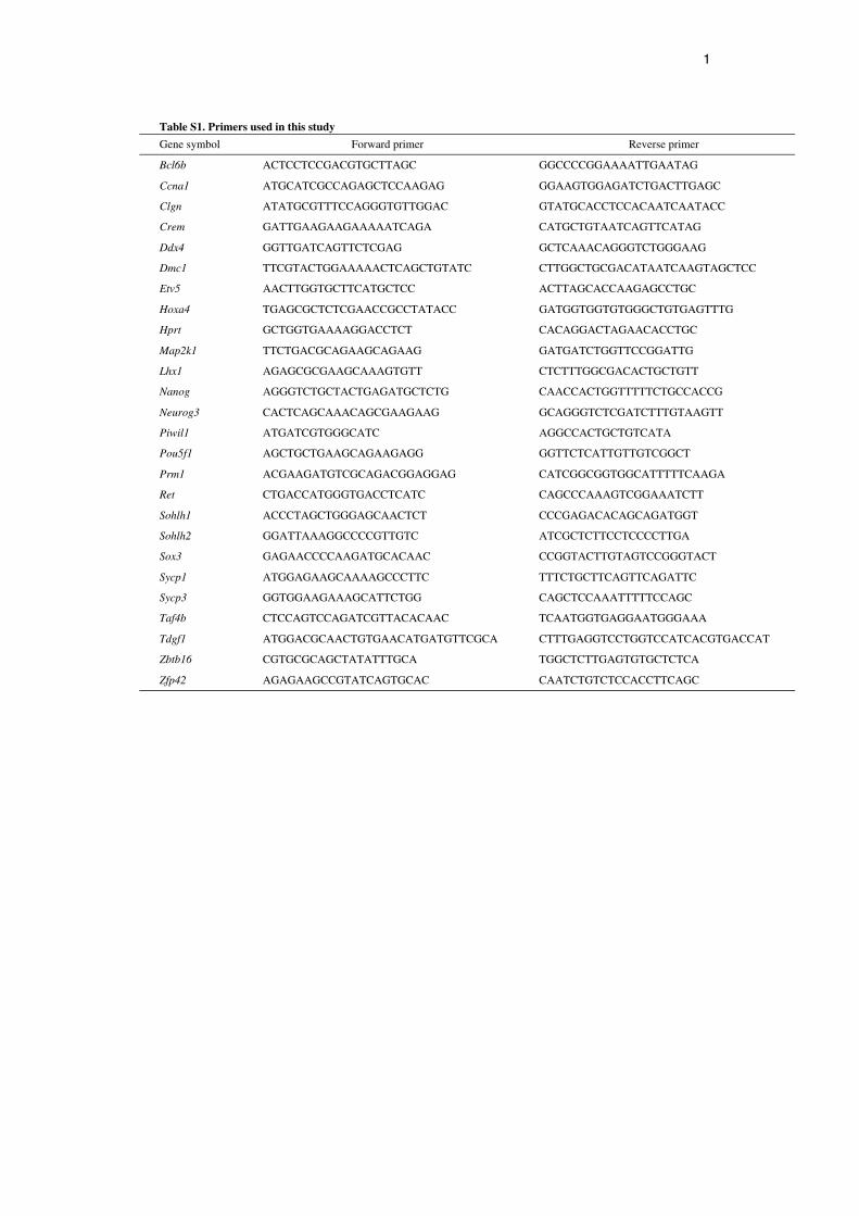
Reverse transcription PCR (RT-PCR)Total RNA was recovered from cultured cells using Trizol following themanufacturer’s protocol (Invitrogen). First-strand cDNA was producedusing a Verso cDNA Synthesis Kit (Thermo Fisher Scientific, Waltham,MA, USA). PCR conditions were 94°C for 5 minutes, then 30 cycles of94°C for 30 seconds, 60°C for 1 minute and 72°C for 1 minute, followedby 72°C for 4 minutes. For real-time PCR analyses, transcript levels werenormalized to those of Hprt using a StepOnePlus Real-Time PCR systemand Power SYBR Green PCR Master Mix (Applied Biosystems,Warrington, UK). PCR conditions were 95°C for 10 minutes, followed by40 cycles of 95°C for 15 seconds and 60°C for 1 minute. PCR primers arelisted in supplementary material Table S1.
Flow cytometryDissociated cultured cells were suspended in PBS containing 1% FCS at1�106 cells/100 l. The primary antibodies used were: monoclonal ratanti-mouse EPCAM (G8.8; BioLegend), monoclonal mouse anti-mouseFUT4 (MC-480; eBioscience), monoclonal rat anti-human ITGA6 (CD49f;GoH3), biotinylated monoclonal hamster anti-rat ITGB1 (CD29; Ha2/5),biotinylated monoclonal rat anti-mouse CD9 (KMC8) and monoclonal ratanti-mouse KIT (CD117; 2B8) (all from BD Biosciences).Allophycocyanin (APC)-conjugated goat anti-rat IgG or APC-conjugatedstreptavidin (BD Biosciences) were used to detect the primary antibodies.The stained cells were analyzed using the FACSCalibur system.
Germ cell transplantationApproximately 2�103 cells were injected into the seminiferous tubules of4- to 6-week-old WBB6F1-W/Wv (designated W) mice (Japan SLC) viathe efferent duct. Testis cells from primary recipients were dissociated 2months after transplantation by two-step enzymatic digestion usingcollagenase and trypsin for serial transplantation (Ogawa et al., 1997).Single cells were suspended in 12-16 l injection medium, which consisted D
EVELO
PMENT
1736
of Dulbecco’s modified Eagle’s medium with 10% FCS. Approximately 4l of the cell suspension was microinjected into each testis of a secondaryW recipient. Recipient animals were treated with anti-CD4 antibody toavoid rejection of allogeneic donor cells (GK1.5; gift from Dr T. Honjo,Kyoto University, Kyoto, Japan) as described previously (Kanatsu-Shinohara et al., 2003a). The Institutional Animal Care and Use Committeeof Kyoto University approved all animal experimentation protocols.
Analysis of recipient testesRecipient mice were sacrificed and their testes were recovered at 6-8 weekspost-transplantation to quantify germ cell colonies. Testes were exposed toUV light, and the fluorescent donor cell clusters were defined as colonieswhen they occupied the entire basal surface of the tubule and were at least0.1 mm in length.
Statistical analysisResults of three to four experiments are presented as mean±s.e.m. Datawere analyzed with Student’s t-test. Effects of cytokines in real-time PCRor western blot analyses were determined using ANOVA followed byTukey’s HSD. P<0.05 was considered significant.
RESULTSActivation of the MAP2K1 pathway in GS cellsGS cells require both GDNF and FGF2 for continuous proliferation(Oatley and Brinster, 2008). Spermatogonia freshly isolated fromthe testes failed to form germ cell colonies in the absence of FGF2
RESEARCH ARTICLE Development 139 (10)
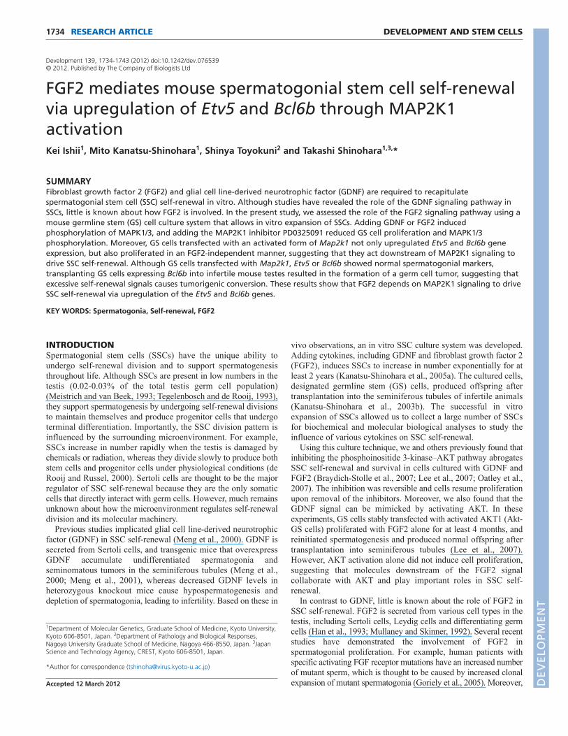
or GDNF (Fig. 1A). Germ cell colonies were established onlywhen both cytokines were added to the culture medium (Fig. 1A).These cytokines are also important for maintaining GS cellsbecause removing FGF2 or GDNF led to increased apoptosis andcessation of growth in established GS cells (Fig. 1B,C).
Although MAP kinase is one of the major signaling moleculesthat acts downstream of FGF signaling, we showed previously thatGS cell proliferation is not influenced by PD98059, a MAP2K1inhibitor, at concentrations that inhibit embryonic stem (ES) cellgrowth (Lee et al., 2007). However, immunohistological stainingof the testis showed that phosphorylated MAPK1/3 is found notonly in 63/721 (8.7%) of EPCAM+ or 11/423 (2.6%) of KIT+
spermatogonia but also in 3/68 (4.4%) of CDH1+ undifferentiatedspermatogonia, which suggested that its activation plays a role inspermatogonia proliferation (Fig. 1D).
In accordance with these results, we found that addingPD0325901, a novel and more specific MAP2K1 inhibitor,significantly reduced GS cell proliferation at lower concentrations(Fig. 1E,F). Whereas control cells could expand ~400% during 6days on MEFs, the number of cells recovered during the sameperiod was reduced in the presence of PD0325901 in a dose-dependent manner. GS cell proliferation was also inhibited at highPD98059 concentrations (50-150 M) (Fig. 1E). These resultssuggest the involvement of MAP2K1 in SSC self-renewal and
Fig. 1. Activation of the MAP2K1 pathway by FGF2. (A)GScell cultures from mouse pup testis. Although adding FGF2 (topright) or GDNF (bottom left) alone was insufficient to induce GScell colonies, typical colony formation occurred when testis cellswere cultured with FGF2 and GDNF (bottom right). The cellsshowed no signs of division without the cytokines and many ofthem underwent apoptosis (top left). The images were taken 9days after transfection. (B)Cumulative growth curve of testis cellsduring GS cell culture initiation. Cells were transferred onto MEFs9 days after initiating the culture. (C)Effect of cytokines on GScell apoptosis. Apoptosis was induced by cytokine deprivation for3 days on MEFs. Both GDNF and FGF2 attenuated GS cellapoptosis. (D)Immunohistological staining of phosphorylatedMAPK1/3 (red) in the adult testis. Phosphorylated MAPK1/3 wasfound in cells on the basement membrane expressingspermatogonia markers (blue, arrowheads). (E)Decreased cellrecovery after PD98059 (left) or PD0325901 (right) treatment(n6). GS cells were cultured on MEFs for 6 days in the presenceof FGF2 and GDNF. Error bars indicate s.e.m. (F) Appearance ofGS cells cultured with PD0325901. Adding PD0325901 inhibitedGS cell proliferation and induced apoptosis of cells cultured onMEFs. DMSO, control. (G)MAPK1/3 phosphorylation by cytokinestimulation. Cells were starved for 3 days on laminin and thesamples were collected 30 minutes after cytokine stimulation.White and black arrowheads indicate phosphorylated MAPK1and MAPK3, respectively. ACTB provided a loading control.(H)Macroscopic appearance of recipient testes transplanted withcontrol or PD0325901-treated GS cells. Green tubules indicatespermatogenesis from donor SSCs. F, FGF2; G, GDNF. Scale bars:20m in A; 10m in D; 50m in F; 1 mm in H.
DEVELO
PMENT
survival. We examined MAPK1/3 phosphorylation by westernblotting to confirm the effect of PD0325901 on MAP2K1 activity.After culturing GS cells on laminin-coated dishes for 3 dayswithout cytokines, the cells were restimulated with FGF2 orGDNF. Although adding either FGF2 or GDNF induced MAPK1/3phosphorylation (Fig. 1G), this phosphorylation by exogenouscytokines was suppressed by PD0325901.
We performed germ cell transplantation experiments to examinethe effect of PD0325901 on SSCs (Brinster and Zimmermann, 1994).Transplanted SSCs colonized empty seminiferous tubules andestablished spermatogenesis for the long term. GS cells were culturedin the presence of PD0325901 (4 M) for 6 days, and ~2�103 cellswere transplanted into the seminiferous tubules of congenitallyinfertile W mice (Fig. 1H). The numbers of colonies generated byPD0325901-treated and control cells in four experiments were1.6±0.2�102 and 1.6±0.3�102 colonies/105 cells, respectively(n18). Although GS cell number changed 3.9-fold in controlcultures, the cell number did not increase in PD0325901-treatedcultures during this period. Therefore, this result suggested thatPD0325901 inhibited the net SSC increase by suppressing MAP2K1.
1737RESEARCH ARTICLESpermatogonia self-renewal
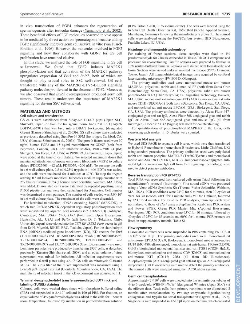
Substitution of the FGF2 signal by a constitutivelyactive MAP2K1To gain insight into the mechanism of MAP2K1 action, we producedgreen GS cells that stably overexpressed the constitutively activeform of MAP2K1 (MEK-DD) by lentivirus-mediated gene delivery.The morphology of MEK-DD-transfected GS cells (MEK-GS) wasindistinguishable from that of parental wild-type green GS (WT-GS)cells (Fig. 2A). Real-time PCR and western blot analyses confirmedincreased expression of the transgene (Fig. 2B and supplementarymaterial Fig. S1). RT-PCR and flow cytometric analyses showed thatthe cells expressed normal levels of spermatogonial markers withoutthe expression of ES cell markers such as FUT4 or Nanog, both ofwhich are expressed in ES-like multipotent germline stem (mGS)cells (Kanatsu-Shinohara et al., 2004) (Fig. 2C,D). Western blotanalyses showed changes in MAPK1/3 phosphorylation aftertransfection (Fig. 2E,F). In WT-GS cells, FGF2 consistently inducedstronger MAPK1/3 phosphorylation than GDNF. However,compared with WT-GS cells, MEK-GS cells showed a 7.4±1.3- and4.6±1.0-fold increase in MAPK1 and MAPK3 phosphorylation,respectively, when cultured without exogenous cytokines (n4).
Fig. 2. Substitution of FGF2 with MEK-DD.(A)Appearance of GS cells transduced with MEK-DD. Nosignificant difference was noted to parental control cells.MEK-GS cells could proliferate without FGF2.(B)Western blot analysis of MAP2K1 expression. (C)Flowcytometric analysis of MEK-GS cells cultured on MEFs.Green lines indicate the control. (D)RT-PCR analyses ofMEK-GS cells. Samples were collected after culturing GScells on laminin for 6 days with GDNF and FGF2.(E)MAPK1/3 phosphorylation by cytokine stimulation.Cells were starved for 3 days on laminin and the sampleswere collected 30 minutes after cytokine stimulation.MEK-GS cells showed strong MAPK1/3 phosphorylationeven in the absence of cytokine stimulation.(F)Quantification of phosphorylated MAPK1/3expression (n4). Error bars indicate s.e.m. (G) Cumulative growth curve of MEK-GS cells culturedunder different cytokine stimulation conditions. MEK-GScells proliferated with GDNF alone, albeit at a slower ratethan with FGF2 and GDNF. Cultures were maintained onMEFs. (H)Culture of MEK-GS cells with GDNF. WT-GScells stopped proliferating after several passages.Cultures were maintained on MEFs. F, FGF2; G, GDNF.Scale bar: 50m.
DEVELO
PMENT
1738 RESEARCH ARTICLE Development 139 (10)
Although co-stimulation by FGF2 and GDNF further enhancedMAPK1/3 phosphorylation in MEK-GS cells, we did not observeincreased MAPK1/3 phosphorylation when either FGF2 or GDNFwas added to the culture.
To examine the effect of MAP2K1 overexpression on GS cellproliferation, we cultured MEK-GS cells under different cytokinestimulations. Despite the increased phosphorylation levels, the speedof MEK-GS cell proliferation was comparable to that of control WT-GS cells when they were cultured with both FGF2 and GDNF. Thesecells could be passaged at a ratio of 4:1 to 6:1 every 5-6 days (Fig.2G). However, we noted that MEK-GS cells continued to proliferatefor more than 8 weeks with only GDNF supplementation, duringwhich time the total cell number increased by ~3.0�103-fold (Fig.2H). The expression level of MAP2K1 did not change significantly,at least at the two points measured during culture (supplementarymaterial Fig. S2). Because MEK-GS cells could expand more than1�106-fold during the experimental period by co-stimulation withFGF2 and GDNF, this suggests that the FGF2 signal provides astronger stimulus than that of MEK-DD. By contrast, MEK-GS cellscultured only with FGF2 stimulation gradually ceased proliferatingand could not be maintained for longer than 4-5 weeks. Similarly,WT-GS cells could not be maintained without FGF2 stimulation afterpassages (Fig. 2H). These results show that MAP2K1 activationpartly substitutes for FGF2 stimulation.
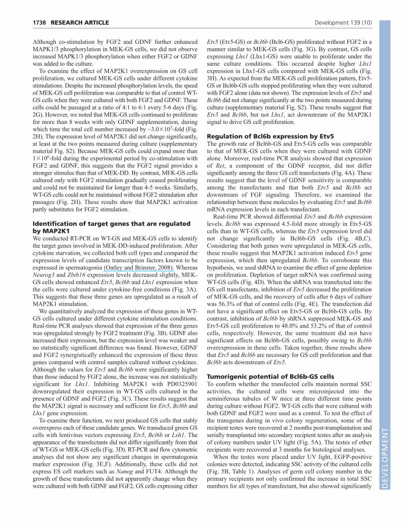
Identification of target genes that are regulatedby MAP2K1We conducted RT-PCR on WT-GS and MEK-GS cells to identifythe target genes involved in MEK-DD-induced proliferation. Aftercytokine starvation, we collected both cell types and compared theexpression levels of candidate transcription factors known to beexpressed in spermatogonia (Oatley and Brinster, 2008). WhereasNeurog3 and Zbtb16 expression levels decreased slightly, MEK-GS cells showed enhanced Etv5, Bcl6b and Lhx1 expression whenthe cells were cultured under cytokine-free conditions (Fig. 3A).This suggests that these three genes are upregulated as a result ofMAP2K1 stimulation.
We quantitatively analyzed the expression of these genes in WT-GS cells cultured under different cytokine stimulation conditions.Real-time PCR analyses showed that expression of the three geneswas upregulated strongly by FGF2 treatment (Fig. 3B). GDNF alsoincreased their expression, but the expression level was weaker andno statistically significant difference was found. However, GDNFand FGF2 synergistically enhanced the expression of these threegenes compared with control samples cultured without cytokines.Although the values for Etv5 and Bcl6b were significantly higherthan those induced by FGF2 alone, the increase was not statisticallysignificant for Lhx1. Inhibiting MAP2K1 with PD0325901downregulated their expression in WT-GS cells cultured in thepresence of GDNF and FGF2 (Fig. 3C). These results suggest thatthe MAP2K1 signal is necessary and sufficient for Etv5, Bcl6b andLhx1 gene expression.
To examine their function, we next produced GS cells that stablyoverexpress each of these candidate genes. We transduced green GScells with lentivirus vectors expressing Etv5, Bcl6b or Lxh1. Theappearance of the transfectants did not differ significantly from thatof WT-GS or MEK-GS cells (Fig. 3D). RT-PCR and flow cytometricanalyses did not show any significant changes in spermatogoniamarker expression (Fig. 3E,F). Additionally, these cells did notexpress ES cell markers such as Nanog and FUT4. Although thegrowth of these transfectants did not apparently change when theywere cultured with both GDNF and FGF2, GS cells expressing either
Etv5 (Etv5-GS) or Bcl6b (Bcl6-GS) proliferated without FGF2 in amanner similar to MEK-GS cells (Fig. 3G). By contrast, GS cellsexpressing Lhx1 (Lhx1-GS) were unable to proliferate under thesame culture conditions. This occurred despite higher Lhx1expression in Lhx1-GS cells compared with MEK-GS cells (Fig.3H). As expected from the MEK-GS cell proliferation pattern, Etv5-GS or Bcl6b-GS cells stopped proliferating when they were culturedwith FGF2 alone (data not shown). The expression levels of Etv5 andBcl6b did not change significantly at the two points measured duringculture (supplementary material Fig. S2). These results suggest thatEtv5 and Bcl6b, but not Lhx1, act downstream of the MAP2K1signal to drive GS cell proliferation.
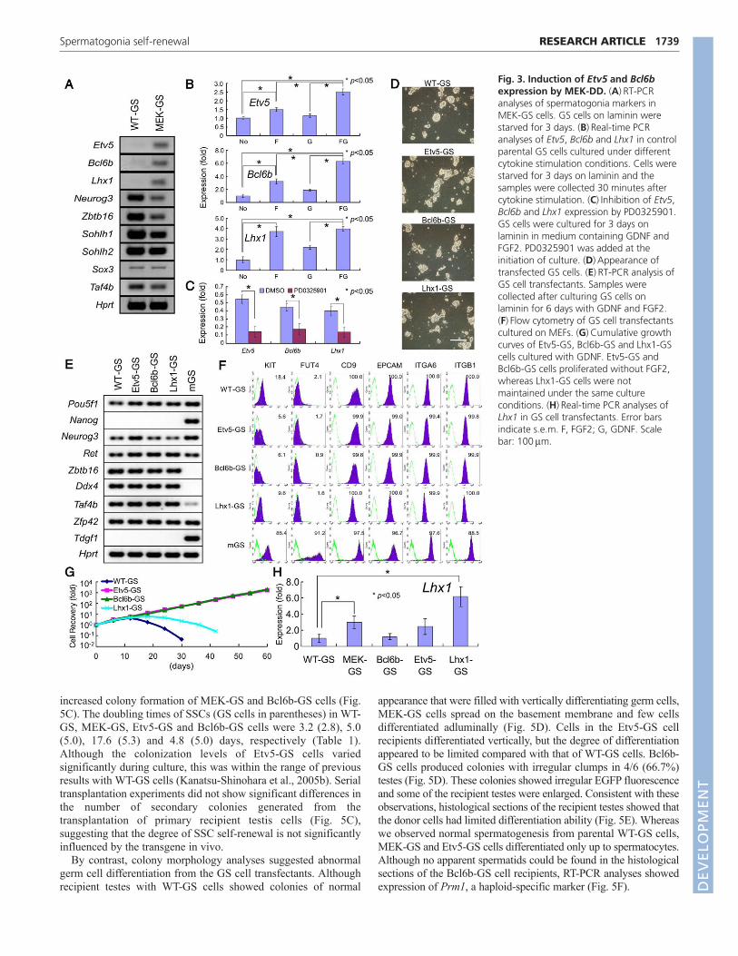
Regulation of Bcl6b expression by Etv5The growth rate of Bcl6b-GS and Etv5-GS cells was comparableto that of MEK-GS cells when they were cultured with GDNFalone. Moreover, real-time PCR analysis showed that expressionof Ret, a component of the GDNF receptor, did not differsignificantly among the three GS cell transfectants (Fig. 4A). Theseresults suggest that the level of GDNF sensitivity is comparableamong the transfectants and that both Etv5 and Bcl6b actdownstream of FGF signaling. Therefore, we examined therelationship between these molecules by evaluating Etv5 and Bcl6bmRNA expression levels in each transfectant.
Real-time PCR showed differential Etv5 and Bcl6b expressionlevels. Bcl6b was expressed 4.5-fold more strongly in Etv5-GScells than in WT-GS cells, whereas the Etv5 expression level didnot change significantly in Bcl6b-GS cells (Fig. 4B,C).Considering that both genes were upregulated in MEK-GS cells,these results suggest that MAP2K1 activation induced Etv5 geneexpression, which then upregulated Bcl6b. To corroborate thishypothesis, we used shRNA to examine the effect of gene depletionon proliferation. Depletion of target mRNA was confirmed usingWT-GS cells (Fig. 4D). When the shRNA was transfected into theGS cell transfectants, inhibition of Etv5 decreased the proliferationof MEK-GS cells, and the recovery of cells after 6 days of culturewas 56.3% of that of control cells (Fig. 4E). The transfection didnot have a significant effect on Etv5-GS or Bcl6b-GS cells. Bycontrast, inhibition of Bcl6b by shRNA suppressed MEK-GS andEtv5-GS cell proliferation to 48.0% and 53.2% of that of controlcells, respectively. However, the same treatment did not havesignificant effects on Bcl6b-GS cells, possibly owing to Bcl6boverexpression in these cells. Taken together, these results showthat Etv5 and Bcl6b are necessary for GS cell proliferation and thatBcl6b acts downstream of Etv5.
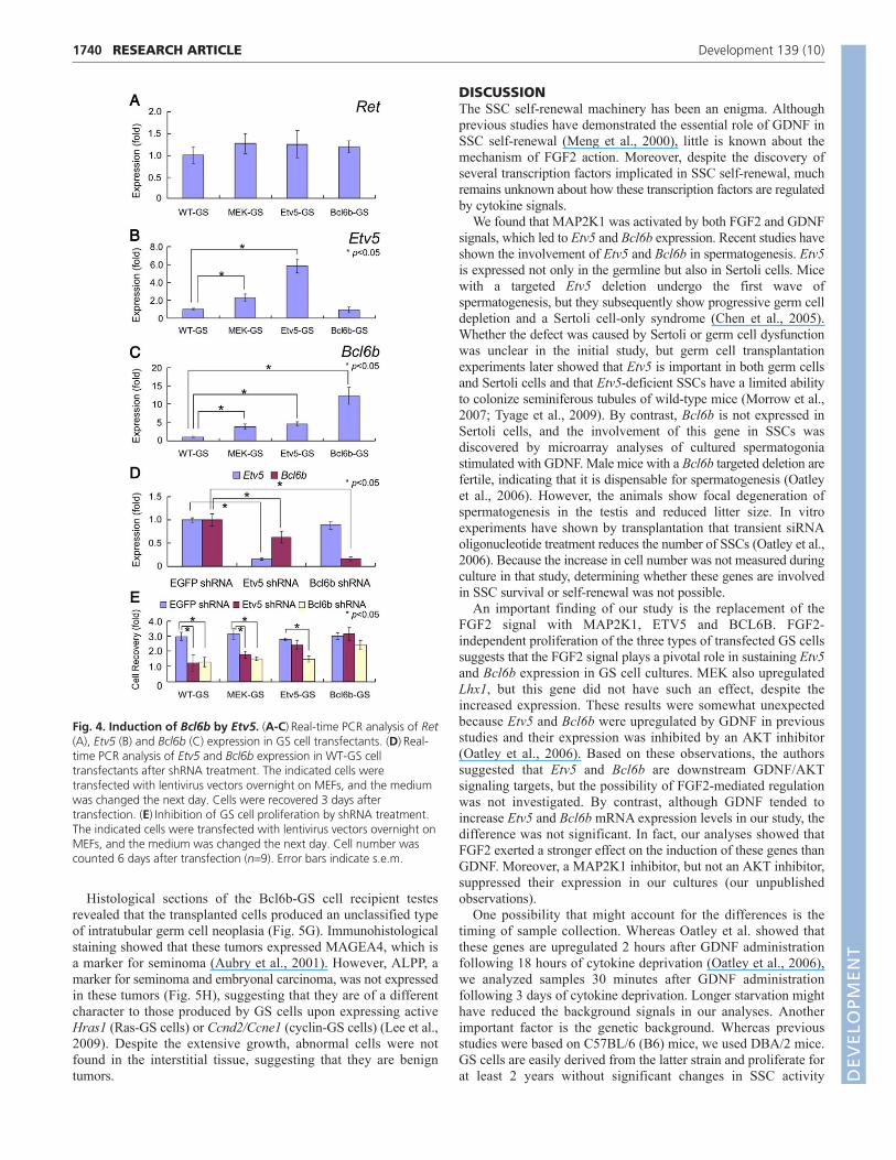
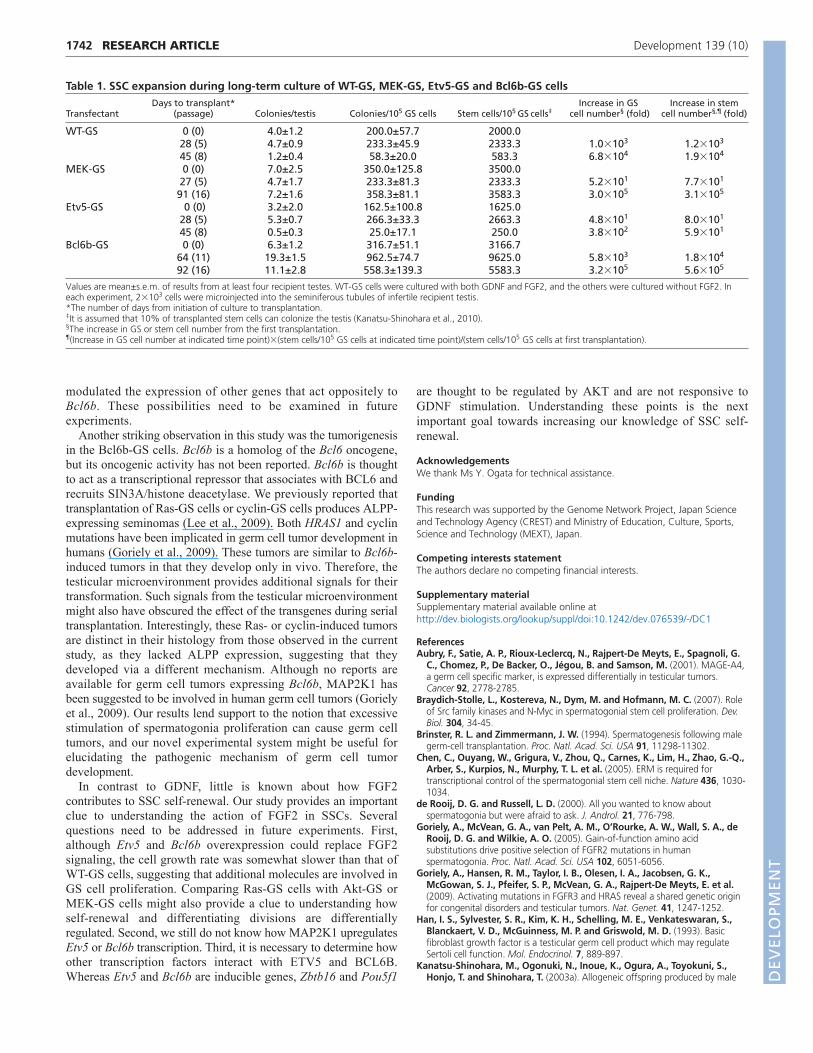
Tumorigenic potential of Bcl6b-GS cellsTo confirm whether the transfected cells maintain normal SSCactivities, the cultured cells were microinjected into theseminiferous tubules of W mice at three different time pointsduring culture without FGF2. WT-GS cells that were cultured withboth GDNF and FGF2 were used as a control. To test the effect ofthe transgenes during in vivo colony regeneration, some of therecipient testes were recovered at 2 months post-transplantation andserially transplanted into secondary recipient testes after an analysisof colony numbers under UV light (Fig. 5A). The testes of otherrecipients were recovered at 3 months for histological analyses.
When the testes were placed under UV light, EGFP-positivecolonies were detected, indicating SSC activity of the cultured cells(Fig. 5B, Table 1). Analyses of germ cell colony number in theprimary recipients not only confirmed the increase in total SSCnumbers for all types of transfectant, but also showed significantly D
EVELO
PMENT
increased colony formation of MEK-GS and Bcl6b-GS cells (Fig.5C). The doubling times of SSCs (GS cells in parentheses) in WT-GS, MEK-GS, Etv5-GS and Bcl6b-GS cells were 3.2 (2.8), 5.0(5.0), 17.6 (5.3) and 4.8 (5.0) days, respectively (Table 1).Although the colonization levels of Etv5-GS cells variedsignificantly during culture, this was within the range of previousresults with WT-GS cells (Kanatsu-Shinohara et al., 2005b). Serialtransplantation experiments did not show significant differences inthe number of secondary colonies generated from thetransplantation of primary recipient testis cells (Fig. 5C),suggesting that the degree of SSC self-renewal is not significantlyinfluenced by the transgene in vivo.
By contrast, colony morphology analyses suggested abnormalgerm cell differentiation from the GS cell transfectants. Althoughrecipient testes with WT-GS cells showed colonies of normal
1739RESEARCH ARTICLESpermatogonia self-renewal
appearance that were filled with vertically differentiating germ cells,MEK-GS cells spread on the basement membrane and few cellsdifferentiated adluminally (Fig. 5D). Cells in the Etv5-GS cellrecipients differentiated vertically, but the degree of differentiationappeared to be limited compared with that of WT-GS cells. Bcl6b-GS cells produced colonies with irregular clumps in 4/6 (66.7%)testes (Fig. 5D). These colonies showed irregular EGFP fluorescenceand some of the recipient testes were enlarged. Consistent with theseobservations, histological sections of the recipient testes showed thatthe donor cells had limited differentiation ability (Fig. 5E). Whereaswe observed normal spermatogenesis from parental WT-GS cells,MEK-GS and Etv5-GS cells differentiated only up to spermatocytes.Although no apparent spermatids could be found in the histologicalsections of the Bcl6b-GS cell recipients, RT-PCR analyses showedexpression of Prm1, a haploid-specific marker (Fig. 5F).
Fig. 3. Induction of Etv5 and Bcl6bexpression by MEK-DD. (A)RT-PCRanalyses of spermatogonia markers inMEK-GS cells. GS cells on laminin werestarved for 3 days. (B)Real-time PCRanalyses of Etv5, Bcl6b and Lhx1 in controlparental GS cells cultured under differentcytokine stimulation conditions. Cells werestarved for 3 days on laminin and thesamples were collected 30 minutes aftercytokine stimulation. (C)Inhibition of Etv5,Bcl6b and Lhx1 expression by PD0325901.GS cells were cultured for 3 days onlaminin in medium containing GDNF andFGF2. PD0325901 was added at theinitiation of culture. (D)Appearance oftransfected GS cells. (E)RT-PCR analysis ofGS cell transfectants. Samples werecollected after culturing GS cells onlaminin for 6 days with GDNF and FGF2.(F)Flow cytometry of GS cell transfectantscultured on MEFs. (G)Cumulative growthcurves of Etv5-GS, Bcl6b-GS and Lhx1-GScells cultured with GDNF. Etv5-GS andBcl6b-GS cells proliferated without FGF2,whereas Lhx1-GS cells were notmaintained under the same cultureconditions. (H)Real-time PCR analyses ofLhx1 in GS cell transfectants. Error barsindicate s.e.m. F, FGF2; G, GDNF. Scalebar: 100m.
DEVELO
PMENT
1740
Histological sections of the Bcl6b-GS cell recipient testesrevealed that the transplanted cells produced an unclassified typeof intratubular germ cell neoplasia (Fig. 5G). Immunohistologicalstaining showed that these tumors expressed MAGEA4, which isa marker for seminoma (Aubry et al., 2001). However, ALPP, amarker for seminoma and embryonal carcinoma, was not expressedin these tumors (Fig. 5H), suggesting that they are of a differentcharacter to those produced by GS cells upon expressing activeHras1 (Ras-GS cells) or Ccnd2/Ccne1 (cyclin-GS cells) (Lee et al.,2009). Despite the extensive growth, abnormal cells were not found in the interstitial tissue, suggesting that they are benigntumors.
RESEARCH ARTICLE Development 139 (10)
DISCUSSIONThe SSC self-renewal machinery has been an enigma. Althoughprevious studies have demonstrated the essential role of GDNF inSSC self-renewal (Meng et al., 2000), little is known about themechanism of FGF2 action. Moreover, despite the discovery ofseveral transcription factors implicated in SSC self-renewal, muchremains unknown about how these transcription factors are regulatedby cytokine signals.
We found that MAP2K1 was activated by both FGF2 and GDNFsignals, which led to Etv5 and Bcl6b expression. Recent studies haveshown the involvement of Etv5 and Bcl6b in spermatogenesis. Etv5is expressed not only in the germline but also in Sertoli cells. Micewith a targeted Etv5 deletion undergo the first wave ofspermatogenesis, but they subsequently show progressive germ celldepletion and a Sertoli cell-only syndrome (Chen et al., 2005).Whether the defect was caused by Sertoli or germ cell dysfunctionwas unclear in the initial study, but germ cell transplantationexperiments later showed that Etv5 is important in both germ cellsand Sertoli cells and that Etv5-deficient SSCs have a limited abilityto colonize seminiferous tubules of wild-type mice (Morrow et al.,2007; Tyage et al., 2009). By contrast, Bcl6b is not expressed inSertoli cells, and the involvement of this gene in SSCs wasdiscovered by microarray analyses of cultured spermatogoniastimulated with GDNF. Male mice with a Bcl6b targeted deletion arefertile, indicating that it is dispensable for spermatogenesis (Oatleyet al., 2006). However, the animals show focal degeneration ofspermatogenesis in the testis and reduced litter size. In vitroexperiments have shown by transplantation that transient siRNAoligonucleotide treatment reduces the number of SSCs (Oatley et al.,2006). Because the increase in cell number was not measured duringculture in that study, determining whether these genes are involvedin SSC survival or self-renewal was not possible.
An important finding of our study is the replacement of theFGF2 signal with MAP2K1, ETV5 and BCL6B. FGF2-independent proliferation of the three types of transfected GS cellssuggests that the FGF2 signal plays a pivotal role in sustaining Etv5and Bcl6b expression in GS cell cultures. MEK also upregulatedLhx1, but this gene did not have such an effect, despite theincreased expression. These results were somewhat unexpectedbecause Etv5 and Bcl6b were upregulated by GDNF in previousstudies and their expression was inhibited by an AKT inhibitor(Oatley et al., 2006). Based on these observations, the authorssuggested that Etv5 and Bcl6b are downstream GDNF/AKTsignaling targets, but the possibility of FGF2-mediated regulationwas not investigated. By contrast, although GDNF tended toincrease Etv5 and Bcl6b mRNA expression levels in our study, thedifference was not significant. In fact, our analyses showed thatFGF2 exerted a stronger effect on the induction of these genes thanGDNF. Moreover, a MAP2K1 inhibitor, but not an AKT inhibitor,suppressed their expression in our cultures (our unpublishedobservations).
One possibility that might account for the differences is thetiming of sample collection. Whereas Oatley et al. showed thatthese genes are upregulated 2 hours after GDNF administrationfollowing 18 hours of cytokine deprivation (Oatley et al., 2006),we analyzed samples 30 minutes after GDNF administrationfollowing 3 days of cytokine deprivation. Longer starvation mighthave reduced the background signals in our analyses. Anotherimportant factor is the genetic background. Whereas previousstudies were based on C57BL/6 (B6) mice, we used DBA/2 mice.GS cells are easily derived from the latter strain and proliferate forat least 2 years without significant changes in SSC activity
Fig. 4. Induction of Bcl6b by Etv5. (A-C)Real-time PCR analysis of Ret(A), Etv5 (B) and Bcl6b (C) expression in GS cell transfectants. (D)Real-time PCR analysis of Etv5 and Bcl6b expression in WT-GS celltransfectants after shRNA treatment. The indicated cells weretransfected with lentivirus vectors overnight on MEFs, and the mediumwas changed the next day. Cells were recovered 3 days aftertransfection. (E)Inhibition of GS cell proliferation by shRNA treatment.The indicated cells were transfected with lentivirus vectors overnight onMEFs, and the medium was changed the next day. Cell number wascounted 6 days after transfection (n9). Error bars indicate s.e.m.DEVELO
PMENT
(Kanatsu-Shinohara et al., 2005a), whereas those from B6 miceexhibit an almost 10-fold decrease in SSC activity by 14-18 monthsand do not proliferate under the current culture conditions(Kanatsu-Shinohara et al., 2003b; Schmidt et al., 2011). Althoughboth GDNF and FGF2 activated the MAP2K1 pathway,augmentation of GDNF signaling by GFRA1 is thought to benecessary to derive SSC cultures from B6 mice (Oatley andBrinster, 2008). Therefore, it is possible that SSCs with differentgenetic backgrounds altered the balance between the two signalingpathways, and these genes might be induced more predominantlyby GDNF in a B6 background.
Our study might provide a clue as to the synergistic nature ofcytokines in GS cells. GS cells could be established only in thepresence of GDNF and FGF2, but why the cells required two
1741RESEARCH ARTICLESpermatogonia self-renewal
cytokines for self-renewal remains unclear. A previous studyshowed that germ cells in Etv5 knockout mice have reduced Retexpression (Tyagi et al., 2009). Ret mRNA expression was ~60%of that in wild-type testes at postnatal day 4, and it progressivelydecreased to 10% by day 24. Therefore, reduced GDNFsignaling caused the gradual decrease in SSCs in the Etv5knockout mice. Because FGF2 induced Etv5 expression, theresults suggest that FGF2 plays a crucial role in enhancing theresponsiveness of GDNF via regulating the Ret expression level.This might explain why both FGF2 and GDNF are required forcontinuous GS cell proliferation. Although we currently do notknow whether regulation of Ret occurs via Bcl6b, Etv5-GS cellsshowed a slightly lower colonization efficiency than Bcl6b-GScells, which suggests that Etv5 overexpression might also have
Fig. 5. Germ cell transplantation of GS celltransfectants. (A)Outline of the experimentalprocedure. (B)Macroscopic appearance of the primary(top) and secondary (bottom) recipient testesfollowing transplantation of GS cell transfectants.Green tubules indicate spermatogenesis from donorSSCs. Note the extensive colonization in Bcl6b-GS cellrecipients after serial transplantation. (C)Colonynumber in the primary recipients (top). Results fromtransplantation experiments during long-term culture(see Table 1). The degree of secondary colonyformation is indicated by the ratio of SSC numbers attwo different time points (bottom). The multiplicationratio was calculated by assuming that 10% of theSSCs can colonize and that each colony is producedby one SSC (Kanatsu-Shinohara et al., 2010). Errorbars indicate s.e.m. (D)Appearance of the germ cellcolonies. WT-GS cells differentiated normally, whereasGS cell transfectants showed abnormal colonymorphology. Arrow indicates an abnormal cell clumpin a Bcl6b-GS cell recipient testis. (E)Histology of therecipient testes. Hematoxylin and eosin staining. GScell transfectants showed abnormal spermatogenesis.(F)RT-PCR analyses of the recipient testes.(G)Histology of the germ cell tumor developed fromBcl6b-GS cells. Hematoxylin and eosin staining.(H)Immunohistological staining of the germ celltumor by anti-ALPP or anti-MAGEA4 antibodies (red),counterstained with Hoechst 33342. (I)Summary ofthe experimental findings. FGF2 induces strongerMAPK1/3 phosphorylation than GDNF. ActivatedMAP2K1 induces the expression of Etv5, whichsubsequently upregulates Bcl6b expression. Bcl6boverexpression results in germ cell tumor formationafter spermatogonial transplantation. Because Retexpression is downregulated in Etv5 knockout mice(Tyagi et al., 2009), ETV5 is probably one of thefactors that sustains Ret expression. Etv5 upregulationby FGF2 activation might contribute to the synergisticeffect of FGF2 and GDNF. Scale bars: 1 mm in B;400m in D; 20m in E,G,H.
DEVELO
PMENT
1742
modulated the expression of other genes that act oppositely toBcl6b. These possibilities need to be examined in futureexperiments.
Another striking observation in this study was the tumorigenesisin the Bcl6b-GS cells. Bcl6b is a homolog of the Bcl6 oncogene,but its oncogenic activity has not been reported. Bcl6b is thoughtto act as a transcriptional repressor that associates with BCL6 andrecruits SIN3A/histone deacetylase. We previously reported thattransplantation of Ras-GS cells or cyclin-GS cells produces ALPP-expressing seminomas (Lee et al., 2009). Both HRAS1 and cyclinmutations have been implicated in germ cell tumor development inhumans (Goriely et al., 2009). These tumors are similar to Bcl6b-induced tumors in that they develop only in vivo. Therefore, thetesticular microenvironment provides additional signals for theirtransformation. Such signals from the testicular microenvironmentmight also have obscured the effect of the transgenes during serialtransplantation. Interestingly, these Ras- or cyclin-induced tumorsare distinct in their histology from those observed in the currentstudy, as they lacked ALPP expression, suggesting that theydeveloped via a different mechanism. Although no reports areavailable for germ cell tumors expressing Bcl6b, MAP2K1 hasbeen suggested to be involved in human germ cell tumors (Gorielyet al., 2009). Our results lend support to the notion that excessivestimulation of spermatogonia proliferation can cause germ celltumors, and our novel experimental system might be useful forelucidating the pathogenic mechanism of germ cell tumordevelopment.
In contrast to GDNF, little is known about how FGF2contributes to SSC self-renewal. Our study provides an importantclue to understanding the action of FGF2 in SSCs. Severalquestions need to be addressed in future experiments. First,although Etv5 and Bcl6b overexpression could replace FGF2signaling, the cell growth rate was somewhat slower than that ofWT-GS cells, suggesting that additional molecules are involved inGS cell proliferation. Comparing Ras-GS cells with Akt-GS orMEK-GS cells might also provide a clue to understanding howself-renewal and differentiating divisions are differentiallyregulated. Second, we still do not know how MAP2K1 upregulatesEtv5 or Bcl6b transcription. Third, it is necessary to determine howother transcription factors interact with ETV5 and BCL6B.Whereas Etv5 and Bcl6b are inducible genes, Zbtb16 and Pou5f1
RESEARCH ARTICLE Development 139 (10)
are thought to be regulated by AKT and are not responsive toGDNF stimulation. Understanding these points is the nextimportant goal towards increasing our knowledge of SSC self-renewal.
AcknowledgementsWe thank Ms Y. Ogata for technical assistance.
FundingThis research was supported by the Genome Network Project, Japan Scienceand Technology Agency (CREST) and Ministry of Education, Culture, Sports,Science and Technology (MEXT), Japan.
Competing interests statementThe authors declare no competing financial interests.
Supplementary materialSupplementary material available online athttp://dev.biologists.org/lookup/suppl/doi:10.1242/dev.076539/-/DC1
ReferencesAubry, F., Satie, A. P., Rioux-Leclercq, N., Rajpert-De Meyts, E., Spagnoli, G.
C., Chomez, P., De Backer, O., Jégou, B. and Samson, M. (2001). MAGE-A4,a germ cell specific marker, is expressed differentially in testicular tumors.Cancer 92, 2778-2785.
Braydich-Stolle, L., Kostereva, N., Dym, M. and Hofmann, M. C. (2007). Roleof Src family kinases and N-Myc in spermatogonial stem cell proliferation. Dev.Biol. 304, 34-45.
Brinster, R. L. and Zimmermann, J. W. (1994). Spermatogenesis following malegerm-cell transplantation. Proc. Natl. Acad. Sci. USA 91, 11298-11302.
Chen, C., Ouyang, W., Grigura, V., Zhou, Q., Carnes, K., Lim, H., Zhao, G.-Q.,Arber, S., Kurpios, N., Murphy, T. L. et al. (2005). ERM is required fortranscriptional control of the spermatogonial stem cell niche. Nature 436, 1030-1034.
de Rooij, D. G. and Russell, L. D. (2000). All you wanted to know aboutspermatogonia but were afraid to ask. J. Androl. 21, 776-798.
Goriely, A., McVean, G. A., van Pelt, A. M., O’Rourke, A. W., Wall, S. A., deRooij, D. G. and Wilkie, A. O. (2005). Gain-of-function amino acidsubstitutions drive positive selection of FGFR2 mutations in humanspermatogonia. Proc. Natl. Acad. Sci. USA 102, 6051-6056.
Goriely, A., Hansen, R. M., Taylor, I. B., Olesen, I. A., Jacobsen, G. K.,McGowan, S. J., Pfeifer, S. P., McVean, G. A., Rajpert-De Meyts, E. et al.(2009). Activating mutations in FGFR3 and HRAS reveal a shared genetic originfor congenital disorders and testicular tumors. Nat. Genet. 41, 1247-1252.
Han, I. S., Sylvester, S. R., Kim, K. H., Schelling, M. E., Venkateswaran, S.,Blanckaert, V. D., McGuinness, M. P. and Griswold, M. D. (1993). Basicfibroblast growth factor is a testicular germ cell product which may regulateSertoli cell function. Mol. Endocrinol. 7, 889-897.
Kanatsu-Shinohara, M., Ogonuki, N., Inoue, K., Ogura, A., Toyokuni, S.,Honjo, T. and Shinohara, T. (2003a). Allogeneic offspring produced by male
Table 1. SSC expansion during long-term culture of WT-GS, MEK-GS, Etv5-GS and Bcl6b-GS cellsDays to transplant* Increase in GS Increase in stem
Transfectant (passage) Colonies/testis Colonies/105 GS cells Stem cells/105 GS cells‡ cell number§ (fold) cell number§,¶ (fold)
WT-GS 0 (0) 4.0±1.2 200.0±57.7 2000.028 (5) 4.7±0.9 233.3±45.9 2333.3 1.0�103 1.2�103
45 (8) 1.2±0.4 58.3±20.0 583.3 6.8�104 1.9�104
MEK-GS 0 (0) 7.0±2.5 350.0±125.8 3500.027 (5) 4.7±1.7 233.3±81.3 2333.3 5.2�101 7.7�101
91 (16) 7.2±1.6 358.3±81.1 3583.3 3.0�105 3.1�105
Etv5-GS 0 (0) 3.2±2.0 162.5±100.8 1625.028 (5) 5.3±0.7 266.3±33.3 2663.3 4.8�101 8.0�101
45 (8) 0.5±0.3 25.0±17.1 250.0 3.8�102 5.9�101
Bcl6b-GS 0 (0) 6.3±1.2 316.7±51.1 3166.764 (11) 19.3±1.5 962.5±74.7 9625.0 5.8�103 1.8�104
92 (16) 11.1±2.8 558.3±139.3 5583.3 3.2�105 5.6�105
Values are mean±s.e.m. of results from at least four recipient testes. WT-GS cells were cultured with both GDNF and FGF2, and the others were cultured without FGF2. Ineach experiment, 2�103 cells were microinjected into the seminiferous tubules of infertile recipient testis.*The number of days from initiation of culture to transplantation.‡It is assumed that 10% of transplanted stem cells can colonize the testis (Kanatsu-Shinohara et al., 2010).§The increase in GS or stem cell number from the first transplantation.¶(Increase in GS cell number at indicated time point)�(stem cells/105 GS cells at indicated time point)/(stem cells/105 GS cells at first transplantation).
DEVELO
PMENT
1743RESEARCH ARTICLESpermatogonia self-renewal
germ line stem cell transplantation into infertile mouse testis. Biol. Reprod. 68,167-173.
Kanatsu-Shinohara, M., Ogonuki, N., Inoue, K., Miki, H., Ogura, A., Toyokuni, S. and Shinohara, T. (2003b). Long-term proliferation in culture and germlinetransmission of mouse male germline stem cells. Biol. Reprod. 69, 612-616.
Kanatsu-Shinohara, M., Inoue, K., Lee, J., Yoshimoto, M., Ogonuki, N., Miki,H., Baba, S., Kato, T., Kazuki, Y., Toyokuni, S. et al. (2004). Generation ofpluripotent stem cells from neonatal mouse testis. Cell 119, 1001-1012.
Kanatsu-Shinohara, M., Ogonuki, N., Iwano, T., Lee, J., Kazuki, Y., Inoue, K.,Miki, H., Takehashi, M., Toyokuni, S., Shinkai, Y. et al. (2005a). Genetic andepigenetic properties of mouse male germline stem cells during long-termculture. Development 132, 4155-4163.
Kanatsu-Shinohara, M., Miki, H., Inoue, K., Ogonuki, N., Toyokuni, S.,Ogura, A. and Shinohara, T. (2005b). Long-term culture of mouse malegermline stem cells under serum- or feeder-free conditions. Biol. Reprod. 72,985-991.
Kanatsu-Shinohara, M., Muneto, T., Lee, J., Takenaka, M., Chuma, S.,Nakatsuji, N., Horiuchi, T. and Shinohara, T. (2008). Long-term culture ofmale germline stem cells from hamster testes. Biol. Reprod. 78, 611-617.
Kanatsu-Shinohara, M., Takashima, S. and Shinohara, T. (2010). Transmissiondistortion by loss of p21 or p27 cyclin-dependent kinase inhibitors followingcompetitive spermaogonial transplantation. Proc. Natl. Acad. Sci. USA 107,6210-6215.
Lee, J., Kanatsu-Shinohara, M., Inoue, K., Ogonuki, N., Miki, H., Toyokuni,S., Kimura, T., Nakano, T., Ogura, A. and Shinohara, T. (2007). Akt mediatesself-renewal division of mouse spermatogonial stem cells. Development 134,1853-1859.
Lee, J., Kanatsu-Shinohara, M., Morimoto, H., Kazuki, Y., Takashima, S.,Oshimura, M., Toyokuni, S. and Shinohara, T. (2009). Genetic reconstructionof mouse spermatogonial stem cell self-renewal in vitro by Ras-cyclin D2activation. Cell Stem Cell 5, 76-86.
Meistrich, M. L. and van Beek, M. E. A. B. (1993). Spermatogonial stem cells. InCell and Molecular Biology of the Testis (ed. C. C. Desjardins and L. L. Ewing),pp. 266-295. New York: Oxford University Press.
Meng, X., Lindahl, M., Hyvönen, M. E., Parvinen, M., de Rooij, D. G., Hess,M. W., Raatikainen-Ahokas, A., Sainio, K., Rauvala, H., Lakso, M. et al.(2000). Regulation of cell fate decision of undifferentiated spermatogonia byGDNF. Science 287, 1489-1493.
Meng, X., de Rooij, D. G., Westerdahl, K., Saarma, M. and Sariola, H. (2001).Promotion of seminomatous tumors by targeted overexpression of glial cell line-derived neurotrophic factor in mouse testis. Cancer Res. 61, 3267-3271.
Morrow, C. M., Hostetler, C. E., Griswold, M. D., Hofmann, M. C., Murphy, K. M., Cooke, P. S. and Hess, R. A. (2007). ETV5 is required forcontinuous spermatogenesis in adult mice and may mediate blood testes barrier function and testicular immune privilege. Ann. N. Y. Acad. Sci. 1120,144-151.
Mullaney, B. P. and Skinner, M. K. (1992). Basic fibroblast growth factor (bFGF)gene expression and protein production during pubertal development of theseminiferous tubule: follicule-stimulating hormone-induced Sertoli cell bFGFexpression. Endocrinology 131, 2928-2934.
Oatley, J. M. and Brinster, R. L. (2008). Regulation of spermatogonial stem cellself-renewal in mammals. Annu. Rev. Cell Dev. Biol. 24, 263-286.
Oatley, J. M., Avarbock, M. R., Telaranta, A. I., Fearon, D. T. and Brinster, R.L. (2006). Identifying genes important for spermatogonial stem cell self-renewaland survival. Proc. Natl. Acad. Sci. USA 103, 9524-9529.
Oatley, J. M., Avarbock, M. R. and Brinster, R. L. (2007). Glial cell line-derivedneurotrophic factor regulation of genes essential for self-renewal of mousespermatogonial stem cells is dependent on Src family kinase signaling. J. Biol.Chem. 282, 25842-25851.
Ogawa, T., Aréchaga, J. M., Avarbock, M. R. and Brinster, R. L. (1997).Transplantation of testis germinal cells into mouse seminiferous tubules. Int. J.Dev. Biol. 41, 111-122.
Schmidt, J. A., Abramowitz, L. K., Kubota, H., Wu, X., Niu, Z., Avarbock, M.R., Tobias, J. W., Bartolomei, M. S. and Brinster, R. L. (2011). In vivo and invitro aging is detrimental to mouse spermatogonial stem cell function. Biol.Reprod. 84, 698-706.
Tegelenbosch, R. A. J. and de Rooij, D. G. (1993). A quantitative study ofspermatogonial multiplication and stem cell renewal in the C3H/101 F1 hybridmouse. Mutat. Res. 290, 193-200.
Tyagi, G., Carnes, K., Morrow, C., Kostereva, N. V., Ekman, G. C., Meling, D.D., Hostetler, C., Griswold, M., Murphy, K. M., Hess, R. A. et al. (2009).Loss of Etv5 decreases proliferation and RET levels in neonatal mouse testiculargerm cells and causes an abnormal first wave of spermatogenesis. Biol. Reprod.81, 258-266.
van Dissel-Emiliani, F. M., de Boer-Brouwer, M. and de Rooij, D. G. (1996).Effect of fibroblast growth factor-2 on Sertoli cells and gonocytes in cocultureduring the perinatal period. Endocrinology 137, 647-654.
Yamamoto, H., Ochiya, T., Tamamushi, S., Toriyama-Baba, H., Takahama, Y.,Hirai, K., Sasaki, H., Sakamoto, H., Saito, I., Iwamoto, T. et al. (2002). HST-1/FGF-4 gene activation induces spermatogenesis and prevents adriamycin-induced testicular toxicity. Oncogene 21, 899-908.
DEVELO
PMENT
1
Table S1. Primers used in this study
Gene symbol Forward primer Reverse primer
Bcl6b ACTCCTCCGACGTGCTTAGC GGCCCCGGAAAATTGAATAG
Ccna1 ATGCATCGCCAGAGCTCCAAGAG GGAAGTGGAGATCTGACTTGAGC
Clgn ATATGCGTTTCCAGGGTGTTGGAC GTATGCACCTCCACAATCAATACC
Crem GATTGAAGAAGAAAAATCAGA CATGCTGTAATCAGTTCATAG
Ddx4 GGTTGATCAGTTCTCGAG GCTCAAACAGGGTCTGGGAAG
Dmc1 TTCGTACTGGAAAAACTCAGCTGTATC CTTGGCTGCGACATAATCAAGTAGCTCC
Etv5 AACTTGGTGCTTCATGCTCC ACTTAGCACCAAGAGCCTGC
Hoxa4 TGAGCGCTCTCGAACCGCCTATACC GATGGTGGTGTGGGCTGTGAGTTTG
Hprt GCTGGTGAAAAGGACCTCT CACAGGACTAGAACACCTGC
Map2k1 TTCTGACGCAGAAGCAGAAG GATGATCTGGTTCCGGATTG
Lhx1 AGAGCGCGAAGCAAAGTGTT CTCTTTGGCGACACTGCTGTT
Nanog AGGGTCTGCTACTGAGATGCTCTG CAACCACTGGTTTTTCTGCCACCG
Neurog3 CACTCAGCAAACAGCGAAGAAG GCAGGGTCTCGATCTTTGTAAGTT
Piwil1 ATGATCGTGGGCATC AGGCCACTGCTGTCATA
Pou5f1 AGCTGCTGAAGCAGAAGAGG GGTTCTCATTGTTGTCGGCT
Prm1 ACGAAGATGTCGCAGACGGAGGAG CATCGGCGGTGGCATTTTTCAAGA
Ret CTGACCATGGGTGACCTCATC CAGCCCAAAGTCGGAAATCTT
Sohlh1 ACCCTAGCTGGGAGCAACTCT CCCGAGACACAGCAGATGGT
Sohlh2 GGATTAAAGGCCCCGTTGTC ATCGCTCTTCCTCCCCTTGA
Sox3 GAGAACCCCAAGATGCACAAC CCGGTACTTGTAGTCCGGGTACT
Sycp1 ATGGAGAAGCAAAAGCCCTTC TTTCTGCTTCAGTTCAGATTC
Sycp3 GGTGGAAGAAAGCATTCTGG CAGCTCCAAATTTTTCCAGC
Taf4b CTCCAGTCCAGATCGTTACACAAC TCAATGGTGAGGAATGGGAAA
Tdgf1 ATGGACGCAACTGTGAACATGATGTTCGCA CTTTGAGGTCCTGGTCCATCACGTGACCAT
Zbtb16 CGTGCGCAGCTATATTTGCA TGGCTCTTGAGTGTGCTCTCA
Zfp42 AGAGAAGCCGTATCAGTGCAC CAATCTGTCTCCACCTTCAGC