Isolation and Characterization of Pluripotent Human Spermatogonial Stem Cell-Derived Cells
12
Isolation and Characterization of Pluripotent Human Spermatogonial Stem Cell-Derived Cells NINA KOSSACK, a,b JUANITO MENESES, c SHAI SHEFI, d,e HA NAM NGUYEN, a SHAWN CHAVEZ, a CORY NICHOLAS, a JOERG GROMOLL, b PAUL J. TUREK, d RENEE A. REIJO-PERA a a Institute for Stem Cell Biology and Regenerative Medicine, Department of Obstetrics and Gynecology, Stanford University School of Medicine, Palo Alto, California, USA; b Center of Reproductive Medicine and Andrology, University of Muenster, Muenster, Germany; c Center for Reproductive Sciences and d Department of Urology, University of California, San Francisco, San Francisco, California, USA; e Sheba Medical Center, Tel Hashomer, Israel Key Words. Human embryonic stem cells • Germline stem cells • Adult stem cells • Spermatogonia • Testis biopsy ABSTRACT Several reports have documented the derivation of pluripo- tent cells (multipotent germline stem cells) from spermato- gonial stem cells obtained from the adult mouse testis. These spermatogonia-derived stem cells express embryonic stem cell markers and differentiate to the three primary germ layers, as well as the germline. Data indicate that deriva- tion may involve reprogramming of endogenous sper- matogonia in culture. Here, we report the derivation of human multipotent germline stem cells (hMGSCs) from a testis biopsy. The cells express distinct markers of pluri- potency, form embryoid bodies that contain derivatives of all three germ layers, maintain a normal XY karyotype, are hypomethylated at the H19 locus, and express high levels of telomerase. Teratoma assays indicate the pres- ence of human cells 8 weeks post-transplantation but limited teratoma formation. Thus, these data suggest the potential to derive pluripotent cells from human testis biopsies but indicate a need for novel strategies to opti- mize hMGSC culture conditions and reprogramming. STEM CELLS 2009;27:138 –149 Disclosure of potential conflicts of interest is found at the end of this article. INTRODUCTION Pluripotent stem cells are characterized by their ability to pro- liferate and self-renew extensively, as well as to differentiate [1– 6]. In humans, both human embryonic stem cell (hESC) and human embryonic germ cell (hEGC) lines were first derived in the late 1990s [7–10]. Like other pluripotent stem cells, hESCs and hEGCs are characterized by their ability to differentiate to the primary germ layers in vitro and in vivo, by expression of diagnostic cell surface markers, by specific epigenetic and ge- netic status, and by unique culture requirements [11, 12]. Early in fetal development, just prior to or during gastrula- tion, mouse germ cells are identified as a cluster of cells that are termed the primordial germ cells (PGCs) and that reside in the extraembryonic tissues at the base of the allantois [13]. These PGCs express specific mRNA and protein markers, such as tissue nonspecific alkaline phosphatase (TNAP), Oct4, and Stella, markers that are also expressed in embryonic stem cells [14 –17]. Following establishment of the initial germ cell pop- ulation, the PGCs expand in number and eventually migrate from the extraembryonic spaces into the nascent embryonic gonads, where they are termed gonocytes [13]. In the female, all of the immature germ cells or gonocytes enter meiosis during fetal development [18 –20]. In contrast, in the male, the imma- ture germ cells migrate to the basement membrane of the sem- iniferous tubules, where they differentiate into spermatogonial stem cells, which will provide the cells for differentiation of sperm throughout male adult life [21]. Over the years, culture conditions for mouse spermatogonial stem cells have been established, facilitating the characterization of these cells and factors involved in self-renewal and differen- tiation [22]. Nonetheless, several lines of evidence have sug- gested that the ability to derive pluripotent germ cell lines was restricted to the earliest stages of development (to PGCs) and that pluripotency of germ cells was not maintained postnatally [13, 18, 23–27]. However, recent results from mice have chal- lenged this assumption [28 –30]. The pluripotency of mouse spermatogonia-derived stem cells termed multipotent germline stem cells (MGSCs), multipotent adult germline stem cells, or multipotent adult spermatogonia-derived stem cells has been demonstrated by several criteria, including the ability to spon- taneously differentiate into derivatives of the three primary germ layers and to contribute to chimeras [28 –30]. Notably, Author contributions: N.K.: conception and design, collection and/or assembly of data, data analysis and interpretation, manuscript writing; J.M. and S.S.: collection and/or assembly of data, data analysis and interpretation; H.N.N., S.C., and C.N.: collection and/or assembly of data, data analysis and interpretation, manuscript writing; J.G.: manuscript writing; P.J.T.: conception and design, provision of study material or patients, manuscript writing; R.A.R.-P.: conception and design, financial support, administrative support, data analysis and interpretation, manuscript writing, final approval of manuscript. Correspondence: Paul J. Turek, M.D., University of California, San Francisco, San Francisco, California 94043, USA. Telephone: 415-392-3200; e-mail: [email protected]; Renee A. Reijo-Pera, Ph.D., Stanford University School of Medicine, Palo Alto, California 94304-5542, USA. Telephone: 650-725-3803; Fax: 650-736-2961; e-mail: [email protected] Received May 5, 2008; accepted for publication September 22, 2008; first published online in STEM CELLS EXPRESS October 16, 2008; available online without subscription through the open access option. ©AlphaMed Press 1066-5099/2008/$30.00/0 doi: 10.1634/stemcells.2008-0439 TISSUE-SPECIFIC STEM CELLS S TEM CELLS 2009;27:138 –149 www.StemCells.com
-
Upload
independent -
Category
Documents
-
view
5 -
download
0
Transcript of Isolation and Characterization of Pluripotent Human Spermatogonial Stem Cell-Derived Cells
Isolation and Characterization of Pluripotent HumanSpermatogonial Stem Cell-Derived Cells
NINA KOSSACK,a,b JUANITO MENESES,c SHAI SHEFI,d,e HA NAM NGUYEN,a SHAWN CHAVEZ,a
CORY NICHOLAS,a JOERG GROMOLL,b PAUL J. TUREK,d RENEE A. REIJO-PERAa
aInstitute for Stem Cell Biology and Regenerative Medicine, Department of Obstetrics and Gynecology, StanfordUniversity School of Medicine, Palo Alto, California, USA; bCenter of Reproductive Medicine and Andrology,University of Muenster, Muenster, Germany; cCenter for Reproductive Sciences and dDepartment of Urology, Universityof California, San Francisco, San Francisco, California, USA; eSheba Medical Center, Tel Hashomer, Israel
Key Words. Human embryonic stem cells • Germline stem cells • Adult stem cells • Spermatogonia • Testis biopsy
ABSTRACT
Several reports have documented the derivation of pluripo-tent cells (multipotent germline stem cells) from spermato-gonial stem cells obtained from the adult mouse testis. Thesespermatogonia-derived stem cells express embryonic stemcell markers and differentiate to the three primary germlayers, as well as the germline. Data indicate that deriva-tion may involve reprogramming of endogenous sper-matogonia in culture. Here, we report the derivation ofhuman multipotent germline stem cells (hMGSCs) from atestis biopsy. The cells express distinct markers of pluri-
potency, form embryoid bodies that contain derivatives ofall three germ layers, maintain a normal XY karyotype,are hypomethylated at the H19 locus, and express highlevels of telomerase. Teratoma assays indicate the pres-ence of human cells 8 weeks post-transplantation butlimited teratoma formation. Thus, these data suggest thepotential to derive pluripotent cells from human testisbiopsies but indicate a need for novel strategies to opti-mize hMGSC culture conditions and reprogramming.STEM CELLS 2009;27:138 –149
Disclosure of potential conflicts of interest is found at the end of this article.
INTRODUCTION
Pluripotent stem cells are characterized by their ability to pro-liferate and self-renew extensively, as well as to differentiate[1–6]. In humans, both human embryonic stem cell (hESC) andhuman embryonic germ cell (hEGC) lines were first derived inthe late 1990s [7–10]. Like other pluripotent stem cells, hESCsand hEGCs are characterized by their ability to differentiate tothe primary germ layers in vitro and in vivo, by expression ofdiagnostic cell surface markers, by specific epigenetic and ge-netic status, and by unique culture requirements [11, 12].
Early in fetal development, just prior to or during gastrula-tion, mouse germ cells are identified as a cluster of cells that aretermed the primordial germ cells (PGCs) and that reside in theextraembryonic tissues at the base of the allantois [13]. ThesePGCs express specific mRNA and protein markers, such astissue nonspecific alkaline phosphatase (TNAP), Oct4, andStella, markers that are also expressed in embryonic stem cells[14–17]. Following establishment of the initial germ cell pop-ulation, the PGCs expand in number and eventually migratefrom the extraembryonic spaces into the nascent embryonic
gonads, where they are termed gonocytes [13]. In the female, allof the immature germ cells or gonocytes enter meiosis duringfetal development [18–20]. In contrast, in the male, the imma-ture germ cells migrate to the basement membrane of the sem-iniferous tubules, where they differentiate into spermatogonialstem cells, which will provide the cells for differentiation ofsperm throughout male adult life [21].
Over the years, culture conditions for mouse spermatogonialstem cells have been established, facilitating the characterizationof these cells and factors involved in self-renewal and differen-tiation [22]. Nonetheless, several lines of evidence have sug-gested that the ability to derive pluripotent germ cell lines wasrestricted to the earliest stages of development (to PGCs) andthat pluripotency of germ cells was not maintained postnatally[13, 18, 23–27]. However, recent results from mice have chal-lenged this assumption [28–30]. The pluripotency of mousespermatogonia-derived stem cells termed multipotent germlinestem cells (MGSCs), multipotent adult germline stem cells, ormultipotent adult spermatogonia-derived stem cells has beendemonstrated by several criteria, including the ability to spon-taneously differentiate into derivatives of the three primarygerm layers and to contribute to chimeras [28–30]. Notably,
Author contributions: N.K.: conception and design, collection and/or assembly of data, data analysis and interpretation, manuscript writing;J.M. and S.S.: collection and/or assembly of data, data analysis and interpretation; H.N.N., S.C., and C.N.: collection and/or assembly of data,data analysis and interpretation, manuscript writing; J.G.: manuscript writing; P.J.T.: conception and design, provision of study material orpatients, manuscript writing; R.A.R.-P.: conception and design, financial support, administrative support, data analysis and interpretation,manuscript writing, final approval of manuscript.
Correspondence: Paul J. Turek, M.D., University of California, San Francisco, San Francisco, California 94043, USA. Telephone:415-392-3200; e-mail: [email protected]; Renee A. Reijo-Pera, Ph.D., Stanford University School of Medicine, Palo Alto, California94304-5542, USA. Telephone: 650-725-3803; Fax: 650-736-2961; e-mail: [email protected] Received May 5, 2008; accepted forpublication September 22, 2008; first published online in STEM CELLS EXPRESS October 16, 2008; available online without subscriptionthrough the open access option. ©AlphaMed Press 1066-5099/2008/$30.00/0 doi: 10.1634/stemcells.2008-0439
TISSUE-SPECIFIC STEM CELLS
STEM CELLS 2009;27:138–149 www.StemCells.com
elegant studies in mice have resulted in the identification of theprogenitor population and delineation of the time course ofacquisition of pluripotency. These studies have suggested that asubpopulation of cells may be “reprogrammed” to a state ofpluripotency [30]. Here, we extend these studies with analysis ofthe derivation of putative human multipotent germline stemcells (hMGSCs) from a testis biopsy.
MATERIALS AND METHODS
Patient InformationTestis biopsies are routinely obtained for the diagnosis of malefertility through the clinical practice of P.J.T. and can be used forresearch following informed consent. In this study, 19 patient sam-ples were obtained, and one hMGSC line (termed [NK (NinaKossack) tissue sample 7 (NK7)]) was generated from an individualwho was diagnosed with azoospermia due to acquired reproductivetract obstruction from trauma (obstructive azoospermia). This indi-vidual donor presented with normal hormone values (5 IU/l folliclestimulating hormone, 2.9 IU/l luteinizing hormone, 408 ng/dl tes-tosterone, and 13 ng/ml prolactin), normal karyotype in a bloodsample, and no detectable Y chromosome microdeletions. Histolog-ically, testis sections showed normal spermatogenesis (supportinginformation data A).
Collection of TissueApproximately 30–50-mg sections of testis tissue were excised andplaced into minimal essential medium � (MEM-�) (Invitrogen,Carlsbad, CA, http://www.invitrogen.com). Tissue was mechani-cally dissected and dissociated via a two-step enzymatic incubationprocess: First, tissues were incubated for 30 minutes at 37°C inMEM-� containing 10 mg/ml collagenase (Invitrogen). Spermato-genic tubules, tissues, and cells were then centrifuged (5 minutes,1,000 rpm) and resuspended in 2 ml of Hanks’ balanced saltsolution (Invitrogen) containing 2.2 mg/ml DNase I (Roche AppliedScience, Inc., Indianapolis, http://www.roche-applied-science.com)and 4 mg/ml trypsin (Invitrogen) and incubated for 10 minutes at37°C. Then, 2.5 volumes of MEM-� containing 10% fetal bovineserum (FBS) (Invitrogen) was added to the cell suspension; cellswere washed three times with phosphate-buffered saline (PBS)(Invitrogen) and resuspended in MEM-� supplemented with 10%FBS and 1% Penicillin-Streptomycin.
Isolation of Spermatogonial Stem CellsInitially, we explored several methods to isolate and propagatehuman spermatogonial stem cells. In one approach, we treatedtesticular biopsy cells as described above and simply transferred thesample in total directly to gelatin-coated tissue culture plates. In asecond approach, we attempted to enrich for the spermatogonialstem cell population via magnetic-activated cell sorting (MACS) forthe glial cell line-derived neurotrophic factor (GDNF) family re-ceptor-�-1 (GFR-�1) [31]. To induce the propagation of ESC-likecells from testis biopsies, we further tested alternatives of platingtesticular cells directly in hESC medium without mouse embryonicfibroblasts (MEFs), plating cells in hESC medium directly ontoMEFs, and plating cells in hESC medium with subsequent transferto MEFs after 8 days; hESC medium are as described [32].
Transfer and Culture Conditions of SpermatogonialStem Cell ColoniesTwo days postplating, most testicular cells were attached to thegrowing surface, and the medium was changed. After approxi-mately 1 week, several small colonies were observed on top of themonolayer of testicular cells. To propagate these colonies underhESC conditions, they were manually transferred onto MEFs andcultured in Knockout Dulbecco’s modified Eagle’s medium(DMEM) (Invitrogen) supplemented with 20% FBS, 1 mM L-glutamine (Invitrogen), 0.1 mM nonessential amino acids (Invitro-gen), 0.1 mM �-mercaptoethanol (Chemicon, Billerica, MA, http://
www.chemicon.com), and 10 ng/ml recombinant human basicfibroblast growth factor (bFGF) (R&D Systems Inc., Minneapolis,http://www.rndsystems.com), referred to hereafter as KSR medium.To inhibit putative stem cells from differentiating, after 5 days, cellswere passaged onto ultralow-attachment dishes. Manual passagingwas performed for the two initial passages and was followed byenzymatic digestion using a combination of 0.01 mg/ml collagenase(100 units/ml, 37°C, 5 minutes) (Invitrogen) and trypsin (0.25% tryp-sin/EDTA, 37°C, 5 minutes) for later passages. Prior to transfer ontoultralow-attachment dishes (Corning Enterprises, Corning, NY, http://www.corning.com), cells were washed three times in KSR medium.After 2 days, putative stem cells were plated onto MEFs.
RNA Isolation and cDNA AmplificationTotal RNA was extracted with the RNeasy Mini Kit (Qiagen,Valencia, CA, http://www1.qiagen.com). cDNA was generatedfrom 100 ng of RNA isolated from hMGSCs cultured on MEFs atpassages 2 and 7, from hMGSCs cultured on human testicularstromal cells, and from testicular tissue (Clontech, Mountain View,CA, http://www.clontech.com) using SuperScript III Reverse Tran-scriptase (Invitrogen). Subsequent polymerase chain reaction (PCR)analysis was performed with Platinum Taq DNA Polymerase (In-vitrogen) using 10 ng of cDNA as template to analyze expression ofthe genes OCT4, SOX-2, NANOG, STELLAR, GDF3, PUMILIO1(PUM1), PUMILIO2 (PUM2), DAZL, VASA, SCP1, SCP3, MLH1,BOULE, and TEKT1 with primers as shown (supporting informationTable 1) and cycling as follows: 94°C for 1 minute followed by 40cycles at 94°C, 30 seconds; 60°C, 30 seconds; and 72°C, 30 seconds.
Immunofluorescence and Alkaline PhosphataseStaining of Undifferentiated ColoniesAlkaline phosphatase staining was accomplished via the Vector RedAlkaline Phosphatase Substrate Kit I (Vector Laboratories, Burlin-game, CA, http://www.vectorlabs.com), with H9 hESCs as a posi-tive control. For immunofluorescence, undifferentiated cells werecultured on MEFs and were fixed in 4% paraformaldehyde (PFA) inPBS (pH 7.4) for 20 minutes. Cells were washed twice with PBS/0.1% Tween-20 to remove residual fixative and incubated in 1%Triton X in PBS for 30 minutes, prior to blocking in 4% normal goatserum in PBS (Jackson Immunoresearch Laboratories, West Grove,PA, http://www.jacksonimmuno.com) for 30 minutes followed byincubation with antibody solution overnight at 4°C. Primary anti-bodies included: OCT4 (1:200; Santa Cruz Biotechnology Inc.,Santa Cruz, CA, http://www.scbt.com), SOX2 (1:200; Chemicon),stage-specific embryonic antigen 4 (SSEA4) (1:200; Chemicon),TRA1–81 (1:200; Chemicon), and DAZL (1:100, protocol as de-scribed [32]). The following day, cells were washed twice withPBS/0.1% Tween-20, 5 minutes, and incubated with appropriatesecondary antibody (1:200; Invitrogen) in PBS. After two washeswith PBS � 0.1% Tween-20 for 5 minutes, cells were mounted withanti-fade mounting media or 4,6-diamidino-2-phenylindole (DAPI)/PBS and viewed on a Leica DM IL microscope (Leica, Heerbrugg,Switzerland, http://www.leica.com) or on a Zeiss LSM 510 Confo-cal Laser Scanning Microscope (Carl Zeiss, Jena, Germany, http://www.zeiss.com) equipped for two-photon excitation.
Spectral KaryotypingGrowing colonies were incubated with 0.1 �g/ml colcemid (Gibco,Grand Island, NY, http://www.invitrogen.com) at 37°C overnight.Cells were enzymatically detached as described above and resus-pended in KSR medium. To achieve single-cell suspension, cellswere pelleted at 1,000 rpm for 5 minutes, resuspended in 0.25%trypsin/EDTA (Gibco), and incubated at 37°C for 5 minutes. KSRwas added to inactivate the trypsin; cells were pelleted and resus-pended in 0.4% sodium citrate and 0.4% KCl at a 1:1 ratio andincubated at 37°C for 15 minutes. An equal volume of Carnoy’ssolution (3:1 ratio of methanol to acetic acid) was added, followedby incubation at room temperature for 5 minutes, to fix cells (thisstep was repeated twice with fresh fixative). Finally, pellets wereresuspended in a small volume of fixative and transferred to micro-scope slides. Spectral karyotyping (SKY) analysis was performedusing SkyPaint Human H-10 according to the manufacturer’s in-
139Kossack, Meneses, Shefi et al.
www.StemCells.com
structions (Applied Spectral Imaging, Vista, CA, http://www.spectral-imaging.com) and visualized on a Leica DMR Microscopewith an Applied Spectral Imaging SD-301-VDS unit.
Short Tandem Repeat/Variable Number of TandemRepeat AnalysisGenomic DNA was extracted from hMGSCs via the QIAamp DNAMini system (Qiagen) and from tissue donor blood via the QIAampDNA Blood Maxi Kit (Qiagen). Genomic DNA from the hESC lineH9 was used as a negative control. Ten microliters of genomic DNAat a concentration of 2.5 ng/�l was submitted for analysis viaAmpFeSTR Identifiler PCR amplification (Applied Biosystems,Foster City, CA, http://www.appliedbiosystems.com). Fifteen tet-ranucleotide repeat loci and the amelogenin gender determiningmarker were analyzed.
Bisulfite Sequencing AnalysishMGSCs were cultured in feeder-free conditions for 2 days, col-lected, washed with PBS, quick-frozen on dry ice, and stored at�80°C. H9 hESCs and sperm and whole-blood genomic DNAserved as controls. Genomic DNA was extracted via the QIAampDNA Mini system. Conversion of unmethylated cytosines wasperformed via the Methyl Easy Xceed Rapid DNA BisulphiteModification Kit (Human Genetic Signatures, Sydney, New SouthWales, Australia, http://www.geneticsignatures.com). For bisulfitetreatment, 0.5–1 �g of genomic DNA was used, resulting in a finalconverted DNA concentration of 15–20 ng/�l. Four microliters ofproduct was amplified. Seminested PCR was performed via tworounds: (a) 94°C, 10 minutes, followed by 30 cycles of 94°C for 45seconds, and 61°C for 45 seconds, 72°C for 1 minute, and a finalextension step of 72°C for 10 minutes; (b) 35 cycles (same condi-tions but second set of primers). Primers were human-specific H19forward, 5�-AGGTGTTTTAGTTTTATGGATGATGG-3�; H19forward 2, 5�-TGTATAGTATATGGGTATTTTTGGAGGTTT-3�;and H19 reverse, 5�-TCCTATAAATATCCTATTCCCAAATA-ACC-3�, as described in Kerjean et al. [33]. PCR products were gelpurified and cloned into a TOPO vector (Invitrogen). In addition, theDNA methylation profile of the 5�-flanking region of the human OCT4gene was analyzed. The region that was investigated was between�2,564 and �153 base pairs (bp) from the transcription start site andcontained the proximal enhancer (PE), the distal enhancer (DE), andthe proximal promoter (PP), as indicated in Figure 3. Primer pairsOCT4–2 forward (F)/2 reverse (R), OCT4-3F/3R, OCT4-5F/R, andOCT4-9F/R and PCR conditions were used as described in Deb-Rinkeret al. [34].
Telomerase ActivityTelomerase activity was analyzed in duplicates using the TRAPezeELISA Telomerase Detection Kit (Chemicon). Cells were grown,feeder-free, for 2 days, collected, washed with PBS, and subse-quently quick-frozen on dry ice. The hESC line HSF8 (XY) wasused as a positive control. Cells were lysed in 1� 3-[(3-cholami-dopropyl)dimethylammonio]-1-propanesulfonic acid (CHAPS) ly-sis buffer, and protein concentration was determined via BCA assay(Pierce, Rockford, IL, http://www.piercenet.com). Sample extractswere diluted 1:100 with 1� CHAPS lysis buffer, using approxi-mately 3 ng of protein per extract for the TRAPeze ELISA Telom-erase Detection Kit assay. Amount of product was determined usinga Multiscan EX automatic microplate reader (Thermo, Inc. Milford,MA, http://www.thermo.com). Absorbance was measured at 450and 620 nm, and telomerase activity was determined as follows:absorbance � A450 � A620. Heat-treated extracts (99°C for 20minutes) were analyzed in parallel as a negative control.
Differentiation of hMGSCsTo induce embryoid body (EB) formation, hMGSCs were dissociatedwith trypsin, neutralized with KSR medium, and washed three timeswith differentiation medium (Knockout DMEM supplemented with20% FBS, 1 mM L-glutamine, 0.1 mM nonessential amino acids, and0.1 mM �-mercaptoethanol) prior to transfer onto ultralow-attachmentdishes. One-third of the resulting EB suspension was collected on days
0, 3, 7, 11, 14, and 21 to determine differentiation status at these timepoints. RNA was isolated via the Pico Pure RNA isolation kit (Arctu-rus, Mountain View, CA, http://www.arctur.com), transcribed intocDNA via the WT-Ovation RNA Amplification System (NuGEN, SanCarlos, CA, http://www.nugeninc.com), and analyzed. H9 hESCs wereused as a positive control for each differentiation experiment. Real-timePCR using TaqMan Gene Expression Assays (Applied Biosystems)was performed to determine the expression levels of OCT4(Hs01895061_u1), MSI1 (Hs 00159291_m1), GATA4 (Hs00171403_m1), and neural cell adhesion molecule (NCAM)(Hs00169851_m1) using 20 ng of cDNA per reaction. Expressionvalues were normalized for GAPDH and calculated as previouslydescribed [35]. KDR expression was analyzed using SYBR green(Applied Biosystems) reverse transcriptase (RT)-PCR. All experimentsincluded controls without any cDNA template for each primer set.
For teratoma assays, cells were cultured under feeder-free con-ditions for 2 days, incubated in 0.25% trypsin/EDTA for 5 minutesat 37°C, and transferred to KSR medium supplemented with 20%FBS and 10 ng/�l bFGF. After two washes, cells were resuspendedin 1 ml of KSR and aliquoted into two 0.5-ml tubes. Cell pelletswere collected to prepare two grafts. Phytohemagglutinin was addedto a final concentration of 0.2 mg/ml, cells were pelleted by cen-trifugation at 10,000g for 1 minute, and the cell pellet was incubatedfor 5 minutes at room temperature. The two cell pellets weretransferred into 0.4-�m Millicell-CM inserts (Millipore, Temecula,CA, http://www.millipore.com) in a 2-cm dish containing KSRmedium. Grafts were incubated overnight at 37°C and implantedunder the kidney capsule of a female SCID recipient mouse, asdescribed at http://mammary.nih.gov/tools/mousework/Cunha001.Grafts were harvested and fixed with 4% PFA 8 weeks post-transplantation. Fixed tissue was paraffin embedded, sectioned, andstained with hematoxylin and eosin.
To investigate whether teratoma formation would be more effi-cient if larger cell numbers and/or support cells were transplanted,approximately 10,000 hMGSCs were combined with 1 million irradi-ated MEFs. After two washes, cells were resuspended in 1 ml of KSRmedium and aliquoted into two 0.5-ml tubes. Cell pellets were col-lected to prepare two grafts, and transplantation of the grafts wasperformed as described above. One million irradiated MEF cells wereused to prepare two grafts, which served as a negative control.
To analyze the origin of cells in grafts, genomic DNA wasisolated using the QIAamp DNA Mini system (Qiagen). Sixtynanograms of DNA was used as a template to detect human sexdetermining region Y (SRY). A 350-bp fragment was amplifiedusing primers SRY forward, 5�-CGCATTCATCGTGTGGTCTCG-3�, and SRY reverse, 5�-AGCTGGTGCTCCATTCTTGAG-3�. PCRwas performed as follows: 94°C for 1 minute and 35 cycles of 94°Cfor 1 minute, 58°C for 45 seconds, and 72°C for 45 seconds.Resulting DNA fragments were separated by gel electrophoresis.Samples included NK7 hMGSC grafts, NK7 hMGSC genomicDNA, human sperm genomic DNA, and genomic DNA isolatedfrom the tail tip of a female SCID mouse.
Immunofluorescence Staining of DifferentiatedhMGSCsTo assess differentiation, hMGSCs were differentiated in EBs for 7days; then, EBs were plated onto gelatin-coated dishes approxi-mately 12 hours prior to immunofluorescence analysis. Markersused were the endoderm marker von Willebrand factor (VWF), themesoderm marker �-smooth muscle actin (ASMA), and the ecto-derm marker nestin (NES). Prior to staining, cells were fixed with4% PFA for 15 minutes, fixed cells were then washed with PBS andblocked in PBS-BT for 30 minutes. Cells were incubated for 90minutes with primary antibodies for VWF (1:400; Abcam, Cam-bridge, MA, http://www.abcam.com) and ASMA (1:20, Abcam)and overnight at 4°C with the NES primary antibody (1:100; Ab-cam). Following incubation, cells were washed with 0.3% bovineserum albumin (Sigma-Aldrich, St. Louis, http://www.sigmaaldrich.com) plus 0.1% Triton X-100 (Sigma-Aldrich) in PBS (PBS-BT) for 5minutes, stained with the corresponding secondary antibody at a 1:200dilution for 1 hour, washed three times in PBS-BT, counterstained withDAPI, and viewed on a Leica DM IL microscope.
140 Human Spermatogonial Stem Cells
Immunofluorescence Staining Following InducedNeural DifferentiationNK7 hMGSCs were plated onto gelatin and were cultured until 80%confluence was achieved. Subsequently, KSR medium was replacedwith DMEM/F12 � GlutaMAX medium (Invitrogen) supplementedwith N-2 (Invitrogen) for 2 weeks. After 2 weeks in culture, themedium was then changed to Neurobasal Medium (Invitrogen)supplemented with B-27 (Invitrogen) for 4 weeks. Subsequently,the expression of ectodermal-specific markers, such as NES, mi-crotubule-associated protein 2 (MAP2), and �-tubulin III (TUB III),was analyzed by immunofluorescence staining. NES primary anti-body was used at a dilution of 1:100 (Abcam), MAP2 (Chemicon)at 1:200, and TUB III (Covance, Berkeley, CA, http://www.covance.com) at 1:750.
RESULTS
Isolation of hMGSCs from Human Testis BiopsiesIn initial attempts to isolate hMGSCs, we obtained testis biop-sies and generated cell suspensions by enzymatic digestion. Wethen sought to enrich for the spermatogonial stem cell popula-tion by MACS with the cell surface marker GFR-� (the receptor
for GDNF). GFR-� had previously been reported to localize toa subset of type A spermatogonia in mice [36]. Isolated cellswere cultured on gelatin-coated dishes in MEM-�. However,although the resulting cells were capable of being propagated invitro, they had an elongated spindle-shaped appearance (similarto fibroblasts), distinctly different from that of hESCs, andlacked characteristic expression of cell surface markers of plu-ripotent cells.
Thus, we explored alternative methods to induce the prop-agation of hESC-like cells from testis biopsies: (a) culture oftesticular cells in hESC medium post-biopsy digestion; (b) cul-ture of testicular cells in hESC medium for 8 days postdigestion,with subsequent transfer onto MEFs; and (c) transfer directlyonto MEFs in hESC media. We noted that all three of theseapproaches, in contrast to MACS separation, resulted in theformation of colonies. However, these colonies could not besuccessfully propagated in vitro; with passaging via trypsindigestion, the cultures would progressively become devoid ofstem cell-like cell colonies. Thus, in 17 of 17 biopsies subjectedto these protocols, no hMGSC line was derived. In contrast, asdescribed below, by manual passaging we succeeded in thederivation of two hMGSC lines (although one patient withdrewfrom the study, and materials were discarded in that case).
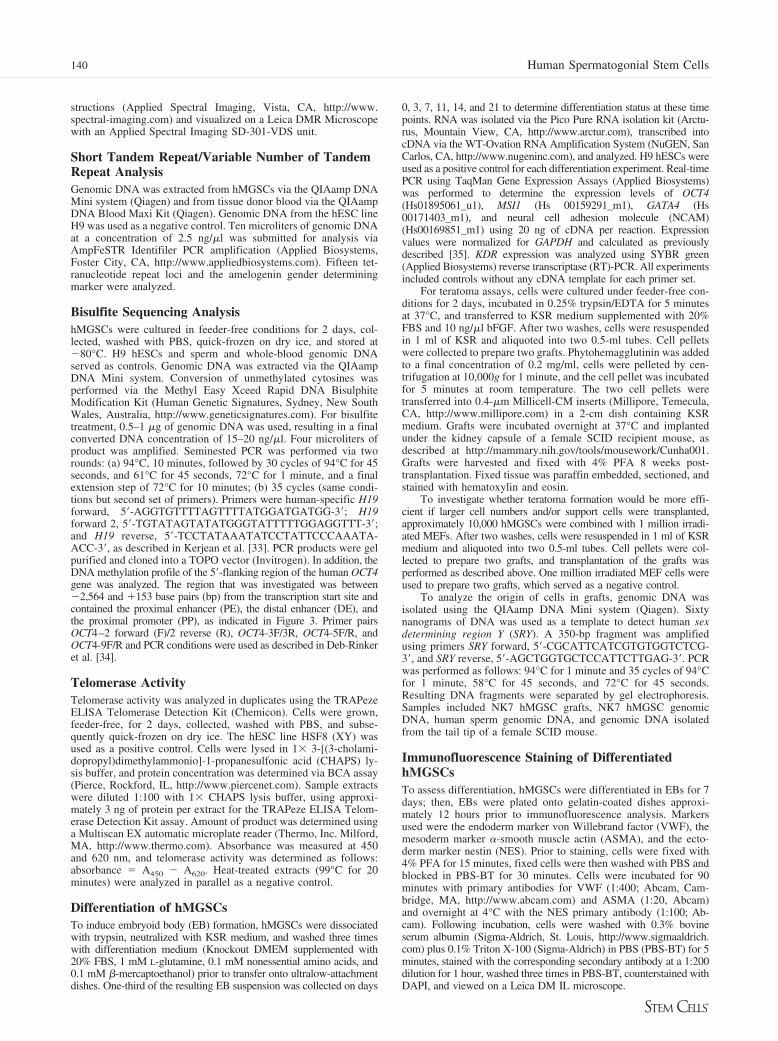
Figure 1. Morphological analysis and anal-ysis for the expression of both pluripotency-and germ cell-specific markers. (A): Lightmicroscopy of spermatogonial stem cell col-onies growing on top of a monolayer oftesticular cells approximately 2 weeks afterplating of the testicular cell suspension. (B):After six passages in culture. (C): Spermato-gonial stem cells in suspension. (Da): Ex-pression analysis of embryonic stem cell-and germ cell-specific markers in NK (NinaKossack) tissue sample 7 (NK7) humanmultipotent germline stem cells(hMGSCs) cultured on mouse embryonicfibroblasts after two passages and seven pas-sages. (Db, Dc): Expression analysis of NK7cells cultured on human testicular stromalcells after 10 passages and of a commer-cially available testis sample. (Dd): Reversetranscriptase-polymerase chain reactionproducts separated by gel electrophoresis.(E–H): Immunofluorescence staining ofhMGSCs at passage 8, for pluripotencymarkers: (E): stage-specific embryonic anti-gen 4. (F): TRA1–81. (G): Staining ofhMGSCs at passage 8 for embryonic stemcell and germ cell-specific marker alkalinephosphatase. Shown are two colonies withdifferent morphologies. (H): OCT4. (I):SOX2. (J): Staining of hMGSCs at passage8 for embryonic stem cell and germ cell-specific marker DAZL. (E, F, I, J) show thecolocalization of 4,6-diamidino-2-phenylin-dole with the corresponding pluripotency-germ cell marker on the left side and therespective marker on the right side of theimage. Scale bars � 50 �m.
141Kossack, Meneses, Shefi et al.
www.StemCells.com
As an alternative, manual passaging of colonies was explored.Following enzymatic dissociation of the testis biopsy, after approx-imately 7–10 days of culture, very small colonies started to grow ontop of the monolayer of testicular cells; these colonies were man-ually transferred onto MEFs and cultured under hESC conditions(Fig. 1A, 1B). These cells, which we have termed hMGSCs, havebeen propagated for approximately 20 passages in vitro; the currentline is designated NK7. The putative NK7 hMGSCs were passagedonce every week and have maintained the ability to form colonieswith characteristic hESC morphology. However, although the cellsin the middle of the colonies have a distinctive hESC-like appear-ance, some of the cells at the periphery appear to differentiate andacquire a spindle-shaped morphology, suggesting the need to op-timize medium and/or culture and derivation conditions (Fig. 1B).In suspension, NK7 cells continued to divide and formed EB-likestructures (Fig. 1C).
Gene Expression AnalysisRT-PCR was performed to analyze the expression of a subset ofpluripotency markers, as well as germ cell-specific genes, in the
isolated hMGSCs at passages 2 and 7 relative to a normalhuman testis sample (Fig. 1D). Results demonstrated that thehMGSCs at passages 2 and 7, grown on MEFs, express a subsetof those genes expressed in the testis, as shown (compare Fig.1Da, 1Db with Fig. 1Dd), which includes the pluripotencymarkers OCT4 (octamer-binding transcription factor-4) andSOX2 (SRY-box 2). NANOG expression, however, could not bedetected in either the isolated hMGSCs or the testis sample.Apart from that, expression of the hESC- and germ cell-enrichedgenes STELLAR (STELLA-related), GDF3 (growth and differ-entiation factor 3), PUM1 (PUMILIO 1), and PUM2 (PUMILIO2) was observed. In addition, the hMGSCs expressed the germcell-specific gene DAZL (Deleted in AZoospermia-Like), as wellas SCP3 (Syntaptonemal Complex Protein 3) and MLH1 (Mut-LHomolog 1). In contrast, expression of the markers VASA andSCP1 was not detected, nor was the expression of the twodevelopmentally late germ cell markers BOULE and TEKT1(Fig. 1D). Notably, however, when we cultured NK7 cells onhuman testicular stromal cells, we observed the induction ofexpression of later germ cell markers, including BOULE and
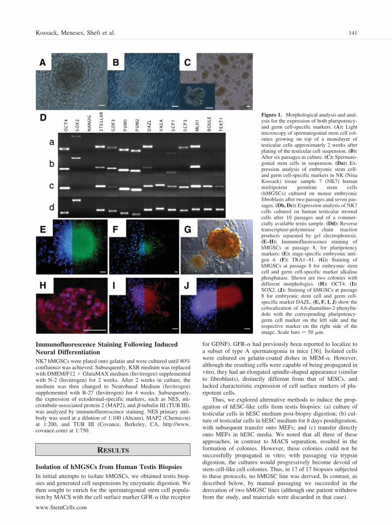
Figure 2. Human multipotent germline stem cells (hMGSCs) have a normal karyotype and express telomerase. (A): The DNA spectral karyotypingexperiment of undifferentiated hMGSCs at passage 8 demonstrates a normal (46,XY) karyotype. The spectral image, the 4,6-diamidino-2-phenylindolestaining, and the resulting chromosome table are shown. (B): Telomerase activity was investigated in hMGSCs at passage 6 (NK7A) and passage 8 (NK7B)using the TRAPeze ELISA. A cell extract from HSF8 hESCs served as a positive control for this experiment. The telomerase activity was calculated asaverage change in absorbance of the sample at 450 nm minus the absorbance at 690 nm. The black bars represent the samples without heat treatment, andthe white bars represent the samples with heat treatment. Abbreviations: A, absorbance; hESC, human embryonic stem cell.
142 Human Spermatogonial Stem Cells
TEKT1, and loss of SOX2 expression (Fig. 1Dc). We thereforeconcluded that NK7 cells lose the expression of later germ cellmarkers if cultured under human ESC conditions and regain theexpression of pluripotency genes, such as SOX2, if cultured onMEFs in human ESC conditions.
Our next aim was to examine the expression of pluripotencymarkers in hMGSCs by immunofluorescence (Fig. 1E–1J). Pu-tative hMGSCs were shown to express the human pluripotencymarkers SSEA4 (Fig. 1E), TRA1–81 (keratin sulfate-relatedantigens; Fig. 1F), OCT4 (Fig. 1H), and SOX2 (Fig. 1I). Inaddition, the hMGSCs also stained positive for the early germcell and hESC marker TNAP (Fig. 1G), as well as the germ celllineage marker DAZL (Fig. 1J). Negative controls for all ex-periments demonstrated that antibodies were specific, as ex-pected.
Spectral KaryotypeTo determine the karyotype of the derived NK7 hMGSC line,SKY analysis was performed. Results demonstrated that theNK7 hMGSC line has a normal karyotype (46, XY) and no Ychromosome microdeletions. No indications of other cytoge-netic abnormalities were detected (Fig. 2A). This indicated thatthe derived cell line was karyotypically identical to the patient’ssomatic cells, at this level of analysis.
Telomerase Activity and Methylation of the H19Differentially Methylated Region and the OCT4Promoter RegionTelomerase activity is indicative of pluripotent stem cells. Weexamined telomerase activity of the hMGSCs at passages 6 and8 relative to the human XY-bearing ESC line HSF8, as apositive control. As expected, hESCs exhibited a very hightelomerase activity with little or no residual activity in theheat-inactivated control. Telomerase activity was also detectedin the two hMGSC extracts, with the level of telomerase activityslightly reduced in cells that had been cultured for eight pas-sages relative to those cultured for six passages (Fig. 2B).
Short Tandem Repeat/Variable Number of TandemRepeat AnalysisShort tandem repeat (STR)/variable number of tandem repeat(VNTR) analysis was performed to determine the origin of theNK7 hMGSCs. Samples analyzed were genomic DNA isolatedfrom NK7 hMGSCs, genomic DNA from the tissue donor’sblood sample, and genomic DNA from H9 hESCs. The results(Table 1) demonstrate that the number of short tandem repeatson both alleles of the 15 loci that were analyzed is identical inNK7 hMGSCs and the tissue donor’s blood sample. The prob-ability that two randomly selected individuals would have anidentical genotype at these 15 loci is minuscule (5.01 � 10�18
[37]). Although H9 cells have the same number of short tandemrepeats as the NK7 hMGSCs on both alleles of the HUMTHO1locus and on one allele of the D16S539, D18S51, and D5S818loci, the number of short tandem repeats at all other examinedloci differed between H9 hESCs and the NK7 hMGSC line.
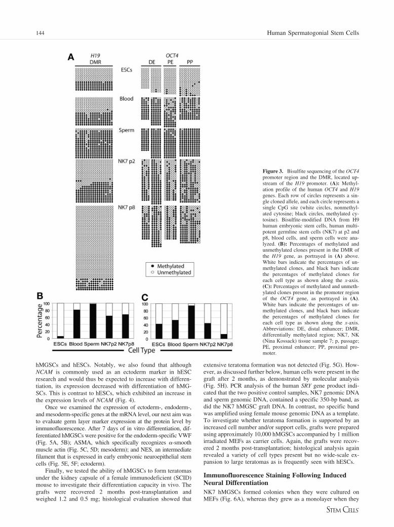
Bisulfite SequencingBisulfite sequencing was performed to investigate the methyl-ation status of 18 CpG (cytosine guanine) dinucleotides in thedifferentially methylated region upstream of the H19 promoter(Fig. 3A). Although the maternal H19 allele is active andtherefore unmethylated, the paternal H19 allele is methylated inall somatic cells [38, 39]. Human ESCs, as well as humansomatic cells, carry one paternal and one maternal allele andshowed a ratio of 70%:30% and 50%:50% unmethylated to
methylated sequences, respectively, as shown (Fig. 3B). Incontrast, in mature sperm, the paternal allele of the H19 genewas completely methylated (100% of clones), indicative of theestablishment of the unique male-specific methylation pattern atthis locus during this stage of development, (Fig. 3A). In con-trast, when we examined the methylation status of H19 in NK7hMGSCs at passage 8, we observed that this locus was hypo-methylated, with 87% of clones unmethylated and only 13%methylated (Fig. 3A, 3C).
In addition, the DNA methylation profile of the 5�-flank-ing region of the human OCT4 gene was analyzed. Theregion investigated contains the PE, the DE, and the PP, asindicated in Figure 3A. In undifferentiated cells the majorityof CpG repeats in this region are unmethylated and the geneis therefore expressed. Analysis showed that 94% of CpGsites in the OCT4 promoter region of human ESCs are un-methylated, whereas only 19% of CpG repeats in blood cellsand 2% of CpG repeats in sperm cells were unmethylated.Analysis of the methylation status of the OCT4 promoterregion of NK7 cells at passages 2 and 8 showed that 36% and32% of CpG repeats were unmethylated, respectively (Fig.3A, 3C). This partial demethylation is in accordance with thefinding that the OCT4 gene is activated in NK7 cells, asdemonstrated by RT-PCR and immunofluorescence staining.
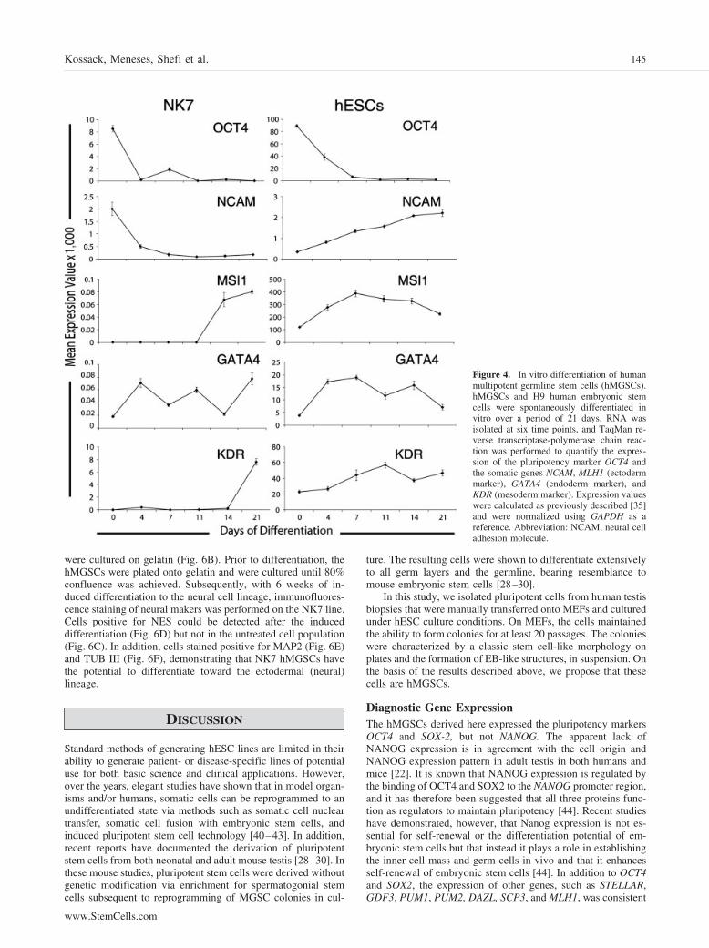
Spontaneous DifferentiationPluripotent stem cells can self-renew or differentiate to the threeprimary germ layers: endoderm, mesoderm, and ectoderm. Toassess whether hMGSCs are able to spontaneously differentiateinto derivatives of the three germ layers in vitro, expression ofectoderm-, endoderm-, and mesoderm-specific genes and pro-teins was analyzed at different time points during differentia-tion. H9 hESCs were used as a positive control (Fig. 4). Asshown, expression of the pluripotency marker OCT4 decreasedwith differentiation, with a concomitant increase in the expres-sion of the somatic markers MSI1 (ectoderm marker), GATA4(endoderm marker), and KDR (mesoderm marker) in both
Table 1. Variable number of tandem repeat/STR analysis of 15STR loci
STR loci NK7 hMGSCs NK7 donor blood H9 hESCs
Sex XY XY XXD8S1179 136/145 136/145 124/149D21S11 205/213 205/213 209/209D7S820 281/281 281/281 268/276CSF1PO 320/328 320/328 324/324D3S1358 133/137 133/137 116/129HUMTHO1 187/187 187/187 187/187D13S317 228/240 228/240 220/220D16S539 272/280 272/280 280/284D2S1338 328/347 328/347 319/343D19S433 110/122 110/122 114/126HUMvWA 168/188 168/188 180/180TPOX 230/230 230/230 238/242D18S51 288/292 288/292 287/288D5S818 152/152 152/152 152/156FGA 231/240 231/240 252/260
Samples analyzed were genomic DNA isolated from NK7hMGSCs, the tissue donor’s blood sample, and genomic DNAfrom H9 hESCs. The name of each STR locus and thecorresponding number of short tandem repeats for both alleles arelisted.Abbreviations: hESC, human embryonic stem cell; hMGSC,human multipotent germline stem cell; NK7, NK (Nina Kossack)tissue sample 7; STR, short tandem repeat.
143Kossack, Meneses, Shefi et al.
www.StemCells.com
hMGSCs and hESCs. Notably, we also found that althoughNCAM is commonly used as an ectoderm marker in hESCresearch and would thus be expected to increase with differen-tiation, its expression decreased with differentiation of hMG-SCs. This is contrast to hESCs, which exhibited an increase inthe expression levels of NCAM (Fig. 4).
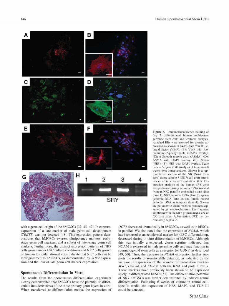
Once we examined the expression of ectoderm-, endoderm-,and mesoderm-specific genes at the mRNA level, our next aim wasto evaluate germ layer marker expression at the protein level byimmunofluorescence. After 7 days of in vitro differentiation, dif-ferentiated hMGSCs were positive for the endoderm-specific VWF(Fig. 5A, 5B); ASMA, which specifically recognizes �-smoothmuscle actin (Fig. 5C, 5D; mesoderm); and NES, an intermediatefilament that is expressed in early embryonic neuroepithelial stemcells (Fig. 5E, 5F; ectoderm).
Finally, we tested the ability of hMGSCs to form teratomasunder the kidney capsule of a female immunodeficient (SCID)mouse to investigate their differentiation capacity in vivo. Thegrafts were recovered 2 months post-transplantation andweighed 1.2 and 0.5 mg; histological evaluation showed that
extensive teratoma formation was not detected (Fig. 5G). How-ever, as discussed further below, human cells were present in thegraft after 2 months, as demonstrated by molecular analysis(Fig. 5H). PCR analysis of the human SRY gene product indi-cated that the two positive control samples, NK7 genomic DNAand sperm genomic DNA, contained a specific 350-bp band, asdid the NK7 hMGSC graft DNA. In contrast, no specific bandwas amplified using female mouse genomic DNA as a template.To investigate whether teratoma formation is supported by anincreased cell number and/or support cells, grafts were preparedusing approximately 10,000 hMGSCs accompanied by 1 millionirradiated MEFs as carrier cells. Again, the grafts were recov-ered 2 months post-transplantation; histological analysis againrevealed a variety of cell types present but no wide-scale ex-pansion to large teratomas as is frequently seen with hESCs.
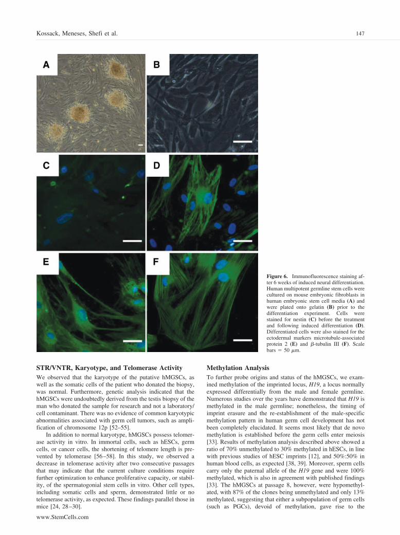
Immunofluorescence Staining Following InducedNeural DifferentiationNK7 hMGSCs formed colonies when they were cultured onMEFs (Fig. 6A), whereas they grew as a monolayer when they
Figure 3. Bisulfite sequencing of the OCT4promoter region and the DMR, located up-stream of the H19 promoter. (A): Methyl-ation profile of the human OCT4 and H19genes. Each row of circles represents a sin-gle cloned allele, and each circle represents asingle CpG site (white circles, nonmethyl-ated cytosine; black circles, methylated cy-tosine). Bisulfite-modified DNA from H9human embryonic stem cells, human multi-potent germline stem cells (NK7) at p2 andp8, blood cells, and sperm cells were ana-lyzed. (B): Percentages of methylated andunmethylated clones present in the DMR ofthe H19 gene, as portrayed in (A) above.White bars indicate the percentages of un-methylated clones, and black bars indicatethe percentages of methylated clones foreach cell type as shown along the x-axis.(C): Percentages of methylated and unmeth-ylated clones present in the promoter regionof the OCT4 gene, as portrayed in (A).White bars indicate the percentages of un-methylated clones, and black bars indicatethe percentages of methylated clones foreach cell type as shown along the x-axis.Abbreviations: DE, distal enhancer; DMR,differentially methylated region; NK7, NK(Nina Kossack) tissue sample 7; p, passage;PE, proximal enhancer; PP, proximal pro-moter.
144 Human Spermatogonial Stem Cells
were cultured on gelatin (Fig. 6B). Prior to differentiation, thehMGSCs were plated onto gelatin and were cultured until 80%confluence was achieved. Subsequently, with 6 weeks of in-duced differentiation to the neural cell lineage, immunofluores-cence staining of neural makers was performed on the NK7 line.Cells positive for NES could be detected after the induceddifferentiation (Fig. 6D) but not in the untreated cell population(Fig. 6C). In addition, cells stained positive for MAP2 (Fig. 6E)and TUB III (Fig. 6F), demonstrating that NK7 hMGSCs havethe potential to differentiate toward the ectodermal (neural)lineage.
DISCUSSION
Standard methods of generating hESC lines are limited in theirability to generate patient- or disease-specific lines of potentialuse for both basic science and clinical applications. However,over the years, elegant studies have shown that in model organ-isms and/or humans, somatic cells can be reprogrammed to anundifferentiated state via methods such as somatic cell nucleartransfer, somatic cell fusion with embryonic stem cells, andinduced pluripotent stem cell technology [40–43]. In addition,recent reports have documented the derivation of pluripotentstem cells from both neonatal and adult mouse testis [28–30]. Inthese mouse studies, pluripotent stem cells were derived withoutgenetic modification via enrichment for spermatogonial stemcells subsequent to reprogramming of MGSC colonies in cul-
ture. The resulting cells were shown to differentiate extensivelyto all germ layers and the germline, bearing resemblance tomouse embryonic stem cells [28–30].
In this study, we isolated pluripotent cells from human testisbiopsies that were manually transferred onto MEFs and culturedunder hESC culture conditions. On MEFs, the cells maintainedthe ability to form colonies for at least 20 passages. The colonieswere characterized by a classic stem cell-like morphology onplates and the formation of EB-like structures, in suspension. Onthe basis of the results described above, we propose that thesecells are hMGSCs.
Diagnostic Gene ExpressionThe hMGSCs derived here expressed the pluripotency markersOCT4 and SOX-2, but not NANOG. The apparent lack ofNANOG expression is in agreement with the cell origin andNANOG expression pattern in adult testis in both humans andmice [22]. It is known that NANOG expression is regulated bythe binding of OCT4 and SOX2 to the NANOG promoter region,and it has therefore been suggested that all three proteins func-tion as regulators to maintain pluripotency [44]. Recent studieshave demonstrated, however, that Nanog expression is not es-sential for self-renewal or the differentiation potential of em-bryonic stem cells but that instead it plays a role in establishingthe inner cell mass and germ cells in vivo and that it enhancesself-renewal of embryonic stem cells [44]. In addition to OCT4and SOX2, the expression of other genes, such as STELLAR,GDF3, PUM1, PUM2, DAZL, SCP3, and MLH1, was consistent
Figure 4. In vitro differentiation of humanmultipotent germline stem cells (hMGSCs).hMGSCs and H9 human embryonic stemcells were spontaneously differentiated invitro over a period of 21 days. RNA wasisolated at six time points, and TaqMan re-verse transcriptase-polymerase chain reac-tion was performed to quantify the expres-sion of the pluripotency marker OCT4 andthe somatic genes NCAM, MLH1 (ectodermmarker), GATA4 (endoderm marker), andKDR (mesoderm marker). Expression valueswere calculated as previously described [35]and were normalized using GAPDH as areference. Abbreviation: NCAM, neural celladhesion molecule.
145Kossack, Meneses, Shefi et al.
www.StemCells.com
with a germ cell origin of the hMGSCs [32, 45–47]. In contrast,expression of a late marker of male germ cell development(TEKT1) was not detected [48]. This expression pattern dem-onstrates that hMGSCs express pluripotency markers, early-stage germ cell markers, and a subset of later-stage germ cellmarkers. Furthermore, the distinct expression patterns of NK7cells grown under ESC culture conditions and NK7 cells grownon human testicular stromal cells indicate that NK7 cells can bereprogrammed to hMGSCs, as demonstrated by SOX2 expres-sion and the loss of late germ cell marker expression.
Spontaneous Differentiation In VitroThe results from the spontaneous differentiation experimentclearly demonstrated that hMGSCs have the potential to differ-entiate into derivatives of the three primary germ layers in vitro.When transferred to differentiation media, the expression of
OCT4 decreased dramatically in hMGSCs, as well as in hESCs,in parallel. We also noted that the expression of NCAM, whichhas been used as an ectodermal marker for hESC differentiation,decreased during in vitro differentiation of hMGSCs. Althoughthis was initially unexpected, closer scrutiny indicated thatNCAM is expressed in male germline cells and may function inspermatogonial stem cells as a receptor for GDNF, as described[49, 50]. Thus, the decrease in NCAM expression further sup-ports the results of somatic differentiation, as indicated by theincrease in expression of the somatic differentiation markersMSI1, GATA4, and KDR at both the RNA and protein levels.These markers have previously been shown to be expressedsolely in differentiated hESCs [51]. The differentiation potentialof NK7 hMGSCs was further demonstrated by induced neuraldifferentiation. Following 6 weeks of culture in neural cell-specific media, the expression of NES, MAP2, and TUB IIIcould be detected.
Figure 5. Immunofluorescence staining ofday 7 differentiated human multipotentgermline stem cells and teratoma analysis.Attached EBs were assessed for protein ex-pression as shown in (A-F). (A): von Wille-brand factor (VWF). (B): VWF with 4,6-diamidino-2-phenylindole (DAPI) overlay.(C): �-Smooth muscle actin (ASMA). (D):ASMA with DAPI overlay. (E): Nestin(NES). (F): NES with DAPI overlay. Scalebars � 50 �m. (G): Analysis of teratomas 8weeks post-transplantation. Shown is a rep-resentative section of the NK (Nina Kos-sack) tissue sample 7 (NK7) cell graft after 8weeks of in vivo differentiation. (H): Ex-pression analysis of the human SRY genewas performed using genomic DNA isolatedfrom an NK7 paraffin-embedded tissue slide(lane 1), NK7 genomic DNA (lane 2), spermgenomic DNA (lane 3), and female mousegenomic DNA as template (lane 4). Shownare polymerase chain reaction products sep-arated by gel electrophoreses. The fragmentamplified with the SRY primers had a size of350 base pairs. Abbreviation: SRY, sex de-termining region Y.
146 Human Spermatogonial Stem Cells
STR/VNTR, Karyotype, and Telomerase ActivityWe observed that the karyotype of the putative hMGSCs, aswell as the somatic cells of the patient who donated the biopsy,was normal. Furthermore, genetic analysis indicated that thehMGSCs were undoubtedly derived from the testis biopsy of theman who donated the sample for research and not a laboratory/cell contaminant. There was no evidence of common karyotypicabnormalities associated with germ cell tumors, such as ampli-fication of chromosome 12p [52–55].
In addition to normal karyotype, hMGSCs possess telomer-ase activity in vitro. In immortal cells, such as hESCs, germcells, or cancer cells, the shortening of telomere length is pre-vented by telomerase [56–58]. In this study, we observed adecrease in telomerase activity after two consecutive passagesthat may indicate that the current culture conditions requirefurther optimization to enhance proliferative capacity, or stabil-ity, of the spermatogonial stem cells in vitro. Other cell types,including somatic cells and sperm, demonstrated little or notelomerase activity, as expected. These findings parallel those inmice [24, 28–30].
Methylation AnalysisTo further probe origins and status of the hMGSCs, we exam-ined methylation of the imprinted locus, H19, a locus normallyexpressed differentially from the male and female germline.Numerous studies over the years have demonstrated that H19 ismethylated in the male germline; nonetheless, the timing ofimprint erasure and the re-establishment of the male-specificmethylation pattern in human germ cell development has notbeen completely elucidated. It seems most likely that de novomethylation is established before the germ cells enter meiosis[33]. Results of methylation analysis described above showed aratio of 70% unmethylated to 30% methylated in hESCs, in linewith previous studies of hESC imprints [12], and 50%:50% inhuman blood cells, as expected [38, 39]. Moreover, sperm cellscarry only the paternal allele of the H19 gene and were 100%methylated, which is also in agreement with published findings[33]. The hMGSCs at passage 8, however, were hypomethyl-ated, with 87% of the clones being unmethylated and only 13%methylated, suggesting that either a subpopulation of germ cells(such as PGCs), devoid of methylation, gave rise to the
Figure 6. Immunofluorescence staining af-ter 6 weeks of induced neural differentiation.Human multipotent germline stem cells werecultured on mouse embryonic fibroblasts inhuman embryonic stem cell media (A) andwere plated onto gelatin (B) prior to thedifferentiation experiment. Cells werestained for nestin (C) before the treatmentand following induced differentiation (D).Differentiated cells were also stained for theectodermal markers microtubule-associatedprotein 2 (E) and �-tubulin III (F). Scalebars � 50 �m.
147Kossack, Meneses, Shefi et al.
www.StemCells.com
hMGSCs or, alternatively, that reprogramming of the hMGSCsled to imprint erasure.
Recent studies have shown that reprogramming of somaticcells is associated with demethylation of OCT4 regulatory re-gions, with the most apparent changes occurring in the PE, DE,and PP regions. Mosaic CpG demethylation has been shown tobe physiologically important, as it leads to the activation of thegene. Analysis of the methylation status of the OCT4 promoterregion of NK7 cells at passages 2 and 8 demonstrated that 36%and 32% of CpG repeats are unmethylated. This partial demeth-ylation is in agreement with the activation of the OCT4 gene andsupports the theory that human spermatogonial stem cells aremultipotent when cultured under human ESC culture conditions.
Teratoma AssayThe results of in vivo differentiation analysis merit furthercomment. We observed that hMGSCs did not induce formationof a large teratoma (which may or may not be beneficial forputative clinical applications). Nonetheless, PCR analysis usingprimers specific for the human SRY gene indicated the presenceof human cells in the graft even after 2 months. The most likelyexplanation for this finding is that some of the human cellspersist but that a larger number of cells is required for furtherteratoma analysis (4,000, as used here, is at the lower limit ofdetection without MEFs serving as a carrier [59]). Repetition ofthe teratoma assay using 10,000 hMGSCs accompanied by 1million irradiated MEF cells did not lead to teratoma formationafter transplantation, even though 500–1,000 murine embryonicstem cells accompanied by 99,000 MEFs have been shown to besufficient to induce tumor growth [59]. These results indicatethat although hMGSCs appear to have the potential to differen-tiate into derivatives of the three germ layers upon spontaneousor induced in vitro differentiation, they may not have beenreprogrammed sufficiently to generate teratomas.
CONCLUSION
The ability to isolate and culture hMGSCs in vitro may facilitatedevelopment of novel therapeutic strategies for the treatment of
infertility. For example, one side effect of cancer treatments isthe potential destruction of spermatogonial stem cells, alongwith the cancer cells, with the possibility of leaving the patientinfertile [60]. To maintain fertility, testicular biopsies could beobtained prior to the treatment, and spermatogonial stem cellscould be propagated in vitro and finally transplanted back intothe patient’s testis when the treatment is completed, if germ celldevelopment can be controlled.
Recent studies in mice have also indicated that spermatogo-nial stem cells can acquire ESC traits via an as yet poorlydefined reprogramming process [28–30]. We anticipate thatpluripotent hMGSCs represent a source of patient-specific stemcells appropriate for the study of genetic diseases in differentcell lineages in vitro and for potential novel therapeutic appli-cations with particular application to fertility [61]. Notably,these cells are not genetically modified as is required for gen-eration of induced pluripotent stem cells that can be derivedfrom fetal or adult somatic cells [41–43]. Nonetheless, ourresults suggest that the efficient derivation of hMGSCs mayrequire full reprogramming through the optimization of theculture conditions. In this regard, recent studies in nonhumanprimates illuminate fundamental properties of spermatogoniathat may inform future efforts [62].
ACKNOWLEDGMENTS
We thank current and previous members of the Reijo-Peralaboratory for helpful discussions and technical support. Thiswork was supported in part by a fellowship from the GottliebDaimler and Karl Benz Foundation (to N.K.) and by grants fromthe California Institute of Regenerative Medicine (to R.A.R.-P.)and the NIH.
DISCLOSURE OF POTENTIAL CONFLICTS
OF INTEREST
The authors indicate no potential conflicts of interest.
REFERENCES
1 Martin G. Isolation of a pluripotent cell line from early mouse embryoscultured in medium conditioned by teratocarcinoma stem cells. Proc NatlAcad Sci U S A 1981;78:7634–7638.
2 Kluin PM, de Rooij DG. A comparison between the morphology and cellkinetics of gonocytes and adult type undifferentiated spermatogonia inthe mouse. Int J Androl 1981;4:475–493.
3 Rossant J. Stem cells from the mammalian blastocyst. STEM CELLS2001;19:477–482.
4 Evans M, Hunter S. Source and nature of embryonic stem cells. C R Biol2002;325:1003–1007.
5 Buehr M, Smith A. Genesis of embryonic stem cells. Philos Trans R SocLond B Biol Sci 2003;358:1397–1402.
6 Donovan PJ, de Miguel MP. Turning germ cells into stem cells. CurrOpin Genet Dev 2003;13:463–471.
7 Thomson J, Itskovitz-Eldor J, Shapiro S et al. Embryonic stem cell linesderived from human blastocysts. Science 1998;282:1145–1147.
8 Shamblott MJ, Axelman J, Wang S et al. Derivation of pluirpotent stemcells from cultured human primordial germ cells. Proc Natl Acad Sci US A 1998;95:13726–13731.
9 Shamblott MJ, Axelman J, Littlefield JW et al. Human embryonic germcell derivatives express a broad range of developmentally distinct mark-ers and proliferate extensively in vitro. Proc Natl Acad Sci U S A2001;98:113–118.
10 Onyango P, Jiang S, Uejima H et al. Monoallelic expression and meth-ylation of imprinted genes in human and mouse embryonic germ celllineages. Proc Natl Acad Sci U S A 2002;99:10599–10604.
11 Brivanlou AH, Gage FH, Jaenisch R et al. Setting standards for humanembryonic stem cells. Science 2003;300:913–916.
12 Adewumi O, Aflatoonian B, Ahrlund-Richter L et al. Characterization ofhuman embryonic stem cell lines by the International Stem Cell Initia-tive. Nat Biotechnol 2007;25:803–816.
13 McLaren A. Primordial germ cells in the mouse. Dev Biol 2003;262:1–15.
14 Chiquoine A. The identification, origin and migration of the primordialgerm cells in the mouse embryo. Anat Rec 1954;118:135–146.
15 Scholer H, Ruppert S, Suzuki N et al. New type of POU domain in germline-specific protein Oct-4. Nature 1990;344:435–439.
16 Scholer H, Dressler G, Balling R et al. Oct-4: A germ line specifictranscription factor mapping to the mouse t-complex. EMBO J 1990;9:2185–2195.
17 Saitou M, Barton SC, Surani MA. A molecular programme for thespecification of germ cell fate in mice. Nature 2002;418:293–300.
18 McLaren A, Southee D. Entry of mouse embryonic germ cells intomeiosis. Dev Biol 1997;187:107–113.
19 Adams IR, McLaren A. Sexually dimorphic development of mouseprimordial germ cells: Switching from oogenesis to spermatogenesis.Development 2002;129:1155–1164.
20 Menke D, Koubova J, Page D. Sexual differentiation of germ cells in XXmouse gonads occurs in an anterior-to-posterior wave. Dev Biol 2003;262:303–312.
21 Brinster RL. Germline stem cell transplantation and transgenesis. Sci-ence 2002;296:2174–2176.
22 Oatley JM, Avarbock MR, Telaranta AI et al. Identifying genes impor-tant for spermatogonial stem cell self-renewal and survival. Proc NatlAcad Sci U S A 2006;103:9524–9529.
23 Durcova-Hills G, Ainscough J, McLaren A. Pluripotential stem cells
148 Human Spermatogonial Stem Cells
derived from migrating primordial germ cells. Differentiation 2001;68:220–226.
24 Feng LX, Chen Y, Dettin L et al. Generation and in vitro differentiationof a spermatogonial cell line. Science 2002;297:392–395.
25 McLean DJ, Friel PJ, Johnston DS et al. Characterization of spermato-gonial stem cell maturation and differentiation in neonatal mice. BiolReprod 2003;69:2085–2091.
26 Buaas F, Kirsh A, Sharma M et al. Plzf is required in adult male germcells for stem cell self-renewal. Nat Genet 2004;36:647–652.
27 Koubova J, Menke D, Zhou Q et al. Retinoic acid regulates sex-specific timing of meiotic initiation in mice. Proc Natl Acad Sci2006;103:2474 –2479.
28 Kanatsu-Shinohara M, Inoue K, Lee J et al. Generation of pluripotentstem cells from neonatal mouse testis. Cell 2004;119:1001–1012.
29 Guan K, Nayernia K, Maier L et al. Pluripotency of spermatogonial stemcells from adult mouse testis. Nature 2006;440:1199–1203.
30 Seandel M, James D, Shmelkov S et al. Generation of functional multi-potent adult stem cells from GPR125� germline progenitors. Nature2007;449:346–350.
31 Meng X, Lindahl M, Hyvonen ME et al. Regulation of cell fatedecision of undifferentiated spermatogonia by GDNF. Science 2000;287:1489 –1493.
32 Clark AT, Bodnar MS, Fox MS et al. Spontaneous differentiation ofgerm cells from human embryonic stem cells in vitro. Hum Mol Genet2004;13:727–739.
33 Kerjean A, Dupont J, Vasseur C et al. Establishment of the paternalmethylation imprint of the human H19 and MEST/PEG1 genes duringspermatogenesis. Hum Mol Genet 2000;9:2183–2187.
34 Deb-Rinker P, Ly D, Jezierski A et al. Sequential DNA methylation ofthe Nanog and Oct-4 upstream regions in human NT2 cells duringneuronal differentiation. J Biol Chem 2005;280:6257–6260.
35 Livak KJ, Schmittgen KD. Analysis of relative gene expression datausing real time quantitative PCR and the 2-deltaCT Method. Methods2001;25:402–408.
36 Hofmann M, Braydich-Stolle L, Dym M. Isolation of male germ-linestem cells; influence of GDNF. Dev Biol 2005;279:114–124.
37 Sensabaugh G. Isozymes in forensic science. Isozymes Curr Top BiolMed Res 1982;6:247–282.
38 Ferguson-Smith AC, Sasaki H, Cattanach BM et al. Parental-origin-specific epigenetic modification of the mouse H19 gene. Nature 1993;362:751–755.
39 Bartolomei MS, Webber AL, Brunkow ME et al. Epigenetic mechanismsunderlying the imprinting of the mouse H19 gene. Genes Dev 1993;7:1663–1673.
40 Wakayama T, Tabar V, Rodriguez I et al. Differentiation of embryonicstem cell lines generated from adult somatic cells by nuclear transfer.Science 2001;292:740–743.
41 Yu J, Vodyanik M, Smuga-Otto K et al. Induced pluripotent stem celllines derived from human somatic cells. Science 2007;318:1917–1920.
42 Takahashi K, Tanabe K, Ohnuki M et al. Induction of pluripotent stemcells from adult human fibroblasts by defined factors. Cell 2007;131:861–872.
43 Park I, Zhao R, West J et al. Reprogramming of human somatic cells topluripotency with defined factors. Nature 2008;451:141–146.
44 Chambers I, Silva J, Colby D et al. Nanog safeguards pluripotency andmediates germline development. Nature 2007;450:1230–1234.
45 Moore FL, Jaruzelska J, Fox MS et al. Human Pumilio-2 is expressed inembryonic stem cells and germ cells and interacts with DAZ (Deleted inAZoospermia) and DAZ-Like proteins. Proc Natl Acad Sci 2003;100:538–543.
46 Reijo RA, Dorfman DM, Slee R et al. DAZ family proteins existthroughout male germ cell development and transit from nucleus tocytoplasm at meiosis in humans and mice. Biol Reprod 2000;63:1490 –1496.
47 Lynn A, Koehler KE, Judis L et al. Covariation of synaptonemal com-plex length and mammalian meiotic exchange rates. Science 2002;296:2222–2225.
48 Larsson M, Norrander J, Graslund S et al. The spatial and temporalexpression of Tekt1, a mouse tektin C homologue, during spermatogen-esis suggest that it is involved in the development of the sperm tail basalbody and axoneme. Eur J Cell Biol 2000;79:718–725.
49 Fox MS, Ares VX, Turek PJ et al. Feasibility of global gene expressionanalysis in testicular biopsies from infertile men. Mol Reprod Dev2003;66:403–421.
50 Ryu BY, Kubota H, Avarbock MR et al. Conservation of spermatogonialstem cell self-renewal signaling between mouse and rat. Proc Natl AcadSci U S A 2005;102:14302–14307.
51 Cai J, Chen J, Liu Y et al. Assessing self-renewal and differentiation inhuman embryonic stem cell lines. STEM CELLS 2006;24:516–530.
52 Suijkerbuijk RF, Sinke RJ, Meloni AM et al. Overrepresentation ofchromosome 12p sequences and karyotopic evolution in i(12p)-negativetesticular germ-cell tumors revealed by fluorescence in situ hybridiza-tion. Cancer Genet Cytogenet 1993;70:85–93.
53 Geurts van Kessel A, Suijkerbuijk RF, Sinke RJ et al. Molecular cyto-genetics of human germ cell tumours: i(12p) and related chromosomalanomalies. Eur Urol 1993;23:23–28.
54 Lothe RA, Peltomaki P, Tommerup N et al. Molecular genetic changesin human male germ cell tumors. Lab Invest 1995;73:606–614.
55 Clark AT, Rodriguez R, Bodnar M et al. Human STELLAR, NANOG,and GDF3 genes are expressed in pluripotent cells and map to chromo-some 12p13, a hot-spot for teratocarcinoma. STEM CELLS 2004;22:169–179.
56 Kim NW, Piatyszek MA, Prowse KR et al. Specific association of humantelomerase activity with immortal cells and cancer. Science 1994;266:2011–2015.
57 Shay JW, Wright WE. Telomerase activity in human cancer. Curr OpinOncol 1996;8:66–71.
58 Ravindranath N, Dalal R, Solomon B et al. Loss of telomerase activityduring male germ cell differentiation. Endocrinology 1997;138:4026 – 4029.
59 Cao F, van der Bogt KE, Sadrzadeh A et al. Spatial and temporal kineticsof teratoma formation from murine embryonic stem cell transplantation.Stem Cells Dev 2007;16:883–891.
60 Kubota H, Brinster RL. Technology insight: In vitro culture of spermato-gonial stem cells and their potential therapeutic uses. Nat Clin PractEndocrinol Metab 2006;2:99–108.
61 Nayernia K. Stem cells derived from testis show promise for treating awide variety of medical conditions. Cell Res 2007;17:895–897.
62 Hermann B, Sukhwani M, Lin C et al. Characterization, cryopreserva-tion, and ablation of spermatogonial stem cells in adult rhesus macaques.STEM CELLS 2007;25:2330–2338.
See www.StemCells.com for supporting information available online.
149Kossack, Meneses, Shefi et al.
www.StemCells.com