Manipulation of spermatogonial stem cells in livestock species
18
REVIEW Open Access Manipulation of spermatogonial stem cells in livestock species Filipp Savvulidi 1,2* , Martin Ptacek 1 , Karina Savvulidi Vargova 2 and Ludek Stadnik 1 Abstract We are entering an exciting epoch in livestock biotechnology during which the fundamental approaches (such as transgenesis, spermatozoa cryopreservation and artificial insemination) will be enhanced based on the modern understanding of the biology of spermatogonial stem cells (SSCs) combined with the outstanding recent advances in genomic editing technologies and in vitro cell culture systems. The general aim of this review is to outline comprehensively the promising applications of SSC manipulation that could in the nearest future find practical application in livestock breeding. Here, we will focus on 1) the basics of mammalian SSC biology; 2) the approaches for SSC isolation and purification; 3) the available in vitro systems for the stable expansion of isolated SSCs; 4) a discussion of how the manipulation of SSCs can accelerate livestock transgenesis; 5) a thorough overview of the techniques of SSC transplantation in livestock species (including the preparation of recipients for SSC transplantation, the ultrasonographic-guided SSC transplantation technique in large farm animals, and the perspectives to improve further the SSC transplantation efficiency), and finally, 6) why SSC transplantation is valuable to extend the techniques of spermatozoa cryopreservation and/or artificial insemination. For situations where no reliable data have yet been obtained for a particular livestock species, we will rely on the data obtained from studies conducted in rodents because the knowledge gained from rodent research is translatable to livestock species to a great extent. On the other hand, we will draw special attention to situations where such translation is not possible. Keywords: CRISPR/Cas9, Genome editing, Livestock transgenesis, Long-term culture systems, Male germline stem cells, Recipient preparation, Sertoli cells, Spermatogonial stem cells, Ultrasonographic-guided cannulation Introduction to mammalian spermatogenesis Spermatogonial stem cells Spermatogenesis is a delicately orchestrated process of continuous sperm cell production when billions of haploid spermatozoa are produced daily. The reproductive system of sexually mature males requires the presence of a stem cell pool to maintain an extremely high level of daily sperm pro- duction (as high as 31.3 billion of spermatozoa in a boar [1], 12.9 billion in a ram [2], 11.5 billion in a bull [3], 8 billion in a stallion [4] or 6.4 billion in a buck [5], [reviewed in [6]). The process of mammalian spermatogenesis occurs in the seminiferous tubules, the gametogenic compartment of the mammalian testes, and in the interstitium, where the steroidogenic activity in the testis occurs. SSCs, a group of spermatogonia, represent the foundation of spermatogen- esis. These cells have a unique self-renewal ability (the ability to divide without differentiation) and a differenti- ation ability while giving rise to the haploid spermatozoa [7]. SSCs originate from the gonocytes (also known as prespermatogonia) in the postnatal testes. Gonocytes, in turn, originate from the primordial germ cells (PGCs) dur- ing fetal development [8–11]. Collectively, PGCs, gonocytes and SSCs are all referred to as male germline stem cells (MGSCs) because of their stem cell potential [12]. Among these, the SSCs are the overall focus of this review. SSC self-renewal: vital support from the niche In postnatal mammalian testes, SSCs reside within a spe- cialized microenvironmental compartment of the semin- iferous epithelium located near the basement membrane. These compartments, populated by SSCs and Sertoli cells, are canonically called niches. Moreover, there are several niche-related cell types and structures in the testicular © The Author(s). 2019 Open Access This article is distributed under the terms of the Creative Commons Attribution 4.0 International License (http://creativecommons.org/licenses/by/4.0/), which permits unrestricted use, distribution, and reproduction in any medium, provided you give appropriate credit to the original author(s) and the source, provide a link to the Creative Commons license, and indicate if changes were made. The Creative Commons Public Domain Dedication waiver (http://creativecommons.org/publicdomain/zero/1.0/) applies to the data made available in this article, unless otherwise stated. * Correspondence: [email protected] 1 Department of Animal Science, Faculty of Agrobiology, Food and Natural Resources, Czech University of Life Sciences Prague, Kamýcká 129, 165 00 Prague, Suchdol, Czech Republic 2 Institute of Pathological Physiology, First Faculty of Medicine, Charles University in Prague, U Nemocnice 5, 128 53 Prague, Czech Republic Savvulidi et al. Journal of Animal Science and Biotechnology (2019) 10:46 https://doi.org/10.1186/s40104-019-0355-4
-
Upload
khangminh22 -
Category
Documents
-
view
3 -
download
0
Transcript of Manipulation of spermatogonial stem cells in livestock species

REVIEW Open Access
Manipulation of spermatogonial stem cellsin livestock speciesFilipp Savvulidi1,2* , Martin Ptacek1, Karina Savvulidi Vargova2 and Ludek Stadnik1
Abstract
We are entering an exciting epoch in livestock biotechnology during which the fundamental approaches (such astransgenesis, spermatozoa cryopreservation and artificial insemination) will be enhanced based on the modernunderstanding of the biology of spermatogonial stem cells (SSCs) combined with the outstanding recent advancesin genomic editing technologies and in vitro cell culture systems. The general aim of this review is to outlinecomprehensively the promising applications of SSC manipulation that could in the nearest future find practicalapplication in livestock breeding. Here, we will focus on 1) the basics of mammalian SSC biology; 2) the approachesfor SSC isolation and purification; 3) the available in vitro systems for the stable expansion of isolated SSCs; 4) adiscussion of how the manipulation of SSCs can accelerate livestock transgenesis; 5) a thorough overview of thetechniques of SSC transplantation in livestock species (including the preparation of recipients for SSC transplantation,the ultrasonographic-guided SSC transplantation technique in large farm animals, and the perspectives to improvefurther the SSC transplantation efficiency), and finally, 6) why SSC transplantation is valuable to extend the techniquesof spermatozoa cryopreservation and/or artificial insemination. For situations where no reliable data have yet beenobtained for a particular livestock species, we will rely on the data obtained from studies conducted in rodents becausethe knowledge gained from rodent research is translatable to livestock species to a great extent. On the other hand, wewill draw special attention to situations where such translation is not possible.
Keywords: CRISPR/Cas9, Genome editing, Livestock transgenesis, Long-term culture systems, Male germline stem cells,Recipient preparation, Sertoli cells, Spermatogonial stem cells, Ultrasonographic-guided cannulation
Introduction to mammalian spermatogenesisSpermatogonial stem cellsSpermatogenesis is a delicately orchestrated process ofcontinuous sperm cell production when billions of haploidspermatozoa are produced daily. The reproductive systemof sexually mature males requires the presence of a stem cellpool to maintain an extremely high level of daily sperm pro-duction (as high as 31.3 billion of spermatozoa in a boar [1],12.9 billion in a ram [2], 11.5 billion in a bull [3], 8 billion ina stallion [4] or 6.4 billion in a buck [5], [reviewed in [6]).The process of mammalian spermatogenesis occurs in
the seminiferous tubules, the gametogenic compartment ofthe mammalian testes, and in the interstitium, where thesteroidogenic activity in the testis occurs. SSCs, a group of
spermatogonia, represent the foundation of spermatogen-esis. These cells have a unique self-renewal ability (theability to divide without differentiation) and a differenti-ation ability while giving rise to the haploid spermatozoa[7]. SSCs originate from the gonocytes (also known asprespermatogonia) in the postnatal testes. Gonocytes, inturn, originate from the primordial germ cells (PGCs) dur-ing fetal development [8–11]. Collectively, PGCs, gonocytesand SSCs are all referred to as male germline stem cells(MGSCs) because of their stem cell potential [12]. Amongthese, the SSCs are the overall focus of this review.
SSC self-renewal: vital support from the nicheIn postnatal mammalian testes, SSCs reside within a spe-cialized microenvironmental compartment of the semin-iferous epithelium located near the basement membrane.These compartments, populated by SSCs and Sertoli cells,are canonically called niches. Moreover, there are severalniche-related cell types and structures in the testicular
© The Author(s). 2019 Open Access This article is distributed under the terms of the Creative Commons Attribution 4.0International License (http://creativecommons.org/licenses/by/4.0/), which permits unrestricted use, distribution, andreproduction in any medium, provided you give appropriate credit to the original author(s) and the source, provide a link tothe Creative Commons license, and indicate if changes were made. The Creative Commons Public Domain Dedication waiver(http://creativecommons.org/publicdomain/zero/1.0/) applies to the data made available in this article, unless otherwise stated.
* Correspondence: [email protected] of Animal Science, Faculty of Agrobiology, Food and NaturalResources, Czech University of Life Sciences Prague, Kamýcká 129, 165 00Prague, Suchdol, Czech Republic2Institute of Pathological Physiology, First Faculty of Medicine, CharlesUniversity in Prague, U Nemocnice 5, 128 53 Prague, Czech Republic
Savvulidi et al. Journal of Animal Science and Biotechnology (2019) 10:46 https://doi.org/10.1186/s40104-019-0355-4
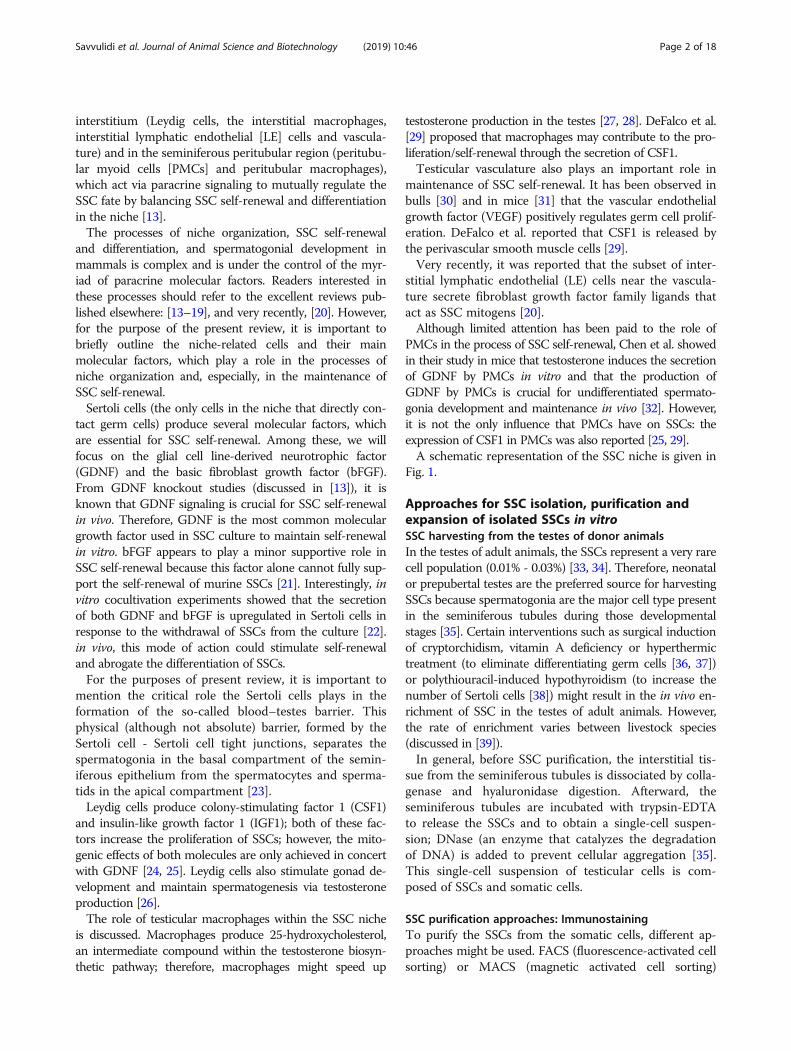
interstitium (Leydig cells, the interstitial macrophages,interstitial lymphatic endothelial [LE] cells and vascula-ture) and in the seminiferous peritubular region (peritubu-lar myoid cells [PMCs] and peritubular macrophages),which act via paracrine signaling to mutually regulate theSSC fate by balancing SSC self-renewal and differentiationin the niche [13].The processes of niche organization, SSC self-renewal
and differentiation, and spermatogonial development inmammals is complex and is under the control of the myr-iad of paracrine molecular factors. Readers interested inthese processes should refer to the excellent reviews pub-lished elsewhere: [13–19], and very recently, [20]. However,for the purpose of the present review, it is important tobriefly outline the niche-related cells and their mainmolecular factors, which play a role in the processes ofniche organization and, especially, in the maintenance ofSSC self-renewal.Sertoli cells (the only cells in the niche that directly con-
tact germ cells) produce several molecular factors, whichare essential for SSC self-renewal. Among these, we willfocus on the glial cell line-derived neurotrophic factor(GDNF) and the basic fibroblast growth factor (bFGF).From GDNF knockout studies (discussed in [13]), it isknown that GDNF signaling is crucial for SSC self-renewalin vivo. Therefore, GDNF is the most common moleculargrowth factor used in SSC culture to maintain self-renewalin vitro. bFGF appears to play a minor supportive role inSSC self-renewal because this factor alone cannot fully sup-port the self-renewal of murine SSCs [21]. Interestingly, invitro cocultivation experiments showed that the secretionof both GDNF and bFGF is upregulated in Sertoli cells inresponse to the withdrawal of SSCs from the culture [22].in vivo, this mode of action could stimulate self-renewaland abrogate the differentiation of SSCs.For the purposes of present review, it is important to
mention the critical role the Sertoli cells plays in theformation of the so-called blood–testes barrier. Thisphysical (although not absolute) barrier, formed by theSertoli cell - Sertoli cell tight junctions, separates thespermatogonia in the basal compartment of the semin-iferous epithelium from the spermatocytes and sperma-tids in the apical compartment [23].Leydig cells produce colony-stimulating factor 1 (CSF1)
and insulin-like growth factor 1 (IGF1); both of these fac-tors increase the proliferation of SSCs; however, the mito-genic effects of both molecules are only achieved in concertwith GDNF [24, 25]. Leydig cells also stimulate gonad de-velopment and maintain spermatogenesis via testosteroneproduction [26].The role of testicular macrophages within the SSC niche
is discussed. Macrophages produce 25-hydroxycholesterol,an intermediate compound within the testosterone biosyn-thetic pathway; therefore, macrophages might speed up
testosterone production in the testes [27, 28]. DeFalco et al.[29] proposed that macrophages may contribute to the pro-liferation/self-renewal through the secretion of CSF1.Testicular vasculature also plays an important role in
maintenance of SSC self-renewal. It has been observed inbulls [30] and in mice [31] that the vascular endothelialgrowth factor (VEGF) positively regulates germ cell prolif-eration. DeFalco et al. reported that CSF1 is released bythe perivascular smooth muscle cells [29].Very recently, it was reported that the subset of inter-
stitial lymphatic endothelial (LE) cells near the vascula-ture secrete fibroblast growth factor family ligands thatact as SSC mitogens [20].Although limited attention has been paid to the role of
PMCs in the process of SSC self-renewal, Chen et al. showedin their study in mice that testosterone induces the secretionof GDNF by PMCs in vitro and that the production ofGDNF by PMCs is crucial for undifferentiated spermato-gonia development and maintenance in vivo [32]. However,it is not the only influence that PMCs have on SSCs: theexpression of CSF1 in PMCs was also reported [25, 29].A schematic representation of the SSC niche is given in
Fig. 1.
Approaches for SSC isolation, purification andexpansion of isolated SSCs in vitroSSC harvesting from the testes of donor animalsIn the testes of adult animals, the SSCs represent a very rarecell population (0.01% - 0.03%) [33, 34]. Therefore, neonatalor prepubertal testes are the preferred source for harvestingSSCs because spermatogonia are the major cell type presentin the seminiferous tubules during those developmentalstages [35]. Certain interventions such as surgical inductionof cryptorchidism, vitamin A deficiency or hyperthermictreatment (to eliminate differentiating germ cells [36, 37])or polythiouracil-induced hypothyroidism (to increase thenumber of Sertoli cells [38]) might result in the in vivo en-richment of SSC in the testes of adult animals. However,the rate of enrichment varies between livestock species(discussed in [39]).In general, before SSC purification, the interstitial tis-
sue from the seminiferous tubules is dissociated by colla-genase and hyaluronidase digestion. Afterward, theseminiferous tubules are incubated with trypsin-EDTAto release the SSCs and to obtain a single-cell suspen-sion; DNase (an enzyme that catalyzes the degradationof DNA) is added to prevent cellular aggregation [35].This single-cell suspension of testicular cells is com-posed of SSCs and somatic cells.
SSC purification approaches: ImmunostainingTo purify the SSCs from the somatic cells, different ap-proaches might be used. FACS (fluorescence-activated cellsorting) or MACS (magnetic activated cell sorting)
Savvulidi et al. Journal of Animal Science and Biotechnology (2019) 10:46 Page 2 of 18

immunostaining approaches are based on the recognitionof the molecular phenotype of the SSCs. The PGP9.5 (pro-tein gene product 9.5) molecular marker is expressed inpremeiotic male germ cells and does not show an affinityfor somatic cells, which makes it an optimal marker torecognize spermatogonia in several livestock species [40–42]. However, a limited number of previously discoveredand canonically used phenotype-specific molecularmarkers to identify and isolate SSCs from livestock species(especially in species other than bull and boar, (Fig. 2)) isheavily obstructing progress in the field. Therefore, thesearch for novel phenotype-specific molecular markers forovine, caprine or equine SSCs is of great importance. Forovine, CDH1 (also known as E-cadherin) is a novel mo-lecular marker that can be used together with PLZF (thepromyelocytic leukemia zinc finger protein), the otherwell-known marker to identify undifferentiated sperm-atogonia [43]. Interestingly, to identify the SSCs amongthe isolated testicular cells in birds, immunostaining forseveral phenotype-specific mammalian molecularmarkers might be used. Pramod et al. identified theSSC subset of spermatogonial cells in Japanese Quail byimmunostaining the testicular cells with the DAZL (de-leted in azoospermia-like), ITGA6 (integrin alpha-6)and GFRα1 (GDNF family receptor alpha-1) molecularmarkers, which are routinely used for SSC identifica-tion in mammals [44].
In relation to FACS, it is worth mentioning that thepurification of spermatogonia based solely on their lightscatter properties was proven to be successful in sal-monid fishes [45] and recently in boars [46]. In the latterstudy, a simple approach to isolate spermatogonia usingFACS was established by the authors. With this ap-proach, the population of spermatogonia was sortedbased on their forward and side light scatter [46]. Noknowledge of the surface proteins presents on spermato-gonia or the availability of highly selective antibodieswas required for this approach. Furthermore, the sortingof cell populations based on the light scatter propertiesdoes not require the staining of cells with nucleic aciddyes such as Hoechst 33342. This dye labels the cellnuclei with blue fluorescence and is a canonical probefor flow cytometry of sperm and somatic cells. Animportant drawback of Hoechst is that the flow cyt-ometer must be equipped with an ultraviolet laser,which can substantially increase the cost of the ma-chine [47].
Other approaches for SSC purificationApart from the phenotype-based approaches (FACS,MACS), other approaches such as Staput velocity sedi-mentation (the separation of SSCs through a linear bovineserum albumin (BSA) gradient) and differential plating(the selection of SSCs with the use of extracellular
Fig. 1 Schematic representation of the SSC niche. GDNF - glial cell line-derived neurotropic factor. bFGF - basic fibroblast growth factor. CSF1 -colony-stimulating factor 1. VEGF - vascular endothelial growth factor. IGF1 – insulin-like growth factor 1. LE cell – interstitial lymphaticendothelial cells. PM cell - peritubular myoid cells. SSC - spermatogonial stem cell with the self-renewal ability. Adapted from [184], modified
Savvulidi et al. Journal of Animal Science and Biotechnology (2019) 10:46 Page 3 of 18

matrix components) equally serves as common prac-tice for SSC enrichment.Using the Staput apparatus, the fraction enriched for
SSCs can be collected out of the initially heterogeneouspopulation of testicular cells [48, 49]. This is because theindividual types of testicular cell sediments in the BSA gra-dient have different sedimentation velocities according tocell size and mass. In contrast to most FACS or MACSprotocols, the Staput method does not require DNA orany other types of staining. An advantage of the Staputmethod over FACS is the ability to isolate highly viableSSCs suitable for subsequent culture after Staput [48]. TheStaput method was successfully applied for the enrichmentof SSCs from buck testes with an average yield of 6 × 10⁸
PGP9.5 positive and highly viable SSCs out of an initialheterogeneous population of 109 testicular cells [49].In the differential plating technique, gelatin, lectin and
laminin can be used for the selection of SSCs. Somaticcells (Sertoli cells, Leydig cells, myoid and peritubularcells) have an affinity towards gelatin and lectin, thusleaving SSCs in the supernatant when differentiallyplated. The α- and β-integrin receptors of SSCs effi-ciently bind to the laminin, resulting in an enrichment
of SSCs through positive selection [50]. Using these ap-proaches, SSCs have been enriched to as high as 75% inthe population of donor testis cells from bull, ram orboar, summarized by Honaramooz and Yang [51].Importantly, all of the above-mentioned approaches
are mainly suitable for isolating SSC from large volumesof testicular tissue and, therefore, could fit only experi-mental settings. Because the castration or surgicalremoval of a part of a testis is not usually possible in anelite animal, testicular biopsy is considered instead [39].Germline stem cells (including SSCs) represent a rarecell population in testes and the number of germlinestem cells is a limiting factor for achieving success in theeventual cell genome editing and/or cell transplantation[12]. Therefore, the ability to expand male germline stemcells [Additional file 1] in vitro is crucial.
The expansion of livestock MGSC in vitroCulture conditions for male germline stem cell expansionin vitro were first established in rodents; these conditionsand concepts behind this expansion are also the basis forthe development of male germline stem cell culture sys-tems in livestock species [52]. Soon after GDNF was
Fig. 2 Phenotype-specific molecular markers to identify and isolate SSCs from several livestock species. PLZF (also known as ZBTB16) -promyelocytic leukemia zinc-finger protein, a transcription factor essential for the maintenance and self-renewal of SSC. PGP9.5 (protein genproduct 9.5; also known as UCHL1) – ubiquitin carboxyl-terminal hydrolase L1, is expressed in a premeiotic male germ cells and does not showan affinity for somatic cells, which makes it an optimal marker for spermatogonia in domestic testes. CD90 (also known as THY1, thymocytedifferentiating antigen) – claster of differentiation 90. GFRα1 - GDNF family receptor α1. DBA - lectin Dolichos biflorus agglutinin. VASA (alsoknown as DEAD-box polypeptide 4, DDX4). NANOG, transcription factor related to the pluripotency of stem cells. POU5F1 (also known as Oct3/4)– POU domain, class 5, a transcription factor related to the pluripotency of stem cells. DAZL - deleted in azoospermialike, a protein localized inthe nucleus of spermatogonia. Similar to several other molecular markers presented in the figure, an expression of this protein is stage-dependent. DAZL protein relocates to the cytoplasm during meiosis where it persists in spermatids and spermatozoa. ITGA6 - Integrin SubunitAlpha 6, protein, mammalian SSC molecular marker. Readers interested in the original reference for each phenotype-specific molecular markershould refer to the excellent reviews published elsewhere [26, 135, 185]
Savvulidi et al. Journal of Animal Science and Biotechnology (2019) 10:46 Page 4 of 18
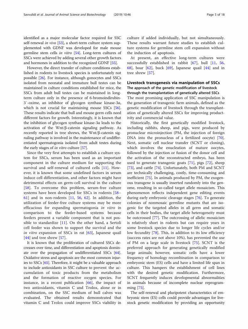
identified as a major molecular factor required for SSCself-renewal in vivo [53], a short-term culture system sup-plemented with GDNF was developed for male mousegermline stem cells in vitro [54]. Long-term cultures ofSSCs were achieved by adding several other growth factorsand hormones in addition to the recognized GDNF [55].However, the direct transfer of culture conditions estab-
lished in rodents to livestock species is unfortunately notpossible [26]. For instance, although gonocytes and SSCsisolated from neonatal and immature bull testes can bemaintained in culture conditions established for mice, theSSCs from adult bull testes can be maintained in long-term culture only in the presence of 6-bromoindirubin-3′-oxime, an inhibitor of glycogen synthase kinase-3α,which is not crucial for maintaining mouse SSCs [56].These results indicate that bull and mouse germ cells needdifferent factors for growth. Interestingly, it is known thatthe inhibition of glycogen synthase kinase-3α leads to theactivation of the Wnt/β-catenin signaling pathway. Asrecently reported in tree shrews, the Wnt/β-catenin sig-naling pathway is involved in the maintenance of undiffer-entiated spermatogonia isolated from adult testes duringthe early stages of in vitro culture [57].Since the very first attempts to establish a culture sys-
tem for SSCs, serum has been used as an importantcomponent in the culture medium for supporting thesurvival and self-renewal of cultured cells [11]. How-ever, it is known that some undefined factors in seruminduce cell differentiation, and other factors might havedetrimental effects on germ-cell survival in the culture[58]. To overcome this problem, serum-free culturesystems have been developed for SSCs in rodents [58–61] and in non-rodents [11, 56, 62]. In addition, theutilization of feeder-free culture systems may be moreefficacious for expanding spermatogonia in vitro incomparison to the feeder-based systems becausefeeders present a variable component that is not pos-sible to standardize [11]. On the other hand, a Sertolicell feeder was shown to support the survival and thein vitro expansion of SSCs in rat [63], Japanese quail[44] and tree shrew [57].It is known that the proliferation of cultured SSCs de-
creases over time, and differentiation and apoptosis domin-ate over the propagation of undifferentiated SSCs [64].Oxidative stress and apoptosis are the most common injur-ies to SSCs [65]. Therefore, it might be a valuable approachto include antioxidants in SSC culture to prevent the ac-cumulation of toxic products from the metabolismand the formation of reactive oxygen species. Forinstance, in a recent publication [66], the impact oftwo antioxidants, vitamin C and Trolox, alone or incombination, on the SSC medium of bull calves wasevaluated. The obtained results demonstrated thatvitamin C and Trolox could improve SSCs viability in
culture if added individually, but not simultaneously.These results warrant future studies to establish cul-ture systems for germline stem cell expansion withoutthe induction of apoptosis.At present, an effective long-term cultures were
successfully established in rabbit [67], bull [11, 56,68], boar [62], buck [69], Japanese quail [44] and intree shrew [57].
Livestock transgenesis via manipulation of SSCsThe approach of the genetic modification of livestockthrough the transplantation of genetically altered SSCsThe most promising application of SSC manipulation isthe generation of transgenic farm animals, defined as thegenetic modification of livestock through the transplant-ation of genetically altered SSCs for improving product-ivity and commercial value.Historically, the first genetically modified livestock,
including rabbits, sheep, and pigs, were produced bypronuclear microinjection (PM, the injection of foreignDNA into the pronucleus of a fertilized oocyte) [70].Next, somatic cell nuclear transfer (SCNT or cloning),which involves the enucleation of mature oocytes,followed by the injection or fusion of the donor cell andthe activation of the reconstructed embryo, has beenused to generate transgenic goats [71], pigs [72], sheep[73], and cattle [74]. Unfortunately, both PM and SCNTare technically challenging, costly, time-consuming andinefficient [75]. In animals produced by PM, the exogen-ous transgene is usually inserted randomly into the gen-ome, resulting in so-called target allele mosaicism. Thisphenomenon reflects independent gene editing eventsduring early embryonic cleavage stages [76]. To generatecolonies of nonmosaic germline mutants that are iso-genic for the targeted alleles in all germ and somaticcells in their bodies, the target allele heterogeneity mustbe outcrossed [77]. The outcrossing of allelic mosaicismis relatively short in rodents but can require years insome livestock species due to longer life cycles and/orlow fecundity [78]. This, in addition to its low efficiency(success rates are not above 10%), has prevented the useof PM on a large scale in livestock [75]. SCNT is thepreferred approach for generating genetically modifiedlarge animals; however, somatic cells have a lowerfrequency of homology recombination in comparison toembryonic stem (ES) cells and have a limited life span inculture. This hampers the establishment of cell lineswith the desired genetic modification. Furthermore,SCNT frequently induces developmental abnormalitiesin animals because of incomplete nuclear reprogram-ming [75].The self-renewal and pluripotent characteristics of em-
bryonic stem (ES) cells could provide advantages for live-stock genetic modification by providing an opportunity
Savvulidi et al. Journal of Animal Science and Biotechnology (2019) 10:46 Page 5 of 18
for the longstanding screening of correctly modified cellsor by improving the efficiency of cloning by nuclear trans-fer [79]. However, for large farm animals (except the re-cent success in bovine [80] [additional file 2] whereauthors were able to establish putative stable pluripotentbovine ES cells), no stable ES cell lines were established.Therefore, the genetic modification of livestock cur-
rently relies on the approach utilizing the fascinating abil-ity of in vitro genetically modified germ cells to colonizethe recipient testes and to produce donor-derived spermupon transplantation. The direct genetic modification ofdonor germ cells avoids the totipotent state of embryo-genesis, thus eliminating the production of mosaic mutantprogeny [81]. Furthermore, spermatogenesis in vivo pro-vides a perfect natural selection to eliminate transgenicsperm with undesired mutations: when any genetic muta-tion is introduced that is incompatible with the survival,proliferation, or differentiation of SSCs, those SSCs will failto form functional transgenic sperm, thus preventing thespread of undesired or lethal mutations [49]. Importantly,the time required for the production of genetically modifiedsperm is significantly shorter using germ cell transplant-ation compared to cloning or ES cell-based technology[75]. Thus, the approach of the genetic modification of live-stock through the transplantation of genetically alteredSSCs is currently being considered to complement the PMor SCNT for the production of transgenic farm animals[82] or genetically-edited birds [83].
Genome editing in livestock MGSCsIn a broad spectrum of genome editing research the ro-dent models still represent a “gold standard”, mainly dueto their perfect laboratory manageability (such as the smallbody size of adult animals, low cost of maintenance, shortreproductive cycle and short life span). Importantly, theknowledge we learn from rodent research is translatableonto other species to a large extent: the essential conceptsor ideas, first originated in rodent research, sooner or laterbecome an integral part of the research in livestock orhumans. For example, the concept of genomic editing ingermline stem cells using engineered nucleases (an artifi-cial genome editing reagents) first originated in mouseand rat research [81, 84–86] provided a platform for engi-neered nuclease-mediated genome editing in germlinestem cells in large farm animals [46].Generally, livestock MGSCs can be genetically edited ei-
ther via the random integration of the exogenous trans-gene into the cell genome or by the approach of precisegenome editing via engineered nucleases.
Genome editing via random integration of the exogenoustransgeneAs the proliferation of MGSCs in vitro is known to beslow, the prevailing method of MGSC genome editing
through random integration commonly relies on the useof viral vectors [75]. The feasibility of viral vectors for thispurpose was demonstrated in the very first report of live-stock transgenesis via germline stem cell transplantation[87]. In that study, the authors used the adeno-associatedvirus (AAV) vector to insert an exogenous gene for greenfluorescence protein (GFP) into the genome of buck germcells. AAV is a small virus from the parvovirus family thatis the preferred viral vector due to its ability to enter bothproliferating and nonproliferating cells and (for in vivo ex-periments) due to its associated low risk of inducing hostimmune responses [88]. The next essential AAV advan-tage, is that AAV remains primarily episomal after enter-ing into cell, so the random integration of the viral geneticmaterial into the host genome is avoided [89].There are several other types of viral vectors available (for
example, based on lenti-, adeno-, or flaviviruses), and eachtype provides a unique set of advantages and limitations.The pros and cons of these types have already been exten-sively reviewed elsewhere [88, 90]. However, it is importantto mention lentivirus (LV)-based vectors, which can also beused as an effective tool for the genomic editing of livestockMGSCs [82]. In this sense, an interesting study was pub-lished not long ago, where mice transgenesis was achievedby the lentiviral transduction of MGSCs in vivo [91]. In thatstudy, a lentiviral vector containing a GFP transgene wasinjected into the mouse testes, resulting in the integration ofthe transgene into the host (pre-founder) genome at the in-jection site. Moreover, after the pre-founder males weremated with wild-type females of the same strain, the trans-gene was found to be expressed in 67.88% of the F1 off-spring. This simple and efficient approach, if appropriatelymodified, could be applied to livestock species, leading tothe advancement of livestock transgenesis.
Genome editing via engineered nucleases (aka precisegenome editing)The approach of precise genome editing is based on theuse of three classes of engineered nucleases: zinc-finger nu-cleases (ZFN), transcription activator-like effector nucleases(TALEN) or RNA-guided nucleases (such as CRISPR/Cas9)[discussed in 84] to induce double-stranded breaks (DSB)in the DNA at very precise locations to initiate the nonho-mologous end-joining (NHEJ) or homologous recombin-ation (HR) repairing processes [additional file 3].ZFN was the first engineered nuclease described for the
approach of precise genome editing [92]. ZFN was dem-onstrated to edit the genome in mouse germline stem cells[84]. However, to the best of our knowledge, ZFN in live-stock species was only successfully used for somatic cellgenome editing in bulls [93] and boars (discussed in [94]);its use for the genomic editing of livestock MGSCs is lim-ited, and the detailed discussion of ZFN is beyond thescope of the present review. Readers interested in genome
Savvulidi et al. Journal of Animal Science and Biotechnology (2019) 10:46 Page 6 of 18
editing via ZFN should refer to review published else-where [95].TALEN, the second generation of engineered nucleases,
has a similar structure to ZFN, consisting of a DNA recog-nition domain and FokI endonuclease; however, their DNArecognition domains are different [96]. The TALEN DNAbinding domain originates from the bacterial plant patho-gen Xanthomonas, which is used by this bacterium to alterthe expression of several host genes [97]. The TALEN engi-neered nuclease has a much simpler design, assembly, anda broader targeting range [98]. Furthermore, in comparisonto ZFN, the smaller size of TALEN results in less sterichindrance and toxicity [99]. TALEN was used for genomicediting in chicken primordial germ cells, generating ov-albumin gene knockout chickens [100], or for targetingthe DDX4 locus and producing DDX4 null offspring[101]. In a recent study, Tang et al. [46] reported theTALEN-mediated editing of the porcine Duchennemuscular dystrophy locus in boar spermatogonia. Thesereports are the first to confirm the achievability ofprecise genomic editing in livestock germ cells. However,commercially available TALENs are expensive, take weeksto obtain, and the technologies of protein engineering toproduce TALENs do not guarantee active nucleases from agiven design [102]. Fortunately, an engineered nuclease isavailable today which is superior to TALEN; compared toTALEN, this superior engineered nuclease is simpler, easierto construct, has a lower cytotoxicity and a higher targetingefficiency. We are now speaking about CRISPR/Cas9.CRISPR (clustered regularly interspaced short palin-
dromic repeats), together with its associated Cas9 nucle-ase, assembles into the CRISPR/Cas9 complex, whichrepresents the most advanced generation of engineerednucleases today. Initially, CRISPR was identified as theadaptive immune system of bacteria against exogenousviral DNA or plasmid DNA [103].In year 2012 Jinek et al. [104] reported the ability of a ri-
bonucleoprotein complex of dual RNA (the so-called guideRNA; gRNA) consisting of a 20-base pair CRISPR RNA(crRNA) and the trans-activating crRNA (tracrRNA) to-gether with the Streptococcus pyogenes type II Cas9 proteinnuclease to induce DSB on DNA at very specific targetsites. Since that report, the CRISPR/Cas9 system has beenrapidly accepted as being fundamental for relatively simple,but precise, time- and cost-effective genome editing tech-nology. The first application of the CRISPR/Cas9 system toedit the mammalian genome was reported in 2013 [105].Compared to TALEN, CRISPR/Cas9 (due to its small size,which is only 20 base pairs) is simpler and easier toconstruct [106]. Furthermore, CRISPR/Cas9 has a lowercytotoxicity and a higher targeting efficiency [107]. Boththe RNA and the protein components of the CRISPR/Cas9ribonucleoprotein complex can be delivered into the cellsin several different ways. The Cas9 protein can be delivered
as a DNA expression plasmid, as an in vitro transcript, oras a recombinant protein bound to the RNA portion. TheRNA component can be delivered either expressed as aDNA plasmid or as an in vitro transcript. There are prosand cons for each delivery method, primarily regardingthe final CRISPR/Cas9 efficiency; these were thoroughlydiscussed elsewhere [102, 108]. Importantly, not onlythe NHEJ pathway but also the HR pathway could beinduced by the CRISPR/Cas9 system; therefore, notonly indels but also an exogenous DNA sequencescould be introduced into a specific target site with theknock-in strategy [109, 110].Moreover, multiplexed genome editing ability (the ability
to target simultaneously two or more genes in a single cell)was demonstrated for the CRISPR/Cas9 system [105, 111],[112]. Multiple guiding sequences can be encoded into asingle CRISPR array, which enables the simultaneous tar-geting of several sites within the genome. This multiplexedability of the CRISPR/Cas9 system was extremely suitablefor inactivating all 62 copies of porcine endogenous retro-virus (PERV) sequences in the genome of an immortalizedporcine cell line [111]. Of great importance, with the use ofCRISPR/Cas9 multiplexed genome editing in primary por-cine fibroblasts, PERV-free pigs were generated throughSCNTand embryo transfer [112].Apart from studies in rodents [105, 81, 84–86], the
feasibility of the CRISPR/Cas9 system for non-rodentMGSC genome editing was reported in chicken PGCs totarget the chicken immunoglobulin heavy chain loci [113]or the ovomucoid egg white allergen gene [114]. In treeshrews (an animal, closely related to primates), CRISPR/Cas9 was also reported to be feasible for SSC genomeediting; with the use of engineered CRIPSR/Cas9, the geneencoding amyloid β precursor protein (App) was success-fully disrupted [57].On the other hand, it is important to mention that the
CRISPR/Cas9 system is not absolute: there is a concernfor the off-target cleavage activity of CRISPR/Cas9 be-cause the system only requires a recognition of 20 basepairs [104, 105] and allows up to five base pair mis-matches for the formation of a DSB [115]. Consequently,if a normal, nontarget gene has enough homology to thetarget gene, it might also be inactivated by CRISPR/Cas9. To overcome its off-target issue, either the bindingspecificity of CRISPR/Cas9 could be improved based onthe bioinformatic analysis or the implementation of amutant “nickase” variants of Cas9 (Cas9-D10A orCas9n) could be considered (discussed in [116]). Inaddition to the Cas9 endonuclease, a class 2-type VCRISPR effector nuclease called Cpf1 (CRISPR from Pre-votella and Francisella 1) was recently identified [117].This nuclease has higher accuracy and therefore hasfewer off-target cleavage activities than Cas9 [118, 119].These novel CRISPR variants with enhanced accuracy
Savvulidi et al. Journal of Animal Science and Biotechnology (2019) 10:46 Page 7 of 18
will further facilitate its broad applications in livestockMGSC genome editing.
SSC transplantation: fundamental for themanipulation of spermatogonial stem cells inlivestock speciesPreparation of the recipients for SSC transplantationUnlike in rodent research, for SSC transplantation inlivestock species, most researchers use immature recip-ients, and this is not exclusively due to the evidentsize-related reasons. The immature intratesticularmicroenvironment is more favorable than adult testesfor the engraftment and expansion of donor SSCs[120].An interesting phenomenon of immunotolerance to
the transplanted genetically unrelated donor-derivedSSCs has been observed in livestock species. Recipientpigs [121, 122], goats [123, 124], sheep [125, 126],and bulls [127] with fully functional immune systemsdid not reject germ cells from unrelated donors(discussed in [128]). The immune privilege of recipi-ent testes was considered to explain the observedphenomenon. That is in contrast to rodents, wheresimilar transplantation experiments resulted in limitedcolonization of recipient testes unless immunesuppression was used (discussed in [128]). Althoughthe reasons for this interspecies difference are notclear, it is generally accepted, that Sertoli cells play acritical role in granting the immunologic privilege tothe testes. Given the limited contribution of theblood-testes barrier into the immunoprotection ofgerm cells [129, 130], it has been suggested and thenconfirmed in the in vitro studies and in vivo studies(where Sertoli cells survive transplantation across bar-rier) that Sertoli cells are potent immunomodulatorsand secrete a broad scale of immunosuppressive mo-lecular factors, which are inhibitors of immune response.Among these, immunomodulating cytokines (transforminggrowth factor β, interleukin 6); inhibitors of B and T cells;inhibitors of complement system (decay-accelerating factor,DAF); inhibitors of granzime-, and FAS-FAS ligand-medi-ated apoptosis (protease inhibitor - 9) and anti-inflamma-tory prostanoids (prostaglandin E synthase) should bementioned. Detailed overview of these factors is given else-where [128, 131–133]).Again, unlike rodents, in livestock animal models, the
elimination of the recipient’s endogenous SSCs to producevacant niches for donor-derived SSCs is not a critical pre-requisite [additional file 4], but it is a valuable approach forimproving the result of SSC transplantation [51, 134, 135].Indisputable, if SSC transplantation is used as a breedingtool, the elimination of the endogenous germline cells mustbe complete; otherwise, a mix of donor- and recipient-originsperm will be produced after transplantation [136].
Elimination of the recipient’s endogenous SSC by busulfantreatmentThe partial ablation of the recipient’s endogenous SSCs (theso-called gonadal ablation) can be achieved by the use ofbusulfan (1,4 - butanediol dimethanesulfonate), a chemo-therapeutic drug [51]. Busulfan is an alkylating agent thatinduces apoptosis in dividing cells. Because SSCs are mitot-ically active, these cells are sensitive to mitosis inhibition[137] and to apoptosis induced by busulfan [136]. Whilebusulfan treatment may appear practical in field conditions(as no expensive equipment is required), it has a cleardisadvantage related to its systemic toxicity. The dose-dependent species-specific systemic toxicity of busulfanduring treatment was demonstrated in pigs [138] and lambs[139]; therefore, researchers must be aware of thebusulfan-related toxicity danger. In lambs, a dose of 8mg/kg induced thin diarrhea with lethargy and a lack of appe-tite beginning at 5 days post busulfan injection; the authorssuggested using reduced doses of busulfan [139]. Becauseof the systemic toxicity of busulfan treatment in postnatalpigs, in utero busulfan treatment (i.e., the administration ofbusulfan to sows in their late gestation to coincide with theperiod of male germ cell proliferation in the fetus) can beutilized instead [138].Nevertheless, the huge disadvantage of busulfan is its
systemic toxicity (i.e., the possibility of damaging all or-gans or systems in the animals after busulfan treatment).Moreover, busulfan does produce a biohazard concern,as it is a chemotherapeutic drug and is eliminated fromthe treated animals via the feces and urine [136].Busulfan treatment not only eliminates the endogen-
ous SSCs but also damages the Sertoli cells [140, 141].Taken together, busulfan treatment must be consid-
ered after all other methods of recipient endogenousSSC elimination have been considered.
Elimination of the recipient’s endogenous SSCs by localtesticular irradiationThe irradiation efficiency depends strongly on the irradi-ation dose and animal age at time of irradiation, and dra-matic interspecies differences in response to irradiation inlivestock were observed [39]. Although testicular irradiationis the preferred method for the ablation of endogenousgerm cells in large animals [126, 135, 136] and does notcause a biohazard concern as a treatment regimen in live-stock [136], it can still negatively affect the neighboringsomatic support cells. Relatively high irradiation doses (>5–6Gy) which are required to eliminate endogenousspermatogonia in rodents, non-rodents and avian specieswere shown to compromise the viability of Sertoli cells andcaused testicular atrophy [139]. Moderate irradiation doses,although not killing the germ cells, were shown to result inthe block of spermatogonial differentiation due to injury tothe neighboring somatic compartment in rats [142]. In very
Savvulidi et al. Journal of Animal Science and Biotechnology (2019) 10:46 Page 8 of 18
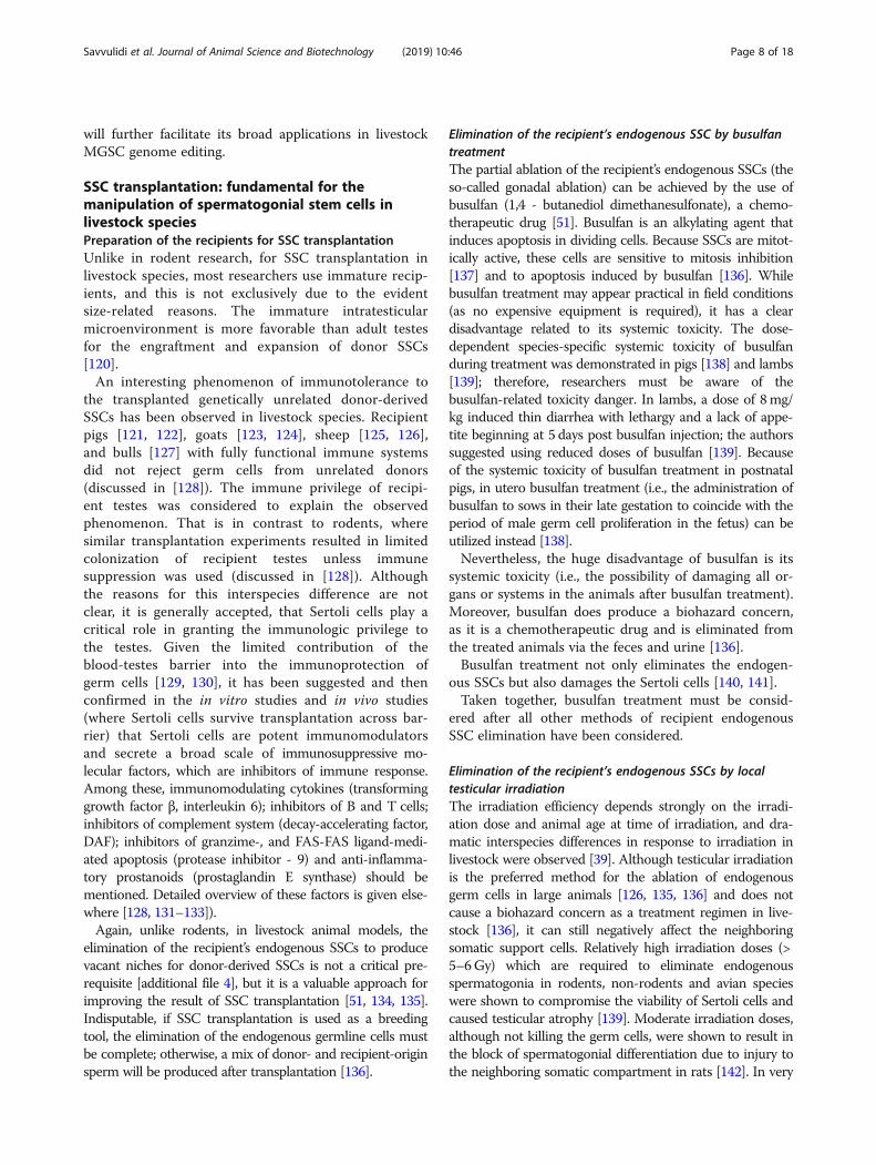
young lambs, the Sertoli cells can regenerate, and theirnumbers return to normal after irradiation; however, theirsupporting functionality is evidently corrupted [139, 142].In young bulls, it was shown that testicular irradiation (atdose of 10Gy) can damage the function of the Leydig cells,which can impair testosterone secretion throughout adult-hood [136]. This is a serious disadvantage of the irradiationapproach; the restoration of spermatogenesis followingirradiation and SSC transplantation can be challenging dueto impaired testosterone secretion. Moreover, someendogenous spermatogonia might still survive after seriousdamage by irradiation and, if developing into spermatids,might transferring gained genomic aberrations to the nextgeneration [143]. Another disadvantage of the procedure oftesticular irradiation is the requirement in a large-sized,sophisticated and expensive radiation source (Fig. 3) andthe requirement of anesthesia to perform the treatment ap-propriately [135]. Therefore, the availability of the testicularirradiation procedure is limited in field conditions.Hence, the field is desperately requiring new techniques
to improve the SSC transplantation efficiency or a betteralternative to both the busulfan and testicular irradiationtechniques.
Improvements in the SSC transplantation efficiencySuccessful SSC transplantation can only be performed inrecipients lacking their endogenous SSCs with preservedintact niches for the donor-derived SSCs. As discussed inthe preceding chapters, gonadal ablation (busulfan treat-ment or testicular irradiation) can compromise the viabil-ity and functionality of Sertoli cells and the steroidogenicactivity of Leydig cells. Therefore, the approach of thecotransplantation of non-compromised donor-derivedniche components (mainly Sertoli and/or Leydig cells)together with donor-derived SSCs into gonadal-ablatedrecipients is of great interest [additional file 5].
Sertoli/SSCs cotransplantationIn mice, the ability of intratesticularly transplanted donor-derived Sertoli cells to form seminiferous tubule structuresin the testes of infertile recipients with busulfan-compro-mised or genetically defective endogenous Sertoli cells wasreported [144]. This is a proof of concept study, as it con-firms the possibility of transplanting niche components(namely, Sertoli cells). Importantly, the morphogenic activ-ity of Sertoli cells was shown to be enhanced if perinataldonor Sertoli cells were used for transplantation [144]. Thisis in concordance with the generally accepted suggestionthat immature (prepubertal) Sertoli cells are able to prolif-erate [145], whereas mature Sertoli cells are mitotically qui-escent [146]. Indeed, immature Sertoli cells derived fromprepubertal cashmere buck testes were used to establishlong-term in vitro culture with significant (up to 20 pas-sages) proliferative potential and stable expression of germcell regulatory factors [147]. Menegazzo et al. reported im-mature Sertoli cells derived from prepubertal boar testeswere used as feeder layer and, when co-cultured with hu-man sperm in vitro, were able to preserve normal spermviability, motility and normal mitochondrial function after7 days of culture [148]. Interestingly, the mitotic “quies-cence” of mature Sertoli cells doesn’t seem to be related totheir terminal differentiation. Instead, mature Sertoli cellsresemble arrested proliferating cells: it was shown that themature Sertoli cells from adult mouse and human testeswere able to resume proliferation in vitro using standardculture conditions with no additives such as hormones orgonadotropins [149, 150].Another strong (albeit indirect) argument in favor of
Sertoli/SSCs cotransplantation potency comes fromstudies [151, 152]. In these studies, male germ cells frominfertile Steel (Sl/Sld) mutant mice (in which the testescontain spermatogonia, but spermatogenesis does notoccur due to the mutation-mediated Sertoli cell dysfunc-tion) were transplanted into the infertile dominant whitespotting (W) mutant male mice (with mutation-medi-ated germ cells dysfunction, but with preserved Sertolicell function) and the transplantation resulted indonor-derived offspring. Evidently, the non-permissive
Fig. 3 The procedure of testicular irradiation in ram. Theradiation source in this image is produced by a linear acceleratorwhich delivered a 6 MV photon beam directly to the testis at adose rate of 2 Gy per minute. Reproduced with a permissionfrom the personal archive of professor Jonathan Hill, PhD
Savvulidi et al. Journal of Animal Science and Biotechnology (2019) 10:46 Page 9 of 18
testicular environment does not support functionalspermatogonial stem cells properly and this can lead toanimal infertility. Thus, the transplantation of only SSCsmight be inefficient to restore fertility in recipients aftergonadal ablation.Several authors reported the Sertoli/SSCs cotransplanta-
tion [126, 153]. In these reports, the donor-derived “tes-ticular cell suspension” (which included both, Sertoli cellsand SSCs) was transplanted into gonadal ablated recipi-ents. However, it is difficult to evaluate the role Sertolicells play in the establishing of donor-derived spermato-genesis from such reports. On the other hand, to the bestof our knowledge, no any study reported the cotransplan-tation of Sertoli cells with in vitro expanded and genomeedited SSCs in livestock. We believe that the approach ofSertoli cells cotransplantation with in vitro expanded andgenome edited SSCs is of great interest and, therefore,should be thoroughly examined in the future.
Mesenchymal or Leydig stem cells/SSCs cotransplantationGong et al. reported several lines of evidence (these in-cluding immunostaining and analysis of cytokines secre-tion) that rat Sertoli cells are, in fact, a kind ofdifferentiated mesenchymal stem cells (MSCs) [154].Moreover, it was shown, that Leydig cells also developfrom undifferentiated mesenchymal-like stem cells [111],and that it is possible to differentiate MSC intotestosterone-producing, Leydig-like cells with the use ofseveral approaches [155, 156]. Therefore, the concept ofMSC-SSC co-transplantation to increase colonizationefficiency of donor-derived SSC by restoring the SSCniche after gonadal ablation in recipients is worth men-tioning. Indeed, the improvements in SSC transplant-ation efficiency after MSC-SSC co-transplantation hasrecently been demonstrated in the mouse model [157].Leydig stem cells could also be isolated based on their
specific surface (CD51+) or intracellular (Nestin+) pheno-typic markers with the use of immunostaining approach,as it was shown in mice [158]. in vitro, these self-renewalcells were capable of extensive proliferation and differenti-ation into mesenchymal or Leydig cells. in vivo, these cellswere capable of differentiation into mature Leydig cells,and the recipient animals (rats) showed a partial recoveryof testosterone production and spermatogenesis [158].Therefore, the concept of Leydig stem cells/SSCs contra-nsplantation to restore impaired testosterone secretionafter testicular irradiation could also be valuable.
Elimination of recipient’s endogenous germ cells viaspermatogonia-specific approachesAn interesting and very promising approach of how toprecisely eliminate the recipient’s endogenous SSCs withminimal (if any) negative impact on the neighboringsomatic support cells might be the application of a gene
editing tool to knockout (inactivate) one of the genes cru-cial for SSC development [159]. Recently, boars with theNANOS2 gene knocked out were generated by a direct in-jection of the engineered CRISPR/Cas9 system into thecytoplasm of fertilized zygotes; this was followed by em-bryo transfer into estrus synchronized surrogate females[160]. The NANOS family of RNA binding proteins playsa crucial role in the development and maintenance ofgermline cells in males [160]. In boars, it was discoveredthat the inactivation of NANOS2 leads to sperm loss inthe ejaculate while preserving the intact seminiferous tu-bules and the functional testicular interstitial tissue [160].Although the mechanisms of knockout-mediated germcell loss have yet to be revealed, such genetically modifiedboars with ablated spermatogenesis might serve as idealrecipients for transplantation of the donor SSCs [160].Recently, Herrid et al. reported selective toxicity of Doli-
chos biflorus agglutinin (DBA), a plant lectin, to spermato-gonia in the bull and the dromedary camel [161]. DBAbinds specifically to terminal N-acetylgalactosamine resi-dues and was widely used for enrichment or labeling gon-ocytes or Type A spermatogonia in several livestockspecies, including bull, boar and stallion (reviewed in[161]). In the dromedary camel, a single dose of 25–50 μg/mL DBA injected into rete testis was shown to deplete en-dogenous stem cells in recipient testes [161]. Therefore, itwas concluded, that DBA could be used effectively toeliminate endogenous stem cells.As discussed by Smith et al. [162], other spermatogonia-
specific approaches are available to eliminate the recipient’sendogenous germ cells in a controllable manner. Forexample, several transgenic approaches were reported inmice studies, which based on the use of the inhibin-alphapromoter/herpes simplex virus thymidine kinase transgene[163], the diphtheria toxin A chain gene directed by thehistone H1t promoter [164] or the transgenic expression ofan inducible primate iDTR within mouse germ cells [165].
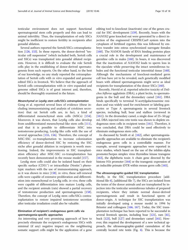
The ultrasonographic-guided SSC transplantationBriefly, in the SSC transplantation procedure [add-itional file 6], [additional file 7], the SSCs are isolated fromthe testes of the donor animals and are transplanted by in-jection into the testicular seminiferous tubules of preparedrecipients, where they initiate normal donor-derivedspermatogenesis and result in functional sperm ofdonor-origin. A technique for SSC transplantation wasinitially developed using a mouse model in 1994 byBrinster and colleagues [166, 167]. Today, the SSC trans-plantation technique has been reported to be successful inseveral livestock species, including boar [121], ram [41],buck [123], bull [127] and dromedary camel [161]. How-ever, this required the development of a new technical ap-proach, the ultrasonographic-guided cannulation of thecentrally located rete testis (Fig. 4). This is because the
Savvulidi et al. Journal of Animal Science and Biotechnology (2019) 10:46 Page 10 of 18
direct injection of donor cells into seminiferous tubules isnot possible via the efferent ducts in large species due toanatomic limitations. Ultrasound-guided cannulation couldbe completed in 15–30min under general anesthesia andaseptic surgical conditions [82]. To enable successful trans-plantation, tens of millions of donor SSCs in the total vol-ume of several mL would be enough to be injected pertestis in large animals [49]. Ultrasound-guided cannulationis usually conducted under a flow rate of approximately0.5–1mL/min [82] and results in filling approximately halfof the recipient’s seminiferous tubules with donor cells [51].
Confirmation of transplanted donor SSC viabilityKim et al. [168] was able to obtain morphologically andfunctionally normal spermatozoa of donor origin after theSSC transplantation procedure in boars. Zeng et al. re-ported the production of transgenic donor-origin spermafter SSC transplantation in bucks [49]. Li et al. [57] demon-strated the self-renewal and spermatogenesis abilities oftransplanted tree shrew SSCs, where donor-derived sperm-atogenesis persisted in recipients at day 250 post-trans-plantation. Recently, Herrid et al. [161] demonstrated forthe first time, that the recipients produce spermatozoa ofdonor origin after heterologous testicular germ cell
transplantation in dromedary camels. In all four studies,the donor origin was confirmed by the positive donor-ori-gin gene expression of spermatozoa by polymerase chainreaction. It is important to mention that in certain livestockspecies, the transplanted transgenic SSCs will home intothe niche, proliferate and produce an amount of transgenicsperm that will be detectable by PCR only after a certainduration of time (as long as 11months in boars, [82]). Theconfirmation of the transplanted donor SSC viability by theproduction of donor-derived offspring (or transgenic IVFembryos) was described in recipient rams [169], bucks [87],boars [82] and cockerels [170]. Recent report ofdonor-origin sperm ejaculated by recipient in dromedarycamel [161] indicated offspring may soon be produced alsoin this specie. In general, the transgene was stably inte-grated into the genome of the recipient animals, as it waspossible to detect the transgene in ejaculates from recipientboars [82] or rams [169] at least 5 years after the transplant.
SSC transplantation to extend the techniques ofspermatozoa cryopreservation and/or artificialinseminationSperm cryopreservation protocols (an integral componentof the artificial insemination technique), if developed for
Fig. 4 Schematic representation of the ultrasonographic-guided SSC transplantation technique. a. SSC transplantation in mouse. In thisspecie, there is a single efferent duct emerging from the rete testis, which is easy to cannulate to inject SSC. b. The ultrasonographic-guided cannulation of the centrally located rete testis in non-rodent animals. In livestock species, several efferent ducts emerge from thetestis. That is why the transplantation of SSC is preferably done by cannulation of SSC into the rete testis with ultrasonographicguidance. c. The ultrasonographic guidance; rete testis is shown by the white arrow. Catheter, injected into the rete testis, is shown bythe red arrow. Adapted from [52], modified. The ultrasonographic guidance picture is reproduced with a permission from the personalarchive of professor Jonathan Hill, PhD
Savvulidi et al. Journal of Animal Science and Biotechnology (2019) 10:46 Page 11 of 18

individual livestock species, are often complicated,time-consuming, not interchangeable among species ornot standardized. For example, due to high viscosity andpoor quality of dromedary camel semen and due to un-availability of the standard protocol for diluting and freez-ing camel sperm, artificial insemination is not regularlyused in camel breeding programs (discussed in [161]).In contrast, SSC isolated from various livestock species
might be successfully cryopreserved for a long period oftime in liquid nitrogen, using invariable and well-established protocol, as is commonly done in cryopreservationof somatic cells [171]. It is important to mention the studyof Redden et al. [172] who investigated the effectiveness ofcryopreservation of bull testicular cells with different pack-aging procedures (large-sized cryostraws, cryobags or cryo-vials). The study confirmed that cryopreservation oftesticular germ cells in 5mL cryostraws at a density of upto 18 × 106 cells/mL in liquid nitrogen appears to be a sim-ple and practical way to preserve cells.Further, it is known from the semen preservation indus-
try that certain sires (so-called “bad freezers”) respondpoorly to conventional freezing protocols [173]. This poorresponse in such sires could be partially improved if theconventional freezing protocol is modified [174].Therefore, it was accepted that for the “bad freezers”, theconventional sperm freezing protocol is simply less-tha-n-optimal and that the “bad freezers” produce sperm witha narrow tolerance to less-than-optimal conditions [174].The “bad freezers” issue could be explained in terms ofthe concept of the spermatozoa heterogeneity (here theheterogeneity is the phenomenon of functional variabilityin the responses of spermatozoa, which is treated thesame way [173]). A central idea behind this concept is thatthe spermatozoa in an individual’s ejaculate are very het-erogeneous for several attributes, including their toleranceto cold-shock [173] and, importantly, their fertilizing po-tential [175]. The ejaculation of millions of sperm, all at asimilar state of fertilizing potential, might suffice only forinduced ovulators, such as cats or rabbits [175]. In con-trast, it is especially important to consider the sperm het-erogeneity in species with variable intervals betweenmating and ovulation, such as sheep [176]. Sperm becomeheterogeneous during their epididymal transit [173], andthe subpopulations of spermatozoa with different fertiliz-ing potentials arise [176].Using techniques assessing sperm heterogeneity (such
as the centrifugal countercurrent distribution technique),it was shown that ejaculate heterogeneity is positively cor-related with sire field fertility [177]. Furthermore, the re-duced fertilizing ability of the stored sperm was shown tobe due to dramatic loss of sperm heterogeneity duringcryopreservation because cooling, freezing and thawingproduce homogeneous sperm samples with very limitedfunctional versatility [173, 178]. Therefore, cryopreserved
sperm represents a very limited genetic resource: the gen-etic variation is limited by the spermatozoa pool, which isderived from the thawed sperm sample.Although it was reported that the cryopreservation
is harmful also for germ cells and that cryosurvivalrate of the preserved germline cells can be as low as50% [179], cryopreserved SSC [additional file 8] areable to self-renew in vitro unlimitedly after thawing,thus providing the source of virtually any individualsire’s genetic program with full genetic variabilitywithin that program [171]. It has also been shown inmice studies that cryopreserved SSCs retain theirspermatogenic function [171] and were able to suc-cessfully produce normal donor-derived offspring aftertransplantationing into testes of busulphan-sterilizedrecipient mice [180].Furthermore, it is broadly accepted that SSC trans-
plantation can be used for the propagation of elitesire genetics in extensive grazing systems (for in-stance, in beef cattle production), where the use ofartificial insemination (AI) requires exhaustive man-agement and is limited by complications related to,for example, estrous synchronization. Indeed, it wasestimated that only approximately 10% of beef cowsin the United States are bred using AI [181]. Thismeans a lost opportunity for the genetic gain of beefcattle populations. On the other hand, the transplant-ation of SSCs between bulls would provide a tool (al-ternative to AI) for expanding the use of the geneticvalue of elite sires, and this will overcome the limita-tions of implementing an AI program [68].
ConclusionApart from its value in extending the techniques ofspermatozoa cryopreservation and/or artificial insem-ination, the immense promise of SSC manipulationlies in the acceleration of transgenesis in farm ani-mals. In present review we covered several topicsclosely related to advances in the isolation and purifi-cation of livestock SSCs with such techniques asFACS, MACS, Staput velocity sedimentation or thedifferential plating technique. We reviewed advancesin the establishment of effective long-term culturesystems for the in vitro expansion of livestock germ-line stem cells. Furthermore, we reviewed advances inthe precise genome editing of livestock MGSCs (espe-cially with the use of CRISPR/Cas9, the most moderngene editing technology).Based on the above reasons and the confirmed success
of the SSC transplantation technique in bulls, boars,rams, bucks and dromedary camel, we conclude that themanipulation of spermatogonial stem cells is currently afeasible and affordable strategy for the genetic modifica-tion of livestock.
Savvulidi et al. Journal of Animal Science and Biotechnology (2019) 10:46 Page 12 of 18

Future directionsIn previous years, several quite important PM- orSCNT-based approaches to generate transgenic farm ani-mals via genomic editing by engineered nucleases in som-atic cells were published: 1) the insertion of the mouseSP110 gene into the genome of bovine macrophages togenerate transgenic cattle with increased resistance to tu-berculosis [86], 2) the knockout of the PRNP gene in im-mortalized bovine fibroblasts to generate prion-free cattle[182], 3) the disruption of the CD163 subdomain 5 genein porcine zygotes to generate pigs that are not susceptibleto infection with porcine reproductive and respiratorysyndrome virus [183], and 4) the targeting of the catalyticcore of the PERV pol gene in primary porcine fetal fibro-blasts to generate PERV-inactivated pigs [112]. In the fu-ture studies, we believe that (due to the revieweddisadvantages of the PM and SCNT methods), it is ofgreat interest to revise or expand the abovementioned ap-proaches using the CRISPR/Cas9 system in the context oflivestock MGSC genomic editing.
Additional files
Additional file 1: The choice of which donor population of germlinestem cells to expand in culture is critical for the outcome of germ celltransplantation. In this sense, it is important to mention, that the use ofPGCs is less practical as these cells are collected from embryo and thereare just few PGCs per embryo [186, 187]. On the other hand, the SSCs(together with gonocytes) offer more practical option due to relativelysimple procedure of their collection from the testes of neonates, juvenileor adult donors [12]. (DOCX 13 kb)
Additional file 2: Although not the focus of present review, it should bementioned that this recent success with the establishment of stable bovineES cell lines open the opportunity to revolutionize the livestock breeding.Using established pluripotent ES cells, germ cells can be induced in vitro toform functional spermatids and oocytes. Next, with the use of in vitrofertilization (IVF), embryos can be obtained from the in vitro generatedspermatids and oocytes. Such an “animal embryo-stem cell breedingsystem” completes the whole livestock breeding scheme “in a dish” byintegrating in vitro germ cell induction, IVF, genome sequencing, andgenomic selection [188]. On the other hand, even the possibility ofproducing sperm in vitro would have had a great impact on livestockindustries in case of success. As Aponte [52] has stated “…in the cattleindustry, keeping animals in large facilities would be a thing of the pastwhen renewable SSC pools from elite bulls produce high numbers of spermin Petri dishes at small biotechnological facilities” (p.672). However, it is veryimportant to take into consideration the possible effect of inbreeding afteronly using a limited number of available elite sires, and the consequentdecrease of genetic variability in population [189]. (DOCX 12 kb)
Additional file 3: Because DSB are potentially lethal, the cell activatesmechanisms to repair the DSB damage through the NHEJ or the HRprocesses, two major cellular DNA repair pathways [190]. The molecularnature of these pathways is complex, and a detailed overview of thesepathways is outside the scope of the present review. Readers interestingin DNA repair by NHEJ or HR should refer reviews published elsewhere[190, 191]. However, for present review, it is important to introduce thedifference between two: NHEJ is the more frequent, although imperfect,error-prone repair pathway that results in insertions and deletions (indels)at the break site [75]. These short DNA indels create targeted geneknockouts by inducing a frameshift of the amino acid codons and theformation of a premature stop codon [192]. On the other hand, HR isknown to be more precise and is able to introduce the specific
exogeneous nucleotide sequences into the repaired DNA (if donortemplate DNA is provided) [94]. (DOCX 12 kb)
Additional file 4: This could indicate either that a) donor stem cells areable to compete successfully with endogenous stem cells for availableniches or b) there are vacant niches in the testes of livestock species thatcan be occupied by transplanted donor cells (discussed in [39]).(DOCX 11 kb)
Additional file 5: It is important to mention the study of Anand et al.,who discussed the restoration of spermatogenesis by testiculartransplantation of donor-derived Sertoli cells into busulphan-treatedrecipient mice [140]. According to the authors, spermatogenesis in therecipient was restored from a pool of endogenous (recipient-derived)very small embryonic-like stem cells (VSELs). These cells survivedgonadal ablation, proliferated and gave rise to spermatogonial cells,but were unable to differentiate because of a compromised niche.Therefore, it is critical to confirm thoroughly the donor-origin of restoredspermatogenesis after Sertoli cells co-transplantation. (DOCX 12 kb)
Additional file 6: In contrast to human research, intratesticular allo- (thetransplantation between the different individuals of the same specie), orthe xenotransplantation (the transplantation between individuals fromdifferent species) is mainly considered in livestock. (DOCX 13 kb)
Additional file 7: Alternativelly, ectopic transplantation of small (1–2mm3)fragments of the testicular tissue isolated from livestock donor animal (theso-called xenografting approach) or of disassociated testicular cell suspen-sion (the so-called de novo morphogenesis approach) under the dorsal skinof immunocompromised recipient mice could also be used to obtain fullyfunctional haploid donor-derived spermatozoa [193, 194]. The capability ofectopically transplanted Sertoli cells to rearrange into seminiferous tubule-like structures to support donor-derived ectopic spermatogenesis is fascinat-ing and is the fundamental of the de novo morphogenesis approach (dis-cussed in [195]). Because of the use of mice models, both the xenograftingand the de novo morphogenesis approaches help to overcome the costlyand time-consuming process of maintaining large animal models in re-search. On the other hand, the practical application of both approaches inlivestock breeding is notably limited by the needs to use the elaborativeand costly techniques of assisted reproduction (such as intracytoplasmicsperm injection, ICSI) to generate the progeny from the obtained donor-derived spermatozoa. Therefore, both approaches are considered as invalu-able in vivo bio-assays to comprehend spermatogenesis, however with lowpractical merit as of today. This is in contrast to SSCs intratesticular trans-plantation, which has its certain disadvantages if exploited as the experi-mental in vivo bio-assay but suits better to practical application in livestockbreeding. Readers interested in the testicular tissue xenografting or de novotesticular morphogenesis should refer to the excellent reviews publishedelsewere [195, 196] or to several original papers, which confirm the exclusiveexperimental merit of these approaches in livestock research [197, 195].(DOCX 13 kb)
Additional file 8: Compared to cryopreservation of single cellsuspension, the approach of whole testis tissue cryopreservation is morechallenging; this is because tissue requires longer exposure tocryoprotectants and this can result in higher cellular toxicity beforefreezing [135]. However, the cryopreservation of the whole testis tissue isrecognized as promising, at least in human regenerative medicine. In thisapproach, the SSCs purified from the thawed tissue and propagatedsubsequently in vitro [198]. Readers interested in this approach shouldrefer the very recent review [199]. (DOCX 12 kb)
AbbreviationsAAV: Adeno-associated virus; AI: Artificial insemination; bFGF: Basic fibroblastgrowth factor; BSA: Bovine serum albumin; Cas9: CRISPR-associated protein 9,nuclease; Cas9-D10A: Variant of Cas9; Cas9n: Variant of Cas9; CD51: Cluster ofdesignation, 51; CDH1: E-cadherin; Cpf1: Nuclease name, CRISPR fromPrevotella and Francisella 1; CRISPR: Clustered regularly interspaced shortpalindromic repeats; crRNA: CRISPR RNA; CSF1: Colony-stimulating factor-1;DAF: Decay-accelerating factor; DAZL: Deleted in azoospermia-like;DBA: Dolichos biflorus agglutinin; DDX4: DEAD-box polypeptide 4;DSB: Double-stranded breaks; EDTA: Ethylenediaminetetraacetic acid;ES: Embryonic stem cells; FACS: Fluorescence-activated cell sorting;FokI: Endonuclease, naturally found in Flavobacterium okeanokoites;
Savvulidi et al. Journal of Animal Science and Biotechnology (2019) 10:46 Page 13 of 18

GDNF: Glial cell line-derived neurotrophic factor; GFP: Green fluorescenceprotein; GFRα1: GDNF family receptor alpha-1; gRNA: Guide RNA; Gy: Grey,the irradiation unit; HR: Homologous recombination; ICSI: Intracytoplasmicsperm injection; iDTR: Mice line name, carrying a Cre-inducible simian diph-theria toxin receptor; ITGA6: Integrin alpha-6; IVF: in vitro fertilization;LE: Interstitial lymphatic endothelial cells; LV: Lentivirus-based vector;MACS: Magnetic activated cell sorting; MGSCs: Male germline stem cells;MSCs: Mesenchymal stem cells; NANOS2: the Nanos gene paralog. TheNANOS family of RNA binding proteins play an essential role in thedevelopment of germline; NHEJ: Nonhomologous end-joining;PCR: Polymerase chain reaction; PERV: Porcine endogenous retrovirus;PGCs: Primordial germ cells; PGP9.5: Protein gene product 9.5;PLZF: Promyelocytic leukemia zinc finger protein; PM: Pronuclearmicroinjection; PMCs: Peritubular myoid cells; pol: (DNA polymerase) refers toa gene for reverse transcriptase in retroviruses. This enzyme transcribes theviral RNA into double-stranded DNA; PRNP: The gene provides instructionsfor making a protein called prion protein; SCNT: Somatic cell nuclear transfer;SP110: Speckled 110 KDa, the nuclear body protein gene. The product of thegene control the growth of Mycobacterium tuberculosis in macrophages andinduce apoptosis in infected cells; SSCs: Spermatogonial stem cells;TALEN: Transcription activator-like effector nucleases; tracrRNA: Trans-activating crRNA; VEGF: Vascular endothelial growth factor; VSELs: Very smallembryonic-like stem cells; ZFN: Zinc-finger nucleases
AcknowledgementsWe thank professor Jonathan Hill, PhD (School of Veterinary Science,University of Queensland, Australia) for his valuable comments, for sharingthe photograph of the procedure of testicular irradiation and theultrasonographic-guided cannulation in ram and for his kind permission touse these photographs in our review. We thank reviewers for helpful com-ments and suggestions.
FundingThis review was supported by the S grant of the Ministry of Education, Youthand Sport (MEYS) of Czech Republic. KSV was supported by the PrimusResearch Programme PRIMUS/17/MED/16 of the Charles University.
Availability of data and materialsNot applicable.
Authors’ contributionsFS was a major contributor in manuscript drafting and figures preparation;KSV wrote chapter “Genome editing in livestock MGSC”; MP and LS werecontributed to the critical discussion and provided an overall supervisionduring the manuscript drafting. All authors revised the manuscript, providedintellectual content and read and approved the revised version.
Ethics approval and consent to participateNot applicable.
Consent for publicationNot applicable.
Competing interestsThe authors declare that they have no competing interests.
Received: 5 December 2018 Accepted: 17 April 2019
References1. Kennelly JJ, Foote RH. Sampling boar testes to study spermatogenesis
quantitatively and to predict sperm production. J Anim Sci. 1964;23:160–7.2. Schanbacher BD, Ford JJ. Photoperiodic regulation of ovine
spermatogenesis: relationship to serum hormones. Biol Reprod. 1979;20:719–26.
3. Amann RP, Almquist JO. Reproductive capacity of dairy bulls. VIII. Direct andindirect measurement of testicular sperm production. J Dairy Sci. 1962;45:774–81.
4. Gebauer MR, Pickett BW, Swierstra EE. Reproductive physiology of thestallion. II. Daily production and output of sperm. J Anim Sci. 1974;39:732–6.
5. Ritar AJ, Mendoza G, Salamon S, White IG. Frequent semen collection andsperm reserves of the male angora goat (Capra hircus). J Reprod Fertil. 1992;95:97–102.
6. Berndtson WE. Sperm production and its harvest. In: Chenoweth P, LortonS, editors. Animal andrology: theories and applications. Wallingford: CABI;2014. p. 11–33.
7. de Rooij DG, Russell LD. All you wanted to know about spermatogonia butwere afraid to ask. J Androl. 2000. https://doi.org/10.1002/j.1939-4640.2000.tb03408.x.
8. Phillips BT, Gassei K, Orwig KE. Philos Trans R Soc Lond B Biol SciPhilos TransR Soc Lond Ser B Biol Sci. 2010. https://doi.org/10.1098/rstb.2010.0026.
9. De Felici M. Primordial germ cell biology at the beginning of the XXIcentury. Int J Dev Biol. 2009. https://doi.org/10.1387/ijdb.082815mf.
10. Kolasa A, Misiakiewicz K, Marchlewicz M, Wiszniewska B. The generation ofspermatogonial stem cells and spermatogonia in mammals. Reprod Biol.2012. https://doi.org/10.1016/S1642-431X(12)60074-6 .
11. Sahare M, Kim SM, Otomo A, Komatsu K, Minami N, Yamada M, et al.Factors supporting long-term culture of bovine male germ cells. ReprodFertil Dev. 2016, 2016. https://doi.org/10.1071/RD15003 .
12. Awang-Junaidi AH, Honaramooz A. Optimization of culture conditions forshort-term maintenance, proliferation, and colony formation of porcinegonocytes. J Anim Sci Biotechnol. 2018. https://doi.org/10.1186/s40104-017-0222-0.
13. Potter SJ, DeFalco T. Role of the testis interstitial compartment inspermatogonial stem cell function. Reproduction. 2017. https://doi.org/10.1530/REP-16-0588.
14. Svingen T, Koopman P. Building the mammalian testis: origins,differentiation, and assembly of the component cell populations. GenesDev. 2013. https://doi.org/10.1101/gad.228080.113.
15. Oatley JM, Brinster RL. The germline stem cell niche unit in mammaliantestes. Physiol Rev. 2012. https://doi.org/10.1152/physrev.00025.2011.
16. de Rooij DG. The nature and dynamics of spermatogonial stem cells.Development. 2017. https://doi.org/10.1242/dev.146571.
17. Lord T, Oatley JM. A revised A-single model to explain stem cell dynamicsin the mouse male germline. Reproduction. 2017. https://doi.org/10.1530/REP-17-0034.
18. Takashima S, Shinohara T. Culture and transplantation of spermatogonialstem cells. Stem Cell Res. 2018. https://doi.org/10.1016/j.scr.2018.03.006 .
19. Yoshida S. Open niche regulation of mouse spermatogenic stem cells.Develop Growth Differ. 2018. https://doi.org/10.1111/dgd.12574 .
20. Kitadate Y, Jorg D, Tokue M, Maruyama A, Ichikawa R, Tsuchiya S, et al.Competition for mitogens regulates Spermatogenic stem cell homeostasis inan open niche. Cell Stem Cell. 2019. https://doi.org/10.1016/j.stem.2018.11.013.
21. Kubota H, Avarbock MR, Brinster RL. Culture conditions and single growthfactors affect fate determination of mouse spermatogonial stem cells. BiolReprod. 2004. https://doi.org/10.1095/biolreprod.104.029207.
22. Jabarpour M, Tajik P. Evaluating the behavior of cultured sertoli cells in thepresence and absence of spermatogonial stem cell. Stem Cell Investig.2018. https://doi.org/10.21037/sci.2018.01.01.
23. Griswold MD. 50 years of spermatogenesis: Sertoli cells and theirinteractions with germ cells. Biol Reprod. 2018. https://doi.org/10.1093/biolre/ioy027 .
24. Huang YH, Chin CC, Ho HN, Chou CK, Shen CN, Kuo HC, et al. Pluripotencyof mouse spermatogonial stem cells maintained by IGF-1- dependentpathway. FASEB J. 2009. https://doi.org/10.1096/fj.08-121939 .
25. Oatley JM, Oatley MJ, Avarbock MR, Tobias JW, Brinster RL. Colonystimulating factor 1 is an extrinsic stimulator of mouse spermatogonial stemcell self-renewal. Development. 2009. https://doi.org/10.1242/dev.032243.
26. Zheng Y, Zhang Y, Qu R, He Y, Tian X, Zeng W. Spermatogonial stem cellsfrom domestic animals: progress and prospects. Reproduction. 2014. https://doi.org/10.1530/REP-13-0466 .
27. Hutson JC. Development of cytoplasmic digitations between Leydig cellsand testicular macrophages of the rat. Cell Tissue Res. 1992. https://doi.org/10.1007/BF00302977 .
28. Nes WD, Lukyanenko YO, Jia ZH, Quideau S, Howald WN, Pratum TK, et al.Identification of the lipophilic factor produced by macrophages thatstimulates steroidogenesis. Endocrinology. 2000. https://doi.org/10.1210/endo.141.3.7350.
29. DeFalco T, Potter SJ, Williams AV, Waller B, Kan MJ, Capel B. Macrophagescontribute to the Spermatogonial niche in the adult testis. Cell Rep. 2015.https://doi.org/10.1016/j.celrep.2015.07.015.
Savvulidi et al. Journal of Animal Science and Biotechnology (2019) 10:46 Page 14 of 18

30. Caires KC, de Avila J, McLean DJ. Vascular endothelial growth factorregulates germ cell survival during establishment of spermatogenesis in thebovine testis. Reproduction. 2009. https://doi.org/10.1530/REP-09-0020.
31. Tian R, Yang S, Zhu Y, Zou S, Li P, Wang J, et al. VEGF/VEGFR2 signalingregulates germ cell proliferation in vitro and promotes mouse testicularregeneration in vivo. Cells Tissues Organs. 2015. https://doi.org/10.1159/000440949.
32. Chen LY, Willis WD, Eddy EM. Targeting the Gdnf gene in peritubular myoidcells disrupts undifferentiated spermatogonial cell development. Proc NatlAcad Sci U S A. 2016. https://doi.org/10.1073/pnas.1517994113 .
33. Helsel AR, Yang QE, Oatley MJ, Lord T, Sablitzky F, Oatley JM. ID4 levelsdictate the stem cell state in mouse spermatogonia. Development. 2017.https://doi.org/10.1242/DEV.146928 .
34. Tegelenbosch RA, de Rooij DG. A quantitative study of spermatogonialmultiplication and stem cell renewal in the C3H/101 F1 hybrid mouse.Mutat Res. 1993. https://doi.org/10.1016/0027-5107(93)90159-D.
35. Bellvé AR, Cavicchia JC, Millette CF, O'Brien DA, Bhatnagar YM, Dym M.Spermatogenic cells of the prepuberal mouse. Isolation and morphologicalcharacterization. J Cell Biol. 1977;74(1):68–85.
36. McLean DJ, Russell LD, Griswold MD. Biological activity and enrichment ofspermatogonial stem cells in vitamin A-deficient and hyperthermia-exposedtestes from mice based on colonization following germ cell transplantation.Biol Reprod. 2002;66:1374–9.
37. Shinohara T, Avarbock MR, Brinster RL. Functional analysis ofspermatogonial stem cells in steel and cryptorchid infertile mouse models.Dev Biol. 2000. https://doi.org/10.1006/dbio.2000.9655.
38. Oatley MJ, Racicot KE, Oatley JM. Sertoli cells dictate spermatogonial stemcell niches in the mouse testis. Biol Reprod. 2011. https://doi.org/10.1095/biolreprod.110.087320.
39. Herrid M, McFarlane JR. Application of testis germ cell transplantation inbreeding Systems of Food Producing Species: a review. Anim Biotechnol.2013. https://doi.org/10.1080/10495398.2013.785431.
40. Luo J, Megee S, Rathi R, Dobrinski I. Protein gene product 9.5 is a spermatogonia-specific marker in the pig testis: application to enrichment and culture of porcinespermatogonia. Mol Reprod Dev. 2006. https://doi.org/10.1002/mrd.20529.
41. Rodriguez-Sosa JR, Dobson H, Hahnel A. Isolation and transplantation ofspermatogonia in sheep. Theriogenology. 2006. https://doi.org/10.1016/j.theriogenology.2006.03.039 .
42. Heidari B, Rahmati-Ahmadabadi M, Akhondi MM, Zarnani AH, Jeddi-TehraniM, Shirazi A, et al. Isolation, identification, and culture of goatspermatogonial stem cells using c-kit and PGP9.5 markers. J Assist ReprodGenet. 2012. https://doi.org/10.1007/s10815-012-9828-5 .
43. Zhang Y, Wu S, Luo F, Baiyinbatu LL, Hu T, et al. CDH1, a novel surfacemarker of Spermatogonial stem cells in sheep testis. J Integrat Agric. 2014.https://doi.org/10.1016/S2095-3119(13)60689-9.
44. Pramod RK, Lee BR, Kim YM, Lee HJ, Park YH, Ono T, et al. Isolation,characterization, and in vitro culturing of Spermatogonial stem cells inJapanese quail (Coturnix japonica). Stem Cells Dev. 2017. https://doi.org/10.1089/scd.2016.0129.
45. Kise K, Yoshikawa H, Sato M, Tashiro M, Yazawa R, Nagasaka Y, et al. Flow-cytometric isolation and enrichment of teleost type a spermatogonia basedon light-scattering properties. Biol Reprod. 2012. https://doi.org/10.1095/biolreprod.111.093161.
46. Tang L, Bondareva A, González R, Rodriguez-Sosa JR, Carlson DF, Webster D,et al. TALEN-mediated gene targeting in porcine spermatogonia. MolReprod Dev. 2018. https://doi.org/10.1002/mrd.22961.
47. Martínez-Pastor F, Mata-Campuzano M, Álvarez-Rodríguez M, Álvarez M,Anel L, De Paz P. Probes and techniques for sperm evaluation by flowcytometry. Reprod Domest Anim. 2010. https://doi.org/10.1111/j.1439-0531.2010.01622.x .
48. Bryant JM, Meyer-Ficca ML, Dang VM, Berger SL, Meyer RG. Separation ofspermatogenic cell types using STA-PUT velocity sedimentation. J Vis Exp.2013. https://doi.org/10.3791/50648 .
49. Zeng W, Tang L, Bondareva A, Luo J, Megee SO, Modelski M, et al. Non-viraltransfection of goat germline stem cells by nucleofection results inproduction of transgenic sperm after germ cell transplantation. Mol ReprodDev. 2012. https://doi.org/10.1002/mrd.22014 .
50. Binsila KB, Selvaraju S, Ghosh SK, Parthipan S, Archana SS, ArangasamyA, et al. Isolation and enrichment of putative spermatogonial stem cellsfrom ram (Ovis aries) testis. Anim Reprod Sci. 2018. https://doi.org/10.1016/j.anireprosci.2018.04.070.
51. Honaramooz A, Yang Y. Recent advances in application of male germ celltransplantation in farm animals. Vet Med Int. 2010. https://doi.org/10.4061/2011/657860.
52. Aponte PM. Spermatogonial stem cells: current biotechnological advancesin reproduction and regenerative medicine. World J Stem Cells. 2015.https://doi.org/10.4252/wjsc.v7.i4.669.
53. Meng X, Lindahl M, Hyvönen ME, Parvinen M, de Rooij DG, Hess MW, et al.Regulation of cell fate decision of undifferentiated spermatogonia by GDNF.Science. 2000. https://doi.org/10.1126/science.287.5457.1489.
54. Nagano M, Ryu BY, Brinster CJ, Avarbock MR, Brinster RL. Maintenance ofmouse male germ line stem cells in vitro. Biol Reprod. 2003. https://doi.org/10.1095/biolreprod.102.014050.
55. Kanatsu-Shinohara M, Ogonuki N, Inoue K, Miki H, Ogura A, Toyokuni S,et al. Long-term proliferation in culture and germline transmission ofmouse male germline stem cells. Biol Reprod. 2003. https://doi.org/10.1095/biolreprod.103.017012.
56. Suyatno KY, Ikeda S, Minami N, Yamada M, Imai H. Long-term culture ofundifferentiated spermatogonia isolated from immature and adult bovinetestes. Mol Reprod Dev. 2018. https://doi.org/10.1002/mrd.22958.
57. Li CH, Yan LZ, Ban WZ, Tu Q, Wu Y, Wang L, et al. Long-termpropagation of tree shrew spermatogonial stem cells in culture andsuccessful generation of transgenic offspring. Cell Res. 2017. https://doi.org/10.1038/cr.2016.156.
58. Kubota H, Avarbock MR, Brinster RL. Growth factors essential for self-renewaland expansion of mouse spermatogonial stem cells. Proc Natl Acad Sci U SA. 2004. https://doi.org/10.1073/pnas.0407063101.
59. Kanatsu-Shinohara M, Inoue K, Ogonuki N, Morimoto H, Ogura A, ShinoharaT. Serum- and feeder-free culture of mouse germline stem cells. BiolReprod. 2011. https://doi.org/10.1095/biolreprod.110.086462.
60. Ryu BY, Kubota H, Avarbock MR, Brinster RL. Conservation ofspermatogonial stem cell self-renewal signaling between mouse and rat.Proc Natl Acad Sci U S A. 2005. https://doi.org/10.1073/pnas.0506970102.
61. Aoshima K, Baba A, Makino Y, Okada Y. Establishment of alternative culturemethod for spermatogonial stem cells using knockout serum replacement.PLoS One. 2013. https://doi.org/10.1371/journal.pone.0077715.
62. Zhang P, Chen X, Zheng Y, Zhu J, Qin Y, Lv Y, et al. Long-term propagationof porcine undifferentiated Spermatogonia. Stem Cells Dev. 2017. https://doi.org/10.1089/scd.2017.0018.
63. Hamra FK, Chapman KM, Nguyen DM, Williams-Stephens AA, Hammer RE,Garbers DL. Self renewal, expansion, and transfection of rat spermatogonialstem cells in culture. Proc Natl Acad Sci U S A. 2005. https://doi.org/10.1073/pnas.0508780102.
64. Liu S, Tang Z, Xiong T, Tang W. Isolation and characterization of humanspermatogonial stem cells. Reprod Biol Endocrinol. 2011. https://doi.org/10.1186/1477-7827-9-141.
65. Kushki D, Azarnia M, Gholami MR. Antioxidant effects of selenium onseminiferous tubules of immature mice testis. Zahedan J Res Med Sci. 2015.https://doi.org/10.17795/zjrms-5188.
66. Jafarnejad A, Aminafshar M, Zandi M, Sanjabi MR, Emamjomeh Kashan N.Optimization of in vitro culture and transfection condition of bovine primaryspermatogonial stem cells. South African J Anim Sci. 2018. https://doi.org/10.4314/sajas.v48i1.13.
67. Kubota H, Wu X, Goodyear SM, Avarbock MR, Brinster RL. Glial cell line-derived neurotrophic factor and endothelial cells promote self-renewal ofrabbit germ cells with spermatogonial stem cell properties. FASEB J. 2011.https://doi.org/10.1096/fj.10-175802.
68. Oatley MJ, Kaucher AV, Yang QE, Waqas MS, Oatley JM. Conditions forLong-term culture of cattle undifferentiated Spermatogonia. Biol Reprod.2016. https://doi.org/10.1095/biolreprod.116.139832.
69. Pramod RK, Mitra A. in vitro culture and characterization of spermatogonialstem cells on Sertoli cell feeder layer in goat (Capra hircus). J Assist ReprodGenet. 2014. https://doi.org/10.1007/s10815-014-0277-1.
70. Gordon JW, Scangos GA, Plotkin DJ, Barbosa JA, Ruddle FH. Genetictransformation of mouse embryos by microinjection of purified DNA. ProcNatl Acad Sci U S A. 1980; Dec;77(12):7380–4.
71. Behboodi E, Ayres SL, Memili E, O'Coin M, Chen LH, Reggio BC, et al. Healthand reproductive profiles of malaria antigen-producing transgenic goatsderived by somatic cell nuclear transfer. Cloning Stem Cells. 2005. https://doi.org/10.1089/clo.2005.7.107.
72. Lai L, Prather RS. Progress in producing knockout models for xenotransplantationby nuclear transfer. Ann Med. 2002. https://doi.org/10.1080/078538902321117706.
Savvulidi et al. Journal of Animal Science and Biotechnology (2019) 10:46 Page 15 of 18
73. Schnieke AE, Kind AJ, Ritchie WA, Mycock K, Scott AR, Ritchie M, et al.Human factor IX transgenic sheep produced by transfer of nuclei fromtransfected fetal fibroblasts. Science. 1997;278:2130–3.
74. Cibelli JB, Stice SL, Golueke PJ, Kane JJ, Jerry J, Blackwell C, et al. Transgenicbovine chimeric offspring produced from somatic cell-derived stem-likecells. Nat Biotechnol. 1998;16:642–6.
75. Tang L, González R, Dobrinski I. Germline modification of domestic animals.Anim Reprod. 2015 Jan-Mar;12(1):93–104.
76. Yen ST, Zhang M, Deng JM, Usman SJ, Smith CN, Parker-Thornburg J, et al.Somatic mosaicism and allele complexity induced by CRISPR/Cas9 RNA injectionsin mouse zygotes. Dev Biol. 2014. https://doi.org/10.1016/j.ydbio.2014.06.017.
77. Wilkie TM, Braun RE, Ehrman WJ, Palmiter RD, Hammer RE. Germ-lineintrachromosomal recombination restores fertility in transgenic MyK-103male mice. Genes Dev. 1991;5(1):38–48.
78. Pursel VG, Pinkert CA, Miller KF, Bolt DJ, Campbell RG, Palmiter RD, et al.Genetic engineering of livestock. Science. 1989;244(4910):1281–8.
79. Soto DA, Ross PJ. Pluripotent stem cells and livestock genetic engineering.Transgenic Res. 2016. https://doi.org/10.1007/s11248-016-9929-5.
80. Bogliotti YS, Wu J, Vilarino M, Okamura D, Soto DA, Zhong C, et al. Efficientderivation of stable primed pluripotent embryonic stem cells from bovineblastocysts. Proc Natl Acad Sci U S A. 2018. https://doi.org/10.1073/pnas.1716161115.
81. Chapman KM, Medrano GA, Jaichander P, Chaudhary J, Waits AE, NobregaMA, et al. Targeted germline modifications in rats using CRISPR/Cas9 andSpermatogonial stem cells. Cell Rep. 2015. https://doi.org/10.1016/j.celrep.2015.02.040.
82. Zeng W, Tang L, Bondareva A, Honaramooz A, Tanco V, Dores C, et al. Viraltransduction of male germline stem cells results in transgene transmissionafter germ cell transplantation in pigs. Biol Reprod. 2013. https://doi.org/10.1095/biolreprod.112.104422.
83. Han JY, Park YH. Primordial germ cell-mediated transgenesis and genomeediting in birds. J Anim Sci Biotechnol. 2018. https://doi.org/10.1186/s40104-018-0234-4.
84. Fanslow DA, Wirt SE, Barker JC, Connelly JP, Porteus MH, Dann CT.Genome editing in mouse spermatogonial stem/progenitor cells usingengineered nucleases. PLoS One. 2014. https://doi.org/10.1371/journal.pone.0112652.
85. Sato T, Sakuma T, Yokonishi T, Katagiri K, Kamimura S, Ogonuki N, et al.Spermatogonial stem cell lines using TALEN and double-nicking CRISPR/Cas9. Stem Cell Reports. 2015. https://doi.org/10.1016/j.stemcr.2015.05.011.
86. Wu H, Wang Y, Zhang Y, Yang M, Lv J, Liu J, et al. TALE nickase-mediatedSP110 knockin endows cattle with increased resistance to tuberculosis.PNAS. 2015. https://doi.org/10.1073/pnas.1421587112.
87. Honaramooz A, Megee S, Zeng W, Destrempes MM, Overton SA, Luo J,et al. Adeno-associated virus (AAV)-mediated transduction of male germline stem cells results in transgene transmission after germ celltransplantation. FASEB J. 2008. https://doi.org/10.1096/fj.07-8935com.
88. Modric T, Mergia A. The use of viral vectors in introducing genes intoagricultural animal species. Anim Biotechnol. 2009. https://doi.org/10.1080/10495390903196380.
89. Mingozzi F, High KA. Therapeutic in vivo gene transfer for genetic diseaseusing AAV: progress and challenges. Nat Rev Genet. 2011. https://doi.org/10.1038/nrg2988.
90. Whitelaw CB, Lillico SG, King T. Production of transgenic farm animals byviral vector-mediated gene transfer. Reprod Domest Anim. 2008. https://doi.org/10.1111/j.1439-0531.2008.01184.x.
91. Qin J, Xu H, Zhang P, Zhang C, Zhu Y, Qu R, et al. An efficient strategy forgeneration of transgenic mice by lentiviral transduction of male germlinestem cells in vivo. J Anim Sci Biotechnol. 2015. https://doi.org/10.1186/s40104-015-0058-4.
92. Kim YG, Cha J, Chandrasegaran S. Hybrid restriction enzymes: zinc fingerfusions to Fok I cleavage domain. Proc Natl Acad Sci U S A. 1996;93(3):1156–60.
93. Yu S, Luo J, Song Z, Ding F, Dai Y, Li N. Highly efficient modification ofbeta-lactoglobulin (BLG) gene via zinc-finger nucleases in cattle. Cell Res.2011. https://doi.org/10.1038/cr.2011.153.
94. Ryu J, Prather RS, Lee K. Use of gene-editing technology to introducetargeted modifications in pigs. J Anim Sci Biotechnol. 2018. https://doi.org/10.1186/s40104-017-0228-7.
95. Carroll D. Genome engineering with zinc-finger nucleases. Genetics. 2011.https://doi.org/10.1534/genetics.111.131433.
96. Miller JC, Tan S, Qiao G, Barlow KA, Wang J, Xia DF, et al. A TALE nucleasearchitecture for efficient genome editing. Nat Biotechnol. 2011. https://doi.org/10.1038/nbt.1755.
97. Boch J, Scholze H, Schornack S, Landgraf A, Hahn S, et al. Breaking the codeof DNA binding specificity of TAL-type III effectors. Science. 2009. https://doi.org/10.1126/science.1178811.
98. Joung JK, Sander JD. TALENs: a widely applicable technology for targetedgenome editing. Nat Rev Mol Cell Biol. 2013. https://doi.org/10.1038/nrm3486.
99. Cooper CA, Doran TJ, Challagulla A, Tizard MLV, Jenkins KA. Innovativeapproaches to genome editing in avian species. J Anim Sci Biotechnol.2018. https://doi.org/10.1186/s40104-018-0231-7.
100. Park TS, Lee HJ, Kim KH, Kim JS, Han JY. Targeted gene knockout inchickens mediated by TALENs. Proc Natl Acad Sci U S A. 2014. https://doi.org/10.1073/pnas.1410555111.
101. Taylor L, Carlson DF, Nandi S, Sherman A, Fahrenkrug SC, McGrew MJ.Efficient TALEN-mediated gene targeting of chicken primordial germ cells.Development. 2017. https://doi.org/10.1242/dev.145367.
102. Kouranova E, Forbes K, Zhao G, Warren J, Bartels A, Wu Y, et al. CRISPRs foroptimal targeting: delivery of CRISPR components as DNA, RNA, and proteininto cultured cells and single-cell embryos. Hum Gene Ther. 2016. https://doi.org/10.1089/hum.2016.009.
103. Barrangou R, Fremaux C, Deveau H, Richards M, Boyaval P, Moineau S, et al.CRISPR provides acquired resistance against viruses in prokaryotes. Science.2007. https://doi.org/10.1126/science.1138140.
104. Jinek M, Chylinski K, Fonfara I, Hauer M, Doudna JA, Charpentier E. Aprogrammable dual-RNA-guided DNA endonuclease in adaptive bacterialimmunity. Science. 2012. https://doi.org/10.1126/science.1225829.
105. Cong L, Ran FA, Cox D, Lin S, Barretto R, Habib N, et al. Multiplex genomeengineering using CRISPR/Cas systems. Science. 2013. https://doi.org/10.1126/science.1231143.
106. Cho SW, Kim S, Kim JM, Kim JS. Targeted genome engineering in humancells with the Cas9 RNA-guided endonuclease. Nat Biotechnol. 2013. https://doi.org/10.1038/nbt.2507.
107. Ding Q, Regan SN, Xia Y, Oostrom LA, Cowan CA, Musunuru K. Enhancedefficiency of human pluripotent stem cell genome editing throughreplacing TALENs with CRISPRs. Cell Stem Cell. 2013. https://doi.org/10.1016/j.stem.2013.03.006.
108. He ZY, Men K, Qin Z, Yang Y, Xu T, Wei YQ. Non-viral and viral deliverysystems for CRISPR-Cas9 technology in the biomedical field. Sci China LifeSci. 2017. https://doi.org/10.1007/s11427-017-9033-0.
109. Ruan J, Li H, Xu K, Wu T, Wei J, Zhou R, et al. Highly efficient CRISPR/Cas9-mediated transgene knockin at the H11 locus in pigs. Sci Rep. 2015. https://doi.org/10.1038/srep14253.
110. Sato M, Kagoshima A, Saitoh I, Inada E, Miyoshi K, Ohtsuka M, et al.Generation of alpha-1,3-galactosyltransferase-deficient porcine embryonicfibroblasts by CRISPR/Cas9-mediated knock-in of a small mutated sequenceand a targeted toxin-based selection system. Reprod Domest Anim. 2015.https://doi.org/10.1111/rda.12565.
111. Yang L, Güell M, Niu D, George H, Lesha E, Grishin D, et al. Genome-wideinactivation of porcine endogenous retroviruses (PERVs). Science. 2015.https://doi.org/10.1126/science.aad1191.
112. Niu D, Wei HJ, Lin L, George H, Wang T, Lee IH. Inactivation of porcineendogenous retrovirus in pigs using CRISPR-Cas9. Science. 2017. https://doi.org/10.1126/science.aan4187.
113. Dimitrov L, Pedersen D, Ching KH, Yi H, Collarini EJ, Izquierdo S, et al.Germline gene editing in chickens by efficient CRISPR-mediatedhomologous recombination in primordial germ cells. PLoS One. 2016.https://doi.org/10.1371/journal.pone.0154303.
114. Oishi I, Yoshii K, Miyahara D, Kagami H, Tagami T. Targeted mutagenesis inchicken using CRISPR/Cas9 system. Sci Rep. 2016. https://doi.org/10.1038/srep23980.
115. Fu Y, Foden JA, Khayter C, Maeder ML, Reyon D, Joung JK, et al. High-frequency off-target mutagenesis induced by CRISPR-Cas nucleases inhuman cells. Nat Biotechnol. 2013. https://doi.org/10.1038/nbt.2623.
116. Merkle FT, Neuhausser WN, Santos D, Valen E, Gagnon JA, Maas K, et al.Efficient CRISPR-Cas9-mediated generation of knock-in human pluripotentstem cells lacking undesired mutations at the targeted locus. Cell Rep. 2015.https://doi.org/10.1016/j.celrep.2015.04.007.
117. Zetsche B, Gootenberg JS, Abudayyeh OO, Slaymaker IM, Makarova KS,Essletzbichler P, et al. Cpf1 is a single RNA-guided endonuclease of a
Savvulidi et al. Journal of Animal Science and Biotechnology (2019) 10:46 Page 16 of 18
class 2 CRISPR-Cas system. Cell. 2015. https://doi.org/10.1016/j.cell.2015.09.038.
118. Kim D, Kim J, Hur JK, Been KW, Yoon SH, Kim JS. Genome-wide analysisreveals specificities of Cpf1 endonucleases in human cells. Nat Biotechnol.2016. https://doi.org/10.1038/nbt.3609.
119. Kleinstiver BP, Tsai SQ, Prew MS, Nguyen NT, Welch MM, Lopez JM, et al.Genome-wide specificities of CRISPR-Cas Cpf1 nucleases in human cells. NatBiotechnol. 2016. https://doi.org/10.1038/nbt.3620.
120. Shinohara T, Orwig KE, Avarbock MR, Brinster RL. Remodeling of thepostnatal mouse testis is accompanied by dramatic changes in stem cellnumber and niche accessibility. Proc Natl Acad Sci U S A. 2001. https://doi.org/10.1073/pnas.111158198.
121. Honaramooz A, Megee SO, Dobrinski I. Germ cell transplantation in pigs.Biol Reprod. 2002. https://doi.org/10.1095/biolreprod66.1.21.
122. Mikkola M, Sironen A, Kopp C, Taponen J, Sukura A, Vilkki J, et al.Transplantation of normal boar testicular cells resulted in complete focalspermatogenesis in a boar affected by the immotile short-tail sperm defect.Reprod Domest Anim. 2006. https://doi.org/10.1111/j.1439-0531.2006.00651.x.
123. Honaramooz A, Behboodi E, Blash S, Megee SO, Dobrinski I. Germ celltransplantation in goats. Mol Reprod Dev. 2003. https://doi.org/10.1002/mrd.10205.
124. Honaramooz A, Behboodi E, Megee SO, Overton SA, Galantino-Homer H,Echelard Y, et al. Fertility and germline transmission of donor haplotypefollowing germ cell transplantation in immunocompetent goats. BiolReprod. 2003. https://doi.org/10.1095/biolreprod.103.018788.
125. Rodriguez-Sosa JR, Silvertown JD, Foster RA, Medin JA, Hahnel A.Transduction and transplantation of spermatogonia into the testis of ramlambs through the extra-testicular rete. Reprod Domest Anim. 2009. https://doi.org/10.1111/j.1439-0531.2007.01030.x.
126. Herrid M, Olejnik J, Jackson M, Suchowerska N, Stockwell S, Davey R, et al.Irradiation enhances the efficiency of testicular germ cell transplantation insheep. Biol Reprod. 2009. https://doi.org/10.1095/biolreprod.109.078279.
127. Herrid M, Vignarajan S, Davey R, Dobrinski I, Hill JR. Successfultransplantation of bovine testicular cells to heterologous recipients.Reproduction. 2006. https://doi.org/10.1530/rep.1.01125.
128. Kaur G, Long CR, Dufour JM. Genetically engineered immune privilegedSertoli cells. A new road to cell based gene therapy. Spermatogenesis. 2012.https://doi.org/10.4161/spmg.19119.
129. Filippini A, Riccioli A, Padula F, Lauretti P, D'Alessio A, De Cesaris P, et al.Control and impairment of immune privilege in the testis and in semen.Hum Reprod Update. 2001;7(5):444–9.
130. Yule TD, Montoya GD, Russell LD, Williams TM, Tung KS. Autoantigenic germcells exist outside the blood testis barrier. J Immunol. 1988;141(4):1161–7.
131. Emerich DF, Hemendinger R, Halberstadt CR. The testicular-derived Sertolicell: cellular immunoscience to enable transplantation. Cell Transplant. 2003.https://doi.org/10.3727/000000003108746894.
132. Doyle TJ, Kaur G, Putrevu SM, Dyson EL, Dyson M, McCunniff WT, et al.Immunoprotective properties of primary Sertoli cells in mice: potentialfunctional pathways that confer immune privilege. Biol Reprod. 2012.https://doi.org/10.1095/biolreprod.110.089425.
133. Mital P, Kaur G, Dufour JM. Immunoprotective sertoli cells: makingallogeneic and xenogeneic transplantation feasible. Reproduction. 2010.https://doi.org/10.1530/REP-09-0384.
134. Hill JR, Dobrinski I. Male germ cell transplantation in livestock. Reprod FertilDev. 2006. https://doi.org/10.1071/RD05123.
135. González R, Dobrinski I. Beyond the mouse monopoly: studying the malegerm line in domestic animal models. ILAR J. 2015. https://doi.org/10.1093/ilar/ilv004.
136. Oatley JM. Recent advances for spermatogonial stem cell transplantation inlivestock. Reprod Fertil Dev. 2017. https://doi.org/10.1071/RD17418 .
137. Iwamoto T, Hiraku Y, Oikawa S, Mizutani H, Kojima M, Kawanishi S. DNAintrastrand cross-link at the 5′-GA-3′ sequence formed by busulfan and itsrole in the cytotoxic effect. Cancer Sci. 2004. https://doi.org/10.1111/j.1349-7006.2004.tb03231.x.
138. Honaramooz A, Behboodi E, Hausler CL, Blash S, Ayres S, Azuma C, etal. Depletion of endogenous germ cells in male pigs and goats inpreparation for germ cell transplantation. J Androl. 2005. https://doi.org/10.2164/jandrol.05032 .
139. Olejnik J, Suchowerska N, Herrid M, Jackson M, Hinch G, Hill JR. Spermatogoniasurvival in young ram lambs following irradiation, Busulfan or thermal treatment.Small Rumin Res. 2018. https://doi.org/10.1016/j.smallrumres.2018.07.017.
140. Anand S, Bhartiya D, Sriraman K, Mallick A. Underlying mechanisms that restorespermatogenesis on transplanting healthy niche cells in Busulphan treatedmouse testis. Stem Cell Rev. 2016. https://doi.org/10.1007/s12015-016-9685-1.
141. Bhartiya D, Anand S. Effects of oncotherapy on testicular stem cells andniche. Mol Hum Reprod. 2017. https://doi.org/10.1093/molehr/gax042.
142. Zhang Z, Shao S, Meistrich ML. The radiation-induced block in spermatogonialdifferentiation is due to damage to the somatic environment, not the germcells. J Cell Physiol. 2007. https://doi.org/10.1002/jcp.20910.
143. Mulder CL, Zheng Y, Jan SZ, Struijk RB, Repping S, Hamer G, et al.Spermatogonial stem cell autotransplantation and germline genomicediting: a future cure for spermatogenic failure and prevention oftransmission of genomic diseases. Hum Reprod Update. 2016;22(5):561–73.https://doi.org/10.1093/humupd/dmw017.
144. Shinohara T, Orwig KE, Avarbock MR, Brinster RL. Restoration ofspermatogenesis in infertile mice by Sertoli cell transplantation. Biol Reprod.2003. https://doi.org/10.1095/biolreprod.102.009977.
145. Orth JM. Cell biology of testicular development in the fetus and neonate. In:Desjardins C, Ewing LL, editors. Cell and molecular biology of the testis.New York: Oxford University Press; 1993. p. 3–42.
146. Russell LD, Ettlin RA, Sinha Hikim AP, Clegg ED. Mammalianspermatogenesis. In: Russell LD, Ettlin RA, Sinha Hikim AP, Clegg ED, editors.Histological and histopathological evaluation of the testis, 1st ed.Clearwater, FL: Cache River press; 1990. p. 1–40.
147. Su H, Luo F, Bao J, Wu S, Zhang X, Zhang Y, et al. Long-term culture andanalysis of cashmere goat Sertoli cells. in vitro Cell Dev Biol Anim. 2014.https://doi.org/10.1007/s11626-013-9648-7.
148. Menegazzo M, Zuccarello D, Luca G, Ferlin A, Calvitti M, Mancuso F, et al.Improvements in human sperm quality by long-term in vitro co-culture withisolated porcine Sertoli cells. Hum Reprod. 2011. https://doi.org/10.1093/humrep/der248.
149. Ahmed EA, Barten-van Rijbroek AD, Kal HB, Sadri-Ardekani H, Mizrak SC, vanPelt AM, et al. Proliferative activity in vitro and DNA repair indicate that adultmouse and human Sertoli cells are not terminally differentiated, quiescentcells. Biol Reprod 2009; https://doi.org/10.1095/biolreprod.108.071662.
150. Chui K, Trivedi A, Cheng CY, Cherbavaz DB, Dazin PF, Huynh AL, et al.Characterization and functionality of proliferative human Sertoli cells. CellTransplant. 2011. https://doi.org/10.3727/096368910X536563.
151. Ogawa T, Dobrinski I, Avarbock MR, Brinster RL. Transplantation of malegerm line stem cells restores fertility in infertile mice. Nat Med. 2000.https://doi.org/10.1038/71496.
152. Kanatsu-Shinohara M, Miki H, Inoue K, Ogonuki N, Toyokuni S, Ogura A,et al. Germline niche transplantation restores fertility in infertile mice. HumReprod. 2005. https://doi.org/10.1093/humrep/dei096.
153. Kanatsu-Shinohara M, Morimoto H, Shinohara T. Fertility of male germlinestem cells following Spermatogonial transplantation in infertile mousemodels. Biol Reprod. 2016. https://doi.org/10.1095/biolreprod.115.137869.
154. Gong D, Zhang C, Li T, Zhang J, Zhang N, Tao N, et al. Are Sertoli cells akind of mesenchymal stem cells? Am J Transl Res. 2017;9(3):1067–74.
155. Yazawa T, Mizutani T, Yamada K, Kawata H, Sekiguchi T, Yoshino M, et al.Differentiation of adult stem cells derived from bone marrow stroma into Leydigor adrenocortical cells. Endocrinology. 2006. https://doi.org/10.1210/en.2006-0162.
156. Gondo S, Okabe T, Tanaka T, Morinaga H, Nomura M, Takayanagi R, et al. Adiposetissue-derived and bone marrow-derived mesenchymal cells develop intodifferent lineage of steroidogenic cells by forced expression of steroidogenicfactor 1. Endocrinology. 2008. https://doi.org/10.1210/en.2007-1808.
157. Kadam P, Ntemou E, Baert Y, Van Laere S, Van Saen D, Goossens E.Co-transplantation of mesenchymal stem cells improves spermatogonialstem cell transplantation efficiency in mice. Stem Cell Res Ther. 2018.https://doi.org/10.1186/s13287-018-1065-0.
158. Zang ZJ, Wang J, Chen Z, Zhang Y, Gao Y, Su Z, et al. Transplantation ofCD51+ stem Leydig cells: a new strategy for the treatment of testosteronedeficiency. Stem Cells. 2017. https://doi.org/10.1002/stem.2569.
159. Yang H, Wu Z. Genome editing of pigs for agriculture and biomedicine.Front Genet. 2018. https://doi.org/10.3389/fgene.2018.00360.
160. Park KE, Kaucher AV, Powell A, Waqas MS, Sandmaier SES, Oatley MJ, et al.Generation of germline ablated male pigs by CRISPR/Cas9 editing of theNANOS2 gene. Sci Rep. 2017. https://doi.org/10.1038/srep40176.
161. Herrid M, Nagy P, Juhasz J, Morrell JM, Billah M, Khazanehdari K, et al.Donor sperm production in heterologous recipients by testis germ celltransplantation in the dromedary camel. Reprod Fertil Dev. 2018.https://doi.org/10.1071/RD18191.
Savvulidi et al. Journal of Animal Science and Biotechnology (2019) 10:46 Page 17 of 18

162. Smith LB, O'Shaughnessy PJ, Rebourcet D. Cell-specific ablation in the testis:what have we learned? Andrology. 2015. https://doi.org/10.1111/andr.12107.
163. Ahtiainen M, Toppari J, Poutanen M, Huhtaniemi I. Indirect Sertoli cell-mediated ablation of germ cells in mice expressing the inhibin-α promoter/herpes simplex virus thymidine kinase transgene. Biol Reprod. 2004. https://doi.org/10.1095/biolreprod.104.028183.
164. Bartell JG, Fantz DA, Davis T, Dewey MJ, Kistler MK, Kistler WS. Elimination ofmale germ cells in transgenic mice by the diphtheria toxin a chain genedirected by the histone H1t promoter. Biol Reprod. 2000. https://doi.org/10.1095/biolreprod63.2.409.
165. Rebourcet D, O'Shaughnessy PJ, Pitetti JL, Monteiro A, O'Hara L, Milne L,et al. Sertoli cells control peritubular myoid cell fate and support adultLeydig cell development in the prepubertal testis. Development. 2014.https://doi.org/10.1242/dev.107029.
166. Brinster RL, Avarbock MR. Germline transmission of donor haplotypefollowing spermatogonial transplantation. Proc Natl Acad Sci U S A. 1994.https://doi.org/10.1073/pnas.91.24.11303.
167. Brinster RL, Zimmermann JW. Spermatogenesis following male germ-celltransplantation. Proc Natl Acad Sci U S A. 1994;91(24):11298–302.
168. Kim BG, Kim YH, Lee YA, Kim BJ, Kim KJ, Jung SE, et al. Production oftransgenic spermatozoa by lentiviral transduction and transplantation ofporcine spermatogonial stem cells. Tissue Eng Regen Med. 2014. https://doi.org/10.1007/s13770-014-0078-8.
169. Stockwell S, Hill JR, Davey R, Herrid M, Lehnert SA. Transplanted germ cellspersist long-term in irradiated ram testes. Anim Reprod Sci. 2013:169.https://doi.org/10.1016/j.anireprosci.2013.09.012.
170. Trefil P, Bakst MR, Yan H, Hejnar J, Kalina J, Mucksová J. Restoration ofspermatogenesis after transplantation of c-kit positive testicular cells in the fowl.Theriogenology. 2010. https://doi.org/10.1016/j.theriogenology.2010.07.002.
171. Avarbock MR, Brinster CJ, Brinster RL. Reconstitution of spermatogenesisfrom frozen spermatogonial stem cells. Nat Med. 1996;2:693–6.
172. Redden E, Davey R, Borjigin U, Hutton K, Hinch G, Hope S, et al. Largequantity cryopreservation of bovine testicular cells and its effect onenrichment of type a spermatogonia. Cryobiology. 2009. https://doi.org/10.1016/j.cryobiol.2008.12.005.
173. Watson PF. Recent developments and concepts in the cryopreservation ofspermatozoa and the assessment of their post-thawing function. ReprodFertil Dev. 1995;7(4):871–91.
174. Parkinson TJ, Whitfield CH. Optimisation of freezing conditions for bovinespermatozoa. Theriogenology. 1987. https://doi.org/10.1016/0093-691X(87)90300-1 .
175. Amann RP, Hammerstedt RH, Veeramachaneni DN. The epididymis andsperm maturation: a perspective. Reprod Fertil Dev. 1993;5(4):361–81.
176. Grasa P, Pérez-Pé R, Abecia A, Forcada F, Muiño-Blanco T, Cebrián-Pérez JA.Sperm survival and heterogeneity are correlated with fertility afterintrauterine insemination in superovulated ewes. Theriogenology. 2005.https://doi.org/10.1016/j.theriogenology.2004.04.003.
177. Pérez-Pé R, Martí JI, Sevilla E, Fernández-Sánchez M, Fantova E, Altarriba J,et al. Prediction of fertility by centrifugal countercurrent distribution (CCCD)analysis: correlation between viability and heterogeneity of ram semen andfield fertility. Reproduction. 2002;123(6):869–75.
178. Ollero M, Blanco TM, López-Pérez MJ, Cebrian Pérez JA. Surface changesassociated with ram sperm cryopreservation revealed by counter-currentdistribution in an aqueous two-phase system. Effect of differentcryoprotectants. J Chromatogr B Biomed Appl. 1996;680(1–2):157–64.
179. Izadyar F, Matthijs-Rijsenbilt JJ, den Ouden K, Creemers LB, Woelders H, deRooij DG. Development of a cryopreservation protocol for type aspermatogonia. J Androl. 2002. https://doi.org/10.1002/j.1939-4640.2002.tb02276.x.
180. Yuan Z, Hou R, Wu J. Generation of mice by transplantation of an adultspermatogonial cell line after cryopreservation. Cell Prolif. 2009. https://doi.org/10.1111/j.1365-2184.2009.00589.x.
181. Johnson SK, Funston RN, Hall JB, Kesler DJ, Lamb GC, Lauderdale JW, et al.Multi-state beef reproduction task force provides science-basedrecommendations for the application of reproductive technologies. J AnimSci. 2011. https://doi.org/10.2527/jas.2010-3719.
182. Choi W, Kim E, Yum SY, Lee C, Lee J, Moon J, et al. Efficient PRNP deletionin bovine genome using gene-editing technologies in bovine cells. Prion.2015. https://doi.org/10.1080/19336896.2015.1071459.
183. Burkard C, Lillico SG, Reid E, Jackson B, Mileham AJ, Ait-Ali T, et al. Precisionengineering for PRRSV resistance in pigs: macrophages from genome
edited pigs lacking CD163 SRCR5 domain are fully resistant to both PRRSVgenotypes while maintaining biological function. PLoS Pathog. 2017.https://doi.org/10.1371/journal.ppat.1006206.
184. Shetty G, Meistrich ML. The missing niche for spermatogonial stem cells: doblood vessels point the way? Cell Stem Cell. 2007. https://doi.org/10.1016/j.stem.2007.09.013.
185. Sahare MG, Suyatno IH. Recent advances of in vitro culture systems forspermatogonial stem cells in mammals. Reprod Med Biol. 2018. https://doi.org/10.1002/rmb2.12087.
Savvulidi et al. Journal of Animal Science and Biotechnology (2019) 10:46 Page 18 of 18