TREATMENT OF OSTEOCHONDRAL DEFECTS OF THE ...
276
Treatment of osteochondral defects of the talus Christiaan van Bergen
-
Upload
khangminh22 -
Category
Documents
-
view
3 -
download
0
Transcript of TREATMENT OF OSTEOCHONDRAL DEFECTS OF THE ...

Treatment of osteochondral defects of the talus C.J.A. van Bergen
Treatment
of osteochondral defects
of the talus
Christiaan van Bergen
27443 Bergen v9_Omslag 05-12-13 10:34 Pagina 1


Treatment of osteochondral defects of the talus
Christiaan J.A. van Bergen
27443_van Bergen.indd 1 27-11-13 09:23

Colophon
© C.J.A. van Bergen, Amsterdam, the Netherlands, 2014
Coverdesign/Layout Ferdinand van Nispen, Citroenvlinder-dtp.nl, Bilthoven, the Netherlands & Christiaan van Bergen
Print: GVO drukkers & vormgevers B.V., Ede, the NetherlandsISBN: 978-90-6464-742-0
The publication of this thesis was kindly supported by:AE SensorsAMC Graduate SchoolAmphia ziekenhuisAnna Fonds ArthrexArthrosurfaceB&CoBiometChipSoftDePuy SynthesGeorge In der Maur orthopedische schoentechniekIGEAImplantcast
Leuk OrthopedieMathys OrthopaedicsMeijer OrthopedieNederlandse Orthopaedische VerenigingNederlandse Vereniging voor TraumachirurgieNETQ HealthcareÖssurPenders VoetzorgPush® BracesReumafondsSmith & NephewVitalHealth SoftwareWellspect HealthCare
The digital version of this thesis is available at www.dare.uva.nl. A digital full-color version of this thesis, including a video, is available for tablet and smartphone by scanning the QR code or by install-ing the YourThesis app from the App store or Play store.
27443_van Bergen.indd 2 05-12-13 11:21

TREATMENT OFOSTEOCHONDRAL DEFECTS
OF THE TALUS
ACADEMISCH PROEFSCHRIFT
ter verkrijging van de graad van doctoraan de Universiteit van Amsterdamop gezag van de Rector Magnificus
prof. dr. D.C. van den Boomten overstaan van een door het college voor promoties ingestelde commissie,
in het openbaar te verdedigen in de Agnietenkapelop dinsdag 21 januari 2014, te 14.00 uur
doorChristiaan Johannes Augustinus van Bergen
geboren te Wijk bij Duurstede
27443_van Bergen.indd 3 27-11-13 09:23
Promotiecommissie
Promotor: Prof. dr. C.N. van Dijk
Co-promotores: Dr. ir. L. Blankevoort Dr. G.M.M.J. Kerkhoffs
Overige leden: Prof. dr. R.J. de Haan Dr. R. Krips Prof. dr. M. Maas Prof. dr. R.J. Oostra Prof. dr. D.B.F. Saris Dr. ir. G.J.M. Tuijthof
Faculteit der Geneeskunde
27443_van Bergen.indd 4 27-11-13 09:23

Voor mijn ouders
27443_van Bergen.indd 5 27-11-13 09:23

Contents
Chapter 1 General introduction 9
Part I – Current concepts
Chapter 2 Treatment of osteochondral defects of the talusRev Chir Orthop Reparatrice Appar Mot 2008;94:398-408.
21
Chapter 3 Advancements in ankle arthroscopyJ Am Acad Orthop Surg 2008;16:635-46.
35
Part II – Primary arthroscopic debridement and bone marrow stimulation
Chapter 4 Computed tomography of the ankle in full plantar flexion: a reliable method for preoperative planning of arthroscopic access to osteo-chondral defects of the talusArthroscopy 2012;28:985-92.
55
Chapter 5 Arthroscopic accessibility of the talus quantified by computed tomography simulationAm J Sports Med 2012;40:2318-24.
65
Chapter 6 Arthroscopic treatment of osteochondral defects of the talus: out-comes after 8 to 20 years of follow-upJ Bone Joint Surg Am 2013;95:519-25.
77
Chapter 7 Potential pitfall in the microfracturing technique during the arthro-scopic treatment of an osteochondral lesionKnee Surg Sports Traumatol Arthrosc 2009;17:184-7.
91
Chapter 8 Pulsed electromagnetic fields after arthroscopic treatment for osteo-chondral defects of the talus: double-blind randomized controlled multicenter trialBMC Musc Dis 2009;10:83.
97
Part III – Secondary surgical treatment with a metal resurfacing inlay implant
Chapter 9 Novel metallic implantation technique for osteochondral defects of the talus: a cadaver studyActa Orthop 2010;81:495-502.
113
Chapter 10 Treatment of secondary osteochondral defects of the talus with a metal resurfacing inlay implant: a prospective studyBone Joint J in press.
125
27443_van Bergen.indd 6 27-11-13 09:23

Chapter 11 Direction of the oblique medial malleolar osteotomy for exposure of the talus.Arch Orthop Trauma Surg 2011;131:893-901.
135
Chapter 12 Clinical tip: aiming probe for a precise medial malleolar osteotomyFoot Ankle Int 2012;33:764-6.
147
Part IV – Alternative treatment
Chapter 13 Osteochondral defects of the talus: a novel animal model in the goatTissue Eng Part C Methods 2013;19:449-57.
155
Chapter 14 Demineralized bone matrix and platelet-rich plasma do not improve healing of osteochondral defects of the talus: an experimental goat studyOsteoarthritis Cartilage 2013;21:1746-54.
169
Part V – Outcome measures
Chapter 15 Outcome measuresTalar OCD. With special emphasis on diagnosis, planning and rehabili-tation in press.
187
Chapter 16 Translation and validation of the German Foot and Ankle Outcome ScoreSubmitted.
197
General discussion and summary
Chapter 17 General discussion 213Summary 223Nederlandse samenvatting 231
Addendum
References 242Dankwoord 260Bibliography 262Curriculum vitae 269
27443_van Bergen.indd 7 27-11-13 09:23

27443_van Bergen.indd 8 27-11-13 09:23

Chapter 1General introduction
27443_van Bergen.indd 9 27-11-13 09:23

Chapter 1
10
Historical perspective
An osteochondral defect or lesion involves the articular cartilage and the subchondral bone. The challenge of treating osteochondral defects and cartilage diseases in general has been rec-ognized for a long time. In 1743, Hunter stated that “From Hippocrates down to the present age, ulcerated cartilage is a troublesome disease; when destroyed, it is not recovered”.191 In 1856, Monro first reported the presence of cartilagi-nous bodies.290 König in 1888 first used the term osteochondritis dissecans (dissecans, derived from the Latin word dissec, which means to sep-arate) to describe loose bodies in the knee joint, theorizing that they were caused by spontaneous necrosis of bone or an inflammatory process.227 In 1922, Kappis noted the similarity of lesions of the ankle to those found in the knee and referred to osteochondritis dissecans of the ankle.206 In 1932, Rendu reported an intra-articular fracture of the talus that appeared to be similar in nature to the lesion of osteochondritis dissecans.332
In 1953, Rödén et al. reported on 55 osteochondritis dissecans-like lesions of the talus.335 They concluded that almost all of the lesions occurring laterally in the talus were sec-ondary to trauma. Conversely, a large percentage of the medial lesions in their series were not sec-ondary to trauma. Berndt and Harty in 1959 demonstrated that both the medial and lateral lesions of osteochondritis dissecans of the talus were in reality transchondral (osteochondral) fractures caused by trauma.49 In their classic work, 43% of the lesions were noted to be in the lateral portion, usually the middle third of the talus, while 57% were noted to be in the medial portion, usually the posterior third.
Etiology
Nowadays, it is widely accepted that a trau-matic insult is the most frequent etiologic factor in osteochondral defects (OCDs) of the talus. Trauma has been reported in 93% to 98% of lat-eral talar lesions and in 61% to 70% of medial lesions.49,135,439
Ankle sprains cause intra-articular pressure impact and have a prominent role in the development of traumatic OCDs (Figure 1).230,422 The trauma causing the lesion can be a single event or a series of less intense (micro) traumas, which may remain unrecognized in some cases.63,344 Lateral lesions usually are shal-low and oval and caused by a shear mechanism. In contrast, medial lesions usually are deep and cup shaped and caused by torsional impaction
Figure 1. During an ankle sprain, the talus twists inside the ankle mortise. Osteochondral talar defects can be caused by local high compression in the articulation during this movement and are typically localized on the medial or lateral talar dome (*).
27443_van Bergen.indd 10 27-11-13 09:23

Introduction
11
1and axial loading of the talocrural joint. Rota-tion of the talus inside the ankle mortise during an ankle sprain can damage the cartilage lining, leading to bruising and subsequent soft ening of the cartilage or cracking of the cartilage with subsequent delamination. Separation in the upper layer of the cartilage occurs as a result of shear stresses. Alternatively, separation may occur in the subchondral bone, giving rise to a subchondral bone lesion. Fragments may break off and fl oat loose in the ankle joint, or they may remain partially attached and stay in position.
Yet, not all patients report a history of ankle injury. Th e etiology of nontraumatic, idiopathic lesions may involve ischemia, necro-sis, or genetics.344 OCDs of the talus have been described in identical twins and siblings.18,455 Furthermore, the defect is bilateral in 4% to 7% of the patients.49,69 Th ese facts suggest a nontraumatic origin and possibly a genetic pre-disposition.
Epidemiology
Th e most common site of osteochondral lesions is the knee, followed by the ankle and elbow.63 Symptomatic OCDs of the talus usually appear in the second or third decade of life.458 Men are aff ected more oft en than women.458 Th e exact incidence of these lesions in the general popu-lation is unknown. Approximately 1 in 10,000 people per day suff ers an ankle injury.303 In athletes, this number can be as high as 9.4 per 10,000 athlete-exposures during active competi-tion.298 Talar OCDs occur in 15% to 25% of these injuries.20 In the military, the overall incidence of OCDs has been reported 27 per 100,000 person-years.306 A cadaver study using 72 paired ankles found (osteo)chondral talocrural lesions in 58 (81%) specimens, of which two thirds were on the talus.88 Th ese data suggest that OCDs are common but not always cause symptoms.
In light of increased awareness and continued evolution of imaging and treatment modalities, more attention has been given to the diagnosis of OCD during the past decades. In the 1990s and early 2000s, full thickness talar defects were found in 3% to 4% of patients with lateral ligament rupture or chronic ankle insta-bility.106,184,422 Later, studies reported OCD rates of 19% to 21% in patients with chronic ankle insta-bility.127,242 An incidence of up to 71% has been reported in patients with ankle fractures.11,259,376
Cartilage physiology
Articular cartilage consists mainly of extra-cellular matrix and a sparse population of chondrocytes. Th e extracellular matrix consists of collagen, hyaluronic acid, proteoglycans, and a small quantity of glycoproteins. Chondrocytes lie in groups in the lacunae of the extracellu-lar matrix they produce.204 Cartilage does not contain lymph vessels or nerves and has a slow metabolism.204 It is avascular and is nourished by diff usion from the intra-articular synovial fl uid. Water accounts for approximately 75% of the total weight of the cartilage. It functions as a transport medium. Th e frictional resistance of the water through the pores of the extracel-lular matrix and the pressurization of the water are the basic mechanisms from which articular cartilage derives its ability to support very high joint loads.
Th e cartilage of the talar dome is thin when compared with that of other joint sur-faces. Th e mean cartilage thickness of the talar dome is in the range 1.2 to 1.4 mm.354,372,444 In comparison, the average thickness of the carti-lage in the hip and knee is 1.6 mm (range, 1.4 – 2.0 mm) and 2.2 mm (range, 1.7 – 2.6 mm), respectively.354
Th e thickness of the cartilage appears to be related to the congruence of a joint. Th e
27443_van Bergen.indd 11 27-11-13 09:23

Chapter 1
12
ankle (with the thinnest articular cartilage) is a congruent joint, and the knee (with the thickest cartilage) is an incongruent joint.359 It has been hypothesized that congruent joint surfaces are covered only by thin articular cartilage because the compressive loads are spread over a wide area, decreasing local joint stresses.354 Incongru-ent joints are covered by thicker cartilage, which deforms more easily.
Cartilage is able to withstand com-pressive stress because of the interaction of its fluid and solid components. Its viscoelasticity is based on the electrostatic connections between collagen fibers and glycosaminoglycan (GAG) side chains of the proteoglycans, the flexibility and sliding qualities of the collagen fibers, and the displacement of water in cartilage.204 The water is a dialysate of synovial fluid. It is incom-pressible but able to flow. It is contained in the matrix by the negatively loaded GAGs. Further-more, the cartilage matrix resembles a sponge with directional pores; the small diameter of these functional pores and their arrangement in circuitous tunnels (created by the hydrophilic collagen and proteoglycan matrix components) offers considerable resistance to interstitial fluid flow. These characteristics provide adequate con-tainment for the fluid to support the loads that joints sustain. Herberhold et al. studied the effect of patellofemoral compression during a 4-hour continuous static loading of 150% of body weight.183 The maximal thickness reduction was 57% in patellar cartilage. These findings suggest that more than 50% of the interstitial fluid can be displaced from the matrix.183 When one part of the joint is in compression, fluid flows from the loaded area to the unloaded area, thereby increasing the load-bearing area and decreasing the stress per unit area.433
The load-bearing area of the ankle joint is relatively small compared to the forces it trans-mits.319,323 The load on the remaining cartilage increases when the contact surface area decreases
in size, for example, after a malreduction of an ankle fracture or by an OCD. Ramsey and Hamil-ton found that a 1-mm lateral talar shift, as occurs after an ankle fracture malunion, reduces the con-tact area by 42%, and a 2-mm lateral shift reduces the contact area by 58%.323 A 1-mm shift gener-ally is considered acceptable, while a 2-mm shift should be surgically corrected because of the high risk of degenerative changes.203 Apparently, the talar cartilage can adapt to an increase in contact stress as great as 42%. These findings concur with studies on contact stresses caused by OCDs. Chris-tensen et al. evaluated the effect of talar OCDs graduated in size.80 Significant changes in contact stresses were demonstrated only for larger lesions (diameter, ≥15 mm). Likewise, Hunt et al. stud-ied progressively larger OCDs up to 12 mm and found no change in peak stress magnitude.189 It has been postulated by van Dijk that the increase in load caused by a small OCD probably is not large enough to cause damage to the remaining cartilage in a normally aligned ankle.434 However, any varus or valgus malalignment increases the likelihood of cartilage damage by high contact stresses.219,382
Importance of the subchondral bone
Articular cartilage is supported by the subchon-dral bone plate. The subchondral bone is crucial for the load-bearing capacity of the joint and for survival of chondrocytes.16 It has been suggested that the release of soluble factors from subchon-dral bone may influence chondrocyte survival.16
Van Dijk has underlined the impor-tance of the subchondral bone in talar OCDs.433,434 The morphologic features suggest that the main area of action is around the sub-chondral bone plate.221 Samples harvested from patients with OCDs of the knee all have (micro)fractured areas in the subchondral and underly-ing cancellous bone, besides a loss of GAGs from the damaged extracellular cartilage matrix and
27443_van Bergen.indd 12 27-11-13 09:23

Introduction
13
1a decrease in the number of chondrocytes.221 Subchondral bone remodelling and areas of enhanced bone resorption are common.221 Dam-aged subchondral bone is less able to support the overlying cartilage, and cartilage that is not supported by the underlying bone plate loses proteoglycans and glycoprotein, which causes a decrease in water containment.64,204,320
In a healthy joint, the cartilage fl uid is not able to enter the subchondral plate. Van Dijk et al. have theorized that traumatic micro-fractures of the subchondral bone plate allow the fl uid not only to fl ow within the cartilage but also to enter the subchondral bone through defects in the subchondral bone surface.433 Every step or other load-bearing activity causes fl uid to be pressed out of the cartilage and into the microfractured areas of the subchondral bone.433 Similarly, the synovial fl uid of the ankle joint may be pressed through the (micro) defect in the cartilage layer into the damaged area of the subchondral bone.349 Diff erent investigations have supported the theory.87,330 Th e high local fl uid pressure can lead to osteonecrosis, bone resorption, and formation of lytic areas, causing a cyst.111,345,421 A vicious circle begins, in which damage to the overlying cartilage leads to fur-ther subchondral bone damage, and the cartilage is further damaged because the underlying bone is unable to provide support.434
Clinical presentation
An OCD may not be recognized and therefore not adequately treated. Th e lack of recognition may be due to the fact that the symptoms resemble those of the previous trauma. Th e symptoms of a nondisplaced acute lesion oft en are masked by swelling and pain from the lateral ligament lesion. Swelling, limited range of motion, and pain on weight bearing may persist aft er the symptoms of the ligament injury have resolved. If the symptoms
of deep ankle pain on weight bearing persist aft er 6 weeks, an OCD should be suspected.
Th e typical symptom of a chronic lesion is persistent or intermittent deep ankle pain during or aft er activity.434 Reactive swelling or joint stiff ness may be present. Most patients have normal range of motion without swelling or tenderness on palpation. Locking and catch-ing may be present in the case of a displaced fragment. However, the absence of swelling, locking, or catching does not rule out the pres-ence of an OCD.
Th e pain probably does not arise from the cartilage but from the innervated subchon-dral bone.112,434 Patients usually feel deep ankle pain caused by the OCD. Increased intraosseous pressure has been reported as a cause of pain.24,345 Th e nerve endings in the subchondral bone are the most probable cause of this pain.263
Imaging
Radiographs (weight-bearing anteroposterior mortise and lateral views) are the preferred ini-tial investigation for a suspected OCD.440 Th e sensitivity and specifi city of the combination of medical history, physical examination and radi-ography are 59% and 91%, respectively.440 An anteroposterior heel rise view with the ankle in a plantarfl exed position may reveal a posteriorly located defect.440 Th e radiographs may show an area of detached bone or radiolucency. Initially, the damage may be too small to be visualized on routine radiography. By repeating the imag-ing in a later stage, the abnormality sometimes becomes apparent.
Computed tomography (CT) and mag-netic resonance imaging (MRI) allow multiplanar and 3-dimensional evaluation. Th e sensitivity and specifi city of CT to detect an OCD are 0.81 and 0.99, respectively; those of MRI are 0.96 and 0.96.440 In diagnosing a talar OCD, CT is as accurate as
27443_van Bergen.indd 13 27-11-13 09:23

Chapter 1
14
MRI (p = 0.33).440 CT is useful in determining the size, location, shape and degree of displacement of osteochondral fragments, and is therefore valuable in preoperative planning.160,370,440,457 MRI offers the advantage of visualizing bone bruises, articular cartilage damage, and other soft tissue insults, but signal patterns in the talus may overestimate the severity of the bone injury.303
Treatment
Treatment options for OCDs are numerous. There is worldwide debate on the optimal strat-egy. In our clinic, several systematic reviews of the literature have been performed through the years.393,439,458 The most recent update by Zenger-ink et al. included 52 studies with a total of 1361 patients.458 The highest success rates were report-ed for bone marrow stimulation (85%) and osteochondral autograft transfer (87%). Because osteochondral autograft transfer can cause knee morbidity, the conclusion was that debridement and bone marrow stimulation remains the treat-ment of choice for primary OCDs (i.e., those without previous surgery) up to 15 mm.458
During debridement and bone marrow stimulation, the OCD is preferably approached by anterior ankle arthroscopy with the ankle in full plantar flexion for adequate exposure of the defect.424 Most lesions of the anterior and central talar dome can be accessed with this technique. However, the ankle is a congruent joint with lim-ited surgical access. Some defects are located so far posteriorly that they may not be accessible by anterior ankle arthroscopy. Alternatively, the OCD is exposed by posterior arthroscopy with the patient in the prone position or by open surgery. Whether the OCD can be reached by anterior ankle arthroscopy may often be unclear preoperatively. A predictive modal-ity is required that preoperatively identifies the accessibility of the defect by the arthroscope.
Moreover, quantification of normative data of the arthroscopic reach of the talus may be help-ful for the planning of the surgical approach. Identifying predictive factors of the arthroscopic reach (e.g., patient characteristics and ankle function) may further improve the preoperative planning process.
Many studies report good to excellent short- and mid-term outcomes of debridement and bone marrow stimulation.458 However, little is known about the long-term outcome. There are concerns that the fibrocartilage that is formed after this procedure may deteriorate over time, resulting in osteoarthritic changes.262,303 There-fore, a long-term follow-up study is required.
The rehabilitation after debridement and bone marrow stimulation for symptomatic OCDs may take up to 1 year postoperatively. Many patients aim to achieve resumption of sport activities. A potential solution to accelerate post-operative rehabilitation and shorten the period to sport resumption is the application of pulsed elec-tromagnetic fields (PEMFs). PEMFs have been shown to suppress inflammation, promote tissue healing, and relieve pain.436 They have been used successfully in healing of nonunited fractures and in recovery after arthroscopic treatment of knee lesions.161,441,462 However, the effectiveness of PEMFs for talar OCDs is unknown.
Lesions after failed previous surgery or large lesions can be treated by various alternative sur-gical methods, including autologous cancellous bone grafting, osteochondral autograft transfer, and (matrix-associated) autologous chondrocyte implantation.457 Although successful results can be achieved, disadvantages of these secondary methods include pain at the donor site, limited availability of graft material, and two surgical procedures, i.e., one for harvesting the graft material and one for graft implantation.224,299,312,357 An alternative without these disadvantages would be desirable.
27443_van Bergen.indd 14 27-11-13 09:23
Introduction
15
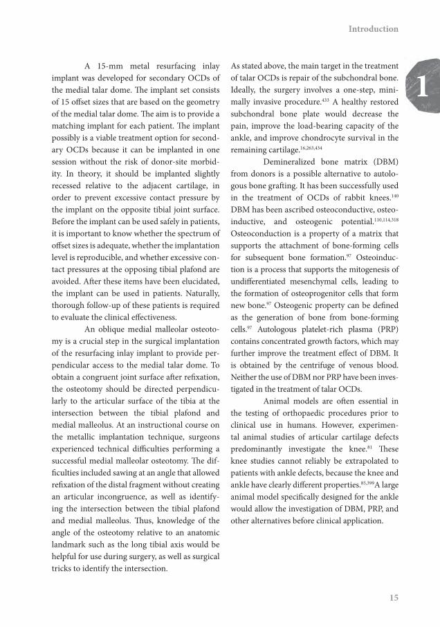
1A 15-mm metal resurfacing inlay
implant was developed for secondary OCDs of the medial talar dome. Th e implant set consists of 15 off set sizes that are based on the geometry of the medial talar dome. Th e aim is to provide a matching implant for each patient. Th e implant possibly is a viable treatment option for second-ary OCDs because it can be implanted in one session without the risk of donor-site morbid-ity. In theory, it should be implanted slightly recessed relative to the adjacent cartilage, in order to prevent excessive contact pressure by the implant on the opposite tibial joint surface. Before the implant can be used safely in patients, it is important to know whether the spectrum of off set sizes is adequate, whether the implantation level is reproducible, and whether excessive con-tact pressures at the opposing tibial plafond are avoided. Aft er these items have been elucidated, the implant can be used in patients. Naturally, thorough follow-up of these patients is required to evaluate the clinical eff ectiveness.
An oblique medial malleolar osteoto-my is a crucial step in the surgical implantation of the resurfacing inlay implant to provide per-pendicular access to the medial talar dome. To obtain a congruent joint surface aft er refi xation, the osteotomy should be directed perpendicu-larly to the articular surface of the tibia at the intersection between the tibial plafond and medial malleolus. At an instructional course on the metallic implantation technique, surgeons experienced technical diffi culties performing a successful medial malleolar osteotomy. Th e dif-fi culties included sawing at an angle that allowed refi xation of the distal fragment without creating an articular incongruence, as well as identify-ing the intersection between the tibial plafond and medial malleolus. Th us, knowledge of the angle of the osteotomy relative to an anatomic landmark such as the long tibial axis would be helpful for use during surgery, as well as surgical tricks to identify the intersection.
As stated above, the main target in the treatment of talar OCDs is repair of the subchondral bone. Ideally, the surgery involves a one-step, mini-mally invasive procedure.433 A healthy restored subchondral bone plate would decrease the pain, improve the load-bearing capacity of the ankle, and improve chondrocyte survival in the remaining cartilage.16,263,434
Demineralized bone matrix (DBM) from donors is a possible alternative to autolo-gous bone graft ing. It has been successfully used in the treatment of OCDs of rabbit knees.140 DBM has been ascribed osteoconductive, osteo-inductive, and osteogenic potential.110,114,318 Osteoconduction is a property of a matrix that supports the attachment of bone-forming cells for subsequent bone formation.97 Osteoinduc-tion is a process that supports the mitogenesis of undiff erentiated mesenchymal cells, leading to the formation of osteoprogenitor cells that form new bone.97 Osteogenic property can be defi ned as the generation of bone from bone-forming cells.97 Autologous platelet-rich plasma (PRP) contains concentrated growth factors, which may further improve the treatment eff ect of DBM. It is obtained by the centrifuge of venous blood. Neither the use of DBM nor PRP have been inves-tigated in the treatment of talar OCDs.
Animal models are oft en essential in the testing of orthopaedic procedures prior to clinical use in humans. However, experimen-tal animal studies of articular cartilage defects predominantly investigate the knee.81 Th ese knee studies cannot reliably be extrapolated to patients with ankle defects, because the knee and ankle have clearly diff erent properties.85,399A large animal model specifi cally designed for the ankle would allow the investigation of DBM, PRP, and other alternatives before clinical application.
27443_van Bergen.indd 15 27-11-13 09:23

Chapter 1
16
Outcome assessment
The reporting of outcome after treatment requires reliable and valid outcome scores. The score preferably should be translated and validated in the target population. A large amount of out-come measures have been used for evaluation of OCDs.458 However, none of the outcome mea-sures has been specifically designed or validated for OCDs. Because of the diversity of the outcome measures, comparison and pooling of studies may be unreliable. A clear guideline as well as the validation of existing outcome measures would enable more consistent reporting of outcomes.
Aims and outline of the thesis
This thesis investigates various aspects of talar OCDs that were introduced in the previous sections. Part I of the thesis describes the cur-rent concepts in the treatment of OCDs. Part II contains chapters on primary arthroscopic debridement and bone marrow stimulation. It aims to improve the preoperative planning of the arthroscopic approach, evaluate the long-term outcomes, and accelerate postoperative rehabili-tation. Part III presents the biomechanical and clinical evaluation of a metal resurfacing inlay implant as a new secondary treatment method. It also aims to refine the medial malleolar oste-otomy, which is used as a surgical approach to the talus. Part IV describes animal studies on an alternative treatment with DBM and PRP. Part V presents an overview and recommendations of outcome measures used for OCD treatment and the validation of the German Foot and Ankle Outcome Score. Part VI finalizes the thesis with a general discussion and a summary.
Part I – Current concepts
As described above, there are numerous man-agement options and surgical strategies in the treatment of OCDs. Chapter 2 aims to provide an overview of treatment options. Based on the literature, a treatment algorithm is proposed. Chapter 3 describes the evolution and the vari-ous possibilities of anterior and posterior ankle arthroscopy.
Part II – Primary arthroscopic debridement and bone marrow stimulation
The optimal arthroscopic approach (i.e., anterior or posterior) can be unclear preoperatively. Dur-ing anterior arthroscopic treatment of an OCD, the ankle is held in full plantar flexion to reach the defect.424 The purpose of chapter 4 is to deter-mine whether preoperative CT of the ankle joint in full plantar flexion is a reliable and accurate tool to determine the anterior arthroscopic reach of talar OCDs. The dual purpose of chapter 5 is (1) to quantify the anterior arthroscopic reach with the ankle in full plantar flexion in a larger group of patients using CT scans, and (2) to iden-tify predictive factors of the arthroscopic reach.
Although arthroscopic debridement and bone marrow stimulation is recommended as the primary treatment, long-term outcome data are scarce. The primary aim of chapter 6 is to assess the long-term clinical and radiographic outcomes of arthroscopic debridement and bone marrow stimulation for talar OCDs. The sec-ondary aim is to identify prognostic factors that affect the long-term results.
Bone marrow stimulation can be achieved by drilling or microfracturing. Chapter 7 identifies a potential pitfall in the arthroscopic microfracturing technique.
Early sport resumption after treatment in the young and active population remains a challenge. Chapter 8 describes a detailed
27443_van Bergen.indd 16 27-11-13 09:23

Introduction
17
1study protocol of a randomized controlled tri-al, which investigates whether PEMF leads to earlier resumption of sports in a higher percent-age of patients with an OCD of the talus aft er arthroscopic debridement and microfracturing.
Part III – Secondary surgical treatment with a metal resurfacing inlay implant
Because of the disadvantages of current sec-ondary treatment methods, such as donor-site morbidity and limited availability, a metal resur-facing inlay implant was developed. Chapter 9 aims to investigate whether the off set sizes are adequate for various cadaveric tali, whether the device can be reproducibly implanted slight-ly recessed relative to the adjacent cartilage, and, whether excessive contact pressures to the opposite cartilage can be avoided with this implantation level.
Th e goal of chapter 10 is to evalu-ate the clinical safety and effi cacy of the metal implant in patients with an OCD of the medial talar dome aft er failed previous treatment.
A medial malleolar osteotomy is indi-cated for the surgical implantation of the metal device. To obtain a congruent joint surface aft er refi xation, the osteotomy should be directed per-pendicularly to the articular surface of the tibia at the intersection between the tibial plafond and medial malleolus. Th e purpose of chapter 11 is to determine this perpendicular direc-tion in relation to the longitudinal tibial axis on radiographs for application during surgery. Chapter 12 describes the use of a right-angled aiming probe to facilitate identifi cation of the optimal terminal point of the medial malleolar osteotomy.
Part IV – Alternative treatment
In the absence of an animal model for the inves-tigation of alternative treatment of talar OCDs, we have developed a caprine model for the ankle joint. Th e aim of chapter 13 is to test the feasi-bility of the developed animal model in a small number of goats.
DBM and PRP possess properties that may enhance the repair of OCDs.140,275 Chapter 14 investigates whether DBM leads to more bone regeneration than control OCDs of the goat’s talus, and whether PRP improves the eff ective-ness of DBM.
Part V – Outcome measures
Because a large amount of outcome measures are used in the literature for evaluation of OCDs, comparison of diff erent studies may be unreli-able. Th e objective of chapter 15 is to describe and discuss frequently used clinical and func-tional outcome scores, as well as postoperative imaging modalities, and provide directions for use.
Th e study protocol of chapter 10 has been designed as a multicenter study in coop-eration with a German university hospital. An important outcome score of the study is the Foot and Ankle Outcome Score (FAOS). A Dutch ver-sion has been translated and validated. To also evaluate the German patients reliably, the objec-tive of chapter 16 is to translate and validate a German version of the FAOS.
General discussion
Chapter 17 provides a general discussion and a summary of the thesis.
27443_van Bergen.indd 17 27-11-13 09:23
27443_van Bergen.indd 18 27-11-13 09:23

Part ICurrent concepts
27443_van Bergen.indd 19 27-11-13 09:23

27443_van Bergen.indd 20 27-11-13 09:23

Chapter 2Treatment of osteochondral
defects of the talus
Christiaan J.A. van BergenPeter A.J. de Leeuw
C. Niek van Dijk
Revue de Chirurgie Orthopédique et Réparatrice de l’Appareil Moteur 2008;94(8 Suppl):635-646.
27443_van Bergen.indd 21 27-11-13 09:23

Chapter 2
22
Introduction
An osteochondral defect (OCD) is the collec-tive term for focal lesions involving the articular cartilage and subchondral bone. If only cartilage is involved in the pathology, the term chondral defect is used. Many synonyms are used, includ-ing osteochondritis dissecans,135 transchondral fracture,49 flake fracture,197 talar dome frac-ture,30 osteochondral fracture,19 osteochondral lesion,235 and osteochondral defect.346 A differen-tiation should be made between traumatic and nontraumatic origin (i.e., osteochondritis dis-secans). A traumatic event may lead to (partial) detachment of an (osteo)chondral fragment, which may further evolve in the formation of a subchondral cyst with or without osteonecrosis. There is sometimes confusion between traumat-ic and nontraumatic, because a nontraumatic OCD may become symptomatic after trauma.
In 1856, Monro first reported the pres-ence of cartilaginous bodies.290 In 1888, König used the term osteochondritis dissecans for loose body formation associated with articular
cartilage and subchondral bone fracture.227 He referred to an inflammatory process, although this has never been proved to be involved in the pathology. It was not until 1922 that the first report on osteochondritis dissecans in the ankle was published.206 The talar dome is the second-ly most common location in the human body; most occur in the knee.457
An OCD is often not recognized and therefore not adequately treated. The nonrecog-nition is mainly due to the fact that the lesion produces symptoms of previous trauma, and it cannot always be identified on plain radio-graphs.440 After standard treatment for acute ankle sprains, residual symptoms are reported in 33% of patients.56 In these cases, the possibility of an OCD should be considered.
The talus has a limited reparative capac-ity because of its restricted vascular supply.344 Inappropriate treatment of OCDs may result in chronic ankle pain, functional impairment, subchondral cyst formation, and eventually osteoarthritis of the ankle.69,268,344
Abstract
This review article provides a current concepts overview of osteochondral defects of the talus, with special emphasis on treatment options, their indications and future developments. Osteochondral defects of the talar dome are mostly caused by a traumatic event. They may lead to deep ankle pain on weight bearing, prolonged swelling, diminished range of motion, and synovitis. Plain radiographs may disclose the lesion. For further diagnostic evaluation, computed tomography (CT) and magnetic resonance imaging have demonstrated similar accuracy. CT scanning is preferred for preoperative planning. Treatment options are diverse and up to the present there is no consensus. Based on the current literature, we present a treatment algorithm that is mainly guided by the size of the lesion. Asymptomatic or low-symptomatic lesions are treated nonoperatively. The primary surgical treat-ment of defects up to 15 mm in diameter consists of arthroscopic debridement and bone marrow stimulation. For large cystic talar lesions, retrograde drilling combined with a bone graft is an impor-tant alternative. In adolescents or in (sub)acute situations, in which the fragment is 15 mm or larger, fixation of the fragment is preferred. Osteochondral autograft transfer and autologous chondrocyte implantation, with or without a cancellous bone graft, are recommended for secondary cases as well as large lesions.
27443_van Bergen.indd 22 27-11-13 09:23

Treatment of OCDs of the talus
23
2
For the last decade, great develop-ments have been made in the surgical treatment. Despite advancements in options like osteo-chondral autograft transfer system (OATS) or autologous chondrocyte implantation (ACI), arthroscopic debridement and bone marrow stimulation remains the best treatment that is currently available for defects up to 15 mm in diameter.393,439 In larger (cystic) defects this treat-ment is less successful, and hence there is more debate.82,149
Th e aim of this article is to provide an overview of treatment options and their indica-tions for OCDs of the talus, based on the current evidence.
Etiology
In 1985, trauma was described in 98% of lateral lesions and in 70% of medial lesions.135 More recently, 93% was reported for lateral lesions and 61% for medial lesions.439 As not all patients report a history of ankle injury, a subdivision can be made in the etiology of nontraumatic and traumatic defects.
Ischemia, subsequent necrosis, and possibly genetics are etiologic factors in nontrau-matic OCDs.344 Furthermore, OCDs in identical twins and in siblings have been described.18,455 Less reported possible causes are metabolic, vascular, endocrine, and degenerative factors, as well as morphologic abnormalities.49,63,457
In the etiology of traumatic OCDs, ankle sprains play the largest role. A severe ankle sprain may cause a small fracture and subsequent impaired vascularity, leading to the formation of an OCD. Alternatively, the cause may not be a sin-gle event but may consist of a series of repeated, less intense injuries.63,344 Microtraumas caused by repetitive surface loading or excessive stress can lead to cartilage cellular degeneration or apopto-sis and thickening of the subchondral bone.137
Mechanism of injury
During an ankle sprain, the talus twists inside the ankle mortise, which may lead to a bruise and subsequent soft ening or even delamination of the cartilage. Separation may occur in the upper layer, as a result of shearing forces, or may occur in the subchondral bone. Osteocartilagi-nous fragments either remain partially attached or become loose bodies in the ankle joint. Th e subchondral fracture has no soft -tissue attach-ments and is highly susceptible to subsequent avascular necrosis.457 Th e repetitive forcing of synovial fl uid into the underlying cancellous bone with every step of walking may create a subchondral cyst.349 Th e repetitive fl uid pressure may prevent healing of a subchondral cyst.
Berndt and Harty clearly described the trauma mechanism in cadaver ankles.49 Th ey were able to reproduce lateral defects by strong inversion of a dorsifl exed ankle, leading to com-pression of the lateral border of the talar dome against the face of the fi bula. Partial detachment of the chip occurred when the lateral ligament ruptured. Th ey reproduced medial lesions by plantar fl exion and inversion of the ankle com-bined with slight anterior displacement and lateral rotation of the tibia upon the talus.
Th e lateral lesions are typically shal-low and wafer-shaped, indicating the shear mechanism of injury.69 Because of their shape, lateral lesions are more frequently displaced than medial lesions. In contrast, medial lesions are generally deep and cup-shaped, indicating a mechanism of torsional impaction.69 Medial lesions are usually larger than lateral lesions.322
Epidemiology
With the increased awareness and newer diag-nostic techniques, the incidence of OCD seems to increase.257 In 1955, Bosien et al described an
27443_van Bergen.indd 23 27-11-13 09:23

Chapter 2
24
incidence of 7% in 113 patients conservatively treated for acute lateral ankle ligament ruptures.56 Later, van Dijk et al. reported 4% fresh talar dome lesions and 67% fresh chondral lesions of any kind in 30 patients who had operative repair of acute ruptures of lateral ligaments.422 More recently, an even higher incidence was reported, namely 41% of 86 patients with anterior talofibular ligament disruptions and 71% of 92 patients with distal fib-ular fractures.376 However, the majority of these reported lesions were located at the cartilage cov-ering the anterior aspect of the medial malleolus and the opposite medial talar facet or the antero-medial rim of the tibial plafond.376,422 Accordingly, talar OCDs were found in 28% to 40% of patients with ankle fractures treated by arthroscopically assisted open reduction and internal fixation.11,259 In these series, the highest incidence was found in patients with distal fibular fractures.11,259
Most OCDs are localized on the postero-medial (58%) or anterolateral (42%) talar dome (Figure 1), although anteromedial, posterolateral and central lesions also occur.322,439 In a large mag-netic resonance imaging (MRI) survey of 428 affected ankles, 53% of the lesions were localized centromedially and 26% centrolaterally.322
In 4% to 7% of patients, the occurrence of the defect is bilateral,49,69,235 suggesting non-traumatic osteochondritis dissecans. Patients with an OCD are frequently 20- to 30-year-old men.49,393,439
Clinical presentation
An OCD often causes deep pain, swelling, recurrent synovitis, and sometimes locking com-plaints. A differentiation has to be made between the acute and chronic situation.457 In the acute situation, symptoms of the OCD compare to those of acute ankle injuries, including lateral or medial ankle pain, swelling, and limited range of motion. In patients with an isolated ligamentous
ankle injury, these symptoms usually resolve after functional treatment within 2 to 3 weeks. If symptoms do not resolve after 3 to 6 weeks, an OCD should be suspected. These patients usually present with persisting symptoms and sometimes a limited range of motion. Lock-ing and catching are symptoms of a displaced fragment. In most patients with a nondisplaced lesion, the symptoms in the acute situation can-not be distinguished from the soft tissue damage.
Chronic lesions typically present as persistent or intermittent deep ankle pain, during or after activity. Reactive swelling and stiffness may be present, but absence of swelling, locking, or catching does not rule out an OCD. There may be a normal range of motion, with the absence of swelling and absence of recognizable tenderness on palpation.
Figure 1. Three-dimensional CT reconstruction of the left ankle of a 26-year-old male patient with a large osteochondral defect typically localized on the posteromedial (PM) talar dome. The usual localization of an anterolateral (AL) defect is also indicated (*).
27443_van Bergen.indd 24 27-11-13 09:23

Treatment of OCDs of the talus
25
2
For further diagnostic evaluation MRI or computed tomography (CT) are oft en used, with similar accuracy.440 A multislice helical CT scan is useful for defi ning the exact size and location of the lesion, and is therefore preferred for preoperative planning (Figure 3).370,457 Th e scanning protocol involves “ultra high resolu-tion” axial slices with an increment of 0.3 mm and a thickness of 0.6 mm. Multiplanar coronal and sagittal reconstructions should be 1 mm.
Classifi cation
A number of classifi cations have been proposed, based on radiography, CT, MRI, and arthros-copy.19,49,182,257,376,381 Th e fi rst and most frequently used classifi cation is from Berndt and Harty:49
Diagnosis
Routine radiographs of the ankle should be obtained aft er careful history taking and physi-cal examination of the ankle. Th ese consist of weight-bearing anteroposterior (mortise) and lateral views of both ankles. Th e sensitivity and specifi city of the combination of medical his-tory, physical examination, and radiography are 59% and 91%, respectively.440 Th e radiographs may not reveal any pathology, or show an area of radiolucency Figure (2A). Initially, the damage may be too small to be visualized on a routine X-ray. Th e OCD sometimes becomes apparent on radiographs at a later stage. A posteromedial or posterolateral defect may be revealed by a heel rise mortise view with the ankle in plantar fl ex-ion (Figure 2C).440
Figure 2. Weight-bearing anteroposterior (A) and lateral (B) radiographs of the right ankle of a 36-year-old male patient showing radiolucency in the medial talar dome (arrow), indicating an osteochondral defect. A 4-cm heel rise view (C) of the same ankle reveals the posteromedial osteochondral defect more clearly (arrow).
Figure 3. CT scans of the right ankle of a 26-year-old female patient showing a cystic posteromedial osteochondral ankle defect. (A) Axial slice, (B) Coronal reconstruction, (C) Sagittal reconstruction.
27443_van Bergen.indd 25 27-11-13 09:23

Chapter 2
26
Stage I A small compression fractureStage II Incomplete avulsion of a fragmentStage III Complete avulsion of a fragment with-
out displacementStage IV Displaced fragmentScranton and McDermott later added Stage V, representing cystic lesions.349
CT scans are increasingly used in the preoperative workup. A CT classification was therefore introduced in 1993, resembling the above classification, with stage V representing a radiolucent defect.257 None of the current grad-ing systems, however, is sufficient to direct the choice of treatment.
Treatment
Various surgical techniques for symptomatic OCDs have been published. These are generally based on one of the following three principles:457
• Debridement and bone marrow stimulation (microfracturing, drilling, abrasion arthro-plasty), with or without loose body removal;
• Securing a lesion to the talar dome (fragment fixation, retrograde drilling, cancellous bone grafting);
• Development or replacement of hyaline cartilage (osteochondral autograft transfer, autologous chondrocyte implantation, allo-grafts).
For years there has been an ongoing debate about the optimal treatment regime. Debridement of the lesion has been performed progres-sively since the 1950s.49 This method was later combined with bone marrow stimulation, by means of drilling or microfracturing, with favor-able results.12 With the development of ankle arthroscopy, this combined procedure gained much popularity.235,346,424 Nowadays, arthroscop-ic debridement and bone marrow stimulation is the mostly performed procedure for OCD.
Publications on treatment options for talar OCDs were bundled in a systematic review, performed in our institution in July 1998, and updated in June 2000.393,439 Twenty-one investigations with a total of 272 patients were identified. The success rate of debridement and bone marrow stimula-tion was superior to other methods. Arthroscopy was successful in 87% and open procedures in 84% of the cases.439 These good results were confirmed more recently.179,343 However, osteo-chondral autograft transfer system (OATS) andautologous chondrocyte implantation (ACI) were not included due to few studies, and sizes of the treated lesions were not described.
In the case of a cystic lesion, debride-ment may be supplemented by cancellous bone grafting.224,343,457 However, the limited availability of cancellous bone and pain at the donor site remain disadvantages.357 Large osteochondral fragments can be fixed by screws, with a success rate of 73% (Figure 4).393
Recently, more developments have tak-en place. OATS and ACI as original procedures for osteochondral lesions of the knee have evolved to suitable treatment methods for certain lesions of the talus.61,177,315 Excellent results have been published, although numbers are still fairly small and no long-term follow-up is available.39,40,176,315
In 2005, an expert consensus on the treatment of OCD was achieved during the World Consensus Conference of the International Soci-ety of Arthroscopy, Knee surgery & Orthopaedic Sports medicine and International Federation of Sports Medicine (ISAKOS-FIMS).429 The expert group agreed that debridement and bone mar-row stimulation is the first step in the treatment of most symptomatic OCDs, and presented a useful guideline. For failed primary treatment they recommended to consider OATS.Based on the current literature, a revised treat-ment guideline is presented (Table 1), which will be further discussed in the Discussion section.
27443_van Bergen.indd 26 27-11-13 09:23

Treatment of OCDs of the talus
27
2
Surgical approach
Th e size and location of the lesion as well as the type of surgical treatment determine the surgi-cal approach. Th e preferred approach of most lesions is by means of anterior arthroscopy.432 Alternative approaches are posterior arthrosco-py by means of a two-portal hindfoot approach and open arthrotomy with or without a medial malleolar osteotomy.200,281,427 Arthroscopy off ers the advantages of outpatient treatment and pos-sibly less postoperative morbidity, faster and functional rehabilitation, and earlier resumption of sports 432. Lateral lesions seldom require a mal-leolar osteotomy. In the rare case of a posteriorly
localized lateral lesion, a fi bular osteotomy pro-vides the best open exposure.142
In the situation of debridement and bone marrow stimulation, the majority of lesions can be treated by means of anterior arthroscopy with the ankle in full plantar fl exion. As a rule of thumb, lesions located in the anterior half or in the anterior part of the posterior half of the talus in patients with unlimited plantar fl exion can be reached and treated this way.432 Some ligament laxity will improve the exposure.
Figure 4. Weight-bearing radiographs of the left ankle of a 13-year-old male patient with progressive complaints of deep ankle pain over 18 months without preceding trauma. Preoperative anteroposterior (A) and lateral (B) radiographs show a large lateral radiolucent area involving more than 50% of the talar dome, representing partial talar necrosis. Th e lesion was secured to the talus by means of two compression screws. Th e radiographs (C) and (D) represent the situation 6 months postoperatively in which the lesion was healed. Screw removal was performed 3 months later.
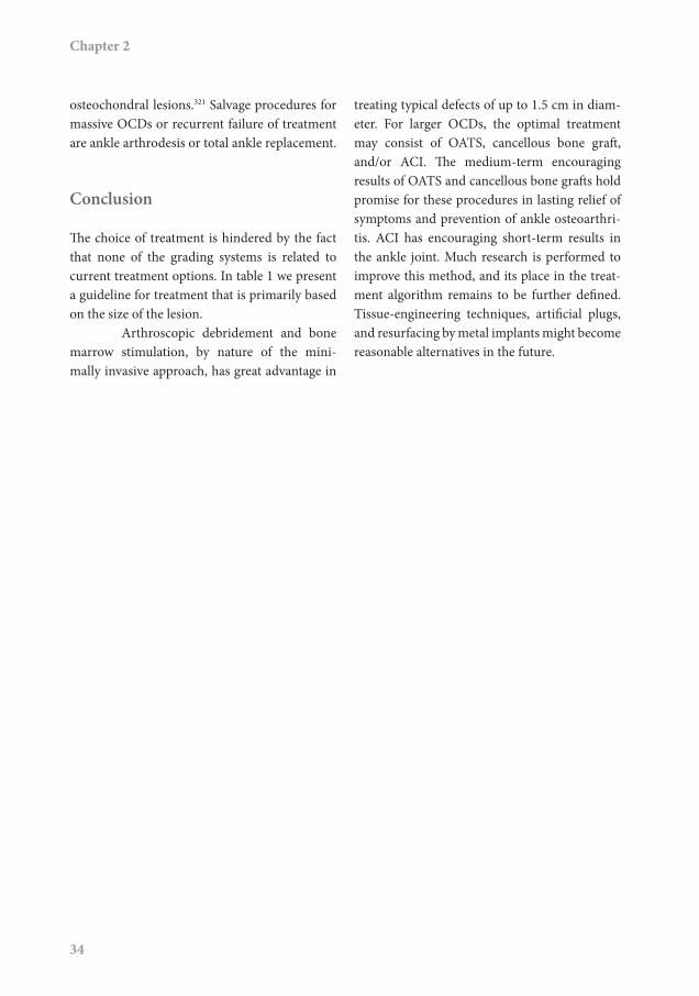
Table 1. Guideline for treatment of osteochondral defects of the talus
Lesion type Treatment optionsAsymptomatic or low-symptomatic ConservativeSymptomatic, <15 mm Conservativea, debridement and drilling/microfracturingSymptomatic, ≥15 mm Fragment fi xation, OATS, ACI, debridement and drilling/microfracturingCystic, ≥15 mm Debridement +/- (retrograde) drilling/microfracturing with cancellous
bone, OATS, ACI with cancellous boneSecondary OATS, ACI, HemiCAP®b, TruFit®b
Massive lesion Allograft , ankle arthrodesis, total ankle replacement
ACI = autologous chondrocyte implantation and OATS = osteochondral autograft transfer system.a A trial period of six months conservative treatment is recommended.b Resurfacing of the medial talar dome by a metal implant (HemiCAP®) and a biodegradable double-layer implant (Tru-Fit®) might become valuable alternatives in the future.
27443_van Bergen.indd 27 27-11-13 09:23

Chapter 2
28
Debridement and bone marrow stimulation
With this technique, all unstable cartilage is removed, including the underlying necrotic bone. Any cysts underlying the defect are opened and curetted. After debridement, several con-nections with the subchondral bone are created by drilling or microfracturing. The objective is to partially destroy the calcified zone that is often present and to create openings into the sub-chondral bone. Intraosseous blood vessels are disrupted and the release of growth factors leads to the formation of a fibrin clot. The formation of local new blood vessels is stimulated, marrow cells are introduced in the defect, and fibrocarti-laginous tissue is formed.301
Advantages of this technique are the possibility of arthroscopy, the relatively easy pro-cedure, and early rehabilitation. A disadvantage is the formation of fibrous cartilage rather than hyaline cartilage. Although often successful, this may be insufficient for large defects.82
Preoperatively, the approach to the defect should be determined (see Surgical approach section). In the case of arthroscopic treatment it has to be decided whether to use a 4.0-mm arthroscope and treat the OCD in the anterior working area by full plantar flexion of the ankle, or to use a 2.7-mm arthroscope in combination with mechanical distraction.424 Arthroscopy with the foot in full plantar flexion is the preferred method in most cases, although skill and experience are required.346,432
The subchondral bone can be perfo-rated using a 2-mm drill, a microfracture awl or a 1.4-mm Kirscher wire (K-wire). A K-wire has the advantage of flexibility, whereas a drill may break more easily if the position of the ankle is changed during drilling. Microfracturing by means of a microfracture awl offers the possi-bility to work “around the corner” and results in microfractures of the trabeculae rather than destruction of the bone,368 but any created small bony particles should be carefully removed.409
Figure 5. Arthroscopic images of the right talus of a 17-year-old female patient with a lateral osteochondral defect (OD) that is treated by debridement and drilling. The arthroscope is in the anteromedial portal (A). With the ankle in the neutral position the defect is out of the arthroscopic view. By bringing the ankle in plantar flexion the defect can be seen (B). The size of the lesion is identified by palpating the cartilage with a probe (arrows), which is inserted through the anterolateral portal (C). A shaver is introduced for debridement of the defect (D). With use of a K-wire small holes are drilled in the subchondral bone (E). Arthroscopic view of the treated lesion after switching portals (F). During loosening of the tourniquet sufficient hemorrhage in the defect is checked (G).
27443_van Bergen.indd 28 27-11-13 09:23

Treatment of OCDs of the talus
29
2
Operative technique
Th e standard anteromedial and anterolateral approaches are created in the fully dorsifl exed position, as described previously.424,432 Introduc-tion of the 4.0-mm arthroscope and a 4.5- or 5.5-mm bonecutter shaver is performed with the ankle in the fully dorsifl exed position to prevent iatrogenic cartilage damage. If osteophytes or synovitis are present, they are removed fi rst by a chisel, burr, or bonecutter shaver, with the ankle in the dorsifl exed position. Th e completeness of removal is checked by plantarfl exing the ankle. During this part of the procedure a soft tissue distractor may be applied (Figure 5A).424,428 It should now be possible to visualise the lesion in the forced plantarfl exed position (Figure 5B) and to identify the defect by palpating the cartilage with a probe or hook (Figure 5C). Debridement is performed with use of the bonecutter shaver
or a small closed-cup curette (Figure 5D). It is important to remove all necrotic bone and over-lying unstable cartilage.378 Aft er full debridement, the sclerotic zone is perforated several times at intervals of approximately 3 mm (Figures 5E and F). Suffi cient hemorrhage can be checked by loosening of the tourniquet (Figure 5G).
Rehabilitation
Active plantar fl exion and dorsifl exion are encouraged. Partial weight bearing (eggshell) is allowed as tolerated. It is the senior author’s practice to allow progress to full weight bearing within 2 to 4 weeks in patients with central or posterior lesions of up to 1 cm. Larger lesions and anterior lesions require partial weight bear-ing up to 6 weeks. Running on even ground is permitted aft er 12 weeks.457 Sport is resumed aft er an average of 15.1 weeks.343 Full return to
Figure 6. Osteochondral defect of the anterolateral talar dome aft er treatment with two osteochondral autograft s (arrows), measuring 6.5 and 4.5 mm in diameter, harvested from the knee.
27443_van Bergen.indd 29 27-11-13 09:23

Chapter 2
30
normal and sporting activities is usually possible 4 to 6 months after surgery.82
Osteochondral autograft transfer
OATS consists of the harvesting of one or more osteochondral plugs in a lesser—weight-bearing area of the knee and transplanting them into the talar defect.177,350 The aim is to restore the articu-lar surface with hyaline cartilage. One single graft or several smaller grafts (i.e., mosaicplasty) may be used. The use of several grafts provides a better match to the curvature of the talar dome and surface area of the defect, and may reduce donor site morbidity.78,176
Although X-ray evaluation and CT may help to determine the extent of the lesion, indication of OATS is rather based on the size determined after excision of the defect. OATS can also be offered to patients in case of failed primary treatment (see Table 1). An essential aspect of the procedure is insertion of the osteochondral plugs perpendicular to the recipient site. Due to the con-strained configuration of the talocrural joint with its highly contoured articular surfaces, the best approach is by means of open arthrotomy, most of the times using a malleolar osteotomy. The pri-mary harvest site is the medial upper part of the medial femoral condyle. As a less frequent option, the lateral supracondylar ridge can also be used through a miniarthrotomy.457 In case the knee is precludes as a donor site, the ipsilateral talar articu-lar facet may also be used as a harvest site of small sized grafts (2.7 or 3.5 mm in diameter).231
Operative technique
For medial lesions a medial malleolar osteotomy is usually required. Once the lesion is exposed, all diseased and suspect cartilage is removed by curette and scalpel dissection to a sharply defined rim. After debridement of the bony base
of the defect by curettage or abrasion arthroplas-ty, the sharp cutting edge of the appropriate-size drill guide helps to determine an ideal filling rate of the defect. The usual size of the drill holes in the talus is 4.5 or 6.5 mm in diameter (Figure 6).
Upon completion of the recipient site preparation, osteochondral grafts are harvested from the ipsilateral knee. Once the site has been clearly identified, the proper size tubular chisel is directed perpendicularly to the articular sur-face and driven by a hammer to the appropriate depth. Minimal graft length should be at least twice its diameter.457 Three to four plugs can be obtained by flexing the knee from 0° to 100°. At the end of the graft harvesting a suction drain is left behind in the knee joint.
After the graft harvest the recipient site is again evaluated. The first hole is drilled through the tubular drill guide, which also serves as the delivery tube. The depth should be 3 – 4 mm deeper than the length of the selected plug. At this stage the hole is enlarged by 0.1 – 0.2 mm with use of a conical dilator, allowing easy inser-tion of the graft. For each graft, drilling, dilation, and delivery are done as a combined step accord-ingly. After the entire set of grafts is implanted (see Figure 6), the ankle is lavaged, observed for loose bodies, and sent through a range of motion to ensure congruency of the mosaicplasty. The osteotomy site is reduced and internal fixation is performed utilizing the predrilled screw holes.
Rehabilitation
Patients are kept non-weight bearing for 3 weeks; 6 weeks for those with a malleolar oste-otomy. Following this period, partial weight bearing up to 30 kg for 3 weeks is allowed to pro-mote integration of the grafts. An orthosis may improve comfort. Range-of-motion exercises are encouraged. Unprotected weight bearing is sub-sequently allowed. Athletic activities may begin at approximately 6 months.
27443_van Bergen.indd 30 27-11-13 09:23

Treatment of OCDs of the talus
31
2
Autologous chondrocyte implantation
ACI is the implantation of in vitro cultured autologous chondrocytes using a periosteal tissue cover aft er expansion of isolated chon-drocytes. ACI has been popularized by Brittberg and Peterson since 1994.61,315 Since that time, ACI has been performed in over 25,000 patients; 95% in the knee, 3% in the ankle, and 2% in other joints.457 Based on promising early results with ACI in the knee, surgeons have now started using ACI for osteochondral lesions of the talus.
For patients with an OCD who remain symptomatic aft er primary surgical treatment ACI is considered a valuable treatment option. Th e defect should be focal, contained, and pref-erably more than 1.5 cm in diameter. Large lesions with subchondral cysts may also be treat-ed with ACI, using the “sandwich technique”, i.e., fi lling the base of the defect with autologous can-cellous bone.40,315
Contraindications to ACI are bipolar lesions (“kissing lesions”) and diff use degen-erative joint changes. Skeletal malalignment and ligamentous instability are also contraindica-tions, unless they are concomitantly corrected at the time of surgery.40
Operative technique
ACI is a staged procedure. Th e initial surgery consists of ipsilateral knee arthroscopy for car-tilage harvesting. Articular cartilage is harvested from non—weight-bearing surfaces such as the intercondylar notch. Approximately 200 – 300 mg of cartilage is harvested with use of curettes and sent to the laboratory for chondrocyte isola-tion and proliferation.
Th e second stage of the procedure is usually at least 4 weeks aft er the harvesting pro-cedure. A medial or lateral malleolar osteotomy is necessary to provide access for the ACI proce-dure.40 All pathologic fi brous and cartilaginous
tissue is debrided. One should not penetrate the subchondral bone during this step, as this would enable marrow elements to contaminate the cul-tured chondrocyte population.
Th e periosteal graft , oversized by 1 – 2 mm, is next obtained from the ipsilateral proximal or distal tibia. With the cambium side facing toward bone, the periosteal graft is placed over the defect and sutured with multifi lament absorbable sutures, size 5.0 or 6.0. Fibrin glue is placed at the interface to help seal the graft . A small opening at the interface is left patent. Saline is injected, to confi rm a watertight com-partment, and subsequently aspirated from the defect. Th e cultured chondrocytes are then placed into the defect and the insertion site is closed with the last stitch and fi brin glue. Th e osteotomy is repaired with two malleolar screws inserted through predrilled holes.
Rehabilitation
Th e patient is kept non-weight bearing and placed in a well-padded short leg cast dur-ing the immediate postoperative period. At 2 weeks postoperatively, the patient is placed in a controlled action motion (CAM) walker boot. Partial weight bearing and gentle ankle range-of-motion exercises are permitted. Weight bearing is advanced based on radiographic evidence of osteotomy healing. At 6 weeks, the patient dis-continues the use of the CAM walker. Repetitive impact activities, such as jogging and aerobics, can be resumed aft er 6 to 8 months. Return to high level sports is permitted aft er 12 months.
Future developments
To overcome the disadvantages of current treat-ment options various attempts are undertaken, aimed at the improvement of current techniques or the development of alternative methods.
27443_van Bergen.indd 31 27-11-13 09:23

Chapter 2
32
To improve ACI, researchers are experimenting with alternatives. The detached osteochondral fragment has been proposed as a source of osteocytes to result in less morbid-ity.148 Furthermore, different scaffolds have been developed that can be implanted with cultured chondrocytes, obviating the need for perios-teal grafting for fixation.75,269,451 Matrix-induced autologous chondrocyte implantation (MACI) makes use of a collagen type I-III membrane, which serves as the scaffold for implanted chon-drocytes. Although this is promising in animal and human knees,35,451 only two short-term ankle cases have been reported.75 Using Hyalograft C as the scaffold, Giannini et al. performed arthroscopic ACI in 30 patients with good short-term results.149
Regarding OATS, the postoperative application of pulsed electromagnetic fields has been recently shown to limit graft resorption and cyst formation in sheep.47
As a tissue-engineering technique of cartilage, bone marrow-derived mesenchymal stem cells have been successfully implanted using different scaffolds. The majority of this research, however, is still experimental.187,209,252,352 Jancewicz et al. were the first to report a clinical series in the talus.199 Demineralized bone matrix has been proposed as an alternative to autolo-gous bone grafts for the treatment of OCD.140
Experimental progress is made with the use of biodegradable composite implants. Müller et al. reported the use of double-layer biodegradable implants consisting of poly-dl-lactide and a polyglactin/polydioxanon fleece.294 Jiang et al. investigated repair with a biphasic osteochondral composite consisting of b-trical-cium phosphate and dl-polylactide-co-glycolide seeded with autologous chondrocytes using single-stage surgery.201 Commercially available composite implants have become available for the treatment of OCDs of the knee (TruFit Plug®, Smith & Nephew, San Antonio, TX, USA).452
Our research group is currently inves-tigating the applicability of a novel resurfacing technique of the medial talar dome by means of a contoured metal implant (HemiCAP®, Arthro-surface Inc., Franklin, MA, USA).
Alignment and potential correction osteotomy are important issues. In case of persis-tent complaints after initial treatment, check the overall alignment and hindfoot alignment. The future role of correction osteotomy in the treat-ment algorithm of OCD has to be established.
Discussion
The choice of treatment depends on several factors, such as the patient’s age, symptoms, duration of complaints, location and size of the defect, and whether it concerns a primary or sec-ondary OCD.82,149,235,457
Asymptomatic or low-symptomatic lesions are treated nonoperatively by rest, ice, temporarily reduced weight bearing, and an orthosis in case of giving way, for a trial period of 6 months.346,457 Although nonoperative ther-apy yields only 45% successful results,439 a trial period does not adversely affect the outcome of surgery.12 Cartilage lesions have demonstrated to deteriorate slowly in the ankle joint. Hence, the advice is to be conservative; there is always time to test the effect of debridement and bone mar-row bone stimulation.
Surgical treatment is considered in the case of failure of nonoperative treatment or continuing symptoms after previous surgery (secondary OCD). According to reviews of the literature, the best currently available treat-ment for primary OCDs is the combination of excision, debridement, and bone marrow stimu-lation.393,439 According to the ISAKOS – FIMS consensus, debridement and drilling or micro-fracturing is the first step in the treatment of symptomatic osteochondral lesions that are too
27443_van Bergen.indd 32 27-11-13 09:23

Treatment of OCDs of the talus
33
2
small to consider fi xation.429 Hence, symptom-atic lesions up to 15 mm are treated primarily by debridement and bone marrow stimulation. Subchondral cystic lesions smaller than 15 mm do not infl uence the postoperative prognosis.174
In the case of a (cystic) defect sized 15 mm or larger, this technique might also be con-sidered as a primary treatment option. In these cases, a cancellous bone graft may be placed in the defect aft er debridement.109,149,343 Retrograde drilling, combined with cancellous bone graft ing if necessary, may be performed if there is a (large) subchondral cyst with intact cartilage.228,381 Alter-natively, the cancellous bone graft may be placed underneath the cartilage fl ap aft er debridement of the subchondral bone.109 A cancellous bone graft is harvested from the ipsilateral iliac crest or locally from the distal tibial metaphysic.224 Pos-sible drawbacks of this procedure are the limited availability and pain at the donor site.357
Fragment fi xation with one or two screws or K-wires is preferred in (sub)acute situations in which the fragment is 15 mm or larger. In adolescents, fi xation of an OCD should always be considered in case of failure of a peri-od of conservative treatment (see Figure 4).370
In case of failed primary surgical treat-ment, OATS and the more recently introduced ACI are reasonable options.23,39,176,177,315 Th ey both aim at creating a new layer of hyaline cartilage.
OATS was originally developed to treat osteochondral lesions in the knee.175 Aft er promising early experiences, the indication was extended to the talus.176 Christel et al. empha-sized the diffi culty of restoring the curvature of the articular surface and reported less favorable clinical outcome in the ankle than in the knee,79 whereas other authors published promising results.23,31,144,176
Although hyaline cartilage of the donor area located in the knee is diff erent from the talar hyaline cartilage, there is no evidence this would represent a negative infl uence on
the results. However, the integration of donor and recipient hyaline cartilage can be impaired because of diff erent mechanical properties and thickness.79,147,241 Other possible disadvantages are the limited availability of graft s and the risk of donor site morbidity.147,327 Furthermore, the use of a medial malleolar osteotomy to approach the lesion has been associated with a worse outcome, i.e., local osteoarthritis and higher morbidity143 A randomized controlled trial with 2 years of follow-up comparing OATS with debridement (with or without microfracture) showed similar results among the methods.156 However, the debridement and microfracture techniques were recommended because of less postoperative pain. Possible alternatives includ-ing artifi cial osteochondral plugs and metal implants may be used in the future.
Th e newer ACI off ers a promising treatment alternative, but long-term data are lacking. Several authors have reported favorable results at short-term follow-up.39,146,229,315,448,457 However, long-term studies are needed to evalu-ate the effi cacy of this technique. Incomplete healing of subchondral cysts has been noted in some patients aft er ACI, although this did not adversely infl uence clinical outcomes at short-term follow-up.457 Other possible disadvantages are insuffi cient graft integration, two-stage sur-gery and high costs.71,147,448 Until further data are available, we cannot advocate ACI as an initial treatment option for most cases of OCD. How-ever, in patients who have a large OCD or failed prior surgical treatment, the short-term data suggest that ACI can provide good results.
For massive OCDs, the transplantation of fresh or frozen allograft s has been described. A number of experts have expressed their con-cerns of use in the talus, based upon the gradual deterioration of the hyaline part of such graft s in the knee and resorption and fragmentation of the graft .162 Th erefore, transplantation of osteo-chondral allograft s is only indicated for massive
27443_van Bergen.indd 33 27-11-13 09:23
Chapter 2
34
osteochondral lesions.321 Salvage procedures for massive OCDs or recurrent failure of treatment are ankle arthrodesis or total ankle replacement.
Conclusion
Th e choice of treatment is hindered by the fact that none of the grading systems is related to current treatment options. In table 1 we present a guideline for treatment that is primarily based on the size of the lesion.
Arthroscopic debridement and bone marrow stimulation, by nature of the mini-mally invasive approach, has great advantage in
treating typical defects of up to 1.5 cm in diam-eter. For larger OCDs, the optimal treatment may consist of OATS, cancellous bone graft , and/or ACI. Th e medium-term encouraging results of OATS and cancellous bone graft s hold promise for these procedures in lasting relief of symptoms and prevention of ankle osteoarthri-tis. ACI has encouraging short-term results in the ankle joint. Much research is performed to improve this method, and its place in the treat-ment algorithm remains to be further defi ned. Tissue-engineering techniques, artifi cial plugs, and resurfacing by metal implants might become reasonable alternatives in the future.
27443_van Bergen.indd 34 27-11-13 09:23

Chapter 3Advancements in ankle
arthroscopy
C. Niek van DijkChristiaan J.A. van Bergen
Journal of the American Academy of Orthopaedic Surgeons2008;16(11):635-646.
27443_van Bergen.indd 35 27-11-13 09:23

Chapter 3
36
Introduction
Although Burman in 1931 found the ankle joint unsuitable for arthroscopy because of its typi-cal anatomy,66 Tagaki and, later, Watanabe made considerable contributions to arthroscopic surgery.374,446 Watanabe in 1972 published the results of a series of 28 ankle arthroscopies.446 Numerous publications followed, and during the past 30 years, arthroscopy of the ankle joint has become an important procedure for the detection and treatment of chronic and post-traumatic problems. The main indications for anterior arthroscopy are for treatment of anterior impingement syndrome and talar osteochondral defects (OCDs).126,424
Endoscopic surgery (i.e., both arthroscopic and endoscopic surgery) offers the possible advantages of direct visualization of structures, improved assessment of articular cartilage, less postoperative morbidity, faster as well as functional rehabilitation, earlier resump-tion of sports, and outpatient treatment.93,348 The value of diagnostic arthroscopy is limited.424,425 Some authors advocate routine mechanical dis-traction combined with a 2.7-mm arthroscope.59 In most procedures, however, ankle arthroscopy can be performed more effectively without rou-tine joint distraction.398,424
Posterior ankle problems pose a diag-nostic and therapeutic challenge because of their nature and deep location. By means of a two-portal hindfoot approach with the patient in the prone position, posterior ankle joint prob-lems (e.g., loose bodies, ossicles, osteophytes, OCDs) can be treated.427 In the case of a poste-rior impingement syndrome, bony impediments (e.g., os trigonum) can be detached and removed via the two-portal hindfoot approach.427
Anterior ankle arthroscopy
Indications
Ankle problems that can be managed by means of routine anterior ankle arthroscopy include soft tissue and bony impingement, synovitis, loose bodies, ossicles, and OCDs.126 Certain ankle fractures (e.g., Weber type B distal fibular frac-ture, Tillaux fracture) also can be successfully treated by means of arthroscopy-assisted (open) reduction and internal fixation, which offers the advantage of direct visualization and treatment of concomitant intra-articular injuries.305,310,379 Other procedures include arthroscopic ankle stabilization by means of radiofrequency and arthroscopy-assisted ankle arthrodesis. In case
Abstract
Important progress has been made during the past 30 years in arthroscopic ankle surgery. Ankle arthroscopy has gradually changed from a diagnostic to a therapeutic tool. Most arthroscopic proce-dures can be performed by using the anterior working area with the ankle in dorsiflexion or plantar flexion; there is no need for routine ankle distraction. Anterior ankle problems, such as the anterior impingement syndrome, are approached by anteromedial and anterolateral portals and, if necessary, an accessory portal. Most osteochondral defects can be reached from anterior with the ankle in plan-tar flexion. For a far posterior location, the osteochondral defect can be approached from posterior. The two-portal hindfoot endoscopic technique (i.e., both arthroscopoic and endoscopic surgery), with the patient in the prone position, provides excellent access to the posterior ankle compartment and to posteriorly located extra-articular structures.
27443_van Bergen.indd 36 27-11-13 09:23

Advancements in ankle arthroscopy
37
3
A B
Figure 1. Lateral (A) and anteromedial impingement (B) radiographs of the right ankle of a 32-year-old women who reported progressive pain in the right ankle for 1 year. On palpation, a recognizable tenderness on the anteromedial distal tibia was noted. In the lateral view, no pathologic structures can be visualized. In the anteromedial impingement view, talar and tibial osteophytes are clearly visible (arrows).
of multiple disorders (e.g., a symptomatic OCD or ankle impingement with concomitant ankle instability), a combined treatment is oft en pos-sible.
Contraindications
Absolute contraindications are infection and severe degenerative changes. Relative con-traindications are degenerative changes with diminished range of motion, narrowing of the joint space, vascular disease, and edema.124
Diagnosis
Th e value of diagnostic ankle arthroscopy without a preoperative diagnosis is limited; only 26% to 43% of patients benefi t from the procedure.424,425 Hence, a diagnosis should be established preoperatively by physical exami-nation and plain radiographs. If the diagnosis remains unclear, additional radiographs (e.g., heel rise view or anteromedial impingement
view) can be obtained (Figure 1).397,440 Fur-thermore, local infi ltration can be performed. Temporary relief from pain aft er intra-articular infi ltration suggests intra-articular pathology, such as soft -tissue impingement or an OCD.When and OCD, a loose body, or an ossicle is suspected, the lesion may be disclosed by magnetic resonance imaging (MRI) or spiral computed tomography (CT).440 If the CT scan is negative and the preoperative diagnosis remains unclear, it is unlikely that the patient will benefi t from diagnostic arthroscopy.424,425
Routine fi xed distraction versus dorsifl exion
Previous investigators have reported the routine use of ankle joint distraction.59 Th e available noninvasive distraction devices are sterile and attach to the operating table. A strap connect-ed to this distraction device is placed around the ankle. Th e surgeon using one of these devices stands beside the patient to perform
27443_van Bergen.indd 37 27-11-13 09:23

Chapter 3
38
the arthroscopic procedure. Ankle arthroscopy without distraction, however, allows the surgeon to stand at the bottom end of the operating table. In this position, the surgeon can lean against the patient’s foot, thereby bringing the ankle joint into the dorsiflexed position (Figure 2).
Several factors favor the use of dorsi-flexion rather than distraction. First, distraction of the joint may result in tightening of the anteri-or capsule, leading to a reduction of the anterior working area (Figure 3).158 Second, loose bodies and osteophytes are usually located in the anteri-or compartment of the ankle joint. Dorsiflexion creates an anterior working area, which makes removal easy. Introduction of saline solution opens the anterior working area. In the case of a loose body and distraction, the loose body may fall into the posterior aspect of the joint, which makes removal more difficult. Third, in the dorsiflexed position, the talus is concealed in the joint, thereby protecting the cartilage from potential iatrogenic damage.158
Mechanical distraction and use of a small-diameter arthroscope may be beneficial in some situations. These include treatment of ossicles, a soft tissue impediment, a loose body caught in the joint space between the fibula and tibia (the intrinsic syndesmotic area), an OCD located in the posterior tibial plafond, and pos-terior ankle problems. An important alternative for the treatment of posterior ankle problems is a two-portal posterior arthroscopy.427
Figure 2. Positioning of the patient during anterior arthroscopy. The affected heel rests on the very end of the operating table, making it possible for the surgeon to fully dorsiflex the ankle joint by leaning against the sole of the foot.
Figure 3. Schematic lateral view of the ankle joint. In dorsiflexion the anterior working is enlarged (A). Distraction of the ankle joint (arrows) results in tightening of the anterior capsule, reducing the anterior working area (B).
27443_van Bergen.indd 38 27-11-13 09:23

Advancements in ankle arthroscopy
39
3
Should distraction be indicated, a resterilizable noninvasive distraction device enables the surgeon to change quickly from the dorsifl exed position to the distracted position and vice versa (Figure 4).428
Operative technique
Th e anterior dorsifl exion procedure is per-formed as outpatient surgery under general or spinal anesthesia.424 Th e patient is placed in the supine position with slight elevation of the ipsi-lateral buttock. A tourniquet is placed around the upper thigh. Th e heel of the aff ected foot rests on the very end of the operating table, thus making it possible for the surgeon to fully dorsifl ex the ankle joint by leaning against the sole of the patient’s foot (see Figure 2). Th e two primary anterior portals used for anterior ankle arthroscopy are the anteromedial and anterolat-eral, located at the level of the joint line. When
their use is indicated, accessory anterior portals are located just in front of the tip of the medial or lateral malleolus. Some surgeons combine the anterior portals with a posterolateral portal.125
Th e anteromedial portal is made fi rst. Aft er a skin incision has been made just medial to the tibialis anterior tendon, the subcutaneous lay-er is bluntly divided with a hemostat. Th e 4-mm, 30°-angle arthroscope, which we use routinely, is introduced in the fully dorsifl exed position. Saline solution is then introduced into the joint. Under arthroscopic control, the anterolateral portal is made by introducing a spinal needle lateral to the peroneus tertius tendon while respecting the superfi cial peroneal nerve (Figure 5).
Depending on the procedure per-formed, the instruments can be exchanged between portals. Aft er removal of the instru-ments, the arthroscopic incisions are closed with Ethilon sutures (Ethicon, Piscataway, NJ) to pre-vent sinus formation.
Figure 4. A resterilizable noninvasive distraction device, which permits the surgeon to move the ankle quickly from thedorsifl exed position to the distracted position and vice versa.
27443_van Bergen.indd 39 27-11-13 09:23

Chapter 3
40
Complications
Several complications have been described, including injury to neurovascular structures, instrument breakage, articular surface dam-age, neuroma formation, infection, and reflex sympathetic dystrophy.33,125,164,403 The superficial peroneal nerve is at highest risk, and injury to this nerve is associated with the anterolateral portal.125
Reports of complications in ankle arthroscopy vary widely. With the use of either invasive or manual constant distraction, complication rates of 9% to 17% have been reported.33,125,164,403 In the largest series published to date, nearly 50% of complications were neu-rologic.125 The use of constant distraction might be indicative of higher complication rates. In a recent survey performed in our department in which 1,300 consecutive patients with ankle arthroscopy without routine joint distraction were included, the overall percentage of compli-cations was 3.4%. This figure includes 1.4% for hindfoot endoscopy.430
Osteochondral defects
An OCD is a lesion involving articular cartilage and subchondral bone. The incidence of OCDs of the talar dome in patients with acute lateral ankle ligament ruptures is 4% to 7%.56,422 Talar OCD are usually posteromedial (58%) or anterolateral (42%).439 Medial lesions are typically deep and cup-shaped; lateral lesions are shallow and wafer-shaped.69 Inappropriate treatment of OCDs may eventually result in osteoarthritis of the ankle.69
Etiology
Previous trauma to the ankle joint is reported in 93% of lateral lesions and 61% of medial lesions.439 In lateral lesions, the trauma mecha-nism is usually a combination of inversion and dorsiflexion; in medial lesions, the combina-tion is inversion, plantar flexion and rotation.49 In nontraumatic osteochondral defects, pos-sible causes are genetic, metabolic, vascular, endocrine, and degenerative factors, as well as morphologic abnormalities.49,457
A B
Figure 5. Anterior arthroscopy of a left ankle; external view (A) and arthroscopic view (B). Under arthroscopic control, the anterolateral portal is made by introducing a spinal needle lateral to the peroneus tertius tendon.AJ = anterior joint capsule, Lat = lateral, Med = medial, SN = spinal needle (guiding needle for lateral portal), Tal = talus, and Tib = tibia.
27443_van Bergen.indd 40 27-11-13 09:23

Advancements in ankle arthroscopy
41
3
Clinical presentation
Patients with a chronic lesion typically experi-ence persistent or intermittent deep ankle pain during or aft er activity, sometimes accompanied by swelling and limited range of motion. Oft en, on examination, few abnormalities are found. Aff ected ankles may have a normal range of motion with the absence of swelling and no rec-ognizable tenderness on palpation.
Diagnosis
Routine radiographs consist of weight-bearing anteroposterior and lateral views of both ankles. Th e radiographs may show an area of detached bone surrounded by radiolucency. Initially, the damage may be too small to be visualized on a routine radiograph.
A heel rise mortise view may reveal a posterior defect.440 For further diagnostic evalu-ation, CT and MRI have demonstrated similar accuracy.440 A multislice helical CT scan is pre-ferred because it is more helpful for preoperative planning (Figure 6).
Classifi cation and staging
Several classifi cations based on radiography, CT, MRI, and arthroscopy have been pro-posed.19,49,182,381 Th e fi rst and most frequently used classifi cation was that of Berndt and Har-ty:49 stage I, a small compression fracture; stage II, incomplete avulsion of a fragment; stage III, complete avulsion of a fragment without dis-placement; and stage IV, displaced fragment. Scranton and McDermott later added stage V, representing cystic lesions.349 None of the cur-rent grading systems, however, is suffi cient to direct the choice of treatment.
Treatment
Various surgical techniques for the treatment of symptomatic osteochondral lesions have been published. Th ese are generally based on one of the following three principles:457 (1) debridement and bone marrow stimulation (microfracturing, drilling, abrasion arthroplasty); (2) securing a lesion to the talar dome (fragment fi xation, retrograde drilling, bone graft ing); and (3)
A B
Figure 6. Coronal (A) and sagittal (B) computed tomography reconstructions of a posteromedial osteochondral defect of the left talus in a 15-year-old girl. Th e patient presented with persistent deep pain on the anterior side of the left ankle and locking and giving way. One year before her visit, she had had a supination trauma, which had been treated nonsurgically.
27443_van Bergen.indd 41 27-11-13 09:23

Chapter 3
42
development or replacement of hyaline cartilage (autologous chondrocyte implantation [ACI], osteochondral autograft transplantation sys-tem [OATS; i.e., mosaicplasty], allografts). The choice of treatment depends on the patient’s age, symptoms, duration of complaints, and location and size of the defect, as well as whether it con-cerns a primary or secondary OCD.149,457
Asymptomatic or low-symptomatic lesions are treated nonsurgically by rest, ice, temporarily reduced weight bearing, and, in case of giving way, an orthosis.346,457 Symptomatic lesions are treated primarily by debridement and bone marrow stimulation.439 With this technique, all unstable cartilage is removed, including the underlying necrotic bone. Any cysts underlying the defect are opened and curetted. The sclerot-ic-calcified zone that is most commonly present is perforated by means of microfracturing into the vascularized subchondral bone. The under-lying intraosseous blood vessels are disrupted and growth factors are released, leading to the formation of a fibrin clot into the created defect. The formation of local new blood vessels is stimulated, marrow cells are introduced into the OCD, and fibrocartilaginous tissue is formed.301 In case of a (cystic) defect ≥15 mm in size, a can-cellous bonegraft may be placed in the defect.149
Retrograde drilling, combined with cancellous bonegrafting when necessary, may be performed for primary OCDs when there is intact cartilage with a large subchondral cyst.381 When primary treatment fails, OATS or ACI are options.39,177 Although both techniques are promising, results are not yet widely published, and the number of patients included in studies is small.
OATS consists of harvesting one or more osteochondral plugs in a lesser—weight-bearing area of the knee, and transplanting them into the talar defect.177 Although most reports show excellent results, the technique is associ-ated with donor site morbidity, and a medial
malleolar osteotomy is often required.31,147,327 ACI is the implantation of in vitro cultured autologous chondrocytes, using a periosteal tissue cover after expansion of isolated chondro-cytes. Despite excellent results reported by some investigators,39,448 disadvantages include two-stage surgery, high costs, and reported donor site morbidity.147,448
Fragment fixation with one or two leg screws is preferred in acute or semiacute situations in which the fragment is ≥15 mm. In adolescents, fixation of an OCD always should be considered following failure of a 6-month period of conservative treatment.
The size and location of the lesion determine whether to use a standard 4.0-mm arthroscope and treat the OCD in the anterior working area by full plantar flexion of the ankle, or to use a 2.7-mm arthroscope in combination with mechanical distraction.346,424 In patients with unlimited plantar flexion, all defects located in the anterior half of the talus or in the anterior part of the posterior half can be reached and treated by anterior arthroscopy.346,424 Other options are to approach the defect from poste-rior by means of a two-portal hindfoot approach or to proceed by means of a medial malleolar osteotomy.12,427
Surgical technique and rehabilitation
A 4.0-mm scope and a 4.5- or 5.5-mm shaver are routinely used. In case of synovitis, a local synovectomy is performed with the ankle in the dorsiflexed position. The lesion is identified in the forced plantarflexed position by palpating the cartilage with a probe or hook (Figure 7). During this part of the procedure a soft-tissue distractor can be applied (see Figure 4). If possi-ble, the full-radius resector is introduced into the defect. In doubtful cases, identifying the defect by introducing a spinal needle, probe, or curette can be useful before introduction of the resector.
27443_van Bergen.indd 42 27-11-13 09:23

Advancements in ankle arthroscopy
43
3
Th e key to success is to identify the anterior part of the defect and remove the unstable cartilage and subchondral necrotic bone. Th e instruments are subsequently brought into the defect to treat the posterior part. Every step in the debridement procedure is checked by regularly switching por-tals. Aft er full debridement, the sclerotic zone is penetrated by means of a microfracture probe or a Kirschner wire (see Figure 7). Postoperatively a compression dressing is applied.
Active plantar fl exion and dorsifl ex-ion are encouraged. Partial weight bearing (i.e., eggshell pressure) is allowed as tolerated. We allow progress to full weight bearing within 2 to 4 weeks in patients with central or posterior lesions of up to 1 cm. Larger lesions and ante-rior lesions require partial weight bearing up to 6 weeks. Running on even ground is per-mitted aft er 12 weeks.457 Full return to normal and sporting activities is usually possible 4 to 6 months aft er surgery.
Results
In the treatment of osteochondral lesions, non-surgical therapy yields a 45% success rate.439 However, a trial period of nonsurgical treatment does not adversely aff ect the outcome of surgery.12
In a systematic review of the literature, debridement and bone marrow stimulation was superior to other methods, with a mean of 86% good or excellent results in 21 studies (total, 272 patients).439 However, OATS and ACI were not included because of the few number of studies using these methods, and sizes of the treated lesions were not described.
A recent randomized controlled that compared chondroplasty, microfracture, and OATS showed similar results among these methods at 2-year follow-up.156 However, the chondroplasty and microfracture techniques are recommended because of less postoperative pain. Furthermore, costs are lower compared with those of other techniques.147
Figure 7. Arthroscopic images of debridement and drilling of a lateral osteochondral defect (OCD) of the right talus. Th e arthroscope is in the anteromedial portal. With the ankle in the neutral position, the OCD is out of the arthroscopic view (A). By bringing the ankle into the forced plantarfl exed position, the OCD can be seen (B). Th e size of the lesion is identifi ed by palpating the cartilage with a probe (arrows), which is inserted through the anterolateral portal (C). Next, a shaver is introduced for debridement of the defect (D). With the use of a Kirschner wire, small holes are drilled in the subchondral bone (E). Arthroscopic view, aft er switching portals, of the microfractured lesion (F). During loosening of the tourniquet, suffi cient hemorrhage in the defect is checked (G).
27443_van Bergen.indd 43 27-11-13 09:23

Chapter 3
44
Anterior ankle impingement
Chronic anterior ankle pain is commonly caused by formation of tibial or talar osteophytes at the anterior part of the ankle joint. Morris and later McMurray named this condition athlete’s ankle or footballer’s ankle;279,292 these terms were later replaced by the anterior ankle impingement syndrome. The condition predominantly affects soccer players, but it also occurs in runners, ballet dancers, high jumpers, and volleyball players.398
Etiology
Various theories exist regarding the causes of anterior impingement: traction, trauma, recurrent microtrauma, and chronic ankle insta-bility.50,93,279,395 The hypothesis of traction is not plausible because the anterior joint capsule is attached more proximally to the site where the tibial spurs originate.394,396,426
In trauma secondary to or associ-ated with supination stress, damage to the non—weight-bearing cartilage rim occurs.422 A repair reaction is initiated, with cartilage prolif-eration, scar tissue formation, and calcification. Ankle sprains resulting from chronic instability as well as forced dorsiflexion enhance this pro-cess.50,232
The pain in anterior ankle impinge-ment likely is caused by the inflamed soft tissue along the anterior tibiotalar joint line, which is compressed by the talar and tibial osteophytes during forced dorsiflexion.396,431
Clinical presentation
Anterior ankle impingement is characterized by anterior pain at the level of the ankle joint, swelling after activity, and, occasionally, lim-ited dorsiflexion.93 Athletes with recurrent ankle sprains are prone to this condition, and sporting activities are often reduced because of the pain.
On palpation, recognizable tender-ness is noted on the anterior bone at the joint level. Tenderness on palpation medial to the tibialis anterior tendon indicates anteromedial impingement; tenderness lateral to the peroneus tertius tendon indicates anterolateral impinge-ment (Figure 8).431 Forced hyperdorsiflexion may provoke the pain, but this test is often false-negative.
Diagnosis
Standard anteroposterior and lateral radiographs may not detect the presence of osteophytes. When anteromedial osteophytes are suspected, an oblique radiograph should be added because some osteophytes may be undetected by stan-dard radiographs.397 Anteromedial tibial or
Figure 8. Localization of anterior impingement. When a patient recognizes the local tenderness on palpation lateral to the peroneus tertius tendon (A) or medial to the tibialis anterior tendon (B), the diagnosis of anterolateral or anteromedial impingement is made, respectively.
AC = anterocentral region, AL = anterolateral region, and AM = anteromedial region.
27443_van Bergen.indd 44 27-11-13 09:23

Advancements in ankle arthroscopy
45
3talar osteophytes may be overprojected by the anterolateral border of the distal tibia or by the lateral part of the talar neck and body, respec-tively. In the oblique anteromedial impingement view, the beam is tilted in a 45° craniocaudal direction, with the leg in 30° external rotation and the foot in plantar fl exion in relation to the standard lateral radiograph position (Figure 9). Th e anteromedial impingement radiograph has a high sensitivity for detecting anterome-dial osteophytes: 93% for tibial and 67% for talar osteophytes (see Figure 1).397
Treatment and rehabilitation
Although nonsurgical treatment such as intra-articular injections and heel lift s is rec-ommended in the early stage of anterior ankle impingement, outcome is frequently disappoint-ing. Th e arthroscopic approach is performed as described above. With the ankle in the dor-sifl exed position, the contour of the anterior tibia is identifi ed by shaving away the tissue just superior to the osteophyte. A 4-mm chisel and/or motorized shaver system is subsequently used to remove them.
A compression bandage is applied, and partial weight bearing for 3 to 5 days is permitted as tolerated. Active dorsifl exion is encouraged.
Results
Open resection and arthroscopic resection of osteophytes were compared by Scranton and McDermott.348 Th e average length of hospital-ization and time to recovery were shorter in the arthroscopic group.
In prospective studies, success rates varied from 73% to 96%.15,38,426 A signifi cant dif-ference in outcome was seen between patients with normal joint spaces and (90% success) and those with joint space narrowing (50% success).426 Th is fi nding was later confi rmed in two long-term follow-up studies.86,394 Because the alternative in these osteoarthritic patients is arthrodesis, a 50% success rate is accept-able. Both series also reported a high rate of recurrence of osteophytes.86,394 However, no sta-tistical correlation was seen between recurrence of osteophytes and return of symptoms.
Hindfoot endoscopy
History
Th e deep location of hindfoot structures makes direct access diffi cult. Historically, the hindfoot was approached by a three portal technique (i.e., anteromedial, anterolateral, posterolateral), with the patient in the supine position.125 Th e tradi-tional posteromedial portal is associated with potential damage to the tibial nerve, the poste-rior tibial artery, and local tendons.124
A two-portal endoscopic approach with the patient in the prone position was intro-duced in 2000.427 Th is technique has been shown to provide excellent access to the posterior ankle compartment, the subtalar joint, and extra-artic-ular structures.427
Figure 9. In the oblique anteromedial impingement view, the beam is tilted 45° in a craniocaudal direction, with the leg in 30° external rotation and the foot in plantar fl exion in relation to the standard lateral radiograph position.
27443_van Bergen.indd 45 27-11-13 09:23

Chapter 3
46
Indications
The main indications for endoscopy in the pos-terior compartment of the talocrural joint are posteriorly located OCDs of the ankle joint, loose bodies, ossicles, posttraumatic calcifica-tions or avulsion fragments, posterior tibial rim osteophytes, chondromatosis, and chronic synovitis.430 In the posterior compartment of the subtalar joint, the main indications are osteophytes, loose bodies, an intraosseous talar ganglion, and subtalar arthrodesis. Other extra-articular structures that can potentially be treated are the posterior tibial tendon, the flexor hallucis longus tendon, the peroneal tendons, the Achilles tendon, the deep portion of the del-toid ligament, and a symptomatic os trigonum or hypertrophic talar process.430
Posterior ankle impingement
Posterior ankle impingement syndrome is a clin-ical diagnosis in which the patient experiences pain in the hindfoot when the ankle is forced into a plantarflexed position (Figure 10). It is caused by overuse or trauma and mainly occurs in ballet dancers, downhill runners, and soccer players.431
After an injection of lidocaine, the forced hyperplantarflexion test should be negative. On plain radiographs and CT, an os trigonum or hypertrophic posterior talar process may be detected. Treatment consists of resection of a symptomatic os trigonum, reduction of a prominent posterior talar process, or removal of a soft-tissue impediment.
Surgical technique and rehabilitation
The procedure is performed as outpatient sur-gery under general or spinal anesthesia. The patient is placed in a prone position. A tourni-quet is applied around the upper leg, and a small support is placed under the lower leg, making it possible to move the ankle freely (Figure 11). A soft-tissue distraction device can be used when indicated.428 For irrigation, normal saline with gravity flow is suitable. A 4.0-mm, 30° arthro-scope is routinely used for posterior ankle arthroscopy. In addition to the standard exci-sional and motorized instruments for treatment of osteophytes and ossicles, a 4-mm chisel and small periosteal elevator can be useful.
With the ankle in the neutral position, a line is drawn from the tip of the lateral malleolus to the Achilles tendon, parallel to the foot sole. The posterolateral portal is situated just above this line, in front of the Achilles tendon (Figure 12A). After a vertical stab incision is made, the subcutaneous layer is split by a mosquito clamp. The mosquito clamp is directed anteriorly, point-ing in the direction of the interdigital web space between the first and second toe (Figure 13).
Figure 10. The forced hyperplantarflexion test. The patient is sitting with the knee flexed in 90°. The test is performed by repetitive, quick, passive hyperplantarflexion movements (arrow). This may be repeated in slight external or internal rotation of the foot. The investigator may apply this rotational movement on the point of maximal plantar flexion, thereby “grinding” the posterior talar process or os trigonum between tibia and calcaneus.
27443_van Bergen.indd 46 27-11-13 09:23

Advancements in ankle arthroscopy
47
3
When the tip of the clamp touches the bone, it is exchanged for a 4.0-mm arthroscope. Th e direc-tion of view is 30° to the lateral side.
Th e posteromedial portal is now made at the same level (Figure 12B). Aft er making a vertical stab incision in front of the medial aspect of the Achilles tendon, a mosquito clamp
is introduced and directed toward the arthro-scope shaft in a 90° angle. When it touches the shaft of the arthroscope, it is moved anteriorly in the direction of the ankle joint, all the way down, touching the arthroscope shaft until it reaches the bone (Figure 14). Th e arthroscope is now pulled slightly backward until the tip of the mosquito
Figure 11. During hindfoot endoscopy the patient is placed in the prone position. A tourniquet (T) is applied around the upper leg, and a small support (S) is placed under the lower leg, which makes it possible to move the ankle freely.
Figure 12. Portal placement in a right ankle during hindfoot endoscopy. Th e posterolateral portal (PL) is made just above the line from the tip of the lateral malleolus (LM) to the Achilles tendon (AT), parallel to the foot sole, just in front of the Achilles tendon (A). Th e posteromedial portal is made at the same level as the posterolateral portal, just above the line from the tip of the lateral malleolus, in front of the medial aspect of the Achilles tendon (B).
1 = posterolateral portal, 2 = lateral malleolus, and 3 = Achilles tendon.
27443_van Bergen.indd 47 27-11-13 09:23

Chapter 3
48
clamp comes in to view. The clamp is used to spread the extra-articular soft tissue in front of the tip of the lens. In situations where scar tissue or adhesions are present, the mosquito clamp is exchanged for a 4.5-mm full-radius shaver.
After removal of the very thin joint capsule of the subtalar joint by a few turns of the shaver, the posterior compartment of the sub-talar joint can be visualized. At the level of the ankle joint, the posterior tibiofibular and talofib-ular ligaments are recognized. The posterior talar process can be freed of scar tissue, and the flexor hallucis longus tendon is identified. The flexor hallucis longus tendon is an important landmark
to prevent damage to the medial neurovascular bundle (Figure 15). One should always stay lat-eral to this tendon and move medially only when release of the neurovascular bundle is indicated (e.g., tarsal tunnel syndrome). After removal of the thin joint capsule of the ankle joint, the ankle joint can be entered and inspected.
Removal of a symptomatic os trigonum or treatment of a nonunion of a posterior talar process fracture involves partial detachment of the posterior talofibular ligament, release of the flexor retinaculum, and release of the posterior talocalcaneal ligament (Figure 16). The os tri-gonum can be lifted from the subtalar joint by means of a small-sized bone elevator (Figure 17) and removed with a grasper. At the end of the procedure, hemorrhage is controlled by elec-trocautery, and the skin is closed with Ethilon sutures. A sterile compression dressing is applied.
Postoperative treatment is functional and consists of weight bearing on crutches as tolerated for 3 days, after which the dressing is removed. The patient is advised to start range-of-motion exercises as soon as possible after surgery. If necessary, physiotherapy is begun.
Results
We performed 146 endoscopic hindfoot proce-dures on 136 consecutive patients.430 The main indications were bony impingement, OCDs, and flexor hallucis longus tendinitis. Treatment was successful in most patients; two minor complications (1.4%) were seen (i.e., an area of diminished sensation over the heel pad of the hindfoot in both cases).430 Similar results have been published by other surgeons.453 Further-more, the technique described is considered to be a safe method, according to a recent anatomic study.250 It is recommended that the procedure be performed by an experienced arthrosco-pist who has practiced this type of surgery in a cadaveric setting at arthroscopy courses.
Figure 13. The posterolateral instrument (2) is directed anteriorly, pointing in the direction of the interdigital web space between the first and second toe (1).
27443_van Bergen.indd 48 27-11-13 09:23

Advancements in ankle arthroscopy
49
3
Figure 14. Schematic transverse section of the right ankle joint at the level of the arthroscope. Th e posterolateral portal is made fi rst (A). Th e arthroscope is directed toward the fi rst web space. Th e posteromedial portal is made at the same level (B). Th e mosquito clamp is directed toward the arthroscope shaft at a 90° angle. When the mosquito clamp touches the shaft of the arthroscope, the clamp is moved anteriorly in the direction of the ankle joint, all the way down, touching the arthroscope shaft until it reaches the bone (curved arrow) (C). All following instruments (e.g., shaver) are introduced in the same manner.
Figure 15. Hindfoot endoscopy of the right ankle. A combination of two separate procedures is shown. Th e fl exor hallucis longus (FHL) is the important landmark in posterior ankle arthroscopy. Instruments should always stay lateral to this tendon. Viewing medial to the FHL (left ) reveals the neurovascular bundle (tibial nerve [N], and the posterior tibial artery and veins [V]). Th is medial view is only indicated when a tarsal tunnel release is performed.
Lat = lateral, M = mosquito clamp in lateral portal, and Med = medial.
27443_van Bergen.indd 49 27-11-13 09:23

Chapter 3
50
Figure 16. Endoscopic picture of a left ankle. Removal of a symptomatic os trigonum or treatment of a nonunion of a posterior talar process fracture involves partial detachment of the posterior talofibular ligament (PTFL), release of the flexor hallucis longus (FHL) retinaculum, and release of the posterior talocalcaneal ligament (TCL).
Lat = lateral and Med = medial.
Figure 17. Endoscopic view of a right ankle. The os trigonum can be lifted from the subtalar joint with a small bone elevator.
Lat = lateral and Med = medial.
27443_van Bergen.indd 50 27-11-13 09:23

Advancements in ankle arthroscopy
51
3
Summary
Over the last three decades, the fi eld of arthroscopic foot and ankle surgery has pro-gressed signifi cantly. Arthroscopy of the ankle joint has become the procedure of choice for the treatment of chronic and posttraumatic pathologies. Th e diagnosis should be established before surgery. If necessary, CT and additional radiographs (e.g., heel rise view, anteromedial impingement view), can be obtained to confi rm a clinical diagnosis and for preoperative plan-ning. When the dorsifl exed position is used with anterior arthroscopy, ankle distraction is neces-sary only in a minority of cases.
Most OCDs can be treated by anterior arthroscopic debridement and microfracturing
with the ankle in planter fl exion. Th e anterior impingement syndrome is treated by arthroscop-ic excision of osteophytes. Posterior ankle problems (e.g., posterior impingement syn-drome) can be eff ectively treated by means of a two-portal hindfoot approach with the patient in the prone position. Th is approach off ers excel-lent access to the posterior ankle compartment, the subtalar joint, and extra-articular structures.
Acknowledgment
Peter A.J. de Leeuw, PhD fellow, is gratefully acknowledged for the kind preparation of fi gures 1, 4 through 13, and 15 through 17.
27443_van Bergen.indd 51 27-11-13 09:23

27443_van Bergen.indd 52 27-11-13 09:23

Part IIPrimary arthroscopic
debridement and bone marrow stimulation
27443_van Bergen.indd 53 27-11-13 09:23

27443_van Bergen.indd 54 27-11-13 09:23

Chapter 4Computed tomography of the ankle in full plantar fl exion:
a reliable method for preoperative planning of arthroscopic access to osteochondral defects of the talus
Christiaan J.A. van BergenGabriëlle J.M. TuijthofLeendert Blankevoort
Mario MaasGino M.M.J. Kerkhoff s
C. Niek van Dijk
Arthroscopy2012;28(7):985-992.
27443_van Bergen.indd 55 27-11-13 09:23

Chapter 4
56
Introduction
Osteochondral defects (OCD) of the talus involve the articular cartilage and subchondral bone and are usually located on the central or posterior third of the medial or lateral talar dome.322 The primary surgical treatment for these lesions is arthroscopic debridement and microfracture.458 Arthroscopic access to the talar dome is restrict-ed by the tibial plafond. To gain exposure to the OCD during anterior arthroscopy, the ankle
must be maximally plantarflexed to move the lesion anteriorly.424,432 However, some defects located in the posterior part of the talus may not be accessible by anterior arthroscopy.296,408 Especially if the OCD is located posteriorly and if there is restricted range of motion of the ankle, determining accessibility of the lesion preop-eratively may be difficult. Accurate preoperative planning will assist the surgeon in choosing whether anterior arthroscopy or another surgi-cal approach would be appropriate.
Abstract
Purpose
The purpose of this study was to determine whether preoperative computed tomography (CT) of the ankle joint in full plantar flexion is a reliable and accurate tool to determine the anterior arthroscopic accessibility of talar osteochondral defects (OCDs).
Methods
Twenty consecutive patients were prospectively studied. All patients had an OCD of the talar dome and had a preoperative CT scan of the affected ankle in maximum plantar flexion. Accessibility of the OCD was defined by the distance between the anterior border of the OCD and the anterior dis-tal tibial rim. This distance was measured on sagittal CT reconstructions by two investigators. The reference standard was the distance between the same landmarks measured during anterior ankle arthroscopy by an orthopedic surgeon blinded to the CT scans. Intraobserver and interobserver reli-ability of CT, as well as the correlation and agreement between CT and arthroscopy, were calculated.
Results
The measured distance between the anterior border of the OCD and the anterior distal tibial rim ranged from -3.1 to 9.1 mm on CT and from -3.0 to 8.5 mm on arthroscopy. The intraobserver and interobserver reliability of the measurements made on CT scans (intraclass correlation coefficients >0.99, p <0.001), as well as the correlation between CT and arthroscopy, were excellent (r = 0.98; p <0.001).
Conclusion
Measurements on CT scans of the ankle in full plantar flexion are a reliable and accurate preoperative method to determine the in situ arthroscopic location of talar OCDs.
27443_van Bergen.indd 56 27-11-13 09:23

CT of plantarfl exed ankle for talar OCD
57
4
Radiographs can be used for the ini-tial diagnosis, but multislice helical computed tomography (CT) and magnetic resonance imaging (MRI) have higher accuracy in detect-ing OCDs.440 CT scanning is preferred for preoperative planning because it visualizes the exact location, size and extent of the OCD.160,408 CT scanning of the ankle is normally performed with the ankle joint in the plantigrade position. Th is position is diff erent from the plantarfl exed ankle on anterior ankle arthroscopy. A CT scan of the ankle in full plantar fl exion, simulating the arthroscopic positioning, may better show the intraoperative location of the OCD and could thus be an important tool for preoperative planning.
Th e purpose of this study is to deter-mine whether a preoperative CT scan of the plantarfl exed ankle is able to predict the ante-rior arthroscopic accessibility of talar OCDs. We hypothesized that the location of the OCD in relation to the anterior distal tibial rim on a CT scan of a fully plantarfl exed ankle would correlate with the location as measured during anterior ankle arthroscopy.
Methods
Study design
A prospective study was undertaken according to a study protocol approved by the local medical ethics committee. Patients aged 18 years or older presenting to our outpatient department with a suspected or diagnosed OCD of the talus start-ing in May 2008 were eligible for the study. Aft er informed consent was obtained, CT scans of the aff ected ankle in both the plantigrade and fully plantarfl exed positions were obtained (Figure 1). Th e surgeon (blinded to the CT scan of the plan-tarfl exed ankle) made the decision for treatment, based on the CT scan of the plantigrade ankle and the clinical assessment of the ankle range of
motion and joint laxity. Th e fi rst 20 patients with an OCD who were scheduled for treatment by anterior ankle arthroscopy (June 2008 to March 2009) were included in the study. A sample size of 19 was able to detect a reliability of 0.9 and a minimally acceptable reliability of 0.7, with α = 0.05 and β = 0.20.443 During the study period, we excluded 10 patients who were scheduled for treatment by methods other than anterior ankle arthroscopy: two were treated by posterior arthroscopy427 – the fi rst for a far posterolateral OCD and the second for a posteromedial OCD combined with a loose body in the posterior ankle compartment – and eight were treated with a focal resurfacing implant412 through a medial malleolar osteotomy for a large OCD of the medial talar dome that failed previous surgical treatment.
Accessibility of the OCD was defi ned by the distance between the anterior border of the OCD and the anterior distal tibial rim. Th is distance was measured on sagittal CT recon-structions and compared to anterior ankle arthroscopy as the reference standard. Th e ante-rior edge of the lesion rather than the middle or posterior portion was chosen as the landmark for accessibility since it is the anterior part of the lesion that has to be identifi ed initially dur-ing arthroscopy. Th e defect is then treated from anteriorly to posteriorly, thus gaining further exposure during treatment. Removal of the ante-rior part of the defect cartilage will automatically create more space for access to the more poste-rior defect area. By forcing the ankle into full plantar fl exion, the ankle opens up anteriorly, thus providing access to the complete defect. Adding a noninvasive ankle distractor can also be helpful at this point.
Computed tomography
With the patient supine, CT scans of the aff ected ankles were acquired in the plantigrade (i.e., the foot sole perpendicular to the table) and
27443_van Bergen.indd 57 27-11-13 09:23

Chapter 4
58
fully plantarflexed ankle positions, using a met-al-free 3-dimensional footplate developed in our department (Figure 1). The affected lower leg was placed on a platform positioned 10 cm above the table to allow slight knee flexion and ankle relax-ation. The foot was attached to the footplate. The ankle was then manually positioned in maximal plantar flexion, and the footplate was secured in place. Before the scan, we measured the degree of plantar flexion of the affected ankle with a goniometer, using the dorsum of the foot and the anterior side of the lower leg as landmarks in both the neutral and the plantarflexed ankle
positions and then subtracting the neutral from the plantarflexed position.
The CT scan was performed analo-gous to the protocol described by Beimers et al.46 Sagittal 1.0-mm slices were reformatted in two planes, because the lateral and medial talar fac-ets have different anatomic orientations; one set of reconstructions was made parallel to the lat-eral talar facet and one set parallel to the medial talar facet. These sagittal planes were identified on the original axial scans and reformatted in a standardized fashion. The reconstructed images parallel to the lateral talar facet were used for the
Figure 1. By use of a specially developed 3-dimensional footplate, the affected ankle was fixed and scanned (A) in theneutral (plantigrade) position and (B) in full plantar flexion.
27443_van Bergen.indd 58 27-11-13 09:23

CT of plantarfl exed ankle for talar OCD
59
4
assessment of OCDs located on the lateral talar dome, whereas the images parallel to the medial facet were used for the defects located on the medial talar dome.
For descriptive purposes, the original axial slices of the neutrally positioned ankles were used to divide the location of the OCDs into nine regions according to the anatomic nine-grid scheme described by Raikin et al.322 For further analysis, the anteromedial, anterocen-tral, and anterolateral locations were combined into the anterior third of the talar dome; the centromedial, centrocentral, and centrolateral into the central third; and the posteromedial, posterocentral, and posterolateral into the pos-terior third.
Th e anterior border of the OCD and the anterior distal tibial rim (i.e., anterior edge of the tibial plafond) were digitally marked on the sagittal reconstructions by the fi rst author in consultation with an orthopaedic surgeon and a musculoskeletal radiologist, using the tools pres-ent on the picture archiving and communication system (Impax 5.3.1, Agfa-Gevaert NV, Mortsel, Belgium) of the radiology department. Th e ante-rior border of the OCD was defi ned as the most anterior point of the bony lesion (Figures 2 and 3). Next, two investigators independently mea-sured the distance from the anterior border of the OCD to the anterior distal tibial rim (Figure 3A) as defi ned by the marks. To analyze intrao-bserver reliability of the CT measurements, one
Figure 2. Sagittal reconstructed images of CT scans of an ankle (A) in neutral position and (B) in full plantar fl exion. Th e tube current was 150 mAs for the neutral position and 26 mAs for the plantarfl exed position. Although low-dose scans had a lower signal-to-noise ratio, this did not hamper delineation of the defects. Th e anterior border of the OCD is indicated by the arrows.
Figure 3. Analysis of a patient with a centromedial OCD. (A) On the CT scan, a tangent line (1) of the anterior distal tibial rim was drawn perpendicularly to the talar dome. Th e distance (2) from the anterior border of the OCD to this line was measured. (B) During arthroscopy, the distance between the anterior distal tibial rim and anterior border of the defect (arrow) was measured with a specially developed arthroscopic ruler. On this ruler, the fi rst line is 2 mm from the tip and each following line is 1 mm more from the tip, up to 7 mm.
27443_van Bergen.indd 59 27-11-13 09:23

Chapter 4
60
investigator (blinded to the first sequence) mea-sured a second time, with a different order of scans.
Arthroscopy
All patients were treated in the supine posi-tion under general or spinal anesthesia, with a tourniquet around the upper leg, by a stand-ard arthroscopic technique as previously described.424,432 Anteromedial and anterolateral portals were created at the level of the joint line with the ankle initially in dorsiflexion. No additional portals were needed. A 4-mm, 30° arthroscope was routinely used. A local synovec-tomy around the portal opposite the defect was performed when necessary. With the arthro-scopic instruments in the ankle joint, the OCD was brought into view by full plantar flexion of the ankle, which was carried out manually by the assistant. The anterior border of the defect was identified with an arthroscopic hook. The surgeon, blinded to the plantarflexion CT scan, measured the anteroposterior distance between the anterior border of the OCD and the ante-rior distal tibial rim with the patient’s ankle in full plantar flexion, using a specially developed arthroscopic ruler (Figure 3B). After the meas-urement, in some difficult cases, a small bony rim or osteophyte of the anterior distal tibia was removed with a bonecutter shaver, or noninva-sive distraction with a strap around the ankle428 was used to improve exposure. The OCD was treated by debridement from anteriorly to pos-teriorly and additional microfracturing with a curved microfracture awl. Routine postoperative care and follow-up were arranged.432
Statistical analysis
The intraobserver and interobserver reliability of the CT measurements were analyzed by compu-tation of intraclass correlation coefficients (ICC)
and systematic differences with 95% confidence intervals (CIs) (paired t-test). According to Fleiss, the reliability is considered good if the ICC is 0.40 to 0.75 and excellent if the ICC is more than 0.75.134 The mean of both observers’ first series of measurements was used for further analysis, as proposed by Bland and Altman.51 The correla-tion of CT and arthroscopic measurements was analyzed by Pearson’s correlation coefficient (r). A scatter plot and linear regression analysis were used to analyze the relationship between CT and arthroscopy. Agreement between both measure-ments was visualized by plotting a Bland and Altman graph.51 This is a rather strict statistical method for assessing agreement between two methods of clinical measurement and tends to reject rather than falsely accept the new method.51 A difference between CT and arthroscopic values of less than 2.0 mm was defined as clinically acceptable. This cut off was based on the experi-ence that OCDs within 2 mm of arthroscopic reach can be accessed by shaving off a small rim of the anterior distal tibia. The limits of agree-ment were calculated (i.e., the mean difference ± 2 × standard deviation of the mean difference). P <0.05 was considered statistically significant.
Results
The study included 20 consecutive patients (17 men and three women) with a mean age of 33 years (range, 18 – 55 years). In 10 patients the right ankle was affected and in 10 the left. The mean degree of plantar flexion was 42° (standard deviation, 10°). According to the anatomic grid scheme, 10 OCDs were located on the centro-medial talar dome, four posteromedial, three centrolateral, one posterolateral, one anterolat-eral, and one centrocentral. When combined in thirds from anterior to posterior, the defect was anterior in one patient (5%), central in 14 (70%), and posterior in five (25%) (Figure 4).
27443_van Bergen.indd 60 27-11-13 09:23

CT of plantarfl exed ankle for talar OCD
61
4
Figure 4. Scatter plot of distance between anterior border of talar osteochondral defect (OCD) and anterior distal tibial rim as measured on CT (x-axis) and anterior ankle arthroscopy (y-axis). Th e dashed line indicates the relation of these measurements as calculated by linear regression (y = 1.069x – 0.557). Th e distribution of diff erent OCD locations (anterior one-third, middle one-third, and posterior one-third of talar dome) is shown.
On CT, the measured distances between the anterior distal tibial rim and the anterior border of the OCD were as follows. Th e fi rst observer’s fi rst sequence ranged from -2.9 to 9.3 mm (mean, 3.0 mm); the second sequence ranged from -2.8 to 9.2 mm (mean, 3.0 mm). Th e second observer’s measurements ranged from -3.3 to 8.9 mm (mean, 2.7 mm). Th e mean of both observers’ fi rst series of measurements ranged from -3.1 to 9.1 mm (mean, 2.8 mm) (Figure 4). When separated for the anterior, cen-tral, and posterior OCDs, the distance measure was 9.1 mm, -0.2 to 7.2 mm (mean, 4.0 mm), and -3.1 to -0.3 mm (mean, -1.8 mm), respec-tively. Both the intraobserver reliability and
interobserver reliability were excellent (ICC, 0.998 and 0.997, respectively; p <0.001). Th e intraobserver systematic diff erence was not statistically signifi cant (mean, 0.051 mm, 95% CI, -0.061 to 0.15 mm), but there was a signifi -cant interobserver systematic diff erence (mean, 0.37 mm; 95% CI, 0.24 to 0.50 mm; p <0.001).
With arthroscopy, the measured dis-tance between the anterior distal tibial rim and the anterior border of the OCD ranged from -3.0 to 8.5 mm (mean, 2.5 mm) (Figure 4). Th e distance measure was 8.5 mm for the anterior lesion, -2.0 to 7.0 mm (mean, 3.8 mm) for the central lesions, and -3.0 to -1.0 mm (mean, -2.4 mm) for the posterior lesions, respectively.
27443_van Bergen.indd 61 27-11-13 09:23

Chapter 4
62
There was an excellent correlation (r = 0.975; p <0.001) between CT and arthroscopy. There was no statistically significant systematic difference (mean, 0.36 mm, 95% CI, -0.051 to 0.77). Linear regression showed that the rela-tion of CT to arthroscopy was expressed by y = 1.069x – 0.557 (Figure 4). There was one subject with a difference of more than 2.0 mm (Figure 5). The limits of agreement were -1.4 to 2.1 mm.
Discussion
This study shows that CT of the ankle in full plantar flexion is an accurate and reliable preop-erative planning tool to determine the anterior arthroscopic accessibility of a talar OCD. This is evidenced by the agreement (Figure 5) and correlation (r = 0.975) of CT and anterior ankle arthroscopy with respect to the distance between the OCD and the anterior distal tibial rim. There was only one patient with a difference between CT and arthroscopy greater than 2.0 mm. Fur-thermore, the formula expressing the relation of CT to arthroscopy approximates a perfect relation of y = x (Figure 4). This scan is now
incorporated into routine preoperative planning in our orthopaedic service.
From the location of the OCD, it can be postulated how accessible the defect will be (Figure 4). All OCDs that were located in the anterior or central third of the talar dome were in easy arthroscopic reach, whereas all posterior OCDs were more difficult to access because they were located posteriorly to the anterior distal tibial rim. Because the location of the OCD on the plantarflexion scan corresponds well to the arthroscopic location, the CT scan can be used reliably to predict the arthroscopic reach of the OCD: if the anterior border of the defect is locat-ed anteriorly to the anterior distal tibial rim on the plantarflexion scan – which was the case in the anterior lesion and in all but one of the cen-tral lesions – the OCD will be accessible through anterior arthroscopy. Even when the OCD is located behind the anterior distal tibial rim with maximal plantar flexion of the ankle, it can often still be reached by anterior arthroscopy. Access in these posterior lesions depends on various parameters, such as ankle range of motion, joint laxity, and the presence of osteophytes, as well as surgical methods. Removal of osteophytes and joint opening in case of ligament laxity will
Figure 5. Bland and Altman plot51 showing difference against mean of distances measured on CT scans and arthroscopy. A difference between CT and arthroscopy of less than 2.0 mm was considered clinically acceptable (dashed lines). The limits of agreement are -1.4 to 2.1 mm.
27443_van Bergen.indd 62 27-11-13 09:23

CT of plantarfl exed ankle for talar OCD
63
4
ease the access. Th erefore, general recommenda-tions for doubtful cases – especially posterior lesions – cannot be given. Th e decision for the most appropriate operative approach in poste-rior lesions thus remains at the discretion of the surgeon. Nevertheless, the CT scan does provide reliable data on the expected arthroscopic loca-tion of the OCD, thus facilitating preoperative planning.
To determine exactly if the scan can diff erentiate between accessible and inacces-sible defects, an additional study would have to be performed in which all plantarfl exed scans are judged preoperatively by the sur-geon, determining whether it is possible to reach the defect with anterior arthroscopy, and then anterior arthroscopy would be per-formed in all lesions – even when the surgeon fi nds this approach inappropriate. Because some patients would undergo a surgical proce-dure without treatment, this study setup seems unethical. Th erefore, the indirect study method was applied as outlined in the paper. With this method, it was shown that the location of the OCDs correlates well between the plantarfl exed CT and anterior arthroscopy.
Diff erent surgical methods could be used to improve the exposure if necessary. For example, three patients in the study group had anterior tibial osteophytes. Th eir removal improved the accessibility of the OCD. Because these osteophytes were shown on CT, they did not interfere with the correlation and agree-ment between CT and arthroscopy, because all measurements were performed before removal of osteophytes. Likewise, access to the defects located posterior to the anterior distal tibial rim could be improved by removing a small part of the rim using a bonecutter shaver (aft er measur-ing the distance of OCD to anterior distal tibial rim with an arthroscopic ruler) or using tempo-rary noninvasive distraction. With noninvasive distraction, the force of distraction was not
actually measured in this group, but it was 110 N in OCD patients in a previous study.428
Th e optimal surgical approach depends on the characteristics of the OCD and the patient, the type of treatment, and the experience of the surgeon.296,408 Anterior arthroscopic ankle surgery is the primary treatment of choice and has the advantages of enhanced visualization, outpatients’ treatment, less postoperative mor-bidity than open surgery, and early functional rehabilitation.93,348,432. Alternative approaches are posterior arthroscopy or open surgery with or without a malleolar osteotomy.296,427,432 Saxena and Eakin treated OCDs arthroscopically when they were located within the anterior 50% of the talar dome.343 Th ey performed an arthroto-my with or without osteotomy in 13 of the 26 patients who underwent microfracture. Like-wise, in a study of 44 patients with 45 OCDs that were initially accessed by anterior ankle arthroscopy, Hankemeier et al. converted to an arthrotomy in 13 and to a medial malleolar osteotomy in 12 because of insuffi cient access.179 Th ese patients may have benefi tted from more precise preoperative planning.
Th e intraobserver and interobserver reliability of measurements on CT was excellent. Th ere was no statistically signifi cant intraobserver systematic diff erence. However, the interobserver systematic diff erence was 0.37 mm (p <0.001). Th e source of this diff erence might have been slight diff erences in drawing the tangent line of the anterior distal tibial rim, or the level in the ankle joint at which the measurements were made. A level of measurement that is more supe-rior results in a higher distance, because the lines of the anterior OCD and the anterior distal tibial rim cross the talus perpendicularly and are thus divergent (see Figure 3). Th ough statistically sig-nifi cant, the interobserver diff erence is so small that it may not be clinically relevant, because 0.37 mm is probably too small to diff erentiate between an accessible and inaccessible OCD.
27443_van Bergen.indd 63 27-11-13 09:23

Chapter 4
64
Alternatively to CT scans, radiographs might be used to estimate the arthroscopic reach, with the advantages of general availability, less radiation and lower costs. However, talar OCDs are rarely visible on lateral radiographs, and the dimensions are underestimated.105 Radiographs thus seem to be inappropriate for preoperative planning.
Strengths of this study are the prospec-tive method, the consecutive and representative series of patients, the blinded comparison of arthroscopy and CT, and the clinical relevance. Th e number of patients, though relatively small, is supported by a power analysis.443 Th e study group is diverse and representative for a larger population. Th e distribution of the OCDs’ loca-tion in the studied population corresponds to a survey of 428 OCDs in 424 patients.322 In this survey, 6% of the lesions were located in the anterior third, 81% in the middle third, and 14% in the posterior third, compared with 5%, 70%, and 25%, respectively, in the present study.
Th ere are also limitations. Th ere are inherent diff erences between CT and arthrosco-py. Th e bony defect was assessed on CT, whereas the articular surface (i.e., the cartilage lesion) was assessed with arthroscopy. Furthermore, the degree of plantar fl exion may diff er between the CT and arthroscopy because of the eff ects of anesthesia and the lack of pain. Th e plantarfl ex-ion angle was not investigated with patients under anesthesia. Despite these diff erences, there was excellent agreement and no signifi cant diff erence of the distances evaluated by CT and
arthroscopy, thus supporting the reliability and usefulness of CT scans.
A disadvantage of the additional CT scan is exposure to radiation. Th e Committee of Radiation at our hospital has estimated the radi-ation of the low-dose plantarfl exion scan to be 0.02 mSv. Th is is categorized as a “trivial” risk for average adults by the International Commission on Radiological Protection.1 Th e natural back-ground eff ective dose rate varies considerably from place to place but typically is around 2.4 mSv/yr. Th e additional radiation of the CT scan can thus be considered acceptable. To reduce the total radiation dose, one may consider acquiring only the plantarfl exion CT scan and omitting the plantigrade scan.
Conclusion
Measurements on CT scans of the ankle in full plantar fl exion are a reliable and accurate preoperative method to determine the in situ arthroscopic location of talar OCDs.
Acknowledgment
Th e authors thank I.N. Sierevelt, M.Sc., from the Orthopaedic Research Center Amsterdam, Department of Orthopaedic Surgery, Academic Medical Center, for statistical advice and M. Poulus and M. Schrijnders from the Department of Radiology, Academic Medical Center, for technical support.
27443_van Bergen.indd 64 27-11-13 09:23

Chapter 5Arthroscopic accessibility of
the talus quantifi ed by computed tomography simulation
Christiaan J.A. van BergenGabriëlle J.M. Tuijthof
Mario MaasInger N. SiereveltC. Niek van Dijk
American Journal of Sports Medicine2012;40(10):2318-2324.
27443_van Bergen.indd 65 27-11-13 09:24

Chapter 5
66
Abstract
Background
Anterior ankle arthroscopy is the preferred surgical approach for the treatment of osteochondral defects of the talus (OCDs). However, the ankle is a congruent joint with limited surgical access.
Purpose
The dual purpose of this study was (1) to quantify the anterior arthroscopic reach (defined as the proportion of the talar dome articular surface located anterior to the anterior distal tibial rim) with the ankle in full plantar flexion, and (2) to identify predictive factors of the arthroscopic reach.
Methods
Computed tomography scans were obtained of 59 ankles (57 patients aged 33 ± 11 years) in full plantar flexion in a non-metallic 3-dimensional footplate. The arthroscopic reach of both the medial and lateral talar domes was assessed on sagittal reconstructions using a custom-made software rou-tine. Intraobserver and interobserver reliability were calculated by intraclass correlation coefficients (ICCs). Various predictive factors of the arthroscopic reach were analyzed by multivariate linear regression analysis.
Results
The arthroscopic reach was 48.2% ± 6.7% (range, 26.7% – 60.7%) of the medial talar dome and 47.8% ± 6.5% (range, 31.2% – 65.1%) of the lateral talar dome (p = 0.62). The intraobserver and interob-server reliability of both measurements were excellent (ICC, 0.99). The clinical plantarflexion angle was a statistically significant predictive factor of both the medial and lateral arthroscopic reaches (i.e., increased plantar flexion corresponded to increased area of access), while joint laxity, gender, and age were not predictive.
Conclusions
Almost half of the talar dome is accessible anterior to the anterior distal tibial rim. The plantarflexion angle is an independent predictive factor of the arthroscopic reach both medially and laterally.
Clinical Relevance
These results may facilitate preoperative planning of the surgical approach for OCDs.
27443_van Bergen.indd 66 27-11-13 09:24

Arthroscopic accessibility of the talus quantifi ed by CT
67
5
identifi cation of the OCD with the ankle in full plantar fl exion, the defect is debrided anteriorly to posteriorly, thus gaining further access dur-ing arthroscopic treatment. Saxena and Eakin approached OCDs arthroscopically when they were located within the anterior 50% of the talar dome, while an arthrotomy with or without oste-otomy was performed for defects located in the posterior half. 343 Similarly, Gobbi et al. withheld patients with far posterior or central lesions from arthroscopic management.156 Hankemeier et al. converted from arthroscopy to arthrotomy, with or without a medial malleolar osteotomy, in 25 of 45 ankles because of insuffi cient access to the OCD.179 Preoperative knowledge of arthroscopic reach of the talus may facilitate the choice of sur-gical approach for talar OCDs.
For preoperative planning, computed tomography (CT) scans accurately assess the size and extent of the bony lesion.129,160 Th e range of motion of the ankle joint has been successfully analyzed using stress CT with forced extreme positions in healthy individuals.402 Using a similar scanning technique in patients with an OCD, van Bergen et al. showed that the location of talar OCDs on CT scans of the ankle in full
Introduction
Osteochondral defects of the talus (OCDs) typi-cally aff ect young, active adults. Th e location of OCDs has been described classically as pos-teromedial or anterolateral.49,135 More recently, a large survey of more than 400 patients showed that these defects are most frequently located on the middle third of the medial or lateral talar dome in a nine-grid scheme.322 Arthroscopic debridement and bone marrow stimulation is considered the primary surgical treatment.153,458 Most OCDs can be reached and treated through anterior ankle arthroscopy.432 During this proce-dure, access to the talar dome is restricted by the tibial plafond due to the high congruence of the ankle joint. Th erefore, the OCD is treated in the anterior working area with full plantar fl exion of the ankle (Figure 1). However, whether the OCD can be reached by anterior ankle arthroscopy may oft en be unclear preoperatively.
As a rule of thumb, OCDs located in the anterior half, including the anterior part of the posterior half of the talus, can be reached and treated by anterior ankle arthroscopy in patients with unlimited plantar fl exion.432 Aft er
A B
Figure 1. Schematic drawing showing the eff ect of plantar fl exion on the arthroscopic reach of an osteochondral defect of the talus (OCD). In the neutral (plantigrade) ankle position, the OCD is covered by the tibial plafond (A). In full plantar fl exion, the OCD can be accessed in the anterior arthroscopic working area (B).
27443_van Bergen.indd 67 27-11-13 09:24

Chapter 5
68
plantar flexion corresponded well with the in situ intraoperative arthroscopic findings of the OCDs.415 Hence, a CT scan of the ankle in full plantar flexion can be used reliably to determine the reach of OCDs with anterior ankle arthros-copy. However, this scanning technique is not readily available in most places. Normal values of arthroscopic reach could thus be helpful for the planning of treatment. Identifying predictive factors of the arthroscopic reach may further improve the preoperative planning process.
The dual purpose of this study was (1) to simulate and quantify, with the use of CT data, the area of the talar dome that can be accessed anteriorly to the anterior distal tibial rim (i.e., arthroscopic reach) with the ankle in full plantar flexion, and (2) to identify predictive factors of the arthroscopic reach. We hypothesized that (1)
50% of the talar dome articular surface is located anteriorly to the anterior distal tibial rim, and (2) plantarflexion angle and joint laxity (anterior drawer test) are independent predictive factors of the arthroscopic reach.
Materials and methods
A prospective study was undertaken according to a study protocol approved by the local Medical Ethics Committee. Fifty-seven patients, aged 18 years or older (mean age, 33 ± 11 years), with a known or suspected OCD in 59 ankles (32 right, 27 left) were included between May 2008 and June 2010, including 20 patients from a previous validation study.415 In two patients, both ankles were scanned because of a bilateral OCD. There
Figure 2. Computed tomography (CT) scanning of an ankle in full plantar flexion using a 3-dimensional footplate. The patient was positioned on the CT table with the affected lower leg attached to the supporting base platform (1) and the foot attached to the footplate (2). The ankle was forced into full plantar flexion and the footplate secured.
27443_van Bergen.indd 68 27-11-13 09:24

Arthroscopic accessibility of the talus quantifi ed by CT
69
5
were 44 male and 13 female patients. Informed consent was obtained.
Computed tomography
A multislice helical CT scan was acquired of each aff ected ankle in full plantar fl exion (Fig-ure 2). A non-metallic 3-dimensional (3-D) footplate (unpublished data, 2011) compatible with CT scanning was constructed to fi t into a commercial CT scanner (Figure 2). Th e 3-D footplate had six degrees of freedom and allowed unconstrained hindfoot joint motion upon load-ing to force the foot in an extreme position. Th e patient lay supine on the CT table, and the aff ected lower leg was fi xed to a base platform of the 3-D footplate with two straps. Th e base platform was 10 cm above the CT table, allowing slight fl exion of the knee and relaxation of the gastrocnemius muscle. Th e foot was attached to the 3-D footplate, and the ankle was positioned in full plantar fl exion and secured by turning the fi xation handle.
Analogous to the protocol as described previously,46 a Philips MX8000 spiral CT scanner (Philips Medical Systems, Eindhoven, the Neth-erlands) was used with the following settings: tube voltage, 120 kV; tube current, 26 mAs; col-limation, 2 ´ 0.5 mm; slice thickness, 0.6 mm; slice increment, 0.3 mm; voxel size, 0.3 ´ 0.3 ´ 0.3 mm; image matrix, 512 ´ 512 pixels; rota-tion time, 0.75 s; resolution, ultra high; kernel, D (sharp); and gantry tilt, 0. Th e examined volume included the distal tibia and the complete talus. Th e Committee of Radiation of our hospital has estimated the radiation of the scan to be 0.02 mSv, which is categorized as a “trivial” risk for average adults by the International Commission on Radiological Protection.1
Th e original axial images were refor-matted into sagittal 1.0-mm slices in two planes: one set of images parallel to the medial talar facet and one set parallel to the lateral talar
facet. Th ese planes were defi ned to correspond with the geometry of the talar dome, which has a divergent anatomic orientation.205 Th e refor-matted images parallel to the medial talar facet were used for the assessment of the medial talar dome, while the images parallel to the lateral facet were used for the lateral talar dome.
Outcome analysis
Arthroscopic reach was defi ned as the propor-tion of the talar dome articular surface located anteriorly to the anterior distal tibial rim.415 Th is defi nition was based on the fact that, initially during arthroscopy, only the anterior part of the lesion has to be identifi ed. Th e OCD is then treated anteriorly to posteriorly, thus gaining further exposure during treatment. Removal of the anterior part of the cartilage lesion cre-ates more space for access to the more posterior defect area. Th e ankle joint opens up anteriorly by forced plantar fl exion, improving access to the complete defect.
To determine the arthroscopic reach, we made the following measurements (Figure 3). Th e anterior and posterior borders of the talar dome and the anterior distal tibial rim were digitally marked on medial and lateral sagit-tal reconstructions using the radiology Picture Archiving and Communication System (Impax 5.3.1, Agfa-Gevaert NV, Mortsel, Belgium), pro-viding two measurements for each scan. In case of a clear anterior distal tibial osteophyte (i.e., fully sclerosed relevant extension of the anterior rim), the osteophyte was excluded from analysis because it is usually removed when performing ankle arthroscopy for OCDs.346 Th e marked CT images were loaded into a custom-made soft ware routine (Matlab, version 7.5.0.342 (R2007b), Th e Mathworks, Natick, Massachusetts). Within this routine, two observers independently indi-cated the talar dome contour with 10 points. Based on these data, two angles, a and β, were
27443_van Bergen.indd 69 27-11-13 09:24

Chapter 5
70
calculated as follows. A circle was fit through the 10 points on the talar dome using a least squares algorithm, providing the circle’s diameter and midpoint coordinates. The latter was used as the imaginary center of rotation of the ankle joint.266 Subsequently, angle a was calculated as the arc length of the circle between the line crossing the anterior border of the talar dome and the mid-point, and the line crossing the anterior distal tibial rim and the midpoint, divided by the total circle’s circumference (Figure 3). Analogously, angle β was calculated as the arc length of the cir-cle between the line crossing the anterior border of the talar dome and the midpoint, and the linecrossing the posterior border of the talar dome and the midpoint, divided by the total circle’s circumference (Figure 3). Finally, the acces-sible area (%) of the talar dome was calculated by dividing angle α (indicating the anterior area) by angle β (indicating the complete talar dome). To assess intraobserver reliability, this complete process was repeated by one of the observers, in different order of scans and blinded to the first sequence.
Predictive factors
To investigate the hypothesis that increased plantar flexion and joint laxity improve the arthroscopic reach, these factors were assessed prior to CT scanning in 46 ankles. The other pre-dictors investigated were gender and age.
The passive plantarflexion angle was measured from a lateral view with a goniom-eter, using the anterior side of the lower leg and dorsum of the foot as landmarks (Figure 4). The measured angles were divided into three groups: group 1 (n = 6), plantarflexion angle >180°; group 2 (n = 18), plantarflexion angle 171° - 180°; and group 3 (n = 22), plantarflexion angle ≤170°. These cutoff values were based on our clinical experience that more than 180° indicates increased range of motion, while less than 170° indicates restriction in range of motion.
The anterior drawer test was performed to assess joint laxity. The assessor determined the amount of force and estimated the excursion. A positive test (n = 9) was defined as an anterior drawer of ≥4 mm and/or a difference between left and right of >2 mm.423
Figure 3. Sagittal computed tomography reconstructions of a fully plantarflexed ankle showing the measurements performed to assess the arthroscopic reach of the medial (A) and lateral (B) talar domes. Using a custom-made software routine, a circle was fit through the talar articular surface. Angle a (black arrow, i.e., area anterior to the distal tibial rim) was defined by the arc length of the circle between the dashed lines from the midpoint of the circle to the anterior border of the talar dome and to the anterior distal tibial rim, respectively, divided by the total circle’s circumference. Angle β (white arrow, i.e., complete talar dome) was defined by the arc length of the circle between the dashed lines from the midpoint to the anterior border of the talar dome and to the posterior border of the talar dome, respectively, divided by the total circle’s circumference. The arthroscopic reach (%) was calculated by dividing angle α by angle β times 100%.
A B
27443_van Bergen.indd 70 27-11-13 09:24

Arthroscopic accessibility of the talus quantifi ed by CT
71
5
Statistical analysis
Th e mean of both observers’ fi rst CT measure-ments is reported. Th e continuous data (age, angles a and β, and arthroscopic reach) were normally distributed and are presented as mean ± standard deviation, supplemented with 95% confi dence intervals (CIs) and ranges for the arthroscopic reach of the talar dome. Th e statisti-cal diff erence between the arthroscopic reach of the medial and lateral talar domes was calculat-ed with the paired t-test. Th e intraobserver and interobserver reliability of the arthroscopic reach were analyzed using intraclass correlation coef-fi cients (ICCs). According to Fleiss, the reliability is considered excellent if the ICC is >0.75.134
To identify predictive factors of the arthroscopic reach, multivariate linear regres-sion analysis was performed with a backward selection procedure. Th e dependent variableswere the medial and lateral arthroscopic reach-es. Th e investigated independent variables were plantarfl exion angle, anterior drawer test, gender, and age. Plantarfl exion angle was a categorical variable with three groups. Each independent variable was initially tested by use of univariate analyses. Th ose variables signifi cantly associated with arthroscopic reach (signifi cance level, 0.10) were entered in the multivariate regression anal-ysis. Th e unstandardized regression coeffi cient
(B) and standard error (SE) were calculated. A p-value of 0.05 indicated statistical signifi cance.
Results
On the medial talar dome, angle a was 51.2° ± 10.5°, and angle β was 106.2° ± 14.9°, resulting in an arthroscopic reach of 48.2% ± 6.7% (95% CI, 46.4% – 49.9%; range, 26.7% – 60.7%) (Figure 5A). On the lateral talar dome, angle a was 49.3° ± 8.3°, and angle β was 103.1° ± 11.5°, indicating an arthroscopic reach of 47.8% ± 6.5% (95% CI, 46.1% – 49.5%; range, 31.2% – 65.1%) (Figure 5B). Th e diff erence between medial and lateral was not statistically signifi cant (p = 0.62). Th e intraobserver and interobserver reliability of the measured angles on CT were excellent (both ICCs, 0.99; p < 0.001).
On the medial talar dome, the arthroscopic reach was 52.6% ± 6.1% in plan-tarfl exion group 1 (>180°), 50.8% ± 5.7% in group 2 (171° - 180°), and 45.5% ± 4.8% in group 3 (≤170°) (Figure 6). On the lateral talar dome, the arthroscopic reach was 53.7% ± 8.7%, 49.2% ± 3.7%, and 45.4% ± 6.0% in plantarfl exion groups 1, 2, and 3, respectively.
With the medial arthroscopic reach as the dependent variable, univariate analysis revealed three signifi cant factors (plantarfl ex-ion angle, gender, and age) to be included in
Figure 4. Schematic view of a lower leg. Th e clinical plantarfl exion angle was measured with the anterior side of the lower leg (1) and dorsum of the foot (2) as landmarks.
27443_van Bergen.indd 71 27-11-13 09:24

Chapter 5
72
Figure 6. Arthroscopic reach (mean and standard deviation) of the medial (continuous line) and lateral (dashed line) talar domes in three patient groups with different plantarflexion angles. The clinical plantarflexion angle was a significant, independent predictor of the arthroscopic reach.
A B
Figure 5. Histogram showing the distribution of the measured arthroscopic reach of the medial (A) and lateral (B) talar domes in the study population.
27443_van Bergen.indd 72 27-11-13 09:24

Arthroscopic accessibility of the talus quantifi ed by CT
73
5
the multivariate linear regression analysis. Multivariate analysis revealed only the plantar-fl exion angle as a predictive factor of the medial arthroscopic reach (group 1 vs. group 3: p = 0.02, B = 6.7, SE = 2.7; group 2 vs. group 3: p = 0.03, B = 4.0, SE = 1.8).
Univariate analysis showed that two factors (plantarfl exion angle and age) were signifi cantly associated with the lateral arthroscopic reach. Multivariate linear regres-sion analysis showed that only the plantarfl exion angle was signifi cantly associated with the lateral arthroscopic reach (group 1 vs. group 3: p = 0.01, B = 7.6, SE = 2.7; group 2 vs. group 3: p = 0.09, B = 3.1, SE 1.8). Th e other investigated factors did not reach statistical signifi cance in the multivari-ate analyses.
Discussion
Th is study aimed to quantify the anterior arthroscopic reach of the talar dome. It is shown that almost half (48%) of the articular surface of the talar dome is situated anteriorly to the tibial plafond with the ankle joint in full plantar fl ex-ion. Th us, on average, an OCD can be reached and treated by anterior ankle arthroscopy if its anterior border is located in the anterior half of the talar dome. Th e anterior defect border is the landmark for accessibility, since only this part of the defect has to be identifi ed initially during arthroscopy. Th e defect is then treated anteriorly to posteriorly, thus gaining further exposure during treatment. As the plantarfl exion angle is an independent predictor of arthroscopic reach, the preoperative estimation of exposure can be improved by measuring the plantarfl exion angle clinically. Th ese fi ndings can serve as a guide-line for preoperative planning of the surgical approach.
Exposure of the talus has been quantifi ed in studies with diff erent open
surgical approaches. Muir et al. compared sev-en approaches, including four arthrotomies (anteromedial, anterolateral, posteromedial, and posterolateral) and three osteotomies (antero-lateral tibial plafond, distal fi bula, and medial malleolus) in nine cadaver specimens.293 Aft er Kirschner wires had been inserted in the talus through the diff erent approaches, lateral radio-graphs were used to measure sagittal access. Using an anteromedial arthrotomy, they could access a mean of 47% (range, 41% - 60%) of the medial talar dome in the sagittal plane. Th e anterolater-al arthrotomy provided 43% (range, 37% - 50%) sagittal access of the lateral talar dome. Young et al. have presented data on cadaveric dissections of fi ve ankles to evaluate the accessible region of the medial talar dome through an anterome-dial and posteromedial open approach without osteotomy.456 Th ey found that a median of 50% (range, 44% - 58%) of the anteroposterior medial talar dome length could be reached through the anteromedial approach. Th e results of our study are in the range of the fi ndings of both cadaveric studies. Th e added value of the present study is the use of a validated method,415 specifi c focus on arthroscopy, the larger and representative study population, and the identifi cation of pre-dictive factors of arthroscopic reach. Th e results may also be relevant for osteochondral graft ing or other methods that require perpendicular access to the OCD. However, in these cases, the posterior border of the OCD (instead of the anterior border) has to be located anteriorly to the anterior distal tibial rim for complete access.
Th e importance of plantar fl exion on the exposure of the talus by anterior ankle arthroscopy can be estimated when comparing the CT angles of this study with radiographic angles of another study. Magerkurth and col-leagues published normal values of hindfoot dimensions in a series of 100 radiographs of neutrally positioned ankles.266 Th ey found that the mean angle of talar coverage by the tibia,
27443_van Bergen.indd 73 27-11-13 09:24

Chapter 5
74
defined by the center of the talus and the bor-ders of the distal tibial joint plane, was 88.1°. In our study, the complete talar dome angle (β) was 104.6° (i.e., mean of medial and lateral). This suggests that only 15.8% (i.e., (104.6° - 88.1°)/104.6° × 100%) of the talar dome articular surface is uncovered by the tibial plafond when the ankle is in a plantigrade position. Our study shows that 48% of the talar dome is uncovered when the ankle is in full plantar flexion, sug-gesting that this area is increased threefold. However, this calculation should be interpret-ed with caution because different diagnostic modalities were used (i.e., plain radiographs by Magerkurth et al. vs. CT by us).
Numerous diagnostic methods are in use for talar OCDs, including plain radiographs, CT, magnetic resonance imaging (MRI), and, more recently, single-photon emission com-puted tomography (SPECT-CT).246,440 Verhagen et al. showed that MRI and multislice helical CT have similar diagnostic accuracy that are both superior to plain radiographs.440 MRI may fur-ther show concomitant soft-tissue injury. The diagnostic value of SPECT-CT has not been reported, but its use has led to a treatment deci-sion making change in 48% compared with MRI. The use of either CT or MRI is recommended for the diagnosis of OCD, but as the subchon-dral bony defect is the main problem in ankle lesions,434 CT scans are preferred for preopera-tive planning because they accurately assess the size of the bony fragment or associated cyst.129,160
With anterior ankle arthroscopy, vision of only the anterior part of the OCD is initially required.432 After identification of the anterior border of the defect, it is debrided anteriorly to posteriorly to further expose the defect, followed by microfracturing.432 We routinely use a 4.0-mm, 30° angle arthroscope, a 4.5-mm or 5.5-mm bonecutter shaver, and a 45° angle microfracture awl. An angled microfracture awl allows treating a lesion “around the corner.”
If access is insufficient with the stan-dard anterior arthroscopy procedure, there are some alternative arthroscopic approaches. The anterior portals can be combined with a posterolateral portal.129,341 Alternatively, a two-portal hindfoot arthroscopic approach with the patient in the prone position is applied to treat OCDs located far posteriorly.432 However, these procedures require more experience. Further-more, distraction of the ankle joint can be used to improve exposure in ankle arthroscopy.129 For example, Kelberine and Frank in 1999 described 48 ankle arthroscopies for the treat-ment of OCDs.210 To gain access to the lesions, distraction was used in 11 patients, and an addi-tional posterolateral portal was made in one patient.210 The maximum joint distraction has been reported to average 1.3 to 4.5 mm.108,428 Continuous distraction, however, could possibly lead to more neurovascular complications.96,108 We prefer an anterior arthroscopy technique without continuous distraction, applying dorsi-flexion and plantar flexion as indicated and, if necessary, temporary noninvasive distraction.432 Alternatives to microfracturing or antegrade drilling for hardly accessible lesions are trans-malleolar or retrograde drilling.228,378 Although good clinical results have been reported, there are disadvantages of both techniques that are not encountered with microfracturing or antegrade drilling. With transmalleolar drilling, healthy tibial cartilage is sacrificed. Retrograde drilling requires intact overlying cartilage of the OCD and is therefore only occasionally applicable.
The present study has limitations. According to the findings of this study, a mean of 48% of the talar articular surface is accessible in the anterior arthroscopic working area. These findings can provide clear clinical guidelines for the optimal surgical approach. However, the percentages reported might be rather strict, as there are different surgical methods to improve exposure, including grooving of the anterior
27443_van Bergen.indd 74 27-11-13 09:24

Arthroscopic accessibility of the talus quantifi ed by CT
75
5
distal tibial rim and using temporary noninva-sive distraction.135,432 Th ese surgical methods were not assessed in this study. Nevertheless, the data can be regarded as the lower limit of access, which can further be improved in practice, thus being on the safe side. Furthermore, there was a high range of arthroscopic reach, that is, 27% to 61% on the medial side and 31% to 65% on the lateral side. Because of this range, a CT scan of the ankle in full plantar fl exion is advised for doubtful cases to optimize the preoperative plan-ning individually. Another possible limitation is that arthroscopic reach was assessed indirectly with CT scans rather than with arthroscopy. For arthroscopic assessment, cadaveric ankles would have to be used, but these are not representa-tive of the patient population and are scarce. Th e obtained results can be regarded as reliable because the methods used were validated in a previous study.415 Finally, diff erent physicians assessed a subset of ankles (46 of 59) for clinical plantar fl exion and joint laxity. Th ese assess-ments may be subject to interobserver variability. Th e variability was minimized by providing clear instructions for measurements, and using easily recognizable anatomic landmarks for clinical plantarfl exion measurement. Th e interobserver reliability of plantarfl exion measurement in another study was good (ICC, 0.72), although
the measurement technique in that study was not specifi cally described.119 Although clinical plantar fl exion was an independent predictor of the arthroscopic reach (Figure 6), joint laxity, in contrast to our hypothesis, was not. Th erefore, the arthroscopic reach variability cannot be reli-ably correlated with ankle laxity.
Conclusion
Th is study quantifi es the anterior arthroscopic reach of the talus with CT scans in a represen-tative patient population. With the ankle in full plantar fl exion, a mean of 48% of the talar dome, both medial and lateral, is located anteriorly to the anterior distal tibial rim. Th e clinical plan-tarfl exion angle is an independent predictor of the arthroscopic reach. Th ese results facilitate preoperative planning of the surgical approach for treatment of OCDs.
Acknowledgment
Th e authors thank Mr. M. Poulus from the Department of Radiology, Academic Medical Center Amsterdam, for technical support with CT scanning and reformatting.
27443_van Bergen.indd 75 27-11-13 09:24

27443_van Bergen.indd 76 27-11-13 09:24

Chapter 6Arthroscopic treatment of
osteochondral defects of the talus: outcomes aft er 8 to 20 years
of follow-up
Christiaan J.A. van BergenLaura S. KoxMario Maas
Inger N. SiereveltGino M.M.J. Kerkhoff s
C. Niek van Dijk
Th e Journal of Bone & Joint Surgery (American volume)2013;95(6):519-525.
27443_van Bergen.indd 77 27-11-13 09:24

Chapter 6
78
Abstract
Background
The primary aim of this study was to assess the long-term clinical and radiographic outcomes of arthroscopic debridement and bone marrow stimulation for talar osteochondral defects. The second-ary aim was to identify prognostic factors that affect the long-term results.
Methods
Fifty (88%) of 57 eligible patients with a primary osteochondral defect treated by arthroscopic debridement and bone marrow stimulation were evaluated after a mean follow-up of 12 years (range, 8 – 20 years). Clinical assessment included the Ogilvie-Harris score, Berndt and Harty outcome ques-tion, American Orthopaedic Foot and Ankle Society (AOFAS) ankle-hindfoot score, and Short Form 36 (SF-36), as well as resumption of work and sports. Weight-bearing radiographs were compared with preoperative radiographs with use of an ankle osteoarthritis classification. The size, location, and classification of the defect, patient age and body mass index, traumatic etiology, and duration of symptoms were recorded and analyzed with use of univariate logistic regression.
Results
The Ogilvie-Harris score was excellent in 20% of patients, good in 58%, fair in 22%, and poor in 0%. According to the Berndt and Harty outcome question, 74% of patients rated the ankle as good, 20% as fair and 6% as poor. The median AOFAS score was 88 (range, 64 – 100). Of the eight subscales of the SF-36, six were comparable with population norms and two were superior in the study group. Ninety-four percent of patients had resumed work and 88% had resumed sports. The radiographs indicated an osteoarthritis grade of 0 in 33% of the patients, I in 63%, II in 4%, and III in 0%. Com-pared with the preoperative osteoarthritis classification, 67% of radiographs showed no progression and 33% showed progression by one grade. None of the prognostic factors was significantly associ-ated with the Ogilvie-Harris score or progression of osteoarthritis.
Conclusions
This study suggests that initial success rates of arthroscopic debridement and bone marrow stimula-tion for osteochondral defects of the talus are maintained over time. No factors that were predictive of the outcome could be identified.
activity. Other symptoms may include stiffness, swelling, and sometimes a locking sensation of the ankle. The natural course of degenerative joint disease following an OCD has not been well defined, although subchondral cysts may
Introduction
Patients presenting with an osteochondral defect (OCD) of the talus are typically young, active adults with deep ankle pain during or after
27443_van Bergen.indd 78 27-11-13 09:24

Arthroscopic treatment of OCDs of the talus
79
6
form and osteoarthritis could be a long-term sequela.69,433
Arthroscopic debridement and bone marrow stimulation is considered the gold stan-dard for treatment of primary talar OCDs, with 85% good or excellent results.458 During this pro-cedure, any loose fragments are removed and the defect is debrided; in addition, small holes in the underlying subchondral bone can be made by drilling or puncturing to promote revasculariza-tion. Th ere are concerns that the fi brocartilaginous tissue formed aft er bone marrow stimulation may be insuffi cient in the long term because it does not have the same biomechanical properties as native hyaline cartilage.229,262
Th e authors of previous studies have attempted to identify prognostic factors associ-ated with such treatment. Such factors included defect size, patient age, body mass index (BMI), history of trauma, and duration of symptoms. Of these factors, the size of the defect appeared to be the most consistently associated with the clinical outcome.76,77,82,156,165
Th e goals of the present study were to evaluate the long-term outcome of arthroscopic debridement and bone marrow stimulation for OCDs of the talus and to identify prognostic fac-tors that aff ect the outcome.
Methods
Th is study was approved by the local Medical Ethics Committee. We identifi ed 105 patients who had undergone arthroscopic treatment of a talar OCD from 1988 to 2000. On the basis of the inclusion and exclusion criteria, 57 patients who had undergone arthroscopic treatment of a primary OCD were invited to participate in the study (Figure 1). Th e stages of the invitation process consisted of mailing a letter (accom-panied by a prepaid envelope) requesting permission to telephone the patient, making
an appointment for a follow-up visit at the hospital, and mailing the patient information, informed consent forms, and questionnaires 1 week in advance of the follow-up visit. Fift y (88%) of the eligible patients provided informed consent and were included. Two of these patients did not consent to visit the hos-pital but completed the questionnaires by mail. Fourteen of the patients included in this study had been evaluated 9 years previously.346 Th ey were reevaluated as a prospective cohort to determine whether their outcomes had dete-riorated with time.
Patient evaluation included a chart review, interview, physical examination, and radiographic evaluation. One author not involved in the surgical procedures assessed the patients and completed custom-designed case report forms. Th e completed case report forms were entered into a digital database with use of custom-designed digital data entry sheets.
Th e data retrieved from the charts and interview included the trauma mechanism, duration of ankle symptoms before surgery, postoperative rehabilitation, any additional sur-gery, pain, stiff ness, swelling, and function. Th e physical examination performed at the follow-up visit included weight and height, gait pattern, hindfoot alignment, swelling, ankle motion and stiff ness, muscle function, stability, sensibility, and tenderness. Special attention was paid to the possibility of neurovascular complications.
Clinical outcome measures
Th e primary outcome measure was the subjec-tive outcome of the patients as assessed with use of the Ogilvie-Harris score.304 Secondary clinical outcome measures were the Berndt and Harty outcome question,49,190 American Orthopaedic Foot & Ankle Society (AOFAS) ankle-hindfoot score,217 Short Form-36 (SF-36),7 and resump-tion of work and sports.
27443_van Bergen.indd 79 27-11-13 09:24

Chapter 6
80
The Ogilvie-Harris instrument consists of five items: pain, swelling, stiffness, limping and activity.304 Each item is rated as excellent, good, fair, or poor. The lowest grade among the items determines the final score. The Ogilvie-Harris score has not been validated but was chosen because of its previous use and the importance of subjective outcome scores in clinical evalua-tion. The Berndt and Harty outcome question is a single question with three possible answers; the patient categorizes the ankle as good, fair, or poor.49 The modification introduced in 2003 was used because the original language was confusing.190 In this modification, patients with a “good” outcome report that they either (1) have no symptoms of pain, swelling, or insta-bility, or (2) experience slightly annoying, but
not disabling, symptoms; patients with a “fair” outcome report that the symptoms are some-what improved but some measure of disability remains; and patients with a “poor” outcome report that, although there are some periods of time without symptoms, the overall symptoms remain unchanged.190 The AOFAS ankle-hind-foot instrument is a combined objective and subjective clinical rating system.217 The subjec-tive part has been validated.192 The AOFAS score can range from 1 to 100 points and is subdivided into pain (40 points), function (50 points) and alignment (10 points), with a score of 100 indi-cating the best outcome. The SF-36 is a widely used and validated instrument for measuring general health.7 It consists of eight subscales with a total of 36 items. All instruments used in the
Figure 1. Flowchart showing the inclusion and exclusion of patients. The values in parentheses indicate the number of patients. OCD = osteochondral defect.
27443_van Bergen.indd 80 27-11-13 09:24

Arthroscopic treatment of OCDs of the talus
81
6
present study have been used in previously pub-lished studies evaluating the outcome of OCDs of the ankle.128,152,190,346
Imaging
Th e original size of the defects was assessed on preoperative images. Computed tomography (CT) was the fi rst-choice imaging modality for measuring the size of the OCD and was used in 26 patients. If CT scans were unavailable, the defect size was measured with use of mag-netic resonance imaging (MRI) (used in three patients) or conventional radiography. CT is preferred for the assessment of the preoperative size of talar OCDs,160,282 whereas plain radio-graphs are less reliable.105 Th e authors have used CT routinely since 1993. Th e MRI scans were acquired in referring hospitals. Th e defect location was assessed with use of a grid consist-ing of nine squares.322 Th e preoperative OCD condition was rated according to an extended version of the Berndt and Harty radiographic staging system.49 On the original scale, defects are divided into four stages (I to IV) according to the amount of detachment and displacement of the osteochondral fragment. Th e addition of a stage V by Scranton and McDermott was used to classify cystic defects.349 An independent mus-culoskeletal radiologist who was blinded to the outcome interpreted the imaging studies.
At the fi nal follow-up visit, weight-bearing ankle radiographs were obtained in three directions: anteroposterior mortise, heel rise mortise, and lateral views.440 Th e presence of a remaining OCD was assessed in each radio-graph by the radiologist.
Th e extent of degenerative change was determined by comparison of the fi nal radio-graphs with the preoperative radiographs (in 46 cases) or with the fi rst postoperative radiographs (in two cases). All radiographs were digitalized and graded independently in a blinded fashion
and random order by two orthopaedic surgeons with use of an ankle osteoarthritis (OA) clas-sifi cation system (Figure 2).425 In the event of disagreement between the observers, the defi nite classifi cation was determined independently by the fi rst author in the same blinded, random fashion. Although CT scans are more accurate for the visualization of the OCDs, radiographs were obtained at the fi nal follow-up visit to allow grading of the OA and comparison with previ-ous studies.
Arthroscopic procedures
All arthroscopic procedures were outpatient surgical procedures utilizing an anterior or pos-terior two-portal approach.432 Th e reason for the posterior approach used in three patients was the location of the defect. Th e anterior approach was performed with the patient in the supine posi-tion and the posterior approach was performed with the patient prone.
A local synovectomy was performed in the event of synovitis. Any anterior osteophytes (in 12 patients) or posterior osteophytes (in one patient) were removed. Th e OCD was identi-fi ed, and the unstable cartilage and subchondral necrotic bone were removed with a shaver or curette. Full debridement of the sclerotic zone was performed. If the entire base of the defect was bleeding aft er release of the tourniquet, no additional drilling was used (in 11 patients). In 39 patients (see Table 1), the base of the OCD was additionally penetrated with multiple drill holes with use of a 1.4-mm Kirshner wire or 2-mm drill through the portal. None of the patients received additional bone-graft ing.
Active ankle motion exercises were started 2 days postoperatively aft er removal of the compression bandage. Full dorsifl exion and plantar fl exion were encouraged. Partial or non-weight bearing was prescribed for 2 to 8 weeks. No boot, cast, or splint was used.
27443_van Bergen.indd 81 27-11-13 09:24

Chapter 6
82
Statistical methods
Statistical analyses were performed with use of SPSS software (version 16.0; Chicago, Illinois). Variables with a normal distribution are present-ed as the mean and standard deviation. Variables with a skewed distribution are presented as the median and range. The SF-36 subscales were com-pared with the Dutch population normative data using the Student’s t-test.7 The ages of patients who did and did not resume work and the ages of patients who did and did not resume sport were compared with use of the Mann-Whitney U test. The Χ2 test was used to compare progression of OA between patients with cystic and noncystic defects and to compare the Ogilvie-Harris score among different defect locations. The clinical results of the prospective cohort of 14 patients reported previously were analyzed with use of the Wilcoxon signed-rank test.
The relationships of various prognos-tic factors to the clinical outcome and to OA progression were calculated with use of univari-ate logistic regression analyses. Specifically, the Ogilvie-Harris score (dichotomized into excel-lent to good and fair to poor) and progression of the OA classification were compared with the defect size, location, and classification; patient sex, age, and BMI; affected side; traumatic eti-ology; duration of symptoms; preoperative OA; type of treatment; and duration of follow-up. The BMI at the time of follow-up was used because the preoperative BMI was unavailable. All continuous prognostic factors were analyzed as continuous variables. In addition, several continuous factors were dichotomized on the basis of information in the literature.44,235,334,457 Specifically, defect size was dichotomized as ≤11 and >11 mm; age, as ≤32 years and >32 years; duration of symptoms, as ≤12 months and >12
A B C
Figure 2. Examples of grades 0, I, and II of the ankle osteoarthritis classification system.425 (Grade III indicates [sub]total disappearance or deformation of the joint space; this was not seen in the study group.) Grade 0, normal joint or subchondral sclerosis (A). Grade I, osteophyte (arrow) without joint space narrowing (B). Grade II, joint space narrowing (arrow) with or without osteophytes (C).
27443_van Bergen.indd 82 27-11-13 09:24

Arthroscopic treatment of OCDs of the talus
83
6
months; and BMI, as ≤25 and >25 kg/m2. Th e odds ratio (OR) and accompanying 95% con-fi dence interval (CI) were calculated for each prognostic factor. A p-value of <0.05 was con-sidered signifi cant.
Results
Th e study group consisted of 30 male and 20 female patients (Table 1). Th e mean age at the time of surgery was 32 ± 10 years (range, 13 – 52 years). Two skeletally mature patients (one 13-year-old girl and one 16-year-old boy) were less than 18 years of age. Th e right ankle was aff ected in 29 patients and the left in 21. Th irty-six patients described a specifi c injury, including one or multiple ankle sprains (24 patients), a fall from a height, or other injuries (mostly sports related). Th e median time interval between the start of symptoms and surgery was 17 months (range, 1 month – 31 years). Th e mean BMI at the time of follow-up was 26.4 ± 4.2 kg/m2. Th e mean duration of follow-up was 141 ± 34 months (range, 101 – 242 months).
Clinical outcome
Th e Ogilvie-Harris score was excellent in 20%, good in 58%, fair in 22%, and poor in 0% (Table 2). According to the Berndt and Harty outcome question, 74% of the patients rated their ankles as good, 20% as fair and 6% as poor. Th e median AOFAS score was 88 (range, 64 – 100). SF-36 subscale scores ranged from a mean of 71 ± 16 for the vitality component to 94 ± 22 for the role emotional component (Figure 3). Th e scores for the general health perceptions and role emotion-al subscales were signifi cantly higher in the study group compared with the Dutch population nor-mative data (p = 0.048 and 0.014, respectively).
Forty-six (94%) of the 49 patients who had worked before the onset of ankle symptoms
had resumed work. Th e age of the patients who had resumed work (mean age at surgery, 31 years) did not diff er signifi cantly from the age of the patients who had not resumed work (mean, 23 years; p = 0.35). Th irty-seven (88%) of the 42 patients who had played sports had resumed play-ing sports. Two of these patients indicated that they had changed the type of sport. Th e sports played primarily were soccer, tennis, jogging, bicycle racing, fi tness training, and fi eld hockey. Th e age of the patients who had resumed playing sports (mean, 31 years) did not diff er signifi cant-ly from the age of patients who had not resumed playing sports (mean, 29 years; p = 0.77).
Baseline radiographic data
Th e mean preoperative size of the defects was 11.4 ± 4.2 in the anteroposterior direction, 7.8 ± 2.8 mm in the mediolateral direction, and 7.1 ± 3.3 mm in the craniocaudal direction. One-half of the OCDs were located on the centromedial aspect of the talar dome (see Table 1). Seven OCDs were classifi ed as Berndt and Harty stage I, one as stage II, six as stage III, two as stage IV, and 29 as stage V. Five defects could not b e classifi ed according to the Berndt and Harty sys-tem because either the OCD was not visible on preoperative radiographs or preoperative radio-graphs were unavailable.
Radiographic outcome
Examination of the radiographs of the 48 patients who attended the follow-up visit revealed an OCD on 42 of the anteroposterior mortise views, 39 of the heel-rise mortise views, and two of the lateral views (Figure 4). Th e OA classifi ca-tion at the time of follow-up was grade 0 on 16 radiographs (33%), grade I on 30 (63%), grade II on two (4%), and grade III on zero. Th irty-two (67%) patients showed no progression and 16 (33%) showed progression by one grade (see
27443_van Bergen.indd 83 27-11-13 09:24

Chapter 6
84
Figure 3. Short Form-36 subscale scores of the study group (gray bar = mean and error bar = 95% confidence interval) compared with the general population norms (× = mean).
Figure 4. Radiographs of an ankle with a centromedial talar osteochondral defect at the time of follow-up. A subtle remnant of the defect (arrow) is visible on the anteroposterior mortise (A) and heel-rise (B) views; the defect cannot be detected on the lateral view (C).
27443_van Bergen.indd 84 27-11-13 09:24
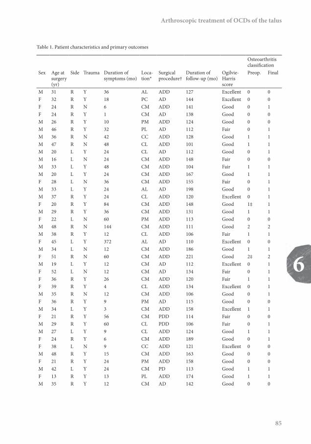
Arthroscopic treatment of OCDs of the talus
85
6
Table 1. Patient characteristics and primary outcomes
Osteoarthritis classifi cation
Sex Age at surgery (yr)
Side Trauma Duration of symptoms (mo)
Loca-tion*
Surgical procedure†
Duration of follow-up (mo)
Ogilvie-Harris score
Preop. Final
M 31 R Y 36 AL ADD 127 Excellent 0 0F 32 R Y 18 PC AD 144 Excellent 0 0F 24 R N 6 CM ADD 141 Good 0 1F 24 R Y 1 CM AD 138 Good 0 0M 26 R Y 10 PM ADD 124 Good 0 0M 46 R Y 32 PL AD 112 Fair 0 1M 36 R N 42 CC ADD 128 Good 1 1M 47 R N 48 CL ADD 101 Good 1 1M 20 L Y 24 CL AD 112 Good 0 1M 16 L N 24 CM ADD 148 Fair 0 0M 33 L Y 48 CM ADD 104 Fair 1 1M 20 L Y 24 CM ADD 167 Good 1 1F 28 L N 36 CM ADD 155 Fair 0 1M 33 L Y 24 AL AD 198 Good 0 1M 37 R Y 24 CL ADD 120 Excellent 0 1F 20 R Y 84 CM ADD 148 Good 1‡ 1M 29 R Y 36 CM ADD 131 Good 1 1F 22 L N 60 PM ADD 113 Good 0 0M 48 R N 144 CM ADD 111 Good 2 2M 38 R Y 12 CL ADD 106 Fair 1 1F 45 L Y 372 AL AD 110 Excellent 0 0M 34 L N 12 CM ADD 186 Good 1 1F 51 R N 60 CM ADD 221 Good 2‡ 2M 19 L Y 12 CM AD 112 Excellent 0 1F 52 L N 12 CM AD 134 Fair 0 1F 36 R Y 26 CM ADD 120 Fair 1 1F 39 R Y 4 CL ADD 134 Excellent 0 1M 35 R N 12 CM ADD 106 Good 0 1F 36 R Y 9 PM AD 115 Good 0 0M 34 L Y 3 CM ADD 158 Excellent 1 1F 21 R Y 56 CM PDD 114 Fair 0 0M 29 R Y 60 CL PDD 106 Fair 0 1M 27 L Y 9 CL ADD 124 Good 1 1F 24 R Y 6 CM ADD 189 Good 0 1F 38 L N 9 CC ADD 121 Excellent 0 0M 48 R Y 15 CM ADD 163 Good 0 0F 21 R Y 24 PM ADD 158 Good 0 0M 42 L Y 24 CM PD 113 Good 1 1F 13 R Y 13 PL ADD 174 Good 1 1M 35 R Y 12 CM AD 142 Good 0 0
27443_van Bergen.indd 85 27-11-13 09:24

Chapter 6
86
Table 1. Patient characteristics and primary outcomes
Osteoarthritis classification
Sex Age at surgery (yr)
Side Trauma Duration of symptoms (mo)
Loca-tion*
Surgical procedure†
Duration of follow-up (mo)
Ogilvie-Harris score
Preop. Final
M 26 R Y 11 PL ADD 139 Good 0 1F 23 R Y 48 CM ADD 152 Good 0 0M 27 L Y 12 CM ADD 112 Fair 0 1F 20 R N 14 CM ADD 242 Excellent § §M 30 L N 45 AL ADD 137 Good § §M 33 R Y 9 CL ADD 115 Good 1 1F 27 L Y 8 PM ADD 218 Good 0 1M 26 L Y 12 AL ADD 205 Fair 0 0M 45 L Y 12 CM ADD 141 Good 0 0
* AL = anterolateral, CC = centrocentral, CL = centrolateral, CM = centromedial, PC = posterocentral, PL = posterolat-eral, and PM = posteromedial. † AD = anterior debridement, ADD = anterior debridement and drilling, PD = posterior debridement, and PDD = posterior debridement and drilling. ‡ The classification of the first postoperative radiographs is given because preoperative radiographs were unavailable. § The patients did not attend the follow-up visit, so final radiographs were not obtained, but the patients completed the questionnaires by mail.
Table 2. Ogilvie-Harris score304 at the time of final follow-up
No. of patientsComponent Poor Fair Good ExcellentPain 0 8 23 19Swelling 0 2 12 36Stiffness 0 6 20 24Limp 0 0 2 48Activity 0 9 12 29Final score* 0 11 29 10
* For each patient, the lowest item determines the final score.
Table 1). Progression of OA had occurred in 47% of the noncystic defects (Berndt and Harty stages I to IV) compared with 32% of the cystic defects (stage V); this difference was not signifi-cant (p = 0.35). The five defects that could not be classified according to Berndt and Harty staging system showed no progression of OA.
Prognostic factors
None of the investigated prognostic factors was found to be significantly associated with the Ogilvie-Harris score or progression of OA. For
example, the Ogilvie-Harris score was good or excellent for 78% of the lateral defects, 100% of the central defects, and 76% of the medial defects (p = 0.63). Likewise, dichotomous analy-sis of the continuous factors did not reveal any significance associations. For example, the Ogil-vie-Harris score was good or excellent for 70% of the OCDs that were ≤11 mm in size compared with 82% of the OCDs that were >11 mm in size (OR, 2.0; 95% CI, 0.42 – 9.6; p = 0.39).
27443_van Bergen.indd 86 27-11-13 09:24

Arthroscopic treatment of OCDs of the talus
87
6
Prospective cohort
Fourteen patients evaluated in the current study had been evaluated 9 years previously in another study.346 A comparison of the Ogilvie-Harris scores in the present study with those in the previous study revealed that the scores of nine patients had remained unchanged, three had worsened, and two had improved (p = 0.66).
Complications and reoperations
Four patients had complications, consisting of an area of hypoesthesia of the skin innervated by the superfi cial peroneal nerve in three patients and hypoesthesia of the fi rst dorsal webspace in one. Th e complications were not reported by the patients but found on the physical examination.
Five patients required a total of six repeat surgical procedures because of persistent or recurrent symptoms, including symptoms due to new trauma. Repeat debridement and bone marrow stimulation was performed arthroscopically in four patients and in an open fashion in one; the latter patient under-went additional arthroscopic debridement 18 months later. In addition, two of these patients received intra-articular hyaluronic acid injec-tions. Th e median time from the initial surgery to the repeat surgery was 21 months (range, 6 – 111 months). Th ree of these patients had a good
Ogilvie-Harris score and two had a fair score at the time of fi nal follow-up. Th e median AOFAS was 80 (range, 75 – 88).
Discussion
To our knowledge, the present study represents the longest reported follow-up of arthroscopic treatment for OCDs of the talus to date. Th e sub-jective outcome was good or excellent in 74% to 78% of the patients, depending on the outcome instrument used. Quality of life was at least com-parable with that of the general population; six subscales of the SF-36 were comparable with population norms and two were superior. Most of the study group had resumed work and sports. Two-thirds of the patients had no radiographic progression of OA and one-third showed pro-gression by one grade; none of the radiographs showed progression by two or three grades.
Th e clinical fi ndings concurred with those of previous studies with a mean follow-up of more than 5 years (Table 3),30,44,77,128,190,346 although there were some diff erences involving the treat-ment and outcome measures used. Th e results of the present study suggest that the intermediate-term results of arthroscopic treatment reported in previous studies are maintained over time.
Th e radiographic results were com-parable with those reported by Ferkel et al. at a
Table 3. Results of published studies with a mean follow-up of 5 years or more
Authors Year N Follow-up* (yr) Outcome measures Good or excellent scoreBaker and Morales 1999 12 10 (8 - 14) Own scoring system 83%Schuman et al. 2002 22 5 (2 - 11) Ogilvie-Harris score 86%Hunt and Sherman† 2003 28 6 (1 - 14) Berndt & Harty score 46%Ferkel et al. 2008 50 6 (2 - 13) Alexander scale 72%Becher et al. 2010 39 6 (1 - 10) Hannover scoring system 79%Choi et al. 2012 173 6 (2 - 10) AOFAS‡ 77%Present study 2013 50 12 (8 - 20) Ogilvie-Harris score 78%
* Th e values are given as the mean, with the range in parentheses. † Eight patients in this study had had previous ankle surgery. ‡ AOFAS = American Orthopaedic Foot & Ankle Society ankle-hindfoot score.
27443_van Bergen.indd 87 27-11-13 09:24

Chapter 6
88
mean follow-up of 6 years.128 In that study, 66% of the patients showed no change in arthritis grade, 30% had a progression of one grade, 2% had a progression of two grades, and 2% had a progression of three grades.
In the present study, osteophytes were observed on the radiographs of 32 of 48 patients at the time of follow-up. It is unknown whether this prevalence can be attributed to the presence of the OCD, to preexisting OA changes, to the active nature of the patient group, or to the treat-ment. The relatively high percentage of patients with stage-I OA (63%; 30 patients) at the time of follow-up may be partially explained by the fact that 14 patients in the study group were at stage I preoperatively (see Table 1). It is known that osteophytes frequently recur, often asymptom-atic, after treatment.394
We had hypothesized that the size of the OCD would be a significant predictor of outcome, as has been reported in some of the previous studies that investigated predictors of outcome of arthroscopic treatment of OCDs (Table 4).44,76,77,82,128,156,165,190,235,244,334 However, none of the predictive factors investigated in the pres-ent study was found to be significantly associated with the clinical or radiographic outcome. The fact that no prognostic factors were identified may have been due to insufficient study power. In comparison, Chuckpaiwong et al. and Choi et al. assessed outcome predictors in cohorts that included more than 100 patients.76,77,82
The long-term results of arthroscopic treatment of OCDs may be superior to those of open treatment. Canale and Belding studied 15 defects treated with open excision and curet-tage.69 After a mean follow-up of 11 years (range, 2 – 26 years), 11 (73%) of the patients had good clinical results and approximately 50% had had degeneration of the joint. In 1989, Angermann and Jensen reported on 18 patients who had undergone open excision and drilling.22 After a mean follow-up of 12 years (range, 9 – 15 years),
almost all patients had persistent symptoms and 17% had OA. Although the results of the pre-sent study compare favorably with those of the previous studies, a direct comparison may not be reliable because patient characteristics could have differed among the studies.
Bone marrow stimulation can be accomplished by several different surgical meth-ods.458 The objective is to remove all unstable cartilage and the underlying necrotic bone and then to stimulate healing of the defect. Opening the subchondral bone can be accomplished with use of a curette, shaver, small drill, Kirschner wire, or microfracture awl. Drilling was used in the 1990s, but microfracture has gained popu-larity since 2000.368 Drilling might cause necrosis because of heating, but the drill or Kirschner wire is cooled by the arthroscopic fluid environ-ment. The use of a microfracture awl is based on the theory that this results in microfractures of the trabeculae rather than destruction of the bone, thereby inducing a healing response.368 A further advantage is that defects can be treated “around the corner” because the end of the awl is curved. However, the microfracture technique may create loose bony particles that become easily detached on withdrawal of the awl.409 An experimental study that compared microfrac-ture and drilling in rabbit knees indicated that microfracture produces fractured and com-pacted bone around the holes, essentially sealing the holes off from viable bone marrow, whereas drilling removes bone from the holes to provide access channels to marrow stroma.73 However, since these differences have not been shown to affect the clinical outcome, the results of the pre-sent study – evaluating bone marrow stimulation performed before the era of microfracture – may be comparable with those of the contemporary microfracture technique.
The present study has limitations. First, its retrospective nature does not allow objec-tive comparison with the preoperative clinical
27443_van Bergen.indd 88 27-11-13 09:24

Arthroscopic treatment of OCDs of the talus
89
6
Table 4. Prognostic factors of success aft er arthroscopic treatment of osteochondral talar defects according to the literatureD
urat
ion
of
follo
w-u
p
NS
NS
Yes
(<1
yr)
NS
AO
FAS
= A
mer
ican
Ort
hopa
edic
Foo
t & A
nkle
Soc
iety
ank
le-h
indf
oot s
core
, BM
I = b
ody
mas
s ind
ex, c
ont.
= co
ntin
uous
(i.e.
, no
cuto
ff po
int d
efi n
ed),
CT
= co
mpu
ted
tom
ogra
phy,
HSS
= H
anno
ver s
corin
g sy
stem
, MRI
= m
agne
tic re
sona
nce i
mag
ing,
NS
= no
t sig
nifi c
ant,
OA
= o
steo
arth
ritis,
SA
NE
= Si
ngle
Ass
essm
ent N
umer
ic E
valu
-at
ion,
and
VA
S, v
isual
ana
logu
e sc
ale.
Dur
atio
n of
sy
mpt
oms
Yes (
<1
yr)
Yes
(con
t.)N
S
NS
NS
NS
Trau
ma
NS
Yes
NS
NS
NS
Aff e
cted
sid
e
NS
NS
Sex
NS
NS
NS
NS
NS
Prog
nost
ic fa
ctor
s BMI
Yes
(con
t.)
NS
Yes (
<25
kg/m
2)
NS
Age
yes
(<30
yr
)
NS
NS
Yes
(con
t.)N
S
NS
NS
NS
NS
NS
NS
Preo
p.
OA
NS
Stag
e
Art
hros
copi
c: N
S
MRI
: yes
(Hep
ple
grad
e 5
vs. 1
-4)
Art
hros
copi
c: ye
s (F
erke
l gra
de A
-C v
s. D
-F);
radi
ogra
phic
, C
T, a
nd M
RI: N
S
Radi
ogra
phic
: NS
Def
ect
loca
tion
NS
NS
NS
NS
NS
NS
Def
ect
size
Yes
(con
t.)Ye
s (<1
5 m
m)
Yes (
<15
mm
2)N
S
Yes (
<10
mm
)
Yes (
<15
mm
2)N
S
Out
com
e
Bern
dt a
nd H
arty
ou
tcom
e qu
estio
n
Bern
dt a
nd H
arty
ou
tcom
e qu
estio
n,
Mar
tin sc
ore,
SAN
E
Bern
dt a
nd H
arty
ou
tcom
e qu
estio
nA
OFA
S
VAS,
AO
FAS,
Rol
es
and
Mau
dsle
y sc
ore
AO
FAS,
Web
er
scor
e
AO
FAS
AO
FAS
Patie
nt g
radi
ng
HSS
, VA
S
AO
FAS
Ogi
lvie
-Har
ris sc
ore,
OA-
clas
sifi c
atio
n
Mea
n fo
llow
-up
(mo)
114
66 42 53 32 71 45 33 24 70 70 141
N 18 28 65 33 105
50 120
35 43 39 173
50
Stud
y
Kum
ai e
t al.,
19
99
Hun
t and
Sh
erm
an, 2
003
Robi
nson
et
al.,
2003
Gob
bi e
t al.,
20
06C
huck
paiw
ong
et a
l., 2
008
Ferk
el e
t al.,
20
08
Cho
i et a
l.,
2009
Lee
et a
l., 2
010
Guo
et a
l.,
2010
Bech
er e
t al.,
20
10C
hoi e
t al.,
20
12Pr
esen
t stu
dy
27443_van Bergen.indd 89 27-11-13 09:24

Chapter 6
90
situation. Although a cohort of patients was analyzed prospectively and compared with those of a previous study,346 only some of the patients in the previous series met the inclusion criteria of the present study. Second, the bone marrow stimulation was performed with use of more than one operative technique. However, in each case, bone marrow stimulation was ensured by suffi cient penetration of the sclerotic base of the OCD followed by observation of bleeding of the base. Th ird, fi ve patients underwent a reopera-tion aft er the initial arthroscopy. Th ese patients were analyzed according to their fi nal outcome rather than being reported as failures, refl ect-ing the focus on subjective patient outcomes. Th is allowed comparison with other studies in which success rates were also reported according
to the fi nal outcome rather than the reoperation rate.128,190,346 Th e AOFAS score of the patients who underwent a reoperation (median, 80) was similar to the score following repeat procedures reported by Savva et al. (mean, 81).341 Finally, univariate analysis was used to assess outcome predictors, as multivariate regression analysis could not be performed because of sample size restrictions.
In conclusion, the results of the pres-ent study suggest that the initial success rates of arthroscopic debridement and bone marrow stimulation for the treatment of OCDs of the talus are maintained over time, and no factors that were predictive of the outcome could be identifi ed.
27443_van Bergen.indd 90 27-11-13 09:24

Chapter 7Potential pitfall in the
microfracturing technique during the arthroscopic treatment of an
osteochondral lesion
Christiaan J.A. van BergenPeter A.J. de Leeuw
C. Niek van Dijk
Knee Surgery, Sports Traumatology, Arthroscopy2009;17(2):184-187.
27443_van Bergen.indd 91 27-11-13 09:24

Chapter 7
92
Figure 1. Preoperative anteroposterior (left) and lateral (right) weight-bearing radiographs of the right ankle of a 30-year-old female with persistent deep ankle pain after a successfully treated bimalleolar ankle fracture. The X-rays reveal an osteochondral lesion located in the central talar dome (arrow). The osteosynthesis material is also seen.
Abstract
Debridement and bone marrow stimulation of the subchondral bone is currently considered to be the primary surgical treatment of most osteochondral lesions of the talus. Different methods of bone marrow stimulation are used, including drilling, abrasion, and microfracturing. The latter has gained recent popularity. This technical describes a potential pitfall in the microfracturing technique. The microfracture awl can easily create small bony particles on retrieval of the probe that may stay behind in the joint. It is emphasized that the joint should be carefully inspected and flushed at the end of each procedure, in order to prevent leaving behind any loose bony particles.
conventional radiographs and additional diag-nostics, such as computed tomography (CT) or magnetic resonance imaging, may reveal the lesion.432
Various treatment options exist, including nonoperative treatment, debride-ment with or without bone marrow stimulation, autologous chondrocyte implantation, allograft transplantation, and osteochondral autograft
Introduction
Osteochondral defects (OCD) of the talus are often preceded by a trauma.422 The lesions typi-cally cause deep ankle pain on weight bearing, and may have a major impact on the patient’s daily life and (sporting) activities. The diagnosis is frequently delayed, since the complaints may be attributed to the previous trauma.422 Both
27443_van Bergen.indd 92 27-11-13 09:24

Potential pitfall in the microfracturing technique
93
7
transplantation or mosaicplasty.432,457 Despite advancements in some of these options, arthro-scopic debridement combined with bone marrow stimulation is still the best currently available treatment.439 It is considered the treat-ment of choice for primary lesions not exceeding 15 mm in diameter.82,147
Diff erent methods are used to approach the lesion during arthroscopy, e.g., full plantar fl exion, distraction, and transmalleolar or ret-rograde drilling.125,334,346,381 Depending on the location of the defect, the arthroscopic approach can be performed from either anterior or pos-terior.432 Furthermore, diff erent tools for bone marrow stimulation can be used, i.e., a K-wire, drill or microfracture awl. Bone marrow stimu-lation by means of the microfracture technique has recently gained popularity.368 One of the advantages of this approach in the ankle joint is its accessibility due to the curved end of the awl. In this report we describe a potentially impor-tant pitfall that is related to this procedure.
Case report
A 30-year-old female presented to our clinic with an OCD in the central talar dome of the right ankle. Th e medical history revealed a bimalleolar ankle fracture, which had been surgically treated 1 year earlier. At the time of her visit, the fracture had healed, but the patient had developed deep ankle pain on weight bearing. On examination, there was no swelling. Th e range of motion was slightly diminished, with a dorsifl exion-plantar fl exion of 10-0-40º, compared to 15-0-45º on the healthy side. On palpation, there was no recog-nizable tenderness.
Anteroposterior and lateral weight-bearing radiographs revealed an OCD located in the central talar dome (Figure 1). A CT scan of the ankle confi rmed this fi nding, and showed the exact location and extent of the lesion.
Treatment by means of arthroscopic debride-ment and microfracturing through an anterior approach was scheduled.
Surgical technique
During the procedure, the patient was placed in a supine position, with slight elevation of the ipsilat-eral buttock and the hip supported. A tourniquet was applied around the involved upper leg and was infl ated up to 300 mmHg. For irrigation, normal saline was used with gravity fl ow. Th e procedure was performed under spinal anesthe-sia. Th e anterior ankle arthroscopic approach was performed by means of routine anteromedial and anterolateral portal placement.432
With the arthroscope in the anterolateral portal, the ankle was fully plantarfl exed until the OCD came into view (Figure 2A). Th e contours of the defect were defi ned with a probe, and the edg-es were sharpened with a curette. Th en a shaver system (Bone Cutter Dyonics, Smith & Nephew, Andover, Massachusetts) was used to debride the OCD and underlying necrotic talar bone.
Next, a microfracture awl, angled 45º, was introduced through the anteromedial portal and the subchondral plate was punctured several times at intervals of approximately 3 mm (Figure 2B).368 On inspection, it was noted that mul-tiple loose bony fragments were created during retrieval of the awl (Figure 3A). Th e fragments were carefully identifi ed, and were removed with a grasper (Figure 3B). At the end of the proce-dure, the joint was extensively fl ushed to wash out all possible remaining bony particles. Aft er removal of the instruments the incisions were sutured with 3.0 Ethilon sutures.
Postoperative course
Postoperatively, the patient was prescribed partial weight bearing for 6 weeks. She had an uneventful recovery.
27443_van Bergen.indd 93 27-11-13 09:24

Chapter 7
94
Discussion
Arthroscopic debridement and bone marrow stimulation is the primary treatment for most osteochondral lesions of the talus, with 87% good or excellent results.439 The objective of the technique is firstly to remove all unstable carti-lage, including the underlying necrotic bone, and secondly to stimulate healing of the defect by opening the subchondral bone. The latter
is achieved by creating several defects into the calcified zone that usually covers the defect. Irre-spective of the technique used, the aim of the procedure is to stimulate the formation of a fibrin clot into the defect. Pluripotential stem cells are recruited from the bone marrow, and the forma-tion of fibrocartilaginous tissue is initiated.368 This can be accomplished by using a 2-mm drill, a 1.4-mm to 2-mm K-wire, or a microfracture awl.
A B
Figure 2. Intraoperative view of the untreated lesion ( ) of the same patient as in figure 1. The arthroscope is located in the anterolateral portal (A). After debridement of the defect, the subchondral bone is pierced with the microfracture awl (B).
A B
Figure 3. Arthroscopic view of the same lesion as in figures 1 and 2, after microfracturing. Multiple loose fragments (*) are seen (A). All fragments are removed with a grasper (B), the lesion is again debrided, and the ankle joint is flushed.
27443_van Bergen.indd 94 27-11-13 09:24

Potential pitfall in the microfracturing technique
95
7
Figure 4. CT scan of a right ankle that shows a loose body, which is situated in the talar cartilage (arrow). Coronal (left ) and sagittal (right) reconstructions are shown.
Th e diff erent instruments that can be used to open the subchondral bone each have specifi c advantages and disadvantages. Small diameter drills and K-wires have been success-fully used in routine ankle arthroscopy.346,424 Eventual necrosis, due to heat caused by the drilling, can be minimized by using low speed and suffi cient fl ushing, which also improves visualization. Compared to a drill, the K-wire has the advantage of fl exibility and thus less risk of breakage. Th e use of either a drill or a K-wire also allows a transmalleolar or retrograde approach to the lesion.334,381 A drawback of the transmalleolar approach is the iatrogenic dam-age of the opposing tibial articular cartilage. Moreover, it has been associated with persistent pain and oedema, and even a stress fracture may occur.334
Th e bone marrow stimulation tech-nique with a microfracture awl is based on the theory that the use of an awl results in microfrac-tures of the trabeculae rather than destruction of the bone, thereby inducing a healing response.368 An advantage is that lesions can be treated “around the corner”, because the end of the awl
is curved.457 Th is makes constant distraction unnecessary, which may lead to fewer complica-tions.432 Furthermore, possible heat necrosis in the case of drilling without cooling is avoided.
We describe an important drawback of the procedure. With the microfracture tech-nique, loose bony particles are created, which easily become detached upon withdrawal of the awl. If the particles are not removed properly, they may act as loose bodies. Th ese might subse-quently give rise to locking and cartilage damage (Figure 4).
Until better alternatives have been suf-fi ciently investigated, debridement and bone marrow stimulation remains the treatment of choice for primary OCDs of the talus. Th e technique is reliable and reproducible, and is associated with a high percentage of good or excellent outcome.439 However, when using the microfracture awl, one must be alert for the cre-ation of intra-articular loose bodies, especially when the microfracturing awl is retrieved. Th e joint should be inspected carefully, any loose bodies should be removed, and we recommend extensive lavage at the end of each procedure.
27443_van Bergen.indd 95 27-11-13 09:24

27443_van Bergen.indd 96 27-11-13 09:24

Chapter 8Pulsed electromagnetic fi elds aft er arthroscopic treatment
for osteochondral defects of the talus: double-blind randomized
controlled multicenter trialChristiaan J.A. van Bergen
Leendert BlankevoortRob J. de Haan
Inger N. SiereveltDuncan E. Meuff els
Pieter R.N. d’HoogheRover Krips
Geert van DammeC. Niek van Dijk
BioMed Central Musculoskeletal Disorders2009;10(1):83.
27443_van Bergen.indd 97 27-11-13 09:24

Chapter 8
98
Abstract
Background
Osteochondral talar defects usually affect athletic patients. The primary surgical treatment consists of arthroscopic debridement and microfracturing. Although this is mostly successful, early sport resumption is difficult to achieve, and it can take up to one year to obtain clinical improvement. Pulsed electromagnetic fields (PEMFs) may be effective for talar defects after arthroscopic treat-ment by promoting tissue healing, suppressing inflammation, and relieving pain. We hypothesize that PEMF-treatment compared to sham-treatment after arthroscopy will lead to earlier resumption of sports, and aim at 25% increase in patients that resume sports.
Methods/Design
A prospective, double-blind, randomized, placebo-controlled trial (RCT) will be conducted in five centers throughout the Netherlands and Belgium. Sixty-eight patients will be randomized to either active PEMF-treatment or sham-treatment for 60 days, 4 h daily. They will be followed-up for 1 year. The combined primary outcome measures are (a) the percentage of patients that resume and main-tain sports, and (b) the time to resumption of sports, defined by the Ankle Activity Score. Secondary outcome measures include resumption of work, subjective and objective scoring systems (American Orthopaedic Foot and Ankle Society – ankle-hindfoot score, Foot Ankle Outcome Score, numeric rating scales of pain and satisfaction, EuroQol-5D), and computed tomography. Time to resumption of sports will be analyzed using Kaplan-Meier curves and log-rank tests.
Discussion
This trial will provide level-1 evidence on the effectiveness of PEMFs in the management of osteo-chondral ankle lesions after arthroscopy.
Trial registration
Netherlands Trial Register (NTR1636)
Background
Osteochondral defects (OCDs) of the talus often have a severe impact on the quality of life of the patients. The patients are usually young and ath-letic; most are male (62%) in the third decade of their lives after a traumatic ankle sprain.457 The primary treatment of a symptomatic OCD consists of arthroscopic debridement and
microfracturing.432 This treatment yields 87% good or excellent results.439 However, it can take up to 1 year to obtain improvement of clinical symptoms. Moreover, it is a great challenge to achieve early resumption of sports, which is the main goal of many of these young patients. In a series published in 2007, 26 “high-demand” athletic patients with an OCD returned to sports at a mean of 15 weeks after debridement and
27443_van Bergen.indd 98 27-11-13 09:24

PEMF for OCDs of the talus
99
8
microfracturing.343 If we could shorten this peri-od, we would considerably improve the quality of life in these active patients.
A potential solution to obtain this goal is the application of pulsed electromagnetic fi elds (PEMFs). Bassett in the 1960s and 1970s introduced and improved the clinical use of this treatment modality.36,37 Since then, PEMFs have been applied increasingly, including their use in the treatment of osteoarthritis and (non-united) fractures.188,353 Th ey are designed as a portable PEMF generator, which consists of electromag-netic fi elds with an on-off eff ect of pulsing. Th is produces athermal eff ects that suppress infl am-mation, promote tissue healing, and relieve pain.436 In vitro and in vivo studies have shown that PEMFs act as adenosine A2a agonists, leading to an increase of transforming growth factor b-1, thereby improving bone development, reducing cartilage damage and increasing chon-drocyte proliferation.6,47,58,83,84,98,99,133,163,316,384,438 Th ese results clearly indicate improved regenera-tion of bone and possibly cartilage in a scientifi c setting.
Clinically, its favorable eff ects are less obvious. PEMF as a solitary treatment for osteoarthritis of the knee has been repeatedly investigated, with confl icting results.131,188,278,385,400 Although the eff ect of PEMFs on osteoarthritis of the knee seems equivocal, their value in the additional treatment of other bony and cartilagi-nous pathologies is promising. PEMFs have been proven as a successful method in fracture heal-ing, especially in the case of nonunion.161,353,441 PEMF-treatment also favors the recovery of patients aft er arthroscopic treatment of chon-dral lesions in the knee, and reduces the use of nonsteroidal anti-infl ammatory drugs.462 To our knowledge, sport resumption with the use of PEMFs has not been investigated. Based on the above data, we believe that PEMFs may act on OCDs by improving bone regeneration and sup-pressing infl ammation evoked by surgery.
When the above results are combined, it seems justifi ed to state that additional PEMF-treatment may contribute to the management of OCDs. Our study question is: “Does treatment with PEMFs compared to sham device lead to earlier resumption of sports in a higher percent-age of patients with an osteochondral defect of the talus aft er arthroscopic debridement and microfracturing?”.
Methods/Design
Study design and informed consent
Th e study is designed as a double-blind, ran-domized, placebo controlled, multicenter trial, which is in accordance with the Declaration of Helsinki.2 Th e methodology will follow CON-SORT (Consolidation of Standards of Reporting Trials) guidelines.57,289 Five centers in the Neth-erlands and Belgium will participate. Approval has been obtained from the local Medical Ethics Committees in the participating centers (MEC 08/236). Written informed consent for par-ticipation in the study will be obtained from all patients at study entry. An information letter notifying the patients’ participation will be sent to their general practitioners.
Randomization
Th e participants will be randomized to receive either active PEMF-treatment or sham device, stratifi ed for participating center, body mass index (≤ / >25 kg/m2),42,82 and diameter of the defect on computed tomography (CT) (≤ / >10 mm).457 Randomization will be performed in randomly allocated blocks of two or four patients using ALEA, a validated, web-based computer program.3 Th e provider of the PEMF-devices (IGEAmedical, Carpi, Italy) will supply an equal number of active and sham devices identifi ed
27443_van Bergen.indd 99 27-11-13 09:24

Chapter 8
100
by code numbers, which correspond to the randomization program. Treatment allocation will be managed by an independent, unblinded research assistant (IS), who will not be involved in patient care or assessment. Patients and treat-ing physicians as well as medical assessors will be blinded to the allocation of treatment. The code numbers will not be broken until all patients have completed the study.
Inclusion criteria
• Patients with a symptomatic OCD of the talus who are scheduled for arthroscopic debride-ment and microfracture432
• OCD diameter <15 mm on CT (in three dimensions: medial-lateral, anterior-posteri-or and superior-inferior)
• Ankle Activity Score (AAS) ≥4 before symp-toms (Table 1)170
• Age 18 years or older
Exclusion criteria
• Concomitant OCD of the tibia• Ankle osteoarthritis grade II or III425
• Ankle fracture <6 months before scheduled arthroscopy
• Surgical treatment of the index ankle per-formed <1 year before scheduled arthroscopy
• Concomitant painful or disabling disease of the lower limb
• Rheumatoid arthritis• Pregnancy• Implanted pacemaker• Participation in concurrent trials• Participation in previous trials <1 year, in
which the subject has been exposed to radia-tion (radiographs or CT)
• Patients who are unable to fill out question-naires and cannot have them filled out
• No informed consent
Device description
PEMFs are applied using a portable generator attached to the ankle (Figure 1). The coil in the active treatment device generates a peak mag-netic field intensity of 1.5 mT, supplied by an electric pulse frequency of 75 Hz.100 The sham devices do not differ from active devices in shape, color, weight, and in acoustic or visual signaling. Neither the active nor the sham device produces noise or sensation, and they are entirely indis-tinguishable. The only difference is the generated magnetic field; the sham device produces a neg-ligible peak of less than 0.05 mT, supplied by the minimal current necessary to power the device indicators.
Figure 1. The application of pulsed electromagnetic fields on the ankle, generated in the green coil and attached with the elastic band (I-ONE, IGEAmedical, Carpi, Italy).
27443_van Bergen.indd 100 27-11-13 09:24

PEMF for OCDs of the talus
101
8
Table 1. Ankle Activity Score by Halasi et al.170
Ankle Activity Scorea
Category Sports and Activities T C R10 American football 10 9 8
Basketball 10 9 8Gymnastics 10 9 8Handball 10 9 8Rugby 10 9 8Soccer 10 9 8
9 Hockey 9 8 7Korfb all 9 8 7Martial arts: judo, karate, kung fu, taekwondo 9 8 7Orienteering 9 8 7Rhythmic gymnastics 9 8 7Volleyball 9 8 7
8 Boxing 8 7 6Freestyle snowboarding 8 7 6Ice hockey 8 7 6Tennis 8 7 6Wrestling 8 7 6
7 Aerobics, fi tness 7 6 5Badminton 7 6 5Baseball 7 6 5Cross-country running (running on uneven ground) 7 6 5Modern pentathlon 7 6 5Squash 7 6 5Surfi ng, windsurfi ng 7 6 5Table tennis 7 6 5Track and fi eld: fi eld events 7 6 5Water skiing 7 6 5
6 Dancing 6 5 4Fencing 6 5 4Floorball 6 5 4Mountain and hill climbing 6 5 4Nordic skiing 6 5 4Parachuting 6 5 4Soft ball 6 5 4Special professions and working activitiesb 6
5 Diving 5 5 4Scuba diving 5 5 4Skating, in-line skating 5 5 4Track and fi eld: track events (running on even ground) 5 5 4Triathlon 5 5 4Weightlift ing, body building 5 5 4All competitive sports of categories 4 and 3 with seasonal conditioning 5Heavy physical work 5
27443_van Bergen.indd 101 27-11-13 09:24

Chapter 8
102
bearing over a period of 6 weeks. During this period, active non–weight-bearing and partial weight-bearing sagittal range-of-motion exercis-es are encouraged, i.e., 15 min twice daily. After this 6-week period, resumption of sports will be permitted as tolerated, and will not be directed by the clinician.
In both groups the investigational treatment (active PEMF-treatment or sham device treatment) will start within 3 days after surgery. It will be applied 4 h daily (in one or two sessions) for a period of 60 days.48,100 The patients’ compliance will be monitored by a clock inside the device that records the hours of stimulation.
The prescription of nonsteroidal anti-inflammatory drugs will be avoided due to their negative effect on bone regenera-tion.45 The use of paracetamol will be allowed
Standard treatment and investigational treatment
All surgical procedures will be performed using a standardized technique.432 Briefly, the ankle joint is approached by arthroscopy using an anterior or posterior approach. The OCD is identified with a probe and debrided with a curette and bonecutter shaver. All unstable bone and car-tilage are removed. After full debridement, the subchondral bone is perforated with a micro-fracture awl, with intervals of approximately 3 mm. At the end of the procedure, a pressure bandage is applied.
After surgery, the protocol-based reha-bilitation program, guided by a physiotherapist, will be equal in both groups. It will be initiated with partial (eggshell) weight bearing on crutch-es, as tolerated, and progressed to full weight
Table 1. Ankle Activity Score by Halasi et al.170
Ankle Activity Scorea
Category Sports and Activities T C R4 Alpine skiing and snowboarding 4 4 4
Bowling/curling 4 4 4Golf 4 4 4Mountain biking/bmx 4 4 4Power lifting 4 4 4Sailing 4 4 4Physical work 4
3 Cycling 3 3 3Equestrian 3 3 3Motorsports, technical sports 3 3 3Rowing, kayaking 3 3 3Shooting, archery 3 3 3Water polo and swimming 3 3 3Able to walk on any uneven ground 3
2 No sports, everyday activities not limited 21 Able to walk on even ground, but everyday activities limited 10 Unable to walk, disabled because of ankle problems 0
If multiple options are applicable, the highest level is chosen.a C = lower competitive levels, R = recreational level (participation should be considered only if it exceeds 50 h per year), and T = top level (international elite, professional, national team, or first division).b Special professions include ballet dancer, professional soldier, special rescue worker, stuntperson, and so forth.
27443_van Bergen.indd 102 27-11-13 09:24

PEMF for OCDs of the talus
103
8
up to a maximum dose of 4 g/d and will be discontinued 1 week before the visits at base-line, 1 month, 2 months, 6 months, and 1 year.
Outcome measures
Th e combined primary outcome measures are: (a) the number of patients that resume and maintain sports during 12 months follow-up, and (b) the time to resumption of sports, defi ned by the AAS.
Secondary outcome measures are:
• Time to resumption of work• American Orthopaedic Foot & Ankle Society
(AOFAS) ankle-hindfoot score• Foot and Ankle Outcome Score (FAOS)• Quality of life (EuroQol-5D)• Pain (numeric rating scale)• Satisfaction (numeric rating scale)• CT• Adverse events
Defi nitions
Primary outcome measures
Because there is no consensus as to defi ning what actual resumption of sport is, as Saxena and Eakin stated,343 we defi ne time to resump-tion of sports as the time aft er arthroscopy (weeks) until initiation of any sport with a mini-mum level of the pre-symptoms level minus 1 point on the AAS, and maintained for at least 30 days. If a patient’s activity level decreases to below the minimum level within 30 days aft er sport resumption, the resumption date will not be counted. To evaluate the level of sport activ-ity, we will use the AAS that has been developed and validated by Halasi and associates.170 Th is 10-point score is based on the type and level of sport or work, with 0 points indicating the
lowest activity and 10 points indicating the high-est activity (Table 1).
Secondary outcome measures
Resumption of work is defi ned as the abil-ity to perform normal work exercises without any defi cits in work quality.358 Th e AOFAS score is a frequently used combined objective-subjective 100-point scale which devotes 40 points to pain, 50 points to function, and 10 points to alignment.217 Th e subjective part was recently validated.192 Th e FAOS is a subjective 42-item questionnaire assessing fi ve subscales: pain, other symptoms, activities of daily living, sports, and quality of life. All items are scored on a Likert-scale, and each of the fi ve subscales is transformed to a score of 0 (worst) to 100 (best). Th e original English version has been validat-ed,336 and the Dutch translation is currently being validated at our institution. Th e EuroQol (EQ-5D) is a validated and extensively used general health questionnaire to measure quality of life.239 It comprises fi ve dimensions: mobility, self-care, usual activities, pain/discomfort and anxiety/depression. Each dimension is marked as no problems, some problems, or severe problems, which results in a 1-digit number expressing the level selected for that dimension. Th e digits for fi ve dimensions are combined in a 5-digit num-ber describing the respondent’s health state. Th e numeric rating scale (NRS) for pain consists of an 11-point scale (0 – 10) which represents the whole spectrum of no pain up to the worst pain imaginable.139 Pain at rest and pain when run-ning will be measured. Patients’ satisfaction will be measured using a NRS where 0 indicates no satisfaction and 10 indicates maximally possible satisfaction.
To objectively assess bone repair, we will obtain multislice helical CT scans of the aff ected ankles at baseline and 1 year aft er sur-gery (Figure 2). CT scanning has been proven
27443_van Bergen.indd 103 27-11-13 09:24

Chapter 8
104
to be accurate in the detection and follow-up of OCDs of the talus, regarding location and extent as well as healing of the defect.440,460 Th e scanning protocol will involve “ultra high resolution” axial slices with an increment of 0.3 mm and a thick-ness of 0.6 mm, and multi-planar coronal and sagittal reconstructions of 1 mm.408 Th e scans will be analyzed twice by a single physician, blinded to both treatment allocation and clinical outcome, measuring completeness, thickness, and level of the subchondral plate (i.e., fl ush, depressed, or proud).154 Additionally, bone volume fi lling of the defect aft er 1 year will be measured, and graded as good (67% to 100%), moderate (34% to 66%), or poor (0% to 33%).288
Adverse events
Any (serious) adverse event during the trial period will be recorded. Adverse events are defi ned as any undesirable experience occurring to a subject during a clinical trial, whether or not considered related to the investigational treat-ment, e.g., infection, numbness, or paraesthesia. A serious adverse event (SAE) is any undesirable experience associated with the use of the inves-tigational treatment that results in death, is life threatening (at the time of the event), requires hospitalization or prolongation of existing inpa-tients’ hospitalization, or results in persistent or
clinically relevant disability or incapacity. All SAEs will be reported to the central Medical Ethics Committee according to their require-ments. Patients suff ering from a SAE will stop their PEMF- or sham-treatment.
Data collection
For each randomized patient, a specially designed digital case report form (CRF) will be com pleted. Th e CRF consists of a sequential set of in structions with provision for data recording.
Internet-based remote data capture will be used for entering, managing, and validating data from the investigative sites. For this purpose, Oracle Clinical will be used, a program designed to meet industry regulations, including FDA 21CFR Part 11 Rule (March 20, 1997), ICH; Good Clinical Practice: Consolidated Guideline (May 9, 1997), and FDA Guidance for Industry “Computerized Systems Used In Clinical Trials” (May 10, 1999).
All randomized patients are identifi ed by a patient identifi cation number (PIN) in com-bination with a center number. Trial personnel will not pass names outside the local hospi-tals. Th e investigator will ensure that patients’ anonymity is maintained. On CRFs or other documents submitted to the coordinating cen-ter, patients will not be identifi ed by their names
Figure 2. Preoperative computed tomography (axial, coronal, and sagittal slices) of the left ankle of a 25-year-old female showing a typical osteochondral defect located on the posteromedial talar dome (arrows).
27443_van Bergen.indd 104 27-11-13 09:24

PEMF for OCDs of the talus
105
8
but by a PIN in combination with a center num-ber. Th e subject identifi cation code list will be safeguarded by the investigator.
Data acquisition and follow-up
Participating patients will be assessed at the fol-lowing time points (Table 2):
1. Preoperatively: information letter, informed consent, baseline characteristics (age, gender, weight, height, aff ected side, duration of symp-toms, past medical history, smoking status), type of sport and profession, AAS (2×: before symptoms and at preoperative assessment), AOFAS score, FAOS, EQ-5D, NRS pain (2×: at rest and when running), CT: size, localization, and classifi cation of the OCD (Table 3)257
2. 1-2 weeks postoperatively: check compliance, (S)AEs, wound inspection (healing, signs of infection)
3. 1 month postoperatively: check compliance, (S)AEs, resumption of work, EQ-5D, NRS pain (at rest) and satisfaction
4. 2 months postoperatively: check compliance, (S)AEs, resumption of sport and work, AAS, AOFAS score, FAOS, EQ-5D, NRS pain (at rest and when running, if applicable) and sat-isfaction, wound inspection, stop PEMF- or sham-treatment
5. 6 months postoperatively: resumption and maintenance of sport and work, AAS, EQ-5D, NRS pain (at rest and when running)
6. 1 year postoperatively: resumption and main-tenance of sport and work, AAS, AOFAS score, FAOS, EQ-5D, NRS pain (at rest and
Table 2. Patient assessment
Physician PatientBaseline charac-teristics*
Sport resump-tion
Work resump-tion
AAS AOFAS score
CT Wound inspec-tion
Com-pli-ance
Adverse events
FAOS EQ-5D
NRS pain
NRS satis-faction
Preoperative X X X X X X X1 - 2 weeks X X X1 month X X X X X X2 months X X X X X X X X X X X6 months X X X X X1 year X X X X X X X X X X
* Baseline characteristics include age, gender, weight, height, aff ected side, duration of symptoms, type of sport and profession, past medical history, smoking status, and size, localization and classifi cation of osteochondral defect on computed tomography.AAS = Ankle Activity Score, AOFAS score = American Orthopaedic Foot & Ankle Society ankle-hindfoot score, CT = computed tomography, EQ-5D = EuroQol questionnaire, FAOS = Foot and Ankle Outcome Score, and NRS = numeric rating scale.
Table 3. Computed tomography classifi cation of osteochondral defects of the talus257
Grade Description I Compression II Partially fractured but undisplacedIII Completely fractured but undisplaced IV Displaced fractureV Radiolucent (fi brous) defect
27443_van Bergen.indd 105 27-11-13 09:24

Chapter 8
106
when running) and satisfaction, (S)AEs, CT: subchondral plate and bone volume filling
Recording sport resumption
To assess the resumption of sports and work, the patients will keep a diary that will be supplied at inclusion. Every time they perform sports they will record the type of sport and activity level (i.e., professional, competitive, or recreational) in this diary. They will also record the resump-tion of work, as defined above. This diary will be used for the monitoring of resumption and maintenance of sports and activity levels. At the postoperative visits, the patients will be asked to check their diaries and report their sport activi-ties and work resumption, to be filled out on the CRF. At 1 year, the diary will be collected for assessment and confirmation of resumption dates.
Sample size
Our sample size calculation is based on the combined primary endpoints (a) number of patients that resume and maintain sports dur-ing 12 months follow-up, and (b) the time to resumption of sport. Based on our experience, it is expected that 50% of patients will resume and maintain sports within 1 year after the surgical intervention. Offering additional PEMF-treat-ment, we aim to improve this outcome to 75%. Of the patients who resume to sport, the mean time to return to sports after debridement and microfracturing is 15 ± 4 weeks.343 We consider a 20% reduction in time to return to sports as clinically relevant, i.e., 3 weeks. A sample size of 30 patients in each group (60 patients in total) will have 80% power to detect a joint difference (control group proportion of 0.50 versus treat-ment proportion of 0.75; control group mean of 15 weeks versus treatment group mean of 12, assuming a common standard deviation of
4), using a Fisher’s combination test with a 0.05 two-sided significance level. In reported clinical trials with this device, 9% to 13% of included patients dropped out.48,462 Therefore, 34 patients will be included in each treatment group (68 patients in total).
Statistical methods
The following baseline characteristics will be summarized using descriptive statistics: number of patients, gender, age, affected side, duration of symptoms (months), prior ankle surgery, body mass index (kg/m2), trauma, smoking, size of lesion (mm), classification, duration of PEMF- or sham-treatment (hours), AAS, NRS pain, FAOS, AOFAS score, and EQ-5D. Continuous data will be presented as mean and standard deviation if normally distributed, or as median and range in case of skewed distribution.
The main analysis of this trial consists of a comparison between the treatment groups of the primary outcomes: number of patients who resume and maintain sports and the time to resumption of sport (of the patients who resume sport). The number of patients who resume sport will be analyzed using the two-group X2 test, whereas the difference in mean weeks to sport resumption will be analyzed by a two-group Student’s t-test. Both p-values will be combined using the Fisher’s combination test. Kaplan-Meier survival curves and the log-rank test will also be used for comparing time to resumption of sport. The stratification variables will be included in the primary analysis.
The repeated datastructure of the sec-ondary outcomes (AAS, AOFAS score, FAOS, EQ-5D, NRS pain, and NRS satisfaction) will be analyzed with linear mixed models, including a time-treatment interaction effect. The number of adverse events and time to resumption of work will be analyzed using a Χ2 test or log-rank test, when appropriate. Analyses will be based on the
27443_van Bergen.indd 106 27-11-13 09:24

PEMF for OCDs of the talus
107
8
intention-to-treat principle and performed in SPSS. Statistical uncertainty will be quantifi ed via 95% confi dence intervals.
Participating centers and inclusion time
Centers that will participate and their estimated annual inclusions are:
1. Academic Medical Center, Amsterdam, the Netherlands (Prof. Dr. C.N. van Dijk and Dr. G.M.M.J. Kerkhoff s): 24 patients
2. Erasmus MC, University Medical Center Rot-terdam, the Netherlands (Dr. D.E. Meuff els and Dr. R. Heijboer): 20 patients
3. Stedelijk Ziekenhuis Roeselare, Belgium (Dr. P.R.N. d’Hooghe): 8 patients
4. Diaconessenhuis, Leiden, the Netherlands (Dr. R. Krips): 8 patients
5. Algemeen Ziekenhuis Sint Lucas, Brugge, Belgium (Dr. G. van Damme): 8 patients
It will take an estimated year to include 68 patients. With 1 year follow-up, this trial will take an expected 2 years to be performed.
Quality assurance
A clinical research associate from our Clinical Research Unit will monitor the trial. All centers will be monitored twice: aft er the fourth includ-ed patient aft er 2 months of follow-up and aft er the last patient’s last visit. Monitoring will con-sist of 100% check informed consent procedure, registration of adverse events, completeness of the trial master fi le, and verifi cation of source data (primary outcome in 10% sample).
Public disclosure and publication policy
Th is trial has been registered in the Netherlands Trial Register (NTR1636). Publication will be in accordance with the basic principles of the
International Committee of Medical Journal Editors on publication policy.4 Th e writing com-mittee will consist of the following people: C.J.A. van Bergen, L. Blankevoort, R.J. de Haan, and C.N. van Dijk. All principal investigators at the participating centers will have the opportunity to scientifi cally contribute to the manuscript, and, if so, will be listed as an author. If they do not wish to contribute to the manuscript, they will be acknowledged in the order of the number of participants randomized. Other individuals who make substantial contributions to the trial will be acknowledged at the discretion of the writing committee.
Discussion
Th is paper describes the rationale and study protocol for conducting a double-blind, ran-domized controlled trial on the eff ectiveness of PEMF in the rehabilitation of ankle arthroscopy for OCDs of the talus.
Th e primary outcome measure focuses on sport resumption. Th is is a diffi cult measure, as several authors wrote previously,34,169,343 since a univocal defi nition does not exist. By clearly defi ning sport resumption, we aim at provid-ing evidence of any relevant diff erences between active and passive PEMF-treatment. Moreover, if our defi nition shows to be useful in the pres-ent study, it can be used for the design of future trials.
Regarding the treatment of OCDs of the talus, we consider bone regeneration more important than cartilage regeneration. Cartilage is not innervated; the patient’s pain probably arises from the bony lesion.112 Additionally, a diff erentiation can be made between OCDs local-ized in the ankle joint and those localized in the knee joint. Most studies concerning OCDs of the knee focus on cartilage repair rather than bone repair.220,256 Th is seems reasonable since
27443_van Bergen.indd 107 27-11-13 09:24

Chapter 8
108
the knee joint is less congruent and the OCDs are usually localized in high–load-bearing areas. Moreover, the knee joint is more susceptible to osteoarthritis than the ankle joint.85,234 The ankle joint, however, has different biomechanical properties. The joint is more congruent and talar articular cartilage is thinner than distal femoral cartilage.102 The load-bearing contact surface of the ankle joint is somewhat larger,236,286,314 and the OCD is often smaller. Hence, the remaining intact surface of the talar dome is usually suf-ficiently large to bear the loads; contact surface pressures do not significantly change with talar defects up to 15 mm in diameter.80 Combining these properties, we believe treatment of OCDs in the ankle joint should primarily aim at repair of the subchondral bone, and secondarily at cov-erage by fibrocartilaginous tissue. In this respect,
PEMF-treatment may be particularly suitable for OCDs of the talus since its bone-healing capac-ity has been proven.68,161,353,441
This trial will contribute to the knowl-edge of the effectiveness of PEMF, and may improve health care of patients with an OCD. Given the modality’s relatively simple technol-ogy and ease of use, it has high potential to provide a safe and effective additional treatment option for OCDs of the talus.
Acknowledgments
The authors are grateful to Stryker, Waarden-burg, the Netherlands, for providing financial support, and to IGEAmedical, Carpi, Italy, for providing the PEMF- and sham-devices.
27443_van Bergen.indd 108 27-11-13 09:24

27443_van Bergen.indd 109 27-11-13 09:24

27443_van Bergen.indd 110 27-11-13 09:24

Part IIISecondary surgical
treatment with a metal resurfacing inlay implant
27443_van Bergen.indd 111 27-11-13 09:24

27443_van Bergen.indd 112 27-11-13 09:24

Chapter 9Novel metallic implantation technique for osteochondral
defects of the talus:a cadaver study
Christiaan J.A. van BergenMaartje Zengerink
Leendert BlankevoortMaayke N. van Sterkenburg
Jakob van OldenrijkC. Niek van Dijk
Acta Orthopaedica2010;81(4):495-502.
27443_van Bergen.indd 113 27-11-13 09:24

Chapter 9
114
Abstract
Background and purpose
A metallic inlay implant (HemiCAP) with 15 offset sizes has been developed for the treatment of localized osteochondral defects of the medial talar dome. The aim of this study was to test the fol-lowing hypotheses: (1) a matching offset size is available for each talus, (2) the prosthetic device can be reproducibly implanted slightly recessed in relation to the talar cartilage level, and (3) with this implantation level, excessive contact pressures on the opposite tibial cartilage are avoided.
Methods
The prosthetic device was implanted in 11 intact fresh-frozen human cadaver ankles, aiming its sur-face 0.5 mm below cartilage level. The implantation level was measured at four margins of each implant. Intra-articular contact pressures were measured before and after implantation, with com-pressive forces of 1,000 to 2,000 N and the ankle joint in plantigrade position, 10˚ of dorsiflexion, and 14˚ of plantar flexion.
Results
There was a matching offset size available for each specimen. The mean implantation level was 0.45 ± 0.18 mm below the cartilage surface. The defect area accounted for a median of 3% (range, 0.02% – 18%) of the total ankle contact pressure before implantation. This was reduced to 0.1% (range, 0.02% – 13%) after prosthetic implantation.
Interpretation
These results suggest that the implant can be applied clinically in a safe way, with appropriate offset sizes for various talar domes and without excessive pressure on the opposite cartilage.
Introduction
Osteochondral talar defects (OCDs) have been reported in 7% to 41% of patients with lateral ankle ligament rupture.251,376,422 Two-thirds of talar OCDs are located on the medial talar dome; these lesions are often deep and cup-shaped and are generally larger than lateral lesions.322 OCDs may develop into subchondral cysts;198,433 this is thought to occur due to intrusion of fluid through a bony defect, and hampers effective treatment.328,334,349,433
The primary treatment of most OCDs is arthroscopic debridement and microfractur-ing, with a success rate of 85%.432,458 For failed primary treatment and large defects, there are various alternative treatment options, includ-ing cancellous bone grafting, osteochondral autograft transfer, and autologous chondrocyte implantation.408,458. However, each of these has disadvantages such as donor site morbidity, lim-ited availability, and two-stage surgery.312,408
27443_van Bergen.indd 114 27-11-13 09:24

Metal implant for OCDs: a cadaver study
115
9
A novel metal inlay implantation tech-nique (HemiCAP, Arthrosurface Inc., Franklin, MA, USA) was developed in 2007 for medial defects aft er failed primary treatment (Figure 1). Its clinical goals are to reduce pain and prevent (further) cyst formation by resurfacing the OCD. Th e talar implant set consists of 15 off set sizes to provide an implant that matches the geometry of the medial talar dome in a variety of talar speci-mens. A precise surgical technique is required in terms of implantation depth, position, and ori-entation because of the biomechanical properties of the ankle joint. A protruding implant may damage the opposite cartilage by causing exces-sive contact pressures during loading, which is thought to be due to “plowing” of the carti-lage.90,254,285 On the other hand, a deep implant might result in collapse of the adjacent cartilage due to insuffi cient support.198. Hence, an opti-mum middle course is aimed for. Th e talar dome cartilage thickness was found to be 1.42 ± 0.18 mm in an in vivo study.444 Talar cartilage peak
contact strain under weight-bearing conditions reached 35% ± 7%.444. We therefore propose an optimal implantation depth of 0.5 mm (35% of 1.4 mm) below the level of the articular cartilage surface.
Th e purpose of this study was to test the following hypotheses: (1) the set of 15 off set sizes is appropriate for a variety of talar dome curvatures; (2) the prosthetic device can be reproducibly implanted slightly recessed; and (3) excessive pressure on the opposite tibial cartilage can be prevented with 0.5 mm recessed implan-tation. To test these hypotheses, the prosthetic devices were implanted in cadaver ankle speci-mens, the implantation levels were measured, and the intra-articular contact pressures were measured before and aft er implantation.
A B
Figure 1. Th e metallic implant consists of a screw (s) and an articular component (a) with a diameter of 15 mm. Th ere are 15 articular components with a variety of off set sizes () with steps of 0.5 mm relative to the center. Anterior-posterior view (A). In this plane, off set sizes range from 3.5 to 5.5 mm. Medial-lateral view (B), with off set sizes ranging from 0.5 to 2.0 mm.
27443_van Bergen.indd 115 27-11-13 09:24

Chapter 9
116
Material and methods
Specimens
Twelve fresh-frozen (–20º) cadaver ankles, amputated halfway along the tibia, were obtained from donors. During their lives, the donors had given informed consent for the use of their bod-ies for scientific or educational purposes. The specimens were thawed at room temperature. Normal ranges of motion and ligament stability were confirmed by physical examination. The talar dome was surgically exposed by a medial malleolar osteotomy. The modified Noyes and Stabler classification, as described by Recht et al., was used to grade macroscopically the talocrural joint cartilage surfaces.300,326 Eleven specimens were graded 0 (intact cartilage). One specimen had a grade 3A lesion and was excluded from the study. The remaining 11 ankles (six left) originated from four males and six females (one bilateral) with a mean age of 79 years (range, 66 – 89 years).
Implant
The articular prosthetic device consists of a fixa-tion screw and an articular component, which are connected via a taper lock (Figure 1). The cannulated fixation screw is made of titanium to allow osseointegration of the subchondral bone. The articular component is made of a cobalt chrome alloy with titanium spray undercoating, and has a diameter of 15 mm. There are 15 offset sizes, with increment sizes of 0.5 mm in the sag-ittal (0.5 – 2.0 mm) and coronal (3.5 – 5.5 mm) planes. The development of the offset sizes was based on geometric data reconstructed at our institution, using computed tomography scans of 52 patients with a talar OCD.
Surgical technique
CNvD and two assistants performed all surgical procedures according to a standardized tech-nique. A longitudinal skin incision was made, and two screw holes were predrilled in the medi-al malleolus from distal to proximal. An anterior arthrotomy was performed, and the periosteum of the medial malleolus was incised. Two Hohm-ann retractors were placed to protect the anterior and posterior vital structures. Using an oscil-lating motorized saw and a chisel, the surgeon exposed the medial talar dome by creating a medial malleolar osteotomy. The osteotomy was aimed at the intersection between tibial plafond and medial malleolus, as described by Seil et al.351 At this stage of the procedure, the preim-plantation contact pressures were measured (see “Pressure measurements” below).
At the second stage of the operation, the prosthetic device was implanted. The lat-eral malleolus of the specimen was placed on a rolled-up apron in order to evert the talus for better exposure. A Kirschner wire was drilled through the distally retracted medial malleolus into the inferior talar body to maintain good exposure. A guide pin was drilled into the talus, perpendicular to the medial talar trochlea, using a special drill guide. This guide pin ensured good orientation of the cannulated instruments throughout the procedure. The position of the guide pin was initially checked with a contact probe, and it was repositioned in case of subop-timal position. After drilling and tapping of the pilot hole, the fixation screw was inserted into the subchondral bone up to a precise depth, indicated by a mark on the screwdriver at the level of the superficial cartilage. After flushing and cleaning, the implantation level of the screw was verified using a small trial button. If neces-sary, the screw level was adjusted carefully by turning it clockwise or anticlockwise.
27443_van Bergen.indd 116 27-11-13 09:24

Metal implant for OCDs: a cadaver study
117
9
Th e level of the adjacent cartilage rela-tive to the screw (i.e., the radius of curvature) was measured anteriorly, posteriorly, medially, and laterally using a contact probe that was pro-vided with the implantation set. Subsequently, a 15-mm circular incision of the cartilage was made using a circle-cutter, and a 15-mm implant bed was reamed. Th e articular component was considered appropriate when its off set sizes corresponded with the surface mapping mea-surements. An articular sizing trial cap was placed, aiming 0.5 mm below the level of the adjacent cartilage. If satisfi ed, the surgeon placed the fi nal implant loosely on the screw to confi rm correct rotation, and engaged it by means of a gentle hammer-stroke on an impactor. Th e ankle joint was fl ushed and cleaned to remove any remaining debris.
Implantation level
Aft er the tali were excised and removed from the specimens, CJAvB measured the implantation level at the transition of the prosthetic implant and the adjacent cartilage surface at four loca-tions (anterior, posterior, medial, and lateral) using a digital calliper in a blinded fashion; the readings were read and recorded by an assis-tant. Th e calliper (Absolute Digimatic Calliper 500-series, Mitutoyo Nederland BV, Veenendaal, the Netherlands) had a resolution of 0.01 mm and an accuracy of 0.02 mm (manufacturer’s data). Each location was measured twice and averaged for analysis. Whether the central part of the implant protruded was assessed visually, specifi cally by a view tangential to the articular surface from both anteriorly and medially.
Testing material
Contact pressure was measured using a thin fi lm pressure-sensitive system (Tekscan Inc., Boston, MA, USA), which is more accurate in measuring
surface area, force, and pressure than the Fuji fi lm pressure-sensitive technique.28 Th e sensors are 0.1 mm thick and consist of electrical contacts in two perpendicular directions (rows and columns), separated by a semi-conductive layer. Th is layer provides an electrical resistance at each of the intersecting points. By measuring the changes in electrical current at each intersection point, the pressure distribution pattern can be measured and displayed on a screen. Sensor type 4000 was used, which has 572 sensing elements on a surface of 28 × 33 mm (62 sensing elements per cm2), and a column and row width of 1.27 mm. Th e fl ex-ible pressure sensor was connected to a computer containing I-scan soft ware (Figure 2).
A mechanical testing apparatus, devel-oped by the Medical Technical Development Department of our institution, loaded the ankle during testing (Figure 2). On top it consisted of a shaft where a tibial nail was inserted; at the base it consisted of a base-plate with a rough surface on a ball bearing. Th e ball bearing allowed anteroposterior translation of the foot in neutral (plantigrade) position but not in the plantarfl exed and dorsifl exed positions. To place and hold the ankle specimens in the desired fl ex-ion angle, custom-made wedge supports were attached to the base-plate (0º for plantigrade, 10º for dorsifl exion, and 14º for plantar fl exion). Th ese angles correspond to the mean sagittal ankle motion during the stance phase of gait in normal subjects.367 Th e wedges were made of polyvinyl chloride and were covered with sand paper to prevent slipping of the specimens. Th e force was applied by gradually moving the base-plate upwards by turning two handle-actuated lead screws. Th e force transducers of the appa-ratus were attached to a data acquisition system that, aft er calibration, converted the output mea-sured by the strain gages into force.
27443_van Bergen.indd 117 27-11-13 09:24

Chapter 9
118
A
B C D
Figure 2. Pressure measurements. (A) Schematic drawing of the testing apparatus and data acquisition. The specimen with an intramedullary tibial nail (1) was mounted on a base-plate (2), with a rough surface, on a ball bearing (3). The ball bearing allowed anteroposterior translation (↔) of the foot in plantigrade position, but not in the plantarflexed and dorsiflexed positions. The force was applied by turning two handle-actuated lead screws (4) that gradually moved the base-plate upwards. The force transducers (5) were attached to a data acquisition system (6) that converted the output from the strain gages into force (N). A Tekscan sensor (7) was placed in the ankle joint and was connected via a USB interface (8) to a computer (9). In the testing apparatus, the specimens were loaded with a force of 1,500 N and with the ankle in the neutral position (B), 2,000 N in 10º of dorsiflexion (C), and 1,000 N in 14º of plantar flexion (D).
27443_van Bergen.indd 118 27-11-13 09:24

Metal implant for OCDs: a cadaver study
119
9
Pressure measurements
Intra-articular contact pressures were measured before and aft er implantation. One particular sensor was used for each specimen to correct for any minor diff erences between the sensors, and thus to permit reliable comparison of pre-implantation and postimplantation pressures. Every test was performed twice and the average of both tests was used for data analysis.
According to the manufacturer’s guide-lines, each sensor was conditioned by applying three loads of 1,600 N with intervals of 30 s. Th e sensitivity was adjusted accordingly. Th e sensors were equilibrated and calibrated before every testing sequence. For equilibration, each sensor was inserted into a uniform pressure applica-tor (pressurized chamber) that was infl ated to 6.5 bar. For calibration, a lever with weights attached was used, which allowed application of known forces to the sensor through a silicone interface. A two-point calibration method was applied by placing the sensor under the lever, between the metal and silicone parts, and load-ing it with forces of 750 N and 3,000 N for 30 s, with an interval of 3 min.
A transverse anterior incision was made, and the sensor was inserted in the ankle joint. It covered the talar dome and was sutured to the surrounding soft tissue. Digital photo-graphs of the sensor positioning were taken at preimplantation testing to verify and ensure the same position during the postimplantation test-ing. Th e osteotomy was closed by placing a Weber bone clamp on the distal tibia, and was fi xated by inserting two malleolar screws in the predrilled holes. Th e bone clamp was left in place to support the screw fi xation during testing. An intramedul-lary nail was inserted into the tibial shaft , and the specimen was mounted on the testing apparatus.
An axial load was gradually applied for approximately 30 s, held in a stationary position for another 30 s, and then gradually released.
Aft er 30 s, a relatively stable condition is reached where changes in cartilage contact deformation and area are negligible.248 To approximate forces and motion during the stance phase of gait, we applied loads of 1,500 N with the ankle in a neu-tral position, 2,000 N with the ankle in 10° of dorsifl exion, and 1,000 N with the ankle in 14° of plantar fl exion. Th ese loads represent 1.5 up to 3 times body weight and are within the range of previously reported biomechanical ankle studies.80,236,264,286 Recordings were obtained with a sample frequency of 1 Hz. Th e data that were acquired 30 s aft er the maximum load had been reached were used for data analysis.
Aft er the sensor was removed from the specimen and cleaned, it was again loaded under the calibration lever to check its accuracy. In one specimen, the sensor proved to be inaccurate; the sensor was replaced and the pressure testing was repeated.
Data analysis
I-scan soft ware version 5.72 was used to quali-tatively analyze the Tekscan-recorded pressure measurements. To determine the pressure (MPa) of the implant relative to the total pres-sure of the sensor, the area of the implant was selected manually. Th e same area was selected for the preimplantation measurements. In both measurements, the percentage of pressure over this area was calculated relative to the total pres-sure of the sensor (Figure 3).
In the case of normal distribution, val-ues are presented as mean ± standard deviation and 95% confi dence interval (CI). In the case of skewed distribution, values are presented as median and range.
27443_van Bergen.indd 119 27-11-13 09:24

Chapter 9
120
Figure 4. Radiographs of a right specimen (no. 7) with an implant. The arrow indicates the medial malleolar osteotomy fixated with two screws.
Figure 3. Contact pressures in the ankle joint before and after implantation of the prosthetic device. The circle indicates the position of the implant. The color range represents the spectrum of pressures (high: red; low: blue). (A) Contact pressure in a representative specimen (no. 8) in neutral position before implantation. (B) Contact pressure in the same specimen in neutral position after implantation. (C) Postimplantation contact pressure of the single specimen (no. 11) that showed the peak pressure over the implant (red), in the plantarflexed position. (D) An excised talus, viewed from superiorly, demonstrating the position of the implant.
27443_van Bergen.indd 120 27-11-13 09:24

Metal implant for OCDs: a cadaver study
121
9
Results
In all cases there was an appropriate articular component available, i.e., corresponding to the contact probe measurements taken during sur-gery (Figure 4). Six of the 15 off set sizes of the articular components were used. Th e required off set sizes ranged from 0.5 to 1.5 in the sagittal plane and from 3.5 to 5.0 in the coronal plane (Table 1).
Th e mean implantation level was 0.45 ± 0.18 mm (95% CI, 0.33 – 0.57) (Table 1). Th e lateral part of the implant, located on the talar dome, was measured to be recessed a mean of 0.57 ± 0.16 mm). Th e medial part of the implant, located on the medial talar facet, was the least recessed (mean, 0.03 ± 0.40 mm). Th e cen-tral part of the implant, as assessed by visual inspection, was recessed in 10 specimens and protruding in one specimen (no. 11) (Figure 5).
During the postimplantation pressure measurements, one specimen (no. 2) fractured (comminutive intra-articular distal tibial frac-ture), and it was excluded from pressure analysis.
In two specimens (nos. 3 and 11) the preimplan-tation contact pressure was not measured, as the screw component had already been implanted during a pilot experiment.
Of the remaining eight specimens, in all positions combined, the median pres-sure of the prosthetic area before implantation was 3.3% (range, 0.02% – 18%) relative to the total contact pressure. Aft er implantation, the median prosthetic pressure decreased to 0.09% (range, 0.02% – 13%) in these eight specimens. Th e highest median contact pressure was mea-sured in the neutral position and the lowest in the plantarfl exed position, both before and aft er implantation (Table 2).
Th e protruding implant (no. 11) resulted in an elevated contact pressure in the plantarfl exed position (relative pressure, 44%) (Figure 3). In the neutral and dorsifl exed posi-tions, the relative prosthetic pressures of this specimen were 13% and 0.92%, respectively (Table 2).
Table 1. Off set sizes and implantation levels of the implants (in mm).
Specimen no. Off set size Implantation levelAP ML Anterior Posterior Medial Lateral Meana
1 1.5 5.0 1.07 0.48 -0.32 0.66 0.472 1.0 3.5 1.47 0.78 0.28 0.73 0.823 1.0 3.5 0.54 0.44 0.33 0.49 0.454 1.0 5.0 0.27 0.77 0.36 0.72 0.535 1.5 3.5 0.59 0.18 0.24 0.36 0.346 0.5 3.5 0.63 0.86 0.19 0.77 0.617 1.5 3.5 0.88 0.77 -0.72 0.65 0.408 1.0 3.5 0.76 0.17 -0.07 0.41 0.329 0.5 3.5 -0.20 0.56 -0.50 0.57 0.1110 0.5 3.5 0.77 0.62 -0.08 0.29 0.4011 1.5 4.5 0.45 0.32 0.59 0.59 0.49
Meanb ± standard deviation
0.66 ± 0.43 0.54 ± 0.25 0.03 ± 0.40 0.57 ± 0.16 0.45 ± 0.18
Negative values represent protruding edges.a Mean of anterior, posterior, medial, and lateral implantation level. b Mean of 11 specimens.AP = anterior-posterior plane and ML = medial-lateral plane.
27443_van Bergen.indd 121 27-11-13 09:24

Chapter 9
122
Discussion
In this study, the novel metallic inlay implant was investigated before clinical use. The range of available offset sizes was found to
be appropriate for the variety of ankle speci-mens used in this study. The prosthetic device was implanted on average 0.45 mm (95% CI, 0.33 – 0.57 mm) below the articular cartilage level. With this implantation level, excessive
A B
Figure 5. Macroscopic anterior view of the implantation level after excision of the talus. (A) Representative specimen (no. 10) showing the implant slightly recessed (n = 10). (B) In one specimen (no. 11), the implant was protruding in the center (arrow).
Table 2. Contact pressures of the implant (%) relative to the entire sensor in the three joint positions.
Specimen no. Neutral 10° dorsiflexion 14° plantar flexionPre Post Pre Post Pre Post
1 3.7 0.14 4.1 0.06 0.10 0.102a - - - - - -3b - 0.04 - 0.02 - 0.114 0.08 0.12 1.7 0.06 0.04 0.125 18 13 14 13 11 3.26 0.49 0.84 0.55 2.2 2.6 0.047 3.0 0.04 0.02 0.02 6.5 0.048 5.8 0.08 3.2 0.16 1.7 0.089 17 0.26 18 0.21 3.3 0.0310 6.9 0.02 4.2 0.02 2.2 0.0511bc - 13 - 0.92 - 44Mediand 4.8 0.13 3.7 0.11 2.4 0.07
a Fracture during loading. b No measurements before implantation. c Protruding central implant. d Median values of specimens 1 and 4 through 10.Pre = before implantation and Post = after implantation.
27443_van Bergen.indd 122 27-11-13 09:24
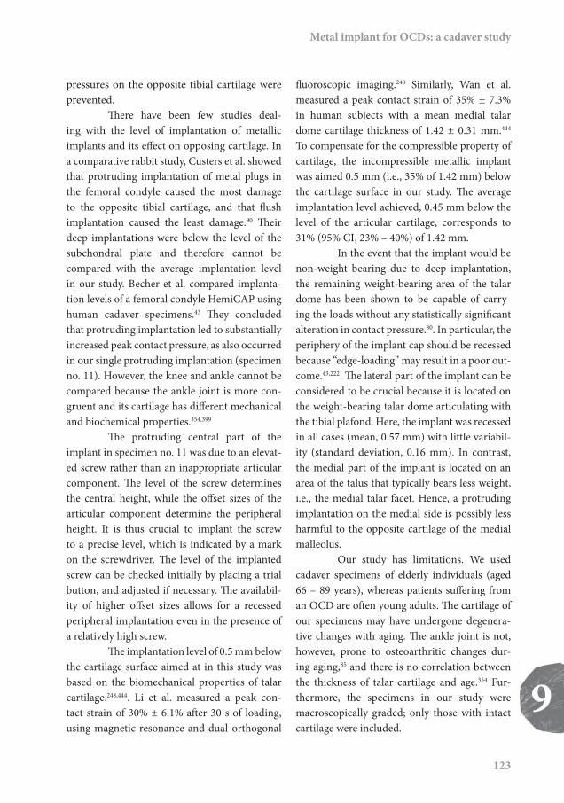
Metal implant for OCDs: a cadaver study
123
9
pressures on the opposite tibial cartilage were prevented.
Th ere have been few studies deal-ing with the level of implantation of metallic implants and its eff ect on opposing cartilage. In a comparative rabbit study, Custers et al. showed that protruding implantation of metal plugs in the femoral condyle caused the most damage to the opposite tibial cartilage, and that fl ush implantation caused the least damage.90 Th eir deep implantations were below the level of the subchondral plate and therefore cannot be compared with the average implantation level in our study. Becher et al. compared implanta-tion levels of a femoral condyle HemiCAP using human cadaver specimens.43 Th ey concluded that protruding implantation led to substantially increased peak contact pressure, as also occurred in our single protruding implantation (specimen no. 11). However, the knee and ankle cannot be compared because the ankle joint is more con-gruent and its cartilage has diff erent mechanical and biochemical properties.354,399
Th e protruding central part of the implant in specimen no. 11 was due to an elevat-ed screw rather than an inappropriate articular component. Th e level of the screw determines the central height, while the off set sizes of the articular component determine the peripheral height. It is thus crucial to implant the screw to a precise level, which is indicated by a mark on the screwdriver. Th e level of the implanted screw can be checked initially by placing a trial button, and adjusted if necessary. Th e availabil-ity of higher off set sizes allows for a recessed peripheral implantation even in the presence of a relatively high screw.
Th e implantation level of 0.5 mm below the cartilage surface aimed at in this study was based on the biomechanical properties of talar cartilage.248,444. Li et al. measured a peak con-tact strain of 30% ± 6.1% aft er 30 s of loading, using magnetic resonance and dual-orthogonal
fl uoroscopic imaging.248 Similarly, Wan et al. measured a peak contact strain of 35% ± 7.3% in human subjects with a mean medial talar dome cartilage thickness of 1.42 ± 0.31 mm.444 To compensate for the compressible property of cartilage, the incompressible metallic implant was aimed 0.5 mm (i.e., 35% of 1.42 mm) below the cartilage surface in our study. Th e average implantation level achieved, 0.45 mm below the level of the articular cartilage, corresponds to 31% (95% CI, 23% – 40%) of 1.42 mm.
In the event that the implant would be non-weight bearing due to deep implantation, the remaining weight-bearing area of the talar dome has been shown to be capable of carry-ing the loads without any statistically signifi cant alteration in contact pressure.80. In particular, the periphery of the implant cap should be recessed because “edge-loading” may result in a poor out-come.43,222. Th e lateral part of the implant can be considered to be crucial because it is located on the weight-bearing talar dome articulating with the tibial plafond. Here, the implant was recessed in all cases (mean, 0.57 mm) with little variabil-ity (standard deviation, 0.16 mm). In contrast, the medial part of the implant is located on an area of the talus that typically bears less weight, i.e., the medial talar facet. Hence, a protruding implantation on the medial side is possibly less harmful to the opposite cartilage of the medial malleolus.
Our study has limitations. We used cadaver specimens of elderly individuals (aged 66 – 89 years), whereas patients suff ering from an OCD are oft en young adults. Th e cartilage of our specimens may have undergone degenera-tive changes with aging. Th e ankle joint is not, however, prone to osteoarthritic changes dur-ing aging,85 and there is no correlation between the thickness of talar cartilage and age.354 Fur-thermore, the specimens in our study were macroscopically graded; only those with intact cartilage were included.
27443_van Bergen.indd 123 27-11-13 09:24

Chapter 9
124
Contact pressures were measured under static loading, while dynamic or cyclic loading may be advocated because of its suppos-edly better imitation of real life. We applied static loading because more cartilage deformation can be obtained by one continuous load than by short-time loads with intervals.183 Because there is more cartilage deformation with static loading, any excessive contact pressure due to elevated implantation might be detected more adequately. Th is implies that, if tested under dynamic loading, a higher implantation level might be erroneously accepted. To imitate the stance phase of gait, the joint was loaded in three positions that correspond to the mean sagittal ankle motion with corresponding forces.367
Th e loads were applied to the tibia, as was done in previous studies.253,264,323 Th e real-life situation might have been better imitated by also loading the fi bula, which transmits 7% of the total force through the lower leg.157 How-ever, loading the fi bula would probably not have had any notable eff ect on the results because the pressure distribution would have shift ed slightly to the lateral side,389 with minor eff ects on the medially located implant.
Variability among specimens is a com-mon fi nding in cadaver experiments.276. Th e contact pressure of the prosthetic area before implantation ranged from 0.02% to 18% in the cadavers we used (see Table 2). Th is variability does not aff ect the conclusions, as each talus served as its own control.
Possible inaccuracies of Tekscan pressure-sensitive sensors are their sensitiv-ity to moisture and temperature, wrinkling, and change of their position during loading.43,373.
By testing in a single lab, any inaccuracy due to diff erence in moisture or temperature was minimized. To detect inaccuracy due to wrin-kling, the sensor was inspected aft er a loading sequence and checked under the lever arm load-ing system. It was replaced in one specimen. Changes in sensor position were minimized by taking digital photographs at the fi rst testing sequence, which we used for verifi cation of posi-tioning at the second sequence.
Our fi ndings form a fi rst basis for clini-cal use of the implant, but its clinical eff ectiveness remains to be investigated. Other designs of this prosthetic implant have been used clinically for diff erent joint surfaces, including the fi rst metatarsal head,180 femoral head,437 humeral head,404 and patella.95 Although the short-term clinical outcome of these designs is promising, it remains to be investigated whether the implant is eff ective for the treatment of OCDs of the talus, in terms of reducing pain and preventing cyst formation.
In conclusion, our study shows that accurate and reproducible implantation of this novel metallic implant can be achieved, prevent-ing excessive prosthetic pressure. Th e results suggest that the implant can be used clinically in a safe way, but the eff ectiveness and safety of this treatment option should be evaluated in a clini-cal study.
Acknowledgments
Th e authors thank the personnel of the Pathol-ogy Department for their kind hosting during the experiments.
27443_van Bergen.indd 124 27-11-13 09:24

Chapter 10Treatment of secondary
osteochondral defects of the talus with a metal resurfacing inlay implant: a prospective study
Christiaan J.A. van Bergen Inge C.M. van Eekeren
Mikel L. Reilingh Inger N. SiereveltC. Niek van Dijk
Bone & Joint Journalin press.
27443_van Bergen.indd 125 27-11-13 09:24

Chapter 10
126
Abstract
The aim of this study was to evaluate the clinical effectiveness of a metal resurfacing inlay implant for osteochondral defects of the medial talar dome after failed prior surgical treatment. Twenty con-secutive patients, aged 20 to 60 years (mean, 38 years), were prospectively studied for 2 to 5 years (mean, 3 years). There was statistically significant reduction of pain in each of four situations (i.e., rest, walking, stair climbing, and running; p ≤0.01). The median American Orthopaedic Foot and Ankle Society ankle-hindfoot score improved from 62 (interquartile range, 46 – 72) preoperatively to 87 (interquartile range, 75 – 95) at final follow-up (p <0.01). The Foot and Ankle Outcome Score improved on all subscales (p ≤0.03). The mean Short Form-36 physical component scale improved from 36 (range, 23 – 50) preoperatively to 45 (range, 29 – 55) at final follow-up (p <0.01); the mental component scale did not change significantly. On radiographs, progressive degenerative changes of the opposing tibial plafond were observed in two patients. One patient required additional surgery for the osteochondral defect. This study shows that the metal implantation technique is a promising treatment for osteochondral defects of the medial talar dome after failed previous treatment.
Introduction
Approximately 63% of osteochondral defects (OCDs) of the talus are located on the medial talar dome.322,458 These medial defects are gen-erally deep and cup-shaped.322 An OCD may sometimes heal and stabilize, but often progress-es to a cystic lesion causing deep ankle pain on weight bearing, prolonged swelling, diminished range of motion, and synovitis.434
Arthroscopic debridement and bone marrow stimulation is considered the prima-ry treatment.458 Current secondary treatment options include osteochondral autograft transfer, autogenous bone graft, and autologous chondro-cyte implantation.458 However, these techniques are associated with donor site morbidity and limited availability or involve two-stage sur-gery.39,312,407
In order to treat OCDs of the medial talar dome after failed primary treatment, a metal resurfacing inlay implant (HemiCAP®, Arthrosurface Inc., Franklin, MA, USA) has been developed.17,411 The implant comprises two components: a cobalt-chromium modular articular component, with a diameter of 15 mm,
and a titanium cannulated screw, which are con-nected together via a taper interlock. Fifteen incremental offset sizes of the articular compo-nent are available, based on the surface geometry of the medial talar dome.411
Promising short-term clinical results have been reported of other designs of the implant for the treatment of various human joints.95,180,404,437 Two biomechanical cadaveric studies provided foundations for use of the talus implant in the ankle joint.17,411 These stud-ies showed that recessing the implant slightly relative to the adjacent cartilage level leads to acceptable contact stresses in the talocrural joint and is beneficial compared to leaving it proud.17,411
The aim of this study was to evaluate the clinical effectiveness of the metal implan-tation technique for OCDs of the medial talar dome after failed previous surgery.
Methods
This prospective case series was approved by the local Medical Ethics Committee. We included
27443_van Bergen.indd 126 27-11-13 09:24

Metal implant for secondary OCDs
127
10patients with an OCD of the medial talar dome, with the largest diameter between 12 and 20 mm on computed tomography, and persistent com-plaints >1 year aft er prior surgical treatment. Exclusion criteria were: age <18 years, defect size >20 mm, ankle osteoarthritis grade III,426 concomitant ankle pathology (tibial osteochon-dral defect, ankle instability, or ankle fracture), advanced osteoporosis, infection, a known aller-gy to implant material, and diabetes mellitus (because diabetes is associated with increased risk of infection as well as soft er and more per-meable talar cartilage).25 All patients provided informed consent.
Computed tomography scans were obtained preoperatively to measure the size in three directions and grade the defect according to the modifi ed Berndt and Harty classifi cation.49,349 In addition, the defects were graded intraoperatively with use of a macro-scopic classifi cation system.377
Operative technique
Th e senior author performed all surgical proce-dures using a standard technique (Figure 1).329 An oblique medial malleolar osteotomy was created to expose the talus. Th e osteotomy was aimed at the intersection between the medial malleolus and the tibial plafond, which was iden-tifi ed with use of an aiming probe, and directed 30° relative to the long tibial axis.413,414
Th e OCD was debrided until a healty cartilage rim remained. A guide pin was placed into the center of the defect, perpendicular to the curvature of the medial talar dome, with use of a drill guide. Th e cannulated screw was pre-drilled and inserted. A contact probe was used to determine the radius of curvature in the sagittal and coronal planes to allow for a precise fi t of the articular component to the existing articular surface. A matching reamer prepared the site for placement of the articular component. A sizing
trial cap with corresponding off sets allowed for fi nal verifi cation of proper fi t. Th e selected articular component was impacted on the screw, thereby engaging the taper interlock. Suffi cient recession of the implant was determined by visu-al inspection. Th e osteotomy was reduced and fi xated with two lag screws in predrilled holes.
Th e postoperative management was initiated with a plaster cast for 2 weeks, followed by a detachable plaster cast or brace (Walker, Össur, Son en Breugel, the Netherlands) for 4 weeks. During this period, non—weight-bearing sagittal range-of-motion exercises were pre-scribed. Radiographs were obtained at 6 weeks aft er surgery to confi rm consolidation of the malleolar osteotomy. Physical therapy was then prescribed to assist in functional recovery and progress to full weight bearing in approximately 1 month.
Outcome assessment
Th e patients were assessed preoperatively and at 2 weeks, 6 weeks, 3 months, 6 months, and annu-ally postoperatively. Authors not involved in the surgical procedures assessed the patients and completed specially designed case report forms.
Th e primary outcome measure was the numeric rating scale (NRS) of pain. Th e NRS consists of an 11-point scale, which represents the spectrum of no pain (0 points) to the worst pain imaginable (10 points).339 Four numeric rating scales were taken, i.e., pain at rest, dur-ing walking, during stair climbing, and during running. NRS during running was not assessed in patients that could not or did not run at any time point.
Secondary outcome measures were assessed preoperatively and at each follow-up visit starting 6 months postoperatively, and included the American Orthopaedic Foot and Ankle Society (AOFAS) ankle-hindfoot score, Foot and Ankle Outcome Score (FAOS),
27443_van Bergen.indd 127 27-11-13 09:24

Chapter 10
128
A B
C D
E
Figure 1. Intraoperative photographs of a right ankle. A medial malleolar osteotomy was performed to expose the medial talar dome (A). The osteochondral defect was debrided (B). The screw was inserted in the center of the defect (C). A trial articular component was placed on the screw (D). The definitive articular component was engaged on the screw (E).
27443_van Bergen.indd 128 27-11-13 09:24

Metal implant for secondary OCDs
129
10and Short Form-36 (SF-36). Th e AOFAS is a 100-point score, with a subjective and an objec-tive component, which devotes 40 points to pain, 50 points to function, and 10 points to align-ment.217 It is frequently used in the assessment of foot and ankle therapy, and the subjective component has been validated.192 Th e FAOS is a validated subjective questionnaire consisting of fi ve subscales: pain, other symptoms (e.g., swelling, locking, mobility), function (activities of daily living), sport and recreational activities, and foot- and ankle-related quality of life.336 Each subscale’s highest possible score is 100. Th e SF-36 is a validated outcome measure to assess general quality of life.7 Th e no rmative value of the Dutch population is 49.2 for the physical component scale and 50.7 for the mental component scale.7
Complications were assessed at each follow-up visit by a patient interview and physical examination. Furthermore, the time needed to resume work and sports was record-ed. Patients indicated the sport level as follows: I, high competitive sportsperson; II, well-trained and frequently sporting; III, sometimes sport-ing; or IV, not sporting. Th e use of analgesics was also recorded. At 2 years of follow-up and annually thereaft er, patients indicated whether they would undergo the procedure again, and whether they would recommend the procedure to friends and family.
Radiography
Weight-bearing radiographs (anteroposterior mortise and lateral views) were obtained at all follow-up visits as of 6 weeks postoperatively. Th e radiographs were reviewed for evidence of implant loosening (periprosthetic osteolysis, subsidence, migration, and disengagement), for malunion or nonunion of the malleolar osteoto-my, and for degenerative changes of the opposite distal tibia (subchondral sclerosis, cyst forma-tion, and joint space narrowing).
Statistical analysis
A power analysis, performed before the start of the study, indicated that a sample size of 20 patients would detect a clinically important mean pain change of 1 point on the NRS, based on a standard deviation of 1.5, with a = 0.05 and a power of 80%.339
Statistical analyses were performed with use of SPSS soft ware (version 19.0; Chi-cago, IL, USA). Categorical data are presented as frequencies. Continuous data are present-ed as means with ranges or as medians with interquartile ranges (IQR), depending on their distribution. One-way repeated-measures analy-ses of variance (ANOVA) were performed to determine diff erences in mean scores at diff er-ent time points for the outcomes with a normal distribution (NRS-rest, NRS-walking, NRS-stair climbing, FAOS, and SF-36). In case of signifi -cance (p <0.05), post-hoc pairwise comparisons were performed using a Bonferroni correction. Th e assumptions of normality and spheric-ity were checked with use of the Shapiro-Wilk test and Mauchly’s test, respectively. Due to the skewed distribution of the NRS-running and AOFAS, these scores were analyzed using the Friedman’s two-way analysis of variance by ranks. Post-hoc pairwise comparisons of these outcome measures were performed with use of Wilcoxon signed-rank tests with Bonferroni correction to adjust for multiple comparisons. Th e SF-36 scales were compared with the nor-mative data for the Dutch population with use of the Student’s t-test.
Results
Twenty-four patients received the implant between October 2007 and November 2010. Four were excluded from the study because they had no previous surgery for the OCD
27443_van Bergen.indd 129 27-11-13 09:24

Chapter 10
130
(two patients), had a combined procedure (one patient), or had diabetes mellitus (one patient). Twenty patients were included in the study. The mean duration of follow-up was 3 years (range, 2 – 5 years). No patients were lost to follow-up. There were 13 female and seven male patients, with a mean age of 38 years (range, 20 – 60 years). The left ankle was affected in 13 patients. The median body mass index was 26.1 kg/m2 (IQR, 24.2 – 27.0 kg/m2)
Eight patients had one, nine patients had two, and three patients had three prior oper-ations. These included one to three procedures of arthroscopic or open debridement and bone marrow stimulation in all cases; cancellous bone grafting in four cases; and osteochondral screw fixation in one case. Five patients received addi-tional hyaluronic acid injections. The median time between the last procedure and the metal implantation was 2 years (IQR, 2 – 6 years).
The mean defect size was 15 mm (range, 11 – 20 mm) in the anteroposterior direction, 10 mm (range, 8 – 14 mm) in the mediolateral direction, and 9 mm (range, 4 – 16 mm) in the craniocaudal direction. On radiographs, one defect was classified as stage III (complete avulsion of a fragment), one as stage IV (displaced fragment), and 18 as stage V
(cystic lesion). Intraoperatively, two defects were macroscopically classified as grade II (soften-ing or fibrillation of the cartilage), five as grade III (fraying of the cartilage), 10 as grade IV (detached fragment), and three as grade V (dis-placed fragment). Sixteen defects were located on the centromedial talar dome and four on the posteromedial talar dome.
Outcomes
The NRS-pain improved significantly in all four situations (Table 1 and Figure 2). For example, repeated-measures ANOVA determined that the mean NRS-walking differed significantly between time points (F(4, 76) = 16.0, p <0.01). Post-hoc pairwise comparisons using Bonferro-ni correction revealed that the NRS-walking was significantly decreased at all postoperative time points compared to the preoperative situation (p <0.01 to p = 0.05).
The median AOFAS improved from 62 (IQR, 46 – 72) preoperatively to 75 (IQR, 68 – 87) at 6 months, 87 (IQR, 76 – 94) at 1 year, and 87 (IQR, 75 – 95) at the final follow-up (p <0.01). Post-hoc tests revealed significant differences at 1 year and at the final follow-up compared to preoperatively (p <0.01).
Table 1. Numeric rating scale pain
Situation Rest p* Walking p*Stair climbing p* Running p***
F(4, 76) = 6.1; p <0.01** F(4, 76) = 16.0; p <0.01** F(4, 72) = 17.6; p <0.01** p = 0.01****
Preoperative 3.6 (0 - 8) 6.7 (4 - 9) 6.6 (4 - 10) 10.0 (9 - 10)3 months 2.4 (0 - 8) 1.00 4.4 (1 - 8) 0.05 4.6 (0 - 8) 0.24 7.0 (5 - 10) 0.106 months 1.7 (0 - 6) 0.09 3.3 (0 - 9) <0.01 2.8 (0 - 7) <0.01 6.0 (3 - 10) 0.031 year 1.3 (0 - 7) 0.01 2.3 (0 - 7) <0.01 2.2 (0 - 6) <0.01 3.0 (1 - 6) <0.01Final 1.1 (0 - 5) 0.01 2.8 (0 - 7) <0.01 2.5 (0 - 7) <0.01 4.5 (1 - 7) <0.01
The values are given as the mean, with the range in parentheses (rest, walking, stair climbing) or as the median, with the interquartile range in parentheses (running).* Bonferroni-adjusted p-value of pairwise comparison with the preoperative NRS** Repeated-measures ANOVA*** Wilcoxon signed-rank test**** Friedman’s two-way analysis of variance by ranks
27443_van Bergen.indd 130 27-11-13 09:24

Metal implant for secondary OCDs
131
10
Figure 2. Graph showing the mean and 95% confi dence interval for each situation of the numeric rating scale (NRS) pain from preoperatively to the fi nal follow-up (p <0.01). Th e NRS-running is presented as median and interquartile range (see Table 1) due to the skewed distribution.
Th e FAOS improved signifi cantly on all subscales (Figure 3). Post-hoc pairwise Bonferroni-adjusted comparisons revealed statistically signifi cant diff erences between pre-operative scores and most postoperative scores (Table 2).
Th e mean SF-36 physical component improved from 36.2 (range, 22.8 – 50.3) preop-eratively to 42.2 (range, 21.0 – 52.3) at 6 months (p = 0.05), 44.0 (range, 28.5 – 57.4) at 1 year (p = 0.01), and 45.0 (range, 28.6 – 54.6) at fi nal follow-up (F(3, 51) = 6.4; p <0.01). Th e SF-36 men-tal component did not change signifi cantly; the mean score was 53.0 (range, 21.9 – 67.9) preop-eratively, 50.6 (range, 34.7 – 62.1) at 6 months,
52.8 (range, 39.8 – 60.3) at 1 year, and 54.3 (range, 24.9 – 66.5) at fi nal follow-up (F(3, 51) = 2.5; p = 0.07). Neither the fi nal physical nor the mental component diff ered signifi cantly from the population norm.7
Preoperatively, 16 patients worked. All 16 patients resumed their work during follow-up. Th e median time to return to work was 8 weeks (IQR, 4 – 23 weeks).
Twelve patients used to play sports before their ankle symptoms had started. Only three were able to play sports preoperatively. Eleven of these 12 patients resumed sports dur-ing follow-up. Th e level of sports decreased in fi ve patients, was equal in four, and improved in
27443_van Bergen.indd 131 27-11-13 09:24

Chapter 10
132
Figure 3. Graph showing the mean and 95% confidence interval for each component of the Foot and Ankle Outcome Score (FAOS) from preoperatively to the final follow-up. The improvement of each subscale was statistically significant (p ≤0.03).
two, as compared to the presymptoms level. Two additional patients, who did not play sports before the symptoms, started playing sports dur-ing follow-up. The median time to resumption of sports was 17 weeks (IQR, 8 – 27 weeks).
Seven patients used analgesics preop-eratively. During follow-up, analgesics were used by none of the patients at 6 weeks and 3 months, by one at 6 months, by three at 1 year, and by none at the final follow-up.
At the final follow-up, 18 patients (90%) indicated that they would undergo the procedure again and that they would recom-mend the procedure to friends and family.
Radiography
There were no signs of implant loosening (Fig-ure 4). The medial malleolar osteotomy healed in all cases by 6 weeks. One patient showed sub-chondral sclerosis of the opposing tibial plafond at final follow-up. Another patient had a pro-gressive subchondral cyst of the opposite tibial plafond.
Complications and reoperations
Some patients had a temporary area of numbness about the scar, which resolved within months. One patient had a superficial wound infection,
27443_van Bergen.indd 132 27-11-13 09:24

Metal implant for secondary OCDs
133
10
which was eff ectively treated with oral antibiot-ics. Th e medial malleolar osteotomy entered the posterior tibial plafond in one patient, but heal-ing of the osteotomy was uneventful.
Th ere were a total of seven addi-tional surgical procedures in six patients. One patient required a lateral displacement calcaneal
osteotomy aft er 26 months because of persistent deep ankle pain and subtle varus malalignment. One patient underwent arthroscopic treatment of anterior ankle impingement. In fi ve patients the malleolar screws were removed aft er a mean of 15 months (range, 8 – 26 months) due to a prominent screw and/or tenderness on
Table 2. Foot and ankle outcome score
Subscale Pain p* Symptoms p*Function (ADL) p* Sport p* Quality of life p*
F(3, 54) = 13.5; p <0.01** F(3, 54) = 3.2; p = 0.03** F(3, 54) = 19.2; p <0.01** F(3, 54) = 11.5; p <0.01** F(3, 54) = 21.6; p <0.01**
Preoperative 50.5 (14 - 78) 52.0 (11 - 96) 58.6 (15 - 85) 25.4 (0 - 70) 15.2 (0 - 44)6 months 74.1 (56 - 100) <0.01 54.9 (21 - 89) 1.00 82.5 (43 - 100) <0.01 37.2 (0 - 75) 0.20 34.8 (0 - 63) <0.011 year 73.1 (39 - 100) <0.01 57.1 (25 - 91) 1.00 81.5 (54 - 100) <0.01 48.0 (10 - 100) <0.01 45.1 (13 - 88) <0.01Final 76.6 (25 - 100) <0.01 66.4 (21 - 96) 0.07 83.7 (46 - 100) <0.01 58.3 (5 - 100) <0.01 52.7 (6 - 100) <0.01
Th e values are given as the mean, with the range in parentheses.* Bonferroni-adjusted p-value of pairwise comparison with the preoperative score** Repeated-measures ANOVAADL = activities of daily living.
Figure 4. Mortise view and lateral weight-bearing radiographs of a left ankle 3 years postoperatively. Suffi cient recession of the implant was confi rmed intraoperatively.
27443_van Bergen.indd 133 27-11-13 09:24

Chapter 10
134
palpation. Th ese patients had NRS scores similar to the complete study group (mean fi nal NRS-rest, walking, stair climbing, and running of 1.2, 2.8, 3.2, and 4.3, respectively).
Discussion
Treatment of OCDs by focal metal resurfacing implants is relatively new.54,95,180,404,437 Th is pro-spective study describes the results of a metal implant designed for the talus. Th e presented outcomes show that patients generally benefi t from the procedure. Th is was evidenced by the statistically signifi cant improvement in almost all outcome measures and high rates of work and sport resumption. Progressive degenera-tive changes were present on radiographs of two patients. One patient required a lateral displace-ment calcaneal osteotomy because of persistent pain. Ninety percent of the patients indicated that they would undergo the procedure again.
We theorize that the eff ectiveness of the resurfacing implant is simply based on the mechanism of fi lling and coverage of the defect. Increased fl uid pressure from the joint into the subchondral bone has been described as the cause of pain and of progressive subchondral cysts in untreated defects.330,434 Th is process possibly is stopped by fi lling and covering the defect.
Th e study population represents a therapeutic challenge. Th e OCDs were cystic, relatively large, and had failed prior surgical treatment. Alternative treatment methods for these cases include osteochondral autograft transfer and cancellous bone graft ing.458 Both can lead to satisfactory clinical results, but disadvan-tages include donor site morbidity in up to 50%, talar surface mismatching, and limited avail-ability.122,343,407 Furthermore, recurrent lesions, cartilage degeneration, and discontinuity of the subchondral bone plate have been observed
aft er osteochondral autograft transfer,407 as well as inferior results in the treatment of secondary OCDs.193 Autologous chondrocyte implantation is another alternative but involves two surgical procedures and is more suitable for superfi cial cartilage defects than for cystic lesions.39
Massive, refractory OCDs can be treated with an allograft , ankle arthrodesis, or total ankle prosthesis. Allograft s are not recom-mended for focal defects, because of the loss of viability and stability in approximately one-third of the graft s.168 Ankle arthrodesis or prosthesis are more defi nite solutions for a recurrent OCD but are rather not used in these relatively young patients because of sacrifi ce of ankle motion and limited durability, respectively. If the metal implant should fail in the long term, it can be removed and the ankle joint can still be fused or replaced.
Strengths of this study are the prospec-tive methodology, completeness of follow-up, and the use of various validated outcome mea-sures. Limitations include the relatively small series, absence of a control group, and lack of long-term follow-up. A limitation of the implant is the fi xed diameter of 15 mm. Th is size is based on the fi nding that primary arthroscopic treat-ment is generally successful for defects up to 15 mm in diameter.82 If the secondary OCD is smaller than 15 mm, some healthy cartilage is sacrifi ced for implantation of the metal device. In contrast, if the defect is larger, a part of it cannot be covered by the implant. In the latter situation, we expect the remainder of the OCD to be fi lled by fi brocartilaginous tissue.
In conclusion, the metal implantation technique is a promising treatment for OCDs of the medial talar dome aft er failed previous treatment. Although the results of this study are encouraging, more patients, longer follow-up, and preferably a control group may determine the place of the implant in the treatment of osteochondral talar defects.
27443_van Bergen.indd 134 27-11-13 09:24

Chapter 11Direction of the oblique medial
malleolar osteotomy forexposure of the talus
Christiaan J.A. van BergenGabriëlle J.M. Tuijthof
Inger N. SiereveltC. Niek van Dijk
Archives of Orthopaedic and Trauma Surgery2011;131(7):893-901.
27443_van Bergen.indd 135 27-11-13 09:24

Chapter 11
136
Abstract
Introduction
A medial malleolar osteotomy is often indicated for operative exposure of posteromedial osteochon-dral defects and fractures of the talus. To obtain a congruent joint surface after refixation, the oblique osteotomy should be directed perpendicularly to the articular surface of the tibia at the intersection between the tibial plafond and medial malleolus. The purpose of this study was to determine this perpendicular direction in relation to the longitudinal tibial axis for use during surgery.
Materials and methods
Using anteroposterior mortise radiographs and coronal computed tomography (CT) scans of 46 ankles (45 patients) with an osteochondral lesion of the talus, two observers independently measured the intersection angle between the tibial plafond and medial malleolus. The bisector of this angle indicated the osteotomy perpendicular to the tibial articular surface. This osteotomy was measured relative to the longitudinal tibial axis on radiographs. Intraclass correlation coefficients (ICCs) were calculated to assess reliability.
Results
The mean osteotomy was 57.2º ± 3.2º relative to the tibial plafond on radiographs and 56.5º ± 2.8º on CT scans. This osteotomy corresponded to 30.4º ± 3.7º relative to the longitudinal tibial axis. The intraobserver (ICC, 0.90 – 0.93) and interobserver (ICC, 0.65 – 0.91) reliability of these measure-ments were good to excellent.
Conclusion
A medial malleolar osteotomy directed at a mean 30° relative to the tibial axis enters the joint per-pendicularly to the tibial cartilage, and will likely result in a congruent joint surface after reduction.
Introduction
A medial malleolar osteotomy is an established approach for the operative treatment of medial osteochondral defects of the talar dome and fractures of the talar body.296,351,390,461 Ray and Coughlin in 1947 first described a transverse osteotomy.325 Different techniques have been described since then, including inverted V,302 oblique,366 crescentic,442 step-cut,13 and inverted U osteotomy309 (Figure 1).
The oblique osteotomy is an estab-lished technique that is used by many surgeons. There are various advantages, including the rela-tively simple technique, excellent exposure of the talus, preservation of the deltoid ligament, and optimal screw compression.351 This technique has been shown to provide reproducibly per-pendicular access to medial talar lesions treated with osteochondral autograft transfer or metal implants.167,176,411 Most surgeons agree that the osteotomy should be aimed at the intersection
27443_van Bergen.indd 136 27-11-13 09:24

Direction of the medial malleolar osteotomy
137
11
CI = 95% confi dence interval.
Figure 1. Anteroposterior (left ) and lateral (right) drawings of an ankle, showing the diff erent medial malleolar osteotomy techniques for exposure of the talus as described in the literature. (1) Transverse osteotomy; (2) Oblique osteotomy; (3a) Step-cut osteotomy; (3b) Modifi ed step-cut osteotomy; (4) Crescentic osteotomy; (5) Inverted U-osteotomy; (6) Inverted V-osteotomy.
Figure 2. Schematic drawings of the correct technique in which there is no articular step off aft er screw fi xation through the predrilled screw holes. Aft er predrilling the screws at 60° in relation to the tibial axis, the osteotomy is made at 30° in relation to the tibial axis (A). A slice of bone is sacrifi ced when performing the osteotomy due to the saw blade thickness. Introduction of the compression screws through the predrilled channels will result in a smooth joint surface (B).
27443_van Bergen.indd 137 27-11-13 09:24

Chapter 11
138
Figure 3. Schematic drawings of wrong techniques in which the osteotomy angle is too vertical (A) or too horizontal (B), resulting in a step off at the articular surface (small arrows).
between the tibial plafond and the articular facet of the medial malleolus.40,281,350,351 Failure to exit at this point may lead to limited exposure (too medial), or violate the weight-bearing cartilage on the tibial plafond (too lateral). Concerns of the technique include the difficulty of reduction and potential for malunion because apposition may not be colinear with respect to the osteot-omy cut.296,390 An incongruent joint surface after fixation could possibly lead to secondary osteo-arthritis of the ankle joint.143,296
In order to obtain a congruent joint surface after fixation, the osteotomy cut is best directed perpendicularly to the articular surface of the tibia (Figure 2). An osteotomy that is too vertical or too horizontal may result in an incon-gruent joint surface (i.e., step off) or shortening of the medial malleolus after fixation (Figure 3). Furthermore, the fixation screws should be directed perpendicularly to the osteotomy plane (Figures 2 and 4).
The longitudinal tibial axis can serve as an intraoperative reference to direct the medial
27443_van Bergen.indd 138 27-11-13 09:24

Direction of the medial malleolar osteotomy
139
11
Figure 4. Schematic drawing showing the importance of the direction of fi xation screws. Th e screws should be placed perpendicularly to the osteotomy (see fi gure 2). Even when the osteotomy is performed correctly, a rather vertical (A) or horizontal (B) insertion of the screws may result in an incongruent joint surface aft er reduction (arrows). Vertical screw placement also causes disruption of the deltoid ligament (A).
malleolar osteotomy. Th is axis is commonly used for several orthopaedic procedures, including total knee arthroplasty and high tibial osteoto-my.29,194,369
Th e objectives of this study were to determine (1) the direction of the oblique medi-al malleolar osteotomy perpendicular to the articular surface in relation to the longitudinal tibial axis, (2) the interindividual variation in a group of patients, and (3) the reliability of the radiographic methods used.
Materials and methods
To determine the optimal direction of the oste-otomy, we assessed ankle radiographs and computed tomography (CT) scans. Using radiog-raphy, we measured the intersection (i.e., angle between the tibial plafond and medial malleolar articular facet), and assessed the tibial axis as a reference to the osteotomy direction. Because the
intersection resembles a curved cylinder rather than a 2-dimensional corner, CT was used as a reference standard to compare with radiography and to assess its intra-articular course.
Patients
We assessed 46 ankle radiographs and CT scans of 45 consecutive patients (one bilateral) with an osteochondral talar lesion who had visited the outpatient department of our institution from 2006 to 2008. According to Walter and associates, a sample size of 40 was able to detect a minimally acceptable level of reliability of 0.6 and a hoped-for reliability of 0.8, with α = 0.05 and a power of 80%.443 Th e patients were derived from two ongo-ing prospective clinical studies on osteochondral lesions of the talus for which approval of the local Medical Ethics Committee was obtained [unpub-lished]. None of the patients had a history of tibial or ankle fracture. Th e mean age was 34 ± 12 years. Th ere were 30 men and 15 women. Twenty-fi ve
27443_van Bergen.indd 139 27-11-13 09:24

Chapter 11
140
Figure 5. Anteroposterior mortise radiograph of a right ankle showing the intersection angle (αα) between the tibial plafond and medial malleolus. The bisector of this angle (dashed line) indicates the optimal medial malleolar osteotomy, which is perpendicular to the articular surface. The angle (β) between the osteotomy and the longitudinal tibial axis was assessed as a reference to the osteotomy.
lesions were located in the right ankle and 21 in the left. The medial talar dome was affected in 34 ankles, the lateral in eight, the central in two, and both the medial and lateral in two.
Radiography
Digital weight-bearing anteroposterior mortise view radiographs included the foot distally and
approximately half of the lower leg proximally (Figure 5). The radiographs (4.0 mAs; 57 kV) were made with the patients’ legs in 15º of inter-nal rotation and the knees extended, with the X-ray beam positioned approximately 1 m from the ankle and directed horizontally, as previous-ly described.240,440
To determine the optimal osteotomy cut, we measured the intersection angle (αα), defined by tangential lines of the tibial plafond and of the medial malleolar articular facet (see figure 5), using a Picture Archiving and Com-munication System (PACS, GE Healthcare, Chicago, IL). The bisector of this angle (α), indicating the osteotomy relative to the tibial plafond, was graphically reconstructed. The tib-ial axis was defined by the line connecting two points in the middle of the proximal and distal tibia.266 The angle (β) between the osteotomy and the longitudinal axis of the tibia was measured (see figure 5).
All radiographic angles were mea-sured by two independent observers to analyze interobserver reliability. One observer measured the angles a second time in a different order 1 month after the first series to determine intrao-bserver reliability.
Computed tomography
Additionally, we assessed 46 multislice helical CT scans of the same ankles in plantigrade posi-tion that included the tibial plafond and talus. Axial CT scans were obtained with an incre-ment of 0.3 mm and a thickness of 0.6 mm, and 1-mm coronal slices were reconstructed (Philips MX8000 spiral CT system, Philips Medical Sys-tems, Eindhoven, the Netherlands).
To determine the intra-articular course of the intersection, both observers measured the intersection angle (αα) on the most anterior and the most posterior coronal reconstructions of the tibial plafond, as well as on the middle
27443_van Bergen.indd 140 27-11-13 09:24

Direction of the medial malleolar osteotomy
141
11
portion of the tibial plafond, using PACS (Figure 6). As with the radiographs, the intersection was defi ned by tangential lines of the tibial plafond and medial malleolar articular facet. Th e mean of the anterior, middle, and posterior intersec-tion angles was calculated to compare with the radiographic measurements. Th e bisector of this angle indicated the osteotomy relative to the tibial plafond.
Statistical analysis
Th e values of the angles are presented as mean ± standard deviation of both observers’ fi rst measurement for each measurement made.51 Intraclass correlation coeffi cients (ICC) were calculated to determine intraobserver and interobserver reliability. According to Fleiss, the reliability is considered good if the ICC is 0.40 – 0.75 and excellent if the ICC is more than 0.75.134 Paired t-tests were performed to assess the systematic diff erence of sequential mea-surements as well as the systematic diff erence between corresponding angles on radiography and CT. Agreement between radiography and CT was assessed by generating a Bland and Alt-man plot.51 We considered an osteotomy angle that diff ered less than 5.0º between radiography and CT as clinically acceptable. Because repeat-ed measurements on the same subject will vary around the true value, measurement error (sw)
of each parameter (i.e., within-subject standard deviation) was calculated as described by Bland and Altman.52 Statistics were performed in SPSS version 16.0 (SPSS, Chicago, IL). A p-value less than 0.05 was considered signifi cant.
Results
Th e results of radiographic measurements are presented in table 1. Th e intersection angle (αα) between the tibial plafond and the articular surface of the medial malleolus was measured 114.5º ± 6.3º. Th e osteotomy (α) relative to the tibial plafond (i.e., the bisector of this angle) was 57.2º ± 3.2º. Th e direction of the proposed oste-otomy (β) relative to the tibial axis was measured 30.4º ± 3.7º (Figure 7). Forty (87%) of 46 ankles were measured within 5.0º (i.e., 25.4º - 35.4º) of the mean value.
Th e intraobserver reliability of radio-graphic measurements was excellent (ICC, 0.90 – 0.93; p <0.001). Accordingly, the interobserver reliability was good to excellent (ICC, 0.73 – 0.82; p <0.001). Th e systematic diff erence between both measurements of one observer ranged from 0.5º (95% confi dence interval [CI], 0.0º – 1.0º) for the osteotomy (α) relative to the tibial plafond to 1.0º (95% CI, 0.0º 1.9º) for the inter-section (αα). Th ere was no systematic diff erence between observers for the osteotomy relative to
Figure 6. Coronal computed tomography scans of the same patient as in fi gure 5 showing the most anterior (A), middle (B), and most posterior (C) intersection (αα).
27443_van Bergen.indd 141 27-11-13 09:24
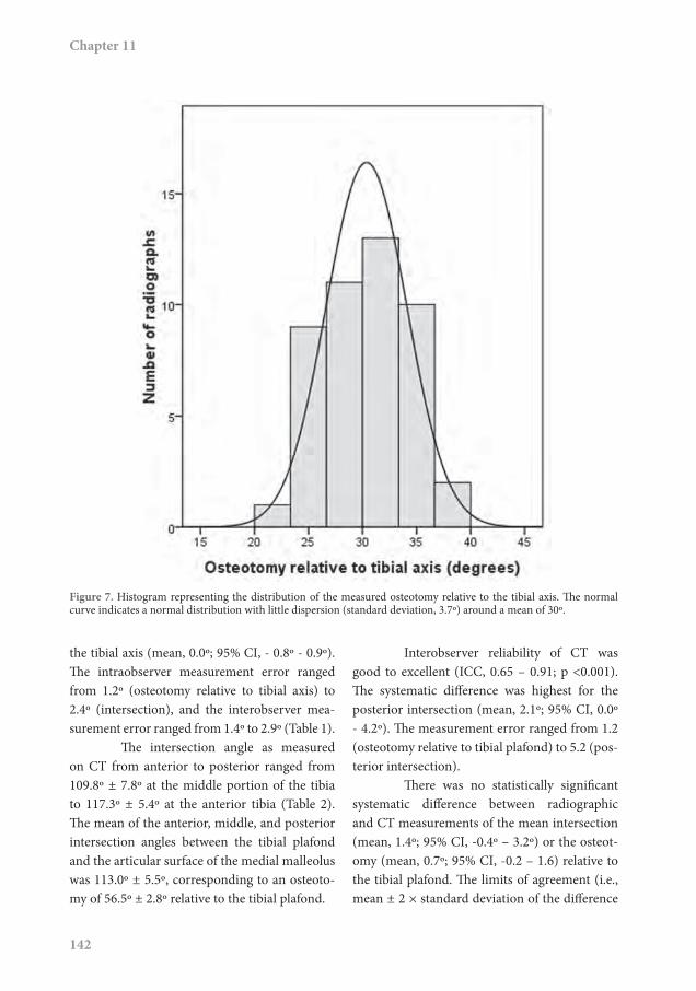
Chapter 11
142
Figure 7. Histogram representing the distribution of the measured osteotomy relative to the tibial axis. The normal curve indicates a normal distribution with little dispersion (standard deviation, 3.7º) around a mean of 30º.
the tibial axis (mean, 0.0º; 95% CI, - 0.8º - 0.9º). The intraobserver measurement error ranged from 1.2º (osteotomy relative to tibial axis) to 2.4º (intersection), and the interobserver mea-surement error ranged from 1.4º to 2.9º (Table 1).
The intersection angle as measured on CT from anterior to posterior ranged from 109.8º ± 7.8º at the middle portion of the tibia to 117.3º ± 5.4º at the anterior tibia (Table 2). The mean of the anterior, middle, and posterior intersection angles between the tibial plafond and the articular surface of the medial malleolus was 113.0º ± 5.5º, corresponding to an osteoto-my of 56.5º ± 2.8º relative to the tibial plafond.
Interobserver reliability of CT was good to excellent (ICC, 0.65 – 0.91; p <0.001). The systematic difference was highest for the posterior intersection (mean, 2.1º; 95% CI, 0.0º - 4.2º). The measurement error ranged from 1.2 (osteotomy relative to tibial plafond) to 5.2 (pos-terior intersection).
There was no statistically significant systematic difference between radiographic and CT measurements of the mean intersection (mean, 1.4º; 95% CI, -0.4º – 3.2º) or the osteot-omy (mean, 0.7º; 95% CI, -0.2 – 1.6) relative to the tibial plafond. The limits of agreement (i.e., mean ± 2 × standard deviation of the difference
27443_van Bergen.indd 142 27-11-13 09:24

Direction of the medial malleolar osteotomy
143
11
between radiography and CT) of the osteotomy relative to the tibial plafond were -5.0º to 5.1º. In 45 of 46 ankles the diff erence was less than 5.0º (Figure 8).
Discussion
In symptomatic osteochondral defects of the talus, surgical treatment is indicated when non-operative treatment has failed.393 Th e choice for open versus arthroscopic surgery depends on the location and size of the defect as well as the type of treatment.296 Th e preferred technique for primary lesions smaller than 15 mm is debride-ment and bone marrow stimulation by means
of arthroscopy.432 For secondary and larger lesions there are various treatment options, including autologous cancellous bone graft -ing, osteochondral autograft transfer (OATS), autologous chondrocyte implantation (ACI), screw fi xation, biodegradable double-layer implants, metal implants, and allograft s.408 If the lesion is located on the medial side of the talar dome (63% of cases),322 most of these treatment options require a medial malleolar osteotomy in order to obtain access to the talar dome.296
Th e purposes of this study were to determine the direction of the oblique medial malleolar osteotomy relative to the long tibial axis, the interindividual variation, and reliability of the radiographic methods used. Th e direction
Table 1. Results of radiographic measurements
Angle Observer 1 (º)
Observer 2 (fi rst mea-surement) (º)
Observer 2 (second mea-surement) (º)
Meana ± SD(º)
Intraobserver reliability Interobserver reliability
ICCDiff erence (mean, 95% CI) Sw ICC
Diff erence (mean, 95% CI) Sw
Intersection 113.8 ± 6.1 115.1 ± 7.1 116.0 ± 7.3 114.5 ± 6.3 0.90 1.0 (0.0 – 1.9) 2.4 0.82 1.3 (0.1 – 2.4) 2.9Osteotomy ∠ tibial plafond
56.9 ± 3.0 57.6 ± 3.6 58.0 ± 3.6 57.2 ± 3.2 0.90 0.5 (0.0 – 1.0) 1.2 0.82 0.6 (0.1 – 1.2) 1.4
Osteotomy ∠ tibial axis
30.4 ± 3.4 30.3 ± 4.5 29.9 ± 4.5 30.4 ± 3.7 0.93 0.6 (0.1 – 1.0) 1.2 0.73 0.0 (-0.8 –0.9) 2.1
Th e intersection was defi ned as the angle between tangential lines of the tibial plafond and the articular facet of the medial malleolus. Th e osteotomy relative to the tibial plafond was defi ned as the bisector of the intersection angle. All ICCs were statistically signifi cant with p-values <0.001.a Mean of both observers’ fi rst measurement. ICC = intraclass correlation coeffi cient, SD = standard deviation, Sw = measurement error, and 95% CI = 95% confi dence interval.
Table 2. Results of CT measurements
Angle Observer 1 (º) Observer 2 (º) Mean ± SD (º)Interobserver reliabilityICC Diff erence (mean, 95% CI) Sw
Anterior intersection 116.4 ± 5.5 118.2 ± 5.8 117.3 ± 5.4 0.81 1.8 (1.0 – 2.7) 2.5Middle intersection 109.8 ± 7.8 109.8 ± 8.2 109.8 ± 7.8 0.91 0.1 (-1.0 – 1.1) 2.5Posterior intersection 110.9 ± 9.5 113.0 ± 7.6 112.0 ± 7.8 0.65 2.1 (0.0 – 4.2) 5.2Mean intersection 112.4 ± 5.4 113.7 ± 6.0 113.0 ± 5.5 0.84 1.3 (0.4 – 2.2) 2.3Osteotomy ∠ tibial plafond 56.2 ± 2.7 56.9 ± 3.0 56.5 ± 2.8 0.84 0.7 (0.2 – 1.1) 1.2
Th e mean intersection according to CT was calculated as the mean of the anterior, middle, and posterior intersections. Th e osteotomy relative to the tibial plafond was defi ned as the bisector of the mean intersection angle.ICC = intraclass correlation coeffi cient, SD = standard deviation, Sw = measurement error, and 95%
27443_van Bergen.indd 143 27-11-13 09:24

Chapter 11
144
Figure 8. Bland and Altman plot51 showing the difference against mean for the osteotomy direction relative to the tibial plafond as measured on radiographs and CT scans. The clinically acceptable difference between radiography and CT was defined at 5.0º (dashed lines). Only one measurement was outside this limit.
of the osteotomy was shown to be 30º ± 4º rela-tive to the long tibial axis. This axis is determined intraoperatively by the center of the knee (i.e., tibial tuberosity) proximally and the center of the ankle (i.e., middle of both malleoli) distally. Applying the osteotomy direction of 30º should minimize the occurrence of a step off of the articular surface after reduction. Radiographic and CT measurements were reliable, and indi-cated that the interindividual variation was small in the studied patient group. Since there was little dispersion (see figure 7), the average 30º angle may be applicable to a larger group of patients. However, if required, the precise osteotomy can be determined reliably for each individual patient according to the methodology described.
The direction of the oblique medial malleolar osteotomy is addressed in few publi-cations. Several authors suggested a direction of approximately 45º to the tibial plafond,143,281,350 which is more horizontal than the direction found in the present study. Others reported a direction ranging from horizontal325 to almost vertical.303. However, none of these directions are based on measurements, and operative methods to achieve the reported angles were not described.
In addition to the direction and loca-tion of the osteotomy, the placement of the fixation screws is important. The lag screws are ideally inserted perpendicularly to the oste-otomy cut to achieve optimal compression and a congruent joint surface (see figure 2).281,366 This
27443_van Bergen.indd 144 27-11-13 09:24

Direction of the medial malleolar osteotomy
145
11
corresponds to a 60º angle relative to the long tibial axis. If the screws are inserted either more horizontally or more vertically, an intra-articular step off might result (see fi gure 4). Th e optimal screw direction is thus rather horizontal, which has the additional advantage of preserving the deltoid ligament that originates more distally.
Reports on outcome and complications aft er oblique medial malleolar osteotomy vary. In a series of 30 patients described by Jarde and colleagues, there was no signifi cant diff erence in outcome between patients treated through a medial malleolar osteotomy, arthrotomy without osteotomy, or arthroscopy.200 Likewise, Bazaz and Ferkel reported no osteotomy complica-tions in nine patients.40 Conversely, Gaulrapp et al. found that a medial malleolar osteotomy frequently led to local osteoarthritis and less favorable clinical fi ndings than arthrotomy without osteotomy.143 Osteoarthritic changes were seen in more than 50% of 22 patients within 5 years aft er treatment.143 Baltzer and Arnold reported malunion in one of 20 cases.31 In their series, a slight reduction in plantar fl ex-ion capacity remained aft er performing a medial malleolar osteotomy, while the range of motion aft er using an anterior arthrotomy (23 patients) became equal to the contralateral ankle joint.31 Unfortunately, it is not possible to relate these outcomes to the direction of the osteotomy used, as the direction was only reported by Gaulrapp et al. who applied 45º.
Although there are diff erent osteotomy techniques of the medial malleolus (see fi gure 1), each has disadvantages. A transverse approach325 is relatively straight forward but exposure of the talar dome may be insuffi cient because it is covered by the contours of the tibial plafond. Accordingly, with the inverted V and inverted U osteotomies, visualization of the talar dome may be inadequate, and they are contraindicated in patients who have large lesions, limited range of motion, or narrow ankle joints.302,309 A crescentic
osteotomy has the advantage of conforming to the contour of the talar dome but is made in a horizontal direction, which restricts perpen-dicular access to the talar dome.442 A step-cut osteotomy, introduced in 1991 and modifi ed in 2008,13,243 provides excellent access but per-pendicular fi xation of the distal fragment at the articular surface is diffi cult because the oste-otomy enters the joint vertically while the screws are inserted obliquely.255 Th e oblique osteotomy is therefore our technique of choice.
Th ere are some limitations of the pres-ent study. Th e measurements utilized a method that relied on 2-dimensional measurements of a 3-dimensional structure. Comparing the intersection angle at the anterior, middle, and posterior portion of the tibia as measured using CT, the biggest diff erence was found between the anterior and the middle portions (mean, 7.5º; see table 2). Th is diff erence corresponds to a diff er-ence in osteotomy direction of 3.8º. Hence, the osteotomy is ideally created in diff erent direc-tions from anteriorly to posteriorly. However, it would be diffi cult to reproducibly create the osteotomy in this manner because the oscillating saw blade and osteotome are straight. Hence, a straight osteotomy is made in clinical practice. Th e mean of the anterior, middle, and posterior osteotomy is therefore the best alternative, and this angle corresponded well to the osteotomy determined by radiography. Another limitation is the absence of intraobserver reliability of CT. Th e radiographic measurements were repeated by an observer but the CT measurements were not. We considered the radiographic measure-ments the most important because these provide a clinical guideline, while the CT measure-ments were made to verify the radiographically measured intersection angle and determine the course of the intersection. Advantages of radiography are the availability, the low costs and the depiction of approximately half of the tibia, allowing assessment of the tibial axis,
27443_van Bergen.indd 145 27-11-13 09:24

Chapter 11
146
which makes it specifi cally useful for preopera-tive planning. Although the proximal tibia was not completely visible, the results of our study and those of another study indicate that the described methodology for assessing the tibial axis is reliable (Table 1).266 However, it remains unknown whether this radiographic tibial axis perfectly corresponds to the intraoperative tibial axis, and if the proposed radiographic osteotomy direction corresponds to the clinical direction. We currently perform the osteotomy routinely in the described direction by using the tibial axis as a reference. Clinical studies are indicated to
assess whether this angle results in congruent fi xation and prevents secondary osteoarthritis.
Conclusion
Th e authors present radiographic measure-ment techniques to determine preoperatively the optimal oblique medial malleolar osteotomy direction. Th e average osteotomy should be aimed 30º relative to the tibial axis, in order to exit perpendicularly to the articular surface at the intersection between tibial plafond and artic-ular facet of the medial malleolus.
27443_van Bergen.indd 146 27-11-13 09:24

Chapter 12Clinical tip:
aiming probe for a precisemedial malleolar osteotomy
Christiaan J.A. van BergenGabriëlle J.M. Tuijthof
Mikel L. ReilinghC. Niek van Dijk
Foot & Ankle International2012;33(9):764-766.
27443_van Bergen.indd 147 27-11-13 09:24

Chapter 12
148
Introduction
Operative exposure of medial osteochondral lesions and fractures of the talus that cannot be treated arthroscopically frequently requires a medial malleolar osteotomy.296,351,390,461 Various techniques have been described since a transverse osteotomy was introduced in 1947.13,302,309,325,366,442 The oblique osteotomy has been ascribed several advantages, including excellent exposure of the talus, preservation of the deltoid ligament, and optimal screw compression.351
Most surgeons agree that the oste-otomy should exit at the intersection between the tibial plafond and the articular facet of the medial malleolus (Figure 1).40,167,281,350,351,413 The optimal angle has been determined to be 30° in relation to the long tibial axis.413 If the osteotomy is created too medially (i.e. in the articular facet of the malleolus), exposure of the talar dome may be insufficient for adequate treatment. Fur-thermore, a small distal fragment may be prone to fracture when fixed at the end of the proce-dure. Conversely, if the osteotomy is created too laterally, it will exit in the tibial plafond. This is undesirable because the medial tibial plafond directly articulates with the medial talar dome,286 and damage to this weight-bearing area might lead to secondary osteoarthritis.143
Precise identification of the intersec-tion is important for an accurate osteotomy. Intraoperatively, it is particularly difficult to identify the posterior part of the intersection because of the tight operative working area and the adjacent neurovascular bundle. This report describes the use of a right-angled aiming probe to identify both the posterior and anterior parts of the intersection between the tibial plafond and medial malleolus in order to make a precise osteotomy.
Technique
The patient is placed in the supine position with a tourniquet around the thigh. A rolled-up apron is placed underneath the lateral malleo-lus to facilitate eversion of the foot and improve exposure of the talus.
A curvilinear skin incision is made over the medial malleolus. The anterior skin is mobi-lized. Using a periosteal elevator, the surgeon dissects over the anterior medial malleolus and distal tibia. A Hohmann retractor is placed over the distal tibia. Some fatty tissue is removed, and a small anterior arthrotomy exposes the anterome-dial talar dome. The level of this anterior superior border of the talar dome will act as a guide to identify the level of the posterior ankle joint.
Next, the tibialis posterior tendon is identified by incising the tendon sheath. A peri-osteal elevator is used to bluntly dissect over the posterior distal tibia at the level of the ankle joint, approximately 0.5 cm below the anterior joint level. After placement of the Hohmann retractor posterior to the medial malleolus, the posterior capsule of the ankle joint can be visual-ized and incised.
A right-angled probe (Figure 2) is now used to identify the posterior intersection between the medial malleolus and tibial plafond.The surgeon carefully inserts the tip of the probe
Figure 1. Schematic coronal view of an ankle showing the optimal oblique medial malleolar osteotomy exiting at the intersection between tibial plafond and articular facet of the medial malleolus.
27443_van Bergen.indd 148 27-11-13 09:24

Aiming probe for a medial malleolar osteotomy
149
12
into the posteromedial joint space by sliding along the posterior aspect of the distal tibia at the intersection with the medial malleolus and
gently pulls proximally and medially. Th e peri-osteum at the level of the intended osteotomy is marked with a surgical knife, sterile marker
Figure 2. Picture of the right-angled probe. Th is probe, with a 5-mm tip, is normally used for arthroscopic surgery.
Figure 3. To identify the anterior (left ) and posterior (right) intersections between the tibial plafond and medial malleolus, two probes can be inserted simultaneously. Th ey are pulled 30° proximally and medially relative to the long tibial axis.
27443_van Bergen.indd 149 27-11-13 09:24

Chapter 12
150
pen, or osteotome. Next, the probe is placed in the anteromedial tibial notch and pulled in a proximal and medial direction, identifying the anterior part of the intersection. The anterior intersection is marked, and this is connected to the posterior intersection as a reference guide to the osteotomy. If available, two probes can be used simultaneously to identify the anterior and posterior intersections (Figures 3 and 4). The probes are best pulled 30° in relation to the lon-gitudinal tibial axis in the coronal plane because this direction is perpendicular to the tibial artic-ular surface at the intersection.413
Two Kirschner wires (K-wires), directed per-pendicularly to the determined osteotomy plane, are predrilled in the medial malleolus. The screw holes are drilled and tapped, using a cannulated drill, and the K-wires are removed.
The screw holes can be placed either parallel or divergent. If the harvest of cancel-lous bone is indicated for the treatment of a talar osteochondral lesion or fracture, a divergent
direction of the screws allows taking a bone graft of the distal tibia. This can be accomplished by creating a window in the cortex of the tibial metaphysis just proximal to the screw holes and harvesting the cancellous bone using a curette.
An oscillating saw is placed on the incised periosteum and directed at the marked intersection of the tibial plafond and medi-al malleolus. The osteotomy is created up to approximately 3 to 4 mm above the articular cartilage, while two Hohmann retractors protect the adjacent soft tissue. The osteotomy is com-pleted with the use of an osteotome. This way, the surgeon controls the osteotomy of the articu-lar part and minimizes the risk of damaging the talar cartilage. If desired, an assistant can place an instrument in the joint or manually distract the ankle to protect the talar cartilage.
After the osteotomy has been complet-ed, the surgeon manually retracts and everts the medial malleolus using gauze. Optionally, the distal part is temporarily transfixed by retrograde
Figure 4. Medial intraoperative view of a left ankle with a probe inserted at each part of the intersection. This figure highlights the limited intraoperative vision of the posterior intersection.
27443_van Bergen.indd 150 27-11-13 09:24

Aiming probe for a medial malleolar osteotomy
151
12
drilling of a large-diameter K-wire into the talus through one of the predrilled holes. Exposure of the talar dome is improved by forced eversion of the heel. Th e fi bula is hereby used as a fulcrum and the talus is tilted (Figure 5). Care must be taken not to use too much force to prevent frac-ture of the lateral malleolus.
At the end of the procedure, the medial malleolus is reduced. Initially, large diameter K-wires are placed through the predrilled screw holes to confi rm correct alignment. Th e diam-eter of the K-wire should correspond to the diameter of the predrilled hole. A Weber bone clamp can be placed for initial compression. Placement of the proximal leg of the Weber clamp is facilitated by creating a small hole in the distal tibial cortex proximal to the osteotomy using a 2.5-mm drill. We routinely use two 3.5-mm cancellous lag screws with a length of 40 or 45 mm. Th ey are inserted into the predrilled
screw holes aft er removal of the K-wires. Th e bone clamp is released, and the wound is closed.
Discussion
Th e use of a right-angled probe is described to facilitate identifi cation of the optimal terminal point of the oblique medial malleolar osteotomy (i.e. the intersection between the medial mal-leolar articular facet and the tibial plafond). Alternatively, intraoperative fl uoroscopy can be used to identify the intersection.308 However, disadvantages of fl uoroscopy include exposure to radiation and increasing the operative time. Moreover, it is not easy to identify exactly both anterior and posterior intersection points on a 2-dimensional view. Instead, when using the aiming probe, fl uoroscopy becomes unneces-sary.
Figure 5. Exposure of the talus through a medial malleolar osteotomy. Aft er the retraction of the medial malleolus (*), a K-wire can be inserted into the talus through one of the predrilled holes to hold the medial malleolus in place.
27443_van Bergen.indd 151 27-11-13 09:24

27443_van Bergen.indd 152 27-11-13 09:24

Part IVAlternative treatment
27443_van Bergen.indd 153 27-11-13 09:24

27443_van Bergen.indd 154 27-11-13 09:24

Chapter 13Osteochondral defects ofthe talus: a novel animal
model in the goat
Christiaan J.A. van BergenGino M.M.J. Kerkhoff s
Nick Marsidi Clara M. Korstjens
Vincent Everts Leo J. van Ruijven C. Niek van Dijk
Leendert Blankevoort
Tissue Engineering Part C: Methods2013;19(6):449-457.
27443_van Bergen.indd 155 27-11-13 09:24

Chapter 13
156
Abstract
Osteochondral defects of the talus pose a difficult therapeutic challenge. An experimental animal model of the ankle joint is not available. The aim of this study was to test a newly developed animal model for osteochondral defects of the ankle in vivo. Osteochondral defects were created in the talus of goat hind legs using a posterolateral surgical approach. The defects were filled with either autolo-gous cancellous bone or donor demineralized bone matrix, or left empty as control. After 12 weeks of healing, the specimens were analyzed with radiography, macroscopy, micro-computed tomography, histology, histomorphometry, and fluorescence microscopy. It was possible to create a standardized defect in each talus. The implanted material remained in place. The analyses showed that most bony tissue was generated in the defects filled with autologous bone and least in the control defects. Our findings show that a standard osteochondral defect can be created in the talus by a relatively simple procedure in a large animal that allows qualitative and quantitative evaluation. The model can be used in future experiments to investigate alternative treatment methods before they are introduced into clinical practice.
Introduction
Osteochondral defects of the talus (OCDs) pose a challenging problem to orthopaedic surgeons. Patients with an OCD are typically young adults who experience deep ankle pain on weight bearing, often long after a supination trauma. Nonoperative treatment yields unsatisfactory results in the majority of patients.458 The primary surgical treatment of most defects is arthroscopic debridement combined with bone marrow stim-ulation.458 If results are unsatisfactory – which happens more often in large defects76,82 – more invasive surgical options are indicated.408 Current treatment options for these secondary OCDs include autologous cancellous bone grafting, osteochondral autograft transfer and autologous chondrocyte implantation.408,458 Each of these options has specific disadvantages, for example, an additional operative site with possible donor-site pain, limited availability of material, impaired graft integration, and the necessity of a medial malleolar osteotomy for exposure.32,147,312 There-fore, experimental studies focus on improvement of these methods and development of alternatives.72,138,140,201,252,411,451 However, almost all studies
investigate articular defects of the knee, while the ankle joint is rarely investigated.81 These knee studies cannot reliably be extrapolated to patients with ankle defects, because biomechanical and biochemical properties of the knee and ankle are clearly different.85,399 The ankle is a congruent joint with thin cartilage that is less susceptible to osteoarthritis than the knee.181,234
The use of animal models is often an essential step in the testing of orthopaedic pro-cedures before clinical use in humans.313 To our knowledge, an animal model for ankle OCDs is not described in the literature. The aim of this study was to test a newly developed caprine model for OCDs of the talus, by evaluating an established treatment (autologous cancellous bone graft), an alternative treatment (deminer-alized bone matrix [DBM]), and no treatment (control).We defined the following criteria for a successful model: 1) the animal’s joint anatomy and body weight are comparable to humans, 2) the surgical technique is reproducible (regard-ing the operative approach as well as size and location of the OCD), 3) the morbidity of the animals is acceptable (defined as full weight bearing and free of pain within 1 week), 4) the
27443_van Bergen.indd 156 27-11-13 09:24

Goat model for ankle OCD
157
13
repair is best aft er applying an established treat-ment and minimal in control defects, and 5) the method must be accurate enough to detect small diff erences between groups.
Materials and methods
Animals, experimental design, and operative technique
Th e study was approved by the Animal Care and Use Committee of the University of Amsterdam, the Netherlands. Th ree adult female Dutch milk goats (Capra Hircus Sana) were used with an approximate age of 4 years and a weight of 50, 80, and 89 kg, respectively.
All goats were healthy, according to physical examination and blood tests by a veterinar-ian. Th ey were kept in group housing starting 2 weeks before surgery.
Surgery was performed on both ankles in a sterile fashion with the goat in the lateral decubitus position under general anesthesia with endotracheal intubation. Before anesthe-sia, intramuscular injections with prophylactic antibiotic (Pen & Strep, Fendigo sa/nv, Brus-sels, Belgium), ketamine 10 mg/kg (Alfasan International BV, Woerden, the Netherlands) and atropine 1.5 mg (Centrafarm Services BV, Etten-Leur, the Netherlands) were administered. Intravenous etomidate (B.Braun Melsungen AG, Melsungen, Germany) was injected to induce
Figure 1. Digital pictures showing key aspects of the surgical technique. Th e talar dome was exposed through a posterolateral approach (A). A cylindrical defect with a diameter and a depth of 6 mm was created (B) and fi lled up to the level of the cartilage (C). DBM = demineralized bone matrix.
27443_van Bergen.indd 157 27-11-13 09:24

Chapter 13
158
general anesthesia. Fentanyl 250 µg (Hameln pharmaceuticals gmbh, Hameln, Germany) and midazolam 15 – 20 mg (Dormicum, Roche Nederland BV, Woerden, the Netherlands) were injected intravenously, and repeated, when nec-essary. Isoflurane 1.0% - 2.5% (Nicholas Piramal Limited, London, UK) was administered by inhalation.
A curvilinear skin incision was made just posterior to the lateral malleolus, parallel to the Achilles tendon (proximal) and the cal-caneus (distal). After mobilization of the lateral saphenous vein, an arthrotomy was performed in the plane between the peroneal tendon (ante-rior) and the lateral saphenous vein (posterior). A Hohmann retractor was placed behind the medial talus to optimize exposure of the talus and protect the tendons and neurovascular structures. Full dorsiflexion of the ankle allowed maximal exposure of the articular surface of the talus (Figure 1A). In the center of the talar troch-lea, a custom-made drill socket with a diameter of 6 mm was placed, which ensured drilling to a precise depth of 6 mm. Under continuous cooling with saline, the defect was drilled per-pendicularly to the talar surface, starting with a sharp drill and finishing with a milling cutter to achieve a flat base of the defect (Figure 1B). A goat study investigating the natural course of knee defects showed that 6-mm defects do not heal spontaneously within 1 year.198
Each defect was treated according to a predefined randomization scheme with autolo-gous cancellous bone (harvested from the iliac crest) or commercially available cortical demin-eralized bone matrix (BonusTM DBM, Biomet BV, Dordrecht, the Netherlands) hydrated with normal saline, or left empty (untreated control). Filling up to the level of the articular cartilage surface (Figure 1C) was ensured by inserting ample material press-fit and moving the ankle through a full range of motion (removing any abundant material above the surface of the
defect due to the congruency of the ankle joint). The joint capsule and subcutaneous tissue were closed with interrupted 2-0 absorbable sutures, and the skin was closed with absorbable sutures placed in a continuous intracutaneous pattern.
Postoperatively, analgesic medication consisted of a single subcutaneous injection of 0.005 mg/kg buprenorfine (Temgesic®, Schering-Plough BV, Utrecht, the Netherlands), twice daily, as pain demanded (indicated by limping). After wound healing was completed, which took ~1 week, the animals were kept outdoors in a large natural environment with food ad libitum and without activity restrictions. Eating habits, ambulatory activities, and health status were monitored daily.
After 12 weeks, the goats – back in the experimental animal center – were sacri-ficed by injecting a lethal dose of pentobarbital (Euthasol 20%, ASTfarma BV, Oudewater, the Netherlands) in the jugular vein, after they had been sedated with an intramuscular injection of ketamine 10 mg/kg (Alfasan International BV, Woerden, the Netherlands), xylazine 2 ml (Sedazine®, ASTfarma, Oudewater, the Nether-lands) and atropine 1 mg (Centrafarm Services BV, Etten-Leur, the Netherlands).
Analyses
Radiography
Lateral radiographs of the ankles were tak-en before surgery and directly after surgery. Anteroposterior and lateral radiographs were taken after autopsy.
Macroscopy
The tali were excised directly after terminating the goats, and digital high-resolution photo-graphs were taken of the talar and opposite articular surfaces. Macroscopic grading was
27443_van Bergen.indd 158 27-11-13 09:24

Goat model for ankle OCD
159
13
performed by two observers. A general mac-roscopic articular evaluation system was used that awards 0 to 2 points to each of fi ve catego-ries: range of motion, intra-articular fi brosis, restoration of contour, cartilage erosion, and appearance.340 Ten points indicate a completely normal appearance, and 0 points indicate a severely damaged joint surface. Furthermore, the talar articular surface was assessed according to the validated International Cartilage Repair Society (ICRS) cartilage repair assessment.62,420 Th is score consists of three items (i.e., degree of defect repair, integration to border zone, and macroscopic appearance) that are each assigned a maximum of 4 points, with a total of 12 indi-cating a normal appearance. Th e scores of the two observers were averaged and outliers with a diff erence of more than 1 point were scored again, until consensus was reached.91
Micro-computed tomography
Th e talar heads were sawn off with a water-cooled band saw to optimize penetration of fi xative for histological evaluation and allow placement of the tali in the micro-computed tomography (mCT) scanner. Specimens were fi xed in 4% phosphate-buff ered formaldehyde for 1 week. Aft er they were submerged in 70% ethanol and subjected to a vacuum to remove all the air from the cancellous bone, they were scanned in a mCT scanner (μCT 40, Scanco Medical AG, Brüt-isellen, Switzerland). With the defects facing down, the specimens were mounted in cylindri-cal specimen holders and secured with synthetic foam. Th is setup ensured consistent scanning of the defects parallel to the axis of the scan tube. Th e resolution of the scans was 18 μm, the voltage 70 kV, the current 114 μA, and the inte-gration time 1000 ms. To discriminate between bone and background, the reconstructions were segmented using an adaptive threshold proce-dure, which determines the minimum between
the two peaks in the gray values histogram, rep-resenting bone and background, respectively.331
For quantitative analysis, two cylin-drical volumes of interest were defi ned: one representing the complete OCD, measuring 6 mm in diameter and 6 mm in depth, and one representing the central OCD, measuring 3 mm in diameter and 5 mm in depth. Th e latter vol-ume of interest was defi ned at 5 mm in depth to avoid analyzing original bone at the base of the OCD. Th e ratio between bone volume and tissue volume (BV/TV) was determined for all vol-umes of interest using morphometric soft ware (Scanco Medical AG, Brütisellen, Switzerland).
In addition to the six operated speci-mens, fi ve unaff ected goat tali from a spinal fusion study (unpublished data) were μCT scanned and analyzed in the same manner to obtain normative data.
Histology
Aft er fi xation, the specimens were dehydrated using ascending grades of ethanol (70% to 100%) in two steps a week, and embedded in methyl-methacrylate (BDH Laboratory Supplies, Poole, England). Aft er cold polymerization, the unde-calcifi ed specimens were cut into coronal 5-μm sections with a Jung-K microtome (R. Jung, Heidelberg, Germany). Seventy-fi ve central sec-tions were obtained from each specimen. Every third section (n = 25) was stained with Gold-ner’s trichrome method, giving the following staining pattern: green for mineralized tissue, red for osteoid, blue or black for cell nuclei, and light orange or red for cytoplasm. Th e Goldner stained sections were used for evaluation of mineralized bone tissue, osteoid, fat cells, osteo-cytes, osteoblasts, and osteoclasts. Every second out of three sections was stained with toluidine blue for examination of structural details and cell appearance of the articular surface of the defects. Every third out of three sections was
27443_van Bergen.indd 159 27-11-13 09:24

Chapter 13
160
left unstained for fl uorescence microscopy. Two observers, blinded to the treatment modality, qualitatively assessed the stained sections using transmitted light microscopy.
Histomorphometry
One representative Goldner-stained mid-sec-tion of each talus was analyzed using a Leica DMRA microscope that was connected to Leica Qwin computer soft ware (Leica Microsystems Imaging Solutions, Cambridge, UK). A special routine was written for quantitative measure-ments of bone parameters.
Th ree areas of interest were defi ned: (1) the center of the OCD, (2) close to the bor-ders of the defect (lateral, medial, and bottom), and (3) close to the articular surface of the defect. Six representative measurement fi elds of 307,000 µm2 were selected and digitized at a magnifi cation of ×200, resulting in one to three measurement fi elds for each area of interest (Figure 2). Th e mineralized bone area (bone%) and osteoid area (osteoid%) were measured. Th e number of osteocytes, osteoblasts, and osteo-clasts was counted at a magnifi cation of ×400.
All measurements were performed twice by one observer and once by a second observer, both blinded to treatment allocation. Th e mean of both observers is reported, and intraobserver and interobserver reliability were calculated.
Fluorescence microscopy
Fluorochrome labels were injected at 3 (oxy-tetracyclin 20 mg/kg), 6 (calcein green 10 mg/kg), and 9 weeks (alizarin red 25 mg/kg) aft er surgery. Th e dosages were based on the recommendations by van Gaalen et al.435 One unstained section that was adjacent to the Goldner stained section was used for histomorphometry. Six measurement fi elds (magnifi cation, ×400), similar to the areas of interest of histomorphometry, were digitized using fl uorescent fi lters. According to the man-ufacturer’s guidelines, the D-fi lter (excitation 355 – 425 nm, emission 470 nm) was best used for oxytetracyclin and alizarin, and the I3-fi lter (excitation 450 – 490 nm, emission 515 nm) was best used for calcein, although each label was visible with both fi lters. One independent observer, blinded to treatment allocation, mea-sured the distance between two consecutive lines at ~10 locations within a measurement fi eld. Th e mineral apposition rate (MAR, mm/day) of the bone was calculated as the average distance between the corresponding edges of two consecutive fl uorescent bone labels, divid-ed by the number of days between start and end of the administration period.
Statistical analysis
Individual quantitative results are presented. Th e treatment pairs were not statistically com-pared because of the small sample size. Mean and standard deviation (SD) were calculated of the μCT scans of fi ve unaff ected goat tali. To assess intra- and interobserver reliability of the
Figure 2. Schematic drawing showing the six measurement fi elds in which histomorphometry analysis was performed. Th e fi elds represent areas in the center (1 – 2), close to the borders (3 – 5), and close to the surface (6) of the defect.
27443_van Bergen.indd 160 27-11-13 09:24

Goat model for ankle OCD
161
13
histomorphometric measurements, intraclass correlation coeffi cients (ICCs) were calculated using SPSS version 18.0 (SPSS, Chicago, IL). ICCs of more than 0.75 are considered excel-lent.134
Results
With the operative technique described, it was possible to create a standardized OCD in the talus. Aft er surgery, the fi rst two animals recov-ered well without complications. Th e goats were able to walk on their hind legs within 24 h. Th e third goat had persistent pain of the right ankle,
Figure 3. Histological analysis of a defect fi lled with DBM, 2 weeks aft er implantation, shows that the material remained in situ.
Figure 4. Macroscopic view of three specimens. (A) Autologous bone. (B) DBM. (C) Control. Th e arrows indicate the osteochondral defects.
27443_van Bergen.indd 161 27-11-13 09:24

Chapter 13
162
despite treatment with subcutaneous analgesics for 2 weeks and intramuscular antibiotics (Pen & Strep, Fendigo sa/nv, Brussels, Belgium) for 1 week. There were no signs of infection. Sur-gical exploration of the painful ankle after two weeks revealed no pathology. Cultures showed no pathologic micro-organisms. An exact cause could not be determined. The goat was terminat-ed for ethical reasons 2 weeks following initial surgery. Obduction revealed no pathologic con-ditions. Histology of the right talus showed that the original DBM was still in situ (Figure 3). In consultation with the Animal Care and Use Committee, the sacrificed animal was replaced by another goat, which recovered well from sur-gery and completed the follow-up period.
Radiography
Preoperative radiographs showed normal ankle joints without signs of osteoarthritis. Immediate
postoperative radiographs showed the OCDs in three cases (one of each treatment); the defects were not visible in the other three. On the final radiographs after the 12-week period, none of the defects was visible.
Macroscopy
Macroscopic inspection of the ankle joints revealed no signs of inflammation. All defects were covered with tissue resembling fibrocarti-lage (Figure 4). The surrounding talar cartilage and opposite joint surfaces appeared completely normal. The highest macroscopic assessment scores were achieved in an autologous bone speci-men and the lowest in a DBM specimen (Table 1).
Micro-CT
In the complete 6-mm OCDs, BV/TV ranged from 0.10 to 0.45 (Table 2). In comparison, mean
Table 1. Results of macroscopy
Treatment (goat) General macroscopic articular evaluation ICRSAB (1L) 6 5.5AB (2R) 9 11.5DBM (2L) 5 4DBM (3R) 6 8Control (1R) 6 6Control (3L) 6 7.5
AB = autologous bone, DBM = demineralized bone matrix, ICRS = International Cartilage Repair Society cartilage repair assessment,62 L = left, and R = right.
Table 2. Results of μCT, showing the ratio between bone volume and tissue volume
Treatment (goat) Complete defect Central 3-mm cylinderAB (1L) 0.44 0.29AB (2R) 0.34 0.22DBM (2L) 0.1 0.01DBM (3R) 0.3 0.13Control (1R) 0.23 0Control (3L) 0.45 0.04Unaffected (mean ± standard deviation) 0.68 ± 0.06 0.70 ± 0.05
27443_van Bergen.indd 162 27-11-13 09:24

Goat model for ankle OCD
163
13BV/TV of the fi ve unaff ected tali was 0.68 (SD, 0.06) in the complete cylinder and 0.70 (SD, 0.05) in the central 3-mm cylinder. Th e 3-mm central cylinders can be considered indicative for the regenerative capacity of the treatment applied because bone had regenerated from the borders and bottoms of the defects. In these 3-mm cylinders, the tali treated with autologous bone showed the most bone tissue, and the con-trol group the least (Figure 5).
Histology
In the OCDs treated with autologous bone, dif-fuse bone formation and near-complete fi lling was observed (Figure 6A). Th ere was continu-ing bone remodeling, as indicated by large areas of osteoid tissue and the presence of abundant osteoblasts and a rare osteoclast. At the articu-lar surface, the defect was mainly composed of (mineralizing) fi brocartilage tissue with some chondrocytes embedded. Underneath the sur-face there were areas of woven and lamellar bone.
In the defects treated with DBM, some bone formation was observed – both lamel-lar and woven – with abundant osteoid in one specimen (Figure 6B). Most bone was formed at the borders but also some in the center of the defects. Th e original DBM was not visible. In one specimen the surface of the defect was composed of a thick layer of fi brocartilage. In the other, the surface was mainly composed of connective tis-sue, fi broblasts, and leukocytes (lymphocytes and granulocytes).
Th e control defects did not heal (Figure 6C). Some woven bone originated from the bor-ders of the defects. Occasional mineralization of cartilaginous tissue at the surface suggested endochondral ossifi cation. Th e greatest part of the defects was fi lled with fat cells and connec-tive tissue.
Histomorphometry
Histomorphometry followed the same pattern as the μCT analysis (Table 3). Most bone formation was observed in the autologous bone pair; most
Figure 5. Th ree-dimensional reconstructions of µCT scans. Th ese volumes of interest represent central 3×5-mm cylinders. (A) Autologous bone. (B) DBM. (C) Control.
27443_van Bergen.indd 163 27-11-13 09:24

Chapter 13
164
Figure 6. Histologic sections with Goldner’s trichome staining; magnification, ×25. (A) Autologous bone. (B) DBM. (C) Control. Scale bar = 1 mm.
27443_van Bergen.indd 164 27-11-13 09:24

Goat model for ankle OCD
165
13
osteoid formation was found in DBM. Th ere was only one specimen (autologous bone) with bony and osteoid tissue in the measurement fi eld of the surface. Osteoclasts were rarely found: only a single osteoclast was seen in one measure-ment fi eld (center) in one specimen (autologous bone).
Th e intra- and interobserver reliability of the histomorphometric measurements were excellent; the lowest ICC was 0.80 (interobserver reliability of osteoblasts in the center; p = 0.016), and the highest was 1.00 (intra- and interobserv-er reliability of bone% in all areas of interest; p <0.001).
Fluorescence microscopy
MAR varied from 0 μm/d in the areas where no bone growth was observed to 11 μm/d in the center of the defect in an autologous bone speci-men (Table 4). Th e calcein green labels were best visible, followed by oxytetracyclin and alizarin,
Table 3. Results of histomorphometry
Bone% Osteoid% Osteocytes (no.) Osteoblasts (no.)
Treatment (goat) Cen
ter
Bord
ers
Surf
ace
Cen
ter
Bord
ers
Surf
ace
Cen
ter
Bord
ers
Surf
ace
Cen
ter
Bord
ers
Surf
ace
AB (1L) 62.1 67.8 19.8 3.2 1.4 0.2 16 24 4 15 11 0AB (2R) 16.7 38.2 0.0 1.7 3.1 0.0 4 17 0 3 9 0DBM (2L) 5.0 15.3 0.0 0.2 1.2 0.0 0 11 0 0 6 0DBM (3R) 26.0 35.7 0.0 9.8 9.8 0.0 4 16 0 8 21 0Control (1R) 0.4 14.8 0.0 2.8 5.1 0.0 1 3 0 0 4 0Control (3L) 0.0 83.7 0.0 0.0 1.0 0.0 0 30 0 0 0 0
Table 4. Results of fl uorescence microscopy (mineral apposition rate, µm/d)
Week 3-6 Week 6-9Treatment (goat) Center Borders Surface Center Borders SurfaceAB (1L) 6.1 2.8 2.7 8.3 5 No labelAB (2R) 11.0 5.7 0.0 6.4 9 0.0DBM (2L) 6.4 7.1 0.0 No label 6.5 0.0DBM (3R) 0.0 3.7 0.0 0.0 6.7 0.0Control (1R) 0.0 5.6 0.0 0.0 5.8 0.0Control (3L) 0.0 5.9 0.0 0.0 4.7 0.0
respectively (Figure 7). Th e MAR could not be analyzed in two occasions because alizarin red labels could not be detected (see Table 4).
Discussion
A new caprine model was developed for the comparison of treatments for osteochondral ankle lesions. Th e model is presented in this study and seems feasible for the evaluation of new treatment options. Similar to the human ankle joint, the caprine ankle is congruent and able to bear high loads. Th e goat’s body weight and metabolic and remodeling rates correspond to those of humans.21 Furthermore, the pro-portion of cartilage to subchondral bone and the subchondral bone consistency are report-edly close to humans.81 Th e operative technique described is reproducible, a standardized OCD can be created in each talus, and the implanted material remains in situ. Most importantly, there
27443_van Bergen.indd 165 27-11-13 09:24

Chapter 13
166
is a clear difference in outcome between defects treated with autologous cancellous bone and control defects. This was particularly shown by the quantitative analyses with μCT and histo-morphometry. The mean BV/TV of the centralOCDs was 0.25 in the autologous bone pair, 0.07 in the DBM pair, and 0.02 in the control pair (see Table 2). Similarly, mean central bone% according to histomorphometry was 39.4 in the autologous bone pair, 15.5 in the DBM pair, and 0.2 in the control pair (see Table 3). However, no definite conclusions can be drawn on the effectiveness of the treatment because of the low number of animals studied.
Previous animal studies have been performed predominantly on the stifle joint.8,81 These studies may be inadequate for assessing treatment modalities for ankle OCDs, as there are some specific differences between the knee
and ankle. First, physiological cartilage proper-ties differ. Congruent joints, such as the ankle, have a thin layer of cartilage to distribute the load and maintain an acceptable level of stress, whereas incongruent joints, such as the knee, have thicker areas of cartilage to increase the contact area between joint surfaces.354 The car-tilage of the ankle has a greater compressive modulus compared with the knee.354,399 Sec-ond, the knee and ankle joints seem to respond differently to cartilage injury. There appears to be a net anabolic response to injury in the ankle cartilage and a net catabolic response in the knee.26,181,311 Ankle cartilage may be more resistant to osteoarthritis development for a number of reasons, including higher equilibrium and compressive moduli, better intercellular communications, increased resis-tance to inflammatory molecules, increased
Figure 7. Fluorescence microscopy with I3 filter (excitation 450 – 490 nm, emission 515 nm), magnication ×400. Fluorochrome labels were injected at 3 (oxytetracyclin; yellow arrow), 6 (calcein; green arrow), and 9 weeks (alizarin; red arrow) to analyze the mineral apposition rate. Calcein was best visible, followed by oxytetracyclin and alizarin, respectively.
27443_van Bergen.indd 166 27-11-13 09:25

Goat model for ankle OCD
167
13
metabolic activity, and higher congruency of the articular surfaces.116,181 Because of these diff erences, an animal model specifi cally devel-oped for the ankle joint is required.
Although most animal models are designed for unilateral surgery,8 a bilateral defect was created in the present model. Th is method reduces the number of animals needed and allows for paired treatment comparison. A paired analysis may be a better strategy than unilateral evaluation or random assignment of treatment in bilateral models, since the regener-ative capacities may diff er from animal to animal (see Tables 2 and 3).
Clinically, plain radiographs are obtained for the initial diagnosis of OCD and for follow-up. However, in the present animal model, radiographic evaluation as an outcome measure proved to be useless because the defects were not visible in most instances. In contrast, histomor-phometry is traditionally used to measure bone formation and is considered the gold standard.258 It is common practice to analyze representa-tive areas of interest to approximate the total defect.21,238,338 Disadvantages of this method are a possible modifi cation of the structure of the tissue by slice preparation and the long time required for all processing steps.151,258 MicroCT provides a fast and nondestructive technique to character-ize and measure the 3-dimensional geometric and density properties of a bone specimen.202 Gielkens et al. compared the intraobserver reli-ability of μCT and histomorphometry.151 Th ey concluded that both analyses are reliable but are used preferably in combination.
Fluorescence microscopy is a use-ful supplement to plain histology to detect the speed of regeneration during the follow-up period.435 When using fl uorescence microscopy, there is no need to sacrifi ce additional animals at a shorter follow-up period. Th e success of fl uo-rochrome detection depends on various aspects, for example, the type, concentration, route of
administration and methods of visualization.435 In the present study, the recommendations of van Gaalen et al. were used.435 Unfortunately, the alizarin red labels were not always visible, with-out an obvious cause. In contrast, calcein green was clearly visible in all cases. No adverse events related to the injections of the fl uorochrome labels were seen in this study.
Th e follow-up period was 12 weeks. Revascularization and conversion of a bone graft into a vital trabecular structure have been reported to occur at approximately 3 months in the goat, which corresponds to 8 months in humans.238 However, the regeneration process was ongoing, observed by large areas of osteoid tissue and the presence of abundant osteoblasts. Th e fi nal situation may thus not be achieved aft er 12 weeks. In studies with multiple follow-up periods, no substantial regeneration of OCDs in the knee joint was observed aft er 24 weeks, suggesting that a follow-up period of 24 weeks may show the fi nal outcome.72,138
Th e principal limitation of the pres-ent study is the limited number of animals used. In consultation with the Animal Care and Use Committee, only three animals were studied because the morbidity of the animals could not be anticipated, as this was the fi rst time the ankle model was used. Out of the initial six ankles, one showed persistent pain. Th is goat was sacrifi ced, and the substitute goat recovered uneventfully. Th us, one out of eight ankles had inacceptable morbidity. Th is morbidity corresponds to that of other goat studies investigating cartilage repair.92,340,342 We therefore believe that following studies with the presented model are justifi ed. Because of the small number of animals and the variability in reparative capacity of the goats, we cannot reliably draw conclusions on the inves-tigated repair procedures. Larger studies with paired analyses are indicated.
27443_van Bergen.indd 167 27-11-13 09:25

Chapter 13
168
Conclusions
Th e caprine model described in the current study seems suitable for the study of osteochon-dral ankle defect treatment in vivo. Th e operative technique is fairly simple and allows the creation of a standardized OCD for qualitative and quan-titative evaluation. Th e goat model can be used in future experiments investigating alternative treatment methods for this challenging condi-tion before introduction into clinical practice.
Acknowledgments
Th e authors would like to thank Mr. K.W. Meijer, Mr. G. Vink, and Mr. P. Sinnige for taking care of the animals. Mrs. J. Hogervorst is acknowledged for support with preparation of the histology sections. Biomet BV, Dordrecht, the Nether-lands, is gratefully acknowledged for fi nancial support and supply of the DBM.
27443_van Bergen.indd 168 27-11-13 09:25

Chapter 14Demineralized bone matrix
and platelet-rich plasma do not improve healing of osteochondral
defects of the talus:an experimental goat study
Christiaan J.A. van BergenGino M.M.J. Kerkhoff s
Murat ÖzdemirClara M. Korstjens
Vincent Everts Leo J. van Ruijven C. Niek van Dijk
Leendert Blankevoort
Osteoarthritis and Cartilage2013;21(11):1746-1754.
27443_van Bergen.indd 169 27-11-13 09:25

Chapter 14
170
Abstract
Objective
The purpose of this study was to evaluate the effectiveness of demineralized bone matrix (DBM) with and without platelet-rich plasma (PRP) in the treatment of osteochondral defects (OCDs) of the talus. We hypothesized that treatment with DBM would result in more bone formation than no treatment in control OCDs, and that PRP would further enhance the regenerative capacity of DBM.
Method
A standardized 6-mm OCD was created in each talus of 16 adult goats. According to a randomiza-tion scheme, one OCD of each goat was treated with allogeneic DBM hydrated with normal saline (n = 8) or hydrated with autologous PRP (n = 8). The contralateral OCD (n = 16) served as control. After 24 weeks, the animals were euthanized and the tali excised. Various outcome parameters were analyzed with use of macroscopic evaluation, micro-computed tomography, histology, histomor-phometry, and fluorescence microscopy.
Results
None of the analyses revealed statistically significant differences between the groups for any of the parameters analyzed in any volume of interest. For example, the mean bone volume fraction of the defect, as measured by micro-computed tomography, was 0.56 (95% confidence interval [CI], 0.44 – 0.68) for DBM hydrated with normal saline and 0.52 (95% CI, 0.40 – 0.65) for DBM hydrated with PRP, compared to 0.53 (95% CI, 0.45 – 0.61) and 0.54 (95% CI, 0.44 – 0.64) for the internal controls, respectively (p >0.05).
Conclusion
In contrast to our hypotheses, no beneficial treatment effect of DBM with or without PRP was found for OCDs of the caprine talus.
Introduction
In the treatment of talar osteochondral defects (OCDs), repair of the subchondral bone is an important aim of the procedure.160 The presence of subchondral bone is essential for survival of chondrocytes.16 The affected subchondral bone is thought to cause the pain.434 Restora-tion of the subchondral bone may improve the
weight-bearing capacity of the ankle and prevent further cyst formation.434
Urist pioneered the use of demineral-ized bone matrix (DBM) for bone defects.405,419 Since his work, there has been increasing expe-rience with DBM in both animals123,140,272,405 and humans.145,283 The sequence of events after implantation of DBM mirrors that of endochon-dral ossification.405 Bone morphogenetic proteins
27443_van Bergen.indd 170 27-11-13 09:25

DBM and PRP for goat OCDs
171
14
(BMP-2, -4, and -7) seem to be responsible for the formation of bone and possibly cartilage that are induced by DBM.318,406 Th e BMPs pres-ent in DBM attract mesenchymal stem cells through chemotaxis and act as morphogens that may direct the diff erentiation of these cells into an osteochondrogenic lineage. Diff erent fl uids can be used for rehydration of the DBM before application, including normal saline, bone mar-row aspirate, antibiotics solution, whole blood, or platelet concentrate.
Platelet-rich plasma (PRP) is a prom-ising biomaterial that contains concentrated growth factors, including transforming growth factor-β (TGF-β) and platelet-derived growth factor (PDGF).14,195 TGF-β in PRP may stimulate chemotaxis and mitogenesis of osteoblast and chondroblast precursors and inhibit osteoclast formation and bone resorption.225,275 PDGF may promote mitogenesis, angiogenesis, and chon-drocyte proliferation.10,225,275 Although, in theory, PRP may enhance the biologic activity of DBM, the combination of DBM and PRP has had con-tradictory results.213,275,324
Th e purpose of the present study was to evaluate the eff ectiveness of DBM with or without PRP in the treatment of ankle OCDs in goats. We hypothesized that (1) treatment with DBM would repair more bone than control OCDs, and that (2) PRP would further enhance the regenerative capacity of DBM.
Materials and methods
Animals and experimental design
Th e study was approved by the Animal Care and Use Committee of the University of Amsterdam. A caprine model was used, specifi -cally designed for ankle OCDs.417 Sixteen adult female Dutch milk goats (Capra Hircus Sana) were included with an approximate age of 4
years. All goats were healthy, according to phys-ical examination and blood tests performed by a veterinarian. Th e goats were weighed on a digital scale before surgery and at fi nal follow-up. Surgery was performed in a sterile manner on both ankles, with the goat under general anesthesia with endotracheal intubation. A single intramuscular dose with prophylactic antibiotics (Pen & Strep, Fendigo sa/nv, Brus-sels, Belgium) was injected preoperatively. Th e ankle joint was exposed through a postero-medial approach. Normal articular surfaces were confi rmed by visual inspection. Standard OCDs of 6 mm in diameter and depth were cre-ated with specially developed instruments.417 According to a predefi ned randomization scheme, one defect of each goat was treated with DBM hydrated either with normal saline (0.9% NaCl solution) (“DBM treatment”; n = 8) or with PRP (“DBM+PRP treatment”; n = 8), and the other served as a control (“DBM con-trol” or “DBM+PRP control”). In each case, the material was inserted press-fi t up to the level of the adjacent cartilage surface. Th e joint capsule and skin were closed in a standard fashion.417
During recovery, the animals were kept outdoors in a large natural environment, without activity restrictions, and with food ad libitum. Eating habits, ambulatory activities, and health status were monitored daily.
Since previous studies showed no sub-stantial change in repair of knee OCDs aft er 24 weeks,72,138 the goats were euthanized 24 weeks aft er surgery by injecting a lethal intravenous dose of pentobarbital. All analyses were per-formed by observers blinded to the treatment provided.
Demineralized bone matrix
Commercially available cortical DBM (BonusTM DBM, Biomet BV, the Netherlands) was used. Th is DBM was obtained from human donors
27443_van Bergen.indd 171 27-11-13 09:25

Chapter 14
172
from qualified tissue banks that were registered with the FDA and accredited by the American Association of Tissue Banks. It was granulated, demineralized with organic solvents, freeze-dried (i.e., lyophilized) and processed aseptically. This process resulted in calcium levels of less than 0.1%.318 It was combined with a collagen-derived carrier (gelatin) from the same donor, packaged in a rehydration syringe, and sterilized by gamma irradiation.
Platelet-rich plasma
Autologous PRP was used for rehydration of the DBM. After induction of anesthesia and before surgery, 27 ml venous goat blood was aspirated into a 30-ml syringe that contained 3 ml of anti-coagulant citrate dextrose A. PRP was isolated by centrifugation at 3200 rpm for 15 min using the gravitational platelet system II (GPS II, Biomet BV, the Netherlands). This preparation system produces 3 ml of PRP with a reported eightfold increase in platelet concentration and a fourfold to sevenfold increase in growth factor concentration compared with whole blood.120,173,454 The concentration of platelets in
the PRP of each subject in the present study was measured using an automated hematology ana-lyzer (XE-5000, Sysmex, Japan) after 5 min of resuspension on a rocker, as recommended by Woodell-May et al.454 The median platelet con-centration of the PRP was 1511 × 109/l (range, 82 – 2090 × 109/l).
Macroscopy
After the goats were euthanized, the tali were excised and digital high-resolution photographs were taken of the talar articular surfaces. Two independent observers macroscopically grad-ed the photographs with use of the validated International Cartilage Repair Society (ICRS) cartilage repair assessment.62,420 This score ranges from 0 to 12 points and is subdivided into degree of defect repair, integration to border zone, and macroscopic appearance (4 points each), with a score of 12 indicating a completely normal appearance. The scores of the two observers were averaged, and outliers with a difference of more than 1 point were scored by consensus.91
\
Figure 1. Micro-CT analysis. Transverse cross-section of the talus (left) and the two cylindrical volumes of interest representing the complete defect (middle) and the central 3×5 mm (right).
27443_van Bergen.indd 172 27-11-13 09:25

DBM and PRP for goat OCDs
173
14
Micro-computed tomography
Th e anterior part of the talus, at safe distance from the OCD, was sawn off with a water-cooled band saw to reduce the size of the specimen, allowing it to be placed in the micro-computed tomography (mCT) scanner, and to optimize penetration of fi xative into the specimen. Aft er 1 week in fi xative (4% phosphate-buff ered formal-dehyde), the specimens were submerged in 70% ethanol and temporarily subjected to a vacuum. In the 70% ethanol solution, they were placed in a mCT scanner (μCT 40, Scanco Medical AG, Bassersdorf, Switzerland) and scanned with a resolution of 18 μm. To minimize the noise in the reconstructions, an integration time of 1000 ms was used.
Micro-CT reconstructions were seg-mented with a threshold level of 467 mg HA/cm3. Two 3-dimensional cylindrical volumes of interest were defi ned: one representing the complete OCD (6 mm in diameter and depth), and one representing the central OCD (3 mm in diameter and 5 mm in depth) (Figure 1). Using morphometric soft ware (Scanco Medi-cal AG, Bassersdorf, Switzerland), the following parameters were analyzed: bone volume fraction (bone volume [BV]/tissue volume [TV]), tissue mineral density (TMD) of BV and of TV, and trabecular number, thickness, and separation. Th e bone volume fraction was the primary out-come of the study.
Histology
Aft er fi xation, the specimens were dehydrated using ascending grades of alcohol and embedded in methyl-methacrylate. Aft er cold polymeriza-tion, the undecalcifi ed specimens were cut into 5-μm sections with a Jung-K microtome (R. Jung, Heidelberg, Germany). Th irty central sections were obtained of each specimen to overcome sampling error. Every third section
was stained with Goldner’s trichrome method or toluidine blue for light microscopy, or left unstained for fl uorescence microscopy. Two observers simultaneously assessed the stained sections and identifi ed the type of healing.
Histomorphometry
A representative Goldner-stained mid-section of each talus was analyzed using a Leica DMRA microscope that was connected to Leica Qwin computer soft ware (Leica Microsystems Imaging Solutions, Cambridge, UK) with a custom-made routine for quantitative measurements of bone parameters. Two observers simultaneously made the choice for the representative section, based on technical quality and overall appear-ance. Th ree areas of interest were defi ned: (1) the center of the OCD, (2) sides and bottom of the defect (close to the lateral, medial, and deep bor-ders), and (3) the surface (close to the articular surface of the defect).417 For each area of interest, one (surface), two (center) or three (sides and bottom) representative measurement fi elds of 3.07 × 105 µm2 were digitized at a magnifi cation of ×200. Mineralized bone surface area (bone%) and osteoid surface area (osteoid%) were assessed. Th e numbers of osteocytes, osteoblasts, and osteoclasts were counted at a magnifi cation of ×400 and expressed per area of mineralized bone tissue. Th ese histomorphometric measure-ments have been shown excellent intraobserver and interobserver reliability.417
Fluorescence microscopy
Fluorochrome labels were administered at week 1 (Oxytetracyclin yellow, 32 mg/kg intramus-cularly) and weeks 6, 12, 18, and 23 (Calcein green, 20 mg/kg subcutaneously). Fluorescence microscopy was used with fl uorescence fi lters and unstained sections to analyze the speed of bone regeneration (mineral apposition rate
27443_van Bergen.indd 173 27-11-13 09:25

Chapter 14
174
[MAR]). Six measurement fields (magnifica-tion, ×400) were digitized, similar to the areas of interest of histomorphometry. The distance between two consecutive fluorescent bone labels was measured at approximately 10 locations within a measurement field by an independent observer blinded to treatment allocation. The MAR (mm/day) was calculated as the average distance divided by the number of days between two injections.
Statistical analysis
The sample size was calculated before the start of the study and was based on the primary outcome BV/TV. Prior data from a pilot study indicated that the standard deviation of BV/TV in the central 3 mm of the OCDs treated with DBM was 0.086.417 With an intended difference in the mean BV/TV of matched pairs of 0.10, a sample size of eight ankle pairs was able to reject the null hypothesis that the response difference was zero with probability (power) 0.80. The Type I error
probability associated with this test of this null hypothesis was 0.05. Because two pairs of treat-ment were investigated, the total sample size was 16 goats.
Statistical analyses were performed with use of SPSS software (version 18.0; Chica-go, Illinois). Variables with normal distribution are reported as mean and 95% confidence inter-vals (CIs). Data with skewed distribution are reported as median and range. The Wilcoxon signed-rank test was used to calculate p-values of matched pairs (DBM vs. control and DBM+PRP vs. control). The Mann-Whitney U test was used to compare the treatment effect of DBM (i.e., dif-ference of DBM and internal control) with the treatment effect of DBM+PRP (i.e., difference of DBM+PRP and internal control). MARs of dif-ferent administration periods were compared using the Wilcoxon signed-rank test.
The results were considered statisti-cally significant if the p-value was less than 0.05for the primary outcome (i.e., BV/TV). To cor-rect for multiple testing, Holm’s method was
Figure 2. Examples of the best (A; ICRS cartilage repair assessment, 10) and worst (B; ICRS cartilage repair assessment, 3.5) macroscopic appearance of the osteochondral defects (arrows) after 24 weeks of healing. Both are control specimens.
27443_van Bergen.indd 174 27-11-13 09:25

DBM and PRP for goat OCDs
175
14
Figure 3. Bar chart showing the mean and 95% confi dence interval of the bone volume fraction (BV/TV), according to µCT analysis, of (A) the complete defect and (B) the central 3×5 mm. Th e diff erences were not statistically signifi cant (p >0.05).
used for the secondary outcome parameters (macroscopy, histology, histomorphometry, and fl uorescence microscopy).9,186 Because four sec-ondary outcome measures were used, adjusted p-values with signifi cance levels of 0.01 (i.e., 0.05/4) and higher were appropriate.
Results
General results
Th e surgical procedures and functional recovery were uneventful in 15 goats. Aft er 24 h, no signs of limping or abnormal motion were observed. In one goat, there was a technical failure of the instruments, resulting in an OCD drilled com-pletely through the talus. In consultation with the Animal Care and Use Committee, this ani-mal was terminated and replaced by another, which had no complications. Th e mean body weight was 73.5 kg (95% CI, 67.1 – 79.8 kg) before surgery and 69.3 kg (95% CI, 63.7 – 75.0 kg) at 24 weeks follow-up.
Macroscopy
All OCDs were macroscopically covered with fi brocartilaginous tissue. Th e mean ICRS car-tilage repair assessment was 8.0 (95% CI, 7.3 – 8.7) for DBM treatment, 8.4 (95% CI, 7.4 – 9.5) for DBM control, 6.9 (95% CI, 5.3 – 8.6) for DBM+PRP treatment, and 7.4 (95% CI, 6.0 – 8.9) for DBM+PRP control (diff erences not statistically signifi cant) (Figure 2). Th e sur-rounding talar cartilage and opposite joint surfaces appeared unaff ected.
Micro-CT
Th ere were no signifi cant diff erences between groups on the primary outcome measure BV/TV or any of the other µCT parameters in either volume of interest (Figure 3 and Table 1). In the DBM+PRP treatment group, there were no sta-tistically signifi cant diff erences between goats with high concentrations of PRP and those with low concentrations. For example, BV/TV in the complete OCDs of the four goats with the high-est PRP concentrations was 0.49 (95% CI, 0.39
27443_van Bergen.indd 175 27-11-13 09:25

Chapter 14
176
Tabl
e 1.
Out
com
e of
µC
T
Cen
tral
3x5
-mm
cyl
inde
r
Tb.S
p (m
m)
1.2
(0.9
- 1.
5)
1.2
(0.7
- 1.
8)
1.1
(0.5
- 1.
6)
1.8
(1.3
- 2.
3)
The
valu
es a
re p
rese
nted
as t
he m
ean,
with
the
95%
confi
denc
e in
terv
al in
par
enth
eses
.Th
ere
wer
e no
stat
istic
ally
sign
ifica
nt d
iffer
ence
s bet
wee
n th
e gr
oups
.BV
= b
one v
olum
e, Tb
.N =
trab
ecul
ar n
umbe
r, Tb
.Sp
= tr
abec
ular
sepa
ratio
n, T
b.Th
= tr
abec
ular
thic
knes
s, TM
D =
tiss
ue m
iner
al d
ensit
y, an
d TV
= ti
ssue
vol
ume.
Tb.Th
(mm
)
0.33
(0.2
5 - 0
.42)
0.26
(0.1
8 - 0
.34)
0.24
(0.1
3 - 0
.34)
0.23
(0.1
9 - 0
.26)
Tb.N
(m
m-1
)
1.4
(1.0
- 1.
9)
1.7
(0.7
- 2.
7)
4.2
(0.0
- 8.
4)
0.9
(0.4
- 1.
4)
TMD
of
TV (m
g H
A/c
m3 )
333
(240
- 42
6)
276
(180
- 37
3)
244
(129
- 35
8)
221
(150
- 29
2)
TMD
of B
V
(mg
HA
/cm
3 )
680
(653
- 70
7)
661
(633
- 68
9)
657
(632
- 68
2)
650
(639
- 66
2)
BV/T
V
0.37
(0.2
3 - 0
.50)
0.28
(0.1
4 - 0
.43)
0.23
(0.0
7 - 0
.39)
0.20
(0.0
9 - 0
.31)
Com
plet
e de
fect
Tb.S
p (m
m)
1.1
(0.7
- 1.
5)
1.2
(0.7
- 1.
6)
1.6
(0.8
- 2.
4)
1.3
(0.9
- 1.
7)
Tb.Th
(mm
)
0.46
(0.3
5 - 0
.57)
0.41
(0.3
2 - 0
.49)
0.45
(0.3
4 - 0
.55)
0.37
(0.3
0 - 0
.43)
Tb.N
(m
m-1
)
1.9
(1.4
- 2.
4)
2.0
(1.3
- 2.
6)
1.6
(0.9
- 2.
3)
1.8
(1.1
- 2.
5)
TMD
of
TV (m
g H
A/c
m3 )
454
(373
- 53
6)
431
(377
- 48
6)
429
(348
- 50
9)
432
(380
- 48
5)
TMD
of
BV (m
g H
A/c
m3 )
709
(682
- 73
5)
700
(680
- 72
1)
695
(681
- 71
0)
684
(666
- 70
1)
BV/T
V
0.56
(0.4
4 - 0
.68)
0.53
(0.4
5 - 0
.61)
0.52
(0.4
0 - 0
.65)
0.54
(0.4
4 - 0
.64)
Trea
tmen
t gro
up
Trea
t-m
ent
Con
trol
Trea
t-m
ent
Con
trol
DBM
DBM
+PR
P
27443_van Bergen.indd 176 27-11-13 09:25
DBM and PRP for goat OCDs
177
14
– 0.58), compared to 0.55 (95% CI, 0.30 – 0.80) in the four goats with the lowest PRP concentra-tions (p = 1.00).
Histology
Four types of healing patterns were recognized (Figure 4). Type 1 was almost completely healed (>75% repair). In type 2, the subchondral bone was (almost) restored but a cystic lesion under-neath the restored bone remained. Type 3 was characterized by regeneration from the mar-gins and bottoms of the defects but a superfi cial defect remained (25% - 75% repair). In type 4, no or only minimal healing was observed (<25% repair); the original defects were fi lled with fatty and connective tissue, and the surface was cov-ered with a thin layer of cells. Th ere was high variation in healing patterns between animals (Figure 5), while the left and right tali within a single animal showed more consistency. Th erewere remnants of DBM in two cases: one DBM treatment and one DBM+PRP treatment. Addi-tionally, macrophages were observed in fi ve defects: one DBM treatment, one DBM control, and three DBM+PRP control. Osteoclasts were not found.
Histomorphometry
Th ere were no statistically signifi cant diff er-ences in histomorphometry between the groups for any of the parameters analyzed in any of the areas of interest (Table 2).
Fluorescence microscopy
Th ere were no statistically signifi cant diff erences between the groups (Table 3). Some data were missing due to either no bone or no detectable label in the measurement fi eld, especially in the fourth administration period (week 18 – 23). Th e MAR of the second administration period
(week 6 – 12) of each group was signifi cantly higher than that of the fi rst administration period (week 1 – 6). Th e MAR did not change signifi cantly in subsequent periods.
Discussion
Th is study aimed to evaluate the eff ectiveness of DBM and PRP in the treatment of ankle OCDs in goats. A previously established goat model was utilized, enabling evaluation of treatment of a standard OCD of the talus in a large animal with qualitative and quantitative analyses.417 Th e control OCDs in this study can be regarded as bone marrow stimulation of the defect without additional fi lling.
In contrast to our hypotheses, DBM did not result in improved repair compared to the control defects, nor did PRP enhance the regenerative capacity of DBM. Th ere were no statistically signifi cant diff erences between the groups on the primary outcome BV/TV, as measured by µCT, or the secondary out-comes, as measured by macroscopy, histology, histomorphometry, and fl uorescence micros-copy. However, some generalization uncertainty remains because of the dispersion of the out-come data, as demonstrated by the width of the 95% CIs (see Figure 3 and Tables 1 and 3). Th ere were no observations of abnormal motion or limping aft er 24 h. Although a signifi cantly higher MAR was shown aft er the fi rst 6 weeks, this fi nding was similar in each treatment group (see Table 3).
Gao et al. investigated the eff ect of allogeneic rabbit demineralized cortical and tra-becular bone matrix for the treatment of 3-mm OCDs in 10 young rabbit medial femoral con-dyles.140 At 6 and 12 weeks postoperatively, the defects were repaired up to 95% of their depth but there was a clear diff erence between cortical and trabecular DBM. In most of the specimens
27443_van Bergen.indd 177 27-11-13 09:25

Chapter 14
178
Figure 4. Goldner-stained sections showing four types of healing. Type 1 (A) was almost completely healed. In type 2 (B) , the subchondral bone was (almost) restored but a cystic lesion underneath the restored bone remained. Type 3 (C) was characterized by regeneration from the margins and bottoms of the defects but a superficial defect remained. In type 4 (D), no or only minimal healing was observed. The original defects are indicated with black lines. Subtle differences in size of the defects may be due to slice preparation.
Figure 5. Distribution of histologic healing types.
27443_van Bergen.indd 178 27-11-13 09:25

DBM and PRP for goat OCDs
179
14
Tabl
e 2.
Out
com
e of
hist
omor
phom
etry
N.O
b (×
10-4
/µm
2 )
Surf
ace
0.0
(0.0
- 9.
4)
0.9
(0.0
- 3.
6)
2.4
(0.0
- 14
.9)
0.0
(0.0
- 12
.1)
Th e
valu
es a
re p
rese
nted
as t
he m
edia
n, w
ith th
e ra
nge
in p
aren
thes
es.
Th er
e w
ere
no st
atist
ical
ly si
gnifi
cant
diff
eren
ces b
etw
een
the
grou
ps.
N.O
b =
num
ber o
f ost
eobl
asts
and
N.O
c = n
umbe
r of o
steo
cyte
s.
Side
s and
bo
ttom
2.7
(1.3
- 8.
5)
2.9
(0.0
- 16
.0)
3.7
(0.0
- 9.
1)
4.2
(0.0
- 17
.1)
Cen
ter
0.0
(0.0
- 9.
0)
0.2
(0.0
- 4.
0)
0.0
(0.0
- 7.
0)
1.1
(0.0
- 5.
6)
N.O
c (×1
0-4/µ
m2 )
Surf
ace
0.0
(0.0
- 12
.8)
2.3
(0.0
- 24
.8)
2.9
(0.0
- 14
.1)
0.0
(0.0
- 8.
4)
Side
s and
bo
ttom
6.4
(4.0
- 7.
9)
6.8
(3.8
- 10
.1)
6.1
(1.7
- 8.
9)
6.2
(5.3
- 9.
2)
Cen
ter
3.1
(0.0
- 8.
0)
4.3
(0.0
- 8.
3)
0.0
(0.0
- 8.
5)
1.3
(0.0
- 6.
8)
Ost
eoid
%
Surf
ace
0.0
(0.0
- 8.
1)
0.0
(0.0
- 7.
6)
0.1
(0.0
- 10
.7)
0.0
(0.0
- 13
.5)
Side
s and
bo
ttom
1.1
(0.4
- 1.
6)
1.3
(0.0
- 2.
9)
0.8
(0.1
- 3.
3)
0.9
(0.2
- 2.
7)
Cen
ter
0.1
(0.0
- 2.
6)
0.1
(0.0
- 1.
6)
0.0
(0.0
- 1.
4)
0.1
(0.0
- 3.
9)
Bone
%
Surf
ace
0.0
(0.0
- 65
.5)
5.5
(0.0
- 71
.5)
9.0
(0.0
- 52
.1)
0.0
(0.0
- 31
.2)
Side
s and
bo
ttom
55.1
(25.
7 - 7
5.1)
58.8
(22.
6 - 6
8.9)
44.8
(29.
5 - 8
3.6)
58.0
(26.
4 - 8
7.2)
Cen
ter
7.7
(0.0
- 64
.3)
33.8
(0.0
- 74
.9)
0.0
(0.0
- 64
.7)
2.6
(0.0
- 56
.5)
Trea
tmen
t gro
up
Trea
t-m
ent
Con
trol
Trea
t-m
ent
Con
trol
DBM
DBM
+PR
P
27443_van Bergen.indd 179 27-11-13 09:25

Chapter 14
180
Tabl
e 3.
Out
com
e of
fluo
resc
ence
mic
rosc
opy
MA
R w
eek
18 -
23
Surf
ace
NA
NA
NA
NA
The
valu
es a
re p
rese
nted
as t
he m
ean,
with
the
95%
confi
denc
e in
terv
al in
par
enth
eses
. Th
e M
AR
of th
e se
cond
adm
inist
ratio
n pe
riod
(wee
k 6
– 12
) of e
ach
grou
p w
as si
gnifi
cant
ly h
ighe
r tha
n th
at o
f the
firs
t adm
inist
ratio
n pe
riod
(wee
k 1
– 6)
. The
MA
R di
d no
t cha
nge
signi
fican
tly in
subs
eque
nt p
erio
ds.
MA
R =
min
eral
appo
sitio
n ra
te a
nd N
A =
not
appl
icab
le.
Side
s and
bo
ttom
1.09
(0.9
9 - 1
.19)
1.24
(0.9
3 - 1
.55)
1.11
1.56
(0.6
3 - 2
.50)
Cen
ter
1.50
(0.8
1 - 2
.19)
NA
1.40
(0.8
3 - 1
.97)
1.46
MA
R w
eek
12 -
18
Surf
ace
2.20 NA
1.43
(0.6
5 - 2
.21)
NA
Side
s and
bo
ttom
1.16
(0.9
1 - 1
.41)
1.31
(1.0
2 - 1
.59)
1.04
(0.6
1 - 1
.48)
1.42
(0.8
4 - 1
.99)
Cen
ter
1.07
(0.5
2 - 1
.62)
1.72
(0.7
5 - 2
.70)
1.09
(1.0
1 - 1
.17)
0.98
(0.7
1 - 1
.25)
MA
R w
eek
6 - 1
2
Surf
ace
0.63
(0.5
2 - 0
.74)
1.13
(0.6
3 - 1
.63)
1.01
(0.9
7 - 1
.04)
0.95
(0.4
7 - 1
.43)
Side
s and
bo
ttom
1.02
(0.8
2 - 1
.22)
1.09
(0.9
0 - 1
.27)
1.53
(1.1
5 - 1
.92)
1.38
(0.8
2 - 1
.93)
Cen
ter
1.02
(0.3
9 - 1
.66)
1.13
(0.9
9 - 1
.26)
1.18
(0.8
3 - 1
.54)
1.11
(0.9
5 - 1
.27)
MA
R w
eek
1 - 6
(µm
/d) Su
rfac
e
0.31
(0.2
0 - 0
.42)
0.53
(0.3
6 - 0
.68)
0.36
(0.2
8 - 0
.43)
0.31
(0.1
9 - 0
.42)
Side
s and
bo
ttom
0.35
(0.2
6 - 0
.45)
0.51
(0.3
6 - 0
.66)
0.52
(0.4
0 - 0
.63)
0.43
(0.3
2 - 0
.54)
Cen
ter
0.29
(0.1
9 - 0
.39)
0.47
(0.2
2 - 0
.71)
0.45
(0.1
4 - 0
.75)
0.41
(0.2
5 - 0
.56)
Trea
tmen
t gro
up
Trea
t-m
ent
Con
trol
Trea
t-m
ent
Con
trol
DBM
DBM
+PR
P
27443_van Bergen.indd 180 27-11-13 09:25

DBM and PRP for goat OCDs
181
14
treated with cortical DBM, the repair tissue was composed of subchondral bone and a top layer of cartilage that was smooth and integrated with the adjacent cartilage. In contrast, trabecular DBM resulted in a fi brillated surface without integration with the adjacent cartilage. Gurevitch et al. studied 1.5-mm OCDs in the intercondylar region of rat knees aft er implantation of cortical DBM particles.166 In contrast to Gao et al., the former authors observed no proper healing of the defects aft er a follow-up of up to 24 weeks. Likewise, Dahlberg and Kreicbergs investigated allogeneic DBM in intercondylar groove OCDs of rabbit distal femora, and found absence of bone diff erentiation toward the joint surface.94 Th ey concluded that the synovial environment seemed to prevent bone formation otherwise induced by DBM. Th e present study investi-gated cortical DBM for talar OCDs and found no benefi cial eff ect. Th e diff erences in outcome between our study and the former studies might be due to the fact that we studied larger and older animals with larger OCDs. Additionally, an impor-tant diff erence between the studies is the joint investigated; the knee and ankle joints diff er in congruency, cartilage thickness, and load-ing characteristics.85,234,399 Th ese joints might thus not be reliably compared in osteochondral defect healing models.
In general, the eff ectiveness of DBM depends on numerous variables, including the age of the donor,347 the tissue bank and the lot,172,173,450 prolonged heat treatment,196 the size or shape of the graft particles,155 the nature of the carrier material, the extent of residual bone mineral content,459 and the method of steriliza-tion.171 However, most of these variables are not applicable to the commercially available DBM used in the current study. Th is type of DBM has shown consistent composition and proven eff ectiveness in previous studies using a heterotopic bone formation model and an in
vitro model.173,317,318 Possibly, the natural circum-stances of the joint (i.e., high loading forces and possible intrusion of fl uid into the OCD) might preclude the eff ectiveness of the DBM in the present study. Remnants of the DBM were found in two specimens, suggesting not all the material had remodeled. Furthermore, DBM was used as a xenograft ; that is, human DBM was implanted in caprine OCDs. Th is might theoretically infl u-ence the eff ectiveness of DBM. However, the use of human DBM in animals did not seem to aff ect positive results in other studies.173,324 Hence, this aspect is probably not a good explanation for the lack of eff ectiveness. Th e immune response of the animals was not clearly related to the implanta-tion of DBM; macrophages were observed in one DBM specimen and in four control specimens.
Th e addition of PRP to DBM has led to equivocal results in the literature. Butcher et al. found a signifi cant increase in DNA content and mineralization level by adding PRP to DBM in vitro.67 Likewise, a viable osteochondral con-struct has been created with DBM and PRP ex vivo.249 In contrast, Ranly et al. found a neutral or even inhibitory eff ect of PRP to the ectopic formation of bone induced by DBM in a nude mouse model.324 Th is negative eff ect of PRP to DBM may have been caused by the activation with thrombin.173 Han et al. demonstrated that PRP can increase the osteoinductivity of DBM, but only when it has not been activated by thrombin.173 Even though in the present study the PRP was not activated with thrombin either, a benefi cial eff ect of PRP could not be demon-strated.
Strengths of this study include the use of an established model with a large animal; a power analysis to identify the minimal number of animals; an internal control in each goat; and numerous qualitative and quantitative analyses aft er relatively long follow-up.
Th e study also has limitations. Th ere was a single follow-up assessment of 24 weeks.
27443_van Bergen.indd 181 27-11-13 09:25

Chapter 14
182
This way, the number of goats required for the study could be limited to 16. Instead of using addi-tional animals for shorter follow-up assessments, fluorochrome labels were injected at various moments during the follow-up period. This allowed analysis of the speed of repair during the recovery period and made the inclusion of addi-tional animals unnecessary. Another limitation was the variable concentration of platelets in the PRP. The mean concentration was approximate-ly four times that of unprepared caprine whole blood, but the dispersion was high. This varia-tion was also observed in previous studies,120,275 and might be due to either the preparation or the analysis of PRP. Although both systems – GPS II for preparation and XE-5000 for analysis – have been developed for human blood, others have successfully used them in animals; e.g., a GPS II system for bovine blood (increasing the plate-let concentration fourfold),454 and an XE-2100 analyzer for rat PRP.208 We therefore believe the systems can be used in other species. We also believe the observed variation in platelet con-centration does not affect the conclusions, as there was no statistically significant difference (p = 1.0) in outcome between specimens with high
concentrations and those with low concentra-tions of PRP. Besides, the ideal concentration of PRP remains unknown.136,195 Further studies are indicated to determine the optimal concentra-tion of PRP as well as the ideal preparation of DBM before they can be uniformly investigated in (pre)clinical studies.
Conclusion
DBM with or without PRP was not beneficial in the treatment of OCDs of the talus in goats.
Acknowledgment
Biomet BV, Dordrecht, the Netherlands, is grate-fully acknowledged for financial support of the study and supply of the demineralized bone matrix and gravitational platelet system. The company was not involved in the study design; in the collection, analysis and interpretation of data; in the writing of the report; or in the deci-sion to submit the paper for publication.
27443_van Bergen.indd 182 27-11-13 09:25

27443_van Bergen.indd 183 27-11-13 09:25

27443_van Bergen.indd 184 27-11-13 09:25

Part VOutcome measures
27443_van Bergen.indd 185 27-11-13 09:25

27443_van Bergen.indd 186 27-11-13 09:25

Chapter 15Outcome measures
Inger N. SiereveltChristiaan J.A. van BergenKarin Grävare Silbernagel
Daniël HaverkampJón Karlsson
Book chapter in: C.N. van Dijk and J.G. Kennedy (eds):Talar OCD. With special emphasis on diagnosis, planning and rehabilitation
in press.
27443_van Bergen.indd 187 27-11-13 09:25

Chapter 15
188
Take home messages
• The AOFAS score is a frequently used outcome measure for talar OCD, but some concerns of the score are discussed
• The FAOS and the FAAM are functional patient-reported outcome scores that are useful in the clinical assessment of patients with talar OCD
• The 11-point NRS is a suitable, valid, and practical scale to assess pain intensity• Postoperative imaging can be used for objective assessment of surgical repair
Introduction
Outcome assessment is critical in evaluating the efficacy of orthopedic procedures. Question-naires are used to assess the patient’s perspective on the degree of impairment, pain, disability, and quality of life. Many outcome scores have been developed to assess the effect of orthope-dic interventions for various ankle disorders. Scoring systems that have been used in the eval-uation of talar osteochondral defects (OCDs) are presented in a systematic review458. The selection of the appropriate outcome measure is not only dependent on the patient population, but to a greater extent on the outcome of interest.
To be able to evaluate treatment effect, the outcome measure should be reliable, valid, and sensitive to changes over time.383 Additional infor-mation on minimal clinically important changes for the outcome measures in this specific patient population may be important to evaluate treat-ment results in the day-by-day clinical practice.
Frequently used outcome scores are discussed in this chapter (Table 1). In addition to these clinical outcome measures, the authors discuss scoring systems based on postopera-tive imaging after the treatment of talar OCDs (Table 2).
Clinical and functional outcome measures
The American Orthopaedic Foot and Ankle Society ankle-hindfoot score
The American Orthopaedic Foot and Ankle Society (AOFAS) has developed four rating sys-tems, in which the clinical status of the ankle and foot is reported217. In the original publi-cation, the AOFAS ankle-hindfoot score was described to be used for ankle replacement, ankle arthrodesis, ankle instability operations, subtalar arthrodesis, subtalar instability opera-tions, talonavicular arthrodesis, calcaneocuboid arthrodesis, calcaneal osteotomy, calcaneus frac-ture, talus fracture, and ankle fractures217. This scale incorporates both subjective and objective factors with a maximal score of 100, indicating no symptoms or impairments. The scale includes nine items that can be divided into three sub-scales (pain, function, and alignment). Pain consists of one item with a maximal score of 40, indicating no pain. Function consists of seven items with a maximal score of 50, indicating full function. Alignment consists of one item with a maximal score of 10, indicating good alignment.
The AOFAS ankle-hindfoot score, as a complete score, has been shown to be valid 265,364,447. The score has shown good responsive-ness over time in two studies, with reported effect sizes of 1.69265 and 1.12365. The subjective
27443_van Bergen.indd 188 27-11-13 09:25

Outcome measures
189
15
the reliability or validity been investigated spe-cifi cally for talar OCD. Th e FAOS is available in numerous languages (www.koos.nu), enabling its use in international multicenter studies.
Th e Hannover Ankle Score
Th e Hannover Ankle score was developed by the Medizinischen Hochschule Hannover, therefore it can also be found as the Medizinischen Hoch-schule Hannover Ankle score or MHH score.
Th e Hannover Ankle Score consist of 20 questions with fi ve graded response options that are fi lled out by the patient. Th e score con-sists of three domains: pain (fi ve questions), swelling (fi ve questions) and function (10 ques-tions). Th ese questions result in a score between 0 and 100.
Th e score was developed and fi rst men-tioned in a study by Th ermann and co-workers386. It is based on the scales for the measurement of severity of symptoms and functional status by Levine and co-workers, which was designed for carpal tunnel syndrome247. Th e English version can be found in the initial publication. However, the questionnaire used in these patients was a German version. No translation protocols were mentioned. Th us, this questionnaire has not been designed according to the methodological guidelines, nor has it been properly validated. Only a test-retest reliability coeffi cient of 0.91 was reported388.
Th e Foot and Ankle Ability Measure
Th e Foot and Ankle Ability Measure (FAAM) is a patient-reported questionnaire and was designed at the University of Pittsburgh. Martin and co-workers in 2005 thoroughly described the design and validation process273. Th e score was designed to evaluate changes in self-reported physical function in individu-als with leg, ankle, and foot musculoskeletal
portion of the scale has been shown to be valid and reliable192. Th e objective portion of the scale has not been evaluated for reliability. Th is is one of the main criticisms of the AOFAS score. Th e second major concern of the AOFAS score is the weighting and calculations of the items; for example, high scores are obtained relatively eas-ily (i.e., ceiling eff ect). Furthermore, the subscale pain is heavily weighted (40 points), and there is a 20-point diff erence between rating pain as severe (almost always present) and moderate (daily). To establish reliability, validity, and responsive-ness, the scale has been evaluated related to a wide spectrum of diagnoses, such as general ankle-hindfoot complaints364, pending ankle or foot surgery192, surgically treated calcaneal frac-tures447 and end-stage ankle arthritis265. However, there is no study that has evaluated the psycho-metric properties in patients with talar OCD.
Th e Foot and Ankle Outcome Score
Th e Foot and Ankle Outcome Score (FAOS)336 is a patient-reported score, which evaluates symptoms and functional limitations related to the foot and ankle (www.koos.nu). It includes fi ve diff erent subscales: pain (nine items), oth-er symptoms (stiff ness, swelling and range of motion; seven items), activities of daily living (17 items), sports and recreational activities (fi ve items), and foot and ankle related quality of life (four items). Th e items are scored on a 0-4 point scale and then normalized, resulting in a subscale score of 0-100. A score of 100 equals no symptoms or diffi culty with activities. Th e FAOS is based on the Knee injury and Osteoarthritis Outcome Score (KOOS) and has been shown to have good validity and reliability in patients with ankle injury336. When used as an outcome measure for patients with Achilles tendinopathy, it has been shown to be responsive to changes over time218,337. No study has evaluated the mini-mal clinically important diff erence, nor have
27443_van Bergen.indd 189 27-11-13 09:25

Chapter 15
190
Table 1. Overview of clinical and functional outcome scores for foot and ankle
Foot and Ankle Outcome Score336 Foot and Ankle Ability Measure273
SymptomsDo you have swelling in your foot/ankle?Do you feel grinding, hear clicking or any other type of noise when your foot/ankle moves?Does your foot/ankle catch or hang up when moving?Can you straighten your foot/ankle fully?Can you bend your foot/ankle fully?How severe is your foot/ankle stiffness after first wakening in the morning?How severe is your foot/ankle stiffness after sitting, lying or rest-ing later in the day?
Foot and Ankle Ability Measure (FAAM)Standing Walking on even ground Walking on even groundwithout shoesWalking up hills Walking down hills Going up stairs Going down stairs Walking on uneven ground Stepping up and down curbs Squatting Coming up on your toes Walking initially Walking 5 min or lessWalking approximately 10 minWalking 15 min or greaterHome Responsibilities Activities of daily living Personal care Light to moderate work (standing, walking)Heavy work (push/pulling ,climbing, carrying)Recreational activities
PainHow often do you experience foot/ankle pain?Twisting/pivoting on your foot/ankleStraightening foot/ankle fullyBending foot/ankle fullyWalking on flat surfaceGoing up or down stairsAt night while in bedSitting or lyingStanding uprightFunction, Daily livingDescending stairsAscending stairsRising from sittingStandingBending to floor/pick up an objectWalking on flat surfaceGetting in/out of carGoing shoppingPutting on socks/stockingsRising from bedTaking off socks/stockingsLying in bed (turning over, maintaining foot/ankle position)Getting in/out of bathSittingGetting on/off toiletHeavy domestic duties (moving heavy boxes, scrubbing floors, etc)Light domestic duties (cooking, dusting, etc)
FAAM Sports ScaleRunning Jumping Landing Starting and stopping quicklyCutting/lateral movements Low impact activities Ability to perform activity with your normal techniqueAbility to participate in your desired sport as long as you would likeHannover Questionnaire386
Symptoms Severity ScaleHow severe is your pain in the evening?How often did you have pain within the past two weeks?Do you feel any pain during the day?How often do you feel pain during the day?How long does your pain last during the day?Do you have swelling around your ankle and/or foot in the evening?How often did you have swelling around your ankle and/or foot during the past two weeks in the evening?How often do you have swelling of your ankle and/or foot during the day?Do you feel any stiffness in your foot or ankle?Does the stiffness bother you?
Function, Sports and recreational activitiesSquattingRunningJumpingTwisting/pivoting on your injured foot/ankleKneelingQuality of LifeHow often are you aware of your foot/ankle problem?Have you modified your life style to avoid potentially damaging activities to your foot/ankle?How much are you troubled with lack of confidence in your foot/ankle?In general, how much difficulty do you have with your foot/ankle?
Questionnaire functional statusDo you have difficulties to climb stairs?Do you have difficulties driving a car (brake, clutch, gas pedal)?Do you have difficulties to walk on uneven or slippery ground?Are you able to walk fast or do jogging?Are you able to jump (small ditch or puddle)?You have difficulties with single leg stance?How long does it take for your leg to get fatigued?Do you feel your operated leg is more weak than the uninjured one?How you would describe your gait?Do you have problems wearing conventional shoes?
AOFAS ankle-hindfoot score217
PainFunction Activity limitations, support requirementMaximum walking distanceWalking surfacesGait abnormalitySagittal motion (flexion plus extension)Hindfoot motion (inversion plus eversion)Ankle-hindfoot stability (anteroposterior, varus-valgus)
Ogilvie-Harris score304
PainSwellingStiffnessLimping ActivityAlignment
27443_van Bergen.indd 190 27-11-13 09:25
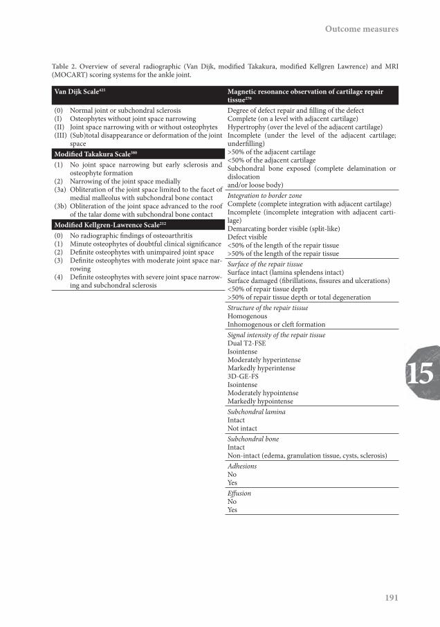
Outcome measures
191
15
Table 2. Overview of several radiographic (Van Dijk, modifi ed Takakura, modifi ed Kellgren Lawrence) and MRI (MOCART) scoring systems for the ankle joint.
Van Dijk Scale425 Magnetic resonance observation of cartilage repair tissue270
(0) Normal joint or subchondral sclerosis(I) Osteophytes without joint space narrowing(II) Joint space narrowing with or without osteophytes(III) (Sub)total disappearance or deformation of the joint
space
Degree of defect repair and fi lling of the defectComplete (on a level with adjacent cartilage)Hypertrophy (over the level of the adjacent cartilage)Incomplete (under the level of the adjacent cartilage; underfi lling)>50% of the adjacent cartilage<50% of the adjacent cartilageSubchondral bone exposed (complete delamination or dislocationand/or loose body)
Modifi ed Takakura Scale380
(1) No joint space narrowing but early sclerosis and osteophyte formation
(2) Narrowing of the joint space medially(3a) Obliteration of the joint space limited to the facet of
medial malleolus with subchondral bone contact(3b) Obliteration of the joint space advanced to the roof
of the talar dome with subchondral bone contact
Integration to border zoneComplete (complete integration with adjacent cartilage)Incomplete (incomplete integration with adjacent carti-lage)Demarcating border visible (split-like)Defect visible<50% of the length of the repair tissue>50% of the length of the repair tissue
Modifi ed Kellgren-Lawrence Scale212
(0) No radiographic fi ndings of osteoarthritis(1) Minute osteophytes of doubtful clinical signifi cance(2) Defi nite osteophytes with unimpaired joint space(3) Defi nite osteophytes with moderate joint space nar-
rowing(4) Defi nite osteophytes with severe joint space narrow-
ing and subchondral sclerosis
Surface of the repair tissueSurface intact (lamina splendens intact)Surface damaged (fi brillations, fi ssures and ulcerations)<50% of repair tissue depth>50% of repair tissue depth or total degenerationStructure of the repair tissueHomogenousInhomogenous or cleft formationSignal intensity of the repair tissueDual T2-FSEIsointenseModerately hyperintenseMarkedly hyperintense3D-GE-FSIsointenseModerately hypointenseMarkedly hypointenseSubchondral laminaIntactNot intactSubchondral boneIntactNon-intact (edema, granulation tissue, cysts, sclerosis)AdhesionsNoYesEff usionNoYes
27443_van Bergen.indd 191 27-11-13 09:25

Chapter 15
192
disorders. The questionnaire was constructed by using the following four steps to develop a self-reported evaluative instrument: 1) genera-tion of potential items, 2) initial item reduction, 3) final item reduction, and 4) acquisition of validity evidence to support interpretation of the score273.
The FAAM comprises two separately scored subscales; the activities of daily living (ADL) subscale (21 items) and the sports sub-scale (eight items). Each item is scored on a five-point Likert scale from 4 to 0, with 4 being “no difficulty” and 0 being “unable to do”. Items without a response are marked as not applicable and are not counted. The total number of items with a response is multiplied by four to get the highest potential score. The total item score is divided by the highest potential score and then multiplied by 100 to produce the FAAM score that ranges between 0 and 100. A higher score represents a higher level of physical function for both the ADL and sports subscales. The minimal clinically important differences for the FAAM are 8 and 9 points for the ADL and sports sub-scales, respectively.
Since its introduction, the construct validity, reliability, and responsiveness have been tested for several indications. In all these indica-tions, the FAAM has been shown to be a valid subjective measurement tool70,159,274. There is, however, no specific information in the litera-ture on psychometric properties for talar OCD. The FAAM has been translated and validated into several languages55,277,295.
In conclusion, the FAAM is a well-vali-dated questionnaire suitable for several foot and ankle pathologies.
The Ogilvie-Harris and Berndt and Harty scores
Both the Ogilvie-Harris and Berndt and Harty scores are simple scoring systems to evaluate the
effect of treatment. They are specifically useful to provide a success rate rather than a score.
The Ogilvie-Harris score consists of five items, including pain, swelling, stiffness, limping, and activity304. Either the patient or the examiner rates each item as excellent, good, fair, or poor. The lowest grade of each of the five items determines the final score.
The Berndt and Harty outcome ques-tion was specifically introduced for ankle OCDs. It is a single question with three possible answers, which allows patients to categorize their ankles into good, fair, or poor49. The score was slightly modified in 2003 because the original language was confusing190. In this modified score, patients with a “good” outcome have no symptoms of pain, swelling or instability, or experience slightly annoying, but not disabling, symptoms; patients with a “fair” outcome report that the symptoms are somewhat improved, although some disabil-ity problems persist; a “poor” outcome indicates that the overall symptoms remain unchanged190.
Both the Ogilvie-Harris and the Berndt and Harty scores have been used in various studies on the treatment of osteochondral ankle defects190,346,416, which makes it possible to com-pare the results of different studies. However, neither score has been validated. The Berndt and Harty outcome question has been shown to have a good correlation with both the single assess-ment numeric evaluation (r = 0.81) and the Martin outcome system (r = 0.69)190.
Pain assessment Measurement of pain intensity is a quantitative estimate of the subjective interpretation of the severity of the pain experienced by the patient. The most frequently used methods to assess pain intensity are (1) the visual analogue scale (VAS), (2) the numeric rating scale (NRS), and (3) the verbal rating scale (VRS). All pain rating
27443_van Bergen.indd 192 27-11-13 09:25

Outcome measures
193
15
scales have been extensively studied in several diff erent populations, and validity and reliabil-ity have been demonstrated185. However, there is high variation in pain descriptors of the scales, time frames used to assess pain intensity (e.g., last week, last month), and specifi c situations for which pain intensity has to be assessed (e.g., rest, activity).
Visual analogue scale
Th e VAS is a 100-mm line that represents the severity of the pain; the ends of the scale are anchored by two extremes of pain, such as “no pain” on one side and “worst imaginable pain” on the other side. Th e patient is asked to mark the line to indicate the pain intensity.
Various minimal clinically important diff erences are proposed for the VAS for mus-culoskeletal conditions. A minimal clinically important diff erence of 15 mm was proposed for low back pain (30% from baseline)307, 20 mm for knee osteoarthritis (41% from baseline), and 15 mm for hip osteoarthritis (32% from baseline)401.
Numeric rating scale
Th e NRS is an 11-, 21-, or 101-point scale where the end points oft en are the extremes “no pain” and “worst imaginable pain” or “unbearable pain”. Th e NRS measures pain severity by asking the patient to select a number that represents the severity of the pain. Th e NRS can be administered graphi-cally (NRS) or verbally (VNRS). Th e 11-point (V)NRS (0 – 10) is used most frequently 185.
Th e minimal clinically important diff erence of the 11-point NRS is 2 points, reported for both low back pain307 and chronic musculoskeletal pain (rheumatoid arthritis; knee, hip, and hand osteoarthritis; and ankylos-ing spondilitis )339, both implying a change from baseline of approximately 30%.
Verbal rating scale
Th e VRS consists of a list of adjectives that are commonly used to describe increasing pain intensity. It should comprise the extremes of the scale, such as “no pain” and “worst imaginable pain”. Th e amount of response options can vary, but has to be suffi cient to capture the gradations of pain intensities experienced by the patient. Rank numbers are assigned to response options, with highest numbers indicating most pain.
All three scales perform generally well, although specifi c studies on reliability, validity and mini-mal clinically important diff erence for patients with talar OCD are lacking. All scales are sen-sitive to changes over time 60,261,360. Th e VRS, however, is slightly less sensitive than the NRS and the VAS60,130,261, possibly due to the limited amount of response options. Th e NRS and the VRS are both easy to administer with good compliance. Th e VAS scale appears to be more complicated than NRS and VRS185. Despite the fact that the scales are highly correlated, they can not be used interchangeably 260,360.
Since the psychometric properties of the pain scales are suffi cient, the choice for the type of scale can be based on practical consider-ations, such as ease of administration and type of population. Th e authors prefer the NRS because of its ease and compliance.
Postoperative imaging
Radiography
Radiographs are frequently obtained in the postoperative assessment of talar OCDs, espe-cially to evaluate degenerative changes in the joint. Several radiographic grading systems have been developed for the osteoarthritic ankle joint211,375,425. Th e scales focus on the presence
27443_van Bergen.indd 193 27-11-13 09:25

Chapter 15
194
of osteophytes and joint space narrowing. The Kellgren and Lawrence system was not designed specifically for the ankle211. The Takakura system focuses mainly on the medial joint space375. The van Dijk OA classification evaluates the com-plete talocrural joint, and has been used for the evaluation of talar OCD346,416.
Moon and co-workers compared the van Dijk scale425, the modified Kellgren-Lawrence scale212, and modified Takakura380 scales, and concluded that all these scales were reliable and valid291. Interobserver and intrao-bserver comparisons (weighted Kappa) of each scale were found to be satisfactory (Kellgren and Lawrence, 0.51 to 0.81; Takakura, 0.65 to 0.88; van Dijk, 0.64 to 0.89). However, the predictabil-ity of the scales for cartilage damage, as observed by arthroscopy, was only moderate (intraclass correlation coefficients, 0.42 to 0.51)291.
Computed tomography
To objectively assess the bone repair, multislice helical computed tomography (CT) scans can be obtained410. CT has been shown to be accurate in the follow-up of talar OCDs460. The scan-ning protocol involves “ultra high resolution” axial slices with an increment of 0.3 mm and a thickness of 0.6 mm, and multi-planar coronal and sagittal 1-mm reconstructions408. One can measure the completeness, thickness, and level of the subchondral plate (i.e., flush, depressed, or proud), as well as bone volume filling of the defect and postoperative loose bony particles409,410. However, to our knowledge, a postoperative grading system based on CT is unavailable.
Magnetic resonance imaging
Magnetic resonance imaging (MRI) evaluation of OCD repair tissue has gained popularity in recent years. The scanning protocol incorporates proton density and fast spin-echo acquisitions
for cartilage evaluation270. Some investigators have quantified MRI results by self-developed criteria44,193, but a more objective, well-known, and frequently used method is the magnetic resonance observation of cartilage repair tissue (MOCART)270,271. Nine variables describe the morphology and signal intensity of the repair tissue compared with the adjacent native car-tilage, the degree of filling of the defect, the integration to the border zone, the description of the surface and structure, the signal inten-sity, the status of the subchondral lamina and subchondral bone, the appearance of adhesions and the presence of synovitis270. This system has good interobserver reliability, with intra-class correlation coefficients of >0.81 in eight of nine variables271. However, the association of the MOCART with the clinical situation is not exactly clear. In a study by Aurich and co-workers there was no relation between the MOCART and clinical outcome after matrix-associated chondrocyte implantation of the talus27. In another study, three out of five vari-ables of the modified MOCART showed good correlation with second-look arthroscopy after autologous chondrocyte implantation in the ankle, while two out of five variables showed poor correlation245.
Conclusions and recommendations
Valid and reliable outcome measures are avail-able for several ankle conditions. However, none of the clinical and functional outcome scores have been psychometrically investigated for the specific patient population with talar OCD. The AOFAS has been used most frequently in studies on the treatment of talar OCD458, but has some serious concerns. Both the FAAM and the FAOS are suitable questionnaires for patients with various ankle conditions. However, minimal clinically important differences of these scales
27443_van Bergen.indd 194 27-11-13 09:25

Outcome measures
195
15
are desirable for proper evaluation of outcomes of OCD treatment.
Pain assessment for patients with talar OCD is important since pain is the predomi-nant symptom. Although the described pain scales have been properly validated, they lack information on the minimal clinically important diff erence for this patient group. Most impor-tant in a clinical or research setting is the use of standardized pain descriptors, clear time frames, and unambiguous description concerning the
context of pain assessment. Th e 11-point NRS has, in our opinion, some advantages and would be the most practical and valid choice for the use of pain assessment.
In addition to specifi c ankle scores, the authors recommend to use a general quality-of-life score in clinical studies, such as the short form-36 or the EuroQoL121,445.
Postoperative imaging can be a useful adjunct to clinical outcome scoring.
27443_van Bergen.indd 195 27-11-13 09:25

27443_van Bergen.indd 196 27-11-13 09:25

Chapter 16Translation and validation
of the German Foot andAnkle Outcome Score
Christiaan J.A. van BergenInger N. SiereveltPaul HoogervorstHazibullah WaizyC. Niek van DijkChristoph Becher
Submitted.
27443_van Bergen.indd 197 27-11-13 09:25

Chapter 16
198
Abstract
Purpose
Outcome assessment is critical in evaluating the efficacy of orthopaedic procedures. The Foot and Ankle Outcome Score (FAOS) is a 42-item questionnaire divided into five subscales, which has been validated in several languages. Germany has no validated outcome score for general foot and ankle pathology. The aim of this study was to develop a German version of the FAOS and to investigate its psychometric properties.
Methods
Forward and backward translation was executed according to official guidelines. The final version of the FAOS was investigated in 150 patients with various foot and ankle disorders. All patients com-pleted the FAOS, Short Form-36, numeric rating scales for pain and disability, and the Hannover questionnaire. The FAOS was re-administered after one week. Test-retest reliability, internal con-sistency, minimal detectable change, construct validity, and floor and ceiling effects were analyzed.
Results
Test-retest reliability and internal consistency of each subscale were excellent (intraclass correlation coefficient, 0.88 to 0.95; Cronbach’s alpha, 0.94 to 0.98). The minimal detectable changes of each subscale were 17.1 to 20.8 at the individual level and 2.0 to 2.4 at group level. There were moderate to strong correlations between FAOS subscales and physical outcomes and low to moderate correlations between FAOS subscales and mental outcomes. Floor and ceiling effects were not present.
Conclusion
The German version of the FAOS is a reliable and valid instrument for use in foot and ankle patients.
Introduction
Outcome assessment is critical in evaluating the efficacy of orthopaedic procedures. The assess-ment of outcome from the patient’s perspective becomes more recognized.115 Questionnaires are used to asses the patient’s perspective on the degree of impairment, pain, disabilities, and quality of life. Translation and validation of the original questionnaire into the target language are essential for the conduction of multinational
studies and for the comparison of outcomes from different countries.
There are few validated outcome mea-sures for disability of the foot and ankle.115 The Foot and Ankle Outcome Score (FAOS) is a valid and reliable instrument.115,336 The score is avail-able in English, Swedish, Portuguese, Persian, Turkish, and Dutch.5,207,297,336,356 Germany has the largest population of Europe but, to our knowl-edge, has no validated outcome score for general foot and ankle pathology. Therefore, the aim of
27443_van Bergen.indd 198 27-11-13 09:25

Validation of the German FAOS
199
16
this study was to translate and validate the FAOS into German, enabling its use in the German-speaking population.
Methods
Translation procedure
Forward and backward translation was executed according to offi cial guidelines.449 Two bilingual translators with diff erent profi les (one physician) whose native language was German produced two independent forward translations. A syn-thesis of these translations was conducted. Two other translators without medical background, with English as the native language and blind-ed to the original version, then translated the questionnaire back into the original language. A committee composed of the translators and a German orthopaedic surgeon consolidated all the versions of the questionnaire and developed the fi nal version for fi eld testing (Supplement). Th e last step of the translation procedure was the pretesting on several random patients. Th e patients were asked whether they understood all the items and whether they had suggestions for improvement. No diffi culties were encountered, and changes to the fi nal version were not indi-cated (Supplement).
Validation procedure
A random sample of 150 outpatients with chronic foot and ankle related disorders were included in the study at a single institution. Exclusion crite-ria were inability to understand or complete the questionnaires, or refusal to sign informed con-sent. Th ere were 65 male and 85 female patients with a mean age of 48 ± 16 years (range, 18 – 79 years). Th e diagnoses of the ankle included osteochondral defects, osteoarthritis, anterior or posterior impingement, instability, loose bodies,
and tendonitis; diagnoses of the foot included plantar fasciitis, metatarsalgia, and pes planus. About 80% were preoperative or nonoperative patients, and 20% were postoperative during routine controls (not in the early postoperative period). Th e study protocol was approved by the local Medical Ethics Committee (Nr. 720/2010). All patients signed informed consent.
Each patient completed a package of questionnaires containing the FAOS, Short Form-36 (SF-36), numeric rating scale (NRS) for pain and disability, and Hannover question-naire. Th e FAOS was re-administered under similar conditions with a 1-week interval. Th e patients had to indicate whether any change in the (extent of) symptoms had occurred during the interval.
Instruments
Th e FAOS is a 42-item questionnaire intended to evaluate symptoms and functional limita-tions related to the foot and ankle.336 It consists of fi ve subscales: pain; other symptoms; activi-ties of daily living (ADL); sport and recreational activities; and foot- and ankle-related qual-ity of life (QoL). Patients score each question on a Likert scale (no, mild, moderate, severe, extreme) resulting in a score ranging from 0 to 4. Th e scores of the subscales are calculated by the sum of the items included. Th e total scores are then transformed to a scale from 0 to 100. Higher total values indicate lesser problems and/or functional limitations.
Th e SF-36 is a widely used, patient-administered, generic health related quality of life instrument,445 which has been translated and validated into German.65 It comprises 36 items across eight subscales, which are each calculated on a 0 to 100 scale (100 indicating no symptoms and 0 indicating extreme symptoms).
Numeric rating scales (NRS) were used to determine the severity of pain and disability
27443_van Bergen.indd 199 27-11-13 09:25

Chapter 16
200
of the ankle and foot. A score of 0 accounts for no pain or disability; a score of 10 represents the most severe pain or disability imaginable.
The Hannover questionnaire rates patients’ complaints and functional status based on a severity-symptom scale and functional sta-tus. It contains 20 subjective questions for the patient with five possible answers to each. A test-retest reliability of r = 0.91 has been reported but the score has not been validated.387
Statistical analysis
The FAOS subscale scores were calculated with use of formulas provided by the developers.5 Missing data and double marks were treated as outlined by the developers.5
Psychometrics is the process that allows researchers to apply scientific methodol-ogy to the measurement of subjective outcomes. The psychometric properties of a questionnaire define how well a questionnaire measures what it is supposed to measure. Reliability and validity of the German version of the FAOS were tested with use of SPSS software (version 19.0; Chicago, Illinois, USA).
The test-retest reliability was assessed by calculation of intraclass correlation coef-ficients (ICCagreement, two-way random effects model).355,383 The test-retest reliability was considered good if the ICC is 0.40 – 0.75 and excellent if the ICC > 0.75.134 Systematic dif-ferences between the first and second test were assessed with use of paired t-tests.
Internal consistency determines whether the questions cover the same con-struct. Cronbach’s alpha was calculated to assess the internal consistency of each subscale of the FAOS.89 This coefficient addresses the homogene-ity of the questions included in a questionnaire. Cronbach’s alphas of 0.7, 0.8, and 0.9 are con-sidered to represent a fair, good, and excellent degree of internal consistency, respectively.53
There is a 95% confidence rate that the health status of a patient improves or dete-riorates when the change in scores exceeds the minimal detectable change. The minimal detect-able change (MDC) at the individual level was calculated as 1.96 × √2 × standard error of measurement (i.e., the square root of the within-subject variance).383 The MDC at group level was calculated by dividing the MDC at the individu-al level by √n.383
Validity relates to the ability of a ques-tionnaire to measure the outcome parameter of interest. Construct validity refers to the asso-ciation with other measures in a manner that is consistent with theoretically derived hypoth-eses, in the absence of a “gold standard”.383 The FAOS subscales were compared with the SF-36, NRS pain and disability, and Hannover ques-tionnaire. Pearson correlation coefficients were calculated to determine construct validity of the FAOS. Correlation coefficients of <0.30, 0.30 – 0.60, and >0.60, are considered low, moder-ate, and strong, respectively.207 Convergent and discriminate validity were evaluated (i.e., are correlations high where they are expected to be high, and low where they are expected to be low). We hypothesized that correlations of FAOS subscales with the physical outcomes (SF-36 subscales Physical functioning, Role physical, and Bodily pain; NRS Pain and Disability; and Hannover questionnaire) would be higher than correlations with the mental outcomes (other SF-36 subscales).
Floor and ceiling effects were regis-tered for each scale. These effects are considered to be present if the amount of minimal or maxi-mal scores exceeds 15%.383
Results
In the complete study population, the mean FAOS subscale Pain was 59.5 ± 22.4 (range,
27443_van Bergen.indd 200 27-11-13 09:25

Validation of the German FAOS
201
16
13.9 – 100); Symptoms was 62.3 ± 20.9 (range, 17.9 – 100); ADL was 70.3 ± 21.9 (range, 7.4 – 100); Sport was 46.2 ± 28.7 (range, 0 – 100); and QoL was 36.9 ± 21.3 (range, 0 – 100). A total of 158 individual items were missing, which corre-sponds to 2.5% of missing data (158 items in 150 patients × 42 items). Each subscale score of all patients could be calculated.
One-hundred and eighteen patients (79%) completed the second FAOS aft er a week. Of these, 42 indicated that the (extent of) symp-toms had changed since the fi rst questionnaire.
Th erefore, data of 76 patients (51%) were used to calculate the reliability.
Test-retest reliability of each subscale and internal consistency were excellent (Table 1). Th e MDCs at the individual level were between 17.1 and 20.8; those at group level were between 2.0 and 2.4 (see Table 1).
Th ere were moderate to strong correla-tions between FAOS subscales and SF-36 Physical functioning, Role physical, and Bodily pain; NRS Pain and Disability; and Hannover ques-tionnaire (Table 2). Th ere were low to moderate
Table 1. FAOS subscale scores (n = 76), test-retest reliability (n = 76), internal consistency (n = 150), and minimal detectable change (n = 76)
FAOS subscale
Test (mean ± SD)
Retest (mean ± SD) ICC (95% CI)
Cronbach’s alpha
Systematic diff erence MDCindividual MDCgroup
Pain 57.6 ± 23.0 58.7 ± 22.6 0.92 (0.88 - 0.95) 0.96 1.1 (p = 0.25) 18.1 2.1Symptoms 60.2 ± 21.1 63.0 ± 22.5 0.88 (0.82 - 0.93) 0.94 2.8 (p = 0.06) 20.0 2.3ADL 69.4 ± 22.6 68.7 ± 23.9 0.89 (0.83 - 0.93) 0.94 0.7 (p = 0.61) 20.8 2.4Sport 47.1 ± 29,0 48.7 ± 29.9 0.95 (0.93 - 0.97) 0.98 1.6 (p = 0.21) 17.2 2.0QoL 38.5 ± 23.8 39.3 ± 22.2 0.93 (0.90 - 0.96) 0.97 0.8 (p = 0.40) 17.1 2.0
ADL = activities of daily living, CI = confi dence interval, ICC = intraclass correlation coeffi cient, MDCgroup = minimal detectable change at group level, MDCindividual = minimal detectable change at individual level, QoL = foot- and ankle-related quality of life, and SD = standard deviation.
Table 2. Construct validity. Correlation (Pearson correlation coeffi cient) between FAOS subscales and other outcome measures (n = 150)
Subscale Pain Symptoms ADL Sport QoLSF-36 Physical functioning 0.66 0.58 0.79 0.75 0.63
Role physical 0.33 0.34 0.54 0.53 0.43Bodily pain 0.80 0.59 0.68 0.62 0.66Social functioning 0.36 0.36 0.38 0.44 0.38Mental health 0.23 0.25 0.25 0.21 0.17Role emotional 0.19 0.27 0.30 0.34 0.27Vitality 0.21 0.22 0.26 0.21 0.26General health perceptions 0.25 0.40 0.42 0.32 0.41
NRS Pain -0.74 -0.47 -0.54 -0.52 -0.59Disability -0.55 -0.51 -0.63 -0.58 -0.61
Hannover questionnaire 0.71 0.74 0.75 0.78 0.64
All correlations were statistically signifi cant (p <0.05). ADL = activities of daily living, NRS = numeric rating scale, SF-36 = Short Form-36, and QoL = foot- and ankle-related quality of life.
27443_van Bergen.indd 201 27-11-13 09:25

Chapter 16
202
correlations between FAOS subscales and SF-36 Social functioning, Mental health, Role emotion-al, Vitality, and General health perceptions.
There were no floor or ceiling effects. The lowest score was obtained by 0%, 0%, 0%, 5%, and 1% of the patients on the subscales Pain, Symptoms, ADL, Sport, and QoL, respectively. The highest score was obtained by 7%, 5%, 10%, 5%, and 5% of the patients on the subscales Pain, Symptoms, ADL, Sport, and QoL, respectively.
Discussion
A German version of the FAOS was developed in the first part of the present study. The translation process was executed according to official guide-lines.449 The translated version was tested in the second part of the study. According to the psy-chometric analyses, the translated version was shown a reliable and valid instrument for use in German-speaking patients with foot and ankle pathology.
Test-retest reliability and internal con-sistency of each subscale were excellent (see Table 1). There were moderate to strong cor-relations between FAOS subscales and physical outcomes, and there were low to moderate cor-relations between FAOS subscales and mental outcomes (see Table 2). In addition, no floor or ceiling effects were observed. According to the MDC, the FAOS may be more suitable for the evaluation at group level than at individual level. The relatively high MDC may be attributed to the diverse study population.101
Although only 76 of 150 patients were available for statistical analysis of test-retest reli-ability (due to a relatively low response rate of the second questionnaire or a changed situa-tion), this number was well above the required number of 50.383 Cross-cultural adaption of the questionnaire was deemed unnecessary, because the German population can be considered
similar to the Swedish population of the original version.336 Likewise, the patient burden imposed by administering the FAOS was not tested because it is considered to be identical to that of the original version (less than 10 min).336
Roos et al. invented the original Eng-lish version (adapted from the Knee injury and Osteoarthritis Outcome Score).336 They tested the Swedish version in 213 patients.336 The psy-chometric properties were generally similar to the German version. Test-retest reliability varied from an ICC of 0.70 for ADL to 0.92 for QoL, and Cronbach’s alpha varied from 0.88 for Symp-toms to 0.97 for ADL. For construct validity, the FAOS subscales were compared with the Karls-son score. Spearman correlation coefficients of 0.58 to 0.67 were found. However, ceiling effects occurred in each subscale. The differences in ceiling effects between the original version and the developed translation may be contributed to the different study population. Roos et al. studied patients with previous ankle ligament reconstructions after a mean follow-up of 12 years, while our study population consisted of patients with a variety of foot and ankle disor-ders.
The original version has also been translated and validated into Turkish and Per-sian.207,297 Karatepe et al. studied 55 Turkish patients with various foot and ankle disorders.207 They found a test-retest reliability (ICC) of 0.70 to 0.96 and a Cronbach’s alpha of 0.79 to 0.97 for each subscale. Correlations between FAOS and SF-36 subscales were comparable to those in our study. Floor or ceiling effects were not reported. Negahban from Iran validated the Per-sian version in 93 patients with foot and ankle disorders, mostly ankle sprain.297 ICCs were excellent but two subscales had low internal consistency (Cronbach’s alpha of 0.39 for Symp-toms and 0.62 for Qol). The correlations between FAOS and several SF-36 subscales were generally somewhat lower than in our study. Floor and
27443_van Bergen.indd 202 27-11-13 09:25

Validation of the German FAOS
203
16
ceiling eff ects were not present. Although the psychometric properties of the other languages are slightly inferior to the German version, all versions meet the quality criteria.383 Th e previ-ous studies and our study indicate that the FAOS can be regarded a reliable and valid international outcome measure.
Other outcome measures developed for the German foot and ankle patient are the Visual Analog Scale Foot and Ankle (VAS FA) and the Foot and Ankle Ability Measure (FAAM).273,333 Th e VAS FA, however, is not avail-able in other languages.333,371 Furthermore, only the construct validity was reported in healthy individuals.333 Th e FAAM is a reliable and valid international instrument with psychometric properties similar to the FAOS.273,277,295 Th e score has two subscales (ADL and Sport). Th e Eng-lish and Persian versions have been validated in a range of foot and ankle disorders. However, the German version has been validated only in
chronic ankle instability.273,277,295 In contrast, the FAOS has been validated in patients with vari-ous pathologic conditions.74,207,267,297,336 Possible advantages of the FAOS over the FAAM are a larger number of subscales and a broader target population.
Th e principal limitation of the present study is the fact that the sensitivity to change (i.e., responsiveness and minimal clinically important change) of the FAOS aft er an intervention was not investigated. Further studies are required to assess the sensitivity to change.
In conclusion, the developed Ger-man version of the FAOS is a valid and reliable instrument for foot and ankle patients. Future use of the score will allow comparison with the international literature and the execution of international multicenter studies. Translation and validation into other languages will expand its applicability.
27443_van Bergen.indd 203 27-11-13 09:25

Chapter 16
204
Supplement
Final translated version of the German Foot and Ankle Outcome Score
„FAOS“ Fragebogen Fuß & Sprunggelenk
Datum: _____/______/______ Geburtsdatum: _____/______/______
Name: _____________________________________
ANLEITUNG: Dieser Fragebogen stellt Ihnen Fragen zum Zustand Ihres Fuß und Sprunggelenks. Die dadurch gewonnenen Informationen erlauben uns Ihre Beurteilung von Ihrem Fuß und Sprung-gelenk zu verfolgen und dokumentiert wie gut Sie in der Lage sind, Ihre üblichen Aktivitäten zu verrichten.Beantworten Sie bitte jede Frage durch ankreuzen des zugehörigen Kästchens.Bitte nur ein Kästchen pro Frage ankreuzen. Wenn Sie sich unsicher sind, wie Sie die Frage beant-worten sollen, wählen Sie die Antwort aus, die Ihnen am zutreffendsten erscheint.
SymptomeDiese Fragen beziehen sich auf Ihre Fuß/Sprunggelenksbeschwerden während der vergangenen Woche.
S1. Hatten Sie Schwellungen an Ihrem Fuß/Sprunggelenk?niemals selten manchmal oft immerp p p p p
S2. Fühlten Sie ein Mahlen, hörten Sie ein Klicken oder irgendein Geräusch, wenn Sie Ihren Fuß/Ihr Sprunggelenk bewegten?
niemals selten manchmal oft immerp p p p p
S3. Blieb Ihr Fuß/Sprunggelenk hängen, oder blockierte er/es bei Bewegung?niemals selten manchmal oft immerp p p p p
S4. Konnten Sie Ihren Fuß/Ihr Sprunggelenk ganz ausstrecken? immer oft manchmal selten niemals
p p p p p
27443_van Bergen.indd 204 27-11-13 09:25

Validation of the German FAOS
205
16
S5. Konnten Sie Ihren Fuß/Ihr Sprunggelenk ganz beugen? immer oft manchmal selten niemals
p p p p p
Steifi gkeitDie nachfolgenden Fragen betreff en die Steifi gkeit in Ihrem Fuß/Sprunggelenk während der letz-ten Woche. Unter Steifi gkeit versteht man ein Gefühl der Einschränkung oder Verlangsamung der Fähigkeit Ihre Gelenke zu bewegen.
S6. Wie stark war Ihre Fuß/Sprunggelenkssteifi gkeit morgens direkt nach dem Aufstehen? keine schwach mäβig stark sehr stark
p p p p p
S7. Wie stark war Ihre Fuß/Sprunggelenksteifi gkeit nach dem Sie saßen, lagen, oder sich im Verlauf des Tages ausruhten? keine schwach mäβig stark sehr stark
p p p p p
SchmerzenP1. Wie oft haben Sie Schmerzen im Fuß/Sprunggelenk? nie monatlich wöchentlich täglich immer
p p p p p
Wie ausgeprägt waren Ihre Schmerzen in der vergangenen Woche als Sie z.B:?
P2. Sich im Fuß/Sprunggelenk drehten keine schwach mäβig stark sehr stark
p p p p p
P3. Ihren Fuß/Ihr Sprunggelenk ganz ausstreckten keine schwach mäβig stark sehr stark
p p p p p
P4. Ihren Fuß/Ihr Sprunggelenk ganz beugten keine schwach mäβig stark sehr stark
p p p p p
P5. Auf ebenem Boden gingen keine schwach mäβig stark sehr stark
p p p p p
27443_van Bergen.indd 205 27-11-13 09:25

Chapter 16
206
P6. Treppen herauf oder herunterstiegen keine schwach mäβig stark sehr stark
p p p p p
P7. Nachts im Bett lagen keine schwach mäβig stark sehr stark
p p p p p
P8. Saßen oder lagen keine schwach mäβig stark sehr stark
p p p p p
P9. Aufrecht standen keine schwach mäβig stark sehr stark
p p p p p
Aktivitäten des täglichen LebensDie nachfolgenden Fragen beziehen sich auf Ihre körperliche Leistungsfähigkeit.Hierunter verstehen wir Ihre Fähigkeit, sich selbständig zu bewegen bzw. sich selbst zu versorgen. Für jede der nachfolgenden Aktivitäten geben Sie bitte das Ausmaß der Schwierigkeiten an, welche Sie aufgrund Ihres Fuß/Sprunggelenks innerhalb der letzten Woche erfahren haben.
Welche Schwierigkeiten hatten Sie in der letzten Woche als Sie z.B.:
A1. Treppen herunterstiegen keine wenig einige groβe sehr groβe
p p p p p
A2. Treppen heraufstiegen keine wenig einige groβe sehr groβe
p p p p p
A3. Vom Sitzen aufstanden keine wenig einige groβe sehr groβe
p p p p p
Welche Schwierigkeiten hatten Sie in der letzten Woche als Sie z.B.:
A4. Aufrecht standen keine wenig einige groβe sehr groβe
p p p p p
27443_van Bergen.indd 206 27-11-13 09:25

Validation of the German FAOS
207
16
A5. Sich bückten um z.B. etwas vom Boden aufzuheben keine wenig einige groβe sehr groβe
p p p p p
A6. Auf ebenem Boden gingen keine wenig einige groβe sehr groβe
p p p p p
A7. Ins Auto ein- oder ausstiegen keine wenig einige groβe sehr groβe
p p p p p
A8. Einkaufen gingen keine wenig einige groβe sehr groβe
p p p p p
A9. Socken/Strümpfe anzogen keine wenig einige groβe sehr groβe
p p p p p
A10. Vom Bett aufstanden keine wenig einige groβe sehr groβe
p p p p p
A11. Socken/Strümpfe auszogen? keine wenig einige groβe sehr groβe
p p p p p
A12. Im Bett lagen und sich drehten, ohne den Fuß/Sprunggelenk dabei zu beugen keine wenig einige groβe sehr groβe
p p p p p
A13. In oder aus der Badewanne stiegen keine wenig einige groβe sehr groβe
p p p p p
A14. Saßen keine wenig einige groβe sehr groβe
p p p p p
A15. Sich auf die Toilette setzten oder aufstanden keine wenig einige groβe sehr groβe
p p p p p
27443_van Bergen.indd 207 27-11-13 09:25

Chapter 16
208
A16. Schwere Hausarbeit verrichteten (schwere Kisten umstellen, Boden schrubben, etc) keine wenig einige groβe sehr groβe
p p p p p
A17. Leichte Hausarbeit verrichteten (kochen, Staub wischen, etc.) keine wenig einige groβe sehr groβe
p p p p p
Aktivitäten bei Sport und FreizeitDie nachfolgenden Fragen beziehen sich auf Ihre körperliche Belastbarkeit im Rahmen von Sport- und Freizeitaktivitäten. Für jede der nachfolgenden Aktivitäten geben Sie bitte das Ausmaß der Schwierigkeiten an, welche Sie aufgrund Ihres Fuß/Sprunggelenks innerhalb der letzten Woche erfahren haben.
SP1. In die Hocke gingen keine wenig einige groβe sehr groβe
p p p p p
SP2. Rannten keine wenig einige groβe sehr groβe
p p p p p
SP3. Hüpften keine wenig einige groβe sehr groβe
p p p p p
SP4. Sich auf Ihrem kranken Fuß umdrehten keine wenig einige groβe sehr groβe
p p p p p
SP5. Sich hinknieten keine wenig einige groβe sehr groβe
p p p p p
Lebensqualität
Q1. Wie oft sind Sie sich Ihres Fuß/Sprunggelenksproblems bewusst? nie monatlich wöchentlich täglich immer
p p p p p
27443_van Bergen.indd 208 27-11-13 09:25

Validation of the German FAOS
209
16
Q2. Haben Sie Ihre Lebensweise verändert, um eventuell Ihrem Fuß/Sprunggelenk schadende Tätig-keiten zu vermeiden? gar nicht wenig etwas stark vollständig
p p p p p
Q3. Wie sehr leiden Sie unter einem Mangel an Vertrauen und Zuversicht hinsichtlich Ihres Fuß/Sprunggelenks? gar nicht wenig etwas stark sehr stark
p p p p p
Q4. Wie würden Sie insgesamt die Schwierigkeiten bewerten die Sie durch Ihren Fuß/Ihr Sprung-gelenk haben? keine wenig einige groβe sehr groβe
p p p p p
Vielen Dank für die Beantwortung aller Fragen.Fragebogen und Anleitung sind auf der folgenden Internetadresse zu fi nden: www.koos.nu
27443_van Bergen.indd 209 27-11-13 09:25

27443_van Bergen.indd 210 27-11-13 09:25

General discussionand summary
27443_van Bergen.indd 211 27-11-13 09:25

27443_van Bergen.indd 212 27-11-13 09:25

Chapter 17General discussion
27443_van Bergen.indd 213 27-11-13 09:25

Chapter 17
214
General introduction
Osteochondral defects (OCDs) of the talar dome can be a disabling disorder that seriously affects the quality of life. Symptomatic OCDs pose a therapeutic challenge to the orthopae-dic surgeon. Numerous treatment methods are available, each with advantages and disadvan-tages. This thesis aimed to evaluate and improve the current primary surgical treatment and to investigate alternative treatment methods.
Part I – Current concepts
Part I of the thesis reviewed the literature, pre-sented a treatment algorithm, and discussed the use of arthroscopic ankle surgery. This part formed the basis for the remainder of the thesis. The review identified arthroscopic debridement and bone marrow stimulation as the primary surgical treatment of defects up to 15 mm. Osteochondral autograft transfer (OATS) and autologous chondrocyte implantation (ACI), with or without a cancellous bone graft, were recommended for secondary cases as well as large lesions.
The development of future treatment modalities described in chapter 2 included matrix-induced ACI, bone marrow-derived mesenchymal stem cells, biodegradable com-posite implants, correction osteotomy in the event of malalignment, metal resurfacing implants, demineralized bone matrix (DBM), and pulsed electromagnetic fields (PEMF). Some of these developments have become true options since the publication of the chapter. Dif-ferent types of matrix-induced ACI have been reported in case series with two to 46 patients and a maximum follow-up of 3 years.299 The overall success rate was 90% but a comparison between the studies was unreliable due to differ-ences in indications, techniques, and outcome
measures.299 Furthermore, the analyses included duplicate follow-up publications from the same author. Bone marrow-derived cell transplanta-tion loaded on a scaffold has been investigated in a series of 49 patients with a mean follow-up of 4 years.150 Although the mean American Orthopaedic Foot and Ankle Society (AOFAS) ankle-hindfoot score improved from 64 ± 14 preoperatively to 82 ± 17 at the final follow-up, a significant decrease was evident between 2 and 4 years. This investigation thus showed promis-ing results but questioned the durability of the technique.150 Biodegradable composite implants have the advantages of a combined repair of car-tilage and subchondral bone without the risk of donor site morbidity. However, after the report of four initial failures of commercially available composite implants, the procedure has been abandoned and scientific progress of this tech-nique has decreased.141 Correction osteotomy for OCDs in malaligned ankles seems a promis-ing solution but clinical results of this procedure are not yet available.113 The applicability of metal resurfacing implants, DBM, and PEMF was investigated in this thesis.
Part II – Primary arthroscopic debridement and bone marrow stimulation
Most OCDs can be treated by an anterior arthroscopic approach with the ankle in full plantar flexion. In case of posterior OCDs, it is challenging to treat the defect with this tech-nique, since the ankle is a congruent joint with limited surgical access to the posterior talar sur-face. A posterior OCD can be approached with a two-portal hindfoot technique developed by van Dijk.427
The purpose of chapter 4 was to assess whether preoperative computed tomography (CT) of the ankle joint in full plantar flexion
27443_van Bergen.indd 214 27-11-13 09:25

General discussion
215
17
was able to predict the anterior arthroscopic accessibility of talar OCDs. Th e study showed that measurements on CT scans of the ankle in full plantar fl exion were a reliable and accurate preoperative method to determine the in situ arthroscopic location of talar OCDs. Th e fi nd-ings of chapter 4 were followed by the study presented in chapter 5. Th e dual purpose of the study described in this chapter was (1) to quan-tify the anterior arthroscopic reach of the talus (i.e., proportion of the talar dome anterior to the anterior distal tibial rim) with the ankle in full plantar fl exion, and (2) to identify predictive factors of the arthroscopic reach. Th e following conclusions were drawn: (1) almost half of the talar dome is accessible anterior to the anterior distal tibial rim, and (2) the clinical plantarfl ex-ion angle is an independent predictive factor of the arthroscopic reach of both the medial and lateral talar dome.
Th is study was the fi rst to quantify the arthroscopic reach of the talus in a representative patient population. Previously, cadaveric studies investigated exposure of the talus with diff erent open surgical approaches.293,456 Muir et al. com-pared seven open approaches in nine cadaver specimens.293 An anteromedial arthrotomy pro-vided a mean access of 47% (range, 41% – 60%) of the medial talar dome in the sagittal plane. An anterolateral arthrotomy provided 43% (range, 37% – 50%) sagittal access of the lateral talar dome. Young et al. found that a median of 50% (range, 44% – 58%) of the anteroposterior medi-al talar dome length could be reached through an anteromedial approach.456 Th e results of our study thus show similar results to the previ-ous cadaveric studies. Th e added value is the use of a validated method and correlation with arthroscopy, a larger and representative study population, and the identifi cation of predictive factors. Th e results may also be relevant for other procedures, such as OATS and metal resurfacing inlay implants. However, for these procedures
the complete OCD has to be exposed, instead of only the anterior border.
Th e results of chapters 4 and 5 give direction to preoperative planning protocols of the surgical approach for OCDs. Th e defect can be accessed by anterior arthroscopy if its ante-rior border is in the anterior half of the talus and ankle motion is not restricted. In doubtful cases, a CT scan of the ankle in full plantar fl exion can be obtained. If the anterior border of the OCD is behind the anterior distal tibial rim with the ankle in full plantar fl exion, access by means of an anterior approach will depend on the joint opening on forced plantar fl exion or distraction. In case of a stiff and highly stable joint and a pos-terior OCD, another approach is preferred, e.g., posterior arthroscopy.
Th e development of new techniques and instruments may reduce the necessity of full surgical exposure. A waterjet technique has been shown to produce cone-shaped holes in the sub-chondral bone of human and animal cadaveric calcanei.103 Th e technique can possibly be applied via fl exible tubings, allowing introduction of a small curved instrument into the joint space and creation of small holes in the subchondral bone at a perpendicular angle.103 However, the technique is still in its infancy and needs further development before it can be applied in clinical practice.
Although debridement and bone marrow stimulation is considered the surgical treatment of choice, there are concerns that the fi brocartilage repair tissue has insuffi cient dura-bility in the long term.229,262 Th e primary aim of chapter 6 was to assess the long-term clinical and radiographic outcomes of arthroscopic debride-ment and bone marrow stimulation for talar OCDs. Fift y patients were evaluated aft er a mean follow-up of 12 years (range, 8 – 20 years). Th e outcomes suggested that the initial success rates of arthroscopic debridement and bone mar-row stimulation for OCDs of the talus last over
27443_van Bergen.indd 215 27-11-13 09:25

Chapter 17
216
time. This finding strengthens the recommen-dations of the treatment guideline presented in part I of the thesis. Other studies that published mid-term to long-term outcomes demonstrated similar results. These studies reported success rates of 72% to 86% after a mean follow-up of 5 to 10 years.30,44,77,128,346
The secondary aim of chapter 6 was to determine prognostic factors that affect the long-term outcome. The study probably was not sufficiently powered to identify prognos-tic factors. Other studies that included more than 100 patients identified the defect size as an independent prognostic factor.76,77,82 These stud-ies found a cutoff value of 15 mm for the defect diameter or 150 mm2 for the defect area. Larger defects posed a higher risk of poor clinical out-comes.76,77,82
Different methods of bone marrow stimulation can be used, i.e., drilling, abrasion, and microfracturing. The latter is used the most at present. Chapter 7 described a potential pitfall in the microfracturing technique. The micro-fracture procedure may create small osseous fragments upon retrieval of the microfracture awl. These fragments may stay behind in the joint and act as loose bodies. Although the described pitfall could possibly influence patient outcome, this was not actually proven in the descriptive paper. Future studies, which specifically address the presence of bony particles with postopera-tive imaging after the microfracture procedure as well as the correlation with clinical outcome, may elucidate to what extent the particles are present and influence the clinical outcome.
Other issues related to the micro-fracturing technique that may require further optimization are the distance and depth of the microfracture holes. Most surgeons use intervals of 3 – 4 mm and penetrate the bone to such a depth that it leads to sufficient blood or fat drop-lets.223 However, no data are available on which technique leads to the best outcome.223 Further
studies that compare different techniques will provide the evidence to answer these questions.
To accelerate the postoperative rehabil-itation of OCDs, the advantageous effect of PEMF was postulated in chapter 2. The postoperative application of PEMF limited osteochondral graft resorption and cyst formation in sheep.47 In vitro and in vivo studies showed that PEMF stimula-tion leads to an increase of transforming growth factor b, thereby improving bone development, reducing cartilage damage and increasing chon-drocyte proliferation.6,47,83,98 Clinical studies showed promising results for fracture nonunion and for recovery after arthroscopic treatment of focal cartilage knee lesions.161,462 Therefore, PEMF stimulation may be effective for talar OCDs after arthroscopic treatment by produc-ing effects that suppress inflammation, promote tissue healing, and relieve pain.436
Chapter 8 described the detailed study protocol of a prospective, double-blind, ran-domized, placebo-controlled multicenter trial. It was hypothesized that PEMF-treatment after arthroscopy leads to earlier resumption of sports and an increase in the number of patients to resume sports. Sixty-eight patients were ran-domized to either active PEMF-treatment or sham-treatment. The final follow-up of the last patient is expected in January 2014. The out-comes of the trial will be presented in future publications. A recent review of the literature identified numerous publications supporting the hypothesis.132 The review concluded that the use of PEMF represents an innovative thera-peutic approach, because this physical stimulus increases the anabolic activity of chondrocytes and cartilage explants with consequent increase of matrix synthesis. At the same time, PEMFs limit the catabolic effects of inflammatory cyto-kines.132
27443_van Bergen.indd 216 27-11-13 09:25

General discussion
217
17
Part III – Secondary surgical treatment with a metal resurfacing inlay implant
If the primary arthroscopic treatment of a talar OCD fails, there are various alternative treat-ment methods. As described in part I, current secondary methods can be successful but have several disadvantages, one being donor-site morbidity. A metal resurfacing inlay implant was developed for the treatment of secondary, localized OCDs of the medial talar dome. It has a standard diameter of 15 mm and a variety of 15 incremental off set sizes. A preliminary assess-ment ascertained the safety, biocompatibility, and functionality of a similar metal implant in the goat knee.216 Chapter 9 described a cadav-eric study to test the following hypotheses: (1) a matching off set size is available for each talus, (2) the prosthetic device can be reproducibly implanted slightly recessed in relation to the talar cartilage level, and (3) with this implanta-tion level, excessive contact pressures on the opposing tibial cartilage are avoided. Th e results suggested that the implant can be applied clini-cally in a safe way, with appropriate off set sizes for various talar domes, and without excessive pressure on the opposing cartilage.
A similar study was published 1 day aft er the study of chapter 9.17 Th is study inves-tigated the eff ect of implantation accuracy on ankle contact mechanics. Diff erent implanta-tion levels (-0.5 to 0.5 mm) were tested in seven ankle specimens. Contact stresses in the talo-crural joint were measured with the specimen axially loaded to 300 N. Th e eff ects of rotation and angulation of the implant were tested in a fi nite element analysis. Th is study concluded that focal resurfacing with a metal implant has the potential to restore joint mechanics in ankles with a large OCD. A recessed to fl ush implan-tation height most closely restored the original contact stress distribution. Th us, in that study,
fl ush implantation was also found adequate, perhaps because the specimens were loaded for a shorter duration and with lower compression forces than in chapter 9. Th e question is whether the load-bearing capacity of the focal resurfac-ing implant is required. A previous study tested the consequence of increasing talar defect area on contact characteristics of the talocrural joint. Th is study demonstrated signifi cant changes for lesions larger than 15 mm (i.e., decreased total contact area and increased mean pressure), but not for smaller lesions.80 Because the metal implant is 15 mm in diameter, recessed implan-tation may not be harmful to the remainder of the ankle joint, even if it is placed so deep that is does not carry any load. A recessed implant reduces the chance of overloading and damaging the opposing cartilage surface.
Aft er the preliminary cadaveric study, the study described in chapter 10 evaluated the clinical eff ectiveness of the implant aft er failed prior surgical treatment. Twenty consecutive patients were prospectively studied. Based on a statistically signifi cant improvement in most outcome measures, it was concluded that the metal implantation technique is a promising treatment for OCDs of the medial talar dome aft er failed previous treatment. Th ese results were equaled by others. Ebskov presented simi-lar outcomes at the 2013 annual meeting of the Danish foot and ankle society. Th e limitations of both studies were the relatively small series, lack of long-term follow-up, and absence of a control group. A limitation of the implant itself is the fi xed diameter. In defects smaller than the implant, some healthy cartilage is sacrifi ced for implantation. In contrast, if the OCD is larger than the implant, some parts of the defect cannot be covered. Th e future development of diff erent sizes may overcome this limitation. Further-more, the implant was designed specifi cally for the medial talar dome. An implant for the lateral dome is focus of future research.
27443_van Bergen.indd 217 27-11-13 09:25

Chapter 17
218
At the moment, the results of the metal resurfacing inlay implant compare with those of other resurfacing techniques such as OATS and allograft transplantation. Hangody et al. pub-lished the largest series of OATS in the talus.178 The authors reported that 93% of 98 patients had good to excellent clinical results on the basis of evaluation with the Hannover score, but the duration of follow-up was not reported. The main disadvantage of OATS is the donor-site morbidity, which has been reported to occur in up to 50% of cases.312,327,407 Allograft trans-plantation has gained increased attention as an alternative for large OCDs in the United States. El-Rashidy et al. and Haene et al. reported sat-isfactory outcomes after a mean follow-up of 3 to 4 years.118,168 However, based on the avail-able evidence, allografts are not recommended for focal defects because of the loss of viability and stability in approximately one-third of the grafts.118,162,168
A disadvantage of treatments such as the metal implantation, OATS, and allograft techniques is the necessity to perform a medial malleolar osteotomy for adequate exposure and perpendicular access to the medial talar dome. An inappropriate osteotomy technique may result in a damaged weight-bearing tibial pla-fond, insufficient access, or in malreduction, which may negatively affect the clinical out-come.143,215 The studies in chapters 11 and 12 aimed to optimize the osteotomy technique.
To obtain a congruent joint surface after refixation, the oblique osteotomy should be directed perpendicularly to the articular surface of the tibia at the intersection between the tibial plafond and medial malleolus. The purpose of chapter 11 was to determine this direction in relation to the longitudinal tibial axis for use during surgery. Forty-six radiographs and CT scans were measured. A medial malleolar oste-otomy was found to be perpendicular to the tibial articular surface when directed at a mean
30° relative to the tibial axis in the frontal plane. This direction likely results in maximum con-gruity of the joint surface after reduction of the osteotomy. The intraobserver and interobserver reliability of the measurements were good to excellent. Hence, the methods used in this study allow for the preoperative planning of the indi-vidual patient, for example in case of abnormal anatomy. The osteotomy angle is rarely reported in the literature. Some surgeons suggested an angle of 45º relative to the tibial plafond.143,281,350 This is more horizontal than the direction found in our study. The 30º angle we found was subse-quently used by others as a reference standard in the evaluation of their technique.237
Since it is difficult to visualize the intersection between the tibial plafond and medial malleolus (especially posterior) during surgery, chapter 12 described the use of a right-angled aiming probe, which is normally used for arthroscopic surgery, to identify both the posterior and anterior parts of the intersection, in order to perform a precise osteotomy. Others have used fluoroscopy to help identify the inter-section, but this involves radiation and is more time-consuming.308
The angle and the probe, described in chapters 11 and 12, were applied during surgery and have subjectively improved the technique. However, clinical studies are required to deter-mine if the techniques improve patient outcome. A next step in the refinement of the medial mal-leolar osteotomy could be the development of an intraoperative aiming device in which the use of probes and the angle are incorporated.
Part IV – Alternative treatment
Animal studies play an important role in the development of a new treatments prior to introduction in clinical practice. Numerous experimental studies focused on cartilage
27443_van Bergen.indd 218 27-11-13 09:25

General discussion
219
17
defects of the knee,81 but an experimental ani-mal model of OCDs in the ankle joint was not available. We therefore developed such a model for talar OCDs in goats. Th e aim of chapter 13 was to test the developed animal model in vivo with use of autologous cancellous bone and donor DBM. It was possible to create a stan-dardized 6-mm defect in each talus through a posterolateral surgical approach. Aft er 12 weeks of healing, the analyses showed that most bony tissue was generated in the defects fi lled with autologous bone and least in the control defects. Our fi ndings showed that a standard OCD can be created in the talus by a relatively simple procedure in a large animal that allows qualita-tive and quantitative evaluation. Various 2- and 3-dimensional parameters could be quantifi ed in diff erent regions with the combination of his-tomorphometry and µCT. No conclusions could be drawn on the eff ectiveness of the investigated materials because of the small number of goats.
Only few studies described the surgi-cal exposure of an animal’s ankle joint.41,104,361 All studies used a posteromedial approach to the ankle of dogs, exposing 36% to 59% of the talar trochlea.41,104 With this approach, a medial mal-leolar osteotomy was sometimes required to gain suffi cient exposure.361 Beale and Goring addi-tionally described an anteromedial approach, exposing 43% to 64% of the medial talar trochle-ar ridge.41 Prior to the study presented in chapter 13, four surgical approaches (anteromedial, anterolateral, posteromedial, and posterolateral) were evaluated on goat cadaver ankles. Th e pos-terolateral approach was found to be the most appropriate because of the excellent exposure of the talar dome and protection of neurovascular structures.
In published studies, various artifi -cial defect sizes have been applied.8,81 A critical size defect does not heal without treatment.8 In the presented ankle model, the defects were sized 6 mm in diameter and depth. A goat study
investigating the natural course of knee defects showed that 6-mm defects do not heal sponta-neously within 1 year but rather develop into cystic lesions.198 In the present model the control defects did not heal either, confi rming that the created defect in the talus was appropriate.
Some points for improvement of the model were encountered. Th ere was variation in outcome between the animals. Th e radiographs were useless for analysis of repair because the OCDs were not consistently visible. Th e fl uo-rochrome labels were not always detectable in the histologic sections. Th e duration of follow-up was possibly too short for full remodeling of the defects, as evidenced by areas with osteoid and osteoblasts. It was concluded that the model, with a few adjustments such as a paired analy-sis and longer follow-up, can be used in future experiments investigating alternative treatment methods for talar OCDs.
DBM and platelet-rich plasma (PRP) possess the possibility of a one-step, minimally invasive procedure that stimulates biologic repair. Previous studies showed the eff ective-ness of DBM, e.g., in ankle fusion, tibial defects, and OCDs of rabbit knees.140,171,391 Platelet-rich plasma (PRP) contains growth factors that could possibly enhance the treatment eff ect of DBM.362 Th e purpose of chapter 14 was to evaluate the eff ectiveness of DBM with and without PRP in the treatment of 32 OCDs with use of the pre-viously developed caprine model. In contrast to our hypotheses, none of the analyses revealed statistically signifi cant diff erences between the groups aft er 24 weeks.
Th e absence of a positive eff ect may be caused by various factors. Th e eff ectiveness of DBM is dependent on donor age, processing technique, and sterilization procedure.171,347,450,459 Th e DBM used in chapter 14, however, has proven osteoinductive and osteoconductive properties.173,317,318 Although the use of DBM has resulted in advantageous outcomes for
27443_van Bergen.indd 219 27-11-13 09:25

Chapter 17
220
nonunions and segmental defects of cysts of long bones,107,117,145,171,392 the literature for intra-articular pathology is scarce.94,140 Perhaps the joint environment precludes the proper healing response of DBM by the ingress of fluid into the repaired area.433
PRP is a promising (adjuvant) treat-ment for OCDs.226,362 Multiple in vitro and in vivo studies delineate the potential of PRP to improve OCD repair.362 The in vitro literature indi-cates that this biological adjunct may increase chondrocyte proliferation as well as synthetic capability, while limiting the catabolic effects of an inflammatory joint environment.10,233,287,418 In a rabbit model, the addition of PRP showed improved healing of OCDs treated with micro-fracture surgery.284 Human studies have reported variable effects of PRP injections alone as well as in addition to surgery. Not all studies con-cluded that PRP had a positive effect on cartilage repair.226,363 The vast majority of studies investi-gated the knee. Extrapolation to the ankle joint is unreliable because of the differences in carti-lage properties, joint congruence, and response to injury.26,181,234,311 One study specifically inves-tigated PRP for talar OCDs.280 Thirty patients received three weekly intra-articular injections of hyaluronic acid or PRP. After 28 weeks of follow-up, the outcomes were significantly better for the PRP group in controlling pain and re-establish-ing function.280 However, the aspect of the repair tissue was not investigated. The specimens in our animal study showed no improvement in healing of the tissue. Perhaps the combination of PRP and DBM is suboptimal, as their interac-tion is not yet well understood.324 Furthermore, the concentration of PRP varied among the goats, but no significant differences in outcomes were found between high and low concentra-tions. Further studies are indicated to determine the ideal preparation of DBM and the optimal concentration of PRP as well as their interaction before they can be uniformly investigated.
Part V – Outcome measures
Outcome assessment is critical in evaluating the efficacy of orthopaedic procedures. Many out-come scores have been used to assess the effect of interventions for OCDs. Chapter 15 discussed frequently used clinical outcome scores and postoperative imaging studies. Some clear rec-ommendations for the use of outcome scores were provided. However, it was stressed that none of the clinical outcome scores has been investigated psychometrically in the specific patient population with a talar OCD. There is a need for reliable and valid outcome measures that have been investigated in the target popula-tion and are available in different languages.
The Foot and Ankle Outcome Score (FAOS), as used in chapter 10, is one of the few validated outcome measures for ankle pathol-ogy. The score was available in Dutch and other languages but not in German. For the purpose of conducting clinical studies in Germany, a translation and validation study of the German FAOS was performed. This study was present-ed in chapter 16. After forward and backward translation, the final translation of the FAOS was validated in a series of 150 German patients with chronic foot and ankle-related problems. The findings indicated that the translated Ger-man version of the FAOS is a reliable and valid instrument for use in foot and ankle patients. Although the German version of the FAOS was not used in the clinical study investigating the metal implant (chapter 10), the results of chapter 16 enables its use in future international multi-center studies and allows for the comparison of studies from different countries. Translating and validating the FAOS into more languages as well as determining the minimal clinically important change will further expand its applicability.
27443_van Bergen.indd 220 27-11-13 09:25

General discussion
221
17
Conclusions and future directions
Th e treatment of talar OCDs provides a chal-lenge for the orthopaedic surgeon. In the recent past, there was discussion in the orthopaedic community on the optimal treatment plan. Although bone marrow stimulation had been applied for a long time and was considered the primary treatment, surgeons questioned the durability of the fi brocartilaginous repair tis-sue.229,262 Furthermore, it was sometimes unclear which surgical approach to the OCD was most appropriate. Previous secondary treatments had disadvantages such as morbidity of the donor site and limited availability. To investigate alter-natives, only knee animal models were available. Lastly, a great diversity of outcome measures was available to report clinical results of talar OCD treatment. Th e choice for the outcome measure used seemed arbitrary.
Th is thesis has contributed to the treat-ment of patients with talar OCDs in various ways. Th e literature was reviewed and a treat-ment algorithm was proposed. Th e long-term outcomes of arthroscopic bone marrow stimu-lation reassured the durability of the technique and set the reference standard for other proce-dures to equal. Evidence-based directions for preoperative planning by CT scanning were provided. CT scans showed that almost half of the talar dome is accessible in the anterior work-ing area with the ankle in full plantar fl exion. Th e trial on acceleration of the rehabilitation with PEMF is only months before fi nal follow-up. A novel metal implantation technique was prospectively investigated. Most patients had good clinical outcomes at a mean follow-up of 3 years. Th e implant can be considered a valu-able alternative to current secondary treatment methods. Th e open surgical approach with use of the medial malleolar osteotomy was refi ned. When directed at a mean 30º angle relative to the long tibial axis, the osteotomy enters the
joint perpendicularly to the tibial cartilage at the intersection with the medial malleolus. A goat model was developed for the ankle joint, which allows reproducible creation of a talar OCD and qualitative and quantitative analysis. DBM and PRP were investigated with use of the developed goat model. Th ey were not eff ective in the treatment of goat OCDs compared to con-trol defects. Finally, various outcome measures were reviewed, and a guideline was provided. Th e German version of the FAOS was translated and validated.
Several recommendations can be made for treatment of talar OCDs on the basis of the thesis and the current literature. Based on the long-term outcomes, arthroscopic bone mar-row stimulation remains the primary surgical treatment for chronic OCDs up to 15 mm. If the primary treatment fails, various second-ary treatment options remain, including OATS, (matrix-induced) ACI, cancellous bone graft -ing, and metal resurfacing. Th e choice depends on patient and lesion characteristics as well as surgeon preferences. Any malalignment of the hindfoot should probably be corrected concomi-tantly. Salvage procedures for refractory defects are allograft transplantation, ankle arthrodesis and total arthroplasty.
Despite the advancements achieved by the studies reported in this thesis, there is an ongoing debate among orthopaedic surgeons. Th ere are still many possibilities for future research to further ameliorate the perspective of patients with a talar OCD. Arthroscopic bone marrow stimulation has been proven to be eff ec-tive over the years. Future research may further increase the success rate of the technique. Th e outcomes of bone marrow stimulation may be improved by identifying the most eff ective depth and distance of subchondral bone penetration. Additionally, in cases where the OCD is diffi cult to reach, the development of new techniques and instruments may assist the surgeon in
27443_van Bergen.indd 221 27-11-13 09:25
Chapter 17
222
treating the OCD adequately with minimal soft -tissue damage. Th e use of pressurized water jets is a promising development in this area.103 Th e presence of postoperative bony particles aft er the microfracture procedure should be identi-fi ed with postoperative imaging techniques to determine to what extent the particles are pres-ent and whether clinical outcome is aff ected. Th e metal resurfacing inlay implant was found to be a promising option for the treatment of secondary OCDs of the medial talar dome. However, more patients, longer follow-up, and a control group will better determine the place of the implant in the treatment of talar OCDs. Th e development of additional implant sizes as well as an implant for the lateral talar dome will expand its use. Correction osteotomy for OCDs in malaligned ankles seems a promising solu-tion but clinical studies are required. Cell-based (adjunct) therapies may become more attractive in the future.362 PRP was not benefi cial in the study described in chapter 14, but its use was promising in other studies.226,363 Determining
the optimal concentration and application is important before the clinical eff ect of PRP can be evaluated reliably. An alternative cell-based therapy is bone marrow aspirate concentrate, but clinical data are scarce.150,214,362 Th e ultimate goal of OCD treatment is to regenerate the complete osteochondral unit by a construct that results in repair of the subchondral bone as well as an integrated layer of hyaline cartilage. However, inferior methods will have to be used until such an ideal treatment is available.
In the current era of evidence based medicine, there is still no optimal treatment for talar OCDs. Great diversity and variability between studies provide the surgeon interesting choices with an important role for the patient and his or her demands. Suffi ciently powered, randomized controlled trials with uniform methodology and validated outcome measures, as well as the optimization of current treatment methods and development of new methods, may form the foundation of the optimal treatment plan for future talar OCDs.
27443_van Bergen.indd 222 27-11-13 09:25

Summary
27443_van Bergen.indd 223 27-11-13 09:25

Chapter 17
224
Chapter 1General introduction
An osteochondral defect (OCD) involves the articular cartilage and its subchondral bone. Symptomatic OCDs of the talus most frequently appear in the second and third decade of life. A traumatic insult, usually ankle sprain, is the most frequent etiologic factor. The typical symptom of a chronic lesion is persistent or intermittent deep ankle pain during or after activity. Plain radiographs may disclose the lesion. For further diagnostic evaluation, computed tomography (CT) and magnetic resonance imaging (MRI) have demonstrated similar accuracy. CT scan-ning is preferred for preoperative planning because it better visualizes the bony defect. The integrity of the subchondral bone seems crucial in the development and the treatment of OCDs. There are numerous treatment methods. The general aim of this thesis was to improve the out-come for the patient by optimizing arthroscopic treatment and evaluating alternative treatment. The specific aims were to review and summa-rize the literature, evaluate primary arthroscopic treatment, improve preoperative planning, accelerate aftertreatment, analyze a novel metal resurfacing inlay implant, develop an animal model specifically for talar OCDs, investigate the effect of demineralized bone matrix and platelet-rich plasma, and improve the reporting of outcomes.
Part I – Current concepts
Chapter 2Treatment of osteochondral defects of the talus
Current concepts of treatment types, their indi-cations, and future developments in treatment were reviewed. Based on the results presented
in the literature, a treatment algorithm was pre-sented that was mainly guided by the size of the lesion. In the algorithm, asymptomatic or low-symptomatic lesions are treated nonoperatively. The primary surgical treatment of defects up to 15 mm in diameter consists of arthroscopic debridement and bone marrow stimulation. Ret-rograde drilling combined with a bone graft is an alternative for large cystic talar lesions. Fixation of the fragment is preferred in adolescents or in (sub)acute situations in which the fragment is 15 mm or larger. Osteochondral autograft transfer and autologous chondrocyte implantation, with or without a cancellous bone graft, were rec-ommended for secondary cases as well as large lesions.
Chapter 3Advancements in ankle arthroscopy
Ankle arthroscopy has gradually changed from a diagnostic to a therapeutic tool during the past 30 years. Most arthroscopic procedures can be performed by using the anterior working area with the ankle in dorsiflexion or plantar flexion; there is no need for routine ankle distraction. Anterior ankle problems, such as the anterior impingement syndrome, are approached by anteromedial and anterolateral portals and, if necessary, an accessory portal. Most OCDs can be reached from anterior with the ankle in plan-tar flexion. For a far posterior location, the OCD can be approached from posterior. The two-portal hindfoot endoscopic technique (i.e., both arthroscopic and endoscopic surgery), with the patient in the prone position, provides excellent access to the posterior ankle compartment and to posteriorly located extra-articular structures.
27443_van Bergen.indd 224 27-11-13 09:25

Summary
225
17
Part II – Primary arthroscopic debridement and bone marrow stimulation
Chapter 4Computed tomography of the ankle in full plantar fl exion: a reliable method for preoperative planning of arthroscopic access to osteochondral defects of the talus
Th e purpose of chapter 4 was to assess wheth-er a preoperative CT of the ankle joint in full plantar fl exion is a reliable and accurate tool to determine the anterior arthroscopic accessibil-ity of talar OCDs. Twenty consecutive patients were prospectively studied. All patients had an OCD of the talar dome and had a preopera-tive CT scan of the aff ected ankle in maximum plantar fl exion in a metal-free 3-dimensional footplate. Accessibility of the OCD was defi ned by the distance between the anterior border of the OCD and the anterior distal tibial rim. Two investigators measured this distance on sagittal CT reconstructions. Th e reference standard was the distance between the same landmarks mea-sured during anterior ankle arthroscopy by an orthopaedic surgeon blinded to the CT scans. Intraobserver and interobserver reliability of CT, as well as the correlation and agreement between CT and arthroscopy, were calculated. Th e mea-sured distance between the anterior border of the OCD and the anterior distal tibial rim ranged from -3.1 to 9.1 mm on CT and from -3.0 to 8.5 mm on arthroscopy. Th e intraobserver and interobserver reliability of the measurements made on CT scans (intraclass correlation coef-fi cients [ICC] >0.99; p <0.001), as well as the correlation between CT and arthroscopy (r = 0.98; p <0.001), were excellent. Th is study showed that measurements on CT scans of the ankle in full plantar fl exion are a reliable and accurate preoperative method to determine the in situ arthroscopic location of talar OCDs.
Chapter 5Arthroscopic accessibility of the talus quantifi ed by computed tomography simulation
Th e dual purpose of this chapter was (1) to quan-tify the anterior arthroscopic reach of the talus (i.e., proportion of the talar dome anterior to the anterior distal tibial rim) with the ankle in full plantar fl exion, and (2) to identify predictive factors of the arthroscopic reach. CT scans were obtained of 59 ankles in full plantar fl exion in the 3-dimensional footplate. Th e arthroscopic reach of both the medial and lateral talar domes was assessed on sagittal reconstructions using a cus-tom-made soft ware routine. Intraobserver and interobserver reliability were calculated by ICCs. Various predictive factors of the arthroscopic reach were analyzed by multivariate linear regression analysis. Th e arthroscopic reach was 48.2% ± 6.7% (range, 26.7% - 60.7%) of the medial talar dome and 47.8% ± 6.5% (range, 31.2% - 65.1%) of the lateral talar dome (p = 0.62). Th e intraobserver and interobserver reli-ability of both measurements were excellent (ICC, 0.99). Th e clinical plantarfl exion angle was a statistically signifi cant predictive factor of both the medial and lateral arthroscopic reaches (i.e., increased plantar fl exion corresponded to increased area of access), while joint laxity, gen-der, and age were not predictive. Th e following conclusions could be drawn: (1) almost half of the talar dome is accessible anterior to the ante-rior distal tibial rim, and (2) the plantarfl exion angle is an independent predictive factor of the arthroscopic reach both medially and laterally.
27443_van Bergen.indd 225 27-11-13 09:25

Chapter 17
226
Chapter 6Arthroscopic treatment for osteochondral defects of the talus: outcomes after 8 to 20 years of follow-up
The primary aim of chapter 6 was to assess the long-term clinical and radiographic outcomes of arthroscopic debridement and bone marrow stim-ulation for talar OCDs. The secondary aim was to identify prognostic factors that affect the long-term results. Fifty (88%) of 57 eligible patients with a primary OCD treated with arthroscopic debridement and bone marrow stimulation were evaluated after a mean follow-up of 12 years (range, 8 – 20 years). Clinical assessment includ-ed the Ogilvie-Harris score, Berndt and Harty outcome question, American Orthopaedic Foot & Ankle Society (AOFAS) ankle-hindfoot score, and Short Form-36 (SF-36) as well as resumption of work and sports. Weight-bearing radiographs were compared with preoperative radiographs with use of an ankle osteoarthritis classifica-tion.425 Various possible prognostic factors were recorded and analyzed with use of univariate logistic regression, including the size, location, and classification of the defect, patient age and body mass index, traumatic etiology, and dura-tion of symptoms. The Ogilvie-Harris score was excellent in 20% of patients, good in 58%, fair in 22%, and poor in 0%. According to the Ber-ndt and Harty outcome question, 74% of patients rated the ankle as good, 20% as fair, and 6% as poor. The median AOFAS score was 88 (range, 64 – 100). Of the eight subscales of the SF-36, six were comparable with population norms and two were superior in the study group. Ninety-four percent of patients had resumed work and 88% had resumed sports. The radiographs indicated an osteoarthritis grade of 0 in 33% of the patients, I in 63%, II in 4%, and III in 0%. Compared with the preoperative osteoarthritis classification, 67% of radiographs showed no progression and 33% showed progression by one grade. None of the
prognostic factors was significantly associated with the Ogilvie-Harris score or progression of osteoarthritis. This study suggested that initial success rates of arthroscopic debridement and bone marrow stimulation for OCDs of the talus are maintained over time. No factors that were predictive of the outcome could be identified.
Chapter 7Potential pitfall in the microfracturing technique during the arthroscopic treatment of an osteochondral lesion
The microfracture procedure may create small osseous fragments upon retrieval of the micro-fracture awl, which may stay behind in the joint and act as loose bodies. It was emphasized that the joint should be carefully inspected and flushed at the end of each procedure.
Chapter 8Pulsed electromagnetic fields after arthroscopic treatment for osteochondral defects of the talus: double-blind randomized controlled multicenter trial
In chapter 8, the detailed study protocol was described of a prospective, double-blind, ran-domized, placebo-controlled multicenter trial conducted in five centers in the Netherlands and Belgium. We hypothesized that pulsed electro-magnetic field (PEMF) treatment compared to sham-treatment after arthroscopy leads to earlier resumption of sports, and aimed at 25% increase in patients that resumed sports. Sixty-eight patients were randomized to either active PEMF-treatment or sham-treatment for 60 days, 4 h daily. The primary outcome measures were (a) the percentage of patients that resumed and maintained sports, and (b) the time to resump-tion of sports, defined by the Ankle Activity Score. Secondary outcome measures included resumption of work, subjective and objective
27443_van Bergen.indd 226 27-11-13 09:25

Summary
227
17
scoring systems (AOFAS score, Foot and Ankle Outcome Score [FAOS], numeric rating scales of pain and satisfaction, and EuroQol-5D), and CT. Th is trial will provide level-1 evidence on the eff ectiveness of PEMF in the management of osteochondral ankle lesions aft er arthroscopy.
Part III – Secondary surgical treatment with a metal resurfacing inlay implant
Chapter 9Novel metallic implantation technique for osteochondral defects of the talus: a cadaver study
A metal resurfacing inlay implant with 15 off set sizes was developed for the treatment of local-ized OCDs of the medial talar dome. Th e aim of chapter 9 was to test the following hypotheses: (1) a matching off set size is available for each talus, (2) the prosthetic device can be repro-ducibly implanted slightly recessed in relation to the talar cartilage level, and (3) with this implantation level, excessive contact pressures on the opposite tibial cartilage are avoided. Th e prosthetic device was implanted in 11 intact fresh-frozen human cadaver ankles, aiming its surface 0.5 mm below cartilage level. Th e implantation level was measured at four edges of each implant. Intra-articular contact pressures were measured before and aft er implantation, with compressive forces of 1,000 - 2,000 N and the ankle joint in plantigrade position, 10˚ of dorsifl exion, and 14˚ of plantar fl exion. Th ere was a matching off set size available for each specimen. Th e mean implantation level was 0.45 ± 0.18 mm below cartilage surface. Th e defect area accounted for a median of 3% (range, 0.02% – 18%) of the total ankle contact pressure before implantation. Th is was reduced to 0.1% (range, 0.02% – 13%) aft er prosthetic implantation.
Th ese results suggested that the implant can be applied clinically in a safe way, with appropriate off set sizes for various talar domes and without excessive pressure on the opposite cartilage.
Chapter 10Treatment of secondary osteochondral defects of the talus with a metal resurfacing inlay implant: a prospective study
Th e aim of chapter 10 was to evaluate the clini-cal eff ectiveness of the metal resurfacing inlay implant for OCDs of the medial talar dome aft er failed prior surgical treatment. Twenty con-secutive patients, aged 20 to 60 years (mean, 38 years), were prospectively studied for 2 to 5 years (mean, 3 years). Th ere was statistically signifi -cant reduction of pain in each of four situations (i.e., rest, walking, stair climbing, and running; p ≤0.01). Th e median AOFAS score improved from 62 (range, 28 – 75) preoperatively to 87 (range, 42 – 100) at fi nal follow-up (p <0.01). Th e FAOS improved on all subscales (p ≤0.03). Th e mean SF-36 physical component scale improved from 36 ± 8.2 preoperatively to 45 ± 7.8 at fi nal follow-up (p <0.01); the mental component scale did not change signifi cantly. On radiographs, progressive degenerative changes of the oppos-ing tibial plafond were observed in two patients. One patient required additional surgery for the OCD. Th is study showed that the metal implan-tation technique is a promising treatment for OCDs of the medial talar dome aft er failed pre-vious treatment.
Chapter 11Direction of the oblique medial malleolar osteotomy for exposure of the talus.
A medial malleolar osteotomy is oft en indicated for operative exposure of posteromedial OCDs and fractures of the talus. To obtain a congru-ent joint surface aft er refi xation, the oblique
27443_van Bergen.indd 227 27-11-13 09:25

Chapter 17
228
osteotomy should be directed perpendicularly to the articular surface of the tibia at the intersection between the tibial plafond and medial malleolus. The purpose of chapter 11 was to determine this direction in relation to the longitudinal tibial axis for use during surgery. Using anteroposte-rior mortise radiographs and coronal CT scans of 46 ankles (45 patients) with an OCD of the talus, two observers independently measured the intersection angle between the tibial plafond and the medial malleolus. The bisector of this angle indicated the osteotomy perpendicular to the tibial articular surface. This osteotomy was measured relative to the longitudinal tibial axis on radiographs. ICCs were calculated to assess reliability. The mean osteotomy was 57.2° ± 3.2° relative to the tibial plafond on radiographs and 56.5° ± 2.8° on CT scans. This osteotomy corresponded to 30.4° ± 3.7° relative to the lon-gitudinal tibial axis. The intraobserver (ICC, 0.90 - 0.93) and interobserver (ICC, 0.65 - 0.91) reliability of these measurements were good to excellent. It was concluded that a medial mal-leolar osteotomy directed at a mean 30° relative to the tibial axis enters the joint perpendicularly to the tibial cartilage, and will likely result in a congruent joint surface after reduction.
Chapter 12Clinical tip: aiming probe for a precise medial malleolar osteotomy
The use of a right-angled aiming probe was described to identify both the posterior and anterior parts of the intersection between the tibial plafond and medial malleolus, in order to perform a precise medial malleolar osteotomy.
Part IV – Alternative treatment
Chapter 13Osteochondral defects of the talus: a novel animal model in the goat
An experimental animal model of the ankle joint was not available. The aim of chapter 13 was to test a newly developed animal model for OCDs of the ankle in vivo. OCDs were created in the talus of goat hind legs using a posterolateral sur-gical approach. The defects were filled with either autologous cancellous bone or donor demineral-ized bone matrix (DBM) or left empty as control. After 12 weeks of healing, the specimens were analyzed with radiography, macroscopy, µCT, histology, histomorphometry, and fluorescence microscopy. It was possible to create a standard-ized defect in each talus. The implanted material remained in place. The analyses showed that most bony tissue was generated in the defects filled with autologous bone and least in the control defects. Our findings showed that a standard OCD can be created in the talus by a relatively simple proce-dure in a large animal that allows qualitative and quantitative evaluation. It was concluded that the model can be used in future experiments to inves-tigate alternative treatment methods before they are introduced into clinical practice.
Chapter 14Demineralized bone matrix and platelet-rich plasma for osteochondral defects of the talus: an experimental goat study
The purpose of chapter 14 was to evaluate the effectiveness of DBM with and without platelet-rich plasma (PRP) in the treatment of OCDs of talus. We hypothesized that treatment with DBM would result in more bone formation than no treatment in control OCDs, and that PRP would further enhance the regenerative capac-ity of DBM. A standardized 6-mm OCD was
27443_van Bergen.indd 228 27-11-13 09:25

Summary
229
17
created in each talus of 16 adult goats. Accord-ing to a randomization scheme, one OCD of each goat was treated with allogeneic DBM hydrated with normal saline (n = 8) or hydrated with autologous PRP (n = 8). Th e contralateral OCD (n = 16) served as control. Aft er 24 weeks, the animals were euthanized and the tali excised. Various outcome parameters were analyzed with use of macroscopic evaluation, µCT, histology, histomorphometry, and fl uorescence micros-copy. None of the analyses revealed statistically signifi cant diff erences between the groups for any of the parameters analyzed in any volume of interest. For example, the mean bone vol-ume fraction of the defect, as measured by µCT, was 0.56 ± 0.17 for DBM hydrated with normal saline and 0.52 ± 0.18 for DBM hydrated with PRP, compared to 0.53 ± 0.12 and 0.54 ± 0.14 for the internal controls, respectively (p >0.05). In contrast to our hypotheses, no benefi cial treat-ment eff ect of DBM with or without PRP was found for OCDs of the caprine talus.
Part V – Outcome measures
Chapter 15Outcome measures
Outcome assessment is critical in evaluating the effi cacy of orthopaedic procedures. Fre-quently used clinical outcome scores for talar OCDs were discussed in chapter 15. In addition, the chapter discussed scoring systems based on postoperative imaging. Th e following con-clusions were drawn: (1) the AOFAS score is a frequently used outcome measure for talar OCD, but has some limitations, (2) the FAOS and the Foot and Ankle Ability Measure are functional patient-reported outcome scores that are useful in the clinical assessment of patients with a talar OCD, (3) the 11-point numeric rating scale is a suitable, valid, and practical scale to assess pain
intensity, and (4) postoperative imaging can be used for objective assessment of repair tissue.
Chapter 16Translation and validation of the German Foot and Ankle Outcome Score
Th e FAOS is a 42-item questionnaire divided into fi ve subscales, which has been validated in several languages. Germany had no validated outcome score for general foot and ankle pathol-ogy. Th e aim of this study was to develop a German version of the FAOS and to investigate its psychometric properties. Forward and back-ward translations were executed according to offi cial guidelines. Th e fi nal version of the FAOS was investigated in 150 patients with various foot and ankle disorders. All patients completed the FAOS, SF-36, numeric rating scales for pain and disability, and the Hannover questionnaire. Th e FAOS was re-administered aft er one week. Test-retest reliability, internal consistency, mini-mal detectable change, construct validity, and fl oor and ceiling eff ects were analyzed.
Test-retest reliability and internal con-sistency of each subscale were excellent (ICC, 0.88 - 0.95; Cronbach’s alpha, 0.94 - 0.98). Th e minimal detectable changes of each subscale were 17.1 to 20.8 at the individual level and 2.0 to 2.4 at group level. Th ere were moderate to strong correlations between FAOS subscales and physi-cal outcomes and low to moderate correlations between FAOS subscales and mental outcomes. Floor and ceiling eff ects were not present. Th ese outcomes indicated that the German version of the FAOS is a reliable and valid instrument for use in foot and ankle patients.
27443_van Bergen.indd 229 27-11-13 09:25

Chapter 17
230
General discussion and summary
Chapter 17General discussion
Th e treatment of talar OCDs provides a chal-lenge for the orthopaedic surgeon. Th is thesis has contributed to the treatment of patients with talar OCDs in various ways. Th e literature was reviewed and a treatment algorithm was pro-posed. Th e long-term outcomes of arthroscopic bone marrow stimulation reassured the durabili-ty of the technique and set the reference standard for other procedures to equal. CT scans showed that almost half of the talar dome is accessible in the anterior working area with the ankle in full plantar fl exion. Th e trial on acceleration of the rehabilitation with PEMF is only months before fi nal follow-up. A novel metal implantation technique led to good clinical outcomes in most patients at a mean follow-up of 3 years. A medial malleolar osteotomy for exposure of the talus enters the joint perpendicularly to the tibial carti-lage at the intersection with the medial malleolus when directed at a mean 30º angle relative to the long tibial axis. DBM and PRP were investigated with use of a newly developed goat model. Th ey were not eff ective in the treatment of goat OCDs compared to control defects. Finally, various out-come measures were reviewed, and a guideline for their use was provided. Th e German version of the FAOS was translated and validated.
Several recommendations can be made for treatment of talar OCDs on the basis of the thesis and the current literature. Arthroscopic bone marrow stimulation remains the primary surgical treatment for chronic OCDs up to 15 mm. If the primary treatment fails, various secondary treatment options remain, includ-ing OATS, (matrix-induced) ACI, cancellous bone graft ing, and metal resurfacing. Th e choice depends on patient and lesion characteristics as well as surgeon preferences.
Th ere are still many possibilities for future research to further ameliorate the per-spective of patients with a talar OCD. Future research should focus on improving the bone marrow stimulation technique, for example, by identifying the most eff ective depth and distance of subchondral bone penetration. Th e ultimate goal of OCD treatment is to regenerate the com-plete osteochondral unit by a construct that results in repair of the subchondral bone as well as an integrated layer of hyaline cartilage. How-ever, inferior methods will have to be used until such an ideal treatment is available.
Great diversity and variability between studies provide the surgeon interesting choices with an important role for the patient and his or her demands. Suffi ciently powered, random-ized controlled trials with uniform methodology and validated outcome measures, as well as the optimization of current treatment methods and development of new methods, may form the foundation of the optimal treatment plan for future talar OCDs.
27443_van Bergen.indd 230 27-11-13 09:25

Nederlandse samenvatting
27443_van Bergen.indd 231 27-11-13 09:25

Chapter 17
232
Hoofdstuk 1Algemene inleiding
Bij een osteochondraal defect (OD) zijn het gewrichtskraakbeen en het onderliggende bot aangetast. Symptomatische OD’s van de talus komen het meeste voor in de leeftijd van 20 tot 40 jaar. Een trauma, zoals enkeldistorsie, is de meest voorkomende oorzaak. Het typische symptoom van een chronische laesie is persister-ende of intermitterende diepe enkelpijn tijdens of na belasten. Standaard röntgenfoto’s kunnen de laesie aantonen. Voor verdere diagnostische evaluatie hebben computertomografie (CT) en magnetische resonantietechnieken (MRI) dezelfde nauwkeurigheid. Voor preoperatieve planning gaat de voorkeur uit naar CT, omdat het benige defect beter gevisualiseerd wordt. De integriteit van het subchondrale bot lijkt cruciaal in de ontwikkeling en behandeling van OD’s. Er zijn diverse behandelmogelijkheden. Het algemene doel van dit proefschrift was het verbeteren van de uitkomst van de patiënt door middel van het optimaliseren van artros-copische behandeling en het evalueren van alternatieve behandelingen. De specifieke doelen waren het geven van een overzicht van de liter-atuur, evalueren van de primaire artroscopische behandeling, verbeteren van de preoperatieve planning, versnellen van de nabehandeling, analyseren van een nieuw metalen implantaat, ontwikkelen van een diermodel specifiek voor OD’s van de talus, onderzoeken van het effect van gedemineraliseerde botmatrix en plaatjes-rijk plasma en verbeteren van de rapportage van uitkomsten.
Deel I – Huidige inzichten
Hoofdstuk 2Behandeling van osteochondrale defecten van de talus
Huidige inzichten van behandelmethoden, de indicaties en toekomstige ontwikkelingen werden besproken. Gebaseerd op de resultaten in de literatuur werd een behandelalgoritme gepresenteerd met als belangrijkste leidraad de afmeting van de laesie. In het algoritme worden asymptomatische en laagsymptomatische laesies niet-operatief behandeld. De primaire chirur-gische behandeling van defecten tot 15 mm in diameter bestaat uit artroscopisch debridement en beenmergstimulatie. Retrograad opboren in combinatie met spongiosaplastiek is een belan-grijk alternatief voor grote, cysteuze laesies. In adolescenten of in (sub-)acute situaties met een fragment van 15 mm of groter gaat de voorkeur uit naar fixatie van het fragment. Mozaïekplas-tiek en autologe chondrocyten implantatie, met of zonder spongiosaplastiek, werden aanbevolen voor secundaire casus en grote laesies.
Hoofdstuk 3Ontwikkelingen in enkelartroscopie
Enkelartroscopie is geleidelijk veranderd van een diagnostisch naar een therapeutisch instru-ment gedurende de afgelopen 30 jaar. De meeste artroscopische procedures kunnen uitgevoerd worden door gebruik te maken van de anterieure werkruimte met de enkel in dorsaalflexie of plantairflexie. Het routinematig gebruiken van enkeldistractie is niet nodig. Voorste enkelprob-lemen, zoals anterieure impingement, worden benaderd door anteromediale en anterolater-ale portals en zo nodig een additionele portal. De meeste OD’s kunnen bereikt worden vanuit anterieur met de enkel in plantairflexie. Een achterin gelokaliseerd defect kan benaderd
27443_van Bergen.indd 232 27-11-13 09:25

Nederlandse samenvatting
233
17
worden via een posterieure enkelartroscopie. De posterieure scopietechniek, met twee portals en de patiënt in buikligging, geeft uitstekende toe-gang tot het achterste enkelcompartiment en tot posterieur gelegen extra-articulaire structuren.
Deel II – Primair artroscopisch debridement en beenmergstimulatie
Hoofdstuk 4Computertomografi e van de enkel in volledige plantairfl exie: een betrouwbare methode voor preoperatieve planning van artroscopische toegang tot osteochondrale defecten van de talus
Het doel van hoofdstuk 4 was na te gaan of een preoperatieve CT van de enkel in volle-dige plantairfl exie een betrouwbare en nauwkeurige methode is om de artroscopische bereikbaarheid van een OD te bepalen. 20 opeenvolgende patiënten met een OD van de talus werden prospectief bestudeerd. Bij alle patiënten werd een preoperatieve CT-scan van de aangedane enkel in maximale plantairfl exie gemaakt in een niet-metalen 3-dimensionale voetplaat. De bereikbaarheid van het OD werd gedefi nieerd als de afstand tussen de anterieure begrenzing van het defect en de voorrand van de distale tibia. Twee beoordelaars maten deze afstand op sagitale CT-reconstructies. De refer-entiestandaard was de afstand tussen dezelfde punten gemeten tijdens anterieure enkelartros-copie door een orthopedisch chirurg geblindeerd voor de CT-scans. De intra- en interobserver betrouwbaarheid van de CT-metingen, eve-nals de correlatie en overeenkomst tussen CT en artroscopie, werden berekend. De gemeten afstand tussen de anterieure begrenzing van het OD en de voorrand van de distale tibia varieerde van -3,1 tot 9,1 mm op CT en van -3,0 tot 8,5 mm bij artroscopie. De intra- en interobserver
betrouwbaarheid van de metingen op CT (intra-class correlatie coëffi ciënt [ICC] >0,99; p <0,001) en de correlatie tussen CT en artroscopie (r = 0,98; p <0,001) waren uitstekend. Deze studie liet zien dat metingen op CT-scans van de enkel in volledige plantairfl exie een betrouwbare en nauwkeurige preoperatieve methode zijn om de in situ artroscopische lokalisatie van het OD te bepalen.
Hoofdstuk 5Artroscopische bereikbaarheid van de talus gekwantifi ceerd met computertomografi e simulatie
Het tweeledige doel van dit hoofdstuk was (1) het kwantifi ceren van het anterieure artrosco-pisch bereik van de talus (i.e. gedeelte van de talusrol anterieur van de voorrand van de distale tibia) met de enkel in volledige plantairfl exie en (2) het identifi ceren van voorspellende factoren voor het artroscopisch bereik. CT-scans werden gemaakt van 59 enkels in volledige plantairfl exie in de 3-dimensionale voetplaat. Het artrosco-pisch bereik van zowel de mediale als laterale talusrol werd gemeten op sagitale reconstruc-ties met behulp van zelfgeprogrammeerde soft ware. Intra- en interobserver betrouwbaar-heid werden berekend met ICC’s. Voorts werden verscheidene factoren die mogelijk voorspellend waren voor het artroscopisch bereik geanaly-seerd door middel van multivariate lineaire regressie analyse. Het artroscopisch bereik was 48,2% ± 6,7% (range 26,7% - 60,7%) van de mediale talusrol en 47,8% ± 6,5% (range 31,2% - 65,1%) van de laterale talusrol. De intra- en interobserver betrouwbaarheid van beide met-ingen waren uitstekend (ICC 0,99). De klinische plantairfl exiehoek was een statistisch signifi -cante voorspellende factor van het artroscopisch bereik van zowel de mediale als de laterale talusrol (i.e. een grotere hoek correspondeerde met een groter bereik), terwijl gewrichtslaxiteit,
27443_van Bergen.indd 233 27-11-13 09:25

Chapter 17
234
geslacht en leeftijd niet voorspellend waren. De volgende conclusies konden worden getrokken: (1) bijna de helft van de talusrol is bereikbaar anterieur van de voorrand van de distale tibia en (2) de plantairflexiehoek is een onafhankeli-jke voorspellende factor van het artroscopisch bereik, zowel mediaal als lateraal.
Hoofdstuk 6Artroscopische behandeling van osteochondrale defecten van de talus: uitkomsten na 8 tot 20 jaar follow-up
Het primaire doel van hoofdstuk 6 was het evalueren van de lange termijn klinische en radiologische uitkomsten van artroscopisch debridement en beenmergstimulatie voor OD’s van de talus. Het secundaire doel was het iden-tificeren van prognostische factoren die de lange termijn resultaten beïnvloeden. 50 (88%) van 57 patiënten met een primair OD, behandeld middels artroscopisch debridement en been-mergstimulatie, werden geëvalueerd na een gemiddelde follow-up van 12 jaar (range 8 – 20 jaar). De klinische uitkomstmaten waren de Ogilvie-Harris score, Berndt en Harty uitkom-stvraag, American Orthopaedic Foot & Ankle Society (AOFAS) score en Short Form-36 (SF-36), evenals werk- en sporthervatting. Belaste röntgenfoto’s werden vergeleken met preopera-tieve röntgenfoto’s met behulp van een artrose classificatie.425 Verscheidene mogelijk prognost-ische factoren (zoals defectgrootte, -lokalisatie en -classificatie, leeftijd, body mass index, trau-ma en duur van de klachten voor de operatie) werden vastgelegd en geanalyseerd door middel van univariate logistische regressie. De Ogil-vie-Harris score was uitstekend in 20% van de patiënten, goed in 58%, matig in 22% en slecht in 0%. Volgens de Berndt en Harty uitkomstvraag gradeerde 74% van de patiënten de enkel goed, 20% matig en 6% slecht. De mediane AOFAS score was 88 (range 64 – 100). Van de acht
subschalen van de SF-36 waren zes vergelijkbaar met het populatiegemiddelde en waren twee beter in de studiegroep. 94% van de patiënten had het werk hervat en 88% de sport. De rönt-genfoto’s toonden artrose graad 0 in 33% van de patiënten, I in 63%, II in 4% en III en 0%. Vergele-ken met de preoperatieve classificatie toonde 67% geen progressie van artrose en toonde 33% progressie van een graad. Geen van de vast-gelegde factoren was significant geassocieerd met de Ogilvie-Harris score of met progressie van artrose. De resultaten van deze studie sug-gereerden dat de initiële succespercentages van artroscopisch debridement en beenmergstimu-latie van OD’s van de talus behouden blijven op de lange termijn. Prognostische factoren konden niet worden geïdentificeerd.
Hoofdstuk 7Mogelijke valkuil van de microfracture techniek tijdens de artroscopische behandeling van een osteochondraal defect
Tijdens de microfracture procedure kunnen kleine benige fragmenten ontstaan bij het ter-ugtrekken van de priem uit het bot, die kunnen achterblijven in het gewricht en leiden tot cor-pora libera. Het werd benadrukt dat het gewricht zorgvuldig geïnspecteerd en gespoeld moet wor-den aan het einde van iedere ingreep.
Hoofdstuk 8Gepulseerde elektromagnetische velden na artroscopische behandeling van osteochondrale defecten van de talus: dubbelblind gerandomiseerde gecontroleerde multicenter studie
In hoofdstuk 8 werd het gedetailleerde stud-ieprotocol beschreven van een prospectieve, dubbelblinde, gerandomiseerde, placebogecon-troleerde multicenter studie, uitgevoerd in vijf centra in Nederland en België. Onze hypothese
27443_van Bergen.indd 234 27-11-13 09:25

Nederlandse samenvatting
235
17
was dat behandeling met gepulseerde elektro-magnetische velden (PEMF), vergeleken met imitatie, tot eerdere sporthervatting zou leiden na artroscopie en we beoogden een verhoging van 25% van de patiënten die de sport hervat-ten. 68 patiënten werden gerandomiseerd voor actieve PEMF-behandeling of imitatie gedurende 60 dagen, 4 uur per dag. De gecombineerde pri-maire uitkomstmaat was (a) het percentage van de patiënten dat sport hervatte en behield en (b) de tijd tot sporthervatting, gedefi nieerd met behulp van de Ankle Activity Score. Secundaire uitkomstmaten waren werkhervatting, subjec-tieve en objectieve scoringssystemen (AOFAS score, Foot en Ankle Outcome Score [FAOS], numerieke rating schalen van pijn en tevreden-heid en EuroQol-5D) en CT. Deze studie zal bewijs van niveau 1 verschaff en over de eff ectiv-iteit van PEMF in de behandeling van OD’s van de talus na artroscopie.
Deel III – Secundaire chirurgische behandeling met een metalen implantaat
Hoofdstuk 9Nieuwe metalen implantatietechniek voor osteochondrale defecten van de talus: een kadaverstudie
Een metalen implantaat met 15 verschillende krommingsmaten was ontwikkeld voor de behandeling van OD’s van de mediale talus-rol. Het doel van hoofdstuk 9 was het testen van de volgende hypothesen: (1) er is een pas-sende krommingsmaat beschikbaar voor iedere talus, (2) het implantaat kan repro-duceerbaar iets verzonken ten opzichte van het omliggende kraakbeen worden geplaatst en (3) met dit implantatieniveau worden verhoogde contactdrukken op het tegenoverliggende tib-iakraakbeen vermeden. Het implantaat werd in
11 intacte, versbevroren, humane kadaverenkels geïmplanteerd, waarbij het beoogde implanta-tieniveau 0,5 cm onder het kraakbeenniveau was. Het implantatieniveau werd gemeten op vier randen van ieder implantaat. Intra-artic-ulaire contactdrukken werden gemeten voor en na implantatie met drukkrachten van 1000 tot 2000 N en het enkelgewricht in plantigrade positie, 10° dorsaalfl exie en 14° plantairfl exie. Een passende kromming van het implantaat was beschikbaar voor iedere talus. Het gemiddelde implantatieniveau was 0,45 ± 0,18 mm onder het kraakbeenniveau. De mediane contact-druk over het defectgebied bedroeg 3% (range 0,02% - 18%) van de totale contactdruk voor implantatie. Na implantatie was dit verminderd naar 0,1% (range 0,02% - 13%). Deze resultaten suggereerden dat het implantaat veilig klinisch toegepast kan worden, met geschikte krom-mingsmaten voor diverse talus geometrieën en zonder verhoogde contactdruk op het tegenover-liggende kraakbeen.
Hoofdstuk 10Behandeling van secundaire osteochondrale defecten van de talus met een metalen implantaat: een prospectieve studie
Het doel van hoofdstuk 10 was het evalueren van de klinische eff ectiviteit van het metalen implantaat voor OD’s van de mediale talusrol na falen van eerdere chirurgische behandeling. 20 opeenvolgende patiënten in de leeft ijd van 20 tot 60 jaar (gemiddelde 38 jaar) werden prospectief bestudeerd gedurende 2 tot 5 jaar (gemiddelde 3 jaar). Er was een statistisch signifi cante verbeter-ing in pijn in elk van vier situaties (i.e. rust, lopen, traplopen en hardlopen; p ≤0,01). De mediane AOFAS score verbeterde van 62 (range 28 – 75) preoperatief naar 87 (range 42 – 100) bij de laat-ste follow-up (p <0,01). De FAOS verbeterde signifi cant op alle subschalen. De gemiddelde
27443_van Bergen.indd 235 27-11-13 09:25

Chapter 17
236
SF-36 fysieke component verbeterde van 36 ± 8,2 preoperatief naar 45 ± 7,8 bij de laatste follow-up (p <0,01). De SF-36 mentale component veran-derde niet significant. Op röntgenfoto’s waren er bij twee patiënten progressieve degeneratieve veranderingen van het tegenoverliggende tibi-aplafond. Bij een patiënt was een aanvullende operatie nodig voor het OD. Deze studie liet zien dat het metalen implantaat een veelbelov-ende behandeling is voor OD’s van de mediale talusrol na falen van eerdere chirurgie.
Hoofdstuk 11Richting van de schuine mediale malleolus osteotomie voor de chirurgische benadering van de talus
Een mediale malleolus osteotomie is vaak geïn-diceerd voor de operatieve benadering van posteromediale OD’s en fracturen van de talus. Om een congruent gewrichtsoppervlak te verkri-jgen na refixatie moet de osteotomie loodrecht uitkomen in de kruising tussen tibiaplafond en de mediale malleolus. Het doel van hoofdstuk 11 was het bepalen van deze richting ten opzichte van de longitudinale tibia-as voor peroperatief gebruik. Gebruik makend van anteroposterieure röntgenfoto’s en coronale CT-scans van 46 enkels (45 patiënten) met een OD van de talus, maten twee onafhankelijke beoordelaars de kruising-shoek tussen tibiaplafond en faces articularis van de mediale malleolus. De bissectrice van deze kruisingshoek was de osteotomie loodrecht op het gewrichtsoppervlak van de tibia. Deze osteotomie werd gemeten ten opzichte van de longitudinale tibia-as op de röntgenfoto’s. ICC’s werden berekend om de betrouwbaarheid van de metingen te beoordelen. De osteotomie was gemiddeld 57,5° ± 3,2° ten opzichte van het tibiaplafond op röntgenfoto’s en 56,5° ± 2,8° op CT-scans. Deze osteotomie correspondeerde met 30,4° ± 3,7° ten opzichte van de longitu-dinale tibia-as. De intra- (ICC 0,90 tot 0,93)
en interobserver (ICC 0,65 tot 0,91) betrou-wbaarheid van deze metingen waren goed tot uitstekend. De conclusie was dat een mediale malleolus osteotomie die onder een gemiddelde hoek van 30º ten opzichte van de tibia-as wordt gericht loodrecht uitkomt in het tibia-kraakbeen en waarschijnlijk resulteert in een congruent gewrichtsoppervlak na repositie.
Hoofdstuk 12Klinische tip: richtingshaakje voor een precieze mediale malleolus osteotomie
Het gebruik van een tasthaakje werd beschreven om de posterieure en anterieure delen van de kruising tussen het tibiaplafond en de mediale malleolus te identificeren, om zodoende een pre-cieze osteotomie te kunnen verrichten.
Deel IV – Alternatieve behandeling
Hoofdstuk 13Osteochondrale defecten van de talus: een nieuw diermodel in de geit
Een experimenteel diermodel van het enkel-gewricht was niet beschikbaar. Het doel van hoofdstuk 13 was het in vivo testen van een nieuw ontwikkeld diermodel voor OD’s van de enkel. OD’s werden gecreëerd in de talus van achterpoten van geiten met behulp van een pos-terolaterale chirurgische benadering. De defecten werden opgevuld met autologe spongiosa of met gedemineraliseerde botmatrix (DBM) of leeg gelaten ter controle. Na 12 weken werden de tali geanalyseerd met behulp van röntgenfoto’s, macroscopie, µCT, histologie, histomorfometrie en fluorescentiemicroscopie. Het bleek mogelijk een gestandaardiseerd OD te creëren in iedere talus. Het geïmplanteerde materiaal bleef in situ. De analyses toonden dat het meeste botweefsel gegenereerd was in de defecten behandeld met
27443_van Bergen.indd 236 27-11-13 09:25

Nederlandse samenvatting
237
17
spongiosa en het minste in de controledefecten. Onze bevindingen lieten zien dat een standaard OD kan worden gecreëerd in de talus middels een relatief eenvoudige procedure in een groot dier dat kwalitatieve en kwantitatieve evalu-atie mogelijk maakt. Er werd geconcludeerd dat het model gebruikt kan worden in toekomstige experimenten om alternatieve behandelmethod-en te onderzoeken voordat ze in de klinische praktijk worden geïntroduceerd.
Hoofdstuk 14Gedemineraliseerde botmatrix en plaatjesrijk plasma voor osteochondrale defecten van de talus: een experimentele geitenstudie
Het doel van hoofdstuk 14 was het evalueren van de eff ectiviteit van DBM met en zonder plaatjes-rijk plasma (PRP) in de behandeling van OD’s van de talus. Onze hypotheses waren dat behan-deling met DBM zou leiden tot meer botvorming vergeleken met controledefecten en dat PRP de regeneratieve capaciteit van DBM verder zou ver-beteren. Een standaard 6 mm OD werd gecreëerd in beide tali van 16 volwassen geiten. Volgens een randomisatieschema werd een OD van iedere geit behandeld met allogeen DBM gehydreerd met fysiologisch zout (n = 8) of gehydreerd met autoloog PRP (n = 8). Het contralaterale OD (n = 16) werd niet behandeld ter controle. Na 24 weken werden de dieren geobduceerd en de tali geëxcideerd. Verscheidene uitkomstpa-rameters werden geanalyseerd met behulp van macroscopische evaluatie, µCT, histologie, histo-morfometrie en fl uorescentiemicroscopie. Geen van de analyses toonde een statistisch signifi cant verschil tussen de groepen. Bijvoorbeeld, de gemiddelde botvolumefractie, gemeten met µCT, was 0,56 ± 0,17 in DBM gehydreerd met fysiolo-gisch zout en 0,52 ± 0,18 in DBM gehydreerd met PRP, vergeleken met 0,53 ± 0,12 en 0,54 ± 0,14 in interne controles, respectievelijk (p >0,05). In
tegenstelling tot onze hypotheses werd geen gun-stig behandeleff ect van DBM met of zonder PRP gevonden in OD’s van de geitentalus.
Deel V – Uitkomstmaten
Hoofdstuk 15Uitkomstmaten
De beoordeling van uitkomsten is essentieel bij de evaluatie van orthopedische procedures. In hoofdstuk 15 werden veelgebruikte klinische uitkomstmaten bediscussieerd. Verder werden scoringssystemen besproken die gebaseerd zijn op postoperatieve beeldvorming. De volgende conclusies werden getrokken: (1) de AOFAS score is een vaak gebruikte uitkomstmaat voor OD’s van de talus, maar heeft enkele beperkin-gen, (2) de FAOS en de Foot and Ankle Ability Measure zijn functionele, patiëntgerapporteerde uitkomstmaten die nuttig zijn bij de klinische beoordeling van patiënten met een OD van de talus, (3) de 11-punts numerieke rating schaal is een geschikte, valide en praktische schaal voor de beoordeling van pijnintensiteit en (4) post-operatieve beeldvorming kan worden gebruikt voor objectieve beoordeling van herstelweefsel.
Hoofdstuk 16Vertaling en validatie van de Duitse Foot en Ankle Outcome Score
De FAOS is een vragenlijst bestaande uit 42 items die worden onderverdeeld in vijf subschalen. De score is gevalideerd in diverse talen. Duit-sland had geen gevalideerde uitkomstmaat voor algemene voet- en enkelpathologie. Het doel van dit hoofdstuk was het ontwikkelen van een Duitse versie van de FAOS en het onderzoeken van de psychometrische eigenschappen. Voor- en achterwaartse vertalingen werden gemaakt volgens offi ciële richtlijnen. De uiteindelijke
27443_van Bergen.indd 237 27-11-13 09:25

Chapter 17
238
vertaling van de FAOS werd onderzocht in 150 patiënten met verscheidene aandoeningen van voet en enkel. Alle patiënten vulden de FAOS, SF-36, numerieke rating schalen voor pijn en invaliditeit en de Hannover vragenlijst in. De FAOS werd 1 week later opnieuw ingevuld. Test-retest betrouwbaarheid en interne consistentie van iedere subschaal waren uitstekend (ICC 0,88 tot 0,95; Cronbach’s alfa 0,94 tot 0,98). De mini-maal detecteerbare verschillen waren 17,1 tot 20,8 op individueel niveau en 2,0 tot 2,4 op gro-epsniveau. Er waren middelmatige tot sterke correlaties tussen FAOS subschalen en fysieke uitkomsten en lage tot middelmatige correlaties tussen FAOS subschalen en mentale uitkomsten. Bodem- en plafondeffecten waren niet aanwezig. Deze bevindingen lieten zien dat de Duitse versie van de FAOS een betrouwbaar en valide instru-ment is voor gebruik in voet- en enkelpatiënten.
Algemene discussie en samenvatting
Hoofdstuk 17Algemene discussie
De behandeling van OD’s van de talus vormt een uitdaging voor de orthopedisch chirurg. Dit proefschrift heeft op verschillende manieren bijgedragen aan de behandeling van patiënten met een OD in de talus. Een overzicht van de literatuur en een behandelalgoritme werden gepresenteerd. De lange termijn uitkomsten van artroscopische beenmergstimulatie bevestigden de duurzaamheid van de techniek en verschaften een referentiestandaard voor andere procedures om te evenaren. CT-scans toonden aan dat bij-na de helft van de talusrol toegankelijk is in de anterieure werkruimte met de enkel in volledige plantairflexie. De studie naar versneld herstel met PEMF duurt nog enkele maanden voor de laatste follow-up. Een nieuwe metalen implantatietech-niek leidde tot goede klinische uitkomsten in de
meeste patiënten na een gemiddelde follow-up van 3 jaar. Een mediale malleolus osteotomie voor de chirurgische benadering van de talus komt loodrecht uit in het gewricht in de kruising tussen tibiaplafond en mediale malleolus wan-neer deze gericht wordt onder een gemiddelde hoek van 30° ten opzicht van de longitudinale tibia-as. DBM en PRP werden onderzocht in een nieuw ontwikkeld geitenmodel. Ze bleken niet effectief in de behandeling van geiten OD’s vergeleken met controledefecten. Ten slotte werden verscheidene uitkomstmaten besproken en werd een richtlijn gegeven voor het gebruik. De Duitse versie van de FAOS werd vertaald en gevalideerd.
Verschillende aanbevelingen kunnen worden gegeven voor de behandeling van OD’s van de talus op basis van het proefschrift en de huidige literatuur. Artroscopische beenmerg-stimulatie blijft de primaire behandeling van chronische defecten tot 15 mm. Als de primaire behandeling faalt, blijven er diverse secundaire behandelingen over, waaronder mozaïekplastiek, (matrix-geïnduceerde) autologe chondrocyten implantatie, spongiosaplastiek en het metalen implantaat. De keuze hangt af van karakteris-tieken van patiënt en OD, evenals de voorkeur van de chirurg.
Er zijn nog vele mogelijkheden voor toekomstig onderzoek om het perspectief van patiënten met een OD van de talus verder te verbeteren. Toekomstig onderzoek zou zich moeten richten op het verbeteren van de been-mergstimulatie techniek, bijvoorbeeld door het identificeren van de meest effectieve diepte en afstand van penetratie van het subchondrale bot. Het ultieme doel van OD behandeling is het rege-nereren van de complete osteochondrale laesie met een constructie die resulteert in herstel van het subchondrale bot en een geïntegreerde laag hyaliene kraakbeen. Inferieure methoden zullen echter moeten worden gebruikt tot een dergeli-jke ideale behandeling beschikbaar is.
27443_van Bergen.indd 238 27-11-13 09:25

Nederlandse samenvatting
239
17
Grote diversiteit en variabiliteit tus-sen studies verschaff en de chirurg interessante keuzes waarin een belangrijke rol voor de patiënt en zijn of haar behoeft en is weggelegd. Gerandomiseerde, gecontroleerde studies met voldoende power en uniforme methodologie
en gevalideerde uitkomsmaten, evenals de opti-malisering van huidige en de ontwikkeling van nieuwe behandelmethoden, kunnen de basis vormen voor het optimale behandelplan van toekomstige OD’s van de talus.
27443_van Bergen.indd 239 27-11-13 09:25

27443_van Bergen.indd 240 27-11-13 09:25

Addendum
27443_van Bergen.indd 241 27-11-13 09:25

Addendum
242
References
1. Radiological protection in biomedical research. A report of committee 3 adopted by the International Commission on Radiological Protection. Ann ICRP 1991;22:1-28.
2. World Medical Association Declaration of Helsinki (http://www.wma.net/e/policy/b3.htm). 2009.
3. ALEA software for randomisation in clinical trials (http://tenalea.net/Documents/ALEA/ALEARan-domisationProgramFeatures.pdf). 2009.
4. International Committee of Medical Journal Edi-tors (http://www.icmje.org). 2009.
5. http://www.koos.nu. 2013. 6. Aaron RK, Wang S, Ciombor DM. Upregulation
of basal TGFbeta1 levels by EMF coincident with chondrogenesis--implications for skeletal repair and tissue engineering. J Orthop Res 2002;20:233-40.
7. Aaronson NK, Muller M, Cohen PD, Essink-Bot ML, Fekkes M, Sanderman R, Sprangers MA, te VA, Verrips E. Translation, validation, and norming of the Dutch language version of the SF-36 Health Survey in community and chronic disease popula-tions. J Clin Epidemiol 1998;51:1055-68.
8. Ahern BJ, Parvizi J, Boston R, Schaer TP. Preclini-cal animal models in single site cartilage defect testing: a systematic review. Osteoarthritis Cartilage 2009;17:705-13.
9. Aickin M, Gensler H. Adjusting for multiple testing when reporting research results: the Bonferroni vs Holm methods. Am J Public Health 1996;86:726-8.
10. Akeda K, An HS, Okuma M, Attawia M, Miya-moto K, Thonar EJ, Lenz ME, Sah RL, Masuda K. Platelet-rich plasma stimulates porcine articular chondrocyte proliferation and matrix biosynthesis. Osteoarthritis Cartilage 2006;14:1272-80.
11. Aktas S, Kocaoglu B, Gereli A, Nalbantodlu U, Guven O. Incidence of chondral lesions of talar dome in ankle fracture types. Foot Ankle Int 2008;29:287-92.
12. Alexander AH, Lichtman DM. Surgical treatment of transchondral talar-dome fractures (osteochon-dritis dissecans). Long-term follow-up. J Bone Joint Surg Am 1980;62:646-52.
13. Alexander IJ, Watson JT. Step-cut osteotomy of the medial malleolus for exposure of the medial ankle joint space. Foot Ankle 1991;11:242-3.
14. Alsousou J, Thompson M, Hulley P, Noble A, Wil-lett K. The biology of platelet-rich plasma and its application in trauma and orthopaedic surgery: a review of the literature. J Bone Joint Surg Br 2009;91:987-96.
15. Amendola A, Petrik J, Webster-Bogaert S. Ankle arthroscopy: outcome in 79 consecutive patients. Arthroscopy 1996;12:565-73.
16. Amin AK, Huntley JS, Simpson AH, Hall AC. Chondrocyte survival in articular cartilage: the influence of subchondral bone in a bovine model. J Bone Joint Surg Br 2009;91:691-9.
17. Anderson DD, Tochigi Y, Rudert MJ, Vaseenon T, Brown TD, Amendola A. Effect of implantation accuracy on ankle contact mechanics with a metal-lic focal resurfacing implant. J Bone Joint Surg Am 2010;92:1490-500.
18. Anderson DV, Lyne ED. Osteochondritis dissecans of the talus: case report on two family members. J Pediatr Orthop 1984;4:356-7.
19. Anderson IF, Crichton KJ, Grattan-Smith T, Cooper RA, Brazier D. Osteochondral fractures of the dome of the talus. J Bone Joint Surg Am 1989;71:1143-52.
20. Anderson JG, Bohay DR. Acute and chronic inju-ries of the ankle. In: Lieberman JR (ed), AAOS Comprehensive orthopaedic review. American Academy of Orthopaedic Surgeons, 2009, 1179-85.
21. Anderson ML, Dhert WJ, de Bruijn JD, Dalmeijer RA, Leenders H, van Blitterswijk CA, Verbout AJ. Critical size defect in the goat’s os ilium. A model to evaluate bone grafts and substitutes. Clin Orthop Relat Res 1999;231-9.
22. Angermann P, Jensen P. Osteochondritis dissecans of the talus: long-term results of surgical treatment. Foot Ankle 1989;10:161-3.
23. Assenmacher JA, Kelikian AS, Gottlob C, Kodros S. Arthroscopically assisted autologous osteo-chondral transplantation for osteochondral lesions of the talar dome: an MRI and clinical follow-up study. Foot Ankle Int 2001;22:544-51.
24. Astrand J, Skripitz R, Skoglund B, Aspenberg P. A rat model for testing pharmacologic treatments of pressure-related bone loss. Clin Orthop Relat Res 2003;409:296-305.
25. Athanasiou KA, Fleischli JG, Bosma J, Laughlin TJ, Zhu CF, Agrawal CM, Lavery LA. Effects of diabetes mellitus on the biomechanical properties of human ankle cartilage. Clin Orthop Relat Res 1999;182-9.
26. Aurich M, Squires GR, Reiner A, Mollenhauer JA, Kuettner KE, Poole AR, Cole AA. Differential matrix degradation and turnover in early cartilage lesions of human knee and ankle joints. Arthritis Rheum 2005;52:112-9.
27. Aurich M, Bedi HS, Smith PJ, Rolauffs B, Muckley T, Clayton J, Blackney M. Arthroscopic treatment of osteochondral lesions of the ankle with matrix-associated chondrocyte implantation: early clinical and magnetic resonance imaging results. Am J Sports Med 2011;39:311-9.
27443_van Bergen.indd 242 27-11-13 09:25

References
243
A
28. Bachus KN, DeMarco AL, Judd KT, Horwitz DS, Brodke DS. Measuring contact area, force, and pressure for bioengineering applications: using Fuji Film and TekScan systems. Med Eng Phys 2006;28:483-8.
29. Bae DK, Song SJ, Yoon KH. Closed-wedge high tibial osteotomy using computer-assisted surgery compared to the conventional technique. J Bone Joint Surg Br 2009;91:1164-71.
30. Baker CL, Jr., Morales RW. Arthroscopic treatment of transchondral talar dome fractures: a long-term follow-up study. Arthroscopy 1999;15:197-202.
31. Baltzer AW, Arnold JP. Bone-cartilage transplanta-tion from the ipsilateral knee for chondral lesions of the talus. Arthroscopy 2005;21:159-66.
32. Banwart JC, Asher MA, Hassanein RS. Iliac crest bone graft harvest donor site morbidity. A statisti-cal evaluation. Spine 1995;20:1055-60.
33. Barber FA, Click J, Britt BT. Complications of ankle arthroscopy. Foot Ankle 1990;10:263-6.
34. Barber-Westin SD, Noyes FR. Assessment of sports participation levels following knee injuries. Sports Med 1999;28:1-10.
35. Bartlett W, Skinner JA, Gooding CR, Car-rington RW, Flanagan AM, Briggs TW, Bentley G. Autologous chondrocyte implantation versus matrix-induced autologous chondrocyte implan-tation for osteochondral defects of the knee: a prospective, randomised study. J Bone Joint Surg Br 2005;87:640-5.
36. Bassett CA, Pawluk RJ, BECKER RO. Eff ects of electric currents on bone in vivo. Nature 1964;204:652-4.
37. Bassett CA, Pawluk RJ, Pilla AA. Augmentation of bone repair by inductively coupled electromagnetic fi elds. Science 1974;184:575-7.
38. Baums MH, Kahl E, Schultz W, Klinger HM. Clini-cal outcome of the arthroscopic management of sports-related “anterior ankle pain”: a prospec-tive study. Knee Surg Sports Traumatol Arthrosc 2006;14:482-6.
39. Baums MH, Heidrich G, Schultz W, Steckel H, Kahl E, Klinger HM. Autologous chondrocyte trans-plantation for treating cartilage defects of the talus. J Bone Joint Surg Am 2006;88:303-8.
40. Bazaz R, Ferkel RD. Treatment of osteochondral lesions of the talus with autologous chondrocyte implantation. Tech Foot Ankle Surg 2004;3:45-52.
41. Beale BS, Goring RL. Exposure of the medial and lateral trochlear ridges of the talus in the dog. Part 1: dorsomedial and plantaromedial surgical approaches to the medial trochlear ridge. J Am Anim Hosp Assoc 1990;26:13-8.
42. Becher C, Driessen A, Th ermann H. Die Tech-nik der Microfrakturierung zur operative Th erapie von Knorpelläsionen am Talus. Ortho-pade 2008;37:196,198-203.
43. Becher C, Huber R, Th ermann H, Paessler HH, Skrbensky G. Eff ects of a contoured articular pros-thetic device on tibiofemoral peak contact pressure: a biomechanical study. Knee Surg Sports Traumatol Arthrosc 2008;16:56-63.
44. Becher C, Driessen A, Hess T, Longo UG, Maf-fulli N, Th ermann H. Microfracture for chondral defects of the talus: maintenance of early results at midterm follow-up. Knee Surg Sports Traumatol Arthrosc 2010;18:656-63.
45. Beck A, Salem K, Krischak G, Kinzl L, Bischoff M, Schmelz A. Nonsteroidal anti-infl ammatory drugs (NSAIDs) in the perioperative phase in traumatol-ogy and orthopedics eff ects on bone healing. Oper Orthop Traumatol 2005;17:569-78.
46. Beimers L, Tuijthof GJ, Blankevoort L, Jonges R, Maas M, van Dijk CN. In-vivo range of motion of the subtalar joint using computed tomography. J Biomech 2008;41:1390-7.
47. Benazzo F, Cadossi M, Cavani F, Fini M, Giavaresi G, Setti S, Cadossi R, Giardino R. Cartilage repair with osteochondral autograft s in sheep: eff ect of biophysical stimulation with pulsed electromag-netic fi elds. J Orthop Res 2008;26:631-42.
48. Benazzo F, Zanon G, Pederzini l, Modonesi F, Car-dile C, Falez F, Ciolli L, Cava FL, Giannini S, Buda R, Setti S, Caruso G, Massari L. Eff ects of biophysi-cal stimulation in patients undergoing arthroscopic reconstruction of anterior cruciate ligament: pro-spective, randomized and double blind study. Knee Surg Sports Traumatol Arthrosc 2008;16:595-601.
49. Berndt AL, Harty M. Transchondral fractures (osteochondritis dissecans) of the talus. J Bone Joint Surg Am 1959;41:988-1020.
50. Biedert R. Anterior ankle pain in sports medicine: aetiology and indications for arthroscopy. Arch Orthop Trauma Surg 1991;110:293-7.
51. Bland JM, Altman DG. Statistical methods for assessing agreement between two methods of clini-cal measurement. Lancet 1986;1:307-10.
52. Bland JM, Altman DG. Measurement error. BMJ 1996;313:744.
53. Bland JM, Altman DG. Cronbach’s alpha. BMJ 1997;314:572.
54. Bollars P, Bosquet M, Vandekerckhove B, Harde-man F, Bellemans J. Prosthetic inlay resurfacing for the treatment of focal, full thickness cartilage defects of the femoral condyle: a bridge between biologics and conventional arthroplasty. Knee Surg Sports Traumatol Arthrosc 2012;20:1753-9.
27443_van Bergen.indd 243 27-11-13 09:25

Addendum
244
55. Borloz S, Crevoisier X, Deriaz O, Ballabeni P, Mar-tin RL, Luthi F. Evidence for validity and reliability of a French version of the FAAM. BMC Musculo-skelet Disord 2011;12:40.
56. Bosien WR, Staples OS, Russell SW. Residual dis-ability following acute ankle sprains. J Bone Joint Surg Am 1955;37-A:1237-43.
57. Boutron I, Moher D, Altman DG, Schulz KF, Ravaud P. Extending the CONSORT statement to randomized trials of nonpharmacologic treat-ment: explanation and elaboration. Ann Intern Med 2008;148:295-309.
58. Boyle DL, Moore J, Yang L, Sorkin LS, Firestein GS. Spinal adenosine receptor activation inhibits inflammation and joint destruction in rat adjuvant-induced arthritis. Arthritis Rheum 2002;46:3076-82.
59. Boynton MD, Parisien JS, Guhl JF, Vetter CS. Setup, distraction, and instrumentation. In: Guhl JF (ed), Foot and Ankle Arthroscopy. New York: Springer, 2004, 87-97.
60. Breivik EK, Bjornsson GA, Skovlund E. A compari-son of pain rating scales by sampling from clinical trial data. Clin J Pain 2000;16:22-8.
61. Brittberg M, Lindahl A, Nilsson A, Ohlsson C, Isaksson O, Peterson L. Treatment of deep cartilage defects in the knee with autologous chondrocyte transplantation. N Engl J Med 1994;331:889-95.
62. Brittberg M, Winalski CS. Evaluation of cartilage injuries and repair. J Bone Joint Surg Am 2003;85 Suppl 2:58-69.
63. Bruns J. Osteochondrosis dissecans. Orthopade 1997;26:573-84.
64. Buckwalter JA, Mankin HJ. Articular cartilage: degeneration and osteoarthritis, repair, regen-eration, and transplantation. Instr Course Lect 1998;47:487-504.
65. Bullinger M. German translation and psy-chometric testing of the SF-36 Health Survey: preliminary results from the IQOLA Project. Inter-national Quality of Life Assessment. Soc Sci Med 1995;41:1359-66.
66. Burman MS. Arthroscopy or the direct visualiza-tion of joints: An experimental cadaver study. J Bone Joint Surg 1931;13:669-95.
67. Butcher A, Milner R, Ellis K, Watson JT, Horner A. Interaction of platelet-rich concentrate with bone graft materials: an in vitro study. J Orthop Trauma 2009;23:195-200.
68. Cadossi R, Traina GC, Massari L. Electric and magnetic stimulation of bone repair: review of the European experience. In: Aaron RK, Bolander ME (eds), Physical regulation of skeletal repair. Illinois: American Academy of Orthopaedic Surgeons, 2005, 39-51.
69. Canale ST, Belding RH. Osteochondral lesions of the talus. J Bone Joint Surg Am 1980;62:97-102.
70. Carcia CR, Martin RL, Drouin JM. Validity of the Foot and Ankle Ability Measure in athletes with chronic ankle instability. J Athl Train 2008;43:179-83.
71. Caumo F, Russo A, Faccioli N, Vecchini E, Costa A, Ricci M, Pozzi MR. Autologous chondrocyte implantation: prospective MRI evalu-ation with clinical correlation. Radiol Med (Torino) 2007;112:722-31.
72. Chang CH, Kuo TF, Lin CC, Chou CH, Chen KH, Lin FH, Liu HC. Tissue engineering-based cartilage repair with allogenous chondrocytes and gelatin-chondroitin-hyaluronan tri-copolymer scaffold: A porcine model assessed at 18, 24, and 36 weeks. Biomaterials 2006;27:1876-88.
73. Chen H, Sun J, Hoemann CD, Lascau-Coman V, Ouyang W, McKee MD, Shive MS, Buschmann MD. Drilling and microfracture lead to different bone structure and necrosis during bone-mar-row stimulation for cartilage repair. J Orthop Res 2009;27:1432-8.
74. Chen L, Lyman S, Do H, Karlsson J, Adam SP, Young E, Deland JT, Ellis SJ. Validation of foot and ankle outcome score for hallux valgus. Foot Ankle Int 2012;33:1145-55.
75. Cherubino P, Grassi FA, Bulgheroni P, Ronga M. Autologous chondrocyte implantation using a bilayer collagen membrane: a preliminary report. J Orthop Surg (Hong Kong ) 2003;11:10-5.
76. Choi WJ, Park KK, Kim BS, Lee JW. Osteochondral lesion of the talus: is there a critical defect size for poor outcome? Am J Sports Med 2009;37:1974-80.
77. Choi WJ, Kim BS, Lee JW. Osteochondral lesion of the talus: could age be an indication for arthroscop-ic treatment? Am J Sports Med 2012;40:419-24.
78. Choung D, Christensen JC. Mosaicplasty of the talus: a joint contact analysis in a cadaver model. J Foot Ankle Surg 2002;41:65-75.
79. Christel P, Versier G, Landreau Ph, Djian P. [Osteochondral grafting using the mosaicplasy technique]. Maitrise Orthop 1998;76:1-13.
80. Christensen JC, Driscoll HL, Tencer AF. Contact characteristics of the ankle joint. Part 2. The effects of talar dome cartilage defects. J Am Podiatr Med Assoc 1994;84:537-47.
81. Chu CR, Szczodry M, Bruno S. Animal models for cartilage regeneration and repair. Tissue Eng Part B Rev 2010;16:105-15.
82. Chuckpaiwong B, Berkson EM, Theodore GH. Microfracture for osteochondral lesions of the ankle: outcome analysis and outcome predictors of 105 cases. Arthroscopy 2008;24:106-12.
27443_van Bergen.indd 244 27-11-13 09:25

References
245
A
83. Ciombor DM, Lester G, Aaron RK, Neame P, Cater-son B. Low frequency EMF regulates chondrocyte diff erentiation and expression of matrix proteins. J Orthop Res 2002;20:40-50.
84. Cohen SB, Gill SS, Baer GS, Leo BM, Scheld WM, Diduch DR. Reducing joint destruction due to septic arthrosis using an adenosine2A receptor agonist. J Orthop Res 2004;22:427-35.
85. Cole AA, Margulis A, Kuettner KE. Distinguishing ankle and knee articular cartilage. Foot Ankle Clin 2003;8:305-16.
86. Coull R, Raffi q T, James LE, Stephens MM. Open treatment of anterior impingement of the ankle. J Bone Joint Surg Br 2003;85:550-3.
87. Cox LG, Lagemaat MW, van Donkelaar CC, van Rietbergen B, Reilingh ML, Blankevoort L, van Dijk CN, Ito K. Th e role of pressurized fl uid in subchon-dral bone cyst growth. Bone 2011;49:762-8.
88. Cromeens B, Patterson R, Sheedlo H, Motley TA, Stewart D, Fisher C, Suzuki S, Su F, Reeves R. Association of hindfoot ligament tears and osteo-chondral lesions. Foot Ankle Int 2011;32:1164-74.
89. Cronbach LJ, Warrington WG. Time-limit tests: Estimating their reliability and degree of speeding. Psychometrika 1951;16:167-88.
90. Custers RJ, Dhert WJ, van Rijen MH, Verbout AJ, Creemers LB, Saris DB. Articular damage caused by metal plugs in a rabbit model for treatment of localized cartilage defects. Osteoarthritis Cartilage 2007;15:937-45.
91. Custers RJ, Saris DB, Dhert WJ, Verbout AJ, van Rijen MH, Mastbergen SC, Lafeber FP, Creemers LB. Articular cartilage degeneration following the treatment of focal cartilage defects with ceramic metal implants and compared with microfracture. J Bone Joint Surg Am 2009;91:900-10.
92. Custers RJ, Dhert WJ, Saris DB, Verbout AJ, van Rijen MH, Mastbergen SC, Lafeber FP, Creemers LB. Cartilage degeneration in the goat knee caused by treating localized cartilage defects with metal implants. Osteoarthritis Cartilage 2010;18:377-88.
93. Cutsuries AM, Saltrick KR, Wagner J, Catanzariti AR. Arthroscopic arthroplasty of the ankle joint. Clin Podiatr Med Surg 1994;11:449-67.
94. Dahlberg L, Kreicbergs A. Demineralized alloge-neic bone matrix for cartilage repair. J Orthop Res 1991;9:11-9.
95. Davidson PA, Rivenburgh D. Focal anatomic patellofemoral inlay resurfacing: Th eoretic basis, surgical technique, and case reports. Orthop Clin N Am 2008;39:337-46.
96. de Leeuw PA, Golano P, Clavero JA, van Dijk CN. Anterior ankle arthroscopy, distraction or dor-sifl exion? Knee Surg Sports Traumatol Arthrosc 2010;18:594-600.
97. De Long WGJ, Einhorn TA, Koval K, McKee M, Smith W, Sanders R, Watson T. Bone graft s and bone graft substitutes in orthopaedic trauma sur-gery. A critical analysis. J Bone Joint Surg Am 2007;89:649-58.
98. De Mattei M, Caruso A, Pezzetti F, Pellati A, Stabellini G, Sollazzo V, Traina GC. Eff ects of pulsed electromagnetic fi elds on human articu-lar chondrocyte proliferation. Connect Tissue Res 2001;42:269-79.
99. De Mattei M, Pasello M, Pellati A, Stabellini G, Massari L, Gemmati D, Caruso A. Eff ects of elec-tromagnetic fi elds on proteoglycan metabolism of bovine articular cartilage explants. Connect Tissue Res 2003;44:154-9.
100. De Mattei M, Fini M, Setti S, Ongaro A, Gemmati D, Stabellini G, Pellati A, Caruso A. Proteoglycan synthesis in bovine articular cartilage explants exposed to diff erent low-frequency low-energy pulsed electromagnetic fi elds. Osteoarthritis Carti-lage 2007;15:163-8.
101. de Vet HC, Terwee CB, Ostelo RW, Beckerman H, Knol DL, Bouter LM. Minimal changes in health status questionnaires: distinction between minimally detec change and minimally important change. Health Qual Life Outcomes 2006;4:54.
102. Demirci S, Jubel A, Andermahr J, Koebke J. Chondral thickness and radii of curvature of the femoral condyles and talar trochlea. Int J Sports Med 2008;29:327-30.
103. den Dunnen S, Kraaij G, Biskup C, Kerkhoff s GM, Tuijthof GJ. Pure waterjet drilling of articu-lar bone: an in vitro feasibility study. J Mech Eng 2013;59:425-32.
104. Dew TL, Martin RA. A caudal approach to the tibio-tarsal joint. J Am Anim Hosp Assoc 1993;29:117-21.
105. Dheer S, Khan M, Zoga AC, Morrison WB. Limi-tations of radiographs in evaluating non-displaced osteochondral lesions of the talus. Skeletal Radiol 2012;41:415-21.
106. DiGiovanni BF, Fraga CJ, Cohen BE, Shereff MJ. Associated injuries found in chronic lateral ankle instability. Foot Ankle Int 2000;21:809-15.
107. Docquier PL, Delloye C. Treatment of aneurysmal bone cysts by introduction of demineralized bone and autogenous bone marrow. J Bone Joint Surg Am 2005;87:2253-8.
27443_van Bergen.indd 245 27-11-13 09:25

Addendum
246
108. Dowdy PA, Watson BV, Amendola A, Brown JD. Noninvasive ankle distraction: relationship between force, magnitude of distraction, and nerve conduc-tion abnormalities. Arthroscopy 1996;12:64-9.
109. Draper SD, Fallat LM. Autogenous bone grafting for the treatment of talar dome lesions. J Foot Ankle Surg 2000;39:15-23.
110. Drosos GI, Kazakos KI, Kouzoumpasis P, Verettas DA. Safety and efficacy of commercially available demineralised bone matrix preparations: a criti-cal review of clinical studies. Injury 2007;38 Suppl 4:13-21.
111. Durr HD, Martin H, Pellengahr C, Schlemmer M, Maier M, Jansson V. The cause of subchondral bone cysts in osteoarthrosis: a finite element analysis. Acta Orthop Scand 2004;75:554-8.
112. Dye SF, Vaupel GL, Dye CC. Conscious neuro-sensory mapping of the internal structures of the human knee without intraarticular anesthesia. Am J Sports Med 1998;26:773-7.
113. Easley ME, Vineyard JC. Varus ankle and osteo-chondral lesions of the talus. Foot Ankle Clin 2012;17:21-38.
114. Edwards JT, Diegmann MH, Scarborough NL. Osteoinduction of human demineralized bone: characterization in a rat model. Clin Orthop Relat Res 1998;219-28.
115. Eechaute C, Vaes P, van Aerschot L, Asman S, Duquet W. The clinimetric qualities of patient-assessed instruments for measuring chronic ankle instability: a systematic review. BMC Musculoskelet Disord 2007;8:6.
116. Eger W, Schumacher BL, Mollenhauer J, Kue-ttner KE, Cole AA. Human knee and ankle cartilage explants: catabolic differences. J Orthop Res 2002;20:526-34.
117. Einhorn TA, Lane JM, Burstein AH, Kopman CR, Vigorita VJ. The healing of segmental bone defects induced by demineralized bone matrix. A radio-graphic and biomechanical study. J Bone Joint Surg Am 1984;66:274-9.
118. El-Rashidy H, Villacis D, Omar I, Kelikian AS. Fresh osteochondral allograft for the treatment of cartilage defects of the talus: a retrospective review. J Bone Joint Surg Am 2011;93:1634-40.
119. Elveru RA, Rothstein JM, Lamb RL. Goniometric reliability in a clinical setting. Subtalar and ankle joint measurements. Phys Ther 1988;68:672-7.
120. Eppley BL, Woodell JE, Higgins J. Platelet quantifi-cation and growth factor analysis from platelet-rich plasma: implications for wound healing. Plast Reconstr Surg 2004;114:1502-8.
121. EuroQol Group. EuroQol--a new facility for the measurement of health-related quality of life. Health Policy 1990;16:199-208.
122. Fansa AM, Murawski CD, Imhauser CW, Nguyen JT, Kennedy JG. Autologous osteochondral trans-plantation of the talus partially restores contact mechanics of the ankle joint. Am J Sports Med 2011;39:2457-65.
123. Feighan JE, Davy D, Prewett AB, Stevenson S. Induction of bone by a demineralized bone matrix gel: a study in a rat femoral defect model. J Orthop Res 1995;13:881-91.
124. Ferkel RD, Fischer SP. Progress in ankle arthros-copy. Clin Orthop Relat Res 1989;210-20.
125. Ferkel RD, Heath DD, Guhl JF. Neurological complications of ankle arthroscopy. Arthroscopy 1996;12:200-8.
126. Ferkel RD, Small HN, Gittins JE. Complications in foot and ankle arthroscopy. Clin Orthop Relat Res 2001;89-104.
127. Ferkel RD, Chams RN. Chronic lateral instability: arthroscopic findings and long-term results. Foot Ankle Int 2007;28:24-31.
128. Ferkel RD, Zanotti RM, Komenda GA, Sgaglione NA, Cheng MS, Applegate GR, Dopirak RM. Arthroscopic treatment of chronic osteochondral lesions of the talus: long-term results. Am J Sports Med 2008;36:1750-62.
129. Ferkel RD, Scranton PE, Jr., Stone JW, Kern BS. Surgical treatment of osteochondral lesions of the talus. Instr Course Lect 2010;59:387-404.
130. Ferreira-Valente MA, Pais-Ribeiro JL, Jensen MP. Validity of four pain intensity rating scales. Pain 2011;152:2399-404.
131. Fini M, Giavaresi G, Carpi A, Nicolini A, Setti S, Giardino R. Effects of pulsed electromagnetic fields on articular hyaline cartilage: review of experi-mental and clinical studies. Biomed Pharmacother 2005;59:388-94.
132. Fini M, Pagani S, Giavaresi G, De MM, Ongaro A, Varani K, Vincenzi F, Massari L, Cadossi M. Functional tissue engineering in articular car-tilage repair: Is there a role for electromagnetic biophysical stimulation? Tissue Eng Part B Rev 2013;19:353-67.
133. Fioravanti A, Nerucci F, Collodel G, Markoll R, Marcolongo R. Biochemical and morphological study of human articular chondrocytes cultivat-ed in the presence of pulsed signal therapy. Ann Rheum Dis 2002;61:1032-3.
134. Fleiss JL. The design and analysis of clinical experi-ments. New York: Wiley, 1986.
27443_van Bergen.indd 246 27-11-13 09:25

References
247
A
135. Flick AB, Gould N. Osteochondritis dissecans of the talus (transchondral fractures of the talus): review of the literature and new surgical approach for medial dome lesions. Foot Ankle 1985;5:165-85.
136. Foster TE, Puskas BL, Mandelbaum BR, Gerhardt MB, Rodeo SA. Platelet-rich plasma: from basic science to clinical applications. Am J Sports Med 2009;37:2259-72.
137. Frenkel SR, Di Cesare PE. Degradation and repair of articular cartilage. Front Biosci 1999;4:D671-D685.
138. Fukuda A, Kato K, Hasegawa M, Hirata H, Sudo A, Okazaki K, Tsuta K, Shikinami Y, Uchida A. Enhanced repair of large osteochondral defects using a combination of artifi cial cartilage and basic fi bro-blast growth factor. Biomaterials 2005;26:4301-8.
139. Gagliese L, Weizblit N, Ellis W, Chan VW. Th e measurement of postoperative pain: a comparison of intensity scales in younger and older surgical patients. Pain 2005;117:412-20.
140. Gao J, Knaack D, Goldberg VM, Caplan AI. Osteo-chondral defect repair by demineralized cortical bone matrix. Clin Orthop Relat Res 2004;S62-6.
141. Garcia EJ, Bear RR, Schoenfeld AJ, Owens BD. Treatment of osteochondral lesions of the talus with a biosynthetic scaff old: a report of four cases. J Bone Joint Surg Am 2010;92:1774-9.
142. Garras DN, Santangelo JA, Wang DW, Easley ME. A quantitative comparison of surgical approaches for posterolateral osteochondral lesions of the talus. Foot Ankle Int 2008;29:415-20.
143. Gaulrapp H, Hagena FW, Wasmer G. Die postop-erative Bewertung der Osteochondrosis dissecans tali unter besonderer Berücksichtigung der Innen-knöchelosteotomie. Z Orthop Ihre Grenzgeb 1996;134:346-53.
144. Gautier E, Kolker D, Jakob RP. Treatment of carti-lage defects of the talus by autologous osteochondral graft s. J Bone Joint Surg Br 2002;84:237-44.
145. Geesink RG, Hoefnagels NH, Bulstra SK. Osteo-genic activity of OP-1 bone morphogenetic protein (BMP-7) in a human fi bular defect. J Bone Joint Surg Br 1999;81:710-8.
146. Giannini S, Buda R, Grigolo B, Vannini F. Autologous chondrocyte transplantation in osteo-chondral lesions of the ankle joint. Foot Ankle Int 2001;22:513-7.
147. Giannini S, Vannini F. Operative treatment of osteochondral lesions of the talar dome: current concepts review. Foot Ankle Int 2004;25:168-75.
148. Giannini S, Buda R, Grigolo B, Vannini F, De Fran-ceschi L, Facchini A. Th e detached osteochondral fragment as a source of cells for autologous chon-drocyte implantation (ACI) in the ankle joint. Osteoarthritis Cartilage 2005;13:601-7.
149. Giannini S, Buda R, Faldini C, Vannini F, Bevoni R, Grandi G, Grigolo B, Berti L. Surgical treatment of osteochondral lesions of the talus in young active patients. J Bone Joint Surg Am 2005;87 Suppl 2:28-41.
150. Giannini S, Buda R, Battaglia M, Cavallo M, Ruffi lli A, Ramponi L, Pagliazzi G, Vannini F. One-step repair in talar osteochondral lesions: 4-year clinical results and t2-mapping capability in outcome pre-diction. Am J Sports Med 2013;41:511-8.
151. Gielkens PF, Schortinghuis J, de Jong JR, Huys-mans MC, Leeuwen MB, Raghoebar GM, Bos RR, Stegenga B. A comparison of micro-CT, microradi-ography and histomorphometry in bone research. Arch Oral Biol 2008;53:558-66.
152. Giza E, Sullivan M, Ocel D, Lundeen G, Mitchell ME, Veris L, Walton J. Matrix-induced autologous chondrocyte implantation of talus articular defects. Foot Ankle Int 2010;31:747-53.
153. Glazebrook MA, Ganapathy V, Bridge MA, Stone JW, Allard JP. Evidence-based indications for ankle arthroscopy. Arthroscopy 2009;25:1478-90.
154. Glenn RE, Jr., McCarty EC, Potter HG, Juliao SF, Gordon JD, Spindler KP. Comparison of fresh osteochondral autograft s and allograft s: a canine model. Am J Sports Med 2006;34:1084-93.
155. Glowacki J, Altobelli D, Mulliken JB. Fate of min-eralized and demineralized osseous implants in cranial defects. Calcif Tissue Int 1981;33:71-6.
156. Gobbi A, Francisco RA, Lubowitz JH, Allegra F, Canata G. Osteochondral lesions of the talus: ran-domized controlled trial comparing chondroplasty, microfracture, and osteochondral autograft trans-plantation. Arthroscopy 2006;22:1085-92.
157. Goh JC, Mech AM, Lee EH, Ang EJ, Bayon P, Pho RW. Biomechanical study on the load-bearing characteristics of the fi bula and the eff ects of fi bular resection. Clin Orthop Relat Res 1992;279:223-8.
158. Golano P, Vega J, Perez-Carro L, Gotzens V. Ankle anatomy for the arthroscopist. Part I: Th e portals. Foot Ankle Clin 2006;11:253-73, v.
159. Goldstein CL, Schemitsch E, Bhandari M, Mathew G, Petrisor BA. Comparison of diff erent outcome instruments following foot and ankle trauma. Foot Ankle Int 2010;31:1075-80.
160. Gomoll AH, Madry H, Knutsen G, van Dijk CN, Seil R, Brittberg M, Kon E. Th e subchondral bone in articular cartilage repair: current problems in the surgical management. Knee Surg Sports Traumatol Arthrosc 2010;18:434-47.
161. Griffi n XL, Warner F, Costa M. Th e role of elec-tromagnetic stimulation in the management of established non-union of long bone fractures: what is the evidence? Injury 2008;39:419-29.
27443_van Bergen.indd 247 27-11-13 09:25

Addendum
248
162. Gross AE, Agnidis Z, Hutchison CR. Osteo-chondral defects of the talus treated with fresh osteochondral allograft transplantation. Foot Ankle Int 2001;22:385-91.
163. Guerne PA, Blanco F, Kaelin A, Desgeorges A, Lotz M. Growth factor responsiveness of human articular chondrocytes in aging and development. Arthritis Rheum 1995;38:960-8.
164. Guhl JF. New concepts (distraction) in ankle arthroscopy. Arthroscopy 1988;4:160-7.
165. Guo QW, Hu YL, Jiao C, Yu CL, Ao YF. Arthroscop-ic treatment for osteochondral lesions of the talus: analysis of outcome predictors. Chin Med J (Engl ) 2010;123:296-300.
166. Gurevitch O, Kurkalli BG, Prigozhina T, Kasir J, Gaft A, Slavin S. Reconstruction of cartilage, bone, and hematopoietic microenvironment with demin-eralized bone matrix and bone marrow cells. Stem Cells 2003;21:588-97.
167. Haasper C, Zelle BA, Knobloch K, Jagodzinski M, Citak M, Lotz J, Krettek C, Zeichen J. No mid-term difference in mosaicplasty in previously treated versus previously untreated patients with osteo-chondral lesions of the talus. Arch Orthop Trauma Surg 2008;128:499-504.
168. Haene R, Qamirani E, Story RA, Pinsker E, Dan-iels TR. Intermediate outcomes of fresh talar osteochondral allografts for treatment of large osteochondral lesions of the talus. J Bone Joint Surg Am 2012;94:1105-10.
169. Hagglund M, Walden M, Bahr R, Ekstrand J. Methods for epidemiological study of injuries to professional football players: developing the UEFA model. Br J Sports Med 2005;39:340-6.
170. Halasi T, Kynsburg A, Tallay A, Berkes I. Develop-ment of a new activity score for the evaluation of ankle instability. Am J Sports Med 2004;32:899-908.
171. Hallfeldt KK, Stutzle H, Puhlmann M, Kessler S, Schweiberer L. Sterilization of partially demin-eralized bone matrix: the effects of different sterilization techniques on osteogenetic properties. J Surg Res 1995;59:614-20.
172. Han B, Tang B, Nimni ME. Quantitative and sensitive in vitro assay for osteoinductive activ-ity of demineralized bone matrix. J Orthop Res 2003;21:648-54.
173. Han B, Woodell-May J, Ponticiello M, Yang Z, Nimni M. The effect of thrombin activation of platelet-rich plasma on demineralized bone matrix osteoinductivity. J Bone Joint Surg Am 2009;91:1459-70.
174. Han SH, Lee JW, Lee DY, Kang ES. Radiographic changes and clinical results of osteochondral defects of the talus with and without subchondral cysts. Foot Ankle Int 2006;27:1109-14.
175. Hangody L, Kish G, Karpati Z, Szerb I, Udvar-helyi I. Arthroscopic autogenous osteochondral mosaicplasty for the treatment of femoral condylar articular defects. A preliminary report. Knee Surg Sports Traumatol Arthrosc 1997;5:262-7.
176. Hangody L, Kish G, Modis L, Szerb I, Gaspar L, Dioszegi Z, Kendik Z. Mosaicplasty for the treat-ment of osteochondritis dissecans of the talus: two to seven year results in 36 patients. Foot Ankle Int 2001;22:552-8.
177. Hangody L, Fules P. Autologous osteochondral mosaicplasty for the treatment of full-thickness defects of weight-bearing joints: ten years of exper-imental and clinical experience. J Bone Joint Surg Am 2003;85 Suppl 2:25-32.
178. Hangody L, Vasarhelyi G, Hangody LR, Sukosd Z, Tibay G, Bartha L, Bodo G. Autologous osteochon-dral grafting--technique and long-term results. Injury 2008;39 Suppl 1:32-9.
179. Hankemeier S, Muller EJ, Kaminski A, Muhr G. 10-Jahres-Ergebnisse knochenmarkstimulieren-der Therapie der Osteochondrosis dissecans tali. Unfallchirurg 2003;106:461-6.
180. Hasselman CT, Shields N. Resurfacing of the first metatarsal head in the treatment of hallux rigidus. Tech Foot Ankle Surg 2008;7:31-40.
181. Hendren L, Beeson P. A review of the differences between normal and osteoarthritis articular carti-lage in human knee and ankle joints. Foot (Edinb ) 2009;19:171-6.
182. Hepple S, Winson IG, Glew D. Osteochondral lesions of the talus: a revised classification. Foot Ankle Int 1999;20:789-93.
183. Herberhold C, Faber S, Stammberger T, Steinlech-ner M, Putz R, Englmeier KH, Reiser M, Eckstein F. In situ measurement of articular cartilage defor-mation in intact femoropatellar joints under static loading. J Biomech 1999;32:1287-95.
184. Hintermann B, Boss A, Schafer D. Arthroscopic findings in patients with chronic ankle instability. Am J Sports Med 2002;30:402-9.
185. Hjermstad MJ, Fayers PM, Haugen DF, Caraceni A, Hanks GW, Loge JH, Fainsinger R, Aass N, Kaasa S. Studies comparing Numerical Rating Scales, Verbal Rating Scales, and Visual Analogue Scales for assessment of pain intensity in adults: a sys-tematic literature review. J Pain Symptom Manage 2011;41:1073-93.
186. Holm S. A simple sequentially rejective multiple test procedure. Scand J Statist 1979;6:65-70.
27443_van Bergen.indd 248 27-11-13 09:25

References
249
A
187. Hui JH, Chen F, Th ambyah A, Lee EH. Treatment of chondral lesions in advanced osteochondritis dissecans: a comparative study of the effi cacy of chondrocytes, mesenchymal stem cells, periosteal graft , and mosaicplasty (osteochondral autograft ) in animal models. J Pediatr Orthop 2004;24:427-33.
188. Hulme J, Robinson V, DeBie R, Wells G, Judd M, Tugwell P. Electromagnetic fi elds for the treat-ment of osteoarthritis. Cochrane Database Syst Rev 2002;CD003523.
189. Hunt KJ, Lee AT, Lindsey DP, Slikker W, Chou LB. Osteochondral Lesions of the Talus. Am J Sports Med 2012;40:895-901.
190. Hunt SA, Sherman O. Arthroscopic treatment of osteochondral lesions of the talus with corre-lation of outcome scoring systems. Arthroscopy 2003;19:360-7.
191. Hunter W. On the structure and diseases of artic-ular cartilage. Philos Trans R Soc London Biol 1743;42:514-21.
192. Ibrahim T, Beiri A, Azzabi M, Best AJ, Taylor GJ, Menon DK. Reliability and validity of the subjective component of the American Orthopaedic Foot and Ankle Society clinical rating scales. J Foot Ankle Surg 2007;46:65-74.
193. Imhoff AB, Paul J, Ottinger B, Wortler K, Lam-mle L, Spang J, Hinterwimmer S. Osteochondral transplantation of the talus: long-term clinical and magnetic resonance imaging evaluation. Am J Sports Med 2011;39:1487-93.
194. Insall JN, Easley ME. Surgical techniques and instrumentation in total knee arthroplasty. In: Insall JN, Scott WN (eds), Surgery of the knee. Philadel-phia: Churchill Livingstone, 2001, 1553-620.
195. Intini G. Th e use of platelet-rich plasma in bone reconstruction therapy. Biomaterials 2009;30:4956-66.
196. Ito T, Sakano S, Sato K, Sugiura H, Iwata H, Murata Y, Seo H. Sensitivity of osteoinductive activity of demineralized and defatted rat femur to tempera-ture and duration of heating. Clin Orthop Relat Res 1995;267-75.
197. Ittner G, Jaskulka R, Fasol P. Zur Behandlung der fl ake fracture des Talus. Z Orthop Ihre Grenzgeb 1989;127:183-6.
198. Jackson DW, Lalor PA, Aberman HM, Simon TM. Spontaneous repair of full-thickness defects of articular cartilage in a goat model. A preliminary study. J Bone Joint Surg Am 2001;83:53-64.
199. Jancewicz P, Dzienis W, Pietruczuk M, Skowronski J, Bielecki M. Osteochondral defects of the talus treated by mesenchymal stem cell implantation--early results. Rocz Akad Med Bialymst 2004;49 Suppl 1:25-7.
200. Jarde O, Trinquier-Lautard JL, Garate F, De LM, Vives P. Lésions ostéochondrales du dome astrag-alien: traitement chirurgical à propos de 30 cas. Rev Chir Orthop Reparatrice Appar Mot 2000;86:608-15.
201. Jiang CC, Chiang H, Liao CJ, Lin YJ, Kuo TF, Shieh CS, Huang YY, Tuan RS. Repair of porcine articular cartilage defect with a biphasic osteochondral com-posite. J Orthop Res 2007;25:1277-90.
202. Jones AC, Milthorpe B, Averdunk H, Limaye A, Senden TJ, Sakellariou A, Sheppard AP, Sok RM, Knackstedt MA, Brandwood A, Rohner D, Hut-macher DW. Analysis of 3D bone ingrowth into polymer scaff olds via micro-computed tomography imaging. Biomaterials 2004;25:4947-54.
203. Joy G, Patzakis MJ, Harvey JP, Jr. Precise evaluation of the reduction of severe ankle fractures. J Bone Joint Surg Am 1974;56:979-93.
204. Junqueira L, Carneiro J, Kelly R. Functionele his-tologie. Maarssen, the Netherlands: Elsevier, 2007.
205. Kanamoto S, Ogihara N, Nakatsukasa M. Th ree-dimensional orientations of talar articular surfaces in humans and great apes. Primates 2011;52:61-8.
206. Kappis M. Weitere Beiträge zur traumatisch-mechanischen Entstehung der “spontanen” Knorpelablösungen (sogen. Osteochondritis dis-secans). Dtsch Z Chir 1922;171:13-29.
207. Karatepe AG, Gunaydin R, Kaya T, Karlibas U, Ozbek G. Validation of the Turkish version of the foot and ankle outcome score. Rheumatol Int 2009;30:169-73.
208. Kawasumi M, Kitoh H, Siwicka KA, Ishiguro N. Th e eff ect of the platelet concentration in platelet-rich plasma gel on the regeneration of bone. J Bone Joint Surg Br 2008;90:966-72.
209. Kayakabe M, Tsutsumi S, Watanabe H, Kato Y, Takagishi K. Transplantation of autologous rabbit BM-derived mesenchymal stromal cells embedded in hyaluronic acid gel sponge into osteochondral defects of the knee. Cytotherapy 2006;8:343-53.
210. Kelberine F, Frank A. Arthroscopic treatment of osteochondral lesions of the talar dome: a retrospec-tive study of 48 cases. Arthroscopy 1999;15:77-84.
211. Kelgrenn JH, LAWRENCE JS. Radiological assessment of osteo-arthrosis. Ann Rheum Dis 1957;16:494-502.
212. Kijowski R, Blankenbaker D, Stanton P, Fine J, De SA. Arthroscopic validation of radiographic grad-ing scales of osteoarthritis of the tibiofemoral joint. AJR Am J Roentgenol 2006;187:794-9.
213. Kim SG, Kim WK, Park JC, Kim HJ. A compara-tive study of osseointegration of Avana implants in a demineralized freeze-dried bone alone or with platelet-rich plasma. J Oral Maxillofac Surg 2002;60:1018-25.
27443_van Bergen.indd 249 27-11-13 09:25

Addendum
250
214. Kim YS, Park EH, Kim YC, Koh YG. Clinical out-comes of mesenchymal stem cell injection with arthroscopic treatment in older patients with osteochondral lesions of the talus. Am J Sports Med 2013;41:1090-9.
215. Kim YS, Park EH, Kim YC, Koh YG, Lee JW. Fac-tors associated with the clinical outcomes of the osteochondral autograft transfer system in osteo-chondral lesions of the talus. Am J Sports Med 2012;40:2709-19.
216. Kirker-Head CA, Van S, Ek SW, McCool JC. Safety of, and biological and functional response to, a novel metallic implant for the management of focal full-thickness cartilage defects: Preliminary assess-ment in an animal model out to 1 year. J Orthop Res 2006;24:1095-108.
217. Kitaoka HB, Alexander IJ, Adelaar RS, Nunley JA, Myerson MS, Sanders M. Clinical rating systems for the ankle-hindfoot, midfoot, hallux, and lesser toes. Foot Ankle Int 1994;15:349-53.
218. Knobloch K, Schreibmueller L, Longo UG, Vogt PM. Eccentric exercises for the management of ten-dinopathy of the main body of the Achilles tendon with or without an AirHeel Brace. A randomized controlled trial. B: Effects of compliance. Disabil Rehabil 2008;30:1692-6.
219. Knupp M, Stufkens SA, van Bergen CJ, Blankevoort L, Bolliger L, van Dijk CN, Hintermann B. Effect of supramalleolar varus and valgus deformities on the tibiotalar joint: a cadaveric study. Foot Ankle Int 2011;32:609-15.
220. Knutsen G, Drogset JO, Engebretsen L, Grontvedt T, Isaksen V, Ludvigsen TC, Roberts S, Solheim E, Strand T, Johansen O. A randomized trial com-paring autologous chondrocyte implantation with microfracture. Findings at five years. J Bone Joint Surg Am 2007;89:2105-12.
221. Koch S, Kampen WU, Laprell H. Cartilage and bone morphology in osteochondritis dissecans. Knee Surg Sports Traumatol Arthrosc 1997;5:42-5.
222. Koh JL, Kowalski A, Lautenschlager E. The effect of angled osteochondral grafting on contact pres-sure: a biomechanical study. Am J Sports Med 2006;34:116-9.
223. Kok AC, Dunnen S, Tuijthof GJ, van Dijk CN, Kerkhoffs GM. Is technique performance a prog-nostic factor in bone marrow stimulation of the talus? J Foot Ankle Surg 2012;51:777-82.
224. Kolker D, Murray M, Wilson M. Osteochondral defects of the talus treated with autologous bone grafting. J Bone Joint Surg Br 2004;86:521-6.
225. Kon E, Filardo G, Di MA, Marcacci M. Platelet-rich plasma (PRP) to treat sports injuries: evidence to support its use. Knee Surg Sports Traumatol Arthrosc 2011;19:516-27.
226. Kon E, Filardo G, Matteo BD, Marcacci M. PRP for the treatment of cartilage pathology. Open Orthop J 2013;7:120-8.
227. König F. Über freie Körper in den Gelenken. Deutsche Z Chir 1888;27:90-109.
228. Kono M, Takao M, Naito K, Uchio Y, Ochi M. Ret-rograde drilling for osteochondral lesions of the talar dome. Am J Sports Med 2006;34:1450-6.
229. Koulalis D, Schultz W, Heyden M. Autologous chondrocyte transplantation for osteochondri-tis dissecans of the talus. Clin Orthop Relat Res 2002;186-92.
230. Krause F, Blatter S, Waehnert D, Windolf M, Weber M. Hindfoot joint pressure in supination sprains. Am J Sports Med 2012;40:902-8.
231. Kreuz PC, Steinwachs M, Erggelet C, Lahm A, Henle P, Niemeyer P. Mosaicplasty with autogenous talar autograft for osteochondral lesions of the talus after failed primary arthroscopic management: a prospective study with a 4-year follow-up. Am J Sports Med 2006;34:55-63.
232. Krips R, van Dijk CN, Halasi T, Lehtonen H, Moy-en B, Lanzetta A, Farkas T, Karlsson J. Anatomical reconstruction versus tenodesis for the treatment of chronic anterolateral instability of the ankle joint: a 2- to 10-year follow-up, multicenter study. Knee Surg Sports Traumatol Arthrosc 2000;8:173-9.
233. Kruger JP, Hondke S, Endres M, Pruss A, Siclari A, Kaps C. Human platelet-rich plasma stimulates migration and chondrogenic differentiation of human subchondral progenitor cells. J Orthop Res 2012;30:845-52.
234. Kuettner KE, Cole AA. Cartilage degeneration in different human joints. Osteoarthritis Cartilage 2005;13:93-103.
235. Kumai T, Takakura Y, Higashiyama I, Tamai S. Arthroscopic drilling for the treatment of osteo-chondral lesions of the talus. J Bone Joint Surg Am 1999;81:1229-35.
236. Kura H, Kitaoka HB, Luo ZP, An KN. Measurement of surface contact area of the ankle joint. Clin Bio-mech 1998;13:365-70.
237. Lamb J, Murawski CD, Deyer TW, Kennedy JG. Chevron-type medial malleolar osteotomy: a func-tional, radiographic and quantitative T2-mapping MRI analysis. Knee Surg Sports Traumatol Arthrosc 2013;21:1283-8.
27443_van Bergen.indd 250 27-11-13 09:25

References
251
A
238. Lamerigts NM, Buma P, Huiskes R, Schreurs W, Gardeniers J, Slooff TJ. Incorporation of mor-sellized bone graft under controlled loading conditions. A new animal model in the goat. Bio-materials 2000;21:741-7.
239. Lamers LM, Stalmeier PF, McDonnell J, Krabbe PF, van Busschbach JJ. Kwaliteit van leven meten in economische evaluaties: het Nederlands EQ-5D-tarief. Ned Tijdschr Geneeskd 2005;149:1574-8 [article in Dutch].
240. Lamm BM, Paley D. Deformity correction plan-ning for hindfoot, ankle, and lower limb. Clin Podiatr Med Surg 2004;21:305-26.
241. Lane JG, Massie JB, Ball ST, Amiel ME, Chen AC, Bae WC, Sah RL, Amiel D. Follow-up of osteo-chondral plug transfers in a goat model: a 6-month study. Am J Sports Med 2004;32:1440-50.
242. Lee J, Hamilton G, Ford L. Associated intra-artic-ular ankle pathologies in patients with chronic lateral ankle instability: arthroscopic fi ndings at the time of lateral ankle reconstruction. Foot Ankle Spec 2011;4:284-9.
243. Lee KB, Yang HK, Moon ES, Song EK. Modifi ed step-cut medial malleolar osteotomy for osteo-chondral graft ing of the talus. Foot Ankle Int 2008;29:1107-10.
244. Lee KB, Bai LB, Chung JY, Seon JK. Arthroscopic microfracture for osteochondral lesions of the talus. Knee Surg Sports Traumatol Arthrosc 2010;18:247-53.
245. Lee KT, Choi YS, Lee YK, Cha SD, Koo HM. Comparison of MRI and arthroscopy in modifi ed MOCART scoring system aft er autologous chon-drocyte implantation for osteochondral lesion of the talus. Orthopedics 2011;34:e356-e362.
246. Leumann A, Valderrabano V, Plaass C, Rasch H, Studler U, Hintermann B, Pagenstert GI. A novel imaging method for osteochondral lesions of the talus--comparison of SPECT-CT with MRI. Am J Sports Med 2011;39:1095-101.
247. Levine DW, Simmons BP, Koris MJ, Daltroy LH, Hohl GG, Fossel AH, Katz JN. A self-administered questionnaire for the assessment of severity of symptoms and functional status in carpal tunnel syndrome. J Bone Joint Surg Am 1993;75:1585-92.
248. Li G, Wan L, Kozanek M. Determination of real-time in-vivo cartilage contact deformation in the ankle joint. J Biomech 2008;41:128-36.
249. Li X, Jin L, Balian G, Laurencin CT, Greg AD. Demineralized bone matrix gelatin as scaff old for osteochondral tissue engineering. Biomaterials 2006;27:2426-33.
250. Lijoi F, Lughi M, Baccarani G. Posterior arthroscop-ic approach to the ankle: an anatomic study. Arthroscopy 2003;19:62-7.
251. Lippert MJ, Hawe W, Bernett P. Operative Th era-pie der fi bularen Kapselbandruptur. Sportverletz Sportschaden 1989;3:6-13.
252. Liu Y, Shu XZ, Prestwich GD. Osteochondral defect repair with autologous bone marrow-derived mesenchymal stem cells in an injectable, in situ, cross-linked synthetic extracellular matrix. Tissue Eng 2006;12:3405-16.
253. Lloyd J, Elsayed S, Hariharan K, Tanaka H. Revisit-ing the concept of talar shift in ankle fractures. Foot Ankle Int 2006;27:793-6.
254. Loening AM, James IE, Levenston ME, Badger AM, Frank EH, Kurz B, Nuttall ME, Hung HH, Blake SM, Grodzinsky AJ, Lark MW. Injurious mechanical compression of bovine articular carti-lage induces chondrocyte apoptosis. Arch Biochem Biophys 2000;381:205-12.
255. Lohrer H. New technique for medial malleolar osteotomy for treatment of osteochondral talar lesions. Foot Ankle Int 2009;30:285-6.
256. Loken S, Jakobsen RB, Aroen A, Heir S, Shahdad-far A, Brinchmann JE, Engebretsen L, Reinholt FP. Bone marrow mesenchymal stem cells in a hyal-uronan scaff old for treatment of an osteochondral defect in a rabbit model. Knee Surg Sports Trauma-tol Arthrosc 2008;16:896-903.
257. Loomer R, Fisher C, Lloyd-Smith R, Sisler J, Cooney T. Osteochondral lesions of the talus. Am J Sports Med 1993;21:13-9.
258. Lopez-Heredia MA, Bongio M, Cuijpers VM, van Dijk NW, van den Beucken JJ, Wolke JG, Jansen JA. Bone formation analysis: eff ect of quantifi cation procedures on the study outcome. Tissue Eng Part C Methods 2012;18:369-73.
259. Loren GJ, Ferkel RD. Arthroscopic assessment of occult intra-articular injury in acute ankle frac-tures. Arthroscopy 2002;18:412-21.
260. Lund I, Lundeberg T, Sandberg L, Budh CN, Kowalski J, Svensson E. Lack of interchangeabil-ity between visual analogue and verbal rating pain scales: a cross sectional description of pain etiology groups. BMC Med Res Methodol 2005;5:31.
261. Lundeberg T, Lund I, Dahlin L, Borg E, Gustafs-son C, Sandin L, Rosen A, Kowalski J, Eriksson SV. Reliability and responsiveness of three diff erent pain assessments. J Rehabil Med 2001;33:279-83.
262. Lynn AK, Brooks RA, Bonfi eld W, Rushton N. Repair of defects in articular joints. Prospects for material-based solutions in tissue engineering. J Bone Joint Surg Br 2004;86:1093-9.
27443_van Bergen.indd 251 27-11-13 09:25

Addendum
252
263. Mach DB, Rogers SD, Sabino MC, Luger NM, Schwei MJ, Pomonis JD, Keyser CP, Clohisy DR, Adams DJ, O’Leary P, Mantyh PW. Origins of skel-etal pain: sensory and sympathetic innervation of the mouse femur. Neuroscience 2002;113:155-66.
264. Macko VW, Matthews LS, Zwirkoski P, Goldstein SA. The joint-contact area of the ankle. The contri-bution of the posterior malleolus. J Bone Joint Surg Am 1991;73:347-51.
265. Madeley NJ, Wing KJ, Topliss C, Penner MJ, Gla-zebrook MA, Younger AS. Responsiveness and validity of the SF-36, Ankle Osteoarthritis Scale, AOFAS Ankle Hindfoot Score, and Foot Function Index in end stage ankle arthritis. Foot Ankle Int 2012;33:57-63.
266. Magerkurth O, Knupp M, Ledermann H, Hin-termann B. Evaluation of hindfoot dimensions: a radiological study. Foot Ankle Int 2006;27:612-6.
267. Mani SB, Brown HC, Nair P, Chen L, Do HT, Lyman S, Deland JT, Ellis SJ. Validation of the Foot and Ankle Outcome Score in Adult Acquired Flat-foot Deformity. Foot Ankle Int 2013;34:1140-6.
268. Mankin HJ. The response of articular carti-lage to mechanical injury. J Bone Joint Surg Am 1982;64:460-6.
269. Marcacci M, Berruto M, Brocchetta D, Delcogliano A, Ghinelli D, Gobbi A, Kon E, Pederzini l, Rosa D, Sacchetti GL, Stefani G, Zanasi S. Articular carti-lage engineering with Hyalograft C: 3-year clinical results. Clin Orthop Relat Res 2005;96-105.
270. Marlovits S, Striessnig G, Resinger CT, Aldrian SM, Vecsei V, Imhof H, Trattnig S. Definition of pertinent parameters for the evaluation of articular cartilage repair tissue with high-resolution magnet-ic resonance imaging. Eur J Radiol 2004;52:310-9.
271. Marlovits S, Singer P, Zeller P, Mandl I, Haller J, Trattnig S. Magnetic resonance observation of cartilage repair tissue (MOCART) for the evalu-ation of autologous chondrocyte transplantation: determination of interobserver variability and correlation to clinical outcome after 2 years. Eur J Radiol 2006;57:16-23.
272. Martin GJ, Jr., Boden SD, Titus L, Scarborough NL. New formulations of demineralized bone matrix as a more effective graft alternative in experimen-tal posterolateral lumbar spine arthrodesis. Spine (Phila Pa 1976 ) 1999;24:637-45.
273. Martin RL, Irrgang JJ, Burdett RG, Conti SF, Van Swearingen JM. Evidence of validity for the Foot and Ankle Ability Measure (FAAM). Foot Ankle Int 2005;26:968-83.
274. Martin RL, Hutt DM, Wukich DK. Validity of the Foot and Ankle Ability Measure (FAAM) in diabe-tes mellitus. Foot Ankle Int 2009;30:297-302.
275. Marx RE, Carlson ER, Eichstaedt RM, Schimmele SR, Strauss JE, Georgeff KR. Platelet-rich plasma: Growth factor enhancement for bone grafts. Oral Surg Oral Med Oral Pathol Oral Radiol Endod 1998;85:638-46.
276. Matricali GA, Bartels W, Labey L, Dereymaeker GP, Luyten FP, Vander Sloten J. High inter-specimen variability of baseline data for the tibio-talar con-tact area. Clin Biomech 2009;24:117-20.
277. Mazaheri M, Salavati M, Negahban H, Sohani SM, Taghizadeh F, Feizi A, Karimi A, Parnianpour M. Reliability and validity of the Persian version of Foot and Ankle Ability Measure (FAAM) to measure functional limitations in patients with foot and ankle disorders. Osteoarthritis Cartilage 2010;18:755-9.
278. McCarthy CJ, Callaghan MJ, Oldham JA. Pulsed electromagnetic energy treatment offers no clinical benefit in reducing the pain of knee osteoarthritis: a systematic review. BMC Musculoskelet Disord 2006;7:51.
279. McMurray TP. Footballer’s ankle. J Bone Joint Surg Br 1950;32:68-9.
280. Mei-Dan O, Carmont MR, Laver L, Mann G, Maf-fulli N, Nyska M. Platelet-rich plasma or hyaluronate in the management of osteochondral lesions of the talus. Am J Sports Med 2012;40:534-41.
281. Mendicino RW, Lee MS, Grossman JP, Shromoff PJ. Oblique medial malleolar osteotomy for the man-agement of talar dome lesions. J Foot Ankle Surg 1998;37:516-23.
282. Merian M, Easley M. Diagnostik und Therapie osteochondraler Läsionen des Talus. Orthopade 2008;37:204, 206-4, 211.
283. Michelson JD, Curl LA. Use of demineralized bone matrix in hindfoot arthrodesis. Clin Orthop Relat Res 1996;203-8.
284. Milano G, Sanna PE, Deriu L, Careddu G, Manunta L, Manunta A, Saccomanno MF, Fabbriciani C. The effect of platelet rich plasma combined with micro-fractures on the treatment of chondral defects: an experimental study in a sheep model. Osteoarthritis Cartilage 2010;18:971-80.
285. Milentijevic D, Torzilli PA. Influence of stress rate on water loss, matrix deformation and chondrocyte viability in impacted articular cartilage. J Biomech 2005;38:493-502.
286. Millington S, Grabner M, Wozelka R, Hurwitz S, Crandall J. A stereophotographic study of ankle joint contact area. J Orthop Res 2007;25:1465-73.
27443_van Bergen.indd 252 27-11-13 09:25

References
253
A
287. Mishra A, Tummala P, King A, Lee B, Kraus M, Tse V, Jacobs CR. Buff ered platelet-rich plasma enhances mesenchymal stem cell proliferation and chondrogenic diff erentiation. Tissue Eng Part C Methods 2009;15:431-5.
288. Mithoefer K, Williams RJ, III, Warren RF, Potter HG, Spock CR, Jones EC, Wickiewicz TL, Marx RG. Th e microfracture technique for the treat-ment of articular cartilage lesions in the knee. A prospective cohort study. J Bone Joint Surg Am 2005;87:1911-20.
289. Moher D, Schulz KF, Altman DG. Th e CONSORT statement: revised recommendations for improving the quality of reports of parallel group randomized trials. BMC Med Res Methodol 2001;1:2.
290. Monro A. Part of the cartilage of the joint, separat-ed and ossifi ed. Edinburgh: Ruddimans 1856;236.
291. Moon JS, Shim JC, Suh JS, Lee WC. Radio-graphic predictability of cartilage damage in medial ankle osteoarthritis. Clin Orthop Relat Res 2010;468:2188-97.
292. Morris LH. Report of cases of athlete’s ankle. J Bone Joint Surg 1943;25:220.
293. Muir D, Saltzman CL, Tochigi Y, Amendola N. Talar dome access for osteochondral lesions. Am J Sports Med 2006;34:1457-63.
294. Muller PE, Schrimpf F, Milz S, Kircher J, Durr HR, Wegener B, Pellengahr C, Jansson V. Repair of osteochondral defects in the knee by resorb-able bioimplants in a rabbit model. Acta Orthop 2006;77:981-5.
295. Nauck T, Lohrer H. Translation, cross-cultural adaption and validation of the German version of the Foot and Ankle Ability Measure for patients with chronic ankle instability. Br J Sports Med 2011;45:785-90.
296. Navid DO, Myerson MS. Approach alternatives for treatment of osteochondral lesions of the talus. Foot Ankle Clin 2002;7:635-49.
297. Negahban H, Mazaheri M, Salavati M, Sohani SM, Askari M, Fanian H, Parnianpour M. Reli-ability and validity of the foot and ankle outcome score: a validation study from Iran. Clin Rheumatol 2010;29:479-86.
298. Nelson AJ, Collins CL, Yard EE, Fields SK, Com-stock RD. Ankle injuries among United States high school sports athletes, 2005-2006. J Athl Train 2007;42:381-7.
299. Niemeyer P, Salzmann G, Schmal H, Mayr H, Sud-kamp NP. Autologous chondrocyte implantation for the treatment of chondral and osteochondral defects of the talus: a meta-analysis of available evidence. Knee Surg Sports Traumatol Arthrosc 2012;20:1696-703.
300. Noyes FR, Stabler CL. A system for grading articu-lar cartilage lesions at arthroscopy. Am J Sports Med 1989;17:505-13.
301. O’Driscoll SW. Th e healing and regenera-tion of articular cartilage. J Bone Joint Surg Am 1998;80:1795-812.
302. O’Farrell TA, Costello BG. Osteochondritis dis-secans of the talus. Th e late results of surgical treatment. J Bone Joint Surg Br 1982;64:494-7.
303. O’Loughlin PF, Heyworth BE, Kennedy JG. Cur-rent concepts in the diagnosis and treatment of osteochondral lesions of the ankle. Am J Sports Med 2010;38:392-404.
304. Ogilvie-Harris DJ, Mahomed N, Demaziere A. Anterior impingement of the ankle treated by arthroscopic removal of bony spurs. J Bone Joint Surg Br 1993;75:437-40.
305. Ono A, Nishikawa S, Nagao A, Irie T, Sasaki M, Kouno T. Arthroscopically assisted treatment of ankle fractures: arthroscopic fi ndings and surgical outcomes. Arthroscopy 2004;20:627-31.
306. Orr JD, Dawson LK, Garcia EJ, Kirk KL. Incidence of osteochondral lesions of the talus in the United States military. Foot Ankle Int 2011;32:948-54.
307. Ostelo RW, de Vet HC. Clinically important outcomes in low back pain. Best Pract Res Clin Rheumatol 2005;19:593-607.
308. Ove PN, Bosse MJ, Reinert CM. Excision of pos-terolateral talar dome lesions through a medial transmalleolar approach. Foot Ankle 1989;9:171-5.
309. Oznur A. Medial malleolar window approach for osteochondral lesions of the talus. Foot Ankle Int 2001;22:841-2.
310. Panagopoulos A, van NL. Arthroscopic assisted reduction and fi xation of a juvenile Tillaux fracture. Knee Surg Sports Traumatol Arthrosc 2007;15:415-7.
311. Patwari P, Cheng DM, Cole AA, Kuettner KE, Grodzinsky AJ. Analysis of the relationship between peak stress and proteoglycan loss follow-ing injurious compression of human post-mortem knee and ankle cartilage. Biomech Model Mechano-biol 2007;6:83-9.
312. Paul J, Sagstetter A, Kriner M, Imhoff AB, Spang J, Hinterwimmer S. Donor-site morbidity aft er osteo-chondral autologous transplantation for lesions of the talus. J Bone Joint Surg Am 2009;91:1683-8.
313. Pearce AI, Richards RG, Milz S, Schneider E, Pearce SG. Animal models for implant biomate-rial research in bone: a review. Eur Cell Mater 2007;13:1-10.
314. Perie D, Hobatho MC. In vivo determination of contact areas and pressure of the femorotibial joint using non-linear fi nite element analysis. Clin Bio-mech 1998;13:394-402.
27443_van Bergen.indd 253 27-11-13 09:25

Addendum
254
315. Petersen L, Brittberg M, Lindahl A. Autologous chondrocyte transplantation of the ankle. Foot Ankle Clin 2003;8:291-303.
316. Pezzetti F, De Mattei M, Caruso A, Cadossi R, Zucchini P, Carinci F, Traina GC, Sollazzo V. Effects of pulsed electromagnetic fields on human chondrocytes: an in vitro study. Calcif Tissue Int 1999;65:396-401.
317. Pietrzak WS, Woodell-May J. The composition of human cortical allograft bone derived from FDA/AATB-screened donors. J Craniofac Surg 2005;16:579-85.
318. Pietrzak WS, Woodell-May J, McDonald N. Assay of bone morphogenetic protein-2, -4, and -7 in human demineralized bone matrix. J Craniofac Surg 2006;17:84-90.
319. Procter P, Paul JP. Ankle joint biomechanics. J Bio-mech 1982;15:627-34.
320. Radin EL, Rose RM. Role of subchondral bone in the initiation and progression of cartilage damage. Clin Orthop Relat Res 1986;34-40.
321. Raikin SM. Stage VI: massive osteochondral defects of the talus. Foot Ankle Clin 2004;9:737-44.
322. Raikin SM, Elias I, Zoga AC, Morrison WB, Besser MP, Schweitzer ME. Osteochondral lesions of the talus: localization and morphologic data from 424 patients using a novel anatomical grid scheme. Foot Ankle Int 2007;28:154-61.
323. Ramsey PL, Hamilton W. Changes in tibiotalar area of contact caused by lateral talar shift. J Bone Joint Surg [Am] 1976;58-A:356-7.
324. Ranly DM, Lohmann CH, Andreacchio D, Boyan BD, Schwartz Z. Platelet-rich plasma inhibits demineralized bone matrix-induced bone formation in nude mice. J Bone Joint Surg Am 2007;89:139-47.
325. Ray RB, Coughlin EJ. Osteochondritis dissecans of the talus. J Bone Joint Surg 1947;29:697-710.
326. Recht MP, Piraino DW, Paletta GA, Schils JP, Belhobek GH. Accuracy of fat-suppressed three-dimensional spoiled gradient-echo FLASH MR imaging in the detection of patellofemoral articular cartilage abnormalities. Radiology 1996;198:209-12.
327. Reddy S, Pedowitz DI, Parekh SG, Sennett BJ, Okereke E. The morbidity associated with osteo-chondral harvest from asymptomatic knees for the treatment of osteochondral lesions of the talus. Am J Sports Med 2007;35:80-5.
328. Reilingh ML, van Bergen CJ, van Dijk CN. Diag-nosis and treatment of osteochondral defects of the ankle. South Afr Orthop J 2009;8:44-50.
329. Reilingh ML, van Bergen CJ, van Dijk CN. Novel metal implantation technique for osteochondral defects of the medial talar dome. Tech Foot Ankle Surg 2012;11:45-9.
330. Reilingh ML, Blankevoort L, van Eekeren IC, van Dijk CN. Morphological analysis of subchondral talar cysts on microCT. Knee Surg Sports Traumatol Arthrosc 2013;21:1409-17.
331. Renders GA, Mulder L, Langenbach GE, van Ruijven LJ, van Eijden TM. Biomechanical effect of mineral heterogeneity in trabecular bone. J Bio-mech 2008;41:2793-8.
332. Rendu A. Fracture intra-articulaire parcellaire de la poulie astragalienne. Lyon Med 1932;150:220-2.
333. Richter M, Zech S, Geerling J, Frink M, Knobloch K, Krettek C. A new foot and ankle outcome score: questionnaire based, subjective, visual-analogue-scale, validated and computerized. Foot Ankle Surg 2006;12:191-9.
334. Robinson DE, Winson IG, Harries WJ, Kelly AJ. Arthroscopic treatment of osteochondral lesions of the talus. J Bone Joint Surg Br 2003;85:989-93.
335. Rödén S, Tillegard P, Unander-Scharin L. Osteo-chondritis dissecans and similar lesions of the talus: report of fifty-five cases with special refer-ence to etiology and treatment. Acta Orthop Scand 1953;23:51-66.
336. Roos EM, Brandsson S, Karlsson J. Validation of the foot and ankle outcome score for ankle ligament reconstruction. Foot Ankle Int 2001;22:788-94.
337. Roos EM, Engstrom M, Lagerquist A, Soderberg B. Clinical improvement after 6 weeks of eccen-tric exercise in patients with mid-portion Achilles tendinopathy -- a randomized trial with 1-year follow-up. Scand J Med Sci Sports 2004;14:286-95.
338. Rutten S, Nolte PA, Korstjens CM, van Duin MA, Klein-Nulend J. Low-intensity pulsed ultrasound increases bone volume, osteoid thickness and min-eral apposition rate in the area of fracture healing in patients with a delayed union of the osteotomized fibula. Bone 2008;43:348-54.
339. Salaffi F, Stancati A, Silvestri CA, Ciapetti A, Grassi W. Minimal clinically important changes in chron-ic musculoskeletal pain intensity measured on a numerical rating scale. Eur J Pain 2004;8:283-91.
340. Saris DB, Dhert WJ, Verbout AJ. Joint homeostasis. The discrepancy between old and fresh defects in cartilage repair. J Bone Joint Surg Br 2003;85:1067-76.
341. Savva N, Jabur M, Davies M, Saxby T. Osteochondral lesions of the talus: results of repeat arthroscopic debridement. Foot Ankle Int 2007;28:669-73.
27443_van Bergen.indd 254 27-11-13 09:25

References
255
A
342. Saw KY, Hussin P, Loke SC, Azam M, Chen HC, Tay YG, Low S, Wallin KL, Ragavanaidu K. Articu-lar cartilage regeneration with autologous marrow aspirate and hyaluronic Acid: an experimental study in a goat model. Arthroscopy 2009;25:1391-400.
343. Saxena A, Eakin C. Articular talar injuries in ath-letes: results of microfracture and autogenous bone graft . Am J Sports Med 2007;35:1680-7.
344. Schachter AK, Chen AL, Reddy PD, Tejwani NC. Osteochondral lesions of the talus. J Am Acad Orthop Surg 2005;13:152-8.
345. Schmalzried TP, Akizuki KH, Fedenko AN, Mirra J. Th e role of access of joint fl uid to bone in periar-ticular osteolysis. A report of four cases. J Bone Joint Surg Am 1997;79:447-52.
346. Schuman L, Struijs PA, van Dijk CN. Arthroscopic treatment for osteochondral defects of the talus. Results at follow-up at 2 to 11 years. J Bone Joint Surg Br 2002;84:364-8.
347. Schwartz Z, Somers A, Mellonig JT, Carnes DL, Jr., Dean DD, Cochran DL, Boyan BD. Ability of commercial demineralized freeze-dried bone allograft to induce new bone formation is depen-dent on donor age but not gender. J Periodontol 1998;69:470-8.
348. Scranton PE, Jr., McDermott JE. Anterior tibiotalar spurs: a comparison of open versus arthroscopic debridement. Foot Ankle 1992;13:125-9.
349. Scranton PE, Jr., McDermott JE. Treatment of type V osteochondral lesions of the talus with ipsilat-eral knee osteochondral autograft s. Foot Ankle Int 2001;22:380-4.
350. Scranton PE, Jr., Frey CC, Feder KS. Outcome of osteochondral autograft transplantation for type-V cystic osteochondral lesions of the talus. J Bone Joint Surg Br 2006;88:614-9.
351. Seil R, Rupp S, Pape D, Dienst M, Kohn D. Zugang-swege zur off enen Behandlung osteochondraler Läsionen am Talus. Orthopade 2001;30:47-52.
352. Shao X, Goh JC, Hutmacher DW, Lee EH, Zigang G. Repair of large articular osteochondral defects using hybrid scaff olds and bone marrow-derived mesenchymal stem cells in a rabbit model. Tissue Eng 2006;12:1539-51.
353. Sharrard WJ. A double-blind trial of pulsed electro-magnetic fi elds for delayed union of tibial fractures. J Bone Joint Surg Br 1990;72:347-55.
354. Shepherd DE, Seedhom BB. Th ickness of human articular cartilage in joints of the lower limb. Ann Rheum Dis 1999;58:27-34.
355. Shrout PE, Fleiss JL. Intraclass correlations: uses in assessing rater reliability. Psychol Bull 1979;86:420-8.
356. Sierevelt IN, Beimers L, van Bergen CJ, Haverkamp D, Kerkhoff s GM. Translation and validation of the Dutch foot and ankle outcome score according to the COSMIN checklist. 2013 submitted.
357. Silber JS, Anderson DG, Daff ner SD, Brislin BT, Leland JM, Hilibrand AS, Vaccaro AR, Albert TJ. Donor site morbidity aft er anterior iliac crest bone harvest for single-level anterior cervical discectomy and fusion. Spine 2003;28:134-9.
358. Simanski CJ, Maegele MG, Lefering R, Lehnen DM, Kawel N, Riess P, Yucel N, Tiling T, Bouillon B. Functional treatment and early weightbearing aft er an ankle fracture: a prospective study. J Orthop Trauma 2006;20:108-14.
359. Simon WH, Friedenberg S, Richardson S. Joint congruence. A correlation of joint congruence and thickness of articular cartilage in dogs. J Bone Joint Surg Am 1973;55:1614-20.
360. Sindhu BS, Shechtman O, Tuckey L. Validity, reli-ability, and responsiveness of a digital version of the visual analog scale. J Hand Th er 2011;24:356-63.
361. Smith MM, Vasseur PB, Morgan JP. Clinical evaluation of dogs aft er surgical and nonsurgical management of osteochondritis dissecans of the talus. J Am Vet Med Assoc 1985;187:31-5.
362. Smyth NA, Murawski CD, Haleem AM, Hannon CP, Savage-Elliott I, Kennedy JG. Establishing proof of concept: Platelet-rich plasma and bone marrow aspirate concentrate may improve cartilage repair following surgical treatment for osteochondral lesions of the talus. World J Orthop 2012;3:101-8.
363. Smyth NA, Murawski CD, Fortier LA, Cole BJ, Kennedy JG. Platelet-rich plasma in the pathologic processes of cartilage: review of basic science evi-dence. Arthroscopy 2013;29:1399-409.
364. SooHoo NF, Shuler M, Fleming LL. Evaluation of the validity of the AOFAS Clinical Rating Sys-tems by correlation to the SF-36. Foot Ankle Int 2003;24:50-5.
365. SooHoo NF, Vyas R, Samimi D. Responsiveness of the foot function index, AOFAS clinical rating sys-tems, and SF-36 aft er foot and ankle surgery. Foot Ankle Int 2006;27:930-4.
366. Spatt JF, Frank NG, Fox IM. Transchondral fractures of the dome of the talus. J Foot Surg 1986;25:68-72.
367. Stauff er RN, Chao EY, Brewster RC. Force and motion analysis of the normal, diseased, and prosthetic ankle joint. Clin Orthop Relat Res 1977;127:189-96.
368. Steadman JR, Rodkey WG, Rodrigo JJ. Micro-fracture: surgical technique and rehabilitation to treat chondral defects. Clin Orthop Relat Res 2001;S362-S369.
27443_van Bergen.indd 255 27-11-13 09:25

Addendum
256
369. Stiehl JB, Cherveny PM. Femoral rotational alignment using the tibial shaft axis in total knee arthroplasty. Clin Orthop Relat Res 1996;47-55.
370. Stone JW. Osteochondral lesions of the talar dome. J Am Acad Orthop Surg 1996;4:63-73.
371. Stuber J, Zech S, Bay R, Qazzaz A, Richter M. Nor-mative data of the Visual Analogue Scale Foot and Ankle (VAS FA) for pathological conditions. Foot Ankle Surg 2011;17:166-72.
372. Sugimoto K, Takakura Y, Tohno Y, Kumai T, Kawate K, Kadono K. Cartilage thickness of the talar dome. Arthroscopy 2005;21:401-4.
373. Svoboda SJ, McHale K, Belkoff SM, Cohen KS, Klemme WR. The effects of tibial malrotation on the biomechanics of the tibiotalar joint. Foot Ankle Int 2002;23:102-6.
374. Tagaki K. The arthroscope. J Jpn Orthop Assoc 1939;14:349-411.
375. Takakura Y, Tanaka Y, Kumai T, Tamai S. Low tibial osteotomy for osteoarthritis of the ankle. Results of a new operation in 18 patients. J Bone Joint Surg Br 1995;77:50-4.
376. Takao M, Ochi M, Uchio Y, Naito K, Kono T, Oae K. Osteochondral lesions of the talar dome associ-ated with trauma. Arthroscopy 2003;19:1061-7.
377. Takao M, Ochi M, Naito K, Uchio Y, Kono T, Oae K. Arthroscopic drilling for chondral, subchondral, and combined chondral-subchondral lesions of the talar dome. Arthroscopy 2003;19:524-30.
378. Takao M, Uchio Y, Kakimaru H, Kumahashi N, Ochi M. Arthroscopic drilling with debridement of remaining cartilage for osteochondral lesions of the talar dome in unstable ankles. Am J Sports Med 2004;32:332-6.
379. Takao M, Uchio Y, Naito K, Fukazawa I, Kakimaru T, Ochi M. Diagnosis and treatment of combined intra-articular disorders in acute distal fibular frac-tures. J Trauma 2004;57:1303-7.
380. Tanaka Y, Takakura Y, Hayashi K, Taniguchi A, Kumai T, Sugimoto K. Low tibial osteotomy for varus-type osteoarthritis of the ankle. J Bone Joint Surg Br 2006;88:909-13.
381. Taranow WS, Bisignani GA, Towers JD, Conti SF. Retrograde drilling of osteochondral lesions of the medial talar dome. Foot Ankle Int 1999;20:474-80.
382. Tarr RR, Resnick CT, Wagner KS, Sarmiento A. Changes in tibiotalar joint contact areas following experimentally induced tibial angular deformities. Clin Orthop Relat Res 1985;72-80.
383. Terwee CB, Bot SD, de Boer MR, van der Windt DA, Knol DL, Dekker J, Bouter LM, de Vet HC. Quality criteria were proposed for measurement properties of health status questionnaires. J Clin Epidemiol 2007;60:34-42.
384. Tesch AM, MacDonald MH, Kollias-Baker C, Ben-ton HP. Effects of an adenosine kinase inhibitor and an adenosine deaminase inhibitor on accumulation of extracellular adenosine by equine articular chon-drocytes. Am J Vet Res 2002;63:1512-9.
385. Thamsborg G, Florescu A, Oturai P, Fallentin E, Tritsaris K, Dissing S. Treatment of knee osteo-arthritis with pulsed electromagnetic fields: a randomized, double-blind, placebo-controlled study. Osteoarthritis Cartilage 2005;13:575-81.
386. Thermann H. Treatment of osteochondritis dis-secans of the talus. Sports Med Arthrosc Rev 1994;2:284-8.
387. Thermann H, Hufner T, Schratt HE, Held C, Tscherne H. Die subtalare Fusion nach konser-vativ oder operativ behandelter intraartikulärer Kalkaneusfraktur. Ein Vergleich der Langzeitergeb-nisse. Unfallchirurg 1999;102:13-22.
388. Thermann H, Hufner T, Schratt E, Held C, von GS, Tscherne H. Long-term results of subtalar fusions after operative versus nonoperative treatment of os calcis fractures. Foot Ankle Int 1999;20:408-16.
389. Thordarson DB, Motamed S, Hedman T, Ebramza-deh E, Bakshian S. The effect of fibular malreduction on contact pressures in an ankle fracture malunion model. J Bone Joint Surg Am 1997;79:1809-15.
390. Thordarson DB. Talar body fractures. Orthop Clin North Am 2001;32:65-77.
391. Thordarson DB, Kuehn S. Use of demineralized bone matrix in ankle/hindfoot fusion. Foot Ankle Int 2003;24:557-60.
392. Tiedeman JJ, Garvin KL, Kile TA, Connolly JF. The role of a composite, demineralized bone matrix and bone marrow in the treatment of osseous defects. Orthopedics 1995;18:1153-8.
393. Tol JL, Struijs PA, Bossuyt PM, Verhagen RA, van Dijk CN. Treatment strategies in osteochondral defects of the talar dome: a systematic review. Foot Ankle Int 2000;21:119-26.
394. Tol JL, Verheyen CP, van Dijk CN. Arthroscopic treatment of anterior impingement in the ankle. J Bone Joint Surg Br 2001;83:9-13.
395. Tol JL, Slim E, van Soest AJ, van Dijk CN. The rela-tionship of the kicking action in soccer and anterior ankle impingement syndrome. A biomechanical analysis. Am J Sports Med 2002;30:45-50.
396. Tol JL, van Dijk CN. Etiology of the anterior ankle impingement syndrome: a descriptive anatomical study. Foot Ankle Int 2004;25:382-6.
397. Tol JL, Verhagen RA, Krips R, Maas M, Wes-sel R, Dijkgraaf MG, van Dijk CN. The anterior ankle impingement syndrome: diagnostic value of oblique radiographs. Foot Ankle Int 2004;25:63-8.
27443_van Bergen.indd 256 27-11-13 09:25

References
257
A
398. Tol JL, van Dijk CN. Anterior ankle impingement. Foot Ankle Clin 2006;11:297-310.
399. Treppo S, Koepp H, Quan EC, Cole AA, Kuettner KE, Grodzinsky AJ. Comparison of biomechanical and biochemical properties of cartilage from human knee and ankle pairs. J Orthop Res 2000;18:739-48.
400. Trock DH, Bollet AJ, Markoll R. Th e eff ect of pulsed electromagnetic fi elds in the treatment of osteoar-thritis of the knee and cervical spine. Report of randomized, double blind, placebo controlled tri-als. J Rheumatol 1994;21:1903-11.
401. Tubach F, Ravaud P, Baron G, Falissard B, Logeart I, Bellamy N, Bombardier C, Felson D, Hochberg M, van der Heijde D, Dougados M. Evaluation of clinically relevant changes in patient reported out-comes in knee and hip osteoarthritis: the minimal clinically important improvement. Ann Rheum Dis 2005;64:29-33.
402. Tuijthof GJ, Zengerink M, Beimers L, Jonges R, Maas M, van Dijk CN, Blankevoort L. Determina-tion of consistent patterns of range of motion in the ankle joint with a computed tomography stress-test. Clin Biomech 2009;24:517-23.
403. Unger F, Lajtai G, Ramadani F, Aitzetmuller G, Orthner E. Arthroskopie des oberen Sprunggelenks. Eine retrospektive Analyse der Komplikationen. Unfallchirurg 2000;103:858-63.
404. Uribe JW, Botto-van Bemden A. Partial humeral head resurfacing for osteonecrosis. J Shoulder Elbow Surg 2009;18:711-6.
405. Urist MR. Bone: formation by autoinduction. Sci-ence 1965;150:893-9.
406. Urist MR, DeLange RJ, Finerman GA. Bone cell diff erentiation and growth factors. Science 1983;220:680-6.
407. Valderrabano V, Leumann A, Rasch H, Egelhof T, Hintermann B, Pagenstert G. Knee-to-ankle mosa-icplasty for the treatment of osteochondral lesions of the ankle joint. Am J Sports Med 2009;37 Suppl 1:105-11.
408. van Bergen CJ, de Leeuw PA, van Dijk CN. Treat-ment of osteochondral defects of the talus. Rev Chir Orthop Reparatrice Appar Mot 2008;94:398-408.
409. van Bergen CJ, de Leeuw PA, van Dijk CN. Potential pitfall in the microfracturing technique during the arthroscopic treatment of an osteochondral lesion. Knee Surg Sports Traumatol Arthrosc 2009;17:184-7.
410. van Bergen CJ, Blankevoort L, de Haan RJ, Sier-evelt IN, Meuff els DE, d’Hooghe PR, Krips R, van DG, van Dijk CN. Pulsed electromagnetic fi elds aft er arthroscopic treatment for osteochondral defects of the talus: double-blind randomized con-trolled multicenter trial. BMC Musculoskelet Disord 2009;10:83.
411. van Bergen CJ, Zengerink M, Blankevoort L, van Sterkenburg MN, van Oldenrijk J, van Dijk CN. Novel metallic implantation technique for osteo-chondral defects of the medial talar dome. A cadaver study. Acta Orthop 2010;81:495-502.
412. van Bergen CJ, Reilingh ML, van Dijk CN. Tertiary osteochondral defect of the talus treated by a novel contoured metal implant. Knee Surg Sports Trau-matol Arthrosc 2011;19:999-1003.
413. van Bergen CJ, Tuijthof GJ, Sierevelt IN, van Dijk CN. Direction of the oblique medial malleolar osteotomy for exposure of the talus. Arch Orthop Trauma Surg 2011;131:893-901.
414. van Bergen CJ, Tuijthof GJ, Reilingh ML, van Dijk CN. Clinical tip: aiming probe for a precise medial malleolar osteotomy. Foot Ankle Int 2012;33:764-6.
415. van Bergen CJ, Tuijthof GJ, Blankevoort L, Maas M, Kerkhoff s GM, van Dijk CN. Computed tomog-raphy of the ankle in full plantar fl exion: a reliable method for preoperative planning of arthroscop-ic access to osteochondral defects of the talus. Arthroscopy 2012;28:985-92.
416. van Bergen CJ, Kox LS, Maas M, Sierevelt IN, Kerk-hoff s GM, van Dijk CN. Arthroscopic treatment of osteochondral defects of the talus: outcomes at eight to twenty years of follow-up. J Bone Joint Surg Am 2013;95:519-25.
417. van Bergen CJ, Kerkhoff s GM, Marsidi N, Korstjens CM, Everts V, van Ruijven LJ, van Dijk CN, Blankevoort L. Osteochondral defects of the talus: a novel animal model in the goat. Tissue Eng Part C Methods 2013;19:449-57.
418. van Buul GM, Koevoet WL, Kops N, Bos PK, Ver-haar JA, Weinans H, Bernsen MR, van Osch GJ. Platelet-rich plasma releasate inhibits infl amma-tory processes in osteoarthritic chondrocytes. Am J Sports Med 2011;39:2362-70.
419. Van de Putte KA, Urist MR. Osteogenesis in the interior of intramuscular implants of decalcifi ed bone matrix. Clin Orthop Relat Res 1965;43:257-70.
420. van den Borne MP, Raijmakers NJ, Vanlauwe J, Victor J, de Jong SN, Bellemans J, Saris DB. Inter-national Cartilage Repair Society (ICRS) and Oswestry macroscopic cartilage evaluation scores validated for use in Autologous Chondrocyte Implantation (ACI) and microfracture. Osteoar-thritis Cartilage 2007;15:1397-402.
421. van der Vis HM, Aspenberg P, Marti RK, Tigche-laar W, Van Noorden CJ. Fluid pressure causes bone resorption in a rabbit model of prosthetic loosen-ing. Clin Orthop Relat Res 1998;350:201-8.
422. van Dijk CN, Bossuyt PM, Marti RK. Medial ankle pain aft er lateral ligament rupture. J Bone Joint Surg Br 1996;78:562-7.
27443_van Bergen.indd 257 27-11-13 09:25

Addendum
258
423. van Dijk CN, Mol BW, Lim LS, Marti RK, Bossuyt PM. Diagnosis of ligament rupture of the ankle joint. Physical examination, arthrography, stress radiography and sonography compared in 160 patients after inversion trauma. Acta Orthop Scand 1996;67:566-70.
424. van Dijk CN, Scholte D. Arthroscopy of the ankle joint. Arthroscopy 1997;13:90-6.
425. van Dijk CN, Verhagen RA, Tol JL. Arthroscopy for problems after ankle fracture. J Bone Joint Surg Br 1997;79:280-4.
426. van Dijk CN, Tol JL, Verheyen CC. A prospective study of prognostic factors concerning the outcome of arthroscopic surgery for anterior ankle impinge-ment. Am J Sports Med 1997;25:737-45.
427. van Dijk CN, Scholten PE, Krips R. A 2-por-tal endoscopic approach for diagnosis and treatment of posterior ankle pathology. Arthroscopy 2000;16:871-6.
428. van Dijk CN, Verhagen RA, Tol HJ. Technical note: Resterilizable noninvasive ankle distraction device. Arthroscopy 2001;17:E12.
429. van Dijk CN. Osteochondral defect. In: Chan KM, Karlsson J (eds), ISAKOS - FIMS World consensus conference on ankle instability. 2005, pp.68-9.
430. van Dijk CN. Hindfoot endoscopy. Foot Ankle Clin 2006;11:391-414.
431. van Dijk CN. Anterior and posterior ankle impinge-ment. Foot Ankle Clin 2006;11:663-83.
432. van Dijk CN, van Bergen CJ. Advancements in ankle arthroscopy. J Am Acad Orthop Surg 2008;16:635-46.
433. van Dijk CN, Reilingh ML, Zengerink M, van Ber-gen CJ. The natural history of osteochondral lesions in the ankle. Instr Course Lect 2010;59:375-86.
434. van Dijk CN, Reilingh ML, Zengerink M, van Bergen CJ. Osteochondral defects in the ankle: why painful? Knee Surg Sports Traumatol Arthrosc 2010;18:570-80.
435. van Gaalen SM, Kruyt MC, Geuze RE, de Bruijn JD, Alblas J, Dhert WJ. Use of fluorochrome labels in in vivo bone tissue engineering research. Tissue Eng Part B Rev 2010;16:209-17.
436. Van Nguyen JP, Marks R. Pulsed electromagnetic fields for treating osteoarthritis. Physiotherapy 2002;88:458-70.
437. van Stralen RA, Haverkamp D, van Bergen CJ, Eijer H. Partial resurfacing with varus osteotomy for an osteochondral defect of the femoral head. Hip Int 2009;19:67-70.
438. Varani K, Gessi S, Merighi S, Iannotta V, Cattabriga E, Spisani S, Cadossi R, Borea PA. Effect of low fre-quency electromagnetic fields on A2A adenosine receptors in human neutrophils. Br J Pharmacol 2002;136:57-66.
439. Verhagen RA, Struijs PA, Bossuyt PM, van Dijk CN. Systematic review of treatment strategies for osteochondral defects of the talar dome. Foot Ankle Clin 2003;8:233-42.
440. Verhagen RA, Maas M, Dijkgraaf MG, Tol JL, Krips R, van Dijk CN. Prospective study on diagnostic strategies in osteochondral lesions of the talus. Is MRI superior to helical CT? J Bone Joint Surg Br 2005;87:41-6.
441. Walker NA, Denegar CR, Preische J. Low-intensity pulsed ultrasound and pulsed electromagnetic field in the treatment of tibial fractures: a systematic review. J Athl Train 2007;42:530-5.
442. Wallen EA, Fallat LM. Crescentic transmalleolar osteotomy for optimal exposure of the medial talar dome. J Foot Surg 1989;28:389-94.
443. Walter SD, Eliasziw M, Donner A. Sample size and optimal designs for reliability studies. Stat Med 1998;17:101-10.
444. Wan L, de Asla RJ, Rubash HE, Li G. In vivo cartilage contact deformation of human ankle joints under full body weight. J Orthop Res 2008;26:1081-9.
445. Ware JE, Jr., Sherbourne CD. The MOS 36-item short-form health survey (SF-36). I. Concep-tual framework and item selection. Med Care 1992;30:473-83.
446. Watanabe M. Sefloc-arthroscope (Watanabe no. 24 arthroscope). Monograph. Tokyo, 1972.
447. Westphal T, Piatek S, Halm JP, Schubert S, Winck-ler S. Outcome of surgically treated intraarticular calcaneus fractures--SF-36 compared with AOFAS and MFS. Acta Orthop Scand 2004;75:750-5.
448. Whittaker JP, Smith G, Makwana N, Roberts S, Harrison PE, Laing P, Richardson JB. Early results of autologous chondrocyte implantation in the talus. J Bone Joint Surg Br 2005;87:179-83.
449. Wild D, Grove A, Martin M, Eremenco S, McElroy S, Verjee-Lorenz A, Erikson P. Principles of Good Practice for the Translation and Cultural Adapta-tion Process for Patient-Reported Outcomes (PRO) Measures: report of the ISPOR Task Force for Translation and Cultural Adaptation. Value Health 2005;8:94-104.
450. Wildemann B, Kadow-Romacker A, Haas NP, Schmidmaier G. Quantification of various growth factors in different demineralized bone matrix preparations. J Biomed Mater Res A 2007;81:437-42.
27443_van Bergen.indd 258 27-11-13 09:25

15
Secretariaat, Marga Lammerts en Rosalie C.M. van de Sandt. Dank voor de administratieve ondersteuning.
Kweekvijvercollega’s, dr. Laurens Kaas, drs. Peter A.J. de Leeuw, drs. Jakob van Oldenrijk, drs. Bas van Ooij, drs. Mikel L. Reilingh, dr. Maayke N. van Sterkenburg, drs. Sjoerd Stufk ens en drs. Maartje Zengerink, een diverse groep mensen met een gemeenschappelijke liefde voor de orthopedie. Dank voor de gezelligheid op de werkvloer en zeker ook daarbuiten. De con-gressen, cursussen, borrels en feestjes houden we er de komende jaren in.
Paranimfen, drs. Jakob van Oldenrijk en drs. Wybren A. van der Wal, collega’s en vrienden. Jakob, uit dezelfde kweekvijver, vooropleiding en vervolgopleiding. Op mijn eerste werkdag vroeg je je openlijk af waarom ik in hemelsnaam voor
onderzoek had gekozen. Dat heb ik mijzelf ook nog wel eens afgevraagd, maar kijk wat er van geworden is. Wybren, fantastisch dat we sinds het eerste studiejaar vrienden zijn, prachtige reizen hebben gemaakt, feesten hebben bezocht, collega’s orthopedisch chirurg worden en nu ook nog op onderzoeksgebied verbonden zijn.
Vrienden. Hoewel jullie niet direct hebben bijgedragen aan het proefschrift heb ik veel baat gehad van de broodnodige afl eiding en jullie rel-ativeringsvermogen. Fenny, over het hele traject ben jij een luisterend oor geweest en heb je me geadviseerd en gesteund waar nodig. Dank voor jullie begrip als ik er weer niet bij kon zijn.
Familie, Jan, Toos, Mathijs, Mariëlle, Rymond en Vasco, dierbaarsten. Ik ben zo gelukkig met ons gezin, teveel mooie herinneringen om op te noemen. Dank voor jullie eeuwige interesse en onvoorwaardelijke steun.
27443_van Bergen klein 2013-12-05 V2.indd 15 05-12-13 19:45

Addendum
260
Dankwoord
Zonder de hulp van velen had dit proefschrift niet tot stand kunnen komen. Naast dank aan alle patiënten die mee hebben gewerkt gaat mijn dank in het bijzonder uit naar:
Promotor, prof. dr. C. Niek van Dijk, een pion-ier in de artroscopische chirurgie en gedreven begeleider met een scala aan onderzoeksideeën, die de promovendus vervolgens volledig vrij spel kan geven in de uitvoering. Ik ben vereerd met een wereldleider in de enkelchirurgie als pro-motor en verbondenheid aan de Amsterdamse school. Dank voor de geboden kansen in het promotie-onderzoek, in de organisatie van de befaamde enkelcursus en niet in de laatste plaats in de specialisatie tot orthopedisch chirurg.
Co-promotor, dr. ir. Leendert Blankevoort, een rasechte wetenschapper die vanuit zijn werk-kamer (die inmiddels tweemaal zo groot is geworden) met immer kritisch commentaar het onderzoek naar een hoger niveau kan tillen. Waar een mapje met PEMF-aantekeningen dat ik op mijn bureau vond al niet toe kan leiden. Dank voor je precieze correcties van de manu-scripten, waardoor de reviewers van tijdschriften vaak vrijwel geen werk meer hadden.
Co-promotor, dr. Gino M.M.J. Kerkhoffs, een bevlogen orthopedisch chirurg met liefde voor het vak, in de breedste zin van het woord, en voor het leven daarbuiten. Bij je terugkomst uit Zwitserland waren de meeste onderzoeken al in volle gang, maar door je betrokkenheid en enthousiasme heb je alsnog essentiële bijdragen geleverd aan een groot deel van het proefschrift. Bij jou heb ik mijn eerste osteochondraal defect behandeld. Je interesse in de mens achter de collega en achter de patiënt vormen een inspi-ratiebron.
Overige leden van de promotiecommissie, prof. dr. Rob J. de Haan, dr. Rover Krips, prof. dr. Mario Maas, prof. dr. Roelof-Jan Oostra, prof. dr. Daniël B.F. Saris en dr. ir. Gabriëlle J.M. Tui-jthof, een veelzijdige commissie vooraanstaande wetenschappers. Dank voor het beoordelen van en interesse in het manuscript.
Statisticus, drs. Inger N. Sierevelt, een gou-deerlijke vraagbaak bij wie de deur altijd open staat voor overleg. Dank voor je rake adviezen en statistische hulp bij vrijwel alle hoofdstukken, zelfs na je vertrek.
Werktuigbouwkundig ingenieur, dr. ir. Gabriëlle J.M. Tuijthof, een gepassioneerde uitvinder van orthopedische instrumenten met innovatieve ideeën. Dank voor de fijne samenwerking bij de CT en mediale malleolus osteotomie studies.
Opvolgers, drs. Mikel L. Reilingh en drs. Inge C.M. van Eekeren, gemotiveerde promov-endi. Dank voor het feilloos oppakken van de onderzoeksthema’s en de 100% follow-up van patiënten uit de prospectieve studies. Ik kijk uit naar jullie proefschriften.
Overige co-auteurs, drs. Christoph Becher, drs. Geert van Damme, prof. dr. Vincent Everts, dr. Daniël Haverkamp, drs. Paul Hoogervorst, drs. Pieter R.N. d’Hooghe, prof. dr. Jón Karlsson, dr. Clara M. Korstjens, drs. Laura S. Kox, drs. Peter A.J. de Leeuw, drs. Nick Marsidi, dr. Duncan E. Meuffels, drs. Jakob van Oldenrijk, drs. Murat Özdemir, dr. Leo J. van Ruijven, drs. Karin G. Silbernagel, dr. Maayke N. van Sterkenburg, drs. Hazibullah Waizy en drs. Maartje Zengerink, zeergewaardeerde collega’s die het mogelijk heb-ben gemaakt een dergelijk veelzijdig proefschrift af te leveren. Veel dank voor eenieders onmis-bare bijdrage.
27443_van Bergen.indd 260 27-11-13 09:25

Dankwoord
261
A
Secretariaat, Marga Lammerts en Rosalie C.M. van de Sandt. Dank voor de administratieve ondersteuning.Kweekvijvercollega’s, dr. Laurens Kaas, drs. Peter A.J. de Leeuw, drs. Jakob van Oldenrijk, drs. Bas van Ooij, drs. Mikel L. Reilingh, dr. Maayke N. van Sterkenburg, drs. Sjoerd Stufk ens en drs. Maartje Zengerink, een diverse groep mensen met een gemeenschappelijke liefde voor de orthopedie. Dank voor de gezelligheid op de werkvloer en zeker ook daarbuiten. De congres-sen, cursussen, borrels en feestjes houden we er de komende jaren in.
Paranimfen, drs. Jakob van Oldenrijk en drs. Wybren A. van der Wal, collega’s en vrienden. Jakob, uit dezelfde kweekvijver, vooropleiding en vervolgopleiding. Op mijn eerste werkdag vroeg je je openlijk af waarom ik in hemelsnaam voor onderzoek had gekozen. Dat heb ik mijzelf ook
nog wel eens afgevraagd, maar kijk wat er van geworden is. Wybren, fantastisch dat we sinds het eerste studiejaar vrienden zijn, prachtige reizen hebben gemaakt, feesten hebben bezocht, collega’s orthopedisch chirurg worden en nu ook nog op onderzoeksgebied verbonden zijn.
Vrienden. Hoewel jullie niet direct hebben bijgedragen aan het proefschrift heb ik veel baat gehad van de broodnodige afl eiding en jullie rel-ativeringsvermogen. Fenny, over het hele traject ben jij een luisterend oor geweest en heb je me geadviseerd en gesteund waar nodig. Dank voor jullie begrip als ik er weer niet bij kon zijn.
Familie, Jan, Toos, Mathijs, Mariëlle, Rymond en Vasco, dierbaarsten. Ik ben zo gelukkig met ons gezin, teveel mooie herinneringen om op te noemen. Dank voor jullie eeuwige interesse en onvoorwaardelijke steun.
27443_van Bergen.indd 261 27-11-13 09:25

Addendum
262
Bibliography
Scientific publications
van Bergen CJA, Commandeur J, Breederveld RS: Injury rates and patterns of windsurfers compared with kitesurfers sustained at the same location at the North Sea. Submitted.
van Eekeren ICM, van Bergen CJA, van Dijk CN: Second-look arthroscopic findings after metal implantation for osteochondral defects of the talus. Submitted.
van Eekeren ICM, van Bergen CJA, Terra MP, van Dijk CN: Technique complications of bone marrow stimulation for osteochondral talar defects. Submitted.
Sierevelt IN, Beimers L, van Bergen CJA, Haverkamp D, Terwee CB, Kerkhoffs: Validation of the Dutch language version of the Foot and Ankle Outcome Score. Submitted.
Kox LS, van Bergen CJA, Maas M, Kerkhoffs GMMJ, van Dijk CN: Repeat arthroscopic treatment of osteochondral defects of the talus: long-term follow-up. Submitted.
Kok AC, van Bergen CJA, Tuijthof GJM, van Dijk CN, Kerkhoffs GMMJ: ICRS macroscopic cartilage repair assessment poorly correlates with the O’Driscoll histological assessment in the evaluation of osteochondral defect repair in a goat model. Submitted.
van Eekeren ICM, Thomas AR, van Bergen CJA, van Dijk CN: Indications and outcomes of cor-rection calcaneal osteotomy: a systematic review. Submitted.
van Bergen CJA, Sierevelt IN, Hoogervorst P, Waizy H, van Dijk CN, Becher C: Translation and validation of the German Foot and Ankle Outcome Score. Submitted.
van Bergen CJA, van Eekeren ICM, Reilingh ML, Gerards RM, van Dijk CN: The use of HemiCAP for the treatment of osteochondral lesions. Oper Tech Orthop accepted.
van Bergen CJA, van Eekeren ICM, Reilingh ML, Sierevelt IN, van Dijk CN: Treatment of osteochondral defects of the talus with a metal resurfacing inlay implant after failed previous surgery: a prospective study. Bone Joint J in press.Outstanding paper
van Bergen CJA, Kerkhoffs GMMJ, Özdemir M, Korstjens CM, Everts V, van Ruijven LJ, van Dijk CN, Blankevoort L: Demineralized bone matrix and platelet-rich plasma do not improve healing of osteochondral defects of the talus: an experimental goat study. Osteoarthritis Carti-lage 2013;21(11):1746-54.
van Bergen CJA, Zwiers R, van Dijk CN: Arthroscopic treatment of osteochondral talar defects. J Bone Joint Surg Surg Tech 2013;3(2):e10.
van Bergen CJA, Kerkhoffs GMMJ, Marsidi N, Korstjens CM, Everts V, van Ruijven LJ, van Dijk CN, Blankevoort L: Osteochondral defects of the talus: a novel animal model in the goat. Tissue Eng Part C: Methods 2013;19(6):449-57.
van Bergen CJA, Kox LS, Maas M, Sierevelt IN, Kerkhoffs GMMJ, van Dijk CN: Arthroscopic treatment of osteochondral defects of the talus: outcomes at 8 to 20 years of follow-up. J Bone Joint Surg Am 2013;95(6):519-25.
27443_van Bergen.indd 262 27-11-13 09:25

Bibliography
263
A
van Bergen CJA, Tuijthof GJM, Maas M, Sierev-elt IN, van Dijk CN: Arthroscopic accessibility of the talus quantifi ed by computed tomography simulation. Am J Sports Med 2012;40(10):2318-24.
van Bergen CJA, Tuijthof GJM, Reilingh ML, van Dijk CN: Clinical tip: aiming probe for a precise medial malleolar osteotomy. Foot Ankle Int. 2012;33(9):764-6.
van Bergen CJA, Tuijthof GJM, Blankevoort L, Maas M, Kerkhoff s GMMJ, van Dijk CN: Com-puted tomography of the ankle in full plantar fl exion: a reliable method for preoperative plan-ning of arthroscopic access to osteochondral defects of the talus. Arthroscopy 2012;28(7):985-92.
van Bergen CJA, Reilingh ML, van Dijk CN: Novel metal implantation technique for second-ary osteochondral defects of the medial talar dome: 1-year results of a prospective study. Fuss Sprunggelenk 2012;10(2):130-7.
Reilingh ML, van Bergen CJA, van Dijk CN: Novel metal implantation technique for osteo-chondral defects of the medial talar dome. Tech Foot Ankle Surg 2012;11(1):45-9.
Stufk ens SA, van Bergen CJA, Blankevoort L, van Dijk CN, Hintermann B, Knupp M: Th e role of the fi bula in varus and valgus deformity of the tibia: a biomechanical study. J Bone Joint Surg Br 2011;93(9):1232-9.
Knupp M, Stufk ens SA, van Bergen CJA, van Dijk CN, Hintermann B: Th e eff ect of supra-malleolar varus and valgus deformities on the tibiotalar joint: an ex vivo study. Foot Ankle Int 2011;32(6):609-15.
van Bergen CJA, Reilingh ML, van Dijk CN: Tertiary osteochondral defect of the talus treated by a novel contoured metal implant. Knee Surg Sports Traumatol Arthrosc 2011;19(6):999-1003.
van Bergen CJA, Tuijthof GJM, Sierevelt IN, van Dijk CN: Direction of the oblique medial mal-leolar osteotomy for exposure of the talus. Arch Orthop Trauma Surg 2011;131(7):893-901.
van Sterkenburg MN, van Bergen CJA, Kerk-hoff s GMMJ: Juvenile wakeboarder locks ankle on shore. Knee Surg Sports Traumatol Arthrosc 2010;18(12):1661-3.
van Bergen CJA, Zengerink M, Blankevoort L, van Sterkenburg MN, van Oldenrijk J, van Dijk CN: Novel metallic implantation technique for osteochondral defects of the medial talar dome: A cadaver study. Acta Orthop 2010;81(4):495-502.
van Dijk CN, Reilingh ML, Zengerink M, van Bergen CJA: Th e natural history of osteochon-dral lesions in the ankle. Instr Course Lect 2010;59:375-86.
van Bergen CJA, van Dijk CN: Re: Current concepts in the diagnosis and treatment of osteochondral lesions of the ankle. Am J Sports Med 2010;38(1):NP7.
van Dijk CN, Reilingh ML, Zengerink M, van Bergen CJA: Osteochondral defects in the ankle: why painful? Knee Surg Sports Traumatol Arthrosc 2010;18(5):570-80.
van Bergen CJA, Sluijs DE, Th ijssen LEH, Pijnen-burg ACM: Functionele resultaten na operatieve behandeling van distale bicepspees rupturen. Sport Geneeskd 2009;42(4):6-13.
27443_van Bergen.indd 263 27-11-13 09:25

Addendum
264
van Bergen CJA, Blankevoort L, de Haan RJ, Sierevelt IN, Meuffels DE, d’Hooghe PRN, Krips R, van Damme G, van Dijk CN: Pulsed electro-magnetic fields after arthroscopic treatment for osteochondral defects of the talus: double-blind randomized controlled multicenter trial. BMC Musculoskeletal Disord 2009;10(1):83.
van Stralen RA, Haverkamp D, van Bergen CJA, Eijer H: Partial resurfacing with varus osteotomy for an osteochondral defect of the femoral head. Hip Int 2009;19(1):67-70.
Reilingh ML, van Bergen CJA, van Dijk CN: Diagnosis and treatment of osteochondral defects of the ankle. S Afr Orthop J 2009;8(2):44-50.
van Bergen CJA, de Leeuw PAJ, van Dijk CN: Potential pitfall in the microfracturing tech-nique during the arthroscopic treatment of an osteochondral lesion. Knee Surg Sports Trauma-tol Arthrosc 2009;17(2):184-7.
van Bergen CJA, de Leeuw PAJ, van Dijk CN: Treatment of osteochondral defects of the talus. Rev Chir Orthop Reparatrice Appar Mot 2008;94(8 Suppl):398-408.
van Dijk CN, van Bergen CJA: Advancements in ankle arthroscopy. J Am Acad Orthop Surg 2008;16(11):635-46.
Book chapters
Sierevelt IN, van Bergen CJA, Silbernagel KN, Haverkamp D, Karlsson J: Outcome scores. In: van Dijk CN and Kennedy JG (ed): Talar OCD With special emphasis on Diagnosis, Planning and Rehabilitation, Springer-Verlag, London, 2014, Ch. 11. In press.
van Eekeren ICN, Eleftheriou KI, van Bergen CJA, Calder JDF: Rehabilitation after bone mar-row stimulation. In: van Dijk CN and Kennedy JG (ed): Talar OCD With special emphasis on Diagnosis, Planning and Rehabilitation, Spring-er-Verlag, London, 2014, Ch. 14. In press.
de Leeuw PAJ, van Sterkenburg MN, van Bergen CJA, van Dijk CN: Posterior ankle arthroscopy and endoscopy. In: Saxena A (ed): International advances in foot and ankle surgery, Springer-Verlag, London, 2012, Ch. 39, p. 419-30.
Podium presentations
van Bergen CJA, Reilingh ML, van Eekeren ICM, Gerards RM, Kerkhoffs GMMJ, Krips R, Meuffels DE, Sierevelt IN, de Haan RJ, Blankevoort L, van Dijk CN: Pulsed electromagnetic fields after arthroscopic treatment for osteochondral defects of the talus: double-blind randomized controlled multicenter trial. 2nd International Congress on Cartilage Repair of the Ankle (ICCRA), Prague, April 11 – 12, 2014 (invited).
Commandeur J, van Bergen CJA, Breederveld RS: Kitesurfing and windsurfing injuries at the North Sea over a 2-year period. 14th European Congress of Trauma and Emergency Surgery (ECTES), Lyon, May 4 – 7, 2013.
van Bergen CJA: Osteochondral defects of the talus: diagnostics and primary treatment. Dan-ish Foot and Ankle Society (DFAS) Annual meeting. Copenhagen, May 1, 2013 (invited).
van Bergen CJA: Talus HemiCAP: the Amster-dam experience. DFAS Annual meeting. Copenhagen, May 1, 2013 (invited).
van Bergen CJA: Osteochondral defects of the talus: state of the art today. DFAS Annual meet-ing. Copenhagen, May 1, 2013 (invited).
27443_van Bergen.indd 264 27-11-13 09:25

Bibliography
265
A
van Eekeren ICM, van Bergen CJA, van Dijk CN: Second look arthroscopic fi ndings aft er metal implantation for osteochondral defects of the talus. 2013 American Academy of Orthopaedic Surgeons (AAOS) Annual meeting, Chicago, March 19 – 23, 2013.
van Eekeren ICM, van Bergen CJA, Reilingh ML, Pereira H, van Dijk CN: Metallic resurfac-ing for the treatment of secondary defects of the talus. Preliminary results of a prospective clinical study. XXXII Congresso Nacional de Orthopedia a Traumatologia, Marina Vilamou-ra, October 17 – 19, 2012.Winner Best free paper foot and ankle
van Dijk CN, van Eekeren ICM, van Bergen CJA: Hemicap. International Cartilage Repair Society (ICRS) Focus meeting foot and ankle, Zurich, August 30 – September 1, 2012 (invited).
van Dijk CN, van Eekeren ICM, van Bergen CJA, Reilingh ML: Metal implantation resurfacing for secondary osteochondral defects of the talus. 15th European Society of Sports traumatology, Knee surgery and Arthroscopy (ESSKA) Con-gress, Geneva, May 2 – 5, 2012 (invited).
Kerkhoff s GMMJ, van Eekeren ICM, Kok AC, van Bergen CJA: Th e role of PRP and other agents/techniques to improve and optimize the results of debridement and bone marrow stimulation. ESSKA – Ankle and Foot Associ-ates (AFAS) Day 2012, Moscow, April 13, 2012 (invited).
van Dijk CN, van Eekeren ICM, van Bergen CJA, Reilingh ML: Metal implantation resurfacing for secondary osteochondral defects of the talus. ESSKA – AFAS Day 2012, Moscow, April 13, 2012 (invited).
van Bergen CJA, van Dijk CN: Metal implan-tation for secondary osteochondral defects of the talus. ICCRA, Dublin, March 9 – 10, 2012 (invited).
van Bergen CJA, Reilingh ML, van Dijk CN: Metal implantation for secondary osteochondral defects of the talus: a prospective study. 2012 AAOS Annual meeting, San Francisco, February 7 – 11, 2012.
van Bergen CJA, Kox LS, Maas M, Sierevelt IN, Kerkhoff s GMMJ, van Dijk CN. Long-term follow-up aft er arthroscopic treatment of osteo-chondral defects of the talus. 2012 AAOS Annual meeting, San Francisco, February 7 – 11, 2012.
Reilingh ML, van Bergen CJA, van Dijk CN: Novel metallic implantation technique for osteo-chondral defects of the medial talar dome. British Orthopaedic Foot & Ankle Society (BOFAS) Annual meeting, Old Windsor, November 2 – 4, 2011.Abstract: J Bone Joint Surg Br 2012;94(Suppl XLIII):41.
Reilingh ML, van Bergen CJA, Gerards RM, van Dijk CN: Postoperatieve losse fragmenten na artroscopische nettoyage en microfracturing van osteochondrale defecten van de talus. Ned-erlandse Orthopaedische Vereniging (NOV) Najaarsvergadering, Noordwijkerhout, October 6 – 7, 2011.Abstract: Ned Tijdschr Orthop 2011;18(3):137-8.
27443_van Bergen.indd 265 27-11-13 09:25

Addendum
266
van Bergen CJA, Özdemir M, Kerkhoffs GMMJ, Korstjens CM, van Ruijven LJ, Everts V, van Dijk CN, Blankevoort L: Demineralized bone matrix and platelet-rich plasma for osteochon-dral defects of the talus: an experimental goat study. 12th European Federation of National Associations of Orthopaedics and Traumatology (EFORT) Congress, Copenhagen, June 1 – 4, 2011.Abstract: J Bone Joint Surg Br 2012;94(Suppl XXXVII):149.
van Bergen CJA, Reilingh ML, van Dijk CN: Metal implantation resurfacing for secondary osteochondral defects of the talus: first results of a prospective clinical study. 12th EFORT Con-gress, Copenhagen, June 1 – 4, 2011.Abstract: J Bone Joint Surg Br 2012;94(Suppl XXXVII):234.
van Bergen CJA, Tuijthof GJM, Blankevoort L, Maas M, Kerkhoffs GMMJ, van Dijk CN: Pre-operative planning of arthroscopic access to the talus with computed tomography of the ankle in full plantar flexion. 12th EFORT Congress, Copenhagen, June 1 – 4, 2011.Abstract: J Bone Joint Surg Br 2012;94(Suppl XXXVII):233. Abstract: Orthop Today 2011;Sept:9.Nomination EFORT free paper award
van Bergen CJA, Özdemir M, Kerkhoffs GMMJ, Korstjens CM, van Ruijven LJ, Everts V, van Dijk CN, Blankevoort L: Demineralized bone matrix and platelet-rich plasma for osteochondral defects of the talus: an experimental goat study. 8th bien-nial International Society of Arthroscopy, Knee Surgery and Orthopaedic Sports Medicine (ISA-KOS) Congress, Rio de Janeiro, May 15 – 19, 2011.Nomination Scientific research award
van Bergen CJA, Kerkhoffs GMMJ, Özdemir M, Korstjens CM, van Ruijven LJ, Everts V, van Dijk CN, Blankevoort L: Gedemineraliseerde botma-trix en plaatjesrijk plasma voor osteochondrale defecten van de talus. NOV Jaarvergadering, Groningen, January 27 – 28, 2011.Abstract: Ned Tijdschr Orthop 2011;18(1):44.
Knupp M, Stufkens SA, Bolliger L, van Bergen CJA, Hintermann B: Supramalleolar osteoto-mies: biomechanical background and clinical results. 26th American Orthopaedic Foot & Ankle Society (AOFAS) Annual summer meeting, Washington, July 7 – 10, 2010.
van Bergen CJA, Kox LS, Maas M, Sierevelt IN, Kerkhoffs GMMJ, van Dijk CN: Long-term outcome after arthroscopic treatment of osteo-chondral talar defects. ESSKA – American Orthopaedic Society for Sports Medicine (AOSSM) Travelling fellowship academic ses-sion, Amsterdam, May 17, 2010 (invited).
Stufkens SA, Knupp M, van Bergen CJA, Blankevoort L, van Dijk CN, Hintermann B: Ankle joint changes in distal tibia varus and val-gus. 2010 AAOS Annual meeting, New Orleans, March 9 – 13, 2010.
van Bergen CJA, Kerkhoffs GMMJ, Marsidi N, Korstjens CM, Everts V, van Ruijven LJ, van Dijk CN, Blankevoort L: Effective evaluation of alternative treatment methods for osteochon-dral lesions of the talus: a new animal model. Nederlandse Vereniging voor Calcium- en Bot-stofwisseling (NVCB) Jaarvergadering, Zeist, November 12 – 13, 2009.
27443_van Bergen.indd 266 27-11-13 09:25

Bibliography
267
A
van Bergen CJA, Kerkhoff s GMMJ, Marsidi N, Korstjens CM, Everts V, van Ruijven LJ, van Dijk CN, Blankevoort L: Osteochondrale laesies van de talus: een nieuw geitenmodel. 22e Symposium Experimenteel Onderzoek Heelkundige Special-ismen (SEOHS), Nijmegen, November 6, 2009. Winner Best presentation
van Bergen CJA, Kox LS, Maas M, Sierevelt IN, van Dijk CN: 8 – 18 years’ results of ankle arthroscopy for osteochondral lesions of the talus. 2nd Mini-Invasive Surgery of the Foot and Ankle (MISFA) International congress, Murcia, April 23 – 25, 2009.
van Bergen CJA, Zengerink M, Blankevoort L, van Dijk CN: Cadaveric study of a novel metallic implant for osteochondral defects of the medial talar dome. 2nd MISFA International congress, Murcia, April 23 – 25, 2009.
van Bergen CJA, Kox LS, Maas M, van Dijk CN: Artroscopische behandeling van osteochondraal defecten van de talus: lange termijn follow-up. NOV Jaarvergadering, Rosmalen, January 15 – 16, 2009.
van Bergen CJA, Sluijs DE, Th ijssen LEH, Pijnenburg ACM: Suture anchor fi xation of the ruptured distal biceps tendon through a single-incision approach. 54th Nordic Orthopaedic Federation (NOF) Congress, Amsterdam, June 11 – 13, 2008.
van Bergen CJA, Zengerink M, Blankevoort L, van Dijk CN: Partial resurfacing for osteo-chondral defects of the medial talar dome: a biomechanical cadaver study. 54th NOF Con-gress, Amsterdam, June 11 – 13, 2008.
van Bergen CJA, Zengerink M, Blankevoort L, van Dijk CN: Partial resurfacing for osteochon-dral defects of the medial talar dome. ESSKA – AOSSM Travelling fellowship academic ses-sion, Amsterdam, May 13, 2008 (invited).
van Bergen CJA, Sluijs DE, Th ijssen LEH, Pijnenburg ACM: Anatomical reconstructions of distal biceps tendon ruptures. 18e Nederlandse Vereniging voor Artroscopie (NVA) Jaarverga-dering, Ermelo, March 21, 2008.
Poster presentations
van Bergen CJA, Tuijthof GJM, Sierevelt IN, van Dijk CN: Optimale richting van de mediale mal-leolus osteotomie ter benadering van de talus. Traumadagen, Amsterdam, November 11 – 12, 2010.
van Bergen CJA, Kox LS, Maas M, Sierevelt IN, van Dijk CN: Ankle arthroscopy for osteochon-dral defects of the talus: outcome aft er 8 to 20 years. 14th ESSKA Congress, Oslo, June 9 – 12, 2010.Abstract: Knee Surg Sports Traumatol Arthrosc 2010;18(Suppl 1):S313.
van Bergen CJA, Reilingh ML, van Dijk CN: Novel metal implantation technique for sec-ondary osteochondral defects of the talus: preliminary results. 14th ESSKA Congress, Oslo, June 9 – 12, 2010.Abstract: Knee Surg Sports Traumatol Arthrosc 2010;18(Suppl 1):S314-15.
van Bergen CJA, Kerkhoff s GMMJ, Marsidi N, Korstjens CM, Everts V, van Ruijven LJ, van Dijk CN, Blankevoort L: Osteochondral defects of the talus: a novel animal model. 14th ESSKA Con-gress, Oslo, June 9 – 12, 2010.Abstract: Knee Surg Sports Traumatol Arthrosc 2010;18(Suppl 1):S310-11.
27443_van Bergen.indd 267 27-11-13 09:25

Addendum
268
van Bergen CJA, Tuijthof GJM, van Dijk CN: Oblique medial malleolar osteotomy for expo-sure of the talus. 14th ESSKA Congress, Oslo, June 9 – 12, 2010.Abstract: Knee Surg Sports Traumatol Arthrosc 2010;18(Suppl 1):S313-14.
van Bergen CJA, Sluijs DE, Thijssen LEH, Pijnenburg ACM: Anatomische reconstructie van distale bicepspeesrupturen. Wetenschap-pelijk sportmedisch congres Vereniging voor Sportgeneeskunde (VSG), Noordwijkerhout, November 29 – 30, 2007.
27443_van Bergen.indd 268 27-11-13 09:25

Curriculum vitae
269
A
Curriculum vitae
Christiaan van Bergen was born in Wijk bij Duurstede, the Netherlands, on November 19, 1980. Aft er graduating from high school (VWO, Revius Lyceum, Doorn) in 1999, he studied med-ical school at Utrecht University. Aft er the fi rst year of study, he went to Australia and South-east Asia for a 1-year sabbatical. During medical study, he visited South Africa and Uganda for clinical internships. In the fi nal year of study, a research internship at the department of ortho-paedic surgery of the University Medical Center Utrecht (prof. dr. R.M. Castelein) and a clinical internship at the Diakonessenhuis Utrecht (dr. A.C.M. Pijnenburg) made him enthusiastic to continue in this working fi eld. Aft er obtaining the medical doctor’s degree in 2006, he worked as a PhD student at the department of ortho-paedic surgery of the Academic Medical Center Amsterdam (prof. dr. C.N. van Dijk), which fi nally resulted in this thesis. During the 3-year period, he organised the 8th and 9th Amsterdam Foot & Ankle Course and presented his research at national and international congresses. In 2010, he started his training for orthopaedic sur-gery at the department of general surgery of the Rode Kruis Ziekenhuis Beverwijk (prof. dr. R.S. Breederveld and dr. H.A. Cense). He continued his residency at the departments of orthopae-dic surgery of the Academic Medical Center Amsterdam (prof. dr. C.N. van Dijk) and the Tergooi Ziekenhuizen Hilversum (dr. A.M.J.S. Vervest). He will continue at the Amphia Ziek-enhuis Breda (dr. D. Eygendaal) and will fi nish his training at the Academic Medical Center Amsterdam at the end of 2015.
27443_van Bergen.indd 269 27-11-13 09:25

27443_van Bergen.indd 270 27-11-13 09:25

27443_van Bergen.indd 271 27-11-13 09:25

27443_van Bergen.indd 272 27-11-13 09:25


Treatment of osteochondral defects of the talus C.J.A. van Bergen
Treatment
of osteochondral defects
of the talus
Christiaan van Bergen
27443 Bergen v9_Omslag 05-12-13 10:34 Pagina 1