Unraveling molecular, cellular and cognitive defects in the ...
271
HAL Id: tel-00868704 https://tel.archives-ouvertes.fr/tel-00868704 Submitted on 1 Oct 2013 HAL is a multi-disciplinary open access archive for the deposit and dissemination of sci- entific research documents, whether they are pub- lished or not. The documents may come from teaching and research institutions in France or abroad, or from public or private research centers. L’archive ouverte pluridisciplinaire HAL, est destinée au dépôt et à la diffusion de documents scientifiques de niveau recherche, publiés ou non, émanant des établissements d’enseignement et de recherche français ou étrangers, des laboratoires publics ou privés. Unraveling molecular, cellular and cognitive defects in the mouse model for mental retardation caused by Rsk2 gene mutation Tahir Mehmood To cite this version: Tahir Mehmood. Unraveling molecular, cellular and cognitive defects in the mouse model for mental retardation caused by Rsk2 gene mutation. Genomics [q-bio.GN]. Université de Strasbourg, 2012. English. NNT : 2012STRAJ112. tel-00868704
-
Upload
khangminh22 -
Category
Documents
-
view
2 -
download
0
Transcript of Unraveling molecular, cellular and cognitive defects in the ...

HAL Id: tel-00868704https://tel.archives-ouvertes.fr/tel-00868704
Submitted on 1 Oct 2013
HAL is a multi-disciplinary open accessarchive for the deposit and dissemination of sci-entific research documents, whether they are pub-lished or not. The documents may come fromteaching and research institutions in France orabroad, or from public or private research centers.
L’archive ouverte pluridisciplinaire HAL, estdestinée au dépôt et à la diffusion de documentsscientifiques de niveau recherche, publiés ou non,émanant des établissements d’enseignement et derecherche français ou étrangers, des laboratoirespublics ou privés.
Unraveling molecular, cellular and cognitive defects inthe mouse model for mental retardation caused by Rsk2
gene mutationTahir Mehmood
To cite this version:Tahir Mehmood. Unraveling molecular, cellular and cognitive defects in the mouse model for mentalretardation caused by Rsk2 gene mutation. Genomics [q-bio.GN]. Université de Strasbourg, 2012.English. �NNT : 2012STRAJ112�. �tel-00868704�

UNIVERSITÉ DE STRASBOURG
Ecole Doctorale des Sciences de la Vie et de la Santé
IGBMC - CNRS UMR 7104 - INSERM U 964
THÈSE Présentée par :
Tahir MEHMOOD
Soutenue le : 24 Février 2012
Pour obtenir le grade de : Docteur de l’université de Strasbourg
Discipline : Sciences de la Vie et de la Santé
Spécialité : Aspects Moléculaires et Cellulaires de la Biologie
Unraveling Molecular, Cellular and Cognitive Defects in the Mouse Model for Mental Retardation Caused by
Rsk2 Gene Mutation
THÈSE dirigée par :
Mons. André Hanauer Docteur, Maître de Conférence, Université de Strasbourg RAPPORTEURS :
Mons. Christian Andres Professeur, Université François Rabelais, Tours Mons. Thierry Leveillard Docteur, Directeur de Recherche, Inserm UMR592, Paris
AUTRES MEMBRES DU JURY : Mme. Claire Gaveriaux-Ruff Professeur, Université de Strasbourg

The work presented in this manuscript was accomplished under the guidance of Dr.
Andre Hanauer (Department of Translational Medicine & Neurogenetics. Institute of
Genetics and Molecular and Cellular Biology (IGBMC). I thank him for his dynamic
supervision and his constructive criticisms, for the stimulating discussions and valuable
suggestions during the research project and the preparation of this manuscript.
I’m highly grateful and wish to say thank to all jury members to accept my request
and evaluate my thesis and research work, including the external jury members; Professor
Christian Andres, Biochemistry and Molecular Biology, Université François Rabelais,
Tours, France and Dr Thierry Leveillard, Directeur de Recherche, Inserm UMR592, 17, rue
Moreau, 75012 Paris. I am also highly thankful to the internal jury member; Professor
Claire Gaveriaux-Ruff, Department of Translational Medicine and Neurogenetics, Institute
of Genetics and Molecular and Cellular Biology (IGBMC).
I am deeply thankful to all members of CLS team, particularly for their helpful
discussions. I wish, in particular, to express my thanks to Anne Schneider for scientific
discussion and Solange Pannetier for providing help, source of material and technical
support. I want to extend my cordially thanks to the all the members of Department of
Translational Medicine & Neurogenetics and also the director of the Institute of Genetics
and Molecular and Cellular Biology (IGBMC) for providing the facilities and an excellent
research environment for my Ph.D. studies.
In addition, I also highly thankful to our collaborators Dr Nathalie Rouach, college
de France, Paris, for providing the facilities for eelectrophysiology and discussion
regarding the glutamate receptors. I want to extend my thanks to the microarray platform of
IGBMC for the transcriptomic analysis of our mouse model. I wish to express special
thanks for cell culture section of IGBMC for providing cells and media facilities, and I also

wish to say thanks microscopic section of Institut Clinique de la Souris (ICS), for
permission to work with different microscope to take images.
I feel highly privileged to extend my great thanks to Higher Education of
Pakistan of Pakistan (HEC) for their financial supports that made my Ph.D. work
possible. Special thanks are waiting to Shaikh Muhammed Ali project director in
HEC to facilate and release the scholarship on time and also give the nice guidance
time to time during my studies.
Sincere thanks are extended to my friends Mr. Ikram ullah Khan, Mr. Muhammad
Rafiq, Mr. Gowher Ali and Mr. Muhammad Shuaib for their valuable discussion and help
regarding in composing my thesis. I also wish to express my thanks to Piskunov Aleksandr
Ph.D. scholar in IGBMC for scientific and cultural discussion. The special thanks are
waiting for all HEC Ph.D. scholars, those who are working in the University of Strasbourg,
Strasbourg, France for providing nice company and general discussion through out my stay
in France.
It is matter of great honor and pleasure for me to express deep sense of gratitude, for
my respected parents whose prayer and hands always rises for my success, prosperous
future and appreciated me throughout my life and encourage me for higher education. I’m
highly and sincerely thankful to my sweet bothers, which always boosted me to fly high to
accomplish my goals.
I extend lovely credit to my wife for giving me useful suggestions nice assistance
and appreciate to complete my Ph.D. studies.
SFERE is an organization who assisted me in every step during my studies
regarding to getting the residence and release the scholarship in each month. This
organization will always remain in my memory.

Last but not least, I am highly thankful to all my friends, in IGBMC, University of
Strasbourg, Strasbourg France and University of Sargodha, Sargodha, Pakistan for helping
me to complete Ph.D. and do prayers for my success. My greatest thanks are waiting to all
technical staff who helped me a lot and make friendly environment to compete my studies.

Table of Contents

Table of Contents
Table of Contents………………………………………………………….... 1
List of Figures…….……………………………………………………….... 5
Abbreviations……….…………………………………………………….… 7
Introduction……………………………………………………………………………..16
1 Intellectual Disability (ID) or Mental Retardation (MR)………………..16
1.1 Definition and classification…………………………………………16
1.2 Prevalence of ID……………………………………………………...16
1.3 Diagnosis……………………………………………………………...16
1.4 Causes of Intellectual disability…………………………………......17
1.5 X-Linked intellectual disability (XLID)....…………………………18
2 Coffin–Lowry Syndrome (CLS)………………………………………… 19
2.1 Clinical Features…………………………………………………......20
2.2 CLS and psychomotor retardation…………………………………21
2.3 Morphological abnormalities………………………………………..21
2.4 Clinical expression in female………………………………………..22
2.5 Diagnosis of CLS patients……………………………………….......23
2.6 Treatment and Life expectancy……………………………………..24
2.7 Molecular and Genetics Basis of CLS…………………………........24
2.8 Genotype/Phenotype Relationship…………………………….........25
3 The RSK Proteins……………….………………………………………...26
3.1 Discovery of RSKs proteins…………………………………………26
3.2 Expression profile of RSKs proteins…………………………..........27
3.3 Expression profile in humans……………………………………….27
3.4 Rsk1, Rsk2 and Rsk3 expression in adult mouse brain…….……...28
3.5 Sub-cellular localization of RSKs…………………………………...29
3.6 Structure-Function Relationship of RSKs Proteins……………….29
3.7 Activation mechanism of RSKs proteins……………………...........32
4 Signaling molecules involved in activation of RSKs proteins…….…….34
4.1 The MAPKs signaling pathways……………………………………34
4.2 The ERK1/2 MAPKs pathway……………………………………...38
4.3 The p38 MAPK pathway……………………………………………40
4.4 The JNK pathway………………………………………………........42
1
Table of Contents
5 MAPKs cascade signals involved in synaptic plasticity……………..…43
5.1. ERK1/2 MAPK and synaptic plasticity…………………………... 43
6 Regulation of cellular processes by RSKs…………………………..…. 45
6.1 RSK mediates the regulation of transcription………………......... 46
6.2 Regulation of Cell growth and synthesis of protein………………..48
6.3 Cell cycle regulation via RSKs………………………………...........49
6.4 RSKs are implicated in synaptic transmission……………………. 50
6.5 RSK acts as cell survival signaling and cellular death…….............51
6.6 Other possible functions of RSK…………………………..………..52
7 Rsk2 deficient animals………………………..………………..…………53
7.1 Rsk2-KO Mouse Model 1…………………………..……………......53
7.2 Rsk2-KO Mouse model 2……..………………………………...……54
7.3 Drosophila…………………………………………………..………..56
8 Physiopathological mechanism of Intellectual disability…….………. 57
9 Glutamate receptors………………………………………………...……58
9.1 Metabotropic glutamate Receptors……………………...………….59
9.2 Ionotropic glutamate Receptor………………………..………….....61
9.3 NMDA receptors…………………………………………..………....61
9.4 Kinate receptors…………………………………………..………….62
9.5 The δ receptors……………………………………………..………..63
9.6 AMPA receptors…………………………………………...………...63
9.7 Regulation of transcription of Glutamate receptor………..……...64
9.8 Transcription of the Gria2 gene……………………………...……..66
9.9 Sp1 regulates the GluR2 expression……………………...…………67
9.10 Role of GluR2 subunit in AMPA Receptor………………...……..68
10 Synaptic plasticity………………………………………………...……..70
10.1 AMPA receptor trafficking………………………………...……....73
10.2 AMPA receptors and Synaptic Plasticity………………...……….74
10.3 NMDA receptor and its role in synaptic plasticity…………...…..76
10.4 Long-term potentiation (LTP)………………………………...…...78
10.5 The activation mechanisms of LTP………………………...……...78
10.6 Long-term depression (LTD)………………………...…………….82
10.7 The activation mechanisms of LTD………………………..……...82
2
Table of Contents
11 Dendritic spines…………………………………………………..………..84
11.1 Dendritic spine structure and function……………………………85
11.2 Organization………………………………………………………...87
11.3 Dendritic spine pathologies………………………………………...90
12 The RSK2 protein and cognitive deficit…………………………………92
Results……………………………………………………………………………………95
2.1 Transcriptome profile reveals AMPA receptor dysfunction in the hippocampus
of the Rsk2-knockout mice, an animal model of Coffin Lowry syndrome………97
2.2 Cobalt staining of primary hippocampal neurons…………………………………99
2.3 Alteration of ERK / MAPK signaling in hippocampal neurons
of RSK2-KO mice…………………………………………………………………...101
2.4 Rsk2 Knockdown in PC12 cells results in Sp1 dependent increased expression of
the Gria2 gene, encoding the AMPA receptor subunit GluR2, via up regulation
of ERK1/2 activity….…………………………………………………………….…103
2.5 NMDA receptor-mediated synaptic transmission………………………………..105
Discussion………………………………………………………………………………107
3.1 Alteration of transcriptional activity in the hippocampus of Rsk2-KO
Mice……………………………………………………………………107
3.2 Genes and biological pathways deregulated in the absence of
RSK2…………………………………………………………………. 108
3.2.1 Alteration of the apoptotic process……………………………….. 109
3.2.2 Potential involvement of additional mechanisms in ID of CLS…. 111
3.2.2.1 Potential causes for alteration of transcription .………...………111
3.2.2.2 Modification of the activity of NF-ĸB …………………………111
3.2.2.3 Modification of the ERK/MAPK signaling…………………….. 112
3.3 Positive regulation of Gria2 gene expression via phospho-Sp1………114
3.4 Consequences or potential consequences of the deregulation of GluR2
Expression…………..……….…………………………………………..115
3.4.1 Alteration of Ca2+ uptake……………………………………….......117
3.4.2 Alteration of synaptic transmission and plasticity……………......117
3

Table of Contents
4
3.5 Consequences or potential consequences of GLUR2 editing and
splicing alterations……………………………………………………...118
3.6 Other potential deregulation: Phosphorylation of AMPA receptors..120
3.7 Potential causes for spine morphology/ maturation alteration…...… 121
3.8 Potential causes of NMDA receptor-mediated synaptic transmission
alteration………………………….……………………………………121
3.9 Other potential cause of cognitive dysfunction: alteration the processes
of Neurogenesis……………………………………………………….....122
Conclusion…………………………………………………………………….. 124
Future prospective …………………………………………………………....127
Material and methods…………………………………………………..………129
Summary and results in French…..………..……………..……………………131
References……… ……………………………………………………………....140

List of Figures

List of Figures
List of Figures 1. Genes implicated in X-Linked Mental Retardation (XLMR) and their position
on the human X-chromosome…………………………………………………………20
2. Morphological and Characteristics of the Coffin Lowry Syndrome.……………….22
3. Facial views of CLS patients in women……………………………………………….23
4. Rsk2 gene structure and spectrum of mutations…..…………………………………25
5. Northern blot analysis of Rsk1, Rsk2 and Rsk3 expression…..……………………...28
6. Expression profile of RSKs in the adult mouse brain, obtained by in situ
Hybridization…………………………………………………………………………...30
7. RSKs schematic structure of proteins and identification of its 3D structure………33
8. A schematic model for RSK2 activation…………………………………………........35
9. The MAPKs module, leading to activation of downstream targets.………………...38
10. Mitogen-activated protein kinase (MAPK) signaling pathways…………………...41
11. Ras-ERK-RSK/MAPK signaling pathway……………………………………..........47
12. Generation and skeletal phenotype of mice Rsk2-KO..…………………………….55
13. Behavioral phenotype Rsk2-KO mice …………………………………………........57
14. Location and schematic structure of the hippocampus…………………………….59
15. General structure of metabotropic receptors (mGluR)…………………………….60
16. Structure of the GluR2 subunit………………………………………………………65
17. Schematic diagram of the proximal promoter regulatory region of Gria2……….67
18. Ser and Thr phosphorylation sites in Sp1……………………………………….......68
19. The proteins that regulate clustering of AMPA receptors at the synapse………...71
20. Regulation of AMPAR trafficking in response to LTP-inducing stimuli…………74
21. Mechanism of activation of the LTP dependent on NMDARs and AMPARs…….83
22. Mechanism of Rab5 dependent AMPAR endocytosis during Long Term
Depression (LTD)…..…………………………………………………………………85
23. Morphological classification of dendritic spines……………………………………87
24. Protein organization in the postsynaptic density (PSD)……………………………89
25. Pathologies associated with an alteration of the density and morphology
of spines……...………………………………………..…………………….…………91
26. Recent advances in the study of the pathophysiology of MR associated with CLS…...93
27. Cobalt staining of primary hippocampal neurons………………………………...100
28. NMDA receptor-mediated synaptic transmission is decreased in Rsk2-/- mice....106
5

List of Figures
6
29. In situ hybridization……………………………………………..……..……………133
30. Significant decrease in basal AMPAR synaptic transmission………………….…134
31. Increased ERK phosphorylation in the mRsk2_KO hippocampus.……………...135
32. Expression levels of Sp1 and phospho-Sp1 (P-Sp1) in untransfected
(UT), transfected with the Rsk2-shRNA (T) or the scrambled-shRNA
(Negative control, NC) PC12 cells...……………………………………………. ….137
33. GluR2 expression is dramatically decreased in Rsk2 knockdown cells treated with the Sp1-siRNA.……………………………………………………………........138

Abbreviations

Abbreviations
Abbreviations:
A
ADAR Adenosine Deaminase Acting on RNA
ADF Actin depolymerizing Factor
AGC Group of protein kinases including PKA, PKG and PKC
AHR Aryl hydrocarbon receptor
AKT Rac-alpha serine/threonine protein kinase
AMPA α-amino-3-hydroxy-5-methyl-4-isoxazolepropionic acid
AMPAR α-amino-3-hydroxy-5-methyl-4-isoxazolepropionic acid Receptor
AP2 Activating Protein 2/ Adaptor complex
APV Amino Phosphono Valerate
Arc Activity-regulated cyctoskeleton-associated protein
Arp2/3 Actin related protein 2/3
Asp Aspartic Acid
ATF cAMP-dependent Transcription Factor
ATF4 Activating Transcription Factor 4
ATP Adenosine-5'-triphosphate
ATR-X alpha-thalassemia X-linked mental retardation syndrome
B
Bad Bcl-2-associated death protein
Bcl B cell-lymphoma
BDNF Brain Derived Neurotrophic Factor
bZIP basic region leucine Zipper
C
C/EBPβ CCAAT/Enhancer Binding Protein β
CA1-3 Cornu Ammonis
Ca2+ Calcium
Cacnb4 Calcium channel voltage-dependent beta 4 subunit
Cacng8 Calcium channel voltage-dependent gamma subunit 8
CaMK Ca2+ / Calmodulin-dependent protein kinase
CaN Calcineurine
CBP CREB-Binding protein
7

Abbreviations
CDK Cyclin-Dependent Kinase
CDKN1B cyclin-dependent kinase inhibitor p27
c-Fos Cellular FBJ murine osteosarcoma viral oncogene homolog
CLS Coffin-Lowry syndrome
Cm Centimeter
CNS Central Nervous System
Cox Cyclooxygenase
CRE cAMP Response Element
CREB cAMP Response Element Binding
CTD Carboxyl-Terminal Domain
CTKD Carboxyl-Terminal Kinase Domain
D
DA Dopamine
Dapi 4', 6-diamidino-2-phenylindole
DAT Dopamine Active Transporter
DIABLO Direct inhibitor of Apoptosis Binding protein with low pI
DIV Day in Vitro
DMSO Dimethylsulfoxide
DNA Deoxyribonucleic acid
DOPAC 3, 4-Dihydroxyphenylacetic acid
DrD2 Dopamine receptor D2
DUSP Dual Specificity phosphatase
E
E2F Eukaryotic transcription Factor 2
EEA1 Early Endosome Antigen 1
EF2K Elongation Factor-2 Kinase
EGF Epidermal Growth Factor
eIF Eukaryotic translation Initiation Factor
ELK1 Ets-Like protein-1
E-LTP Early long-term potentiation
EPSC Excitatory Post-Synaptic Current
ER Endoplasmic Reticulum
ERK Extracellular signal-regulated kinases
8
Abbreviations
ESM Extrasynaptic Membrane
Etv3 Ets variant gene 3
Exip Exon-Skip
F
F-actine Filamentous actin
FGF Fibroblast Growth Factor
FGFR3 Fibroblast Growth factor Receptor-3
FMRP Fragile X Mental Retardation Protein
Fxn Frataxin
FXS Fragile X Syndrome
G
G1/2 First/Second Gap
GABA γ-Aminobutyric acid
GABA gamma-Aminobutyric acid
G-actin Globular actin
GAPDH Glyceraldehyde 3-phosphate dehydrogenase
GDP Guanosine diphosphatg
GEFs Guanine nucleotide exchange factors
GFP Green Fluorescent Protein
Gln (Q) Glutamine
Glu (E) Glutamic acid
GluR Glutamate receptor subunit
Gly (G) Glycine
Grb2 Growth factor receptor-bound protein 2
Gria2 Glutamate receptor ionotropic AMPA2
GSK-3 Glycogen synthase kinase 3
GTP Guanosine-5'-triphosphate
GTPase Guanosine Triphosphate Hydrolase
H
HAT Histone Acetyl Transferase
HEK-293 Human Embryonic Kidney 293
Hela cell Henrietta Lacks cell
HIAA 5-Hydroxyindole-3-acetic acid
9

Abbreviations
HMGN1 High-mobility group N1
HPLC High performance Liquid Chromatography
HSP90 Heat Shock Protein 90
HT29 Human colon adenocarcinoma grade II cell line
HVA 3-Methoxy-4-hydroxyphenyl acetic acid
I
IAP inhibitor of Apoptosis
ID Intellectual disability
IEGs Immediate early genes
IGF Insulin-like growth Factor
iGluR Ionotropic Glutamate Receptor
IL-1β Interleukin 1 β
Ile or I Isoleucine
IQ Intelligence quotient
IκBα inhibitor protein kappa B
J
JNK c-jun NH2- terminal Kinase
K
KAR Kainate Receptor
Kb Kilobase
kDa KiloDalton
KO Knock Out
KSR Kinase Suppressor of Ras
L
L1CAM L1 cell adhesion molecule
LBD Ligand Binding Domain
Leu or L Leucine
LIMK Lin-11, Isl-1 and Mec-3
L-LTP late- long-term potentiation
LTD Long Term Depression
LTP Long-term potentiation
LTP Long Term Potentiation
Lys or K Lysine
10

Abbreviations
M
M Mitotic phase
mAChRs Muscarinic Acetylcholine Receptors
MAP1B Microtubule-Associated Protein 1B
MAP2K MAP kinase kinase
MAP3K MAP2K Kinase
MAP4K MAP3K Kinase
MAPK Mitogen-activated protein kinase
Mcl-1 Induced Myeloid Leukemia Cell differentiation protein
MEK MAPK ERK Kinase
mEPSCs miniature Excitatory Post-Synaptic Currents
Mg2+ Magnesium
mGluR Metabotopic Glutamate receptor
MK MAPK-activated Kinase
MKP MAPK phosphatase
MLCK Myosin Light Chain Kinase
MM Multiple myeloma
MNK1 MAPK signal-integrating Kinase 1
MOR μ Opioid Receptor
MPEP 2-Methyl-6-(phenylethynyl) pyridine
MR Mental Retardation
MRCK Myotonic dystrophy kinase-related Cdc42-binding kinase
mRNA messenger-RNA
MSK Mitogen and stress activated protein kinase
mTOR Mammalian Target of Rapamycine
N
Na+ Sodium
Neo Neomycin
NES Nuclear Export Signal
NF1 Neurofibromin 1
NF-1 Neurofibromatosis type-1
NFkB Nuclear Factor Kappa B
NHE1 Sodium/hydrogen exchanger Isoform-1
11

Abbreviations
NLS Nuclear Localization Signal
NMDA N-Methyl-D-aspartic acid
NMDARs N-Methyl-D-aspartic acid Receptors
nNOS Neuronal NO synthase
NO Nitric Oxide
NR NMDA receptor subunit
NRF-1 Nuclear Respiratory Factor-1
NRSE Neuronal Restrictive Silencing Element
NS-XLMR Non-Syndromic X-Linked Mental Retardation
NTD N-terminal domain
NTKD N-terminal Kinase domain
O
OPHN1 Oligophrenin-1
P
P27kip1 Cyclin-dependent kinase inhibitor 1B
p70S6K 70kDa ribosomal protein S6 Kinase
PA Phosphatidic acid
PAK p21- activated Kinase
PARP Poly (ADP-ribose) polymerase
PARs Proteniase-Activated Receptors
PC12 Pheochromocytoma Cells
PDGF Plateled- Derived Growth Factor
PDK1 3-phosphoinositide dependent protein kinase-1
PDZ Postsynaptic density fraction Discs large ZO-1
pH Potential Hydrogen
PI3K PhosphoInositide-3-Kinase
PKA Protein kinase A
PKA/C Protein Kinase A/C
PKD1 Protein Kinase D1
PLD1 Lipid modifying enzyme phospholipase D isoform 1
PMA Phorbol 12-Myristate 13- Acetate
PP1 Protein Phosphatase 1
PP2A Protein Phosphatase 2A
12

Abbreviations
PP2Ac Protein Phosphatase 2A catalytic subunit
PS Postsynaptic
PSD Post-Synaptic Density
PSD post-synaptic density
PSD95 Post- Synaptic Density Protein 95
Ptgs2 Prostaglandin-endoperoxidase synthase 2
Q
QRT-PCR Quantitative Reverse Transcriptase Polymerase Chain Reaction
R
Rac Ras-related C3 botulinum toxin substrate
RanBP3 Ran-Binding protein-3
Ras Rat Sarcoma
RE1 Restrictive Element 1
RNA RiboNucleic Acid
ROCK Rho-associated Coiled-coil forming protein kinase
RpS6 Ribosomal Protein S6
Rps6ka3 Ribosomal protein S6 kinase 90kDa polypeptide 3
RSK Ribosomal S6 Kinase
Runx2 Runt-related transcription factor 2
S
SAPK Stress-activated protein kinase
Ser Serine
SH2 Src Homology 2
SH3 Sarcoma Homology 3
Shnak SH3 and multiple ankyrin repeat domains protein
shRNA short hairpin RNA
SIDAs Stimulus Induced Drop Attacks
siRNA small interfering RNA
SMAC Second Mitochondria Derived Activator of caspases
SNAP25 Synaptosomal Associated protein 25
SNARE Soluble NSF attachment protein receptor
Sod2 Superoxidase dismutase 2
13

Abbreviations
SOS Son of Senvenless
Sp1 Specificity Protein 1
SRF Serum Response Factor
SSH Slingshot Phosphatase
STEP Striatal Enriched tyrosine phosphatase
Stk3 Serine/threonine kinase 3
S-XLMR Syndromic X-Linked mental retardation
SynGAP Synaptic Ras GTPase- Activating Protein
T
TARP Transmembrane AMPAR Regulatory Proteins
TCF Ternary Complex Factor
TDG Thymidine DNA glycosylase
TESK Testicular protein kinase
Thr Threonine
TMD Transmemebrane Domain
TMN Tubero Mamillary Nucleus
TNFα Tumor Necrosis Factor α
TPA 12-O-tetradecanoylphorbol-13-acetate
Trp or W Tryptophan
TSC2 Tumor-suppressor protein Tuberous Sclerosis complex-2
TUNEL Terminal deoxynucleotidyl transferase dUTP nick end labeling
Tyr or Y Tyrosine
U
UO126 1, 4-diamino-2, 3-dicyano-1, 4-bis [2- aminophenylthio] butadiene
UTR Un-translated Region
UV Ultra Violet
W
WAIS Wechsler intelligence Scale for Adults
WHO World Health organization
WISC Wechsler intelligence Scale for Children
WISC Wechsler intelligence scale for children
X
14

Abbreviations
15
XIAP X-linked inhibitor of apoptosis protein
XLMR X-Linked mental retardation
Z
Zif268 Zinc finger protein 268
Zn2+ Zinc
3’5-DHPG 3’5-DihydroxyPhenylGlycine
4E-BP1 Eukaryotic initiation factor 4E-binding protein 1
5-HT 5-hydroxytryptamine (Serotonin)

Introduction

Chapter 1 Introduction
16
1. Intellectual disability (ID) or Mental Retardation (MR):
1.1. Definition and classification:
The spectrum of the brain disorders covers hundreds of disorders that are listed
either as mental or neurological disorder. Human brain is one of the most important organ
that plays essential and effective roles in functioning, but also one of most fragile one.
Disability is defined by the World Health Organization (WHO) (Oliver and Sapey,
1999:38) as a restriction of the ability to perform an activity in the manner or within the
range considered normal for a human being. Mental retardation (MR) now called
Intellectual disability, is a form of developmental disability characterized by significant
limitation both in intellectual functioning and adaptive behavior, as expressed in
conceptual, practical, daily living skills and social adaptive skills that onset in early age.
(Inlow and Restifo, 2004; Tarpey et al., 2009).
1.2. Prevalence of ID:
ID is an important socio- economic problem of health care but unfortunately, it has
received less attention than other disorders like autism etc. Many factors contribute the ID;
cultural deprivation, malnutrition, fetal alcohol exposure, poor health care, and parental
consanguinity. ID affects 1-3% of the total population of the world, out of which 0.3-0.5%
are moderately affected (Chelly et al., 2006). The prevalence of ID is inversely correlated
with socio-economic standards, both within and between countries. The poor or
underdeveloped countries have more ID and its frequency is two- to threefold higher than
in high-income countries (Gustavson, 2005). In industrialized countries, measures against
the specific causes of ID have proved to be very effective (Silverman, 2009).
1.3. Diagnosis:
In order to diagnose ID and assess severity, an overall assessment of intellectual
functioning is achieved through standardized psychometric tests (the best known are the
Wechsler tests, adapted for children (WISC) or adults (WAIS)) (Ropers, 2010). These tests
are used to estimate verbal and motor performance of individuals. The result of these tests
is expressed by a score called the intelligence quotient (IQ). In the general population, the

Chapter 1 Introduction
17
average IQ is 100. An IQ value below 70 is synonymous with ID (Chelly and Mandel,
2001). WHO distinguishes four degrees of ID: light for IQ between 50 and 70, moderate
between 35 to 49, severe between 20 and 34 and profound below 20 (they are totally
dependent) (Source: WHO website). (Ropers, 2010). The disorder severities are given in
(Table. 1) (Chelly and Mandel, 2001).
Table 1. ID Classification in terms of Degree of Severity
Terminology IQ Level of Functioning
Profound <20 Fully reliant on caregivers
Often diagnosed with a neurological disorder and
epilepsy. Speech and self care skills impaired
Severe 20-34 They are seriously impaired in their motor and speech
development, and only learn basic language, are
dependent on others.
Moderate
35-49 They are trainable
Able to perform academic activities
Mild
intellectual
disability
50-70 Individuals educable
Can be minimally self-supporting
Can acquire basic social and vocational Skills.
1.4. Causes of Intellectual disability:
The causes of ID are highly heterogeneous ranging from non-genetics to genetics or
combination of both (McLaren and Bryson, 1987). Several non-genetics factors contribute
to ID, including infection during pregnancy such as cytomegalovirus and toxoplasmosis. In
western countries, excessive alcohol consumption (Fetal alcohol syndrome) also contributes
to ID. Genetic causes include chromosomal abnormalities (such as aneuploidy,
microdeletions, subtelomeric rearrangement) and single-gene defects (involvement of a
single gene in the pathology) (Chelly et al. 2006; Ropers, 2010). While environmental
factors are believed to be involved mainly in light to moderate ID, genetic abnormalities
seem to be the cause of 25 to 50% of severe ID (Chelly et al. 2006; Ropers, 2010).

Chapter 1 Introduction
18
However, in about 50% of ID cases the precise cause of the deficit remains unknown
(Chelly et al., 2006).
Monogenic causes of ID are caused by mutations in both autosomal and X-liked
genes (X-linked Intellectual disability or XLID). Currently, it is believed that X-linked gene
defects account for about 10-12% of the ID in males (Ropers and Hamel, 2005). Defects of
X-linked genes are the most important causes of ID, based on the observation that ID is
more common in males than in females (Ropers and Hamel, 2005).
1.5. X-Linked intellectual disability (XLID):
Clinical observations and linkage studies in families revealed that X-linked
intellectual disability (XLID) is a highly heterogeneous condition. To date, mutations in 91
X-linked genes have been reported to cause ID (Ropers, 2010). The most common form of
XLID is the Fragile X (Fra(X)) mental-retardation syndrome (FRX). X-Linked ID (XLID)
is usually divided into two groups: syndromic XLID (SXLID) and non-syndromic
(NSXLID). This distinction is made according to the association (SXLID) or not (NSXLID)
of other clinical features to ID (Chelly 2000; Ropers, 2010).
SXLID are associated with other clinical disorders, for instance radiological,
metabolic, or biological abnormalities (Stevenson and Schwartz, 2009), which may help to
determine the diagnosis. Genes leading to SXLID are involved in various cellular functions,
such as neurogenesis, neuronal migration, synaptic function, and transcription (Ropers,
2010). Finding molecular causes in NSXLID cases is a great challenges because the
genetically distinct subtype are clinically impossible to differentiate and their elucidation is
often difficult (Ropers, 2010). NS or non-specific XLID are not progressive and NSXLID
cases represent two-thirds of XLID cases (Ropers and Hamel, 2005). But, the development
of molecular tools and closer examination of patients (especially after puberty or in
adulthood) reveals sometimes that some forms of ID previously classified as non-
syndromic are in fact syndromic (Ropers and Hamel, 2005). For example, the OPHN1 gene
was originally described as a NSXLID gene. More recently, revisiting some patients
carrying mutations in the OPHN1 gene revealed ataxia, epilepsy and cerebellar hypoplasia
Chapter 1 Introduction
19
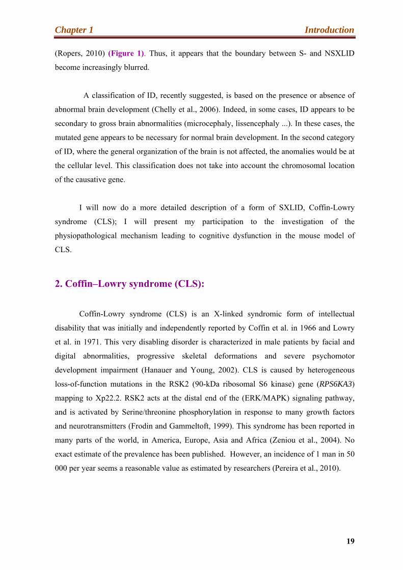
(Ropers, 2010) (Figure 1). Thus, it appears that the boundary between S- and NSXLID
become increasingly blurred.
A classification of ID, recently suggested, is based on the presence or absence of
abnormal brain development (Chelly et al., 2006). Indeed, in some cases, ID appears to be
secondary to gross brain abnormalities (microcephaly, lissencephaly ...). In these cases, the
mutated gene appears to be necessary for normal brain development. In the second category
of ID, where the general organization of the brain is not affected, the anomalies would be at
the cellular level. This classification does not take into account the chromosomal location
of the causative gene.
I will now do a more detailed description of a form of SXLID, Coffin-Lowry
syndrome (CLS); I will present my participation to the investigation of the
physiopathological mechanism leading to cognitive dysfunction in the mouse model of
CLS.
2. Coffin–Lowry syndrome (CLS):
Coffin-Lowry syndrome (CLS) is an X-linked syndromic form of intellectual
disability that was initially and independently reported by Coffin et al. in 1966 and Lowry
et al. in 1971. This very disabling disorder is characterized in male patients by facial and
digital abnormalities, progressive skeletal deformations and severe psychomotor
development impairment (Hanauer and Young, 2002). CLS is caused by heterogeneous
loss-of-function mutations in the RSK2 (90-kDa ribosomal S6 kinase) gene (RPS6KA3)
mapping to Xp22.2. RSK2 acts at the distal end of the (ERK/MAPK) signaling pathway,
and is activated by Serine/threonine phosphorylation in response to many growth factors
and neurotransmitters (Frodin and Gammeltoft, 1999). This syndrome has been reported in
many parts of the world, in America, Europe, Asia and Africa (Zeniou et al., 2004). No
exact estimate of the prevalence has been published. However, an incidence of 1 man in 50
000 per year seems a reasonable value as estimated by researchers (Pereira et al., 2010).
Chapter 1 Introduction
20
Figure 1: Genes implicated in X-Linked Mental Retardation (XLMR) and their position on the human X-chromosome.
Genes associated with syndromic XLMR are grouped on the left of the
chromosome, while non-syndromic XLMR are shown on the right. Colors relate to
different functional classes (Adapted from Ropers, 2010 and Stevenson and Schwartz,
2009).
2.1. Clinical Features:
The clinical symptoms associated with CLS are highly variable and generally more
severe in men than in women. Most of them settled gradually over the life, very few signs
being observed at birth.

Chapter 1 Introduction
21
2.2. CLS and psychomotor retardation:
The main characteristic of CLS is a cognitive impairment of variable severity, with
IQ that can range from 15 to 60. However, the majority of male patients have a profound ID
(Pereira et al., 2010). Development of speech is always impaired in CLS patients, with most
patients mastering only a few words. About 30% of patients have partial or total
sensorineural hearing loss. Some patients exhibit also epilepsy and/or cataplexy (drop
attacks). In early childhood patient’s exhibit generalized hypotonia. Despite limited verbal
skills, these individuals are communicative and cheerful. The average age at which affected
children do their first steps is usually extended to three years, (Zeniou et al., 2004).
Behavioral problems have also been reported in rare cases of female patients (Pereira et al.,
2010).
2.3. Morphological abnormalities:
Facial dysmorphism and skeletal abnormalities are the major morphological features
of CLS. The typical facial aspect of adult CLS patients includes hypertelorism, prominent
forehead and thick lips, palpebral fissures slanted downward and out, ears low and broad, a
thick nasal septum, thick lips, and missing teeth and poorly located (Pereira et al., 2010).
This feature becomes more pronounced with age.
At birth, the size of affected children is usually normal, growth retardation settling
gradually. An analysis of 250 patients showed that the average size of adult male patients
was 143cm (Hanauer and Young, 2002). Skeletal malformations include delayed bone
formation, spinal scoliosis or kyphosis and chest deformities (Pereira et al., 2010).
Patients have small fleshy hands with tapered fingers (Figure. 2). This is a
diagnostic clue because the characteristic shape of the hand is found at birth and is
relatively specific to the CLS. Spine deformities may become progressively worse and
often require surgery in childhood (Hanauer and Young, 2002; Hanauer, 2008).
Radiological defects have been observed in the skull, the spine or hands, including
hyperostosis of reduced inter-vertebral spaces and a reduction in size of the distal phalanges
(Pereira et al., 2010).

Chapter 1 Introduction
22
i hg
f e
d c a b
Figure 2: Morphology and Characteristics of the CLS patients.
(a–d) Facial views of a boy with CLS at different ages showing evolution during infancy of
facial gestalt. (a) At 9 months, (b) at 18 months, (c) at 3 years, and (d) at 6 years. Note the large
forehead, hypertelorism, downslanting Palpebral fissure , long philtrum, anteverted nares, and thick
lips. This boy carries an RPS6KA3 intra-genic duplication previously reported. (e–f) The facial
dysmorphism becomes more pronounced with age. (g-i) Views of the hands of the same patient.
Note the typical broad tapering fingers (g) at 9 months, (h) at 18 months, and (i) at 5 years (Adapted
from, Pereira et al., 2010 and Hanauer and Young, 2002).
2.4. Clinical expression in female:
Some of the physical features seen in males may also be present in female.
However, they are neither as prominent as in males nor as constant. Carrier females usually
exhibit a slight facial dysmorphism, small fleshy hands with tapering fingers and a
tendency to obesity (Figure 3). Cognitive impairment may not be present at all. However,
some heterozygous women have been reported to have difficulties at school. A few were

Chapter 1 Introduction
23
suffering from depression, psychotic behavior and schizophrenia. Psychiatric disorder
appeared around the age of 20 years and response to therapy was variable (Hanauer and
Young, 2002).
Figure 3: Facial and hand views of CLS patients in women.
Photographs (1-4) of four female patients suffering from CLS, showing clinical symptoms
of varying severity. A prominent forehead, hypertelorism and small fleshy hands with tapering
fingers can be observed. (Adapted from Jurkiewicz et al., 2010).
2.5. Diagnosis of CLS patients:
Since, the clinical findings can be very variable both in severity and in terms of
features, the diagnosis based on clinical criteria is often difficult to establish. On the other
hand, although some clinical signs are usually quite suggestive in adult patients, they are

Chapter 1 Introduction
24
much less at birth, in young children or women. There is also a great similarity of the
symptoms of CLS with other syndromes, such as alpha-thalassemia with ID (ATR-X) or
Williams Syndrome and Pitt-Hopkins syndrome. On the other hand, the high proportion of
sporadic cases does not facilitate the diagnosis. In fact, 70-80% of patients have no family
history. Thus, in most cases, the diagnosis requires molecular analysis (screening for
mutations in the Rsk2 gene) in order to confirm it by the identification of the genetic defect
(Pereira et al., 2010).
2.6. Treatment and Life expectancy:
There is no specific treatment currently available for the CLS. However, rapid
diagnosis can allow early regular monitoring of patients. Treatment for individuals with
CLS who experience drop attacks includes medication such as volporate and clonazepam or
selective serotonin uptake inhibitor. When symptomatic treatment are introduced very
early, clinical complications associated with symptoms such as deafness, seizures and heart
or/and orthopedic problems can be limited (Pereira et al., 2010).
Life expectancy of CLS patients is lower than the normal population. In 13.5% of
CLS male patients and 4.5% of carrier females death occurred between 13 and 34 years.
(Mean age of 20.5 years) (Pereira et al., 2010). The reported causes of death are heart
abnormalities (cardiomyopathy), pneumonia and surgical complications due to general
anesthesia (Hanauer and Young, 2002).
2.7. Molecular and Genetics Basis of CLS:
CLS is caused by the loss of function mutations in the RPS6KA3 gene, which maps
to Xp22.2. The coding region of this gene is split into 22 exons and encodes a
serine/threonine kinase: RSK2 (ribosomal S6 Kinase2) (Jacquot et al., 1998a). Over 140
distinct mutations associated with CLS have so far been listed in this gene (Pereira et al.,
2010). This number is constantly growing; a list of all these described mutations is
available online: (http://www-ulpmed.u-strasbg.fr/chimbio/diag/coffin). Mutations are
distributed throughout the gene, with no clustering and the vast majority is unique to a
single family. Approximately 30% of mutations are missense mutations, 15% nonsense

Chapter 1 Introduction
25
mutations, 20% splicing errors, and 30% short deletion or insertion events (Delaunoy et al.,
2006) (Figure 4 also explain the type of mutation). About two-thirds of these mutations
lead directly or indirectly to premature translation termination, resulting in complete loss of
function of the mutant allele. Misssense mutations often alter the catalytic activity of the
RSK2 protein. (Delaunoy et al. 2006; Delaunoy et al. 2006; Jurkiewicz et al., 2010).
3’UTR 1 2 3 4 5 6 7 8 9 10 11 12 13 14 15 16 17 18 19 20 21 22 5’UTR
Figure 4: Rsk2 gene structure and spectrum of mutations.
The Rps6ka3 gene comprises 22 exons, represented by rectangles numbered 1 to 22. Since
the identification of this gene, over 140 mutations associated with CLS have been recorded and
this number is constantly growing. The distribution of mutations (as identified in 2002) is shown
above (Adapted from Hanauer and Young, 2002).
Strikingly, two thirds of the mutations were de novo. This very high proportion of new
mutation is not common in an X-linked disease and is still unexplained (Hanauer and
Young, 2002).
2.8. Genotype/Phenotype Relationship:
No obvious correlation exists between phenotype and location or type of RPS6KA3
Large
Small insertion
Missense
Nonsense
Reading frame shift

Chapter 1 Introduction
26
mutation. However, individuals with certain missense mutations, leading only to partial loss
of kinase activity, tend to have milder disease expression (Delaunoy et al., 2001). For
example in one family classified as having a form of non-syndromic intellectual disability
segregated a missense mutation, which caused only a 80% reduction in ribosomal S6 kinase
enzyme activity, in contrast the majority of mutations in CLS patients that cause a total loss
of ribosomal S6 kinase enzyme activity (Merienne et al., 1999). Some other similar cases
were subsequently reported suggesting an essential role of residual enzyme activity in
determining severity of symptoms. However, this observation cannot be extended to all
known missense mutation. It is also interesting to note that phenotypic expression
variability within the same family was found in some families (Hanauer and Young, 2002).
Taken together, available data suggest that the mutation is not the only factor leading to the
phenotype but that other genes and environment factor are involved in this disease.
It should also be mentioned here that in 2005, a study suggested that truncating
mutations, either in, or upstream of the N-terminal kinase domain, might lead to
susceptibility to stimulus Induced Drop Attacks (SIDAs). (Nakamura et al., 2005).
However, this result could not be confirmed in the series of CLS patients analyzed in
Strasbourg.
3. The RSK Proteins:
RSK proteins are serine/threonine kinases, implicated in important and ubiquitous
signaling processes. RSKs constitute the protein ribosomal S6 kinase (RSKs) family. RSK
proteins are serine/threonine kinases acting at the end of the signaling Ras-ERK/MAPK
pathway and are actived by phosphorylation directly by ERK1/2 kinase in response to
extracellular stimulation by growth factor or neurotransmitters.
RSK proteins play a key role in several significant cellular processes including cell
growth and division, cell differentiation, cell cyclic, gene expression and apoptosis (Kang
and Chen, 2011).
3.1. Discovery of RSKs proteins:

Chapter 1 Introduction
27
The first RSK protein was purified in 1985 in Xenopus laevis oocytes and identified as a
serine proteins kinase (Erikson and Maller, 1985, 1986). Subsequently, RSKs proteins have
been identified in several vertebrate and invertebrate organisms. These include drosophila
melanogaster, C. elegan, chickien, rabbit, rat, mouse, and human. Whereas in C. elegans,
chicken, and rabbit, only one RSK is expressed (Alcorta et al., 1989; Wassarman et al.,
1994), in mammals four distinct RSK proteins have been identified, referred as RSK1-4
(Moller et al., 1994). The size of these protein kinases are very similar, they are made up of
735, 740, 733 and 745 amino acids respectively, and have a molecular weight of about
90kDa. Their amino acid composition is also very similar; they share 75-80% sequence
identity. The four RSK family members are encoded by distinct genes (official names
RPS6KA1, 2, 3, 4) mapping to chromosomes 3 (Rsk1), Xp22.2 (Rsk2), 6q27 (Rsk3), and
Xq21 (Rsk4) (Hanauer and Young, 2002).
3.2. Expression profile of RSKs proteins:
In humans and mice, RSKs proteins are widely expressed, and different RSK
proteins may be co-expressed in some tissues and organs. However, as stated below, their
expression profiles may be different, suggesting specific functions for each of these
kinases.
3.3. Expression profile in humans:
The expression of Rsks mRNAs was investigated in various adult human tissues by
Northern blot by Zeniou et al., 2002. RSKs are all widely expressed with, however,
variability in the levels. Results revealed a strong expression of Rsk1 in the kidneys, lungs,
pancreas, and in the cerebellum. Expressions of Rsk2 and Rsk3 are strong in skeletal
muscle, heart and pancreas. While Rsk2 messenger is strongly detected in the cerebellum,
the frontal lobe and the occipital pole, that of Rsk3 is predominant in the medulla. The
transcripts corresponding to Rsk4 were mainly found at high levels in the kidneys and brain
(Figure 5).
Another study examined Rsk2 expression during embryonic development (Zeniou et
al., 2002; Guimiot et al., 2004). It revealed that expression of RSK2 is strictly regulated,

Chapter 1 Introduction
28
spatially and temporally, during embryogenesis. Indeed, after being highly expressed in
many parts of the brain (hippocampus, ventricular zone) in the lungs and liver at nine
weeks of fetal life, the expression of Rsk2 decreases at the thirteenth week of
embryogenesis.
RSK1 3,5Kb
8,5Kb RSK2 3,5Kb
RSK3 7Kb
β-actin
Figure 5: Northern blot analysis of Rsk1, Rsk2 and Rsk3 expression.
Rsk1, 2 and 3 have been identified in all tissues analyzed by northern blot. However,
stronger levels of expression of Rsk1 and Rsk2 were found in the cerebellum. In all structures, two
RSK2 transcripts were visible. RSK3 showed strongest expressed in the medulla (Adapted from
Zeniou et al., 2002).
3.4. RSK1, RSK2 and RSK3 expression in adult mouse brain:
In adult mouse brain, RSK1 was primarily detected in the granular cell layer of the
cerebellum, whereas RSK2 showed the strongest expression in the hippocampus, an
essential structure in learning and memory (C1-C3) (Zeniou et al. 2002). In addition, high

Chapter 1 Introduction
29
levels of RSK2 mRNA were also detected in the neocortex, and purkinje cell layer and
some deep nuclei of the cerebellum. Darcq et al. (2011) reported also substantial RSK2
expression in the habenula. RSK3 was very highly expressed in the amygdala, the bed
nucleus and accumbens nucleus. In the hippocampus, RSK3 was detected in the dentate
gyrus (Zeniou et al., 2002), whereas RSK2 is highly expressed in the dentate gyrus and in
the CA1-3 areas. Rsk3 mRNA staining was also seen in external layers of the cortex and
several thalamic and hypothalamic nuclei (Zeniou et al., 2002) (Figure 6).
These studies showed that in particular RSK2 is highly expressed in humans and
mouse brain regions with high synaptic activity. These regions are key structures in the
process of learning and memory. The expression pattern of RSK2 suggested a function of
this gene in cognitive processes.
3.5. Sub-cellular localization of RSKs:
Localization of RSK proteins is also regulated at the sub-cellular level. They are
localized mainly in the cytoplasm at the baseline state. After mitogenic activation, a portion
of phosphorylated RSK molecules translocates into the nuclear compartment (Chen et al.,
1992). The exact mechanism of the translocation is not yet defined. A NLS sequence (core:
X-Lys-Lys-Leu-Arg-Arg-Lys-Ser-Arg,) has been identified in RSK3 (Zhao et al., 1995),
but not in the other RSK family members. However, there is no evidence so far that this
putative NLS is used in the transport of RSK3 to the nucleus (Anjum and Blenis, 2008).
Recent studies have localized RSK proteins also to the membrane and especially at the
presynaptic compartment (Zeniou-Meyer et al., 2008) and post-synaptic densities of
dendritic spines (Thomas et al., 2005). Together, the data suggest that RSK kinases play a
role in the whole cell.
3.6. Structure-Function Relationship of RSKs Proteins:
Comparison of the RSK structure and sequences to those of other serine/threonine

Chapter 1 Introduction
30
kinases, revealed that the four RSK family members have an unusual structure that they
share only with MSK kinases (Hauge and Frodin, 2006; Jones et al., 1988).
Figure 6: Expression profile of RSKs in the adult mouse brain, obtained by in situ
hybridization.
(A–F) Sagittal sections of adult brain. (B) Rsk1 expression was primarily observed in the
granular cell layer of the cerebellum. (D and F) Rsk2 expression in the Purkinje cell layer and
the deep nuclei of the cerebellum (arrows in D) and the hippocampus (arrows in F),
respectively. The weak staining observed in the dentate gyrus was not specific. (G–L) Frontal
sections of adult brain. (H) Arrows show Rsk3 expression in the dentate gyrus, the amygdala
and the pyriform cortex (Adapted from Zeniou et al., 2002).
In fact, they have two functionally distinct kinase domains separated by a linker
region. The N-terminal kinase domain (NTK) belongs to the AGC kinase family and is
responsible for phosphorylation of substrates. The C-terminal kinase domain (CTK)

Chapter 1 Introduction
31
belongs to the CamK family and its only known function is the activation of the
NTK.(Anjum and Blenis, 2008; Frodin et al., 2002; Hauge and Frodin, 2006.) (Figure
7A).
The N-terminal kinase domain (NKTD) is highly homologous to other kinases of
the AGC family, such as PKG, PKC, PKA or p70S6K (Bjorbaek et al. 1995b; Anjum
and Blenis, 2008). The NKTD phosphorylates RSK proteins substrates by recognizing a
consensus motif (RxRxxS or RRxS) (Flotow and Thomas, 1992; Leighton et al., 1995).
This same pattern is also recognized by the p70S6K. These two families of proteins
kinases share several substrates.
The three-dimensional structures of the NTKD of RSK1 and CTKD of RSK2 have
been resolved recently by Ikuta et al., (2007) and Malakhova et al. (2008). Each of these
two domains contains a binding site for ATP. The N-terminal region of each domain is
composed mainly of β sheets, while a majority of α helices form the C-terminal region.
The resolution of the structure and CTKD and NTKD allowed the authors to propose a
model for the regulation of the RSK enzymatic activity. Malakhova et al. (2008)
postulated that the location of the helix between helices αF, αL and G prevents
activation of RSK2. Following binding of ERK at the C-terminus of the RSK protein,
the hydrogen bond Tyr707-Ser603 (stabilizing the position of the helix αL) is broken,
leading to the relocation of this helix. This event allows the activation of CTKD by
repositioning the helix αD, alignment of the Glu500 residue with the ATP binding site
(this residue is indeed essential for the Binding of these molecules) and rearrangement
of the loop T opposite to the catalytic site (Figure 7C) NTKD and CTKD of RSK
proteins domains are connected by a 100 amino acids called "linker" (Pereira et al.,
2010). Phosphorylation of Ser386, in the linker, generates a docking site that recruits 3-
phosphoinositide-dependent protein kinase 1 (PDK1) (Hauge and Frodin, 2006). The
binding of the PDK1 protein is essential for the activation of the NTKD.
An ERK-docking motif known as the D domain (Leu-Arg-Gln-Arg-Arg) (Roux et
al., 2003) is present at the C-terminus. Binding of ERK to this motif is necessary for the
activation of RSK proteins. Thomas et al., (2005) identified another region present in all
RSKs at the C-terminus of the protein. This sequence (S-T-X-L, where X is an amino
Chapter 1 Introduction
32
acid) allows the binding of RSKs to the PDZ domain, an interaction domain found, in
particular, in many synaptic proteins (Figure 7A).
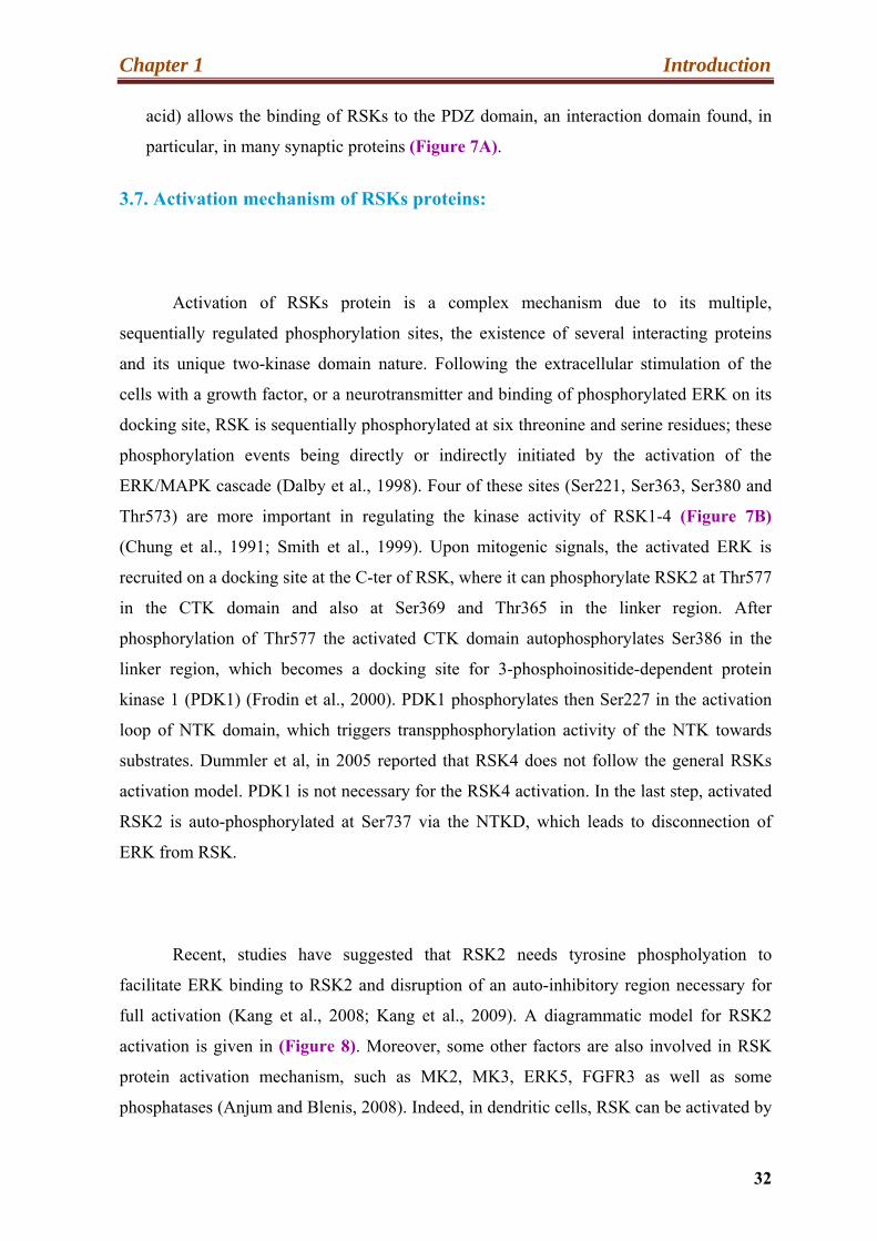
3.7. Activation mechanism of RSKs proteins:
Activation of RSKs protein is a complex mechanism due to its multiple,
sequentially regulated phosphorylation sites, the existence of several interacting proteins
and its unique two-kinase domain nature. Following the extracellular stimulation of the
cells with a growth factor, or a neurotransmitter and binding of phosphorylated ERK on its
docking site, RSK is sequentially phosphorylated at six threonine and serine residues; these
phosphorylation events being directly or indirectly initiated by the activation of the
ERK/MAPK cascade (Dalby et al., 1998). Four of these sites (Ser221, Ser363, Ser380 and
Thr573) are more important in regulating the kinase activity of RSK1-4 (Figure 7B)
(Chung et al., 1991; Smith et al., 1999). Upon mitogenic signals, the activated ERK is
recruited on a docking site at the C-ter of RSK, where it can phosphorylate RSK2 at Thr577
in the CTK domain and also at Ser369 and Thr365 in the linker region. After
phosphorylation of Thr577 the activated CTK domain autophosphorylates Ser386 in the
linker region, which becomes a docking site for 3-phosphoinositide-dependent protein
kinase 1 (PDK1) (Frodin et al., 2000). PDK1 phosphorylates then Ser227 in the activation
loop of NTK domain, which triggers transpphosphorylation activity of the NTK towards
substrates. Dummler et al, in 2005 reported that RSK4 does not follow the general RSKs
activation model. PDK1 is not necessary for the RSK4 activation. In the last step, activated
RSK2 is auto-phosphorylated at Ser737 via the NTKD, which leads to disconnection of
ERK from RSK.
Recent, studies have suggested that RSK2 needs tyrosine phospholyation to
facilitate ERK binding to RSK2 and disruption of an auto-inhibitory region necessary for
full activation (Kang et al., 2008; Kang et al., 2009). A diagrammatic model for RSK2
activation is given in (Figure 8). Moreover, some other factors are also involved in RSK
protein activation mechanism, such as MK2, MK3, ERK5, FGFR3 as well as some
phosphatases (Anjum and Blenis, 2008). Indeed, in dendritic cells, RSK can be activated by
Chapter 1 Introduction
33
the p38/MAPK signaling pathway via two proteins, MK2/3, which are homologous to the
RSK CTKD (Zaru et al., 2007).
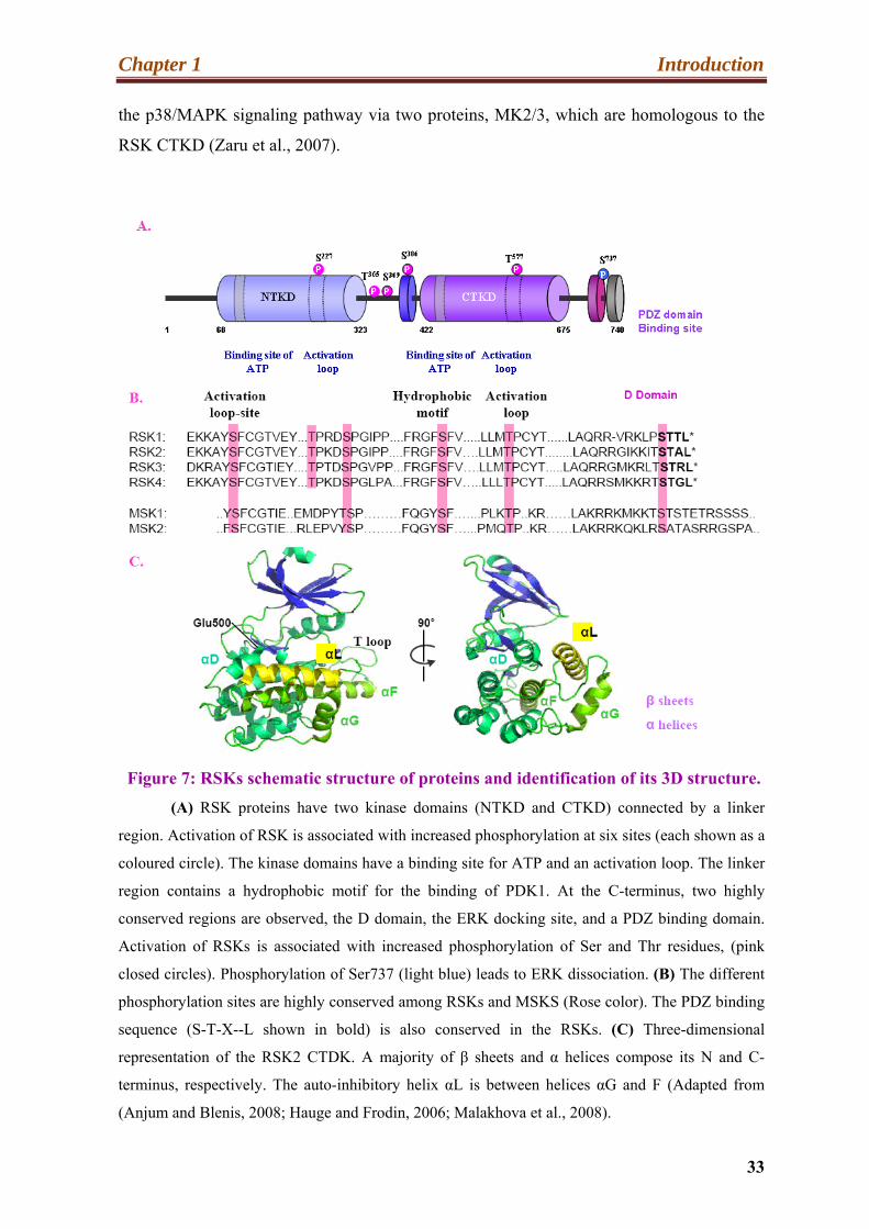
Figure 7: RSKs schematic structure of proteins and identification of its 3D structure.
(A) RSK proteins have two kinase domains (NTKD and CTKD) connected by a linker
region. Activation of RSK is associated with increased phosphorylation at six sites (each shown as a
coloured circle). The kinase domains have a binding site for ATP and an activation loop. The linker
region contains a hydrophobic motif for the binding of PDK1. At the C-terminus, two highly
conserved regions are observed, the D domain, the ERK docking site, and a PDZ binding domain.
Activation of RSKs is associated with increased phosphorylation of Ser and Thr residues, (pink
closed circles). Phosphorylation of Ser737 (light blue) leads to ERK dissociation. (B) The different
phosphorylation sites are highly conserved among RSKs and MSKS (Rose color). The PDZ binding
sequence (S-T-X--L shown in bold) is also conserved in the RSKs. (C) Three-dimensional
representation of the RSK2 CTDK. A majority of β sheets and α helices compose its N and C-
terminus, respectively. The auto-inhibitory helix αL is between helices αG and F (Adapted from
(Anjum and Blenis, 2008; Hauge and Frodin, 2006; Malakhova et al., 2008).

Chapter 1 Introduction
34
Ranganathan et al., (2006) also showed that ERK5 as well is able to phosphorylate RSKs.
This activation requires binding of the "common domain" of ERK5 with the domain D of
these kinases. A study in the laboratory of Chen has identified Tyr529 as another important
site for the activation of RSK. Indeed, phosphorylation of this residue by FGFR3 allows the
recruitment of ERK at the domain D and thus the activation of the protein (Kang et al.,
2007). In 2009, the same group reported that phosphorylation of Tyr707 by FGFR3 is
involved in activation of RSK2 CTKD, probably by disruption of a self-inhibitory left
handed helix (αL) (Kang et al., 2009).
In addition to the kinases, the activity of the RSK proteins may also be regulated by
protein phosphatases. The combination of PP2Cδ with the N-terminus of RSK2 leads, for
example, to a decrease in its kinase activity (Doehn et al., 2004). Another group described
an interaction between PP2Ac and RSK1, and observed PP2A phosphatase activity by
measuring dephosphorylation (Chaturvedi et al., 2009). RSK activated proteins can
phosphorylate various substrates both cytosolic and nuclear, and thus participate in
important cellular events such as neuronal development, regulation of transcription,
proliferation, gene expression and cell survival (Anjum and Blenis, 2008; Kang and Chen,
2011). MSK1 and -2, two kinases that are highly homologous to RSKs, can be
phosphorylated by both the MAPK/ERK and the p38/MAPK pathways (Hauge and Frodin,
2006).
Before detailing the regulation of different cellular processes mediates by RSKs, I
am going briefly to discuss the MAPKs signaling pathways, which are responsible to
activate the RSKs protein.
4. Signaling molecules involved in activation of RSKs proteins:
4.1. The MAPKs signaling pathways:
To be functional, a cell must be able to receive extracellular signals from other cells
and integrate them. To this end, cells have developed sophisticated signaling pathways to
Chapter 1 Introduction
35
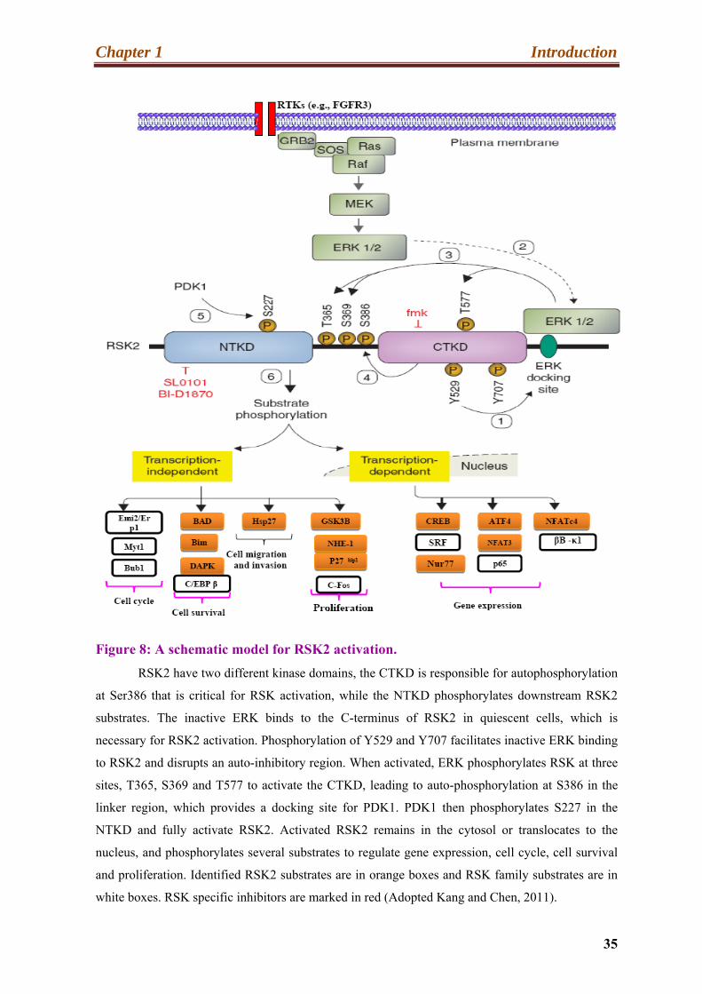
Figure 8: A schematic model for RSK2 activation.
RSK2 have two different kinase domains, the CTKD is responsible for autophosphorylation
at Ser386 that is critical for RSK activation, while the NTKD phosphorylates downstream RSK2
substrates. The inactive ERK binds to the C-terminus of RSK2 in quiescent cells, which is
necessary for RSK2 activation. Phosphorylation of Y529 and Y707 facilitates inactive ERK binding
to RSK2 and disrupts an auto-inhibitory region. When activated, ERK phosphorylates RSK at three
sites, T365, S369 and T577 to activate the CTKD, leading to auto-phosphorylation at S386 in the
linker region, which provides a docking site for PDK1. PDK1 then phosphorylates S227 in the
NTKD and fully activate RSK2. Activated RSK2 remains in the cytosol or translocates to the
nucleus, and phosphorylates several substrates to regulate gene expression, cell cycle, cell survival
and proliferation. Identified RSK2 substrates are in orange boxes and RSK family substrates are in
white boxes. RSK specific inhibitors are marked in red (Adopted Kang and Chen, 2011).

Chapter 1 Introduction
36
receive extra-cellular signals at the membrane, to transmit the information to various
intracellular effector proteins and finally to respond to these stimuli. The vast majority of
these signal transduction mechanisms are dependent on post-translational modifications
such as phosphorylation. Theses signaling pathways, allowing enzymatic amplification of
the signal, provide mechanisms for the integration of different inputs and allow a diverse
array of cellular responses to take place. Mitogen-activated protein kinases (MAPKs) are
Ser/Thr kinases that convert extra-cellular stimuli into a broad range of cellular responses.
The MAPK pathway is a cascade of proteins in the cell that transmits signals
received by a receptor at the surface of the cell to various cytosolic and nuclear effectors,
necessary to perform the numerous functions of the cell. The signal initiates when a growth
factor binds to a specific receptor on the cell surface and ends when a cytosolic or a nuclear
effector is activated and produces some changes in the cell, leading for instance to cell
division. The MAPKs phosphorylate many substrates; including members of a family of
protein kinases termed MAPK activated protein kinases (MAPKAPKs). This family
includes the RSKs, MSKs, MNKs, MK5 and MK2/3. MAPKAPKs (Arthur, 2008; Buxade
et al., 2008; Carriere et al., 2008; Gaestel, 2008; Perander et al., 2008; Ronkina et al., 2008;
Roux and Blenis, 2004). The MAPKAPK family represents an extra enzymatic and
amplification step in MAPK catalytic cascades. MAPKs pathway are ancient and
extensively studied signal transduction pathways, which are used in many physiological
processes (Widmann et al., 1999). MAPKs are serine/ threonine protein kinases
ubiquitously found in eukaryotes. They are involved in important cellular processes such as
proliferation, differentiation, cell survival, mitosis, metabolism, motility; regulate gene
expression and apoptosis (Cargnello and Roux, 2011; Dhillon et al., 2007; Torii et al.,
2006).
The mammalian MAPKs consist of three kinases families, including extracellular
signal regulated kinase (ERK), (p38) and c-Jun N-terminal kinase (JNK). Each of these
families is represented by several isoforms: eight ERK isoforms, (ERK1-8), four p38
isoforms (p38α, β, γ, δ) and three JNK isoforms (JNK1/2/3) have been reported in the
literature (Bogoyevitch and Court, 2004; Bogoyevitch et al., 2010; Cuadrado and Nebreda,
2010; Kim and Choi, 2010). Splicing variants have also been identified, including for
example ERK 1b and 1c (Raman et al., 2007).

Chapter 1 Introduction
37
There are three main kinases acting in the MAPK pathways, including MAPK, MAPK
kinases (MAPKK), and MAPKK kinases (MAPKKK). The MAPKKK is activated by a
small GTP-binding protein of the Ras/Rho family in response to extracellular stimuli. The
activated MAPKKK phosphorylates and activates then the MAPKK, which in turn
phosphorylates and activates the MAPK through dual phosphorylation at Thr and Tyr
residues within a conserved Thr-X-Tyr motif situated in the activation loop of the kinase
domain. (Kim and Choi, 2010; Cargnello and Roux, 2011). MAPKs can then activate many
cytosolic and nuclear substrates, by phosphorylating Ser/Thr residues located in proline rich
regions (Raman et al., 2007). Diagrammatic representation of the MAPKs module, which
leads to activate downstream targets are shown in (Figure 9).
To be able to transmit the signal, the various MAP Kinases must be close to each
other, because the cascade of phosphorylation requires interaction between the different
kinases. These interactions are stabilized by formation of a protein complex. The formation
of such a complex requires the use of scaffolding proteins (Morrison and Davis, 2003).
Activation of the MAPK/ERK pathway is, for example dependent on KSR. Indeed, it has
been shown that KSR1 allows the localization of MAPKK/ MEK with the activated form of
Raf-1 at the plasma membrane, forming a docking area for MAPK/ERK. KSR1 in this
manner facilitates the phosphorylation cascade leading to ERK activation (Cacace et al.,
1999, Müller et al., 2000).
The three MAPK families are activated by different intra and extra cellular stimuli.
The JNK and p38 signaling pathways are activated by pro-inflammatory cytokines such as
tumor necrosis factor (TNFα) and interleukin (IL-1β) or in response to cellular stresses such
as genotoxic, osmotic, hypoxic, or oxidative stress, whereas the Ras-ERK pathway is
mainly stimulated by growth factors, hormones and neurotransmitters (Raman et al., 2007,
Kim and Choi, 2010).
Finally, MAPKs regulate a broad range of biological functions and control a vast
array of physiological processes.
Chapter 1 Introduction
38
4.2. The ERK1/2 MAPKs pathway:
Extracellular signal-regulated kinase 1 (ERK1) was the first mammalian MAPK to
be identified, followed by ERK2. ERK1 and ERK2 share 85% amino acid residues identity
(Lopez-Bergami, 2011). ERK1 has a molecular weight of 44 kDa and ERK2 42 kDa.
ERK1/2 are ubiquitously expressed, with the highest levels in brain, heart, thymus and
skeletal muscle (Boulton et al., 1990).
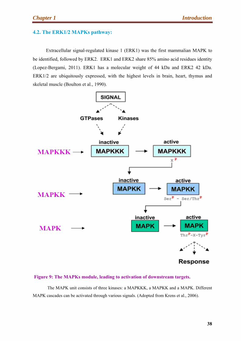
Figure 9: The MAPKs module, leading to activation of downstream targets.
The MAPK unit consists of three kinases: a MAPKKK, a MAPKK and a MAPK. Different
MAPK cascades can be activated through various signals. (Adopted from Krens et al., 2006).
Chapter 1 Introduction
39
ERK1/2 are activated by growth factors, platelet-derived growth factor (PDGF), epidermal
growth factor (EGF), and nerve growth factor (NGF) (Boulton et al., 1990).
In addition, ERKs can also be activated by cytokines, osmotic stress, virus infection,
and ligands for heterotrimeric G protein-coupled receptors (Raman et al., 2007). Moreover,
it was shown that transforming agents and carcinogens could also activate ERK1/2
(Johnson and Lapadat, 2002). The ERK1/2 module in mammals includes A-Raf, B-Raf and
Raf-1 (MAPKKKs), MEK1 and MEK2 (MAPKKs) and ERK1 and -2 (MAPKs). Receptor-
linked tyrosine kinases such as, for example, the epidermal growth factor receptor (EGFR)
are stimulated by extracellular ligands. Binding of epidermal growth factor (EGF) to the
EGFR stimulates the tyrosine kinase activity of the cytoplasmic domain of the receptor.
The EGFR becomes phosphorylated on tyrosine residues. Docking proteins such as GRB2
contains an SH2 domain that binds to the phosphotyrosine residues of the activated receptor
(Schulze et al., 2005). Then GRB2 binds to the guanine nucleotide exchange factor SOS
through the two SH3 domains of GRB2. When the GRB2-SOS complex docks to
phosphorylated EGFR, SOS turns into its activated form (Karnoub and Weinberg, 2008;
Zarich et al., 2006). Activated SOS then promotes the removal of GDP from a member of
the Ras subfamily. Ras then binds GTP and becomes active. Ras activates Raf, which in
turn binds to and phosphorylates the dual specificity kinases MEK1 and MEK2, which, as a
result, phosphorylate ERK1/2. Now activated ERK1/2 can phosphorylate many cytoplasm
and nuclear substrate proteins. (Yoon and Seger, 2006), including RSKs. ERK1/2 can
phosphorylate more than 160 proteins (Roberts and Der, 2007; Yoon and Seger, 2006). In
the nucleus, ERK1/2 regulate gene expression by phosphorylating several transcription
factors such as Sp1, E2F, Elk-1, AP-1 , NF-AT , STAT3 and c-Fos etc (Murphy and Blenis,
2006; Yoon and Seger, 2006). For example, Elk1 a member of the TCF family is
phosphorylated by ERK at Ser383 and 389 in its C-terminal domain. This phosphorylation
increases the activity of Elk-1 DNA binding and facilitates its interaction with the co-
activator p300. It was also shown (Li et al., 2003) that this modification stimulates the
acetyltransferase activity of the Elk-1-p300 complex that is critical for chromatin
remodeling and gene activation. Elk-1 is, implicated in the regulation of immediate-early
(IE) genes expression, such as c-Fos (Gille et al., 1995). ERK1/2 stabilize also the c-Fos
protein through direct phosphorylation (Murphy et al., 2002), thus permitting c-Fos to bind
to c-Jun and form transcriptionally dynamic AP-1 complexes. AP-1 activity is required for
expression of cyclin D1 (Shaulian and Karin, 2001) and, thus, allow G1/S transition and

Chapter 1 Introduction
40
cell cycle progression. ERK1/2 plays an essential role in the control of cell proliferation
(Katz et al., 2007). Through numerous mechanisms, including induction of positive
regulators of the cell cycle (Meloche and Pouyssegur, 2007). Some studies indicated that
ERK would act as a negative regulator for cell proliferation and can induce apoptosis when
its activity is highly increased (Nakata et al., 2011). However, Campbell et al. in 2010
reported that a secondary metabolite, TLN-4601, inhibits Ras-ERK signaling and decreases
cell viability by increasing apoptosis. Ras/ERK has also been implicated in migration and
cell motility by regulating cytoskeleton proteins. Erk1/2 activates by phosphorylation the
myosin light chain kinase (MLCK), and active myosin promotes the polymerization of
actins fibers (Katz et al., 2007) and the formation of membrane extensions necessary for
cell migration. The ERK/MAPK pathway and their target substrates are shown in (Figure
10).
4.3. The p38 MAPK pathway:
The p38 protein belongs to an other important MAPK pathway. Initially four
groups identified p38 as a 38kDa protein (p38). They observed quick phosphorylation at a
tyrosine residue in response to lipopolysaccharide (LPS) stimulation (Han et al., 1994).
The p38 is also known under various other names like CSBP, RK, and SAPK2. This
MAPK member is generally more responsive to stress stimuli and shows 50% identity with
ERK2 (Lee et al., 1994; Rouse et al., 1994). The p38 MAPK has four isoforms (p38α, β, γ,
δ), that are approximately 60% identical in their amino acid sequence. Distinct genes
encode the different p38 isoforms, which have a different expression pattern in the cell.
The first two isoforms (p38α, β) are ubiquitously expressed whereas the last two (p38γ, δ)
have more restricted expression patterns and have some specific functions (Jiang et al.,
1996).
Because p38α is expressed at higher levels than p38β, the majority of the published
literature on p38 MAPKs refers to the former. In mammals, the four-p38 isoforms are
activated by stress and inflammatory cytokines, including UV irradiation, ischemia, and
interleukin-1. In addition, the p38 isoforms are also activated via G protein-coupled
receptors: Rho family GTPases and Cdc42 (Bagrodia et al., 1995). MKK3 and MKK6 are
the MAPKK responsible for p38 MAPK activation (Derijard et al., 1995; Stein et al., 1996).
Chapter 1 Introduction
41
The p38 isoforms are present in the cytoplasm and nucleus and play a significant role in
normal immune and inflammatory responses (Cuadrado and Nebreda, 2010).
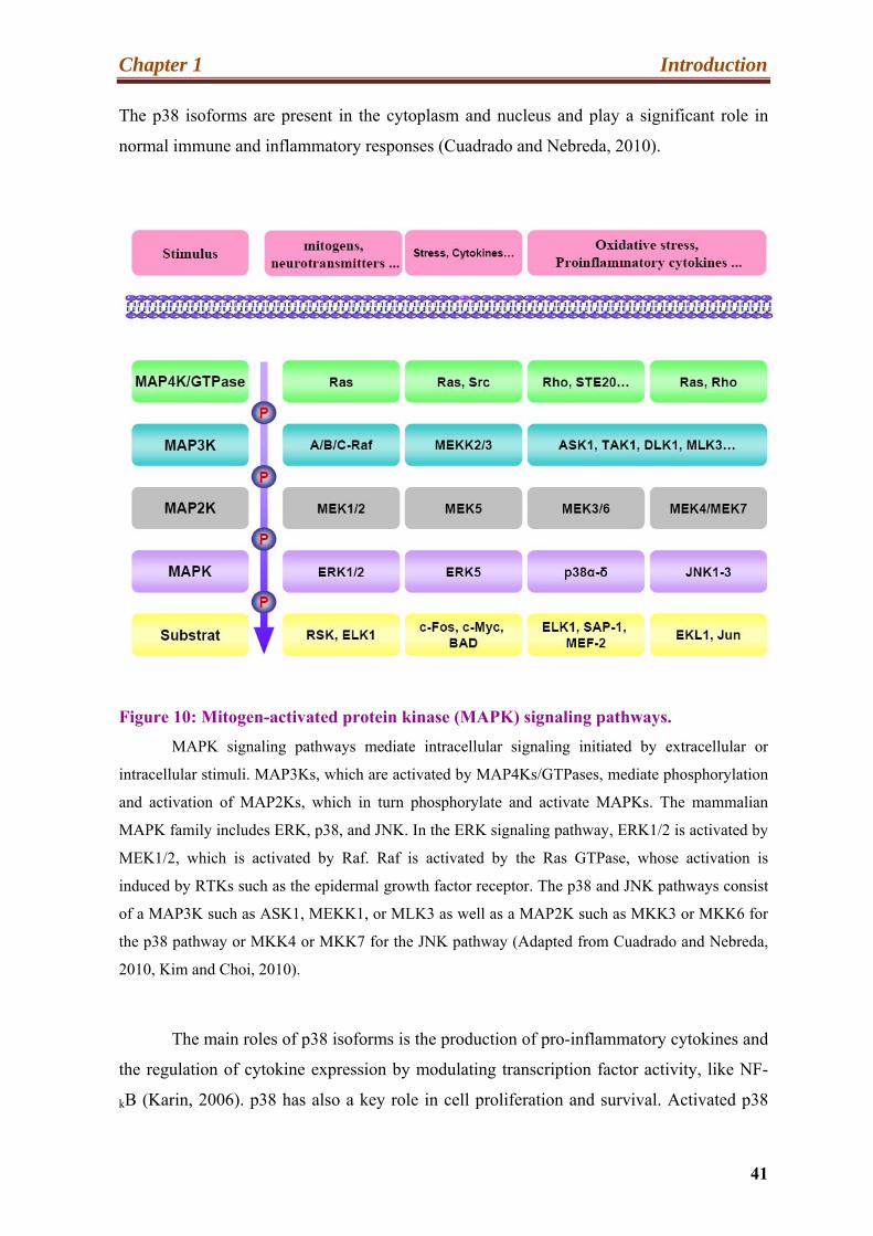
Figure 10: Mitogen-activated protein kinase (MAPK) signaling pathways.
MAPK signaling pathways mediate intracellular signaling initiated by extracellular or
intracellular stimuli. MAP3Ks, which are activated by MAP4Ks/GTPases, mediate phosphorylation
and activation of MAP2Ks, which in turn phosphorylate and activate MAPKs. The mammalian
MAPK family includes ERK, p38, and JNK. In the ERK signaling pathway, ERK1/2 is activated by
MEK1/2, which is activated by Raf. Raf is activated by the Ras GTPase, whose activation is
induced by RTKs such as the epidermal growth factor receptor. The p38 and JNK pathways consist
of a MAP3K such as ASK1, MEKK1, or MLK3 as well as a MAP2K such as MKK3 or MKK6 for
the p38 pathway or MKK4 or MKK7 for the JNK pathway (Adapted from Cuadrado and Nebreda,
2010, Kim and Choi, 2010).
The main roles of p38 isoforms is the production of pro-inflammatory cytokines and
the regulation of cytokine expression by modulating transcription factor activity, like NF-
kB (Karin, 2006). p38 has also a key role in cell proliferation and survival. Activated p38

Chapter 1 Introduction
42
phosphorylates many substrates in the cytoplasm such as, MNK1/2, MK2/3, phospholipase
A2, microtubule associated protein Tau and Bax and nuclear targets including Elk-1, p53,
Ets1, ATF2 and NF-kB (Cuadrado and Nebreda, 2010; Kyriakis and Avruch, 2001).
Thornton and Rincon in 2009 reported that p38α negatively regulates cell cyclic
progression at both G1/S and G2/M transition through a number of mechanisms, including
up-regulation of CDK inhibitor and down-regulation of cyclins.
The p38 activity also involves the induction of apoptosis by cellular stresses. These
function can be mediated via transcriptional and posttranscriptional modification
mechanisms, affecting activity of pro-apoptotic and anti- apoptotic proteins of the Bcl-2
family, and survival pathway (Cuenda and Rousseau, 2007).
Moreover, many reports have established the implication of p38 MAPK in
numerous other biological processes, including, chromatin remodeling, protein degradation,
mRNA stability, endocytosis, cytoskeleton dynamics or cell migration (Cuadrado and
Nebreda, 2010).
4.4. The JNK pathway:
The c-jun N-terminal kinases are also MAPK members. JNKs, also known as stress
activated kinase protein (SAKP), were originally purified using c-jun protein bound to
beads (Hibi et al., 1993; Kyriakis and Avruch, 1990). It was subsequently found that stress
leads to JNK phosphorylation at Thr183 and Tyr185 residues. JNK has three isoforms,
JNK1-3.
The three isoforms are more than 85% identical and are encoded by distinct genes,
giving rise to more than 10 spliced forms ranging from 46 to 55 kDa (Gupta et al., 1996;
Kyriakis et al., 1994). JNK1 and 2 are ubiquitously expressed (Bode and Dong, 2007a),
while JNK3 is expressed primarily in neuronal tissues and cardiac myocytes (Bode and
Dong, 2007b). JNKs are strongly activated by cellular stress, heat shock, oxidative stress,

Chapter 1 Introduction
43
UV radition, DNA damming agents, growth factor deprivation and cytokines (Bogoyevitch
et al., 2010). Activation of JNK isoforms needs dual phosphorylation at Thr and Tyr
residues inside a conserved Thr-Pro-Tyr (TPY) motif in their activation loops. The
MAPKKs catalyzing this reaction include MKK4 and MKK7 (Lawler et al., 1998).
Following stimulation, activated JNKs relocalize from the cytoplasm to the nucleus
(Mizukami et al., 1997). The most important JNK substrate is the transcription factor c-jun.
JNK phosphorylates c-jun at Ser63/73 (Weston and Davis, 2002). Some studies revealed
differences between JNKs isoforms functions with regard to the regulation of c-jun
(Sabapathy et al., 2004). Jaeschke et al., in 2006 and Bogoyevitch et al., in 2010, reported
that JNKs are positive regulators of c-jun expression as well as cell proliferation. Several
other transcription factors are also phosphosrylated by JNKs, including Elk-1, p53, HSF-1,
ATF-2. c-Myc and STAT3. In addition, JNK activity, through c-jun, promotes AP-1
complex formation and transcription of genes including AP-1 binding sites, such as the
cyclin D1 gene (Sabapathy et al., 2004). Park et al. in 2007 suggested an anti-apoptotic
function for JNK. Indeed, they observed that phosphorylation by JNK of a mitochondrial
pro-apoptotic protein, SMAC, prevents the action of the inhibitor of apoptosis, XLAP. This
leads to inhibition of the activity of Caspase3 and thus apoptosis. However, the role of the
JNK signaling pathway in the processes of cell survival is still controversial. The JNK
pathway also regulates molecular mechanisms controlling autophagy (Bogoyevitch et al.,
2010). JNKs also participate in the regulation of protein degradation and of the cell cycle,
via Mcl-1 and p27kip respectively. Finally, JNKs play a role in other important cellular
events including protein degradation, neurodegeneration and cytokines production
(Oltmanns et al., 2003; Sherrin et al., 2011).
5. MAPKs cascade signals involved in synaptic plasticity:
5.1. ERK1/2 MAPK and synaptic plasticity:
ERK is the most important neuronal regulator and in the central nervous system, it
has a critical role in learning and memory formation. ERK plays a key role in the regulation
of neuronal plasticity by direct actions on synaptic effectors or through modifying gene

Chapter 1 Introduction
44
expression. ERK is also implicated in brain long-term adaptive alteration underlying
striatum-dependent behavioral plasticity. In mature neurons, ERK1/2 are activated in
synaptic activity, and are crucial for synaptic plasticity associated to learning and memory
formation in mammals (Davis and Laroche, 2006). Neuronal ERK1/2 activation in
response to direct membrane depolarization or glutamatergic signaling (Zhu et al., 2002),
proceeds via a specific way. ERK activation induced via these stimuli is Ras-dependent, but
activation of Ras in response to these signals requires calcium influx (Walker et al., 2003).
This calcium influx can be made possible by NMDA (N-methyl- D-aspartate) type
glutamate receptors or voltage-gated calcium channels. Precisely how high calcium levels
trigger Ras activation is yet to be elucidated. Previous studies demonstrated an absolute
requirement for ERK1/2 activity in induction of LTP, and more studies that are recent have
established its requirement for NMDA dependent and independent forms of LTP induction
and maintenance. Thomas and Huganir (2004) have also shown that regulation of AMPA
receptor activity is ERK-dependent. Recently Harvey et al., (2008), reported also that
glutamate stimulation of a single dendritic spine resulted in continued alteration of the
volume of the stimulated spine and subsequent enlargement of adjacent spines that was
clearly dependent upon ERK activation. This study argued that such mechanisms might be
the basis of the development of LTP. ERKs control synaptic plasticity through their
participation to the regulation of transcription and translation. ERK have also newly
appreciated functions in memory consolidation. Eckel et al., in 2008 reported, for example,
that there is circadian oscillation of ERK activation in the hippocampus, which is necessary
for persistent and stable memory formation.
Davis and Laroche, in 2006 reported behavioral analyses on ERK inhibitor treated
mice. They demonstrated that ERK activity is crucial for learning and memory tasks (Davis
and Laroche, 2006). Mazzucchelli et al (2002) and Selcher et al, (2001) reported ERK1-KO
mice showing only a slight behavioral phenotype. However, the mutant animals showed a
generalized behavioral excitement phenotype with altered responses to psychostimulant
drugs like amphetamine (Engel et al., 2009). On the other hand, ERK1 deficient mice show
a paradoxical improvement in a Striatal-based long-term memory task and facilitation of
LTP in the accumbens nucleus (Mazzucchelli et al., 2002). Results of another study
suggested compensatory action of ERK2 (Kumar et al., 2008). A more extensive behavioral

Chapter 1 Introduction
45
analysis of these animals is ongoing. Satoh et al., in 2007 reported that ERK2 knockout is
embryonically lethal, but studies based on ERK2-hypomorphic mutant mice, in which
ERK2 expression was partially (20–40%) reduced, showed a significant deficits in long-
term memory. Moreover, in 2008, Samuels and co-worker reported that mice in which
ERK2 was deficient in telencephalic radial glial progenitor’s, exhibit intense shortage in
associative learning in a fear conditioning assay. A number of studies suggested that
ERK1/2 play an important role in the genesis of neural progenitors. They also suggested
that ERK1/2 have a death promoting apoptotic roles in neuronal cells. Other studies
reported that up regulation of ERK1/2 is critically involved in the pathogenesis of autism
(Yang et al., 2011).
It is still controversial whether the ERK isoforms, (ERK1 and ERK2), redundantly
share their numerous brain functions and compensate for each other or whether they play
distinct roles. In the ERK signaling studies, most experiments use inhibitors of the upstream
kinase, MEK. ERK1 and 2 are both activated by MEK, and thus, it is difficult to examine
the specific contribution of each isoform to physiological functions. All previous reports
suggested that even a small dysregulation of ERK function is adequate to result in
behavioral impairment. Finally, the various neuro-modulators effects on LTP and LTD
induction depend on ERK signaling. Indeed, ERKs are needed for long-term memory
formation across a variety of tasks.
6. Regulation of cellular processes by RSKs:
Since initial identification of RSK in xenopus, many proteins have been identified
as substrates for RSK kinases. These proteins have diverse biological functions in the cell
and may reside and function in different sub-cellular compartments, including the plasma
membrane, the cytosol and the nucleus. The substrate specificity of RSKs has been
determined with a synthetic peptide library, and is defined by the motifs that are mentioned
above in this manuscript. However, these sequences are also recognized by other protein
kinases such as AKT/PKB, S6K1/2 and MSKs (Anjum and Blenis, 2008). A number of
RSK functions can be assumed from the nature of RSKs substrates. Some reported data

Chapter 1 Introduction
46
suggest that RSKs are, in particular, implicated in transcription and translation regulation,
as well as in synaptic transmission. Overviews of a diagrammatic representation of RSK
substrates are shown in (Figure 11).
6.1. RSK mediates the regulation of transcription:
As mentioned earlier in the introduction, activated RSKs phosphorylate numerous
transcription factors, some of which contributing to the immediate early genes (IEGs)
response or being IEG gene products themselves. Two studies using primary fibroblasts
isolated from RSK2 deficient mice and human cells from CLS patients demonstrated that
RSK2 mediates mitogen- induced c-Fos transcription (Bruning et al., 2000; De Cesare et
al., 1998).
The mechanism proposed included activation of the Elk-1/serum response factor
complex and phosphorylation of the cAMP response element-binding protein (CREB) via
RSK2. Ginty et al. (1994) and Xing et al. (1996) reported that CREB is phosphorylated by
RSK2 at Ser133; However, Wiggin and colleagues in 2002 reported that not RSK2 but
MSK is the predominant CREB kinase in somatic cells. However, there data also suggested
that RSK and PKA could cooperate with MSK1 and -2 to phosphorylate CREB. The same
group reported also that histone H3, previously reported to be phosphorylated by RSK2 at
Ser4 (Sassone Corsi et al., 1999) is in fact predominantly phosphorylated by MSK1/2
kinase in response to both mitogenic stimulation and stress. RSK phosphorylates also
directly SRF (Rivera et al., 1993), c-Fos (Chen et al., 1993), and Nur77 (Davis et al., 1993;
Wingate et al., 2006). In addition, RSK co-operates with the ETS transcription factor ER81
and increases ER81 dependent transcription as a result of the phosphorylation of Ser216
and Ser191 (Wu and Janknecht, 2002). The ER81 factor is implicated in several essential
functions including, homeostasis signaling response, and development, potentially
connecting RSK with these processes. The transcription initiator factor TIF-1A is also
phosphorylated via RSK2 (at two Serine residues) (Zhao et al., 2003), an event which was
shown to be necessary for RNA polymerase I transcription and rRNA synthesis. These
results suggested that RSK signaling controls growth associated transcription initiation. In
addition, RSK phosphorylates ERα at Ser167, thereby enhancing ERα mediated
transcription (Yamnik and Holz, 2010).
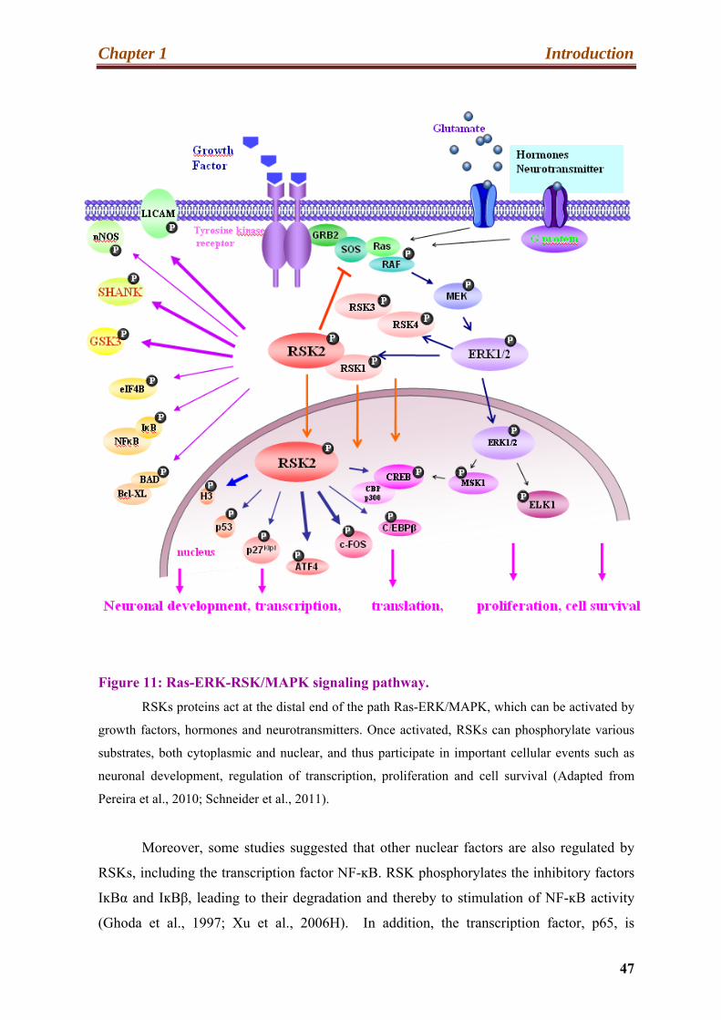
Chapter 1 Introduction
47
Figure 11: Ras-ERK-RSK/MAPK signaling pathway.
RSKs proteins act at the distal end of the path Ras-ERK/MAPK, which can be activated by
growth factors, hormones and neurotransmitters. Once activated, RSKs can phosphorylate various
substrates, both cytoplasmic and nuclear, and thus participate in important cellular events such as
neuronal development, regulation of transcription, proliferation and cell survival (Adapted from
Pereira et al., 2010; Schneider et al., 2011).
Moreover, some studies suggested that other nuclear factors are also regulated by
RSKs, including the transcription factor NF-ĸB. RSK phosphorylates the inhibitory factors
IĸBα and IĸBβ, leading to their degradation and thereby to stimulation of NF-ĸB activity
(Ghoda et al., 1997; Xu et al., 2006H). In addition, the transcription factor, p65, is

Chapter 1 Introduction
48
phosphorylated at Ser563 by RSK (Bohuslav et al., 2004; Zhang et al., 2005). The role of
this phosphorylation event remains still a matter of discussion (Douillette et al., 2006).
RSK2 phosphorylates the CREB family member ATF4 (Yang et al., 2004), a transcription
factor that is necessary for osteoblast differentiation during development. Indeed, RSK2
protein was found to be required for osteoblast differentiation and function, suggesting a
mechanism by which loss of RSK2 activity leads to the CLS related skeletal abnormalities.
The transcriptional co-activator CREB binding protein (CBP) has also been recognized as a
binding target of RSK1 (Nakajima et al., 1996). Activation of the Ras/ERK pathway
promotes interaction between CBP and RSK (Wang et al., 2003), but the precise role of this
connection remains to be determined. Some investigations suggested that CBP cooperates
with transcription factors that are phosphorylated via RSK1 and RSK2, including CREB, c-
Fos, ERα, NF-KB and ER81. These data suggest that RSK may facilitate the recruitment of
CBP to promote and regulate these transcription factors.
Together, all the published data support an important implication of RSKs in gene
expression. This prompted us to study the transcriptome from Rsk2-KO mice and our
results further support this implication.
6.2. Regulation of Cell growth and synthesis of protein:
A number of studies revealed the involvement of the Ras/ MAPK signaling pathway
in translation regulation, namely by increasing the rates of translation initiation and
elongation (Holland et al., 2004). It was also shown that following stimulation of
metabotropic receptors in the hippocampus, RSKs translocate into the synaptoneurosomes
fraction where they associate with polyribosomes. Once activated, RSK inhibits by
phosphorylation the constitutionally active kinase GSK3β. This allows activation of the
initiation translation factor eIF2B and thus translation.(Angenstein et al., 1998).
RSK also phosphorylates another important component of the translation
machinery, the eukaryotic translation initiation factor eIF4B (Shahbazian et al., 2006).
Following phosphorylation of the Ser422 residue, eIF4B is recruited to the eIF3 translation
initiation complex (Figure 11). The eIF4B is then able to increase the helicase activity of
eIF4E, thereby stimulating the activity of eIF4F. This later protein interacts then with the

Chapter 1 Introduction
49
initiation complex and facilitates the translation by linking the mRNA and the ribosome.
RSK proteins regulate other factors involved in translation, including EF2K and TSC2
(Anjum and Blenis, 2008; Inoki et al., 2006) indicating their crucial role in the mechanisms
of protein synthesis (Anjum and Blenis, 2008).
Finally, Ras/MAPK signaling was also found to be involved in the stimulation of
activity of mammalian targets of rapamycin (mTOR) via the regulation of the tuberous
sclerosis complex. The protein mTOR is a main regulator of cell growth and proteins
synthesis, and its activity is controlled through various growth related pathways. RSK can
phosphorylate Tuberous sclerosis protein-2 at Ser664 and Ser1798, an event which
negatively regulates the Guanine nucleotide activating Protein (GAP) activity of Tuberous
sclerosis protein-2 towards the small GTPase Rheb (Rolfe et al., 2005; Roux et al., 2004).
6.3. Cell cycle regulation via RSKs:
The cell cycle is a four-step mechanism (G1, S, G2, and M), leading to division of a
mother cell in two daughter cells. All steps in the cell cycle are regulated by many factors;
including RSKs. RSK2 was shown to promote G1-phase progression by phosphorylating
the Cyclin-dependent kinase 2 (CDK2) inhibitor p27kip1 at the Thr198 residue (Fujita et al.,
2003; Larrea et al., 2009). RSK mediated phosphorylation of p27kip1 promotes its association
with protein 14-3-3, which prevents its translocation to the nucleus (Fujita et al., 2003). In
addition, RSK2 is an important regulator of cell transformation, since ectopic expression of
RSK2 was found to increase proliferation and anchorage independent transformation (Cho
et al., 2007). Kang and his group in 2007 reported a role of RSK2 in tumorigenesis, FGFR3
has been shown to promote hematopoietic transformation through activation of RSK2 in a
two-step manner: facilitation of ERK-RSK2 interaction and subsequent phosphorylation of
RSK2 by ERK (Kang et al., 2007). By using the Xenopus oocyte model system, RSK2 was
shown to contribute to the control of the meiotic cell cycle (Schmitt and Nebreda, 2002).
RSK2 was also shown to participate in the progression of oocytes through the G2/M phase
of meiosis I via inhibition by phosphorylation of the Myt1 kinase (Gross et al., 2001; Ruiz
et al., 2010). It remains unknown whether this mechanism is conserved in other species, but
in 2002, the Okumura’s group demonstrated that Akt can also act as a Myt1 kinase in
starfish oocytes (Okumura et al., 2002).

Chapter 1 Introduction
50
Cho et al., in 2005 reported the involvement of RSK2 in regulating the activity of
the p53 protein. The tumour suppressor p53 is a transcription factor acting as a central
regulatory switch in networks controlling cell proliferation and apoptosis. RSK2 allows
activation of p53 by phosphorylating the protein at Ser15. Activation of p53 regulates the S
phase of the cell cycle. It was also shown that RSK2, p53 and histone H3 form a nuclear
complex, in which histone H3 activation by RSK2 is p53 dependent (Cho et al., 2005). The
RSK2-p53-histone H3 complex may likely contribute to chromatin remodeling and in cell
cycle progression.
RSK was also involved in cell cycle control through C/EBPβ, a transcription factor
of the bZIP family. RSK activates this protein by phosphorylating Ser273 in the "Leucine
Zipper" domain (Lee et al., 2010). It results in homodimerization and increased DNA
binding. According to Lee et al. homodimer C/EBPβ-β formation rather than C/EBPβ-γ
heterodimers has important consequences for cell growth and proliferation. Homodimers
block the passage of the G1-S cell cycle by abolishing the action of the transcription factor
E2F (Sebastian et al., 2005). Thus, according to the large amount of data published the cell
cycle progression is largely controlled via RSKs signaling.
6.4. RSKs are implicated in synaptic transmission:
RSKs play roles in neuronal development and brain functions, as suggested by their
expression patterns. In 1996, Wong and his co-worker showed that these kinases could
phosphorylate an adhesion molecule of the immunoglobulin super family, L1CAM. The
activity of this protein can be regulated by phosphorylation via the casein kinase II as well
as RSKs. Following phosphorylation of L1CAM at Ser1152, a decrease of neurite growth
was observed. This study demonstrated that RSKs regulate the cytoskeleton through
L1CAM and, thus, can alter synaptic plasticity. The major role of RSK1 in the development
of neuronal processes was described eight years later, through analysis of the differentiation
of PC12 cells (Silverman et al., 2004).
RSK2/3 are highly expressed in brain regions, such as hippocampus and cortex, that
are important for learning and memory (Zeniou et al., 2002). Some findings suggested that
RSK2 is required for neuronal development and/or function and that other RSKs cannot

Chapter 1 Introduction
51
compensate for the loss of RSK2 (Dufresne et al., 2001). Several reports provided also
some evidence that RSKs play a role in synaptic transmission and plasticity. The discovery
at the RSK C-terminus of a PDZ domain-binding site, suggested that RSKs are able to
interact with synaptic proteins and to regulate their activity by phosphorylation. Thomas et
al. (2005) described phosphorylation of Shank1 and /3, two PDZ domain containing
proteins, by RSK2. This phosphorylation leads to dissociation of Shank with cortactin (a
protein involved in synaptic maturation). However, the effect of this dissociation on the
structure or synaptic transmission is not yet clear. On the other hand, a decrease of AMPA
receptor transmission was observed when the protein RSK2 was inactivated (Thomas et al.,
2005). In 1999, a study reported that RSK2 binds to the NMDA receptor, suggesting that it
plays a role in NMDA receptor mediated synaptic transmission (Poteet-Smith et al., 1999).
6.5. RSK acts as cell survival signaling and cellular death:
The development and maintenance of healthy tissues is critically dependent on a
balance between cell survival and apoptosis. The program of cell death is a very complex
mechanism involving many factors in mammalians (mitochondrial proteins, "death receptor
pathway" and apoptosis inducing factor (AIF). Deregulation of one of these actors can be
pathogenic and lead to neurodegenerative disorders or cancer. There is increasing evidence
that RSKs play a role in these processes.
In the early 2000s, studies have revealed the anti-apoptotic function of RSKs
(Bonni et al. 1999; She et al., 2002). Indeed, after stimulation with BDNF or UV-B they
observed an increase of the level of phosphorylation of Bad (pro-apoptotic protein) at
Ser112. This phosphorylation is RSK2 dependent and leads to dissociation of the Bad/Bcl-
XL (anti-apoptotic protein complex). The 14-3-3 protein then recruits bad, and Bcl-XL can
promote cell survival.(Bonni et al., 1999; Shimamura et al., 2000).
RSK1 and RSK2 also regulate proliferation and cell survival through activation of
the transcription factor NF-ĸB. At baseline, NF-ĸB is inhibited by sequestration in the
cytoplasm through binding to α-IĸB. After stimulation of the cells with
tetradecanoylphorbol 13-acetate (TPA) or TNF-α, α-IĸB is phosphorylated at Ser32 by
RSKs, thus, allowing its degradation by ubiquitination (Schouten et al., 1997, Peng et al.,

Chapter 1 Introduction
52
2010). NF-ĸB becoming free moves into the nucleus and activates transcription of many
genes, including pro-survival genes.
More recently, Anjum et al, in 2005 reported that RSKs phosphorylate and
inactivate death related protein kinase (DAPK). Phosphorylation of DAPK at Ser289
inhibits its pro-apoptotic activity and, thus, promotes increased cell survival (Anjum et al.,
2005). DAPK acts as a tumor suppressor and its expression is usually silenced in tumors by
DNA methylation (Bialik and Kimchi, 2004). RSK2 has also been implicated in the
survival of cancer cell lines and in the development of other human cancers like prostate
cancer (Eisinger-Mathason et al., 2010).
6.6. Other possible functions of RSK:
RSKs have many functions in various other biological processes, like protein
transport and control of the intracellular pH (via RanBP3 and NHE1) (Anjum and Blenis,
2008). RSKs also phosphorylate filamin A (FLNA) a membrane related cytoskeleton
protein that crosslinks actin filaments, which are basic need for cell motility (Woo et al.,
2004).
RSK2 phosphorylates also the Na+/ H+ exchanger isoforms 1 (NHE-1), at Ser703
residue, which was reported to play a role in maintaining cell pH and volume (Takahashi et
al., 1997; Takahashi et al., 1999). Another event controlled by RSKs and more particularly
by RSK1, is the accumulation of nitric oxide (NO) in the brain. In 2007, a study indicated
that phosphorylation of neuronal nitric oxide synthase (nNOS) at Ser847 in cells treated
with mitogens, leads to inhibition of NOS activity. It was shown that RSK1 phosporylates
this residue in rat hippocampal neurons and cerebellar granule cells (Song et al., 2007),
thereby inhibiting the NO production. These findings revealed that the Ras-ERK-RSK1
might have a therapeutic value for neurodegenerative diseases, associated with a NO-
dependent toxicity.
Moreover, RSKs phosphorylate the Akt substrate of 160 kDa (AS160), a protein
implicated in the translocation of the glucose transporter 4 (GLUT4) to the plasma
membrane in response to insulin. Deregulation of glucose transporter type-4 (GLUT4)
Chapter 1 Introduction
53
translocation occurs early in the pathophysiology of insulin resistance and type 2 diabetes
(Geraghty et al., 2007).
A negative feedback function for RSK2 has also been disclosed. Indeed RSK2
regulates the activation of the Ras-ERK through phosphorylation of SOS, a nucleotide
exchange factor (guanine nucleotide exchange factor, GEF) (Douville et al., 1997). As
already mentioned, the ERK-MAPK pathway is activated by a succession of events leading
to the recruitment of Grb2, SOS, and Ras to the activated receptor. Activated Ras then
activates the MAPK cascade of phosphorylation. However, the activation of the C-terminal
domain of SOS by RSK2 leads either to its dissociation from Grb2 (an adapter molecule) or
to the dissociation of the SOS-Grb2 complex from the membrane receptor. This results in a
decreased activity of Ras and the, hence, of ERK signaling. The exact mechanism remains
controversial (Douville and Downward, 1997).
7. Rsk2 deficient animals:
In CLS, the pathophysiological mechanisms leading to Intellectual disability and to
skeletal abnormality due to Rsk2 deficiency are still poorly understood. The knowledge of
this mechanism might suggest therapeutic approaches. For this study, various animal
models have been created.
7.1. Rsk2-KO mouse Model 1:
A first mouse model was reported in 2001. It was generated by targeted
disruption of the Rsk2 gene, but details about the construction used to generate the KO were
not reported. The KO mice were more or less 10-15 % smaller, 14% shorter and 24%
lighter than wild type (WT) littermates and had deficit in learning and motor co-ordination.
At the molecular level, increased glycogen synthase activity was found. This led to a
decrease of the concentration of muscle glycogen, and resulted from GSK3 inhibition by
AKT. AKT activity increased in the mouse model following treatment with insulin
(Dufresne et al., 2001). Expression of RSK2 appeared not to be essential for glycogen
synthase activity according to the study of this mouse model (Dufresne and Al, 2001).
However, another study based on the same mutant mice linked the expression of RSK2 to
Chapter 1 Introduction
54
the glucose metabolism. In addition, El-Haschimi et al. (2003) reported that the weight loss
of the Rsk2-KO mice was due to a reduction of white adipose tissue. This reduction was
shown to be caused by a decreased concentration of leptin (peptide hormone), a lower
glucose tolerance and a resistance to insulin. This later study characterized this mutant
mouse as a new model of lipoatrophic diabetes. This symptom is not found in CLS patients.
The phenotype suggested that this mouse is not a good model of the human disease.
However, it is interesting to note that an increase in the phosphorylation of ERK kinase was
also found in the muscles of these mice (Dufresne et al., 2001). This higher activity of ERK
was found in many tissues. This mouse model for unknown reasons is no more available.
7.2. Rsk2-KO mouse model 2:
The second mouse model was generated in 2004, also by homologous
recombination, by the Group of A. Hanauer. The targeting vector was constructed by
inserting a neomycine resistance gene, flanked by two loxP sites and followed by three stop
codons (in the three forward reading frames), in a BseRI site located in exon 2 of the Rsk2
gene (Yang et al., 2004). To excise the floxed neo cassette, mice carrying the mutated allele
were crossed with a CMV-Cre transgenic mouse. These Rsk2-KO mice exhibited
behavioral and skeletal abnormalities. Indeed a decrease in bone formation and mass was
observed. Yang et al. in 2004 showed that Rsk2 plays an important role in the
differentiation and function of osteoblasts. A delay of osteogenesis was observed during
embryogenesis of these mutant mice. A decreased level of phosphorylation and thus of
activation of the transcription factor ATF4 was identified in the mutant mice. ATF4 was
shown to be a RSK2 substrate, and is essential for the maturation of osteoblasts and bone
matrix synthesis. A reduction in its activity would cause the decreased osteogenesis by
reducing the synthesis of type I collagen and osteocalcin expression (Figure 12).
Another study showed that adult Rsk2- KO mice develop osteopenia due to
progressive dysfunction of osteoblasts while osteoclast differentiation is normal (David et
al., 2005). Yet such an imbalance of the coupling between resorption and bone formation
disrupts its integrity, and may explain some skeletal malformations observed in CLS
patients. A mineralization defect was also noted in adult mice. The authors implicated a
decreased expression of the Phex endopeptidase in this phenomenon. Moreover, RSK2 was

Chapter 1 Introduction
55
also shown to be involved in the development of c-Fos-dependent osteosarcoma, by
studying H2-c-FosLTR/Rsk2-/y mice (David et al., 2005).
Figure 12: Generation and skeletal phenotype of mice Rsk2-KO.
(A) Rsk2-KO mice were generated by homologous recombination. A floxed neomycin
cassette followed by three stop codons (***) were inserted into exon 2 of the murine Rps6ka3 gene.
Rsk2-KO mice have skeletal abnormalities, reduced bone formation, bone mass and mineralization.
(B) Histological observation of a bone in a Rsk2-KO and a WT mouse. A decrease in bone matrix
mineralization (in black) is observed in KO animals. The ratio of bone volume (BV) on the total
tissue volume (TV) is decreased. (C) Rsk2-KO mice are also about 5% smaller than the non-
mutated animal of the same littermates (Adapted from Yang et al., 2004).
Indeed, absence of RSK2 reduced tumor growth by increasing apoptosis and decreasing c-
Fos stabilization (David et al., 2005).

Chapter 1 Introduction
56
Poirier et al. in 2007 showed alterations in long-term memory and spatial learning in
these Rsk2-KO mice. The phenotypic analysis revealed no deficit of exploratory or
locomotors behavior, confirming the suitability of Rsk2-deficient mice for behavioral
testing. Rsk2-deficient mice showed a mild impairment of spatial working memory,
whereas deficits in acquisition of a spatial reference memory task and of long-term spatial
memory were more severe. No anxiety was found in this model, but the lack of behavioral
flexibility could participate in the observed behavioral cognitive problems. In a recent
study, Davis et al. (2010) described a lack of long-term memory for spatial recognition of
objects after reactivation in the Rsk2-KO mice. This points to an involvement of RSK2 in
the process of reconsolidation of spatial memory for object recognition after a reminder,
rather than to its initial consolidation (Davis et al., 2010) (Figure 13).
This second mouse model of CLS has skeletal and behavioral modification
consistent with some of the events described in humans. It is the best current animal model
for studying the physiopathological mechanisms leading to the ID and skeletal
malformations associated with CLS. While the skeletal phenotype appears to be related, at
least in part, to the activity of ATF4, the mechanism leading to cognitive deficits is not yet
clear.
7.3. Drosophila:
In contrast to the mouse and human, the drosophila has only one Rsk (s6KII) gene,
which shows the highest homology with the RSK2 gene in human. A complete deletion of
this gene does not lead to visible morphological defects, but the mutant flies have cognitive
impairment, including a short-term memory deficit revealed by operant conditioning (type
II) and classical (type I) (thermal and olfactory test, respectively (Putz et al., 2004).
Investigation of the mutant flies revealed also involvement of RSK in the circadian rhythm
as well as in synaptic architecture. RSK regulates by phosphorylation the activity of casein
kinase II and thereby the expression of genes involved in circadian rhythm. Akten et al.
(2009) described RSK as a new circadian regulator. Fischer et al. (2009b) reported that
RSK is involved in the regulation of synaptic boutons of the neuromuscular junction. In
their study, they observed an increased number of synaptic boutons in knockout RSK
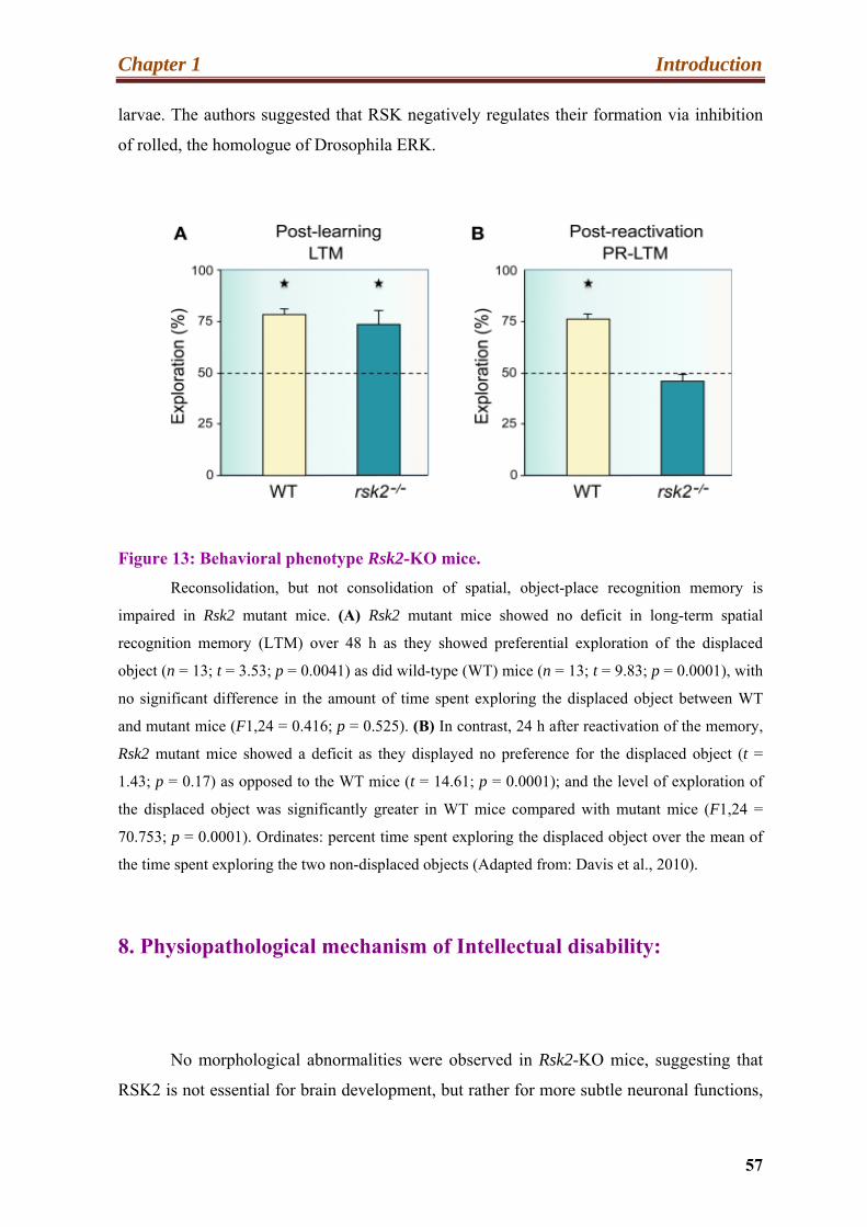
Chapter 1 Introduction
57
larvae. The authors suggested that RSK negatively regulates their formation via inhibition
of rolled, the homologue of Drosophila ERK.
Figure 13: Behavioral phenotype Rsk2-KO mice.
Reconsolidation, but not consolidation of spatial, object-place recognition memory is
impaired in Rsk2 mutant mice. (A) Rsk2 mutant mice showed no deficit in long-term spatial
recognition memory (LTM) over 48 h as they showed preferential exploration of the displaced
object (n = 13; t = 3.53; p = 0.0041) as did wild-type (WT) mice (n = 13; t = 9.83; p = 0.0001), with
no significant difference in the amount of time spent exploring the displaced object between WT
and mutant mice (F1,24 = 0.416; p = 0.525). (B) In contrast, 24 h after reactivation of the memory,
Rsk2 mutant mice showed a deficit as they displayed no preference for the displaced object (t =
1.43; p = 0.17) as opposed to the WT mice (t = 14.61; p = 0.0001); and the level of exploration of
the displaced object was significantly greater in WT mice compared with mutant mice (F1,24 =
70.753; p = 0.0001). Ordinates: percent time spent exploring the displaced object over the mean of
the time spent exploring the two non-displaced objects (Adapted from: Davis et al., 2010).
8. Physiopathological mechanism of Intellectual disability:
No morphological abnormalities were observed in Rsk2-KO mice, suggesting that
RSK2 is not essential for brain development, but rather for more subtle neuronal functions,

Chapter 1 Introduction
58
such as neurotransmission and synaptic plasticity. This hypothesis is in part supported by
the fact that in human and mouse brain, RSK2 is highly expressed in the hippocampus, in
the cortex and in the Purkinje cells of the cerebellum, two brain structures playing a
prominent role in cognitive function, in learning, and having a strong synaptic activity
(Zeniou et al., 2002). Very little is known about the cellular functions of RSK2 in neurons.
The hippocampus plays a critical role in learning and consolidation of spatial long-term
memory, is closely related to the cerebral cortex and contains two key interlocking parts,
ammon’s horn and dentate gyrus (Pearce, 2001). Location and schematic structure of the
hippocampus is given in (Figure 14).
In the CLS patients analyzed by Kesler et al. (2007) the cerebellum and the
hippocampus volume were particularly impacted by the decreased brain volume observed
in some CLS patients. In addition, the Rsk2-KO mice showed spatial learning and long-
term memory deficits, which are hippocampus dependent functions. Before describing
possible causes of ID, I want to introduce some basic information about glutamate
receptors, neurotransmission and synaptic plasticity, and its role in learning and memory.
9. Glutamate receptors:
Although more than sixty neurotransmitters have been described, L-Glutamate is the
major excitatory neurotransmitter in the mammalian CNS (Santos et al., 2009). It acts via
two classes of receptors: ligand gated ion channels, called ionotropic receptors, and G-
protein coupled receptors called metabotropic receptors (Palmada and Centelles, 1998).
Activation of these receptors is responsible for basal excitatory synaptic transmission and
many forms of synaptic plasticity such as long-term potentiation (LTP) and long-term
depression (LTD). They have also been involved in memory and learning processes.
Glutamate is released from terminal buttons of pre-synaptic neuron, in response to an action
potential, crosses the synaptic cleft and acts on the glutamate receptors located on the
postsynaptic neuron. The depolarizing effect of glutamate is rapid, suggesting involvement
of channel receptors. Previous research showed that glutamate receptors are present in CNS
glial cells as well as neurons (Teichberg, 1991).

Chapter 1 Introduction
59
A.
B.
C
C
Figure 14: Location and schematic structure of the hippocampus.
(A) Schematic representation of the location of the hippocampus in the brain. In primates,
it is located in the temporal lobe, beneath the surface of the cortex. (B) The hippocampus consists of
the dentate gyrus (DG) and Ammon's horn, itself divided into three parts (CA1, 2 and 3). Three
types of excitatory neurons are found in this structure: the granule cells of the GD project their
axons (mossy fibers) to CA3 pyramidal cells. These neurons send their axons (Schaeffer collaterals)
to CA1, where they form synaptic contacts with pyramidal cells. These neurons project axons in
turn toward the subiculum and entorhinal cortex (Adapted from Kerchner and Nicoll, 2008).
9.1. Metabotropic glutamate Receptors:
Metabotropic glutamate receptors (mGluRs) belong to the family of G protein-

Chapter 1 Introduction
60
coupled receptors. They allow a “slow” transmission of the signal via second messengers
(Ferraguti and Shigemoto, 2006), and consist of seven transmembrane domains, a C-
terminal cytosolic domain and an N-terminal large extracellular domain (called "Venus
Flytrap Domain"), allowing ligand binding (Niswender and Conn, 2010). Glutamatergic
stimulation induces a conformational change of the receptor and the activation of the
heterotrimeric G protein, allowing signal transduction (Figure15).
A. B.
Activation of mGluRs
Figure 15: General structure of metabotropic receptors (mGluR).
(A). Schematic representation of a dimeric mGluR, at baseline. These receptors are composed of a
C-terminal cytosolic (C-Ter), seven transmembrane domains and an N-terminal extracellular
domain, the "Venus Flytrap Domain" (VFD), allowing ligand binding (B). the binding of glutamate
in one or both VFDs induces a conformational change of these receptors, for signal
transduction.(Adapted from ( Niswender and Conn, 2010).
Activation of the G protein is produced by the exchange of guanosine triphosphate
(GTP) to guanosine diphosphate (GDP) in the α subunit (Niswender and Conn, 2010).

Chapter 1 Introduction
61
Eight members have been identified in the mGluR family. They are grouped into three
classes according to their sequence similarity, the coupled G protein and their agonists
(Ferraguti and Shigemoto, 2006). Group I comprises mGluR1 and -5 that can be activated
by 3, 5-DHPG, whereas mGluR2 and 3 belong to group II. Group III comprises mGluR4-6-
7 and 8. The mGluRs are found in pre-synaptic and postsynaptic neurons in the
hippocampus, the cortex and some other parts of brain (Chu and Hablitz, 2000). The
mGluRs are found in synaptic and extra-synaptic areas and have diverse functions in
synaptic transmission, including regulation of ion channels and neurotransmitter release.
(Ferraguti and Shigemoto, 2006). They are implicated in memory, anxiety, learning and
perception of pain (Ohashi et al., 2002).
9.2. Ionotropic glutamate Receptor:
Ionotropic receptors (iGluR) are multimeric ion channels that allow “fast” synaptic
transmission (Ferraguti and Shigemoto, 2006). They consist of four subunits, each
composed of four semi-autonomous areas (Traynelis et al., 2010): two large extracellular
domains, the N-terminal domain (NTD) and the ligand-binding domain (LBD). This latter
consists of two segments called S1 and S2. They are both parts of the zone of interaction
with the ligand. The transmembrane domain (TMD) is involved in the formation of the ion
channel and contains 3 helices (M1, M3 and M4) and a stem-loop (M2). The TMD is
connected to the intracellular C-terminal domain (CTD), which allows interaction with
proteins in the postsynaptic density (PSD) controlling the cytoskeleton and the receptor
addressing. This family of glutamate receptors is divided into four groups according to their
structural homology and ligand specificity (Traynelis et al., 2010). These groups are
designated according to the name of the most selective ligand.
Thus, they include; AMPA (alpha-amino-3-hydroxy-5-methyl-4-isoxazole-4-
ropionic acid) receptor, NMDA (N-methyl-D-aspartate) receptor, kainite receptor and the δ
receptors receptors (Traynelis et al., 2010).
9.3. NMDA receptors:
The NMDA receptor is the predominant molecular tool controlling synaptic

Chapter 1 Introduction
62
plasticity and memory (Li and Tsien, 2009). NMDA receptors (NMDAR) are tetrameric
receptors, formed by the assembly of two GluN1 subunits together with two GluN2 or
GluN3 (Traynelis et al., 2010). A single gene encodes the GluN1 subunit, but there are
eight splicing variants. The GluN2 and GluN3 ubunits are represented by four and two
members respectively (N2A, B, C and D, and N3A and B) (Rebola et al., 2010). The
NMDAR differs from other ionotropic receptors, because it requires the attachment of two
co-agonists during their activation, and the others not. Glycine binds to GluN1, whereas
glutamate binds to the N2 subunit. A number of endogenous and exogenous compounds
modulate the NMDA receptor activity, including Mg2+ Na+, Ca2+, Zn2+ and also CDK5,
which regulates the amount of NR2B containing NMDA receptors on the synaptic
membrane (Eby and Eby, 2006; Horning and Trombley, 2001; Huggins and Grant, 2005).
Glutamate or NMDA (N-Methyl-D-aspartic acid) binding to NMDARs, leads to
activation of the NMDARs and to opening of the ion channel that are nonselective to
cations with an equilibrium potential near to zero millivolt (mV). The exceptional property
of the NMDAR is its voltage dependent activation. This permits the flow of Ca2+ and Na+
ions into cell and of K+ out of the cell. The response of NMDAR is slower than that of
AMPA receptors because its resting potential is blocked by divalent ions (Mg 2+). The
activity of these receptors is also influenced by the binding of Zn2+ ion at the NTD, or by
phosphorylation events in the CTD. Several consensus sites for PKA and PKC have been
identified in the CTD. It has been shown that these regulatory mechanisms of NMDAR
play a central role in LTP and LTD. (Traynelis et al., 2010; Rebola et al., 2010)
9.4. Kinate receptors:
Kinate receptors play only a minor role in signaling at synapses (Song and Huganir,
2002). The kinate receptors (KARs) are composed of five members, grouped into two
families who share only 40% sequence homology. The first family contains receptors
GluR5, 6 and 7 showing 75 to 80% sequence homology, while the second group contains
KA1 and 2, which are 70% homologous. The second family has a better affinity for
glutamate. The KARs are found as homo-or heteromers (Bloss and Hunter, 2010).

Chapter 1 Introduction
63
9.5. The δ receptors:
The δ receptor family contains two members, GluRδ1 and GluRδ2, which are
homomers (Traynelis et al., 2010). Their classification in the group of iGluRs comes from
their sequence homology with some members of the ionotropic receptor family.
Surprisingly, GluRδ1 and GluRδ2 do not bind glutamate and are not ionic channels
(MacLean, 2009). However, δ receptors are involved in glutamatergic transmission, the
exact mechanism being not yet known.
9.6. AMPA receptors:
The name of AMPA receptor is derived from its ability to be activated by the
artificial glutamate analog AMPA (α-amino-3-hydroxy-5-methyl-4-isoxazolepropionic
acid). This receptor was first discovered in 1982 by Tage Honore and his group at the
Copenhagen pharamacy school (Honore et al., 1982). AMPA glutamate receptor channels
are expressed ubiquitously in brain neurons and mediate “fast” excitatory
neurotransmission in most excitatory synapses. AMPA receptors are composed through
combinations of four subunits, GluR1, 2, 3 and/ or GluR4, which are encoded by different
genes. In the pyramidal cells of the adult hippocampus GluR1-3 subunits are the most
commonly expressed, forming predominantly GluR1/2 and GluR2/3 heterodimers
(Malinow and Malenka, 2002). The number, composition and localization, as well as post-
transcriptional and translational regulation, of AMPA receptors in neurons are critically
significant factors that determine the neuronal response to glutamate (Dingledine et al.,
1999). The subunit composition of AMPA receptor will, for example, affect the conductive
properties of the receptor. Of all ionotropic receptors, only the AMPA receptors may be
impermeable to Ca2+. This property is controlled by the GluR2 subunit and its issue
depends on post-transcriptional edition of the Q/R codon at position 607 in the TMD. The
presence of an arginine (R) at position 607 gives the channel a linear current-voltage
relationship (Boulter et al., 1990) and blocks the Ca2+ permeability of GluR2 (Seeburg et al.
1998; Greger et al., 2002).
A second edition site, R/G, at position 743 in the LBD of GluR2 and GluR4 has
been described as influencing the kinetics, assembly and membrane expression of AMPA

Chapter 1 Introduction
64
receptors (Lomeli et al., 1994). The RNA editing process of GluR is catalyzed by adenosine
deaminases that act on the mRNA (ADARs).
The properties of AMPA receptors are also controlled by alternative splicing in the
LBD (flip and flop variants) (Pei et al, 2009) (Figure16). It determines the speed of
desensitization (Mosbacher et al., 1994) of the receptor, the speed at which the receptor is
re-sensitized (Sommer et al., 1990), the rate of channel closing and the receptor export (Pei
et al., 2007). The function of AMPA receptors can also be regulated by phosphorylation
(Santos et al., 2009). While NMDA receptors allow long-term changes of synaptic
transmission, AMPA receptor function is to transmit the initial depolarization after
glutamate stimulation. AMPA receptors are also critical for the expression of various forms
of long-lasting synaptic plasticity, including LTP and LTD (Malenka and Bear, 2004;
Luscher and Huber, 2010).
The crucial role of GluR2 in neuronal functions may explain why it is the most
tightly regulated of the AMPA receptor subunits. Moderate changes, in GluR2 expression
may have severe functional consequences on receptor subunit rearrangement and lead to
functional differences in the functioning of synaptic circuits.
I observed an abnormally increased level of GluR2 expression in the hippocampus
of Rsk2-KO mice.
9.7. Regulation of transcription of Glutamate receptor:
Expression of glutamate receptors is the result of a balance between transcription,
translation, protein constancy, assembly of receptor and its arrangement at the cell surface,
all of which may be regulated by a number of cellular and environmental stimuli.
Therefore, the any particular subunits that every neuron expresses are strong determinants
of synaptic phenotype, and this is the basis for understanding how the genetic cis elements
and trans factors control gene transcription in neural cells (Traynelis et al., 2010). Gene
encoding glutamate receptors have some common features including several transcriptional
starting sites and TATAA- less promoters with a high GC rich content.

Chapter 1 Introduction
65
Figure 16: Structure of the GluR2 subunit.
(A). Schematic representation of GluR2 at the plasma membrane. The N-terminal domain (ATD)
and ligand binding domain (LBD), consisting of segments S1 and S2 are the extracellular part. The
transmembrane domain (TMD) is composed of three helices (M1, M3 and M4) and a stem-loop
(M2). It is linked to the intracellular C-terminal domain (CTD), which allows protein interactions.
The editing sites Q / R and R / G and the region of alternative splicing (flip / flop) are also
represented.
(B) Three-dimensional structure of a GluR2 homotetramer, showing a certain asymmetry
between the TMD and the extracellular domains (adapted from Isaac et al., 2007 and Traynelis et
al., 2010).

Chapter 1 Introduction
66
The 5' untranslated region (UTR) ranges between 200bp of the Gria1 (encoding GluR1)
genes to more than 1200bp of Grin2a (encoding GRIN2A). Sp1 elements reside close to the
major transcriptional start site of all genes involved in expression of glutamate receptor.
Many glutamate receptor promoters contain CRE, NRF-1, NFkB and NRSE sites that are
implicated in response to neuronal activity (Traynelis et al., 2010).
We were particularly interested in determining why GluR2 expression is stronger in
Rsk2-KO as compared to WT littermate. In the following paragraph, I am going to discuss
briefly the transcription regulation of the gria2 gene.
9.8. Transcription of the Gria2 gene:
GluR2 is the most critical subunit of the AMPA receptors because it determines in
particular, the Ca2+ permeability of the receptor. Gria2 expression is influenced strongly at
the transcriptional level by two positive (Nrf-1 and Sp1) and one negative (Re1/Nrse-like
silencer) regulatory elements in the 5' proximal region of the promoter (Borges and
Dingledine, 2001). Myers et al. (1998) showed that the GluR2 Re1/Nrse-like silencer binds
the RE1-silencing transcription factor (NRSF/REST), and co-transfection of REST into
neurons reduced GluR2 promoter activity in a silencer-dependent manner. The transcription
factor Sp1 recruits basal transcription factor TFIID to DNA and induces transcription. It
was previously shown that Sp1 activity increases when phosphorylated by Erk 1/2
(Merchant et al., 1999). In 2011, a study confirmed that mutation of Sp1 reduces the
expression of the Gria2 gene. (Ekici et al., 2011). The nuclear respiratory factor 1 (NRF-1)
is a transcription factor that was also shown to regulate expression of NMDA receptors
subunit genes in neurons (Dhar et al., 2009). NRF-1 has also been associated with the
regulation of neurite outgrowth. Dhar et al., 2009 showed that NRF-1 functionally binds to
the Gria2 promoter.
I showed that Sp1 up regulated activity is responsible for increased transcription of
the Gria2 gene in RSK2 deficient cells. The schematic organization of the promoter regions
of Gria2 is presented in (Figure 17) (Traynelis et al., 2010).

Chapter 1 Introduction
67
Figure 17: Schematic diagram of the proximal promoter regulatory region of Gria2.
The proximal promoter regions of the Gria2 gene are shown as a thin line. The 5’-
untranslated exon sequences is represented by an open bar; the blackened bar designates the protein
coding domains (Traynelis et al., 2010).
9.9. Sp1 regulates the GluR2 expression:
Specificity proteins 1 (Sp1) is the prototypic C2H2 type zinc finger containing DNA
binding proteins. This transcription factor can activate or repress transcription in response
to various physiological and pathological stimuli. It binds to the GC rich motif (5′-G/T-
GGGCGG-G/A-G/A-C/T-3′ or 5′-G/T-G/A-GGCG-G/T-G/A-G/A-C/T-3′) and can regulate
the expression of TATA- containing and TATA less genes via proteins-protein interaction
or with other transcription factors (Naar et al., 1998). Sp1 protein contains 785 amino acid
and its molecular weight is approximately 110 kDa. It was reported that phosphoraylation
events of Sp1 influence its transcription activity (Lin et al., 1997). Various kinases
phosphorylate different Ser and Thr sites within the Sp1 proteins. Kinases regulating Sp1
activity include, cyclin-dependent kinase (CDK), extracellular signal-regulated Kinase
(ERK) and casein kinase (CKII) (Armstrong et al., 1997; Fojas de Borja et al., 2001;
Milanini-Mongiat et al., 2002). Phosphorylation sites are shown in (Figure. 18).
Interestingly Milanini- mongiat and their colleague (2002) demonstrated, that
ERK1/2 phosphorylate two Sp1 sites at residues Thr453 and Thr 739. Phosphorylation at
both sites is required for maximal Sp1 DNA binding and transcriptional activity (Tan and
Khachigian, 2009).

Chapter 1 Introduction
68
Figure 18: Ser and Thr phosphorylation sites in Sp1.
The Ser and Thr residues are indicated in red and blue, respectively. ZnF, zinc finger
domain. Acetylation, sumoylation, ubiquitylation, and glycosylation are among other
posttranslational modifications that influence the transcriptional activity and stability of Sp1. Pink
color arrows indicate the twos sites that are phosphorylated by phospho-ERK42/44 (Adopted from
Tan and khachigian, 2009).
During my thesis, I confirmed that these two sites are phosphorylated, by ERK1/2
kinase activity in PC12 cell line as well as in the hippocampus of adult mice.
9.10. Role of GluR2 subunit in AMPA Receptor:
The AMPA-type glutamate receptor channels are expressed ubiquitously in brain
neurons and mediate fast excitatory neurotransmission in most excitatory synapses. AMPA
receptors are assembled from combinations of four subunits, GluR1, GluR2, GluR3 and/or
GluR4. GluR1 and GluR2 show higher levels of expression in the hippocampus and cortex
as compared to GluR3/4. GluR2 exists mainly in AMPARs receptor as GluR1/2 and
GluR2/3 heterotetramers. Previous studies reported that 70% of GluR2 is associated with
GluR1 (Wenthold et al., 1996). AMPA receptors play also a crucial role in synaptogenesis
and formation of neuronal circuitry, as well as in synaptic plasticity (Santos et al., 2009).

Chapter 1 Introduction
69
There is a huge amount of evidence indicating that the GluR2 subunit plays a pivotal role in
AMPA receptors function. The Ca2+ permeability, rectification, and single-channel
conductance of AMPA receptors are all dominantly influenced by inclusion of an edited
GluR2 subunit in the receptor complex (Hollmann et al., 1991; Burnashev et al., 1992;
Bowie and Mayer, 1995; Koh et al., 1995; Seeburg, 1996; Swanson et al., 1997; Washburn
et al., 1997). Receptors that contain a single edited GluR2 subunit have maximally reduced
Ca2+ permeability; whereas inward rectification is reduced in a graded manner as the
number of GluR2 subunits in a receptor increases (Washburn et al., 1997). Thus, moderate
changes in GluR2 expression may have functional consequences on receptor subunit
rearrangement and lead to functional differences in the functioning of synaptic circuits.
GluR2 expression is governed by neuronal activity (Bai and Wong-Riley, 2003), and its
synaptic distribution and levels are modulated as part of the mechanism of synaptic
plasticity (Keifer and Zheng, 2010; Sprengel, 2006). GluR2 interacts also with various
molecules implicated in receptor trafficking, including the glutamate receptor-interacting
protein (GRIP1) containing seven PDZ domains (Dingledine et al., 1999; Collingridge et
al., 2004; Isaac et al., 2007). Some studies suggested the GluR2-GRIP1 interaction is
important for the synaptic accumulation of the receptor, binding to the cytoskeleton through
PSD95 and its insertion into the synaptic membrane.
The N-ethylmaleimide-sensitive fusion protein (NSF) is ubiquitously found in the
cytoplasm of eukaryotic cells. It is a central component of the cellular machinery in the
transfer of membrane vesicles from one membrane compartment to another. The surface
expression of AMPAR is inhibited via a peptide (Lys-Arg-Met-Lys-Val-Ala-Lys-asn-Ala-
Gln) that blocks the GluR2-NSF interaction, suggesting that NSF is involved in the
insertion of AMPARs into the post-synaptic membrane (Noel et al., 1999). However,
whether the NSF-interfering peptide stimulates the loss of AMPARs through blocking their
surface delivery or through destabilizing them in the synaptic plasma membrane is not yet
clear. The Disruption of this NSF-GluR2 interaction by specific peptides causes a rundown
of excitatory post- synaptic currents (EPSC) (Lee et al., 2002; Noel et al., 1999; Song et al.,
1998) and the mutated GluR2 in absence of NSF interaction is not delivered to synapses in
hippocampal slice cultures (Shi et al., 2001). Moreover, GluR2-NSF also co-assembles with
α and β- Synaptosomal-associated protein (SNAPs) to form a multi molecular complex that
can be reversible subsequent to ATP hydrolysis. In addition, α-SNAP dissociates the
GluR2–PICK1 interaction and stabilizes GluR2 expression on the cell surface, which as a
Chapter 1 Introduction
70
result affects PICK1 mediated endocytosis (Hanley et al., 2002; Osten et al., 1998). PICK1
is a peripheral membrane protein containing a PDZ domain that binds a large number of
membrane proteins. In addition another protein, the Activating Protein 2 (AP2) also
cooperates with GluR2, and its binding site in the GluR2 C-terminal region overlaps, but is
not the same as the NSF binding site (Lee et al., 2002; Kastning et al., 2007). The AP2 is a
clathrin adaptor complex involved in endocytosis processes, and its interaction with GluR2
seems to be involved in clathrin-mediated endocytosis during NMDAR mediated LTD.
Kasting et al. (2007) demonstrated that blocking AP2–GluR2 binding increases AMPA
receptor-mediated transmission (Kastning et al., 2007). An earlier study established that
blocking this interaction has no effect on basal transmission but selectively stops the LTD
(Lee et al., 2002) (Figure 19).
The extra-cellular domain of GluR2 has a protein-protein interaction site that is
significant for the growth, formation and maintenance of dendritic spines. There is
suggestion that over-expression of GluR2 in cultures of primary hippocampal neurons
increases the length, width and density of spine (Bassani et al., 2009). Passafaro and his
colleague in 2003 showed that silencing of GluR2 inhibits spine morphogenesis. (Figure
19) shows the mechanism that directs the interaction of the N-terminal region of GluR2
with N-cadherin, and believed to stimulate the development and formation of synaptic and
dendritic spines via novel structural communication at the synaptic junction (Saglietti et al.,
2007).
Now in the following chapters, I will present the synaptic plasticity and its role in
memory formation.
10. Synaptic plasticity (SP):
One of the most important and fascinating properties of the mammalian brain is its
plasticity; the capacity of the neural activity generated by an experience to modify neural
circuit function and thereby modify subsequent thoughts, feelings, and behavior.
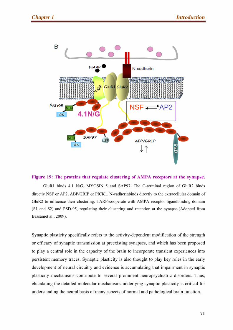
Chapter 1 Introduction
71
NSF AP2 4.1N/G
Figure 19: The proteins that regulate clustering of AMPA receptors at the synapse.
GluR1 binds 4.1 N/G, MYOSIN 5 and SAP97. The C-terminal region of GluR2 binds
directly NSF or AP2, ABP/GRIP or PICK1. N-cadherinbinds directly to the extracellular domain of
GluR2 to influence their clustering. TARPscooperate with AMPA receptor ligandbinding domain
(S1 and S2) and PSD-95, regulating their clustering and retention at the synapse.(Adopted from
Bassaniet al., 2009).
Synaptic plasticity specifically refers to the activity-dependent modification of the strength
or efficacy of synaptic transmission at preexisting synapses, and which has been proposed
to play a central role in the capacity of the brain to incorporate transient experiences into
persistent memory traces. Synaptic plasticity is also thought to play key roles in the early
development of neural circuitry and evidence is accumulating that impairment in synaptic
plasticity mechanisms contribute to several prominent neuropsychiatric disorders. Thus,
elucidating the detailed molecular mechanisms underlying synaptic plasticity is critical for
understanding the neural basis of many aspects of normal and pathological brain function.
Chapter 1 Introduction
72
The brain is composed of hundreds of trillions of neurons, forming functional neural
networks (Hotulainen and Hoogenraad, 2010). Functioning of these networks is related to
the connections of the neurons through highly specialized cell junctions called synapses. A
chemical synapse allows the passage of electrical information from a presynaptic neuron to
its postsynaptic partner. The majority of excitatory synapses in the hippocampus are located
on small outgrowths on the dendrite called dendritic spines (Hotulainen and Hoogenraad,
2010). Synaptic plasticity in excitatory synapses is mainly dependent upon calcium
(Gerrow and Triller, 2010). Formation of memory requires modification of the strength of
synaptic transmission and remodeling of neuronal networks, and these modifications span
temporal domains ranging from milliseconds to enduring modifications that might continue
for days or weeks and maybe even longer. This refers to synaptic plasticity (SP). This is
possible because the synapse can integrate a large number of signals and adapt the cellular
response (Laroche, 2010). At excitatory synapses, plasticity is mediated through molecular
machinery that detects local Ca2+ signals in the dendritic spines. For yet unknown reasons,
spine size is strongly correlated with synaptic strength. Short forms of synaptic plasticity
have been associated with short-term adaptations to sensory inputs, fleeting changes in
behavioral states, and short-lasting forms of memory. Lasting changes are believed to play
crucial roles in the construction of neural circuits during development and in long-term
forms of memory in the adult nervous system. The relationship between synaptic plasticity
and memory has emerged following the study of Bliss and Lomo in 1973. They showed
that a sustainable form of SP, called the long-term potentiation (LTP) is obtained by
repeated stimulation of the dentate gyrus granule cells. Sah et al. in 2008 confirmed the link
between LTP and learning, with tests of fear conditioning in the amygdala.
Other types of SP have been described (habituation, facilitation ...). However, we
will focus in our manuscript on two of them, the long-term potentiation (LTP) and long-
term depression (LTD) and their activation mechanism. The mechanisms of two forms of
synaptic plasticity involve both the AMPA and NMDA glutamate receptors. In the
following paragraphs, I will discuss the roles of AMPA and NMDA receptors in synaptic
plasticity.

Chapter 1 Introduction
73
10.1. AMPA receptor trafficking:
AMPA receptors are found throughout neurons including, at synaptic, intracellular
and extra-cellular sites (Figure 20). AMPA receptor can move in and out of synapses,
which is called receptor trafficking. Receptor are removed from synapses through lateral
diffusion or endocytosis and introduced into synapses via exocytosis or lateral diffusion
from intracellular as well as extra-synaptic sites respectively (Newpher and Ehlers, 2008;
Triller and Choquet, 2008). AMPA receptors are highly clustered at the postsynaptic
density (PSD) of a synapse, which is the protein-rich domain in the postsynaptic membrane
that is directly apposed to the pre-synaptic active zone. Receptors are localized within the
PSD through interactions with various scaffolding proteins and cytoskeletal elements.
There is a large body of experimental evidence suggesting that fast trafficking of AMPA
receptors into and out of the PSD contributes to activity-dependent long-lasting changes in
synaptic strength (Malinow and Malenka, 2002; Sheng and Kim, 2002; Song and Huganir,
2002; Bredt and Nicoll, 2003; Triller and Choquet, 2003, 2005; Collingridge et al., 2004;
Kennedy and Ehlers, 2006). Such changes are thought to be a necessary sub-cellular
component of certain forms of learning and memory (Bliss and Collingridge, 1993;
Malenka and Nicoll, 1999). AMPA receptor trafficking appears to occur through a
combination of two major mechanisms: exocytic/endocytic exchange of surface receptors
with intracellular receptor pools, and lateral diffusion of receptors between the PSD and the
surrounding extra-synaptic membrane (ESM). It follows that, under basal conditions, the
steady-state receptor concentration within a synapse is determined by a dynamical
equilibrium in which the various receptor fluxes, into and out of the PSD, are balanced.
Activity-dependent changes of one or more of these fluxes can then modify the number of
receptors in the PSD and thus alter the strength of a synapse.
The precise mechanisms underlying the activity-dependent regulation of AMPA
receptor trafficking are currently not known. However, they are likely to involve one or
more of the following processes: changes in the interaction between receptors and
scaffolding proteins within the PSD, changes in the rates of exocytosis/endocytosis, and
modifications in membrane or receptor structure that alter the surface transport of receptors.

Chapter 1 Introduction
74
Figure 20: Regulation of AMPAR trafficking in response to LTP-inducing stimuli.
After post-ER processing in the golgi apparatus, AMPARs are released into the peri-
synaptic membrane as a reserve waiting for the LTP process to be initiated. Glutamate binding to
NMDARs leads to the influx of Ca2+ through the NMDAR receptors and consequently the
activation of CaMKII. CaMKII has two known modes of activation to trigger the incorporation of
AMPA receptors into the peri-synaptic membrane. The first is by direct phosphorylation of the
synaptic-associated protein 97 (SAP97), which leads to binding of the complex SAP97 / Myosin-VI
to the C-terminus of AMPARs. The AMPAR/Stargazin/SAP97/Myosin-VI complex in then inserted
into the peri-synaptic membrane. The second mode of activation is via the MAPK pathway.
CaMKII activates the Ras proteins, leading to MAPK/ERK activation, which drives AMPAR
insertion directly into the peri-synaptic membrane (Shepherd and Huganir, 2007).
10.2. AMPA receptors and Synaptic Plasticity:
Synaptic plasticity refers to an extraordinary ability of the communication between

Chapter 1 Introduction
75
two neurons to change in strength. These changes in the strength of synaptic transmission
affect the activity of neuronal networks and, finally the behavior of the whole animal. It is
supposed that synaptic plasticity represents the cellular correlate of information storage and
motivates vital physiological and pathophysiological processes (Citri and Malenka, 2008;
Kessels and Malinow, 2009).
In 1949 D.O. Hebb, for the first time reported the basic mechanism of activity
dependent synaptic plasticity. It was described as “When an axon of cell X is near enough
to excite a cell Y and repeatedly or persistently takes part in firing it, some growth process
or metabolic change takes place in one or both cells such that X's efficiency, as one of the
cells firing Y, is increased.” (Hebb, 1949). In 1973, the Hebb’s theory achieved the
experimental support with seminal observation that repetitive activation of excitatory
synapses into the rabbit hippocampus prompted a persistent increase in synaptic
transmission that can last hours and even days (Bliss and Gardner-Medwin, 1973; Bliss and
Lomo, 1973). This long lasting enhancement in synaptic strength has been known as LTP,
and a persistent weakening of synaptic strength relative to baseline has been known as
long-term depression (LTD). In last decades, several advances in studies of learning and
memory have been done, and a huge literature suggested that LTP and LTD are involved in
mechanism of learning and memory (Fleming and England, 2010; Neves et al., 2008;
Pastalkova et al., 2006). In 2008, Citri and Malenka reported that two molecular
mechanisms are involved in the change of synaptic strength: i) the amount of
neurotransmitter released by the pre-synaptic neuron onto postsynaptic neuron and ii)
changes in the number and function of receptor on the postsynaptic neuron that respond to
that neurotransmitter (Citri and Malenka, 2008).
AMPAR is the most important mediator of excitatory synaptic transmission
(Traynelis et al., 2010), and regulation of its function is vital for modulating different forms
of SP (Turrigiano and Nelson, 2004). GluR2 expression is governed by neuronal activity
(Bai and Wong-Riley, 2003) and its synaptic distribution and levels are modulated as part
of the mechanism of synaptic plasticity (Keifer and Zheng, 2010). Indeed, GluR2 interacts
with various molecules and plays a critical role in AMPA Receptor trafficking (Isaac et al.,
2007). The role of GluR2 in AMPAR trafficking has attracted special attention because it

Chapter 1 Introduction
76
provides a key mechanism for regulating the number of synaptic AMPA receptors and
plasticity (Malinow and Malenka, 2002; Bredt and Nicoll, 2003; Collingridge et al., 2004;
Shepherd and Huganir, 2007; Kerchner and Nicoll, 2008). The trafficking of post-synaptic
AMPA receptors has been shown to be an important mechanism underlying activity-
induced alterations in synaptic transmission (Bredt and Nicoll, 2003; Kessels and Malinow,
2009). However, the exact mechanisms by which GluR2 regulates AMPA receptor
trafficking and synaptic plasticity are not yet clear. Moreover, the previous studies
suggested that AMPA receptors in CNS are complexed with auxiliary subunits:
transmembrane AMPA receptor regulatory proteins (TARPs) (Kato et al., 2008; Milstein et
al., 2007). TARPs have a profound impact on AMPA receptors, by affecting the trafficking
of the receptor, gating ions permeability and pharmacology of the receptors (Bats et al.,
2007; Menuz et al., 2009; Tomita et al., 2005).
As above stated, the main function of AMPA receptor is to mediate fast excitatory
synaptic transmission to ensuring the rapid responses to glutamate. There is strong evidence
that alteration in the number of AMPA receptors localized at synapses triggers changes in
the strength of synaptic transmission at a variety of synapses (Citri and Malenka, 2008;
Derkach et al., 2007; Kauer and Malenka, 2007).
10.3. NMDA receptor and its role in synaptic plasticity:
The N-methyl-D- aspartate receptors (NMDARs) are glutamate-gated ion channels
extensively expressed in the CNS and playing also crucial roles in excitatory synaptic
transmission, memory function (Tsien, 2000). The NMDAR is a specific type of ionotropic
glutamate receptor. The NMDA receptor forms a heterotetramer between two NR1 (NR1A
and NR1B) and two NR2 (NR2A and NR2B) subunits; two obligatory NR1 subunits and
two regionally localized NR2 subunits. The structure of NMDA receptors includes: an
extracellular domain containing two globular structures, a modulatory domain and a ligand-
binding domain. NR1 subunits bind to the co-agonist glycine and NR2 subunits bind the
neurotransmitter glutamate. An agonist-binding module links to a membrane domain,
which consists of three trans-membrane segments and a re-entrant loop reminiscent of the
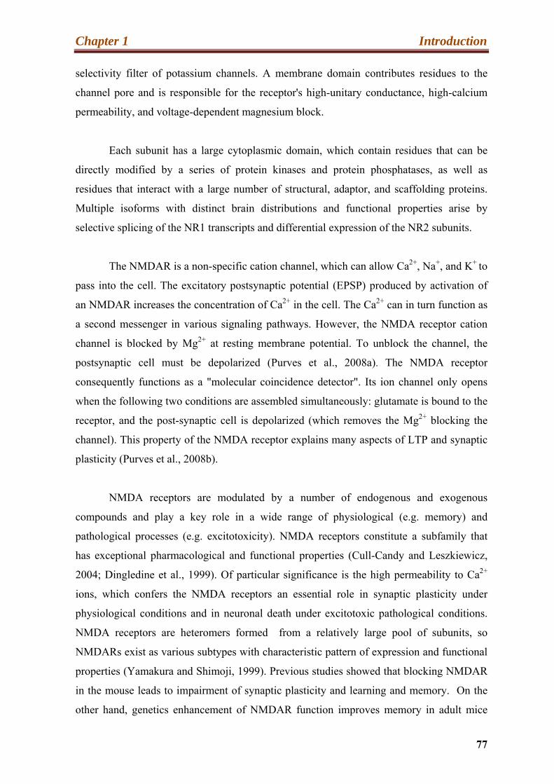
Chapter 1 Introduction
77
selectivity filter of potassium channels. A membrane domain contributes residues to the
channel pore and is responsible for the receptor's high-unitary conductance, high-calcium
permeability, and voltage-dependent magnesium block.
Each subunit has a large cytoplasmic domain, which contain residues that can be
directly modified by a series of protein kinases and protein phosphatases, as well as
residues that interact with a large number of structural, adaptor, and scaffolding proteins.
Multiple isoforms with distinct brain distributions and functional properties arise by
selective splicing of the NR1 transcripts and differential expression of the NR2 subunits.
The NMDAR is a non-specific cation channel, which can allow Ca2+, Na+, and K+ to
pass into the cell. The excitatory postsynaptic potential (EPSP) produced by activation of
an NMDAR increases the concentration of Ca2+ in the cell. The Ca2+ can in turn function as
a second messenger in various signaling pathways. However, the NMDA receptor cation
channel is blocked by Mg2+ at resting membrane potential. To unblock the channel, the
postsynaptic cell must be depolarized (Purves et al., 2008a). The NMDA receptor
consequently functions as a "molecular coincidence detector". Its ion channel only opens
when the following two conditions are assembled simultaneously: glutamate is bound to the
receptor, and the post-synaptic cell is depolarized (which removes the Mg2+ blocking the
channel). This property of the NMDA receptor explains many aspects of LTP and synaptic
plasticity (Purves et al., 2008b).
NMDA receptors are modulated by a number of endogenous and exogenous
compounds and play a key role in a wide range of physiological (e.g. memory) and
pathological processes (e.g. excitotoxicity). NMDA receptors constitute a subfamily that
has exceptional pharmacological and functional properties (Cull-Candy and Leszkiewicz,
2004; Dingledine et al., 1999). Of particular significance is the high permeability to Ca2+
ions, which confers the NMDA receptors an essential role in synaptic plasticity under
physiological conditions and in neuronal death under excitotoxic pathological conditions.
NMDA receptors are heteromers formed from a relatively large pool of subunits, so
NMDARs exist as various subtypes with characteristic pattern of expression and functional
properties (Yamakura and Shimoji, 1999). Previous studies showed that blocking NMDAR
in the mouse leads to impairment of synaptic plasticity and learning and memory. On the
other hand, genetics enhancement of NMDAR function improves memory in adult mice

Chapter 1 Introduction
78
(Cao et al., 2007; Tsien, 2000) . The NR2 subunit directs the properties of NMDA receptor
channels and the degree of synaptic plasticity; a relative abundance of NR2 confers a
greater plasticity (Li and Tsien, 2009). In 2009, Ng et al. demonstrated that the Neto1
protein, a synaptic transmembrane protein, interacts with the NMDA receptor (Ng et al.,
2009). The intercellular domain of Neto1 binds the postsynaptic density protein 95 (PSD-
95), Ng et al., showed, on the basis of Neto1 deficient mice, that the synaptic NR2A was
reduced in the mutant mice whereas the level of NR2B was unchanged. This discriminating
decrease in synaptic NR2A suggests that Neto1 might be obligatory for achieving a normal
abundance of NR2A-containing synaptic NMDA receptors. The NR2B subunits are
present in the brains of Neto1 mutant mice and are the rate-limiting factors in controlling
NMDA receptor mediated synaptic plasticity and memory formation (Tsien, 2000).
Our preliminary findings suggest that NMDAR based neurotransmission is also
significantly decreased in RSK2 deficient mice.
10.4. Long-term potentiation (LTP):
Long-term potentiation is a type of activity dependent plasticity, which results in a
constant enhancement of synaptic transmission. LTP has been a source of large attraction to
neuroscientists since its discovery in the early 1970s. LTP is a persistent increase of the
efficiency of synaptic transmission following high-frequency stimulation (Bliss and Lomo,
1973). The synapse has the ability to maintain these changes for weeks, months or even
years (Laroche, 2010). LTP is a synaptic strengthening, which is the basis of information
storage and learning (Malinow and Malenka, 2002). Several phases have been identified in
this process, early LTP (E-LTP) and lasting LTP (L-LTP). E-LTP is based on post-
translational modifications, whiles the consolidation of memory and synaptic changes,
observed in L-LTP, requires expression of various genes and protein synthesis (Laroche,
2010). LTP is believed to account for various type of learning, including classical
conditioning and higher-level cognition observed in human (Cooke and Bliss, 2006).
10.5. The activation mechanisms of LTP:
Chapter 1 Introduction
79
LTP can be induced by several mechanisms, but it seems that the critical event is the
increase of intracellular Ca2+ concentration. In the following text, I am going to discuss the
involvement of AMPA and NMDA receptors in the process of hippocampal LTP.
The prevailing view is that NMDARs play an essential function in the induction of
several forms of activity-dependent LTP and LTD, by acting as a coincidence detector of
pre-synaptic and postsynaptic firing (Malenka and Bear, 2004). The use of
aminophosphonovalerique, (APV), a specific inhibitor of the NMDAR, blocks the
induction of LTP. The entry of Ca2+ in the postsynaptic neuron is in fact dependent on the
activation of NMDAR (Rebola et al., 2010). However as previously mentioned Mg2+ blocks
that ion channel in a voltage “voltage-dependent”. Opening of this channel is only possible
if a ligand (glutamate) binds to the receptor in parallel with a depolarization of the post-
synaptic membrane (Humeau et al., 2009). Previous studies indicated that AMPA receptors
are involved in the process of LTP in hippocampus. Activation of AMPAR on the surface
induces the initial depolarization necessary for NMDA receptor activation. However, some
synapses called silent, do not possess AMPA receptors, and the mechanism of NMDA
receptor activation is then AMPAR independent. The calcium channels voltage-dependent
or the mGluR might in these cases induce the initial depolarization (Rebola et al., 2010).
The synapses called silent may be matured via the insertion of AMPAR (Kerchner and
Nicoll, 2008).
The entry of Ca2 + in the postsynaptic compartment allows the maintenance of LTP
through the activation of many protein kinases such as CaMKII, PKC or PKA and ERK1/2
(Rebola et al., 2010). Maintaining this type of long-term plasticity requires the expression
of several genes, protein synthesis but also the recruitment of AMPA receptors at the
surface. Indeed, recycling of AMPA receptors after activation of the NMDA receptor
represents a key step in strengthening synaptic plasticity (Malinow and Malenka, 2002). Shi
et al. (1999) showed that the insertion of GluR1 at surface is regulated through the
activation of the NMDA receptor after a tetanic stimulation. GluR1 traffic was inhibited by
APV, an antagonist of the NMDAR. On the other hand, many researchers have
demonstrated the role of GluR1 subunits in the expression of LTP. Zamanillo et al., in 1999
have shown that the absence of this subunit in the GluR1-KO mice leads to a loss of LTP.
The mechanism of insertion of AMPA receptors into the synapse involves two steps (Isaac
et al., 2007; Humeau et al., 2009). Initially, during the E-LTP, the homomeric GluR1/1
Chapter 1 Introduction
80
(permeable to Ca2+) arrives at the membrane in a PKA dependent manner, they allow entry
of Ca2+ into the neuron and an increase in its concentration. An increase in the proportion of
GluR1/GluR2 receptors, impermeable to Ca2+, is then observed at the surface, which allows
the transition to L-LTP. This biphasic mechanism remains, however, controversial.
It is interesting to note that the recycling activity of AMPA receptors is regulated by
phosphorylation; many phosphorylation sites have been reported for all AMPA receptors
(Santos et al., 2009). This phosphorylation permits recycling and contributes to the
maintenance of LTP. Oh et al., in 2006, for example, have identified Ser845 of GluR1 as
one of the sites recognized by PKA. Following forskolin stimulaztion they observed that
phosphorylation of this residue leads to the anchoring of GluR1 at the cell surface. The
activation of NMDA receptors allows parallel expression and synaptic induction of LTP. In
2005, Qin and his group described the insertion of GluR1 and GluR2 at the synapse,
following the activation of the Ras/MAPK pathway. The stimulation of the NMDA
receptors induces the phosphorylation of GluR2 at Ser841 through ERK/MAPK and its
synaptic expression. The authors proposed the intervention of other kinases, such as the
CaMKII, PKA but also RSKs in this process (Qin et al., 2005). These studies confirmed the
link between the activation of the NMDA receptor, the stimulation of a channel and
recycling of the AMPA receptor in the establishment of the L-LTP. Other studies have also
involved PKC in this process (Santos et al., 2009).
The transport of the AMPA receptor to the synaptic surface requires a process of
membrane fusion, involving the SNARE protein, and a mechanism for the lateral diffusion,
depending on post-synaptic protein connections (Santos et al., 2009). In brief, an induction
of LTP leads to the fusion of endosomes containing the AMPA receptor with the cell
membrane, usually in extra-synaptic regions. This fusion is in fact a process of exocytosis
whose main actors are the SNARE proteins. Their importance has been described by the
work of Lledo et al. in 1998. A decrease in LTP induced by tetanic stimulation was
observed after injection of botulinum toxin in hippocampal slices. This toxin inhibits the
formation of the SNARE anchoring proteins and therefore their action. The authors also
showed that injection of SNARE increases synaptic transmission. Once anchored at the
plasma membrane, AMPA receptors diffuse laterally, moving to the PSD. It seems that this
mechanism is dependent on Stargazin and PSD95. Stargazin, belonging to the TARPs
protein family, interacts and regulates the AMPA receptors. PSD95 is a major postsynaptic
Chapter 1 Introduction
81
protein, interacting with Stargazin through PDZ domains. Through the use of mutants of
compensation (alteration of the interaction between the PDZ and ligand), Bats et al. in 2007
showed that the interaction of these two proteins is necessary for the accumulation of
AMPA receptor in the synapse by lateral diffusion (Schnell et al., 2002).
During LTP, the fusion of endosomes with the plasma membrane allows the
recycling of AMPA receptors but it also regulates the growth of dendritic spines. These
small protrusions rich in actin are the major sites of glutamatergic synapses. Analysis and
electron microscopy imaging of living cells showed that the fusion of vesicle recycling
induced during LTP, enhances the membrane surface and thus contribute to the expansion
and growth of dendritic spines (Park et al., 2006). In addition, constitutive recycling of
AMPA receptors was also observed at baseline, especially for heteromeric GluR2 /3. This
is much slower than the previous one and does not lead to a change in synaptic activity.
AMPA receptor traffic in this case represents a way to maintain basal synaptic transmission
and synaptic concentration of AMPA receptors (Hu et al., 2007). The C-terminal
cytoplasmic end of the subunits GluR2/3 can interact with different PDZ proteins and thus
regulates the trafficking and stability of AMPAR in the synapse (Passafaro et al., 2001).
Maintain of LTP, requires the synthesis of new proteins. The local translation in
dendritic spines is possible because the molecular machinery of translation is found in the
spines and dendrites. In 2002, Ostroff and his group reported translocation of
polyribosomes in these structures following a tetanic stimulation, confirming the
establishment of a local translation. Recent studies have shown the importance of local
synthesis of Arc, an IEG, in the LTP. Activation of NMDA receptors, inducing the LTP,
activates the Rho kinase, which in turn regulates the polymerization of actin and the
activation of the ERK kinase. These two mechanisms facilitate the transport of the mRNA
of Arc into the dendrites, and its local translation (Huang et al., 2007). Another study
suggested that early synthesis of this protein contributes to the expression of LTP, while the
maintenance of LTP requires a more sustained synthesis of Arc. These results have been
achieved through the inhibition of Arc by injection of antisense oligonucleotides in the
dentate gyrus (Messaoudi et al., 2007). Study of the Arc-KO mouse showed the importance
of this protein in the late phase of the LTP, but also in the formation of the LTD (Bramham
et al., 2008). A diagrammatic representation showing the activation mechanism of
NMDARs and AMPARs dependent LTP is shown in (Figure 21).
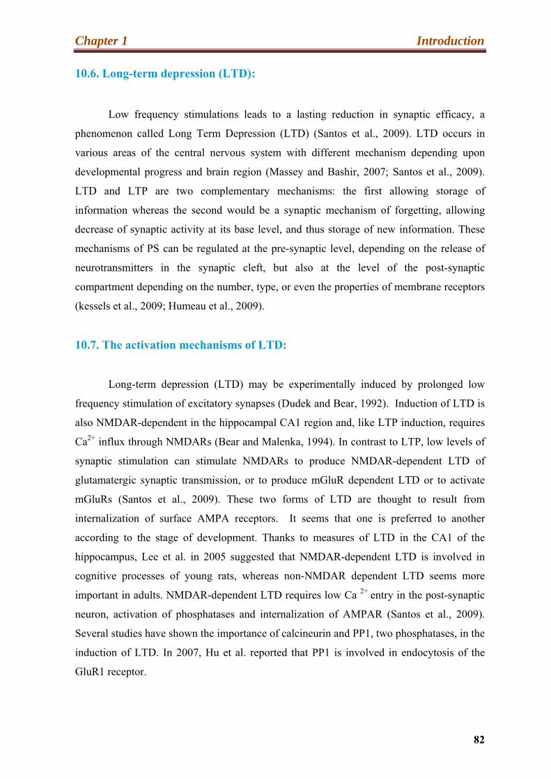
Chapter 1 Introduction
82
10.6. Long-term depression (LTD):
Low frequency stimulations leads to a lasting reduction in synaptic efficacy, a
phenomenon called Long Term Depression (LTD) (Santos et al., 2009). LTD occurs in
various areas of the central nervous system with different mechanism depending upon
developmental progress and brain region (Massey and Bashir, 2007; Santos et al., 2009).
LTD and LTP are two complementary mechanisms: the first allowing storage of
information whereas the second would be a synaptic mechanism of forgetting, allowing
decrease of synaptic activity at its base level, and thus storage of new information. These
mechanisms of PS can be regulated at the pre-synaptic level, depending on the release of
neurotransmitters in the synaptic cleft, but also at the level of the post-synaptic
compartment depending on the number, type, or even the properties of membrane receptors
(kessels et al., 2009; Humeau et al., 2009).
10.7. The activation mechanisms of LTD:
Long-term depression (LTD) may be experimentally induced by prolonged low
frequency stimulation of excitatory synapses (Dudek and Bear, 1992). Induction of LTD is
also NMDAR-dependent in the hippocampal CA1 region and, like LTP induction, requires
Ca2+ influx through NMDARs (Bear and Malenka, 1994). In contrast to LTP, low levels of
synaptic stimulation can stimulate NMDARs to produce NMDAR-dependent LTD of
glutamatergic synaptic transmission, or to produce mGluR dependent LTD or to activate
mGluRs (Santos et al., 2009). These two forms of LTD are thought to result from
internalization of surface AMPA receptors. It seems that one is preferred to another
according to the stage of development. Thanks to measures of LTD in the CA1 of the
hippocampus, Lee et al. in 2005 suggested that NMDAR-dependent LTD is involved in
cognitive processes of young rats, whereas non-NMDAR dependent LTD seems more
important in adults. NMDAR-dependent LTD requires low Ca 2+ entry in the post-synaptic
neuron, activation of phosphatases and internalization of AMPAR (Santos et al., 2009).
Several studies have shown the importance of calcineurin and PP1, two phosphatases, in the
induction of LTD. In 2007, Hu et al. reported that PP1 is involved in endocytosis of the
GluR1 receptor.
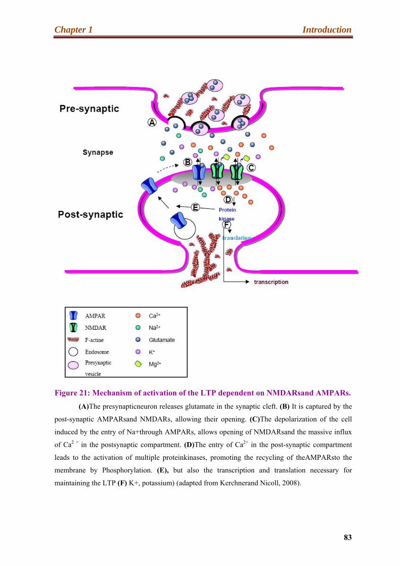
Chapter 1 Introduction
83
Figure 21: Mechanism of activation of the LTP dependent on NMDARsand AMPARs.
(A)The presynapticneuron releases glutamate in the synaptic cleft. (B) It is captured by the
post-synaptic AMPARsand NMDARs, allowing their opening. (C)The depolarization of the cell
induced by the entry of Na+through AMPARs, allows opening of NMDARsand the massive influx
of Ca2 + in the postsynaptic compartment. (D)The entry of Ca2+ in the post-synaptic compartment
leads to the activation of multiple proteinkinases, promoting the recycling of theAMPARsto the
membrane by Phosphorylation. (E), but also the transcription and translation necessary for
maintaining the LTP (F) K+, potassium) (adapted from Kerchnerand Nicoll, 2008).
Chapter 1 Introduction
84
PP1 is a phosphatase that interacts with a protein of the actin cytoskeleton, Neurabine I.
This interaction allows the localization of synaptic PP1 and its action on membrane
receptors. PP1 participates in the organization of LTD by dephosphorylating GlR1 at
Ser831 and Ser845. Moreover, the mechanism of phosphorylation and dephosphorylation,
involved in the endocytosis of AMPAR is clathrin-dependent. In 2002, Lee et al. described
that AP2, an adapter of clathrins, is required for internalization of GluR2. By using GluR2
and AP2 inhibiting peptides, the authors blocked the interaction of these two proteins. It
resulted in a decrease of GluR2 endocytosis associated with a reduction of NMDAR-
dependent LTD. Another important factor in this process was identified in 2005: Rab5, a
member of the small GTPases family. Brown et al. showed that induction of LTD triggers
the activation of this factor. Once activated Rab5 is involved in clathrin-dependent
internalization of GluR1 and GluR2, as indicated by the use of a mutant GluR2 unable to
bind the clathrin adapter AP2 (Brown et al., 2005). Following recycling, the authors also
identified a decrease in GluR1 phosphorylation at Ser845 and Ser831. This
dephosphorylation appears to be important for the expression of LTD as mentioned
previously (Hu et al., 2007) (Figure 22).
In the next section, I will discuss briefly dendritic spines their functions and
pathology of spines.
11. Dendritic spines:
The dendritic spine is a small membranous protrusion from a neuron’s dendrite that
typically receives input from a single synapse of an axon. The dendritic spines serve as a
storage site for synaptic strength and assist to transmit signals to the neuron’s cell body.
The human brain consists of hundred billion neurons interconnected into useful neuronal
circuits that cause all our emotions, dreams, behaviors and memories. The chemical
synapses control the electrical communication in neuronal system and pass information
from pre-synaptic axons terminals to postsynaptic dendritic regions (Hotulainen and
Hoogenraad, 2010). Previous studies suggested that alteration in spine morphology account
for functional differences at the synaptic level (Kasai et al., 2003; Yuste and Bonhoeffer,
2001).

Chapter 1 Introduction
85
Figure 22: Mechanism of Rab5 dependent AMPAR endocytosis during Long Term
Depression (LTD).
(a). A small entry of Ca2 + in the postsynaptic neuron induces LTD, by phosphorylation of
Rab-GDI, an inhibitor of Rab-GDP dissociation (b). Its activation leads to translocation of Rab5-
GDP from early endosome (EP) to the plasma membrane (c), where Rab5 is activated (to Rab-GTP)
and is involved in clathrin-dependent internalization of GluR1 and GluR2 (Adapted from Brown et
al., 2005).
Now it is widely accepted by scientists that the functional and structural changes at
spines and synapses are the basis of learning and memory in human brain (Holtmaat and
Svoboda, 2009; Kasai et al., 2010).
11.1. Dendritic spine structure and function:
The spines occur at a density of 1-10 spines/ μm of dendrite length, and some
neurons, like a hippocampal neurons, have thousand of spines all over the dendritic arbors

Chapter 1 Introduction
86
(Sorra and Harris, 2000). A bulbous head, allowing contact with a presynaptic partner,
represents the morphology of a typical spine. It is attached to the dendrite through the spine
neck (Hotulainen and Hoogenraad, 2010). Although this classic description, the
morphology of dendritic spines is extremely variable, depending on the development but
also of synaptic activity (Hering and Sheng, 2001; von Bohlen und Halbach, 2009). The
spines can be grouped into categories according to their shape: mature spines, and
immature spines (Boda et al., 2004, Grossman et al., 2010). On the other hand, the electron
microscopic studies suggested that spines can be categorized into three groups, based on
morphology; thin, flopodia-like protrusions, short spines without a well defined spine neck
(also called stubby spines), and spines with a large bulbous head (mushroom spines)
(Bourne and Harris, 2008). The most interesting characteristic of the spine structures is that
they change morphology continuously, even through adulthood, which reflects the plastic
nature of synaptic connections (Grutzendler et al., 2002; Trachtenberg et al., 2002). The
volume of a spine can vary from 0.001 to 1 μm3. A difference in length has also been
reported, spines from 0.2 to 2μm were observed (Hotulainen and Hoogenraad, 2010).
The proposed mechanism of synaptogenesis is as follows (von Bohlen und Halbach,
2009. In general, a newly synthesized dendrite does not yet have synapses or dendritic
protrusions. In the early stages of synaptogenesis, filopodia appear, and can grow and
retract rapidly to meet a pre-synaptic partner. Following this meeting, the filopodium
retracts to approximate the continuation pre-synaptic dendrite of its own, allowing the
synapse formation and maturation of by stabilizing the spine. The explanation is shown in
(Figure23).
A large number of studies revealed that morphology of spines could be modified by
a neuronal activity in vitro as well as in vivo (Holtmaat et al., 2006; Matsuzaki et al., 2004;
Roberts et al., 2010). For example, LTP causes the enlargement of spine heads (Kasai et al.,
2003; Yuste and Bonhoeffer, 2001). An attractive mechanism for regulating the
modification in spine morphology is the local addition or removal of synaptic membrane
and turnover of postsynaptic receptors. Indeed, synaptic stimulation assembles AMPA
receptors in recycling endosomal compartment into spines, an event necessary for
subsequent spine enlargement (Park et al., 2004; Park et al., 2006).
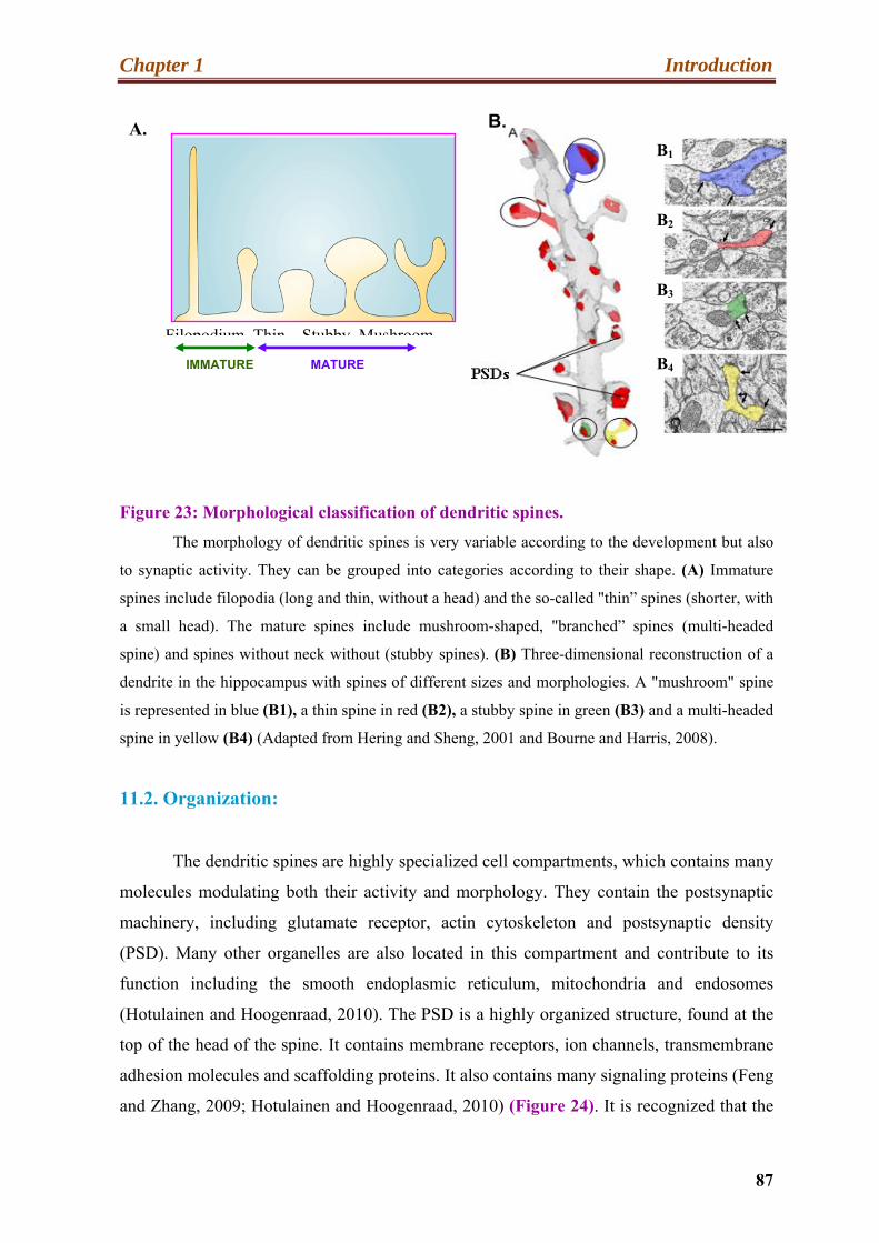
Chapter 1 Introduction
87
Figure 23: Morphological classification of dendritic spines.
The morphology of dendritic spines is very variable according to the development but also
to synaptic activity. They can be grouped into categories according to their shape. (A) Immature
spines include filopodia (long and thin, without a head) and the so-called "thin” spines (shorter, with
a small head). The mature spines include mushroom-shaped, "branched” spines (multi-headed
spine) and spines without neck without (stubby spines). (B) Three-dimensional reconstruction of a
dendrite in the hippocampus with spines of different sizes and morphologies. A "mushroom" spine
is represented in blue (B1), a thin spine in red (B2), a stubby spine in green (B3) and a multi-headed
spine in yellow (B4) (Adapted from Hering and Sheng, 2001 and Bourne and Harris, 2008).
11.2. Organization:
The dendritic spines are highly specialized cell compartments, which contains many
molecules modulating both their activity and morphology. They contain the postsynaptic
machinery, including glutamate receptor, actin cytoskeleton and postsynaptic density
(PSD). Many other organelles are also located in this compartment and contribute to its
function including the smooth endoplasmic reticulum, mitochondria and endosomes
(Hotulainen and Hoogenraad, 2010). The PSD is a highly organized structure, found at the
top of the head of the spine. It contains membrane receptors, ion channels, transmembrane
adhesion molecules and scaffolding proteins. It also contains many signaling proteins (Feng
and Zhang, 2009; Hotulainen and Hoogenraad, 2010) (Figure 24). It is recognized that the
A. B.
IMMATURE MATURE
Filopodium Thin Stubby Mushroom
B1
B2
B3
B4
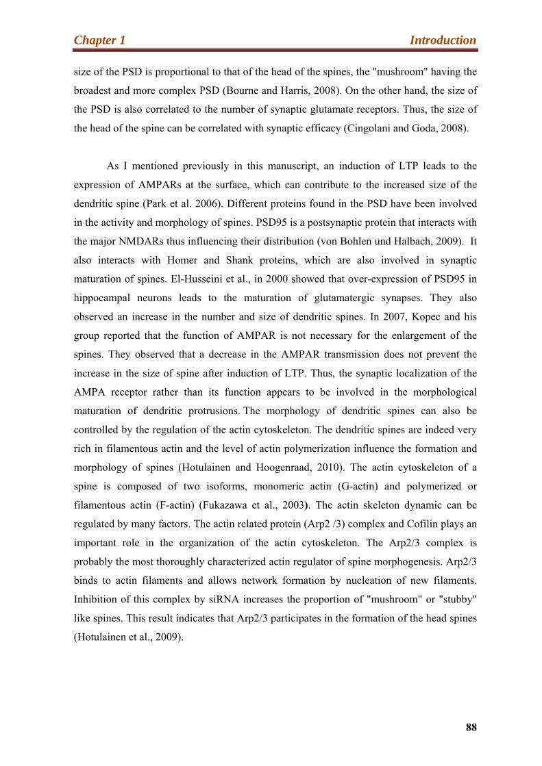
Chapter 1 Introduction
88
size of the PSD is proportional to that of the head of the spines, the "mushroom" having the
broadest and more complex PSD (Bourne and Harris, 2008). On the other hand, the size of
the PSD is also correlated to the number of synaptic glutamate receptors. Thus, the size of
the head of the spine can be correlated with synaptic efficacy (Cingolani and Goda, 2008).
As I mentioned previously in this manuscript, an induction of LTP leads to the
expression of AMPARs at the surface, which can contribute to the increased size of the
dendritic spine (Park et al. 2006). Different proteins found in the PSD have been involved
in the activity and morphology of spines. PSD95 is a postsynaptic protein that interacts with
the major NMDARs thus influencing their distribution (von Bohlen und Halbach, 2009). It
also interacts with Homer and Shank proteins, which are also involved in synaptic
maturation of spines. El-Husseini et al., in 2000 showed that over-expression of PSD95 in
hippocampal neurons leads to the maturation of glutamatergic synapses. They also
observed an increase in the number and size of dendritic spines. In 2007, Kopec and his
group reported that the function of AMPAR is not necessary for the enlargement of the
spines. They observed that a decrease in the AMPAR transmission does not prevent the
increase in the size of spine after induction of LTP. Thus, the synaptic localization of the
AMPA receptor rather than its function appears to be involved in the morphological
maturation of dendritic protrusions. The morphology of dendritic spines can also be
controlled by the regulation of the actin cytoskeleton. The dendritic spines are indeed very
rich in filamentous actin and the level of actin polymerization influence the formation and
morphology of spines (Hotulainen and Hoogenraad, 2010). The actin cytoskeleton of a
spine is composed of two isoforms, monomeric actin (G-actin) and polymerized or
filamentous actin (F-actin) (Fukazawa et al., 2003). The actin skeleton dynamic can be
regulated by many factors. The actin related protein (Arp2 /3) complex and Cofilin plays an
important role in the organization of the actin cytoskeleton. The Arp2/3 complex is
probably the most thoroughly characterized actin regulator of spine morphogenesis. Arp2/3
binds to actin filaments and allows network formation by nucleation of new filaments.
Inhibition of this complex by siRNA increases the proportion of "mushroom" or "stubby"
like spines. This result indicates that Arp2/3 participates in the formation of the head spines
(Hotulainen et al., 2009).
Chapter 1 Introduction
89
Figure 24: Protein organization in the postsynaptic density (PSD). The PSD is an electron-dense structure found at the top of the head of the spines. The PSD can be divided into three layers: the first layer contains membrane receptors AMPARs, NMDARs, mGluR), ion channels (K + channels) and transmembrane adhesion molecule (N-cadherin), the second layer consists of scaffolding proteins arranged perpendicularly to the membrane (PSD95), the last layer is formed of Shank proteins and proteins of the GKAP family. They are organized parallel to the membrane and are connected to the actin filaments. Many other signaling proteins are found including AKAP, adenylate-kinase anchoring protein; EphR, ephrin receptor; GKAP, guanylate kinase-associated protein; GRASP, GRIP-associated protein; GRIP, glutamate receptor interacting protein; IP3R, inositol-1,4,5-trisphosphate receptor; MAP1A, microtubule-associated protein 1A; mGluR, metabotropic glutamate receptor; nNOS, neuronal nitric oxide synthase; PICK1, protein interacting with PRKCA1; SER, smooth endoplasmic reticulum; SPAR, spine-associated RAPGAP; SV, synaptic vesicle; SyNGAP, synaptic Ras GTPase-activating protein; TIAM1, T-cell lymphoma invasion and metastasis 1; TRAP, C-terminal receptor-binding region (Adapted from Cingolani and Goda, 2008 and Carlisle et al., 2008).
Cofilin regulates the dynamics of the actin cytoskeleton by binding and
depolymerizating F-actin. Hotulainen et al. showed in their study in 2009 that inhibition by

Chapter 1 Introduction
90
siRNA of cofilin causes an increase of "branched" spines. In addition, it is interesting to
note that the activity of cofilin is inhibited by phosphorylation at Ser3. Several factors may
control its activity, including the LIM kinase and phosphatases such as SSH or calcineurin
(Kurita et al., 2007, Meng et al., 2002, Carlisle et al., 2008). In 2007, Zeng et al. described
an alteration of dendritic spines after injection of kainate. A decreased level of phospho-
Cofilin and thereby polymerized actin was the cause. The authors succeeded in improving
the dendritic phenotype by using a calcineurin inhibitor. Gu et al., in 2010 reported that the
state of phosphorylation of Cofilin regulates the morphology of spines.
11.3. Dendritic spine pathologies:
Some neurological diseases or cognitive impairment disorders are due to a loss of
spines or spine morphological disturbance (Blanpied and Ehlers, 2004). For example,
spines become abnormal or are lost in stroke, epilepsy, major depression, normal aging and
chronic substance abuse (Fiala et al., 2002; Nimchinsky et al., 2002; Swann et al., 2000).
The spine morphology being linked to synaptic function, spine abnormalities are likely to
have diverse functional effects (Calabrese et al., 2006) (Figure 25). They are, as already
mentioned, the support of the glutamatergic synapses in the hippocampus and their
structure is related to this synaptic activity.
Altered density and morphology of dendritic spines has been identified in patients
with Fragile X Syndrome (XFRA). Indeed the post-mortem observation of the neo-cortex
of these patients showed an increase in the density of these spines, including the proportion
of immature spines. The study of FID1-KO mouse model confirmed the dendritic
phenotype in the neo-cortex (von Bohlen und Halbach, 2009). Grossman et al., in 2010
described an increase in the amount of "thin" immature spines and a decline of mature
spines ("mushroom" and "stubby") in the dentate gyrus of these mice (Grossman et al.,
2006). Mutation in the PAK3, a serine/thréonine kinase of the family of the “p21-activated
kinase” (PAK1-3), cause a nonsyndromic ID. Boda et al., in 2004 described that
inactivation of PAK3 (by siRNA or antisense oligonucleotides) leads to alterations in the
function and morphology of dendritic spines. Indeed, following PAK3 gene silencing in
hippocampal organotypic cultures, they observed a reduction in the number of mature
spines and of LTP. The molecular mechanism is not yet defined, but it seems that activation

Chapter 1 Introduction
91
Decrease in density: MR, epilepsy, Alzheimer's malnutrition
Increase in density: X syndrome, Fragile, sudden death syndrome of the newborn
Decrease in size: Schizophrenia, Down syndrome
Change in morphology: MR, epilepsy, malnutrition
of PAK3 by phosphorylation (by Rac or Cdc42) is necessary for activation of the LIMK
and Cofilin.
Figure 25: Pathologies associated with an alteration of the density and morphology of
spines.
Morphological defects of dendritic spines have been identified in many diseases, such as MR
(Fragile X Syndrome and Down syndrome), schizophrenia, epilepsy or malnutrition. Alterations in
the density, the size and morphology of spines have particularly been reported (Adapted from Fiala
et al., 2002).
Boda et al., in 2008 showed that a constitutionally active form of PAK1, another member of
this family, could restore the dendritic phenotype caused by the inhibition of PAK3. It is
important to note that Meng et al. in 2005 did not observe these spine abnormalities in their
PAK3-KO mouse model. However, these mice showed a reduction in L-LTP. The authors
suggested that it might be due to a decrease in the level of phosphorylation of CREB and
thus of gene transcription. The role of PAK3 in the maturation of dendritic spines remains

Chapter 1 Introduction
92
controversial. More recently, a study in the laboratory of JC Lauterborn has demonstrated a
role for PAK kinases in the dendritic phenotype of FID1-KO mice. Indeed, a decreased
activity of PAKs as well as their activator Rac1 was observed in the hippocampus of KO
animals after "theta burst" stimulation. The authors suggested the involvement of these
proteins in the stabilization of newly formed actin filaments (Chen et al., 2010).
A Ph.D. student, Anne Scneider, in our laboratory has shown that the spine
morphology in Rsk2-KO neurons is altered. Anne preliminary results showed that there is
no difference in total spine density between WT and Rsk2-KO neurons, whereas the
proportion of mature spine was significantly higher in Rsk2-KO neurons.
12. The RSK2 protein and cognitive deficit:
The development of animal models for Rsk2 mutation and shRNA or siRNA
technology are major advances to study the disease mechanisms of CLS and cognitive
impairment. The main goal of our team is to explain the cognitive and behavioral
alterations of CLS, based on our Rsk2-KO mouse model. A previous study described a
deregulation of the dopaminergic system in the mrsk2_KO mouse. Based on HPLC
analysis, an increased level of dopamine expression (neurotransmitter monoamines was
identified in the cortex of Rsk2-KO mice (Figure 26A1). It was shown that absence of
RSK2 leads to increased phosphorylation of ERK1/2. This higher activity of ERK in
parallel with a decreased Protein phosphatase 2 (PP2A) activities resulted in an increased
Tyrosine hydroxylase (TH) activity (Marques Pereira et al., 2008) (Figure 26A2). Indeed,
this deregulation may contribute to the Cognitive impairment. It is interesting to note that a
deregulation of the dopaminergic system was also found in other pathologies such as
Parkinson's or schizophrenia.
A gene silencing study of the Rsk2 gene by siRNA in neuroendocrine cells,
(Zeniou-Meyer et al., 2008) showed the importance of RSK2 in the process of exocytosis
and in neurotransmission. It was shown that RSK2 regulates the activity, by
phosphorylation, of PDL1, a phospholipase required for membrane fusion process.
Phosphorylation of PDL1 at Thr147 activates PDL1, allowing biosynthesis of phosphatidic
acid (PA) required for exocytosis (Zeniou-Meyer et al., 2008) (Figure 26C1-C2).

Chapter 1 Introduction
93
Figure 26: Recent advances in the study of the pathophysiology of MR associated with
CLS.
(A) In 2008, Marques Pereira et al. described a dysregulation of the dopaminergic system in
the cortex of Rsk2-KO mice. An increase in dopamine was identified by HPLC (A.1.)Data showed
that it was due to increased tyrosine hydroxylase (TH) activity (A.2.). (B) In 2009 Fischer et al.
identified, in motoneurone cultures, increased axonal length and of the number of their connections
in the absence of RSK2 (B.1.). The authors rescued the phenotype by inhibiting the activity of ERK
kinase (B.2.). (C) Zeniou-Meyer et al. in 2008 and 2009 describe the importance of RSK2 in
exocytosis. Inhibition of RSK2 in endocrine cells led to reduced PDL1 activity (C.1.) and to the
release of growth hormone (C.2.). These observations may explain the decreased expression of
presynaptic LTP that was observed in the lateral amygdala by Zeniou-Meyer et al.in2010. (C3).
(Adapted from Marques Pereira et al., 2008, Fischer et al., 2009a; Zeniou-Meyer et al. 2008-2009-
2010).
Chapter 1 Introduction
94
This production is further enhanced by the recruitment of membrane Spo20p-GFP (a sensor
of the synthesis of PA, homologous to SNAP25). The mobilization of this sensor is also
regulated by RSK2 (Zeniou-Meyer et al., 2009). Moreover, it seems that another critical
factor in the process of exocytosis, Scribble, allows the membrane localization of RSK2
(due to its PDZ domain) and its action on the biosynthesis of PA (unpublished but cited by
Zeniou-Meyer and al., 2008). This study suggested that growth retardation and cognitive
impairment associated with CLS could partially be explained by a defect in release of
hormones or neurotransmitters.
Other studies involved RSK2 in axonal growth and branching, and in synapse
formation. Fischer et al. observed in 2009 that axonal growth of Rsk2 deficient motor
neurons is affected. An increase in the length of axons and the number of connections has
been identified in cultures of isolated neurons derived from KO mice (Figure 26B1).
This effect is dependent on ERK activity because inhibition of ERK activity
reversed this phenotype (Figure26B2). The authors proposed that the increased level of
ERK activity observed in RSK2 deficient neurons leads to a stronger activation of GSK3β
and MAP1B and their substrate. Up regulation of the activity of these factors induces
increased growth of microtubules as well as of axons.
In addition, recent data confirmed the role in the pre-synaptic function assigned to
RSK2. A decrease in the expression of pre-synaptic LTP was identified in the lateral
amygdala, whereas neither the release of neurotransmitters nor the probability or the
recycling of synaptic vesicles was altered in mice Rsk2-KO (Zeniou-Meyer et al., 2010).

Results
Chapter 2 Results
95
Results:
The RSK2 protein is member of the RSK serine/threonine protein kinase. Although
many cellular functions have been known through the RSK2 kinase. RSK2 belongs to a
family of four highly homologous (Ser/Thr kinases, RSK1-4) members in human, RSK2
encoded by Rps6ka3 gene and phosphorylated by ERK kinase. In order to understand the
molecular and cellular consequences of Rsk2 gene mutation, the physiopathological
mechanisms of Intellectual disability associated with CLS patients and to develop a
therapeutic strategy, our laboratory developed a Rsk2-KO mouse in 2004. This mouse
displays learning and long-term spatial memory deficits (Poirier et al., 2007). In addition,
although ubiquitously expressed, RSK2 shows, in the adult and embryonic brain, its highest
levels of expression in the hippocampus (Zeniou et al., 2002) a structure involved in these
cognitive processes. Some studies suggested that RSKs are involved in synaptic function,
and especially in the synthesis and release of hormones and neurotransmitters. Together all
the data suggested that RSK2 dysfunction might lead to deficiencies in synaptic
transmission and plasticity, and especially in the hippocampus.
The objectives of my thesis were to further investigate the role of RSK2 in the
hippocampus. My study was focused initially on identifying target gene of RSK2 by
comparing global gene expression profiles of hippocampi from Rsk2-KO and WT littermate
mice. My subsequent goal was to try to unravel the increase of ERK1/2 signaling in
hippocampal neurons of Rsk2-KO mice. The third objective was to explore the signaling
mechanism involved in the increased expression of one gene identified by transcriptome
profiling, Gria2, encoding for the GluR2 protein. This GluR2 protein is important for the
function of glutamergic AMPA receptors and play and important role in synaptic plasticity
and neurotransmission.
We conducted most of the studies on hippocampal tissues or on hippocampal
derived primary neuronal. Moreover, to understand the molecular mechanism involved in
the alteration of expression of GluR2 in RSK2 deficient cells, we used RNA interference
technology to knockdown Rsk2 gene in the PC12 cell line.
Our findings may be helpful, to develop new therapeutic strategies for Coffin-

Chapter 2 Results
96
Lowry syndrome, e.g. by modulating the glutamatergic neurotransmission and probably
synaptic plasticity. The studies were done in the laboratory of Dr. Andre Hanauer.

Publication-1
Chapter 2 Results-Publication 1
97
2.1. Transcriptome profile reveals AMPA receptor dysfunction in
the hippocampus of the Rsk2-knockout mice, an animal model
of Coffin Lowry syndrome
Tahir Mehmood, Anne Schneider, Jérémie Sibillec, Patricia Marques Pereira, Solange
Pannetier, Mohamed Raafet Ammar, Doulaye Dembele, Christelle Thibault-Carpentier,
Nathalie Rouach and André Hanauer. (2011).
Hum Genet 129, 255-269.
The first aim of my PhD project was to identify new target genes of Rsk2 in the
hippocampus of Rsk2-KO mice, with the hope that they may help us to identify cellular
processes affected by the absence of the RSK2 kinase. To this end we performed a
comparison of the hippocampal gene expression profiles from Rsk2-KO and WT littermate.
There was increasing evidence that RSK2 plays a role in regulating gene expression at the
transcription level. As discussed in detail in the introduction, RSK2 is implicated in the
control of the activity of many transcription factors by direct phosphorylation (CREB,
ATF4, ER, p53….) (Cho et al., 2005; Sutherland et al., 1993) or via phosphorylation of an
intermediate molecule, as for GSK3 and NFĸB (Peng et al., 2010a). Furthermore, RSK2
deficiency leads to decreased expression of immediate early genes, including the
transcription factors c-Fos in fibroblasts of patients or in osteoblasts (David et al., 2005; De
Cesare et al., 1998). Importantly, there is a huge body of evidence showing that the ERK
pathway plays a pivotal role in neuronal and cognitive functions. Therefore, we speculated
that deregulation of gene expression in the hippocampus might play a role in the
mechanism leading to cognitive deficits.
By microarray analysis we have identified differential expression of 100 genes (75
increased and 25 decreased in Rsk2-KO mice) encoding proteins acting in various
biological pathways. The genes identified by the study are involved in various biological
pathways, including cell cycle, cellular development, growth and proliferation apoptosis,
and contain molecule transporters, enzymes, transcription or translation factors and some
are also involved in different metabolic pathways, such as the metabolism of iron, calcium
or in various genetic diseases. Deregulation of many of these genes was confirmed at RNA
level by QRT-PCR and protein level by Western blot and immunohistochemistry.
Chapter 2 Results-Publication 1
98
We focused on functional studies for one of these genes, which showed an abnormal
two-fold increased expression at the protein and RNA levels, Gria2. The Gria2 gene
encodes the GluR2 proteins, a subunit of the glutamate ionotropic AMPA receptor. We
showed that Gria2 expression is up regulated in the hippocampus of Rsk2-KO mice by in
situ hybridization and that GluR2 subunit is over represented at the surface of synapses in
primary neuronal cultures. Electrophysiology studies conducted in collaboration with N.
Rouach (College de France, Paris), showed a 25% reduction of basal AMPA receptor-
mediated transmission in the hippocampus of Rsk2-KO mice. Furthermore, analysis of the
coding sequence of Gria2 showed a modification of splicing and editing of gria2 in Rsk2-
KO animals. An increase in the flop variant and a decrease in editing R/G were actually
observed.
In this study, we identified a number of genes whose expression is altered in the
hippocampus of mouse Rsk2-KO as compared to WT mice littermate. This definitively
confirms that RSK2 is implicated in regulating many cellular processes. The main
conclusion of this study was that deregulation of the expression, splicing and editing Gria2
in Rsk2-KO mice, and leading to altered AMPA neurotransmission, may contribute to the
cognitive dysfunction in Rsk2-KO mice and in CLS patients. However, many other
interesting genes identified by the transcriptional study remain to be explored for their
implication in neuronal and cognitive dysfunction in Rsk2-KO mice.
A more detailed description and illustration of these results are found in
publication- 1.
ORIGINAL INVESTIGATION
Transcriptome profile reveals AMPA receptor dysfunctionin the hippocampus of the Rsk2-knockout mice, an animalmodel of Coffin–Lowry syndrome
Tahir Mehmood • Anne Schneider • Jeremie Sibillec • Patricia Marques Pereira •
Solange Pannetier • Mohamed Raafet Ammar • Doulaye Dembele •
Christelle Thibault-Carpentier • Nathalie Rouach • Andre Hanauer
Received: 1 September 2010 / Accepted: 5 November 2010 / Published online: 30 November 2010
� Springer-Verlag 2010
Abstract Coffin–Lowry syndrome (CLS) is a syndromic
form of mental retardation caused by loss of function
mutations in the X-linked RPS6KA3 gene, which encodes
RSK2, a serine/threonine kinase acting in the MAPK/ERK
pathway. The mouse invalidated for the Rps6ka3 (Rsk2-
KO) gene displays learning and long-term spatial memory
deficits. In the current study, we compared hippocampal
gene expression profiles from Rsk2-KO and normal litter-
mate mice to identify changes in molecular pathways.
Differential expression was observed for 100 genes
encoding proteins acting in various biological pathways,
including cell growth and proliferation, cell death and
higher brain function. The twofold up-regulated gene
(Gria2) was of particular interest because it encodes the
subunit GLUR2 of the AMPA glutamate receptor. AMPA
receptors mediate most fast excitatory synaptic transmis-
sion in the central nervous system. We provide evidence
that in the hippocampus of Rsk2-KO mice, expression of
GLUR2 at the mRNA and at the protein levels is signifi-
cantly increased, whereas basal AMPA receptor-mediated
transmission in the hippocampus of Rsk2-KO mice is sig-
nificantly decreased. This is the first time that such dere-
gulations have been demonstrated in the mouse model of
the Coffin–Lowry syndrome. Our findings suggest that a
defect in AMPA neurotransmission and plasticity contrib-
ute to mental retardation in CLS patients.
Introduction
Coffin–Lowry syndrome (CLS; MIM#303600) is a rare
syndromic form of mental retardation that is characterized
by moderate to severe psychomotor retardation, growth
retardation, facial and digital dysmorphisms, as well as
progressive skeletal deformations (Hanauer and Young
2002). The gene mutated in CLS patients (RPS6KA3)
encodes a protein of 740 amino acids, RSK2 (alternative
names: p90RSK2, MAPKAPK1B), which belongs to a
family of four highly homologous proteins (RSK1–4),
encoded by distinct genes. RSKs are Ser/Thr protein
kinases that act at the distal end of the mitogen-activated
protein kinase/extracellular signal-regulated kinases
(MAPK/ERK) signaling pathway. RSKs are directly
phosphorylated and activated by ERK1/2 in response to
many growth factors and neurotransmitters (Frodin and
T. Mehmood and A. Schneider contributed equally to this work.
Electronic supplementary material The online version of thisarticle (doi:10.1007/s00439-010-0918-0) contains supplementarymaterial, which is available to authorized users.
T. Mehmood � A. Schneider � P. Marques Pereira �S. Pannetier � M. R. Ammar � A. Hanauer (&)
Department of Translational Medicine and Neurogenetics,
Institut de Genetique et de Biologie Moleculaire et Cellulaire
(IGBMC), Institut National de Sante et de Recherche Medicale
(INSERM) U964/Centre National de Recherche Scientifique
(CNRS) UMR 7104/Universite de Strasbourg,
67404 Illkirch, France
e-mail: [email protected]
T. Mehmood
Department of Chemistry, University of Sargodha,
Sargodha 40100, Pakistan
D. Dembele � C. Thibault-Carpentier
Microarray and Sequencing Platform, Institut de Genetique et de
Biologie Moleculaire et Cellulaire (IGBMC), Institut National de
Sante et de Recherche Medicale (INSERM) U964/Centre
National de Recherche Scientifique (CNRS) UMR
7104/Universite de Strasbourg, 67404 Illkirch, France
J. Sibillec � N. Rouach
College de France, 75005 Paris, France
123
Hum Genet (2011) 129:255–269
DOI 10.1007/s00439-010-0918-0

Gammeltoft 1999). RSK2 phosphorylates a wide range of
cytosolic substrates, such as GSK3 and IjB, and nuclear
substrates including ATF4, c-FOS and NUR77, CREB and
histone H3 (De Cesare et al. 1998; Sassone-Corsi et al.
1999). Activation of RSK2 is, therefore, thought to influ-
ence gene expression and to be involved in cell prolifera-
tion and survival. Numerous studies implicate the MAPK/
ERK signaling cascade and CREB-mediated gene tran-
scription in synaptic plasticity and memory (Davis and
Laroche 2006). In human and mouse brain, RSK2 is highly
expressed in the hippocampus, that is, an essential brain
structure in cognitive function and learning (Zeniou et al.
2002; Guimiot et al. 2004). RSK2-deficient mice show
delayed acquisition of a spatial memory reference task and
long-term spatial memory deficits (Poirier et al. 2007).
Thus, together the data suggest that RSK2 plays an
important role in cognitive function in human and in mice.
To gain greater insight into the molecular mechanisms
leading to learning and memory impairments in the Rsk2-
KO mice and to mental retardation in CLS, we examined in
the present study global gene expression profiles in hip-
pocampus from KO mice. The data revealed significant
alteration of 100 genes acting in a great variety of bio-
logical pathway in Rsk2-KO hippocampi. We further
investigated the function of one of these genes, Gria2,
which showed a twofold up-regulation in mutant mice.
Gria2 encodes the subunit GLUR2 of the AMPA receptor
(AMPAR). AMPARs are ligand-activated cation channels
that mediate the fast component of excitatory postsynaptic
currents in neurons of the central nervous system. The
GLUR2 subunit controls AMPAR Ca?? permeability, and
is involved in several forms of long-term synaptic plas-
ticity. Our results show that the expression of GLUR2 is
increased at the mRNA and at the protein level in the
hippocampus, as well as at the surface of synapses in
hippocampal primary cell cultures. Furthermore, basal
excitatory synaptic transmission through AMPARs is
impaired in the hippocampus of Rsk2 mutants.
Materials and methods
Animals and tissue dissection
Male Rsk2-KO and WT mice with a C57Bl/6x genetic
background were killed by cervical dislocation. Brains
were rapidly dissected and the hippocampus was isolated
using a standard dissection procedure. Tissue samples were
immediately frozen in liquid nitrogen and kept at -80�C
until use. All experiments were carried out in accordance
with the European Communities Council Directive of 24th
November 1986 (86/609/EEC). Every effort was made to
minimize the number of animals used and their suffering.
Microarray hybridization
To reduce variability, but also to obtain enough RNA for
the hybridization of each DNA chip, all probes for the gene
array experiments consisted of pooled RNA samples from
either two WT or two KO animals. In brief, total RNA was
extracted from hippocampi of six KO and six WT 5-month-
old male mice and purified using the TRIzol reagent
(Invitrogen, Cergy Pontoise, France) according to the
manufacturer’s instructions. The quality of total RNA was
monitored by Agilent 2100 Bioanalyzer (LabChip, Agilent
technologies, Massy, France). Two RNA samples for each
genotype were then pooled in equal quantities (thus,
resulting in a total of three arrays for each genotype).
Generation of double-stranded cDNA from 2.5 lg of total
RNA of each pooled RNA sample, preparation and labeling
of cRNA, hybridization to 430A 2.0 mouse genome arrays
(Affymetrix, Santa Clara, CA), washing, and scanning
were performed according to the protocols recommended
by Affymetrix in their GeneChip� Expression Analysis
Technical Manual (Affymetrix). The data of the expression
arrays produced for this report have been deposited in the
Gene Expression Omnibus (GEO) databank.
Microarray data analysis
Data were processed using the Affymetrix GeneChip
Operating Software [GCOS v1.4; Microarray Suite (MAS
5.0) algorithm]. Genes differentially expressed were
selected using the following steps: (1) selection of probe-
sets having a signal value above 15 (35th percentile of all
expression values) in at least one array out the 6, (2)
selection of probesets called present in at least two out of
three arrays for one of the two genotypes, (3) selection of
probesets with a t test p \ 0.03, (4) selection of probesets
having a fold change greater than 1.5. We finally verified
that selected probes have acceptable false discovery rate
(10%) (Benjamini and Hochberg 1995).
Ingenuity pathways analysis
Biologically relevant networks were created using the
ingenuity pathways analysis program (http://www.
Ingenuity.com), using the default parameters. Based on
the algorithmically generated connectivity between gene–
gene, gene–protein, and protein–protein interactions, the
program develops functional molecular networks that over-
lay genes in the dataset. This program calculated p values for
each network by comparing the number of focus genes that
were mapped in a given network, relative to the total number
of occurrences of those genes in all networks. The score for
each network is shown as the negative log of the p value,
which indicates the likelihood of finding a set of genes in the
256 Hum Genet (2011) 129:255–269
123

network by random chance. A score of 20 indicates that there
is a 10-20 chance that the focus genes would be in a network
because of random chance.
Real-time QRT-PCR analysis
QRT-PCR assays were performed on hippocampal RNA
samples obtained from WT and KO mice different from
those used for transcriptional profiling. RNA extraction
and QRT-PCR was performed as previously described
(Marques Pereira et al. 2008). A probe set for detection of
mouse Gapdh was used as an endogenous control gene.
The sequences of primers of the tested genes are listed in
Supplemental Table 1.
Western blot analysis
Protein extractions and Western blot analyses were per-
formed as previously described (Marques Pereira et al.
2008). Quantifications were carried out with the GeneTool
software of the Chemigenius apparatus (Syngene, Freder-
ick, MD, USA). Data were normalized either to GAPDH or
to b-TUBULIN. Student’s t test (two-tailed) was used to
determine the significance between the control and Rsk2-
KO samples, and p B 0.05 was considered significant.
Antibodies against GLUR2 (Millipore Corporation),
CACNG8 and VAMP4 (Abcam), EIF3A (Cell Signaling
Technology), DIABLO (Calbiochem), GAPDH (Chem-
icon) and b TUBULIN (Millipore) were used.
Immunohistochemistry
Frozen brain section was left 10 min at room temperature,
fixed for 10 min with 4% PFA and washed twice (19 PBS).
Endogenous peroxidase was inhibited by a treatment with
0.3% H2O2 solution. After washing, slides were incubated
in 10% normal goat serum in 19 PBS for 1 h. Primary
antibodies were added to the sections in 10% normal goat
serum and incubated overnight at 4�C. Antibody dilutions
were as follows: rabbit anti-GLUR2 (1:1000, Millipore
Corporation), rabbit anti-VAMP4 (1:1,000, Abcam), and
rabbit anti-IGF-1 (1:100, Abcam). Slides were subsequently
washed four times for 10 min in 19 PBS and incubated
with biotinylated secondary antibody for 2 h. After wash-
ing, sections were incubated for 30 min in Vectastain elite
ABC reagent and treated with peroxidase substrate solution
until desired stain intensity. After washing, samples were
mounted with KAISER’s glycerol gelatin (Merck).
In situ hybridization
Plasmids containing 30UTR regions of mouse Gria1 (encod-
ing GLUR1) or Gria2 (encoding GLUR2) cDNAs were
amplified by PCR using vector-specific primers and PCR
reactions were purified using Montage 96 (Millipore Corpo-
ration, Bedford, USA). Amplicons were then used as template
for in vitro transcription of sense and anti-sense Dig-labeled
riboprobes. To this aim, 1 lg linearized DNA was transcribed
using T7, T3 or Sp6 polymerases and the 109 DIG RNA
labeling mix (Roche Diagnostics, Meylan, France) according
to the manufacturer’s instructions. Brain sections 25-lm
thick were processed for ISH using GenePaint robotic
equipment and procedures (http://www.genepaint.org) as
previously described (Nakamoto et al. 2007).
Primary hippocampal cultures
Hippocampi dissected from WT and Rsk2-KO male mice at
embryonic day 17 were triturated and plated into wells of
24-well plates containing poly-D-lysine-coated coverslips
(Sigma), at a density of *1,00,000 neurons/well. Growth
media consisted of NeuroBasal (GIBCO, Invitrogen) supple-
mented with 19 B27 (GIBCO, Invitrogen), 0.5 mM L-gluta-
mine and 19 penicillin/streptomycin. The cultures were
maintained at 37�C in a humidified atmosphere containing 5%
CO2 and cultivated for 14 DIV prior to experimentation.
Immunocytochemistry
To label surface GLUR1 (sGLUR1) and GLUR2 (sGLUR2)-
containing AMPARs, 14 DIV live neurons were treated as
previously described (Ghate et al. 2007) with minor modi-
fications. Cells were incubated with rabbit anti-N-terminal
GLUR1 (Calbiochem) or mouse anti-N-terminal GLUR2
(Millipore) and mouse anti-PSD95 (NeuroMab).
Microscopy and data analysis
All images acquisitions and quantifications were performed
using standardized settings on a microscope (model DM4000
B, Leica) equipped with CCD camera (CoolSnap CF, color)
with a 963 objective. Obtained Tiff files were subjected to
quantification with ImageJ software (http://www.rsb.info.
nih.gov/ij/). For sGLURs quantification, the three thickest
dendrites per pyramidal neuron and five neurons per sample
were blindly chosen and the dendritic branches were manu-
ally traced and measured. AMPA receptors clusters were
counted and the number of clusters was normalized with the
dendritic length. Student’s t tests were used for comparison
between WT and Rsk2-KO cultures.
Determination of relative Gria2 Q/R and R/G editing,
and flip/flop splice levels
Total RNA was extracted (as above) from five Rsk2-KO
and five WT hippocampi, the Gria2 mRNA amplified by
Hum Genet (2011) 129:255–269 257
123

RT–PCR (three times each from independent RNA
preparations) and the product sequenced to determine the
relative levels of editing and splicing (Lee et al. 1998).
Electrophysiology
Standard techniques were used to prepare transverse acute
hippocampal slices (400-lm thick) from 4-week-old mice.
Slices were maintained at room temperature in a storage
chamber that was perfused with an artificial cerebrospinal
fluid (ACSF) (mM: 119 NaCl, 2.5 KCl, 1.3 MgSO4, 2.5
CaCl2, 1 NaH2PO4, 26 NaHCO3 and 11 glucose and
equilibrated with 95% O2 and 5% CO2) for at least 1 h
prior to recording. For synaptic recordings, a cut was made
between the CA3 and CA1 region to prevent bursting, and
the slices were bathed in a modified ACSF containing 100-
lm picrotoxin to block GABAAreceptor- mediated inhib-
itory postsynaptic currents. Field excitatory postsynaptic
potentials (fEPSPs) were recorded with glass pipettes
(2–5 MX) filled with ACSF, by stimulating Schaffer col-
laterals in stratum radiatum (0.1 Hz) with a monopolar
stimulating electrode. Responses were collected with
Axopatch-1D amplifier (Axon Instruments), filtered at
2 kHz, digitized at 10 kHz, and analyzed online using
Clampfit software (Molecular Devices).
Results
Expression profiling of wild-type and Rsk2-KO mice
To identify molecular changes potentially responsible for
the phenotype associated with RSPS6KA3 gene mutation in
the hippocampus, we performed a detailed comparison of
the transcriptional profiles of hippocampi isolated from six
KO and six WT 5-month-old male mice. To reduce vari-
ability, equivalent amounts of RNA from two mice with
the same genotype were pooled and processed for hybrid-
ization to the genome wide oligonucleotide microarray
(thus, three arrays per genotype). Out of the 22,690
probesets represented on the microarray, filter A selected
16,865 probesets that were restricted to 14,348 probesets
by filter b and to 635 probesets by filter c. Filter d, selected
a final number of 109 probesets. Eight of these 109 dif-
ferentially expressed genes were verified by two or, in one
case, three distinct probe sets. These multiple probe sets of
eight genes displayed consistent direction and similar
extent of changes in abundances of corresponding mRNAs.
The final list of 100 significant non-redundant genes is
shown in Table 1. Genes are tabulated according to func-
tional category and degree of over-expression/repression.
Most of them have a recognized function and can be
assigned to functional categories and subcategories.
Among them, 75 genes were transcriptionally up-regulated,
whereas 25 genes were down-regulated. The most upregu-
lated genes were thymosin beta 10 (Tmsb10, fold change of
3.51), followed by establishment of cohesion 1 homolog
A (Esco 1, fold change of 2.91) and thyroid hormone
receptor interactor 11 (Trip 11, fold change of 2.27). The
most downregulated genes were erythroid differentiation
regulator 1 (Erdr1) and serpin peptidase inhibitor, clade C,
member 1 (Serpin1) (-2.9 and -2.4 fold downregulations,
respectively). Major functional categories include enzymes
with various activities (17 up-regulated/2 down-regulated),
growth factors (2/0), ion channels (3/0), kinases (1/3),
peptidases (2/1), transcription regulators (6/1), transmem-
brane receptors (1/0), transporters (7/2) and molecules with
other functions (33/14).
Validation of microarray data by QRT-PCR
Twenty-four of these differentially expressed genes were
selected for validation, by QRT-PCR, based on the known
or putative neuronal functional roles (Table 1). These
genes represent different categories: genes implicated in
exocytosis (Stxbp3 and Vamp4), in mental retardation
(Cul4b, Lamp2, Vldlr, Igf1), in apoptosis (Stk3, Rasl10a,
Diablo), in cell differentiation and cytoskeleton organiza-
tion (Phkg1, Pdlim5, Timsb10, Enc1, Nptxr, Ptpn2, Car-
hsp1, Phip, Plek, Arhgap12, Cfl1), in translation regulation
(Eif3A) and finally genes encoding ion channel sub-units
(Cacnb4, Cacng8, Gria2). The results of the microarray
findings were validated in all the genes tested by real-time
PCR, although the fold change was not always accurately
replicated. We also confirmed by QRT-PCR unaltered
expression of some genes (including cFos, CREB) that had
similar levels of expression in KO and WT animals on
microarrays (results not shown).
Identification of biologically relevant networks
To gain insight into interactions among the differentially
expressed genes, we constructed biologically relevant
networks using the ingenuity pathway analysis software.
From the 100 differentially expressed genes, 78 genes were
mapped and assembled into five biological networks with a
score of C20. The network with the most significant score
(of 45) contains 23 of the differentially expressed genes.
This network contains genes involved in cell cycle, cellular
development, growth and proliferation and centers on the
NFjB complex. This network contains also Rb1 and Sod2,
both important actors in cellular growth and apoptosis, and
over expressed in Rsk2-KO neurons. The network with the
second highest score (of 34) is centered on TGFb1, which
controls proliferation, cellular differentiation, but also
various other functions. Fourteen genes with altered
258 Hum Genet (2011) 129:255–269
123

Ta
ble
1L
ist
of
gen
esd
iffe
ren
tial
lyex
pre
ssed
inth
eh
ipp
oca
mp
us
of
Rsk
2-K
Om
ice
Fu
nct
ion
al
cate
go
ry
Pro
be
set
Gen
ban
kG
ene
sym
bo
l
Gen
en
ame
Mic
roar
ray
qR
T-P
CR
FC
KO
/WT
pW
Tm
ean
Ct
±S
D
KO
mea
n
Ct
±S
D
FC
p
Gen
essh
ow
ing
dec
reas
edex
pre
ssio
n
Kin
ases
14
22
31
5_
x_
atN
M_
01
10
79
Ph
kg1
Ph
osp
ho
ryla
sek
inas
eg
amm
a1
0.5
40
.01
62
1±
0.0
30
.68
±0
.11
0.6
80
.00
3
14
18
05
2_
atN
M_
02
35
56
Mvk
Mev
alo
nat
ek
inas
e0
.55
0.0
19
0
14
27
28
2_
a_at
NM
_0
08
04
4F
xnF
rata
xin
0.6
10
.00
74
En
zym
es1
44
83
30
_at
NM
_0
10
35
8G
stm
5G
luta
thio
ne
S-t
ran
sfer
ase
M5
0.6
10
.02
3
14
27
97
5_
atN
M_
14
52
16
Ra
sl1
0a
Ras
-lik
e,fa
mil
y1
0,
mem
ber
A0
.62
0.0
15
1±
0.0
50
.70
±0
.05
90
.70
0.0
04
Pep
tid
ase
14
33
91
8_
atN
M_
03
49
41
Atg
4d
0.5
90
.00
57
Cy
tok
ine
14
18
34
5_
atN
M_
02
35
17
Tn
fsf1
3T
um
or
nec
rosi
sfa
cto
r(l
igan
d)
sup
erfa
mil
y,
mem
ber
13
0.6
00
.00
57
Tra
nsc
rip
tio
nre
gu
lato
r1
41
86
36
_at
NM
_0
01
08
33
18
Etv
3E
tsv
aria
nt
gen
e3
0.5
50
.02
6
Tra
nsp
ort
ers
14
23
92
7_
atN
M_
02
86
62
Slc
35
b2
So
lute
carr
ier
fam
ily
35
,m
emb
erB
20
.59
0.0
11
14
50
07
3_
atN
M_
00
84
44
Kif
3b
Kin
esin
fam
ily
,m
emb
er3
B0
.64
0.0
00
93
Tra
nsm
emb
ran
ere
cep
tor
14
50
14
7_
atN
M_
03
06
89
Np
txr
Neu
ron
alp
entr
axin
rece
pto
r0
.62
0.0
13
1±
0.0
08
0.8
7±
0.3
50
.87
0.0
35
Oth
erfu
nct
ion
s1
45
24
06
_x
_at
NM
_1
33
36
2E
rdr1
Ery
thro
idd
iffe
ren
tiat
ion
reg
ula
tor
10
.35
0.0
29
14
17
90
9_
atN
M_
08
08
44
Ser
pin
c1S
erp
inp
epti
das
ein
hib
ito
r,cl
ade
C,
mem
ber
10
.41
0.0
23
14
15
97
6_
a_at
NM
_0
25
82
1C
arh
sp1
Cal
ciu
mre
gu
late
dh
ead
stab
lep
rote
in1
,24
kD
a0
.50
0.0
00
36
1±
0.0
70
.63
±0
.07
0.6
30
.03
4
14
52
65
2_
a_at
NM
_0
01
00
22
67
Tm
em1
58
Tra
nsm
emb
ran
ep
rote
in1
58
0.5
50
.01
5
14
48
57
7_
x_
atN
M_
00
93
04
Syn
gr2
Sy
nap
tog
yri
n2
0.5
60
.02
1
14
55
09
8_
a_at
NM
_0
11
70
7V
tnV
itro
nec
tin
0.5
70
.00
22
14
53
11
1_
a_at
NM
_0
26
54
2S
lc2
5a
39
So
lute
carr
ier
fam
ily
25
,m
emb
er3
90
.61
0.0
11
14
18
39
6_
atN
M_
13
41
16
Gp
sm3
G-p
rote
insi
gn
alin
gm
od
ula
tor
30
.63
0.0
16
14
23
94
0_
atN
M_
02
65
53
Yif
1A
Yip
1in
tera
ctin
gfa
cto
rh
om
olo
gA
(S.
cere
visi
ae)
0.6
30
.01
6
14
18
12
3_
atN
M_
01
16
76
Un
c11
9U
nc-
11
9h
om
olo
g(C
.el
ega
ns)
0.6
40
.00
16
14
50
46
8_
atN
M_
01
08
65
Myo
cM
yo
cili
n0
.64
0.0
07
8
14
15
88
6_
atN
M_
01
37
81
Sh
2d
3c
SH
2d
om
ain
con
tain
ing
3C
0.6
40
.00
1
14
22
59
6_
atN
M_
02
14
26
Nka
in4
Na?
/K?
tran
spo
rtin
gA
TP
ase
inte
ract
ing
40
.65
0.0
22
14
50
25
5_
atN
M_
02
02
60
Cd
ga
pC
dc4
2G
TP
ase-
acti
vat
ing
pro
tein
0.6
50
.00
9
Gen
essh
ow
ing
incr
ease
dex
pre
ssio
n
Kin
ases
14
18
51
3_
atN
M_
01
96
35
Stk
3S
erin
e/th
reo
nin
ek
inas
e3
1.7
60
.02
31
±0
.05
1.4
5±
0.0
81
.45
0.0
42
Ph
osp
hat
ase
14
38
56
2_
a_at
NM
_0
01
12
71
77
Ptp
n2
Pro
tein
tyro
sin
ep
ho
sph
atas
e,n
on
-rec
epto
rty
pe
21
.95
0.0
16
1±
0.0
91
1.8
5±
0.2
81
.83
0.0
45
Gro
wth
fact
ors
14
37
40
1_
atN
M_
01
05
12
Igf1
Insu
lin
-lik
eg
row
thfa
cto
r1
1.6
50
.00
93
1±
0.0
92
.24
±0
.32
2.2
40
.04
2
14
17
06
9_
a_at
NM
_0
22
02
3G
mfb
Mat
ura
tio
nfa
cto
r,b
eta
1.5
40
.00
55
Hum Genet (2011) 129:255–269 259
123

Ta
ble
1co
nti
nu
ed
Fu
nct
ion
al
cate
go
ry
Pro
be
set
Gen
ban
kG
ene
sym
bo
l
Gen
en
ame
Mic
roar
ray
qR
T-P
CR
FC
KO
/WT
pW
Tm
ean
Ct
±S
D
KO
mea
n
Ct
±S
D
FC
p
En
zym
es1
44
87
34
_at
NM
_0
01
04
26
11
Cp
Cer
ulo
pla
smin
2.2
90
.02
9
14
17
26
2_
atN
M_
01
11
98
Ptg
s2P
rost
agla
nd
in-e
nd
op
ero
xid
ase
syn
thas
e2
2.2
80
.00
47
14
52
48
4_
atN
M_
05
30
70
Ca
7C
arb
on
ican
hy
dra
seV
II2
.23
0.0
28
14
17
19
4_
atN
M_
01
36
71
So
d2
Su
per
ox
ide
dis
mu
tase
21
.98
0.0
08
9
14
52
15
8_
atN
M_
02
97
35
Ep
rsG
luta
my
l-p
roly
l-tR
NA
syn
thet
ase
1.9
50
.01
5
14
53
92
8_
a_at
NM
_0
09
27
8S
sbS
jog
ren
syn
dro
me
anti
gen
B1
.86
0.0
20
14
51
43
6_
atN
M_
00
10
81
20
3S
bn
o1
sno
,st
raw
ber
ryn
otc
hh
om
olo
g1
(Dro
sop
hil
a)
1.7
80
.02
2
14
54
69
6_
atN
M_
00
81
42
Gn
b1
Gu
anin
en
ucl
eoti
de
bin
din
gp
rote
in,
bet
ap
oly
pep
tid
e1
1.7
00
.00
16
1±
0.0
33
1.3
6±
0.0
61
1.3
60
.00
05
14
19
06
4_
a_at
NM
_0
11
67
4U
gt8
UD
P-g
lyco
sylt
ran
sfer
ase
81
.69
0.0
05
9
14
16
49
7_
atN
M_
00
97
87
Pd
ia4
Pro
tein
dis
ulfi
de
iso
mer
ase
fam
ily
A,
mem
ber
41
.68
0.0
06
1
14
23
03
3_
atN
M_
00
84
08
Stt
3a
Su
bu
nit
of
the
oli
go
sacc
hay
ltra
nsf
eras
eco
mp
lex
,h
om
olo
gA
1.6
60
.02
8
14
35
16
4_
s_at
NM
_0
11
66
6U
be1
cU
biq
uit
in-a
ctiv
atin
gen
zym
eE
1C
1.6
50
.00
62
14
53
88
_at
NM
_1
83
02
8P
cmtd
1p
rote
in-L
-iso
asp
arta
teO
-met
hy
ltra
nsf
eras
ed
om
ain
con
tain
ing
11
.60
0.0
26
14
16
34
3_
a_at
NM
_0
10
68
5L
am
p2
Ly
soso
mal
-ass
oci
ated
mem
bra
ne
pro
tein
21
.58
0.0
23
1±
0.0
51
.65
±0
.19
1.6
50
.03
8
14
18
90
8_
atN
M_
01
36
26
Pa
mP
epti
dy
lgly
cin
eal
ph
a-am
idat
ing
mo
no
ox
yg
enas
e1
.56
0.0
21
14
51
82
8_
a_at
NM
_2
07
62
5A
csl4
Acy
l-C
oA
syn
thet
ase
lon
gch
ain
fam
ily
mem
ber
41
.54
0.0
03
5
14
17
69
7_
atN
M_
00
92
30
So
at1
Ste
rol
O-a
cylt
ran
sfer
ase
1.5
10
.01
5
Pep
tid
ase
14
20
96
4_
atN
M_
00
79
30
En
c1E
cto
der
mal
-neu
ral
cort
ex2
.07
0.0
15
1±
0.0
51
.77
±0
.08
1.7
70
.00
2
14
49
71
8_
s_at
NM
_0
26
27
3C
3o
rf3
8C
hro
mo
som
e3
op
enre
adin
gfr
ame
38
ho
mo
log
(hu
man
)1
.54
0.0
19
Tra
nsc
rip
tio
nre
gu
lato
rs1
42
74
06
_at
NM
_0
28
44
6T
rip
11
Th
yro
idh
orm
on
ere
cep
tor
inte
ract
or
11
2.2
70
.00
45
14
23
50
1_
atN
M_
00
85
58
Ma
xM
YC
-ass
oci
ated
fact
or
X1
.91
0.0
23
14
17
18
7_
atN
M_
01
67
86
Hip
2H
un
tin
gto
nin
tera
ctin
gp
rote
in2
1.7
60
.00
71
14
24
70
4_
atN
M_
00
11
45
92
0R
un
x2R
un
t-re
late
dtr
ansc
rip
tio
nfa
cto
r2
1.6
70
.00
68
14
18
26
5_
s_at
NM
_0
08
39
1Ir
f2In
terf
ero
nre
gu
lato
ryfa
cto
r1
.60
0.0
23
14
17
85
0_
atN
M_
00
90
29
Rb
1R
etin
ob
last
om
a1
1.5
80
.00
53
Tra
nsl
atio
nre
gu
lato
r1
41
66
61
_at
NM
_0
10
12
3E
if3
aE
uk
ary
oti
ctr
ansl
atio
nin
itia
tio
nfa
cto
r3
,su
bu
nit
A1
.74
0.0
08
71
±0
.05
1.8
4±
0.1
11
.84
0.0
05
Nu
clea
rre
cep
tors
14
22
63
1_
atN
M_
01
34
64
Ah
rA
ryl
hy
dro
carb
on
rece
pto
r1
.61
0.0
28
14
16
95
8_
atN
M_
01
15
84
Nri
D2
Nu
clea
rre
cep
tor
sub
fam
ily
1,
gro
up
D,
mem
ber
21
.54
0.0
06
4
Tra
nsp
ort
ers
14
22
96
6_
a_at
NM
_0
11
63
8T
frc
Tra
nsf
erri
nre
cep
tor
2.1
00
.00
10
14
16
65
3_
atN
M_
01
15
04
Stx
bp
3S
yn
tax
in-b
ind
ing
pro
tein
31
.92
0.0
13
1±
0.0
32
.19
±0
.62
.19
0.0
38
14
42
16
9_
atN
M_
01
37
03
Vld
lrV
ery
low
den
sity
lip
op
rote
inre
cep
tor
1.7
90
.00
68
1±
0.0
51
.44
±0
.05
51
.44
0.0
00
7
14
24
92
4_
atN
M_
15
30
55
Sec
63
SE
C6
3h
om
olo
g(S
.ce
revi
sia
e)1
.74
0.0
15
14
19
97
5_
atN
M_
01
13
27
Scp
2S
tero
lca
rrie
rp
rote
in2
1.6
60
.02
3
14
16
37
4_
atN
M_
01
88
29
Ap
3m
1A
dap
tor-
rela
ted
pro
tein
com
ple
x3
mu
1su
bu
nit
1.5
90
.00
32
14
34
51
3_
atN
M_
00
11
28
09
6A
tp1
3a
3A
TP
ase
typ
e1
3A
31
.52
0.0
05
4
Ion
chan
nel
s1
45
30
98
_at
NM
_0
13
54
0G
ria
2G
luta
mat
ere
cep
tor,
ion
otr
op
ic,
AM
PA
22
.01
0.0
24
1±
0.0
32
1.8
7±
0.0
91
.87
0.0
00
3
14
52
08
9_
atN
M_
00
10
37
09
9C
acn
b4
Cal
ciu
mch
ann
el,
vo
ltag
e-d
epen
den
t,b
eta
4su
bu
nit
1.8
50
.00
31
1±
0.0
22
1.3
4±
0.0
21
.34
0.0
22
14
51
86
4_
atN
M_
13
31
90
Ca
cng
8C
alci
um
chan
nel
,v
olt
age-
dep
end
ent,
gam
ma
sub
un
it8
1.7
90
.01
21
±0
.08
1.8
4±
0.2
61
.84
0.0
05
260 Hum Genet (2011) 129:255–269
123

Ta
ble
1co
nti
nu
ed
Fu
nct
ion
al
cate
go
ry
Pro
be
set
Gen
ban
kG
ene
sym
bo
l
Gen
en
ame
Mic
roar
ray
qR
T-P
CR
FC
KO
/WT
pW
Tm
ean
Ct
±S
D
KO
mea
n
Ct
±S
D
FC
p
Oth
erfu
nct
ion
s1
45
59
46
_x
_at
NM
_0
01
03
93
92
Tm
sb1
0T
hy
mo
sin
,b
eta
10
3.5
10
.00
52
1±
0.0
42
.56
±0
.05
2.5
60
.02
8
14
24
32
5_
atN
M_
00
10
81
22
2E
sco
1E
stab
lish
men
to
fco
hes
ion
1h
om
olo
g1
(S.
cere
visi
ae)
2.9
10
.02
4
14
25
76
8_
atN
M_
02
32
32
Dia
blo
Dia
blo
ho
mo
log
(Dro
sop
hil
a)
2.0
50
.02
31
±0
.03
22
.16
±0
.05
2.1
60
.00
02
14
55
47
5_
atN
M_
02
66
22
C4
orf
29
Ch
rom
oso
me
4o
pen
read
ing
fram
e2
9h
om
olo
g(h
um
an)
2.0
00
.02
3
14
23
19
8_
a_at
NM
_1
34
03
4S
mek
2S
ME
Kh
om
olo
g2
,su
pp
ress
or
of
mek
1(D
icty
ost
eliu
m)
1.9
30
.01
4
14
24
07
4_
atN
M_
02
74
53
Btf
3l4
Bas
ictr
ansc
rip
tio
nfa
cto
r3
-lik
e4
1.8
80
.01
8
14
20
48
5_
atN
M_
02
35
54
No
l7N
ucl
eola
rp
rote
in7
1.7
80
.01
6
14
38
41
8_
atN
M_
14
45
35
Mu
den
gM
U-2
/AP
1M
2d
om
ain
con
tain
ing
,d
eath
-in
du
cin
g1
.77
0.0
14
14
21
94
5_
a_at
NM
_0
01
04
25
56
Bxd
c1B
rix
do
mai
nco
nta
inin
g1
1.7
70
.00
77
14
50
41
8_
a_at
NM
_0
26
55
3Y
if1
aY
ip1
-in
tera
ctin
gfa
cto
rh
om
olo
gA
(S.
cere
visi
ae)
1.7
40
.02
9
14
49
88
4_
atN
M_
02
66
26
Efc
ab2
EF
-han
dca
lciu
mb
ind
ing
do
mai
n2
1.7
10
.02
0
14
18
00
8_
atN
M_
02
61
10
C2
1o
rf6
6C
hro
mo
som
e2
1o
pen
read
ing
fram
e6
6h
om
olo
g(h
um
an)
1.6
80
.02
3
14
38
53
5_
atN
M_
00
10
81
21
6P
hip
Ple
ckst
rin
ho
mo
log
yd
om
ain
inte
ract
ing
pro
tein
1.6
50
.01
11
±0
.19
2.0
8±
0.0
92
.08
0.0
3
14
51
52
5_
atN
M_
00
10
39
69
2A
rhg
ap1
2R
ho
GT
Pas
e-ac
tiv
atin
gp
rote
in1
21
.64
0.0
20
1±
0.0
91
.54
±0
.09
1.5
40
.03
3
14
26
27
1_
atN
M_
15
38
08
Sm
c5S
tru
ctu
ral
mai
nte
nan
ceo
fch
rom
oso
me
51
.63
0.0
15
14
17
17
0_
atN
M_
03
33
22
Lzt
fl1
Leu
cin
ezi
pp
ertr
ansc
rip
tio
nfa
cto
r-li
ke
11
.62
0.0
29
1452247_at
NM
_001113188
Fxr1
Fra
gil
eX
men
tal
reta
rdat
ion,
auto
som
alhom
olo
g1
1.6
20.0
047
14
51
32
5_
atN
M_
02
72
26
Fy
ttd
1F
ort
y-t
wo
–th
ree
do
mai
nco
nta
inin
g1
1.6
10
.00
50
14
25
91
3_
a_at
NM
_1
44
88
2D
nap
tp6
Vir
alD
NA
po
lym
eras
e-tr
ans-
acti
vat
edp
rote
in6
1.6
00
.01
2
14
21
94
0_
atN
M_
00
92
82
Sta
g1
Str
om
alan
tig
en1
1.5
90
.01
1
14
48
74
8_
atN
M_
01
95
49
Ple
kP
leck
stri
n1
.58
0.0
21
1±
0.0
51
.12
±0
.09
1.1
20
.00
4
14
51
83
0_
a_at
NM
_0
09
26
0S
pn
b2
Sp
ectr
in,
bet
a,n
on
-ery
thro
cyti
c1
1.5
80
.01
9
14
37
46
3_
x_
atN
M_
01
15
77
Tg
fbI
Tra
nsf
orm
ing
gro
wth
fact
or,
bet
a-in
du
ced
1.5
70
.01
6
14
23
84
1_
atN
M_
02
63
96
Bx
dc2
Bri
xd
om
ain
con
tain
ing
21
.57
0.0
26
14
27
10
4_
atN
M_
17
54
80
Zn
f23
Zin
cfi
ng
erp
rote
in6
12
1.5
60
.01
5
14
26
80
6_
atN
M_
02
86
96
Ob
fc2
aO
lig
on
ucl
eoti
de/
oli
go
sacc
har
ide-
bin
din
gfo
ldco
nta
inin
g2
A1
.55
0.0
23
14
22
89
6_
atN
M_
01
67
96
Vam
p4
Ves
icle
-ass
oci
ated
mem
bra
ne,
pro
tein
41
.55
0.0
16
1±
0.0
71
.48
±0
.16
1.4
80
.03
4
14
37
79
0_
atN
M_
00
10
81
19
1E
ml5
Ech
ino
der
mm
icro
tub
ule
asso
ciat
edp
rote
inli
ke
51
.54
0.0
20
14
17
45
3_
atN
M_
00
11
10
14
2C
ul4
bC
ull
in4
B1
.53
0.0
28
1±
0.0
31
.12
±0
.09
1.1
20
.01
8
14
18
06
6_
atN
M_
00
76
88
Cfl
2C
ofi
lin
21
.53
0.0
19
14
50
40
7_
a_at
NM
_0
09
67
2A
np
32
aA
cid
icn
ucl
ear
ph
osp
ho
pro
tein
32
fam
ily
,m
emb
erA
1.5
30
.01
3
14
50
78
6_
atN
M_
01
98
08
Pd
lim
5P
DZ
and
LIM
do
mai
n5
1.5
30
.00
57
1±
0.1
11
.44
±0
.05
91
.44
0.0
47
14
25
59
7_
a_at
NM
_0
21
88
1Q
kQ
uak
ing
ho
mo
log
,K
Hd
om
ain
RN
Ab
ind
ing
1.5
20
.02
9
Rea
l-ti
me
qu
anti
tati
ve
RT
–P
CR
was
per
form
edfo
r2
4g
enes
inh
ipp
oca
mp
io
fR
sk2-K
O(n
=6
)an
dW
T(n
=6
)m
ice,
usi
ng
the
pri
mer
pai
rssh
ow
nin
Tab
leS
1.
Rel
ativ
eex
pre
ssio
nle
vel
sar
esh
ow
nw
ith
the
aver
age
exp
ress
ion
val
ue
of
the
WT
gro
up
set
to1
Ct
cycl
eth
resh
old
val
ues
,S
Dst
and
ard
dev
iati
on
,F
Cfo
ldch
ang
e,p
pv
alu
e
Hum Genet (2011) 129:255–269 261
123

expression in Rsk2-KO mice are associated with this sec-
ond network, among which Nptxr, Cacng8, Pdlim5 and
Soat1. The third network centered on MYC is mainly
implicated in lipid metabolism and cell death. Thirteen
focus genes are associated with this network, including
Acsl4, Eif3a, Ca7, Cfl2 and Fxr1. The last two networks
are mainly implicated in molecular transport and lipid
metabolism (4th network) and amino acid metabolism and
protein synthesis (5th network). The fourth network is
centered on Ptgs2 and includes 12 focus genes, among
which Igf1, Vamp4, Mvk, Stxbp3 and Gnb1. The fifth
network is centered on retinoic acid and contains 13 focus
genes, among which Cacnb4 and Carhsp1. Figure 1 shows
the two most significant networks (the others are available
on request to AH). Supplemental Table 2 lists the top
related biological functions and diseases. Top biological
functions include organismal injury and abnormalities
(9 genes, including Fxn, Sod2, Igf1, Ahr and Ptgs2), cell
cycle (12 genes including Rb1, Igf1 and Runx2), nervous
system development and function (10 genes including Igf1,
Ptgs2, Sod2, Gria2, Vldlr, Vtn and Rb1), organismal devel-
opment and free radical scavenging (9 and 4 genes, respec-
tively). Interestingly, five genes, including Gria2, Igf1,
Ptgs2, Sod2 and Vldlr have been implicated in behavior, with
the first four genes specifically in spatial memory formation.
These five genes are all up-regulated in the hippocampus of
Rsk2-KO mice. The p values in the range of 2.52 9 10-5 to
1.10 9 10-2 indicate statistical significance.
Confirmation of altered expression at the protein level
We confirmed increased expression at the protein level of
GLUR2 (Gria2 gene), CACNG8, VAMP4, EIF3A and
DIABLO using quantitative western blot analysis (Fig. 2).
These findings are in line with the changes detected by
microarray-based analysis. We also confirmed differential
expression of GLUR2, VAMP4 and IGF1 using immuno-
histochemical analysis. As shown in Fig. 3, we found
increased expression of GLUR2 in the CA1 and CA3
regions and in the dentate gyrus of Rsk2-KO hippocampus.
VAMP4 and IGF1 were increased in the whole hippo-
campus. Although the lack of specific antibodies precluded
similar experimental validations for many other genes lis-
ted in Table 1, these observations suggested that the tran-
scriptional changes observed in mutant mice may be
generally reflected by matching changes in the levels of
expression of their corresponding protein products.
Confirmation by in situ hybridization
of Gria2 up-regulation
The expression of a number of genes involved in neuro-
transmission, including vesicle and receptor trafficking
proteins, neurotransmitter receptors and ion channels, were
altered in Rsk2-KO mice (Table 1). Of particular interest
was the twofold increased level of expression of Gria2,
encoding the subunit GLUR2 of the AMPAR. Since AM-
PARs mediate fast synaptic transmission at excitatory
synapses in the brain and are thought to play key roles in
synaptic plasticity, learning and memory (Seidenman et al.
2003) we wanted to further confirm up-regulation of the
Gria2 gene by in situ hybridization of WT and KO mice
hippocampi. The expression of Gria2 was significantly
increased in all areas of the anterior hippocampus of
mutant mice (Fig. 4a), whereas in the posterior hippo-
campus, the level of Gria2-mRNA was mainly increased in
the dentate gyrus, in CA1 and in the CA3 region (Fig. 4b).
We did not find any change in the level of expression of
GLUR1 (not shown).
Increase in GLUR2 surface expression
We next wondered whether expression of GLUR2 at sur-
face of synapses was also up-regulated since AMPARs in
hippocampal neurons are mainly expressed as heteromers
of GLUR1/2 as well as GLUR2/3. We compared the
amount of GLUR2 at synapses in KO and WT cultured
primary hippocampal neurons by staining surface GLUR2
(or GLUR1) clusters and counterstaining for PSD95.
GLUR2 surface staining was punctate (Fig. 5a, b), and the
number of GLUR2 puncta that were synaptic did signifi-
cantly differ among WT and Rsk2-KO cultures (WT
8.1 ± 1.4, n = 12 embryos, KO 14.9 ± 2.2, n = 12,
p = 0.015). This provided evidence that increase in total
GLUR2 was correlated with increased surface-expressed
GLUR2. No significant difference of surface GLUR1 was
detected (WT 12.4 ± 7.4, n = 4, KO 10.7 ± 5, n = 6,
p = 0.8) (Supplemental Fig. 1).
Determination of relative Gria2 R/G editing
and flip/flop splice levels
The great majority of native AMPA receptors are imper-
meable to calcium ions, due to the presence of the GLUR2
subunit. This subunit confers calcium impermeability on the
channel due to RNA editing of a glutamine (Q) to an arginine
(R) at codon 607. In addition to the Q/R site in GLUR2, the
GLUR2, 3 and 4 subunits undergo RNA editing (arginine to
glycine) at codon 764 (in GLUR2) (Lomeli et al. 1994). The
pre-messenger RNA transcripts of all the four GLUR sub-
units can finally be alternatively spliced to produce either the
flip or flop isoforms. Because the level of expression of
GLUR2 was increased in Rsk2-KO mice, we wondered
whether Gria2 RNA editing and splicing were altered.
To determine whether there are changes in the Q/R, R/G
site editing and flip/flop splice levels of the Gria2
262 Hum Genet (2011) 129:255–269
123
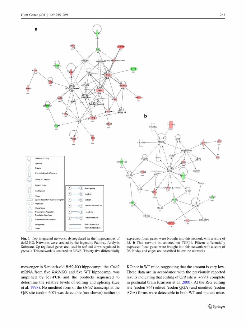
messenger in 5-month-old Rsk2-KO hippocampi, the Gria2
mRNA from five Rsk2-KO and five WT hippocampi was
amplified by RT-PCR and the products sequenced to
determine the relative levels of editing and splicing (Lee
et al. 1998). No unedited form of the Gria2 transcript at the
Q/R site (codon 607) was detectable (not shown) neither in
KO nor in WT mice, suggesting that the amount is very low.
These data are in accordance with the previously reported
results indicating that editing of Q/R site is *99% complete
in postnatal brain (Carlson et al. 2000). At the R/G editing
site (codon 764) edited (codon GGA) and unedited (codon
AGA) forms were detectable in both WT and mutant mice.
Fig. 1 Top integrated networks dysregulated in the hippocampus of
Rsk2-KO. Networks were created by the Ingenuity Pathway Analysis
Software. Up-regulated genes are listed in red and down-regulated in
green. a This network is centered on NFjB. Twenty-five differentially
expressed focus genes were brought into this network with a score of
45. b This network is centered on TGFb1. Fifteen differentially
expressed focus genes were brought into this network with a score of
26. Nodes and edges are described below the networks
Hum Genet (2011) 129:255–269 263
123
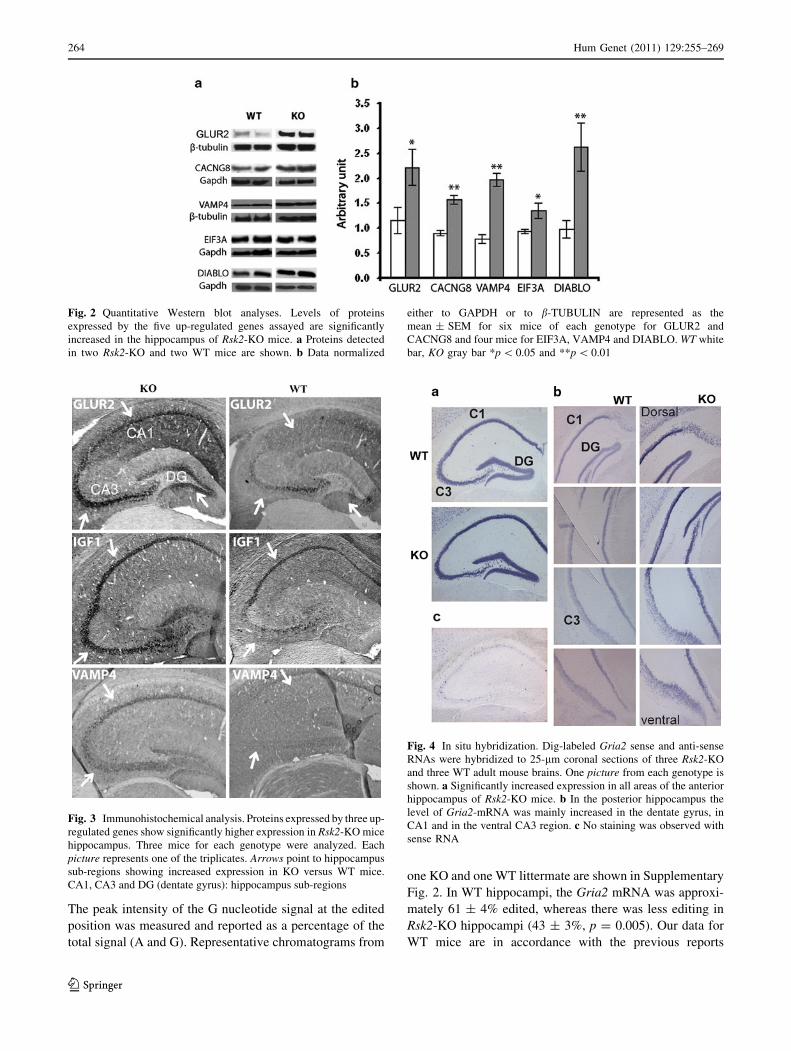
The peak intensity of the G nucleotide signal at the edited
position was measured and reported as a percentage of the
total signal (A and G). Representative chromatograms from
one KO and one WT littermate are shown in Supplementary
Fig. 2. In WT hippocampi, the Gria2 mRNA was approxi-
mately 61 ± 4% edited, whereas there was less editing in
Rsk2-KO hippocampi (43 ± 3%, p = 0.005). Our data for
WT mice are in accordance with the previous reports
Fig. 2 Quantitative Western blot analyses. Levels of proteins
expressed by the five up-regulated genes assayed are significantly
increased in the hippocampus of Rsk2-KO mice. a Proteins detected
in two Rsk2-KO and two WT mice are shown. b Data normalized
either to GAPDH or to b-TUBULIN are represented as the
mean ± SEM for six mice of each genotype for GLUR2 and
CACNG8 and four mice for EIF3A, VAMP4 and DIABLO. WT white
bar, KO gray bar *p \ 0.05 and **p \ 0.01
Fig. 3 Immunohistochemical analysis. Proteins expressed by three up-
regulated genes show significantly higher expression in Rsk2-KO mice
hippocampus. Three mice for each genotype were analyzed. Each
picture represents one of the triplicates. Arrows point to hippocampus
sub-regions showing increased expression in KO versus WT mice.
CA1, CA3 and DG (dentate gyrus): hippocampus sub-regions
Fig. 4 In situ hybridization. Dig-labeled Gria2 sense and anti-sense
RNAs were hybridized to 25-lm coronal sections of three Rsk2-KO
and three WT adult mouse brains. One picture from each genotype is
shown. a Significantly increased expression in all areas of the anterior
hippocampus of Rsk2-KO mice. b In the posterior hippocampus the
level of Gria2-mRNA was mainly increased in the dentate gyrus, in
CA1 and in the ventral CA3 region. c No staining was observed with
sense RNA
264 Hum Genet (2011) 129:255–269
123

demonstrating an editing status at the R/G site of approxi-
mately 64% (Lai et al. 1997). Thus, in Rsk2-KO mice the
extent of R/G editing was significantly decreased (rel.
decrease: 18%, p = 0.003) in the hippocampal tissue.
To determine the ratio of transcripts in the flip/flop
alternative splice form, the peak intensity of the first
nucleotide difference (C vs. A) between the two variants
was measured (Lee et al. 1998). In the WT hippocampi,
there were approximately 55 ± 5% of Glur2 transcript in
the flip form, whereas the percentage was significantly
lower (43 ± 3%, p = 0.002) in the Rsk2-KO hippocampi
(Supplemental Fig. 2c, d). No significant difference for the
Gria1 mRNA (encoding GLUR1) for the R/G editing site
or flip/flop splice levels was found between WT and mutant
mice (Supplemental Fig. 2a, b).
Reduced AMPA synaptic transmission
We then investigated whether changes in Gria2 expression,
editing and splicing affect basal AMPAR-mediated syn-
aptic transmission in hippocampal slices from 4-week-old
Rsk2-KO mice. To assess the strength of synaptic trans-
mission, we compared the size of the presynaptic fiber
volley (input) to the slope of the EPSP (output) in striatum
radiatum and found a *25% significant reduction in Rsk2-
KO (n = 9) mice when compared with WT littermates
(n = 7) (Fig. 6a). We evaluated also paired-pulse facilita-
tion (PPF), a measure of release probability from presyn-
aptic terminals. The PPF curves were essentially identical
in slices from control and Rsk2-KO mice (Fig. 6b) indi-
cating that RSK2 most likely modulates AMPA neuro-
transmission postsynaptically with no effect on presynaptic
function.
Discussion
The absence of gross structural alterations in the brain of
Rsk2-KO mice strongly indicates that their defective cog-
nitive phenotype should be linked to subtler molecular or
cellular alterations. In an effort to identify such alterations,
we carried out a detailed characterization of the differences
existing between the transcriptional profiles of the hippo-
campus of WT and KO animals. Our analysis by oligo-
nucleotide microarrays yielded a list of 100 differentially
expressed genes with high degree of statistical significance.
These results were further confirmed for 24 genes by
quantitative RT-PCR demonstrating their robustness.
Our study revealed a great variety of RSK2-influenced
genes acting in various biological pathways. Indeed, the
network with the highest score (as determined by inge-
nuity pathway analysis) centers on the NFjB complex,
which plays a prominent role in cell differentiation and
proliferation and apoptosis (Brand et al. 1997). Twelve
genes are implicated at various stages of the cell cycle in
Rsk2-KO neurons (including Igf1, Rb1, Max, Sod2, and,
Ptgs2). This result suggests strongly that abnormal cell
proliferation contributes to the CLS phenotype. In addi-
tion, 34 of the altered genes have been implicated in cell
death or survival, out of which 7 (Cacng8, Diablo, Gria2,
Igf1, Ptgs2, Rb1, Sod2) have been specifically associated
with neuronal cell death. Interestingly, previous studies
suggested that apoptotic and antiapoptotic cascades are
tightly associated with cognitive dysfunctions and neuro-
logical disorders (Lutz 2007). Further studies are, there-
fore, necessary to investigate cell proliferation and death
in Rsk2-KO mice. Four genes, including Sod2, Fxn, Gmfb
and Cp are implicated in free radical scavenging, sug-
gesting also a possible involvement of free radicals in the
Fig. 5 Surface expression of AMPAR. a WT and Rsk2-KO hippo-
campal neurons were labeled with N-terminal GLUR2 antibody under
non-permeabilized condition to stain surface GLUR2 (sGLUR2),
followed by PSD95 staining (a post-synaptic marker). Arrows point to
post-synaptic surface-expressed GLUR2. Scale bar 10 lm. b Quan-
tification of sGLUR2 puncta. Data represent mean ± SEM of
detected sGLUR2 clusters per unit dendrite length, from n = 12
(WT) and 12 (KO) embryos. *p \ 0.05
Hum Genet (2011) 129:255–269 265
123

CLS phenotype. Ten genes (among which Gria2, Igf1,
Ptgs2, Sod2, Nptxr, Ahr and Vtn) play a role in nervous
system development and function. IGF1 for instance is
essential for normal dendritic growth (Cheng et al. 2003).
NPTXR is thought to be involved in activity-dependent
synaptic plasticity (Xu et al. 2003). The AHR homologs
in Drosophila, Spineless (Ss), and in Caenorhabditis
elegans, ahr-1, regulate dendrite morphology (Kim et al.
2006) and neuronal differentiation (Qin and Powell-
Coffman 2004). The expression of a number of genes
involved in neurotransmission was also found affected. Up-
regulation of the Vamp4 and Stxbp3 genes in mutant mice
points to a role of RSK2 in pre-synaptic vesicle trafficking
(Wang and Tang 2006). Up-regulation of Cacnb4, encoding
a b-subunit of Voltage-gated Ca?? channels, suggests that
influx of Ca?? into the cell upon membrane polarization is
regulated via RSK2 (Birnbaumer et al. 1998). Moreover,
alteration of Gnb1 and Gria2 expression suggests that
RSK2 is involved in glutamate receptor signaling. GNB1
mediates the fast voltage-dependent inhibition of N-type
Ca?? channels (Fu and Cheung 1999). The function of
Gria2 will be discussed below. Finally, the expression of
several genes encoding proteins implicated in gene
expression was altered in Rsk2-KO hippocampi (including
the transcription regulators Etv3, Hip2, Rb1, Irf2, Max,
Runx2 and Trip11) suggesting that some of the RSK2
effects may be direct and others indirect. Strikingly, two of
the genes with altered expression in Rsk2-KO hippocampi,
Lamp2 and Cul4b, have previously been associated with
syndromic forms of X-linked mental retardation (Nishino
et al. 2000; Zou et al. 2007).
Among the genes associated with a specific neuronal
function, Gria2 was of particular interest because GLUR2
controls the key biophysical properties of AMPA receptors,
which are implicated in learning and memory (Kessels and
Malinow 2009). Most excitatory synaptic transmission in
the brain being mediated through AMPAR, changes in the
properties of these receptors are likely to have a major
impact on brain function. Furthermore, GLUR2 was shown
to bind directly to RSK2 in murine neurons suggesting a
direct influence of RSK2 on GLUR2 function (Thomas
et al. 2005). The GLUR family contains four closely
related members (GLUR1-4). AMPARs are tetrameric,
composed of various combinations of GLUR1-4 subunits,
and the conductance properties of the receptors are highly
dependent on their subunit composition (Kuner et al.
2001). GLUR2-lacking receptors have a higher Ca??
permeability, channel conductance, open probability and
rectification than GLUR2-containing receptors (Isaac et al.
2007). Therefore, the presence or absence of the GLUR2
subunit can dramatically alter AMPAR properties and
thereby synaptic transmission.
Fig. 6 Patch-clamp analysis. aInput–output curves for basal
synaptic transmission in
hippocampal slices. As
illustrated in the sample traces
and the graph, for each input
(fiber volley C 0.15 mV), the
output (fEPSP) is reduced by
25% in Rsk2-KO slices
(p B 0.05, WT n = 10; KO
n = 9). Scale bar 0.1 mV,
5 ms. b Paired-pulse facilitation
(PPF) does not differ between
Rsk2-KO (n = 9) and WT
(n = 6) cells. Sample traces are
illustrated above the bar graph.
Scale bar 0.05 mV, 10 ms
266 Hum Genet (2011) 129:255–269
123

Our results provide evidence that in the hippocampus of
Rsk2-KO mice total expression of GLUR2 is increased, and
that the expression is also increased at the surface of syn-
apses in cultured primary hippocampal cells. It was
reported previously that surface insertion of GLUR2 occurs
constitutively under basal conditions (Passafaro et al.
2001). Our results are compatible with these data. It may be
speculated that over-representation of the GLUR2 subunit
in synaptic AMPARs results in decreasing of Ca?? per-
meability and channel conductance. We show that there is,
indeed, a 25% reduction in basal AMPAR-mediated
transmission in the hippocampus of Rsk2-KO mice.
The Q/R site was completely edited in both WT and
Rsk2-KO hippocampi, whereas the extent of R/G editing
was significantly decreased. The GLUR2 subunit confers
Ca?? impermeability on the channel due to a single argi-
nine (R) residue located at amino acid position 607, which
is a glutamine (Q) in the other AMPA receptor subunits.
RNA editing of the Q/R site is specific to GLUR2 and is
complete in postnatal brain. Our results in WT mice con-
firm further these latter data and show that Q/R editing is
unaltered in Rsk2-KO mice. GLUR2-4 undergo also RNA
editing [arginine (R) to glycine (G)] at amino acid position
743 (Lomeli et al. 1994). The presence of edited GLUR
subunits at position 743 yields channels with faster kinet-
ics. The R/G editing state of GLUR2–4 influences also the
assembly and surface expression of AMPAR complexes.
We show that R/G editing is significantly altered in Rsk2-
KO mice. Finally, alternative splicing in the extracellular
ligand binding domain of the AMPARs generates two
variants, i.e., flip and flop. Native AMPAR are heteromeric
assemblies of different subunits that may have different flip
and flop isoforms. We show that in Rsk2-KO mice the
proportion of GLUR2 molecules with a flop exon is sig-
nificantly higher than in WT littermates. The flop variants
desensitize at least 3 times faster but recover more slowly
from desensitization than the flip counterparts (Pei et al.
2009). It was also previously shown that the flip/flop
splicing has an effect on the maturation and cellular traf-
ficking of AMPARs (Brorson et al. 2004). Alteration of R/
G editing and splicing of GLUR2 in Rsk2-KO mice are
therefore expected to alter AMPARs channel kinetic,
desensitization and trafficking. Further functional studies
are required to address the precise functional consequences
of these editing and splicing changes in Rsk2-KO neurons.
Furthermore, it was shown that proteins binding to GLUR2
are necessary for constitutive replacement of newly inser-
ted GLUR1-containing receptors to maintain synaptic
strength during LTP (Malinow and Malenka 2002). It has
also been proposed that the expression of hippocampal
LTD is critically dependent on GLUR2 (Malinow and
Malenka 2002). Further studies will address the conse-
quences of up-regulation of GLUR2 in Rsk2-KO mice for
LTP and LTD. The signaling mechanisms involved in the
increased levels of transcription of Gria2 in Rsk2-KO
hippocampal neurons are not yet known. The regulation of
Gria2 splicing and editing events is poorly understood as
well. Gria2 expression is influenced strongly at the tran-
scriptional level by at least three regulatory elements in the
50 proximal region of the promoter (Borges and Dingledine
2001). RNA editing is mediated by adenosine deaminase
acting on RNA (ADAR) enzymes. Three structurally
related ADARs (ADAR1 to ADAR3) have been identified
in mammals. ADAR2 predominantly catalyzes RNA edit-
ing at the Q/R site of GLUR2 (Peng et al. 2006), whereas it
is not yet clear how the R/G site is edited. The underlying
mechanism of the R/G editing dysregulation may be caused
by altered function or expression of one or several ADAR
enzymes. However, the fact that editing of the Q/R site in
GLUR2 is not affected in Rsk2-KO mice suggesting that
ADAR2 is excluded. Further investigations are necessary
to determine precisely the molecular events leading to up-
regulation of the Gria2 gene and alteration of RNA editing,
and the contribution of each of these dysregulations to the
cognitive dysfunction. The contribution of other pathways
remains also to be investigated. Among the deregulated
genes at least one other participates in regulation of AM-
PAR function: Cacng8. This gene encodes a synaptic
protein, TARPc-8 that participates in consolidation phase
of memory and is involved in modulating neurotransmitter
release. Evidence was provided that TARPc-8 is critical for
basal AMPAR expression and localization at extrasynaptic
sites in the hippocampus (Rouach et al. 2005). Up-regu-
lation of Cacng8 may thus contribute to AMPAR
dysfunction.
The data in this study provide a first glimpse of the gene
expression profile of adult hippocampi in the absence of
RSK2 expression. However, the Rsk2-KO animals repre-
sent a value model to study human Coffin–Lowry syn-
drome, it has significant limitations due to potential
compensatory adaptation mechanisms in the developing
nervous system (in particular through other RSK family
members). Thus, it would be interesting to perform
expression profiling following Rsk2 gene silencing by RNA
interference technology. Indeed, cellular or animal models
based on this technology could offer further clues about the
function of RSK2.
In conclusion, functional impairment of neurotransmis-
sion and plasticity due to AMPAR dysfunction may,
indeed, contribute to the cognitive deficit of Rsk2-KO
mice. However, further investigations are necessary to
determine precisely the molecular events leading to alter-
ation of GLUR2 expression and the contribution of this
dysregulation to the cognitive dysfunction. The involve-
ment of other pathways, including in particular cellular
proliferation and apoptosis, remains also to be investigated.
Hum Genet (2011) 129:255–269 267
123

Finally, the genes identified by our microarray analysis will
help in further unravel the various functions of RSK2 in the
hippocampus can be speculated to play a role in the patho-
genesis of mental retardation in Coffin–Lowry syndrome and
may provide targets for pharmaceutical intervention.
Accession number
The data of the expression arrays produced for this report
have been submitted to NCBIs Gene Expression Omnibus
(GEO: http://www.ncbi.nlm.nih.gov/geo/) and are accessi-
ble through GEO Series accession number GSE22137.
Acknowledgments The authors wish to thank Jessica Heringer for
technical assistance and helpful discussions, and the IGBMC, Institut
de Genetique et de Biologie Moleculaire et Cellulaire for assistance.
This work was supported by the French National Agency for Research
(Grant number 08-MNPS-021-01, to A.H.), the Fondation Jerome
Lejeune (to A.H.), the Centre National de la Recherche Scientifique,
the Institut National de la Sante et de la Recherche, the College de
France and the University of Strasbourg. AS was supported by a
fellowship from the Ministere pour la Recherche et Technologie
of France and TM by a scholarship from the Higher Education
Commission (HEC) of Pakistan.
Conflict of interest The authors declare that they have no conflict
of interest.
References
Benjamini Y, Hochberg Y (1995) Controlling the false discovery rate:
a practical and powerful approach to multiple testing. J R Stat
Soc B 57:289–300
Birnbaumer L, Qin N, Olcese R, Tareilus E, Platano D, Costantin J,
Stefani E (1998) Structures and functions of calcium channel
beta subunits. J Bioenerg Biomembr 30:357–375
Borges K, Dingledine R (2001) Functional organization of the GluR1
glutamate receptor promoter. J Biol Chem 276:25929–25938
Brand K, Page S, Walli AK, Neumeier D, Baeuerle PA (1997) Role of
nuclear factor-kappa B in atherogenesis. Exp Physiol
82:297–304
Brorson JR, Li D, Suzuki T (2004) Selective expression of
heteromeric AMPA receptors driven by flip-flop differences.
J Neurosci 24:3461–3470
Carlson NG, Howard J, Gahring LC, Rogers SW (2000) RNA editing
(Q/R site) and flop/flip splicing of AMPA receptor transcripts in
young and old brains. Neurobiol Aging 21:599–606
Cheng CM, Mervis RF, Niu SL, Salem N Jr, Witters LA, Tseng V,
Reinhardt R, Bondy CA (2003) Insulin-like growth factor 1 is
essential for normal dendritic growth. J Neurosci Res 73:1–9
Davis S, Laroche S (2006) Mitogen-activated protein kinase/extra-
cellular regulated kinase signalling and memory stabilization: a
review. Genes Brain Behav 5(Suppl 2):61–72
De Cesare D, Jacquot S, Hanauer A, Sassone-Corsi P (1998) Rsk-2
activity is necessary for epidermal growth factor-induced
phosphorylation of CREB protein and transcription of c-fos
gene. Proc Nat Acad Sci USA 95:12202–12207
Frodin M, Gammeltoft S (1999) Role and regulation of 90 kDa
ribosomal S6 kinase (RSK) in signal transduction. Mol Cell
Endocrinol 51:65–77
Fu AK, Cheung WM (1999) Identification of genes induced by
neuregulin in cultured myotubes. Mol Cell Neurosci 14:241–253
Ghate A, Befort K, Becker JA, Filliol D, Bole-Feysot C, Demebele D,
Jost B, Koch M, Kieffer BL (2007) Identification of novel striatal
genes by expression profiling in adult mouse brain. Neuroscience
146:1182–1192
Guimiot F, Delezoide AL, Hanauer A, Simonneau M (2004)
Expression of the RSK2 gene during early human development.
Gene Expr Patterns 4:111–114
Hanauer A, Young ID (2002) Coffin–Lowry syndrome: clinical and
molecular features. J Med Genet 39:705–713
Isaac JT, Ashby M, McBain CJ (2007) The role of the GluR2 subunit
in AMPA receptor function and synaptic plasticity. Neuron
54:859–871
Kessels HW, Malinow R (2009) Synaptic AMPA receptor plasticity
and behavior. Neuron 61:340–350
Kim MD, Jan LY, Jan YN (2006) The bHLH-PAS protein Spineless
is necessary for the diversification of dendrite morphology of
Drosophila dendritic arborization neurons. Genes Dev
20:2773–2778
Kuner T, Beck C, Sakmann B, Seeburg PH (2001) Channel-lining
residues of the AMPA receptor M2 segment: structural envi-
ronment of the Q/R site and identification of the selectivity filter.
J Neurosci 21:4162–4172
Lai F, Chen CX, Carter KC, Nishikura K (1997) Editing of glutamate
receptor B subunit ion channel RNAs by four alternatively
spliced DRADA2 double-stranded RNA adenosine deaminases.
Mol Cell Biol 17:2413–2424
Lee JC, Greig A, Ravindranathan A, Parks TN, Rao MS (1998)
Molecular analysis of AMPA-specific receptors: subunit com-
position, editing, and calcium influx determination in small
amounts of tissue. Brain Res Protoc 3:142–154
Lomeli H, Mosbacher J, Melcher T, Hoger T, Geiger JR, Kuner T,
Monyer H, Higuchi M, Bach A, Seeburg PH (1994) Control of
kinetic properties of AMPA receptor channels by nuclear RNA
editing. Science 266:1709–1713
Lutz RE (2007) Trinucleotide repeat disorders. Semin Pediatr Neurol
14:26–33
Malinow R, Malenka RC (2002) AMPA receptor trafficking and
synaptic plasticity. Annu Rev Neurosci 25:103–126
Marques Pereira P, Gruss M, Braun K, Foos N, Pannetier S, Hanauer
A (2008) Dopaminergic system dysregulation in the mrsk2_KO
mouse, an animal model of the Coffin–Lowry syndrome.
J Neurochem 107:1325–1334
Nakamoto N, Nalavadi V, Epstein MP, Narayanan U, Bassell GJ,
Warren ST (2007) Fragile X mental retardation protein defi-
ciency leads to excessive mGluR5-dependent internalization of
AMPA receptors. Proc Nat Acad Sci USA 39:15537–15542
Nishino I, Fu J, Tanji K, Yamada T, Shimojo S, Koori T, Mora M,
Riggs JE, Oh SJ, Koga Y et al (2000) Primary LAMP-2
deficiency causes X-linked vacuolar cardiomyopathy and myop-
athy (Danon disease). Nature 406:906–910
Passafaro M, Piech V, Sheng M (2001) Subunit-specific temporal and
spatial patterns of AMPA receptor exocytosis in hippocampal
neurons. Nat Neurosci 4:917–926
Pei W, Huang Z, Wang C, Han Y, Park JS, Niu L (2009) Flip and
flop: a molecular determinant for AMPA receptor channel
opening. Biochemistry 48:3767–3777
Peng PL, Zhong X, Tu W, Soundarapandian MM, Molner P, Zhu D,
Lau L, Liu S, Liu F, Lu Y (2006) ADAR2-dependent RNA
editing of AMPA receptor subunit GluR2 determines vulnera-
bility of neurons in forebrain ischemia. Neuron 49:719–733
268 Hum Genet (2011) 129:255–269
123

Poirier R, Jacquot S, Vaillend C, Soutthiphong AA, Libbey M, Davis
S, Laroche S, Hanauer A, Welzl H, Lipp HP, Wolfer DP (2007)
Deletion of the Coffin–Lowry syndrome gene Rsk2 in mice is
associated with impaired spatial learning and reduced control of
exploratory behavior. Behav Genet 37:31–50
Qin H, Powell-Coffman JA (2004) The Caenorhabditis elegans aryl
hydrocarbon receptor, AHR-1, regulates neuronal development.
Dev Biol 270:64–75
Rouach N, Byrd K, Petralia RS, Elias GM, Adesnik H, Tomita S,
Karimzadegan S, Kealey C, Bredt DS, Nicoll RA (2005)
TARP gamma-8 controls hippocampal AMPA receptor num-
ber, distribution and synaptic plasticity. Nat Neurosci 8:1525–
1533
Sassone-Corsi P, Mizzen CA, Cheung P, Crosio C, Monaco L,
Jacquot S, Hanauer A, Allis CD (1999) Requirement of Rsk-2
for epidermal growth factor-activated phosphorylation of histone
H3. Science 285:886–891
Seidenman KJ, Steinberg JP, Huganir R, Malinow R (2003)
Glutamate receptor subunit 2 serine 880 phosphorylation mod-
ulates synaptic transmission and mediates plasticity in CA1
pyramidal cells. J Neurosci 23:9220–9228
Thomas GM, Rumbaugh GR, Harrar DB, Huganir RL (2005)
Ribosomal S6 kinase 2 interacts with and phosphorylates PDZ
domain-containing proteins and regulates AMPA receptor
transmission. Proc Nat Acad Sci USA 102:15006–15011
Wang Y, Tang BL (2006) SNAREs in neurons—beyond synaptic
vesicle exocytosis. Mol Membr Biol 23:377–384
Xu D, Hopf C, Reddy R, Cho RW, Guo L, Lanahan A, Petralia RS,
Wenthold RJ, O’Brien RJ, Worley P (2003) Narp and NP1 form
heterocomplexes that function in developmental and activity-
dependent synaptic plasticity. Neuron 39:513–528
Zeniou M, Ding T, Trivier E, Hanauer A (2002) Expression analysis
of RSK gene family members: the RSK2 gene, mutated in
Coffin–Lowry syndrome, is prominently expressed in brain
structures essential for cognitive function and learning. Hum
Mol Genet 11:2929–2940
Zou Y, Liu Q, Chen B, Zhang X, Guo C, Zhou H, Li J, Gao G, Guo Y,
Yan C et al (2007) Mutation in CUL4B, which encodes a
member of cullin-RING ubiquitin ligase complex, causes
X-linked mental retardation. Am J Hum Genet 80:561–566
Hum Genet (2011) 129:255–269 269
123

Supplemental Fig. 1 (a) WT and Rsk2-KO hippocampal neurons were labeled with N-terminal
GLUR1 antibody under non-permeabilized condition to stain surface GLUR1 (sGLUR1), followed
by PSD95 staining (a post-synaptic marker). Arrows point to post-synaptic surface-expressed
GLUR1. (b) No significant difference for GLUR1 surface expression was found between WT and
Rsk2-KO mice (n = 4 WT and 6 KO embryos).
Supplemental data:

Supplemental Fig. 2. Determination of relative Gria2 R/G editing and Flip/Flop splice
levels. The Gria1(encoding GLUR1) and Gria2 (encoding GLUR2) mRNAs from five Rsk2-
KO and five WT hippocampi were amplified by RT-PCR in triplicates and the products
sequenced. Representative chromatograms from one KO and one WT littermate is shown. (a)
Gria1 for WT, (b) Gria1 for KO, (c) Gria2 for WT, (d) Gria2 for KO. * points the G/R
editing site (A: unedited, G: edited). The peak intensity of the G nucleotide signal was
measured and reported as the mean percentage for the five mice of the total signal (A + G).
** Points the first nucleotide of the flip/flop exon (flop starts with A and flip with C). The
peak intensity of the first nucleotide between the two variants was measured and reported as
the mean percentage for the five mice of the total signal (A + C).
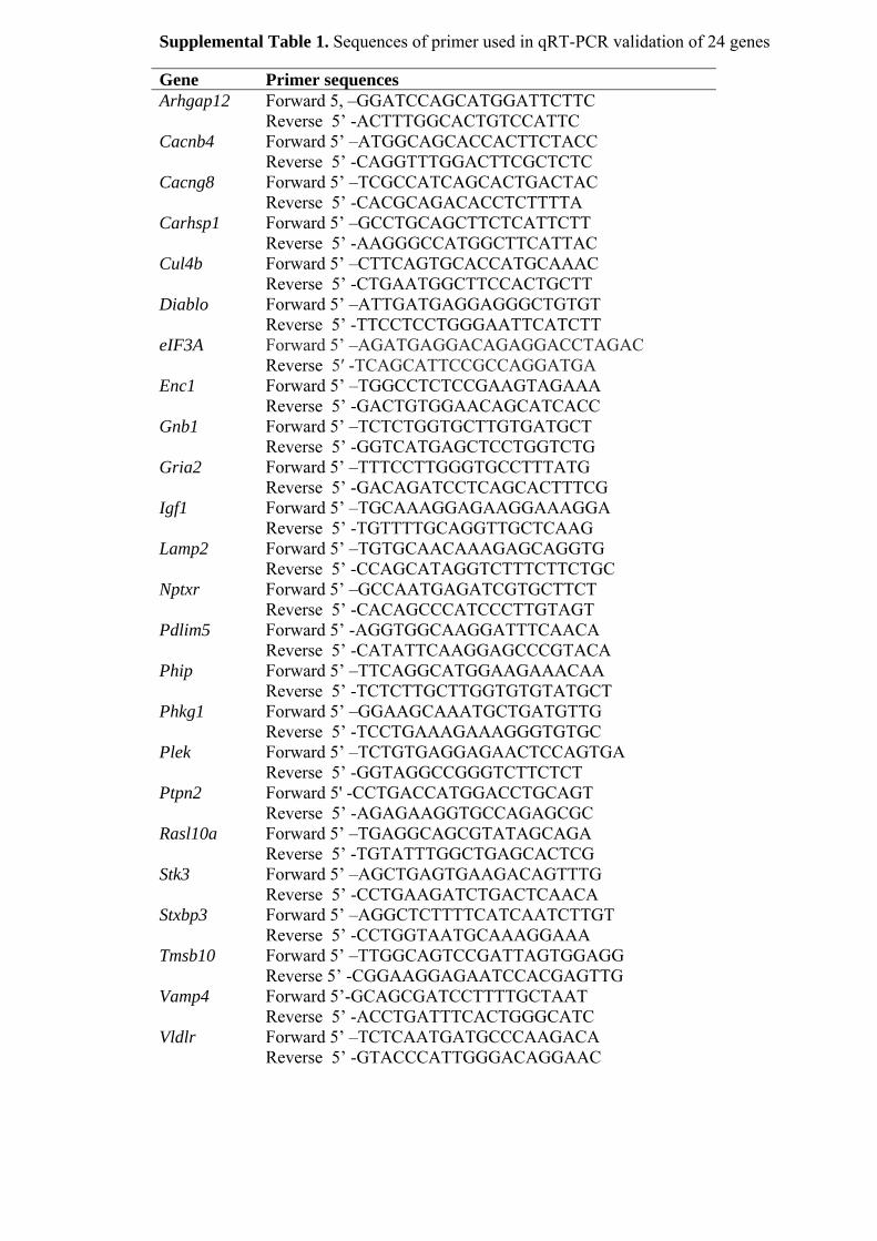
Supplemental Table 1. Sequences of primer used in qRT-PCR validation of 24 genes Gene Primer sequences Arhgap12 Forward 5, –GGATCCAGCATGGATTCTTC
Reverse 5’ -ACTTTGGCACTGTCCATTC Cacnb4 Forward 5’ –ATGGCAGCACCACTTCTACC
Reverse 5’ -CAGGTTTGGACTTCGCTCTC Cacng8 Forward 5’ –TCGCCATCAGCACTGACTAC
Reverse 5’ -CACGCAGACACCTCTTTTA Carhsp1 Forward 5’ –GCCTGCAGCTTCTCATTCTT
Reverse 5’ -AAGGGCCATGGCTTCATTAC Cul4b Forward 5’ –CTTCAGTGCACCATGCAAAC
Reverse 5’ -CTGAATGGCTTCCACTGCTT Diablo Forward 5’ –ATTGATGAGGAGGGCTGTGT
Reverse 5’ -TTCCTCCTGGGAATTCATCTT eIF3A Forward 5’ –AGATGAGGACAGAGGACCTAGAC
Reverse 5′ -TCAGCATTCCGCCAGGATGA Enc1 Forward 5’ –TGGCCTCTCCGAAGTAGAAA
Reverse 5’ -GACTGTGGAACAGCATCACC Gnb1 Forward 5’ –TCTCTGGTGCTTGTGATGCT
Reverse 5’ -GGTCATGAGCTCCTGGTCTG Gria2 Forward 5’ –TTTCCTTGGGTGCCTTTATG
Reverse 5’ -GACAGATCCTCAGCACTTTCG Igf1 Forward 5’ –TGCAAAGGAGAAGGAAAGGA
Reverse 5’ -TGTTTTGCAGGTTGCTCAAG Lamp2 Forward 5’ –TGTGCAACAAAGAGCAGGTG
Reverse 5’ -CCAGCATAGGTCTTTCTTCTGC Nptxr Forward 5’ –GCCAATGAGATCGTGCTTCT
Reverse 5’ -CACAGCCCATCCCTTGTAGT Pdlim5 Forward 5’ -AGGTGGCAAGGATTTCAACA
Reverse 5’ -CATATTCAAGGAGCCCGTACA Phip Forward 5’ –TTCAGGCATGGAAGAAACAA
Reverse 5’ -TCTCTTGCTTGGTGTGTATGCT Phkg1 Forward 5’ –GGAAGCAAATGCTGATGTTG
Reverse 5’ -TCCTGAAAGAAAGGGTGTGC Plek Forward 5’ –TCTGTGAGGAGAACTCCAGTGA
Reverse 5’ -GGTAGGCCGGGTCTTCTCT Ptpn2 Forward 5' -CCTGACCATGGACCTGCAGT
Reverse 5’ -AGAGAAGGTGCCAGAGCGC Rasl10a Forward 5’ –TGAGGCAGCGTATAGCAGA
Reverse 5’ -TGTATTTGGCTGAGCACTCG Stk3 Forward 5’ –AGCTGAGTGAAGACAGTTTG
Reverse 5’ -CCTGAAGATCTGACTCAACA Stxbp3 Forward 5’ –AGGCTCTTTTCATCAATCTTGT
Reverse 5’ -CCTGGTAATGCAAAGGAAA Tmsb10 Forward 5’ –TTGGCAGTCCGATTAGTGGAGG
Reverse 5’ -CGGAAGGAGAATCCACGAGTTG Vamp4 Forward 5’-GCAGCGATCCTTTTGCTAAT
Reverse 5’ -ACCTGATTTCACTGGGCATC Vldlr Forward 5’ –TCTCAATGATGCCCAAGACA
Reverse 5’ -GTACCCATTGGGACAGGAAC
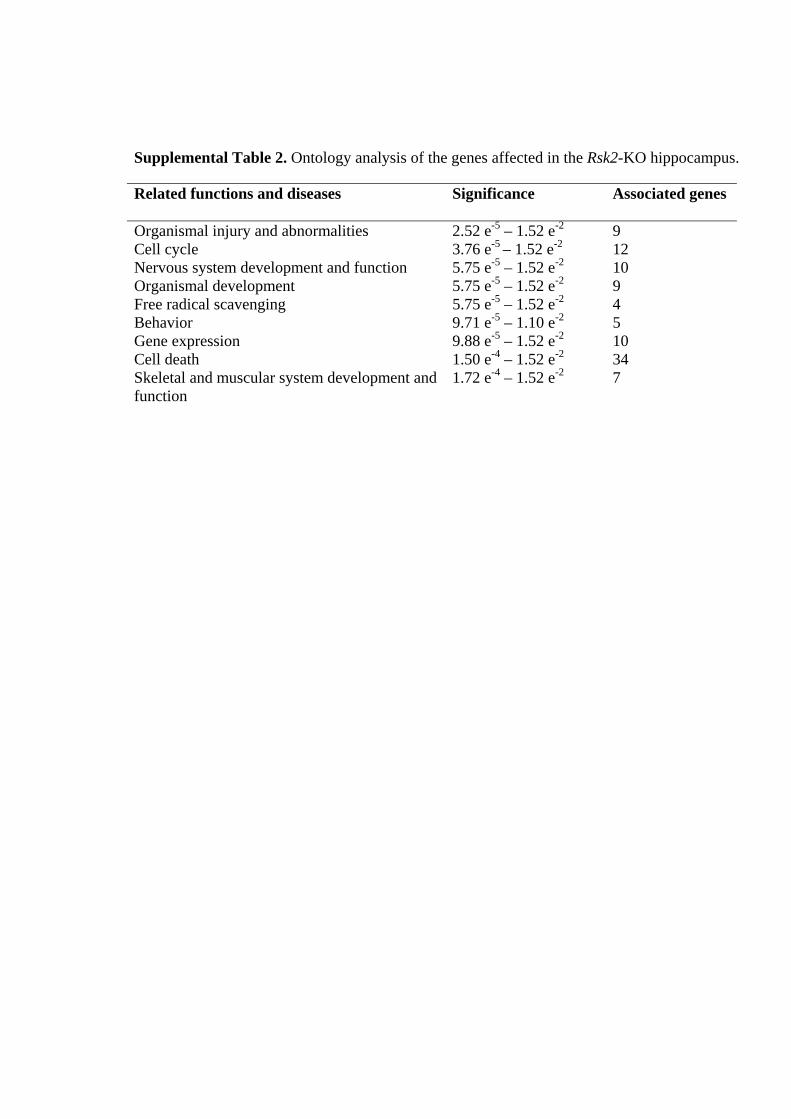
Supplemental Table 2. Ontology analysis of the genes affected in the Rsk2-KO hippocampus. Related functions and diseases Significance Associated genes
Organismal injury and abnormalities 2.52 e-5 – 1.52 e-2 9 Cell cycle 3.76 e-5 – 1.52 e-2 12 Nervous system development and function 5.75 e-5 – 1.52 e-2 10 Organismal development 5.75 e-5 – 1.52 e-2 9 Free radical scavenging 5.75 e-5 – 1.52 e-2 4 Behavior 9.71 e-5 – 1.10 e-2 5 Gene expression 9.88 e-5 – 1.52 e-2 10 Cell death 1.50 e-4 – 1.52 e-2 34 Skeletal and muscular system development and function
1.72 e-4 – 1.52 e-2 7

Chapter 2 Results
99
2.2. Cobalt staining of primary hippocampal neurons:
NMDA receptors are highly permeable to Ca2+ ions, whereas AMPA receptors are
normally less permeable to Ca2+, due to the presence of GluR2 subunit in AMPA receptor.
We have observed a two-fold higher level of GluR2 protein expression in Rsk2-KO mice
than in WT mice, and the electrophysiology studies showed a significant reduction of basal
AMPA receptor-mediated transmission in the hippocampus of Rsk2-KO mice. This
prompted us to further explore the AMPA receptor functions, by using the Co2+ staining
protocol on primary hippocampal neuronal cultures. The cobalt staining technique involves
the pharmacological stimulation of AMPAR in the presence of CoCl2, which leads to the
uptake of Co2+ through Ca2+-permeable AMPARs. Subsequently, one can visualize
activated cells by precipitating histochemically the intracellular Co2+. This technique allows
identification of cells expressing Ca2+-permeable AMPARs. Thus, this protocol was
expected to reveal whether the proportion of cells with Ca2+-permeable AMPARs is
different in Rsk2-KO cultures than in WT cultures. We took the images of stained neuronal
culture by using the light compound microscope. Co2+ treated neurons, without AMPA
stimulation, showed no or very rare stained cells as shown in (Figure 27A), in both KO and
WT mice. Treatment with Co2+ and stimulation with AMPA (100µM) resulted in less
stained neurons in Rsk2-KO cultures than in WT cultures. In addition the staining was
stronger in WT neurons as compared to the KO cells (Figure 27B). In these experiments
AMPA act as an agonist on the AMPA receptor, resulting in uptake of Co2+. Accumulation
of Co2+ inside the neurons depends on the non-desensitization of AMPA receptors.
Cyclothiazide is a positive allosteric modulator of the AMPA receptor, capable of reducing
or essentially eliminating rapid desensitization of the receptor. Therefore, we tested a third
condition by treating cells with Co2+, AMPA (100µM) and cyclothiazide (30 µM). Again
there were much more stained cells in WT cultures than in Rsk2-KOcultures (Figure 27C).
Although, it was difficult to quantify the results, they suggested strongly that Ca2+
uptake through AMPA receptors/channels is affected by the over expression of the GluR2
subunit.
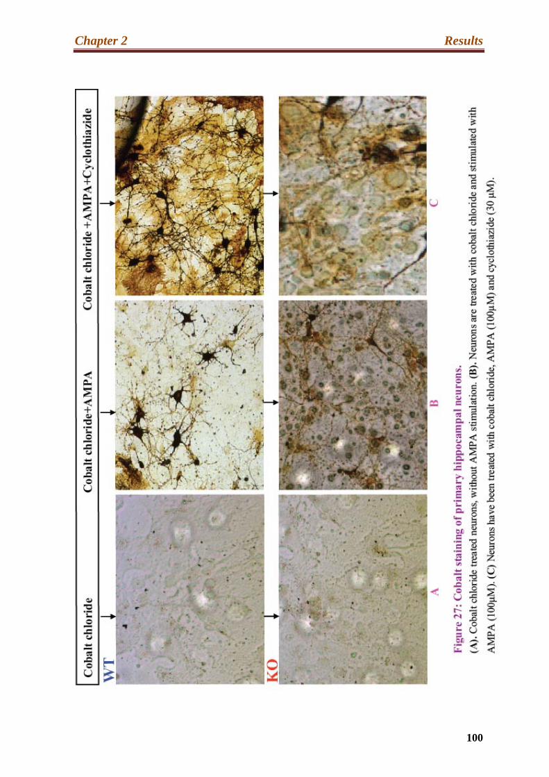
Chapter 2 Results
100

Publication-2

Chapter 2 Results-Publication 2
101
2.3. Alteration of ERK / MAPK signaling in hippocampal neurons
of Rsk2-KO mice
Anne Schneider1, Tahir Mehmood1, 2, Solange Pannetier1, André Hanauer1.
J. Neurochem. (2011) 119, 447–459.
The objective of this work was to study the mechanism of regulation of ERK
activity in the hippocampus of KO animals and to identify factors that may explain the
deregulation of the transcriptional activity previously reported (Mehmood et al., 2011). To
this end we analyzed in primary cultures of hippocampal neurons the activity of several
upstream and downstream proteins of ERK1/2, at basal level and in response to glutamate
stimulation.
RSK2 is a protein kinase acting at the distal end of the Ras-ERK/MAPK signaling
pathway. ERK1/2 activates RSK2 by phosphorylating serine and threonine residues. After
activation RSK2 in turn can phosphorylate numerous cytosolic and nuclear substrates. A
few studies have identified RSK2 as a negative regulator of its own pathway. In particular,
it was shown on cultured motor neurons that RSK2 negatively regulates axonal growth via
inhibition of ERK (Fischer et al., 2009a), but also that the activity of ERK1/2 is increased
in the cortex of KO mice, compared to the cortex of WT mice (Marques Pereira et al.,
2008). A negative feedback of RSK2 on the ERK pathway, via phosphorylation of SOS,
was proposed by Douville and Downward in 1997. We hypothesized that a deregulation of
the ERK/ MAPK in the Rsk2-KO hippocampus, resulting from the absence of this feedback
regulation, could cause alterations of many cellular processes and, thus, play a role in the
molecular pathologic mechanism leading to cognitive impairment. We wanted to know if
such a feedback exists in neurons of the hippocampus. We first analyzed the expression
levels of ERK1/2 and P-ERK1/2 in the hippocampus of Rsk2-KO and WT littermate mice.
A two- and threefold increase of the levels of Phospho-ERK1 and Phospho-ERK2,
respectively, were observed when compared to WT littermates. We then analyzed
phosphorylation of ERK1/2 in un-stimulated and glutamate stimulated hippocampal
primary cultures derived from WT and Rsk2-KO littermate mice.

Chapter 2 Results-Publication 2
102
Glutamate is a major neurotransmitter that activates the ERK pathway in many
brain regions (Kawasaki et al., 2004; Paul et al., 2010). Level of ERK1/2 phosphorylation
was, indeed, significantly higher in Rsk2-KO neurons than in WT cells, at basal conditions
and after glutamergic stimulation.
To identify the mechanism leading to this increase, we investigated the
phosphorylation of kinases and phosphatases that may regulate the activity of ERK. The
levels of phosphorylation of Raf-1 and MEK, two ERK upstream kinases, were also higher
in WT and KO neurons, supporting the RSK2 retro control hypothesis reported by
(Douville and Downward, 1997).
We then investigated the consequences of the increased ERK1/2 activity on the
phosphorylation of some ERK1/2 substrates. Higher levels of phosphorylation of Elk1 and
CREB (two transcription factors) were observed in Rsk2-KO hippocampal neurons than in
WT cells. Inhibition of MEK with U0126 confirmed that the increased level of
phosphorylation of CREB is ERK dependent. However, U0126 did not abolish glutamate-
induced phosphorylation of ELK1, providing evidence that glutamate-induced ELK1
phosphorylation is not mediated by ERK1/2 in hippocampal neurons. Further data
suggested that ELK1 activity is regulated via the p38 and JNK pathways Q-RT-PCR
analyses showed an increased expression of target genes of these transcription factors (c-
Fos, Zif268 and Arc) in Rsk2-KO neurons following glutamergic stimulation.
These results provided clear evidence that the transcriptional activity is impaired in
RSK2 deficient neurons, which may account for the dysregulation of some genes evidenced
by the transcriptional study. In this work we identified a dysregulation of the ERK/MAPK
signaling pathway in RSK2-KO derived hippocampal neurons.
A comprehensive description and illustration of these results are found in
publication-2.

Loss-of-function mutations in the gene encoding the ribo-somal S6 protein kinase 2 (RSK2) are responsible for theCoffin-Lowry syndrome (CLS; OMIM 303600), a syndromicform of X-linked mental retardation. Cardinal features ofCLS are growth and psychomotor retardation, typical facialand digital abnormalities, and progressive skeletal deforma-tions (Hanauer and Young 2002). RSK2 is a serine/threoninekinase and belongs to the 90 kDa RSK family. Four highlyrelated members are found in humans and mice, RSK1–4.These isoforms are derived from four independent genes.RSKs act at the distal end of the Ras/mitogen-activatedprotein kinase (MAPK) signaling pathway and are directlyphosphorylated and activated by the extracellular signal-regulated kinases 1 and 2 (ERK1/2) (Frodin and Gammeltoft1999).
Received May 14, 2011; revised manuscript received July 26, 2011;accepted August 9, 2011.Address correspondence and reprint requests to Andre Hanauer,
Department of Translational Medicine and Neurogenetics, Institut deGenetique et de Biologie Moleculaire et Cellulaire (IGBMC), 1, rueLaurent Fries, 67404 Illkirch, France. E-mail: [email protected] used: Arc, activity-regulated cytoskeleton-associated
protein (also known as Arg3.1); c-Fos, cellular FBJ murine osteosarcomaviral oncogene homolog; CLS, Coffin-Lowry syndrome; CREB, cAMPresponse element binding; DUSP, dual specificity phosphatase; ELK1, EtsLiKe gene1; ERK, extracellular signal-regulated kinase; IEG, immediateearly gene; KO, knockout; MAPK, mitogen-activated protein kinase;MEK, MAPK/Extracellular signal-regulated Kinase; MSK, mitogen- andstress-activated protein kinase; Nur77, nuclear receptor subfamily; PDL1,phospholipase D; RAF-1, Rapidly Accelerated Fibrosarcoma-1; RSK,ribosomal s6 kinase; SDS–PAGE, sodium dodecyl sulfate–polyacryl-amide gel electrophoresis; Shank1/3, SH3 and multiple ankyrin repeatdomains protein 1/3; Zif268, zinc finger protein 268 (also known as Egr1).
,
*Department of Translational Medicine and Neurogenetics, Institut de Genetique et de Biologie
Moleculaire et Cellulaire (IGBMC), Institut National de Sante et de Recherche Medicale (INSERM)
U964/Centre National de Recherche Scientifique (CNRS) UMR 1704/Universite de Strasbourg, Illkirch,
France
�Department of Chemistry, University of Sargodha, Sargodha, Pakistan
Abstract
Coffin–Lowry syndrome is a syndromic form of mental retar-
dation caused by mutations of the Rps6ka3 gene encoding
ribosomal s6 kinase (RSK)2. RSK2 belongs to a family con-
taining four members in mammals: RSK1–4. RSKs are serine/
threonine kinases and cytosolic substrates of extracellular
signal-regulated kinase (ERK) in the Ras/MAPK signaling
pathway. RSK2 is highly expressed in the hippocampus, and
mrsk2_KO mice display spatial learning and memory impair-
ment. In the present study, we provide evidence of abnormally
increased phosphorylation of ERK1/2 in the hippocampus of
mrsk2_KO mice. Further studies based on cultured hippo-
campal neurons revealed that glutamate activates ERK1/2
and RSKs, and confirmed a stronger activation of ERK1/2 in
mrsk2_KO neurons than in WT cells. We, thus, provide further
evidence that RSK2 exerts a feedback inhibitory effect on the
ERK1/2 pathway. We also observed a transient sequestration
of P-ERK1/2 in the cytoplasm upon glutamate stimulation. In
addition, the transcription factors cAMP response element
binding and Ets LiKe gene1 show over-activation in RSK2-
deficient neurons. Finally, c-Fos, Zif268 and Arc were signifi-
cantly over-expressed in mrsk2_KO neurons upon glutamate
stimulation. Importantly, the increased phosphorylation of
other RSK family members observed in mutant neurons was
unable to compensate for RSK2 deficiency. This aberrant
ERK1/2 signaling can influence various neuronal functions,
and thus play a significant role in cognitive dysfunction in
mrsk2_KO mice and in the Coffin-Lowry syndrome.
Keywords: Coffin-Lowry syndrome, CREB, ERK, hippo-
campus, IEG, RSK2.
J. Neurochem. (2011) 119, 447–459.
JOURNAL OF NEUROCHEMISTRY | 2011 | 119 | 447–459 doi: 10.1111/j.1471-4159.2011.07423.x
� 2011 The AuthorsJournal of Neurochemistry � 2011 International Society for Neurochemistry, J. Neurochem. (2011) 119, 447–459 447

ERK1/2 respond in neurons to a wide variety of stimuliincluding glutamate receptor activation (Bading and Greenberg1991), synaptic activity that induces either long-termpotentiation or long-term depression (Dudek and Fields2001; Thiels et al. 2002) and growth factors (Segal andGreenberg 1996). Upon activation, both ERK1 and ERK2can phosphorylate regulatory targets in the cytosol ortranslocate to the nucleus and phosphorylate commonsubstrates, including extracellular proteins, effectors andtranscription factors (Frodin and Gammeltoft 1999). Thus,ERK activation results in a variety of responses, such as cellproliferation, differentiation, gene expression, and cell cycleregulation in response to neural activity (Kolkova et al.2000). Direct nuclear targets of ERK1/2 include the Ets LiKegene1 (ELK1) transcription factor (Yang et al. 1998) and theimmediate early gene (IEG) c-Fos (Murphy et al. 2002). ThecAMP response element-binding (CREB) transcription fac-tor is also a downstream nuclear target of the Ras-ERK
pathway, and its phosphorylation can be carried out byseveral factors of the RSK and its homologous mitogen- andstress-activated protein kinase (MSK) families (De Cesareet al. 1998; Chwang et al. 2007). In addition to c-Fos, theexpression of two IEGs, the Zinc finger protein 268 (Zif268)and the activity-regulated cytoskeleton protein (Arc), aredependent of the Ras-ERK/MAPK pathway, through mech-anisms involving CREB, ELK1 or even Zif268 (Li et al.2005; Davis and Laroche 2006; Chotiner et al. 2010).Numerous studies implicate the ERK/MAPK signalingcascade and CREB and ELK1-mediated gene transcriptionin synaptic plasticity and memory (Thiels et al. 2002; Davisand Laroche 2006). The induction of c-Fos, Zif268 and Arcexpressions have also been linked to synaptic plasticitymechanism and memory processes (Davis and Laroche2006; Messaoudi et al. 2007; Alberini 2009; Davis et al.2010). Figure 1 represents schematically the simplifiedMAPK pathways.
Fig. 1 Among the signaling cascades involved in the response to
many extracellular signals in eukaryotic organisms are those that
activate the MAPK family of serine-threonine kinases. Three major
MAPK families have been identified in mammalian cells, such as the
extracellular signal-regulated kinase (ERK) pathway and the stress-
activated p38 and Jun kinase (JNK) pathways. The ERK pathway is
strongly activated by growth factors, mitogens and neurotransmitters
via the Ras protooncogene and is involved in cell proliferation, dif-
ferentiation, death and neuronal function. RSK proteins are targeted
by the MAPK/ERK pathway. Stress-activated MAPKs respond to
stress factors or cytokines, but also growth factors and neurotrans-
mitters, leading to cell death, survival, or defense and neuronal
function. U0126 is a highly selective inhibitor of both MEK1 and
MEK2.
Journal of Neurochemistry � 2011 International Society for Neurochemistry, J. Neurochem. (2011) 119, 447–459� 2011 The Authors
448 | A. Schneider et al.

Activation of ERK leads also to activation of RSKs inessentially all the cases where ERK and RSK kinaseactivities have been measured. The coordinate manner ofactivation is partially explained by the fact that ERK andRSK are physically associated in the cell (Scimeca et al.1992). Once activated RSKs can target a wide range ofcytosolic and nuclear proteins. Some of the known RSK2substrates are, in the cytosol glycogen synthase kinase 3(Angenstein et al. 1998), phospholipase D (PLD1) (Zeniou-Meyer et al. 2008) and the SH3 and multiple ankyrin repeatdomains protein 1/3 (Shank1 and Shank3) (Thomas et al.2005), and nuclear proteins including the activating tran-scription factor 4, c-FOS, the nuclear receptor subfamily 77(NUR77), CREB and histone H3 (De Cesare et al. 1998;Sassone-Corsi et al. 1999; Yang et al. 2004). Together, thedata suggest that RSK2 is involved in cellular and molecularevents such as proliferation, apoptosis, and neuronal devel-opment as well as in learning and memory processes.Involvement of RSK2 in cognitive functions was furtherconfirmed by the study of RSK2-deficient mice. Indeed, theyshow delayed acquisition of a spatial memory reference taskand long-term spatial memory deficits (Poirier et al. 2007).Moreover, RSK2 shows a high level of expression in thehippocampus, which plays an important role in cognitivefunction and learning (Zeniou et al. 2002).
Recent studies showed that RSK has also an inhibitory rolein the Ras-ERK pathway in Drosophila by anchoring ERK inthe cytoplasm (Kim et al. 2006), and during mouse embry-onic development by decreasing ERK activation and targetgene expression (Myers et al. 2004). Previously, anotherstudy, based on PC12 cells, also pointed out that RSK2 couldnegatively regulate the Ras-ERK pathway, by phosphorylat-ing son of sevenless (Douville and Downward 1997).
Glutamate is among the neurotransmitters that readilyactivate ERK. A large number of reports have welldocumented this point in neurons from various brain regions,such as the cortex, hippocampus, striatum and cerebellum(Wang et al. 2007). All three subtypes of ionotropicglutamate receptors and three subgroups of metabotropicglutamate receptors seem to modulate consistently theMAPK pathway in a stimulatory fashion. Active MAPKstranslocate to the nucleus to activate a specific set oftranscription factors for the facilitation of target geneexpression. This glutamate receptor-dependent MAPK med-iated transcription controls the development of multipleforms of synaptic plasticity related to various normal andabnormal neural activities, including memory. Very little isknown about the specific role of RSK2 in glutamatesignaling in neuronal cells.
We show in the present study that ERK1/2 is over-activated in the hippocampus of mrsk2_KO mice. To furtherinvestigate the cause and consequences of this ERK1/2increased activation, we examined the phosphorylation ofupstream actors [ERK, MAPK/Extracellular signal-regulated
Kinase (MEK), Rapidly Accelerated Fibrosarcoma-1 (RAF-1)] of RSK2 in the ERK/MAPK pathway as well as thephosphorylation and/or expression of ERK1/2 and/or RSK2targets (CREB, ELK1, c-Fos, Zif268) in mrsk2-defectivecultured hippocampal neurons stimulated or not with gluta-mate. In addition, since there is evidence that Arc is regulatedby Zif268 (Li et al. 2005), we also tested the expression ofthis later gene.
Experimental procedures
Ethics statementAll experimental procedures for care and use of mice were performedaccording to agreements with the Departmental Direction ofVeterinarian Services (Prefecture du Bas-Rhin, France No. 67-225)and IGBMC Animal Welfare Insurance (NIH, PHS No. A5100-01).mrsk2-KO mice were previously generated in our laboratory (Yanget al. 2004). WT and KO animals used in this study were male fromthe same littermates and housed four per cage in a 12 h dark/lightcycle (light on from 7 AM to 7 PM), under controlled conditions oftemperature and humidity. Killing of the mice used to derive primarycell cultures was carried out in accordance with the EuropeanCommunities Council Directive of 24th November 1986 (86/609/EEC), and the study was specifically approved by the EthicsCommittee in Animal Experiments (permit number 17) of the Institutde Genetique et de Biologie Moleculaire et Cellulaire.
Whole hippocampal protein extractionsSix male mrsk2_KO and 6 WT littermates with a C57Bl/6· geneticbackground were killed by cervical dislocation. Brains were rapidlydissected and the hippocampus was isolated using a standarddissection procedure. Whole protein extractions were performed aspreviously described (Marques Pereira et al. 2008).
Primary hippocampal culturesPrimary cultures of hippocampal neurons were obtained from WTand mrsk2_KO male mice, with a C57Bl/6· genetic background, atembryonic day E17. Dissected hippocampi were triturated andplated into wells of 24-well plates coated with poly-D-lysine (Sigma,St Louis, MO, USA), at a density of �300 000 neurons/well.Growth media consisted in NeuroBasal (Gibco, Rockville, MD,USA; Invitrogen, Carlsbad, CA, USA) supplemented with 1· B27(Gibco; Invitrogen), 0.5 mM L-glutamine and 1· penicillin/strepto-mycin. The cultures were maintained at 37�C in a humidifiedatmosphere containing 5% CO2 and cultivated for 7 days in vitroprior to experimentation.
Cell culture treatmentsAt 7 days in vitro, hippocampal neurons were activated byglutamate. Glutamate was added to the primary culture media at aconcentration of 100 lM for the duration indicated. The ERKpathway was inhibited with the MEK specific inhibitor U0126(9903; Cell Signaling Technology, Beverly, MA, USA). Hippo-campal cells were pre-treated for 30 min with 5 lMU0126 and thenstimulated with glutamate. All drugs were prepared as concentratedstock solution in distilled water and diluted to final concentration inculture medium just before application. After treatment, cells were
� 2011 The AuthorsJournal of Neurochemistry � 2011 International Society for Neurochemistry, J. Neurochem. (2011) 119, 447–459
Altered ERK/MAPK in the mrsk2_KO hippocampus | 449
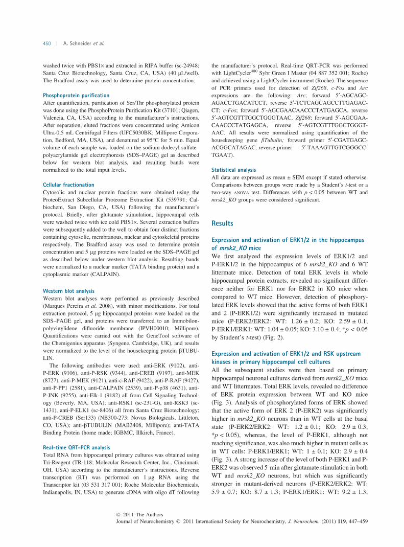
washed twice with PBS1· and extracted in RIPA buffer (sc-24948;Santa Cruz Biotechnology, Santa Cruz, CA, USA) (40 lL/well).The Bradford assay was used to determine protein concentration.
Phosphoprotein purificationAfter quantification, purification of Ser/Thr phosphorylated proteinwas done using the PhosphoProtein Purification Kit (37101; Qiagen,Valencia, CA, USA) according to the manufacturer’s instructions.After separation, eluted fractions were concentrated using AmiconUltra-0,5 mL Centrifugal Filters (UFC5030BK; Millipore Corpora-tion, Bedford, MA, USA), and denatured at 95�C for 5 min. Equalvolume of each sample was loaded on the sodium dodecyl sulfate–polyacrylamide gel electrophoresis (SDS–PAGE) gel as describedbelow for western blot analysis, and resulting bands werenormalized to the total input levels.
Cellular fractionationCytosolic and nuclear protein fractions were obtained using theProteoExtract Subcellular Proteome Extraction Kit (539791; Cal-biochem, San Diego, CA, USA) following the manufacturer’sprotocol. Briefly, after glutamate stimulation, hippocampal cellswere washed twice with ice cold PBS1·. Several extraction bufferswere subsequently added to the well to obtain four distinct fractionscontaining cytosolic, membranous, nuclear and cytoskeletal proteinsrespectively. The Bradford assay was used to determine proteinconcentration and 5 lg proteins were loaded on the SDS–PAGE gelas described below under western blot analysis. Resulting bandswere normalized to a nuclear marker (TATA binding protein) and acytoplasmic marker (CALPAIN).
Western blot analysisWestern blot analyses were performed as previously described(Marques Pereira et al. 2008), with minor modifications. For totalextraction protocol, 5 lg hippocampal proteins were loaded on theSDS–PAGE gel, and proteins were transferred to an Immobilon-polyvinylidene difluoride membrane (IPVH00010; Millipore).Quantifications were carried out with the GeneTool software ofthe Chemigenius apparatus (Syngene, Cambridge, UK), and resultswere normalized to the level of the housekeeping protein bTUBU-LIN.
The following antibodies were used: anti-ERK (9102), anti-P-ERK (9106), anti-P-RSK (9344), anti-CREB (9197), anti-MEK(8727), anti-P-MEK (9121), anti-c-RAF (9422), anti-P-RAF (9427),anti-P-PP1 (2581), anti-CALPAIN (2539), anti-P-p38 (4631), anti-P-JNK (9255), anti-Elk-1 (9182) all from Cell Signaling Technol-ogy (Beverly, MA, USA); anti-RSK1 (sc-231-G), anti-RSK3 (sc-1431), anti-P-ELK1 (sc-8406) all from Santa Cruz Biotechnology;anti-P-CREB (Ser133) (NB300-273; Novus Biologicals, Littleton,CO, USA); anti-bTUBULIN (MAB3408, Millipore); anti-TATABinding Protein (home made; IGBMC, Illkirch, France).
Real-time QRT-PCR analysisTotal RNA from hippocampal primary cultures was obtained usingTri-Reagent (TR-118; Molecular Research Center, Inc., Cincinnati,OH, USA) according to the manufacturer’s instructions. Reversetranscription (RT) was performed on 1 lg RNA using theTranscriptor kit (03 531 317 001; Roche Molecular Biochemicals,Indianapolis, IN, USA) to generate cDNA with oligo dT following
the manufacturer’s protocol. Real-time QRT-PCR was performedwith LightCycler480 Sybr Green I Master (04 887 352 001; Roche)and achieved using a LightCycler instrument (Roche). The sequenceof PCR primers used for detection of Zif268, c-Fos and Arcexpressions are the following: Arc; forward 5¢-AGCAGC-AGACCTGACATCCT, reverse 5¢-TCTCAGCAGCCTTGAGAC-CT; c-Fos; forward 5¢-AGCGAACAACCCTATGAGCA, reverse5¢-AGTCGTTTGGCTGGGTAAC, Zif268; forward 5¢-AGCGAA-CAACCCTATGAGCA, reverse 5¢-AGTCGTTTGGCTGGGT-AAC. All results were normalized using quantification of thehousekeeping gene bTubulin; forward primer 5¢-CGATGAGC-ACGGCATAGAC, reverse primer 5¢-TAAAGTTGTCGGGCC-TGAAT).
Statistical analysisAll data are expressed as mean ± SEM except if stated otherwise.Comparisons between groups were made by a Student’s t-test or atwo-way ANOVA test. Differences with p < 0.05 between WT andmrsk2_KO groups were considered significant.
Results
Expression and activation of ERK1/2 in the hippocampusof mrsk2_KO miceWe first analyzed the expression levels of ERK1/2 andP-ERK1/2 in the hippocampus of 6 mrsk2_KO and 6 WTlittermate mice. Detection of total ERK levels in wholehippocampal protein extracts, revealed no significant differ-ence neither for ERK1 nor for ERK2 in KO mice whencompared to WT mice. However, detection of phosphory-lated ERK levels showed that the active forms of both ERK1and 2 (P-ERK1/2) were significantly increased in mutatedmice (P-ERK2/ERK2: WT: 1.26 ± 0.2; KO: 2.59 ± 0.1;P-ERK1/ERK1: WT: 1.04 ± 0.05; KO: 3.10 ± 0.4; *p < 0.05by Student’s t-test) (Fig. 2).
Expression and activation of ERK1/2 and RSK upstreamkinases in primary hippocampal cell culturesAll the subsequent studies were then based on primaryhippocampal neuronal cultures derived from mrsk2_KO miceand WT littermates. Total ERK levels, revealed no differenceof ERK protein expression between WT and KO mice(Fig. 3). Analysis of phosphorylated forms of ERK showedthat the active form of ERK 2 (P-ERK2) was significantlyhigher in mrsk2_KO neurons than in WT cells at the basalstate (P-ERK2/ERK2: WT: 1.2 ± 0.1; KO: 2.9 ± 0.3;*p < 0.05), whereas, the level of P-ERK1, although notreaching significance, was also much higher in mutant cells asin WT cells: P-ERK1/ERK1; WT: 1 ± 0.1; KO: 2.9 ± 0.4(Fig. 3). A strong increase of the level of both P-ERK1 and P-ERK2 was observed 5 min after glutamate stimulation in bothWT and mrsk2_KO neurons, but which was significantlystronger in mutant-derived neurons (P-ERK2/ERK2: WT:5.9 ± 0.7; KO: 8.7 ± 1.3; P-ERK1/ERK1: WT: 9.2 ± 1.3;
Journal of Neurochemistry � 2011 International Society for Neurochemistry, J. Neurochem. (2011) 119, 447–459� 2011 The Authors
450 | A. Schneider et al.
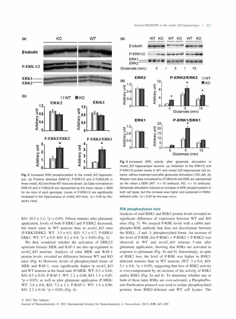
KO: 20.5 ± 2.1; *p < 0.05). Fifteen minutes after glutamateapplication, levels of both P-ERK1 and P-ERK2 decreased,but much more in WT neurons than in mrsk2_KO ones(P-ERK2/ERK2: WT: 3.5 ± 0.5; KO: 5.3 ± 0.7; P-ERK1/ERK1: WT: 5.7 ± 0.9; KO: 8.2 ± 0.8; *p < 0.05) (Fig. 3).
We then wondered whether the activation of ERK1/2upstream kinases MEK and RAF-1 are also up-regulated inmrsk2_KO neurons. Analysis of total MEK and RAF-1protein levels, revealed no difference between WT and KOmice (Fig. 4) However, levels of phosphorylated forms ofMEK and RAF-1, were significantly higher in mrsk2_KOand WT neurons at the basal state (P-MEK: WT: 0.3 ± 0.04;KO: 0.5 ± 0.03; P-RAF-1: WT: 1.2 ± 0.06; KO: 1.5 ± 0.05;*p < 0.05), as well as after glutamate application (P-MEK:WT: 3.6 ± 0.6; KO: 7.2 ± 1.1; P-RAF-1: WT: 1.4 ± 0.08;KO: 2.2 ± 0.16; *p < 0.05) (Fig. 4).
RSK phosphorylation levelAnalysis of total RSK1 and RSK3 protein levels revealed nosignificant difference of expression between WT and KOmice (Fig. 5). We assayed P-RSK levels with a rabbit anti-phospho-RSK antibody that does not discriminate betweenthe RSK1, -2 and -3. phosphorylated forms. An increase ofthe level of P-RSK (for P-RSK1 + P-RSK2 + P-RSK3) wasobserved in WT and mrsk2_KO neurons 5 min afterglutamate application, showing that RSKs are activated inresponse to glutamate (Fig. 5a and b). Interestingly, in spiteof RSK2 loss, the level of P-RSK was higher in RSK2-deficient neurons than in WT neurons (WT: 3 ± 0.4; KO:5.1 ± 0.6; *p < 0.05), suggesting that loss of RSK2 activityis over-compensated by an increase of the activity of RSK1and/or RSK3 (Fig. 5a and b). To determine whether one orboth of these latter RSKs are over-activated, a PhosphoPro-tein Purification protocol was used to isolate phosphorylatedproteins from RSK2-deficient and WT cell lysates. The
Fig. 2 Increased ERK phosphorylation in the mrsk2_KO hippocam-
pus. (a) Proteins detected (ERK1/2, P-ERK1/2 and b-TUBULIN) in
three mrsk2_KO and three WT mice are shown. (b) Data normalized to
ERK1/2 and b-TUBULIN are represented as the mean values ± SEM
for six mice of each genotype. Levels of P-ERK1/2 are significantly
increased in the hippocampus of mrsk2_KO mice. *p < 0.05 by Stu-
dent’s t-test.
Fig. 3 Increased ERK activity after glutamate stimulation in
mrsk2_KO hippocampal neurons. (a) Detection of the ERK1/2 and
P-ERK1/2 protein levels in WT and mrsk2_KO hippocampal cell ex-
tracts, without treatment and after glutamate stimulation (100 lM). (b)
Western blot data normalized to bTUBULIN and ERK are represented
as the mean ± SEM (WT: n > 10 embryos; KO: n > 13 embryos).
Glutamate stimulation induced an increase of ERK phosphorylation in
both cell types, but this increase was higher and sustained in RSK2-
deficient cells. *p < 0.05 by two-way ANOVA.
� 2011 The AuthorsJournal of Neurochemistry � 2011 International Society for Neurochemistry, J. Neurochem. (2011) 119, 447–459
Altered ERK/MAPK in the mrsk2_KO hippocampus | 451
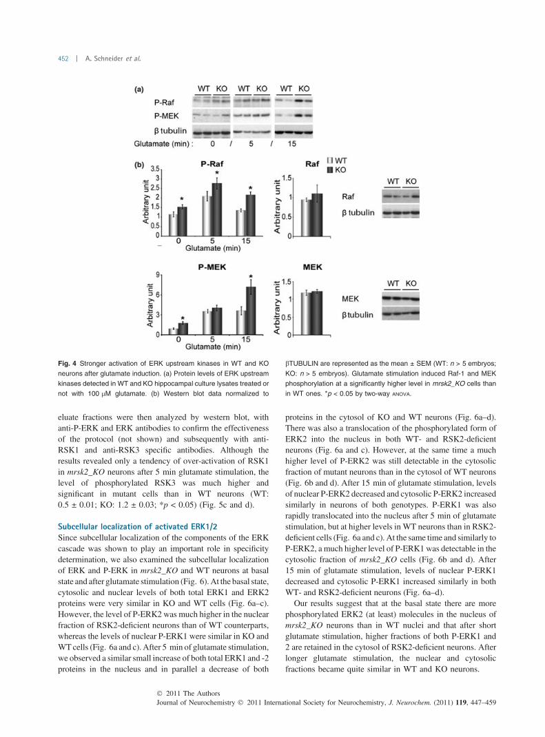
eluate fractions were then analyzed by western blot, withanti-P-ERK and ERK antibodies to confirm the effectivenessof the protocol (not shown) and subsequently with anti-RSK1 and anti-RSK3 specific antibodies. Although theresults revealed only a tendency of over-activation of RSK1in mrsk2_KO neurons after 5 min glutamate stimulation, thelevel of phosphorylated RSK3 was much higher andsignificant in mutant cells than in WT neurons (WT:0.5 ± 0.01; KO: 1.2 ± 0.03; *p < 0.05) (Fig. 5c and d).
Subcellular localization of activated ERK1/2Since subcellular localization of the components of the ERKcascade was shown to play an important role in specificitydetermination, we also examined the subcellular localizationof ERK and P-ERK in mrsk2_KO and WT neurons at basalstate and after glutamate stimulation (Fig. 6). At the basal state,cytosolic and nuclear levels of both total ERK1 and ERK2proteins were very similar in KO and WT cells (Fig. 6a–c).However, the level of P-ERK2 was much higher in the nuclearfraction of RSK2-deficient neurons than of WT counterparts,whereas the levels of nuclear P-ERK1 were similar in KO andWT cells (Fig. 6a and c). After 5 min of glutamate stimulation,we observed a similar small increase of both total ERK1 and -2proteins in the nucleus and in parallel a decrease of both
proteins in the cytosol of KO and WT neurons (Fig. 6a–d).There was also a translocation of the phosphorylated form ofERK2 into the nucleus in both WT- and RSK2-deficientneurons (Fig. 6a and c). However, at the same time a muchhigher level of P-ERK2 was still detectable in the cytosolicfraction of mutant neurons than in the cytosol of WT neurons(Fig. 6b and d). After 15 min of glutamate stimulation, levelsof nuclear P-ERK2 decreased and cytosolic P-ERK2 increasedsimilarly in neurons of both genotypes. P-ERK1 was alsorapidly translocated into the nucleus after 5 min of glutamatestimulation, but at higher levels in WT neurons than in RSK2-deficient cells (Fig. 6a and c). At the same time and similarly toP-ERK2, a much higher level of P-ERK1was detectable in thecytosolic fraction of mrsk2_KO cells (Fig. 6b and d). After15 min of glutamate stimulation, levels of nuclear P-ERK1decreased and cytosolic P-ERK1 increased similarly in bothWT- and RSK2-deficient neurons (Fig. 6a–d).
Our results suggest that at the basal state there are morephosphorylated ERK2 (at least) molecules in the nucleus ofmrsk2_KO neurons than in WT nuclei and that after shortglutamate stimulation, higher fractions of both P-ERK1 and2 are retained in the cytosol of RSK2-deficient neurons. Afterlonger glutamate stimulation, the nuclear and cytosolicfractions became quite similar in WT and KO neurons.
Fig. 4 Stronger activation of ERK upstream kinases in WT and KO
neurons after glutamate induction. (a) Protein levels of ERK upstream
kinases detected in WT and KO hippocampal culture lysates treated or
not with 100 lM glutamate. (b) Western blot data normalized to
bTUBULIN are represented as the mean ± SEM (WT: n > 5 embryos;
KO: n > 5 embryos). Glutamate stimulation induced Raf-1 and MEK
phosphorylation at a significantly higher level in mrsk2_KO cells than
in WT ones. *p < 0.05 by two-way ANOVA.
Journal of Neurochemistry � 2011 International Society for Neurochemistry, J. Neurochem. (2011) 119, 447–459� 2011 The Authors
452 | A. Schneider et al.

To determine the consequences of the increased P-RSKlevel on subcellular distribution in mrsk2_KO cells, we alsoassessed RSK (for RSK1, 2 and 3) and P-RSK subcellularlocalization. Glutamate stimulation induced an increase ofnuclear total RSK and P-RSK in bothWT- andRSK2-deficientneurons, but the increase was higher and more sustained, forP-RSK, in mutant than in WT neurons (Fig. 6a–d).
Expression or activation of ERK1/2 and/or RSK2 targetsTo investigate whether ERK over-activation has an influenceon activity of ERK nuclear substrates, we analyzed phos-phorylation of ELK1 and CREB. ELK1 is a member of theEts family of transcription factors, which regulates serumresponse element-dependent gene expression. We tried todetermine the level of total ELK-1 protein in WT and KOneurons, but unfortunately the antibody gave a strongbackground and no accurate quantification could be done.However, by visual examination the intensity of the 47 kDaband corresponding to the ELK-1 size was quite similar inKO and WT neuronal extracts. At the basal state, the level ofP-ELK1 was significantly higher in RSK2-deficient neuronsthan in WT neurons (WT: 1 ± 0.1; KO: 2.1 ± 0.2;*p < 0.05) (Fig. 7a and b). Five minutes of glutamatestimulation resulted in an increase of the phosphorylatedform of ELK1 in WT and mrsk2_KO cells. After 15 min ofglutamate treatment, the level of P-ELK1 decreased belowthe initial basal level in WT- and RSK2-deficient hippocam-
pal neurons. However, the level of P-ELK1 always remainedsignificantly higher in KO than in WT neurons (5 min: WT:1.8 ± 0.12; KO: 2.51 ± 0.14; 15 min: WT: 0.87 ± 0.06; KO:1.43 ± 0.11; *p < 0.05) (Fig. 7). CREB is a transcriptionfactor regulated via the ERK/MAPK pathway by RSK2 andMSK1. Total CREB protein was very similar in WT and KOneurons (Fig. 7). At basal state, the level of P(Ser133)-CREBwas also significantly higher in RSK2_deficient neurons thanin WT cells (WT: 1.2 ± 0.15; KO: 2 ± 0.27; *p < 0.05).Glutamate treatment induced an increase of CREB phos-phorylation in WT and KO neurons, but which wassignificantly higher in RSK2-deficient than in WT neurons(WT: 3.2 ± 0.23; KO: 3.9 ± 0.3; *p < 0.05) (Fig. 7a and b).
U0126, a MEK specific inhibitor, was then used to confirmthe ERK-dependent activation of RSK, ELK1 and CREB.Neurons were treated with U0126 for 30 min, beforeglutamate stimulation. As expected, glutamate-inducedERK phosphorylation was completely inhibited in WT andmrsk2_KO neuronal cultures (Fig. 8a). A dramatic decreaseof RSK and CREB phosphorylation was also observed (P-CREB: decrease of 2.3 and 18.9* times in WT at basal versusbasal + U0126 conditions, and activated versus activated +U0126 conditions respectively, and a decrease of 4.9* and6.7* times respectively in KO cells; P-RSK: 1.5 and 6.1*respectively in WT neurons, and 4.3* and 6.4* respectivelyin KO cells, *p < 0.05 by Student’s t-test) (Fig. 8b).However, glutamate-induced ELK1 phosphorylation was
Fig. 5 Increased RSK phosphorylation in response to glutamate
induction in mrsk2_KO hippocampal neurons. (a) Detection of RSK
and P-RSK1/2/3 protein level in WT and KO hippocampal lysates,
without stimulation, and after 5 and 15 min glutamate stimulation
(100 lM). (b) Western blot data normalized to bTUBULIN are repre-
sented as the mean ± SEM (WT: n > 8 embryos; KO: n > 9 embryos).
Glutamate stimulation induced an increased RSK phosphorylation in
both cell types, but this increase was higher in RSK2-deficient cells
after 5 min stimulation. *p < 0.05 by two-way ANOVA. (c) Protein level
detection of RSK1 and 3 after phosphoprotein purification of WT and
KO neuronal extracts activated or not (Eluate fraction). (d) Western
blot results normalized to both bTUBULIN and total input levels are
represented as the mean ± SEM (for three embryos of each geno-
type). RSK3 phosphorylation was induced after glutamate stimulation,
but at a significantly higher level in mrsk2_KO neurons than in WT
cells. *p < 0.05 by two-way ANOVA. (F.T., flow through)
� 2011 The AuthorsJournal of Neurochemistry � 2011 International Society for Neurochemistry, J. Neurochem. (2011) 119, 447–459
Altered ERK/MAPK in the mrsk2_KO hippocampus | 453

not inhibited by U0126 treatment neither in WT nor in KOneurons (Fig. 9a and b). As ELK1 activity is also regulatedvia the p38 and JNK pathways, we analyzed the phosphor-ylation state of P-p38 and P-JNK. P-JNK and -p38 revealedincreased levels of phosphorylation in both cases (P-p38:increase of 1.5 and 3.6* times at activated versus activated +U0126 conditions in WT and KO cells respectively; P-JNK:increase of 2.5* and 0.9 times in WT and KO neurons
respectively, *p < 0.05 by Student’s t-test, but not by two-way ANOVA) (Fig. 9a and c).
Expression of immediate-early genesTo check whether increase of CREB and ELK1 activities inRSK2-deficient neurons leads to changes in transcriptionalactivity, we investigated the induction of the expression ofthree IEGs upon glutamate stimulation at the mRNA level
Fig. 6 Increased P-ERK and P-RSK localize in distinct compartments
in mrsk2_KO neurons. (a, b) Level of ERK, P-ERK, RSK and P-RSK
detected in WT and mrsk2_KO in (a) nuclear and (b) cytoplasmic
fractions (N.F. and C.F. respectively). Subcellular fractionation was
performed on basal and activated hippocampal cultures (100 lM
glutamate) and analyzed by western blot. (c, d) Quantification of
protein level, normalized to TBP (nuclear control), CALPAIN (cyto-
plasmic marker) or ERK are represented as the mean ± SEM (WT:
n = 1 embryo; KO: n = 2 embryos). (c) Five minutes glutamate stim-
ulation induced a similar increase of P-ERK2 in the nucleus of WT and
KO neurons, whereas a higher proportion of P-ERK1 molecules were
translocated to the nucleus in WT neurons than in KO cells. A higher
increase of P-RSK level was observed in the KO nucleus compared to
WT ones after treatment. (d) The cytosolic fractions of P-ERK1/2 in-
creased in both WT and KO cells after 5 min glutamate stimulation, but
at a much higher level in KO cells, suggesting retention in the cytosol
of KO neurons. Cytosolic P-RSK was not modified by glutamate
stimulation in both groups.
Journal of Neurochemistry � 2011 International Society for Neurochemistry, J. Neurochem. (2011) 119, 447–459� 2011 The Authors
454 | A. Schneider et al.

by QRT-PCR. Increase of transcription of all three IEGs,c-Fos, Zif268 and Arc, was observed in WT and mrsk2_KOneurons after cellular stimulation, but, for the three genes, ata significantly higher level in RSK2-deficient neurons than inWT neurons (c-Fos: WT: 0.059 ± 0.009; KO: 0.08 ± 0.012;Zif268: WT: 0.1 ± 0.019; KO: 0.16 ± 0.015; Arc: WT:0.013 ± 0.002; KO: 0.02 ± 0.004; *p < 0.05) (Fig. 10).
Discussion
We show in the present study that the levels of Phospho-ERK1 and Phospho-ERK2 are significantly higher in thehippocampus of mrsk2_KO mice as in WT littermates. To
further investigate the cause and consequences of this ERK1/2 increased activation, we analyzed ERK1/2 signaling inRSK2-deficient cultured hippocampal neurons stimulated ornot with glutamate.
Phosphorylation of ERK1 and ERK2 at the basal state andsubsequent to glutamate stimulation was also stronger inRSK2-deficient neurons than in WT neurons. We firstwondered whether this increased phosphorylation levelmight be ascribed to the loss of the RSK2 feedbackinhibitory effect on the ERK pathway previously described(Douville and Downward 1997). Our data show that bothMEK and RAF-1 are activated more strongly in RSK2-deficient neurons as in WT cells, confirming further thismechanism in hippocampal neurons. Strikingly, even inabsence of RSK2, the level of phospho-RSK is much higherin mutant neurons than in WT counterparts, suggesting anover-compensation by other RSK members. We show thatRSK3 is predominantly responsible for this over-compensa-tion, the level of glutamate-induced RSK3 phosphorylationin KO hippocampal neurons being twice that in WT cells.The mechanism of this over-compensation is not yet known,but is likely to be the direct consequence of the over-activation of the upstream kinases ERK1/2.
The subcellular localization of ERK, an important factorcontrolling its activity, was also investigated in RSK2-deficient neurons. At the basal state, there are clearly higherlevels of P-ERK2 in the nucleus of mrsk2_KO neurons thanin WT nuclei. After glutamate stimulation, similar levels ofERK1/2 are detected in the nuclei of both WT and RSK2-deficient neurons, whereas high fractions of both P-ERK1and -2 are transiently retained in the cytosol of RSK2-deficient neurons, but not in WT cells. Nuclear localizationof ERK1/2 is essential for some of the phenotypic programsto which they contribute, including differentiation, trans-formation and altered transcription. Therefore, we wonderedwhether the transcription factors ELK1 and CREB aresimilarly activated in RSK2-deficient and WT neurons inresponse to glutamate stimulation. Increases of phosphor-ylation levels of both CREB and ELK1 were observed inWT- and RSK2-deficient neurons, which were, however,significantly stronger in RSK2-deficient neurons. As ELK-1has been described as a direct nuclear target of ERK1/2(Yang et al. 1998), this result suggested that the increasedELK-1 phosphorylation is directly the consequence ofincreased ERK1/2 activity in RSK2-deficient neurons.However, inhibition of ERK activity by U0126 did notabolish glutamate-induced phosphorylation of ELK1,providing evidence that glutamate-induced ELK1 phosphor-ylation is not mediated by ERK1/2 in hippocampal neurons.As glutamate also activates JNKs and p38 (Wang et al.2007) we suspected involvement of these pathways inELK1 activation. Both p38 and JNKs showed an increaseof phosphorylation levels in WT and RSK2-deficientneurons after inhibition, with U0126, of the ERK/MAPK
Fig. 7 Increased activation of transcription factors in mrsk2_KO cells
after glutamate induction. (a) Western blot analysis of ERK targets,
involved in regulation of gene expression, in WT and mrsk2_KO
neurons untreated or treated with 100 lM glutamate. (b) Protein levels
were normalized to bTUBULIN and are represented as the mean ±
SEM (WT: n > 12 embryos; KO: n > 14 embryos). The level of P-
ELK1 increased in both WT and KO neurons in response to glutamate
stimulation. However, levels were significantly higher in mrsk2_KO
neurons than in WT ones. Increase of CREB phosphorylation induced
by glutamate was significantly stronger in KO neurons. *p < 0.05 by
two-way ANOVA.
� 2011 The AuthorsJournal of Neurochemistry � 2011 International Society for Neurochemistry, J. Neurochem. (2011) 119, 447–459
Altered ERK/MAPK in the mrsk2_KO hippocampus | 455

pathway. A cross-talk between different MAPK pathwayswas already reported (Monick et al. 2006). The authorsshowed that ERK inhibition leads to JNK activation inmacrophages. Our results indicate that it is also the case inneurons.
The increased CREB phophorylation observed in RSK2-deficient neurons indicated that RSK2 is not the major kinaseof CREB in hippocampal neurons upon glutamate stimula-tion. However, it could not be excluded that the higher levelof P-RSK, and mainly of P-RSK3, observed in mutant
Fig. 9 ELK1 activation is ERK, p38 and JNK dependent in hippo-
campal neurons. (a) Protein levels of P-ELK1, P-p38 and P-JNK in WT
and mrsk2_KO neurons treated with U0126 (5 lM) and glutamate
(100 lM). (b, c) Western blot data normalized to bTUBULIN are rep-
resented as the mean ± SEM (WT: n > 2 embryos; KO: n > 3 em-
bryos). (b) U0126 treatment leads to a decrease of ELK1
phosphorylation in KO cells at the basal state. However, an increase of
P-ELK1 is observed after glutamate stimulation in WT and mrsk2_KO
neurons. (c) Levels of phosphorylation of p38 and JNK were also
increased in both cell types after U0126 treatment and glutamate
stimulation. This suggests that ELK1 phosphorylation is in part due to
p38 and JNK activation in these hippocampal cells.
Fig. 8 RSK and CREB activation are dependent on ERK activity in
hippocampal neurons. (a) Protein levels detection of P-RSK and
P-CREB from WT and mrsk2_KO neurons treated with U0126 (5 lM),
a MEK specific inhibitor and glutamate (100 lM). (b) Protein levels
quantified by western blot analysis and normalized to bTUBULIN are
represented as the mean ± SEM (WT: n > 2 embryos; KO: n > 3
embryos). U0126 treatment led to a strong decrease of ERK phos-
phorylation in both WT and KO cells. RSK and CREB phosphoryla-
tions were also significantly blocked by U0126 treatment in KO cells at
the basal state and after glutamate stimulation in both WT and KO
neurons. This suggests that ERK is the main upstream kinase of RSK
and CREB in hippocampal cells.
Journal of Neurochemistry � 2011 International Society for Neurochemistry, J. Neurochem. (2011) 119, 447–459� 2011 The Authors
456 | A. Schneider et al.
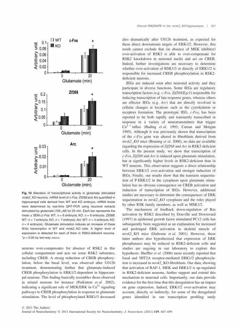
neurons over-compensates for absence of RSK2 in thiscellular compartment and acts on some RSK2 substrates,including CREB. A strong reduction of CREB phosphory-lation, below the basal level, was observed after U0126treatment, demonstrating further that glutamate-inducedCREB phosphorylation is ERK1/2-dependent in hippocam-pal neurons. This finding basically resembles those observedin striatal neurons for instance (Perkinton et al. 2002),indicating a significant role of MEK/ERK in Ca2+ signalingpathways to CREB phosphorylation in response to glutamatestimulation. The level of phosphorylated RSK1/3 decreased
also dramatically after U0126 treatment, as expected forthese direct downstream targets of ERK1/2. However, thisresult cannot exclude that (in absence of MEK inhibitor)over-activation of RSK3 is able to over-compensate forRSK2 knockdown in neuronal nuclei and act on CREB.Indeed, further investigations are necessary to determinewhether over-activation of RSK1/3 or directly of ERK1/2 isresponsible for increased CREB phosphorylation in RSK2-deficient neurons.
IEGs are induced soon after neuronal activity and theyparticipate in diverse functions. Some IEGs are regulatorytranscription factors (e.g. c-Fos, Zif268/Egr1) responsible forinducing transcription of late-response genes, whereas othersare effector IEGs (e.g. Arc) that are directly involved incellular changes at locations such as the cytoskeleton orreceptors formation. The prototypic IEG, c-Fos, has beenreported to be both rapidly and transiently transcribed inresponse to a variety of neurotransmitters that triggerCa2 + influx (Bading et al. 1993; Curran and Morgan1995). Although it was previously shown that transcriptionof the c-Fos gene was altered in fibroblasts derived frommrsk2_KO mice (Bruning et al. 2000), no data are availableregarding the expression of Zif268 and Arc in RSK2-deficientcells. In the present study, we show that transcription ofc-Fos, Zif268 and Arc is induced upon glutamate stimulation,but at significantly higher levels in RSK2-deficient than inWT neurons. This observation suggests a direct relationshipbetween ERK1/2 over-activation and stronger induction ofIEGs. Finally, our results show that the transient sequestra-tion of P-ERK1/2 in the cytoplasm upon glutamate stimu-lation has no obvious consequence on CREB activation andinduction of transcription of IEGs. However, additionalstudies are necessary to determine the consequences of ERKsequestration in mrsk2_KO cytoplasm and the roles playedby other RSK family members, as well as MSK1/2.
The mechanism of feedback down-regulation of Rasactivation by RSK2 described by Douville and Downward(1997) in epidermal growth factor stimulated PC12 cells hassubsequently been suggested as an explanation for increasedand prolonged ERK activation in skeletal muscle ofmrsk2_KO mice (Dufresne et al. 2001). However, theselatter authors also hypothesized that expression of ERKphosphatases may be reduced in RSK2-deficient cells andstudies are ongoing in our laboratory to explore thishypothesis. Sheffler et al. (2006) more recently reported thatbasal and 5HT2A receptor-mediated ERK1/2 phosphoryla-tion is increased in mrsk2_KO fibroblasts. Our data, showingthat activation of RAF-1, MEK and ERK1/2 is up-regulatedin RSK2-deficient neurons, further support and extend thismechanism to neuronal cells. Importantly, our data provideevidence for the first time that this deregulation has an impacton gene expression. Indeed, ERK1/2 over-activation mayaccount, directly or indirectly, for some of the deregulatedgenes identified in our transcription profiling study
Fig. 10 Alteration of transcriptional activity in glutamate stimulated
mrsk2_KO neurons. mRNA level of c-Fos, Zif268 and Arc quantified in
hippocampal cells derived from WT and KO embryos. mRNA levels
were determined by real-time QRT-PCR using neuronal extracts
stimulated by glutamate (100 lM) for 20 min. Each bar represents the
mean ± SEM (c-Fos: WT: n = 6 embryos; KO: n = 6 embryos; Zif268:
WT: n = 7 embryos; KO: n = 7 embryos; Arc: WT: n = 4 embryos; KO:
n = 4 embryos). Glutamate stimulation induces an increase of these
IEGs transcription in WT and mrsk2_KO cells. A higher level of
expression is detected for each of them in RSK2-deficient neurons.
*p < 0.05 by two-way ANOVA.
� 2011 The AuthorsJournal of Neurochemistry � 2011 International Society for Neurochemistry, J. Neurochem. (2011) 119, 447–459
Altered ERK/MAPK in the mrsk2_KO hippocampus | 457

(Mehmood et al. 2011), through over-activation of transcrip-tion factors or over-induction of IEGs. Further studies, tooexplore this hypothesis, are ongoing in our laboratory.
Finally, our data provide an important contribution to afascinating issue not yet clarified: do the various RSK proteins,which are highly homologous and all widely expressed,regulate distinct or unique (plus overlapping?) cellular func-tions? Our results show that increased phosphorylation ofRSK1/3 in RSK2-deficient neurons neither is able to restore anormal ERK1/2 activity, nor to correct the expression ofCREB, ELK1 and the IEG gene expression. The feedbackinhibition exerted byRSK2on theERK1/2 pathway is clearly aspecific function of RSK2 that cannot be compensated by othermembers of the RSK family. The phosphorylation/activationof other substrates of ERK1/2 has now to be investigated in thecontext of absence of RSK2 activity.
The aberrant ERK1/2 signaling report here is susceptibleto influence various neuronal functions, and can, indeed, playa significant role in cognitive dysfunction of mrsk2_KO miceand of CLS patients.
Acknowledgements
The authors wish to thank the IGBMC, Institut de Genetique et deBiologie Moleculaire et Cellulaire, for assistance. The authorsdeclare no conflict of interest. This work was supported by theFrench National Agency for Research (grant number 08-MNPS-021-01, to AH), the Fondation Jerome Lejeune (to AH), the CentreNational de la Recherche Scientifique, the Institut National de laSante et de la Recherche, the College de France and the Universityof Strasbourg. AS was supported by a fellowship from the Ministerepour la Recherche et Technologie (France) and the AssociationXtraordinaire (France), and TM by a scholarship from the HigherEducation Commission (HEC) of Pakistan.
References
Alberini C. M. (2009) Transcription factors in long-term memory andsynaptic plasticity. Physiol. Rev. 89, 121–45.
Angenstein F., Greenough W. T. and Weiler I. J. (1998) Metabotropicglutamate receptor-initiated translocation of protein kinase p90rskto polyribosomes: a possible factor regulating synaptic proteinsynthesis. Proc. Natl Acad. Sci. USA 95, 15078–15083.
Bading H. and Greenberg M. E. (1991) Stimulation of protein tyrosinephosphorylation by NMDA receptor activation. Science 253, 912–914.
Bading H., Ginty D. D. and Greenberg M. E. (1993) Regulation of geneexpression in hippocampal neurons by distinct calcium signalingpathways. Science 260, 181–186.
Bruning J. C., Gillette J. A., Zhao Y. et al. (2000) Ribosomal subunitkinase-2 is required for growth factor-stimulated transcription ofthe c-Fos gene. Proc. Natl Acad. Sci. USA 97, 2462–2467.
Chotiner J. K., Nielson J., Farris S. et al. (2010) Assessment of the roleof MAP kinase in mediating activity-dependent transcriptionalactivation of the immediate early gene Arc/Arg3.1 in the dentategyrus in vivo. Learn. Mem. 17, 117–129.
Chwang W. B., Arthur J. S., Schumacher A. and Sweatt J. D. (2007) Thenuclear kinase mitogen- and stress-activated protein kinase 1 reg-
ulates hippocampal chromatin remodeling in memory formation.J. Neurosci. 27, 12732–12742.
Curran T. and Morgan J. I. (1995) Fos: an immediate-early transcriptionfactor in neurons. J. Neurobiol. 26, 403–412.
Davis S. and Laroche S. (2006) Mitogen-activated protein kinase/extracellular regulated kinase signaling and memory stabilization: areview. Genes Brain Behav. 5, 61–72.
Davis S., Renaudineau S., Poirier R., Poucet B., Save E. and Laroche S.(2010) The formation and stability of recognition memory: whathappens upon recall? Front. Behav. Neurosci. 4, 177.
De Cesare D., Jacquot S., Hanauer A. and Sassone-Corsi P. (1998) Rsk-2 activity is necessary for epidermal growth factor-induced phos-phorylation of CREB protein and transcription of c-fos gene. Proc.Natl Acad. Sci. USA 95, 12202–12207.
Douville E. and Downward J. (1997) EGF induced SOS phosphorylationin PC12 cells involves P90 RSK-2. Oncogene 15, 373–383.
Dudek S. M. and Fields R. D. (2001) Mitogen-activated protein kinase/extracellular signal-regulated kinase activation in somatodendriticcompartments: roles of action potentials, frequency, and mode ofcalcium entry. J. Neurosci. 21, RC122.
Dufresne S. D., Bjorbaek C., El-Haschimi K. et al. (2001) Alteredextracellular signal-regulated kinase signaling and glycogenmetabolism in skeletal muscle from p90 ribosomal S6 kinase 2knockout mice. Mol. Cell. Biol. 21, 81–87.
Frodin M. and Gammeltoft S. (1999) Role and regulation of 90 kDaribosomal S6 kinase (RSK) in signal transduction. Mol. Cell.Endocrinol. 151, 65–77.
Hanauer A. and Young I. D. (2002) Coffin-Lowry syndrome: clinicaland molecular features. J. Med. Genet. 39, 705–713.
Kim M., Lee J. H., Koh H. et al. (2006) Inhibition of ERK-MAP kinasesignaling by RSK during Drosophila development. EMBO J. 25,3056–3067.
Kolkova K., Novitskaya V., Pedersen N., Berezin V. and Bock E.(2000) Neural cell adhesion molecule-stimulated neurite out-growth depends on activation of protein kinase C and the Ras-mitogen-activated protein kinase pathway. J. Neurosci. 20, 2238–2246.
Li L., Carter J., Gao X., Whitehead J. and Tourtellotte W. G. (2005) Theneuroplasticity-associated arc gene is a direct transcriptional targetof early growth response (Egr) transcription factors. Mol. Cell.Biol. 25, 10286–10300.
Marques Pereira P., Gruss M., Braun K., Foos N., Pannetier S. andHanauer A. (2008) Dopaminergic system dysregulation in themrsk2_KO mouse, an animal model of the Coffin-Lowry syn-drome. J. Neurochem. 107, 1325–34.
Mehmood T., Schneider A., Sibille J. et al. (2011) Transcriptome profilereveals AMPA receptor dysfunction in the hippocampus of theRsk2-knockout mice, an animal model of Coffin-Lowry syndrome.Hum. Genet. 129, 255–69.
Messaoudi E., Kanhema T., Soule J. et al. (2007) Sustained Arc/Arg3.1synthesis controls long-term potentiation consolidation throughregulation of local actin polymerization in the dentate gyrus invivo. J. Neurosci. 27, 10445–10455.
Monick M. M., Powers L. S., Gross T. J., Flaherty D. M., Barrett C. W.and Hunninghake G. W. (2006) Active ERK contributes to proteintranslation by preventing JNK-dependent inhibition of proteinphosphatase 1. J. Immunol. 177, 1636–1645.
Murphy L. O., Smith S., Chen R. H., Fingar D. C. and Blenis J. (2002)Molecular interpretation of ERK signal duration by immediateearly gene products. Nat. Cell Biol. 4, 556–564.
Myers A. P., Corson L. B., Rossant J. and Baker J. C. (2004) Charac-terization of mouse Rsk4 as an inhibitor of fibroblast growth factor-RAS-extracellular signal-regulated kinase signaling. Mol. Cell.Biol. 24, 4255–4266.
Journal of Neurochemistry � 2011 International Society for Neurochemistry, J. Neurochem. (2011) 119, 447–459� 2011 The Authors
458 | A. Schneider et al.

Perkinton M. S., Ip J. K., Wood G. L., Crossthwaite A. J. and WilliamsR. J. (2002) Phosphatidylinositol 3-kinase is a central mediator ofNMDA receptor signaling to to MAP kinase (Erk1/2), Akt/PKBand CREB in striatal neurones. J. Neurochem. 80, 239–54.
Poirier R., Jacquot S., Vaillend C. et al. (2007) Deletion of the Coffin-Lowry syndrome gene Rsk2 in mice is associated with impairedspatial learning and reduced control of exploratory behavior.Behav. Genet. 37, 31–50.
Sassone-Corsi P., Mizzen C. A., Cheung P. et al. (1999) Requirement ofRsk-2 for epidermal growth factor-activated phosphorylation ofhistone H3. Science 285, 886–891.
Scimeca J. C., Nguyen T. T., Filloux C. and Van Obberghen E. (1992)Nerve growth factor-induced phosphorylation cascade in PC12pheochromocytoma cells. Association of S6 kinase II with themicrotubule-associated protein kinase, ERK1. J. Biol. Chem. 267,17369–17374.
Segal R. A. and Greenberg M. E. (1996) Intracellular signaling pathwaysactivated by neurotrophic factors. Annu. Rev. Neurosci. 19, 463–489.
Sheffler D. J., Kroeze W. K., Garcia B. G. et al. (2006) p90 ribosomalS6 kinase 2 exerts a tonic brake on G proteincoupled receptorsignaling. Proc. Natl Acad. Sci. USA 103, 4717–4722.
Thiels E., Kanterewicz B. I., Norman E. D., Trzaskos J. M. and Klann E.(2002) Long-term depression in the adult hippocampus in vivo
involves activation of extracellular signal-regulated kinase andphosphorylation of Elk-1. J. Neurosci. 22, 2054–2062.
Thomas G. M., Rumbaugh G. R., Harrar D. B. and Huganir R. L. (2005)Ribosomal S6 kinase 2 interacts with and phosphorylates PDZdomain-containing proteins and regulates AMPA receptor trans-mission. Proc. Natl Acad. Sci. USA 102, 15006–15011.
Wang J. Q., Fibuch E. E. and Mao L. (2007) Regulation of mitogen-activated protein kinases by glutamate receptors. J. Neurochem.100, 1–11.
Yang S. H., Whitmarsh A. J., Davis R. J. and Sharrocks A. D. (1998)Differential targeting of MAP kinases to the ETS-domain tran-scription factor Elk-1. EMBO J. 17, 1740–1749.
Yang X., Matsuda K., Bialek P. et al. (2004) ATF4 is a substrate ofRSK2 and an essential regulator of osteoblast biology; implicationfor Coffin-Lowry Syndrome. Cell 117, 387–398.
Zeniou M., Ding T., Trivier E. and Hanauer A. (2002) Expressionanalysis of RSK gene family members: the RSK2 gene, mutated inCoffin-Lowry syndrome, is prominently expressed in brain struc-tures essential for cognitive function and learning. Hum. Mol.Genet. 11, 2929–2940.
Zeniou-Meyer M., Liu Y., Begle A. et al. (2008) The Coffin-Lowrysyndrome-associated protein RSK2 is implicated in calcium-regu-lated exocytosis through the regulation of PLD1. Proc. Natl Acad.Sci. USA 105, 8434–8439.
� 2011 The AuthorsJournal of Neurochemistry � 2011 International Society for Neurochemistry, J. Neurochem. (2011) 119, 447–459
Altered ERK/MAPK in the mrsk2_KO hippocampus | 459

Publication-3

Chapter 2 Results-Publication 3
103
2.4: Rsk2 Knockdown in PC12 cells results in Sp1 dependent
increased expression of the Gria2 gene, encoding the AMPA
receptor subunit GluR2, via up regulation of ERK1/2 activity
Tahir Mehmood, 1, 2 Anne Schneider1, Solange Pannetier, 1, & André Hanauer 1*
The main objective of this study was to understand the molecular mechanism of up
regulation of GluR2 (subunit of AMPA receptors) in RSK2 deficient cells. Previously we
showed that in the hippocampus of Rsk2-KO mice GluR2 expression is up regulated at the
transcriptional and protein levels (Mehmood et al., 2011). We also showed that ERK1/2
activity is abnormally increased in the hippocampus of Rsk2-KO mice (Schneider et al.,
2011). The mechanism through which RSK2 deficiency, affects the expression of GluR2 in
neural cells was unknown. Thus, we were interested to investigate, whether the GluR2
expression is directly controlled by the RSK2 kinase or indirectly via a RSK2 or ERK1/2
substrate. To the best of our knowledge no one else has reported on regulation of GluR2
expression by RSK2.
Since primary neural cultures are difficult to manipulate and transfect, we used, as a
first step to understand the mechanism leading to GluR2 up regulation of expression, the
RNA interference (shRNA) technology to knockdown the Rsk2 gene in PC12 cells. PC12
cells are immortalized cells derived from a pheochromocytoma tumor of the rat adrenal
gland. NGF treated PC12 cell line serves as a model system for primary neuronal cells.
(Greene and Tischler, 1976; Satoh et al., 1988; Xia et al., 1995). First we investigated the
efficiency to block RSK2 expression in PC12 cells by using three plasmids generating
different RSK2-specific shRNA, targeted to different coding regions of the mRNA of Rsk2.
We observed a dramatic decrease of the Rsk2 mRNA level for one of them, shRNA (clone
ID7). At the protein level RSK2 expression was decreased by 90% in cells transfected with
this shRNA. There was also a strong increase of GluR2 expression at the RNA and protein
levels in the cells transfected with clone ID7, as well as an increased level of ERK1/2
phosphorylation. Treatment of the Rsk2-knockdown cells with U0126, a MEK inhibitior,
resulted in inhibition of ERK activity and in a dramatic decrease of GluR2 expression.

Chapter 2 Results-Publication 3
104
Therefore, we assumed that the increased GluR2 expression in Rsk2-KO mice and
Rsk2-knockdown PC12 cells might be indirectly regulated by over activation of ERK1/2
activity. Ten years ago it was reported that the Gria2 gene is regulated at the transcriptional
level by the Sp1 transcription factor (Ekici et al., 2011; Sroka et al., 2007). In addition, Chu
and Ferro, (2005) reported that the MEK-ERK signal pathway mediates the Sp1
phosphorylation at two sites T 453 and T739.
Therefore, we investigated the levels of Sp1 phosphorylation at both of these sites.
We observed an abnormally increased level of phospho-Sp1 at both sites (T453 and T739)
in Rsk2-knockdown cells compared to WT PC12 cells, where as there was no difference in
total Sp1 expression. We further found that treatment of Rsk2-knockdown PC12 cells with
the MEK inhibitor U0126, completely abolished phosphorylation of Sp1 at T453 and T739,
confirming the specific implication of ERK1/2 in the phosphorylation of these sites. Partial
silencing of Sp1 expression in Rsk2-knockdown PC12 cells with Sp1-siRNA resulted in a
remarkable decrease of GluR2 expression, confirming that Sp1 plays a prominent role in
the mechanism of GluR2 up-regulation of transcription in RSK2 deficient cells. I showed
subsequently that in the hippocampus of Rsk2-KO mice, the level of pospho-Sp1 is
significantly higher than in WT littermate mouse, supporting our results in PC12 cells.
Thus, the conclusion of our study is that over expression of GluR2 in RSK2 deficient cells
is caused by increased Sp1 transcriptional activity on the Gria2 gene which itself is the
consequence of ERK1/2 increased activity resulting from loss of retro inhibition activity
exerted by RSK2 on the ERK pathway.
A complete description and illustration of these results are documented in
publication-3 and paper is submitted in journal.

1
Rsk2 knockdown in PC12 cells results in a Sp1 dependent strong increase of expression
of the Gria2 gene, encoding the AMPA receptor subunit GLUR2, via up regulation of
ERK1/2 activity
Tahir Mehmood1,21,21,21,2 Anne Schneider,1
Solange Pannetier,1
and André Hanauer 1 §
1 Institut de Génétique et de Biologie Moléculaire et Cellulaire (IGBMC), Institut National de
Santé et de Recherche Médicale (INSERM), Department of Translational Medicine and
Neurogenetics, U964/Centre National de Recherche Scientifique (CNRS) UMR
1704/Université de Strasbourg, 67404 Illkirch, France.
2 Department of Chemistry, University of Sargodha, 40100, Sargodha, Pakistan
Email addresses:
§ Corresponding author
Andre´ Hanauer, PhD
IGBMC
1, rue Laurent Fries
67404 Illkirch, France
Phone: 33-388653400
Fax: 33-388653246
E-mail: [email protected]

2
Abstract
Background: The RSK2 protein is a member of the RSK serine-threonine protein kinase
family and is encoded by the X-linked rps6ka3 gene in human. Highly heterogeneous loss-of-
function mutations affecting this gene are responsible for a severe syndromic form of
cognitive impairment, Coffin-Lowry syndrome. RSK2, which is highly conserved in
mammals, acts at the distal end of the Ras-ERK signaling pathway and is activated in
response to growth factors and neurotransmitters. RSK2 is highly expressed in the
hippocampus, and Rsk2-KO mice display spatial learning and memory impairment. We
recently showed that ERK1/2 activity is abnormally increased in the hippocampus of Rsk2-
KO mice as well as the expression of the AMPA receptor subunit GluR2. The mechanism via
which RSK2 deficiency affects the expression of GluR2 in neural cells was unknown.
Results: To address this issue, we constitutively suppressed in the present study the
expression of Rsk2 in PC12 cells via vector-based shRNA. We show that Rsk2 silencing leads
also to up regulation of ERK1/2 activity and GluR2 expression and that the increase of GluR2
expression results from up regulated ERK1/2 activity on the transcription factor Sp1. Our
results strongly suggest that RSK2 modulates ERK1/2 activity on Sp1, which regulates GluR2
expression through transcriptional activation. Preliminary results show that Sp1 activity is
also up regulated in the hippocampus of Rsk2-KO mice suggesting that the mechanism of
GluR2 transcriptional up regulation is similar in mouse and PC12 cells.
Conclusions: Our present findings suggest an indirect role of RSK2 in modulating the
expression of AMPA receptor GluR2 subunit and, therefore, in regulating glutamatergic
neurotransmission and probably synaptic plasticity. Future studies are warranted to elucidate

3
the molecular mechanisms that underline the interplay between the Rsk2 gene and the
glutamatergic system.
4
Background
The 90kDa ribosomal S6 kinases (RSKs) constitute a family of four homologous Ser/Thr
kinases (RSK1-4) that are activated by Mitogen activated protein kinase / extracellular-
regulated kinases 1 and 2 (MAPK/ERK1/2) in response to growth factors, hormones,
chemokines and neurotransmitters. When activated by Extracellular signal-regulated kinase
(ERK), a fraction of RSK2 in turn phosphorylates a number of cytoplasmic substrates such as
BCL2-associated agonist of cell death (Bad), p53, L1 cell adhesion molecule (L1CAM) and
Glycogen synthase kinase 3 (GSK3) [1]. Another fraction of RSK2 translocates to the nucleus
where it is thought to regulate gene expression through phosphorylation of transcription
factors such as cAMP-response element binding protein (CREB), the cellular FBJ murine
osteosarcoma viral oncogene homolog (c-fos), the oestrogen nuclear receptor, the nuclear
receptor subfamily 77 (Nurr 77) and the Activating transcription factor 4 (ATF4) [1,2]. The
roles of RSKs in cellular signaling, cell survival, growth and differentiation has been well
established, however their implication in biological processes in vivo is less well known. An
important physiological role of one RSK member, RSK2, was uncovered by the discovery of
Rsk2 (following the nomenclature: Rps6ka3 gene, but we will name it Rsk2 gene) gene
defects in the Coffin-Lowry Syndrome (CLS) [3]. CLS is an X-linked disorder with
progressive skeletal abnormalities, facial dimorphism, and strong psychomotor impairment
[4]. The vast majority of male CLS patients show severe cognitive deficiency with
intelligence quotient (IQ) ranging from 15 to 60. Delay in speech acquisition is particularly
common with most affected males having a very limited vocabulary. The mouse knockout
(KO) model (Rsk2-KO) for CLS exhibits a severe impairment in spatial learning and a deficit
in long-term spatial memory [5]. Moreover, in human and mouse brain, RSK2 is highly
expressed in the hippocampus, an essential brain structure in cognitive function and learning

5
[6, 7]. Together the data suggest that RSK2 plays an important role in cognitive function in
human and in mice.
To gain greater insight into the molecular mechanisms leading to learning and memory
impairments in the Rsk2-KO mice and to mental retardation in CLS, we examined recently
global gene expression profiles in hippocampus from KO mice, It revealed altered expression
of 100 genes encoding proteins acting in various biological pathways [8]. We investigated in
detail the consequences of the two-fold up-regulation of one of these genes, Gria2, which
encodes the subunit GluR2 of the alpha-amino-3-hydroxy-5-methyl-4-isoxazolepropionic acid
(AMPA) receptor. Abnormally increased expression of GluR2 at the protein level was
confirmed in the hippocampus of Rsk2-KO mice and at synapses from Rsk2-KO hippocampus
primary neuron cultures. Basal AMPA receptor-mediated activity was found significantly
decreased [8]. RSK2 deficient mice have also an increased baseline level of phospho-ERK1/2
as well as enhanced activation of ERK1/2 after glutamate stimulation of hippocampal neurons
[9, 10]. These latter findings demonstrated that RSK2 exerts a feedback inhibitory effect on
the ERK1/2 pathway in physiological conditions.
To define the molecular events leading to GluR2 over expression in RSK2 deficient
neurons we used in the present study the RNA interference technology to knockdown the
Rsk2 gene in PC12 cells. We show that Rsk2 silencing in PC12 cells leads also to an increase
of ERK1/2 activity and of GluR2 expression at the mRNA and protein levels. Furthermore,
our data reveal that the increased GluR2 expression results from the increased ERK1/2
activity on the transcription factor Specificity Protein 1 (Sp1).
6
Results
Rsk2 knockdown of PC12 cells
For this study we used the PC12 cell line, an immortalized cell line derived from a
pheochromocytoma tumor of the rat adrenal gland. NGF-treated PC12 cells cease
proliferation, grow long neurites, and show changes in cellular composition associated with
neuronal differentiation [11], and can therefore serve as a model system for primary neuronal
cells. PC12 cells express a high level of RSK2 as shown in Fig. 1. We tested the efficacy to
block Rsk2 expression in PC12 cells of three plasmids producing different Rsk2-specific small
hairpin RNAs (Rsk2-shRNA) targeted to different coding regions of the mRNA of Rsk2. The
recombinant plasmids were transfected into the PC12 cells by lipofecamine 2000,
recombinant cells selected 72 hours on G418, and mRNA and protein expression levels of the
Rsk2 gene were assayed by QRT-PCR and Western blot. One shRNAs (shRNA clone ID7),
decreased Rsk2 gene expression dramatically (Fig. 1a). Quantification of the immunoblots
revealed a 90% reduction in the expression of the RSK2 protein, compared to untransfected
PC12 cells. No significant change was found in the level of RSK2 mRNA and protein
expression in cells transfected with a shRNA containing a scrambled sequence (negative
control) (Fig. 1a). (Expression of the RSK2 protein in Rsk2-shRNA transfected cells: 0.07 ±
0.03; untransfected cells: 1.0 ± 0.09; scrambled-shRNA transfected cells: 0.917 ± 0.1;
*p<0.05 by Student’s t-test) (Fig. 1a). QRT-PCR revealed also a dramatic decrease of Rsk2
mRNA expression (Expression of Rsk2 mRNA in Rsk2-shRNA transfected cells: 0.316 ±
0.05; untransfected cells: 1.0 ± 0.16; scrambled-shRNA transfected cells; 0.865± 0.15;
*p<0.05 by Student’s t-test) (Fig. 1c). Thus, the shRNA clone ID7 was used in all subsequent
studies. We then asked whether the severe down regulation of RSK2 would also affect the

7
expression of other RSK proteins in PC12 cells. The result for RSK1 is shown in Fig. 1b.
(Expression of RSK1 protein in Rsk2-shRNA transfected cells: 0.895 ± 0.1; untransfected
cells: 1.0 ± 0.28; scrambled-shRNA transfected cells: 0.894 ± 0.09; *p<0.05 by Student’s t-
test). RSK3 was very weakly expressed in untransfected PC12 cells and was unchanged in
transfected PC12 cells (result not shown). Thus, none of these two latter RSK family
members apparently exhibited any significant change in its expression levels, documenting
that the Rsk2-shRNA approach was highly specific for RSK2 and reducing the possibility of
compensation for the RSK2 loss via over expression of other RSK family members.
Immunocytofluorescence analysis of RSK2 expression
We also examined the expression of RSK2 in PC12 cells by immunocytofluorescence
analysis. Most of untransfected, or scrambled-shRNA transfected PC12 cells showed strong
RSK2 staining, whereas Rsk2-shRNA transfected cells expressed RSK2 very weakly (Fig. 2).
It was concluded that the Rsk2-shRNA recombinant plasmid can effectively suppress the
expression of the Rsk2 gene in PC12 cells.
Rsk2 silencing increased ERK1/2 MAPK phosphorylation
We then asked whether Rsk2 knockdown affects the level of ERK1/2 phosphorylation in
PC12 cells. Detection of total ERK levels revealed no significant difference of total ERK
protein expression between untransfected PC12 cells and cells transfected with the Rsk2- or
scrambled-shRNA (Expression of ERK1 protein in Rsk2-shRNA transfected cells: 1.0 ± 0.11;
untransfected cells: 0.875 ± 0.04; scrambled-shRNA transfected cells: 0.748 ± 0.08,
Expression of ERK2 protein in Rsk2-shRNA transfected cells: 0.830 ± 0.07; untransfected
cells: 0.950 ± 0.05; scrambled-shRNA transfected cells: 1.0 ± 0.09; *p<0.05 by Student’s t-
test) (Fig. 3a and b). In contrast, the analysis of phosphorylated ERK1 and ERK2 showed

8
that the active forms of both ERK1 (P-ERK1) and ERK2 (P-ERK2) were significantly higher
in Rsk2-shRNA transfected PC12 cells than in scrambled-shRNA transfected or untransfected
cells (Expression of P-ERK1 in Rsk2-shRNA transfected cells: 1.0 ± 0.07; untransfected cells:
0.03 ± 0.003; scrambled-shRNA transfected cells: 0.42 ± 0.06, Expression of P-ERK2 in
Rsk2-shRNA transfected cells: 1.0 ± 0.09; untransfected cells: 0.04 ± 0.003; scrambled-
shRNA transfected cells: 0.32 ± 0.05; *p<0.05 by Student’s t-test) (Fig. 3a and c) . These
results were consistent with our previous data in the hippocampus of Rsk2-KO mice, which
also showed increased levels of ERK1/2 phosphorylation [9]. An increase of P-ERK1/2 levels
was also observed in scrambled-shRNA transfected PC12 cells, but much lower than in Rsk2-
shRNA transfected cells. The cause is not yet known but it might be due to the G418
selection.
Rsk2 knockdown results in up regulation of Gria2 gene (encoding GluR2) expression
We next examined the expression of GluR2 in Rsk2 knockdown PC12 cells by Western blot
analysis and QRT-PCR. Detection of GluR2 protein levels, revealed a very strong increase of
expression in Rsk2 knockdown PC12 cells (Expression of GluR2 protein in Rsk2-shRNA
transfected cells: 1.0 ± 0.02; untransfected cells: 0.03 ± 0.008; scrambled-shRNA transfected
cells: 0.382 ± 0.04; *p<0.05 by Student’s t-test) (Fig. 4a). QRT-PCR revealed also an
approximately four-fold higher Gria2 mRNA expression in Rsk2-silenced PC12 cells
compared to untransfected cells (Expression of Gria2 mRNA in Rsk2-shRNA transfected
cells: 1.0 ± 0.04; untransfected cells: 0.225 ± 0.007; scrambled-shRNA transfected cells:
0.476 ± 0.06; *p<0.05 by Student’s t-test) (Fig. 4b). Cells transfected with the scrambled-
shRNA showed a small increase of both GluR2 protein and mRNA but much lower than
Rsk2-shRNA transfected cells, and which may be related to the small increase of ERK1/2
phosphorylation observed in these cells (Fig. 4a and b). We assayed also GluR1 and GluR3
9
levels of expression. Detection of GluR1 protein levels revealed a small decrease of
expression in Rsk2-shRNA transfected PC12 cells compared to untransfected PC12 cells or
cells transfected with the scrambled-shRNA (Expression of GluR1 protein in Rsk2-shRNA
transfected cells: 0.803 ± 0.11; untransfected cells: 1.0 ± 0.14; scrambled-shRNA transfected
cells: 0.951 ± 0.09; *p<0.05 by Student’s t-test) (Fig. 4c). GluR3 was hardly detectable in
untransfected PC12 cells as well as in Rsk2- and scrambled-shRNA transfected cells (not
shown), suggesting that expression of GluR3 was not altered in RSK2 deficient cells. These
results showed that only the expression of GluR2 is up regulated in response to RSK2
deficiency and suggested also that the regulatory mechanisms of expression are different for
the various GluR genes.
Inhibition of GluR2 over expression in Rsk2 knockdown PC12 cells by U0126
We then evaluated if the increased level of active ERK may be the cause of the strong
increase of GluR2 expression in Rsk2 knockdown PC12 cells. To this end, Rsk2- and
scrambled-shRNA transfected PC12 cells as well as untransfected cells were treated for 24 h,
before being harvested, with U0126 (20 uM), a specific MEK inhibitor (MEK being the
ERK1/2 upstream kinase in the ERK/MAPK pathway). U0126 dramatically inhibited the
GluR2 up-regulation of expression induced by Rsk2 silencing, as detected by Western blot
analysis (Expression of GluR2, protein without U0126 treatment, in Rsk2-shRNA transfected
cells: 1.0 ± 0.043; untransfected cells: 0.05 ± 0.03; scrambled-shRNA transfected cells: 0.35 ±
0.08; Expression of GluR2 protein after U0126 treatment in Rsk2-shRNA transfected cells:
0.08 ± 0.004 and scrambled-shRNA transfected cells: 0.043 ± 0.005; *p<0.05 by Student’s t-
test) (Fig. 5). These results suggested that ERK1/2 activity plays a crucial role in the up
regulation of GluR2 expression induced by RSK2 depletion.

10
Involvement of Sp1 in the up regulation of GluR2 expression in Rsk2 knockdown PC12
cells
It was previously shown that GluR2 expression is strongly influenced at the transcriptional
level by a positive Sp1 regulatory element in the 5’ proximal region of the Gria2 gene
promoter [12]. The transcription factor Sp1 recruits basal transcription factor TFIID to DNA
and induces transcription. It was also reported that Sp1 activity increases when
phosphorylated by ERK at two specific threonine residues (Thr453 and Thr739) [13]. Thus,
we wondered whether the expression and/or the phosphorylation of Sp1 are also up regulated
in Rsk2 knockdown PC12 cells. Total protein was extracted for analysis of Sp1 and Phospho-
Sp1 (P-Sp1) expression by Western blotting. Total Sp1 levels, revealed no difference of Sp1
mRNA and protein expression between untransfected and Rsk2-shRNA transfected PC12
cells (mRNA expression of Sp1 in Rsk2-shRNA transfected cells: 1.0 ± 0.009; untransfected
cells: 1.0 ± 0.006; scrambled-shRNA transfected cells: 0.921 ± 0.01; expression of Sp1
protein in Rsk2-shRNA transfected cells: 1.0 ± 0.12; untransfected cells: 0.89 ± 0.1;
scrambled-shRNA transfected cells: 0.961 ± 0.06 *p<0.05 by Student’s t-test) (Fig. 6a, b and
c). However, immunoblot analysis of phosphorylated forms of Sp1 showed that the levels of
phosphorylation at both phosphorylation sites, T453 and T739, were significantly higher in
Rsk2-shRNA transfected cells than in untransfected cells (Expression of P-Sp1 (T453) in
Rsk2-shRNA transfected cells: 1.0 ± 0.06; untransfected cells: 0.07 ± 0.01; scrambled-shRNA
transfected cells: 0.376 ± 0.1; Expression of P-Sp1 (T739) in Rsk2-shRNA transfected cells:
1.0 ± 0.1; untransfected cells: 0.29 ± 0.03; scrambled-shRNA transfected cells: 0.26 ± 0.03;
*p<0.05 by Student’s t-test (Fig. 6 d and e).
To further determine whether treatment with the MEK inhibitor U0126 impacts Sp1
phosphorylation, Rsk2-shRNA transfected and untransfected PC12 cells were treated with this
inhibitor (20uM) for 24 h before being harvested. Total proteins were extracted for Western
11
blot analysis and Sp1 protein levels, revealed no difference between Rsk2- transfected and
scrambled shRNA transfected cells (expression of Sp1 in Rsk2-shRNA transfected cells: 1.0
± 0.05; scrambled shRNA transfected cells: 0.91 ± 0.04). (Fig. 6a and b) Inhibition of ERK1/2
activity resulted in a dramatic decrease of Sp1 phosphorylation at both T739 and T453 sites
(Expression of P-Sp1 (T453) after U0126 treatment in Rsk2-shRNA transfected cells: 0.13 ±
0.02; scrambled-shRNA transfected cells: 0.001 ± 0.005). Expression of P-Sp1 (T739) after
U0126 treatment was undetectable (Fig. 6a, d and e). These results demonstrated that ERK is
the major kinase that phosphorylates Sp1 at both of these sites.
To further confirm the crucial role of Sp1 in the up-regulation of Gria2 expression, we
down-regulated partially the expression of Sp1 by RNAi. Forty-eight hours after transfection,
cells were harvested, total protein extracted and Western blotting performed. Sp1-siRNA
reduced by about two-thirds the levels of Sp1 protein expression in Rsk2-shRNA transfected
PC12 cells (Expression of Sp1 protein after Sp1-siRNA treatment in Rsk2-shRNA transfected
cells: 0.37 ± 0.01; scrambled-shRNA transfected cells: 0.36 ± 0.08) (Fig. 6a and b).
Expression of P-Sp1 was also drastically decreased (Expression of P-Sp1 (T453) after Sp1-
siRNA treatment in Rsk2-shRNA transfected cells: 0.15 ± 0.01; scrambled-shRNA transfected
cells: 0.30 ± 0.04 (Fig. 6a and d) and expression of P-Sp1 (T739) after Sp1-siRNA treatment
in Rsk2-shRNA transfected cells: 0.53 ± 0.05; scrambled-shRNA transfected cells: 0.18 ±
0.06 (Fig. 6a and e).
The level of GluR2 expression was also much lower in Sp1-siRNA transfected Rsk2
knockdown PC12 cells as in Rsk2 knowkdown cells not transfected with Sp1-siRNA, and
similar to untransfected PC12 cells (Expression of GluR2 protein before Sp1-siRNA
transfection in Rsk2-shRNA transfected: 1.0 ± 0.03; untransfected cells: 0.084 ± 0.04;
scrambled-shRNA transfected cells: 0.341 ± 0.04; and after Sp1-siRNA transfection in Rsk2-
shRNA transfected cells: 0.08 ± 0.008; scrambled-shRNA transfected cells: 0.126 ± 0.04;

12
*p<0.05 by Student’s t-test). (Fig. 7 a). QRT-PCR revealed also a similar decrease of Gria2
mRNA expression following Sp1-siRNA application (Expression of Gria2 mRNA before
Sp1-siRNA transfection in Rsk2-shRNA transfected cells: 1.0 ± 0.04; untransfected cells:
0.225 ± 0.07; scrambled-shRNA transfected cells: 0.475 ± 0.06; and after Sp1-siRNA
transfection, Rsk2-shRNA transfected cells: 0.05 ± 0.008 ; scrambled-shRNA transfected
cells: 0.03 ± 0.001; *p<0.05 by Student’s t-test) (Fig. 7b). These results further demonstrate
that Sp1 is involved in the increased GluR2 expression in RSK2 deficient PC12 cells.
Together, with previously reported data on regulation of the Gria2 gene and Sp1
activity, our results show that an abnormally increased level of Sp1 phosphorylation in
response to ERK1/2 over activation is responsible for up regulated transcription of the Gria2
gene in RSK2 deficient cells.
Significant increase of Sp1 phosphorylation in the hippocampus of Rsk2-KO mice
Western blot analysis of total protein extracts from hippocampi of Rsk2-KO adult mice
revealed also significantly increased levels of P-Sp1 (T453) as compared to WT littermate.
No significant difference was observed for Sp1 protein levels (Sp1: WT: 1.0 ± 0.18; KO: 0.95
± 0.08; and P-Sp1 (T453): WT: 0.34 ± 0.04; KO: 1.0 ± 0.07. *p<0.05 by Student’s t-test.)
(Fig. 8).
Discussion
The (AMPA)-type glutamate receptor channels are expressed ubiquitously in brain neurons
and mediate fast excitatory neurotransmission in most excitatory synapses. AMPA receptors
are assembled from combinations of four subunits, GluR1, GluR2, GluR3 and/or GluR4.
AMPA receptors play also a crucial role in synaptogenesis and formation of neuronal

13
circuitry, as well as in synaptic plasticity [14]. There is a huge amount of evidence indicating
that the GluR2 subunit plays a pivotal role in AMPA receptors function. The Ca++
permeability, rectification, and single-channel conductance of AMPA receptors are all
dominantly influenced by inclusion of an edited GluR2 subunit in the receptor complex [15-
21]. Receptors that contain a single edited GluR2 subunit have maximally reduced Ca++
permeability; whereas inward rectification is reduced in a graded manner as the number of
GluR2 subunits in a receptor increases [21]. Thus, moderate changes in GluR2 expression
may have functional consequences on receptor subunit rearrangement and lead to functional
differences in the functioning of synaptic circuits. GluR2 expression is governed by neuronal
activity [22], and its synaptic distribution and levels are modulated as part of the mechanism
of synaptic plasticity [23,24]. GluR2 interacts also with various molecules implicated in
receptor trafficking [25-27]. The crucial role played by GluR2 in neuronal function may
therefore explain why it is the most tightly regulated of the AMPA receptor subunits [27].
The two-fold increased level of GluR2 expression in Rsk2-KO mice prompted us to
elucidate the molecular mechanism leading to this alteration of expression by silencing the
Rsk2 gene in PC12 cells. The PC12 cell line, cloned from rat pheochromocytoma cells, can be
induced to differentiate and to acquire a neuronal-like phenotype [11]. PC12 cells express
endogenous RSK2 at a relatively high level, whereas the level of GluR2 is low (present study,
Fig. 1a and Fig. 4a and b). Therefore, the PC12 cell line was a good model for the present
study. As anticipated from our previous data in Rsk2-KO neurons [9], silencing of the Rsk2
gene led to an up-regulation of ERK1/2 activity and to an increase of GluR2 expression, both
being, however, stronger than those observed in Rsk2-KO neurons [8 ,9]. Increase of GluR2
expression was observed at both the protein and mRNA levels, indicating that it was
predominantly the transcription of the Gria2 gene that was affected by the absence of RSK2
activity. Since it was previously reported that the ERK pathway plays a role in the regulation

14
of GluR2 expression [28], we hypothesized that the increased GluR2 expression level in the
Rsk2-KO mice and in Rsk2 knockdown PC12 cells might be related to the elevated ERK1/2
activity. As expected, inhibition of ERK1/2 activity in the Rsk2 knockdown PC12 cells with
the U0126 inhibitor reversed the effect of RSK2 depletion on GluR2 expression, implicating
clearly ERK1/2 in the elevated GluR2 expression.
Previous investigations demonstrated a positive regulatory role for Sp1 in the
transcriptional regulation of Gria2 gene via cis-acting elements in its proximal promoter
regions [12]. Multiple signaling pathways converge on Sp1, including ERK, Akt, and c-Jun
N-terminal protein kinase [29]. ERK-regulated phosphorylation sites on Sp1 were identified
at T453 and T739 in response to growth factor regulation of the vascular endothelial growth
factor gene [30]. Phosphorylation of Sp1 on T453 and T739 increases Sp1 DNA binding
activity [30]. Our data show that levels of phosphorylation at both Sp1 sites is strongly
increased in Rsk2 knockdown PC12 cells, whereas treatment with the MEK inhibitor U0126
inhibits phosphorylation at both of these sites, and also abolishes the increased level of GluR2
expression in RSK2 deficient cells. Thus, our results confirm the implication of ERK1/2 in
Sp1 phosphorylation at both T453 and T739 Sp1 sites, and indicate that the increased levels
of Sp1 phosphorylation and GluR2 expression in RSK2 deficient cells are related to the
elevated ERK1/2 activity. Finally, the evidence that a significant reduction of Sp1 expression,
with a Sp1-specific siRNA, prevents the increased level of GluR2 expression in RSK2
deficient cells further supports the critical role of Sp1 in the up regulation of GluR2
expression. How phosphorylated Sp1 activates the Gria2 promoter remains now to be
investigated. It is possible that Sp1 phosphorylation may change its interaction with other
transcription factors. Sp1 phosphorylation induced by the ERK pathway was also shown to
cause the release of a histone deacetylase corepressor complex from a gene promoter [31].
15
Previously it was shown that changes in the expression levels of RSK2 can modify both the
expression levels and the subcellular distribution of GluR2 subunits [8]. Our present data,
showing that RSK2 can modulate indirectly GluR2 expression by increasing or decreasing
ERK1/2 activity, provide now a potential mechanism for these modifications. RSK2 was also
shown previously to directly interact with GluR2 [32]. Together the data provide increasing
evidence that RSK2 plays an important role in the regulation of the function of AMPA
receptors.
Gria1 (encoding GluR1) and Gria2 promoters share common features, such as
multiple transcription start sites, GC rich, lack of a TATA box, and presence of Sp1 binding
site in vitro [33, 12]. However, RSK2 deficiency causes only an enhancement of GluR2 but
not GluR1 or GluR3 protein expression levels both in hippocampal neurons [8] and PC12
cells (present study), which further underscores the specific role of the GluR2 AMPA receptor
subunit. In the present study, the expression of GluR1 is even slightly down regulated in
RSK2 depleted PC12 cells. Our data provide evidence that the regulation of Gria1 and Gria3
genes expression might be under the control of distinct signals that remain to be identified.
The vast majority of male patients affected with Coffin-Lowry syndrome have severe-
to-profound intellectual disability, and currently there is no cure for this disorder. Our finding
of an abnormally increased level of GluR2 expression in RSK2 deficient neurons could lead
to therapeutic strategies for CLS. Indeed, in the Fragile X syndrome (FXS), observations
suggesting increased metabotropic glutamate receptor (mGluR5) expression led to preclinical
studies showing that the inhibition of mGluR5 can ameliorate multiple mutant phenotypes in
mouse and drosophila models of FXS, and clinical trials based on this therapeutic strategy are
underway [34]. Modulation of GluR2 levels may be a useful pharmaceutical approach for
improving cognitive function. However, it is first necessary to validate the mechanism
described in the present report in Rsk2-KO mice. We have already shown in the present report

16
that the basal level of phospho-Sp1 is significantly higher in the hippocampus of RSK2
deficient mice than of WT mice (Fig. 8). It is also essential to further dissect the molecular
mechanism of Gria2 transcriptional control, including the identification by which Sp1
activates the Gria2 promoter. In addition to Sp1, Gria2 expression is also influenced at the
transcriptional level by another positive (Nrf-1) and a negative (Re1/Nrse-like silencer)
regulatory element in the 5’ proximal region of the promoter [12]. Myers et al. [33] showed
that the Gria2 Re1/Nrse-like silencer binds the RE1-silencing transcription factor
(NRSF/REST), and that co-transfection of REST into neurons reduced Gria2 promoter
activity in a silencer-dependent manner. The nuclear respiratory factor 1 (NRF-1) is a
transcription factor that was also shown to regulate Gria2 gene transcription in neuroblastoma
cells [35]. It is clearly necessary to understand the roles of the different regulatory elements
and their interplay in neurons. In addition, although there is evidence that rapid (AMPAR)
excitatory synaptic transmission is affected in Rsk2 mutant mice [8], the extent of glutamate
transmission alteration, and whether synaptic plasticity is modified, remain to be explored.
These studies will provide more insights into the feasibility of targeting Gria2 transcription in
Coffin-Lowry syndrome treatment.
Conclusion
Our results provide evidence that loss of RSK2 activity results in over activation of the
transcription factor Sp1, via an abnormally increased level of ERK1/2 activity, which in turn
leads to over expression of GluR2. Thus, these findings suggest an indirect role of RSK2 in
modulating the expression of AMPA receptor GluR2 subunit and, therefore, in regulating
glutamatergic neurotransmission. It can also be anticipated that the increased level of ERK1/2
activity is responsible for several additional functional deregulations in RSK2 deficient cells.
Further studies, too explore these hypotheses, are ongoing in our laboratory.

17
Methods
Animals and tissue dissection
All experimental procedures for care and use of mice were performed according to
agreements with the Departmental Direction of Veterinarian Services (Prefecture du Bas-
Rhin, France No. 67-225) and IGBMC Animal Welfare Insurance (NIH, PHS No. A5100-01).
WT and KO animals used in this study were males from the same littermates and housed 4
per cage in a 12h dark/light cycle (light on from 7 am to 7 pm), under controlled conditions of
temperature and humidity. Euthanasia of the mice was carried out in accordance with the
European Communities Council Directive of 24th November 1986 (86/609/EEC), and the
study was specifically approved by the Ethics Committee in Animal Experiments (permit
number 17) of the Institut de Génétique et de Biologie Moléculaire et Cellulaire.
Male Rsk2-KO and WT mice with a C57Bl/6x genetic background were killed by cervical
dislocation. Brains were rapidly dissected and the hippocampus was isolated using a standard
dissection procedure. Tissue samples were immediately frozen in liquid nitrogen and kept at -
80 °C until use, whole protein extractions were performed as previously described [10].
Cell Culture
PC12 cells were grown on Dulbecco's Modified Eagle Medium (DMEM) medium (glucose
4.5g/L) (Gibco by Invitrogen, Carlsbad, Calif) supplemented with 10% horse serum and 5%
fetal bovine serum. To induce terminal differentiation, we added 50 ng/ml Nerve Growth
Factor-β (NGF), (Chemicon Millipore). NGF was supplied for a minimum of 4-5 days before
transfection.
Transfection of PC12 cells with shRNA vectors

18
We first tested two mouse shRNAs (from SureSilencing) matching completely with the rat
Rsk2 gene, which reduced only modestly Rsk2 expression in PC12 cells. We then tested a
shRNA directed against the human Rsk2 gene (according to the nomenclature: RPS6KA3
gene) (SureSilencing TM shRNA Plasmids for Human RPS6KA3, Clone ID7) and matching
completely with the rat Rsk2 gene, which decreased Rsk2 expression dramatically. Thus, the
shRNA clone ID7 was used in all subsequent studies (and named Rsk2-shRNA).
Approximately 103 to 10
4 undifferentiated cells were plated in each of six well plates. After
differentiation the cells were used for transfection. Briefly, 5 µg of Rsk2- or scrambled-
shRNA, diluted in Opti-MEM® I, were added to each well and transfection was performed
with Lipofectamine 2000 according to the manufacturer’s instructions. Cells were incubated
for 6 hours at 37°C in a CO2 incubator, then the medium was changed for fresh medium and
the plates stored again in the incubator. After 24 hours of transfection, G418 (0.6mg/mL)
(Gibco by Invitrogen) was added to the medium and the plates again stored in the incubator.
siRNA Transfection
Untransfected and shRNA transfected PC12 cells were used 24 hours after transfection for a
second transfection to silence the Sp1 gene expression. The complex of siRNA (Sp1-siRNA:
sc-61895, Santa Cruz Biotechnology) and Lipofectamine® RNAiMAX in Opti-MEM® I
(both from Invitrogen, Carlsbad, CA, USA) was prepared according to the manufacturer’s
instructions. After this second transfection cells were incubated for 48 hour at 37° C in a CO2
incubator, and then proteins were extracted using the RIPA buffer (Santa Cruz
Biotechnology) (150-200µl/well).
Cell cultures treatment with the MEK inhibitor

19
U0126 (20mM) (9903; Cell Signaling Technology, Beverly, MA, USA). was added to the
culture medium of shRNA transfected PC12 cells four days after transfection and stored at
37°C in the incubator. Twenty-four hours later proteins were extracted as above.
Whole hippocampal protein extractions
Five male Rsk2-KO and 5 WT littermates with a C57Bl/6 genetic background were killed by
cervical dislocation. Brains were rapidly dissected and the hippocampus was isolated using a
standard dissection procedure. Whole protein extractions were performed as previously
described [10].
Western blot analysis
Western blot analyses were performed as previously described [10]. Thirty µg of protein
extracts for each sample were loaded on the SDS-PAGE gel. After scanning of the
autoradiography films, quantifications were carried out with the GeneTool software of the
Chemigenius apparatus (Syngene), and results were normalized to the level of the
housekeeping protein GAPDH.
The following antibodies were used: anti-ERK (9102), anti-P-ERK (9106), anti-RSK1 (9333)
(all three from Cell Signaling Technology, Beverly, MA, USA), anti-RSK2 (sc-1430) and
anti-RSK3 (sc-1431) (both from Santa Cruz Biotechnology), GluR2 (AB 1768-25UG), Sp1
(07-645), p-Sp1 (T453, ab37707), p-Sp1(T739, BS4755) and glyceraldehyde-3-phosphate
dehydrogenase (Gapdh) (MAB374) (all five from Millipore Corporation, Bedford, MA,
USA).
Real-time QRT-PCR analysis
20
Total RNA from PC12 cultures was extracted using Tri-Reagent (TR-118, Molecular
Research Center Inc., Cincinnati, OH, USA) according to the manufacturer’s instructions.
Reverse transcription (RT) was performed on 1µg RNA using the Transcriptor kit (03 531
317 001; Roche Molecular Biochemicals, Indianapolis, IN, USA)) to generate cDNA with
oligo dT following the manufacturer’s protocol. Real time QRT-PCR was performed with
LightCycler480 Sybr Green I Master (04 887 352 001, Roche) and achieved using a Light
Cycler instrument (Roche) [10]. The sequence of PCR primers used for detection of Rsk2
(Forward 5’ ACAAGGGGTGGTTCACAGAG, Reverse 5’ GCATCATAACCTTGCCGT
TT), Gria2 (Forward 5’ TTTCCTTGGGTGCCTTTATG, Reverse 5’ GACAGATCCTCAG
CACTTTCG) and Sp1 (Forward 5’ CAGACTCAGTATGTGGACCAA, Reverse 5’
GTTGAATAGCTGTTGGCAT). All results were normalized using quantification of the
housekeeping gene Gapdh (Forward 5’ CCAAAAGGGTCATCATCTCC, Reverse 5’
GAGGGGCCATCCACAGTCTT).
Immunohistofluorescence
Untransfected and Transfected PC12 cells grown on slides were washed three times with 1X
Phosphate Buffered Saline (PBS), fixed for 10min with 4% PFA and 2.5M glycine and
washed again 3 times (1X PBS). Slides were then immersed in PBS-Triton 0.1% for 15min
and rinsed again 3 times with PBS. The cells were then incubated with the primary
monoclonal antibody (Goat-anti-RSK2, 1:500, Santa Cruz Biotechnology) overnight,
followed by incubation with a fluorescence-conjugated secondary antibody for 1 h, and
finally dehydrated and mounted with KAISER’s glycerol gelatin (Merck).
Statistical analysis

21
All data are expressed as mean ± SEM and comparisons between groups were made by a
Student’s t-test. Differences with p<0.05 were considered significant.
Abbreviations
AMPA, alpha-amino-3-hydroxy-5-methyl-4-isoxazolepropionic acid; ATF4, Activating
transcription factor 4; Bad, BCL2-associated agonist of cell death; c-Fos, cellular FBJ murine
osteosarcoma viral oncogene homolog; CREB, cAMP response element-binding; ERK,
Extracellular signal-regulated kinase; GAPDH, glyceraldehyde-3-phosphate dehydrogenase
GluR, Glutamate receptor; GSK3, Glycogen synthase kinase 3; gria, glutamate receptor,
ionotropic; IQ, intelligence quotient; KO: knockout; L1CAM, L1 cell adhesion molecule;
MAPK, Mitogen activated protein kinase; NRSF/REST, RE1-silencing transcription factor /
RE1-silencing transcription factor; Nur77, nuclear receptor subfamily 77; RSK: Ribosomal
s6 kinase; shRNA, Small hairpin RNA; siRNA, small interfering RNA; Sp1, Specificity
Protein 1.
Acknowledgements
The authors wish to thank the IGBMC, Institut de Génétique et de Biologie Moléculaire et
Cellulaire, for assistance. This work was supported by the French National Agency for
Research (grant number 08-MNPS-021-01, to AH), the Fondation Jérôme Lejeune (to AH),
the Centre National de la Recherche Scientifique, the Institut National de la Santé et de la
Recherche, the Collège de France and the University of Strasbourg. TM was supported by a
fellowship from the Higher Education Commission (HEC) of Pakistan and AS by a

22
fellowship from the Ministère pour la Recherche et Technologie (France) and the Association
Xtraordinaire (France),
Authors’ contributions
AH designed and directed the study, and wrote the manuscript. TM prepared the figures. TM,
AS and SP conducted the experiments and data analysis. AH, TM, AS and SP read and
approved the final manuscript.
References
1. Frödin M, Gammeltoft S: Role and regulation of 90 kDa ribosomal S6 kinase (RSK) in
signal transduction. Mol Cell Endocrinol 1999, 151: 65-77.
2. Yang X, Matsuda K, Bialek P, Jacquot S, Masuoka HC, Schinke T et al.: ATF4 is a
substrate of RSK2 and an essential regulator of osteoblast biology; implication for
Coffin-Lowry Syndrome. Cell 2004, 117: 387-98.
3. Trivier E, De Cesare D, Jacquot S, Pannetier S, Zackai E, Young I et al.: Mutations in the
kinase Rsk-2 associated with Coffin-Lowry syndrome. Nature 1996, 384: 567-70.
4. Hanauer A, Young ID: Coffin-Lowry syndrome: clinical and molecular features. J Med
Genet 2002, 39: 705–713.
5. Poirier R, Jacquot S, Vaillend C, Soutthiphong AA, Libbey M, Davis S et al.: Deletion of
the Coffin-Lowry syndrome gene Rsk2 in mice is associated with impaired spatial
learning and reduced control of exploratory behavior. Behav Genet 2007, 37: 31-50.
6. Guimiot F, Delezoide AL, Hanauer A, Simonneau M: Expression of the RSK2 gene
during early human development. Gene Exp. Patterns 2004, 4: 111-4.
23
7. Zeniou M, Ding T, Trivier E, Hanauer A: Expression analysis of RSK gene family
members: the RSK2 gene, mutated in Coffin-Lowry syndrome, is prominently
expressed in brain structures essential for cognitive function and learning. Hum Mol
Genet 2002, 11: 2929-2940.
8. Mehmood T, Schneider A, Sibille J, Marques Pereira P, Pannetier S, Ammar MR et al.:
Transcriptome profile reveals AMPA receptor dysfunction in the hippocampus of
the Rsk2-knockout mice, an animal model of Coffin-Lowry syndrome. Hum Genet
2011, 129: 255-69.
9. Schneider A, Mehmood T, Pannetier S, Hanauer A: Altered ERK/MAPK signaling in the
hippocampus of the mrsk2 KO mouse model of Coffin-Lowry syndrome. J
Neurochem 2011, 119: 447-59.
10. Marques Pereira P, Gruss M, Braun K, Foos N, Pannetier S, Hanauer A : Dopaminergic
system dysregulation in the mrsk2_KO mouse, an animal model of the Coffin-
Lowry syndrome. J Neurochem 2008, 107: 1325-1334.
11. Greene L A, Tischler AS: Establishment of a noradrenergic clonal line of rat adrenal
pheochromocytoma cells which respond to nerve growth factor. Proc Natl Acad Sci
U S.A 1976, 73: 2424-2428.
12. Borges K, Dingledine R: Functional organization of the GluR1 glutamate receptor
promoter. J Biol Chem 2001, 276: 25929-38.
13. Merchant JL, Du M, Todisco A: Sp1 phosphorylation by Erk 2 stimulates DNA
binding. Biochem Biophys Res Commun 1999, 254: 454-61.
14. Santos SD, Carvalho AL, Caldeira MV, Duarte CB: Regulation of AMPA receptors and
synaptic plasticity. Neuroscience 2009, 158: 105-25.

24
15. Hollmann M, Hartley M, Heinemann S: Ca2+ permeability of KA-AMPA--gated
glutamate receptor channels depends on subunit composition. Science 1991, 252:
851-3.
16. Burnashev N, Monyer H, Seeburg PH, Sakmann B: Divalent ion permeability of AMPA
receptor channels is dominated by the edited form of a single subunit. Neuron 1992,
8: 189-98.
17. Bowie D, Mayer ML: Inward rectification of both AMPA and kainate subtype
glutamate receptors generated by polyamine-mediated ion channel block. Neuron
1995, 15: 453-62.
18. Koh DS, Burnashev N, Jonas P: Block of native Ca(2+)-permeable AMPA receptors in
rat brain by intracellular polyamines generates double rectification. J Physiol 1995,
486: 305-12.
19. Seeburg PH: The role of RNA editing in controlling glutamate receptor channel
properties. J Neurochem 1996, 66: 1-5.
20. Swanson GT, Kamboj SK, Cull-Candy SG: Single-channel properties of recombinant
AMPA receptors depend on RNA editing, splice variation, and subunit composition.
J Neurosci 1997, 17: 58-69.
21. Washburn MS, Numberger M, Zhang S, Dingledine RW: Differential dependence on
GluR2 expression of three characteristic features of AMPA receptors. J Neurosci
1997, 17: 9393-406.
22. Bai X, Wong-Riley MT: Neuronal activity regulates protein and gene expressions of
GluR2 in postnatal rat visual cortical neurons in culture. J Neurocytol 2003, 32: 71-8.
23. Keifer J, Zheng Z: AMPA receptor trafficking and learning. Eur J Neurosci 2010, 32:
269-77.

25
24. Sprengel R: Role of AMPA receptors in synaptic plasticity. Cell Tissue Res 2006, 326:
447-55.
25. Dingledine R, Borges K, Bowie D, Traynelis SF: The glutamate receptor ion channels.
Pharmacol Rev 1999, 51: 7-61.
26. Collingridge GL, Isaac JT, Wang YT: Receptor trafficking and synaptic plasticity. Nat
Rev Neurosci 2004, 5: 952-62.
27. Isaac JT, Ashby MC, McBain CJ: The role of the GluR2 subunit in AMPA receptor
function and synaptic plasticity. Neuron 2007, 54: 859-71.
28. Grooms SY, Noh KM, Regis R, Bassell GJ, Bryan MK, Carroll RC, Zukin RS : Activity
bidirectionally regulates AMPA receptor mRNA abundance in dendrites of
hippocampal neurons. J Neurosci 2006, 26: 8339-51.
29. Sroka IC, Nagle RB, Bowden GT: Membrane-type 1 matrix metalloproteinase is
regulated by sp1 through the differential activation of AKT, JNK, and ERK
pathways in human prostate tumor cells. Neoplasia 2007, 9: 406–417.
30. Milanini-Mongiat J, Pouysségur J, Pagès G: Identification of two Sp1 phosphorylation
sites for p42/p44 mitogen-activated protein kinases: their implication in vascular
endothelial growth factor gene transcription. J Biol Chem 2002, 277: 20631-9.
31. Liao M, Zhang Y, Dufau ML: Protein kinase Calpha-induced derepression of the
human luteinizing hormone receptor gene transcription through ERK-mediated
release of HDAC1/Sin3A repressor complex from Sp1 sites. Mol Endocrinol 2008, 22:
1449-63.
32. Thomas GM, Rumbaugh GR, Harrar DB: Huganir RL: Ribosomal S6 kinase 2 interacts
with and phosphorylates PDZ domain-containing proteins and regulates AMPA
receptor transmission. Proc Natl Acad Sci USA 2005, 102: 15006-11.

26
33. Myers SJ, Peters J, Huang Y, Comer MB, Barthel F, Dingledine R: Transcriptional
regulation of the GluR2 gene: neural-specific expression, multiple promoters, and
regulatory elements. J Neurosci 1998, 18: 6723-39.
34. Krueger DD. and Bear MF: Toward fulfilling the promise of molecular medicine in
fragile x syndrome. Annu Rev Med 2011, 62: 411-429.
35. Dhar SS, Liang H, Wong-Riley MT: Transcriptional coupling of synaptic transmission
and energy metabolism: role of nuclear respiratory factor 1 in coregulating
neuronal nitric oxide synthase and cytochrome c oxidase genes in neurons. Biochim
Biophys Acta 2009, 1793: 1604-13.
LEGENDS OF FIGURES
Fig. 1 Silencing of the Rsk2 gene in PC12 cells.
(A). Detection of the RSK2 protein level in Untransfected (UT), transfected with the Rsk2-
shRNA (T) or the scrambled-shRNA (negative control, NC) PC12 cells. Data normalized to
glyceraldehyde-3-phosphate dehydrogenase (GAPDH) are represented as the mean values ±
SEM (UT: n>8, T: n>8, NC: n>8). RSK2 expression is drastically decreased in Rsk2-shRNA
transfected cells (*p <0.05 by Student’s t-test). (B) RSK1 protein expression in PC12 cells.
Western blots results showed no significant difference between UT, T and NC cells (UT: n>3,
T: n>3, NC: n>3). (C) Quantification of the expression level of the Rsk2 mRNA by real time
QRT-PCR (T: n>3, UT: n>3, NC: n>3). Data are normalized to the Gapdh gene expression.
Bar represents the mean ± SEM. *p <0.05 by Student’s t-test.

27
Fig. 2 RSK2 expression by immunocytofluorescence analysis.
RSK2 expression in untransfected (UT), transfected with the Rsk2-shRNA (T) or the
scrambled-shRNA (negative control, NC) PC12 cells. The anti-RSK2 antibody reveals a
strong RSK2 staining in UT and NC cells, whereas the signal is very faint in Rsk2-shRNA
transfected (T) cells, confirming the dramatically decreased expression of RSK2 in Rsk2-
shRNA transfected cells (UT: n>3, T: n>3, NC: n>3). DAPI: 4',6'-diamidino-2-phénylindole.
Fig. 3
Strong increase of Phospho-ERK1/2 (P-ERK1/2) activity in RSK2 depleted PC12 cells.
(A) Western blot analysis of the ERK1/2 and P-ERK1/2 protein levels in untransfected (UT),
transfected with the Rsk2-shRNA (T) and scrambled-shRNA (negative control, NC) PC12
cells. The results revealed a much higher level of P-ERK1/2 in RSK2 depleted PC12 cell than
in the other conditions. (B) Quantification of ERK1 and 2 normalized to GAPDH revealed no
significant difference between the various conditions. (C) Quantification of P-ERK1 and 2
expressions, normalized to ERK1 or 2, respectively. The levels were significantly higher in
Rsk2-shRNA transfected (T) cells than in the other conditions. Data are represented as the
mean ± SEM (UT: n > 6, T: n > 6, NC: n > 6, *p <0.05 by Student’s t-test).
Fig. 4 Quantitative analysis of GluR2 protein and Gria2 gene expression.
Untransfected (UT), transfected with the Rsk2-shRNA (T) or the scrambled-shRNA (negative
control, NC) PC12 cells. (A) The GluR2 protein expression level is much higher in Rsk2-
shRNA transfected cells as compared with (UT) and (NC). Data, normalized to GAPDH
expression, are represented as the mean ± SEM (UT: n >6, T: n > 6, NC: n > 6, *p <0.05 by
Student’s t-test. (B) Quantification of the expression level of the Gria2 mRNA by real time
QRT-PCR and normalized to the Gapdh gene expression. A significantly higher expression of

28
the Gria2 mRNA was observed in Rsk2-shRNA (T) transfected cells, than in UT or NC PC12
cells (UT: n>3, T: n>3, NC: n>3). Bar represents the mean ± SEM *p <0.05 by Student’s t-
test. (C) Quantitative analysis of GluR1 protein expression. After normalization with
GAPDH, no significant difference was observed between the three conditions (UT: n>3, T:
n>3, NC: n>3).
Fig. 5 Treatment with the MEK inhibitor U0126 inhibits GluR2 increased expression
Untransfected (UT), transfected with Rsk2-shRNA (T) or with scrambled-shRNA (negative
control, NC) PC12 cells. The level of expression of the GluR2 protein was dramatically
decreased in the Rsk2-shRNA transfected PC12 cell line and treated with U0126 when
compared to the same Rsk2-shRNA transfected cells but without U0126 treatment. Data are
normalized to GAPDH and are represented as the mean ± SEM (UT: n > 4, T: n > 4, NC: n >
4, *p <0.05 by Student’s t-test).
Fig. 6 Expression levels of Sp1 and phospho-Sp1 (P-Sp1)
Untransfected (UT), transfected with the Rsk2-shRNA (T) or the scrambled-shRNA (negative
control, NC) PC12 cells. (A). Western blot results. (B) Quantification of Sp1 protein
expression levels. Data, normalized with GAPDH, are represented as the mean ± SEM (UT: n
>3, T: n > 3, NC: n > 3). Sp1 protein expression is strongly decreased in Sp1-siRNA
transfected cells, whereas in all other conditions it is very similar. (C). Quantification of Sp1
mRNA expression by QRT-PCR and normalized to the Gapdh gene expression (UT: n>3, T:
n>3, NC: n>3). Bar represents the mean ± SEM. *p <0.05 by Student’s t-test. (D, E). Levels
of P-Sp1 (T453 and T739) are much higher in Rsk2-shRNA transfected (T) cells as in all
other conditions. Treatment with U0126 or Sp1-siRNA reduced drastically Sp1
phosphorylation at both T453 and T739 sites in RSK2 depleted cells (T) (UT: n >3, T: n > 3,

29
NC: n > 3). Differences with *p<0.05 between UT and T and NC groups were considered
significant by Student’s t-test.
Fig. 7 GluR2 expression is dramatically decreased in Rsk2 knockdown cells treated with
the Sp1-siRNA.
(a) Western blot results revealed a strong decrease of GluR2 expression in Sp1 silenced Rsk2
knockdown PC12 cell, as compared to Rsk2 knockdown cells without Sp1 silencing. (b)
Quantification of Gria2 mRNA expression by QRT-PCR. Data are normalized to Gapdh
mRNA expression and are represented as the mean ± SEM (UT: n > 3, T: n > 3, NC: n > 3)
(*p <0.05 by Student’s t-test).
Fig. 8 Sp1 and Phospho-Sp1 (P-Sp1) expression levels in the hippocampus of Rsk2-KO
and WT littermate male mice.
(A). Western Blot data and quantification of Sp1 expression levels, normalized to GAPDH
expression and represented as the mean values ± SEM (WT: n>5; KO: n>5). (B) Western blot
results and quantification of P-Sp1 (T453) expression levels. Data are normalized to Sp1
protein expression and are represented as the mean values ± SEM (WT: n>5; KO: n>5). The
level of phospho-Sp1 (T453) expression is significantly higher in Rsk2-KO mice than in WT
littermate. *p <0.05 by Student’s t-test.

30

31

32

33

34
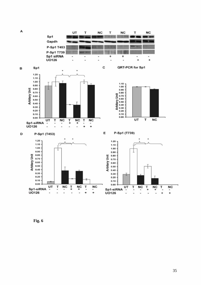
Fig. 6
35

36

37

Chapter 2 Results
105
2.5. NMDA receptor-mediated synaptic transmission:
The electrophysiology result obtained by our collaborator Nathalie Rouach shows
that the AMPA based mediated transmission is significantly decreased in the hippocampus
of Rsk2-KO mice. Her data showed also that the ratio of AMPA to NMDA currents was
unaltered, suggesting that the NMDA basal transmission was also decreased. Therefore,
Nathalie Rouach further investigated the level of basal NMDA receptor mediated synaptic
transmission. To this end, 4 weeks old Rsk2-KO mice as well as WT littermate were used.
NMDA receptor-mediated input-output curves in hippocampal slices show that for each
input (fiber volley ≥ 0.2mV), the output (fEPSP) is reduced in Rsk2-/- slices (p ≤ 0.05, +/+
n = 7; -/- n = 6). Scale bar 0.1 mV, 50 ms. These preliminary results show that NMDA
synaptic transmission is also decreased by ~ 40% in Rsk2-KO mice as compared with WT
littermate mice. However, at this stage we do not know the mechanism, which may account
for this alteration of NMDA synaptic neurotransmission in RSK2 deficient mice.
The curves obtained for NMDA transmission are given in (Figure 28A).

Chapter 2 Results
106
Figure 28: NMDA receptor-mediated synaptic transmission is decreased in Rsk2-/- mice.
(A). The curves obtained for NMDA based transmission and (B) NMDA receptor-mediated input-output curves in hippocampal slices show that for each input (fiber volley 0.2mV), the output (fEPSP) is reduced in Rsk2-/- slices (p 0.05, +/+ n = 7; -/- n = 6). Scale bar 0.1 mV, 50 ms.

Discussion

Chapter 3 Discussion
107
Discussion:
Loss of function mutations in the RPS6KA3 gene cause coffin-Lowry syndrome
(CLS), a syndromic form of intellectual disability (ID). The RPS6KA3 gene encodes the
RSK2 protein, which acts at the distal end of the MAPK/ERK cell signaling pathway.
RSK2 is a member of the RSK family, which are implicated in many fundamental cellular
mechanisms.
To facilitate the study of the physiopathology of ID and other symptoms of
CLS, a mouse model (Rsk2-KO) was previously generated in our team. This mouse exhibits
delayed acquisition of a spatial memory reference task and long-term spatial memory
deficits. To gain better insight in the molecular mechanisms leading to learning and
memory impairment in the Rsk2-KO mice, and to intellectual disability in CLS, we
performed a transcriptomic analysis of the Rsk2-KO hippocampus. This study aimed to
identify genes with altered expression, and as a result altered biological pathways, in
absence of RSK2 kinase activity. My particular interest was to understand the molecular
events leading to over expression of one of the genes identified in this study, the Gria2
gene encoding the AMPA receptor subunit GluR2 and the consequence on synaptic
neurotransmission in RSK2 deficient neurons. I will now discuss the results, the
mechanisms involved and their possible role in the molecular and cellular physiopathology
of intellectual disability in CLS patients.
3.1. Alteration of transcriptional activity in the hippocampus of
Rsk2-KO mice:
The activation of the RSK2 protein by ERK1/2 leads to the translocation of
a fraction of this kinase into the nuclear compartment, where it participates in regulation of
gene expression, (as previously mentioned). We therefore hypothesized that altered
transcriptional activity in the hippocampus of Rsk2-KO animals could participate in the
pathological mechanism underlying, cognitive impairment.

Chapter 3 Discussion
108
Our transcriptomic analysis revealed change in expression, with a 1.5-fold or more
difference at a p-value <0.03, of 100 genes. Expression of one fourth of these genes was
down regulated; whereas the remaining three fourth were up regulated in RSK2 deficient
mouse.
3.2. Genes and biological pathways deregulated in the absence of RSK2:
Because information about the function was available for most of the genes they have
been grouped by categories. Genes coding for enzymes (Rasl10A, Sod2), regulating the
transcription factors (Etv3, Runx2), kinases (FXN, Stk3) and ion channels (Gria2, Cacng8
and Cacnb4) have been identified for example in this study.
As anticipated, the data suggest that several cellular mechanisms are affected in the
absence of RSK2, including the regulation of cell cycle, gene expression and cell death, and
the development and functioning of the skeletal muscles and the nervous system. In
addition to determine whether there are potential interactions among the differentially
expressed genes, we assembled biologically networks by using the computer soft wear
“ingenuity pathway.” Seventy-eight genes out of 100 differently expressed genes were
mapped and assembled into five major biological networks with a score ≥20 (publication-
1).
Strikingly, several genes with important functions in the central nervous system were
altered in the Rsk2-KO mouse model, including GRIA2, CACNG8, VAMP4, and EIF3A.
The Gria2 gene was particularly interesting because it encodes the GluR2 protein, a subunit
of the AMPA receptor. This receptor is an ionotropic glutamate receptors, is the
predominant excitatory neurotransmitter receptors in the mammalian brain and is activated
in a variety of normal neurophysiologic processes. This protein is sensitive to AMPA, and
acts as a ligand-activated cation channel. The Gria2 gene is subject to RNA editing (CAG-
>CGG; Q->R) within the second transmembrane domain, which is thought to render the
channel impermeable to Ca2+. The CACNG8 gene encodes the type I transmembrane
AMPAR regulatory protein (TARP).

Chapter 3 Discussion
109
TARP controls both trafficking and channel gating of AMPA receptors, supports their
targeting to the cell membrane and synapses and modulates their gating properties by
controlling their rate of activation, deactivation and desensitization and by mediating their
re-sensitization. (Chu et al., 2001; Kato et al., 2010). The Vesicle-associated membrane
protein 4 is a protein that in humans is encoded via the VAMP4 gene (Advani et al., 1998).
This protein is a member of the VAMP/synaptobrevin family and plays a role in trans-golgi
network-to-endosome transport. The eukaryotic translation initiation factor 3 subunit A is a
protein that is is encoded by the EIF3A gene. This protein is required for the initiation of
the protein synthesis mechanism. The two fold increased expression of the GRIA2 gene was
particularly attractive, because it encodes the AMPA receptor subunit GluR2 which has
been described as a critical actor in learning and memory (Kessels and Malinow, 2009).
In addition we also observed that many of the identified genes play a role in nervous
system development including the IGF1 and AHR genes. Previously it was reported that
IGF1 is essential for normal dendritic growth (Cheng et al., 2003).
3.2.1. Alteration of the apoptotic process:
The transcriptomic results detailed in publication-1 revealed that one third of the
deregulated genes have been implicated in apoptosis and cell survival. Seven of them are
involved specifically in neuronal death (Cacng8, Diablo, Gria2, Igf1, Ptgs2, Rb1, sod2) and
their expression is increased in the absence of RSK2. These results were not surprising
because several previous studies have assigned such a role to RSK2. Subsequently, Anne
Schneider In our laboratory obtained confirmatory results, based on cultures of primary
hippocampal neurons and by using the tunel method. She showed that the number of
apoptotic neurons was significantly higher in KO than in WT cultures after treatment with
staurosporine. Staurosporine, an inhibitor of protein kinases, can induce apoptosis via the
mitochondrial pathway (Kajta et al., 2006). Stimulation of this pathway allows the release
of cytochrome C from the mitochondria, released cytochrome C binds with Apoptotic
protease activating factor - 1 (Apaf-1) and ATP, which then bind to pro-caspase-9 to create
a protein complex known as an apoptosome. The apoptosome cleaves the pro-caspase to its
active form of caspase-9, which in turn activates the effector caspase-3. This cascade
involves also the activation of caspase-3 and -7, and ultimately cleavage of Poly (ADP-

Chapter 3 Discussion
110
ribose) polymerase (PARP). Anne Schneider observed, by Western blot, an increase of the
expressions of cleaved Caspase-3, -9 and PARP in KO neurons. Thus, her findings revealed
a greater induction of apoptosis in RSK2 deficient cells.
One other gene identified by microarray encodes a protein that is a second
mitochondria-derived activator of caspases, Smac/DIABLO. This mitochondrial protein
induces apoptosis after release into the cytosol. Therefore it binds to the X-linked inhibitor
of apoptosis protein (XIAP), which prevents it from inhibiting caspases. The expression of
this gene is regulated by the transcription factor E2F1, and decreased expression of
DIABLO by siRNA reduces apoptosis (Xie et al., 2006). However, RSKs can
phosphorylate C/EBPβ, leading to its homodimerization and, hence, inhibition of factor
E2F1 (Sebastian et al., 2005, Lee et al., 2010). Thus, absence of RSK2 could result in
maintenance of E2F1 in active form and promote the expression of its target genes,
including Diablo. However, although, increased DIABLO gene and protein expression was
confirmed in the hippocampus of KO animals (Publication-1), no significant increase was
found in neuronal cultures. To determine whether its release from the mitochondria can still
be involved, a mitochondrial fractionation was performed. This experience indicated a
greater release of DIABLO in the cytosol of KO neurons after treatment with staurosporine.
This could contribute to greater sensitivity to staurosporine in KO neurons, but this
hypothesis should be confirmed with a larger number of animals.
In addition, several other deregulated genes are attractive candidates to explain the
susceptibility of Rsk2-KO neurons to apoptosis (as SOD2, NFκB, Bad…). Bad is
particularly an attractive target because its activity can be regulated by RSKs (She et al.,
2002, Chaturvedi et al., 2009). Bad in its non-phosphorylated form inhibits the anti-
apoptotic factor Bcl-2, preventing the release of cytochrome C and DIABLO.
In addition, many studies showed that ERKs are involved in regulating cell survival,
by assigning them either an anti-apoptotic or a pro-apoptotic function (Boston et al., 2011;
Campbell et al., 2010; Nakata et al., 2011).
We may, through the use of the inhibitor UO126, investigate whether the
inactivation of ERK promotes or reduces the susceptibility of neurons to apoptosis.

Chapter 3 Discussion
111
Lutz et al. (2007) suggested that apoptotic and anti-apoptotic cascades can be
associated with cognitive dysfunctions and neurological disorders. Taken together, our
results suggest that apoptotic processes are altered in the hippocampus of KO mice, which
can contribute to the mechanism of cognitive deficits. Apoptosis and cell survival of Rsk2-
KO neurons are functions that will be further investigated in the team. Following the
description of a decrease in the total size of the brain of people with CLS, Kesler et al.
suspected already in 2007 a deregulation of the cell death process.
3.2.2. Potential involvement of additional mechanisms in ID of CLS:
As mentioned above, various cellular mechanisms appear to be affected in the
absence of RSK2 in the hippocampus. In addition to the deregulated ERK1/2 function and
synaptic transmission, many other biological functions may be affected, as suggested by
our transcripomic data.
3.2.2.1. Potential causes for alteration of transcription:
Before discussing the molecular mechanisms potentially altered in mice Rsk2-KO,
we will try to understand some potential mechanisms leading to gene expression alteration.
Our study suggests several possible molecular mechanisms that may explain the
transcriptional deregulation observed in the Rsk2-KO mouse model.
3.2.2.2. Modification of the activity of NF-ĸB:
A link between RSK2 and NF-ĸB had already been described by Peng et al. in
2010. They observed that RSK2 can phosphorylate IĸBα at Ser-32 directly, which
enhances IĸBα degradation and releases NF-ĸB, allowing its passage into the nucleus and
activation of transcription.
The deregulation of the expression of some genes observed in Rsk2-KO mice might
be the result of a decreased phosphorylation of IĸBα, itself due to the absence of RSK2.
Consequently, it would result in a higher retention of NF-ĸB in the cytoplasm, where its
activity is blocked. This hypothesis could be interesting and should be confirmed, in

Chapter 3 Discussion
112
particular through analysis of cell fractionations to locate NF-ĸB in the cell. Inhibition of
IĸBα could also be undertaken to determine whether the expression of certain deregulated
genes (Use of mutants or constitutional siRNA) can be restored.
NF-ĸB activity is also controlled by phosphorylation, which contributes to its
freeing from the inhibitor, its transport into the nucleus and binding to DNA. It has been
shown that phosphorylation of the p65 subunit of NF-ĸB at Ser276 by MSK1, allows
induction of transcription of target genes through co-operation of the histone H3 and p300
(Vermeulen et al., 2003). This residue, highly homologous to CREB Ser133 is part of a
consensus sequence recognized by RSKs, suggesting that NFkB activity might also be
regulated via the RSKs. Wang et al. (2010a) confirmed RSK dependent phosphorylation of
the p65 subunit following stimulation of protease-activated receptors (PAR) in epithelial
cells HT-29. Inhibition of ERK either with UO126 or silencing of RSKs by siRNA led to
decrease of Ser276 phosphorylation of p65 and reduction of NF-kB-dependent
transcription. In addition, the authors noted that the action of RSKs is isoform specific. The
absence of RSK2 could therefore also influence in this manner gene transcription.
Preliminary data of Anne Schneider suggested, however, that phosphorylation of this factor
is not altered in the absence of RSK2 in hippocampal cell cultures. These studies must be
repeated for confirmation, but also after mitogenic or glutamate stimulation.
3.2.2.3. Modification of the ERK/MAPK signaling:
Our transcriptomic results revealed that most of the genes (75/100) with altered
expression are over-expressed in hippocampus of Rsk2-KO mice. This was expected,
because RSK2 is mainly known for its role in activation of gene expression.
In the second article of this thesis, we have shown that RSK2 exerts a negative
feedback on its own pathway. The mechanism leading to over-activation of ERK is
described by Douville and Downward in 1997 and reported that a loop of feedback-
inhibition to regulate the way, ERK/MAPK dependent on SOS factor. Following
stimulation of PC12 cells by EGF, they observed that RSK2 phosphorylates SOS between
two proline-rich regions, the region for binding to the SH3 domain of Grb2. This
phosphorylation leads to dissociation of the SOS-Grb2 complex, which affects the
Chapter 3 Discussion
113
activation of the signaling pathway. While the phosphorylation of SOS participates in the
regulation of the channel Ras-ERK/MAPK in PC12 cells, previously it does not seem
involved in hippocampal neurons. Indeed, we did not observe any difference in total MEK
and RAF-1 proteins level, however the glutamate activated neurons shown the levels of
phosporylated forms of the MEK and RAF-1 were significantly higher in Rsk2-KO neurons
than WT. activation of MEK and Raf-1, activating kinases ERK and RSK in KO neurons.
Dysregulation of the entire signaling pathway is therefore included (publication-2).
This regulation leads to an increased activity of ERK1/2 (ERK pathway / MAPK) in
Rsk2-KO hippocampal neurons, but also to an increase in activity of two transcription
factors and substrates of ERK: Elk1 and CREB. This activation stimulates their
transcriptional activity because the induction of c-Fos, Zif268 and Arc (three IEGs)
expression is stronger in RSK2 deficient neurons than in WT cells in response to
glutamatergic stimulation. A change in the expression of these IEGs could lead to an
alteration of transcription of late response genes. It can thus be anticipated that the
increased level of ERK activation, in Rsk2-KO neurons, can participate to the deregulated
transcriptional activity, either in response to over-activation of transcription factors or to
over expression of IEGs. Moreover, some genes whose expression is increased in the Rsk2-
KO hippocampus have been described as being regulated by ERK at the transcriptional
level (Yoon and Seger, 2006). This is an interesting hypothesis to explain the expression
alteration of at least some of the genes reported. An abnormal increase in ERK
phosphorylation at the baseline level has been reported in many other tissues in the absence
of RSK2 (Dufresne et al. 2001; Marques Pereira et al., 2008). Deregulation of the
MAPK/ERK pathway and its influence on gene expression may, indeed, have deleterious
consequences for many cell functions. In order to confirm this hypothesis, partial in vitro
inhibition of ERK through the MEK inhibitor U0126, may be considered followed by
transcriptome analysis. In addition this study would determine whether the altered
expression of some of these genes can be rescued.
At least six other nuclear and cytoplasmic MAPKs can regulate the activation of
ERK have been described (Bermudez et al., 2010) and are potential targets.
Indeed, we showed in the last part of my thesis work that phosphorylation of the
transcription factor P-Sp1 is increased at T453 and T739 in RSK2 depleted cells. Milanini-
mongiat et al., in 2002 reported that ERK kinase phosphotylate transcription factor Sp1 at

Chapter 3 Discussion
114
these two sites (Thr453 and Thr739). Phosphorylated form of Sp1 influences positively its
transcriptional activity (Tan and Khachigian, 2009). We observed higher levels of
phosphorylation for both sites in RSK2 silenced PC12 cells, leading to up regulation of
Gria2 transcription. This result confirmed the assumption made above about the role of
ERK1/2 up-regulated activity on gene expression.
3.3. Positive regulation of Gria2 gene expression via phospho-Sp1:
(Boggio et al., 2007), by using immunolabelling, showed that in vivo stimulation
increases p-ERK expression at presynaptic and postsynaptic sites of axospinous junctions,
suggesting that ERK plays an important role in the local modulation of synaptic function.
Our results showed that the expression and function of the AMPA receptor subunit
GluR2, a crucial actor of synaptic function, are positively regulated via ERK1/2 and that
RSK2 can indirectly modulate this action. Expression of the Gria2 gene is strongly
controlled by a Sp1 regulatory element located in the GC-rich promoter region (Myers et
al., 1998). In 1999, Merchant et al. observed that the DNA binding activity of the
transcription factor Sp1 is stimulated by ERK phosphorylation. Interestingly, in 2009 it was
reported that phosphorylation of Sp1 influences positively its transcriptional activity (Tan
and Khachigian, 2009). The Sp1 transcription factor is phosphorylated at various sites by
different kinases as mentioned in the introduction of this thesis. To elucidate the molecular
mechanism leading to GluR2 over expression, we constitutively suppressed the expression
of RSK2 in PC12 cells via a vector-based shRNA. The PC12 cell line, cloned from rat
pheochromocytoma cells, can be induced to differentiate and to acquire a neuronal-like
phenotype (Greene & Tischler, 1976). PC12 cells express endogenous RSK2 at a relatively
high level, whereas the level of GluR2 is low (present study) and can be easily transfected.
Therefore, the PC12 cell line was a good model for the present study. We showed that Rsk2
silencing in PC12 cells also leads to an elevation of ERK1/2 phosphorylation as well as of
GluR2 expression and that the increased level of GluR2 expression results from the
increased ERK1/2 activity on the transcription factor Sp1 (publication-3). Our results
strongly suggest that RSK2 modulates ERK1/2 activity on Sp1, which regulates GluR2
expression through transcriptional activation. Together our data provide evidence that
RSK2 plays an important role in the regulation of the function of AMPA receptors. How

Chapter 3 Discussion
115
phosphorylated Sp1 activates the Gria2 promoter remains now to be investigated. It is
possible that Sp1 phosphorylation may change its interaction with other transcription
factors.
Sp1 phosphorylation induced by the ERK pathway was also shown to cause the
release of a histone deacetylase co-repressor complex from a gene promoter (Liao et al.,
2008). Furthermore, in addition to Sp1, Gria2 expression is also influenced at the
transcriptional level by another positive (Nrf-1) and a negative (Re1/Nrse-like silencer)
regulatory element (Borges and Dingledine, 2001). Myers et al. (1998) showed that the
Gria2 Re1/Nrse-like silencer binds the RE1-silencing transcription factor (NRSF/REST),
and that co-transfection of REST into neurons reduced Gria2 promoter activity in a
silencer-dependent manner. The nuclear respiratory factor 1 (NRF-1) is a transcription
factor that was also shown to regulate Gria2 gene transcription in neuroblastoma cells
(Dhar et al., 2009). It is clearly necessary to understand the roles of the different regulatory
elements and their interplay in neurons. Finally, it is now necessary to validate the
mechanism described in Rsk2-Knocckdown PC12 cells in Rsk2-KO mice. Preliminary
results show that Sp1 activity is also up regulated in the hippocampus of Rsk2-KO mice
suggesting that the mechanism of GluR2 transcriptional up regulation is similar in mouse
and PC12 cells. Sp1 phosphorylation and GluR2 expression can, for example, be assayed in
cultured primary hippocampal neurons treated with the MEK inhibitor, and Sp1 can be
knocked down partially in cultured neurons using lentiviral-mediated shRNA expression
vectors.
3.4. Consequences or potential consequences of the deregulation of
GluR2 expression:
No systematic descriptions of anatomical abnormalities of the brain in CLS patients
or animal models have been reported. Therefore, we hypothesized that more subtle cellular
events were altered.
Transmission of neuronal signals being dependent on the type of receptors and their
neurotransmitters, alteration of one of these factors can lead to a failure of synaptic
transmission. In our study, we have revealed alteration of the expression and function of the

Chapter 3 Discussion
116
AMPA-type ionotropic receptor. Preliminary results very recently obtained reveal also
decreased NMDA basal neurotransmission. The molecular basis of this later deficit is not
yet known.
These alterations may, indeed, contribute to the cognitive impairment of CLS
patients. In the following sections, we will analyze our results and propose a molecular
hypothesis for the occurrence of these defects.
It is now well established that the nervous system response to a short lasting change
in the efficiency of synaptic transmission requires the expression of new genes. Many genes
have been implicated in this process, Gria2 is one of them. AMPA receptors play a crucial
role in synaptogenesis and formation of neuronal circuitry, as well as in synaptic plasticity
(Santos et al., 2009). There is a huge amount of evidence indicating that the GluR2 subunit
plays a pivotal role in AMPA receptors function. The Ca2+ permeability, rectification, and
single-channel conductance of AMPA receptors are all dominantly influenced by inclusion
of an edited GluR2 subunit in the receptor complex (Hollmann et al., 1991; Burnashev et
al., 1992; Bowie and Mayer, 1995; Koh et al., 1995; Seeburg, 1996; Swanson et al., 1997;
Washburn et al., 1997). Receptors that contain a single GluR2 subunit have maximally
reduced Ca2+ permeability; whereas inward rectification is reduced in a graded manner as
the number of GluR2 subunits in a receptor increases (Washburn et al., 1997). GluR2
expression is governed by neuronal activity (Bai and Wong-Riley, 2003), and its synaptic
distribution and levels are modulated as part of the mechanism of synaptic plasticity (Keifer
and Zheng, 2010; Sprengel, 2006). GluR2 interacts with various molecules implicated in
receptor trafficking (Dingledine et al., 1999; Collingridge et al., 2004; Isaac et al., 2007).
Therefore, it can be anticipated that moderate changes in GluR2 expression may have
functional consequences on receptor subunit rearrangement, neurotransmission and lead to
functional differences in the functioning of synaptic circuits.
Our data reported in publication 1 showed that basal AMPA neurotransmission is
altered in the hippocampus of Rsk2-KO mice. Preliminary data, not yet published, reveal
also that NMDA basal neurotransmission is significantly decreased in Rsk2-KO mouse.

Chapter 3 Discussion
117
3.4.1. Alteration of Ca2+ uptake:
The fact that AMPA receptor dependent neurotransmission was altered in RSK2
deficient mouse, prompted us also to investigate Ca2+ uptake of hippocampal neurons. For
this purpose we used the cobalt chloride staining approach. As already mentioned AMPA
receptors without GluR2 subunit are permeable to Ca2+ (or other divalent cations, like Co2+)
(Cull-Candy et al., 2006; Konig et al., 2001), whereas in the presence of GluR2 subunit(s)
they are impermeable to Ca2+. The results obtained with this approach suggested strongly a
decrease of AMPA dependent divalent ions uptake in RSK2 deficient neurons. Thus, our
results strongly suggest that up regulation of GluR2 expression in Rsk2-KO neurons, affects
the AMPA receptor subunit composition, resulting in decreased Ca2+ entrance in neurons
after AMPA receptors stimulation. Unfortunately, the cobalt approach does not allow
quantification of the uptake. Therefore, our conclusion has to be further supported by
additional electrophysiological studies, as well as more precise calcium imaging
technologies that allows quantification.
3.4.2. Alteration of synaptic transmission and plasticity:
Many studies have reported an alteration of synaptic transmission following
changes of expression and recycling of GluR2. The increased synaptic expression of this
subunit, suggests an increased proportion of AMPA Ca2+ impermeable receptors in Rsk2-
KO mice. This would well fit with the decreased synaptic transmission observed in
hippocampus slices.
In addition to the increased synaptic GluR2 expression, other molecular defects can
contribute to alter synaptic transmission. The editing and splicing also play a role in the
receptors activity. With regard to the R/G site, Lomeli et al. (1994) reported that edition
increases the kinetics of the channel. A reduction of edition is thus assumed to result in a
decreased receptor activity, which could contribute to the decrease of transmission in RSK2
deficient neurons. In addition, in 2009, Pei et al. discovered that the flop variant leads to a
faster closing of the channel than the flip variant; as a result inactivation of the receptor is
quicker. Thus, the increased proportion of flop variants to the detriment of the flip variants

Chapter 3 Discussion
118
may also contribute to the reduction of basal AMPA receptor transmission in the
hippocampus of Rsk2-KO mice.
The cause of the decreased AMPA receptor dependent basal transmission, might
also be the presence of silent synapses in KO hippocampal neurons. This hypothesis seems
interesting because in our lab Anne Schneider observed a decrease of the number of
potential excitatory synapses in primary neuronal culture. To test this hypothesis, we have
determined in collaboration with Nathalie Rouach the ration of AMPARs/NMDARs
currents. Silent synapses are expected to express less AMPAR than NMDAR, or in our
case, less functional AMPA receptors. However, the results indicated that the ratio is
similar in WT and Rsk2-KO mice. More specific studies of the AMPA receptor properties
(as the conductance, the rectification, the closing of the channal…) would give more
information as to the origin of the alteration of AMPA receptor transmission. The
consequences of AMPA receptor dysfunction for synaptic plasticity should also be studied,
by assessing LTP and LTD. The alteration of these forms of synaptic plasticity would also
explain part of the cognitive deficits associated with MR in CLS patients.
3.5. Consequences or potential consequences of GLUR2 editing and splicing alterations:
AMPA receptor subunits are synthesized and assembled in the Endoplasmic
reticulum (ER), before insertion in the plasma membrane. Their assembly and exit from the
ER are highly controlled. They can be influenced by post-transcriptional mechanisms, such
as RNA splicing and editing.
Two editing sites were identified for Gria2, the Q / R site in the transmembrane
domain (TMD) domain and the R / G site (residue 607) in the ligand binding domain
(LBD). Both of these sites are involved in the membrane recycling of GluR2. Greger et al.,
in 2002 reported that the Q/R site controls the function and trafficking in AMPA receptors.
Editing at the GluR2 Q/R site regulates AMPAR assembly at the step of tetramerization.
Specifically, edited R subunits are largely unassembled and ER retained, whereas unedited
Q subunits readily tetramerize and traffic to synapses (Greger et al. (2003). This assembly
mechanism restricts the number of the functionally critical R subunits in AMPAR
tetramers. The same mechanism was subsequently implicated in the edition of the R/G site
Chapter 3 Discussion
119
at position 743 (Greger et al., 2006). Edition of Q / R and R / G sites thus controls the
maturation of the GluR2 subunit. The results reported in publication 1 of the present thesis
showed that edition of the Q / R site is not altered whereas the R / G site is significantly less
edited in KO mice than in WT animals. By taking into account the data from Greger et al.
(2006), our results opens the possibility than the increased expression of unedited GluR2
subunits leads to an increased formation of unedited GluR2 homotetramers in Rsk2-KO
cells. The over representation of GluR2 at the synapse in Rsk2- KO mice might thus be due,
at least in part, to the decreased R/G edition.
On the other hand, AMPA receptors are subject to alternative splicing in the LBD
domain, generating flip and flop variants. This alternative splicing influences certain
parameters of the AMPA receptor, including their assembly and synaptic expression as
outlined by Brorson et al. in 2004. Our results (reported in publication 1) revealed an
increase of the flop isoform in the hippocampus of Rsk2-KO mice. In their study in 2006,
Greger et al. showed that the flop isoform promotes ER export and synaptic expression of
AMPA receptors. Therefore, an increase in the proportion of this variant could also
contribute to the stronger expression of synaptic GluR2 in RSK2 deficient neurons.
It would be interesting now to analyze the cause of abnormal Gria2 editing in RSK2
deficient neurons. Editing is regulated by adenosine deaminases (ADAR) generating the
conversion of adenosine to inosine at the pre-mRNA level (Seeburg et al., 1998). Three
ADAR have been identified in mammals (ADAR1, 2 and 3) and the Q/R site is
predominantly ADAR2 dependent (Peng et al., in 2006). Through the use of siRNA
stereotaxically injected in the hippocampus of rats, they showed that ADAR2 is required
for the edition of the Q / R site and thus for the expression of synaptic Ca2 + impermeable
AMPA receptors. We have shown that edition of the Q/R is not altered in Rsk2-KO animal,
leading to the conclusion that the ADAR2 function is not affected. However, Sergeeva et al.
(2007) implicated ADAR2 in the regulation of Q/R site editing, which is decreased in Rsk2-
KO mice.
They showed that increased R/G editing in neurons of the tuberomammillary
nucleus (TMN) is correlated with higher expression of ADAR2 and inhibition of ADAR3.
But they also stated that editing of the Q/R site is not always associated with ADAR2. An
analysis of the levels of expression and phosphorylation of the various ADAR isoforms

Chapter 3 Discussion
120
might give a first indication of the factor involved in the reduction of R/G editing in the
hippocampus of Rsk2-KO mice. We may then try to influence the editing rate by inhibiting
or over-expressing the defective ADAR isoform.
3.6. Other potential deregulation: Phosphorylation of AMPA
receptors:
As mentioned in the introduction, AMPA receptor recycling can be regulated by
phosphorylation, and many kinases appear to participate in this activity. It has to be
reminded here that in addition to its impact on nuclear targets, deregulation of ERK has
also an impact on the function of cytoplasmic substrates. For example, an increased level of
phosphorylation of RSK1 and 3 was observed in Rsk2-KO neurons (publication-2). In
addition to suggest a compensatory mechanism in the absence of RSK2 by other RSK
family member, this result further demonstrates that the activity of ERK cytosolic
substrates is affected. The wide variety of cytosolic ERK substrates suggests that many
other cellular mechanisms, and not depending on transcriptional alterations, can be altered
in Rsk2-KO mice. A role in regulating synaptic transmission has been attributed to ERK via
phosphorylation of glutamate receptors (Thomas and Huganir, 2004). Alteration of these
regulatory mechanisms could contribute to ID in CLS patients.
Insertion of GluR1 and GluR2 at the synapse is dependent on the activation of the
Ras-ERK/MAPK. Qin et al. (2005) noted that stimulation of NMDA receptors induces
phosphorylation of GluR2 at Ser841 by ERK/MAPK, thus promoting synaptic expression.
Other studies also assigned a role to ERK1/2 in synaptic trafficking. Li and Keifer (2009)
reported that synaptic localisation of GluR1 and 4, following stimulation with BDNF,
correlates with ERK activation. These studies showed that over-activation of ERK results in
an increased density of these subunits at the synapse.
The increased density of synaptic GluR2 subunits observed in Rsk2-KO neurones
(publication-1) may thus be explained by an increased level of GluR2 phosphorylation.
Thus, to determine the role of ERK in this increased synaptic GluR2 expression, it would
be interesting to assay the level of GluR2 phosphorylation, and particularly at Ser841 (Qin
Chapter 3 Discussion
121
et al., 2005). The influence of ERK on synaptic GluR2 expression may also be investigated
by immunofluorescent labeling of hippocampal neurons treated with the inhibitor U0126.
3.7. Potential causes for spine morphology/ maturation alteration:
It was reported that the size of the PSD, and thus the head of the spine, is correlated
to the number of synaptic glutamate receptors on its surface (Cingolani and Goda, 2008).
The proportion of synaptic AMPAR can influence the morphological development of
dendritic spines. Passafaro et al. in 2003 described that GluR2 over-expression leads to an
increase of the size of the spines head of hippocampal neurons at 22 DIV. This is specific to
GluR2 over expression, whereas GluR1 or GluR3 over expression do not produce the same
effect. The authors confirmed the role of GluR2 by showing that its over expression leads
to the formation of spines in GABAergic interneurons. Their results were validated by the
subsequent study of GluR2-KO mouse model in which a decrease in the volume of the
"mushrooms" has been reported (Medvedev et al., 2008). The increased expression of
GluR2 protein in the hippocampus of Rsk2-KO mice (publication-1) could therefore
contribute to the increased proportion of mature dendritic spines observed in the derived
primary neuronal cultiures (Anne Schneider, manuscript in preparation). To confirm this
hypothesis, a new analysis of the morphology of spines could be carried out after partial
silencing of GluR2 expression in cultured neurons or by injecting directly siRNA or shRNA
stereotacticaly in the hippocampus of Rsk2-KO mice.
3.8. Potential causes of NMDA receptor-mediated synaptic transmission alteration:
Preliminary results show that NMDA synaptic transmission is also significantly
decreased in Rsk2-KO mice. The NMDA receptor has a key role in the regulation of
synaptic transmission in the central nervous system of mammalians. In particular, he has a
long channel open time and high Ca2+ permeability and in consequence a prolonged
synaptic conductance coupled with a large Ca2+ influx. Activation of the NMDA receptors
triggers widespread signal transduction cascades that regulate synaptic plasticity as well as
the formation and elimination of synapses (Malenka and Nicoll, 1999; Carroll and Zukin,
2002). Little is known about plasticity of the NMDAR-mediated transmission (Malenka
and Bear, 2004). Extensive studies indicated that NMDA receptor activity in the

Chapter 3 Discussion
122
hippocampus is essential for the formation of both spatial and non-spatial memory (Li and
Tsien, 2009; Nakazawa et al., 2004; Tsien, 2000).
There are several potential causes for NMDA receptor dysfunction, which may not
be exclusive. In 2005, Morishita and his group reported that inhibition of endogenous
cofilin by the Ser3-p-cofilin peptide blocks the actin-dependent NMDA-receptor-LTD
(Morishita et al., 2005). They showed that actin anchors NMDA receptors by interacting
with a cohort of scaffolding protein. Anne Schneider in our team showed that the level of
phosphorylated Cofilin, a well known regulator of actin polymerization and spine
morphology, was increased abnormally in Rsk2-KO cells. While LIMK activity, a Cofilin
upstream kinase, was found unaltered, preliminary data suggest that the Cofilin phosphatase
SSH1 activity may be affected. Anne Schneider also detected altered activation of
SynGAP, a Ras GTPase-activating protein, which participates in actin dynamics regulation.
The SynGAP also play an important role in NMDA glutamate receptors trafficking
(Rumbaugh et al., 2006). Thus, altered trafficking of NMDA receptors may contribute to
the decreased neurotransmission. Another hypothesis concerns a possible implication of
CaMKII. Ca2+/calmodulin-dependent protein kinase II (CaMKII) is a serine/threonine
kinase regulated by intracellular calcium and by the activity of the NMDAR in spine PSDs.
In dendritic spines, CaMKII is associated with the NMDA receptor subunits NR1 and
NR2B (Merrill et al., 2005). The results obtained by Anne Schneider showed that the
activity of CaMKII is significantly decreased in the Rsk2-KO hippocampus. The molecular
mechanism leading to NMDA receptor dysfunctions as well as the consequences have now
to be further investigated.
3.9. Other potential cause of cognitive dysfunction: alteration the
processes of neurogenesis:
As mentioned above, many cellular mechanisms appear to be affected by the absence
of RSK2 in the hippocampus. In addition to the regulation of ERK / MAPK and
mechanisms of plasticity and synaptic transmission that we have mentioned, preliminary
results (a collaboration with S. Laroche, Orsay) also show that the rate of neurogenesis is
decreased in the hippocampus of Rsk2-KO mice.

Chapter 3 Discussion
123
In a study conducted in 2010, Dugani et al. suggested that the decrease in the total
size of the brain of patients CLS would be the consequence of a reduction in the
neurogenesis. They indeed showed that the inhibition of RSK2 by shRNA does not affect
cell survival but rather decreases the differentiation of cortical precursors into neuronal
cells. This decrease in neuronal cells was attributed to the maintenance of cell proliferation
at the expense of the differentiation process. According to these authors, RSK2 would
regulate the process via phosphorylation of transcription factors such as C/ EBP or via the
regulation of chromatin structure by CBP. RSKs, and RSK1 in particular, can
phosphorylate C/EBP at Thr217 and, hence, control neurogenesis (Menard et al., 2002).
Activation of C/EBP allows the transcription of genes such as neuron-specific Tα-1 tubulin,
which promotes the differentiation of cortical progenitors into neurons. On the other hand,
Wang et al. in 2010b indicated that the histone acetyltransferase (HAT) activity of CBP is
essential for the expression of genes necessary for the differentiation of cortical precursors
into neuronal cells, glia or oligodendrocytes. In 2001, Merienne et al. showed that RSK2
could interact with CBP at baseline and their dissociation following stimulation increases
its HAT activity. Impairment of this activity can be anticipated in the absence of RSK2.
This could contribute, in addition to a deregulation of C/EBP, to the decreased
neurogenesis.
To understand the molecular mechanisms irresponsible for the decreased rate of
neurogenesis in Rsk2-KO mice, further studies have to be performed, including analysis of
C/EBP and CBP activities in neural precursors from our mouse model.

Conclusion

Chapter 4 Conclusion
124
Conclusion:
To date, various studies have been conducted in order to understand the
pathophysiology of CLS. Several assumptions have been made, but as yet, the molecular
and cellular mechanisms leading to the different CLS clinical symptom are largely
unknown. However, studies aimed at understanding the causes of the skeletal anomalies
have revealed the important role of RSK2 in osteoblast differentiation, by regulating the
activity of the transcription factor ATF4 (Yang et al., 2004). Interestingly, it was shown on
the basis of these results, that a diet rich in proteins improves significantly osteogenesis in
the mouse Rsk2-KO (Elefteriou et al., 2006). At the neuronal level, RSK2 has been
implicated in exocytosis of pre-synaptic vesicles via phospholipase D1 (PLD1), in the
synthesis of dopamine and in the formation of axons and synapses (Marques Pereira et al.
2008; Zeniou-Meyer et al., 2008; Fischer et al., 2009). These data suggested that alterations
in the process of synaptic transmission and plasticity are involved in cognitive impairment
associated with Intellectual disability. Our results confirm this hypothesis and provide new
and important insights. We showed for the first time that RSK2 deficiency causes an
impaired glutamatergic synaptic neurotransmission. Altered AMPA receptors transmission
and GluR2 expression subunit has been identified in the hippocampus-Rsk2-KO. In
addition, although the mechanism is not yet known, preliminary results show that NMDA
neurotransmission is also severely altered. We have shown that the up regulation of the
gria2 gene expression is directly related to the up regulation of ERK/MAPK activity in
RSK2 deficient cells. Our findings suggest an indirect role of RSK2 in modulating the
expression of AMPA receptor GluR2 subunit and, therefore, in regulating glutamatergic
neurotransmission and perhaps synaptic plasticity. Although highly likely, it is not yet
known if synaptic plasticity is affected in the hippocampus of Rsk2-KO mice. LTP and
LTD experiments will soon be performed in collaboration with Nathalie Rouach. Further
investigations are also now necessary to determine contribution of this deregulation of
GluR2 expression to the cognitive dysfunction.
Our results have also revealed alteration of expression of many other genes in the
hippocampus of RSK2 deficient mice. The up regulated level of ERK1/2 activity is
certainly the cause, directly or indirectly, for the deregulation of a fraction of them. The
numerous abnormally expressed genes identified by our microarray analysis will help in

Chapter 4 Conclusion
125
further unraveling the various functions of RSK2 in the hippocampus. It can also be
speculated that some of these genes play a significant role in the pathogenesis of cognitive
deficiency in Coffin–Lowry syndrome.
Our results are too preliminary to consider a therapeutic approach for cognitive
impairment in CLS, but they suggest a critical role for ERK and GluR2 in the pathology.
Further studies, including living animals, for example, by injecting stereotaxically in the
hippocampus viral mediated shRNA vectors must be implemented now to confirm the
involvement of GluR2 in cognitive impairment. If the implication is validated, the use of
inhibitory molecules targeting the ERK pathway or GluR2 could be interesting. The MEK
inhibitor (U0126) has been tested in clinical trials as anti-cancer molecule (Marampon et
al., 2009, Ito et al., 2010) and Neuroprotection (Farrokhnia et al., 2008) and may be tested
for its efficacy in mice. Du et al., in 2008 described the use of lithium and valproate to
inhibit the expression of synaptic GluR1/GluR2 in hippocampal neurons in vitro and in
vivo. However, there is no currently specific inhibitor of GluR2 available. The use of
antagonists of type I mGluRs glutamate receptor have been used in clinical trials for XFRA
(Krueger and Bear, 2011). In 1997, the first link between FMRP and mGluR has been
established. Many studies have subsequently demonstrated the involvement of FMRP in the
regulation of mGluR dependent protein synthesis. Mice doubly heterozygous for FMRP
and mGluR targeted gene disruption allowed the restoration of some phenotypic
manifestations associated with the FMRP mutation. These results prompted the researchers
to study the consequences for animal models of XFRA of a treatment with a specific
inhibitor of mGluR5, MPEP. This inhibitor, indeed, improves the cognitive phenotype in
mice, Drosophila and "zebrafish" mutated in the FMRP gene. These encouraging data led to
the establishment of clinical trials in humans. Several pharmacological molecules have
been tested, such as acramprosate, lithium or the GABA inhibitor, baclofen. The results
obtained with these studies are very encouraging. (Krueger and Bear, 2011).
If the importance of GluR2 in the pathogenesis of cognitive impairment in CLS is
established, the FRAXA story could serve as an example for a therapeutic strategy for
RSK2 deficiency.
In conclusion, together available data show that RSK2 acts in a broad variety of
functions. However, investigations undertaken in our team in the recent years have mainly
Chapter 4 Conclusion
126
focused on its involvement in the regulation of plasticity and synaptic transmission. Anne
Schneider has shown, for example, that spinogenesis and the underlying cytoskeleton
dynamic are altered in the hippocampus of Rsk2-KO mice.
The deregulation of cellular processes, such as neurotransmitter release, axonal and
dendritic growth, functional maturation of synapses, is becoming increasingly evident to
explain the cognitive impairment in CLS patients, but also in other cognitive dysfunctions
or psychiatric disorders. The Results so far obtained do not answer to all the questions, but
already allow us to make hypotheses.

Future Perspectives

Chapter 5 Future prospects
127
Future prospects:
Further studies are needed to explore in more detail the mechanisms leading to up-
regulation of the Gria2 gene, and its consequences on synaptic transmission and plasticity.
It is essential to further dissect the molecular mechanism of Gria2 transcriptional control,
including the identification by which Sp1 activates the Gria2 promoter. It is possible that
Sp1 phosphorylation may change its interaction with other transcription factors, as it was
previously described for other genes. Sp1 phosphorylation induced by the ERK pathway
was also shown to cause the release of a histone deacetylase co-repressor complex from the
luteinizing hormone receptor gene promoter, which may also be involved in the Gria2 gene
expression. On the other hand, it has been shown that, in addition to Sp1, Gria2 expression
is also influenced at the transcriptional level by another positive (Nrf-1) and a negative
(Re1/Nrse-like silencer) regulatory element in the 5' proximal region of the promoter
(Borges and Dingledine, 2001). It is clearly necessary to understand the roles of these
different regulatory elements of the Gria2 gene and their interplay in neurons.
Our finding of an abnormally increased level of GluR2 expression in RSK2
deficient neurons could lead to therapeutic strategies for CLS, e.g. by modulating the
glutamatergic system. Indeed, in the Fragile X syndrome (FXS), observations suggesting
increased metabotropic glutamate receptor (mGluR5) led to preclinical studies showing that
inhibition of mGluR5 can ameliorate multiple mutant phenotypes in mouse and drosophila
models of FXS, and clinical trials based on this therapeutic strategy are underway (Krueger
and Bear 2011). However, the extent of glutamate transmission alteration, and whether
synaptic plasticity is modified remain first to be explored. Alteration of NMDAR
transmission has to be confirmed, and the mechanism involved, as well as its contribution
to cognitive impairment, to be investigated. Levels of synaptic and extrasynaptic glutamate
using pharmacology (with low affinity AMPA and NMDA receptors competitive antagonist
(γ-DGG and D-AA, respectively), should also be assayed to investigate whether the
decrease in AMPA receptor-mediated transmission identified in Rsk2 knockout mice is also
associated to alterations in extracellular glutamate levels. Density of functional surface,
and extra-synaptic AMPARs, using local application of AMPA in whole-cell recordings
and in somatic outside-out patches from CA1 pyramidal cells, respectively should also be
measured. Since the GluR2 subunit is involved in LTP, LTD and plays an important role in
Chapter 5 Future prospects
128
learning and memory, short and long-term plasticity in the hippocampus of Rsk2 knockout
mice should be investigated in detail. These data should give insights into the altered
hippocampal cellular physiology accounting for the psychomotor impairments in the Rsk2-
KO mice. Studies designed to understand the contribution of GluR2 over expression in the
cognitive dysfunction of Rsk2-KO mice have already been undertaken. The goal is to
determine whether the RSK2 deficient phenotype can be restored by a partial inactivation
of GluR2 in mice. No specific inhibitor of GluR2 being currently available, AAV-mediated
shRNA are used to knockdown partially GluR2 in Rsk2-KO mice. Viral particles will be
injected stereotaxically into the hippocampus.
The contribution of other pathways to the AMPA receptor dysfunction remains also
to be investigated. Among the deregulated genes identified by our transcriptome analysis at
least one other participates in regulation of AMPAR function: Cacng8. This gene encodes a
synaptic protein, TARPc-8 that participates in consolidation phase of memory and is
involved in modulating neurotransmitter release. Evidence was provided that TARPc-8 is
critical for basal AMPA receptor expression and localization at extrasynaptic sites in the
hippocampus (Rouach et al. 2005). Up-regulation of Cacng8 may thus contribute to AMPA
receptor dysfunction.
Indeed we expect that our studies will provide approaches for pharmaceutical
intervention in Coffin-Lowry Syndrome.

Experimental Procedure

Chapter 6 Material and Methods
129
Material and Methods:
6.1. Ethics statement:
All experimental procedures for care and use of mice were performed according to
agreements with the Departmental Direction of Veterinarian Services (Prefecture du Bas-
Rhin, France No. 67-225) and IGBMC Animal Welfare Insurance (NIH, PHS No. A5100-
01). Mice were housed 4 per cage in a 12h dark/light cycle (light on from 7 am to 7 pm),
under controlled conditions of temperature and humidity. Euthanasia of the mice used to
derive primary cell cultures was carried out in accordance with the European Communities
Council Directive of 24th November 1986 (86/609/EEC), and the study was specifically
approved by the Ethics Committee in Animal Experiments (permit number 17) of the
Institute of Genetics and Molecular and Cellular Biology.
6.2. Primary hippocampal neuronal Cultures and Cobalt staining:
Primary cultures of hippocampal neurons were derived from one day old WT and
Rsk2-KO male mice, with a C57Bl/6x genetic background. After treatment of the
hippocampi with Papain, dissociated cells were plated onto 24-well plates containing poly-
d-lysine-coated cover slips (Sigma), at a density of ~100 000 neurons/well. Growth media
consisted of Neuro-Basal (GIBCO, Invitrogen) supplemented with 1x B27 (GIBCO,
Invitrogen), 0.5mM L-Glutamine and 1x penicillin/streptomycin. The cultures were stored
in an incubator at 37°C in a humidified atmosphere containing 5% CO2 for 8 days.
The cobalt staining procedure was essentially performed as described previously
(Jensen et al., 1998), with small modifications. The initial medium was removed from the
wells after 8 days, and cells were washed twice with HEPES buffer (146mM NaCl, 4.2mM
KCl, 0.5mM MgCl2, 0.8mM CaCl2, 55.6mM glucose, 20mM HEPES, pH 7.4) at 37°C.
Cells were then incubated with 5mM CoCl2 in HEPES buffer.
Three Conditions were then used; i) without AMPA treatment (control); ii) addition
of AMPA (to a final concentration of 100 µM) to activate AMPA receptors and allow the
influx of Co++ into the neurons iii) addition of AMPA (100 µM final) and cyclothiazide (30

Chapter 6 Material and Methods
130
µM final) which act as non-desensitizing agent on AMPA receptors in the presence of
AMPA.
Following 30-35 min incubation the cells were washed twice with HEPES buffer
supplemented with 2mM EDTA (10 min each) in order to remove the excess of extra
cellular Co++. The intracellular Co++ was then precipitated by using HEPES buffer
containing 0.12% Na2S, for 5 min. The cells were then washed once with HEPES buffer
and fixed with 4% paraformaldehyde and 4% sucrose in phosphate-buffered saline (PBS; 8
g/l NaCl, 0.2 g/l KCl, 2.9 g/l Na2HPO4.12 H2O, 0.2 g/l KH2PO4, pH 7.4), for 30 min at
room temperature. The cells were then washed with development solution (292mM sucrose,
15.5mM hydroquinone, 42mM citric acid) at 50ºC. The development was performed in a
dark room in the developmental solution containing 1 mg/ml AgNO3 at 50°C for 50–60
min. When the Co2+-positive neurons appeared dark colored the development was stopped
by removing the developing solution and washed once with warm development solution. To
preserve the stain, the neurons were incubated with 5% sodium thio-sulfate at room
temperature for 10 min (Malva et al., 2003). Finally, the neurons were air dried and
mounted with KAISER’s glycerol gelatin (Merck). The stained slides were visualized under
bright field microscope to monitor the intensity of cobalt chloride staining.

Summary and Results in French
Chapter 7 French Section
131
Identification des déficits moléculaires, cellulaires et cognitifs chez le
modèle souris du retard mental causé par la mutation du gène Rsk2
Résumé
Le retard mental (RM), le trouble humain le plus fréquent, est présent dès
l'enfance et se caractérise par une limitation de l'intelligence et des capacités d'adaptation:
la déficience cognitive affecte environ 3% de la population dans les pays industrialisés. Des
mutations dans les gènes liés à l'X sont la cause génétique la plus fréquente de déficience
mentale. Le syndrome de Coffin-Lowry (CLS), une forme de retard mental syndromique
liée à l’X, est caractérisé par un retard psychomoteur sévère, une dysmorphie faciale, des
anomalies chiffres et progressive des déformations du squelette. Le CLS est causé par des
mutations perte de fonction dans le gène RPS6KA3, localisé en Xp22.2, qui code pour la
protéine S6 ribosomale kinase (RSK2). La déficience cognitive est habituellement sévère
chez les patients masculins avec un QI allant de 15 à 60 et un retard dans l'acquisition du
langage, la plupart des patients ayant un vocabulaire très limité. Le développement moteur
est également toujours retardé, et environ un tiers des patients présentent des crises de
cataplexie. Les mutations affectant le gène de Rsk2 chez les patients CLS sont extrêmement
hétérogènes et conduisent à l'arrêt prématuré de la traduction et / ou à la perte de l'activité
phosphotransférase. A un niveau macroscopique, aucune anomalie cérébrale spécifique n’a
été signalée chez les patients CLS, suggérant que RSK2 n'est pas requis pour les principaux
aspects développementaux du cerveau. RSK2 appartient à une famille de quatre membres
très homologues chez les mammifères, RSK1-4, codées par des gènes distincts. Les RSKs
sont des sérine/thréonine kinases dont l’activité est régulée par les facteurs de croissance et
certains neurotransmetteurs, et qui agissent dans la voie Ras-MAPK. Les substrats
nucléaires des RSK2 incluent des facteurs de transcription (dont CREB et ATF4) et des
histones, suggérant un rôle dans la régulation de l'expression génique. Les souris invalidées
pour le gène Rps6ka3 (souris Rsk2-KO) montrent des déficits d'apprentissage et de
mémoire spatiale à long terme. RSK2 est fortement exprimé dans l'hippocampe une
structure impliquée dans ces processus cognitifs.
Chapter 7 French Section
132
Pour comprendre les conséquences de l’absence d’expression de RSK2 dans
l'hippocampe nous avons effectué une comparaison des profils d'expression génique
d'hippocampe de souris Rsk2-KO et de souris WT de mêmes fratries. Elle a révélé une
altération d’expression de 100 gènes (l’expression de 75 était anormalement augmenté et
celle de 25 diminué chez les souris Rsk2-KO) codant pour des protéines agissant dans
divers processus biologiques. Nous avons analysé les conséquences de la dérégulation de
l'un de ces gènes, Gria2 (Figure 29), qui code pour la protéine GluR2, une sous-unité du
récepteur glutamate (ionotropique) AMPA. Dans l’hippocampe des souris Rsk2-KO, le
niveau d’expression de GluR2 était le double de celui observé chez les souris WT. C’était
également le cas de la protéine GluR2 exprimée à la surface des synapses des neurones
d’hippocampe en culture. Les récepteurs AMPA, des canaux cationiques activés par des
ligands, interviennent dans la composante rapide des courants postsynaptiques excitateurs
dans les neurones du système nerveux central. Ils jouent également un rôle important dans
la plasticité synaptique. Les études électrophysiologiques ont révélé une réduction de 25%
de la transmission basale des récepteurs AMPA dans l'hippocampe de souris KO-Rsk2
(Figure 30). Etant donné que la sous-unité GluR2 contrôle la perméabilité au Ca2+ des
récepteurs AMPA, nous avons aussi utilisé une approche par coloration au cobalt (un ion
divalent, Co2+, comme le calcium) pour tester la fonction des récepteurs AMPA. La
coloration au chlorure de cobalt, après stimulation des récepteurs AMPA, était diminuée
dans les cultures primaires de neurones d hippocampe de souris Rsk2-KO par comparaison
à des neurones de souris WT. Ces résultats ont montré que la fonction des récepteurs
AMPA était affectée dans l'hippocampe des souris Rsk2-KO. C'est la première fois qu’une
telle dérégulation est démontrée dans le modèle murin du syndrome de Coffin-Lowry. Cette
neurotransmission AMPA anormale est susceptible de contribuer à la dysfonction
cognitive des souris Rsk2-KO et des patients CLS. Nous avons aussi montré que le
glutamate active ERK1/2 dans des cultures primaires de neurones hippocampiques et que le
niveau d'activation de ERK1/2 est nettement plus élevé dans les neurones dérivés des souris
Rsk2-KO que dans ceux dérivés de souris WT, en conformité avec une fonction de
rétroaction inhibitrice de RSK2 sur l'activité de ERK1/2 dans les cellules WT.

Chapter 7 French Section
133
Figure 29 : Hybridation in situ.
Des ARN Gria2 sens et antisens marqués à la digoxigénine (Dig) ont été hybridées à des
coupes coronales de 25 mm de trois cerveaux de trois souris Rsk2-KO et trois souris WT adultes.
Une photo est montrée pour chaque génotype. (a) On observe une augmentation significative de
l'expression dans tous les domaines de l'hippocampe antérieur de Rsk2-KO chez la souris. (b) Dans
l'hippocampe postérieur le niveau du mARN de Gria2 est surtout augmenté dans le gyrus denté,
dans CA1 et dans la région CA3 ventrale. (c) Aucune coloration n'a été observée avec l'ARN sens.
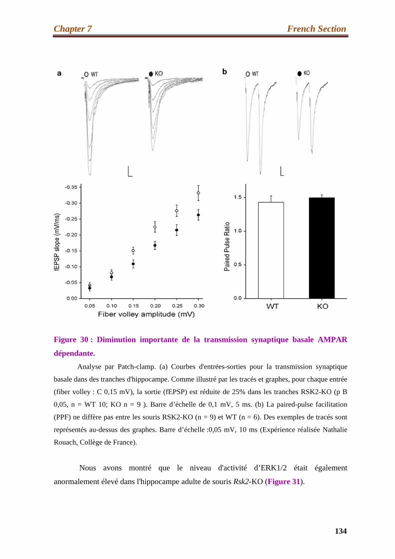
Chapter 7 French Section
134
Figure 30 : Diminution importante de la transmission synaptique basale AMPAR
dépendante.
Analyse par Patch-clamp. (a) Courbes d'entrées-sorties pour la transmission synaptique
basale dans des tranches d'hippocampe. Comme illustré par les tracés et graphes, pour chaque entrée
(fiber volley : C 0,15 mV), la sortie (fEPSP) est réduite de 25% dans les tranches RSK2-KO (p B
0,05, n = WT 10; KO n = 9 ). Barre d’échelle de 0,1 mV, 5 ms. (b) La paired-pulse facilitation
(PPF) ne diffère pas entre les souris RSK2-KO (n = 9) et WT (n = 6). Des exemples de tracés sont
représentés au-dessus des graphes. Barre d’échelle :0,05 mV, 10 ms (Expérience réalisée Nathalie
Rouach, Collège de France).
Nous avons montré que le niveau d'activité d’ERK1/2 était également
anormalement élevé dans l'hippocampe adulte de souris Rsk2-KO (Figure 31).

Chapter 7 French Section
135
Figure 31 : Augmentation de la phosphorylation d’ERK dans l'hippocampe Rsk2-KO.
(a) les protéines détectées (ERK1 / 2, P-ERK1 / 2 et b-tubuline) dans trois souris Rsk2-KO
et trois souris WT sont montrés. (b) Les données représentées sont les valeurs moyennes
normalisées avec ERK1/2 et la b-tubuline ± SEM pour six souris de chaque génotype. Les niveaux
de P-ERK1/2 sont significativement augmentés dans l'hippocampe de souris Rsk2-KO. * p <0,05
par t-test de Student.
En outre, le niveau de phosphorylation des deux facteurs de transcription nucléaire
CREB et Elk1, des cibles nucléaires de ERK1/2, est aussi plus élevé dans les neurones
déficients en activité RSK2. Ces résultats suggèrent que le niveau anormalement élevé de
ERK1/2 dans les neurones Rsk2-KO peut-être impliqué, directement ou indirectement, dans

Chapter 7 French Section
136
la dérégulation de certains des gènes identifiés dans notre précédente étude de comparaison
de transcriptomes.
Mon but fut ensuite d'essayer de comprendre les mécanismes de
signalisation impliqués dans l'augmentation du niveau de transcription du gène GluR2 dans
les neurones déficients pour RSK2. Pour répondre à cette question, nous avons utilisé la
technologie d'interférence ARN pour bloquer l’expression de RSK2 dans les cellules PC12.
Nous avons testé l'efficacité de trois plasmides produisant différents ARN en épingle à
cheveux de petite taille (shRNA) ciblées contre différentes régions codantes de l'ARNm de
Rsk2. Un de ceux-ci a induit une diminution drastique de l’expression de la protéine RSK2
et de son ARNm (Rsk2-Knockdown). Nous avons observé dans les cellules RSK2-
knockdown une augmentation importante de l'expression GluR2 au niveau de l'ARN et de
la protéine ainsi qu’une augmentation du niveau de phosphorylation de ERK1/2, de
manière similaire à ce qui avait été observé dans les neurones hippocampiques de souris
Rsk2-KO. L'inhibition de la voie ERK, dans les cellules Rsk2-knockdown avec l’inhibiteur
de MEK U0126, résulta en une diminution dramatique du niveau d’expression de GluR2,
montrant que ERK1/2 joue un rôle crucial dans la surexpression de GluR2. Il avait été
montré précédemment que l’expression de GluR2 est influencée au niveau transcriptionnel
par le facteur de transcription Sp1. Cependant nous n’avons trouvé aucune différence dans
le niveau d'expression de la protéine Sp1 entre cellules RSK2-knockdown et cellules non
transfectées. Comme il avait été aussi montré que deux sites de Sp1 sont phosphorylés par
ERK1/2, nous avons analysé les niveaux de phospho-Sp1 (Figure 32). Nous avons ainsi
observé un niveau anormalement élevé de phospho-Sp1 dans les cellules PC12 RSK2-
knockdown par rapport aux cellules PC12 non transfectées (WT). Le traitement par U0126
abolit la phosphorylation de Sp1, confirmant ainsi le rôle spécifique de ERK1/2 dans cette
phosphorylation. Le blocage partiel de l’expression de Sp1 dans les cellules PC12 Rsk2-
knockdown avec un siRNA entraîna une diminution dramatique du niveau d’expression de
GluR2, confirmant ainsi le rôle crucial que joue Sp1 dans le mécanisme de surexpression de
GluR2 dans les cellules déficientes pour RSK2 (Figure 33). J'ai par la suite montré que
dans l'hippocampe des souris Rsk2-KO le niveau de pospho-Sp1 est également plus élevé
que chez les souris WT de mêmes fratries, supportant ainsi nos résultats dans les cellules
PC12.
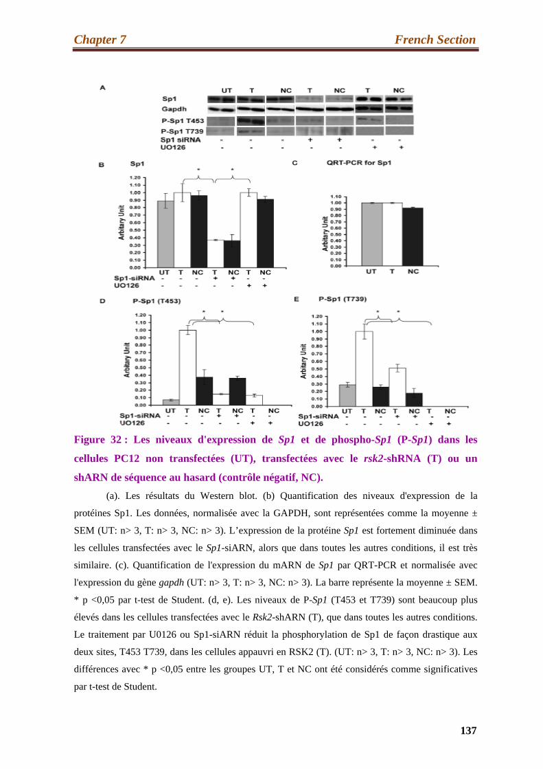
Chapter 7 French Section
137
Figure 32 : Les niveaux d'expression de Sp1 et de phospho-Sp1 (P-Sp1) dans les
cellules PC12 non transfectées (UT), transfectées avec le rsk2-shRNA (T) ou un
shARN de séquence au hasard (contrôle négatif, NC).
(a). Les résultats du Western blot. (b) Quantification des niveaux d'expression de la
protéines Sp1. Les données, normalisée avec la GAPDH, sont représentées comme la moyenne ±
SEM (UT: n> 3, T: n> 3, NC: n> 3). L’expression de la protéine Sp1 est fortement diminuée dans
les cellules transfectées avec le Sp1-siARN, alors que dans toutes les autres conditions, il est très
similaire. (c). Quantification de l'expression du mARN de Sp1 par QRT-PCR et normalisée avec
l'expression du gène gapdh (UT: n> 3, T: n> 3, NC: n> 3). La barre représente la moyenne ± SEM.
* p <0,05 par t-test de Student. (d, e). Les niveaux de P-Sp1 (T453 et T739) sont beaucoup plus
élevés dans les cellules transfectées avec le Rsk2-shARN (T), que dans toutes les autres conditions.
Le traitement par U0126 ou Sp1-siARN réduit la phosphorylation de Sp1 de façon drastique aux
deux sites, T453 T739, dans les cellules appauvri en RSK2 (T). (UT: n> 3, T: n> 3, NC: n> 3). Les
différences avec * p <0,05 entre les groupes UT, T et NC ont été considérés comme significatives
par t-test de Student.

Chapter 7 French Section
138
Figure 33: L’expression de GluR2 est considérablement diminuée dans les cellules
Rsk2-knockdown traitées avec le Sp1-siARN.
(A) Les résultats de Western blot ont révélé une forte diminution de l'expression GluR2
dans les cellules Rsk2-knockdown, par rapport aux cellules Rsk2-knockdown non traitées avec le
Sp1-siARN. (B) Quantification de l’expression de l’ARNm de Gria2 par QRT-PCR. Les données
sont normalisées avec l'expression du mARN gapdh et sont représentées comme la moyenne ± SEM
(UT: n> 3, T: n> 3, NC: n> 3) (* p <0,05 par t-test de Student.

Chapter 7 French Section
139
Ensemble, mes résultats ont suggéré que la surexpression de GluR2 dans les cellules
déficientes pour RSK2, était causée par une activation accrue de la transcription du gène
Gria2 par Sp1, conséquence d’un niveau accru d’activité de ERK1/2.
Nos résultats sont très prometteurs, car ils pourraient conduire à des stratégies
thérapeutiques pour le syndrome de Coffin-Lowry, par exemple, en modulant la
neurotransmission glutamatergique. Toutefois, d'autres investigations sont encore
nécessaires pour déterminer la contribution de la surexpression de GluR2 au déficit
d'apprentissage et de mémoire spatiale chez les souris Rsk2-KO. À cette fin, l'équipe a
l’intention de normaliser la fonction du récepteur AMPA dans les souris Rsk2-KO en
utilisant des vecteurs d'expression lentiviral-shRNA injectée par stéréotaxie dans
l'hippocampe, afin de déterminer si cela permet d’améliorer le déficit cognitif.

References
Chapter 8 References
140
References:
A:
Advani, R.J., Bae, H.R., Bock, J.B., Chao, D.S., Doung, Y.C., Prekeris, R., Yoo,
J.S., and Scheller, R.H. (1998). Seven novel mammalian SNARE proteins localize
to distinct membrane compartments. The Journal of biological chemistry 273,
10317-10324.
Akten, B., Tangredi, M.M., Jauch, E., Roberts, M.A., Ng, F., Raabe, T., and
Jackson, F.R. (2009). Ribosomal s6 kinase cooperates with casein kinase 2 to
modulate the Drosophila circadian molecular oscillator. J Neurosci 29, 466-475.
Alcorta, D.A., Crews, C.M., Sweet, L.J., Bankston, L., Jones, S.W., and Erikson,
R.L.
(1989). Sequence and expression of chicken and mouse rsk: homologs of Xenopus
laevis ribosomal S6 kinase. Mol Cell Biol 9, 3850-3859.
Angenstein, F., Greenough, W.T., and Weiler, I.J. (1998). Metabotropic glutamate
receptor-initiated translocation of protein kinase p90rsk to polyribosomes: a
possible factor regulating synaptic protein synthesis. Proc Natl Acad Sci U S A 95,
15078-15083.
Anjum, R., and Blenis, J. (2008). The RSK family of kinases: emerging roles in
cellular signalling. Nat Rev Mol Cell Biol 9, 747-758.
Anjum, R., Roux, P.P., Ballif, B.A., Gygi, S.P., and Blenis, J. (2005). The tumor
suppressor DAP kinase is a target of RSK-mediated survival signaling. Current
biology : CB 15, 1762-1767.
Armstrong, S.A., Barry, D.A., Leggett, R.W., and Mueller, C.R. (1997). Casein
kinase II-mediated phosphorylation of the C terminus of Sp1 decreases its DNA
binding activity. J Biol Chem 272, 13489-13495.
Arthur, J.S. (2008). MSK activation and physiological roles. Frontiers in bioscience
: a journal and virtual library 13, 5866-5879.
Chapter 8 References
141
B :
Bagrodia, S., Derijard, B., Davis, R.J., and Cerione, R.A. (1995). Cdc42 and PAK- mediated signaling leads to Jun kinase and p38 mitogen-activated protein kinase
activation. J Biol Chem 270, 27995-27998.
Bai, X., and Wong-Riley, M.T. (2003). Neuronal activity regulates protein and gene
expressions of GluR2 in postnatal rat visual cortical neurons in culture. J Neurocytol
32, 71-78.
Bassani, S., Valnegri, P., Beretta, F., and Passafaro, M. (2009). The GLUR2 subunit
of AMPA receptors: synaptic role. Neuroscience 158, 55-61.
Bats, C., Groc, L., and Choquet, D. (2007). The interaction between Stargazin and
PSD-95 regulates AMPA receptor surface trafficking. Neuron 53, 719-734.
Bear, M.F., and Malenka, R.C. (1994). Synaptic plasticity: LTP and LTD. Current
opinion in neurobiology 4, 389-399.
Bermudez, O., Pages, G., and Gimond, C. (2010). The dual-specificity MAP kinase
phosphatases: critical roles in development and cancer. Am J Physiol Cell Physiol
299, C189-202.
Bialik, S., and Kimchi, A. (2004). DAP-kinase as a target for drug design in cancer
and diseases associated with accelerated cell death. Semin Cancer Biol 14, 283-294.
Bjorbaek, C., Vik, T.A., Echwald, S.M., Yang, P.Y., Vestergaard, H., Wang, J.P.,
Webb, G.C., Richmond, K., Hansen, T., Erikson, R.L., et al. (1995a). Cloning of a
human insulin-stimulated protein kinase (ISPK-1) gene and analysis of coding
regions and mRNA levels of the ISPK-1 and the protein phosphatase-1 genes in
muscle from NIDDM patients. Diabetes 44, 90-97.
Bjorbaek, C., Zhao, Y., and Moller, D.E. (1995b). Divergent functional roles for
p90rsk kinase domains. J Biol Chem 270, 18848-18852.
Blanpied, T.A., and Ehlers, M.D. (2004a). Microanatomy of dendritic spines:
emerging principles of synaptic pathology in psychiatric and neurological disease.
Biol Psychiatry 55, 1121-1127.
Bliss, T.V., and Collingridge, G.L. (1993). A synaptic model of memory: long-term
potentiation in the hippocampus. Nature 361, 31-39.
Bliss, T.V., and Gardner-Medwin, A.R. (1973). Long-lasting potentiation of
synaptic transmission in the dentate area of the unanaestetized rabbit following
stimulation of the perforant path. The Journal of physiology 232, 357-374.

Chapter 8 References
142
Bliss, T.V., and Lomo, T. (1973). Long-lasting potentiation of synaptic transmission
in the dentate area of the anaesthetized rabbit following stimulation of the perforant
path. The Journal of physiology 232, 331-356.
Bloss, E.B., and Hunter, R.G. (2010). Hippocampal kainate receptors. Vitam Horm
82, 167-184.
Boda, B., Alberi, S., Nikonenko, I., Node-Langlois, R., Jourdain, P., Moosmayer,
M., Parisi-Jourdain, L., and Muller, D. (2004). The mental retardation protein PAK3
contributes to synapse formation and plasticity in hippocampus. J Neurosci 24,
10816-10825.
Boda, B., Jourdain, L., and Muller, D. (2008). Distinct, but compensatory roles of
PAK1 and PAK3 in spine morphogenesis. Hippocampus 18, 857-861.
Bode, A.M., and Dong, Z. (2007a). The functional contrariety of JNK. Mol
Carcinog 46, 591-598.
Bode, A.M., and Dong, Z. (2007b). The functional contrariety of JNK. Molecular
carcinogenesis 46, 591-598.
Boggio, E.M., Putignano, E., Sassoe-Pognetto, M., Pizzorusso, T., and Giustetto, M.
(2007). Visual stimulation activates ERK in synaptic and somatic compartments of
rat cortical neurons with parallel kinetics. PLoS One 2, e604.
Bogoyevitch, M.A., and Court, N.W. (2004). Counting on mitogen-activated protein
kinases--ERKs 3, 4, 5, 6, 7 and 8. Cell Signal 16, 1345-1354.
Bogoyevitch, M.A., Ngoei, K.R., Zhao, T.T., Yeap, Y.Y., and Ng, D.C. (2010). c-
Jun N-terminal kinase (JNK) signaling: recent advances and challenges. Biochim
Biophys Acta 1804, 463-475.
Bohuslav, J., Chen, L.F., Kwon, H., Mu, Y., and Greene, W.C. (2004). p53 induces
NF-kappaB activation by an IkappaB kinase-independent mechanism involving
phosphorylation of p65 by ribosomal S6 kinase 1. The Journal of biological
chemistry 279, 26115-26125.
Bonni, A., Brunet, A., West, A.E., Datta, S.R., Takasu, M.A., and Greenberg, M.E.
(1999). Cell survival promoted by the Ras-MAPK signaling pathway by
transcription-dependent and -independent mechanisms. Science 286, 1358-1362.
Borges, K., and Dingledine, R. (2001). Functional organization of the GluR1
glutamate receptor promoter. The Journal of biological chemistry 276, 25929-
25938.

Chapter 8 References
143
Boston, S.R., Deshmukh, R., Strome, S., Priyakumar, U.D., Mackerell, A.D., Jr.,
and Shapiro, P. (2011). Characterization of ERK Docking Domain Inhibitors that
Induce Apoptosis by Targeting Rsk-1 and Caspase-9. BMC Cancer 11, 7.
Boulter, J., Hollmann, M., O'Shea-Greenfield, A., Hartley, M., Deneris, E., Maron,
C., and Heinemann, S. (1990). Molecular cloning and functional expression of
glutamate receptor subunit genes. Science 249, 1033-1037.
Boulton, T.G., Nye, S.H., Robbins, D.J., Ip, N.Y., Radziejewska, E., Morgenbesser,
S.D., DePinho, R.A., Panayotatos, N., Cobb, M.H., and Yancopoulos, G.D. (1991).
ERKs: a family of protein-serine/threonine kinases that are activated and tyrosine
phosphorylated in response to insulin and NGF. Cell 65, 663-675.
Boulton, T.G., Yancopoulos, G.D., Gregory, J.S., Slaughter, C., Moomaw, C., Hsu,
J., and Cobb, M.H. (1990). An insulin-stimulated protein kinase similar to yeast
kinases involved in cell cycle control. Science 249, 64-67.
Bourne, J.N., and Harris, K.M. (2008). Balancing structure and function at
hippocampal dendritic spines. Annu Rev Neurosci 31, 47-67.
Bowie, D., and Mayer, M.L. (1995). Inward rectification of both AMPA and kainate
subtype glutamate receptors generated by polyamine-mediated ion channel block.
Neuron 15, 453-462.
Bramham, C.R., Worley, P.F., Moore, M.J., and Guzowski, J.F. (2008). The
immediate early gene arc/arg3.1: regulation, mechanisms, and function. J Neurosci
28, 11760-11767.
Bredt, D.S., and Nicoll, R.A. (2003). AMPA receptor trafficking at excitatory
synapses. Neuron 40, 361-379.
Brorson, J.R., Li, D., and Suzuki, T. (2004). Selective expression of heteromeric
AMPA receptors driven by flip-flop differences. J Neurosci 24, 3461-3470.
Brown, T.C., Tran, I.C., Backos, D.S., and Esteban, J.A. (2005). NMDA receptor-
dependent activation of the small GTPase Rab5 drives the removal of synaptic
AMPA receptors during hippocampal LTD. Neuron 45, 81-94.
Bruning, J.C., Gillette, J.A., Zhao, Y., Bjorbaeck, C., Kotzka, J., Knebel, B., Avci,
H., Hanstein, B., Lingohr, P., Moller, D.E., et al. (2000b). Ribosomal subunit
kinase-2 is required for growth factor-stimulated transcription of the c-Fos gene.
Proceedings of the National Academy of Sciences of the United States of America
97, 2462-2467.
Chapter 8 References
144
Burnashev, N., Khodorova, A., Jonas, P., Helm, P.J., Wisden, W., Monyer, H.,
Seeburg, P.H., and Sakmann, B. (1992). Calcium-permeable AMPA-kainate
receptors in fusiform cerebellar glial cells. Science 256, 1566-1570.
Buxade, M., Parra-Palau, J.L., and Proud, C.G. (2008). The Mnks: MAP kinase-
interacting kinases (MAP kinase signal-integrating kinases). Frontiers in bioscience
: a journal and virtual library 13, 5359-5373.
C:
Cacace, A.M., Michaud, N.R., Therrien, M., Mathes, K., Copeland, T., Rubin,
G.M., and Morrison, D.K. (1999). Identification of constitutive and ras-inducible
phosphorylation sites of KSR: implications for 14-3-3 binding, mitogen-activated
protein kinase binding, and KSR overexpression. Mol Cell Biol 19, 229-240.
Calabrese, B., Wilson, M.S., and Halpain, S. (2006). Development and regulation of
dendritic spine synapses. Physiology (Bethesda) 21, 38-47.
Campbell, P.M., Boufaied, N., Fiordalisi, J.J., Cox, A.D., Falardeau, P., Der, C.J.,
and Gourdeau, H. (2010). TLN-4601 suppresses growth and induces apoptosis of
pancreatic carcinoma cells through inhibition of Ras-ERK MAPK signaling. J Mol
Signal 5, 18.
Cao, X., Cui, Z., Feng, R., Tang, Y.P., Qin, Z., Mei, B., and Tsien, J.Z. (2007).
Maintenance of superior learning and memory function in NR2B transgenic mice
during ageing. The European journal of neuroscience 25, 1815-1822.
Cargnello, M., and Roux, P.P. (2011). Activation and function of the MAPKs and
their substrates, the MAPK-activated protein kinases. Microbiol Mol Biol Rev 75,
50-83.
Carlisle, H.J., Manzerra, P., Marcora, E., and Kennedy, M.B. (2008). SynGAP
regulates steady-state and activity-dependent phosphorylation of cofilin. J Neurosci
28, 13673-13683.
Carriere, A., Ray, H., Blenis, J., and Roux, P.P. (2008). The RSK factors of
activating the Ras/MAPK signaling cascade. Frontiers in bioscience : a journal and
virtual library 13, 4258-4275.

Chapter 8 References
145
Carroll, R.C., and Zukin, R.S. (2002). NMDA-receptor trafficking and targeting:
implications for synaptic transmission and plasticity. Trends in neurosciences 25,
571-577.
Chaturvedi, D., Cohen, M.S., Taunton, J., and Patel, T.B. (2009). The PKARIalpha
subunit of protein kinase A modulates the activation of p90RSK1 and its function. J
Biol Chem 284, 23670-23681.
Chelly, J. (2000). [Recent advances in the genetics and physiopathology of X
chromosomal mental retardation]. Arch Pediatr 7 Suppl 2, 114s-118s.
Chelly, J., Khelfaoui, M., Francis, F., Cherif, B., and Bienvenu, T. (2006). Genetics
and pathophysiology of mental retardation. Eur J Hum Genet 14, 701-713.
Chelly, J., and Mandel, J.L. (2001). Monogenic causes of X-linked mental
retardation. Nat Rev Genet 2, 669-680.
Chen, L.Y., Rex, C.S., Babayan, A.H., Kramar, E.A., Lynch, G., Gall, C.M., and
Lauterborn, J.C. (2010). Physiological activation of synaptic Rac>PAK (p-21
activated kinase) signaling is defective in a mouse model of fragile X syndrome. J
Neurosci 30, 10977-10984.
Chen, R.H., Abate, C., and Blenis, J. (1993). Phosphorylation of the c-Fos
transrepression domain by mitogen-activated protein kinase and 90-kDa ribosomal
S6 kinase. Proc Natl Acad Sci U S A 90, 10952-10956.
Chen, R.H., Sarnecki, C., and Blenis, J. (1992). Nuclear localization and regulation
of erk- and rsk-encoded protein kinases. Mol Cell Biol 12, 915-927.
Cheng, C.M., Mervis, R.F., Niu, S.L., Salem, N., Jr., Witters, L.A., Tseng, V.,
Reinhardt, R., and Bondy, C.A. (2003). Insulin-like growth factor 1 is essential for
normal dendritic growth. J Neurosci Res 73, 1-9.
Cho, Y.Y., He, Z., Zhang, Y., Choi, H.S., Zhu, F., Choi, B.Y., Kang, B.S., Ma,
W.Y., Bode, A.M., and Dong, Z. (2005). The p53 protein is a novel substrate of
ribosomal S6 kinase 2 and a critical intermediary for ribosomal S6 kinase 2 and
histone H3 interaction. Cancer Res 65, 3596-3603.
Cho, Y.Y., Yao, K., Kim, H.G., Kang, B.S., Zheng, D., Bode, A.M., and Dong, Z.
(2007). Ribosomal S6 kinase 2 is a key regulator in tumor promoter induced cell
transformation. Cancer Res 67, 8104-8112.
Chu, P.J., Robertson, H.M., and Best, P.M. (2001). Calcium channel gamma
subunits provide insights into the evolution of this gene family. Gene 280, 37-48.

Chapter 8 References
146
Chu, S., and Ferro, T.J. (2005). Sp1: regulation of gene expression by
phosphorylation. Gene 348, 1-11.
Chu, Z., and Hablitz, J.J. (2000). Quisqualate induces an inward current via mGluR
activation in neocortical pyramidal neurons. Brain Res 879, 88-92.
Chung, J., Pelech, S.L., and Blenis, J. (1991). Mitogen-activated Swiss mouse 3T3
RSK kinases I and II are related to pp44mpk from sea star oocytes and participate in
the regulation of pp90rsk activity. Proc Natl Acad Sci U S A 88, 4981-4985.
Cingolani, L.A., and Goda, Y. (2008). Actin in action: the interplay between the
actin cytoskeleton and synaptic efficacy. Nat Rev Neurosci 9, 344-356.
Citri, A., and Malenka, R.C. (2008). Synaptic plasticity: multiple forms, functions,
and mechanisms. Neuropsychopharmacology 33, 18-41.
Coffin GS, Siris E, Wegienka LC (1966). "Mental retardation with
osteocartilaginous anomalies". Am. J. Dis. Child. 112: 205–213.
Collingridge, G.L., Isaac, J.T., and Wang, Y.T. (2004). Receptor trafficking and
synaptic plasticity. Nat Rev Neurosci 5, 952-962.
Collingridge, G.L., Olsen, R.W., Peters, J., and Spedding, M. (2009). A
nomenclature for ligand-gated ion channels. Neuropharmacology 56, 2-5.
Cooke, S.F., and Bliss, T.V. (2006). Plasticity in the human central nervous system.
Brain 129, 1659-1673.
Cuadrado, A., and Nebreda, A.R. (2010). Mechanisms and functions of p38 MAPK
signalling. Biochem J 429, 403-417.
Cuenda, A., and Rousseau, S. (2007). p38 MAP-kinases pathway regulation,
function and role in human diseases. Biochim Biophys Acta 1773, 1358-1375.
Cull-Candy, S., Brickley, S., and Farrant, M. (2001). NMDA receptor subunits:
diversity, development and disease. Current opinion in neurobiology 11, 327-335.
Cull-Candy, S., Kelly, L., and Farrant, M. (2006). Regulation of Ca2+-permeable
AMPA receptors: synaptic plasticity and beyond. Curr Opin Neurobiol 16, 288-297.
Cull-Candy, S.G., and Leszkiewicz, D.N. (2004). Role of distinct NMDA receptor
subtypes at central synapses. Sci STKE 2004, re16.

Chapter 8 References
147
D:
Dalby, K.N., Morrice, N., Caudwell, F.B., Avruch, J., and Cohen, P. (1998).
Identification of regulatory phosphorylation sites in mitogen-activated protein
kinase (MAPK)-activated protein kinase-1a/p90rsk that are inducible by MAPK. J
Biol Chem 273, 1496-1505.
Darcq, E., Koebel, P., Del Boca, C., Pannetier, S., Kirstetter, A.S., Garnier, J.M.,
Hanauer, A., Befort, K., and Kieffer, B.L. (2011). RSK2 signaling in brain habenula
contributes to place aversion learning. Learn Mem 18, 574-578.
David, J.P., Mehic, D., Bakiri, L., Schilling, A.F., Mandic, V., Priemel, M.,
Idarraga, M.H., Reschke, M.O., Hoffmann, O., Amling, M., et al. (2005). Essential
role of RSK2 in c-Fos-dependent osteosarcoma development. The Journal of
clinical investigation 115, 664-672.
Davis, I.J., Hazel, T.G., Chen, R.H., Blenis, J., and Lau, L.F. (1993). Functional
domains and phosphorylation of the orphan receptor Nur77. Mol Endocrinol 7, 953-
964.
Davis, S., and Laroche, S. (2006). Mitogen-activated protein kinase/extracellular
regulated kinase signalling and memory stabilization: a review. Genes Brain Behav
5 Suppl 2, 61-72.
Davis, S., Renaudineau, S., Poirier, R., Poucet, B., Save, E., and Laroche, S. (2010).
The formation and stability of recognition memory: what happens upon recall?
Front Behav Neurosci 4, 177.
De Cesare, D., Jacquot, S., Hanauer, A., and Sassone-Corsi, P. (1998). Rsk-2
activity is necessary for epidermal growth factor-induced phosphorylation of CREB
protein and transcription of c-fos gene. Proc Natl Acad Sci U S A 95, 12202-12207.
Delaunoy, J., Abidi, F., Zeniou, M., Jacquot, S., Merienne, K., Pannetier, S.,
Schmitt, M., Schwartz, C., and Hanauer, A. (2001). Mutations in the X-linked
RSK2 gene (RPS6KA3) in patients with Coffin-Lowry syndrome. Hum Mutat 17,
103-116.
Delaunoy, J.P., Dubos, A., Marques Pereira, P., and Hanauer, A. (2006).
Identification of novel mutations in the RSK2 gene (RPS6KA3) in patients with
Coffin-Lowry syndrome. Clin Genet 70, 161-166.

Chapter 8 References
148
Derijard, B., Raingeaud, J., Barrett, T., Wu, I.H., Han, J., Ulevitch, R.J., and Davis,
R.J. (1995). Independent human MAP-kinase signal transduction pathways defined
by MEK and MKK isoforms. Science 267, 682-685.
Derkach, V.A., Oh, M.C., Guire, E.S., and Soderling, T.R. (2007). Regulatory
mechanisms of AMPA receptors in synaptic plasticity. Nature reviews
Neuroscience 8, 101-113.
Dhar, S.S., Liang, H.L., and Wong-Riley, M.T. (2009). Nuclear respiratory factor 1
co-regulates AMPA glutamate receptor subunit 2 and cytochrome c oxidase: tight
coupling of glutamatergic transmission and energy metabolism in neurons. Journal
of neurochemistry 108, 1595-1606.
Dhillon, A.S., Hagan, S., Rath, O., and Kolch, W. (2007). MAP kinase signalling
pathways in cancer. Oncogene 26, 3279-3290.
Dingledine, R., Borges, K., Bowie, D., and Traynelis, S.F. (1999). The glutamate
receptor ion channels. Pharmacol Rev 51, 7-61.
Doehn, U., Gammeltoft, S., Shen, S.H., and Jensen, C.J. (2004). p90 ribosomal S6
kinase 2 is associated with and dephosphorylated by protein phosphatase 2Cdelta.
Biochem J 382, 425-431.
Douillette, A., Bibeau-Poirier, A., Gravel, S.P., Clement, J.F., Chenard, V., Moreau,
P., and Servant, M.J. (2006). The proinflammatory actions of angiotensin II are
dependent on p65 phosphorylation by the IkappaB kinase complex. The Journal of
biological chemistry 281, 13275-13284.
Douville, E., and Downward, J. (1997). EGF induced SOS phosphorylation in PC12
cells involves P90 RSK-2. Oncogene 15, 373-383.
Du, J., Creson, T.K., Wu, L.J., Ren, M., Gray, N.A., Falke, C., Wei, Y., Wang, Y.,
Blumenthal, R., Machado-Vieira, R., et al. (2008). The role of hippocampal GluR1
and GluR2 receptors in manic-like behavior. J Neurosci 28, 68-79.
Dudek, S.M., and Bear, M.F. (1992). Homosynaptic long-term depression in area
CA1 of hippocampus and effects of N-methyl-D-aspartate receptor blockade.
Proceedings of the National Academy of Sciences of the United States of America
89, 4363-4367.
Dufresne, S.D., Bjorbaek, C., El-Haschimi, K., Zhao, Y., Aschenbach, W.G.,
Moller, D.E., and Goodyear, L.J. (2001). Altered extracellular signal-regulated

Chapter 8 References
149
kinase signaling and glycogen metabolism in skeletal muscle from p90 ribosomal
S6 kinase 2 knockout mice. Mol Cell Biol 21, 81-87.
Dugani, C.B., Paquin, A., Kaplan, D.R., and Miller, F.D. (2010). Coffin-Lowry
syndrome: a role for RSK2 in mammalian neurogenesis. Dev Biol 347, 348-359.
Dummler, B.A., Hauge, C., Silber, J., Yntema, H.G., Kruse, L.S., Kofoed, B.,
Hemmings, B.A., Alessi, D.R., and Frodin, M. (2005). Functional characterization
of human RSK4, a new 90-kDa ribosomal S6 kinase, reveals constitutive activation
in most cell types. J Biol Chem 280, 13304-13314.
E:
Eby, G.A., and Eby, K.L. (2006). Rapid recovery from major depression using
magnesium treatment. Med Hypotheses 67, 362-370.
Eisinger-Mathason, T.S., Andrade, J., and Lannigan, D.A. (2010). RSK in
tumorigenesis: connections to steroid signaling. Steroids 75, 191-202.
Ekici, M., Keim, A., Rossler, O.G., Hohl, M., and Thiel, G. (2011). Chromatin
structure and expression of the AMPA receptor subunit GluR2 in human glioma
cells: major regulatory role of REST and Sp1. J Cell Biochem.
El-Haschimi, K., Dufresne, S.D., Hirshman, M.F., Flier, J.S., Goodyear, L.J., and
Bjorbaek, C. (2003). Insulin resistance and lipodystrophy in mice lacking ribosomal
S6 kinase 2. Diabetes 52, 1340-1346.
El-Husseini, A.E., Schnell, E., Chetkovich, D.M., Nicoll, R.A., and Bredt, D.S.
(2000). PSD-95 involvement in maturation of excitatory synapses. Science 290,
1364-1368.
Elefteriou, F., Benson, M.D., Sowa, H., Starbuck, M., Liu, X., Ron, D., Parada,
L.F., and Karsenty, G. (2006). ATF4 mediation of NF1 functions in osteoblast
reveals a nutritional basis for congenital skeletal dysplasiae. Cell Metab 4, 441-451.
Eckel-Mahan KL, Phan T, Han S, Wang H, Chan GC, Scheiner ZS, Storm DR
(2008) Circadian oscillation of hippocampal MAPK activity and cAmp:
Implications for memory persistence. Nat Neurosci 11:1074 –1082.
Engel, S.R., Creson, T.K., Hao, Y., Shen, Y., Maeng, S., Nekrasova, T., Landreth,
G.E., Manji, H.K., and Chen, G. (2009). The extracellular signal-regulated kinase

Chapter 8 References
150
pathway contributes to the control of behavioral excitement. Mol Psychiatry 14,
448-461.
Erikson, E., and Maller, J.L. (1985). A protein kinase from Xenopus eggs specific
for ribosomal protein S6. Proc Natl Acad Sci U S A 82, 742-746.
Erikson, E., and Maller, J.L. (1986). Purification and characterization of a protein
kinase from Xenopus eggs highly specific for ribosomal protein S6. The Journal of
biological chemistry 261, 350-355.
F:
Farrokhnia, N., Ericsson, A., Terent, A., and Lennmyr, F. (2008). MEK-inhibitor
U0126 in hyperglycaemic focal ischaemic brain injury in the rat. Eur J Clin Invest
38, 679-685.
Feng, W., and Zhang, M. (2009). Organization and dynamics of PDZ-domain-
related supramodules in the postsynaptic density. Nat Rev Neurosci 10, 87-99.
Ferraguti, F., and Shigemoto, R. (2006). Metabotropic glutamate receptors. Cell
Tissue Res 326, 483-504.
Fiala, J.C., Spacek, J., and Harris, K.M. (2002). Dendritic spine pathology: cause or
consequence of neurological disorders? Brain Res Brain Res Rev 39, 29-54.
Fischer, M., Pereira, P.M., Holtmann, B., Simon, C.M., Hanauer, A., Heisenberg,
M., and Sendtner, M. (2009a). P90 Ribosomal s6 kinase 2 negatively regulates axon
growth in motoneurons. Mol Cell Neurosci 42, 134-141.
Fischer, M., Raabe, T., Heisenberg, M., and Sendtner, M. (2009b). Drosophila RSK
negatively regulates bouton number at the neuromuscular junction. Dev Neurobiol
69, 212-220.
Fleming, J.J., and England, P.M. (2010). AMPA receptors and synaptic plasticity: a
chemist's perspective. Nat Chem Biol 6, 89-97.
Flotow, H., and Thomas, G. (1992). Substrate recognition determinants of the
mitogen-activated 70K S6 kinase from rat liver. J Biol Chem 267, 3074-3078.
Fojas de Borja, P., Collins, N.K., Du, P., Azizkhan-Clifford, J., and Mudryj, M.
(2001). Cyclin A-CDK phosphorylates Sp1 and enhances Sp1-mediated
transcription. EMBO J 20, 5737-5747.

Chapter 8 References
151
Frodin, M., Antal, T.L., Dummler, B.A., Jensen, C.J., Deak, M., Gammeltoft, S.,
and Biondi, R.M. (2002). A phosphoserine/threonine-binding pocket in AGC
kinases and PDK1 mediates activation by hydrophobic motif phosphorylation.
EMBO J 21, 5396-5407.
Frodin, M., and Gammeltoft, S. (1999). Role and regulation of 90 kDa ribosomal S6
kinase (RSK) in signal transduction. Mol Cell Endocrinol 151, 65-77.
Frodin, M., Jensen, C.J., Merienne, K., and Gammeltoft, S. (2000). A
phosphoserine-regulated docking site in the protein kinase RSK2 that recruits and
activates PDK1. EMBO J 19, 2924-2934.
Fujita, N., Sato, S., and Tsuruo, T. (2003). Phosphorylation of p27Kip1 at threonine
198 by p90 ribosomal protein S6 kinases promotes its binding to 14-3-3 and
cytoplasmic localization. J Biol Chem 278, 49254-49260.
Fukazawa, Y., Saitoh, Y., Ozawa, F., Ohta, Y., Mizuno, K., and Inokuchi, K.
(2003). Hippocampal LTP is accompanied by enhanced F-actin content within the
dendritic spine that is essential for late LTP maintenance in vivo. Neuron 38, 447-
460.
G:
Gaestel, M. (2008). Specificity of signaling from MAPKs to MAPKAPKs: kinases'
tango nuevo. Frontiers in bioscience : a journal and virtual library 13, 6050-6059.
Geraghty, K.M., Chen, S., Harthill, J.E., Ibrahim, A.F., Toth, R., Morrice, N.A.,
Vandermoere, F., Moorhead, G.B., Hardie, D.G., and MacKintosh, C. (2007).
Regulation of multisite phosphorylation and 14-3-3 binding of AS160 in response to
IGF-1, EGF, PMA and AICAR. The Biochemical journal 407, 231-241.
Gerrow, K., and Triller, A. (2010). Synaptic stability and plasticity in a floating
world. Curr Opin Neurobiol 20, 631-639.
Ghoda, L., Lin, X., and Greene, W.C. (1997a). The 90-kDa ribosomal S6 kinase
(pp90rsk) phosphorylates the N-terminal regulatory domain of IkappaBalpha and
stimulates its degradation in vitro. J Biol Chem 272, 21281-21288.
Gille, H., Kortenjann, M., Thomae, O., Moomaw, C., Slaughter, C., Cobb, M.H.,
and Shaw, P.E. (1995). ERK phosphorylation potentiates Elk-1-mediated ternary
complex formation and transactivation. EMBO J 14, 951-962.

Chapter 8 References
152
Ginty, D.D., Bonni, A., and Greenberg, M.E. (1994). Nerve growth factor activates
a Ras-dependent protein kinase that stimulates c-fos transcription via
phosphorylation of CREB. Cell 77, 713-725.
Greene, L.A., and Tischler, A.S. (1976). Establishment of a noradrenergic clonal
line of rat adrenal pheochromocytoma cells which respond to nerve growth factor.
Proc Natl Acad Sci U S A 73, 2424-2428.
Greger, I.H., Akamine, P., Khatri, L., and Ziff, E.B. (2006). Developmentally
regulated, combinatorial RNA processing modulates AMPA receptor biogenesis.
Neuron 51, 85-97.
Greger, I.H., Khatri, L., Kong, X., and Ziff, E.B. (2003). AMPA receptor
tetramerization is mediated by Q/R editing. Neuron 40, 763-774.
Greger, I.H., Khatri, L., and Ziff, E.B. (2002). RNA editing at arg607 controls
AMPA receptor exit from the endoplasmic reticulum. Neuron 34, 759-772.
Gross, S.D., Lewellyn, A.L., and Maller, J.L. (2001). A constitutively active form of
the protein kinase p90Rsk1 is sufficient to trigger the G2/M transition in Xenopus
oocytes. The Journal of biological chemistry 276, 46099-46103.
Grossman, A.W., Aldridge, G.M., Lee, K.J., Zeman, M.K., Jun, C.S., Azam, H.S.,
Arii, T., Imoto, K., Greenough, W.T., and Rhyu, I.J. (2010). Developmental
characteristics of dendritic spines in the dentate gyrus of Fmr1 knockout mice. Brain
Res 1355, 221-227.
Grossman, A.W., Elisseou, N.M., McKinney, B.C., and Greenough, W.T. (2006).
Hippocampal pyramidal cells in adult Fmr1 knockout mice exhibit an immature-
appearing profile of dendritic spines. Brain Res 1084, 158-164.
Grutzendler, J., Kasthuri, N., and Gan, W.B. (2002). Long-term dendritic spine
stability in the adult cortex. Nature 420, 812-816.
Grutzendler, J., Kasthuri, N., and Gan, W.B. (2002). Long-term dendritic spine
stability in the adult cortex. Nature 420, 812-816.
Gu, J., Lee, C.W., Fan, Y., Komlos, D., Tang, X., Sun, C., Yu, K., Hartzell, H.C.,
Chen, G., Bamburg, J.R., et al. (2010). ADF/cofilin-mediated actin dynamics
regulate AMPA receptor trafficking during synaptic plasticity. Nat Neurosci 13,
1208-1215.
Guimiot, F., Delezoide, A.L., Hanauer, A., and Simonneau, M. (2004). Expression
of the RSK2 gene during early human development. Gene Expr Patterns 4, 111-114.

Chapter 8 References
153
Gupta, S., Barrett, T., Whitmarsh, A.J., Cavanagh, J., Sluss, H.K., Derijard, B., and
Davis, R.J. (1996). Selective interaction of JNK protein kinase isoforms with
transcription factors. EMBO J 15, 2760-2770.
Gustavson, K.H. (2005). Prevalence and aetiology of congenital birth defects, infant
mortality and intellectual disability in Lahore, Pakistan: a prospective cohort study.
Acta Paediatr 94, 769-774.
H:
Han, J., Lee, J.D., Bibbs, L., and Ulevitch, R.J. (1994). A MAP kinase targeted by
endotoxin and hyperosmolarity in mammalian cells. Science 265, 808-811.
Hanauer, A. (2008). Coffin-lowery Syndrome. Encyclopedia of life Science.
Hanauer, A., and Young, I.D. (2002). Coffin-Lowry syndrome: clinical and
molecular features. J Med Genet 39, 705-713.
Hanley, J.G., Khatri, L., Hanson, P.I., and Ziff, E.B. (2002). NSF ATPase and
alpha-/beta-SNAPs disassemble the AMPA receptor-PICK1 complex. Neuron 34,
53-67.
Harvey, C.D., Yasuda, R., Zhong, H., and Svoboda, K. (2008). The spread of Ras
activity triggered by activation of a single dendritic spine. Science 321, 136-140.
Hauge, C., and Frodin, M. (2006). RSK and MSK in MAP kinase signalling. J Cell
Sci 119, 3021-3023.
Hebb, D.O. (1949). The Organization of Behavior: a Neuropathology Theory
(Wiley,
New York).
Hering, H., and Sheng, M. (2001). Dendritic spines: structure, dynamics and
regulation. Nat Rev Neurosci 2, 880-888.
Hibi, M., Lin, A., Smeal, T., Minden, A., and Karin, M. (1993). Identification of an
oncoprotein- and UV-responsive protein kinase that binds and potentiates the c-Jun
activation domain. Genes Dev 7, 2135-2148.
Holland, E.C., Sonenberg, N., Pandolfi, P.P., and Thomas, G. (2004). Signaling
control of mRNA translation in cancer pathogenesis. Oncogene 23, 3138-3144.

Chapter 8 References
154
Hollmann, M., Hartley, M., and Heinemann, S. (1991). Ca2+ permeability of KA-
AMPA--gated glutamate receptor channels depends on subunit composition.
Science 252, 851-853.
Holtmaat, A., and Svoboda, K. (2009). Experience-dependent structural synaptic
plasticity in the mammalian brain. Nature reviews Neuroscience 10, 647-658.
Holtmaat, A., Wilbrecht, L., Knott, G.W., Welker, E., and Svoboda, K. (2006).
Experience-dependent and cell-type-specific spine growth in the neocortex. Nature
441, 979-983.
Honore, T., Lauridsen, J., and Krogsgaard-Larsen, P. (1982). The binding of
[3H]AMPA, a structural analogue of glutamic acid, to rat brain membranes. J
Neurochem 38, 173-178.
Horning, M.S., and Trombley, P.Q. (2001). Zinc and copper influence excitability
of rat olfactory bulb neurons by multiple mechanisms. J Neurophysiol 86, 1652-
1660.
Hotulainen, P., and Hoogenraad, C.C. (2010). Actin in dendritic spines: connecting
dynamics to function. J Cell Biol 189, 619-629.
Hotulainen, P., Llano, O., Smirnov, S., Tanhuanpaa, K., Faix, J., Rivera, C., and
Lappalainen, P. (2009). Defining mechanisms of actin polymerization and
depolymerization during dendritic spine morphogenesis. J Cell Biol 185, 323-339.
Hu, X.D., Huang, Q., Yang, X., and Xia, H. (2007). Differential regulation of
AMPA receptor trafficking by neurabin-targeted synaptic protein phosphatase-1 in
synaptic transmission and long-term depression in hippocampus. J Neurosci 27,
4674-4686.
Huang, F., Chotiner, J.K., and Steward, O. (2007). Actin polymerization and ERK
phosphorylation are required for Arc/Arg3.1 mRNA targeting to activated synaptic
sites on dendrites. J Neurosci 27, 9054-9067.
Huggins, D.J., and Grant, G.H. (2005). The function of the amino terminal domain
in NMDA receptor modulation. J Mol Graph Model 23, 381-388.
Humeau, Y., Gambino, F., Chelly, J., and Vitale, N. (2009). X-linked mental
retardation: focus on synaptic function and plasticity. J Neurochem 109, 1-14.

Chapter 8 References
155
I:
Ikuta, M., Kornienko, M., Byrne, N., Reid, J.C., Mizuarai, S., Kotani, H., and
Munshi, S.K. (2007). Crystal structures of the N-terminal kinase domain of human
RSK1 bound to three different ligands: Implications for the design of RSK1 specific
inhibitors. Protein Sci 16, 2626-2635.
Inlow, J.K., and Restifo, L.L. (2004). Molecular and comparative genetics of mental
retardation. Genetics 166, 835-881.
Inoki, K., Ouyang, H., Zhu, T., Lindvall, C., Wang, Y., Zhang, X., Yang, Q.,
Bennett, C., Harada, Y., Stankunas, K., et al. (2006). TSC2 integrates Wnt and
energy signals via a coordinated phosphorylation by AMPK and GSK3 to regulate
cell growth. Cell 126, 955-968.
Isaac, J.T., Ashby, M., and McBain, C.J. (2007). The role of the GluR2 subunit in
AMPA receptor function and synaptic plasticity. Neuron 54, 859-871.
Ito, M., Zhao, N., Zeng, Z., Chang, C.C., and Zu, Y. (2010). Synergistic growth
inhibition of anaplastic large cell lymphoma cells by combining cellular ALK gene
silencing and a low dose of the kinase inhibitor U0126. Cancer Gene Ther 17, 633-
644.
J:
Jacquot, S., Merienne, K., De Cesare, D., Pannetier, S., Mandel, J.L., Sassone-
Corsi, P., and Hanauer, A. (1998a). Mutation analysis of the RSK2 gene in Coffin-
Lowry patients: extensive allelic heterogeneity and a high rate of de novo mutations.
Am J Hum Genet 63, 1631-1640.
Jacquot, S., Merienne, K., Pannetier, S., Blumenfeld, S., Schinzel, A., and Hanauer,
A. (1998b). Germline mosaicism in Coffin-Lowry syndrome. Eur J Hum Genet 6,
578-582.
Jaeschke, A., Karasarides, M., Ventura, J.J., Ehrhardt, A., Zhang, C., Flavell, R.A.,
Shokat, K.M., and Davis, R.J. (2006). JNK2 is a positive regulator of the cJun
transcription factor. Mol Cell 23, 899-911.
Jensen, J.B., Schousboe, A., and Pickering, D.S. (1998). Development of calcium-
permeable alpha-amino-3-hydroxy-5-methyl-4-isoxazolepropionic acid receptors in

Chapter 8 References
156
cultured neocortical neurons visualized by cobalt staining. J Neurosci Res 54, 273-
281.
Jiang, Y., Chen, C., Li, Z., Guo, W., Gegner, J.A., Lin, S., and Han, J. (1996).
Characterization of the structure and function of a new mitogen-activated protein
kinase (p38beta). J Biol Chem 271, 17920-17926.
Johnson, G.L., and Lapadat, R. (2002). Mitogen-activated protein kinase pathways
mediated by ERK, JNK, and p38 protein kinases. Science 298, 1911-1912.
Jones, S.W., Erikson, E., Blenis, J., Maller, J.L., and Erikson, R.L. (1988). A
Xenopus ribosomal protein S6 kinase has two apparent kinase domains that are each
similar to distinct protein kinases. Proc Natl Acad Sci U S A 85, 3377-3381.
Jurkiewicz, D., Jezela-Stanek, A., Ciara, E., Piekutowska-Abramczuk, D., Kugaudo,
M., Gajdulewicz, M., Chrzanowska, K., Popowska, E., and Krajewska-Walasek, M.
(2010). Four novel RSK2 mutations in females with Coffin-Lowry syndrome. Eur J
Med Genet 53, 268-273.
K:
Kajta, M., Trotter, A., Lason, W., and Beyer, C. (2006). Impact of 17beta-estradiol
on cytokine-mediated apoptotic effects in primary hippocampal and neocortical cell
cultures. Brain Res 1116, 64-74.
Kang, S., and Chen, J. (2011). Targeting RSK2 in human malignancies. Expert Opin
Ther Targets 15, 11-20.
Kang, S., Dong, S., Gu, T.L., Guo, A., Cohen, M.S., Lonial, S., Khoury, H.J.,
Fabbro, D., Gilliland, D.G., Bergsagel, P.L., et al. (2007). FGFR3 activates RSK2 to
mediate hematopoietic transformation through tyrosine phosphorylation of RSK2
and activation of the MEK/ERK pathway. Cancer Cell 12, 201-214.
Kang, S., Dong, S., Guo, A., Ruan, H., Lonial, S., Khoury, H.J., Gu, T.L., and Chen,
J. (2008). Epidermal growth factor stimulates RSK2 activation through activation of
the MEK/ERK pathway and src-dependent tyrosine phosphorylation of RSK2 at
Tyr-529. The Journal of biological chemistry 283, 4652-4657.
Kang, S., Elf, S., Dong, S., Hitosugi, T., Lythgoe, K., Guo, A., Ruan, H., Lonial, S.,
Khoury, H.J., Williams, I.R., et al. (2009a). Fibroblast growth factor receptor 3
Chapter 8 References
157
associates with and tyrosine phosphorylates p90 RSK2, leading to RSK2 activation
that mediates hematopoietic transformation. Mol Cell Biol 29, 2105-2117.
Kang, S., Elf, S., Dong, S., Hitosugi, T., Lythgoe, K., Guo, A., Ruan, H., Lonial, S.,
Khoury, H.J., Williams, I.R., et al. (2009b). Fibroblast growth factor receptor 3
associates with and tyrosine phosphorylates p90 RSK2, leading to RSK2 activation
that mediates hematopoietic transformation. Molecular and cellular biology 29,
2105-2117.
Karin, M. (2006). Nuclear factor-kappaB in cancer development and progression.
Nature 441, 431-436.
Karnoub, A.E., and Weinberg, R.A. (2008). Ras oncogenes: split personalities.
Nature reviews Molecular cell biology 9, 517-531.
Kasai, H., Fukuda, M., Watanabe, S., Hayashi-Takagi, A., and Noguchi, J. (2010).
Structural dynamics of dendritic spines in memory and cognition. Trends Neurosci
33, 121-129.
Kasai, H., Matsuzaki, M., Noguchi, J., Yasumatsu, N., and Nakahara, H. (2003).
Structure-stability-function relationships of dendritic spines. Trends in
neurosciences 26, 360-368.
Kastning K, Kukhtina V, Kittler JT, Chen G, Pechstein A, Enders S, Lee SH, Sheng
M, Yan Z, Haucke V (2007) Molecular determinants for the interaction between
AMPA receptors and the clathrin adaptor complex AP-2. Proc Natl Acad Sci USA
104:2991–2996.
Kato, A.S., Gill, M.B., Ho, M.T., Yu, H., Tu, Y., Siuda, E.R., Wang, H., Qian,
Y.W., Nisenbaum, E.S., Tomita, S., et al. (2010). Hippocampal AMPA receptor
gating controlled by both TARP and cornichon proteins. Neuron 68, 1082-1096.
Kato, A.S., Siuda, E.R., Nisenbaum, E.S., and Bredt, D.S. (2008). AMPA receptor
subunit-specific regulation by a distinct family of type II TARPs. Neuron 59, 986-
996.
Katz, M., Amit, I., and Yarden, Y. (2007). Regulation of MAPKs by growth factors
and receptor tyrosine kinases. Biochim Biophys Acta 1773, 1161-1176.
Kauer, J.A., and Malenka, R.C. (2007). Synaptic plasticity and addiction. Nature
reviews Neuroscience 8, 844-858.
Kawasaki, Y., Kohno, T., Zhuang, Z.Y., Brenner, G.J., Wang, H., Van Der Meer,
C., Befort, K., Woolf, C.J., and Ji, R.R. (2004). Ionotropic and metabotropic

Chapter 8 References
158
receptors, protein kinase A, protein kinase C, and Src contribute to C-fiber-induced
ERK activation and cAMP response element-binding protein phosphorylation in
dorsal horn neurons, leading to central sensitization. J Neurosci 24, 8310-8321.
Keifer, J., and Zheng, Z. (2010). AMPA receptor trafficking and learning. Eur J
Neurosci 32, 269-277.
Kennedy, M.B., Beale, H.C., Carlisle, H.J., and Washburn, L.R. (2005). Integration
of biochemical signalling in spines. Nat Rev Neurosci 6, 423-434.
Kennedy MJ, Ehlers M (2006) Organelles and trafficking machinery for
postsynaptic plasticity. Annu Rev Neurosci 29:325–362.
Kerchner, G.A., and Nicoll, R.A. (2008). Silent synapses and the emergence of a
postsynaptic mechanism for LTP. Nat Rev Neurosci 9, 813-825.
Kesler, S.R., Simensen, R.J., Voeller, K., Abidi, F., Stevenson, R.E., Schwartz,
C.E., and Reiss, A.L. (2007). Altered neurodevelopment associated with mutations
of RSK2: a morphometric MRI study of Coffin-Lowry syndrome. Neurogenetics 8,
143-147.
Kessels, H.W., and Malinow, R. (2009). Synaptic AMPA receptor plasticity and
behavior. Neuron 61, 340-350.
Kim, E.K., and Choi, E.J. (2010). Pathological roles of MAPK signaling pathways
in human diseases. Biochim Biophys Acta 1802, 396-405.
Koh, D.S., Burnashev, N., and Jonas, P. (1995). Block of native Ca(2+)-permeable
AMPA receptors in rat brain by intracellular polyamines generates double
rectification. J Physiol 486 ( Pt 2), 305-312.
Konig, N., Poluch, S., Estabel, J., Durand, M., Drian, M.J., and Exbrayat, J.M.
(2001). Synaptic and non-synaptic AMPA receptors permeable to calcium. Jpn J
Pharmacol 86, 1-17.
Kopec, C.D., Real, E., Kessels, H.W., and Malinow, R. (2007). GluR1 links
structural and functional plasticity at excitatory synapses. J Neurosci 27, 13706-
13718.
Krens, S.F., Spaink, H.P., and Snaar-Jagalska, B.E. (2006). Functions of the MAPK
family in vertebrate-development. FEBS letters 580, 4984-4990.
Krueger, D.D., and Bear, M.F. (2011). Toward fulfilling the promise of molecular
medicine in fragile X syndrome. Annu Rev Med 62, 411-429.

Chapter 8 References
159
Kumar, R.A., KaraMohamed, S., Sudi, J., Conrad, D.F., Brune, C., Badner, J.A.,
Gilliam, T.C., Nowak, N.J., Cook, E.H., Jr., Dobyns, W.B., et al. (2008). Recurrent
16p11.2 microdeletions in autism. Hum Mol Genet 17, 628-638.
Kurita, S., Gunji, E., Ohashi, K., and Mizuno, K. (2007). Actin filaments-stabilizing
and -bundling activities of cofilin-phosphatase Slingshot-1. Genes Cells 12, 663-
676.
Kyriakis, J.M., and Avruch, J. (1990). pp54 microtubule-associated protein 2
kinase. A novel serine/threonine protein kinase regulated by phosphorylation and
stimulated by poly-L-lysine. J Biol Chem 265, 17355-17363.
Kyriakis, J.M., and Avruch, J. (2001). Mammalian mitogen-activated protein kinase
signal transduction pathways activated by stress and inflammation. Physiol Rev 81,
807-869.
Kyriakis, J.M., Banerjee, P., Nikolakaki, E., Dai, T., Rubie, E.A., Ahmad, M.F.,
Avruch, J., and Woodgett, J.R. (1994). The stress-activated protein kinase subfamily
of c-Jun kinases. Nature 369, 156-160.
Laroche, S. (2010). Cellular and molecular mechanisms of memory. Biol
Aujourdhui 204, 93-102.
L:
Larrea, M.D., Hong, F., Wander, S.A., da Silva, T.G., Helfman, D., Lannigan, D.,
Smith, J.A., and Slingerland, J.M. (2009). RSK1 drives p27Kip1 phosphorylation at
T198 to promote RhoA inhibition and increase cell motility. Proceedings of the
National Academy of Sciences of the United States of America 106, 9268-9273.
Lawler, S., Fleming, Y., Goedert, M., and Cohen, P. (1998). Synergistic activation
of SAPK1/JNK1 by two MAP kinase kinases in vitro. Curr Biol 8, 1387-1390.
Lee, H.K., Min, S.S., Gallagher, M., and Kirkwood, A. (2005). NMDA receptor-
independent long-term depression correlates with successful aging in rats. Nat
Neurosci 8, 1657-1659.
Lee, J.C., Laydon, J.T., McDonnell, P.C., Gallagher, T.F., Kumar, S., Green, D.,
McNulty, D., Blumenthal, M.J., Heys, J.R., Landvatter, S.W., et al. (1994). A
protein kinase involved in the regulation of inflammatory cytokine biosynthesis.
Nature 372, 739-746.
Chapter 8 References
160
Lee, S., Shuman, J.D., Guszczynski, T., Sakchaisri, K., Sebastian, T., Copeland,
T.D., Miller, M., Cohen, M.S., Taunton, J., Smart, R.C., et al. (2010). RSK-
mediated phosphorylation in the C/EBP{beta} leucine zipper regulates DNA
binding, dimerization, and growth arrest activity. Mol Cell Biol 30, 2621-2635.
Lee, S.H., Liu, L., Wang, Y.T., and Sheng, M. (2002). Clathrin adaptor AP2 and
NSF interact with overlapping sites of GluR2 and play distinct roles in AMPA
receptor trafficking and hippocampal LTD. Neuron 36, 661-674.
Leighton, I.A., Dalby, K.N., Caudwell, F.B., Cohen, P.T., and Cohen, P. (1995).
Comparison of the specificities of p70 S6 kinase and MAPKAP kinase-1 identifies a
relatively specific substrate for p70 S6 kinase: the N-terminal kinase domain of
MAPKAP kinase-1 is essential for peptide phosphorylation. FEBS Lett 375, 289-
293.
Li, F., and Tsien, J.Z. (2009). Memory and the NMDA receptors. N Engl J Med
361, 302-303.
Li L, C.J., Gao X, Whitehead J, Tourtellotte WG. (2005). The neuroplasticity-
associated arc gene is a direct transcriptional target of early growth response (Egr)
transcription factors. Mol Cell Biol 25, 10286-10300.
Li, Q.J., Yang, S.H., Maeda, Y., Sladek, F.M., Sharrocks, A.D., and Martins-Green,
M. (2003). MAP kinase phosphorylation-dependent activation of Elk-1 leads to
activation of the co-activator p300. EMBO J 22, 281-291.
Li, W., and Keifer, J. (2008). Coordinate action of pre- and postsynaptic brain-
derived neurotrophic factor is required for AMPAR trafficking and acquisition of in
vitro classical conditioning. Neuroscience 155, 686-697.
Li, W., and Keifer, J. (2009). BDNF-induced synaptic delivery of AMPAR subunits
is differentially dependent on NMDA receptors and requires ERK. Neurobiol Learn
Mem 91, 243-249.
Liao M., Zhang Y. and Dufau M.L. (2008) Protein kinase Calpha-induced
derepression of the human luteinizing hormone receptor gene transcription through
ERK-mediated release of HDAC1/Sin3A repressor complex from Sp1 sites. Mol.
Endocrinol. 22, 1449-63.
Lin, M.C., Almus-Jacobs, F., Chen, H.H., Parry, G.C., Mackman, N., Shyy, J.Y.,
and Chien, S. (1997). Shear stress induction of the tissue factor gene. J Clin Invest
99, 737-744.
Chapter 8 References
161
Liu, S.J., and Cull-Candy, S.G. (2005). Subunit interaction with PICK and GRIP
controls Ca2+ permeability of AMPARs at cerebellar synapses. Nat Neurosci 8,
768-775.
Liu, Y., Formisano, L., Savtchouk, I., Takayasu, Y., Szabo, G., Zukin, R.S., and
Liu, S.J. (2010). A single fear-inducing stimulus induces a transcription-dependent
switch in synaptic AMPAR phenotype. Nat Neurosci 13, 223-231.
Lledo, P.M., Zhang, X., Sudhof, T.C., Malenka, R.C., and Nicoll, R.A. (1998).
Postsynaptic membrane fusion and long-term potentiation. Science 279, 399-403.
Lomeli, H., Mosbacher, J., Melcher, T., Hoger, T., Geiger, J.R., Kuner, T., Monyer,
H., Higuchi, M., Bach, A., and Seeburg, P.H. (1994). Control of kinetic properties
of AMPA receptor channels by nuclear RNA editing. Science 266, 1709-1713.
Lopez-Bergami, P. (2011). The role of mitogen- and stress-activated protein kinase
pathways in melanoma. Pigment Cell Melanoma Res 24, 902-921.
Lowry B, Miller JR, Fraser FC (June 1971). "A new dominant gene mental
retardation syndrome. Association with small stature, tapering fingers, characteristic
facies, and possible hydrocephalus". Am. J. Dis. Child. 121 (6): 496–500
Lu, Y., Allen, M., Halt, A.R., Weisenhaus, M., Dallapiazza, R.F., Hall, D.D.,
Usachev, Y.M., McKnight, G.S., and Hell, J.W. (2007). Age-dependent requirement
of AKAP150-anchored PKA and GluR2-lacking AMPA receptors in LTP. Embo J
26, 4879-4890.
Luscher, C., and Huber, K.M. (2010). Group 1 mGluR-dependent synaptic long-
term depression: mechanisms and implications for circuitry and disease. Neuron 65,
445-459.
Lutz, R.E. (2007). Trinucleotide repeat disorders. Semin Pediatr Neurol 14, 26-33.
M:
MacLean, D.M. (2009). The delta2 glutamate-like receptor undergoes similar
conformational changes as other ionotropic glutamate receptors. J Neurosci 29,
6767-6768.
Malakhova, M., Tereshko, V., Lee, S.Y., Yao, K., Cho, Y.Y., Bode, A., and Dong,
Z. (2008). Structural basis for activation of the autoinhibitory C-terminal kinase
domain of p90 RSK2. Nat Struct Mol Biol 15, 112-113.

Chapter 8 References
162
Malenka, R.C., and Bear, M.F. (2004). LTP and LTD: an embarrassment of riches.
Neuron 44, 5-21.
Malenka, R.C., and Nicoll, R.A. (1999). Long-term potentiation--a decade of
progress? Science 285, 1870-1874.
Malinow, R., and Malenka, R.C. (2002). AMPA receptor trafficking and synaptic
plasticity. Annu Rev Neurosci 25, 103-126.
Malva, J.O., Vieira, A.P., Ambrosio, A.F., and Oliveira, C.R. (2003). Cobalt
staining of hippocampal neurons mediated by non-desensitizing activation of
AMPA but not kainate receptors. Neuroreport 14, 847-850.
Marampon, F., Bossi, G., Ciccarelli, C., Di Rocco, A., Sacchi, A., Pestell, R.G., and
Zani, B.M. (2009). MEK/ERK inhibitor U0126 affects in vitro and in vivo growth
of embryonal rhabdomyosarcoma. Mol Cancer Ther 8, 543-551.
Marchetti, S., Gimond, C., Chambard, J.C., Touboul, T., Roux, D., Pouyssegur, J.,
and Pages, G. (2005). Extracellular signal-regulated kinases phosphorylate mitogen-
activated protein kinase phosphatase 3/DUSP6 at serines 159 and 197, two sites
critical for its proteasomal degradation. Mol Cell Biol 25, 854-864.
Marques Pereira, P., Gruss, M., Braun, K., Foos, N., Pannetier, S., and Hanauer, A.
(2008). Dopaminergic system dysregulation in the mrsk2_KO mouse, an animal
model of the Coffin-Lowry syndrome. J Neurochem 107, 1325-1334.
Massey, P.V., and Bashir, Z.I. (2007). Long-term depression: multiple forms and
implications for brain function. Trends in neurosciences 30, 176-184.
Matsuzaki, M., Honkura, N., Ellis-Davies, G.C., and Kasai, H. (2004). Structural
basis of long-term potentiation in single dendritic spines. Nature 429, 761-766.
Mazzucchelli, C., Vantaggiato, C., Ciamei, A., Fasano, S., Pakhotin, P., Krezel, W.,
Welzl, H., Wolfer, D.P., Pages, G., Valverde, O., et al. (2002). Knockout of ERK1
MAP kinase enhances synaptic plasticity in the striatum and facilitates striatal-
mediated learning and memory. Neuron 34, 807-820.
McLaren, J., and Bryson, S.E. (1987). Review of recent epidemiological studies of
intellectual disability: prevalence, associated disorders, and etiology. Am J Ment
Retard 92, 243-254.
Medvedev, N.I., Rodriguez-Arellano, J.J., Popov, V.I., Davies, H.A., Tigaret, C.M.,
Schoepfer, R., and Stewart, M.G. (2008). The glutamate receptor 2 subunit controls

Chapter 8 References
163
post-synaptic density complexity and spine shape in the dentate gyrus. Eur J
Neurosci 27, 315-325.
Mehmood, T., Schneider, A., Sibille, J., Marques Pereira, P., Pannetier, S., Ammar,
M.R., Dembele, D., Thibault-Carpentier, C., Rouach, N., and Hanauer, A. (2011).
Transcriptome profile reveals AMPA receptor dysfunction in the hippocampus of
the Rsk2-knockout mice, an animal model of Coffin-Lowry syndrome. Hum Genet
129, 255-269.
Meloche, S., and Pouyssegur, J. (2007). The ERK1/2 mitogen-activated protein
kinase pathway as a master regulator of the G1- to S-phase transition. Oncogene 26,
3227-3239.
Menard, C., Hein, P., Paquin, A., Savelson, A., Yang, X.M., Lederfein, D.,
Barnabe-Heider, F., Mir, A.A., Sterneck, E., Peterson, A.C., et al. (2002). An
essential role for a MEK-C/EBP pathway during growth factor-regulated cortical
neurogenesis. Neuron 36, 597-610.
Meng, J., Meng, Y., Hanna, A., Janus, C., and Jia, Z. (2005). Abnormal long-lasting
synaptic plasticity and cognition in mice lacking the mental retardation gene Pak3. J
Neurosci 25, 6641-6650.
Meng, Y., Zhang, Y., Tregoubov, V., Janus, C., Cruz, L., Jackson, M., Lu, W.Y.,
MacDonald, J.F., Wang, J.Y., Falls, D.L., et al. (2002). Abnormal spine
morphology and enhanced LTP in LIMK-1 knockout mice. Neuron 35, 121-133.
Menuz, K., Kerchner, G.A., O'Brien, J.L., and Nicoll, R.A. (2009). Critical role for
TARPs in early development despite broad functional redundancy.
Neuropharmacology 56, 22-29.
Merchant, J.L., Du, M., and Todisco, A. (1999). Sp1 phosphorylation by Erk 2
stimulates DNA binding. Biochem Biophys Res Commun 254, 454-461.
Merienne, K., Jacquot, S., Pannetier, S., Zeniou, M., Bankier, A., Gecz, J., Mandel,
J.L., Mulley, J., Sassone-Corsi, P., and Hanauer, A. (1999). A missense mutation in
RPS6KA3 (RSK2) responsible for non-specific mental retardation. Nat Genet 22,
13-14.
Merienne, K., Pannetier, S., Harel-Bellan, A., and Sassone-Corsi, P. (2001).
Mitogen-regulated RSK2-CBP interaction controls their kinase and acetylase
activities. Mol Cell Biol 21, 7089-7096.
Chapter 8 References
164
Merrill, M.A., Chen, Y., Strack, S., and Hell, J.W. (2005). Activity-driven
postsynaptic translocation of CaMKII. Trends Pharmacol Sci 26, 645-653.
Messaoudi, E., Kanhema, T., Soule, J., Tiron, A., Dagyte, G., da Silva, B., and
Bramham, C.R. (2007). Sustained Arc/Arg3.1 synthesis controls long-term
potentiation consolidation through regulation of local actin polymerization in the
dentate gyrus in vivo. J Neurosci 27, 10445-10455.
Milanini-Mongiat, J., Pouyssegur, J., and Pages, G. (2002). Identification of two
Sp1 phosphorylation sites for p42/p44 mitogen-activated protein kinases: their
implication in vascular endothelial growth factor gene transcription. J Biol Chem
277, 20631-20639.
Milstein, A.D., Zhou, W., Karimzadegan, S., Bredt, D.S., and Nicoll, R.A. (2007).
TARP subtypes differentially and dose-dependently control synaptic AMPA
receptor gating. Neuron 55, 905-918.
Mizukami, Y., Yoshioka, K., Morimoto, S., and Yoshida, K. (1997). A novel
mechanism of JNK1 activation. Nuclear translocation and activation of JNK1
during ischemia and reperfusion. J Biol Chem 272, 16657-16662.
Moller, D.E., Xia, C.H., Tang, W., Zhu, A.X., and Jakubowski, M. (1994). Human
rsk isoforms: cloning and characterization of tissue-specific expression. Am J
Physiol 266, C351-359.
Morishita, W., Marie, H., and Malenka, R.C. (2005). Distinct triggering and
expression mechanisms underlie LTD of AMPA and NMDA synaptic responses.
Nat Neurosci 8, 1043-1050.
Morrison, D.K., and Davis, R.J. (2003). Regulation of MAP kinase signaling
modules by scaffold proteins in mammals. Annu Rev Cell Dev Biol 19, 91-118.
Mosbacher, J., Schoepfer, R., Monyer, H., Burnashev, N., Seeburg, P.H., and
Ruppersberg, J.P. (1994). A molecular determinant for submillisecond
desensitization in glutamate receptors. Science 266, 1059-1062.
Muller, J., Cacace, A.M., Lyons, W.E., McGill, C.B., and Morrison, D.K. (2000).
Identification of B-KSR1, a novel brain-specific isoform of KSR1 that functions in
neuronal signaling. Mol Cell Biol 20, 5529-5539.
Murphy, L.O., and Blenis, J. (2006). MAPK signal specificity: the right place at the
right time. Trends Biochem Sci 31, 268-275.
Chapter 8 References
165
Murphy, L.O., Smith, S., Chen, R.H., Fingar, D.C., and Blenis, J. (2002). Molecular
interpretation of ERK signal duration by immediate early gene products. Nat Cell
Biol 4, 556-564.
Myers, S.J., Peters, J., Huang, Y., Comer, M.B., Barthel, F., and Dingledine, R.
(1998). Transcriptional regulation of the GluR2 gene: neural-specific expression,
multiple promoters, and regulatory elements. J Neurosci 18, 6723-6739.
N:
Naar, A.M., Ryu, S., and Tjian, R. (1998). Cofactor requirements for transcriptional
activation by Sp1. Cold Spring Harb Symp Quant Biol 63, 189-199.
Nakajima, T., Fukamizu, A., Takahashi, J., Gage, F.H., Fisher, T., Blenis, J., and
Montminy, M.R. (1996). The signal-dependent coactivator CBP is a nuclear target
for pp90RSK. Cell 86, 465-474.
Nakamura, M., Yamagata, T., Mori, M., and Momoi, M.Y. (2005). RSK2 gene
mutations in Coffin-Lowry syndrome with drop episodes. Brain Dev 27, 114-117.
Nakata, K., Suzuki, Y., Inoue, T., Ra, C., Yakura, H., and Mizuno, K. (2011).
Deficiency of SHP1 leads to sustained and increased ERK activation in mast cells,
thereby inhibiting IL-3-dependent proliferation and cell death. Mol Immunol 48,
472-480.
Nakazawa, K., McHugh, T.J., Wilson, M.A., and Tonegawa, S. (2004). NMDA
receptors, place cells and hippocampal spatial memory. Nature reviews
Neuroscience 5, 361-372.
Neves, G., Cooke, S.F., and Bliss, T.V. (2008). Synaptic plasticity, memory and the
hippocampus: a neural network approach to causality. Nature reviews Neuroscience
9, 65-75.
Newpher, T.M., and Ehlers, M.D. (2008). Glutamate receptor dynamics in dendritic
microdomains. Neuron 58, 472-497.
Ng, D., Pitcher, G.M., Szilard, R.K., Sertie, A., Kanisek, M., Clapcote, S.J., Lipina,
T., Kalia, L.V., Joo, D., McKerlie, C., et al. (2009). Neto1 is a novel CUB-domain
NMDA receptor-interacting protein required for synaptic plasticity and learning.
PLoS Biol 7, e41.
Chapter 8 References
166
Nimchinsky, E.A., Sabatini, B.L., and Svoboda, K. (2002). Structure and function
of dendritic spines. Annu Rev Physiol 64, 313-353.
Nishimune, A., Isaac, J.T., Molnar, E., Noel, J., Nash, S.R., Tagaya, M.,
Collingridge, G.L., Nakanishi, S., and Henley, J.M. (1998). NSF binding to GluR2
regulates synaptic transmission. Neuron 21, 87-97.
Niswender, C.M., and Conn, P.J. (2010). Metabotropic glutamate receptors:
physiology, pharmacology, and disease. Annu Rev Pharmacol Toxicol 50, 295-322.
Noel J, Ralph GS, Pickard L, Williams J, Molnar E, Uney JB, Collingridge GL,
Henley JM (1999) Surface expression of AMPA receptors in hippocampal neurons
is regulated by an NSF-dependent mechanism. Neuron 23:365–376.
O:
Oh, M.C., Derkach, V.A., Guire, E.S., and Soderling, T.R. (2006). Extrasynaptic
membrane trafficking regulated by GluR1 serine 845 phosphorylation primes
AMPA receptors for long-term potentiation. J Biol Chem 281, 752-758.
Ohashi, H., Maruyama, T., Higashi-Matsumoto, H., Nomoto, T., Nishimura, S., and
Takeuchi, Y. (2002). A novel binding assay for metabotropic glutamate receptors
using [3H] L-quisqualic acid and recombinant receptors. Z Naturforsch C 57, 348-
355.
Okumura, E., Fukuhara, T., Yoshida, H., Hanada Si, S., Kozutsumi, R., Mori, M.,
Tachibana, K., and Kishimoto, T. (2002). Akt inhibits Myt1 in the signalling
pathway that leads to meiotic G2/M-phase transition. Nature cell biology 4, 111-
116.
Oliver M, Sapey B (1999) Social Work with Disabled People. 2nd
edn. MacMillan,
Basingstoke.
Oltmanns, U., Issa, R., Sukkar, M.B., John, M., and Chung, K.F. (2003). Role of c-
jun N-terminal kinase in the induced release of GM-CSF, RANTES and IL-8 from
human airway smooth muscle cells. Br J Pharmacol 139, 1228-1234.
Osten, P., Srivastava, S., Inman, G.J., Vilim, F.S., Khatri, L., Lee, L.M., States,
B.A., Einheber, S., Milner, T.A., Hanson, P.I., et al. (1998). The AMPA receptor
GluR2 C terminus can mediate a reversible, ATP-dependent interaction with NSF
and alpha- and beta-SNAPs. Neuron 21, 99-110.
Chapter 8 References
167
Ostroff, L.E., Fiala, J.C., Allwardt, B., and Harris, K.M. (2002). Polyribosomes
redistribute from dendritic shafts into spines with enlarged synapses during LTP in
developing rat hippocampal slices. Neuron 35, 535-545.
P:
Palmada, M., and Centelles, J.J. (1998). Excitatory amino acid neurotransmission.
Pathways for metabolism, storage and reuptake of glutamate in brain. Front Biosci
3, d701-718.
Park, B.D., Ham, Y.M., Jeong, H.J., Cho, S.J., Je, Y.T., Yoo, K.D., and Lee, S.K.
(2007). Phosphorylation of Smac by JNK3 attenuates its interaction with XIAP.
Biochem Biophys Res Commun 361, 994-999.
Park, M., Penick, E.C., Edwards, J.G., Kauer, J.A., and Ehlers, M.D. (2004).
Recycling endosomes supply AMPA receptors for LTP. Science 305, 1972-1975.
Park, M., Salgado, J.M., Ostroff, L., Helton, T.D., Robinson, C.G., Harris, K.M.,
and Ehlers, M.D. (2006). Plasticity-induced growth of dendritic spines by exocytic
trafficking from recycling endosomes. Neuron 52, 817-830.
Passafaro, M., Nakagawa, T., Sala, C., and Sheng, M. (2003). Induction of dendritic
spines by an extracellular domain of AMPA receptor subunit GluR2. Nature 424,
677-681.
Passafaro, M., Piech, V., and Sheng, M. (2001). Subunit-specific temporal and
spatial patterns of AMPA receptor exocytosis in hippocampal neurons. Nat
Neurosci 4, 917-926.
Pastalkova, E., Serrano, P., Pinkhasova, D., Wallace, E., Fenton, A.A., and Sacktor,
T.C. (2006). Storage of spatial information by the maintenance mechanism of LTP.
Science 313, 1141-1144.
Paul, S., and Connor, J.A. (2010). NR2B-NMDA receptor-mediated increases in
intracellular Ca2+ concentration regulate the tyrosine phosphatase, STEP, and ERK
MAP kinase signaling. J Neurochem 114, 1107-1118.
Pearce, J.M. (2001). Ammon's horn and the hippocampus. J Neurol Neurosurg
Psychiatry 71, 351.
Pei, W., Huang, Z., and Niu, L. (2007). GluR3 flip and flop: differences in channel
opening kinetics. Biochemistry 46, 2027-2036.
Chapter 8 References
168
Pei, W., Huang, Z., Wang, C., Han, Y., Park, J.S., and Niu, L. (2009). Flip and flop:
a molecular determinant for AMPA receptor channel opening. Biochemistry 48,
3767-3777.
Peng, C., Cho, Y.Y., Zhu, F., Xu, Y.M., Wen, W., Ma, W.Y., Bode, A.M., and
Dong, Z. (2010). RSK2 mediates NF-{kappa}B activity through the
phosphorylation of IkappaBalpha in the TNF-R1 pathway. FASEB J 24, 3490-3499.
Peng, C., Cho, Y.Y., Zhu, F., Zhang, J., Wen, W., Xu, Y., Yao, K., Ma, W.Y.,
Bode, A.M., and Dong, Z. (2010b). Phosphorylation of caspase-8 (Thr263) by
ribosomal S6 kinase 2 (RSK2) mediates caspase-8 ubiquitination and stability. J
Biol Chem.
Peng, P.L., Zhong, X., Tu, W., Soundarapandian, M.M., Molner, P., Zhu, D., Lau,
L., Liu, S., Liu, F., and Lu, Y. (2006). ADAR2-dependent RNA editing of AMPA
receptor subunit GluR2 determines vulnerability of neurons in forebrain ischemia.
Neuron 49, 719-733.
Peng, S., Zhang, Y., Zhang, J., Wang, H., and Ren, B. (2010c). ERK in learning and
memory: A review of recent research. Int J Mol Sci 11, 222-232.
Perander, M., Keyse, S.M., and Seternes, O.M. (2008). Does MK5 reconcile
classical and atypical MAP kinases? Frontiers in bioscience : a journal and virtual
library 13, 4617-4624.
Pereira, P.M., Schneider, A., Pannetier, S., Heron, D., and Hanauer, A. (2010).
Coffin-Lowry syndrome. Eur J Hum Genet 18, 627-633.
Poirier, R., Jacquot, S., Vaillend, C., Soutthiphong, A.A., Libbey, M., Davis, S.,
Laroche, S., Hanauer, A., Welzl, H., Lipp, H.P., et al. (2007). Deletion of the
Coffin-Lowry syndrome gene Rsk2 in mice is associated with impaired spatial
learning and reduced control of exploratory behavior. Behav Genet 37, 31-50.
Poteet-Smith, C.E., Smith, J.A., Lannigan, D.A., Freed, T.A., and Sturgill, T.W.
(1999). Generation of constitutively active p90 ribosomal S6 kinase in vivo.
Implications for the mitogen-activated protein kinase-activated protein kinase
family. The Journal of biological chemistry 274, 22135-22138.
Purves, D.G.J.A., David Fitzpatrick, William C. Hall, Anthony-Samuel LaMantia,
James O. McNamara, Leonard E. White (2008a). Neuroscience (Sinauer
Associates.), pp. 129–131.

Chapter 8 References
169
Purves, D.G.J.A., David Fitzpatrick, William C. Hall, Anthony-Samuel LaMantia,
James O. McNamara, Leonard E. White (2008b). Neuroscience, 4th Ed.
Putz, G., Bertolucci, F., Raabe, T., Zars, T., and Heisenberg, M. (2004). The S6KII
(rsk) gene of Drosophila melanogaster differentially affects an operant and a
classical learning task. J Neurosci 24, 9745-9751.
Q:
Qin, Y., Zhu, Y., Baumgart, J.P., Stornetta, R.L., Seidenman, K., Mack, V., van
Aelst, L., and Zhu, J.J. (2005). State-dependent Ras signaling and AMPA receptor
trafficking. Genes Dev 19, 2000-2015.
R:
Raman, M., Chen, W., and Cobb, M.H. (2007). Differential regulation and
properties of MAPKs. Oncogene 26, 3100-3112.
Ranganathan, A., Pearson, G.W., Chrestensen, C.A., Sturgill, T.W., and Cobb, M.H.
(2006). The MAP kinase ERK5 binds to and phosphorylates p90 RSK. Arch
Biochem Biophys 449, 8-16.
Rebola, N., Srikumar, B.N., and Mulle, C. (2010). Activity-dependent synaptic
plasticity of NMDA receptors. J Physiol 588, 93-99.
Rivera, V.M., Miranti, C.K., Misra, R.P., Ginty, D.D., Chen, R.H., Blenis, J., and
Greenberg, M.E. (1993). A growth factor-induced kinase phosphorylates the serum
response factor at a site that regulates its DNA-binding activity. Mol Cell Biol 13,
6260-6273.
Roberts, P.J., and Der, C.J. (2007). Targeting the Raf-MEK-ERK mitogen-activated
protein kinase cascade for the treatment of cancer. Oncogene 26, 3291-3310.
Roberts, T.F., Tschida, K.A., Klein, M.E., and Mooney, R. (2010). Rapid spine
stabilization and synaptic enhancement at the onset of behavioural learning. Nature
463, 948-952.
Rolfe, M., McLeod, L.E., Pratt, P.F., and Proud, C.G. (2005). Activation of protein
synthesis in cardiomyocytes by the hypertrophic agent phenylephrine requires the
Chapter 8 References
170
activation of ERK and involves phosphorylation of tuberous sclerosis complex 2
(TSC2). The Biochemical journal 388, 973-984.
Ronkina, N., Kotlyarov, A., and Gaestel, M. (2008). MK2 and MK3--a pair of
isoenzymes? Frontiers in bioscience : a journal and virtual library 13, 5511-5521.
Ropers, H.H. (2010). Genetics of early onset cognitive impairment. Annu Rev
Genomics Hum Genet 11, 161-187.
Ropers, H.H., and Hamel, B.C. (2005). X-linked mental retardation. Nat Rev Genet
6, 46-57.
Rouach, N., Byrd, K., Petralia, R.S., Elias, G.M., Adesnik, H., Tomita, S.,
Karimzadegan, S., Kealey, C., Bredt, D.S., and Nicoll, R.A. (2005). TARP gamma-
8 controls hippocampal AMPA receptor number, distribution and synaptic
plasticity. Nat Neurosci 8, 1525-1533.
Rouse, J., Cohen, P., Trigon, S., Morange, M., Alonso-Llamazares, A., Zamanillo,
D., Hunt, T., and Nebreda, A.R. (1994). A novel kinase cascade triggered by stress
and heat shock that stimulates MAPKAP kinase-2 and phosphorylation of the small
heat shock proteins. Cell 78, 1027-1037.
Roux, P.P., Ballif, B.A., Anjum, R., Gygi, S.P., and Blenis, J. (2004). Tumor-
promoting phorbol esters and activated Ras inactivate the tuberous sclerosis tumor
suppressor complex via p90 ribosomal S6 kinase. Proceedings of the National
Academy of Sciences of the United States of America 101, 13489-13494.
Roux, P.P., and Blenis, J. (2004). ERK and p38 MAPK-activated protein kinases: a
family of protein kinases with diverse biological functions. Microbiol Mol Biol Rev
68, 320-344.
Roux, P.P., Richards, S.A., and Blenis, J. (2003). Phosphorylation of p90 ribosomal
S6 kinase (RSK) regulates extracellular signal-regulated kinase docking and RSK
activity. Mol Cell Biol 23, 4796-4804.
Roux, P.P., Shahbazian, D., Vu, H., Holz, M.K., Cohen, M.S., Taunton, J.,
Sonenberg, N., and Blenis, J. (2007). RAS/ERK signaling promotes site-specific
ribosomal protein S6 phosphorylation via RSK and stimulates cap-dependent
translation. J Biol Chem 282, 14056-14064.
Ruiz, E.J., Vilar, M., and Nebreda, A.R. (2010). A two-step inactivation mechanism
of Myt1 ensures CDK1/cyclin B activation and meiosis I entry. Current biology :
CB 20, 717-723.
Chapter 8 References
171
Rumbaugh, G., Adams, J.P., Kim, J.H., and Huganir, R.L. (2006). SynGAP
regulates synaptic strength and mitogen-activated protein kinases in cultured
neurons. Proc Natl Acad Sci U S A 103, 4344-4351.
S:
Sabapathy, K., Hochedlinger, K., Nam, S.Y., Bauer, A., Karin, M., and Wagner,
E.F. (2004). Distinct roles for JNK1 and JNK2 in regulating JNK activity and c-Jun-
dependent cell proliferation. Mol Cell 15, 713-725.
Saglietti, L., Dequidt, C., Kamieniarz, K., Rousset, M.C., Valnegri, P., Thoumine,
O., Beretta, F., Fagni, L., Choquet, D., Sala, C., et al. (2007). Extracellular
interactions between GluR2 and N-cadherin in spine regulation. Neuron 54, 461-
477.
Sah, P., Westbrook, R.F., and Luthi, A. (2008). Fear conditioning and long-term
potentiation in the amygdala: what really is the connection? Ann N Y Acad Sci
1129, 88-95.
Santos, S.D., Carvalho, A.L., Caldeira, M.V., and Duarte, C.B. (2009). Regulation
of AMPA receptors and synaptic plasticity. Neuroscience 158, 105-125.
Sassone-Corsi, P., Mizzen, C.A., Cheung, P., Crosio, C., Monaco, L., Jacquot, S.,
Hanauer, A., and Allis, C.D. (1999). Requirement of Rsk-2 for epidermal growth
factor-activated phosphorylation of histone H3. Science 285, 886-891.
Satoh, T., Nakamura, S., Taga, T., Matsuda, T., Hirano, T., Kishimoto, T., and
Kaziro, Y. (1988). Induction of neuronal differentiation in PC12 cells by B-cell
stimulatory factor 2/interleukin 6. Mol Cell Biol 8, 3546-3549.
Satoh, Y., Endo, S., Ikeda, T., Yamada, K., Ito, M., Kuroki, M., Hiramoto, T.,
Imamura, O., Kobayashi, Y., Watanabe, Y., et al. (2007). Extracellular signal-
regulated kinase 2 (ERK2) knockdown mice show deficits in long-term memory;
ERK2 has a specific function in learning and memory. The Journal of neuroscience
: the official journal of the Society for Neuroscience 27, 10765-10776.
Schmitt, A., and Nebreda, A.R. (2002). Signalling pathways in oocyte meiotic
maturation. J Cell Sci 115, 2457-2459.

Chapter 8 References
172
Schneider, A., Mehmood, T., Pannetier, S., and Hanauer, A. (2011). Altered
ERK/MAPK signaling in the hippocampus of the mrsk2_KO mouse model of
Coffin-Lowry syndrome. J Neurochem 119, 447-459.
Schnell, E., Sizemore, M., Karimzadegan, S., Chen, L., Bredt, D.S., and Nicoll,
R.A. (2002). Direct interactions between PSD-95 and stargazin control synaptic
AMPA receptor number. Proc Natl Acad Sci U S A 99, 13902-13907.
Schouten, G.J., Vertegaal, A.C., Whiteside, S.T., Israel, A., Toebes, M., Dorsman,
J.C., van der Eb, A.J., and Zantema, A. (1997). IkappaB alpha is a target for the
mitogen-activated 90 kDa ribosomal S6 kinase. EMBO J 16, 3133-3144.
Schulze, W.X., Deng, L., and Mann, M. (2005). Phosphotyrosine interactome of the
ErbB-receptor kinase family. Mol Syst Biol 1, 2005 0008.
Sebastian, T., Malik, R., Thomas, S., Sage, J., and Johnson, P.F. (2005). C/EBPbeta
cooperates with RB:E2F to implement Ras(V12)-induced cellular senescence.
EMBO J 24, 3301-3312.
Seeburg, P.H. (1996). The role of RNA editing in controlling glutamate receptor
channel properties. Journal of neurochemistry 66, 1-5.
Seeburg, P.H., Higuchi, M., and Sprengel, R. (1998). RNA editing of brain
glutamate receptor channels: mechanism and physiology. Brain Res Brain Res Rev
26, 217-229.
Selcher, J.C., Nekrasova, T., Paylor, R., Landreth, G.E., and Sweatt, J.D. (2001).
Mice lacking the ERK1 isoform of MAP kinase are unimpaired in emotional
learning. Learn Mem 8, 11-19.
Sergeeva, O.A., Amberger, B.T., and Haas, H.L. (2007). Editing of AMPA and
serotonin 2C receptors in individual central neurons, controlling wakefulness. Cell
Mol Neurobiol 27, 669-680.
Shahbazian, D., Roux, P.P., Mieulet, V., Cohen, M.S., Raught, B., Taunton, J.,
Hershey, J.W., Blenis, J., Pende, M., and Sonenberg, N. (2006). The mTOR/PI3K
and MAPK pathways converge on eIF4B to control its phosphorylation and activity.
EMBO J 25, 2781-2791.
Shaulian, E., and Karin, M. (2001). AP-1 in cell proliferation and survival.
Oncogene 20, 2390-2400.
Chapter 8 References
173
She, Q.B., Ma, W.Y., Zhong, S., and Dong, Z. (2002). Activation of JNK1, RSK2,
and MSK1 is involved in serine 112 phosphorylation of Bad by ultraviolet B
radiation. J Biol Chem 277, 24039-24048.
Sheng, M., and Kim, M.J. (2002). Postsynaptic signaling and plasticity mechanisms.
Science 298, 776-780.
Shepherd, J.D., and Huganir, R.L. (2007). The cell biology of synaptic plasticity:
AMPA receptor trafficking. Annu Rev Cell Dev Biol 23, 613-643.
Sherrin, T., Blank, T., and Todorovic, C. (2011). c-Jun N-terminal kinases in
memory and synaptic plasticity. Rev Neurosci 22, 403-410.
Shi, S., Hayashi, Y., Esteban, J.A., and Malinow, R. (2001). Subunit-specific rules
governing AMPA receptor trafficking to synapses in hippocampal pyramidal
neurons. Cell 105, 331-343.
Shi, S.H., Hayashi, Y., Petralia, R.S., Zaman, S.H., Wenthold, R.J., Svoboda, K.,
and Malinow, R. (1999). Rapid spine delivery and redistribution of AMPA
receptors after synaptic NMDA receptor activation. Science 284, 1811-1816.
Shimamura, A., Ballif, B.A., Richards, S.A., and Blenis, J. (2000). Rsk1 mediates a
MEK-MAP kinase cell survival signal. Curr Biol 10, 127-135.
Silverman, E., Frodin, M., Gammeltoft, S., and Maller, J.L. (2004). Activation of
p90 Rsk1 is sufficient for differentiation of PC12 cells. Mol Cell Biol 24, 10573-
10583.
Silverman, W. (2009). Prevention of intellectual and developmental disabilities.
Intellect Dev Disabil 47, 320-322.
Smith, J.A., Poteet-Smith, C.E., Malarkey, K., and Sturgill, T.W. (1999).
Identification of an extracellular signal-regulated kinase (ERK) docking site in
ribosomal S6 kinase, a sequence critical for activation by ERK in vivo. J Biol Chem
274, 2893-2898.
Sommer, B., Keinanen, K., Verdoorn, T.A., Wisden, W., Burnashev, N., Herb, A.,
Kohler, M., Takagi, T., Sakmann, B., and Seeburg, P.H. (1990). Flip and flop: a
cell-specific functional switch in glutamate-operated channels of the CNS. Science
249, 1580-1585.
Song, I., and Huganir, R.L. (2002). Regulation of AMPA receptors during synaptic
plasticity. Trends Neurosci 25, 578-588.
Chapter 8 References
174
Song, I., Kamboj, S., Xia, J., Dong, H., Liao, D., and Huganir, R.L. (1998).
Interaction of the N-ethylmaleimide-sensitive factor with AMPA receptors. Neuron
21, 393-400.
Song, T., Sugimoto, K., Ihara, H., Mizutani, A., Hatano, N., Kume, K., Kambe, T.,
Yamaguchi, F., Tokuda, M., and Watanabe, Y. (2007). p90 RSK-1 associates with
and inhibits neuronal nitric oxide synthase. Biochem J 401, 391-398.
Sprengel, R. (2006). Role of AMPA receptors in synaptic plasticity. Cell Tissue Res
326, 447-455.
Sorra, K.E., and Harris, K.M. (2000). Overview on the structure, composition,
function, development, and plasticity of hippocampal dendritic spines.
Hippocampus 10, 501-511.
Sroka, I.C., Nagle, R.B., and Bowden, G.T. (2007). Membrane-type 1 matrix
metalloproteinase is regulated by sp1 through the differential activation of AKT,
JNK, and ERK pathways in human prostate tumor cells. Neoplasia 9, 406-417.
Stein, B., Brady, H., Yang, M.X., Young, D.B., and Barbosa, M.S. (1996). Cloning
and characterization of MEK6, a novel member of the mitogen-activated protein
kinase kinase cascade. J Biol Chem 271, 11427-11433.
Stevenson, R.E., and Schwartz, C.E. (2009). X-linked intellectual disability: unique
vulnerability of the male genome. Dev Disabil Res Rev 15, 361-368.
Samuels, I.S., Karlo, J.C., Faruzzi, A.N., Pickering, K., Herrup, K., Sweatt, J.D.,
Saitta, S.C., and Landreth, G.E. (2008). Deletion of ERK2 mitogen-activated
protein kinase identifies its key roles in cortical neurogenesis and cognitive
function. J. Neurosci. 28, 6983–6995.
Sutherland, C., Leighton, I.A., and Cohen, P. (1993). Inactivation of glycogen
synthase kinase-3 beta by phosphorylation: new kinase connections in insulin and
growth-factor signalling. Biochem J 296 ( Pt 1), 15-19.
Swann, J.W., Al-Noori, S., Jiang, M., and Lee, C.L. (2000). Spine loss and other
dendritic abnormalities in epilepsy. Hippocampus 10, 617-625.
Swanson, G.T., Kamboj, S.K., and Cull-Candy, S.G. (1997). Single-channel
properties of recombinant AMPA receptors depend on RNA editing, splice
variation, and subunit composition. The Journal of neuroscience : the official
journal of the Society for Neuroscience 17, 58-69.
Chapter 8 References
175
T:
Takahashi, E., Abe, J., and Berk, B.C. (1997). Angiotensin II stimulates p90rsk in
vascular smooth muscle cells. A potential Na(+)-H+ exchanger kinase. Circ Res 81,
268-273.
Takahashi, E., Abe, J., Gallis, B., Aebersold, R., Spring, D.J., Krebs, E.G., and
Berk, B.C. (1999). p90(RSK) is a serum-stimulated Na+/H+ exchanger isoform-1
kinase. Regulatory phosphorylation of serine 703 of Na+/H+ exchanger isoform-1.
The Journal of biological chemistry 274, 20206-20214.
Tan, N.Y., and Khachigian, L.M. (2009). Sp1 phosphorylation and its regulation of
gene transcription. Mol Cell Biol 29, 2483-2488.
Tarpey, P.S., Smith, R., Pleasance, E., Whibley, A., Edkins, S., Hardy, C., O'Meara,
S., Latimer, C., Dicks, E., Menzies, A., et al. (2009). A systematic, large-scale
resequencing screen of X-chromosome coding exons in intellectual disability. Nat
Genet 41, 535-543.
Tau, G.Z., and Peterson, B.S. (2010). Normal development of brain circuits.
Neuropsychopharmacology 35, 147-168.
Teichberg, V.I. (1991). Glial glutamate receptors: likely actors in brain signaling.
The FASEB journal : official publication of the Federation of American Societies
for Experimental Biology 5, 3086-3091.
Thomas, G.M., and Huganir, R.L. (2004). MAPK cascade signalling and synaptic
plasticity. Nat Rev Neurosci 5, 173-183.
Thomas, G.M., Rumbaugh, G.R., Harrar, D.B., and Huganir, R.L. (2005).
Ribosomal S6 kinase 2 interacts with and phosphorylates PDZ domain-containing
proteins and regulates AMPA receptor transmission. Proc Natl Acad Sci U S A 102,
15006-15011.
Thornton, T.M., and Rincon, M. (2009). Non-classical p38 map kinase functions:
cell cycle checkpoints and survival. Int J Biol Sci 5, 44-51.
Tomita, S., Adesnik, H., Sekiguchi, M., Zhang, W., Wada, K., Howe, J.R., Nicoll,
R.A., and Bredt, D.S. (2005). Stargazin modulates AMPA receptor gating and
trafficking by distinct domains. Nature 435, 1052-1058.
Torii, S., Yamamoto, T., Tsuchiya, Y., and Nishida, E. (2006). ERK MAP kinase in
G cell cycle progression and cancer. Cancer Sci 97, 697-702.

Chapter 8 References
176
Trachtenberg, J.T., Chen, B.E., Knott, G.W., Feng, G., Sanes, J.R., Welker, E., and
Svoboda, K. (2002). Long-term in vivo imaging of experience-dependent synaptic
plasticity in adult cortex. Nature 420, 788-794.
Traynelis, S.F., Wollmuth, L.P., McBain, C.J., Menniti, F.S., Vance, K.M., Ogden,
K.K., Hansen, K.B., Yuan, H., Myers, S.J., and Dingledine, R. (2010). Glutamate
receptor ion channels: structure, regulation, and function. Pharmacol Rev 62, 405-
496.
Triller, A., and Choquet, D. (2008). New concepts in synaptic biology derived from
single-molecule imaging. Neuron 59, 359-374.
Triller, A., and Choquet, D. (2003). Synaptic structure and diffusion dynamics of
synaptic receptors. Biol Cell 95, 465-476.
Triller A, Choquet D (2005) Surface trafficking of receptors between synaptic and
extrasynaptic membranes. Trends Neurosci 28:133–139.
Trivier, E., De Cesare, D., Jacquot, S., Pannetier, S., Zackai, E., Young, I., Mandel,
J.L., Sassone-Corsi, P., and Hanauer, A. (1996). Mutations in the kinase Rsk-2
associated with Coffin-Lowry syndrome. Nature 384, 567-570.
Tsien, J.Z. (2000). Building a brainier mouse. Sci Am 282, 62-68.
Turrigiano, G.G., and Nelson, S.B. (2004). Homeostatic plasticity in the developing
nervous system. Nature reviews Neuroscience 5, 97-107.
V:
Vermeulen, L., De Wilde, G., Van Damme, P., Vanden Berghe, W., and Haegeman,
G. (2003). Transcriptional activation of the NF-kappaB p65 subunit by mitogen-
and stress-activated protein kinase-1 (MSK1). Embo J 22, 1313-1324.
von Bohlen Und Halbach, O. (2009). Structure and function of dendritic spines
within the hippocampus. Ann Anat 191, 518-531.
W:
Walker, S.A., Cullen, P.J., Taylor, J.A., and Lockyer, P.J. (2003). Control of Ras
cycling by Ca2+. FEBS letters 546, 6-10.

Chapter 8 References
177
Wang, H., Moreau, F., Hirota, C.L., and MacNaughton, W.K. (2010a). Proteinase-
activated receptors induce interleukin-8 expression by intestinal epithelial cells
through ERK/RSK90 activation and histone acetylation. Faseb J 24, 1971-1980.
Wang, J., Weaver, I.C., Gauthier-Fisher, A., Wang, H., He, L., Yeomans, J.,
Wondisford, F., Kaplan, D.R., and Miller, F.D. (2010b). CBP histone
acetyltransferase activity regulates embryonic neural differentiation in the normal
and Rubinstein-Taybi syndrome brain. Dev Cell 18, 114-125.
Wang, J.Q., Fibuch, E.E., and Mao, L. (2007). Regulation of mitogen-activated
protein kinases by glutamate receptors. J Neurochem 100, 1-11.
Wang, X., Li, W., Williams, M., Terada, N., Alessi, D.R., and Proud, C.G. (2001a).
Regulation of elongation factor 2 kinase by p90(RSK1) and p70 S6 kinase. EMBO J
20, 4370-4379.
Wang, X., Li, W., Williams, M., Terada, N., Alessi, D.R., and Proud, C.G. (2001b).
Regulation of elongation factor 2 kinase by p90(RSK1) and p70 S6 kinase. The
EMBO journal 20, 4370-4379.
Wang, Z., Zhang, B., Wang, M., and Carr, B.I. (2003). Persistent ERK
phosphorylation negatively regulates cAMP response element-binding protein
(CREB) activity via recruitment of CREB-binding protein to pp90RSK. The Journal
of biological chemistry 278, 11138-11144.
Washburn, M.S., Numberger, M., Zhang, S., and Dingledine, R. (1997). Differential
dependence on GluR2 expression of three characteristic features of AMPA
receptors. The Journal of neuroscience : the official journal of the Society for
Neuroscience 17, 9393-9406.
Wassarman, D.A., Solomon, N.M., and Rubin, G.M. (1994). The Drosophila
melanogaster ribosomal S6 kinase II-encoding sequence. Gene 144, 309-310.
Wenthold, R.J., Petralia, R.S., Blahos, J., II, and Niedzielski, A.S. (1996). Evidence
for multiple AMPA receptor complexes in hippocampal CA1/CA2 neurons. J
Neurosci 16, 1982-1989.
Weston, C.R., and Davis, R.J. (2002). The JNK signal transduction pathway. Curr
Opin Genet Dev 12, 14-21.
Widmann, C., Gibson, S., Jarpe, M.B., and Johnson, G.L. (1999). Mitogen-activated
protein kinase: conservation of a three-kinase module from yeast to human. Physiol
Rev 79, 143-180.

Chapter 8 References
178
Wiggin, G.R., Soloaga, A., Foster, J.M., Murray-Tait, V., Cohen, P., and Arthur,
J.S. (2002). MSK1 and MSK2 are required for the mitogen- and stress-induced
phosphorylation of CREB and ATF1 in fibroblasts. Molecular and cellular biology
22, 2871-2881.
Wingate, A.D., Campbell, D.G., Peggie, M., and Arthur, J.S. (2006). Nur77 is
phosphorylated in cells by RSK in response to mitogenic stimulation. Biochem J
393, 715-724.
Wong, E.V., Schaefer, A.W., Landreth, G., and Lemmon, V. (1996). Involvement of
p90rsk in neurite outgrowth mediated by the cell adhesion molecule L1. J Biol
Chem 271, 18217-18223.
Woo, M.S., Ohta, Y., Rabinovitz, I., Stossel, T.P., and Blenis, J. (2004). Ribosomal
S6 kinase (RSK) regulates phosphorylation of filamin A on an important regulatory
site. Molecular and cellular biology 24, 3025-3035.
Wu, J., and Janknecht, R. (2002). Regulation of the ETS transcription factor ER81
by the 90-kDa ribosomal S6 kinase 1 and protein kinase A. J Biol Chem 277,
42669-42679.
X:
Xia, Z., Dickens, M., Raingeaud, J., Davis, R.J., and Greenberg, M.E. (1995).
Opposing effects of ERK and JNK-p38 MAP kinases on apoptosis. Science 270,
1326-1331.
Xie, W., Jiang, P., Miao, L., Zhao, Y., Zhimin, Z., Qing, L., Zhu, W.G., and Wu, M.
(2006). Novel link between E2F1 and Smac/DIABLO: proapoptotic Smac/DIABLO
is transcriptionally upregulated by E2F1. Nucleic Acids Res 34, 2046-2055.
Xing, J., Ginty, D.D., and Greenberg, M.E. (1996). Coupling of the RAS-MAPK
pathway to gene activation by RSK2, a growth factor-regulated CREB kinase.
Science 273, 959-963.
Xu, S., Bayat, H., Hou, X., and Jiang, B. (2006). Ribosomal S6 kinase-1 modulates
interleukin-1beta-induced persistent activation of NF-kappaB through
phosphorylation of IkappaBbeta. Am J Physiol Cell Physiol 291, C1336-1345.
Chapter 8 References
179
Y:
Yamakura, T., and Shimoji, K. (1999). Subunit- and site-specific pharmacology of
the NMDA receptor channel. Prog Neurobiol 59, 279-298.
Yamnik, R.L., and Holz, M.K. (2010). mTOR/S6K1 and MAPK/RSK signaling
pathways coordinately regulate estrogen receptor alpha serine 167 phosphorylation.
FEBS Lett 584, 124-128.
Yang, K., Sheikh, A.M., Malik, M., Wen, G., Zou, H., Brown, W.T., and Li, X.
(2011). Upregulation of Ras/Raf/ERK1/2 signaling and ERK5 in the brain of
autistic subjects. Genes, brain, and behavior 10, 834-843.
Yang, S.H., Whitmarsh, A.J., Davis, R.J., and Sharrocks, A.D. (1998). Differential
targeting of MAP kinases to the ETS-domain transcription factor Elk-1. EMBO J
17, 1740-1749.
Yang, X., Matsuda, K., Bialek, P., Jacquot, S., Masuoka, H.C., Schinke, T., Li, L.,
Brancorsini, S., Sassone-Corsi, P., Townes, T.M., et al. (2004). ATF4 is a substrate
of RSK2 and an essential regulator of osteoblast biology; implication for Coffin-
Lowry Syndrome. Cell 117, 387-398.
Yoon, S., and Seger, R. (2006). The extracellular signal-regulated kinase: multiple
substrates regulate diverse cellular functions. Growth Factors 24, 21-44.
Yuste, R., and Bonhoeffer, T. (2001). Morphological changes in dendritic spines
associated with long-term synaptic plasticity. Annual review of neuroscience 24,
1071-1089.
Z:
Zamanillo, D., Sprengel, R., Hvalby, O., Jensen, V., Burnashev, N., Rozov, A.,
Kaiser, K.M., Koster, H.J., Borchardt, T., Worley, P., et al. (1999). Importance of
AMPA receptors for hippocampal synaptic plasticity but not for spatial learning.
Science 284, 1805-1811.
Zarich, N., Oliva, J.L., Martinez, N., Jorge, R., Ballester, A., Gutierrez-Eisman, S.,
Garcia-Vargas, S., and Rojas, J.M. (2006). Grb2 is a negative modulator of the
intrinsic Ras-GEF activity of hSos1. Mol Biol Cell 17, 3591-3597.

Chapter 8 References
180
Zaru, R., Ronkina, N., Gaestel, M., Arthur, J.S., and Watts, C. (2007). The MAPK-
activated kinase Rsk controls an acute Toll-like receptor signaling response in
dendritic cells and is activated through two distinct pathways. Nat Immunol 8,
1227-1235.
Zeng, L.H., Xu, L., Rensing, N.R., Sinatra, P.M., Rothman, S.M., and Wong, M.
(2007). Kainate seizures cause acute dendritic injury and actin depolymerization in
vivo. J Neurosci 27, 11604-11613.
Zeniou-Meyer, M., Begle, A., Bader, M.F., and Vitale, N. (2009). The Coffin-
Lowry syndrome-associated protein RSK2 controls neuroendocrine secretion
through the regulation of phospholipase D1 at the exocytotic sites. Ann N Y Acad
Sci 1152, 201-208.
Zeniou-Meyer, M., Gambino, F., Ammar, M.R., Humeau, Y., and Vitale, N. (2010).
The Coffin-Lowry syndrome-associated protein RSK2 and neurosecretion. Cell Mol
Neurobiol 30, 1401-1406.
Zeniou-Meyer, M., Liu, Y., Begle, A., Olanich, M.E., Hanauer, A., Becherer, U.,
Rettig, J., Bader, M.F., and Vitale, N. (2008). The Coffin-Lowry syndrome-
associated protein RSK2 is implicated in calcium-regulated exocytosis through the
regulation of PLD1. Proc Natl Acad Sci U S A 105, 8434-8439.
Zeniou, M., Ding, T., Trivier, E., and Hanauer, A. (2002). Expression analysis of
RSK gene family members: the RSK2 gene, mutated in Coffin-Lowry syndrome, is
prominently expressed in brain structures essential for cognitive function and
learning. Hum Mol Genet 11, 2929-2940.
Zeniou, M., Gattoni, R., Hanauer, A., and Stevenin, J. (2004). Delineation of the
mechanisms of aberrant splicing caused by two unusual intronic mutations in the
RSK2 gene involved in Coffin-Lowry syndrome. Nucleic Acids Res 32, 1214-1223.
Zhang, H., Webb, D.J., Asmussen, H., Niu, S., and Horwitz, A.F. (2005a). A
GIT1/PIX/Rac/PAK signaling module regulates spine morphogenesis and synapse
formation through MLC. J Neurosci 25, 3379-3388.
Zhang, L., Ma, Y., Zhang, J., Cheng, J., and Du, J. (2005). A new cellular signaling
mechanism for angiotensin II activation of NF-kappaB: An IkappaB-independent,
RSK-mediated phosphorylation of p65. Arterioscler Thromb Vasc Biol 25, 1148-
1153.
Chapter 8 References
181
Zhao, J., Yuan, X., Frodin, M., and Grummt, I. (2003). ERK-dependent
phosphorylation of the transcription initiation factor TIF-IA is required for RNA
polymerase I transcription and cell growth. Mol Cell 11, 405-413.
Zhao, Y., Bjorbaek, C., Weremowicz, S., Morton, C.C., and Moller, D.E. (1995).
RSK3 encodes a novel pp90rsk isoform with a unique N-terminal sequence: growth
factor-stimulated kinase function and nuclear translocation. Mol Cell Biol 15, 4353-
4363.
Zhu, J.J., Qin, Y., Zhao, M., Van Aelst, L., and Malinow, R. (2002). Ras and Rap
control AMPA receptor trafficking during synaptic plasticity. Cell 110, 443-455.
Zhu, Y., Pak, D., Qin, Y., McCormack, S.G., Kim, M.J., Baumgart, J.P., Velamoor,
V., Auberson, Y.P., Osten, P., van Aelst, L., et al. (2005). Rap2-JNK removes
synaptic AMPA receptors during depotentiation. Neuron 46, 905-916.

Summary
Intellectual disability (ID) is the most frequent human disorder that begins in childhood and is characterized by limitation in both intelligence and adaptive skills: it affects approximately 3% of the population in industrialized countries. Mutations in X-linked genes are the most common genetic cause of ID. Coffin-Lowry syndrome (CLS) is a syndromic form of ID that is caused by mutations in the RPS6KA3 gene located in Xp22.2 and encoding the ribosomal S6 kinase 2 protein (RSK2). Rsk2 belongs to a family of four highly homologous members in mammals, RSK1-4, encoded by distinct genes. RSKs are widely expressed growth factor-regulated serine/threonine protein kinases, acting in the Ras-MAPK signaling pathway. Rsk2-KO mice display spatial learning and memory impairment. RSK2 is highly expressed in the hippocampus a structure involved in these cognitive processes. To understand the consequences of RSK2 deficiency in the hippocampus we performed a comparison of the hippocampal gene expression profiles from Rsk2-KO and WT littermate mice. It revealed differential expression of 100 genes, encoding proteins acting in various biological pathways. We further analyzed the consequences of deregulation of one of these genes, Gria2, which encodes the GluR2 protein, a subunit of the glutamate AMPA receptor. An abnormal two-fold increased expression of GluR2 at the protein and RNA levels was found in the hippocampus of Rsk2-KO mice and also at synapses surface in Rsk2-KO hippocampal cultures. Electrophysiology studies showed a 25% reduction of basal AMPA receptor-mediated transmission, in the hippocampus of Rsk2-KO mice. Less cobalt chloride staining upon AMPA stimulation was observed in Rsk2-KO cultured primary hippocampal neurons compared to WT cells. These results provided evidence that the AMPA receptor function is affected in the hippocampus of Rsk2-KO mice. Indeed, abnormal AMPA neurotransmission may contribute to the cognitive dysfunction of Rsk2-KO mice and CLS patients. This is the first time that such deregulations have been demonstrated in the mouse model of the Coffin-Lowry syndrome. Preliminary results show also a 40% reduction of NMDA receptor-mediated transmission.
We showed also that glutamate activates ERK1/2 and RSKs in primary hippocampal neuronal cultures and that ERK1/2 activation is significantly stronger in Rsk2-KO than in WT neurons, in accordance with a feedback inhibitory function of RSK2 on ERK1/2 activity in WT cells. ERK1/2 activity is also abnormally increased in the adult hippocampus of Rsk2-KO mice. In addition, two nuclear transcription factors CREB and ELK1 were shown to be over activated by the abnormal nuclear activity of ERK1/2 in RSK2 deficient neurons. These results suggest that ERK1/2 over activation may account, directly or indirectly, for some of the deregulated genes identified in our transcription profiling study through over activation of transcription factors or over induction of IEGs. My subsequent goal was to try to unravel the signaling mechanisms involved in the increased levels of transcription of the Gria2 gene in RSK2 mutant neurons. To address this issue, we used the RNA interference technology to knockdown the Rsk2 gene in PC12 cells. ShRNA, caused a 90% decrease of the RSK2 protein expression. We observed in Rsk2-knockdown PC12 cells a strong increase of GluR2 expression at the RNA and protein levels as well as, an increased level of ERK1/2 phosphorylation, similar to our previous observations in Rsk2-KO adult mouse hippocampal neurons. Inhibition of the ERK pathway, in Rsk2- knockdown PC12 cells with the MEK inhibitor U0126 resulted, in a dramatic decrease of GluR2 expression, showing that ERK1/2 plays a crucial role in GluR2 up-regulation. Since it was previously reported that GluR2 expression is influenced at the transcriptional level by several transcription factor, including Sp1, we assayed the expression of Sp1. We observed no difference in total Sp1 expression between Rsk2-knockdown and untransfected PC12 cells. Since it was also shown that two sites of Sp1 are phosphorylated by ERK kinases, by using Sp1 specific phospho-antibodies raised against these sites, we analyzed levels of phospho-Sp1. We observed an abnormally increased level of phospho-Sp1 in Rsk2-knockdown PC12 cells. Treatment with U0126 abolished phosphorylation of Sp1, confirming the specific role of ERK1/2 in this phosphorylation. Partial silencing of Sp1 expression in Rsk2-knockdown PC12 cells with siRNA resulted in a dramatic decrease of GluR2 expression, confirming further that Sp1 plays a prominent role in the mechanism of GluR2 up-regulation in RSK2 deficient cells. I subsequently showed that in the hippocampus of Rsk2-KO mice the level of pospho-Sp1 is, indeed, significantly higher than in WT littermate mice, supporting our results in PC12 cells. Together, my results suggested that over expression of GluR2 in RSK2 deficient cells, is caused by increased Sp1 transcriptional activity on the Gria2 gene, which, itself, is the consequence of ERK1/2 increased signaling.
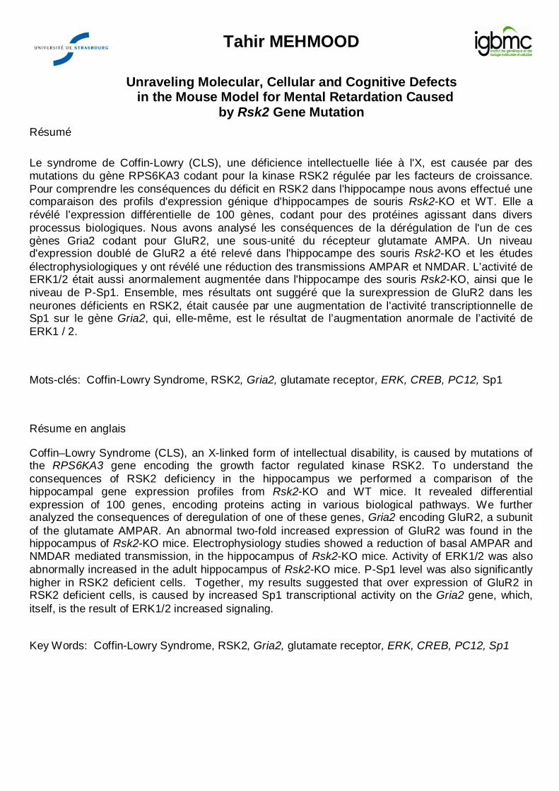
Tahir MEHMOOD
Unraveling Molecular, Cellular and Cognitive Defects in the Mouse Model for Mental Retardation Caused
by Rsk2 Gene Mutation
Résumé
Le syndrome de Coffin-Lowry (CLS), une déficience intellectuelle liée à l'X, est causée par des mutations du gène RPS6KA3 codant pour la kinase RSK2 régulée par les facteurs de croissance. Pour comprendre les conséquences du déficit en RSK2 dans l'hippocampe nous avons effectué une comparaison des profils d'expression génique d'hippocampes de souris Rsk2-KO et WT. Elle a révélé l'expression différentielle de 100 gènes, codant pour des protéines agissant dans divers processus biologiques. Nous avons analysé les conséquences de la dérégulation de l'un de ces gènes Gria2 codant pour GluR2, une sous-unité du récepteur glutamate AMPA. Un niveau d'expression doublé de GluR2 a été relevé dans l'hippocampe des souris Rsk2-KO et les études électrophysiologiques y ont révélé une réduction des transmissions AMPAR et NMDAR. L’activité de ERK1/2 était aussi anormalement augmentée dans l'hippocampe des souris Rsk2-KO, ainsi que le niveau de P-Sp1. Ensemble, mes résultats ont suggéré que la surexpression de GluR2 dans les neurones déficients en RSK2, était causée par une augmentation de l'activité transcriptionnelle de Sp1 sur le gène Gria2, qui, elle-même, est le résultat de l’augmentation anormale de l’activité de ERK1 / 2.
Mots-clés: Coffin-Lowry Syndrome, RSK2, Gria2, glutamate receptor, ERK, CREB, PC12, Sp1
Résume en anglais
Coffin–Lowry Syndrome (CLS), an X-linked form of intellectual disability, is caused by mutations of the RPS6KA3 gene encoding the growth factor regulated kinase RSK2. To understand the consequences of RSK2 deficiency in the hippocampus we performed a comparison of the hippocampal gene expression profiles from Rsk2-KO and WT mice. It revealed differential expression of 100 genes, encoding proteins acting in various biological pathways. We further analyzed the consequences of deregulation of one of these genes, Gria2 encoding GluR2, a subunit of the glutamate AMPAR. An abnormal two-fold increased expression of GluR2 was found in the hippocampus of Rsk2-KO mice. Electrophysiology studies showed a reduction of basal AMPAR and NMDAR mediated transmission, in the hippocampus of Rsk2-KO mice. Activity of ERK1/2 was also abnormally increased in the adult hippocampus of Rsk2-KO mice. P-Sp1 level was also significantly higher in RSK2 deficient cells. Together, my results suggested that over expression of GluR2 in RSK2 deficient cells, is caused by increased Sp1 transcriptional activity on the Gria2 gene, which, itself, is the result of ERK1/2 increased signaling.
Key Words: Coffin-Lowry Syndrome, RSK2, Gria2, glutamate receptor, ERK, CREB, PC12, Sp1