Clinical treatment options for patients with homonymous visual field defects
10
© 2008 Lane et al, publisher and licensee Dove Medical Press Ltd. This is an Open Access article which permits unrestricted noncommercial use, provided the original work is properly cited. Clinical Ophthalmology 2008:2(1) 93–102 93 REVIEW Clinical treatment options for patients with homonymous visual field defects Alison R Lane Daniel T Smith Thomas Schenk Cognitive Neuroscience Research Unit, Durham University, Durham, UK Correspondence: Alison Lane CNRU, Wolfson Research Institute, Durham University, Queens Campus, Stockton-on-Tees, TS17 6BH, UK Tel +44 191 3340431 Fax +44 191 3340006 Email [email protected] Abstract: The objective of this review is to evaluate treatments for homonymous visual field defects (HVFDs). We distinguish between three treatments: visual restoration training (VRT), optical aids, and compensatory training. VRT is both the most ambitious and controversial approach, aiming to restore portions of the lost visual field. While early studies suggested that VRT can reduce the visual field defect, recent studies using more reliable means of monitor- ing the patients’ fixation could not confirm this effect. Studies utilizing modern optical aids have reported some promising results, but the extent to which these aids can reliably reduce the patients’ visual disability has yet to be confirmed. Compensatory approaches, which teach patients more effective ways of using their eyes, are currently the only form of treatment for which behavioral improvements have been demonstrated. However, with the exception of one study using a reading training, placebo-controlled clinical evaluation studies are lacking. It is also not yet clear whether the training benefits found in laboratory tasks lead to reliable improvements in activities of daily living and which of the various forms of compensatory training is the most promising. It is therefore too early to recommend any of the currently available treatment approaches. Keywords: homonymous hemianopia, rehabilitation, treatment outcome Introduction The visual field is the entire space in which visual stimuli can be perceived when the eyes are fixating. If someone has a visual field defect this means that they have lost the ability to see visual stimuli which are presented in one part of the visual field. With homonymous visual field defects (HVFDs), the same part of the visual field is affected for both the right and left eye. Approximately 20% of people with acquired brain injury may develop a HVFD (Kasten et al 1999), which occurs following dam- age to the neural visual pathway, specifically damage posterior to the optic chiasm. Homonymous hemianopia (see Figure 1), in which one half of the visual field is blind, occurs in approximately 75% of cases (Zihl 1995a). HVFDs can be very disabling. They impair the patient’s ability to obtain a complete visual overview (Zihl 1995a), which can impact on their ability to interact with their environment. In addition, patients can experience further impairment in their daily life; for example, driving is prohibited in the majority of cases (Kooijman et al 2004) and HVFDs can lead to severe reading difficulties (Leff et al 2001). Such difficulties arising from visual problems can have additional effects on the social and emotional functioning of the individual. Traditionally HVFDs were considered untreatable, but recent advances in our understanding of the neural capacities for functional reorganization has led to an upsurge of attempts to achieve a reduction of the field loss through training. At the same time new rehabilitation procedures have been studied which might allow patients to more efficiently perceive the whole visual world and to improve reading
-
Upload
lmu-munich -
Category
Documents
-
view
2 -
download
0
Transcript of Clinical treatment options for patients with homonymous visual field defects
© 2008 Lane et al, publisher and licensee Dove Medical Press Ltd. This is an Open Access article which permits unrestricted noncommercial use, provided the original work is properly cited.
Clinical Ophthalmology 2008:2(1) 93–102 93
R E V I E W
Clinical treatment options for patients with homonymous visual fi eld defects
Alison R LaneDaniel T SmithThomas Schenk
Cognitive Neuroscience Research Unit, Durham University, Durham, UK
Correspondence: Alison LaneCNRU, Wolfson Research Institute, Durham University, Queens Campus, Stockton-on-Tees, TS17 6BH, UKTel +44 191 3340431Fax +44 191 3340006Email [email protected]
Abstract: The objective of this review is to evaluate treatments for homonymous visual fi eld
defects (HVFDs). We distinguish between three treatments: visual restoration training (VRT),
optical aids, and compensatory training. VRT is both the most ambitious and controversial
approach, aiming to restore portions of the lost visual fi eld. While early studies suggested that
VRT can reduce the visual fi eld defect, recent studies using more reliable means of monitor-
ing the patients’ fi xation could not confi rm this effect. Studies utilizing modern optical aids
have reported some promising results, but the extent to which these aids can reliably reduce
the patients’ visual disability has yet to be confi rmed. Compensatory approaches, which teach
patients more effective ways of using their eyes, are currently the only form of treatment for
which behavioral improvements have been demonstrated. However, with the exception of
one study using a reading training, placebo-controlled clinical evaluation studies are lacking.
It is also not yet clear whether the training benefi ts found in laboratory tasks lead to reliable
improvements in activities of daily living and which of the various forms of compensatory
training is the most promising. It is therefore too early to recommend any of the currently
available treatment approaches.
Keywords: homonymous hemianopia, rehabilitation, treatment outcome
IntroductionThe visual fi eld is the entire space in which visual stimuli can be perceived when the
eyes are fi xating. If someone has a visual fi eld defect this means that they have lost
the ability to see visual stimuli which are presented in one part of the visual fi eld.
With homonymous visual fi eld defects (HVFDs), the same part of the visual fi eld is
affected for both the right and left eye. Approximately 20% of people with acquired
brain injury may develop a HVFD (Kasten et al 1999), which occurs following dam-
age to the neural visual pathway, specifi cally damage posterior to the optic chiasm.
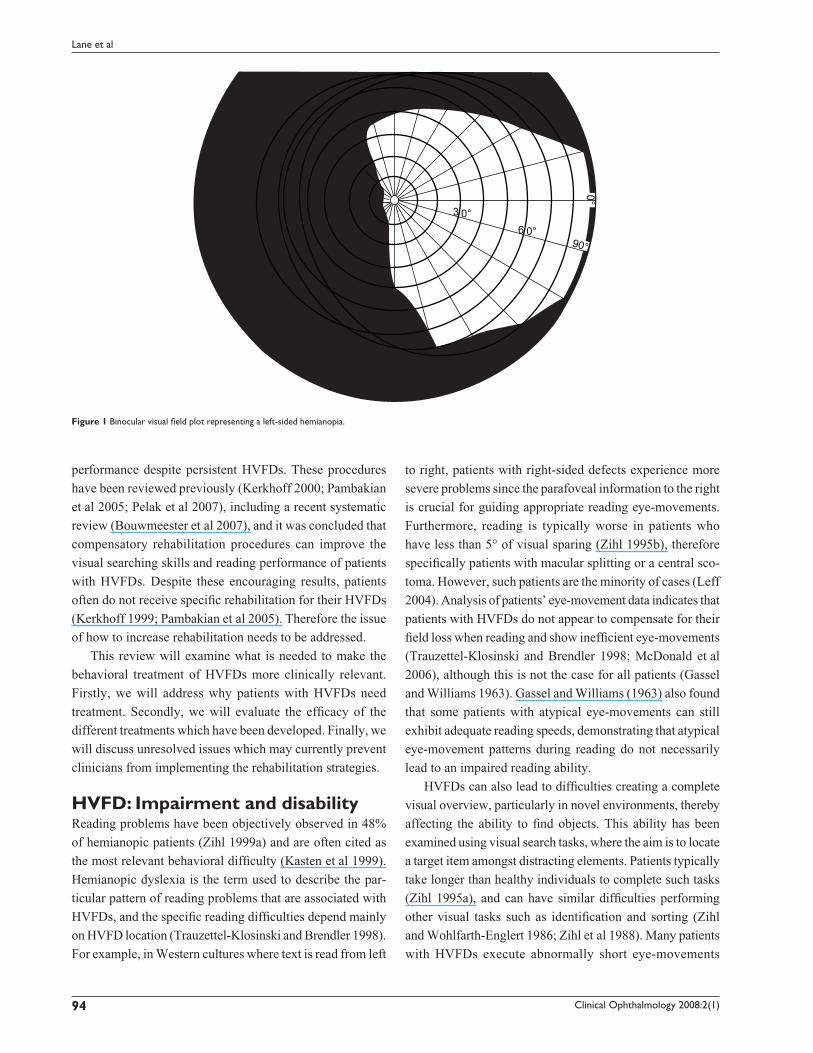
Homonymous hemianopia (see Figure 1), in which one half of the visual fi eld is blind,
occurs in approximately 75% of cases (Zihl 1995a).
HVFDs can be very disabling. They impair the patient’s ability to obtain a complete
visual overview (Zihl 1995a), which can impact on their ability to interact with their
environment. In addition, patients can experience further impairment in their daily
life; for example, driving is prohibited in the majority of cases (Kooijman et al 2004)
and HVFDs can lead to severe reading diffi culties (Leff et al 2001). Such diffi culties
arising from visual problems can have additional effects on the social and emotional
functioning of the individual.
Traditionally HVFDs were considered untreatable, but recent advances in our
understanding of the neural capacities for functional reorganization has led to an
upsurge of attempts to achieve a reduction of the fi eld loss through training. At
the same time new rehabilitation procedures have been studied which might allow
patients to more effi ciently perceive the whole visual world and to improve reading
Clinical Ophthalmology 2008:2(1)94
Lane et al
performance despite persistent HVFDs. These procedures
have been reviewed previously (Kerkhoff 2000; Pambakian
et al 2005; Pelak et al 2007), including a recent systematic
review (Bouwmeester et al 2007), and it was concluded that
compensatory rehabilitation procedures can improve the
visual searching skills and reading performance of patients
with HVFDs. Despite these encouraging results, patients
often do not receive specifi c rehabilitation for their HVFDs
(Kerkhoff 1999; Pambakian et al 2005). Therefore the issue
of how to increase rehabilitation needs to be addressed.
This review will examine what is needed to make the
behavioral treatment of HVFDs more clinically relevant.
Firstly, we will address why patients with HVFDs need
treatment. Secondly, we will evaluate the effi cacy of the
different treatments which have been developed. Finally, we
will discuss unresolved issues which may currently prevent
clinicians from implementing the rehabilitation strategies.
HVFD: Impairment and disabilityReading problems have been objectively observed in 48%
of hemianopic patients (Zihl 1999a) and are often cited as
the most relevant behavioral diffi culty (Kasten et al 1999).
Hemianopic dyslexia is the term used to describe the par-
ticular pattern of reading problems that are associated with
HVFDs, and the specifi c reading diffi culties depend mainly
on HVFD location (Trauzettel-Klosinski and Brendler 1998).
For example, in Western cultures where text is read from left
to right, patients with right-sided defects experience more
severe problems since the parafoveal information to the right
is crucial for guiding appropriate reading eye-movements.
Furthermore, reading is typically worse in patients who
have less than 5° of visual sparing (Zihl 1995b), therefore
specifi cally patients with macular splitting or a central sco-
toma. However, such patients are the minority of cases (Leff
2004). Analysis of patients’ eye-movement data indicates that
patients with HVFDs do not appear to compensate for their
fi eld loss when reading and show ineffi cient eye-movements
(Trauzettel-Klosinski and Brendler 1998; McDonald et al
2006), although this is not the case for all patients (Gassel
and Williams 1963). Gassel and Williams (1963) also found
that some patients with atypical eye-movements can still
exhibit adequate reading speeds, demonstrating that atypical
eye-movement patterns during reading do not necessarily
lead to an impaired reading ability.
HVFDs can also lead to diffi culties creating a complete
visual overview, particularly in novel environments, thereby
affecting the ability to fi nd objects. This ability has been
examined using visual search tasks, where the aim is to locate
a target item amongst distracting elements. Patients typically
take longer than healthy individuals to complete such tasks
(Zihl 1995a), and can have similar diffi culties performing
other visual tasks such as identifi cation and sorting (Zihl
and Wohlfarth-Englert 1986; Zihl et al 1988). Many patients
with HVFDs execute abnormally short eye-movements
3 0°6 0°
90°
0°
Figure 1 Binocular visual fi eld plot representing a left-sided hemianopia.
Clinical Ophthalmology 2008:2(1) 95
The treatment of HVFDs
when looking into their blind regions and each movement is
typically slower than that of a normally-sighted individual.
This strategy is known as saccadic hypometria (Zihl 2000) and
limits the patients’ ability to effectively search their environ-
ment, which contributes to their disorientation and obstacle
avoidance problems. Approximately 70% of patients show
such disorganized searching strategies (Kerkhoff 1999).
Due to the enjoyment which can be gained from reading
and other leisure activities requiring visual search skills,
any impairment in these has obvious consequences for
the emotional well-being of the patient (Stelmack 2001).
Similarly, HVFDs can restrict many other activities such as
driving, which can lead to a loss of independence, thereby
affecting social and emotional functioning. The primary
cause of HVFD is stroke (Huber 1992) and accordingly
most patients are elderly (Cairns 2004). Therefore, visual
problems increase the risk of accidents such as falls, to which
this age-group are already prone (Anderson 2002). HVFDs
also reduce the effi cacy of other rehabilitation procedures
which may be aimed at increasing the patient’s mobility (for
example physiotherapy). Bearing these factors in mind many
patients have low scores in activities of daily living (ADL)
measures (Patel et al 2000; Sànchez-Blanco et al 2002), and
functional rehabilitation outcomes are poor (Reding and
Potes 1988).
Spontaneous recoverySome spontaneous visual fi eld restoration is widely accepted
to occur. However, the number of patients who experience
any restoration is undetermined, with reports ranging from
7% to 85% of cases (Kasten et al 1999). The amount of
fi eld recovery that an individual can experience is similarly
variable (Zihl and Kennard 1996). In an extensive review
of spontaneous recovery, Zhang and colleagues (2006)
observed that the degree of natural recovery decreased as
the amount of time since the onset of the HVFD increased.
The approximate maximal period of spontaneous recovery
is typically 3 months (Pambakian and Kennard 1997). In
summary, spontaneous visual fi eld recovery does not occur
in all patients and complete recovery is rare, therefore reha-
bilitation for such patients is important. Knowing the likely
pattern of natural recovery is important for assessment and
rehabilitation, and is useful for determining the time at which
training will be maximally effective.
Patients may also try to adapt to their visual loss. The
obvious way of compensating for HVFDs is to make larger
and more frequent eye-movements, specifi cally into the
blind areas. Unfortunately not all patients adopt this strategy
(Kerkhoff 1999). Rather, several researchers have shown
that the eye-movements of many HVFD patients are very
small and their scan-paths (the pattern of eye-movements
used to scan a complex visual representation) are disorga-
nized (Meienberg et al 1981; Zihl 1995a, 1999b; Pambakian
et al 2000). Patients who have a chronic HVFD show more
organized scanning strategies than those patients whose
diffi culties are a recent occurrence (Zihl 1995a; Pambakian
et al 2000). However, many patients’ eye-movements are
still disorganized 14 months after onset (Kerkhoff 1999).
These abnormal eye-movements can also be observed during
reading (Zihl 1995b).
Patients may also use other forms of behavioral compen-
sation. One method is the use of eccentric fi xation; the eye
is rotated slightly towards the blind hemi-fi eld rather than
straight ahead (Gassel and Williams 1963). This means that
the centre of the observed image does not fall exactly on
the fovea, but instead the image falls further into the seeing
fi eld at a slightly eccentric position. This strategy is found in
approximately 30% of cases and can increase reading speed
(Trauzettel-Klosinski 1997), although the impact of such a
strategy on other activities has not been clearly determined.
It is a strategy which may be of use for patients with little
central vision, for example those with macular splitting or a
central scotoma, which as already mentioned is the minority
of patients (Leff 2004). However, eccentric fi xation is not
a useful strategy for patients with an intact fovea. Another
strategy witnessed in children involves to-and-fro rocking
motions, to bring greater portions of the visual fi eld into view
(Boyle et al 2005).
In summary, HVFDs are debilitating and the sensory loss
is exacerbated by the adoption of slow and ineffi cient search
strategies when exploring the blind fi eld. The prognosis for
spontaneous recovery appears poor, and although adaptation
is possible many patients do not develop effective ways of
compensating for their defi cits. Specifi c interventions are
required for these patients.
InterventionMost research has focused on three treatment approaches:
restorative training, optical aids, and compensatory train-
ing. The restorative approach is the most ambitious, aiming
to reduce the visual fi eld loss through prolonged training.
The second approach uses optical aids to artifi cially expand
the patient’s visual fi eld such that parts of the visual world
which would otherwise fall into the blind fi eld now appear
in the seeing fi eld. The third approach is compensatory
training. This therapy is based on the assumption that the
Clinical Ophthalmology 2008:2(1)96
Lane et al
visual fi eld defect cannot be changed signifi cantly, and
therefore attempts to alleviate the resulting disability by
teaching patients to make more effi cient eye-movements.
We will discuss each of these three rehabilitation strategies
in separate sections.
Restorative trainingRestorative training aims to restore vision (at least in part)
to the blind visual fi eld, based on evidence which supports
plasticity in the visual system of both animals (Cowey and
Weiskrantz 1963; Cowey 1967; Mohler and Wurtz 1977;
Eysel and Schwiegart 1999) and humans (Donoghue 1997).
Whilst there was early promise for the potential of such
training (Zihl and Von Cramon 1979; Zihl and Von Cramon
1985), some dismissed the approach as ineffective, with any
supposed fi eld increase being regarded as the product of eye-
movements (Balliet et al 1985).
Restorative training was later revived by Kasten, Sabel,
and colleagues who introduced a computerized therapy
called Vision Restoration Therapy (VRT; Kasten and Sabel
1995; Kasten et al 1997). During VRT patients fi xate a
central point whilst visual stimuli are repeatedly presented
in the border region between the blind and seeing fi eld (the
transition zone). The training is typically conducted in daily
one-hour sessions for 6 months. Placebo-controlled studies
have suggested that VRT leads to signifi cant increases in
the visual fi eld (Kasten et al 1998, 2000), although more so
for patients with optic nerve as opposed to cortical damage.
These fi eld increases were still observed at least 6 months
after the end of the treatment (Kasten and Sabel 1995; Kasten
et al 2001).
However, despite the apparent success of VRT critics
have challenged the claim that it is an effective treatment
(Horton 2005; Plant 2005; McFadzean 2006). This is because
the claims of signifi cant fi eld increases found in the VRT
studies mentioned above were based on a method of assess-
ing the visual fi eld which was incorporated into the training
device and did not allow a reliable way of controlling the
patients’ fi xation. The effect of VRT has been re-evaluated
using techniques which allowed a much more reliable means
of monitoring the patients’ fi xation, specifi cally the use of
a scanning laser ophthalmoscope (SLO; Jamara et al 2003)
combined with microperimetry. In studies whereby they
reliably monitored fi xation during visual fi eld assessment no
signifi cant visual fi eld increases were obtained (Sabel et al
2004; Reinhard et al 2005; Schreiber et al 2006).
Recently Kasten and colleagues (2006) responded to
this challenge by publishing a study in which they used a
video-based system to monitor their patients’ fi xation and
found a signifi cant (but somewhat modest) fi eld increase of
1.8 degrees. This fi nding is insuffi cient to re-establish the
therapeutic value of VRT for two reasons. Firstly, because
the employed fi xation-control technique is inferior to that
used by other studies which did not fi nd signifi cant fi eld
increases. Secondly, even if we accept the reported fi eld
increase, such an increase of 1.8° is clearly not enough to
convey a clinically signifi cant benefi t to patients who have
endured 3 months of daily training and invested a consider-
able amount of money. In this context it is important to note
that these expenses are currently not covered by medical
insurance companies (Pelak et al 2007).
Even if the claim that VRT produces a clinically-relevant
field increase is dismissed, the widespread subjective
improvements reported by many participants of this train-
ing cannot be disregarded (Mueller et al 2003; Sabel et al
2004). The fact that improvements are sometimes reported by
patients in the absence of any training-induced fi eld recovery
(Mueller et al 2003) raises an interesting question: why do
patients report that subsequent to the training they fi nd it
easier to fi nd objects and avoid obstacles, if their visual fi eld
is unchanged? It might be that these reports simply refl ect
the patients’ desire to justify a training in which they have
invested a lot of time, effort, and money. However, it is also
possible that VRT leads to behavioral improvements. For
example, it is possible that VRT cues patients to allocate more
attention into their blind fi eld, and such attentional cueing
can improve target detection (Poggel et al 2006). Although
visual costimulation was found to be no more effective than
single stimulation VRT at expanding the visual fi eld, it is
clear that VRT has benefi cial effects on attentional perfor-
mance (Kasten et al 2007). Further research into the role of
attention for visual fi eld rehabilitation is required.
In addition to possible effects on attention, VRT could
also inadvertently lead to eye-movements being more fre-
quently directed into the blind fi eld, although this explana-
tion is denied by Kasten and colleagues (2006). However,
it could explain why restorative training leads to changes
in cortical activity as reported by several small-sample (n =
1–5) imaging and electrophysiology studies (Julkunen et al
2003, 2006; Pleger et al 2003), since saccadic changes due
to compensatory mechanisms could infl uence widespread
neural activity. Initially these changes in cortical activity
were interpreted as evidence for the training-induced brain
plasticity underlying the recovery of visual fi eld loss. How-
ever, in the absence of reliable evidence for such visual fi eld
recovery this interpretation appears unlikely.
Clinical Ophthalmology 2008:2(1) 97
The treatment of HVFDs
More encouraging results have been obtained with
children. Werth and colleagues presented fi ndings from ran-
domized placebo-controlled trials with a restorative training
used with children aged between 1 and 15 years (Werth and
Moehrenschlager 1999; Werth and Seelos 2005). In some
cases the visual fi eld defect disappeared completely, and
the mean increase was 65 degrees. Such dramatic increases
coincide with evidence suggesting that there is greater
potential for recovery from damage sustained early in life
(Payne et al 1996; Boyle et al 2005), possibly because child
and adult cases typically differ with regards to etiology and
lesion location (Kedar et al 2006), or perhaps due to greater
neuronal plasticity more generally in children. However, due
to the age of these patients conventional perimetry could
not be performed and instead the researchers had to rely
on observed changes in target-directed eye-movements to
estimate the extent of visual fi eld recovery. Accordingly the
visual fi eld measurements and reported fi eld increases are dif-
fi cult to interpret and could refl ect compensatory mechanisms
rather than restorative ones. Given the large improvements
which appear possible, this training would appear worthwhile
pursuing with children who have HVFDs.
In conclusion, restorative training in adults has failed to
fulfi ll its early promise. Recent studies suggest that VRT does
not lead to signifi cant increases in visual fi eld size but con-
sistently yields subjective improvements. The basis of these
subjective improvements is still unclear but it is possible that
VRT leads to compensatory changes in behavior which are
as yet unconfi rmed. However, even if we were to assume that
VRT leads to signifi cant behavioral improvements it would
still be inferior to other forms of compensatory training (see
below) which appear to produce effective behavioral com-
pensation with signifi cantly less effort, cost and time.
Optical aidsOptical aids such as prism glasses can be used to reduce the
apparent visual fi eld loss by shifting visual stimuli from the
blind fi eld into the patient’s seeing fi eld. These prisms are
fi tted to spectacles but need to be restricted to just one half
of each of the lenses (typically on the side of the blind fi eld).
If the prisms were fi tted across the entire lens then the visual
space corresponding to the unaffected side would be moved
outside the fi eld of view, thereby simply replacing one blind
fi eld with another. Such prisms can be fi tted to just one eye
(monocular sector prisms) or both eyes (binocular sector
prisms). Whilst such aids appear to enhance visual function-
ing (Gottlieb et al 1998; Lee and Perez 1999; Szlyk et al
2005) they have their limitations. Monocular prisms provide
an expansion of the visual fi eld but at the cost of creating
central double-vision (diplopia) which patients experience as
unpleasant. Binocular prisms lead to fi eld relocation rather
than fi eld expansion. Such problems probably explain why
so far these optical aids proved only moderately successful
in HVFD rehabilitation.
Peli (2000; 2001) has introduced a new set of spectacles
with monocularly fi tted sector prisms which extend across the
entire width of the spectacle lens but which spare the central
aspect. This is known as vision multiplexing and using this
technique fi eld expansion is achieved without central dip-
lopia. Peli (2000) reported fi eld expansion of about 20° and
noted on the basis of subjective reports that patients seemed
to benefi t from the spectacles. Vision multiplexing seems
promising but randomized controlled trials using objective
measures of functional improvement are required to evaluate
the clinical potential of such a technique.
Compensatory trainingEven if training cannot achieve a signifi cant reduction in the
visual fi eld loss, it might still be possible to help patients to
cope more effectively with their HVFD. Scanning the visual
world systematically with large sweeping eye-movements
would appear to be the most obvious form of compensation,
however many patients do not spontaneously adopt this strat-
egy (Kerkhoff 1999). In order to improve patients’ ability
to compensate for their visual loss, several researchers have
developed training schemes designed to teach patients more
effi cient strategies for visual scanning.
The compensatory training approaches typically use
target-localization tasks to train patients to make large eye-
movements and use visual search tasks to teach patients
to use systematic scanning strategies when searching their
visual world. Sometimes additional training is included
which helps the patient to utilize the strategies in everyday
situations such as crossing the street or fi nding objects around
the home (Kerkhoff et al 1992, 1994; Pambakian et al 2004).
With compensation training patients usually receive daily one
hour training sessions for about four weeks making it much
less demanding than VRT.
Compensatory training in general leads to improved
search performance and effi ciency (Kerkhoff et al 1992,
1994; Zihl 1995a; Nelles et al 2001; Pambakian et al 2004;
Verlohr and Dannheim 2007). Two studies confi rmed that
the improvements obtained after training were signifi cantly
greater than those observed during untrained periods and
are thus training specifi c (Kerkoff et al 1994; Pambakian
et al 2004). For example, Pambakian and colleagues (2004)
Clinical Ophthalmology 2008:2(1)98
Lane et al
found that 76% of patients had faster search times following
training, while 14% remained unchanged, and 10% were
actually slower. Those with slower search times showed an
improvement in their detection rates. Compensatory training
has also been found to signifi cantly enlarge the search fi eld
(Kerkhoff et al 1992, 1994; Pambakian et al 2004). Addi-
tionally, studies using eye-tracking show that compensatory
training leads to better organized scanning strategies and
larger saccades (Zihl 1995a). Furthermore, it was found that
the post-training search improvements could be maintained
for at least one month, and in some cases up to 22 months
(Kerkhoff et al 1992).
Not only does it appear from these studies that com-
pensatory training can signifi cantly improve search but it is
possible that it may actually increase the visual fi eld itself
(Kerkhoff et al 1992, 1994). However, not all studies have
found signifi cant visual fi eld increases after compensatory
training (Zihl 1995; Nelles et al 2001; Pambakian et al 2004).
Caution is required when interpreting these fi ndings as the
same limitations relating to fi xation control during perimetry
are present as in studies examining restorative approaches.
The reading performance of patients with HVFDs is also
often impaired (Leff et al 2001). Consequently, specifi c com-
pensatory reading training procedures have been developed
to directly address this defi cit (Zihl 1995b; Han et al 2004;
Spitzyna et al 2007). Zihl (1995b) showed that such training
can improve reading accuracy and speed although unfortu-
nately there was no control group and so it is not known to
what extent the effects were due to the training provided. In
contrast, a recent study by Spitzyna and colleauges (2007)
included a placebo training to assess the specifi c effects of a
reading training for patients with hemianopic dyslexia. They
tested 19 patients with right-sided hemianopia and divided
them into two groups. Group 1 received a reading training for
two 4-week blocks. During this training, patients practiced
reading moving text which scrolled from right to left. In group
2 patients received a 4-week block of placebo training and
a 4-week block of reading training. In the placebo-training
patients received pairs of pictures which differed only in a
number of minor features and they had to detect these dif-
ferences. The reading training but not the placebo training
induced signifi cant improvements in reading speed.
The results surrounding compensatory training indicate
the promise which such an approach holds for helping
patients to adapt to their visual loss, and whilst controversy
continues to surround the use of VRT, compensation would
appear to be a viable rehabilitation option for the patients
with HVFDs. However, it is important to note that for most
compensatory training regimes a placebo-controlled study
examining their effi cacy is still missing.
Unresolved issues of HVFD rehabilitationAs described above the lack of placebo-controlled evaluation
trials (for an exception see Spitzyna et al 2007) means that
the clinical effi cacy has not yet been established for any of
the described rehabilitation procedures. It is thus too early
to give a fi rm recommendation for any of these approaches.
However, the compensatory training approach is the only
one for which behavioral improvements in the form of
improved search times, increased reading speed and larger
eye-movements have been demonstrated. In fact, for one
form of compensatory reading training its superiority over a
placebo-training has already been established (Spitzyna et al
2007). In contrast the same is not true for either restorative
training or optical aids. In the case of restorative training the
early claims of increased visual fi eld size following train-
ing have not been confi rmed by studies using more reliable
means of assessing the visual fi eld size and there is currently
no evidence that the reported subjective improvements cor-
respond to measurable behavioral improvements. In the case
of optical aids evidence for behavioral improvements are
also lacking, in particular for their most promising forms,
the vision multiplexing prisms. Thus on the basis of current
evidence the compensatory approach appears to be in our
view the most promising, and we will therefore now turn
our attention to those aspects of the compensatory therapy
which require further research.
Transfer of training benefi ts to activities of daily livingThe fi rst issue relates to the question of whether the achieved
training gains also lead to relevant improvements in activities
of daily living (ADL), an aspect which is crucial to the clinical
evaluation of any rehabilitation procedure. Unfortunately most
studies examining compensatory search training either do not
assess its impact on ADL tasks or rely solely upon subjective
reports. Using questionnaires several researchers have found
that patients do report improvements in such activities as fi nding
objects in a room or on a table, and crossing the street (Kerkhoff
et al 1994; Nelles et al 2001; Pambakian et al 2004), indicating
that compensatory training produces functional improvements.
It is however important to establish the functional benefi ts of
the training with behavioral measures since subjective gains
can be unreliable indicators of rehabilitation success.
Clinical Ophthalmology 2008:2(1) 99
The treatment of HVFDs
There are a few studies which have used objective
measures to assess the transfer of training benefi ts to ADL
tasks. Kerkhoff and colleagues (1994) demonstrated that
combined compensatory search training (ie, training which
combines search tasks and exercises such as fi nding objects
around the home) can yield improved search performance in
more naturalistic forms of visual search (eg, searching for an
item amongst distracters on a table) which use a wider fi eld
of view than that used during the training. Following training
the patients in this study showed a 50% reduction in search
time on the table test. Further to this, they also reported that
91% of the sample returned to some sort of part-time work
after the training, indicating a positive functional outcome.
Pambakian and colleagues (2004) reported training-related
improvements in performance on activities representing ADL
tasks, such as threading beads onto string. Whilst this does
indicate the successful transfer to the visuomotor domain of
the training benefi ts, there is a question of how much such
tasks actually tell us about improvements in more common-
place everyday activities. Future work should examine more
closely the effi cacy of the training in relation to more relevant
examples of ADL.
Driving is a major activity for which transfer of train-
ing gains would be benefi cial since it is prohibited for the
majority of patients with HVFDs. Unfortunately Kooijman
and colleagues (2004) found that only 2 out of the 17 hom-
onymous hemianopic patients who failed a test of practical
driving fi tness passed this test after a form of compensatory
training. Given the social and emotional impact that the loss
of driving has on patients with HVFDs the effect that com-
pensatory training has on driving ability should be further
examined. It is worth noting that patients with HVFDs may
be able to drive as adequately as normal, healthy individuals
(Schulte et al 1999) and therefore perhaps driving guidelines
should be modifi ed such that an HVFD is not an automatic
cause for license revocation.
Nelles and colleagues (2001) reported that patients’
subjective impression of their reading ability had improved
after the compensatory search training. However, since read-
ing performance has not been measured it is not possible to
conclude if there is transfer from search training to reading
tasks. Again reading is an activity which many patients report
having diffi culties with and which can severely impact on
their quality of life, and as such should be considered an
important outcome measure in future research. If it is found
that general compensatory training can benefi t reading (or
that reading training can benefi t other everyday activities)
then only one type of training would be required.
Currently there is insuffi cient information about how the
gains achieved with compensatory training transfer to other
relevant activities like driving, reading, visuomotor control,
and visual searching in natural surroundings. Furthermore,
it is currently unclear whether some ADL tasks benefi t more
from training than others. If it is confi rmed that some ADL
tasks do not benefi t suffi ciently from compensatory training
(for example reading), then training which addresses the
specifi c requirements of those tasks will be needed.
Predictors of good training outcomeNot all patients benefi t from compensatory training. In order
to maximize its effi cacy it is necessary to identify factors that
contribute to the success or failure of this training. Conven-
tional predictors of rehabilitation outcome, which include
the cause of the HVFD (etiology) and the time since onset of
the HVFD when training takes place have been examined.
Etiology does not seem to have a signifi cant effect on outcome
(Kerkhoff et al 1992, 1994), although this is hardly surpris-
ing given that most patients share the same etiology, namely
stroke. With respect to the timing of training, the results are
mixed as some studies suggest that earlier training is more
benefi cial (Zihl 1995a), whilst others fail to fi nd such an asso-
ciation (Kerkhoff et al 1992, 1994). Several studies have failed
to fi nd any association between age and training outcome
(Kerkhoff et al 1992, 1994; Zihl 1995a). Kerkhoff and col-
leagues (1992) found that those patients who initially had the
severest visual problems were those who showed the largest
improvements after the training, thus pre-training impairment
level could predict the possible success of the training.
A very plausible predictor for good training outcome is
the degree of spared visual ability in the blind fi eld. Some
patients can respond quite accurately to visual stimuli pre-
sented to their blind fi eld (for example by pointing to it) even
though they insist that they cannot see it. This phenomenon
has been called blindsight (Weiskrantz et al 1974) and it is
estimated that 15%–20% of HVFD patients show this (Blythe
et al 1987). It has been suggested that training patients to be
aware of their blindsight capacity could be a useful reha-
bilitation strategy (Boyle et al 2005) and repeatedly testing
blindsight can lead to improved blindsight performance (Zihl
1980). It seems plausible that patients with blindsight are
more successful in making accurate saccadic eye-movements
to targets in their blind fi eld and could therefore benefi t more
from compensatory training. However, this prediction has
not yet been tested.
Cognitive variables, such as the patient’s ability to allo-
cate visual attention or their spatial memory capacity, which
Clinical Ophthalmology 2008:2(1)100
Lane et al
might contribute to the success or failure of compensatory
training, have not yet been examined. In summary, it seems
clear that although compensatory treatments benefi t the
majority of participants, the factors which predict successful
treatment outcome on an individual basis remain unclear.
ComorbiditiesSome previous studies examining compensatory training
have excluded people with HVFDs who suffered from addi-
tional disorders such as oculomotor defi cits or hemispatial
neglect. Disorder comorbidities are common (Anderson and
Rizzo 1995) and patients with multiple diffi culties are typi-
cally more functionally impaired than those with a single
defect (Patel et al 2000). The impact that this may have on
rehabilitation has not been fully established.
Hemispatial neglect is a disorder which typically occurs
after right-hemispheric damage and leads to patients ignor-
ing sensory information from the contralesional half of their
body or surroundings. It might be predicted that patients with
such additional disorders will gain less from compensatory
training. However, some authors have argued that patients
with both HVFD and hemispatial neglect can benefi t from the
training but require a more intensive schedule (Kerkhoff et al
1992, 1994). Similarly it has been shown in a case-study that
a patient with multiple visual problems such as amblyopia
and impaired form vision could still benefi t from compensa-
tory training (Hiramaya et al 2004).
In summary, HVFD patients with comorbidities such as
hemispatial neglect may require more intensive training, but
may still benefi t from compensatory training. Having said
that, the current evidence is scant and rigorous studies com-
paring the training benefi ts in large samples of patients with
and without such co-morbidities have yet to be conducted.
Parameters of effective compensatory trainingAlthough it has been shown that compensatory training in
general can lead to signifi cant functional gains (Pambakian
et al 2004), a number of different training regimens have
been used and it is unclear which is the most effective. These
regimens differ with respect to the required effort and cost,
and it is important to establish whether the simpler and less
costly forms are as effective as the more laborious ones. For
example, training displays of various size have been used
ranging from computer or television monitors, which train
only the central 25 degrees of the visual fi eld (Pambakian
et al 2004), to displays which fi ll the entire visual fi eld (Nelles
et al 2001). It is obvious that training on a small screen is
less costly because it means that the training can potentially
be performed by the patient in their own home, as was done
in the study by Pambakian and colleagues (2004), and as is
the case with VRT (Kasten and Sabel 1995).
Treatment duration also differs signifi cantly, ranging
between 12 and 60 sessions (Kerkhoff et al 1992). In several
studies (Kerkhoff et al 1992, 1994; Zihl 1995a) patients
receive training until their performance plateaus or their
search fi eld increases by a specifi ed amount. However in
the other studies patients all receive a standardized amount
of training. Research should attempt to determine what the
maximal amount of training required is, and possible fac-
tors that may infl uence the amount of training required by a
specifi c individual. This will ensure that time and resources
are utilized to their best advantage.
Different types of training programs have not been com-
pared directly and so we do not know yet whether the differ-
ent regimens are equally effective. A confounding problem
is that different outcome measures are also used in many of
the studies making it diffi cult to compare them. Standardizing
the outcome measures may aid this process, and Verlohr and
Dannheim (2007) recently proposed the visual performance
test as a standardized outcome measure for the purpose of
assessing search times. This task involves patients having to
visually locate as quickly as possible a series of targets which
can be at one of eleven positions on a screen. Reaction time
is the main outcome measure.
Another version of compensatory search training combined
auditory cues with visual search displays and reported signifi -
cant improvements in exploratory eye-movements and transfer
to ADL (Bolognini et al 2005). However, it is not known
whether the achieved gains are superior to those observed
with training using purely visual displays. A direct comparison
between conventional and combined (ie, visual plus auditory
stimulation) training is required to determine if adding auditory
cues increases the effi cacy of compensatory training.
In summary, there are different versions of compensatory
training available, varying specifi cally in relation to the size of
the training stimuli, the duration of the training, and the addi-
tion of attentional aids. Currently it is unclear which version
produces the best clinical outcome. Standardizing outcome
measures will make it easier to directly compare the benefi ts
of different training techniques, which will allow researchers
to develop the maximally effective training paradigm.
ConclusionWith the exception of one form of a compensatory reading
training (Spitzyna et al 2007) clinical effi cacy has not been
Clinical Ophthalmology 2008:2(1) 101
The treatment of HVFDs
unequivocally established for any of the above described
rehabilitation procedures. To establish clinical effi cacy random-
ized placebo-controlled clinical evaluation trials are needed.
Currently it is therefore too early to recommend any of the
described rehabilitation procedures. However, we would like to
argue that compensatory approaches have come further towards
the aim of establishing their clinical effi cacy than either the VRT
approaches or the use of optical aids. In the case of compensa-
tory approaches several studies found signifi cant behavioral
improvements following the training. The same can not be said
for either VRT or optical aids. In both cases their claim of clini-
cal effi cacy currently rests on subjective patient reports and it is
yet unknown whether these subjective reports of improvement
correspond to measurable behavioral improvements. Apart
from the need for placebo-controlled clinical evaluation trials
we have also identifi ed a number of other issues which need to
be addressed by future research. These include the question of
transfer, which is whether or not the compensatory training leads
to improvements in relevant ADL tasks, the issue of outcome
predictors and also which specifi c version of the compensatory
training is the most effective form of treatment.
DisclosureThe researchers are supported by a grant from the Wolfson
Research Institute and A. Lane is supported by a studentship
jointly provided by the ESRC and MRC.
ReferencesAnderson SW. 2002. Visuoperceptual Impairments. In: Eslinger PJ ed.
Neuropsychological Interventions: Clinical Research and Practice. New York: The Guildford Press. pp. 163–81.
Anderson SW, Rizzo M. 1995. Recovery and rehabilitation of visual cortical dysfunction. NeuroRehabilitation, 5:129–40.
Balliet R, Blood KMT, Bach-y-Rita P. 1985. Visual fi eld rehabilitation in the cortically blind? J Neurol Neurosurg Psychiatry, 48:1113–24.
Blythe IM, Kennard C, Ruddock KH. 1987. Residual vision in patients with retrogeniculate lesions of the visual pathways. Brain, 110:887–905.
Bolognini N, Rasi F, Coccia M, et al. 2005. Visual search improve-ment in hemianopic patients after audio-visual stimulation. Brain, 128:2830–42.
Bouwmeester L, Heutink J, Lucas C. 2007. The effect of visual training for patients with visual fi eld defects due to brain damage: a systematic review. J Neurol Neurosurg Psychiatry, 78:555–64.
Boyle NJ, Jones DH, Hamilton R, et al. 2005. Blindsight in children: does it exist and can it be used to help the child? Observations on a case series. Dev Med Child Neurol, 47:699–702.
Cairns NJ. 2004. Neuroanatomy and Neuropathology. In: Goldstein LH, McNeil JE eds. Clinical Neuropsychology: A Practical Guide to Assessment and Management for Clinicians. Chichester: John Wiley and Sons Ltd. pp. 23–56.
Cowey A. 1967. Perimetric study of fi eld defects in monkeys after cortical and retinal ablations. Q J Exp Psychol, 19:232–45.
Cowey A, Weiskrantz L. 1963. A perimetric study of visual fi eld defects in monkeys. Q J Exp Psychol, 15:90–115.
Donoghue JP. 1997. Limits of reorganization in cortical circuits. Cereb Cortex, 7:97–9.
Eysel UT, Schweigart G. 1999. Increased receptive field size in the surround of chronic lesions in the adult cat visual cortex. Cereb Cortex, 9:101–9.
Gassel MM, Williams D. 1963. Visual function in patients with homony-mous hemianopia. Part II: Oculomotor mechanisms. Brain, 86:1–36.
Gottlieb DD, Fuhr A, Hatch WV, et al. 1998. Neuro-optometric facilitation of vision recovery after acquired brain injury. NeuroRehabilitation, 11:175–99.
Han Y, Ciuffreda KJ, Kapoor N. 2004. Reading-related oculomotor testing and training protocols for acquired brain injury in humans. Brain Res Protocols, 14:1–12.
Hiramaya K, Sakai S, Yamawaki R, et al. 2004. Visual search training for a case of homonymous fi eld defect with multiple visual dysfunctions [abstract]. No To Shinkei, 56:403–13.
Horton JC. 2005. Vision restoration therapy: confounded by eye movements. Br J Ophthalmol, 89:792–4.
Huber A. 1992. Homonymous hemianopia. Neuro-Ophthalmology, 12:351–66.
Jamara RJ, Van De Velde F, Peli E. 2003. Scanning eye movements in hom-onymous hemianopia documented by scanning laser ophthalmoscope retinal perimetry. Optom Vis Sci, 80:495–504.
Julkunen L, Tenovuo O, Jääskeläinen S, et al. 2003. Rehabilitation of chronic post-stroke visual fi eld defect with computer-assisted training. Restor Neurol Neurosci, 21:19–28.
Julkunen L, Tenovuo O, Vorobyev V, et al. 2006. Functional brain imaging, clinical and neurophysiological outcome of visual rehabilitation in a chronic stroke patient. Restor Neurol Neurosci, 24:123–32.
Kasten E, Bunzenthal U, Müller-Oehring E, et al. 2007. Vision restoration therapy does not benefi t from costimulation: A pilot study. J Clin Exp Neuropsychol, 29:569–84.
Kasten E, Bunzenthal U, Sabel BA. 2006. Visual fi eld recovery after vision restoration therapy (VRT) is independent of eye-movements: An eye-tracker study. Behav Brain Res, 175:18–26.
Kasten E, Müller-Oehring E, Sabel BA. 2001. Stability of visual fi eld enlargements following computer-based restitution training – results of a follow-up. J Clin Exp Neuropsychol, 23:297–305.
Kasten E, Poggel DA, Müller-Oehring E, et al. 1999. Restoration of vision II: Residual functions and training-induced visual fi eld enlargement in brain-damaged patients. Restor Neurol Neurosci, 15:273–87.
Kasten E, Poggel DA, Sabel BA. 2000. Computer-based training of stimulus detection improves colour and simple pattern recognition in the defec-tive fi eld of hemianopic subjects. J Cogn Neurosci, 12:1001–12.
Kasten E, Sabel BA. 1995. Visual fi eld enlargement after computer training in brain-damaged patients with homonymous defi cits: an open pilot trial. Restor Neurol Neurosci, 8:113–27.
Kasten E, Strasburger H, Sabel BA. 1997. Programs for diagnosis and therapy of visual field deficits in vision rehabilitation. Spat Vis, 10:499–503.
Kasten E, Wüst S, Behrens-Baumann W, et al. 1998. Computer-based train-ing for the treatment of partial blindness. Nature Med, 4:1083–87.
Kedar S, Zhang X, Lynn MJ, et al. 2006. Pediatric Homonymous Hemi-anopia. JAAPOS, 10:249–52.
Kerkhoff G. 1999. Restorative and compensatory therapy approaches in cerebral blindness – a review. Restor Neurol Neurosci, 15:255–71.
Kerkhoff G. 2000. Neurovisual rehabilitation: recent developments and future directions. J Neurol Neurosurg Psychiatry, 68:691–706.
Kerkhoff G, Münßinger U, Haaf E, et al. 1992. Rehabilitation of homony-mous scotomata in patients with postgeniculate damage of the visual system: saccadic compensation training. Restor Neurol Neurosci, 4:245–54.
Kerkhoff G, Münßinger U, Meier EK. 1994. Neurovisual rehabilitation in cerebral blindness. Arch Neurol, 51:474–81.
Kooijman AC, Brouwer WH, Coeckelbergh TRM, et al. 2004. Compensa-tory viewing training improves practical fi tness to drive of subjects with impaired vision. Vis Impair Res, 6:1–27.
Lee AG, Perez AM. 1999. Improving awareness of peripheral visual fi eld using sectorial prism. J Am Optom Assoc, 70:624–8.
Clinical Ophthalmology 2008:2(1)102
Lane et al
Leff A. 2004. A historical review of the representation of the visual fi eld in primary visual cortex with special reference to the neural mechanisms underlying macular sparing. Brain Lang, 88:268–78.
Leff AP, Crewes H, Plant GT, et al. 2001. The functional anatomy of single-word reading in patients with hemianopic and pure alexia. Brain, 124:510–21.
McDonald SA, Spitsyna G, Shillcock RC, et al. 2006. Patients with hemi-anopic alexia adopt an ineffi cient eye movement strategy when reading text. Brain, 129:158–67.
McFadzean RM. 2006. NovaVision: vision restoration therapy. Curr Opin Ophthalmol, 17:498–503.
Meienberg O, Zangemeister WH, Rosenberg M, et al. 1981. Saccadic eye movement strategies in patients with homonymous hemianopia. Ann Neurol, 9:537–44.
Mohler CW, Wurtz RH. 1977. Role of striate cortex and superior colliculus in visual guidance of saccadic eye movements in monkeys. J Neurophysiol, 40:74–94.
Mueller I, Poggel DA, Kenkel S, et al. 2003. Vision restoration therapy after brain damage: Subjective improvements of activities of daily life and their relationship to visual fi eld enlargements. Vis Impair Res, 5:157–78.
Nelles G, Esser J, Exkstein A, et al. 2001. Compensatory visual fi eld training for patients with hemianopia after stroke. Neurosci Lett, 306:189–92.
Pambakian A, Currie J, Kennard C. 2005. Rehabilitation strategies for patients with homonymous visual fi eld defects. J Neuro-Ophthalmol, 25:136–42.
Pambakian ALM, Kennard C. 1997. Can visual function be restored in patients with homonymous hemianopia? Br J Ophthalmol, 81:324–8.
Pambakian ALM, Mannan SK, Hodgson TL, et al. 2004. Saccadic visual search training: a treatment for patients with homonymous hemianopia. J Neurol Neurosurg Psychiatry, 75:1443–8.
Pambakian ALM, Wooding DS, Patel N, et al. 2000. Scanning the visual world: a study of patients with homonymous hemianopia. J Neurol Neurosurg Psychiatry, 69:751–9.
Patel AT, Duncan PW, Lai SM, et al. 2000. The relation between impair-ments and functional outcomes poststroke. Arch Phys Med Rehabil, 81:1357–63.
Payne BR, Lomber SG, Macneil MA, et al. 1996. Evidence for greater sight in blindsight following damage of primary visual cortex early in life. Neuropsychologia, 34:741–74.
Pelak VS, Dubin M, Whitney E. 2007. Homonymous hemianopia: A critical analysis of optical devices, compensatory training, and NovaVision. Curr Treat Opt Neurol, 9:41–7.
Peli E. 2000. Field expansion for homonymous hemianopia by optically induced peripheral exotropia. Optom Vis Sci, 77:453–64.
Peli E. 2001. Vision multiplexing: an engineering approach to vision reha-bilitation device development. Optom Vis Sci, 78:304–15.
Plant GT. 2005. A work out for hemianopia. Br J Ophthalmol, 89:2.Pleger B, Foerster A-F, Widdig W, et al. 2003. Functional magnetic reso-
nance imaging mirrors recovery of visual perception after repetitive tachistoscopic stimulation in patients with partial cortical blindness. Neurosci Lett, 335:192–6.
Poggel DA, Kasten E, Müller-Oehring EM, et al. 2006. Improving residual vision by attentional cueing in patients with brain lesions. Brain Res, 1097:142–8.
Reding MJ, Potes E. 1988. Rehabilitation outcome following initial unilateral hemispheric stroke: life table analysis approach. Stroke, 19:1354–58.
Reinhard J, Schreiber A, Schiefer U, et al. 2005. Does visual restitution training change absolute homonymous visual fi eld defects? A fundus controlled study. Br J Ophthalmol, 89:30–5.
Sabel BA, Kenkel S, Kasten E. 2004. Vision restoration therapy (VRT) effi cacy as assessed by comparative perimetric analysis and subjective questionnaires. Restor Neurol Neurosurg Neurosci, 22:399–420.
Sànchez-Blanco I, Ochoa-Sangrador C, López-Munaín L, et al. 2002. Predictive model of functional independence in stroke patients admitted to a rehabilitation programme. Clin Rehabil, 13:464–75.
Schreiber A, Vonthein R, Reinhard J, et al. 2006. Effect of visual restitution training on absolute homonymous scotomas. Neurology, 67:143–5.
Schulte T, Strasburger H, Muller-Oehring, et al. 1999. Automobile driving performance of brain-injured patients with visual fi eld defects. Am J Phys Med Rehabil, 78:136–42.
Spitzyna GA, Wise RJS, McDonald SA, et al. 2007. Optokinetic therapy improves text reading in patients with hemianopic alexia: A controlled trial. Neurology, 68:1922–30.
Stelmack J. 2001. Quality of life of low-vision patients and outcomes of low vision rehabilitation. Optom Vis Sci, 78:335–42.
Szlyk JP, Seiple W, Stelmack J, et al. 2005. Use of prisms for navigation and driving in hemianopic patients. Ophthal Physiol Opt, 25:128–35.
Trauzettel-Klosinski S. 1997. Eccentric fi xation with hemianopic fi eld defects: A valuable strategy to improve reading ability and an indication of cortical plasticity. Neuro-Ophthalmology, 18:117–31.
Trauzettel-Klosinski S, Brendler K. 1998. Eye movements in reading with hemianopic fi eld defects: the signifi cance of clinical parameters. Graefes Arch Clin Exp Ophthalmol, 236:91–102.
Verlohr D, Dannheim F. 2007. The visual performance test: indications for compensational visual rehabilitation training and fi rst results. Strabismus, 15:63–8.
Weiskrantz L, Warrington EK, Sanders MD, et al. 1974. Visual capacity in the hemianopic fi eld following a restricted occipital ablation. Brain, 97:709–28.
Werth R, Moehrenschlager M. 1999. The development of visual functions in cerebrally blind children during a systematic visual fi eld training. Restor Neurol Neurosci, 15:229–41.
Werth R, Seelos K. 2005. Restitution of visual functions in cerebrally blind children. Neuropsychologia, 43:2011–23.
Zhang X, Kedar S, Lynn MJ, et al. 2006. Natural history of homonymous hemianopia. Neurology, 66:901–5.
Zihl J. 1980. “Blindsight”: Improvement of visually guided eye movements by systematic practice in patients with cerebral blindness. Neuropsy-chologia, 18:71–7.
Zihl J. 1995a. Visual scanning behavior in patients with homonymous hemianopia. Neuropsychologia, 33:287–303.
Zihl J. 1995b. Eye movement patterns in hemianopic dyslexia. Brain, 118:891–912.
Zihl J. 1999a. Cerebral Disturbances of Elementary Visual Functions. In: Brown JW (ed). Neuropsychology of Visual Perception. New York: Lawrence Erlbaum Associates, Inc. pp. 35–58.
Zihl J. 1999b. Oculomotor scanning performance in subjects with homony-mous visual fi eld disorders. Vis Impair Res, 1:23–31.
Zihl J. 2000. Rehabilitation of Visual Disorders After Brain Injury. East Sussex: Psychology Press Ltd.
Zihl J, Kennard C. 1996. Disorders of Higher Visual Function. In: Brandt T, Caplan LR, Dichgans J, et al eds. Neurological Disorders: Course and Treatment. California: Academic Press. pp. 201–12.
Zihl J, Roth W, Kerkhoff G, et al. 1988. The infl uence of homonymous visual fi eld disorders on colour sorting performance in the FM 100-hue test. Neuropsychologia, 26:869–76.
Zihl J, Von Cramon D. 1979. Restitution of visual function in patients with cerebral blindness. J Neurol Neurosurg Psychiatry, 42:312–22.
Zihl J, Von Cramon D. 1985. Visual fi eld recovery from scotoma in patients with postgeniculate damage: a review of 55 cases. Brain, 108:335–65.
Zihl J, Wohlfarth-Englert A. 1986. The infl uence of visual fi eld disorders on visual identification tasks. Eur Arch Psychiatry Neurol Sci, 236:61–4.