A Drosophila Calcium Channel a1 Subunit Gene Maps to a Genetic Locus Associated with Behavioral and...
12
A Drosophila Calcium Channel a1 Subunit Gene Maps to a Genetic Locus Associated with Behavioral and Visual Defects Lee A. Smith, 1 XinJing Wang, 2 Alexandre A. Peixoto, 1 Eric K. Neumann, 1 Linda M. Hall, 2 and Jeffrey C. Hall 1 1 Department of Biology, Brandeis University, Waltham, Massachusetts 02254, and 2 Department of Biochemical Pharmacology, State University of New York at Buffalo, Buffalo, New York 14260 We have cloned cDNAs that encode a complete open reading frame for a calcium channel a1 subunit from Drosophila mela- nogaster. The deduced 1851 amino acid protein belongs to the superfamily of voltage-gated sodium and calcium channels. Phylogenetic analysis shows that the sequence of this subunit is relatively distant from sodium channel a subunits and most similar to genes encoding the A, B, and E isoforms of calcium channel a1 subunits. To indicate its similarity to this subfamily of vertebrate isoforms, we name this protein Dmca1A, for Drosophila melanogaster calcium channel a1 subunit, type A. Northern blot analysis detected a single 10.5 kb transcript class that is regulated developmentally, with expression peaks in the first larval instar, midpupal, and late pupal stages. In late-stage embryos, Dmca1A is expressed preferentially in the nervous system. Variant transcripts are generated by alternative splic- ing. In addition, single nucleotide variations between cDNAs and genomic sequence are consistent with RNA editing. Dmca1A maps to a chromosomal region implicated in, and is the likely candidate for, the gene involved in the generation of behavioral, physiological, and lethal phenotypes of the cacoph- ony, nightblind-A, and lethal(1)L13 mutants. Key words: cDNA sequence; RNA editing; alternative splic- ing; phenylalkylamine binding site; chromosome aberrations; vital gene Calcium channels are involved in functions including membrane excitability, synaptic transmission, regulated secretion, and cell differentiation. They conduct currents with heterogeneous con- ductances, kinetics, and pharmacological sensitivities (Hille, 1992). Calcium channels are hetero-oligomeric assemblies of a1, a2, d, b, and g subunits (Campbell et al., 1988; Catterall et al., 1988; Ahlijanian et al., 1990; McEnergy et al., 1991; Witcher et al., 1993; Leveque et al., 1994); the a1 subunit forms the calcium- conducting pore of the channel. Channel diversity is generated by multiple genes, alternative splicing of transcripts from a given gene, and perhaps by combinatorial assembly of variant isoforms of the subunits (reviewed in Hofmann et al., 1994; Stea et al., 1995). There is evidence for calcium channel diversity in Drosophila melanogaster. Both high- and low-affinity binding of phenylalky- lamines were identified in Drosophila head extracts (Pauron et al., 1987; Greenberg et al., 1989). In cultured Drosophila embryonic neurons and myocytes, cell-attached patch-clamp studies have identified currents with variable properties, including inactivating and noninactivating barium currents with differential sensitivity to purified Hololena spider toxin (HoTX) (Leung et al., 1989; Leung and Byerly, 1991). Reconstitution of Drosophila head membrane extracts into artificial bilayers revealed calcium conductances with eight distinct conductance levels; some classes were sensitive to dihydropyridines and others to phenylalkylamines (Pelzer et al., 1989). Gielow et al. (1995) distinguished whole-cell calcium cur- rents in Drosophila larval body wall muscles with differential sensitivity to dihydropyridines and amiloride. Genetic studies of calcium channels are beginning to define the functional significance of various a1 subunits. An a1 subunit (Dmca1D) similar to L-type vertebrate channels has been cloned from Drosophila melanogaster (Zheng et al., 1995). Mutations in the Dmca1D gene cause embryonic lethality (D. F. Eberl and L. M. Hall, unpublished observations). A partial a1 cDNA se- quence from the Caenorhabditis elegans unc-2 locus has similarity to vertebrate non-L-type channels, and mutations in this gene disrupt physiological adaptation to dopamine and serotonin (Schafer and Kenyon, 1995). A single-base deletion mutation leading to a frame shift in a skeletal muscle a1 subunit gene has been found in the muscular dysgenesis mutant mouse (Chaudhari, 1992). We have been analyzing a genetic locus defined by the courtship song mutant cacophony, the visually defective nightblind-A mu- tants, and by lethal(1)L13 variants. We report here the identifi- cation and molecular analysis of a calcium channel a1 subunit. This is only the fourth such subunit cloned from invertebrates (see above; see also, Grabner et al., 1994). The Dmca1A transcript spans deletion and inversion breakpoints associated with, and is therefore likely a product of the gene responsible for, these genetic variants. In addition to providing further evidence per- taining to invertebrate calcium-channel diversity, this new a1 subunit gene may permit genetically based studies of a1 subunit variation and its connection to these behavioral- and visual system-specific phenotypes. MATERIALS AND METHODS cDNA cloning. As part of an analysis of transcripts from the cytogenetic region 11A1–2, known to encode several genes of interest, clone pNB53 was isolated from a 12–24 hr embryonic cDNA library (Brown and Received July 15, 1996; revised Sept. 11, 1996; accepted Sept. 30, 1996. This work was supported by National Institutes of Health (NIH) Grant GM-21473 to J.C.H. and by NIH Merit Award HL-39369 and NIH Javits Award NS-16204 to L.M.H. We thank Thomas J. Goralski for supplying the “starter” genomic clones and Barry Ganetzky, Stephen F. Goodwin, and Christopher Miller for comments on this manuscript. Correspondence should be addressed to Dr. Jeffrey C. Hall, Department of Biology, 235 Bassine Building, Brandels University, 415 South Street, Waltham, MA 02254-9110. Dr. Neumann’s present address: Bolt, Beranek, and Newman, 70 Fawcett Street, Cambridge, MA 02138. Copyright q 1996 Society for Neuroscience 0270-6474/96/167868-12$05.00/0 The Journal of Neuroscience, December 15, 1996, 16(24):7868 –7879
-
Upload
independent -
Category
Documents
-
view
7 -
download
0
Transcript of A Drosophila Calcium Channel a1 Subunit Gene Maps to a Genetic Locus Associated with Behavioral and...
A Drosophila Calcium Channel a1 Subunit Gene Maps to a GeneticLocus Associated with Behavioral and Visual Defects
Lee A. Smith,1 XinJing Wang,2 Alexandre A. Peixoto,1 Eric K. Neumann,1 Linda M. Hall,2 and Jeffrey C. Hall1
1Department of Biology, Brandeis University, Waltham, Massachusetts 02254, and 2Department of BiochemicalPharmacology, State University of New York at Buffalo, Buffalo, New York 14260
We have cloned cDNAs that encode a complete open readingframe for a calcium channel a1 subunit from Drosophila mela-nogaster. The deduced 1851 amino acid protein belongs to thesuperfamily of voltage-gated sodium and calcium channels.Phylogenetic analysis shows that the sequence of this subunitis relatively distant from sodium channel a subunits and mostsimilar to genes encoding the A, B, and E isoforms of calciumchannel a1 subunits. To indicate its similarity to this subfamilyof vertebrate isoforms, we name this protein Dmca1A, forDrosophila melanogaster calcium channel a1 subunit, type A.Northern blot analysis detected a single 10.5 kb transcript classthat is regulated developmentally, with expression peaks in the
first larval instar, midpupal, and late pupal stages. In late-stageembryos, Dmca1A is expressed preferentially in the nervoussystem. Variant transcripts are generated by alternative splic-ing. In addition, single nucleotide variations between cDNAsand genomic sequence are consistent with RNA editing.Dmca1A maps to a chromosomal region implicated in, and isthe likely candidate for, the gene involved in the generation ofbehavioral, physiological, and lethal phenotypes of the cacoph-ony, nightblind-A, and lethal(1)L13 mutants.Key words: cDNA sequence; RNA editing; alternative splic-
ing; phenylalkylamine binding site; chromosome aberrations;vital gene
Calcium channels are involved in functions including membraneexcitability, synaptic transmission, regulated secretion, and celldifferentiation. They conduct currents with heterogeneous con-ductances, kinetics, and pharmacological sensitivities (Hille,1992). Calcium channels are hetero-oligomeric assemblies of a1,a2, d, b, and g subunits (Campbell et al., 1988; Catterall et al.,1988; Ahlijanian et al., 1990; McEnergy et al., 1991; Witcher et al.,1993; Leveque et al., 1994); the a1 subunit forms the calcium-conducting pore of the channel. Channel diversity is generated bymultiple genes, alternative splicing of transcripts from a givengene, and perhaps by combinatorial assembly of variant isoformsof the subunits (reviewed in Hofmann et al., 1994; Stea etal., 1995).There is evidence for calcium channel diversity in Drosophila
melanogaster. Both high- and low-affinity binding of phenylalky-lamines were identified in Drosophila head extracts (Pauron et al.,1987; Greenberg et al., 1989). In cultured Drosophila embryonicneurons and myocytes, cell-attached patch-clamp studies haveidentified currents with variable properties, including inactivatingand noninactivating barium currents with differential sensitivity topurified Hololena spider toxin (HoTX) (Leung et al., 1989; Leungand Byerly, 1991). Reconstitution of Drosophila head membraneextracts into artificial bilayers revealed calcium conductances witheight distinct conductance levels; some classes were sensitive to
dihydropyridines and others to phenylalkylamines (Pelzer et al.,1989). Gielow et al. (1995) distinguished whole-cell calcium cur-rents in Drosophila larval body wall muscles with differentialsensitivity to dihydropyridines and amiloride.Genetic studies of calcium channels are beginning to define the
functional significance of various a1 subunits. An a1 subunit(Dmca1D) similar to L-type vertebrate channels has been clonedfrom Drosophila melanogaster (Zheng et al., 1995). Mutations inthe Dmca1D gene cause embryonic lethality (D. F. Eberl andL. M. Hall, unpublished observations). A partial a1 cDNA se-quence from the Caenorhabditis elegans unc-2 locus has similarityto vertebrate non-L-type channels, and mutations in this genedisrupt physiological adaptation to dopamine and serotonin(Schafer and Kenyon, 1995). A single-base deletion mutationleading to a frame shift in a skeletal muscle a1 subunit genehas been found in the muscular dysgenesis mutant mouse(Chaudhari, 1992).We have been analyzing a genetic locus defined by the courtship
song mutant cacophony, the visually defective nightblind-A mu-tants, and by lethal(1)L13 variants. We report here the identifi-cation and molecular analysis of a calcium channel a1 subunit.This is only the fourth such subunit cloned from invertebrates (seeabove; see also, Grabner et al., 1994). The Dmca1A transcriptspans deletion and inversion breakpoints associated with, and istherefore likely a product of the gene responsible for, thesegenetic variants. In addition to providing further evidence per-taining to invertebrate calcium-channel diversity, this new a1subunit gene may permit genetically based studies of a1 subunitvariation and its connection to these behavioral- and visualsystem-specific phenotypes.
MATERIALS AND METHODScDNA cloning. As part of an analysis of transcripts from the cytogeneticregion 11A1–2, known to encode several genes of interest, clone pNB53was isolated from a 12–24 hr embryonic cDNA library (Brown and
Received July 15, 1996; revised Sept. 11, 1996; accepted Sept. 30, 1996.This work was supported by National Institutes of Health (NIH) Grant GM-21473
to J.C.H. and by NIH Merit Award HL-39369 and NIH Javits Award NS-16204 toL.M.H. We thank Thomas J. Goralski for supplying the “starter” genomic clones andBarry Ganetzky, Stephen F. Goodwin, and Christopher Miller for comments on thismanuscript.Correspondence should be addressed to Dr. Jeffrey C. Hall, Department of
Biology, 235 Bassine Building, Brandels University, 415 South Street, Waltham, MA02254-9110.Dr. Neumann’s present address: Bolt, Beranek, and Newman, 70 Fawcett Street,
Cambridge, MA 02138.Copyright q 1996 Society for Neuroscience 0270-6474/96/167868-12$05.00/0
The Journal of Neuroscience, December 15, 1996, 16(24):7868–7879
Kafatos, 1988). It was subcloned into pBS(1) (Stratagene, La Jolla, CA)after a complete NotI and partial HindIII digestion to generate the clonecSK53. Subclones generated by restriction digestions and by digestioninto 200–500 nucleotide fragments with DNAaseI in the presence ofmanganese (Sambrook et al., 1989) were ligated into pBluescript IISK(1)
for sequencing. Additional sequence was obtained from cSK53 withinsert-specific primers.A ClaI fragment of cSK53 corresponding to the region encoding amino
acids 603–749 in Dmca1A in Figure 2 was used to probe 1.2 3 106 pfu ofa l gt11 Drosophila head cDNA library (Itoh et al., 1986), resulting in theacquisition of 11 clones. One of them, c31, extended the sequence in the59 direction but still was missing the 59 end of the open reading frame.Two probes, corresponding to the regions encoding amino acids 259–637and 608–1076 in Dmca1A (Fig. 2), were generated by PCR from clonesc31 and cSK53, respectively; these were used together to screen 1.2 3 106
pfu from a l-zapII Drosophila head cDNA library (DiAntonio et al.,1993) to isolate the 59 clones cS14a and cS25a, as well as 43 additionalpartial cDNAs; the latter included cS26a and cS29b. These were excisedinto pBluescript IIKS(1) and sequenced with vector- and insert-specificprimers.Nineteen of 45 cDNAs isolated from the original screen of the l-zap
library could not be subcloned into pBluescript IIKS(1). Suspecting thatsome of these might represent 39 cDNAs, we screened excised filamen-tous phage supernatants by PCR, using a 59 insert-specific primer corre-sponding to the region encoding amino acids 1139–1145 and T3 or T7vector-specific primers (cf. Chiang et al., 1994) to detect the inclusion andsize of potential 39 clones. Clones cS9a and cS11 were found by thisanalysis to extend 39 to the existing cDNAs. We were unable to propagatecDNAs containing the 39 ends of the open reading frame in plasmidvectors, so they were sequenced directly from PCR products.Clone c3p1 (which extended 520 bases past the 39 end of cS9a) was
obtained in a screen of an additional 2.5 3 105 pfu from the l-zap libraryprobed with a PCR product generated with cS9a as template; that probecorresponds to the region encoding amino acids 1556–1802 in Dmca1A.Clone c3p1 was sequenced as described for cS9a and cS11 above.Preparation of probes and sequencing templates. Probes for cDNA
screens were labeled with 32P-dCTP by random priming by either theRandom Primer DNA Labeling System (BRL, Grand Island, NY) orPrime-IT II (Stratagene). PCR products were purified for labeling orsequencing either directly with the QIAquickSpin PCR Purification Kit(Qiagen, Chatsworth, CA) or after gel purification with the QIAEXII GelPurification Kit. All sequencing used double-stranded templates pre-pared either with the Qiagen Plasmid Kit or by alkaline lysis and LiClprecipitation (Sambrook et al., 1989). Most sequencing was done on anABI 373A sequencer using vector- or gene-specific primers with thePRISM DyeDeoxy Terminator Cycle Sequencing Kit [Applied Biosys-tems (ABI), Foster City, CA] and/or using labeled T3 or T7 primers withthe Taq Dye Primer Cycle Sequencing kit (ABI). DNA was sequenced atleast twice in each direction, except as noted in Results. When sequencingwas conducted from PCR products, sequence was derived from at leasttwo independent reactions. Sequence analysis and contig assembly weredone by the GCG package of programs (Genetics Computer Group,1991). Database searches were performed by the BLAST network serviceat National Center for Biotechnology Information.RNA preparation and Northern blots. Samples from different develop-
mental stages were grown, collected, and synchronized at 258C, as de-scribed by Ashburner and Thompson (1978). Preparation of poly(A1)mRNA, Northern blots, and hybridization conditions were as described inZheng et al. (1995). Transcript abundances were quantitated with anUltroScan scanning laser densitometer with GelScan XL software, ver-sion 2.1 (Pharmacia, Piscataway, NJ).In situ hybridization to embryo whole mounts. Whole-mount in situ
hybridization to Drosophila embryos (stage 16) was done as described byTautz and Pfeifle (1989). A single-stranded 245-base digoxigenin-labeledcDNA probe (corresponding to the region encoding amino acids 970–1052 in Dmca1A in Fig. 2) was prepared and applied as described byZheng et al. (1995).Southern blotting. The deletion Df(1)HF368, inversion In(1)N66, and
balancer chromosome In(1)FM7,B carried in flies that were the DNAsource for this experiment are described in Goralski (1985), Kulkarni andHall (1987), and Lindsley and Zimm (1992). One breakpoint of In(1)N66is in cytogenetic region 11A2; this rearrangement fails to complement thephenotypes of cacophony, nightblind-A, and l(1)L13 mutations (Kulkarniand Hall, 1987; Homyk and Pye, 1989). Df(1)HF368 also is broken in11A2 and fails to complement these mutations; this deletion removes a
portion of the chromosome toward the centromere from 11A2. Prepara-tion of DNA and probe, restriction digestions, blotting, and hybridizationwere performed as described in Sambrook et al. (1989). Five microgramsof genomic DNA were electrophoresed in each lane. The template formaking probe was prepared by digesting the genomic phage clone 320 (cf.Goralski, 1985) with EcoRI, followed by electrophoretic purification ofthe insert. This genomic clone is homologous to portions of cDNA clonesc31, cS14a, cS25a, and cS11 (see Fig. 1). The ultimate autoradiograph wasscanned with a ScanJet IIc scanner and DeskScan II software (Hewlett-Packard). The scanned image was filtered to reduce mid-densities (thusreducing background), and the figure was printed from the scanned imageby a commercial pictrography service (Pageworks, Cambridge, MA).
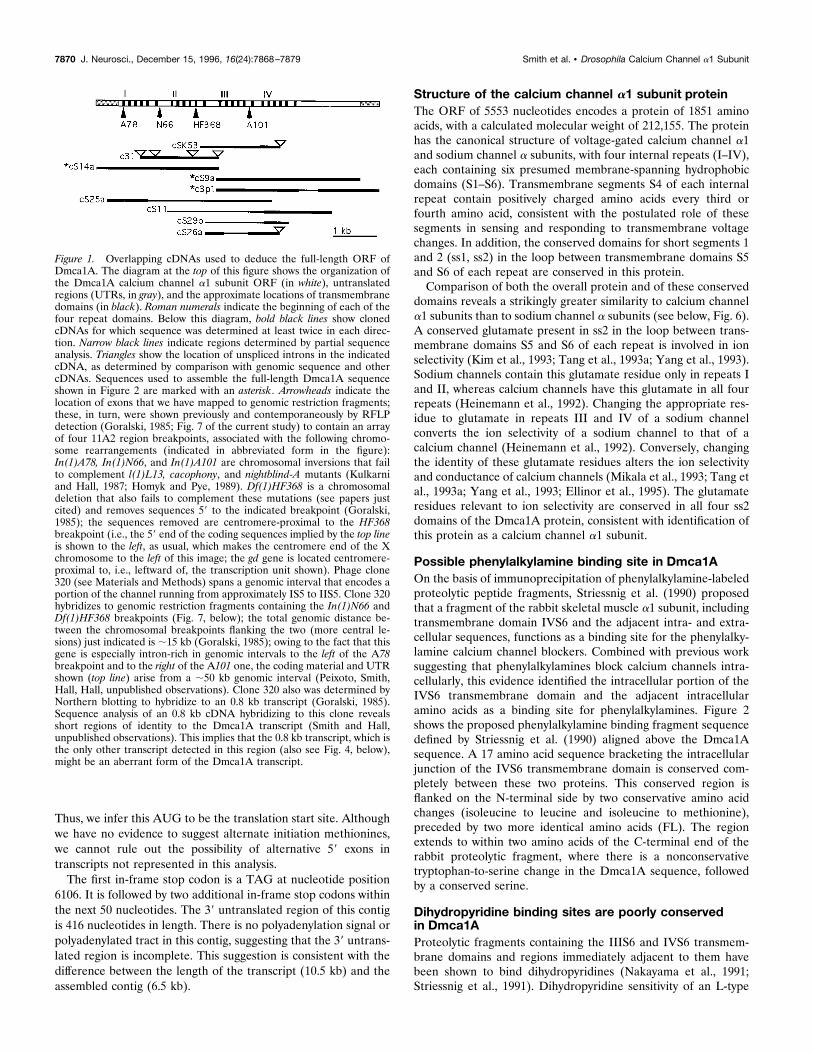
RESULTSIsolation of cDNAs encoding a new calcium channela1 subunitThe cDNA encoding the calcium channel a1 subunit reportedhere was isolated during the analysis of a region of the X chro-mosome known to contain the gene cacophony (cac) and theinteracting genetic variants nightblind-A (nbA) and lethal(1)L13(also known as l(1)11Aa; Lindsley and Zimm, 1992). The cac locuswas mapped cytogenetically using inversions and deletions to theX chromosomal region 11A2 (Kulkarni and Hall, 1987). Genomicphage clones 320 and 0371 (Goralski, 1985) were derived from achromosome walk through the flanking gastrulation defective (gd)locus (which is ;10 kb from the left-hand, centromere-proximalend of the putative cac-locus, as depicted in Fig. 1). Clones 320and 0371 recognized restriction-fragment-length polymorphisms(RFLPs) associated with breakpoints in this region [Goralski(1985); also see Fig. 7, below]. Northern blots of adult RNAprobed with fragments of clone 320 recognized two transcripts: 0.8kb (Goralski, 1985) and.10 kb (our preliminary data, not shown;also see Fig. 1 legend and Fig. 4, below). To clone cDNAsencoding these transcripts, we probed a 12–24 hr embryoniccDNA library (Brown and Kafatos, 1988) with genomic clone0371 and isolated the cDNA clone cSK53 (Fig. 1). In situ hybrid-ization of cSK53 to salivary gland chromosomes from third-instarlarvae mapped this cDNA to the distal portion of region 11A,consistent with the origin of the original genomic probes (data notshown). Northern blotting showed that the cSK53 cDNA corre-sponds to a subset of the large mRNA transcribed from this Xchromosomal region (see below, Fig. 4). The genomic intervalthat gives rise to the coding (plus untranslated) RNA indicated onthe top line of Figure 1 is approximately eight times longer thanthe amount of sequence so depicted (see Fig. 1 legend).Sequence analysis showed that cSK53 contains an open reading
frame (ORF) encoding a fragment similar to a voltage-sensitivecalcium channel a1 subunit (Fig. 2). Several rounds of cDNAisolation from Drosophila head cDNA libraries, starting with aprobe derived from cSK53 and continuing in later rounds withprobes from newly isolated cDNAs, isolated a total of 57 cDNAs.A subset of these was chosen for further analysis based on lengthand overlap with other cDNAs (Fig. 1). A 6522 nucleotide cDNAcontig—assembled from the overlapping cDNAs cS14a, cS9a, andc3p1—contains a single large ORF of 5553 nucleotides, whichencodes a voltage-sensitive calcium channel a1 subunit (Fig. 2).An AUG at nucleotide positions 553–555 is the only in-framemethionine codon between five upstream in-frame stop codonsand sequences coding for the first transmembrane domain (IS1).The sequence flanking this methionine codon (UAGA AUG)shows two of four matches to the Drosophila translation initiationconsensus sequence (C/A AA A/C AUG) (Cavener, 1987). Itincludes the highly conserved A at the 23 position, and the G at22 is the second most frequently used nucleotide at this position.
Smith et al. • Drosophila Calcium Channel a1 Subunit J. Neurosci., December 15, 1996, 16(24):7868–7879 7869
Thus, we infer this AUG to be the translation start site. Althoughwe have no evidence to suggest alternate initiation methionines,we cannot rule out the possibility of alternative 59 exons intranscripts not represented in this analysis.The first in-frame stop codon is a TAG at nucleotide position
6106. It is followed by two additional in-frame stop codons withinthe next 50 nucleotides. The 39 untranslated region of this contigis 416 nucleotides in length. There is no polyadenylation signal orpolyadenylated tract in this contig, suggesting that the 39 untrans-lated region is incomplete. This suggestion is consistent with thedifference between the length of the transcript (10.5 kb) and theassembled contig (6.5 kb).
Structure of the calcium channel a1 subunit proteinThe ORF of 5553 nucleotides encodes a protein of 1851 aminoacids, with a calculated molecular weight of 212,155. The proteinhas the canonical structure of voltage-gated calcium channel a1and sodium channel a subunits, with four internal repeats (I–IV),each containing six presumed membrane-spanning hydrophobicdomains (S1–S6). Transmembrane segments S4 of each internalrepeat contain positively charged amino acids every third orfourth amino acid, consistent with the postulated role of thesesegments in sensing and responding to transmembrane voltagechanges. In addition, the conserved domains for short segments 1and 2 (ss1, ss2) in the loop between transmembrane domains S5and S6 of each repeat are conserved in this protein.Comparison of both the overall protein and of these conserved
domains reveals a strikingly greater similarity to calcium channela1 subunits than to sodium channel a subunits (see below, Fig. 6).A conserved glutamate present in ss2 in the loop between trans-membrane domains S5 and S6 of each repeat is involved in ionselectivity (Kim et al., 1993; Tang et al., 1993a; Yang et al., 1993).Sodium channels contain this glutamate residue only in repeats Iand II, whereas calcium channels have this glutamate in all fourrepeats (Heinemann et al., 1992). Changing the appropriate res-idue to glutamate in repeats III and IV of a sodium channelconverts the ion selectivity of a sodium channel to that of acalcium channel (Heinemann et al., 1992). Conversely, changingthe identity of these glutamate residues alters the ion selectivityand conductance of calcium channels (Mikala et al., 1993; Tang etal., 1993a; Yang et al., 1993; Ellinor et al., 1995). The glutamateresidues relevant to ion selectivity are conserved in all four ss2domains of the Dmca1A protein, consistent with identification ofthis protein as a calcium channel a1 subunit.
Possible phenylalkylamine binding site in Dmca1AOn the basis of immunoprecipitation of phenylalkylamine-labeledproteolytic peptide fragments, Striessnig et al. (1990) proposedthat a fragment of the rabbit skeletal muscle a1 subunit, includingtransmembrane domain IVS6 and the adjacent intra- and extra-cellular sequences, functions as a binding site for the phenylalky-lamine calcium channel blockers. Combined with previous worksuggesting that phenylalkylamines block calcium channels intra-cellularly, this evidence identified the intracellular portion of theIVS6 transmembrane domain and the adjacent intracellularamino acids as a binding site for phenylalkylamines. Figure 2shows the proposed phenylalkylamine binding fragment sequencedefined by Striessnig et al. (1990) aligned above the Dmca1Asequence. A 17 amino acid sequence bracketing the intracellularjunction of the IVS6 transmembrane domain is conserved com-pletely between these two proteins. This conserved region isflanked on the N-terminal side by two conservative amino acidchanges (isoleucine to leucine and isoleucine to methionine),preceded by two more identical amino acids (FL). The regionextends to within two amino acids of the C-terminal end of therabbit proteolytic fragment, where there is a nonconservativetryptophan-to-serine change in the Dmca1A sequence, followedby a conserved serine.
Dihydropyridine binding sites are poorly conservedin Dmca1AProteolytic fragments containing the IIIS6 and IVS6 transmem-brane domains and regions immediately adjacent to them havebeen shown to bind dihydropyridines (Nakayama et al., 1991;Striessnig et al., 1991). Dihydropyridine sensitivity of an L-type
Figure 1. Overlapping cDNAs used to deduce the full-length ORF ofDmca1A. The diagram at the top of this figure shows the organization ofthe Dmca1A calcium channel a1 subunit ORF (in white), untranslatedregions (UTRs, in gray), and the approximate locations of transmembranedomains (in black). Roman numerals indicate the beginning of each of thefour repeat domains. Below this diagram, bold black lines show clonedcDNAs for which sequence was determined at least twice in each direc-tion. Narrow black lines indicate regions determined by partial sequenceanalysis. Triangles show the location of unspliced introns in the indicatedcDNA, as determined by comparison with genomic sequence and othercDNAs. Sequences used to assemble the full-length Dmca1A sequenceshown in Figure 2 are marked with an asterisk . Arrowheads indicate thelocation of exons that we have mapped to genomic restriction fragments;these, in turn, were shown previously and contemporaneously by RFLPdetection (Goralski, 1985; Fig. 7 of the current study) to contain an arrayof four 11A2 region breakpoints, associated with the following chromo-some rearrangements (indicated in abbreviated form in the figure):In(1)A78, In(1)N66, and In(1)A101 are chromosomal inversions that failto complement l(1)L13, cacophony, and nightblind-A mutants (Kulkarniand Hall, 1987; Homyk and Pye, 1989). Df(1)HF368 is a chromosomaldeletion that also fails to complement these mutations (see papers justcited) and removes sequences 59 to the indicated breakpoint (Goralski,1985); the sequences removed are centromere-proximal to the HF368breakpoint (i.e., the 59 end of the coding sequences implied by the top lineis shown to the left, as usual, which makes the centromere end of the Xchromosome to the left of this image; the gd gene is located centromere-proximal to, i.e., leftward of, the transcription unit shown). Phage clone320 (see Materials and Methods) spans a genomic interval that encodes aportion of the channel running from approximately IS5 to IIS5. Clone 320hybridizes to genomic restriction fragments containing the In(1)N66 andDf(1)HF368 breakpoints (Fig. 7, below); the total genomic distance be-tween the chromosomal breakpoints flanking the two (more central le-sions) just indicated is ;15 kb (Goralski, 1985); owing to the fact that thisgene is especially intron-rich in genomic intervals to the left of the A78breakpoint and to the right of the A101 one, the coding material and UTRshown (top line) arise from a ;50 kb genomic interval (Peixoto, Smith,Hall, Hall, unpublished observations). Clone 320 also was determined byNorthern blotting to hybridize to an 0.8 kb transcript (Goralski, 1985).Sequence analysis of an 0.8 kb cDNA hybridizing to this clone revealsshort regions of identity to the Dmca1A transcript (Smith and Hall,unpublished observations). This implies that the 0.8 kb transcript, which isthe only other transcript detected in this region (also see Fig. 4, below),might be an aberrant form of the Dmca1A transcript.
7870 J. Neurosci., December 15, 1996, 16(24):7868–7879 Smith et al. • Drosophila Calcium Channel a1 Subunit
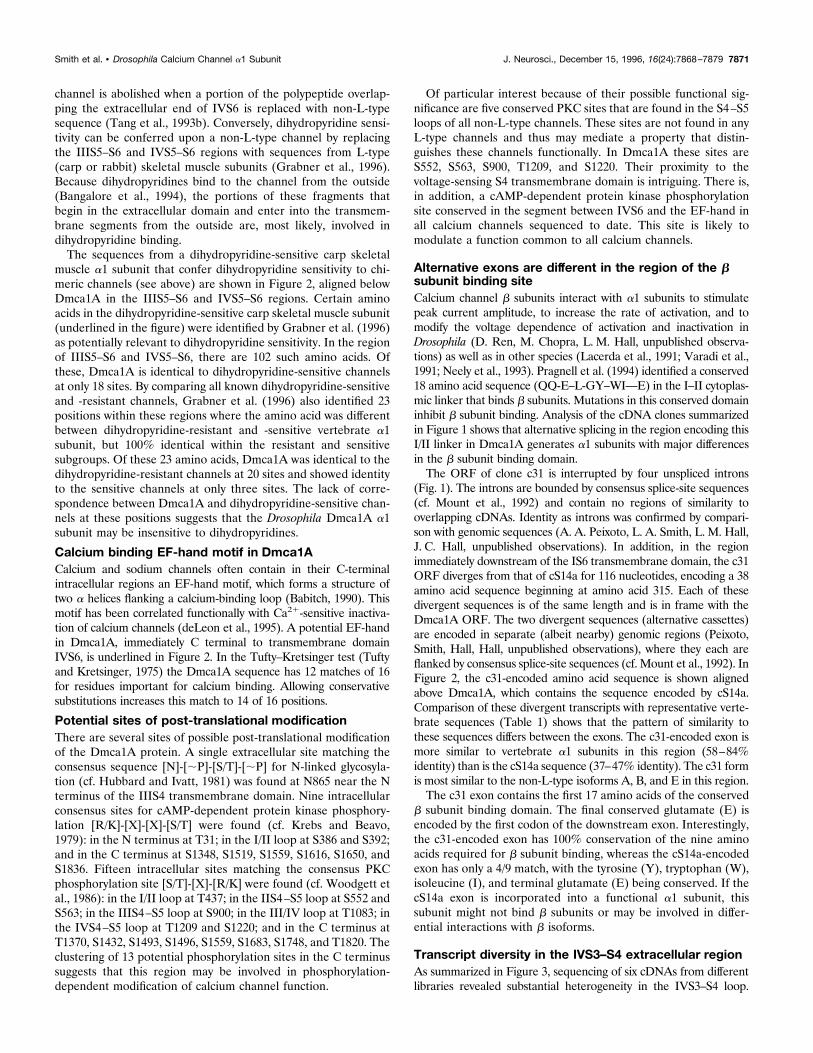
channel is abolished when a portion of the polypeptide overlap-ping the extracellular end of IVS6 is replaced with non-L-typesequence (Tang et al., 1993b). Conversely, dihydropyridine sensi-tivity can be conferred upon a non-L-type channel by replacingthe IIIS5–S6 and IVS5–S6 regions with sequences from L-type(carp or rabbit) skeletal muscle subunits (Grabner et al., 1996).Because dihydropyridines bind to the channel from the outside(Bangalore et al., 1994), the portions of these fragments thatbegin in the extracellular domain and enter into the transmem-brane segments from the outside are, most likely, involved indihydropyridine binding.The sequences from a dihydropyridine-sensitive carp skeletal
muscle a1 subunit that confer dihydropyridine sensitivity to chi-meric channels (see above) are shown in Figure 2, aligned belowDmca1A in the IIIS5–S6 and IVS5–S6 regions. Certain aminoacids in the dihydropyridine-sensitive carp skeletal muscle subunit(underlined in the figure) were identified by Grabner et al. (1996)as potentially relevant to dihydropyridine sensitivity. In the regionof IIIS5–S6 and IVS5–S6, there are 102 such amino acids. Ofthese, Dmca1A is identical to dihydropyridine-sensitive channelsat only 18 sites. By comparing all known dihydropyridine-sensitiveand -resistant channels, Grabner et al. (1996) also identified 23positions within these regions where the amino acid was differentbetween dihydropyridine-resistant and -sensitive vertebrate a1subunit, but 100% identical within the resistant and sensitivesubgroups. Of these 23 amino acids, Dmca1A was identical to thedihydropyridine-resistant channels at 20 sites and showed identityto the sensitive channels at only three sites. The lack of corre-spondence between Dmca1A and dihydropyridine-sensitive chan-nels at these positions suggests that the Drosophila Dmca1A a1subunit may be insensitive to dihydropyridines.
Calcium binding EF-hand motif in Dmca1ACalcium and sodium channels often contain in their C-terminalintracellular regions an EF-hand motif, which forms a structure oftwo a helices flanking a calcium-binding loop (Babitch, 1990). Thismotif has been correlated functionally with Ca21-sensitive inactiva-tion of calcium channels (deLeon et al., 1995). A potential EF-handin Dmca1A, immediately C terminal to transmembrane domainIVS6, is underlined in Figure 2. In the Tufty–Kretsinger test (Tuftyand Kretsinger, 1975) the Dmca1A sequence has 12 matches of 16for residues important for calcium binding. Allowing conservativesubstitutions increases this match to 14 of 16 positions.
Potential sites of post-translational modificationThere are several sites of possible post-translational modificationof the Dmca1A protein. A single extracellular site matching theconsensus sequence [N]-[;P]-[S/T]-[;P] for N-linked glycosyla-tion (cf. Hubbard and Ivatt, 1981) was found at N865 near the Nterminus of the IIIS4 transmembrane domain. Nine intracellularconsensus sites for cAMP-dependent protein kinase phosphory-lation [R/K]-[X]-[X]-[S/T] were found (cf. Krebs and Beavo,1979): in the N terminus at T31; in the I/II loop at S386 and S392;and in the C terminus at S1348, S1519, S1559, S1616, S1650, andS1836. Fifteen intracellular sites matching the consensus PKCphosphorylation site [S/T]-[X]-[R/K] were found (cf. Woodgett etal., 1986): in the I/II loop at T437; in the IIS4–S5 loop at S552 andS563; in the IIIS4–S5 loop at S900; in the III/IV loop at T1083; inthe IVS4–S5 loop at T1209 and S1220; and in the C terminus atT1370, S1432, S1493, S1496, S1559, S1683, S1748, and T1820. Theclustering of 13 potential phosphorylation sites in the C terminussuggests that this region may be involved in phosphorylation-dependent modification of calcium channel function.
Of particular interest because of their possible functional sig-nificance are five conserved PKC sites that are found in the S4–S5loops of all non-L-type channels. These sites are not found in anyL-type channels and thus may mediate a property that distin-guishes these channels functionally. In Dmca1A these sites areS552, S563, S900, T1209, and S1220. Their proximity to thevoltage-sensing S4 transmembrane domain is intriguing. There is,in addition, a cAMP-dependent protein kinase phosphorylationsite conserved in the segment between IVS6 and the EF-hand inall calcium channels sequenced to date. This site is likely tomodulate a function common to all calcium channels.
Alternative exons are different in the region of the bsubunit binding siteCalcium channel b subunits interact with a1 subunits to stimulatepeak current amplitude, to increase the rate of activation, and tomodify the voltage dependence of activation and inactivation inDrosophila (D. Ren, M. Chopra, L. M. Hall, unpublished observa-tions) as well as in other species (Lacerda et al., 1991; Varadi et al.,1991; Neely et al., 1993). Pragnell et al. (1994) identified a conserved18 amino acid sequence (QQ-E–L-GY–WI—E) in the I–II cytoplas-mic linker that binds b subunits. Mutations in this conserved domaininhibit b subunit binding. Analysis of the cDNA clones summarizedin Figure 1 shows that alternative splicing in the region encoding thisI/II linker in Dmca1A generates a1 subunits with major differencesin the b subunit binding domain.The ORF of clone c31 is interrupted by four unspliced introns
(Fig. 1). The introns are bounded by consensus splice-site sequences(cf. Mount et al., 1992) and contain no regions of similarity tooverlapping cDNAs. Identity as introns was confirmed by compari-son with genomic sequences (A. A. Peixoto, L. A. Smith, L. M. Hall,J. C. Hall, unpublished observations). In addition, in the regionimmediately downstream of the IS6 transmembrane domain, the c31ORF diverges from that of cS14a for 116 nucleotides, encoding a 38amino acid sequence beginning at amino acid 315. Each of thesedivergent sequences is of the same length and is in frame with theDmca1A ORF. The two divergent sequences (alternative cassettes)are encoded in separate (albeit nearby) genomic regions (Peixoto,Smith, Hall, Hall, unpublished observations), where they each areflanked by consensus splice-site sequences (cf. Mount et al., 1992). InFigure 2, the c31-encoded amino acid sequence is shown alignedabove Dmca1A, which contains the sequence encoded by cS14a.Comparison of these divergent transcripts with representative verte-brate sequences (Table 1) shows that the pattern of similarity tothese sequences differs between the exons. The c31-encoded exon ismore similar to vertebrate a1 subunits in this region (58–84%identity) than is the cS14a sequence (37–47% identity). The c31 formis most similar to the non-L-type isoforms A, B, and E in this region.The c31 exon contains the first 17 amino acids of the conserved
b subunit binding domain. The final conserved glutamate (E) isencoded by the first codon of the downstream exon. Interestingly,the c31-encoded exon has 100% conservation of the nine aminoacids required for b subunit binding, whereas the cS14a-encodedexon has only a 4/9 match, with the tyrosine (Y), tryptophan (W),isoleucine (I), and terminal glutamate (E) being conserved. If thecS14a exon is incorporated into a functional a1 subunit, thissubunit might not bind b subunits or may be involved in differ-ential interactions with b isoforms.
Transcript diversity in the IVS3–S4 extracellular regionAs summarized in Figure 3, sequencing of six cDNAs from differentlibraries revealed substantial heterogeneity in the IVS3–S4 loop.
Smith et al. • Drosophila Calcium Channel a1 Subunit J. Neurosci., December 15, 1996, 16(24):7868–7879 7871
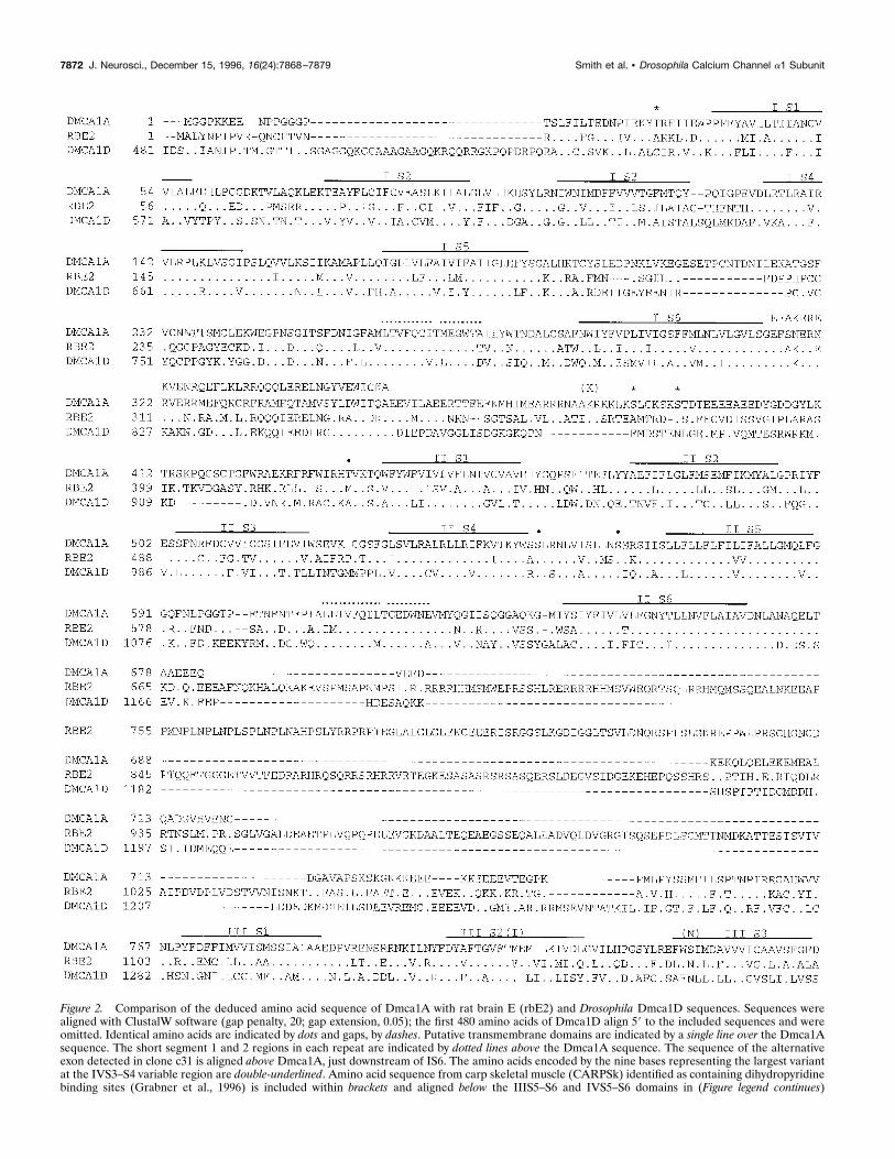
Figure 2. Comparison of the deduced amino acid sequence of Dmca1A with rat brain E (rbE2) and Drosophila Dmca1D sequences. Sequences werealigned with ClustalW software (gap penalty, 20; gap extension, 0.05); the first 480 amino acids of Dmca1D align 59 to the included sequences and wereomitted. Identical amino acids are indicated by dots and gaps, by dashes. Putative transmembrane domains are indicated by a single line over the Dmca1Asequence. The short segment 1 and 2 regions in each repeat are indicated by dotted lines above the Dmca1A sequence. The sequence of the alternativeexon detected in clone c31 is aligned above Dmca1A, just downstream of IS6. The amino acids encoded by the nine bases representing the largest variantat the IVS3–S4 variable region are double-underlined. Amino acid sequence from carp skeletal muscle (CARPSk) identified as containing dihydropyridinebinding sites (Grabner et al., 1996) is included within brackets and aligned below the IIIS5–S6 and IVS5–S6 domains in (Figure legend continues)
7872 J. Neurosci., December 15, 1996, 16(24):7868–7879 Smith et al. • Drosophila Calcium Channel a1 Subunit
4
Dmca1A. A proposed phenylalkylamine binding sequence (Striessnig et al., 1990) is aligned above the IVS6 region of Dmca1A. A calcium-bindingEF-hand structure downstream of IVS6 is single-underlined in Dmca1A. The potential N-glycosylation site at the N terminus of IIIS4 is marked with C.cAMP kinase sites are marked with *; PKC sites with ●. Sites of potential RNA editing are indicated by aligning the unedited codon identity in parenthesesabove the Dmca1A sequence. The GenBank accession number for Dmca1A is U55776.
Smith et al. • Drosophila Calcium Channel a1 Subunit J. Neurosci., December 15, 1996, 16(24):7868–7879 7873
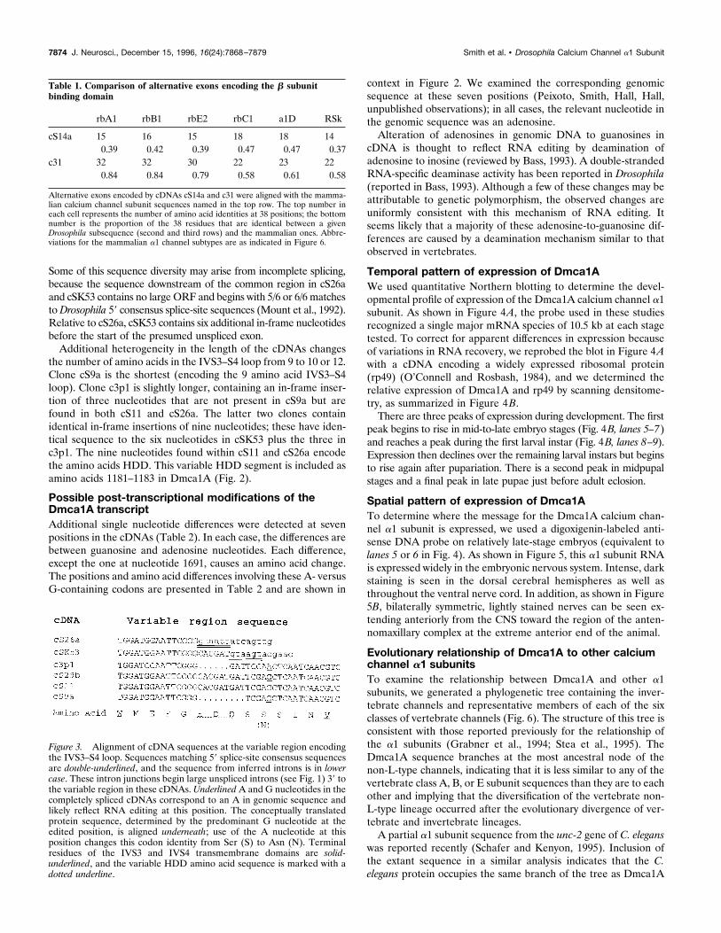
Some of this sequence diversity may arise from incomplete splicing,because the sequence downstream of the common region in cS26aand cSK53 contains no large ORF and begins with 5/6 or 6/6 matchestoDrosophila 59 consensus splice-site sequences (Mount et al., 1992).Relative to cS26a, cSK53 contains six additional in-frame nucleotidesbefore the start of the presumed unspliced exon.Additional heterogeneity in the length of the cDNAs changes
the number of amino acids in the IVS3–S4 loop from 9 to 10 or 12.Clone cS9a is the shortest (encoding the 9 amino acid IVS3–S4loop). Clone c3p1 is slightly longer, containing an in-frame inser-tion of three nucleotides that are not present in cS9a but arefound in both cS11 and cS26a. The latter two clones containidentical in-frame insertions of nine nucleotides; these have iden-tical sequence to the six nucleotides in cSK53 plus the three inc3p1. The nine nucleotides found within cS11 and cS26a encodethe amino acids HDD. This variable HDD segment is included asamino acids 1181–1183 in Dmca1A (Fig. 2).
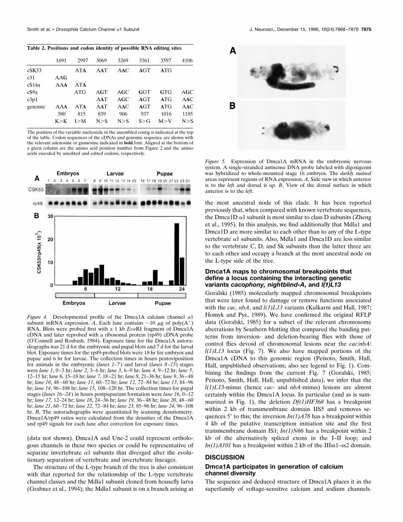
Possible post-transcriptional modifications of theDmca1A transcriptAdditional single nucleotide differences were detected at sevenpositions in the cDNAs (Table 2). In each case, the differences arebetween guanosine and adenosine nucleotides. Each difference,except the one at nucleotide 1691, causes an amino acid change.The positions and amino acid differences involving these A- versusG-containing codons are presented in Table 2 and are shown in
context in Figure 2. We examined the corresponding genomicsequence at these seven positions (Peixoto, Smith, Hall, Hall,unpublished observations); in all cases, the relevant nucleotide inthe genomic sequence was an adenosine.Alteration of adenosines in genomic DNA to guanosines in
cDNA is thought to reflect RNA editing by deamination ofadenosine to inosine (reviewed by Bass, 1993). A double-strandedRNA-specific deaminase activity has been reported in Drosophila(reported in Bass, 1993). Although a few of these changes may beattributable to genetic polymorphism, the observed changes areuniformly consistent with this mechanism of RNA editing. Itseems likely that a majority of these adenosine-to-guanosine dif-ferences are caused by a deamination mechanism similar to thatobserved in vertebrates.
Temporal pattern of expression of Dmca1AWe used quantitative Northern blotting to determine the devel-opmental profile of expression of the Dmca1A calcium channel a1subunit. As shown in Figure 4A, the probe used in these studiesrecognized a single major mRNA species of 10.5 kb at each stagetested. To correct for apparent differences in expression becauseof variations in RNA recovery, we reprobed the blot in Figure 4Awith a cDNA encoding a widely expressed ribosomal protein(rp49) (O’Connell and Rosbash, 1984), and we determined therelative expression of Dmca1A and rp49 by scanning densitome-try, as summarized in Figure 4B.There are three peaks of expression during development. The first
peak begins to rise in mid-to-late embryo stages (Fig. 4B, lanes 5–7)and reaches a peak during the first larval instar (Fig. 4B, lanes 8–9).Expression then declines over the remaining larval instars but beginsto rise again after pupariation. There is a second peak in midpupalstages and a final peak in late pupae just before adult eclosion.
Spatial pattern of expression of Dmca1ATo determine where the message for the Dmca1A calcium chan-nel a1 subunit is expressed, we used a digoxigenin-labeled anti-sense DNA probe on relatively late-stage embryos (equivalent tolanes 5 or 6 in Fig. 4). As shown in Figure 5, this a1 subunit RNAis expressed widely in the embryonic nervous system. Intense, darkstaining is seen in the dorsal cerebral hemispheres as well asthroughout the ventral nerve cord. In addition, as shown in Figure5B, bilaterally symmetric, lightly stained nerves can be seen ex-tending anteriorly from the CNS toward the region of the anten-nomaxillary complex at the extreme anterior end of the animal.
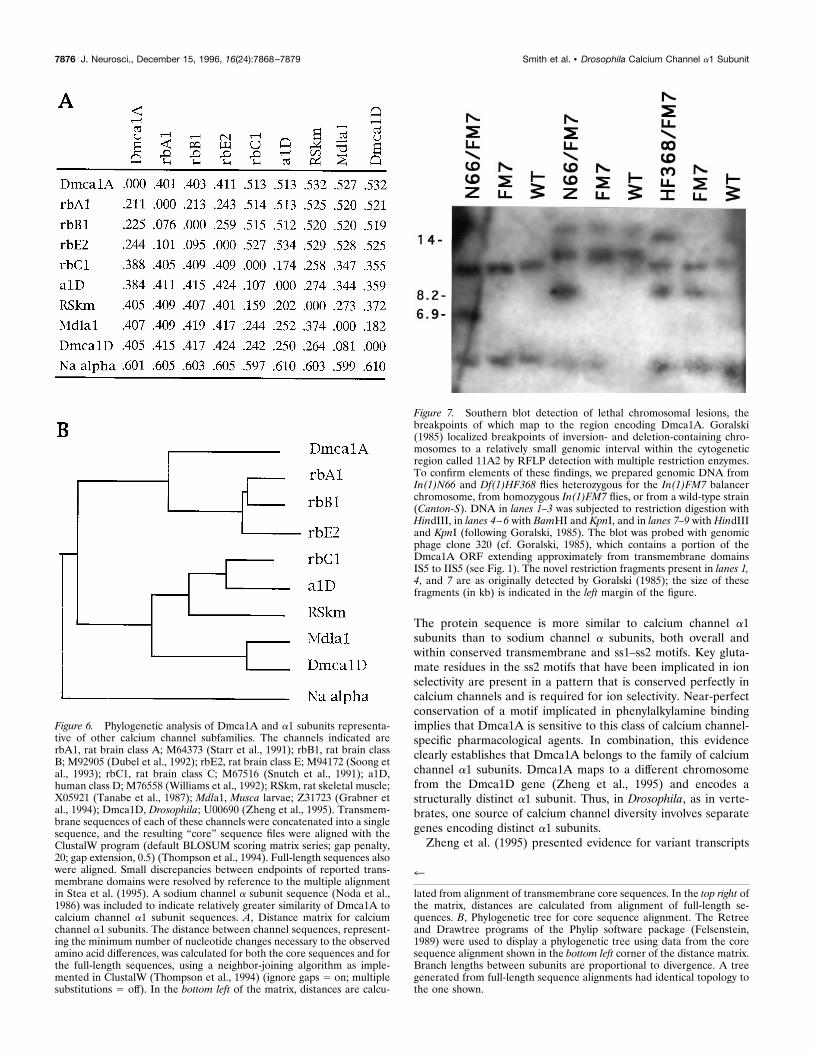
Evolutionary relationship of Dmca1A to other calciumchannel a1 subunitsTo examine the relationship between Dmca1A and other a1subunits, we generated a phylogenetic tree containing the inver-tebrate channels and representative members of each of the sixclasses of vertebrate channels (Fig. 6). The structure of this tree isconsistent with those reported previously for the relationship ofthe a1 subunits (Grabner et al., 1994; Stea et al., 1995). TheDmca1A sequence branches at the most ancestral node of thenon-L-type channels, indicating that it is less similar to any of thevertebrate class A, B, or E subunit sequences than they are to eachother and implying that the diversification of the vertebrate non-L-type lineage occurred after the evolutionary divergence of ver-tebrate and invertebrate lineages.A partial a1 subunit sequence from the unc-2 gene of C. elegans
was reported recently (Schafer and Kenyon, 1995). Inclusion ofthe extant sequence in a similar analysis indicates that the C.elegans protein occupies the same branch of the tree as Dmca1A
Figure 3. Alignment of cDNA sequences at the variable region encodingthe IVS3–S4 loop. Sequences matching 59 splice-site consensus sequencesare double-underlined, and the sequence from inferred introns is in lowercase. These intron junctions begin large unspliced introns (see Fig. 1) 39 tothe variable region in these cDNAs. UnderlinedA and G nucleotides in thecompletely spliced cDNAs correspond to an A in genomic sequence andlikely reflect RNA editing at this position. The conceptually translatedprotein sequence, determined by the predominant G nucleotide at theedited position, is aligned underneath; use of the A nucleotide at thisposition changes this codon identity from Ser (S) to Asn (N). Terminalresidues of the IVS3 and IVS4 transmembrane domains are solid-underlined, and the variable HDD amino acid sequence is marked with adotted underline.
Table 1. Comparison of alternative exons encoding the b subunitbinding domain
rbA1 rbB1 rbE2 rbC1 a1D RSk
cS14a 15 16 15 18 18 140.39 0.42 0.39 0.47 0.47 0.37
c31 32 32 30 22 23 220.84 0.84 0.79 0.58 0.61 0.58
Alternative exons encoded by cDNAs cS14a and c31 were aligned with the mamma-lian calcium channel subunit sequences named in the top row. The top number ineach cell represents the number of amino acid identities at 38 positions; the bottomnumber is the proportion of the 38 residues that are identical between a givenDrosophila subsequence (second and third rows) and the mammalian ones. Abbre-viations for the mammalian a1 channel subtypes are as indicated in Figure 6.
7874 J. Neurosci., December 15, 1996, 16(24):7868–7879 Smith et al. • Drosophila Calcium Channel a1 Subunit
(data not shown). Dmca1A and Unc-2 could represent ortholo-gous channels in these two species or could be representative ofseparate invertebrate a1 subunits that diverged after the evolu-tionary separation of vertebrate and invertebrate lineages.The structure of the L-type branch of the tree is also consistent
with that reported for the relationship of the L-type vertebratechannel classes and the Mdla1 subunit cloned from housefly larva(Grabner et al., 1994); the Mdla1 subunit is on a branch arising at
the most ancestral node of this clade. It has been reportedpreviously that, when compared with known vertebrate sequences,the Dmca1D a1 subunit is most similar to class D subunits (Zhenget al., 1995). In this analysis, we find additionally that Mdla1 andDmca1D are more similar to each other than to any of the L-typevertebrate a1 subunits. Also, Mdla1 and Dmca1D are less similarto the vertebrate C, D, and Sk subunits than the latter three areto each other and occupy a branch at the most ancestral node onthe L-type side of the tree.
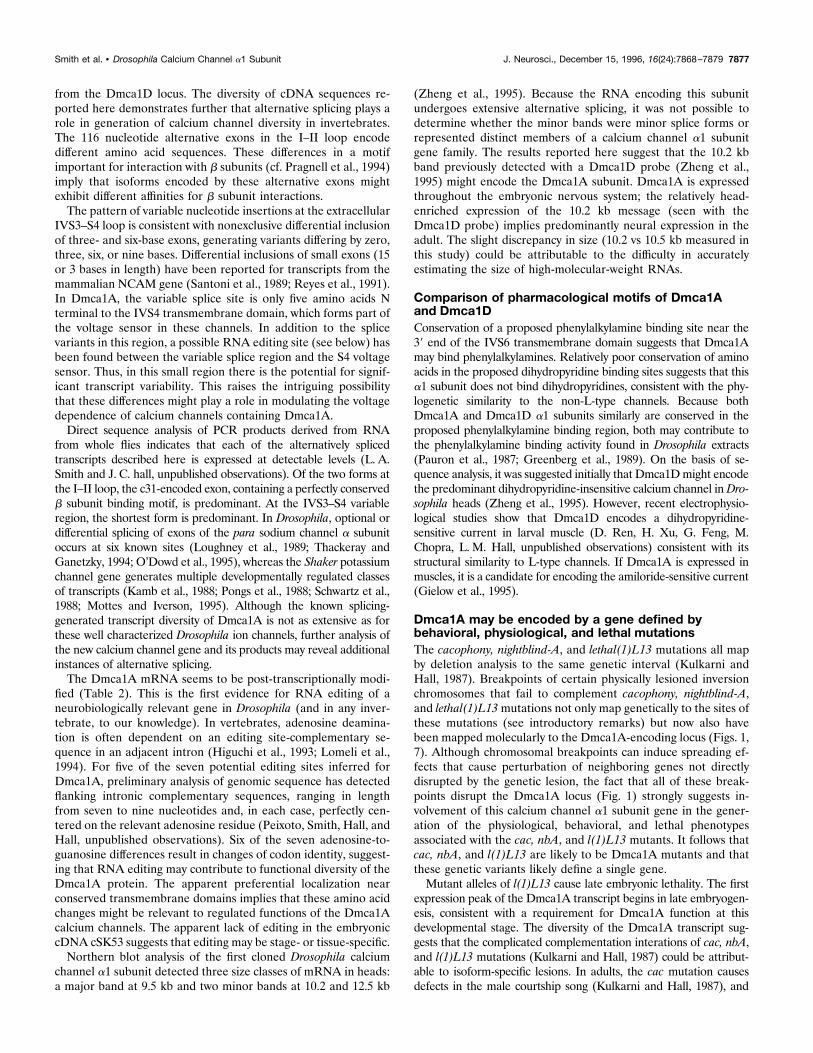
Dmca1A maps to chromosomal breakpoints thatdefine a locus containing the interacting geneticvariants cacophony, nightblind-A, and l(1)L13Goralski (1985) molecularly mapped chromosomal breakpointsthat were later found to damage or remove functions associatedwith the cac, nbA, and l(1)L13 variants (Kulkarni and Hall, 1987;Homyk and Pye, 1989). We have confirmed the original RFLPdata (Goralski, 1985) for a subset of the relevant chromosomeaberrations by Southern blotting that compared the banding pat-terns from inversion- and deletion-bearing flies with those ofcontrol flies devoid of chromosomal lesions near the cac/nbA/l(1)L13 locus (Fig. 7). We also have mapped portions of theDmca1A cDNA to this genomic region (Peixoto, Smith, Hall,Hall, unpublished observations; also see legend to Fig. 1). Com-bining the findings from the current Fig. 7 (Goralski, 1985;Peixoto, Smith, Hall, Hall, unpublished data), we infer that thel(1)L13-minus (hence cac- and nbA-minus) lesions are almostcertainly within the Dmca1A locus. In particular (and as is sum-marized in Fig. 1), the deletion Df(1)HF368 has a breakpointwithin 2 kb of transmembrane domain IIS5 and removes se-quences 59 to this; the inversion In(1)A78 has a breakpoint within4 kb of the putative transcription initiation site and the firsttransmembrane domain IS1; In(1)N66 has a breakpoint within 2kb of the alternatively spliced exons in the I–II loop; andIn(1)A101 has a breakpoint within 2 kb of the IIIss1–ss2 domain.
DISCUSSIONDmca1A participates in generation of calciumchannel diversityThe sequence and deduced structure of Dmca1A places it in thesuperfamily of voltage-sensitive calcium and sodium channels.
Figure 4. Developmental profile of the Dmca1A calcium channel a1subunit mRNA expression. A, Each lane contains ;10 mg of poly(A1)RNA. Blots were probed first with a 1 kb EcoRI fragment of Dmca1AcDNA and later reprobed with a ribosomal protein (rp49) cDNA probe(O’Connell and Rosbash, 1984). Exposure time for the Dmca1A autora-diographs was 21 d for the embryonic and pupal blots and 7 d for the larvalblot. Exposure times for the rp49-probed blots were 18 hr for embryos andpupae and 6 hr for larvae. The collection times in hours postovipositionfor animals in the embryonic (lanes 1–7 ) and larval (lanes 8–15) stageswere lane 1, 0–3 hr; lane 2, 3–6 hr; lane 3, 6–9 hr; lane 4, 9–12 hr; lane 5,12–15 hr; lane 6, 15–18 hr; lane 7, 18–21 hr; lane 8, 21–36 hr; lane 9, 36–48hr; lane 10, 48–60 hr; lane 11, 60–72 hr; lane 12, 72–84 hr; lane 13, 84–96hr; lane 14, 96–108 hr; lane 15, 108–120 hr. The collection times for pupalstages (lanes 16–24 ) in hours postpuparium formation were lane 16, 0–12hr; lane 17, 12–24 hr; lane 18, 24–36 hr; lane 19, 36–48 hr; lane 20, 48–60hr; lane 21, 60–72 hr; lane 22, 72–84 hr; lane 23, 85–96 hr; lane 24, 96–108hr. B, The autoradiographs were quantitated by scanning densitometry.Dmca1A/rp49 ratios were calculated from the densities of the Dmca1Aand rp49 signals for each lane after correction for exposure times.
Figure 5. Expression of Dmca1A mRNA in the embryonic nervoussystem. A single-stranded antisense DNA probe labeled with digoxigeninwas hybridized to whole-mounted stage 16 embryos. The darkly stainedareas represent regions of RNA expression. A, Side view in which anterioris to the left and dorsal is up. B, View of the dorsal surface in whichanterior is to the left.
Table 2. Positions and codon identity of possible RNA editing sites
1691 2997 3069 3269 3361 3597 4106
cSK53 ATA AAT AAC AGT ATGc31 AAGcS14a AAA ATAcS9a ATG AGT AGC GGT GTG AGCc3p1 AAT AGC AGT ATG AACgenome AAA ATA AAT AAC AGT ATG AAC
380 815 839 906 937 1016 1185K.K I.M N.S N.S S.G M.V N.S
The position of the variable nucleotide in the assembled contig is indicated at the topof the table. Codon sequences of the cDNAs and genomic sequence are shown withthe relevant adenosine or guanosine indicated in bold font. Aligned at the bottom ofa given column are the amino acid position number from Figure 2 and the aminoacids encoded by unedited and edited codons, respectively.
Smith et al. • Drosophila Calcium Channel a1 Subunit J. Neurosci., December 15, 1996, 16(24):7868–7879 7875
The protein sequence is more similar to calcium channel a1subunits than to sodium channel a subunits, both overall andwithin conserved transmembrane and ss1–ss2 motifs. Key gluta-mate residues in the ss2 motifs that have been implicated in ionselectivity are present in a pattern that is conserved perfectly incalcium channels and is required for ion selectivity. Near-perfectconservation of a motif implicated in phenylalkylamine bindingimplies that Dmca1A is sensitive to this class of calcium channel-specific pharmacological agents. In combination, this evidenceclearly establishes that Dmca1A belongs to the family of calciumchannel a1 subunits. Dmca1A maps to a different chromosomefrom the Dmca1D gene (Zheng et al., 1995) and encodes astructurally distinct a1 subunit. Thus, in Drosophila, as in verte-brates, one source of calcium channel diversity involves separategenes encoding distinct a1 subunits.Zheng et al. (1995) presented evidence for variant transcripts
4
lated from alignment of transmembrane core sequences. In the top right ofthe matrix, distances are calculated from alignment of full-length se-quences. B, Phylogenetic tree for core sequence alignment. The Retreeand Drawtree programs of the Phylip software package (Felsenstein,1989) were used to display a phylogenetic tree using data from the coresequence alignment shown in the bottom left corner of the distance matrix.Branch lengths between subunits are proportional to divergence. A treegenerated from full-length sequence alignments had identical topology tothe one shown.
Figure 6. Phylogenetic analysis of Dmca1A and a1 subunits representa-tive of other calcium channel subfamilies. The channels indicated arerbA1, rat brain class A; M64373 (Starr et al., 1991); rbB1, rat brain classB; M92905 (Dubel et al., 1992); rbE2, rat brain class E; M94172 (Soong etal., 1993); rbC1, rat brain class C; M67516 (Snutch et al., 1991); a1D,human class D; M76558 (Williams et al., 1992); RSkm, rat skeletal muscle;X05921 (Tanabe et al., 1987); Mdla1, Musca larvae; Z31723 (Grabner etal., 1994); Dmca1D, Drosophila; U00690 (Zheng et al., 1995). Transmem-brane sequences of each of these channels were concatenated into a singlesequence, and the resulting “core” sequence files were aligned with theClustalW program (default BLOSUM scoring matrix series; gap penalty,20; gap extension, 0.5) (Thompson et al., 1994). Full-length sequences alsowere aligned. Small discrepancies between endpoints of reported trans-membrane domains were resolved by reference to the multiple alignmentin Stea et al. (1995). A sodium channel a subunit sequence (Noda et al.,1986) was included to indicate relatively greater similarity of Dmca1A tocalcium channel a1 subunit sequences. A, Distance matrix for calciumchannel a1 subunits. The distance between channel sequences, represent-ing the minimum number of nucleotide changes necessary to the observedamino acid differences, was calculated for both the core sequences and forthe full-length sequences, using a neighbor-joining algorithm as imple-mented in ClustalW (Thompson et al., 1994) (ignore gaps 5 on; multiplesubstitutions 5 off). In the bottom left of the matrix, distances are calcu-
Figure 7. Southern blot detection of lethal chromosomal lesions, thebreakpoints of which map to the region encoding Dmca1A. Goralski(1985) localized breakpoints of inversion- and deletion-containing chro-mosomes to a relatively small genomic interval within the cytogeneticregion called 11A2 by RFLP detection with multiple restriction enzymes.To confirm elements of these findings, we prepared genomic DNA fromIn(1)N66 and Df(1)HF368 flies heterozygous for the In(1)FM7 balancerchromosome, from homozygous In(1)FM7 flies, or from a wild-type strain(Canton-S). DNA in lanes 1–3 was subjected to restriction digestion withHindIII, in lanes 4–6 with BamHI and KpnI, and in lanes 7–9 with HindIIIand KpnI (following Goralski, 1985). The blot was probed with genomicphage clone 320 (cf. Goralski, 1985), which contains a portion of theDmca1A ORF extending approximately from transmembrane domainsIS5 to IIS5 (see Fig. 1). The novel restriction fragments present in lanes 1,4, and 7 are as originally detected by Goralski (1985); the size of thesefragments (in kb) is indicated in the left margin of the figure.
7876 J. Neurosci., December 15, 1996, 16(24):7868–7879 Smith et al. • Drosophila Calcium Channel a1 Subunit
from the Dmca1D locus. The diversity of cDNA sequences re-ported here demonstrates further that alternative splicing plays arole in generation of calcium channel diversity in invertebrates.The 116 nucleotide alternative exons in the I–II loop encodedifferent amino acid sequences. These differences in a motifimportant for interaction with b subunits (cf. Pragnell et al., 1994)imply that isoforms encoded by these alternative exons mightexhibit different affinities for b subunit interactions.The pattern of variable nucleotide insertions at the extracellular
IVS3–S4 loop is consistent with nonexclusive differential inclusionof three- and six-base exons, generating variants differing by zero,three, six, or nine bases. Differential inclusions of small exons (15or 3 bases in length) have been reported for transcripts from themammalian NCAM gene (Santoni et al., 1989; Reyes et al., 1991).In Dmca1A, the variable splice site is only five amino acids Nterminal to the IVS4 transmembrane domain, which forms part ofthe voltage sensor in these channels. In addition to the splicevariants in this region, a possible RNA editing site (see below) hasbeen found between the variable splice region and the S4 voltagesensor. Thus, in this small region there is the potential for signif-icant transcript variability. This raises the intriguing possibilitythat these differences might play a role in modulating the voltagedependence of calcium channels containing Dmca1A.Direct sequence analysis of PCR products derived from RNA
from whole flies indicates that each of the alternatively splicedtranscripts described here is expressed at detectable levels (L. A.Smith and J. C. hall, unpublished observations). Of the two forms atthe I–II loop, the c31-encoded exon, containing a perfectly conservedb subunit binding motif, is predominant. At the IVS3–S4 variableregion, the shortest form is predominant. In Drosophila, optional ordifferential splicing of exons of the para sodium channel a subunitoccurs at six known sites (Loughney et al., 1989; Thackeray andGanetzky, 1994; O’Dowd et al., 1995), whereas the Shaker potassiumchannel gene generates multiple developmentally regulated classesof transcripts (Kamb et al., 1988; Pongs et al., 1988; Schwartz et al.,1988; Mottes and Iverson, 1995). Although the known splicing-generated transcript diversity of Dmca1A is not as extensive as forthese well characterized Drosophila ion channels, further analysis ofthe new calcium channel gene and its products may reveal additionalinstances of alternative splicing.The Dmca1A mRNA seems to be post-transcriptionally modi-
fied (Table 2). This is the first evidence for RNA editing of aneurobiologically relevant gene in Drosophila (and in any inver-tebrate, to our knowledge). In vertebrates, adenosine deamina-tion is often dependent on an editing site-complementary se-quence in an adjacent intron (Higuchi et al., 1993; Lomeli et al.,1994). For five of the seven potential editing sites inferred forDmca1A, preliminary analysis of genomic sequence has detectedflanking intronic complementary sequences, ranging in lengthfrom seven to nine nucleotides and, in each case, perfectly cen-tered on the relevant adenosine residue (Peixoto, Smith, Hall, andHall, unpublished observations). Six of the seven adenosine-to-guanosine differences result in changes of codon identity, suggest-ing that RNA editing may contribute to functional diversity of theDmca1A protein. The apparent preferential localization nearconserved transmembrane domains implies that these amino acidchanges might be relevant to regulated functions of the Dmca1Acalcium channels. The apparent lack of editing in the embryoniccDNA cSK53 suggests that editing may be stage- or tissue-specific.Northern blot analysis of the first cloned Drosophila calcium
channel a1 subunit detected three size classes of mRNA in heads:a major band at 9.5 kb and two minor bands at 10.2 and 12.5 kb
(Zheng et al., 1995). Because the RNA encoding this subunitundergoes extensive alternative splicing, it was not possible todetermine whether the minor bands were minor splice forms orrepresented distinct members of a calcium channel a1 subunitgene family. The results reported here suggest that the 10.2 kbband previously detected with a Dmca1D probe (Zheng et al.,1995) might encode the Dmca1A subunit. Dmca1A is expressedthroughout the embryonic nervous system; the relatively head-enriched expression of the 10.2 kb message (seen with theDmca1D probe) implies predominantly neural expression in theadult. The slight discrepancy in size (10.2 vs 10.5 kb measured inthis study) could be attributable to the difficulty in accuratelyestimating the size of high-molecular-weight RNAs.
Comparison of pharmacological motifs of Dmca1Aand Dmca1DConservation of a proposed phenylalkylamine binding site near the39 end of the IVS6 transmembrane domain suggests that Dmca1Amay bind phenylalkylamines. Relatively poor conservation of aminoacids in the proposed dihydropyridine binding sites suggests that thisa1 subunit does not bind dihydropyridines, consistent with the phy-logenetic similarity to the non-L-type channels. Because bothDmca1A and Dmca1D a1 subunits similarly are conserved in theproposed phenylalkylamine binding region, both may contribute tothe phenylalkylamine binding activity found in Drosophila extracts(Pauron et al., 1987; Greenberg et al., 1989). On the basis of se-quence analysis, it was suggested initially that Dmca1Dmight encodethe predominant dihydropyridine-insensitive calcium channel inDro-sophila heads (Zheng et al., 1995). However, recent electrophysio-logical studies show that Dmca1D encodes a dihydropyridine-sensitive current in larval muscle (D. Ren, H. Xu, G. Feng, M.Chopra, L. M. Hall, unpublished observations) consistent with itsstructural similarity to L-type channels. If Dmca1A is expressed inmuscles, it is a candidate for encoding the amiloride-sensitive current(Gielow et al., 1995).
Dmca1A may be encoded by a gene defined bybehavioral, physiological, and lethal mutationsThe cacophony, nightblind-A, and lethal(1)L13 mutations all mapby deletion analysis to the same genetic interval (Kulkarni andHall, 1987). Breakpoints of certain physically lesioned inversionchromosomes that fail to complement cacophony, nightblind-A,and lethal(1)L13mutations not only map genetically to the sites ofthese mutations (see introductory remarks) but now also havebeen mapped molecularly to the Dmca1A-encoding locus (Figs. 1,7). Although chromosomal breakpoints can induce spreading ef-fects that cause perturbation of neighboring genes not directlydisrupted by the genetic lesion, the fact that all of these break-points disrupt the Dmca1A locus (Fig. 1) strongly suggests in-volvement of this calcium channel a1 subunit gene in the gener-ation of the physiological, behavioral, and lethal phenotypesassociated with the cac, nbA, and l(1)L13 mutants. It follows thatcac, nbA, and l(1)L13 are likely to be Dmca1A mutants and thatthese genetic variants likely define a single gene.Mutant alleles of l(1)L13 cause late embryonic lethality. The first
expression peak of the Dmca1A transcript begins in late embryogen-esis, consistent with a requirement for Dmca1A function at thisdevelopmental stage. The diversity of the Dmca1A transcript sug-gests that the complicated complementation interations of cac, nbA,and l(1)L13 mutations (Kulkarni and Hall, 1987) could be attribut-able to isoform-specific lesions. In adults, the cac mutation causesdefects in the male courtship song (Kulkarni and Hall, 1987), and
Smith et al. • Drosophila Calcium Channel a1 Subunit J. Neurosci., December 15, 1996, 16(24):7868–7879 7877
nbA mutants exhibit increased light thresholds for optomotor andphototactic behaviors (Heisenberg and Gotz, 1975) as well as defectsin the shape and amplitude of the electroretinogram (Homyk andPye, 1989). The particular song defect exhibited by cacmales—largerthan normal numbers of cycles within a given “burst” of tone—couldbe rationalized in terms of modified calcium channel function [cf.Hille (1992), Chapter 5]. The cellular etiology of the abnormalsinging behavior is difficult to speculate on, because it could involvedefects in neural or muscular physiology (or even anatomy), yet it isdifficult to imagine a non-neural etiology for the abnormal ERG innbA mutants. Taken together, this analysis implies involvement ofthe Dmca1A voltage-dependent calcium channel in visual transduc-tion and may suggest involvement of calcium-dependent beating orbursting cells in the generation of the rhythmic wingbeat behaviorunderlying the generation of courtship song. Further experiments onthe molecular etiologies of these three types of mutants may revealhow variation within the Dmca1A gene can cause either severe andrather global neurobiological problems or more subtle ones involvingthese discrete elements of behavior and physiology.
REFERENCESAhlijanian MK, Westenbroek RE, Catterall WA (1990) Subunit struc-ture and localization of dihydropyridine-sensitive calcium channels inmammalian brain, spinal cord, and retina. Neuron 4:819–832.
Ashburner M, Thompson Jr JN (1978) The laboratory culture of Dro-sophila. In: The genetics and biology of Drosophila, Vol 2A (AshburnerM, Wright TFR, eds), pp 1–109. New York: Academic.
Babitch J (1990) Channel hands. Nature 346:321–322.Bangalore R, Baindur N, Rutledge A, Triggle DJ, Kass RS (1994) L-typecalcium channels: asymetrical intramembrane binding domain revealedby variable length, permanently charged 1,4-dihydropyridines. MolPharmacol 46:660–666.
Bass BL (1993) RNA editing: new uses for old players in the RNA world.In: The RNA world (Gesteland RF, Atkins JF, eds), pp 383–418.Plainview, NY: Cold Spring Harbor Laboratory.
Brown NH, Kafatos FC (1988) Functional cDNA libraries from Drosoph-ila embryos. J Mol Biol 203:425–437.
Campbell KP, Leung AT, Sharp AH (1988) The biochemistry and mo-lecular biology of the dihydropyridine-sensitive calcium channel. TrendsNeurosci 11:425–430.
Catterall WA, Seagar MJ, Takahashi M (1988) Molecular properties ofdihydropyridine-sensitive calcium channels in skeletal muscle. J BiolChem 263:3535–3538.
Cavener DR (1987) Comparison of the consensus sequence flankingtranslational start sites in Drosophila and vertebrates. Nucleic Acids Res15:1353–1361.
Chaudhari N (1992) A single nucleotide deletion in the skeletal muscle-specific calcium channel transcript of muscular dysgenesis (mdg) mice.J Biol Chem 267:25636–25639.
Chiang PW, Martin T, Osemlak-Hanzlik M, Karnit DM (1994) RapidPCR-based method to directionally pull out longer cDNA fragmentsfrom cDNA libraries. Biotechniques 18:37–40.
deLeon M, Wang Y, Jones L, Perez-Reyes E, Wei X, Soong TW, SnutchTP, Yue DT (1995) Essential Ca21-binding motif for Ca21-sensitiveinactivation of L-type Ca21 channels. Science 270:1502–1506.
DiAntonio A, Burgess RW, Chin AC, Deitcher DL, Scheller RH, SchwarzTL (1993) Identification and characterization of Drosophila genes forsynaptic vesicle proteins. J Neurosci 13:4924–4935.
Dubel SJ, Starr TV, Hell J, Ahlijanian MK, Enyeart JJ, Catterall WA,Snutch TP (1992) Molecular cloning of the a1 subunit of an omega-conotoxin-sensitive calcium channel. Proc Natl Acad Sci USA89:5058–5062.
Ellinor PT, Yang J, Sather WA, Zhang JF, Tsien RW (1995) Ca21
channel selectivity at a single locus for high-affinity Ca21 interactions.Neuron 15:1121–1132.
Felsenstein J (1989) PHYLIP-phylogeny inference package (version 3.2).Cladistics 5:164–166.
Genetics Computer Group (1991) Program manual for the GCG pack-age, Version 7.
Gielow ML, Gu GG, Singh S (1995) Resolution and pharmacologicalanalysis of the voltage-dependent calcium channels of Drosophila larvalmuscles. J Neurosci 15:6085–6093.
Goralski TJ (1985) A molecular analysis of the female sterile locus gas-trulation defective (gd) of Drosophila melanogaster. PhD thesis, IndianaUniversity, Bloomington, IN.
Grabner M, Bachmann A, Rosenthal F, Striessnig J, Schultz C, Tautz D,Glossman H (1994) Insect calcium channels. Molecular cloning of ana1-subunit from housefly (Musca domestica) muscle. FEBS Lett339:189–194.
Grabner M, Wang Z, Hering S, Striessnig J, Glossman H (1996) Transferof 1,4-dihydropyridine sensitivity from L-type to class A (BI) calciumchannels. Neuron 16:207–218.
Greenberg RM, Streissnig J, Koza A, Devay P, Glossman H, Hall LM(1989) Native and detergent-solubilized membrane extracts from Dro-sophila heads contain binding sites for the phenylalkylamine calciumchannel blockers. Insect Biochem 19:309–322.
Heinemann SH, Terlau H, Stuhmer W, Imoto K, Numa S (1992) Cal-cium channel characteristics conferred on the sodium channel by singlemutations. Nature 356:441–443.
Heisenberg M, Gotz KG (1975) The use of mutations for the partialdegradation of vision in Drosophila melanogaster. J Comp Physiol [A]98:217–241.
Higuchi M, Single FN, Kohler M, Sommer B, Sprengel R, Seeburg PH(1993) RNA editing of AMPA receptor subunit GluR-B: a base-pairedintron-exon structure determines position and efficiency. Cell75:1361–1370.
Hille B (1992) Ionic channels of excitable membranes, 2nd Ed. Sunder-land, MA: Sinauer.
Hofmann F, Biel M, Flockerzi V (1994) Molecular basis for Ca21 chan-nel diversity. Annu Rev Neurosci 17:399–418.
Homyk Jr T, Pye Q (1989) Some mutations affecting neural or musculartissues alter the physiological components of the electroretinogram inDrosophila. J Neurogenet 5:37–48.
Hubbard SC, Ivatt RP (1981) Synthesis and processing of asparagine-linked oligosaccharides. Annu Rev Biochem 50:555–583.
Itoh N, Slemmon JR, Kawke DH, Williamson R, Morita E, Itakura K,Roberts E, Shively JE, Crawford GD, Salvaterra PM (1986) Cloning ofDrosophila choline acetyltransferase cDNA. Proc Natl Acad Sci USA83:4081–4085.
Kamb A, Tseng-Crank J, Tanouye MA (1988) Multiple products of theDrosophila Shaker gene may contribute to potassium channel diversity.Neuron 1:421–430.
KimMS,Morii T, Sun LX, Imoto K,Mori Y (1993) Structural determinantsof ion selectivity in brain calcium channels. FEBS Lett 318:145–148.
Krebs EG, Beavo JA (1979) Phosphorylation-dephosphorylation of en-zymes. Annu Rev Biochem 48:923–959.
Kulkarni SJ, Hall JC (1987) Behavioral and cytogenetic analysis of thecacophony courtship song mutant and interacting genetic variants inDrosophila melanogaster. Genetics 115:461–475.
Lacerda AE, Kim HS, Ruth P, Perez-Reyes E, Flockerzi V, Hofmann F,Birnbaumer L, Brown AM (1991) Normalization of current kinetics byinteraction between the alpha 1 and beta subunits of the skeletal muscledihydropyridine Ca21 channel. Nature 352:527–530.
Leung HT, Byerly L (1991) Characterization of single calcium channelsin Drosophila nerve and muscle cells. J Neurosci 11:3047–3059.
Leung HT, Branton WD, Phillips HS, Jan L, Byerly L (1989) Spider toxinsselectively block calcium currents in Drosophila. Neuron 3:767–772.
Leveque C, ElFar O, Martin-Moutot N, Sato K, Kato R, Takahashi M,Seagar M (1994) Purification of the N-type calcium channel associatedwith syntaxin and synaptotagmin. A complex implicated in synapticvesicle exocytosis. J Biol Chem 269:6306–6312.
Lindsley DL, Zimm GG (1992) The genome of Drosophila melanogaster.San Diego: Academic.
Lomeli H, Mosbacher J, Melcher T, Hoger T, Geiger JR (1994) Controlof kinetic properties of AMPA receptor channels by nuclear RNAediting. Science 266:1709–1713.
Loughney K, Kreber R, Ganetzky B (1989) Molecular analysis of thepara locus, a sodium channel gene in Drosophila. Genetics 134:847–858.
McEnery MW, Snowman AM, Sharp AH, Adams ME, Snyder SH (1991)Purified omega-conotoxin GVIA receptor of rat brain resembles adihydropyridine-sensitive L-type calcium channel. Proc Natl Acad SciUSA 88:11095–11099.
7878 J. Neurosci., December 15, 1996, 16(24):7868–7879 Smith et al. • Drosophila Calcium Channel a1 Subunit
Mikala G, Bahinski A, Yatani A, Tang S, Schwartz A (1993) Differentialcontribution by conserved glutamate residues to an ion-selectivity site inthe L-type Ca21 channel pore. FEBS Lett 335:265–269.
Mottes JR, Iverson LE (1995) Tissue-specific alternative splicing of hy-brid Shaker/lacZ genes correlates with kinetic differences in Shaker k1
currents in vivo. Neuron 14:613–623.Mount SM, Burks C, Hertz G, Stormo GD, White O, Fields C (1992)Splicing signals in Drosophila: intron size, information content, andconsensus sequences. Nucleic Acids Res 20:4255–4262.
Nakayama H, Taki M, Striessnig J, Glossman H, Catterall WA, KanaokaY (1991) Identification of 1,4-dihydropyridine binding regions withinthe a1 subunit of skeletal muscle Ca21 channels by photoaffinity label-ing with diazipine. Proc Natl Acad Sci USA 88:9203–9207.
Neely A, Wei X, Olcese R, Birmbaumer L, Stefani E (1993) Potentiationby the beta subunit of the ratio of the ionic current to the chargemovement in the cardiac calcium channel. Science 262:575–578.
Noda M, Ikeda T, Kayano T, Suzuki H, Takeshima H, Kurasaki M,Takahashi H, Numa S (1986) Existence of distinct sodium channelmessenger RNAs in rat brain. Nature 320:188–192.
O’Connell PO, Rosbash M (1984) Sequence, structure, and codon pref-erence of the Drosophila ribosomal protein 49 gene. Nucleic Acids Res12:5495–5513.
O’Dowd DK, Gee JR, Smith MA (1995) Sodium current density corre-lates with expression of specific alternatively spliced sodium channelmRNAs in single neurons. J Neurosci 15:4005–4012.
Pauron D, Qar J, Barhanin J, Fournier D, Cuany A, Pralavorio M, BergeJB, Lazdunski M (1987) Identification and affinity labeling of very highaffinity binding sites for the phenylalkylamine series of Ca21 channelblockers in the Drosophila nervous system. Biochemistry 26:6311–6315.
Pelzer S, Barhanin J, Pauron D, Trautwein W, Lazdunski M, Pelzer D(1989) Diversity and novel pharmacological properties of Ca21 chan-nels in Drosophila head membranes. EMBO J 8:2365–2371.
Pongs O, Kecskemethy N, Muller R, Krah-Jengens I, Baumann A, KiltzHH, Canal I, Llamazares S, Ferrus A (1988) Shaker encodes a familyof putative potassium channel proteins in the nervous system of Dro-sophila. EMBO J 7:1087–1096.
Pragnell M, De Waard M, Mori Y, Tanabe T, Snutch TP, Campbell KP(1994) Calcium channel b-subunit binds to a conserved motif in theI–II cytoplasmic linker of the a1-subunit. Nature 368:67–70.
Reyes AA, Small SJ, Akeson R (1991) At least 27 alternatively splicedforms of the neural cell adhesion molecule mRNA are expressed duringrat heart development. Mol Cell Biol 11:1654–1661.
Sambrook J, Fritsch EF, Maniatis T (1989) Molecular cloning: a labora-tory manual, 2nd Ed. Cold Spring Harbor, NY: Cold Spring HarborLaboratory.
Santoni MJ, Barthels D, Vopper G, Boned A, Goridis C, Wille W (1989)Differential exon usage involving an unusual splicing mechanism gen-erates at least eight types of NCAM cDNA in mouse brain. EMBO J8:385–392.
Schafer WR, Kenyon CJ (1995) A calcium channel homologue requiredfor adaptation to dopamine and serotonin in Caenorhabditis elegans.Nature 375:73–78.
Schwartz T, Tempel B, Papazian D, Jan YN, Jan LY (1988) Multiplepotassium channel components are produced by alternative splicing atthe Shaker locus in Drosophila. Nature 331:137–142.
Snutch TP, Tomlinson WJ, Leonard JP, Gilbert MM (1991) Distinctcalcium channels are generated by alternative splicing and are differ-entially expressed in the mammalian CNS. Neuron 7:45–57.
Soong TW, Stea A, Hodson CD, Dubel SJ, Vincent SR, Snutch TP (1993)Structure and functional expression of a member of the low voltage-activated calcium channel family. Science 260:1133–1136.
Starr TV, Prystay W, Snutch TP (1991) Primary structure of a calciumchannel that is highly expressed in the rat cerebellum. Proc Natl AcadSci USA 88:5621–5625.
Stea A, Soong TW, Snutch TP (1995) Voltage-gated calcium channels.In: Ligand- and voltage-gated ion channels (North RA, ed), pp 114–151. Boca Raton, FL: CRC.
Striessnig J, Glossman H, Catterall WA (1990) Identification of a phe-nylalkylamine binding region within the a1 subunit of skeletal muscleCa21 channels. Proc Natl Acad Sci USA 87:9108–9112.
Striessnig J, Murphy BJ, Catterall WA (1991) Dihydropyridine receptorof L-type Ca21 channels: identification of binding domains for [3H](1)-PN200–110 and [3H]-azidopine within the a1 subunit. Proc Natl AcadSci USA 88:10769–10773.
Tanabe T, Takeshima H, Mikami A, Flockerzi V, Takahashi H, KangawaK, Kojima M, Matsuo H, Hirose T, Numa S (1987) Primary structureof the receptor for calcium channel blockers from skeletal muscle.Nature 328:313–318.
Tang S, Mikala G, Babinski A, Yatani A, Varadi G, Schwartz A (1993a)Molecular localization of ion selectivity sites within the pore of a humanL-type cardiac calcium channel. J Biol Chem 268:13026–13029.
Tang S, Yatani A, Bahinski A, Mori Y, Schwartz A (1993b) Molecularlocalization of regions in the L-type calcium channel critical for dihy-dropyridine action. Neuron 11:1013–1021.
Tautz D, Pfeifle C (1989) A non-radioactive in situ hybridization methodfor the localization of specific RNAs in Drosophila embryos revealstranslational control of the segmentation gene hunchback. Chromo-soma 98:81–85.
Thackeray JR, Ganetzky B (1994) Developmentally regulated alternativesplicing generates a complex array of Drosophila para sodium channelisoforms. J Neurosci 14:2569–2578.
Thompson JD, Higgins DG, Gibson TJ (1994) ClustalW: improving thesensitivity of progressive multiple alignment through sequence weight-ing, position-specific gap penalties, and weight matrix choice. NucleicAcids Res 22:4673–4680.
Tufty RM, Kretsinger RH (1975) Troponin and parvalbumin calciumbinding regions predicted in myosin light chain and T4 lysozyme. Sci-ence 187:167–169.
Varadi G, Lory P, Schultz D, Varadi M, Schwartz A (1991) Accelerationof activation and inactivation by the beta subunit of the skeletal musclecalcium channel. Nature 352:159–162.
Williams ME, Feldman DH, McCue AF, Brenner R, Velicelebi G, EllisSB, Harpold MM (1992) Structure and functional expression of a1, a2,and b subunits of a novel human neuronal calcium channel subtype.Neuron 8:71–84.
Witcher DR, De Waard M, Sakamoto J, Franzini-Armstrong C, PragnellM, Kahl SD, Campbell KP (1993) Subunit identification and reconsti-tution of the N-type Ca21 channel complex purified from brain. Science261:486–489.
Woodgett JR, Gould KC, Hunter T (1986) Substrate specificity of pro-tein kinase C. Use of synthetic peptides corresponding to physiologicalsites as probes for substrate recognition requirements. Eur J Biochem161:177–84.
Yang J, Ellinor PT, Sather WA, Zhang JF, Tsien RW (1993) Moleculardeterminants of Ca21 selectivity and ion permeation in L-type Ca21
channels. Nature 366:158–161.Zheng W, Feng G, Ren D, Eberl DF, Hannan F, Dubald M, Hall LM(1995) Cloning and characterization of a calcium channel a1 subunitfrom Drosophila melanogaster with similarity to the rat brain type Disoform. J Neurosci 15:1132–1143.
Smith et al. • Drosophila Calcium Channel a1 Subunit J. Neurosci., December 15, 1996, 16(24):7868–7879 7879