W O 2015/089462 A1
293
(12) INTERNATIONAL APPLICATION PUBLISHED UNDER THE PATENT COOPERATION TREATY (PCT) (19) World Intellectual Property Organization International Bureau (10) International Publication Number (43) International Publication Date W O 2015/089462 A1 18 June 2015 (18.06.2015) W I PO I P CT (51) International Patent Classification: 02120 (US). ZHANG, Feng; 100 Pacific Street, Apt. 11, A61K 48/00 (2006.01) C12N15/10 (2006.01) Cambridge, MA 02139 (US). (21) International Application Number: (74) Agents: KOWALSKI, Thomas, J. et al.; Vedder Price PCT/US2014/070127 P.C., 1633 Broadway, New York, NY 10019 (US). (22) International Filing Date: (81) Designated States (unless otherwise indicated, for every 12 December 2014 (12.12.2014) kind of national protection available): AE, AG, AL, AM, AO, AT, AU, AZ, BA, BB, BG, BH, BN, BR, BW, BY, (25) Filing Language: English BZ, CA, CH, CL, CN, CO, CR, CU, CZ, DE, DK, DM, (26) Publication Language: English DO, DZ, EC, EE, EG, ES, Fl, GB, GD, GE, GH, GM, GT, HN, HR, HU, ID, IL, IN, IR, IS, JP, KE, KG, KN, KP, KR, (30) Priority Data: KZ, LA, LC, LK, LR, LS, LU, LY, MA, MD, ME, MG, 61/915,145 12 December 2013 (12.12.2013) US MK, MN, MW, MX, MY, MZ, NA, NG, NI, NO, NZ, OM, 61/915,148 12 December 2013 (12.12.2013) US PA, PE, PG, PH, PL, PT, QA, RO, RS, RU, RW, SA, SC, 61/915,107 12 December 2013 (12.12.2013) US SD, SE, SG, SK, SL, SM, ST, SV, SY, TH, TJ, TM, TN, 61/915,153 12 December 2013 (12.12.2013) US TR, TT, TZ, UA, UG, US, UZ, VC, VN, ZA, ZM, ZW. 61/915,176 12 December 2013 (12.12.2013) US 61/915,192 12 December 2013 (12.12.2013) US (84) Designated States (unless otherwise indicated, for every 61/915,215 12 December 2013 (12.12.2013) US kind of regional protection available): ARIPO (BW, GH, GM, KE, LR, LS, MW, MZ, NA, RW, SD, SL, ST, SZ, (71) Applicants: THE BROAD INSTITUTE INC. [US/US]; TZ, UG, ZM, ZW), Eurasian (AM, AZ, BY, KG, KZ, RU, 415 Main Street, Cambridge, MA 02142 (US). MAS- TJ, TM), European (AL, AT, BE, BG, CH, CY, CZ, DE, SACHUSETTS INSTITUTE OF TECHNOLOGY DK, EE, ES, Fl, FR, GB, GR, HR, HU, IE, IS, IT, LT, LU, [US/US]; 77 Massachusetts Ave., Cambridge, MA 02142 LV, MC, MK, MT, NL, NO, PL, PT, RO, RS, SE, SI, SK, (US). SM, TR), OAPI (BF, BJ, CF, CG, CI, CM, GA, GN, GQ, (72) Inventors: CONG, Le; 375 Harvard Street, Apt. 14, Cam- GW, KM, ML, MR, NE, SN, TD, TG). bridge, MA 02138 (US). COX, David Benjamin, Turitz; Published: 375a Harvard Street, Apt. 12a, Cambridge, MA 02138 (US). HEIDENREICH, Matthias; 53 Chester Street, with international search report (Art. 21(3)) Somerville, MA 02144 (US). PLATT, Randall, Jeffrey; - before the expiration of the time limit for amending the 72 Sixth Street, Apt. 1, Cambridge, MA 02141 (US). claims and to be republished in the event of receipt of SWIECH, Lukasz; 6 Greewich Street, Apt. 3, Boston, MA amendments (Rule 48.2(h)) (54) Title: DELIVERY, USE AND THERAPEUTIC APPLICATIONS OF THE CRISPR-CAS SYSTEMS AND COMPOSITIONS FOR GENOME EDITING H.A N~~~~aNS IA 2 gN SlPnHp Op' AAV;pudMep2) (57) Abstract: The invention provides for delivery, engineering and optimization of systems, methods, and compositions for manip ulation of sequences and/or activities of target sequences. Provided are delivery systems and tissues or organ which are targeted as f4 sites for delivery. Also provided are vectors and vector systems some of which encode one or more components of a CRISPR com plex, as well as methods for the design and use of such vectors. Also provided are methods of directing CRISPR complex formation in eukaryotic cells to ensure enhanced specificity for target recognition and avoidance of toxicity and to edit or modify a target site in a genomic locus of interest to alter or improve the status of a disease or a condition.
-
Upload
khangminh22 -
Category
Documents
-
view
0 -
download
0
Transcript of W O 2015/089462 A1

(12) INTERNATIONAL APPLICATION PUBLISHED UNDER THE PATENT COOPERATION TREATY (PCT)
(19) World Intellectual Property Organization
International Bureau (10) International Publication Number
(43) International Publication Date W O 2015/089462 A1 18 June 2015 (18.06.2015) W I PO I P CT
(51) International Patent Classification: 02120 (US). ZHANG, Feng; 100 Pacific Street, Apt. 11, A61K 48/00 (2006.01) C12N15/10 (2006.01) Cambridge, MA 02139 (US).
(21) International Application Number: (74) Agents: KOWALSKI, Thomas, J. et al.; Vedder Price PCT/US2014/070127 P.C., 1633 Broadway, New York, NY 10019 (US).
(22) International Filing Date: (81) Designated States (unless otherwise indicated, for every 12 December 2014 (12.12.2014) kind of national protection available): AE, AG, AL, AM,
AO, AT, AU, AZ, BA, BB, BG, BH, BN, BR, BW, BY, (25) Filing Language: English BZ, CA, CH, CL, CN, CO, CR, CU, CZ, DE, DK, DM, (26) Publication Language: English DO, DZ, EC, EE, EG, ES, Fl, GB, GD, GE, GH, GM, GT,
HN, HR, HU, ID, IL, IN, IR, IS, JP, KE, KG, KN, KP, KR, (30) Priority Data: KZ, LA, LC, LK, LR, LS, LU, LY, MA, MD, ME, MG,
61/915,145 12 December 2013 (12.12.2013) US MK, MN, MW, MX, MY, MZ, NA, NG, NI, NO, NZ, OM, 61/915,148 12 December 2013 (12.12.2013) US PA, PE, PG, PH, PL, PT, QA, RO, RS, RU, RW, SA, SC, 61/915,107 12 December 2013 (12.12.2013) US SD, SE, SG, SK, SL, SM, ST, SV, SY, TH, TJ, TM, TN, 61/915,153 12 December 2013 (12.12.2013) US TR, TT, TZ, UA, UG, US, UZ, VC, VN, ZA, ZM, ZW. 61/915,176 12 December 2013 (12.12.2013) US 61/915,192 12 December 2013 (12.12.2013) US (84) Designated States (unless otherwise indicated, for every 61/915,215 12 December 2013 (12.12.2013) US kind of regional protection available): ARIPO (BW, GH,
GM, KE, LR, LS, MW, MZ, NA, RW, SD, SL, ST, SZ, (71) Applicants: THE BROAD INSTITUTE INC. [US/US]; TZ, UG, ZM, ZW), Eurasian (AM, AZ, BY, KG, KZ, RU,
415 Main Street, Cambridge, MA 02142 (US). MAS- TJ, TM), European (AL, AT, BE, BG, CH, CY, CZ, DE, SACHUSETTS INSTITUTE OF TECHNOLOGY DK, EE, ES, Fl, FR, GB, GR, HR, HU, IE, IS, IT, LT, LU, [US/US]; 77 Massachusetts Ave., Cambridge, MA 02142 LV, MC, MK, MT, NL, NO, PL, PT, RO, RS, SE, SI, SK, (US). SM, TR), OAPI (BF, BJ, CF, CG, CI, CM, GA, GN, GQ,
(72) Inventors: CONG, Le; 375 Harvard Street, Apt. 14, Cam- GW, KM, ML, MR, NE, SN, TD, TG).
bridge, MA 02138 (US). COX, David Benjamin, Turitz; Published: 375a Harvard Street, Apt. 12a, Cambridge, MA 02138 (US). HEIDENREICH, Matthias; 53 Chester Street, with international search report (Art. 21(3))
Somerville, MA 02144 (US). PLATT, Randall, Jeffrey; - before the expiration of the time limit for amending the 72 Sixth Street, Apt. 1, Cambridge, MA 02141 (US). claims and to be republished in the event of receipt of SWIECH, Lukasz; 6 Greewich Street, Apt. 3, Boston, MA amendments (Rule 48.2(h))
(54) Title: DELIVERY, USE AND THERAPEUTIC APPLICATIONS OF THE CRISPR-CAS SYSTEMS AND COMPOSITIONS FOR GENOME EDITING
H.A N~~~~aNS IA 2 gN SlPnHp
Op' AAV;pudMep2)
(57) Abstract: The invention provides for delivery, engineering and optimization of systems, methods, and compositions for manip ulation of sequences and/or activities of target sequences. Provided are delivery systems and tissues or organ which are targeted as
f4 sites for delivery. Also provided are vectors and vector systems some of which encode one or more components of a CRISPR complex, as well as methods for the design and use of such vectors. Also provided are methods of directing CRISPR complex formation in eukaryotic cells to ensure enhanced specificity for target recognition and avoidance of toxicity and to edit or modify a target site in a genomic locus of interest to alter or improve the status of a disease or a condition.

WO 2015/089462 PCT/US2014/070127
DELIVERY, ULSE AND THERAPEUTIC APPLICATIONS OF THE CRISPR-CAS SYSTEMS AND COMPOSITIONS FOR GENOM E EDITING
RELATED APPLICATIONS AND INCORPORATION BY REFERENCE
[00011 This application claims priority to US provisional patent applications: 61/915,176;
61/915,192; 61/95,215; 61/915,107, 61/915,145; 61/915,148; and 61/915,153 each filed
December 12, 2013.
[00021 The foregoing applications, and all documents cited therein or during their
prosecution ("appin cited documents") and all documents cited or referenced in the appin cited
documents, and all documents cited or referenced herein ("herein cited documents"), and all
documents cited or referenced in herein cited documents, together with any manufacturer's
instructions, descriptions, product specifications, and product sheets for any products mentioned
herein or in any document incorporated by reference herein, are hereby incorporated herein by
reference, and may be employed in the practice of the invention. More specifically, all
referenced documents are incorporated by reference to the same extent as if each individual
document was specifically and individually indicated to be incorporated by reference.
FIELD OFTHE INVENTION
[00031 The present invention generally relates to the delivery, engineering, optimization and
therapeutic applications of systems, methods, and compositions used for the control of gene
expression involving sequence targeting, such as genome perturbation or gene-editing, that relate
to Clustered Regularly Interspaced Short Palindromic Repeats (CRISPR) and components
thereof. In particular, the present invention relates to in vitro, ex vivo and/or in vivo systems,
methods, and compositions for delivery of the CRISPR-Cas system to achieve therapeutic
benefits via genome editing in animals, including mammals.
STATEMENT AS TO FEDERALLY SPONSORED RESEARCH
[00041 This invention was made with government support under the NIH Pioneer Award
(IDPIMH[1100706) awarded by the National Institutes of Health and grant 1DP10D009552 also
provided by National Insitutes of Health. The government has certain rights in the invention.
I

WO 2015/089462 PCT/US2014/070127
BACKGROUND OF THE INVENTION
[00051 Recent advances in genome sequencing techniques and analysis methods have
significantly accelerated the ability to catalog and map genetic factors associated with a diverse
range of biological functions and diseases. Precise genome targeting technologies are needed to
enable systematic reverse engineering of causal genetic variations by allowing selective
perturbation of individual genetic elements, as well as to advance synthetic biology,
biotechnological, and medical applications. Although genome-editing techniques such as
designer zinc fingers (ZFN), transcription activator-like effectors (TALEs), or homing
meganucleases are available for producing targeted genome perturbations, there remains a need
for new genome engineering technologies that are affordable, easy to set up, scalable, and
amenable to targeting multiple positions within the eukaryotic genome.
SU.JMMARY OF THE INVENTION
[00061 Despite valid therapeutic hypotheses and strong efforts in drug development, there
have only been a limited number of successes using small molecules to treat diseases with strong
genetic contributions. Thus, there exists a pressing need for alternative and robust systems for
therapeutic strategies that are able to modify nucleic acids within disease-affected cells and
tissues. Adding the CRISPR-Cas system to the repertoire of therapeutic genome engineering
methods significantly simplifies the methodology and accelerates the ability to catalog and map
genetic factors associated with a diverse range of biological functions and diseases, develop
animal models for genetic diseases, and develop safe, effective therapeutic alternatives. To
utilize the CRISPR-Cas system effectively for genome editing without deleterious effects, it is
critical to understand aspects of engineering, optimization and cell-type/tissue/organ specific
delivery of these genome engineering tools, which are aspects of the claimed invention. Aspects
of this invention address this need and provide related advantages.
[00071 An exemplary CRISPR complex may comprise a CRISPR enzyme (e.g. Cas9)
complexed with a guide sequence hybridized to a target sequence within the target
polytucleotide. The guide sequence is linked to a tracr mate sequence, which in turn hybridizes
to a tracr sequence. Applicants have optimized components of the CRISPR-Cas genome
engineering system, including using SaCas9 fiom Staphylococcus aureus. Various delivery
means may be employed for delivering components of the CRISPR-Cas stem to cells, tissues
2

WO 2015/089462 PCT/US2014/070127
and organs, ex vivo and/or in vivo. Applicants have effectively packaged CRISPR-Cas system
components (e.g., comprising SaCas9) into a viral delivery vector, e.g., AAV, and have
demonstrated that it can be used to modify endogenous genome sequence in mammalian cells in
vivo. A key feature of Applicants' present invention it that it effectively addresses the challenges
of low efficiency of in vivo delivery (of therapeutic components) and low efficiency of homology
directed repair (HDR) and in particular challenges associated with co-delivery are solved by the
small Cas9, SaCas9 from Staphylococcus aureus, which can be readily packaged into a single
Adeno-associated virus (AAV) vector to express both the Cas9 protein and its corresponding
sgRNA(s). Further, importantly, Applicants have shown that introduction of small SaCas9, has
reduced the number of viral vectors required to perform HDR from 3 vectors to 2 vectors. In
aspects of the invention particles may be used for delivery of one or more components of the
CRISPR-Cas system. And the number of particles to be contacted with can be one or two. In one
aspect, the invention provides methods for using one or more elements of a CRISPR-Cas system.
The CRISPR complex of the invention provides an effective means for modifying a target
polynucleotide in a genomic locus, wherin the genomic locus is associated with a mutation,
including muations associated with an aberrant protein expression or with a disease condition or
state. The CRISPR( complex of the invention has a wide variety of utilities including modifying
(e.g., deleting, inserting, translocating, inactivating, activating) a target polynucleotide within a
genomic locus, including within a copding, non-coding or regulatory element of such a target
locus.. As such the CRISPR complex of the invention has a broad spectrum of applications in,
e.g., gene or genome editing, gene therapy, drug discovery, drug screening, disease diagnosis,
and prognosis. Aspects of the invention relate to Cas9 enzymes having improved targeting
specificity in a CRISPR-Cas9 system having guide RNAs having optimal activity, smaller in
length than wild-type Cas9 enzymes and nucleic acid molecules coding therefor, and chimeric
Cas9 enzymes, as well as methods of improving the target specificity of a Cas9 enzyme or of
designing a CRISPR-Cas9 system comprising designing or preparing guide RNAs having
optimal activity and/or selecting or preparing a Cas9 enzyme having a smaller size or length than
wild-type Cas9 whereby packaging a nucleic acid coding therefor into a delivery vector is more
advanced as there is less coding therefor in the delivery vector than for wild-type Cas9, and/or
generating chimeric Cas9 enzymes. Also provided are uses of the present sequences, vectors,
enzymes or systems, in medicine. Also provided are uses of the same in gene or genome editing.
3

WO 2015/089462 PCT/US2014/070127
[00081 In the invention, the Cas enzyme can be wildtype Cas9 including any naturally
occurring bacterial Cas9. Cas9 orthologs typically share the general organization of 3-4 RuvC
domains and a HNH domain. The 5' most RuvC domain cleaves the non-complementary strand,
and the HNH domain cleaves the complementary strand. All notations are in reference to the
guide sequence. The catalytic residue in the 5' RuvC domain is identified through homology
comparison of the Cas9 of interest with other Cas9 orthologs (from S. pyogenes type II CRISPR
locus, S. thermophilus CRISPR locus 1, S. thermophilus CRISPR locus 3, and Franciscilla
novicida type II CRISPR locus), and the conserved Asp residue (DIG) is mutated to alanine to
convert Cas9 into a complementary-strand nicking enzyme. Similarly, the conserved His and
Asn residues in the HNH domains are mutated to Alanine to convert Cas9 into a non
comnp lementary-s trand nicking enzyme. In some embodiments, both sets of mutations may be
made, to convert Cas9 into a non-cutting enzyme. Accordingly, the Cas enzyme can be wildtype
Cas9 including any naturally-occurring bacterial Cas9. The CRISPR, Cas or Cas9 enzyme can be
codon optimized for human cells, including specific types of human cells, or a modified version,
including any chimeras, mutants, homologs or orthologs. In an additional aspect of the invention,
a Cas9 enzyme may comprise one or more mutations and may be used as a generic DNA binding
protein with or without fusion to a functional domain. The mutations may be artificially
introduced mutations or gain- or loss-of-function mutations. The mutations may include but are
not limited to mutations in one of the catalytic domains (D 10 and H840) in the RuvC and HNH
catalytic domains, respectively. Further mutations have been characterized. In one aspect of the
invention, the transcriptional activation domain may be V1P64, In other aspects of the invention,
the transcriptional repressor domain may be KRAB or SID4X. Other aspects of the invention
relate to the mutated Cas 9 enzyme being fused to domains which include but are not limited to a
transcriptional activator, repressor, a recombinase, a transposase, a histone remodeler, a
demnethylase, a DNA rethyltransferase, a cryptochrome, a. light inducible/controllable domain or
a chemically inducible/controllable domain. The invention can involve sgRNAs or tracrRNAs or
guide or chimeric guide sequences that allow for enhancing performance of these RNAs in cells.
The CRISPR enzyme can be a type I or III CRISPR enzyme, preferably a type II CRISPR
enzyme. This type II CRISPR enzyme may be any Cas enzyme. A preferred Cas enzyme may
be identified as Cas9 as this can refer to the general class of enzymes that share homology to the
biggest nuclease with multiple nuclease domains from the type II CRISPR system. Most
4

WO 2015/089462 PCT/US2014/070127
preferably, the Cas9 enzyme is from, or is derived from, spCas9 or saCas9. By derived,
Applicants mean that the derived enzyme is largely based, in the sense of having a high degree of
sequence homology with, a wildtype enzyme, but that it has been mutated (modified) in some
way as described herein.
[00091 It will be appreciated that the terms Cas and CRISPR enzyme are generally used
herein interchangeably, unless otherwise apparent. As mentioned above, many of the residue
numberings used herein refer to the Cas9 enzyme from the type II CRISPR locus in
Streptococcus pyogenes. However, it will be appreciated that this invention includes many more
Cas9s from other species of microbes, such as SpCas9, SaCas9, StiCas9 and so forth. Further
examples are provided herein. The skilled person will be able to determine appropriate
corresponding residues in Cas9 enzymes other than SpCas9 by comparison of the relevant amino
acid sequences. Thus, where a specific amino acid replacement is referred to using the SpCas9
numbering, then, unless the context makes it apparent this is not intended to refer to other Cas9
enzymes, the disclosure is intended to encompass corresponding modifications in other Cas9
enzymes. An example of a codon optimized sequence, in this instance optimized for humans (ie.
being optimized for expression in humans) is provided herein, see the SaCas9 human codon
optimized sequence. Whilst this is preferred, it will be appreciated that other examples are
possible and codon optimization for a host species is known. The invention comprehends
methods wherein the Cas9 is a chimeric Cas9 proteins. These methods may comprise N-terminal
fragment(s) of one Cas9 homolog with C-terminal fragment(s) of one or more other or another
Cas9 homolog. It will be appreciated that in the present methods, where the organism is an
animal, the modification may occur ex vivo or in vitro, for instance in a cell culture and in some
instances not in vivo. In other embodiments, it may occur in vivo. The invention comprehends
in some embodiments a composition of the invention or a CRISPR enzyme thereof (including or
alternatively mnRNA encoding the CRISPR enzyme), wherein the target sequence is flanked at its
3' end by a PANM! (protospacer adjacent motif) sequence comprising 5'-motif, especially where
the Cas9 is (or is derived from) S. pyogenes or S. aureus Cas9. For example, a suitable PAM is
5'-NRG or 5'-NNGRR (where N is any Nucleotide) for SpCas9 or SaCas9 enzymes (or derived
enzymes). It will be appreciated that SpCas9 or SaCas9 are those from or derived from S.
pyogenes or S. aureus Cas9.
5

WO 2015/089462 PCT/US2014/070127
[00101 In one aspect, the invention provides a method of modifying an organism or a non
human organism by manipulation of a target sequence in a genomic locus of interest, wherein the
genomic locus is associated with a mutation associated with an aberrant protein expression or
with a disease condition or state comprising:
delivering a non-naturally occurring or engineered composition comprising:
A) L a CRISPR-Cas system chimeric RNA (chiRNA) polynucleotide
sequence, wherein the polynucleotide sequence comprises:
(a) a guide sequence capable of hybridizing to a target
sequence in a eukaryotic cell,
(b) a tracr mate sequence, and
(c) a tracr sequence, and
II. a polynucleotide sequence encoding a CRISPR enzyme
comprising at least one or more nuclear localization sequences,
wherein (a), (b) and (c) are arranged in a 5' to 3' orientation,
wherein when transcribed, the tracr mate sequence hybridizes to the tracr sequence and the guide
sequence directs sequence-specific binding of a CR]SPR complex to the target sequence, and
wherein the CRISPR complex comprises the CRISPIR enzyme complexed with (1) the guide
sequence that is hybridized to the target sequence, and (2) the tracr mate sequence that is
hybridized to the tracr sequence and the polynucleotide sequence encoding a CRISPR enzyme is
DNA or RNI A; and
the method may optionally include also delivering a HDR template, e.g., via a viral
delivery vector or a particle, the HDR template wherein the HDR template provides expression
of a normal or less aberrant form of the protein; wherein "normal" is as to wild type, and
"aberrant" can be a protein expression that gives rise to a condition or disease state; and
optionally the method may include isolating or obtaining cells expressing said aberrant
protein from the organism or non-human organism, optionally expanding the cell population,
performing contacting of the viral vector or particle(s) with said cells to obtain a modified cell
population, optionally expanding the population of modified cells, and optionally administering
modified cells to the organism or non-human organism.
100111 In one aspect, the invention provides a method of modifying an organism or a non
human organism by manipulation of a target sequence in a genomic locus of interest, wherein the
6

WO 2015/089462 PCT/US2014/070127
genomic locus of interest is associated with a mutation associated with an aberrant protein
expression or with a disease condition or state, comprising: contacting a cell with a viral vector
or particle containing, a non-naturally occurring or engineered composition comprising: I. (a) a
guide sequence capable of hybridizing to a target sequence in a HSC, and (b) at least one or more
tracr mate sequences, II. a CRISPR enzyme optionally having one or more NLSs, and 1I. a
polynucleotide sequence comprising a traer sequence, wherein the tracr mate sequence
hybridizes to the tracr sequence and the guide sequence directs sequence-specific binding of a
CRISPR complex to the target sequence, and wherein the CRISPR complex comprises the
CRISPR enzyme complexed with (1) the guide sequence that is hybridized to the target
sequence, and (2) the tracr mate sequence that is hybridized to the tracr sequence; and
the method may optionally include also delivering a HDR template, e.g.. via a viral
delivery vector or a particle, the HDR template wherein the HDR template provides expression
of a normal or less aberrant form of the protein; wherein "normal" is as to wild type, and
"aberrant" can be a protein expression that gives rise to a condition or disease state; and
optionally the method may include isolating or obtaining cells expressing said aberrant
protein from the organism or non-human organism, optionally expanding the cell population,
performing contacting of the viral vector or particle(s) with ssaid cells to obtain a modified cell
population, optionally expanding the population of modified cells, and optionally administering
modified cells to the organism or non-human organism.
[00121 The delivery can be of one or nore polynucleotides encoding any one or more or all
of the CRIS PR-complex, advantageously linked to one or more regulatory elements for in vivo
expression, e.g. via particle(s), containing a vector containing the polynucleotide(s) operably
linked to the regulatory element(s). Any or all of the polynucleotide sequence encoding a
CRISPR enzyme, guide sequence, tracr mate sequence or tracr sequence, may be RNA. It will
be appreciated that where reference is made to a polyniucleotide, which is RNA and is said to
'comprise' a feature such a tracr mate sequence, the RNA sequence includes the feature. Where
the polynucleotide is DNA and is said to comprise a feature such a tracr mate sequence, the DNA
sequence is or can be transcribed into the RNA including the feature at issue. Where the feature
is a protein, such as the CRISPR enzyme, the DNA or RNA sequence referred to is, or can be,
translated (and in the case of DNA transcribed first).
7

WO 2015/089462 PCT/US2014/070127
[00131 In certain embodiments the invention provides a method of modifying an organism,
e.g., mamnal including human or a non-human mammal or organism by manipulation of a target
sequence in a genomic locus of interest e.g., wherein the genomic locus of interest is associated
with a mutation associated with an aberrant protein expression or with a disease condition or
state, comprising delivering, e.g., via contacting of a non-naturally occurring or engineered
composition with a cell or cell population , wherein the composition comprises one or more
delivery vectors or particles comprising viral, plasmid or nucleic acid molecule vector s) (eg.
RNA) operably encoding a composition for expression thereof, wherein the composition
comprises: (A) . a first regulatory element operably linked to a CRISPR-Cas system chirneric
RNA (chiRNA) polynucleotide sequence, wherein the polynucleotide sequence comprises (a) a
guide sequence capable of hybridizing to a target sequence in a eukaryotic cell, (b) a tracr mate
sequence, and (c) a tracr sequence, and 11. a second regulatory element operably linked to an
enzyme-coding sequence encoding a CRISPR enzyme comprising at least one or more nuclear
localization sequences (or optionally at least one or more nuclear localization sequences as some
embodiments can involve no NLS), wherein (a), (b) and (c) are arranged in a 5' to 3' orientation,
wherein components I and II are located on the same or different vectors of the system, wherein
when transcribed, the tracr mate sequence hybridizes to the tracr sequence and the guide
sequence directs sequence-specific binding of a CRISPR complex to the target sequence, and
wherein the CRISPR complex comprises the CRISPR enzyme complexed with (1) the guide
sequence that is hybridized to the target sequence, and (2) the tracr mate sequence that is
hybridized to the traer sequence, or (1) a non-naturally occurring or engineered composition
comprising a vector system comprising one or more vectors comprising L a first regulatory
element operably linked to (a) a guide sequence capable of hybridizing to a target sequence in a
eukaryotic cell, and (b) at least one or more tracr mate sequences. IL a second regulatory element
operably linked to an enzyme-coding sequence encoding a CR-ISPR enzyme, and IIL a third
regulatory element operably linked to a tracr sequence, wherein components I, 11 and III are
located on the same or different vectors of the system, wherein when transcribed, the tracr mate
sequence hybridizes to the tracr sequence and the guide sequence directs sequence-specific
binding of a CRISPR complex to the target sequence, and wherein the CRISPR complex
comprises the CRISPR enzyme complexed with (1) the guide sequence that is hybridized to the
target sequence, and () the traer mate sequence that is hybridized to the tracr sequence; the
8

WO 2015/089462 PCT/US2014/070127
method may optionally include also delivering a HDR template, e.g., via the delivery vectors or
particle contacting the cell or cell population or contacting the cell cell or cell population with
another delivery vector or particle containing, the HDR template wherein the HDR template
provides expression of a normal or less aberrant form of the protein; wherein "nornial" is as to
wild type, and "aberrant" can be a protein expression that gives rise to a condition or disease
state; and optionally the method may include isolating or obtaining cells expressing said aberrant
proteins from the organism or non-human organism, optionally expanding said cell population,
performing contacting of the delivery vector or particle(s) with said cells expressing said
aberrant proteins to obtain a modified cell population, optionally expanding the population of
modified cells and optionally administering modified cells to the organism or non-human
organism. In some embodiments, components I, II and III are located on the same vector. In
other embodiments, components I and II are located on the same vector, while component III is
located on another vector. In other embodiments, components I and III are located on the same
vector, while component II is located on another vector. In other embodiments, components II
and III are located on the same vector, while component I is located on another vector. In other
embodiments, each of components I, 1I and III is located on different vectors. The invention also
provides a viral or plasmid vector system as described herein.
[00141 By manipulation of a target sequence, Applicants also mean the epigenetic
manipulation of a target sequence. This may be of the chromatin state of a target sequence, such
as by modification of the rnethylation state of the target sequence (i.e. addition or removal of
methylation or methylation patterns or CpG islands), histone modification, increasing or
reducing accessibility to the target sequence, or by promoting 3D folding. It will be appreciated
that where reference is made to a method of modifying an organism or mammal including human
or a non-human mammal or organism by manipulation of a target sequence in a genomic locus of
interest, this may apply to the organism (or mammal) as a whole or just a single cell or
population of cells from that organism (if the organism is multicellular). In the case of humans,
for instance, Applicants envisage, inter alia, a single cell or a population of cells and these may
preferably be modified ex vivo and then re-introduced. In this case, a biopsy or other tissue or
biological fluid sample may be necessary. Stem cells are also particularly preferred in this
regard. But, of course, in iivo embodiments are also envisaged. And the invention is especially
9

WO 2015/089462 PCT/US2014/070127
advantageous as to ocular cells, retinal cells, vascular cells, epithelial cells, endothelial cells, and
cochlear cells.
100151 The invention in some embodiments comprehends a method of modifying an
organism or a non-human organism by manipulation of a first and a second target sequence on
opposite strands of a DNA duplex in a genomic locus of interest in a cell or cell population e.g.,
wherein the genomic locus of interest is associated with a mutation associated with an aberrant
protein expression or with a disease condition or state, comprising delivering, e.g., by contacting
the cell or cell population with a delivery vector, e.g., viral vectors or particles comprising a non
naturally occurring or engineered composition comprising
. a first CRISPR-Cas system chimeric RNA (chiRNA) polynucleotide
sequence, wherein the first polynucleotide sequence comprises:
(a) a first guide sequence capable of hybridizing to the first target
sequence,
(b) a first traer mate sequence, and
(c) a first tracr sequence,
Il a second CRISPR-Cas system chiRNA polynucleotide sequence, wherein the
second polynucleotide sequence comprises:
(a) a second guide sequence capable of hybridizing to the second target
sequence,
(b) a second tracr mate sequence, and
(c) a second tracr sequence, and
II. a polynucleotide sequence encoding a CRISPR enzyme comprising at least
one or more nuclear localization sequences and comprising one or more
mutations, wherein (a), (b) and (c) are arranged in a 5' to 3' orientation; or
IV. expression product(s) of one or more of . to IIl., e.g., the the first and the
second tracr mate sequence, the CRISPR enzyme;
wherein when transcribed, the first and the second tracr mate sequence hybridize to the
first and second tracr sequence respectively and the first and the second guide sequence directs
sequence-specific binding of a first and a second CRISPR complex to the first and second target
sequences respectively, wherein the first CRISPR complex comprises the CRISPR enzyme
complexed with (1) the first guide sequence that is hybridized to the first target sequence, and (2)
10

WO 2015/089462 PCT/US2014/070127
the first tracr mate sequence that is hybridized to the first tracr sequence, wherein the second
CRISPR complex comprises the CRISPR enzyme complexed with (1) the second guide sequence
that is hybridized to the second target sequence, and (2) the second tracr mate sequence that is
hybridized to the second tracr sequence, wherein the polynucleotide sequence encoding a
CRISPR enzyme is DNA or RNA, and wherein the first guide sequence directs cleavage of one
strand of the DNA duplex near the first target sequence and the second guide sequence directs
cleavage of the other strand near the second target sequence inducing a double strand break,
thereby modifying the organism or the non-human organism; and the mthod may optionally
include also delivering a HDR template, e.g., via the delivery vector contacting the cell or cell
population containing or contacting the cell or cell population with another delivery vector
containing, the HDR template wherein the HDR ternplate provides expression of a normal or less
aberrant form of the protein; wherein "normal" is as to wild type, and "aberrant" can be a protein
expression that gives rise to a condition or disease state; and optionally the method may include
isolating or obtaining a cell or cell population from the organism or non-human organism,
optionally expanding the cell population, performing contacting of the delivery vector or
particles) with the cell or cell population to obtain a modified cell population, optionally
expanding the population of modified cells A method of modeling a disease associated with a
genoniic locus in a eukaryotic organism or a non-human organism comprising manipulation of a
target sequence within a coding, non-coding or regulatory element of said genomic locus
comprising delivering a non-naturally occurring or engineered composition comprising a viral
vector system comprising one or more viral vectors operably encoding a composition for
expression thereof, wherein the composition comprises:
(A) a non-naturally occurring or engineered composition comprising a vector system
comprising one or more vectors comprising
I. a first regulatory element operably linked to a CRISPR-Cas system RNA polynucleotide
sequence, wherein the polynucleotide sequence comprises
(a) a guide sequence capable of hybridizing to the target sequence,
(b) a tracr mate sequence, and
(c) a tracr sequence, and
11. a second regulatory element operably linked to an enzyme-coding sequence encoding
SaCas9, optionally comprising at least one or more nuclear localization sequences,
II

WO 2015/089462 PCT/US2014/070127
wherein (a), (b) and (c) are arranged in a 5' to 3' orientation,
wherein components I and IlI are located on the same or different vectors of the system,
wherein when transcribed, the tracr mate sequence hybridizes to the tracr sequence and the guide
sequence directs sequence-specific binding of a CRISPR complex to the target sequence, and
wherein the CRISPR complex comprises the SaCas9 complexed with (1) the guide sequence that
is hybridized to the target sequence, and (2) the tracr mate sequence that is hybridized to the tracr
sequence,
or
(B) a non-naturally occurring or engineered composition comprising a vector system
comprising one or more vectors comprising
1. a first regulatory element operably linked to
(a) a guide sequence capable of hybridizing to the target sequence, and
(b) at least one or more tracr mate sequences,
IL, a second regulatory element operably linked to an enzyme-coding sequence encoding
SaCas9. and
III. a third regulatory element operably linked to a tracr sequence,
wherein components 1, II and III are located on the same or different vectors of the system,
wherein when transcribed, te tracr mate sequence hybridizes to the tracr sequence and the guide
sequence directs sequence-specific binding of a CRISP3R complex to the target sequence, and
wherein the CRISPR complex comprises the SaCas9 comnplexed with (1) the ouide sequence that
is hybridized to the target sequence, and (2) the tracr mate sequence that is hybridized to the tracr
sequence., and optionally administering modified cells to the organism or non-human organism.
In some methods of the invention any or all of the polynucleotide sequence encoding the
CRISPR enzyme, the first and the second guide sequence, the first and the second tracr mate
sequence or the first and the second tracr sequence, is/are RNA. In further embodiments of the
invention the polynucleotides encoding the sequence encoding the CRIS1PR enzyme, the first and
the second guide sequence, the first and the second tracr mate sequence or the first and the
second tracr sequence, is/are RNA and are delivered via liposomes, nanoparticles, exosomes,
microvesicles, or a gene-gun; but, it is advantageous that the delivery is via a viral vector or a
particle. In certain embodiments of the invention, the first and second traer mate sequence share
100% identity and/or the first and second tracr sequence share 100% identity. In some
12

WO 2015/089462 PCT/US2014/070127
embodiments, the polynucleotides may be comprised within a vector system comprising one or
more vectors. In preferred embodiments of the invention the CRISPR enzyme is a. Cas9 enzyme,
e.g. SpCas9 or SaCas9. In an aspect of the invention the CRISPR enzyme comprises one or more
mutations in a catalytic domain, wherein the one or more mutations, with reference to SpCas9
are selected from the group consisting of DIA, E762A, 1840A, N854A, N863A and D986A,
e.g., a DIA mutation. In preferred embodiments, the first CRISPR enzyme has one or more
mutations such that the enzyme is a complementary strand nicking enzyme, and the second
CRISPR enzyme has one or more mutations such that the enzyme is a non-complementary strand
nicking enzyme. Alternatively the first enzyme may be a non-complementary strand nicking
enzyme, and the second enzyme may be a complementary strand nicking enzyme. In preferred
methods of the invention the first guide sequence directing cleavage of one strand of the DNA
duplex near the first target sequence and the second guide sequence directing cleavage of the
other strand near the second target sequence results in a 5' overhang. In embodiments of the
invention the 5' overhang is at most 200 base pairs, preferably at most 100 base pairs, or more
preferably at most 50 base pairs. In embodiments of the invention the 5' overhang is at least 26
base pairs, preferably at least 30 base pairs or more preferably 34-50 base pairs.
100161 With respect to mutations of the CRISPR enzyme, when the enzyme is not SpCas9,
mutations may be made at any or all residues corresponding to positions 10, 762, 840, 854, 863
and/or 986 of SpCas9 ( which may be ascertained for instance by standard sequence comparison
tools). In particular, any or all of the following mutations are preferred in SpCas9: D10A,
E762A, 1-1840A, N854A, N863A and/or D986A; as well as conservative substitution for any of
the replacement amino acids is also envisaged. In an aspect the invention provides as to any or
each or all embodiments herein-discussed wherein the CRISPR enzyme comprises at least one or
more, or at least two or more mutations,. wherein the at least one or more mutation or the at least
two or more mutations is as to DIG, E762, 1-1840, N854, N863, or D986 according to SpCas9
protein, e.g., DIA, E762A, H840A, N854A, N863A and/or D986A as to SpCas9, or _N580
according to SaCas9, e.g., N580A as to SaCas9, or any corresponding mutation(s) in a Cas9 of
an ortholog to Sp or Sa, or the CRISPR enzyme comprises at least one mutation wherein at least
H840 or N863A as to Sp Cas9 or N580A as to Sa Cas9 is mutated; e.g., wherein the CRISPR
enzyme comprises 1-840A, or DIGA and [1840A, or D10A and N863A, according to SpCas9
protein, or any corresponding mutation(s) in a Cas9 of an ortholog to Sp protein or Sa protein.
13

WO 2015/089462 PCT/US2014/070127
[00171 The invention in some embodiments comprehends a method of modifying an
organism or a non-hum an organism by manipulation of a first and a second target sequence on
opposite strands of a DNA duplex in a genomic locus of interest in a cell or cell population e.g.,
wherein the genomic locus of interest is associated with a mutation associated with an aberrant
protein expression or with a disease condition or state, comprising delivering, e.g., by contacting
the cells or cell population with a delivery vector or particle(s) comprising a non-naturally
occurring or engineered composition comprising :
L a first regulatory element operably linked to
(a) a first guide sequence capable of hybridizing to the first target
sequence, and
(b) at least one or more tracr mate sequences,
I. a second regulatory element operably linked to
(a) a second guide sequence capable of hybridizing to the second target
sequence, and
(b) at least one or more tracr mate sequences,
1I. a third regulatory element operably linked to an enzyme-coding sequence
encoding a CRISPR enzyme, and
IV. a fourth regulatory element operably linked to a tracr sequence,
V. expression product(s) of one or more of I. to IV., e.g., the the first and the
second tracr mate sequence, the CRISPR enzyme;
wherein components I, II, I1 and IV are located on the same or different vectors of the system,
when transcribed. the tracr mate sequence hybridizes to the tracr sequence and the first and the
second guide sequence direct sequence-specific binding of a first and a second CRISPR complex
to the first and second target sequences respectively, wherein the first CRISPR complex
comprises the CRISPR enzyme complexed with (1) the first guide sequence that is hybridized to
the first target sequence, and (2) the tracr mate sequence that is hybridized to the tracr sequence,
wherein the second CRISPR complex comprises the CRISPR enzyme complexed with (1) the
second guide sequence that is hybridized to the second target sequence, and (2) the tracr mate
sequence that is hybridized to the tracr sequence, wherein the polynucleotide sequence encoding
a CRISPR enzyme is DNA or RNA, and wherein the first guide sequence directs cleavage of one
strand of the DNA duplex near the first target sequence and the second guide sequence directs
14

WO 2015/089462 PCT/US2014/070127
cleavage of the other strand near the second target sequence inducing a double strand break,
thereby modifying the organism or the non-human organism; and the method may optionally
include also delivering a HDR template, e.g., via the delivery vector or particle contacting the
cell or cell population containing or contacting the cell or cell population with another particle
containing, the FDR template wherein the DR template provides expression of a normal or less
aberrant form of the protein; wherein "normal" is as to wild type, and "aberrant" can be a protein
expression that gives rise to a condition or disease state; and optionally the method may include
isolating or obtaining a cell or cell population from the organism or non-human organism,
optionally expanding the cells, performing contacting of the delivery vector or particle(s) with
the cell or cell population to obtain a modified cell population, optionally expanding the
population of modified cells, and optionally administering modified HSCs to the organism or
non-human organism.
[00181 The invention also provides a vector system as described herein. The system may
comprise one, two, three or four different vectors. Components 1, 11, 111 and IV may thus be
located on one, two, three or four different vectors, and all combinations for possible locations of
the components are herein envisaged, for example: components 1, II, 1I and IV can be located on
the same vector; components I, II, Ill and IV can each be located on different vectors;
components I, II, 11 I and IV may be located on a total of two or three different vectors, with all
combinations of locations envisaged, etc. In some methods of the invention any or all of the
polyiucleotide sequence encoding the CRISPR_ enzyme, the first and the second guide sequence,
the first and the second tracr mate sequence or the first and the second traer sequence, is/are
RNA. In further embodiments of the invention the first and second tracr mate sequence share
100% identity and/or the first and second traer sequence share 100% identity. In preferred
embodiments of the invention the CRISPR enzyme is a Cas9 enzyme, e.g. SpCas9. In an aspect
of the invention the CRISPR_ enzyme comprises one or more mutations in a catalytic domain,
wherein the one or more mutations with reference to SpCas9 are selected from the group
consisting of DIA, E762A, H840A, N854A, N863A and D986A; e.g., DIA mutation. In
preferred embodiments, the first CRISPR enzyme has one or more mutations such that the
enzyme is a complementary strand nicking enzyme, and the second CRISPR enzyme has one or
more mutations such that the enzyme is a non-complementary strand nicking enzyme.
Alternatively the first enzyme may be a non -complementary strand nicking enzyme, and the
15

WO 2015/089462 PCT/US2014/070127
second enzyme may be a complementary strand nicking enzyme. In a further embodiment of the
invention, one or more of the viral vectors are delivered via liposomes, nanoparticles, exosomes,
microvesicles, or a gene-gun; but, viral delivery or particle delivery is advantageous
[00191 In preferred methods of the invention the first guide sequence directing cleavage of
one strand of the DNA duplex near the first target sequence and the second guide sequence
directing cleavage of other strand near the second target sequence results in a 5' overhang. In
embodiments of the invention the 5' overhang is at most 200 base pairs, preferably at most 100
base pairs, or more preferably at most 50 base pairs. In embodiments of the invention the 5'
overhang is at least 26 base pairs, preferably at least 30 base pairs or more preferably 34-50 base
pairs.
[00201 The invention in some embodiments comprehends a method of modifying a genomic
locus of interest in cell or cell poulation e.g., wherein the genomic locus of interest is associated
with a mutation associated with an aberrant protein expression or with a disease condition or
state, by introducing into the cell or cell population, e.g., by contacting the cells or cell
population with delivery vectors or particle(s) comprising, a Cas protein having one or more
mutations and two guide RNAs that target a first strand and a second strand of the DNA
molecule respectively in the cell or cell population, whereby the guide RNAs target the DNA
molecule and the Cas protein nicks each of the first strand and the second strand of the DNA
molecule, whereby a target in the cell or cell population is altered; and, wherein the Cas protein
and the two guide RNAs do not naturally occur together and the mthod may optionally include
also delivering a DR template, e.g., via the delivery vector or particle contacting the cell or cell
poulation containing or contacting the cell or population with another delivery vector or particle
containing the HDR template wherein the DR template provides expression of a normal or less
aberrant form of the protein; wherein "normal" is as to wild type, and "aberrant" can be a protein
expression that gives rise to a condition or disease state; and optionally the method may include
isolating or obtaining cells from the organism or non-human organism, optionally expanding the
cell population, performing contacting of the delivery vector or particle(s) with the cells to obtain
a modified cell population, optionally expanding the population of modified cells and optionally
administering modified cells to the organism or non-human organism. In preferred methods of
the invention the Cas protein nicking each of the first strand and the second strand of the )NA
molecule results in a 5' overhang. In embodiments of the invention the 5' overhang is at most
16

WO 2015/089462 PCT/US2014/070127
200 base pairs, preferably at most 100 base pairs, or more preferably at most 50 base pairs. In
embodiments of the invention the 5' overhang is at least 26 base pairs, preferably at least 30 base
pairs or more preferably 34-50 base pairs. Embodiments of the invention also comprehend the
guide RNAs comprising a guide sequence fused to a tracr mate sequence and a tracr sequence. In
an aspect of the invention the Cas protein is codon optimized for expression in a eukaryotic cell,
preferably a mammalian cell or a human cell. In further embodiments of the invention the Cas
protein is a type II CRISPR-Cas protein, e.g. a Cas 9 protein. In a highly preferred embodiment
the Cas protein is a Cas9 protein, e.g. SpCas9 or SaCas9. In aspects of the invention the Cas
protein has one or more mutations in respect of SpCas9 selected from the group consisting of
DIA, E762A, H840A, N854A, N863A and D986A; e.g., a D10A mutation. Aspects of the
invention relate to the expression of a gene product being decreased or a template polynucleotide
being further introduced into the DNA molecule encoding the gene product or an intervening
sequence being excised precisely by allowing the two 5' overhangs to reanneal and ligate or the
activity or function of the gene product being altered or the expression of the gene product being
increased. In an embodiment of the invention, the gene product is a protein.
100211 The invention in some embodiments comprehends a method of modifying a genomic
locus of interest in a cell or cell population e.g., wherein the genomic locus of interest is
associated with a nmutation associated with an aberrant protein expression or with a disease
condition or state, by introducing into the cell or cell population, e.g., by contacting the cell or
cell population with a delivery vector or particle(s) comprising,
a) a first regulatory element operably linked to each of two CRISPR-Cas system
guide RNAs that target a first strand and a second strand respectively of a
double stranded DNA molecule of the cell or cells within the cell population,
and
b) a second regulatory element operably linked to a Cas protein, or
c) expression product(s) of a) or b),
wherein components (a) and (b) are located on same or different vectors of the system, whereby
the guide RNAs target the DNA molecule of the cells or cells within the cell population and the
Cas protein nicks each of the first strand and the second strand of the DNA molecule of the cells
or cells within the cell ; and, wherein the Cas protein and the two guide RiNAs do not naturally
occur together; and the method may optionally include also delivering a HDR template, e.g., via
17

WO 2015/089462 PCT/US2014/070127
the delivery vector or particle contacting the cell or cell population containing or contacting the
cell or cell population with another particle containing, the HDR template wherein the HDR
template provides expression of a normal or less aberrant form of the protein; wherein "normal"
is as to wild type, and "aberrant" can be a protein expression that gives rise to a condition or
disease state; and optionally the method may include isolating or obtaining cells from the
organism or non-human organism, optionally expanding said cell population, performing
contacting of the delivery vector or particle(s) with the cells to obtain a modified cell population,
optionally expanding the population of modified cells, and optionally administering modified
cells to the organism or non-human organism. In aspects of the invention the guide RiN As may
comprise a guide sequence fused to a tracr mate sequence and a tracr sequence. In an
embodiment of the invention the Cas protein is a type 11 CRISPR-Cas protein. In an aspect of the
invention the Cas protein is codon optimized for expression in a eukaryotic cell, preferably a
mammalian cell or a human cell. In further embodiments of the invention the Cas protein is a
type H1 CRISPR-Cas protein, e.g. a C'as 9 protein. In a highly preferred embodiment the Cas
protein is a Cas9 protein, e.g. SpCas9 or SaCas9. In aspects of the invention the Cas protein has
one or more mutations with reference to SpCas9 selected from the group consisting of D1 A,
E762A, H840A, N854A, N863A and D986A; e.g., the DIA mutation. Aspects of the invention
relate to the expression of a gene product being decreased or a template polynucleotide being
further introduced into the DNA molecule encoding the gene product or an intervening sequence
being excised precisely by allowing the two 5' overhangs to reanneal and ligate or the activity or
function of the gene product being altered or the expression of the gene product being increased.
In an embodiment of the invention, the gene product is a protein. In preferred embodiments of
the invention the vectors of the system are viral vectors. In a further embodiment, the vectors of
the system are delivered via liposomes, nanoparticles, exosomes, microvesicles, or a gene-gun;
and particles are preferred. In one aspect, the invention provides a method of modifying a target
polynueleotide in a cell or cell population. In some embodiments, the method comprises
allowing a CRISPR complex to bind to the target polynucleotide to effect cleavage of said target
polynucleotide thereby modifying the target polynucleotide, wherein the CRISPR complex
comprises a CRISPR enzyme complexed with a guide sequence hybridized to a target sequence
within said target polynucleotide, wherein said guide sequence is linked to a tracr mate sequence
which in turn hybridizes to a tracr sequence. In some embodiments, said cleavage comprises
18

WO 2015/089462 PCT/US2014/070127
cleaving one or two strands at the location of the target sequence by said CRISPR enzyme. In
some embodiments, said cleavage results in decreased transcription of a target gene. In some
embodiments, the method further comprises repairing said cleaved target polynucleotide by
homologous recombination with an exogenous template polynucleotide, wherein said repair
results in a mutation comprising an insertion, deletion, or substitution of one or more nucleotides
of said target polynucleotide. In some embodiments, said mutation results in one or more amino
acid changes in a protein expressed from a gene comprising the target sequence. In some
embodiments, the method further comprises delivering one or more vectors or expression
products) thereof, e.g., via a delivery vector or particle(s), to said cell or cell population,
wherein the one or more vectors drive expression of one or more of: the CRISPR enzyme, the
guide sequence linked to the tracr mate sequence, and the tracr sequence. In some embodiments,
said vectors are delivered to a cell or a cell population in a subject. In some embodiments, said
modifying takes place in said cell or cell population in a cell culture. In some embodiments, the
method further comprises isolating said cell or cell population from a subject prior to said
modifying. In some embodiments, the method further comprises returning said cell or cell
population and/or cells derived therefrom to said subject.
100221 In one aspect, the invention provides a method of generating a cell or cell population
comprising a mutated disease gene. In some embodiments, a disease gene is any gene associated
with an increase in the risk of having or developing a disease. In some embodiments, the method
comprises (a) introducing one or more vectors or expression product(s) thereof, e.g., via a
delivery vector or particle(s), into a cell or cell population, wherein the one or more vectors drive
expression of one or more of: a CRISPR enzyme, a guide sequence linked to a traer mate
sequence, and a tracr sequence; and (b) allowing a CRISPR complex to bind to a target
polyntucleotide to effect cleavage of the target polynucleotide within said disease gene, wherein
the CRISPR complex comprises the CRISPR enzyme complexed with (1) the guide sequence
that is hybridized to the target sequence within the target polynucleotide, and (2) the tracr mate
sequence that is hybridized to the tracr sequence, thereby generating a cell or cell population
comprising a mutated disease gene. In some embodiments, said cleavage comprises cleaving
one or two strands at the location of the target sequence by said CRISPR enzyme. In some
embodiments, said cleavage results in decreased transcription of a target gene. In some
embodiments, the method further comprises repairing said cleaved target polynucleotide by
19

WO 2015/089462 PCT/US2014/070127
homologous recombination with an exogenous template polynucleotide, wherein said repair
results in a mutation comprising arn insertion, deletion, or substitution of one or more nucleotides
of said target polynucleotide. In some embodiments, said mutation results in one or more amino
acid changes in a protein expression from a gene comprising the target sequence. In some
embodiments the modified cell or cell population is administered to an animal to thereby
generate an animal model.
100231 In one aspect, the invention provides for methods of modifying a target
polynucleotide in a cell or cell population. In some embodiments, the method comprises
allowing a CR-ISPR complex to bind to the target polynucleotide to effect cleavage of said target
polynucleotide thereby modifying the target polynucleotide, wherein the CRISPR complex
comprises a CRISPR enzyme complexed with a guide sequence hybridized to a target sequence
within said target polynucleotide, wherein said guide sequence is linked to a tracr mate sequence
which in turn hybridizes to a tracr sequence. In other embodiments, this invention provides a
method of modifying expression of a polynucleotide in a eukarvotic cell that arises from a cell or
cell population expressing an aberrant protein. The method comprises increasing or decreasing
expression of a target polynucleotide by using a CRISPR complex that binds to the
polynucleotide in the cell or cell population; advantageously the CRISPR complex is delivered
via a viral delivery vector or particle(s).
[00241 In some methods, a target polynucleotide can be inactivated to effect the modification
of the expression in a cell or cell population. For example, upon the binding of a CRISPR
complex to a target sequence in a cell, the target polynucleotide is inactivated such that the
sequence is not transcribed,, the coded protein is not produced, or the sequence does not function
as the wild-type sequence does.
[00251 In some embodiments, the functional domain is a transcriptional activation domain,
preferably VP64.In some embodiments, the functional domain is a transcription repression
domain, preferably KRAB. In some embodiments, the transcription repression domain is SID, or
concateniers of SID (eg SID4X). In some embodiments, the functional domain is an epigenetic
modifying domain, such that an epigenetic modifying enzyme is provided. In some
embodiments, the functional domain is an activation domain, which may be the P65 activation
domain.
20

WO 2015/089462 PCT/US2014/070127
[00261 The invention further comprehends a composition of the invention or a CRISPR
complex or enzyme thereof or RNI A thereof (including or alternatively mRNA encoding the
CRISPR enzyme) for use in medicine or in therapy. In some embodiments the invention
comprehends a composition according to the invention or components thereof for use in a
method according to the invention. In some embodiments the invention provides for the use of a
composition of the invention or a CRISPR complex or enzyme thereof or RNA thereof
(including or alternatively mRNA encoding the CRISPR enzyme) in ex vivo gene or genome
editing, especially in a cell or cell population which optionally may then be introduced into an
organism or non-human organism from which the cells or cell population were obtained or
another organism or non-human organism of the same species. In certain embodiments the
invention comprehends use of a composition of the invention or a CRISPR complex or enzyme
thereof or RNA thereof (including or alternatively mRNA encoding the CRISPR enzyme) in the
manufacture of a medicament for ex vivo gene or genome editing or for use in a method
according of the invention. In certain embodiments the invention provides a method of treating
or inhibiting a condition caused by a defect in a target sequence in a genomic locus of interest in
a subject (e.g., mammal or human) or a non-human subject (e.g., mammal) in need thereof
comprising modifying a cell or a cell population of the subject or a non-human subject by
manipulation of the target sequence in the cell or cell population and administering the niodified
cells to the subject or non-human subject, advantageously the modifying of the cells is through
contacting the cells with a delivery vector (e.g., viral) or particle containing the CRISPR
complex or the components thereof, advantageously in certain embodiments the delivery vector
(viral) or particle also provides a HDR template or another particle or a vector provides the HDR
template, and wherein the condition is susceptible to treatment or inhibition by manipulation of
the target sequence.
[00271 Certain RNA of the CRISPR Cas complex is also known and referred to as sgRNA
(single guide RNA). In advantageous embodiments RNA of the CRISPR Cas complex is
sgRNA. The CRISPR-Cas9 system has been engineered to target genetic locus or loci in a cell or
cell population. Cas9 protein, advantageously codon-optimized for a eukaryotic cell and
especially a mammalian cell, e.g., a human cell, (e.g., ocular cell, vascular cell, cochclear cell,
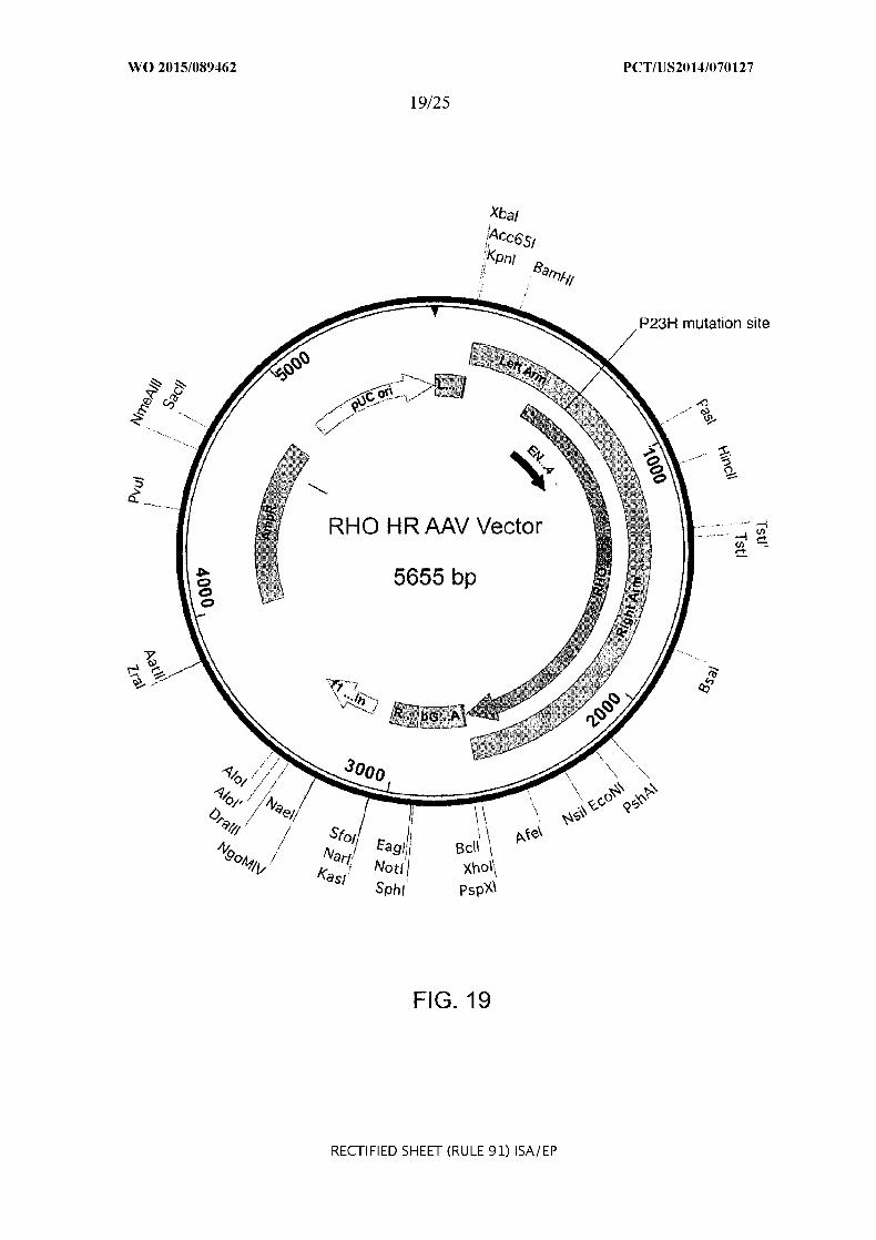
etc.) and soRNA targeting a locus or loci in the cell, e.g,, the gene RHO, A TOH1, VEGFA were
prepared, and are exemplified herein. These were advantageously delivered via a viral delivery
21

WO 2015/089462 PCT/US2014/070127
(AAV). When delivered via particles, the particles are formed by the Cas9 protein and the
sgRNA being admixed. The sgRNA and Cas9 protein mixture is admixed with a mixture
comprising or consisting essentially of or consisting of surfactant, phospholipid, biodegradable
polymer, lipoprotein and alcohol, whereby particles containing the sgRNA and Cas9 protein are
formed. The invention comprehends so making particles and particles from such a method as
well as uses thereof More generally, particles were formed using an efficient process. First, Cas9
protein and sgRNA targeting a gene or a control gene LacZ are mixed together at a suitable,
e.g.,3:1 to 1:3 or 2:1 to 1:2 or 1:1 molar ratio, at a suitable temperature, e.g., 15-30C, e.g., 20
25C, e.g., room temperature, for a suitable time, e.g., 15-45, such as 30 minutes, advantageously
in sterile, nuclease free buffer, e.g., IX PBS. Separately, particle components such as or
comprising: a surfactant, e.g., cationic lipid, e.g., I 2-dioileoyl-3-trimethylimmonium-propane
(DOTAP); phospholipid, e.g., dimyristoylphosphatidylcholi ne (DMPC); biodegradable polymer,
such as an ethylene-glycol polymer or PEG, and a lipoprotein, such as a low-density lipoprotein,.
e.g., cholesterol were dissolved in an alcohol, advantageously a C.1-6 alkyl alcohol, such as
methanol, ethanol, isopropanol, e.g., 100% ethanol. The two solutions are mixed together to form
particles containing the Cas9-sgRNA complexes. In certain embodiments the particle can contain
an HDR template. That can be a particle co-administered with sgRNA- Cas9 protein-containing
particle, or i.e., in addition to contacting a cell or cell population with an sgRNA-+Cas9 protein
containing particle, the cell or cell population is contacted with a particle containing an HDR
template; or the HSC is contacted with a particle containing all of the sgRNA, Cas9 and the HDR
template. The HDR template can be administered by a separate vector, whereby in a first
instance the particle penetrates an HSC cell and the separate vector also penetrates the cell,
wherein the HSC genome is modified by the sgRNACas9 and the HDR template is also
present, whereby a genomic loci is modified by the HDR; for instance, this may result in
correcting a mutation. The particle in the herein discussion is advantageously obtained or
obtainable from admixing an sgRNA(s) and Cas9 protein mixture (optionally containing HDR
template(s) or such mixture only containing HDR template(s) when separate particles as to
template(s) is desired) with a mixture comprising or consisting essentially of or consisting of
surfactant, phospholipid, biodegradable polymer, lipoprotein and alcohol (wherein one or more
sgRNA targets agenetic locus or loci associated with a mutation associated with an aberrant
protein xpression or with a disease condition or state).
22

WO 2015/089462 PCT/US2014/070127
[00281 In one aspect, the invention provides for methods of modeling a disease associated
with a genomic locus in a eukarvotic organism or a non-human organism comprising
manipulation of a target sequence within a coding, non-coding or regulatory element of said
genonic locus comprising delivering a non-naturally occurring or engineered composition
comprising
(A) - . a CRISPR-Cas system RNA polynucleotide sequence, wherein the
polynucleotide sequence comprises:
(a) a guide sequence capable of hybridizing to the target sequence,
(b) a tracr mate sequence, and
(c) a tracr sequence, and
IL a polynucleotide sequence encoding Cas9, optionally comprising at
least one or more nuclear localization sequences,
wherein (a), (b) and (c) are arranged in a 5' to 3' orientation,
wherein when transcribed, the tracr mate sequence hybridizes to the tracr sequence and the guide
sequence directs sequence-specific binding of a CRISPR complex to the target sequence, and
wherein the CRISPR complex comprises Cas9 complexed with (1) the guide sequence that is
hybridized to the target sequence, and (2) the tracr mate sequence that is hybridized to the traer
sequence and the polynucleotide sequence encoding Cas9 is DNA or RNA,
or
(B) . polynucleotides comprising:
(a) a guide sequence capable of hybridizing to the target sequence, and
(b) at least one or more tracr mate sequences,
IL a polynucleotide sequence encoding Cas9, and
IIL a polynucleotide sequence comprising a tracr sequence,
wherein when transcribed, the tracr mate sequence hybridizes to the tracr sequence and the guide
sequence directs sequence-specific binding of a CRISPR complex to the target sequence, and
wherein the CRJISPR complex comprises the Cas9 complexed with (1) the guide sequence that is
hybridized to the target sequence, and (2) the tracr mate sequence that is hybridized to the tracr
sequence, and the polynucleotide sequence encoding Cas9 is DNA or RNA.
100291 In certain preferred embodiments, the Cas9 is SaCas9.
23

WO 2015/089462 PCT/US2014/070127
[00301 In one aspect, the invention provides for methods of modeling a disease associated
with a genomic locus in a eukaryotic organism or a non-human organism comprising
manipulation of a target sequence within a coding, non-coding or regulatory element of said
genomic locus comprising delivering a non-naturally occurring or engineered composition
comprising a viral vector system comprising one or more viral vectors operably encoding a
composition for expression thereof, wherein the composition comprises:
(A) a non-naturally occurring or engineered composition comprising a vector
system comprising one or more vectors comprising
I. a first regulatory element operably linked to a CRISPR-Cas system RNA
polynucleotide sequence, wherein the polynucleotide sequence comprises
(a) a guide sequence capable of hybridizing to the target sequence,
(b) a tracr mate sequence, and
(c) a tracr sequence, and
II. a second regulatory element operably linked to an enzyme-coding
sequence encoding Cas9, (preferably SaCas9) optionally comprising at least one or more nuclear
localization sequences,
wherein (a), (b) and (c) are arranged in a 5' to 3' orientation,
wherein components I and II are located on the same or different vectors of the system,
wherein when transcribed, the tracr mate sequence hybridizes to the tracr sequence and the guide
sequence directs sequence-specific binding of a CRISPR complex to the target sequence, and
wherein the CRISPR complex comprises the Cas9 complexed with (1) the guide sequence that is
hybridized to the target sequence, and (2) the tracr mate sequence that is hybridized to the tracr
sequence,
or
(B) a non-naturally occurring or engineered composition comprising a vector
system comprising one or more vectors comprising
I. a first regulatory element operably linked to
(a) a guide sequence capable of hybridizing to the target sequence, and
(b) at least one or more tracr mate sequences,
IL. a second regulatory element operably linked to an enzyme-coding
sequence encoding Cas9, and
24

WO 2015/089462 PCT/US2014/070127
IIL a third regulatory element operably linked to a tracr sequence,
wherein components I, II and III are located on the same or different vectors of the system,
wherein when transcribed, the tracr mate sequence hybridizes to the tracr sequence and the guide
sequence directs sequence-specific binding of a CRISPR complex to the target sequence, and
wherein the CRISPR complex comprises the Cas9 complexed with (1) the guide sequence that is
hybridized to the target sequence, and (2) the tracr mate sequence that is hybridized to the tracr
sequence.
[00311 In one aspect the invention provides methods of treating or inhibiting a condition or a
disease caused by one or more mutations in a genomic locus in a eukaryotic organism or a non
human organism comprising manipulation of a target sequence within a coding, non-coding or
regulatory element of said genornic locus in a target sequence in a subject or a non-hunan
subject in need thereof comprising modifying the subject or a non-human subject by
manipulation of the target sequence and wherein the condition or disease is susceptible to
treatment or inhibition by manipulation of the target sequence comprising providing treatment
comprising:
delivering a non-naturally occurring or engineered composition comprising an AAV or
lentivirus vector system, comprising one or more AAV or lentivirus vectors operably encoding a
composition for expression thereof, wherein the target sequence is manipulated by the
composition when expressed, wherein the composition comprises:
(A) a non-naturally occurring or engineered composition comprising a vector
system comprising one or more vectors comprising
I. a first regulatory element operably linked to a CRISPR-Cas system RNA
polynucleotide sequence, wherein the polynucleotide sequence comprises
(a) a guide sequence capable of hybridizing to the target sequence in a
eukaryotic cell,
(b) a tracr mate sequence, and
(c) a traer sequence, and
IL a second regulatory element operably linked to an enzyme-coding
sequence encoding Cas9, preferably SaCas9, comprising at least one or more nuclear localization
sequences,
wherein (A), (b) and (c) are arranged in a 5' to 3' orientation,
25

WO 2015/089462 PCT/US2014/070127
wherein components I and II are located on the same or different vectors of the system,
wherein when transcribed, the tracr mate sequence hybridizes to the tracr sequence and the guide
sequence directs sequence-specific binding of a CRISPIR complex to the target sequence, and
wherein the CRISPR complex comprises the Cas9 complexed with (1) the guide sequence that is
hybridized to the arget sequence, and (2) the traer mate sequence that is hybridized to the tracr
sequence,
or
(B) a non-naturally occurring or engineered composition comprising a vector
system comprising one or more vectors comprising
L a first regulatory element operably linked to
(a) a guide sequence capable of hybridizing to an target sequence in a
eukaryotic cell, and
(b) at least one or more tracr mate sequences,
II. a second regulatory element operably linked to an enzyme-coding
sequence encoding Cas9, preferably SaCas9, and
IIIL a third regulatory element operably linked to a tracr sequence,
wherein components I, II and III are located on the same or different vectors of the system,
wherein when transcribed, the tracr mate sequence hybridizes to the tracr sequence and the guide
sequence directs sequence-specific binding of a CRISP3R complex to the target sequence, and
wherein the CRISPR complex comprises Cas9 comnplexed with (1) the guide sequence that is
hybridized to the target sequence, and (2) the tracr mate sequence that is hybridized to the tracr
sequence.
[00321 In certain embodiments, the invention provides method of preparing the AAV or
lentivirus vector for use in accordance with any of the methods of the invention, comprising
transfecting plasnid(s) containing or consisting essentially of nucleic acid molecule(s) coding
for the AAV or lentivirus into AAV-infected or lentivirus-infected cells, and supplying AAV
AAV or lentivirus rep and/or cap and/or helper nucleic acid molecules obligatory for replication
and packaging of the AAV or lentivirus.
[00331 In one aspect, the invention provides a composition for use in any of the methods of
invention (e.g., method of modeling a disease associated with a genetic locus in a eukaryotic
organism or a non-human organism) comprising manipulation of a target sequence within a
26

WO 2015/089462 PCT/US2014/070127
coding, non-coding or regulatory element of said genetic locus. In certain embodiments, the
invention provides for uses of the composition in ex vivo or in vivo gene or genome editing,
including therapeutic uses.
[00341 In one aspect, the invention provides a composition for use in the manufacture of a
medicament for in vitro, ex vivo or in vivo gene or genome editing or for use in a method of
modifying an organism or a non-human organism by manipulation of a target sequence in a
genomic locus associated with a disease or in a method of treating or inhibiting a condition or
diesease caused by one or more mutations in a genomic locus in a eukaryotic organism or a non
human organism.
[00351 In one aspect, the invention provides a composition comprising:
(A) - . a CRISPR-Cas system RNA polynucleotide sequence, wherein the
polynucleotide sequence comprises:
(a) a guide sequence capable of hybridizing to a target sequence in a
eukaryotic cell,
(b) a tracr mate sequence, and
(c) a tracr sequence, and
IL a polynucleotide sequence encoding Cas9, preferably Sa Cas9, optionally
comprising at least one or more nuclear localization sequences,
wherein (a), (b) and (c) are arranged in a 5' to 3' orientation,
wherein when transcribed, the tracr mate sequence hybridizes to the tracr sequence and the guide
sequence directs sequence-specific binding of a CR] SPR complex to the target sequence, and
wherein the CRISPR complex comprises the Cas9 compilexed with (1) the guide sequence that is
hybridized to the target sequence, and (2) the tracr mate sequence that is hybridized to the tracr
sequence and the polynucleotide sequence encoding Cas9 is DNA or RNA,
or
(B) . polynucleotides comprising:
(a) a guide sequence capable of hybridizing to an target sequence in a
eukaryotic cell, and
(b) at least one or more tracr mate sequences,
IL a polynucleotide sequence encoding Cas9, preferably SaCas9, and
II. a polynucleotide sequence comprising a tracr sequence,
27

WO 2015/089462 PCT/US2014/070127
wherein when transcribed, the tracr mate sequence hybridizes to the tracr sequence and the guide
sequence directs sequence-specific binding of a CRISPR complex to the target sequence, and
wherein the CRISPR complex comprises SaCas9 complexed with (1) the guide sequence that is
hybridized to the target sequence, and (2) the tracr mate sequence that is hybridized to the tracr
sequence, and the polynucleotide sequence encoding Cas9 is DNA or RNA;
for use in medicine or therapy; or for use in a method of modifying an organism or a non-human
organism by manipulation of a target sequence in a genomic locus associated with a disease or
disorder; or for use in a method of treating or inhibiting a condition caused by one or more
mutations in a genetic locus associated with a disease in a eukaryotic organism or a non-human
or ganism.; or for use in in vitro, ex vivo or in vivo gene or genome editing.
[00361 In one aspect, the invention provides a therapeutic genome editing method for
treating or inhibiting a condition or a disease caused by one or more mutations in a genomic
locus in a eukaryotic organism or a non-human organism comprising manipulation of a target
sequence within a coding, non-coding or regulatory element of said genomic locus in a target
sequence in a subject or a non-human subject in need thereof comprising modifying the subject
or a non-human subject by manipulation of the target sequence and wherein the condition or
disease is susceptible to treatment or inhibition by manipulation of the target sequence
comprising providing treatment comprising:
delivering a non-naturally occurring or engineered composition comprising an AAV or
lentivirus vector system, comprising one or more A-AV or lentivirus vectors operably encoding a
composition for expression thereof, wherein the target sequence is manipulated by the
composition when expressed, wherein the composition comprises:
(A) a non-naturally occurring or engineered composition comprising a vector system
comprising one or more vectors comprising
I a first regulatory element operably linked to a CRISPR-Cas system RNA
polynucleotide sequence, wherein the polynucleotide sequence comprises
(a) a guide sequence capable of hybridizing to the target sequence in a eukaryotic
cell,
(b) a tracr mate sequence, and
(c) a tracr sequence, and
28

WO 2015/089462 PCT/US2014/070127
II. a second regulatory element operably linked to an enzyme-coding sequence
encoding Cas9, preferably SaCas9, comprising at least one or more nuclear localization
sequences,
wherein (a), (b) and (c) are arranged in a 5' to 3' orientation,
wherein components I and II are located on the same or different vectors of the system,
wherein when transcribed, the tracr mate sequence hybridizes to the tracr sequence and
the guide sequence directs sequence-specific binding of a CRISPR complex to the target
sequence, and
wherein the CRISPR complex comprises the Cas9 complexed with (1) the guide
sequence that is hybridized to the arget sequence, and (2) the tracr mate sequence that is
hybridized to the tracr sequence,
or
(B) a non-naturally occurring or engineered composition comprising a vector system
comprising one or more vectors comprising
. a first regulatory element operably linked to
(a) a guide sequence capable of hybridizing to an target sequence in a eukaryotic cell,
and
(b) at least one or more tracr mate sequences,
II. a second regulatory element operably linked to an enzyme-coding sequence
encoding SaCas9, and
Ill. a third regulatory element operably linked to a traer sequence,
wherein components I, II and III are located on the same or different vectors of the
system,
wherein when transcribed, the tracr mate sequence hybridizes to the tracr sequence and
the guide sequence directs sequence-specific binding of a CRISPR complex to the target
sequence, and
wherein the CRISPR complex comprises Cas9 complexed with (1) the guide sequence
that is hybridized to the target sequence, and (2) the tracr mate sequence that is hybridized to the
tracr.
100371 In one aspect, the invention provides a method of individualized or personalized
treatment of a genetic disease in a subject in need of such treatment comprising:
29

WO 2015/089462 PCT/US2014/070127
(a) introducing multiple mutations ex vivo in a tissue, organ or a cell line comprising
Cas9-expressing eukaryotic cell(s) (preferably Sa Cas9), or in vivo in a transgenic non-human
mammal having cells that express Cas9, comprising delivering to cell(s) of the tissue, organ, cell
or mammal the vector as herein-discussed, wherein the specific mutations or precise sequence
substitutions are or have been correlated to the genetic disease;
(b) testing treatment(s) for the genetic disease on the cells to which the vector has been
delivered that have the specific mutations or precise sequence substitutions correlated to the
genetic disease; and
(c) treating the subject based on results from the testing of treaunent(s) of step (b).
[00381 In certain embodiments of any of the aforementioned aspects and embodiments of the
invention, the viral vector may be an AAV, e.g., AAVI, AAV2, AAV5, AAV7, AAV8, AAV DJ
or any combination thereof.
[00391 In herein discussions concerning the target being associated with a mutation or with a
disease condition, such mutation or disease condition can be, for instance a neuronal disease;
ocular disease (e.g., retina disease, e.g., retinitis pigmentosa; achromtaopsia; age-related macular
degeneration; visual impairment), auditory disease (e.g., cochlear-cell associated disease, hearing
impairment, deafness) etc.
[00401 Accordingly, it is an object of the invention to not encompass within the invention
any previously known product, process of making the product, or method of using the product
such that Applicants reserve the right and hereby disclose a disclaimer of any previously known
product, process, or method. It is further noted that the invention does not intend to encompass
within the scope of the invention any product, process, or making of the product or method of
using the product, which does not meet the written description and enablement requirements of
the USPTO (35 U.S.C. § 112, first paragraph) or the EPO (Article 83 of the EPC), such that
Applicants reserve the right and hereby disclose a disclaimer of any previously described
product, process of making the product, or method of using the product.
[00411 It is noted that in this disclosure and particularly in the claims aid/or paragraphs,
terms such as "comprises", "comprised", "comprising" and the like can have the meaning
attributed to it in U.S. Patent law; e.g., they can mean "includes", "included", "including", and
the like; and that terms such as "consisting essentially of" and "consists essentially of' have the
meaning ascribed to them in U.S. Patent law, e.g., they allow for elements not explicitly recited,
30

WO 2015/089462 PCT/US2014/070127
but exclude elements that are found in the prior art or that affect a basic or novel characteristic of
the invention. It may be advantageous in the practice of the invention to be in compliance with
Art. 53(c) EPC and Rule 28(b) and (c) EPC. There are no promises in this document.
[00421 These and other embodiments are disclosed or are obvious from and encompassed by,
the following Detailed Description.
BRIEF DESCRIPTION OF THE DRAWINGS
[00431 The novel features of the invention are set forth with particularity in the appended
claims. A better understanding of the features and advantages of the present invention will be
obtained by reference to the following detailed description that sets forth illustrative
embodiments, in which the principles of the invention are utilized, and the accompanying
drawings of which:
[00441 Figure 1A-H shows CRISPR-Cas9 system delivery and targeting of Mecp2 locus in the
mouse brain. (a) AAV-SpCas9 and AAV-SpGuide(Mlecp2) expression vectors. The sgRNA
vector contains encoding sequence of the GFP-KASH fusion protein for identification of
transduced neurons. (b) Expression of IA-Cas9 and GFP-KASI in the dorsal dentate gyrus
(DG) of mouse hippocampus. Scale bar, 100 pm. (c) Quantification of cells efficiently targeted
by the dual-vector Cas9-CRISPR system. (d) Graphical representation of the mouse cecp2 locus
showing Cas9 target location; sgRNA indicated in blue. PAM sequence marked in purple.
Representative mutation patterns detected by sequencing of Mecp2 locus were shown below:
green - wild-type sequence; red dashes - deleted bases; red bases: insertion or nmtations; red
arrowhead indicates CRISPR-Cas9 cutting site. (e) SURVEYORTM' assay gel showing
modification of the Miep2 locus, 2 weeks after AAV delivery in the DG region. (f) Western blot
analysis of MeCP2 protein expression in the targeted brain region and quantification of MeCP2
protein levels in dorsal DG (t-test, **p<0.001, n:::4 from 3 animals, error bars: s.e.m.). (g) Images
of the dorsal DG region, 2 weeks after CRISPR-Cas9 targeting of Mecp2 locus. Scale bar, 150
qm. (h) Quantification of MeCP2 positive cells population within all detected cells (DAPI
staining) in the targeted brain region in compare to control collateral site (t-test, ****p<0.000i,
n=290 and 249 cells from 2 animals, respectively; error bars: s.e.m). (ITR - inverted terminal
repeat; HA --- hemagglutinin tag; NLS --- nuclear localization signal; spA --- synthetic
polyadenylation signal; U6 - PollI promoter; sgRNA - single guide RNA; hSyn - human
synapsin I promoter; GFP- green fluorescent protein; KASI - Klarsicht, ANC], Syne Homology
31

WO 2015/089462 PCT/US2014/070127
nuclear transmembrane domain; bGH pA bovine growth hormone polyadenylatio signal;
WPRE - Woodchuck Hepatitis virus posttranscriptional regulatory element).
[00451 Figure 2A-B shows analysis of gene expression in Cas9-mediated MeCP2 knockdown
neurons. (a) Strategy for cell nuclei purification of CRISPR-Cas9 targeted cells from the mouse
brain. (b) Hierarchical clustering of differentially expressed genes (t-test, P<0.01, n=19
populations of sorted nuclei from 8 animals) detected by RNAseq. Relative log2(TPM+1)
expression levels of genes are normalized for each row and displayed in red-blue color scale.
Each column represents a population of targeted 100 neuronal nuclei FACS sorted from the
isolated, dentate gyrus population of cells, either from control or Acp2 sgRNA transduced
animals, as indicated.
[00461 Figure 3A-E shows cell-autonomous defects in cellular response properties of neurons
after CRISPR-mediated [MIeCP2 knockdown. (a) Cartoon showing in vivo experiment
configuration from mouse visual cortex and visual stimulation parameter. GFP' neuron is shown.
Scale bar, 20 un. (b) Cartoon showing recording configuration in layer 2/3 excitatory neurons
that receive both contra- and ipsilateral eye specific input. Genome modified GFP* cells are in
green whereas unmodified cells are in gray. Normalized spike shape shows regular spiking
excitatory neurons. (cd) Average OSI (c) and evoked FR (d) were measured from GFPF cells
expressing MecpI and control sgRNA, respectively ([-test, *p<0.05; numbers in graph indicate
numbers of recorded cells; n=2-3 animals; error bars: s.e.m).
[00471 Figure 4A-F shows simultaneous, multiplex gene editing in the mouse brain. (a)
Schematic illustration of CRISPR-Cas9 system designed for multiplex genome targeting. (b)
Graphical representation of targeted DNMT mouse loci. Guide RNAs are indicated in blue. PAM
sequences are marked in purple. (c) SURVEYORTM assay gel showing modification of DNMTs
loci in FACS sorted GFP-KASH positive cells, 4 weeks after AAV delivery in the DG region.
(d) Deep sequencing-based analysis of DNMTs loci modification in single cells, showing co
occurrence of modification in multiple loci. (e) Western blot analysis for Dnmt3a and Dnmtl
proteins after in vivo delivery of CRISPR-Cas9 system targeting DNMT family genes (top).
Western blot quantification of Dnmt3a and Dnmtl protein levels in DG after in vivo CRISPR
Cas9 targeting (bottom; t-test, **p<0.001, *p<0.05, Dnmt3a: n=7; Dnmtl: n=5 from 5 animals;
error bars: s.e.m). (f) Contextual learning deficits, 8 weeks after targeting of DN MT genes using
32

WO 2015/089462 PCT/US2014/070127
SpCas9 in the DG region of hippocampus, tested in training and altered context (t-test,
***p<0.0001, n=18 animals, 2 independent experiments; error bars: s.e.m).
100481 Figure 5A-F shows cloning and expression of HA-tagged SpCas9 (HA-SpCas9) for
AAV packaging. (a) Schematic overview of different cloning strategies to minimize SpCas9
expression cassette size using short rat Maplb pronotor (pMaplb), a truncated version of the
mouse Mecp2 promoter (pMecp2) and a short polyA motif (spA). (b) Western blot analysis of
primary cortical neuron culture expressing HA-SpCas9 using different SpCas9 expression
cassettes. (e) Mecp2 promoter drives HA-SpCas9 (red) expression in neurons (Map1 b, NeuN;
arrows) but not in astroglia (GFAP, arrowheads). Co-expressioin of HA-SpCas9 with GFP
KASH is shown (bottom). Nuclei were labeled with DAPI (blue). Scale bars, 20 lam. (d)
Schematic overview of GFP-labeling. Enhanced green fluorescent protein (GFP) fused to the
nuclear transmembrane KASH domain and integration of GFP-KASH to the outer nuclear
membrane is illustrated. (e) Co-infection efficiency calculation, showing populations of cell
expressing both HA-SpCas9 and GFP-KAS-l (n:=973 neurons from 3 cultures; error bars: s.e.m).
() Cells were stained with LIFE/DEAD" kit 7 days after virus delivery. Quantification of DAPI
and dead (DEAl) cells (control n=518 DAPFI nuclei; SpCas9/GFP-KASF n=1003 DAPI
nuclei from 2 cultures; error bars: s.e.m). (ITR - inverted terminal repeat; HA - hemagglutinin
tag; NLS - nuclear localization signal; spA - synthetic polyadenylation signal; U6 - PolIII
promoter; sgRNA - single guide RNA; hSyn - human synapsin 1 promoter; GFP- green
fluorescent protein; KASH - Klarsiclit, ANCI, Syne Homology nuclear transmnembrane domain;
bGH pA - bovine growth hormone polyadenylation signal; WPRE -- Woodchuck Hepatitis virus
posttranscriptional regulatory element).
[00491 Figure 6A-B shows targeting of Mecp2 in Neuro-2a cells. (a) Vecp2 targeting sequences
and corresponding protospacer adjacent motifs (PAM). (b) Evaluation of 6 Mecp2 sgRNAs co
transfected with SpCas9 into Neuro-2a cells. Locus modification efficiencies were analyzed 48 h
after transfection using SURVEYORTM assay.
[00501 Figure 7A-D shows CRISPR-SpCas9 targeting of Mecp2 in primary cortical neurons.
(a) Immunofluorescent staining of MeCP2 (red) in cultured neurons 7 days after AAV-CRISPR
transduction (green, GFP-KASH). Nuclei were labeled with DAPI (blue). Scale bar. 20 pam. (b)
Evaluation of Mecp2 locus targeting using SpCas9 or dSpCas9, together with Mecp2 sgRNA or
control (targeting bacterial iacZ gene) sgRNA, using SURVEYORTM assay gel. (e)
33

WO 2015/089462 PCT/US2014/070127
Quantification of MeCP2 positive nuclei in targeted population of neurons (GFP). (Id) Western
blot of MeCP2 protein levels after CRISPR-SpCas9 targeting of Mecp2 locus and quantification
of [MIeCP2 protein levels (t-test, * *p<0.00 1, n=5 from 3 cultures, error bars: s.e.m).
[00511 Figure 8A-E shows morphological changes in dendritic tree of neurons after SpCas9
mediated MeCP2 knockdown in vitro. (a) Reduced complexity of dendritic tree in neurons after
CRISPR-SpCas9 targeting of Mecp2 locus. Scale bar. 20 pim. (b) Changes in dendritic spines
morphology in neurons targeted with SpCas9 and Mfecp2 sgRNA. Scale bar, 10 jpm. Morphology
of cells was visualized with co-transfection with mCherrv construct. Cells for morphology
analysis were chosen based on the result of Mecp2 staining. (c) Dendritic tree morphology
assessed with number of dendritic ends and (d) Sholl analysis (t-test, ***p<0.0001, n=40 from 2
cultures). (e) Spine density quantification ([-test, ***p<0.0001, n::40 from 2 cultures, error bars:
s.e.m).
[00521 Figure 9 shows RNAseq of neuronal nuclei from control animals and SpCas9-mediated
Meep2 knockdown. Box plot presenting the number of detected genes across the RNA-seq
libraries (19 libraries each of 100 nuclei taken from control sgRNA or Mecp2 sgRINA transduced
nuclei; n=::4 animals/group) per quanti le of expression level. All genes are divided to 10 quantiles
by their mean log2(TPM+_1i) expression level, then for each quantile the number of genes that are
detected (log2(TPM+ 1)>2) was counted in each sample. The three target sequences shown are
SEQ ID NO: , SEQ ID NO: and SEQ ID NO: , for Dnmt3a, Dnmtl and Dnmt3b,
respectively.
100531 Figure 10 A-B shows multiplex genome targeting of DNMT family members in vitro. (a)
Dnnt3a, Dnmnt] and Dnnt3b targeting sequences and corresponding protospacer adjacent motifs
(PAM). (b) SURVEYORTM nuclease assay analysis of Neuro-2a cells 48 hours after transfection
with SpCas9 and DNMT 3xsgRNA vector targeting Dnnt3a, Dnmtl and Dnnt3b loci. Efficient
genome editing of all three targeted genes is shown.
100541 Figure 11 A-C shows next generation sequencing of targeted Dnmt3a, Dunti and
Dnnt3b loci. Examplsof seqUcingC f reuts of mutated Dnnt3a (a), Dnmul (b) and Dnnt3b (c)
loci after in vivo delivery of SpCas9 and DNMT 3xsgRNA into the mouse dentate gyrus. Green:
wild-type sequence, red dashes: deleted bases, red bases: insertion or mutations. Red arrowheads
indicate CRISPR-SpCas9 cutting site. The full sequences used in this figure are provide as SEQ
ID NO: , SEQ ID NO: , and SEQ ID NO: for the Dnmt3a, the Dnmti and the Dnmt3b loci,
34

WO 2015/089462 PCT/US2014/070127
respectively. They are: SEQ ID NO: (Dnmt3a): CCT CCG TGT CAG CGA CCC ATG CCA A,
SEQ ID NO: (Dnmtil): CCA GCG TCG AAC AGC TCC AGC CCG and SEQ ID NO:
(Dnmt3b) AGA GGG TGC CAG CGG GTA TAT GAG G
[00551 Figure 12 shows comparison of different programmable nuclease platforms.
[0056] Figure 13A-C show types of therapeutic genome modifications. The specific type of
genome editing therapy depends on the nature of the mutation causing disease. a, In gene
disruption, the pathogenic function of a protein is silenced by targeting the locus with NHEIJ.
Formation of indels on the gene of interest often result in frameshift mutations that create
premature stop codons and a non-functional protein product, or non-sense mediated decay of
transcripts, suppressing gene function. b, HDR gene correction can be used to correct a
deleterious nation. DSB is targeted near the mutation site in the presence of an exogenously
provided, corrective HDR template. HDR repair of the break site with the exogenous template
corrects the mutation, restoring gene function. e, An alternative to gene correction is gene
addition. This mode of treatment introduces a therapeutic transgene into a safe-harbor locus in
the genome. DSB is targeted to the safe-harbor locus and an HDR template containing homology
to the break site, a promoter and a transgene is introduced to the nucleus. HDR repair copies the
promoter-transgene cassette into the safe-harbor locus, recovering gene function, albeit without
true physiological control over gene expression.
[00571 Figure 14 shows a schematic representation of ex vivo vs. in vivo editing therapy. In
ex vivo editing therapy cells are removed fronm a patient, edited and then re-engrafted (top panel).
For this mode of therapy to be successful, target cells must be capable of survival outside the
body and homing back to target tissues post-transplantation. In vivo therapy involves genome
editing of cells in situ (bottom panels). For in vivo systemic therapy, delivery agents that are
relatively agnostic to cell identity or state would be used to effect editing in a wide range of
tissue types. Although this mode of editing therapy may be possible in the future, no delivery
systems currently exist that are efficient enough to make this feasible. In viva targeted therapy,
where delivery agents with tropisnm for specific organ systems are administered to patients are
feasible with clinically relevant viral vectors.
[00581 Figure 15 shows SaCas9 system for ocular gene therapy
100591 Figure 16 shows a schematic representation of gene therapy via Cas9 Homologous
Recombination (HR) vectors.
35

WO 2015/089462 PCT/US2014/070127
[00601 Figure 17 shows an exemplary protocol for ocular gene therapy.
[00611 Figure 18A-B show the human R110 locus (allele showing P23H mutation). Fig. 7A
shows the guide design for RHO locus. Fig. 7B shows the in vitro guide screening results using
the SURVEYOR assay.
[00621 Figure 19 shows R1-I HR AAV vector.
[00631 Figure 20A-B shows guide selection for CNGA3 and CNGB3. (a) shows human
CNGA3 locus (allele showing two disease mutations) and guide selection. (b) shows human
CNGB3 locus (allele showing disease mutation) and guide selection.
[00641 Figure 21 shows CNGA3 HR AAV vector.
[00651 Figure 22 shows CNGB3 HIR AAV vector.
[00661 Figure 23A-B show guide selection for VEGFA. (a) shows human VEGFA icous
(common region 1); (b) shows human VEGFA Icons (common region 2);
[00671 Figure 24 shows design of dCas9-based epigenetic modulation system (3 components
of the system, dSaCas9, fusion effector, and sgRNA are shown).
[00681 Figure 25A-C shows guide selection for ATOHI. (a) shows two higly accessible
regions which were selected; (b) shows highly accessible region I-blue lines indicate guide
sequence and magenta lines indicate PANi; (c) shows highly accessible region 2-blue lines
indicate guide sequence and magenta lines indicate PAM.
[00691 The figures herein are for illustrative purposes only and are not necessarily drawn to
scale.
DETAILED DESCRIPTION OF THE INVENTION
[0070] With respect to general information on CRISPR-Cas Systems, components thereof,
and delivery of such components, including methods, materials, delivery vehicles, vectors,
particles, AAV, and making and using thereof, including as to amounts and formulations, all
useful in the practice of the instant invention, reference is made to: US Patents Nos. 8,697,359,
8,771,945, 8,795,965, 8,865,406, 8,871,445, 8,889,356, 8,889,418 and 8,895,308; US Patent
Publications US 2014-0310830 (US APP. Ser. No. 14/105,031), US 2014-0287938 Al (U.S.
App. Ser. No. 14/213,991), US 2014-0273234 Al (U.S. App. Ser. No. 14/293,674), US2014
0273232 Al (U.S. App Ser. No. 14/290,575), TS 2014-0273231 (U,S, App. Ser. No.
14/259,4420), US 2014-0256046 Al (U.S. App. Ser. No. 14/226,274), US 2014-0248702 Al
(UT.S. App. Ser. No. 14/258,458), US 2014-0242700 Al (U,S, App. Ser. No. 14/222,930), US
36

WO 2015/089462 PCT/US2014/070127
2014-0242699 Al (U.S. App. Ser. No. 14/183,512), US 2014-0242664 Al (U.S. App. Ser. No.
14/104,990), US 2014-0234972 A] (U.S. App. Ser. No. 14/183,471), US 2014-0227787 Al
(U.S. App. Ser. No. 14/256,912), US 2014-0189896 Al (U.S. App. Ser. No. 14/105,035), US
2014-0186958 (U.S. App. Ser. No. 14/105,017), US 2014-0186919 Al (U.S. App. Ser. No.
14/104,977), US 2014-0186843 Al (U.S. App. Ser. No. 14/104,900), US 2014-0179770 Al
(U.S. App. Ser. No. 14/104,837) and US 2014-0179006 Al (U.S. App. Ser. No. 14/183,486), US
2014-0170753 (US App Ser No 14/183,429); European Patent Applications P 2 771 468
(EP13818570.7). EP 2 764 103 (EP138242326), and EP 2 784 162 (EP14170383.5); and PCT
Patent Publications WO 2014/093661 (PCT/US2013/074743), WO 2014/093694
(PCT/US2013/074790), WO 2014/093595 (PCT/US2013/07461 1), WO 2014/093718
(PCT/US201 3/074825), WO 2014/093709 (PCTUIJS2013/074812), WO 2014/093622
(PCT/lUS2013/074667), WO 2014/093635 (PCT/US20 13/074691), WO 2014/093655
(PCT/US20 13/074736), WO 2014/093712 (PCT/US20 13/074819), W020 14/093701
(PCT/US20 13/074800), and W'2014/018423 (PCI/US2013/051418). Reference is also made to
US provisional patent applications 61/758.468; 61/802,174; 61/806,375; 61/814,263; 61/819,803
and 61/828,130, filed on January 30, 2013; March 15, 2013; March 28, 2013; April 20, 2013;
May 6, 2013 and May 28, 2013 respectively. Reference is also made to US provisional patent
application 61/836,123, filed on June 17, 2013. Reference is additionally made to US provisional
patent applications 61/835,931, 61/835,936, 61/836,127, 61/836, 101, 61/836,080 and
61/835,973, each filed June 17, 2013. Further reference is made to US provisional patent
applications 61/862,468 and 61/862,355 filed on August 5, 2013; 61/871,301 filed on August 28,
2013; 61/960,777 filed on September 25, 2013 and 61/961,980 filed on October 28, 2013.
Reference is yet further made to: PCI Patent applications Nos: PCI/US2014/041803,
PCT/US2014/041800, PCT/US2014/041809, PCT/US2014/041804 and PCT/US2014/041806,
each filed June 10, 2014 6/10/14; PCT/LT2014/041808 filed June 11, 2014; and
PCT/US2014/62558 filed October 28, 2014, and US Provisional Patent Applications Serial Nos.:
61/915,150, 61/915,301, 61/915,267 and 61/915,260, each filed December 12, 2013; 61/757,972
and 61/768,959, filed on January 29, 2013 and February 25, 2013; 61/835,936, 61/836,127,
61/836,101, 61/836,080, 61/835,973, and 61/835,931, filed June 17, 2013; 62/010,888 and
62/010,879, both filed June 11, 2014; 62/010,329 and 62/010,441, each filed June 10, 2014;
61/939,228 and 61/939,242, each filed February 12, 2014; 61/980,012, filed April 15,2014;
37
WO 2015/089462 PCT/US2014/070127
62/038,358, filed August 17, 2014; 62/054,490, 62/055,484, 62/055,460 and 62/055,487, each
filed September 25, 2014; and 62/069,243, filed October 27, 2014. Reference is also made to US
provisional patent applications Nos. 62/055,484, 62/055,460, and 62/055,487, filed September
25, 2014; US provisional patent application 61/980,012, filed April 15, 2014; and US provisional
patent application 61/939,242 filed February 12, 2014. Reference is made to PCIT application
designating, inter alia, the United States, application No. PCT/US14/41806, filed June 10, 2014.
Reference is made to US provisional patent application 61/930,214 filed on January 22, 2014.
Reference is made to US provisional patent applications 61/915,251; 61/915,260 and
61/915,267, each filed on December 12, 2013. Reference is made to US provisional patent
application USSN 61/980,012 filed April 15, 2014. Reference is made to PCIT application
designating, inter alia, the United States, application No. PCT/LT14/41806, filed June 10, 2014.
Reference is made to US provisional patent application 61/930,214 filed on January 22, 2014.
Reference is made to US provisional patent applications 61/915,251; 61/915,260 and
61/915,267, each filed on December 12, 2013. Each of these patents, patent publications, and
applications, and all documents cited therein or during their prosecution ("appln cited
documents") and all documents cited or referenced in the appln cited documents, together with
any instructions, descriptions, product specifications, and product sheets for any products
mentioned therein or in any document therein and incorporated by reference herein, are hereby
incorporated herein by reference, and may be employed in the practice of the invention. All
documents (e.g., these patents, patent publications and applications and the appln cited
documents) are incorporated herein by reference to the same extent as if each individual
document was specifically and individually indicated to be incorporated by reference.
[00711 Also with respect to general information on CRISPR-Cas Systems, mention is made
of the following (also hereby incorporated herein by reference):
* Multiplex genoine engineering using CRJSPR/Cas systems. Cong, L., Ran, F.A., Cox, D., Lin, S., Barretto, R., Habib, N., Hsu, P.D., Wu, X., Jiang, W., Marraffini, L.A., & Zhang, F. Science Feb 15;339(6121):819-23 (2013);
> RNA-guided editing of bacterial genomes using CRISPR-Cas svstenis. Jiang W., Bikard D., Cox D., Zhang F, Marraffini LA. Nat Biotechnol Mar;31(3):233-9 (2013);
> One-Step Generation of Mice Carrying Mutations in Multiple Genes by CRJSPR/CsMediated Genone Engineering. Wang [., Yang H., Shivalila CS., Dawlaty MM., Cheng AW., Zhang F., Jaenisch R. Cell May 9;153(4):910-8 (2013);
38

WO 2015/089462 PCT/US2014/070127
> Optical control of mammalian endogenous transcription ant epigenetic states. Konermann S, Brigham MD, Trevino AE, Hsu PD, Heidenreich M, Cong L, Platt RI, Scott DA, Church GM, Zhang F. Nature. 2013 Aug 22;500(7463):472-6. doi: 10.1038/Nature12466. Epub 2013 Aug 23;
> Double Nicking by RNA-Guided CRISPR Cas9 for Enhanced Genoie Editing Specificity. Ran, FA., Isu, PD., Lin, CY., Gootenberg, JS., Konermann, S., 'Trevino, AE., Scott, DA., Inoue, A., Matoba, S., Zhang, Y., & Zhang, F. Cell Aug 28. pii: S0092-8674(13)01015-5. (2013);
> DNA targeting specificity of RNA- guided Cas9 nucleases. Hsu, P., Scott, D., Weinstein, J., Ran, FA., Konermann, S., Agarwala, V., Li, Y., Fine, E., Wu, X., Shalem, 0., Cradick, TJ., Marraffini, LA., Bao, G., & Zhang, F. Nat Biotechnol doi:10.1038/nbt.2647 (2013);
> Genome engineering using the CRISPR-Cas9 system. Ran, FA., Hsu, PD., Wright, J., Agarwala, V., Scott, DA., Zhang, F. Nature Protocols Nov;8(i 1):2281-308. (2013);
> Genome-Scale CRISPR-Cas9 Knockout Screening in Human Cells. Shalem, 0., Saiana, NE., Hartenian, E., Shi, X., Scott, DA., Mikkelson, T., Heckl, D., Ebert, BL., Root, DE., Doench, JG., Zhang, F. Science Dec 12. (2013). [Epub ahead of print];
Crystal structure ofcas9 in complex with guide RNA and target DNA. Nishimasu, H., Ran, FA., Hsu, PD., Konermann, S., Shehata, SI., Dohmae, N., Ishitani, R., Zhang, F., Nureki, 0. Cell Feb 27. (2014). 156(5):935-49;
> Genone-wide binding of the CRISPR endonuclease Cas9 in mammalian cells. Wu X.. Scott DA., Kriz A., Chiu AC., Hsu PD., Dadon DB., Cheng AW., Trevino AE., Konermain S., Chen S., Jaenisch R., Zhang F., Sharp PA. Nat Biotechnol. (2014) Apr 20. doi: 10.1038/nbt.2889,
> CRISPR--Cas9 Knockin Alice for Genome Editing and Cancer Modeling, Platt et al.. Cell 159(2): 440-455 (2014) DOI: 10.1016/j.cell.2014.09.014,
> Development and Applications of CRISPR-Cas9 for Genome Engineering, Hsu et al, Cell 157, 1262-1278 (June 5, 2014) (Fu 2014),
> Genetic screens in human cells using the CRISPR/Cas9 system, Wang et al., Science. 2014 January 3; 343(6166): 80-84. doi:10.11 26/science.1246981,
> Rational design of highlv active sgRNAs for CRISPR-Cas9-mediated gene inactivation, Doench et al., Nature Biotechnology published online 3 September 2014; doi:10.1038/nbt.3026, and
> In vivo interrogation of gene function in the mammalian brain using CIJSPR-Cas9, Swiech et al, Nature Biotechnology ; published online 19 October 2014; doi:10.1038/nbt.3055.
39

WO 2015/089462 PCT/US2014/070127
each of which is incorporated herein by reference, and discussed briefly below:
> Cong et al. engineered type 1I CRISPR/Cas systems for use in eukaryotic cells based on
both Streptococcus therniophilus Cas9 and also Streptoccocus pyogenes Cas9 and
demonstrated that Cas9 nucleases can be directed by short RNAs to induce precise
cleavage of DNA in human and mouse cells. Their study further showed that Cas9 as
converted into a nicking enzyme can be used to facilitate homology-directed repair in
eukaryotic cells with minimal mutagenic activity. Additionally, their study demonstrated
that multiple guide sequences can be encoded into a single CRISPR array to enable
simultaneous editing of several at endogenous genomic loci sites within the mammalian
genome, demonstrating easy programmability and wide applicability of the kNA-guided
nuclease technology. This ability to use RNA to program sequence specific DNA
cleavage in cells defined a new class of genome engineering tools. These studies further
showed that other CRISPR loci are likely to be transplantable into mammalian cells and
can also mediate mammalian genome cleavage. Importantly, it can be envisaged that
several aspects of the CRISPR/Cas system can be further improved to increase its
efficiency and versatility.
> Jiang et al used the clustered, regularly interspaced, short palindromic repeats
(CRISPR)-associated Cas9 endonuclease complexed with dual-RNAs to introduce
precise mutations in the genomes of Streptococcus pneumoniae and Escherichia coli. The
approach relied on dua l-RNA:Cas9-directed cleavage at the targeted genomic site to kill
unmutated cells and circumvents the need for selectable markers or counter-selection
systems. The study reported reprogramming dual-RNA:Cas9 specificity by changing the
sequence of short CRISPR RNA (crRNA) to make single- and multinucleotide changes
carried on editing templates. The study showed that simultaneous use of two crRNAs
enabled multiplex mutagenesis. Furthennore, when the approach was used in
combination with recombineering, in S. pneumoniae, nearly 100% of cells that were
recovered using the described approach contained the desired mutation, and in E. coli,
65% that were recovered contained the mutation.
> Konerman et al. addressed the need in the art for versatile and robust technologies that
enable optical and chemical modulation of DNA-binding domains based CRISPR Cas9
enzyme and also Transcriptional Activator Like Effectors
40

WO 2015/089462 PCT/US2014/070127
> Cas9 nuclease from the microbial CRISPR-Cas system is targeted to specific genomic
loci by a 20 nt guide sequence, which can tolerate certain mismatches to the DNA target
and thereby promote undesired off-target mutagenesis. To address this, Ran et al.
described an approach that combined a Cas9 nickase mutant with paired guide RNAs to
introduce targeted double-strand breaks. Because individual nicks in the genome are
repaired with high fidelity, simultaneous nicking via appropriately offset guide RNAs is
required for double-stranded breaks and extends the number of specifically recognized
bases for target cleavage. The authors demonstrated that using paired nicking can reduce
off-target activity by 50- to 1,500-fold in cell lines and to facilitate gene knockout in
mouse zygotes without sacrificing on-target cleavage efficiency. This versatile strategy
enables a wide variety of genorne editing applications that require high specificity.
> Hsu et al. characterized SpCas9 targeting specificity in human cells to inform the
selection of target sites and avoid off-target effects. The study evaluated >700 guide RNA
variants and SpCas9-induced indel mutation levels at >100 predicted genomic off-target
loci in 293T and 293FT cells. The authors reported that SpCas9 tolerates mismatches
between guide RNA and target DNA at different positions in a sequence-dependent
manner, sensitive to the number, position and distribution of mismatches. The authors
further showed that SpCas9-mediated cleavage is unaffected by DNA methylation and
that the dosage of SpCas9 and sgRNA can be titrated to minimize off-target modification.
Additionally, to facilitate mammalian genome engineering applications, the authors
reported providing a web-based software tool to guide the selection and validation of
target sequences as well as off-target analyses.
> Ran et al. described a set of tools for Cas9-mediated genome editing via non-homologous
end joining (NHEJ) or homology-directed repair (HDR) in mammalian cells, as well as
generation of modified cell lines for downstream functional studies. To minimize off
target cleavage, the authors further described a double-nicking strategy using the Cas9
nickase mutant with paired guide RNAs. The protocol provided by the authors
experimentally derived guidelines for the selection of target sites, evaluation of cleavage
efficiency and analysis of off-target activity. The studies showed that beginning with
target design, gene modifications can be achieved within as little as 1-2 weeks, and
modified clonal cell lines can be derived within 2-3 weeks.
41

WO 2015/089462 PCT/US2014/070127
> ShalCm et al. described a new way to interrogate gene function on a genome-wide scale.
Their studies showed that delivery of a genome-scale CRISPR-Cas9 knockout (GeCKO)
library targeted 18,080 genes with 64,751 unique guide sequences enabled both negative
and positive selection screening in human cells. First, the authors showed use of the
GeCKO library to identify genes essential for cell viability in cancer and pluripotent stem
cells. Next, in a melanoma model, the authors screened for genes whose loss is involved
in resistance to vemurafenib, a therapeutic that inhibits mutant protein kinase BRAF.
Their studies showed that the highest-ranking candidates included previously validated
genes NF1 and MED12 as well as novel hits NF2, CUL3, TADA21 B, and TADA1. The
authors observed a high level of consistency between independent guide RN As targeting
the same gene and a high rate of hit confirmation, and thus demonstrated the promise of
genome-scale screening with Cas9.
> Nishimasu et al reported the crystal structure of Streptococcus pyogenes Cas9 in
complex with sgRNA and its target DNA at 2.5 A0 resolution. The structure revealed a
bilobed architecture composed of target recognition and nuclease lobes, accommodating
the sgRNA:DNA heteroduplex in a positively charged groove at their interface. Whereas
the recognition lobe is essential for binding sgRNA and DNA, the nuclease lobe contains
the HNH and RuvC nuclease domains, which are properly positioned for cleavage of the
complementary and non-complementary strands of the target DNA, respectively. The
nuclease lobe also contains a carboxyl-terrninal domain responsible for the interaction
with the protospacer adjacent motif (PAM). This high-resolution structure and
accompanying functional analyses have revealed the molecular mechanism of RNA
guided DNA targeting by Cas9, thus paving the way for the rational design of new,
versatile genome-editing technologies.
> Wu et al mapped genome-wide binding sites of a catalytically inactive Cas9 (dCas9)
from Streptococcus pyogenes loaded with single guide RNAs (sgRNAs) in mouse
embryonic stem cells (mESCs). The authors showed that each of the four sgRNAs tested
targets dCas9 to between tens and thousands of genomic sites, frequently characterized
by a 5-nucleotide seed region in the sgRNA and an NGG protospacer adjacent motif
(PAM). Chromatin inaccessibility decreases dCas9 binding to other sites with matching
seed sequences; thus 70% of off-target sites are associated with genes. The authors
42

WO 2015/089462 PCT/US2014/070127
showed that targeted sequencing of 295 dCas9 binding sites in mESCs transfected with
catalytically active Cas9 identified only one site mutated above background levels. The
authors proposed a two-state model for Cas9 binding and cleavage, in which a seed
match triggers binding but extensive pairing with target DNA is required for cleavage.
> Fsu 2014 is a review article that discusses generally CRISPR-Cas9 history from yogurt
to genome editing, including genetic screening of cells, that is in the information, data
and findings of the applications in the lineage of this specification filed prior to June 5,
2014. The general teachings of Hsu 2014 do not involve the specific models, animals of
the instant specification.
[00721 Mention is also made of Tsai et al, "Dimeric CRISPR RNA-guided FokI nucleases
for highly specific genorne editing," Nature Biotechnology 32(6): 569-77 (2014) which is not
believed to be prior art to the instant invention or application, but which may be considered in
the practice of the instant invention.
[00731 In addition, mention is made of concurrently filed PCT application
Attorney Reference 47627.99.2060 and BI-2013/107 entitiled "DELIVERY, USE AND
THFIERAPEUTIC APPLICATIONS OF TIE CRISPR-CAS SYSTEMS AND COMPOSITIONS
FOR TARGETING DISORDERS AND DISEASES USING PARTICLE DELIVERY
COMPONENTS (claiming priority from one or more or all of US provisional patent
applications: 62/054,490, filed September 24, 2014; 62/010,441, filed June 10, 2014; and
61/915,118, 61/915,215 and 61/915,148, each filed on December 12, 2013) ("the Particle
Delivery PCT"), incorporated herein by reference, with respect to a method of preparing an
sgRNA-and-Cas9 protein containing particle comprising admixing a mixture comprising an
sgRNA and Cas9 protein (and optionally -IDR template) with a mixture comprising or consisting
essentially of or consisting of surfactant. phospholipid, biodegradable polymer, lipoprotein and
alcohol; and particles from such a process. For example, wherein Cas9 protein and sgRNA were
mixed together at a suitable, e.g.,3:1 to 1:3 or 2:1 to 1:2 or 1:1 molar ratio, at a suitable
temperature, e.g., 15-30C, e.g., 20-25C, e.g., room temperature, for a suitable time, e.g., 15-45,
such as 30 minutes, advantageously in sterile, nuclease free buffer, e.g., IX PBS. Separately,
particle components such as or comprising: a surfactant, e.g., cationic lipid, e.g., 1,2-dioleoyl-3
trimethyl ammoni um-propane (DOTAP); phospholipid, e.g., dimyristoylphosphatidylcholine
(DMPC); biodegradable polymer, such as an ethylene-glycol polymer or PEG, and a lipoprotein,
43

WO 2015/089462 PCT/US2014/070127
such as a low-density lipoprotein, e.g., cholesterol were dissolved in an alcohol, advantageously
a C1 6 alkyl alcohol, such as methanol, ethanol, isopropanol, e.g., 100% ethanol. The two
solutions were mixed together to form particles containing the Cas9-sgRNA complexes.
Accordingly, sgRNA may be pre-complexed with the Cas9 protein, before formulating the entire
complex in a particle. Formulations may be made with a different molar ratio of different
components known to promote delivery of nucleic acids into cells (e.g. 1,2-dioleoyl-3
trimethylammonium-propane (DOTAP), 1,2-ditetradecanoyl-sn-glycero-3 -phosphocholine
(DMPC), polyethylene glycol (PEG), and cholesterol) For example DOTAP : DMPC : PEG
Cholesterol Molar Ratios may be DOTAP 100, DMPC 0, PEG 0, Cholesterol 0; or DOTAP 90,
DMPC 0, PEG 10, Cholesterol 0; or DOTAP 90, DMPC 0, PEG 5, Cholesterol 5. DOTAP 100,
DMPC 0, PEG 0, Cholesterol 0. That application accordingly comprehends admixing sgRNA,
Cas9 protein and components that fonn a particle; as well as particles from such admixing.
Aspects of the instant invention can involve particles; for example, particles using a process
analogous to that of the Particle Delivery PCT, e.g., by admixing a mixture comprising sgRNA
and/or Cas9 as in the instant invention and components that form a particle, e.g., as in the
Particle Delivery PCT, to form a particle and particles from such admixing (or, of course, other
particles involving sgRNA and/or Cas9 as in the instant invention).
[00741 The invention relates to the engineering and optimization of systems, methods and
compositions used for the control of gene expression involving sequence targeting, such as
genome perturbation or gene-editing, that relate to the CRISPR-Cas system and components
thereof In advantageous embodiments, the Cas enzyme is Cas9, preferably SpCas9 or SaCas9.
[00751 An advantage of the present methods is that the CRISPR system avoids off-target
binding and its resulting side effects. This is achieved using systems arranged to have a high
degree of sequence specificity for the target DNA.
[00761 Recent advances in the development of genome editing technologies based on
programmable nucleases such as zinc finger nucleases, transcription activator like effector
nucleases, and CRISPR-Cas9 have significantly improved Applicants' ability to make precise
changes in the genomes of eukaryotic cells. Genome editing is already broadening Applicants'
ability to elucidate the contribution of genetics to disease by facilitating the creation of more
accurate cellular and animal models of pathological processes. A particularly tantalizing
application of programmable nucleases is the potential to directly correct genetic mutations in
44

WO 2015/089462 PCT/US2014/070127
affected tissues and cells to treat genetic diseases that are refractory to traditional therapies.
Applicants provide a discussion herein of the current progress towards developing programmable
nuclease-based therapies as well as future prospects and challenges.
[00771 Of the approximately 25,000 annotated genes in the human genome, mutations in
over 3,000 genes have already been linked to disease phenotypes
(www.omim.org/statistics/geneMap), and more disease-relevant genetic variations are being
uncovered at a staggeringly rapid pace. Now, due to sharp drops in sequencing cost, the
completion of the human genome project, and the exponential growth of genome sequencing
data from patients, the role of genetics in human health has become a major area of focus for
research, clinical medicine and the development of targeted therapeutics [Lander, E.S. Nature
470, 187-197 (2011)]. These advances in Applicants' understanding of the genetic basis of
disease have improved Applicants' understanding of disease mechanisms and pointed toward
potential therapeutic strategies. However, despite valid therapeutic hypotheses and strong efforts
in drug development, there have only been a limited number of successes using small molecules
to treat diseases with strong genetic contributions [Thoene, J.G. Small molecule therapy for
genetic disease, (Cambridge University Press, Cambridge, LK ; New York, 20 10)]. Thus,
alternative approaches are needed. Emerging therapeutic strategies that are able to modify
nucleic acids within disease-affected cells and tissues hold enormous potential for treatment.
Monogenic, highly penetrant diseases, such as severe-combined immunodeficiency (SCID),
hemophilia, and certain enzyme deficiencies have been the focus of such therapies due to their
well-defined genetics and often lack of safe, effective therapeutic alternatives.
[00781 Two of the most powerful genetic therapeutic strategies developed thus far are viral
gene therapy, which enables complementation of missing gene function via transgene
expression, and RNA interference (RNAi), which mediates targeted repression of defective
genes by knockdown of the target mRNA (reviewed in Kay, M.A. Nature reviews. Genetics 12,
316-328 (2011) andVaishnaw, A.K., et al. Silence 1, 14 (2010)). Viral gene therapy has been
used to successfully treat monogenic recessive disorders affecting the hematopoietic system,
such as SCID and Wiskott-Aldrich syndrome, by semi-randomly integrating functional copies of
affected genes into the genome of hematopoietic sten/progenitor cells [Gaspar, H.B.. et al.
Science translational medicine 3, 97ra79 (2011), 1owe, S.J., et al. The Journal of clinical
investigation 118, 3143-3150 (2008), Aiuti, A., et al. Science 341, 1233151 (2013)]. RNAi has
45

WO 2015/089462 PCT/US2014/070127
been used to repress the function of genes implicated in cancer, age related macular degeneration
and TTR-amyloidosis among others, to generate a therapeutic effect in clinical trials
(www.clinicaltrials.gov, trial numbers: NCT00689065, NCT01961921 and NCT00259753.
Despite promise and recent success, viral gene therapy and RNAi have limitations that prevent
their utility for a large number of diseases. For example, viral gene therapy may cause insertional
mutagenesis and dysregulated transgene expression [Howe, S.J., et al. The Journal of clinical
investigation 118, 3143-3150 (2008)]. Alternatively, RNAi can only repress the expression of
target genes, therefore limiting its use to targets where knockdown is beneficial. Also, RNAi
often cannot fully repress gene expression, and is therefore unlikely to provide a benefit for
diseases where complete ablation of gene function is necessary for therapy. An exciting
alternative that might overcome these limitations would be precise modification of the genomes
of target cells, resulting in the removal or correction of deleterious mutations or the insertion of
protective mutations. Cartier.
[00791 Watts, "[Iematopoietic Stem Cell Expansion and Gene Therapy" Cytotherapy
13(10):1164-1171. doi:10.3109/14653249.2011.620748 (2011), incorporated herein by reference
along with the documents it cites, as if set out in full, discusses hematopoietic stem cell (-ISC)
gene therapy, e.g., virus-mediated hematopoetic stem cell (HSC) gene thereapy, as an highly
attractive treatment option for many disorders including hemnatologic conditions,
immunodeficiencies including H IV/AIDS, and other genetic disorders like lysosomal storage
diseases, including SCID-XI, ADA-SCID, j3-thalassemia, X-linked CGD, Wiskott-Aldrich
syndrome, Fanconi anemia, adrenoleukodystrophy (ALD), and metachromatic leukodystrophy
(MLD).
[00801 Williams, "Broadening the Indications for lematopoietic Stein Cell Genetic
Therapies," Cell Stem Cell 13:263-264 (2013), incorporated herein by reference along with the
documents it cites, as if set out in full, report lentivirus-mediated gene transfer into ISC/P cells
from patients with the lysosomal storage disease metachromatic leukodystrophy disease (MLD),
a genetic disease caused by deficiency of arylsulfatase A (ARSA), resulting in nerve
demyclination; and lentivirus-mediated gene transfer into H SCs of patients with Wiskott-Aldrich
syndrome (WAS) (patients with defective WAS protein, an effector of the small GTPase CDC42
that regulates cytoskeletal function in blood cell lineages and thus suffer from immune
deficiency with recurrent infections, autoimmune symptoms, and thrombocytopenia with
46

WO 2015/089462 PCT/US2014/070127
abnormally small and dysfunctional platelets leading to excessive bleeding and an increased risk
of leukemia and lymphorna). In contrast to using lentivirus, with the knowledge in the art and the
teachings in this disclosure, the skilled person can correct HSCs as to MLD (deficiency of
arylsuilfatase A (ARSA)) using a CRISPR-Cas9 system that targets and corrects the mutation
(deficiency of arylsulfatase A (ARSA)) (e.g., with a suitable HDR template that delivers a
coding sequence for ARSA). In contrast to using lentivirus, with the knowledge in the art and the
teachings in this disclosure, the skilled person can correct HSCs as to WAS using a CRISPR
Cas9 system that targets and corrects the mutation (deficiency of WAS protein) (e.g., with a
suitable HDR template that delivers a coding sequence for WAS protein); specifically, the
sgRNA can target mutation that gives rise to WAS (deficient WAS protein), and the HDR can
provide coding for proper expression of WAS protein.
100811 With the knowledge in the art and the teachings in this disclosure the skilled person
can correct HSCs as to immunodeficiency condition such as HIV / AIDS comprising contacting
an HSC with a CRISPR-Cas9 system that targets and knocks out CCR5. An sgRNA (and
advantageously a dual guide approach, e.g., a pair of different sgRNAs; for instance, sgRNAs
targeting of two clinically relevant genes, B2M and CCR5, in primary human CD4-14+ T cells and
CD34+ hematopoietic stem and progenitor cells (HSPCs)) that targets and knocks out CCR5
and-Cas9 protein can be introduced into HSCs. The cells can be administered; and optionally
treated / expanded; ef. Cartier. See also Kiem, "Hematopoietic stem cell-based gene therapy for
HIV disease," Cell Stem Cell. Feb 3, 2012; 10(2): 137-147; incorporated herein by reference
along with the documents it cites; Mandal et al, "Efficient Ablation of Genes in Human
Hematopoictic Stem and Effector Cells using CRISPR/Cas9," Cell Stem Cell, Volume 15, Issue
5, p643-652, 6 November 2014; incorporated herein by reference along with the documents it
cites. Mention is also made of Ebina, "CRISPR/Cas9 system to suppress HIV-i expression by
editing HIV-1 integrated proviral DNA" SCIENTIFIC REPORTS 1 3 : 2510 1 DOI:
10.1038/srepO2510, incorporated herein by reference along with the documents it cites, as
another means for combatting HIV/AIDS using a CRISPR-Cas9 system.
[00821 Genome editing technologies based on programmable nucleases such as zinc finger
nuclease (reviewed in Urnov, F.D., et al. Nature reviews. Genetics 11, 636-646 (2010)),
transcription activator-like effector nucleases (reviewed in Bogdanove, AJ. & Voytas, D.F.
Science 333, 1843-1846 (2011)), and clustered regularly interspaced short palindromic repeat
47

WO 2015/089462 PCT/US2014/070127
(CRISPR)-associated nuclease Cas9 (reviewed in Hsu, P.D., et al. Cell 157, 1262-1278 (2014))
are opening the possibility of achieving therapeutic genome editing in diseased cells and tissues.
Applicants provide a recent review herein.
Genoine Editing Technologies
[00831 Programmable nucleases enable precise genome editing by introducing targeted DNA
double strand breaks (DJSBs) at specific genomic loci. DSBs subsequently signal DNA damage
and recruit endogenous repair machinery for either non-homologous end-joining (NHEJ) or
homology directed repair (HDR) to the DSB site to mediate genome editing.
[00841 To date, three major classes of nucleases, zinc finger nucleases (ZFNs, Figure 12, left
panel) [Kim, Y.G., et al. Proceedings of the National Academy of Sciences of the United States
of America 93, 1156-1160 (1996); Wolfe, S.A., et al. Annual review of biophysics and
biomolecular structure 29, 183-212 (2000); Bibikova, M., et al. Science 300, 764 (2003);
Bibikova, M., et al. Genetics 161, 1169-1175 (2002); Miller, J., et al. The EMBO journal 4,
1609-1614 (1985); Miller, J.C., et al. Nature biotechnology 25, 778-785 (2007)], transcription
activator like effector nucleases (TALENs, Figure 1 middle panel) [loch, J., et al. Science 326,
1509-1512 (2009); Moscou, M.J. & Bogdanove, A.J. Science 326, 1501 (2009); Christian, M., et
al. Genetics 186, 757-761 (2010): Miller, J.C., et al. Nature biotechnology 29, 143-148 (2011)],
and the CRISPR-associated nuclease Cas9 (Figure 1, right panel) [Bolotin, A., et al.
Microbiology 151, 2551-2561 (2005); Barrangou, R., et al. Science 315, 1709-1712 (2007);
Garneau, J.E., et al. Nature 468, 67-71 (2010); Deltcheva, E., et al. Nature 471, 602-607 (2011);
Sapranauskas, R., et al. Nucleic acids research 39, 9275-9282 (2011); Jinek, M., et al. Science
337, 816-821 (2012); Gasiunas, G., et al. Proceedings of the National Academy of Sciences of
the United States of America 109, E2579-2586 (2012); Cong, L., et al. Science 339, 819-823
(2013): Mali, P., et al. Science 339, 823-826 (2013)], have been developed to enable site-specific
genome editing. These three types of nuclease systems can be broadly classified into two
categories based on their mode of DNA recognition - whereas ZFN and TALEN achieve specific
DNA binding via protein-DNA interactions, Cas9 is targeted to specific DNA sequences via a
short RNA guide molecule that base-pairs directly with the target DNA (Fig. 13). ZFNs and
TALENs are chimeric enzymes consisting of a DNA binding domain fused to the sequence
agnostic nuclease domain, FokI [Kim, Y.G., et al. Proceedings of the National Academy of
Sciences of the United States of America 93, 1156-1160 (1996); Christian, M., et al. Genetics
48

WO 2015/089462 PCT/US2014/070127
186, 757-761 (2010)]. Re-targeting ZFNs and TALENs require protein engineering of the DNA
binding domain, which is particularly challenging for ZFNs and still difficult for TALENs
[Isalan, M. Nature methods 9, 32-34 (2012); Sun, N. & Zhao, H. Biotechnology and
bioengineering 110, 1811-1821 (2013)]. In contrast, the Cas9 protein is invariant and can be
easily retargeted to new genomic loci by changing a small portion of the sequence of an
accompanying RNTA guide. All three nucleases have been demonstrated to achieve efficient
genome editing in a wide range of model organisms and mammalian cells and efforts are now
underway in both industry and academia to develop these tools as therapeutics [Tebas, P., et al.
The New England journal of medicine 370, 901-910 (2014); Genovese, P., et al. Nature 510,
235-240 (2014); Li, H., et al. Nature 475, 217-221 (2011); Yin, H., et al. Nature biotechnology
32 551-553 (2014)].
100851 Once the DSB has been made, the lesion may be repaired by either NHEJ or HDR
depending on the cell state and the presence of a repair template. NHEJ may repair the lesion by
directly rejoining the two )SB ends in a process that does not require a repair template,
Although NHEJ-mediated DSB repair can be accurate. repeated repair of the Same DSB by
NI-IEJ machinery due to nuclease activity eventually results in the formation of small insertion or
deletion mutations bridging the break site [Bibikova, M., et al. Genetics 161, 1169-1175 (2002)].
Such insertions or deletions (indels) that are introduced into the coding sequence of a gene can
cause a frameshift mutations that lead to mRNA degradation via nonsense-mediated decay to
deplete the functional gene, or results in the production of nonfunctional truncated proteins
[flentze, M.W. & Kulozik, A.E. Cell 96, 307-3 10 (1999)]. Thus, NHEJ may be used to suppress
gene function similar to RNAi, however, it leads to continued suppression of gene expression in
targeted cells by introducing permanent covalent modifications to the genome.
[00861 In comparison, HDR allows researchers to use an exogenous DNA template to
specify the outcome of the DSB repair [Bibikova, M., et al. Science 300, 764 (2003); Choulika,
A., et al. Molecular and cellular biology 15, 1968-1973 (1995); Bibikova, M., et al. Molecular
and cellular biology 21, 289-297 (2001); Krejci, L., et al. Nucleic acids research 40, 5795-5818
(2012); Plessis, A., et al. Genetics 130, 451-460 (1992); Rouet, P., et al. Molecular and cellular
biology 14, 8096-8106 (1994); Rudin, N., et al. Genetics 122, 519-534 (1989)]. Upon
introduction of a targeted DSB, HDR machinery may use exogenously provided single or double
stranded DNA templates with sequence homology to the break site to synthesize DNA that is
49

WO 2015/089462 PCT/US2014/070127
used to repair the lesion, in the process incorporating any changes encoded in the template DNA.
For example, HDR may be used along with an appropriately designed repair template to correct a
deleterious mutation directly, thereby restoring gene function while preserving physiological
regulation of gene expression.
Considerations/fbr Therapeutic Applications
100871 The first consideration in genome editing therapy is the choice of sequence-specific
nuclease. Each nuclease platform possesses its own unique set of strengths and weaknesses,.
many of which must be balanced in the context of treatment to maximize therapeutic benefit
(Figure 12).
[00881 Thus far, two therapeutic editing approaches with nucleases have shown significant
promise: gene disruption and gene correction. Gene disruption involves stimulation of N HEJ to
create targeted indels in genetic elements, often resulting in loss of function mutations that are
beneficial to patients (Figure13A). In contrast, gene correction uses FDR to directly reverse a
disease causing mutation, restoring function while preserving physiological regulation of the
corrected element (Figure 131). HDR may also be used to insert a therapeutic transgene into a
defined 'safe harbor' locus in the genome to recover missing gene function (Figure 13C).
[00891 For a specific editing therapy to be efficacious, a sufficiently high level of
modification must be achieved in target cell populations to reverse disease symptoms. This
therapeutic modification 'threshold' is determined by the fitness of edited cells following
treatment and the amount of gene product necessary to reverse symptoms.
Cell Fitness and Outcome
[00901 With regard to fitness, editing creates three potential outcomes for treated cells
relative to their unedited counterparts: increased, neutral, or decreased fitness. In the case of
increased fitness, for example in the treatment of SCID-Xi, modified hematopoietic progenitor
cells selectively expand relative to their unedited counterparts. SCID-XI is a disease caused by
mutations in the IL2RG gene, the function of which is required for proper development of the
hematopoietic lymphocyte lineage [Leonard, W.J., et al. Immunological reviews 138., 61-86
(1994); Kaushansky, K. & Williams, W.J. Williarns hematology, (McGraw-Hill Medical, New
York, 2010)]. In clinical trials with patients who received viral gene therapy for SCID-XI, and a
rare example of a spontaneous correction of SCD-X1 mutation, corrected hematopoietic
50

WO 2015/089462 PCT/US2014/070127
progenitor cells were able to overcome this developmental block and expand relative to their
diseased counterparts to mediate therapy [Bousso, P., et al. Proceedings of the National
Academy of Sciences of the United States of America 97, 274-278 (000); Hacein-Bey-Abina,
S., et al. The New England journal of medicine 346, 1185-1193 (2002); Gaspar, H.B., et al.
Lancet 364, 2181-2187 (2004)]. In this case, where edited cells possess a selective advantage,
even low numbers of edited cells can be amplified through expansion, providing a therapeutic
benefit to the patient. In contrast, editing for other hematopoietic diseases, like chronic
granulomatous disorder (CGD), would induce no change in fitness for edited hematopoietic
progenitor cells, increasing the therapeutic modification threshold. CGD is caused by mutations
in genes encoding phagocytic oxidase proteins, which are nonnally used by neutrophils to
generate reactive oxygen species that kill pathogens [Mukherjee, S. & Thrasher, A.J. Gene 525,
174-181 (2013)]. As dysfunction of these genes does not influence hematopoietic progenitor cell
fitness or development, but only the ability of a mature hematopoietic cell type to fight
infections, there would be likely no preferential expansion of edited cells in this disease. Indeed,
no selective advantage for gene corrected cells in CGD has been observed in gene therapy trials,
leading to difficulties with long-term cell engraftment [Malech, H.L., et al. Proceedings of the
National Academy of Sciences of the United States of America 94, 12 133-12138 (1997); Kang,
H.J., et al. Molecular therapy: the journal of the American Society of Gene Therapy 19, 2092
2101 (2011)]. As such, significantly higher levels of editing would be required to treat diseases
like CGD, where editing creates a neutral fitness advantage, relative to diseases where editing
creates increased fitness for target cells. If editing imposes a fitness disadvantage, as would be
the case for restoring function to a tumor suppressor gene in cancer cells, modified cells would
be outcompeted by their diseased counterparts, causing the benefit of treatment to be low relative
to editing rates. This latter class of diseases would be particularly difficult to treat with genome
editing therapy. X-linked Chronic granulornatous disease (CGD) is a hereditary disorder of host
defense due to absent or decreased activity of phagocyte NADPH oxidase. From this disclosure
and knowledge in the art, the skilled person is enabled to use a CRISPR-Cas9 systern that targets
and corrects the mutation (absent or decreased activity of phagocyte NADPH oxidase) (e.g., with
a suitable HDR template that delivers a coding sequence for phagocyte NADPH oxidase);
specifically, the sgRiNA can target mutation that gives rise to CGD (deficient phagocyte NADPH
oxidase), and the HDR can provide coding for proper expression of phagocyte NADPH oxidase.
51

WO 2015/089462 PCT/US2014/070127
[00911 In addition to cell fitness, the amount of gene product necessary to treat disease also
influences the minimal level of therapeutic genome editing that must be achieved to reverse
symptoms. Haemophilia B is one disease where a small change in gene product levels can result
in significant changes in clinical outcomes. This disease is caused by mutations in the gene
encoding factor IX, a protein normally secreted by the liver into the blood, where it functions as
a component of the clotting cascade. Clinical severity of hemophilia B is related to the amount
of factor IX activity. Whereas severe disease is associated with less than I% of normal activity,
milder forms of the diseases are associated with greater than 1% of factor IX activity
[Kaushansky, K. & Williams, W.J. Williams hematology, (McGraw-Hill Medical, New York,
2010); Lofqvist, T., et al. Journal of internal medicine 241, 395-400 (1997)]. This suggests that
editing therapies that can restore factor IX expression to even a small percentage of liver cells
could have a large impact on clinical outcomes. A study using ZFNs to correct a mouse model of
haemophilia B shortly after birth demonstrated that 3-7% correction was sufficient to reverse
disease symptoms, providing preclinical evidence for this hypothesis [Li, H., et al. Nature 475,
217-221 (2011)].
100921 Disorders where a small change in gene product levels can influence clinical
outcomes and diseases where there is a fitness advantage for edited cells, are ideal targets for
genome editing therapy, as the therapeutic modification threshold is low enough to permit a high
chance of success given the current technology.
[00931 Targeting these diseases has now resulted in successes with editing therapy at the
preclinical level and a phase I clinical trial (see Table below). Improvements in DSB repair
pathway manipulation and nuclease delivery are needed to extend these promising results to
diseases with a neutral fitness advantage for edited cells, or where larger arnounts of gene
product are needed for treatment. The Table below shows examples of applications of genome
editing to therapeutic models.
Disease Type Nucease Platform Therapeutic Strategy References Emp loved Hemophilia B ZFN HDR-mediated insertion of correct - ., ea atare
gene sequence 44
HIV ZFN and CRISPR NHEJ-mediated inactivation of CCR5 New England journal
of medicine 370, 90t
910 (2014), olt, N., c6
at Nature
biotechnoog u22, 839
52

WO 2015/089462 PCT/US2014/070127
Disease Type Nucease Platform Therapeutic Strategy References Employed
84'/ (20105), Perez,
E, et at Nature
bintechnoogy 26, 808
816 (2008), Ye L., ei
al. Proceedings ofthe
National Academy of
Sciences of ite United
States of America il1,
9591-9596 (2014)
DMD CRISPR and TALEN NHEJ-mediated removal of stop codon, and HDR-mediated gene loiccuiar therapy :
correction journal onfte American Society of
Gene Therapy 21,
1718-1726 (2013),
Long, C., et al Science
345, 1i84-1188 (2014)
HBV TALEN and CRISPR NHEJ-mediated depletion of viral 11oom , e a
DNA l'MIo1cula therapy : e
DIN A Meena iea'0
journal of the'
American Society of
Gene Therapy 21,
S.R, at al. Nueleic
acids 3, c 126 (2014)
SCID ZFN H DR-mediated insertion of correct " P*n"
gene sequence Natur 510, 235-240
(20i4)
Cataract CRISPR HDRmediated correction of mutation "o Y' atai. Cell stem
in mouse zygote c 3'ho9-662 mot3)
Cystic fibrosis CRISPR [DR-roediated correction of CF TR in schawanI G., et al ceii
intestinal stem cell organoid stem cell i3. 653-658
(20t3)
Hereditary tyrosinemia CRISPR HDR-mediated correction of mutation Yin, H. et at Nature
in liver biotechnology 32, 551
553 (2014)
[00941 An embodiment comprehends contacting a Hemophilia B, SCID (e.g., SCID-XI.
ADA-SCID) or Hereditary tyrosinernia mutation-carrying hiematopoetic stem cell with an
sgRNA and Cas9 protein targeting a genomic locus of interest as to Hemophilia B, SCID (e.g..
SCID-XI, ADA-SCID) or Hereditary tyrosinemia (e.g., as in Li, Genovese or Yin);
advantageously with a suitable HDR template to correct the mutation.
Efficiency of DSB Repair Path ways
53

WO 2015/089462 PCT/US2014/070127
[00951 The activity of NHEJ and HDR DSB repair varies significantly by cell type and cell
state. NHEJ is not highly regulated by the cell cycle and is efficient across cell types, allowing
for high levels of gene disruption in accessible target cell populations. In contrast, HDR acts
primarily during S/G2 phase, and is therefore restricted to cells that are actively dividing,
limiting treatments that require precise genome modifications to mitotic cells [Ciccia, A. &
Elledge, S.J. Molecular cell 40, 179-204 (2010); Chapman, JR., et al. Molecular cell 47, 497
510 (2012)].
[00961 The efficiency of correction via HDR may be controlled by the epigenetic state or
sequence of the targeted locus, or the specific repair template configuration (single vs. double
stranded, long vs. short homology arms) used [Hacein-Bey-Abina, S., et al. The New England
journal of medicine 346, 1185-1193 (2002); Gaspar, HB., et al. Lancet 364, 2181-2187 (2004);
Beumer, K.J., et al. G3 (2013)]. The relative activity of NHEJ and HDR machineries in target
cells may also affect gene correction efficiency, as these pathways may compete to resolve DSBs
[Beumer, K L, et al. Proceedings of the National Academy of Sciences of the United States of
America 105, 19821-19826 (2008)]. HDR also imposes a delivery challenge not seen with NHEJ
strategies, as it requires the concurrent delivery of nucleases and repair templates. In practice,
these constraints have so far led to low levels of HDR in therapeutically relevant cell types.
Clinical translation has therefore largely focused on N[IEJ strategies to treat disease, although
proof-of-concept preclinical HDR treatments have now been described for mouse models of
hemophilia B and hereditary tyrosinemia [Li, H., et al. Nature 475, 217-221 (2011); Yin, H., et
al, Nature biotechnology 32, 551 -553 (2014)].
Cell and Tissue Targeting
[00971 Any given genome editing application may comprise combinations of proteins, small
RNA molecules, and/or repair templates, making delivery of these multiple parts substantially
more challenging than small molecule therapeutics. Two main strategies for delivery of genome
editing tools have been developed: ex vimv and in vivo. In ex vivo treatments, diseased cells are
removed from the body, edited and then transplanted back into the patient (Figure 14, top panel).
Ex vvo editing has the advantage of allowing the target cell population to be well defined and
the specific dosage of therapeutic molecules delivered to cells to be specified. The latter
consideration may be particularly important when off-target modifications are a concern. as
titrating the amount of nuclease may decrease such mutations (Hsu et al., 2013). Another
54

WO 2015/089462 PCT/US2014/070127
advantage of ex vivo approaches is the typically high editing rates that can be achieved, due to
the development of efficient delivery systems for proteins and nucleic acids into cells in culture
for research and gene therapy applications.
[00981 However, there are two large drawbacks with ex vivo approaches that limit their
application to a small number of diseases. First, target cells must be capable of surviving
manipulation outside the body. For many tissues, like the brain, culturing cells outside the body
is a major challenge, because cells either fail to survive, or lose properties necessary for their
function in vivo. Thus, ex vivo therapy is largely limited to tissues with adult stem cell
populations amenable to ex vivo culture and manipulation, such as the hematopoietic system.
Second, cultured cells often engraft poorly upon re-introduction into a patient, decreasing the
effectiveness of treatment. However, engraftment may be enhanced by ablative conditioning
regimens that deplete host cells prior to transplantation, which are clinically feasible but
introduce significant risks to patients [Bunn, H.F. & Aster, J. Pathophysiology of blood
disorders, (McGraw-Hill, New York, 2011)]
[00991 In vivo genome editing involves direct delivery of editing systems to cell types in
their native tissues (Figure 14, bottom panels). In vivo editing allows diseases in which the
affected cell population is not amenable to ex vivo manipulation to be treated. Furthermore,
delivering nucleases to cells in situ allows for the treatment of multiple tissue and cell types.
These properties probably allow in vivo treatment to be applied to a wider range of diseases than
ex vivo therapies.
100100] To date, in vivo editing has largely been achieved through the use of viral vectors
with defined, tissue-specific tropism. Such vectors are currently limited in terms of cargo
carrying capacity and tropism, restricting this mode of therapy to organ systems where
transduction with clinically useful vectors is efficient, such as the liver, muscle and eye
[Kotterrnan, M.A. & Schaffer, D.V. Nature reviews. Genetics 15, 445-451 (2014); Nguyen, T.H.
& Ferry, N. Gene therapy II Suppl 1, S76-84 (2004); Boye, S.E., et al. Molecular therapy: the
journal of the American Society of Gene Therapy 21, 509-519 (2013)].
[001011 A major potential barrier for in vivo delivery is the immune response that may be
created in response to the large amounts of virus necessary for treatment, but this phenomenon is
not unique to genome editing and is observed with other virus based gene therapies [Bessis, N.,
et al. Gene therapy 11 Suppl 1, S10-17 (2004)]. It is also possible that peptides from editing
55

WO 2015/089462 PCT/US2014/070127
nucleases themselves are presented on MHC Class I molecules to stimulate an immune response,
although there is little evidence to support this happening at the preclinical level. Another major
difficulty with this mode of therapy is controlling the distribution and consequently the dosage of
genome editing nucleases in vivo, leading to off-target mutation profiles that ay be difficult to
predict.
Examples of Successful Genome Editing Therapeutic Strategies
Ex Vivo Editing Therapy
[001021 The long standing clinical expertise with the purification, culture and transplantation
of hematopoictic cells has made diseases affecting the blood system such as SClD, Fanconi
anemia, Wiskott-Aldrich syndrome and sickle cell anemia the focus of ex vivo editing therapy.
Another reason to focus on hematopoietic cells is that, thanks to previous efforts to design gene
therapy for blood disorders, delivery systems of relatively high efficiency already exist. Despite
these advantages, the often low efficiency of cell engraftment upon transplantation necessitates
that this mode of therapy be applied to diseases where edited cells possess a fitness advantage, so
that a small number of engraftled, edited cells can expand and treat disease.
[001031 Fanconi anemia: Mutations in at least 15 genes (FANCA, FANCB, FANCC,
FANC'DJ/BRCA2, FANCD2, FIANCE, FANCF, FANCG, FANCY, FANC/BA CHI/BRIPI,
FANCL/PHF9/POG, IANCMf, ANCN/PALB2, F1!CO/Rad51C, and FANC/I ISLX A 4B[TD12)
can cause Fanconi anemia. Proteins produced from these genes are involved in a cell process
known as the FA pathway. The FA pathway is turned on (activated) when the process of making
new copies of DNA, called DNA replication, is blocked due to DNA damage. The FA pathway
sends certain proteins to the area of damage, which trigger DNA repair so DNA replication can
continue. The FA pathway is particularly responsive to a certain type of DNA damage known as
interstrand cross-links (ICLs). ICLs occur when two DNA building blocks (nucleotides) on
opposite strands of DNA are abnormally attached or linked together, which stops the process of
DNA replication. ICLs can be caused by a buildup of toxic substances produced in the body or
by treatment with certain cancer therapy drugs. Eight proteins associated with Fanconi anemia
group together to form a complex known as the FA core complex. The FA core complex
activates two proteins, called FA NCD2 and FANC. The activation of these two proteins brings
DNA repair proteins to the area of the ICL so the cross-link can be removed and DNA
56

WO 2015/089462 PCT/US2014/070127
replication can continue. the FA core complex. More in particular, the FA core complex is a
nuclear multiprotein complex consisting of FANCA, FANCB, FA.NCC, FANCE, FANCF,
FANCG, FAN CL, and FANCM, functions as an E3 ubiquitin ligase and mediates the activation
of the ID complex, which is a heterodimer composed of FANCD2 and FANCI. Once
monoubiquitinated, it interacts with classical tumor suppressors downstream of the PA pathway
including FANCD1/BRCA2, FA NCN/PALB2, FANCJ/BRIP1, and FANCO/Rad5IC and
thereby contributes to DNA repair via homologous recombination (HR). Eighty to 90 percent of
FA cases are due to mutations in one of three genes, FAA,. FANCC, and FANCG. These genes
provide instructions for producing components of the FA core complex. Mutations in such genes
associated with the FA core complex will cause the complex to be nonfunctional and disrupt the
entire FA pathway. As a result, DNA damage is not repaired efficiently and ICLs build up over
time. Geiselhart, "Review Article, Disrupted Signaling through the Fanconi Anemia Pathway
Leads to Dysfunctional Hematopoietic Stem Cell Biology: Underlying Mechanisms and
Potential Therapeutic Strategies," Anemia Volume 2012 (2012), Article l[) 265790,
http:/xdoi .org/0i1552012/265790 discussed FA and an animal experiment involving
intrafemoral injection of a lentivirus encoding the FANCC gene resulting in correction of HSCs
in vivo. From this disclosure and the knowledge in the art, one can use a CRISPR-Cas9 system
that targets and one or more of the mutations associated with FA, for instance a CRISPR-Cas9
system having sgRNA(s) and HDR template(s) that respectively targets one or more of the
mutations of FA NCA, PANACC, or FAN/CG that give rise to FA and provide corrective expression
of one or more of FANCA, FANCC or FANCG.
[001041 One such disease is HIV. where infection results in a fitness disadvantage to CD4-I- T
cells.
[00105] The rationale for genome editing for HIV treatment originates from the observation
that individuals homozygous for loss of function mutations in CCR5, a cellular co-receptor for
the virus, are highly resistant to infection and otherwise healthy, suggesting that mimicking this
mutation with genome editing could be a safe and effective therapeutic strategy [Liu, R., et al.
Cell 86, 367-377 (1996)]. This idea was clinically validated when an HIV infected patient was
given an allogeneic bone marrow transplant from a donor homozygous for a loss of function
CCR5 mutation, resulting in undetectable levels of HIV and restoration of normal C)4 '-cell
counts [Hutter, G., et al. The New England journal of medicine 360, 692-698 (2009)]. Although
57

WO 2015/089462 PCT/US2014/070127
bone marrow transplantation is not a realistic treatment strategy for most HIV patients, due to
cost and potential graft vs. host disease, HIV therapies that convert a patient's own T-cells are
realistic.
[00106] Early studies using ZFNs and NHEJ to knockout CCR5 in humanized mouse models
of HIV showed that transplantation of CCR5 edited CD4 T cells improved viral load and CD4 T
cell counts [Perez, E.E., et al. Nature biotechnology 26, 808-816 (2008)]. Importantly, these
models also showed that HIV infection resulted in selection for CCR5 null cells, suggesting that
editing confers a fitness advantage and potentially allowing a small number of edited cells to
create a therapeutic effect.
[001071 As a result of this and other promising preclinical studies, genome editing therapy
that knocks out CCR5 in patient T cells has now been tested in humans [Holt, N., et al. Nature
biotechnology 28, 839-847 (2010); Li, L., et al. Molecular therapy : the journal of the American
Society of Gene Therapy 21, 12 59-1269 (2013)]. In a recent phase I clinical trial, CD4- T cells
from patients with [IV were removed, edited with ZFNs designed to knockout the C'CR5 gene,
and autologously transplanted back into patients [Tebas. P., et al. The New England journal of
medicine 370, 901-910 (2014)]. Early results from this trial suggest that genome editing through
ZFNs of the CCR5 locus is safe, although the follow up time is too short to fully understand the
risks and efficacy of treatment.
[001081 Ex vivo editing therapy has been recently extended to include gene correction
strategies. The barriers to HDR ex vivo were overcome in a recent paper fronm Genovese and
colleagues, who achieved gene correction of a mutated IL2RG gene in hematopoietic stem cells
(HSCs) obtained from a patient suffering from SCID-X1 [Genovese, P., et al. Nature 510, 235
240 (2014)]. Genovese et. al. accomplished gene correction in iSCs using a multimodal
strategy. First, HSCs were transduced using integration-deficient lentivirus containing an HDR
template encoding a therapeutic cDNA for IL2RG. Following transduction, cells were
electroporated with mRNA encoding ZFNs targeting a mutational hotspot in IL2RG to stimulate
HDR based gene correction. To increase HDR rates, culture conditions were optimized with
small molecules to encourage HSC division. With optimized culture conditions, nucleases and
HDR templates. gene corrected HSCs from the SCID-X1 patient were obtained in culture at
therapeutically relevant rates. HSCs from unaffected individuals that underwent the same gene
correction procedure could sustain long-term hematopoiesis in mice, the gold standard for HSC
58

WO 2015/089462 PCT/US2014/070127
function. HSCs are capable of giving rise to all hematopoietic cell types and can be autologously
transplanted, making them ian extremely valuable cell population for all henatopoietic genetic
disorders [Weissman, I.L. & Shizuru, I.A. Blood 112, 3543-3553 (2008)]. Gene corrected HSCs
could, in principle, be used to treat a wide range of genetic blood disorders making this study an
exciting breakthrough for therapeutic genome editing.
In Vivo Editing Therapy
[001091 In vivo editing therapy faces similar challenges to ex vivo strategies and is also limited
by the small number of efficient delivery systems. Inefficient modification of target loci are
compounded by any inefficiencies in delivery, making tissues lacking robust delivery platfonns
particularly difficult to treat with this node of therapy. For organ systems where delivery is
efficient, however, there have already been a number of exciting preclinical therapeutic
successes.
[001101 The first example of successful in vivo editing therapy was demonstrated in a mouse
model of haemophilia B [Li, H., et al. Nature 475, 217-221 (2011)]. As noted earlier,
Haemophilia B is an X-linked recessive disorder caused by loss-of-function mutations in the
gene encoding Factor IX, a crucial component of the clotting cascade. Recovering Factor IX
activity to above 1% of its levels in severely affected individuals can transform the disease into a
significantly milder form, as infusion of recombinant Factor IX into such patients
prophylactically from a young age to achieve such levels largely ameliorates clinical
complications [Lofqvist, T., et al. Journal of internal medicine 241, 395-400 (1997)]. Thus, only
low levels of HDR gene correction would be necessary to change clinical outcomes for patients.
In addition, Factor IX is synthesized and secreted by the liver, an organ that can be transduced
efficiently by viral vectors encoding editing systems. With the knowledge in the art and the
teachings in this disclosure, the skilled person can correct HSCs as to Haemophilia B using a
CRISPR-Cas9 system that targets and corrects the mutation (X-linked recessive disorder caused
by loss-of-function mutations in the gene encoding Factor IX) (e.g., with a suitable HDR
template that delivers a coding sequence for Factor IX); specifically, the sgRNA can target
mutation that give rise to Haemophilia B, and the HDR can provide coding for proper expression
of Factor IX.
[001111 Using hepatotropic adeno-associated viral (AAV) serotypes encoding ZFNs and a
corrective HDR template, up to 7% gene correction of a mutated, humanized Factor IX gene in
59

WO 2015/089462 PCT/US2014/070127
the murine liver was achieved [Li, H., et al. Nature 475, 217-221 (2011)]. This resulted in
improvement of clot formation kinetics, a measure of the function of the clotting cascade,
demonstrating for the first time that in vivo editing therapy is not only feasible, but also
efficacious.
[001121 Building on this study, other groups have recently used in vim genome editing of the
liver with CRISPR-Cas9 to successfully treat a mouse model of hereditary tyrosinemia and to
create mutations that provide protection against cardiovascular disease. These two distinct
applications demonstrate the versatility of this approach for disorders that involve hepatic
dysfunction [Yin, H., et al. Nature biotechnology 32, 551-553 (2014); Ding, Q., et al. Circulation
research 115, 488-492 (2014)]. Application of in vivo editing to other organ systems are
necessary to prove that this strategy is widely applicable. Currently, efforts to optimize both viral
and non-viral vectors are underway to expand the range of disorders that can be treated with this
mode of therapy [Kotterman, M.A. & Schaffer, D.V. Nature reviews. Genetics 15, 445-451
(2014); Yin, H., et al Nature reviews. Genetics 15, 541-555 (2014)].
Specificity of Editing Nuclcases
[001131 The specificity of genome editing tools is one of the main safety concerns for clinical
application. Genetic modifications are permanent, and deleterious off-target mutations have the
potential to create cells with oncogenic potential and other undesirable side effects. Furthermore,
oncogenic mutations resulting from off-target editing may lead to expansion of the edited the
cells, thus, even low levels of off-target mutagenesis may have devastating consequences.
[001141 Two issues remain outstanding: evaluating and reducing off-target effects. A number
of studies have attempted to evaluate the targeting specificity of ZFN, TALEN, and Cas9
nucleases. The limited number of studies characterizing ZFN [Pattanayak, V.. et al. Nature
methods 8, 765-770 (2011); Gabriel, R., et al. Nature biotechnology 29, 816-823 (2011)] and
TALEN [Guilinger, J.P., et al. Nature methods 11, 429-435 (2014)] specificity have only
highlighted the challenges of detecting ZFN and TALEN off-target activity. Of note, the two
independent studies attempting to characterize the off-target profile of the same pair of CCR5
targeting ZFNs have returned distinct and non-overlapping off-target sites, which highlights the
challenges associated with analysis of nuclease specificity.
[001151 Many studies have attempted to evaluate the specificity of Cas9, partly owing to the
simplicity of the RNA-guided DNA targeting mechanism of Cas9, which makes it significantly
60

WO 2015/089462 PCT/US2014/070127
easier to establish hypotheses regarding possible off-targeting mechanisms based on Watson
Crick base pairing rules. While initial bacterial [Sapranauskas, R., et al. Nucleic acids research
39. 9275-9282 (2011)], biochemical [Jinek, M., et al. Science 337, 816-821 (2012); Gasiunas,
G., et al. Proceedings of the National Academy of Sciences of the United States of America 109,
E2579-2586 (2012)], and mammalian [Cong, L., et al. Science 339, 819-823 (2013)] experiments
have suggested that the 3' 8-12bp seed region of the guide sequence can be sensitive to single
base mismatches, further work have shown that this rule-of-thumb is not necessarily accurate,
especially in situations where there is high concentration of Cas9 and guide RNA [Fu, Y., et al.
Nature biotechnology 31, 822-826 (2013); Cho, S.W., et al. Genorne research 24, 132-141
(2014); Hsu, P.D., et al. Nature biotechnology 31, 827-832 (2013); Mali, P., et al. Nature
biotechnology 31, 833-838 (2013); Pattanayak, V., et al. Nature biotechnology 31, 839-843
(2013)]. Many of these studies were carried out in cell lines and examined Cas9-mediated
mutagenesis at genomic sites bearing high levels of homology to the on-target sequence and
found that, unsurprisingly, subsets of highly homologous off-target sites were significantly
mutated by the nuclease. However, the scope of possible off-target sites evaluated by these
studies was limited to computationally predicted sites. More recently, whole-genome sequencing
of Cas9-edited cell lines revealed low incidence of off-target mutation, which suggests that Cas9
mediated genone editing can be specific [Veres, A., et al. Cell stem cell 15, 27-30 (2014)].
Despite these studies, unbiased assessment of genome wide off-targeting using more advanced
methods like direct capture of DSBs [Croseto, N., et al. Nature methods 10, 361-365 (2013)]
and techniques that can detect larger structural variations (i.e, translocations) potentially imposed
by nuclease treatment remains an urgent need and should be undertaken to understand the true
risk of mutagenesis imposed by programmable nucleases. It is worth noting that off-target effects
may be cell-type specific; for example off-target effects in transformed cell lines with
dysergulated DSB repair pathways may overestimate off-target effects that would be observed
in primary healthy cells.
[00116] In order to reduce the frequency of off-target effects, many groups are rapidly
improving the targeting specificity of Cas9. For example, transformation of Cas9 into a single
strand DNA nickase that functions as an obligate heterodimer dramatically reduces off-target
indel formation at computationally predicted off target sites [Mali, P., et al. Nature
biotechnology 31, 833-838 (2013); Ran, FA., et al. Cell 154, 1380-1389 (2013)]. Additionally,
61

WO 2015/089462 PCT/US2014/070127
truncation of the guide RNA as well as RNA-guided FokI nuclease based on fusion between
catalytically inactive Cas9 and the FokI nuclease domain are also able to achieve improved
levels of targeting specificity [Fu, Y., et al. Nature biotechnology 32, 279-284 (2014); Guilinger,
J.P., et al. Nature biotechnology 32, 577-582 (2014); Tsai, S.Q., et al. Nature biotechnology 32,
569-576 (2014)]. These, and future, improved nuclease strategies are considered, whenever
possible, for therapeutic applications.
Crispr-Cas systems and compositions for therapeutic applications, e.g., genome editing
[00117] In general, in addition to discussion throughout this document concerning the
CRISPR-Cas or CRISPR system, the CRISPR-Cas or CRISPR system is as used in the herein
cited documents, such as WO 2014/093622 (PCT/US2013/074667) and refers collectively to
transcripts and other elements involved in the expression of or directing the activity of CRISPR
associated ("Cas") genes, including sequences encoding a Cas gene, a tracr (trans-activating
CR ISPR) sequence (e.g. tracrRNA or an active partial tracrRNA), a tracr-mate sequence
(encompassing a "direct repeat" and a tracrRNA-processed partial direct repeat in the context of
an endogenous CRISPR system), a guide sequence (also referred to as a "spacer" in the context
of an endogenous CRISPR system), or "RNA(s)" as that term is herein used (e.g., RNA(s) to
guide Cas9, e.g. CRISPR RNA and transactivating (tracr) RNA or a single guide RNA (sgRNA)
(chimeric RNA)) or other sequences and transcripts from a CRISPR locus. In general, a
CRISPR system is characterized by elements that promote the formation of a CRISPR complex
at the site of a target sequence (also referred to as a protospacer in the context of an endogenous
CRISPR system). In the context of formation of a CRISPR complex, "target sequence" refers to
a sequence to which a guide sequence is designed to have complementarity, where hybridization
between a target sequence and a guide sequence promotes the formation of a CRISPR complex.
A target sequence may comprise any polynucleotide, such as DNA or RNA polynucleotides. In
some embodiments, a target sequence is located in the nucleus or cytoplasm of a cell. In some
embodiments, direct repeats may be identified in silico by searching for repetitive notifs that
fulfill any or all of the following criteria: 1. found in a 2Kb window of genomic sequence
flanking the type II CRISPR locus; 2. span from 20 to 50 bp; and 3. interspaced by 20 to 50 bp.
In some embodiments, 2 of these criteria may be used, for instance 1 and 2, 2 and 3, or I and 3,
In some embodiments, all 3 criteria may be used. In some embodiments it may be preferred in a
CRISPR complex that the tracr sequence has one or more hairpins and is 30 or more nucleotides
62

WO 2015/089462 PCT/US2014/070127
in length, 40 or more nucleotides in length, or 50 or more nucleotides in length; the guide
sequence is between 10 to 30 nucleotides in length, the CRISPRi/Cas enzyme is a Type II Cas9
enzyme. In embodiments of the invention the terms guide sequence and guide RNA are used
interchangeably as in foregoing cited documents such as WO 2014/093622
(PCT/LJS201 3/074667). In general, a guide sequence is any polynucleotide sequence having
sufficient complementarity with a target polynucleotide sequence to hybridize with the target
sequence and direct sequence-specific binding of a CRISPR complex to the target sequence. In
some embodiments, the degree of complementarity between a guide sequence and its
corresponding target sequence, when optimally aligned using a suitable alignment algorithm, is
about or more than about 50%, 60%, 75%, 80%, 85%, 90%, 95%, 97.5%, 99%, or more.
Optimal alignment may be determined with the use of any suitable algorithm for aligning
sequences, non-limiting example of which include the Smith-Waterman algorithm, the
Needleman-Wunsch algorithm, algorithms based on the Burrows-Wheeler Transform (e.g. the
Burrows Wheeler Aligner), ClustalW, Chistal X, BLAT, Novoalign (Novocraft Technologies;
available at www.novoerat com), ELAND (Illumina, San Diego, CA), SOAP (available at
soap.genomics.org.cn), and Maq (available at maq.sourceforge.net). In some embodiments, a
guide sequence is about or more than about 5, 10, 11, 12, 13, 14, 15, 16, 17, 18, 19, 20, 21, 22,
23, 24, 25, 26, 27, 28, 29, 30, 35, 40, 45, 50, 75, or more nucleotides in length. In some
embodiments, a guide sequence is less than about 75, 50, 45, 40, 35, 30, 25, 20, 15, 12, or fewer
nucleotides in length. Preferably the guide sequence is 10 - 30 nucleotides long. The ability of a
guide sequence to direct sequence-specific binding of a CRISPR complex to a target sequence
may be assessed by any suitable assay. For example, the components of a CRISPR system
sufficient to form a CRISPR complex, including the guide sequence to be tested, may be
provided to a host cell having the corresponding target sequence, such as by transfection with
vectors encoding the components of the CRISPR sequence, followed by an assessment of
preferential cleavage within the target sequence, such as by Surveyor assay as described herein.
Similarly, cleavage of a target polynucleotide sequence may be evaluated in a test tube by
providing the target sequence, components of a CRISPR complex, including the guide sequence
to be tested and a control guide sequence different from the test guide sequence, and comparing
binding or rate of cleavage at the target sequence between the test and control guide sequence
reactions. Other assays are possible, and will occur to those skilled in the art.A guide sequence
63

WO 2015/089462 PCT/US2014/070127
may be selected to target any target sequence. In some embodiments, the target sequence is a
sequence within a genome of a cell. Exemplary target sequences include those that are unique in
the target genome. For example, for the S. pyogenes Cas9, a unique target sequence in a genome
may include a Cas9 target site of the form MMMMMMMMNNNNNNNNiN NNNXGG where
N'NNNNNNNNNNNXGC (N is A, G, T, or C; and X can be anything) has a single occurrence
in the genome. A unique target sequence in a genome may include an S. pyogenes Cas9 target
site of the form M MMMMM M NNNNNNNNNNNNNXGG where NNNNNNNNNNNXG (N
is A, G, T, or C; and X can be anything) has a single occurrence in the genome. For the S.
theriophilus CR-ISPRI Cas9, a unique target sequence in a genome may include a Cas9 target
site of the form VIM MMMMMMNNN NN NN NN NNNXXAGAAW where
NNNNNNNNNNNNXXAGAAW (N is A, G, T, or C; X can be anything; and W is A or T) has
a single occurrence in the genome. A unique target sequence in a genome may include an S.
thermophilus CRISPRI Cas9 target site of the form
MMMM [IM MMIINNNNNNNNNNNXXACAAW where NNNNNN NN NN NXXAGAAW (N
is A, G, T. or C; X can be anything; and W is A or T) has a single occurrence in the genome. For
the S. pyogenes Cas9, a unique target sequence in a genome may include a Cas9 target site of the
form MMM [IMMIMMN'tN' NN NN NN NNNXGGXG where N NN NN NN NN NN NXGGXG (N
is A, G, T, or C; and X can be anything) has a single occurrence in the genome. A unique target
sequence in a genome may include an S. pyogenes Cas9 target site of the form
MMMMMMIMNN'N N fN:NNNNNXGGXG where NNNNNNNNNNNXGGXG (N is A, G,
T, or C; and X can be anything) has a single occurrence in the genome. In each of these
sequences "M" may be A, G, T, or C, and need not be considered in identifying a sequence as
unique. In some embodiments, a guide sequence is selected to reduce the degree secondary
structure within the guide sequence. In some embodiments, about or less than about 75%, 50%,
40%, 30%, 25%, 20%, 15%, 10%, 5%, 1%, or fewer of the nucleotides of the guide sequence
participate in self-complementary base pairing when optimally folded. Optimal folding may be
determined by any suitable polynucleotide folding algorithm. Some programs are based on
calculating the minimal Gibbs free energy. An example of one such algorithm is mFold, as
described by Zuker and Stiegler (Nucleic Acids Res. 9 (1981), 133-148). Another example
folding algorithm is the online webserver RNAfold, developed at Institute for Theoretical
Chemistry at the University of Vienna, using the centroid structure prediction algorithm (see e.g.
64

WO 2015/089462 PCT/US2014/070127
A.R. Gruber et al., 2008, Cell 106(1): 23-24; and PA Carr and GN' Church, 2009, Nature
Biotechnology 27(12): 1151-62).
1001181 In general, a tracr mate sequence includes any sequence that has sufficient
complementarity with a tracr sequence to promote one or more of: (1) excision of a guide
sequence flanked by traer mate sequences in a cell containing the corresponding traer sequence;
and (2) formation of a CRISPR complex at a target sequence, wherein the CRISPR complex
comprises the traer mate sequence hybridized to the tracr sequence. In general, degree of
complementarity is with reference to the optimal alignment of the tracr mate sequence and tracr
sequence, along the length of the shorter of the two sequences. Optimal alignment may be
determined by any suitable alignment algorithm, and may further account for secondary
structures, such as self-complementarity within either the tracr sequence or tracr mate sequence.
In some embodiments, the degree of complementarity between the tracr sequence and tracr mate
sequence along the length of the shorter of the two when optimally aligned is about or more than
about 25%, 30%, 40%, 50%, 60%, 70%, 80%, 90%, 95%, 97.5%, 99%, or higher. In some
embodiments, the tracr sequence is about or more than about 5, 6, 7, 8, 9, 10, 11, 12, 13, 14, 15,
16, 17, 18, 19, 20, 25, 30, 40, 50, or more nucleotides in length. In some embodiments, the tracr
sequence and tracr mate sequence are contained within a single transcript, such that hybridization
between the two produces a transcript having a secondary structure, such as a hairpin. In an
embodiment of the invention, the transcript or transcribed polynucleotide sequence has at least
two or more hairpins. In preferred embodiments, the transcript has two, three, four or five
hairpins. In a further embodiment of the invention, the transcript has at most five hairpins. In a
hairpin structure the portion of the sequence 5' of the final "N" and upstream of the loop
corresponds to the tracr mate sequence, and the portion of the sequence 3' of the loop
corresponds to the tracr sequence Further non-limiting examples of single polynucleotides
comprising a guide sequence, a tracr mate sequence, and a tracr sequence are as follows (listed 5'
to 3'), where "N" represents a base of a guide sequence, the first block of lower case letters
represent the traer mate sequence, and the second block of lower case letters represent the tracr
sequence, and the final poly-T sequence represents the transcription terminator: (1)
NNNhINN INNNNNNgNNgtttttgtactctcaagatttaGAAAtaaatettgcagaagctacaaagataa
ggettcatgecgaaatcaacaccctgtcattttatggcagggtgttttegttatttaaT'"T; (2)
NNNtNNNINN7 N'NNNNNNgtttttgtactetca GAAAtgeagaagctac a aagataaggettcatgccg
65

WO 2015/089462 PCT/US2014/070127
aaatcaacaccctgtcattttatggcagggtgttttcgttatttaaTTTT; (3)
1 N:N NNNNNNNNNNNNNNNNNNgttttgtactetcaGAAAtgcagaagetacaaagataaggctteatgccg
aaatcacaccectgtcattttatggcagggtgtTT TT TT; (4)
NN1-NN NNNNNNNNNNNNNgtttagagetaGAAAtagcaagttaaaataaggctagtccgttatcaactt
gaaaaagtggcacegagtcggtgcTTTITT; (5)
NNNNNNNNNNNNNNNNNNNgttttagagctaGAAATAGcaagttaaaataaggctagtccgttatcaac
ttgaaaaagtgTTTTTT; and (6)
NNNNNNNNNNNNNNNNNNNNNNgttttagagetagAAATAGcaagttaaaataaggctagtccgttateaT
TTTTTT. In some embodiments, sequences (1) to (3) are used in combination with Cas9 from S.
therniophilus CRISPRI. In some embodiments, sequences (4) to (6) are used in combination
with Cas9 from S pyogenes. In some embodiments, the tracr sequence is a separate transcript
from a transcript comprising the tracr mate sequence.
[001191 In some embodiments, candidate tracrRNA may be subsequently predicted by
sequences that fulfill any or all of the fol lowing criteria: 1. sequence homology to direct repeats
(motif search in Geneious with up to 18-bp mismatches); 2. presence of a predicted Rho
independent transcriptional terminator in direction of transcription; and 3. stable hairpin
secondary structure between tracrRNA and direct repeat. In some embodiments, 2 of these
criteria may be used, for instance I and 2, 2 and 3, or I and 3. In some embodiments, all 3
criteria may be used.
[001201 In some embodiments, chimeric synthetic guide RNAs (sgRNAs) designs may
incorporate at least 12 bp of duplex structure between the direct repeat and tracrRNA.
[001211 For minimization of toxicity and off-target effect, it will be important to control the
concentration of CRISPR enzyme mRNA and guide RNA delivered. Optimal concentrations of
CRISPR enzyme mRNA and guide RNA can be determined by testing different concentrations
in a cellular or non-human eukaryote animal model and using deep sequencing the analyze the
extent of modification at potential off-target genomic loci. For example, for the guide sequence
targeting 5'-GAGTCCGAGCAGAAGAAGA-A-3' in the EMX1 gene of the human genorne,
deep sequencing can be used to assess the level of modification at the following two off-target
loc, 1: 5'-GAGTCCTAGCAGGAGAAGAA-3' and 2: 5'-GAGTCTAAGCAGAAGAAGAA
3'. The concentration that gives the highest level of on-target modification while minimizing the
level of off-target modification should be chosen for in vivo delivery. Alternatively, to minimize
66

WO 2015/089462 PCT/US2014/070127
the level of toxicity and off-target effect, CRISPR enzyme nickase mRNA (for example S.
pyogenes Cas9 with the DI0A mutation) can be delivered with a pair of guide RNAs targeting a
site of interest. The two guide RNAs need to be spaced as follows. Guide sequences and
strategies to mimize toxicity and off-target effects can be as in WO 2014/093622
(PCT/US20 1.3/074667).
[001221 The CRISPR system is derived advantageously from a type II CRISPR system. In
some embodiments, one or more elements of a CRISPR system is derived from a particular
organism comprising an endogenous CRISPR system, such as Streptococcus pyogenes. In
preferred embodiments of the invention, the CRISPR system is a type II CR ISPR- system and the
Cas enzyme is Cas9, which catalyzes DNA cleavage. Non-limiting examples of Cas proteins
include Casi, Cas1B, Cas2, Cas3, Cas4, Cas5, Cas6, Cas7, CasS, Cas9 (also known as Csnl and
Csxi2), Cas1O, Csyl, Csy2, Csy3, Csel, Cse2, Cscl, Csc2, Csa5, Csn2, Csm2, Csm3, Csm4,
Csm5, Csm6, Cmrl, Cmr3, Cmr4, Cmr5. Cmr6, Csbl, Csb2, Csb3, Csxi7, Csx1l4, Csxl0,
Csxi6, CsaX, Csx3, Csxl, Csx15, Csfl, Csf2, Csf3, Csf4, homologues thereof, or modified
versions thereof.
1001231 In some embodiments, the unmodified CRISPR enzyme has DNA cleavage activity,
such as Cas9. In some embodiments, the CRISPR enzyme directs cleavage of one or both
strands at the location of a target sequence, such as within the target sequence and/or within the
complement of the target sequence. In some embodiments, the CRISPR enzyme directs cleavage
of one or both strands within about 1, 2, 3, 4, 5, 6, 7, 8, 9, 10, 15, 20, 25, 50, 100, 200, 500, or
more base pairs from the first or last nucleotide of a target sequence. In some embodiments, a
vector encodes a CRISPR enzyme that is mutated to with respect to a corresponding wild-type
enzyme such that the mutated CRISPR enzyme lacks the ability to cleave one or both strands of
a target polynucleotide containing a target sequence. For example, an aspartate-to-alanine
substitution (DI0A) in the RuvC I catalytic domain of Cas9 from S. pyogenes converts Cas9
from a nuclease that cleaves both strands to a nickase (cleaves a single strand). Other examples
of mutations that render Cas9 a nickase include, without limitation, H840A, N854A, and
N863A. As a further example, two or more catalytic domains of Cas9 (RuvC I, RuvC II, and
RuvC III or the HNH domain) may be mutated to produce a mutated Cas9 substantially lacking
all DNA cleavage activity. In some embodiments, a Dl0A mutation is combined with one or
more of H840A, N854A, or N863A mutations to produce a Cas9 enzyme substantially lacking
67

WO 2015/089462 PCT/US2014/070127
all DNA cleavage activity. In some embodiments, a CRISPIR enzyme is considered to
substantially lack all DNA cleavage activity when the DNA cleavage activity of the mutated
enzyme is about no more than 25%, 10%, 5%, 1%, 0.1%, 0.01%, or less of the DNA cleavage
activity of the non-mutated fornm of the enzyme; an example can be when the DNA cleavage
activity of the mutated form is nil or negligible as compared with the non-mutated form. Where
the enzyme is not SpCas9. mutations may be made at any or all residues corresponding to
positions 10, 762, 840, 854, 863 and/or 986 of SpCas9 (which may be ascertained for instance by
standard sequence comparison tools). In particular, any or all of the following mutations are
preferred in SpCas9: DIA, E762A, H840A, N854A, N863A and/or D986A; as well as
conservative substitution for any of the replacement amino acids is also envisaged. The same (or
conservative substitutions of these mutations) at corresponding positions in other Cas9s are also
preferred. Particularly preferred are D10 and H840 in SpCas9. However, in other Cas9s,
residues corresponding to SpCas9 D10 and H840 are also preferred. For instance in Sa Cas9, a
mutation at N580, e.g., N580A is advantageous. Orthologs of SpCas9 can be used in the practice
of the invention. A Cas enzyme may be identified Cas9 as this can refer to the general class of
enzymes that share homology to the biggest nuclease with multiple nuclease domains from the
type II CRISPR system. Most preferably, the Cas9 enzyme is from, or is derived from, spCas9
(S. pyogenes Cas9) or saCas9 (S. aureus Cas9). StCas9" refers to wild type Cas9 from S.
thermophilus, the protein sequence of which is given in the SwissProt database under accession
number G3ECR1. Similarly, S pyogenes Cas9 or spCas9 is included in SwissProt under
accession number Q99ZW2. By derived, Applicants mean that the derived enzyme is largely
based, in the sense of having a high degree of sequence homology with, a wildtype enzyme, but
that it has been mutated (modified) in some way as described herein. It will be appreciated that
the terms Cas and CRISPR enzyme are generally used herein interchangeably, unless otherwise
apparent. As mentioned above, many of the residue numberings used herein refer to the Cas9
enzyme from the type II CRISPR locus in Streptococcus pyogenes. However, it will be
appreciated that this invention includes many more Cas9s from other species of microbes, such
as SpCas9, SaCa9, StICas9 and so forth. Enzymatic action by Cas9 derived from Streptococcus
pyogenes or any closely related Cas9 generates double stranded breaks at target site sequences
which hybridize to 20 nucleotides of the guide sequence and that have a protospacer-adjacent
motif (PAM) sequence (examples include NGG/,NRG or a PAM that can be determined as
68

WO 2015/089462 PCT/US2014/070127
described herein) following the 20 nucleotides of the target sequence. CRI SPR activity through
Cas9 for site-specific DNA recognition and cleavage is defined by the guide sequence, the tracr
sequence that hybridizes in part to the guide sequence and the PANM! sequence. More aspects of
the CRISPR system are described in Karginov and Hannon, The CRISPR_ system: small R NA
guided defence in bacteria and archaea, Mole Cell 2010, January 15; 37(1): 7. The type II
CRISPR locus from Streptococcus pyogenes SF370., which contains a cluster of four genes Cas9,
Cas], Cas2, and Csnl, as well as two non-coding RNA elements, tracrRNA and a characteristic
array of repetitive sequences (direct repeats) interspaced by short stretches of non-repetitive
sequences (spacers, about 30bp each). In this system, targeted DNA double-strand break (DSB)
is generated in four sequential steps. First, two non-coding RNAs, the pre-crRNA array and
tracrRNA, are transcribed from the CRISPR locus. Second, tracrRNA hybridizes to the direct
repeats of pre-crRNA, which is then processed into mature crRNAs containing individual spacer
sequences. Third, the mature crRNA:tracrRNA complex directs Cas9 to the DNA target
consisting of the protospacer and the corresponding PAM via heteroduplex formation between
the spacer region of the crRNA and the protospacer DNA. Finally, Cas9 mediates cleavage of
target DNA upstream of PAM to create a DSB within the protospacer. A pre-crRNA array
consisting of a single spacer flanked by two direct repeats (DRs) is also encompassed by the term
"tracr-mate sequences"). In certain embodiments, Cas9 may be constitutively present or
inducibly present or conditionally present or administered or delivered. Cas9 optimization may
be used to enhance function or to develop new functions, one can generate chimeric Cas9
proteins. And Cas9 may be used as a generic DNA binding protein.
[001241 Typically, in the context of an endogenous CRISPR system, formation of a CRISPR
complex (comprising a guide sequence hybridized to a target sequence and complexed with one
or more Cas proteins) results in cleavage of one or both strands in or near (e.g. within 1, 2, 3, 4,
5, 6, 7, 8, 9, 10, 20, 50, or more base pairs from) the target sequence. Without wishing to be
bound by theory, the tracr sequence, which may comprise or consist of all or a portion of a wild
type tracr sequence (e.g. about or more than about 20, 26, 32, 45, 48, 54, 63, 67, 85, or more
nucleotides of a wild-type tracr sequence), may also form part of a CRISPR complex, such as by
hybridization along at least a portion of the tracr sequence to all or a portion of a tracr mate
sequence that is operably linked to the guide sequence.
69

WO 2015/089462 PCT/US2014/070127
[001251 An example of a codon optimized sequence, is in this instance a sequence optimized
for expression in a eukaryote, e.g., humans (i.e. being optimized for expression in humans), or
for another eukaryote, animal or mammal as herein discussed; see, e.g., SaCas9 human codon
optimized sequence in WO 2014/093622 (PCT/US2013/074667). Whilst this is preferred, it will
be appreciated that other examples are possible and codon optimization for a host species other
than human, or for codon optimization for specific organs is known. In some embodiments, an
enzyme coding sequence encoding a CRISPR enzyme is codon optimized for expression in
particular cells, such as eukaryotic cells. The eukaryotic cells may be those of or derived from a
particular organism, such as a mammal, including but not limited to human, or non-human
eukarvote or animal or mammal as herein discussed, e.g., mouse, rat, rabbit, dog, livestock, or
non-human mammal or primate. In some embodiments, processes for modifying the germ line
genetic identity of human beings and/or processes for modifying the genetic identity of animals
which are likely to cause them suffering without any substantial medical benefit to man or
animal, and also animals resulting from such processes, may be excluded. In general, codon
optimization refers to a process of modifying a nucleic acid sequence for enhanced expression in
the host cells of interest by replacing at least one codon (e.g. about or more than about 1, 2, 3, 4,
5, 10, 15, 20, 25, 50, or more codons) of the native sequence with codons that are more
frequently or most frequently used in the genes of that host cell while maintaining the native
amino acid sequence. Various species exhibit particular bias for certain codons of a particular
amino acid. Codon bias (differences in codon usage between organisms) often correlates with
the efficiency of translation of messenger RNA (mRNA), which is in turn believed to be
dependent on, among other things, the properties of the codons being translated and the
availability of particular transfer RNA (tRNA) molecules. The predominance of selected tRNAs
in a cell is generally a reflection of the codons used most frequently in peptide synthesis.
Accordingly, genes can be tailored for optimal gene expression in a given organism based on
codon optimization. Codon usage tables are readily available, for example, at the "Codon Usage
Database" available at www.kazusa.orjeodon/ and these tables can be adapted in a number of
ways. See Nakamura, Y., et al. "Codon usage tabulated from the international DNA sequence
databases: status for the year 2000" Nucl. Acids Res. 28:292 (2000). Computer algorithms for
codon optimizing a particular sequence for expression in a particular host eel I are also available,
such as Gene Forge (Aptagen; Jacobus, PA), are also available. In some embodiments, one or
70

WO 2015/089462 PCT/US2014/070127
more codons (e.g. 1, 2, 3, 4, 5, 10, 15, 20, 25, 50, or more, or all codons) in a sequence encoding
a CRISPR enzyme correspond to the most frequently used codon for a particular amino acid.
1001261 In some embodiments, a vector encodes a CRISPR enzyme comprising one or more
nuclear localization sequences (NLSs), such as about or more than about 1, 2, 3, 4, 5, 6, 7, 8, 9,
10, or more NLSs. In some embodiments, the CRISPR enzyme comprises about or more than
about 1, 2, 3, 4, 5, 6, 7, 8. 9, 10, or more NLSs at or near the amino-terminus, about or more than
about 1, 2, 3, 4, 5, 6, 7, 8, 9, 10, or more NLSs at or near the carboxy-terminus, or a combination
of these (e.g. zero or at least one or more NLS at the amino-terminus and zero or at one or more
NTLS at the carboxy terminus). When more than one NLS is present, each may be selected
independently of the others, such that a single NLS may be present in more than one copy and/or
in combination with one or more other NLSs present in one or more copies. In a preferred
embodiment of the invention, the CRISPR enzyme comprises at most 6 NLSs. In some
embodiments, an NLS is considered near the N- or C-terminus when the nearest amino acid of
the NLS is within about 1, 2, 3, 4, 5, 10, 15, 20, 25, 30, 40, 50, or more amino acids along the
polypeptide chain from the N- or C-terminus. Non-limiting examples of NLSs include an NLS
sequence derived from: the NLS of the SV40 virus large T-antigen, having the amino acid
sequence PKKKRKV; the N'LS from nucleoplasmin (e.g. the nucleoplasmin bipartite NLS with
the sequence KRPAATKKAGQAKKKK); the c-mye NILS having the amino acid sequence
PAAKRVKLD or RQRRNELKRSP; the hRNPAI N19 NLS having the sequence
NQSSNFGPMKGG-NFGGRSSGPYGGGGQYFAKPRNQGGY; the sequence
RMRIZFKNK GKDTA ELRRRR VEN' VSVE LRKAKKD EQILKRRNV of the I1B domain from
importin-alpha; the sequences VSRKRPRP and PPKKARED of the myoma T protein; the
sequence POPKKKPL of human p53; the sequence SALIKKKKKMAP of mouse c-abl IV; the
sequences DRLRR and PKQKKRK of the influenza virus NS 1; the sequence RKLKKKIKKL of
the Hepatitis virus delta antigen; the sequence R-EKKIKFLKRR of the mouse Mxi protein; the
sequence KRKGDEVDGVDEVAKKKSKK of the human poly(ADP-ribose) polymerase; and
the sequence RKCLQAGMNLEARKTKK of the steroid hormone receptors (human)
glucocorticoid. In general, the one or more NLSs are of sufficient strength to drive accumulation
of the CRISPR enzyme in a detectable amount in the nucleus of a eukaryotic cell. In general,
strength of nuclear localization activity may derive from the number of NLSs in the CRISPR
enzyme, the particular NLS(s) used, or a combination of these factors. Detection of
71

WO 2015/089462 PCT/US2014/070127
accumulation in the nucleus may be performed by any suitable technique. For example, a
detectable marker may be fused to the CRISPR enzyme, such that location within a cell may be
visualized, such as in combination with a means for detecting the location of the nucleus (e.g. a
stain specific for the nucleus such as DAPI). Cell nuclei may also be isolated from cells, the
contents of which may then be analyzed by any suitable process for detecting protein, such as
immunohistochemistry, Western blot, or enzyme activity assay. Accumulation in the nucleus
may also be determined indirectly, such as by an assay for the effect of CRISPR complex
formation (e.g. assay for DNA cleavage or mutation at the target sequence, or assay for altered
gene expression activity affected by CRISPR complex formation and/or CRISPR enzyme
activity), as compared to a control no exposed to the CRISPR enzyme or complex, or exposed to
a CRISPR enzyme lacking the one or more NLSs.
1001271 Aspects of the invention relate to the expression of the gene product being decreased
or a template polynucleotide being further introduced into the DNA molecule encoding the gene
product or an intervening sequence being excised precisely by allowing the two 5' overhangs to
reanneal and ligate or the activity or function of the gene product being altered or the expression
of the gene product being increased. In an embodiment of the invention, the gene product is a
protein. Only sgRNA pairs creating 5' overhangs with less than 8bp overlap between the guide
sequences (offset greater than -8 bp) were able to mediate detectable indel formation.
Importantly, each guide used in these assays is able to efficiently induce indels when paired with
wildtype Cas9, indicating that the relative positions of the guide pairs are the most important
parameters in predicting double nicking activity. Since Cas9n and Cas9HI840A nick opposite
strands of DNA, substitution of Cas9n with Cas9H840A with a given sgRNA pair should have
resulted in the inversion of the overhang type; but no indel formation is observed as with
Cas9H840A indicating that Cas9H840A is a CRISPR enzyme substantially lacking all DNA
cleavage activity (which is when the DNA cleavage activity of the mutated enzyme is about no
more than 25%, 10%, 5%, 1%, 0.1%, 0.01%, or less of the DNA cleavage activity of the non
mutated form of the enzyme; whereby an example can be when the DNA cleavage activity of the
mutated form is nil or negligible as compared with the non-mutated form, e.g., when no indel
formation is observed as with Cas9H840A in the eukaryotic system in contrast to the
biochemical or prokaryotic systems). Nonetheless, a pair of sgRNAs that will generate a 5'
overhang with Cas9n should in principle generate the corresponding 3' overhang instead, and
72

WO 2015/089462 PCT/US2014/070127
double nicking. Therefore, sgRNA pairs that lead to the generation of a 3 overhang with Cas9n
can be used with another mutated Cas9 to generate a 5' overhang, and double nicking.
Accordingly, in some embodiments, a recombination template is also provided. A recombination
template may be a component of another vector as described herein, contained in a separate
vector, or provided as a separate polynucleotide. In some embodiments, a recombination
template is designed to serve as a template in homologous recombination, such as within or near
a target sequence nicked or cleaved by a CRISPR enzyme as a part of a CRISPR complex. A
template polynucleotide may be of any suitable length, such as about or more than about 10, 15,
20, 25, 50, 75, 100, 150, 200, 500, 1000, or more nucleotides in length. In some embodiments,
the template polynucleotide is complementary to a portion of a polynucleotide comprising the
target sequence. When optimally aligned, a ternplate polynucleotide might overlap with one or
more nucleotides of a target sequences (e.g. about or more than about 1, 5, 10, 15, 20, or more
nucleotides). In some embodiments, when a template sequence and a polynucleotide comprising
a target sequence are optimally aligned, the nearest micleotide of the template polynucleotide is
within about 1, 5, 10, 15, 20, 25, 50, 75, 100, 200, 300, 400, 500, 1000, 5000, 10000, or more
nucleotides from the target sequence.
[001281 In some embodiments, one or more vectors driving expression of one or more
elements of a CR ISPR system are introduced into a host cell such that expression of the elements
of the CRISPR system direct formation of a CRISPIR complex at one or more target sites. For
example, a Cas enzyme, a guide sequence linked to a tracr-mate sequence, and a tracr sequence
could each be operably linked to separate regulatory elements on separate vectors. Or, RNA(s) of
the CRISPR System can be delivered to a transgenic Cas9 animal or mammal, e.g., an animal or
mammal that constitutively or inducibly or conditionally expresses Cas9; or an animal or
mammal that is otherwise expressing Cas9 or has cells containing Cas9, such as by way of prior
administration thereto of a vector or vectors that code for and express in vivo Cas9.
Alternatively, two or more of the elements expressed from the same or different regulatory
elements, may be combined in a single vector, with one or more additional vectors providing any
components of the CRISPR system not included in the first vector. CRISPR system elements
that are combined in a single vector may be arranged in any suitable orientation, such as one
element located 5' with respect to ("upstream" of) or 3' with respect to ("downstream" of) a
second element. The coding sequence of one element may be located on the same or opposite
73

WO 2015/089462 PCT/US2014/070127
strand of the coding sequence of a second element, and oriented in the same or opposite
direction. In some embodiments, a single promoter drives expression of a transcript encoding a
CRISPR enzyme and one or more of the guide sequence, traer mate sequence (optionally
operably linked to the guide sequence), and a tracr sequence embedded within one or more intron
sequences (e.g. each in a different intron, two or more in at least one intron, or all in a single
intron). In some embodiments, the CRISPR enzyme, guide sequence, tracr mate sequence, and
traer sequence are operably linked to and expressed from the same promoter. Delivery vehicles,
vectors, particles, nanoparticles, formulations and components thereof for expression of one or
more elements of a CR-ISPR system are as used in the foregoing documents, such as WO
2014/093622 (PCT/US2013/074667). In some embodiments, a vector comprises one or more
insertion sites, such as a restriction endonuclease recognition sequence (also referred to as a
"cloning site"). In some embodiments, one or more insertion sites (e.g. about or more than about
1, 2, 3, 4, 5, 6, 7, 8, 9, 10, or more insertion sites) are located upstream and/or downstream of one
or more sequence elements of one or more vectors. In some embodiments, a vector comprises an
insertion site upstream of a tracr mate sequence, and optionally downstream of a regulatory
element operably linked to the tracr mate sequence, such that following insertion of a guide
sequence into the insertion site and upon expression the guide sequence directs sequence-specific
binding of a CR ISPR complex to a target sequence in a eukaryotic cell. In son embodiments, a
vector comprises two or more insertion sites, each insertion site being located between two tracr
mate sequences so as to allow insertion of a guide sequence at each site. In such an arrangement,
the two or more guide sequences may comprise two or more copies of a single guide sequence,
two or more different guide sequences, or combinations of these. When multiple different guide
sequences are used, a single expression construct may be used to target CRISPR activity to
multiple different. corresponding target sequences within a cell. For example. a single vector
may comprise about or more than about 1, 2, 3, 4, 5, 6, 7, 8, 9, 10, 15, 20, or more guide
sequences. In some embodiments, about or more than about 1, 2, 3, 4, 5, 6, 7, 8, 9, 10, or more
such guide-sequence-containing vectors may be provided, and optionally delivered to a cell. In
some embodiments, a vector comprises a regulatory element operably linked to an enzyme
coding sequence encoding a CRISPR enzyme, such as a Cas protein. CRISPR enzyme or
CRISPR enzyme nRNA or CRISPR guide RNA or RNA(Is) can be delivered separately; and
advantageously at least one of these is delivered via a nanoparticle complex. CRISPR enzyme
74

WO 2015/089462 PCT/US2014/070127
mRNA can be delivered prior to the guide RNA to give time for CRISPR enzyme to be
expressed. CRISPR enzyme mRNA might be administered 1-12 hours (preferably around 2-6
hours) prior to the administration of guide RNA. Alternatively, CRISPR enzyme mRNA and
guide RNA can be administered together. Advantageously, a second booster dose of guide RNA
can be administered 1-12 hours (preferably around 2-6 hours) after the initial administration of
CRISPR enzyme mRNA + guide RNA. Additional administrations of CRISPR enzyme mRNA
and/or guide RNA might be useful to achieve the most efficient levels of genome modification.
[001291 In one aspect, the invention provides methods for using one or more elements of a
CR ISPR system. The CRISPR complex of the invention provides an effective means for
modifying a target polynucleotide. The CRISPR complex of the invention has a wide variety of
utility including modifying (e.g., deleting, inserting, translocating, inactivating, activating) a
target polynucleotide in a multiplicity of cell types. As such the CRISPR complex of the
invention has a broad spectrum of applications in, e.g., gene therapy, drug screening, disease
diagnosis, and prognosis. An exemplary CRISPR complex comprises a CRISPR enzyme
complexed with a guide sequence hybridized to a target sequence within the target
polynucleotide. The guide sequence is linked to a tracr mate sequence, which in turn hybridizes
to a tracr sequence. In one embodiment, this invention provides a method of cleaving a target
polynucleotide. The method comprises modifying a target polynucleotide using a CRISPR
complex that binds to the target polynucleotide and effect cleavage of said target polynucleotide.
Typically, the CRISPR complex of the invention, when introduced into a cell, creates a break
(e.g., a single or a double strand break) in the genome sequence. For example, the method can
be used to cleave a disease gene in a cell. The break created by the CRISPR complex can be
repaired by a repair processes such as the error prone non-homologous end joining (NH EJ)
pathway or the high fidelity homology-directed repair (HDR). During these repair process, an
exogenous polynucleotide template can be introduced into the genone sequence. In some
methods, the HDR process is used modify genome sequence. For example, an exogenous
polynucleotide template comprising a sequence to be integrated flanked by an upstream sequence
and a do-wnstream sequence is introduced into a cell. The upstream and downstream sequences
share sequence similarity with either side of the site of integration in the chromosome. Where
desired, a donor polynucleotide can be DNA, e.g., a DNA plasmid, a bacterial artificial
chromosome (BAC), a yeast artificial chromosome (YAC), a viral vector, a linear piece of DNA,
75

WO 2015/089462 PCT/US2014/070127
a PCR fragment, a naked nucleic acid, or a nucleic acid complexed with a delivery vehicle such
as a liposome or poloxarner. The exogenous polynucleotide template comprises a sequence to be
integrated (e.g., a mutated gene). The sequence for integration may be a sequence endogenous
or exogenous to the cell. Examples of a sequence to be integrated include polynucleotides
encoding a protein or a non-coding RNA (e.g,, a microRNA). Thus, the sequence for integration
may be operably linked to an appropriate control sequence or sequences. Alternatively, the
sequence to be integrated may provide a regulatory function. The upstream and downstream
sequences in the exogenous polynucleotide template are selected to promote recombination
between the chromosomal sequence of interest and the donor polynucleotide. The upstream
sequence is a nucleic acid sequence that shares sequence similarity with the genome sequence
upstream of the targeted site for integration. Similarly, the downstream sequence is a nucleic
acid sequence that shares sequence similarity with the chromosomal sequence downstream of the
targeted site of integration. The upstream and downstream sequences in the exogenous
polynucleotide template can have 75%, 80%, 85%, 90%, 95%, or 100% sequence identity with
the targeted genome sequence. Preferably, the upstream and downstream sequences in the
exogenous polynucleotide template have about 95%, 96%, 97%, 98%, 99%, or 100% sequence
identity with the targeted genome sequence. In some methods, the upstream and do-wnstream
sequences in the exogenous polynucleotide template have about 99% or 100% sequence identity
with the targeted genome sequence. An upstream or downstream sequence ma'y comprise from
about 20 bp to about 2500 bp, for example, about 50, 100, 200, 300, 400, 500, 600, 700, 800,
900, 1000, 1100, 1200, 1300, 1400, 1500, 1600, 1700, 1800, 1900, 2000, 2100, 2200, 2300,
2400, or 2500 bp. In some methods, the exemplary upstream or downstream sequence have
about 200 bp to about 2000 hp, about 600 bp to about 1000 bp, or more particularly about 700 bp
to about 1000 bp. In some methods, the exogenous polynucleotide template may further
comprise a marker. Such a marker may make it easy to screen for targeted integrations.
Examples of suitable markers include restriction sites, fluorescent proteins, or selectable
markers. The exogenous polynucleotide template of the invention can be constructed using
recombinant techniques (see, for example, Sambrook et al., 2001 and Ausubel et al., 1996). In a
method for modifying a target polynucleotide by integrating an exogenous polynucleotide
template, a double stranded break is introduced into the genome sequence by the CRISPR
complex, the break is repaired via homologous recombination an exogenous polynucleotide
76

WO 2015/089462 PCT/US2014/070127
template such that the template is integrated into the genome. The presence of a double-stranded
break facilitates integration of the template. In other embodiments, this invention provides a
method of modifying expression of a polynucleotide in a eukarvotic cell. The method comprises
increasing or decreasing expression of a target polynucleotide by using a CRISPR complex that
binds to the polynucleotide. In some methods, a target polynucleotide can be inactivated to effect
the modification of the expression in a cell. For example, upon the binding of a CRISPR
complex to a target sequence in a cell, the target polynucleotide is inactivated such that the
sequence is not transcribed,, the coded protein is not produced, or the sequence does not function
as the wild-type sequence does. For example, a protein or microRNA coding sequence may be
inactivated such that the protein or microkNA or pre-microRNA transcript is not produced. In
some methods, a control sequence can be inactivated such that it no longer functions as a control
sequence. As used herein, "control sequence" refers to any nucleic acid sequence that effects the
transcription, translation, or accessibility of a nucleic acid sequence. Examples of a control
sequence include, a promoter, a transcription terminator, and an enhancer are control sequences.
The target polynucleotide of a CRISPR complex can be any polynucleotide endogenous or
exogenous to the eukaryotic cell. For example, the target polynucleotide can be a polynucleotide
residing in the nucleus of the eukaryotic cell. The target polynucleotide can be a sequence
coding a gene product (e.g. a protein) or a non-coding sequence (e.g., a regulatory
polynucleotide or a junk DNA). Examples of target polynucleotides include a sequence
associated with a signaling biochemical pathway, e.g., a signaling biochemical pathway
associated gene or polynucleotide. Examples of target polynucleotides include a disease
associated gene or polynucleotide. A "disease-associated" gene or polynucleotide refers to any
gene or polynucleotide which is yielding transcription or translation products at an abnormal
level or in an abnormal form in cells derived from a disease-affected tissues compared with
tissues or cells of a non disease control. It may be a gene that becomes expressed at an
abnormally high level; it may be a gene that becomes expressed at an abnormally low level,
where the altered expression correlates with the occurrence and/or progression of the disease. A
disease-associated gene also refers to a gene possessing mutation(s) or genetic variation that is
directly responsible or is in linkage disequilibrium with a gene(s) that is responsible for the
etiology of a disease. The transcribed or translated products may be known or unknown, and
may be at a normal or abnormal level. The target polynucleotide of a CRISPR complex can be
77

WO 2015/089462 PCT/US2014/070127
any polynucleotide endogenous or exogenous to the eukaryotic cell. For example, the target
polynucleotide can be a polynucleotide residing in the nucleus of the eukaryotic cell. The target
polynucleotide can be a sequence coding a gene product (e.g., a protein) or a non-coding
sequence (e.g., a regulatory polynucleotide or a junk DNA). Without wishing to be bound by
theory, it is believed that the target sequence should be associated with a PAM (protospacer
adjacent motif); that is, a short sequence recognized by the CRISPR complex. The precise
sequence and length requirements for the PAM differ depending on the CRISPR enzyme used,
but PAMs are typically 2-5 base pair sequences adjacent the protospacer (that is, the target
sequence) Examples of PAM sequences are given in the examples section below, and the skilled
person will be able to identify further PAM sequences for use with a given CRISPR enzyme. In
some embodiments, the method comprises allowing a CRISPR complex to bind to the target
polynucleotide to effect cleavage of said target polynucleotide thereby modifying the target
polynucleotide, wherein the CRISPR complex comprises a CRISPR enzyme complexed with a
guide sequence hybridized to a target sequence within said target polynucleotide, wherein said
guide sequence is linked to a tracr mate sequence which in turn hybridizes to a tracr sequence.
In one aspect, the invention provides a method of modifying expression of a polynucleotide in a
eukaryotic cell. In some embodiments, the method comprises allowing a CRISPR complex to
bind to the polynucleotide such that said binding results in increased or decreased expression of
said polynucleotide; wherein the CRISPR complex comprises a CRISPR enzyme complexed
with a guide sequence hybridized to a target sequence within said polynucleotide, wherein said
guide sequence is linked to a traer mate sequence which in turn hybridizes to a tracr sequence.
Similar considerations and conditions apply as above for methods of modifying a target
polynucleotide. In fact, these sampling, culturing and re-introduction options apply across the
aspects of the present invention. In one aspect, the invention provides for methods of modifying
a target polynucleotide in a eukaryotic cell, which may be in vivo, ex vivo or in vitro. In some
embodiments, the method comprises sampling a cell or population of cells from a human or non
human animal, and modifying the cell or cells. Culturing may occur at any stage ex vivo. The
cell or cells may even be re-introduced into the non-human animal or plant. For re-introduced
cells it is particularly preferred that the cells are stem cells.
100130] Indeed, in any aspect of the invention, the CRISPR complex may comprise a CRISPR
enzyme complexed with a guide sequence hybridized to a target sequence, wherein said guide
78

WO 2015/089462 PCT/US2014/070127
sequence may be linked to a tracr mate sequence which in turn may hybridize to a tracr
sequence.
[001311 The invention relates to the engineering and optimization of systems, methods and
compositions used for the control of gene expression involving sequence targeting, such as
genome perturbation or gene-editing, that relate to the CRISPR-Cas system and components
thereof In advantageous embodiments, the Cas enzyme is Cas9. An advantage of the present
methods is that the CRISPR system minimizes or avoids off-target binding and its resulting side
effects. This is achieved using systems arranged to have a high degree of sequence specificity
for the target D NA.
Self-inactivating systenis
[001321 Once intended alterations have been introduced, such as by editing intended copies of
a gene in the genome of a cell, continued CRISRP/Cas9 expression in that cell is no longer
necessary. Indeed, sustained expression would be undesirable in certain casein case of off-target
effects at unintended genomic sites, etc. Thus time-limited expression would be useful. Inducible
expression offers one approach, but in addition Applicants have engineered a Self-Inactivating
CRISPR-Cas9 system that relies on the use of a non-coding guide target sequence within the
CRIS1PR vector itself. Thus, after expression begins, the CRISPR system will lead to its own
destruction, but before destruction is complete it will have time to edit the genomnic copies of the
target gene (which, with a normal point mutation in a diploid cell, requires at most two edits).
Simply, the self inactivating CRISPR-Cas system includes additional RNA (i.e., guide RNA) that
targets the coding sequence for the CRISPR enzyme itself or that targets one or more non-coding
guide target sequences complementary to unique sequences present in one or more of the
following: (a) within the promoter driving expression of the non-coding RNA elements, (b)
within the promoter driving expression of the Cas9 gene, (c) within 100bp of the ATG
translational start codon in the Cas9 coding sequence, (d) within the inverted terminal repeat
(iTR) of a viral delivery vector, e.g., in an AAV genome.
The E fficiency of IHDR
[001331 Although the amount of genome modification in a target cell population required to
create a therapeutic effect differs depending on the disease, the efficacy of most editing
treatments are improved with increased editing rates. As previously noted, editing rates are
controlled by the activity of DS3 repair pathways and the efficiency of delivery to cells of
79

WO 2015/089462 PCT/US2014/070127
interest. Therefore improvements to either one of these factors are likely to improve the efficacy
of editing treatments.
1001341 Attempts to increase the activity rates of DSB repair pathways have largely focused
on IDR, as cell cycle regulation and the challenge of delivering an HDR template with
nucleases makes strategies employing this pathway less efficient than NIEJ. Cell cycle
regulation has now been somewhat by-passed for slowly cycling cell types through stimulation
of mitosis with pharmacologic agents ex vivo [Korrann, M.S., et al. Nature biotechnology 29,
154-157 (2011)]. However, truly post-mitotic cells are unlikely to be amenable to such
manipulation, limiting the applicability of this strategy. Attempts have been made to completely
circumvent the need for HDR through direct ligation of DNA templates containing therapeutic
transoenes into targeted DSBs. Such ligation events have been observed, but the rates are too low
to be useful for therapy [Ran, F.A., et al. Cell 154, 1380-1389 (2013); Orlando, S.J., et al.
Nucleic acids research 38, e152 (2010)]. Likely dramatically new approaches are necessary to
improve IDR efficiency and increase the therapeutic efficacy of strategies requiring precise
genomic correction.
1001351 Genome editing presents tantalizing opportunities for tackling a number of intractable
diseases. Nevertheless, the technology is still in its infancy and require a number of iterations to
systematically optimize its efficacy, safety, and specificity. Additionally, despite the enormous
excitement surrounding genome editing, strategic planning and rigorous but enabling regulatory
processes are necessary to ensure successful development of this class of potentially life
changing medicine.
Delivery, including Delivery generally
[001361 A variety of nucleic acid or protein delivery methods may be used to introduce
genome editing nucleases into target cells ex vivo or in vivo. Depending on the choice of delivery
method, the nucleases may either be transiently or permanently expressed in the target cell.
Given that nucleases may exhibit off-target cleavage activity or trigger immune responses, the
delivery system should be carefully selected. For ex vivo applications, such as editing of
hematopoictic stem cells, electroporation may be used to achieve transient nuclease expression
through delivery of DNA-based nuclease expression vectors, mRNA, or protein. Both
integration-competent and deficient lentiviral vectors have also been successfully used to drive
nuclease expression. However, integrating lentiviral vectors may be less desirable as they drive
80

WO 2015/089462 PCT/US2014/070127
constitutive expression and may result in more off-target activity. In addition, all three nuclease
platfrn have also been demonstrated to be amenable to modifications so that proteins can be
directly delivered into cells either through engineered cell-penetrating or chemical conjugation
[Guilinger, J.P., et al. Nature methods 11, 429-435 (2014); Zuris, J.A., et at. Nature
biotechnology (2014); Gaj, T., et al. Nature methods 9, 805-807 (2012)].
[00137] For in vivo applications, the most promising delivery systems are viral vectors,
particularly adeno-associated viral (AAV) vectors, which have recently been approved for
clinical use [Wirth, T., et al. Gene 525, 162-169 (2013)]. AAV comes in many serotypes and
have been shown to have high delivery efficacy for a variety of tissue types including the eye,
brain, liver, and muscle [Samulski, R.J. & Muzyczka, N. Annual Review of Virology 1, 427-451
(2014)]. However, the relatively small packaging capacity of AAV vectors post some challenges
for nuclease delivery. Whereas ZFNs are relatively small and a dimeric ZFN pair can be
packaged into a single AAV. a dimeric TALEN pair is much larger and likely need to be
packaged into two separate AAV vectors. For Cas9, short orthologs may be packaged along with
guide RNAs into a single AAV. So far, AAV-mediated nuclease expression has been
demonstrated to be successful in several tissue types, including liver and brain [Li, H., et al.
Nature 475, 217-221 (2011); Swiech, L., et al. Nature biotechnology (2014)].
[00138] Despite the potential of AAV-mediated in vivo nuclease expression, there are several
challenges that require further development. First, AAV-mediated nuclease expression is often
constitutive and it would be more desirable to be able to shut down nuclease expression after the
genome editing event has successfully occurred in the target cell. Second, patients who have
already been naturally exposed to AAV likely have developed immunity against specific
serotypes. Therefore AAV may not be an appropriate delivery vehicle for these patients. To
overcome these challenges faced by viral vectors, nanoparticle- and lipid-based in vivo mRNA or
protein delivery systems may provide an attractive alternative [Zuris, J.A., et al. Nature
biotechnology (2014); Kormann, M.S., et al. Nature biotechnology 29, 154-157 (2011)].
[00139] Through this disclosure and the knowledge in the art, CRISPR-Cas system, or
components thereof or nucleic acid molecules thereof (including, for instance HDR template) or
nucleic acid molecules encoding or providing components thereof may be delivered by a
delivery system herein described both generally and in detail.
81

WO 2015/089462 PCT/US2014/070127
[001401 Vector delivery, e.g., plasmid, viral delivery: The CRISPR enzyme, for instance a
Cas9, and/or any of the present RNAs, for instance a guide RNI A, can be delivered using any
suitable vector, e.g., plasmid or viral vectors, such as adeno associated virus (AAV), lentivirus,
adenovirus or other viral vector types, or combinations thereof. Cas9 and one or more guide
RNAs can be packaged into one or more vectors, e.g., plasmid or viral vectors. In some
embodiments, the vector, e.g., plasmid or viral vector is delivered to the tissue of interest by, for
example, an intramuscular injection, while other times the delivery is via intravenous,
transdermal, intranasal, oral, mucosal, or other delivery methods. Such delivery may be either
via a single dose, or multiple doses. One skilled in the art understands that the actual dosage to
be delivered herein may vary greatly depending upon a variety of factors, such as the vector
choice, the target cell, organism, or tissue, the general condition of the subject to be treated, the
degree of transformation/modification sought, the administration route, the administration mode,
the type of transformation/modification sought, etc.
[001411 Such a dosage may further contain, for example, a carrier (water, saline, ethanol,
glycerol, lactose, sucrose, calcium phosphate, gelatin, dextran, agar, pectin, peanut oil, sesame
oil, etc.), a diluent, a pharmaceutically-acceptable carrier (e.g., phosphate-buffered saline), a
pharmaceutically-acceptable excipient, and/or other compounds know n in the art. The dosage
may further contain one or more pharmaceutically acceptable salts such as, for example, a
mineral acid salt such as a hydrochloride, a hydrobromide, a phosphate, a sulfate, etc.; and the
salts of organic acids such as acetates, propionates, malonates, benzoates, etc. Additionally,
auxiliary substances, such as wetting or emulsifying agents, pH buffering substances, gels or
gelling materials, flavorings, colorants, microspheres, polymers, suspension agents, etc. may also
be present herein. In addition, one or more other conventional pharmaceutical ingredients, such
as preservatives, humectants, suspending agents, surfactants, antioxidants, anticaking agents,
fillers, chelating agents, coating agents, chemical stabilizers, etc. may also be present, especially
if the dosage form is a reconstitutable form. Suitable exemplary ingredients include
microcrystalline cellulose, carbox ymethyicellulose sodium, polysorbate 80, phenylethyl alcohol,
chlorobutanol, potassium sorbate, sorbic acid, sulfur dioxide, propyl gallate, the parabens, ethyl
vanillin, glycerin, phenol, parachlorophenol, gelatin, albumin and a combination thereof A
thorough discussion of pharmaceutically acceptable excipients is available in RENINGTON'S
82

WO 2015/089462 PCT/US2014/070127
PHARMACEUTICAL SCIENCES (Mack Pub. Co., N.J. 1991) which is incorporated by
reference herein.
1001421 In an embodiment herein the delivery is via an adenovirus, which may be at a single
booster dose containing at least I x 105 particles (also referred to as particle units, pu) of
adenoviral vector. In an embodiment herein, the dose preferably is at least about 1 x 10⁶
particles (for example, about I x 10' 1 x 101 particles), more preferably at least about 1 x 10⁷
particles, more preferably at least about I x 108 particles (e.g., about I x 108-1 x 10¹¹ particles or
about I x 10'8-1 x 101 particles), and most preferably at least about I x 100 particles (e.g., about
1 X 10⁹-1 x 10¹⁰ particles or about I x 109- x 1012 particles), or even at least about 1 x 10¹⁰
particles (e.g., about 1 x 10¹⁰-1 x 10¹² particles) of the adenoviral vector. Alternatively, the dose
comprises no more than about 1 x 101 particles, preferably no more than about 1 x 101
particles, even more preferably no more than about I x 101 particles, even more preferably no
more than about I x 101 particles, and most preferably no more than about I x 101 particles
(e.g., no more than about I x 109 articles). Thus, the dose may contain a single dose of
adenoviral vector with, for example, about 1 x 10⁶ particle units (pu), about 2 x 10⁶ pu, about 4 x
106 pu, about I x 107 pu, about 2 x 10 pu, about 4 x 10' p, about I x 108 pu, about 2 x 10⁸ pu,
about 4 x 10⁸ pu, about I x 109 pu, about 2 x 10I pu, about 4 x 10⁹ pu, about 1 x 10¹⁰ pu, about 2
x 10 pu, about 4 x 10¹⁰ pu, about I x 10" pu, about 2 x 10" pu, about 4 x 10" pu, about 1 x
101 pu, about 2 x 101 pu, or about 4 x 101 pu of adenoviral vector. See, for example, the
adenoviral vectors in U.S. Patent No. 8,454,972 B2 to Nabel, et. al., granted on June 4, 2013;
incorporated by reference herein, and the dosages at col 29, lines 36-58 thereof. In an
embodiment herein, the adenovirus is delivered via multiple doses.
[001431 In an embodiment herein, the delivery is via an AAV. A therapeutically effective
dosage for in vivo delivery of the AAV to a human is believed to be in the range of from about
20 to about 50 ml of saline solution containing from about 1 x 10"0 to about 1 x 10"0 functional
AAV/ml solution. The dosage may be adjusted to balance the therapeutic benefit against any side
effects. In an embodiment herein, the AAV dose is generally in the range of concentrations of
from about I x 10 to I x 105 genomes AAV, from about I x 108 to I x 1020 genomes AAV,
from about I x 101m to about I x 106 genomes, or about 1 x 10" to about 1 x 10¹⁰ genomes
AAV. A human dosage may be about 1 x 10³ genomes AAV. Such concentrations may be
delivered in from about 0.001 ml to about 100 ml, about 0.05 to about 50 ml, or about 10 to
83

WO 2015/089462 PCT/US2014/070127
about 25 ml of a carrier solution. Other effective dosages can be readily established by one of
ordinary skill in the art through routine trials establishing dose response curves. See, fbr
example, U.S. Patent No. 8,404,658 B2 to Hajar, et al., granted on March 26, 2013, at col. 27,
lines 45-60.
[00144] In an embodiment herein the delivery is via a plasmid. In such plasmid compositions,
the dosage should be a sufficient amount of plasmid to elicit a response. For instance, suitable
quantities of plasmid DNA in plasmid compositions can be from about 0.1 to about 2 mg, or
from about 1 ig to about 10 ptg per 70 kg individual. Plasmids of the invention will generally
comprise (i) a promoter; (ii) a sequence encoding a CRISPR enzyme, operably linked to said
promoter; (iii) a selectable marker; (iv) an origin of replication; and (v) a transcription terminator
downstream of and operably linked to (ii). The plasmid can also encode the RNA components of
a CRISPR complex, but one or more of these may instead be encoded on a different vector.
[001451 The doses herein are based on an average 70 kg individual. The frequency of
administration is within the ambit of the medical or veterinary practitioner (e.g., physician,
veterinarian), or scientist skilled in the art. It is also noted that mice used in experiments are
typically about 20g and from mice experiments one can scale up to a 70 kg individual.
[001461 In some embodiments the RNA molecules of the invention are delivered in liposome
or lipofectin formulations and the like and can be prepared by methods well known to those
skilled in the art. Such methods are described, for example, in U.S. Pat. Nos. 5,593,972,
5,589,466, and 5,580,859, which are herein incorporated by reference. Delivery systems aimed
specifically at the enhanced and improved delivery of siRNA into mammalian cells have been
developed, (see, for example, Shen et al FEBS Let. 2003, 539:111-114; Xia et al., Nat. Biotech.
2002, 20:1006-1010; Reich et al., Mol. Vision. 2003, 9: 210-216 Sorensen et al,, J. Mol. Biol.
2003, 327: 761-766; Lewis et al., Nat. Gen. 2002, 32 107-108 and Simeoni et al., NAR 2003,
31, 11: 2717-2724) and may be applied to the present invention. siRNA has recently been
successfully used for inhibition of gene expression in primates (see for example. Tolentino et al.,
Retina 24(4):660 which may also be applied to the present invention.
[001471 Indeed, RNA delivery is a useful method of in vivo delivery. It is possible to deliver
Cas9 and gRNA (and, for instance, HR repair template) into cells using liposomes or
nanoparticles. Thus delivery of the CRISPR enzyme, such as a Cas9 and/or delivery of the
RNAs of the invention may be in RNA form and via microvesicles, liposomes or nanoparticles.
84

WO 2015/089462 PCT/US2014/070127
For example, Cas9 mRNA and gRNA can be packaged into liposomal particles for delivery in
vivo. Liposomal transfection reagents such as lipofectanine from Life Technologies and other
reagents on the market can effectively deliver RNA molecules into the liver.
[00148] Means of delivery of RNA also preferred include delivery of RNA via nanoparticles
(Cho, S., Goldberg, N, Son, S., Xu, Q., Yang, F,, Mei, Y., Bogatyrev, S., Langer, R. and
Anderson, D., Lipid-like nanoparticles for small interfering RNA delivery to endothelial cells,
Advanced Functional Materials, 19: 3112-3118, 2010) or exosomes (Schroeder, A., Levins, C,
Cortez, C., Langer, R., and Anderson, D., Lipid-based nanotherapeutics for siRNA delivery,
Journal of Internal Medicine, 267: 9-21, 2010, PMID: 20059641). Indeed, exosomes have been
shown to be particularly useful in delivery siRNA, a system with some parallels to the CRISPR
system. For instance, El-Andaloussi S, et al. ("Exosome-mediated delivery of siRNA in vitro
and in vivo." Nat Protoc. 2012 Dec;7(12):2112-26. doi: 10.1038/nprot.2012.131. Epub 2012 Nov
15.) describe how exosomes are promising tools for drug delivery across different biological
barriers and can be harnessed for delivery of siRNA in vitro and in vivo. Their approach is to
generate targeted exosomes through transfection of an expression vector, comprising an
exosomal protein fused with a peptide ligand. The exosomes are then purify and characterized
from transfected cell supernatant, then RNA is loaded into the exosomes. Delivery or
administration according to the invention can be performed with exosom es, in particular but not
limited to the brain. Vitamin E (a-tocopherol) may be conjugated with CRISPR Cas and
delivered to the brain along with high density lipoprotein (HDL), for example in a similar
manner as was done by Uno et alt (HUMAN GENE THIERAPY 22:711-719 (June 2011)) for
delivering short-interfering RNA (siRNA) to the brain. Mice were infused via Osmotic
minipumps (model 10071): Alzet, Cupertino, CA) filled with phosphatc-buffered saline (PBS) or
free ToesiBACE or Toc-siBACE/HDL and connected with Brain Infusion Kit 3 (Alzet), A brain
infusion cannula was placed about 0.5mm posterior to the bregnia at midline for infusion into the
dorsal third ventricle. Uno et al. found that as little as 3 nmol of Toc-siRNA with HDL could
induce a target reduction in comparable degree by the same ICV infusion method. A similar
dosage of CRISPR Cas conjugated to a-tocopherol and co-administered with HDL targeted to
the brain may be contemplated for humans in the present invention, for example, about 3 nmol to
about 3 pmol of CRISPR Cas targeted to the brain may be contemplated. Zou et al. ((HUMAN
GENE THERAPY 22:465-475 (April 2011)) describes a method of lentiviral-mediated delivery
85

WO 2015/089462 PCT/US2014/070127
of short-hairpin RNAs targeting PKCy for in vivo gene silencing in the spinal cord of rats. Zou et
al. administered about 10 pl of a recombinant lentivirus having a titer of I x 109 transducing
units (TU)/ml by an intrathecal catheter. A similar dosage of CRISPR Cas expressed in a
lentiviral vector targeted to the brain may be contemplated for humans in the present invention,
for example, about 10-50 nil of CRISPR Cas targeted to the brain in a lentivirus having a titer of
1 x 10⁹ transducing units (TU)/ml may be contemplated.
1001491 In terms of local delivery to the brain, this can be achieved in various ways. For
instance, material can be delivered intrastriatally e.g. by injection. Injection can be performed
stereotactically via a craniotomy.
[001501 In some aspects, the invention provides methods comprising delivering one or more
polynucleotides, such as or one or more vectors as described herein, one or more transcripts
thereof, and/or one or proteins transcribed therefrom, to a host cell. In some aspects, the
invention further provides cells produced by such methods, and animals comprising or produced
from such cells. In some embodiments, a CRISPR enzyme in combination with (and optionally
complexes with) a guide sequence is delivered to a cell. Conventional viral and non-viral based
gene transfer methods can be used to introduce nucleic acids in mammalian cells or target
tissues.
[001511 Such methods can be used to administer nucleic acids encoding components of a
CRISPIR system to cells in culture, or in a host organism. Non-viral vector delivery systems
include DNA plasmids, RNA (e.g. a transcript of a vector described herein), naked nucleic acid,
and nucleic acid complexed with a delivery vehicle, such as a liposome. Viral vector delivery
systems include DNA and RNA viruses, which have either episomal or integrated genomes after
delivery to the cell, For a review of gene therapy procedures, see Anderson, Science 256:808
813 (1992); Nabel & FeIgner, TIBTECH 11:211-217 (1993); Mitani & Caskey. TIBTECH
11:162-166 (1993); Dillon, TIBTECH 11:167-175 (1993); Miller, Nature 357:455-460 (1992);
Van Brunt, Biotechnology 6(10):1149-1154 (1988); Vigne, Restorative Neurology and
Neuroscience 8:35-36 (1995); Kremer & Perricaudet, British Medical Bulletin 51(i):31-44
(1995); Haddada et al., in Current Topics in Microbiology and Immunology Doerfier and B6hm
(eds) (1995); and Yu et al., Gene Therapy 1:13-26 (1994). Methods of non-viral delivery of
nucleic acids include lipofection, microinjection, biolistics, virosomes, liposomes,
immunoliposomes, polycation or lipid:nucleic acid conjugates, naked DNA, artificial virions.
86

WO 2015/089462 PCT/US2014/070127
and agent-enhanced uptake of DNA. Lipofection is described in e.g., U.S. Pat. Nos. 5,049,386,
4,946,787; and 4,897,355) and lipofection reagents are sold commercially (e.g., TransfectamiM
and LipofectinT. Cationic and neutral lipids that are suitable for efficient receptor-recognition
lipofection of polynucleotides include those of Feigner, WO 91/1 7424; WO 91/16024. Delivery
can be to cells (e.g. in vitro or ex vivo administration) or target tissues (e.g. in vivo
administration). The preparation of lipid:nucleic acid complexes, including targeted liposomes
such as immunolipid complexes, is well known to one of skill in the art (see, e.g., Crystal,
Science 270:404-410 (1995); Blaese et al., Cancer Gene Ther. 2:291-297 (1995); Behr et al.,
Bioconjugate Cheni. 5:382-389 (1994) Remy et al., Bioconjugate Chem. 5:647-654 (1994); Gao
et al., Gene Therapy 2:710-722 (1995); Ahmad et al., Cancer Res. 52:4817-4820 (1992); U.S.
Pat. Nos. 4,186,183, 4,217,344, 4,235,871, 4,261,975, 4,485,054, 4,501,728, 4,774,085,
4,837,028, and 4,946,787). The use of RNA or DNA viral based systems for the delivery of
nucleic acids take advantage of highly evolved processes for targeting a virus to specific cells in
the body and trafficking the viral payload to the nucleus. Viral vectors can be administered
directly to patients (in vivo) or they can be used to treat cells in vitro, and the modified cells may
optionally be administered to patients (ex vivo). Conventional viral based systems could include
retroviral, lentivirus, adenoviral, adeno-associated and herpes simplex virus vectors for gene
transfer. Integration in the host genonie is possible with the retrovirus, lentivirus, and adeno
associated virus gene transfer methods, often resulting in long term expression of the inserted
transoene. Additionally, high transduction efficiencies have been observed in many different cell
types and target tissues. The tropism of a retrovirus can be altered by incorporating foreign
envelope proteins, expanding the potential target population of target cells. Lentiviral vectors
are retroviral vectors that are able to transduce or infect non-dividing cells and typically produce
high viral titers. Selection of a retroviral gene transfer system would therefore depend on the
target tissue. Retroviral vectors are comprised of cis-acting long terminal repeats with packaging
capacity for up to 6-10 kb of foreign sequence. The minimum cis-acting LTRs are sufficient for
replication and packaging of the vectors, which are then used to integrate the therapeutic gene
into the target cell to provide permanent transgene expression. Widely used retroviral vectors
include those based upon urine leukemia virus (MuLV), gibbon ape leukemia virus (GaLV),
Simian Immuno deficiency virus (SIV), human immune deficiency virus (H1IV), and
combinations thereof (see, e.g., Buchscher et al., J. Virol. 66:2731-2739 (1992) Johann et al., J.
87

WO 2015/089462 PCT/US2014/070127
Virol. 66:1635-1640 (1992); Sommnerfelt et al., Virol. 176:58-59 (1990); Wilson et al., J. Virol.
63:2374-2378 (1989); Miller et al., J. Virol. 65:2220-2224 (1991); PCT/US94/05700). In another
embodiment, Cocal vesiculovirus envelope pseudotyped retroviral vector particles are
contemplated (see, e.g., US Patent Publication No. 20120164118 assigned to the Fred
Hutchinson Cancer Research Center). Cocal virus is in the Vesiculovirus genus, and is a
causative agent of vesicular stomatitis in mammals. Cocal virus was originally isolated from
mites in Trinidad (Jonkers et al., Am. J, Vet. Res, 25:236-242 (1964)), and infections have been
identified in Trinidad, Brazil, and Argentina from insects, cattle, and horses. Many of the
vesiculoviruses that infect mammals have been isolated from naturally infected arthropods,
suggesting that they are vector-borne. Antibodies to vesiculoviruses are common among people
living in rural areas where the viruses are endemic and laboratory-acquired; infections in humans
usually result in influenza-like symptoms. The Cocal virus envelope glycoprotein shares 71.5%
identity at the amino acid level with VSV-G Indiana, and phylogenetic comparison of the
envelope gene of vesiculoviruses shows that Cocal virus is serologically distinct from, but most
closely related to, VSV-G Indiana strains among the vesiculoviruses. Jonkers et al., Am. J. Vet.
Res. 25:236-242 (1964) and Travassos da Rosa et al., Am. J. Tropical Med. & Hygiene 33:999
1006 (1984). The Cocal vesiculovirus envelope pseudotyped retroviral vector particles may
include for example, lentiviral, alpharetroviral, betaretroviral, gammaretroviral, deltaretroviral,
and epsilonretroviral vector particles that may comprise retroviral Gag, Pol, and/or one or more
accessory protein(s) and a Cocal vesiculovirus envelope protein. Within certain aspects of these
embodiments, the Gag, Pol, and accessory proteins are lentiviral and/or gammaretroviral. In
applications where transient expression is preferred, adenoviral based systems may be used.
Adenoviral based vectors are capable of very high transduction efficiency in many cell types and
do not require cell division. With such vectors, high titer and levels of expression have been
obtained. This vector can be produced in large quantities in a relatively siniple system. Adeno
associated virus ("AAV") vectors may also be used to transduce cells with target nucleic acids,
e.g., in the in vitro production of nucleic acids and peptides, and for in vivo and ex vivo gene
therapy procedures (see, e.g., West et al., Virology 160:38-47 (1987); U.S. Pat. No. 4,797,368;
WO 93/24641; Kotin, Human Gene Therapy 5:793-801 (1994); Muzyczka, J. Clin. Invest.
94:1351 (1994). Construction of recombinant AAV vectors are described in a number of
publications, including U.S. Pat. No. 5,173,414; Tratschin et al., Mol. Cell. Biol. 5:3251-3260
88

WO 2015/089462 PCT/US2014/070127
(1985); Tratschin, et al., Mol. Cell. Biol. 4:2072-2081 (1984); Hermonat & Muzyczka, PNAS
81:6466-6470 (1984); and Samulski et al., J. Virol. 63:038221-3828 (1989). Packaging cells are
typically used to form virus particles that are capable of infecting a host cell. Such cells include
293 cells, which package adenovirus, and V2 cells or PA31 7 cells, which package retrovirus.
Viral vectors used in gene therapy are usually generated by producer a cell line that packages a
nucleic acid vector into a viral particle. The vectors typically contain the minimal viral
sequences required for packaging and subsequent integration into a host, other viral sequences
being replaced by an expression cassette for the polynucleotide(s) to be expressed. The missing
viral functions are typically supplied in trans by the packaging cell line. For example, AAV
vectors used in gene therapy typically only possess ITR sequences from the AAV genome which
are required for packaging and integration into the host genome. Viral DNA is packaged in a
cell line, which contains a helper plasmid encoding the other AAV genes, namely rep and cap.
but lacking ITR sequences. The cell line may also be infected with adenovirus as a helper. The
helper virus promotes replication of the AAV vector and expression of AAV genes from the
helper plasmid. The helper plasmid is not packaged in significant amounts due to a lack of ITR
sequences. Contamination with adenovirus can be reduced by, e.g., heat treatment to which
adenovirus is more sensitive than AAV. Accordingly, AV is considered an ideal candidate for
use as a transducing vector. Such AAV transducing vectors can comprise sufficient cis-acting
functions to replicate in the presence of adenovirus or herpesvirus or poxvirus (e.g., vaccinia
virus) helper functions provided in trans. Recombinant AAV (rAAV) can be used to carry
exogenous genes into cells of a variety of lineages. In these vectors, the AAV cap and/or rep
genes are deleted from the viral genome and replaced with a DNA segment of choice. Current
AAV vectors may accommodate up to 4300 bases of inserted DNA. There are a number of ways
to produce rAAV, and the invention provides rAAV and methods for preparing rAAV. For
example, plasmid(s) containing or consisting essentially of the desired viral construct are
transfected into AAV-infected cells. In addition, a second or additional helper plasmid is
cotransfected into these cells to provide the AAV rep and/or cap genes which are obligatory for
replication and packaging of the recombinant viral construct. Under these conditions, the rep
and/or cap proteins of AAV act in trans to stimulate replication and packaging of the rAAV
construct. Two to Three days after transfection, rAAV is harvested. Traditionally rAAV is
harvested from the cells along with adenovirus. The contaminating adenovirus is then
89

WO 2015/089462 PCT/US2014/070127
inactivated by heat treatment. In the instant invention, rAAV is advantageously harvested not
from the cells themselves, but from cell supernatant. Accordingly, in an initial aspect the
invention provides for preparing rAAV, and in addition to the foregoing, rAAV can be prepared
by a method that comprises or consists essentially of: infecting susceptible cells with a rAAV
containing exogenous DNA including DNA for expression, and helper virus (e.g., adenovirus,
herpesvirus. poxvirus such as vaccinia virus) wherein the rAAV lacks functioning cap and/or rep
(and the helper virus (e.g., adenovirus, herpesvirus, poxvirus such as vaccinia virus) provides the
cap and/or rev function that the rAAV lacks); or infecting susceptible cells with a rAAV
containing exogenous DNA including DNA for expression, wherein the recombinant lacks
functioning cap and/or rep, and transfecting said cells with a plasmid supplying cap and/or rep
function that the rAAV lacks; or infecting susceptible cells with a rAAV containing exogenous
DNA including DNA for expression, wherein the recombinant lacks functioning cap and/or rep,
wherein said cells supply cap and/or rep function that the recombinant lacks: or transfecting the
susceptible cells with an AAV lacking functioning cap and/or rep and plasmids for inserting
exogenous DN:A into the recombinant so that the exogenous DNA is expressed by the
recombinant and for supplying rep and/or cap functions whereby transfection results in an rAAV
containing the exogenous DNA including DNA for expression that lacks functioning cap and/or
rep. The rAAV can be from an AAV as herein described, and advantageously can be an rAAV1,
rAAV2. AAV5 or rAAV having hybrid or capsid which may comprise AAV1, AAV2, AAV5 or
any combination thereof. One can select the AAV of the rAAV with regard to the cells to be
targeted by the rAAV; e.g., one can select AAV serotypes 1, 2, 5 or a hybrid or capsid AAV1,
AAV2, AAV5 or any combination thereof for targeting brain or neuronal cells; and one can
select AAV4 for targeting cardiac tissue. In addition to 293 cells, other cells that can be used in
the practice of the invention and the relative infectivity of certain AAV serotypes in vitro as to
these cells (see Grimm, D. et al, J. Virol. 82: 5887-5911 (2008)) are as follows:
Cell Line AAV-1 AAV-2 AAV-3 AAV-4 AAV-5 AAV-6 AAV-8 AAV-9 Huh-7 13 100 2.5 0.0 0.1 10 0.7 0.0 HEK293 25 100 2.5 0.1 0.1 5 0.7 0.1 HeLa 3 100 2.0 0.1 6.7 1 0.2 0.1 HepG2 3 100 16.7 0.3 1.7 5 0.3 ND HeplA 20 100 0.2 1.0 0.1 1 0.2 0.0 911 17 100 11 0.2 0.1 17 0.1 ND CHO 100 100 14 1.4 333 50 10 1.0 Cos 33 100 33 3.3 5.0 14 2.0 0.5
90

WO 2015/089462 PCT/US2014/070127
Cell Line AAV-1 AAV-2 AAV-3 AAV-4 AAV-5 AAV-6 AAV-8 AAV-9 MeWo 10 100 20 03 6.7 10 1.0 0.2 NIH3T3 10 100 2.9 2.9 0.3 10 0.3 ND A549 14 100 20 ND 0.5 10 0.5 0.1 HT1180 20 100 10 0.1 0.3 33 0.5 0.1 Monocytes 1111 100 ND ND 125 1429 ND ND ImmatureDC 2500 100 ND ND 222 2857 ND ND Mature DC 2222 100 ND ND 333 3333 ND ND
1001521 The invention provides rAAV that contains or consists essentially of an exogenous
nucleic acid molecule encoding a CRISPR (Clustered Regularly Interspaced Short Palindromic
Repeats) system, e.g., a plurality of cassettes comprising or consisting a first cassette comprising
or consisting essentially of a promoter, a nucleic acid molecule encoding a CRISPR-associated
(Cas) protein (putative nuclease or helicase proteins), e.g., Cas9 and a terminator, and a two, or
more, advantageously up to the packaging size limit of the vector. e.g., in total (including the
first cassette) five, cassettes comprising or consisting essentially of a promoter, nucleic acid
molecule encoding guide RNA (gRNA) and a terminator (e.g., each cassette schematically
represented as Pronoter-gRNAi-terminator, Pronoter-gR NA2-terrinator.... Promoter
gRNA(N)-terminator (where N is a number that can be inserted that is at an upper limit of the
packaging size limit of the vector), or two or more individual rAAVs, each containing one or
more than one cassette of a C.RISPR system, e.g., a first rAAV containing the first cassette
comprising or consisting essentially of a promoter, a nucleic acid molecule encoding Cas, e.g.,
Cas9 and a tenninator, and a second rAAV containing a plurality, four, cassettes comprising or
consisting essentially of a promoter, nucleic acid molecule encoding guide RNA (gRNA) and a
terminator (e.g., each cassette schematically represented as Promnoter-gRNAI-terminator,
Promoter-gRNA2-terminator ... Promoter-gRNA(-N)-terminator (where N is a number that can be
inserted that is at an upper limit of the packaging size limit of the vector). As rAAV is a DNA
virus, the nucleic acid molecules in the herein discussion concerning AAV or rAAV are
advantageously DNA. The promoter is in some embodiments advantageously human Synapsin I
promoter (hSyn). Additional methods for the delivery of nucleic acids to cells are known to those
skilled in the art. See, for example, US20030087817, incorporated herein by reference. In some
embodiments, a host cell is transiently or non-transiently transfected with one or more vectors
described herein. In some embodiments, a cell is transfected as it naturally occurs in a subject.
In some embodiments, a cell that is transfected is taken from a subject. In some embodiments,
91

WO 2015/089462 PCT/US2014/070127
the cell is derived from cells taken from a subject, such as a cell line. A wide variety of cell lines
for tissue culture are known in the art. Examples of cell lines include, but are not limited to,
C8161, CCRF-CEM, [MOLT, mIMCD-3, NHDF, HeLa-S3, Huhl, Huh4, Huh7, HUVEC,
HASMC, HEKn, HEKa, MiaPaCell, Panel, PC-3, TF1, CTLL-2, C1R, Rat6, CV1, RPTE, A1O,
T24, J82, A375, ARH-77, Calul, SW480, SW620, SKOV3, SK-UT, CaCo2, P388D1, SEM-K2,
WEHI-231. HB56, TIB55, Jurkat, J45.01, LRMB, Bel-i, BC-3. IC21. DLD2, Raw264.7, NRK,
NRK-52E, MRC 5, MFF, Hep G2, HeLa 13, HeLa 14, COS, COS-1, COS-6, COS-M6A, BS-C-i
monkey kidney epithelial, BALB/ 3T3 mouse embryo fibroblast, 3T3 Swiss, 3T3-Li, 132-d5
human fetal fibroblasts; 10.1 mouse fibroblasts, 293-T, 3T3, 721, 9L, A2780, A2780ADR.
A2780cis, A172, A20, A253, A431, A-549, ALC, B16, B35, BCP-i cells, BEAS-2B, bEnd.3,
BHK-21, BR 293, BxPC3, C3H-10T1/2, C6/36, Cal-27, CHO, CHO-7, CHO-IR, CHO-K-1,
CHO-K2, CHO-T, CHO Dhfr -/-, COR-L23, COR-L23/CPR, COR-L23/5010, COR-L23/R23.
COS-7, COV-434, CML TI, CMT, CT26, D17, DH82, DUi45, DuCaP, EL4, EM2, EM3,
EMT6/ARi, ENT6/AR10.0, FM3, H1299, 1169, H1354, B11155, lHICA2, HEIK-293, HeLa,
Hepall el7, HL-60, HMEC, HT-29, Jurkat, JY cells, K562 cells, Ku812, KCL22, KG1, KYO1,
LNCap, Ma-Mel 1-48, MC-38, MCF-7, MCF-10A, MDA-MB-231, MDA-MB-468, MDA-MB
435, MDCK II, MDCK II, MOR/0.2R, MONO-MAC 6, MTD-IA, [MIyEnd, NCI-H69/CPR,
NCI-H69/LXIO, NCI-H69/LX20, NCI-169/LX4, NI[1-3T3, NALM-1, NW-145, OPCN / OPCT
cell lines, Peer, PNT-1A / PNT 2, RenCa, RIN-5F, RMA/RMAS, Saos-2 cells, Sf-9, SkBr3, T2,
T-47D, T84, THPi cell line, U373, U87, U937, VCaP, Vero cells, WM39, WT-49, X63, YAC-1,
YAR, and transgenic varieties thereof. Cell lines are available from a variety of sources known
to those with skill in the art (see, e.g., the American Type Culture Collection (ATCC) (Manassus,
Va.)). In some embodiments, a cell transfected with one or more vectors described herein is
used to establish a new cell line comprising one or more vector-derived sequences. In some
embodiments, a cell transiently transfected with the components of a CRISPR system as
described herein (such as by transient transfection of one or more vectors, or transfection with
RNA), and modified through the activity of a CRISPR complex, is used to establish a new cell
line comprising cells containing the modification but lacking any other exogenous sequence. In
some embodiments, cells transiently or non-transiently transfected with one or more vectors
described herein, or cell lines derived from such cells are used in assessing one or more test
compounds.
92

WO 2015/089462 PCT/US2014/070127
[001531 Enhancing NHEJ or HR efficiency is also helpful for delivery. It is preferred that
NHEJ efficiency is enhanced by co-expressing end-processing enzymes such as Trex2
(Dumitrache et al. Genetics. 2011 August; 188(4): 787-797). It is preferred that HR efficiency is
increased by transiently inhibiting NHEJ machineries such as Ku70 and Ku86. HR efficiency can
also be increased by co-expressing prokaryotic or eukaryotic homologous recombination
enzymes such as RecBCD, RecA.
Packaging and Promoters generally
[001541 Ways to package Cas9 coding nucleic acid molecules, e.g., DNA, into vectors, e.g.
viral vectors, to mediate genonie modification in vivo include:
To achieve NHEJ-mediated gene knockout:
Single virus vector:
Vector containing two or more expression cassettes:
Promoter-Cas9 coding nucleic acid molecule-terminator
Promoter-gRNA I -terminator
Promoter-gRNA2-terminator
Promoter-gRNA(N)-terinator (up to size limit of vector)
Double virus vector:
Vector I containing one expression cassette for driving the expression of Cas9
Promoter-Cas9 coding nucleic acid molecule-terminator
Vector 2 containing one more expression cassettes for driving the expression of
one or more guideRNAs
Promoter-gRNAI-terminator
Promoter-gRNA(N)-terminator (up to size limit of vector)
To mediate homology-directed repair.
In addition to the single and double virus vector approaches described above, an
additional vector is used to deliver a homology-direct repair template.
[00155] The promoter used to drive Cas9 coding nucleic acid molecule expression can
include:
AA T ITR can serve as a promoter: this is advantageous for eliminating the need for an
additional promoter element (which can take up space in the vector). The additional space
freed up can be used to drive the expression of additional elements (gRNA, etc.). Also, ITR
93

WO 2015/089462 PCT/US2014/070127
activity is relatively weaker, so can be used to reduce potential toxicity due to over expression
of Cas9.
For ubiquitous expression, can use promoters: CMV, CAG, CBh, PGK, SV40, Ferritin
heavy or light chains, etc.
For brain or other CNS expression, can use promoters: Synapsini for all neurons,
CaMKIIalpha for excitatory neurons, GAD67 or GAD65 or VGAT for GABAergic neurons,
etc.
For liver expression, can use Albumin promoter.
For lung expression, can use SP-B.
For endothelial cells, can use ICAM.
For hernatopoietic cells can use IFNbeta or CD45.
For Osteoblasts can use OG-2.
[001561 The promoter used to drive guide RNA can include:
Pol III promoters such as 116 or H1
Use of Pol II promoter and intronic cassettes to express gR-NA
Adeno associated virus (4A V)
[001571 Cas9 and one or more guide RNA can be delivered using adeno associated virus
(AAV), lentivirus, adenovirus or other plasmid or viral vector types, in particular, using
formulations and doses from, for example, US Patents Nos. 8,454,972 (formulations, doses for
adenovirus), 8,404,658 (formulations, doses for AAV) and 5,846,946 (formulations, doses for
DNA plasmids) and from clinical trials and publications regarding the clinical trials involving
lentivirus, AAV and adenovirus. For examples, for AAV, the route of administration,.
formulation and dose can be as in US Patent No. 8,454,972 and as in clinical trials involving
AAV. For Adenovirus, the route of administration, formulation and dose can be as in US Patent
No. 8,404,658 and as in clinical trials involving adenovirus. For plasmid delivery, the route of
administration, formulation and dose can be as in US Patent No 5,846,946 and as in clinical
studies involving plasmids. Doses may be based on or extrapolated to n average 70 kg
individual (e.g. a male adult human), and can be adjusted for patients, subjects, mammals of
different weight and species. Frequency of administration is within the ambit of the medical or
veterinary practitioner (e.g., physician, veterinarian), depending on usual factors including the
age, sex, general health, other conditions of the patient or subject and the particular condition or
94

WO 2015/089462 PCT/US2014/070127
symptoms being addressed. The viral vectors can be injected into the tissue of interest. For cell
type specific genome modification, the expression of Cas9 can be driven by a cell-type specific
promoter. For example, liver-specific expression might use the Albumin promoter and neuron
specific expression (e.g. for targeting CNS disorders) might use the Synapsin I promoter.
[00158] In terms of in vivo delivery, AAV is advantageous over other viral vectors for a
couple of reasons:
Low toxicity (this may be due to the purification method not requiring ultra centrifugation of
cell particles that can activate the immune response)
Low probability of causing insertional mutagenesis because it doesn't integrate into the host
genome.
[001-59] AAV has a packaging limit of 4.5 or 4.75 Kb. This neans that Cas9 as well as a
promoter and transcription terminator have to be all fit into the same viral vector. Constructs
larger than 4.5 or 4.75 Kb will lead to significantly reduced virus production. SpCas9 is quite
large, the gene itself is over 4.1 Kb, which makes it difficult for packing into AAV. Therefore
embodiments of the invention include utilizing homologs of Cas9 that are shorter. For example:
Species Cas9 Size Cornebacter diphtheria 3252 Eubacterium ventriosum 3321 Streptococcus pasteurianus 339.0 Lactobacillus farciminis 3378 Sphaerochaeta globus 3537 Azospirillum B5 10 3504 Gluconacetobacter diazotrophicus 3150 Neisseria cinerea 3246 Roseburia intestinalis 3420 Parvibaculum lavamentivorans 3111 Staphylococcus aureus 3159 Nitratifractor salsuginis DSM 16511 3396 Campylobacter lari CF89-12 3009 Streptococcus thermophilus LMD-9 3396
[00160] These species are therefore, in general, preferred Cas9 species.
[001611 As to AAV, the AAV can be AAVi, AAV2, AAV5 or any combination thereof One
can select the AAV of the AAV with regard to the cells to be targeted; e.g., one can select AAV
95
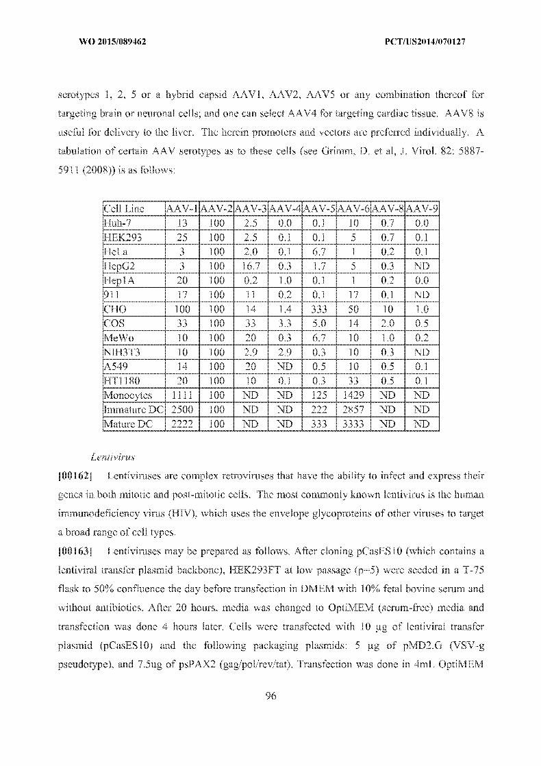
WO 2015/089462 PCT/US2014/070127
serotypes 1, 2, 5 or a hybrid capsid AAV1, AAV2, AAV5 or any combination thereof for
targeting brain or neuronal cells; and one can select AAV4 for targeting cardiac tissue. AAV8 is
useful for delivery to the liver. The herein promoters and vectors are preferred individually. A
tabulation of certain AAV serotypes as to these cells (see Grimm, D. et al, J. Virol. 82: 5887
5911 (2008)) is as follows:
Cell Line AAV- I AAV-2 AAV-3 AAV-4 AAV-5 AAV-6 AAV-8 AAV-9 Huh-7 13 100 2.5 0.0 0.1 10 0.7 0.0 HEK293 25 100 2.5 O 0.1 5 0.7 0.1
HeLa 3 100 2.0 0.1 6.7 1 0.2 0.1 HepG2 3 100 16,7 0.3 1.7 5 0.3 ND HepIA 20 100 0.2 1.0 0.1 1 0.2 0.0 911 17 100 11 0.2 0.1 17 0.1 ND
CHO 100 100 14 1.4 333 50 10 LO COS 33 100 33 3.3 5.0 14 2.0 0.5 MeWo 10 100 20 0.3 6.7 10 1 0 0.2
NIH3T3 10 100 .9 2.9 0.3 10 0.3 ND A549 14 100 20 ND 0.5 10 0.5 0.1 HT11180 20 100 10 0.1 0.3 33 0.5 0.1 Monocytes 1111 100 ND ND 125 1429 ND ND
Immature DC 2500 100 NI) ND 222 2857 ND ND Mature DC 2222 100 ND ND 333 3333 ND ND
Lentivirus
[001621 Lentiviruses are complex retroviruses that have the ability to infect and express their
genes in both mitotic and post-mitotic cells. The most commonly known lentivirus is the human
immunodeficiency virus (HIV), which uses the envelope glycoproteins of other viruses to target
a broad range of cell types.
[001631 Lentiviruses may be prepared as follows. After cloning pCasES 10 (which contains a
lentiviral transfer plasmid backbone), HEK293FT at low passage (p:::5) were seeded in a T-75
flask to 50% confluence the day before transfection in DMEM with 10% fetal bovine serum and
without antibiotics. After 20 hours, media was changed to OptiMEM (serum-free) media and
transfection was done 4 hours later. Cells were transfected with 10 Ug of lentiviral transfer
plasmid (pCasESlO) and the following packaging plasmids: 5 pg of pMD2.G (VSV-g
pseudotype), and 7.5ug of psPAX 2 (gag/pol/rev/tat). iTransfection was done in 4mL OptiMlF:M
96

WO 2015/089462 PCT/US2014/070127
with a cationic lipid delivery agent (50uL Lipofectamine 2000 and 100ul Plus reagent). After 6
hours, the media was changed to antibiotic-free DMEM with 1 0% fetal bovine serum. These
methods use serum during cell culture, but serum-free methods are preferred.
[00164] Lentivirus may be purified as follows. Viral supernatants were harvested after 48
hours. Supernatants were first cleared of debris and filtered through a 0.45um low protein
binding (PVDF) filter. They were then spun in a ultracentrifuge for 2 hours at 24,000 rpm. Viral
pellets were resuspended in 50ul of DMIEM overnight at 4C. They were then aliquotted and
immediately frozen at -80'C.
[00165] In another embodiment, minimal non-primate lentiviral vectors based on the equine
infectious anemia virus (EIAV) are also contemplated, especially for ocular gene therapy (see,
e.g., Balagaan, J Gene Med 2006; 8: 275 - 285). In another embodiment, RetinoStat@, an
equine infectious anemia virus-based lentiviral gene therapy vector that expresses angiostatic
proteins endostatin and angiostatin that is delivered via a subretinal injection for the treatment of
the web form of age-related macular degeneration is also contemplated (see, e.g., Binley et al.,
HUMAN GENE THERAPY 23:980-991 (September 2012)) and this vector may be modified for
the CRISPR-Cas system of the present invention.
[001661 In another embodiment, self-inactivating lentiviral vectors with an siRNA targeting a
common exon shared by HIV tat/rev, a nucleolar-localizing TAR decoy, and an anti-CCR5
specific hammerhead ribozyme (see, e.g., DiGiusto et al. (2010) Sci Transl Med 2:36ra43) may
be used/and or adapted to the CRISPR-Cas system of the present invention. A minimum of 2.5 x
106 CD34+ cells per kilogram patient weight may be collected and prestimulated for 16 to 20
hours in X-VIVO 15 medium (Lonza) containing 2 pmol/L-glutamine, stem cell factor (100
ng/ml), Flt-3 ligand (Flt-3L) (100 ng/ml), and thrombopoietin (10 ng/ml) (CellGenix) at a
density of 2 x 10⁶ cells/mI. Prestimulated cells may be transduced with lentiviral at a multiplicity
of infection of 5 for 16 to 24 hours in 75-cm2 tissue culture flasks coated with fibronectin (25
mg/cm2) (RetroNectin,Takara Bio Inc.).
[00167] Lentiviral vectors have been disclosed as in the treatment for Parkinson's Disease,
see, e.g., US Patent Publication No. 20120295960 and US Patent Nos. 7303910 and 7351585.
Lentiviral vectors have also been disclosed for the treatment of ocular diseases, see e.g., US
Patent Publication Nos. 20060281180, 20090007284, US20110117189; US20090017543;
US20070054961, US20100317109. Lentiviral vectors have also been disclosed for delivery to
97

WO 2015/089462 PCT/US2014/070127
the brain, see, e.g., US Patent Publication Nos. US20110293571; US20110293571,
US20040013648, US20070025970, US20090111106 and US Patent No. US721 59015.
RNA delivery
[00168] RNA delivery: The CRISPR enzyme, for instance a Cas9, and/or any of the present
RNAs, for instance a guide RNA, can also be delivered in the forn of RNA. Cas9 mRNA can be
generated using in vitro transcription. For example, Cas9 mRNA can be synthesized using a PCR
cassette containing the following elements: T7_promoter-kozak sequence (GCCACC)-Cas9-3'
UTR from beta globin-polyA tail (a string of 120 or more adenines). The cassette can be used for
transcription by T17 polymerase. Guide RNI As can also be transcribed using in vitro transcription
from a cassette containing T7_promoter-GG-guide RNA sequence.
[001691 To enhance expression and reduce possible toxicity, the CRISPR enzyme-coding
sequence and/or the guide R.NA can be modified to include one or more modified nucleoside e.g.
using pseudo-U or 5-Methyl-C.
[001701 mRNA delivery methods are especially promising for liver delivery currently.
[001711 Much clinical work on RNA delivery has focused on RNAi or antisense, but these
systems can be adapted for delivery of RNA for implementing the present invention. References
below to RNAi etc. should be read accordingly.
Particle delivery systems and/or formulations:
[001721 Several types of particle delivery systems and/or formulations are known to be useful
in a diverse spectrum of biomedical applications. In general, a particle is defined as a small
object that behaves as a whole unit with respect to its transport and properties. Particles are
further classified according to diameter. Coarse particles cover a range between 2,500 and
10,000 nanometers. Fine particles are sized between 100 and 2,500 nanometers. Ultrafine
particles, or nanoparticles, are generally between I and 100 nanometers in size. The basis of the
100-nm limit is the fact that novel properties that differentiate particles from the bulk material
typically develop at a critical length scale of under 100 nm.
[00173] As used herein, a particle delivery system/formulation is defined as any biological
delivery system/formulation which includes a particle in accordance with the present invention.
A particle in accordance with the present invention is any entity having a greatest dimension
(e.g. diameter) of less than 100 microns (pI). In some embodiments, inventive particles have a
greatest dimension of less than 10 gm. In some embodiments, inventive particles have a greatest
98

WO 2015/089462 PCT/US2014/070127
dimension of less than 2000 nanometers (nm). In some embodiments, inventive particles have a
greatest dimension of less than 1000 nanometers (nn). In some embodiments, inventive particles
have a greatest dimension of less than 900 rim, 800 nm, 700 nm, 600 rim, 500 nm, 400 nm, 300
nrn, 200 nm, or 100 nm. Typically, inventive particles have a greatest dimension (e.g., diameter)
of 500 nm or less. In some embodiments, inventive particles have a greatest dimension (e.g.,
diameter) of 250 nm or less. In some embodiments, inventive particles have a greatest dimension
(e.g., diameter) of 200 nm or less. In some embodiments, inventive particles have a greatest
dimension (e.g., diameter) of 150 nim or less. In some embodiments, inventive particles have a
greatest dimension (e.g., diameter) of 100 nni or less. Smaller particles, e.g., having a greatest
dimension of 50 nm or less are used in some embodiments of the invention. In some
embodiments, inventive particles have a greatest dimension ranging between 25 nm and 200 nm.
1001741 Particle characterization (including e.g., characterizing morphology, dimension, etc.)
is done using a variety of different techniques. Common techniques are electron microscopy
(TEM, SEM), atomic force microscopy (AFM), dynamic light scattering (DLS), X-ray
photoelectron spectroscopy (XPS), powder X-ray diffraction (XRD), Fourier transform infrared
spectroscopy FTIR), matrix-assisted laser desorption/ionization time-of-flight mass
spectrometry(MALDI-TOF), ultraviolet-visible spectroscopy, dual polarisation interferometry
and nuclear magnetic resonance (NMR). Characterization (dimension measurements) may be
made as to native particles (i.e., preloading) or after loading of the cargo (herein cargo refers to
e.g., one or more components of CRISPR-Cas system e.g., CRISPR enzyme or mRNA or guide
RNA, or any combination thereof, and may include additional carriers and/or excipients) to
provide particles of an optimal size for delivery for any in vitro, ex vivo and/or in vivo
application of the present invention. In certain preferred embodiments, particle dimension (e.g.,
diameter) characterization is based on measurements using dynamic laser scattering (DLS).
Mention is made of US Patent No. 8,709,843; US Patent No. 6,007,845; US Patent No.
5,855,913; US Patent No. 5,985,309; US. Patent No. 5,543,158; and the publication by James E.
Dahlman and Carmen Barnes et al. Nature Nanotechnology (2014) published online 11 May
2014, doi: 10.1038/nnano.2014.84, concerning particles, methods of making and using them and
measurements thereof.
1001751 Particles delivery systems within the scope of the present invention may be provided
in any form, including but not limited to solid, semi-solid, emulsion, or colloidal particles. As
99

WO 2015/089462 PCT/US2014/070127
such any of the delivery systems described herein, including but not limited to, e.g., lipid-based
systems, liposomes, micelles, microvesicles, exosomes, or gene gun may be provided as particle
delivery systems within the scope of the present invention.
Mnoparticles
[00176] CRISPR enzyme mRNA and guide RNA may be delivered simultaneously using
nanoparticles or lipid envelopes.
100177] For example, Su X, Fricke J, Kavanagh DG, Irvine DJ ("In vitro and in vivo mRNA
delivery using lipid-enveloped pH-responsive polymer nanoparticles" Mol Pharm. 2011 Jun
6:8(3):774-87. doi: 10.1021/mp100390w. Epub 2011 Apr 1) describes biodegradable core-shell
structured nanoparticles with a poly(p-amino ester) (PBAE) core enveloped by a phospholipid
bilayer shell. These were developed for in vivo mRNA delivery. The p1--responsive PBAE
component was chosen to promote endosome disruption, while the lipid surface layer was
selected to minimize toxicity of the polycation core. Such are, therefore. preferred for delivering
RNA of the present invention.
[001781 In one embodiment, nanoparticles based on self assembling bioadhesive polymers are
contemplated, which may be applied to oral delivery of peptides, intravenous delivery of
peptides and nasal delivery of peptides, all to the brain. Other embodiments, such as oral
absorption and ocular delivery of hydrophobic drugs are also contemplated. The molecular
envelope technology involves an engineered polymer envelope which is protected and delivered
to the site of the disease (see, e.g., Mazza., M. et al. ACSNano, 2013. 7(2): 1016-1026; Slew, A.,
et al. Mol Pharm, 2012. 9(1):14-28; Lalatsa, A., et al. J Contr Rel, 2012. 161(2):523-36; Lalatsa,
A.. et al., Mol Pharm, 2012. 9(6):1665-80; Lalatsa, A., et al. Mol Pharm, 2012. 9(6):1764-74;
Garrett, N.L., et al. J Biophotonics, 2012. 5(5-6):458-68; Garrett, N.L,, et alt J Raman Spect,
2012. 43(5):681-688; Ahmad, S., et al. J Royal Soc Interface 2010. 7:S423-33; Uchegbu, I.F.
Expert Opin Drug Deliv, 2006. 3(5):629-40; Qu, X..et al. Biornacromolecules, 2006. 7(12):3452
9 and Uchegbu, I.F., et al. Int J Pharm, 2001. 224:185-199). Doses of about 5 mg/kg are
contemplated, with single or multiple doses, depending on the target tissue.
[001791 In one embodiment, nanoparticles that can deliver RNA to a cancer cell to stop tumor
growth developed by Dan Anderson's lab at MIT may be used/and or adapted to the CRISPR
Cas system of the present invention. In particular, the Anderson lab developed fully automated,
combinatorial systems for the synthesis, purification, characterization, and formulation of new
100

WO 2015/089462 PCT/US2014/070127
biomaterials and nanoformulations. See, e.g., Alabi et al., Proc Natl Acad Sci U S A. 2013 Aug
6;110(32):12881-6; Zhang et al., Adv Mater. 2013 Sep 6;25(33):4641-5; Jiang et al., Nano Lett.
2013 Mar 13;13(3):1059-64; Karagiannis ct al., ACS Nano. 2012 Oct 23;6(10):8484-7;
Whitehead et al., ACS Nano. 2012 Aug 28;6(8):6922-9 and Lee et al., Nat Nanotechnol. 2012
Jun 3;7(6):389-93.
[00180] US patent application 20110293703 relates to lipidoid compounds are also
particularly useful in the administration of polynucleotides, which may be applied to deliver the
CRISPR Cas system of the present invention. In one aspect, the aminoalcohol lipidoid
compounds are combined with an agent to be delivered to a cell or a subject to form
microparticles, nanoparticles, liposomes, or micelles. The agent to be delivered by the particles,
liposomes, or micelles may be in the forn of a gas, liquid, or solid, and the agent may be a
polynucleotide, protein, peptide, or small molecule. The minoalcohol lipidoid compounds may
be combined with other aminoalcohol lipidoid compounds, polymers (synthetic or natural),
surfactants, cholesterol, carbohydrates, proteins, lipids, etc. to form the particles. These particles
may then optionally be combined with a pharmaceutical excipient to form a pharmaceutical
composition.
1001811 US Patent Publication No. 20110293703 also provides methods of preparing the
aminoalcohol lipidoid compounds. One or more equivalents of an amine are allowed to react
with one or more equivalents of an epoxide-.terminated compound under suitable conditions to
form i anminoalcohol lipidoid compound of the present invention. In certain embodiments, all
the amino groups of the amine are fully reacted with the epoxide-terminated compound to form
tertiary amines. In other embodiments, all the amino groups of the amine are not fully reacted
with the epoxide-terminated compound to form tertiary amines thereby resulting in primary or
secondary amines in the aminoalcohol lipidoid compound. These primary or secondary amines
are left as is or may be reacted with another electrophile such as a different epoxide-terminated
compound. As will be appreciated by one skilled in the art, reacting an amine with less than
excess of epoxide-terminated compound will result in a plurality of different aminoalcohol
lipidoid compounds with various numbers of tails. Certain amines may be fully functionalized
with two epoxide-derived compound tails while other molecules will not be completely
functionalized with epoxide-derived compound tails. For example, a diamine or polyamine may
include one, two, three, or four epoxide-derived compound tails off the various amino moieties
101

WO 2015/089462 PCT/US2014/070127
of the molecule resulting in primary, secondary, and tertiary amines. In certain embodiments, all
the amino groups are not fully functionalized. In certain embodiments, two of the same types of
epoxide-terminated compounds are used. In other embodiments, two or more different epoxide
terminated compounds are used. The synthesis of the aminoalcohol lipidoid compounds is
performed with or without solvent, and the synthesis may be performed at higher temperatures
ranging from 30-100 'C., preferably at approximately 50-90 'C. The prepared aminoalcohol
lipidoid compounds may be optionally purified. For example, the mixture of aminoalcohol
lipidoid compounds may be purified to yield an aminoalcohol lipidoid compound with a
particular number of epoxide-derived compound tails. Or the mixture may be purified to yield a
particular stereo- or regioisomer. The aminoalcohol lipidoid compounds may also be alkylated
using an alkyl halide (e.g., methyl iodide) or other alkylating agent, and/or they may be acylated.
1001821 US Patent Publication No. 20110293703 also provides libraries of aminoalcohol
lipidoid compounds prepared by the inventive methods. These aminoalcohol lipidoid compounds
may be prepared and/or screened using high-throughput techniques involving liquid handlers,
robots, microtiter plates, computers, etc. In certain embodiments, the aminoalcohol lipidoid
compounds are screened for their ability to transfect polymicleotides or other agents (e.g.,
proteins, peptides, small molecules) into the cell.
[00183] US Patent Publication No. 20130302401 relates to a class of poly(beta-amino
alcohols) (PBAAs) has been prepared using combinatorial polymerization. The inventive PBAAs
may be used in biotechnology and biomedical applications as coatings (such as coatings of fihns
or multilayer films for medical devices or implants), additives, materials, excipients, non
biofouling agents, micropatterning agents, and cellular encapsulation agents. When used as
surface coatings, these PBAAs elicited different levels of inflammation, both in vitro and in vivo,
depending on their chemical structures. The large chemical diversity of this class of materials
allowed us to identify polymer coatings that inhibit macrophage activation in vitro. Furthermore,
these coatings reduce the recruitment of inflammatory cells, and reduce fibrosis, following the
subcutaneous implantation of carboxylated polystyrene microparticles. These polymers may be
used to form polyelectrolyte complex capsules for cell encapsulation. The invention may also
have many other biological applications such as antimicrobial coatings, DNA or siRNA delivery,
and stem cell tissue engineering. The teachings of US Patent Publication No. 20130302401 may
be applied to the CRISPR Cas system of the present invention.
102

WO 2015/089462 PCT/US2014/070127
[001841 In another embodiment, lipid nanoparticles (LNPs) are contemplated. An
antitransthyretin small interfering RNA has been encapsulated in lipid nanoparticles and
delivered to humans (see, e.g., Coelho et al., N Engl J Med 2013;369:819-29), and such a
system may be adapted and applied to the CRISPR Cas system of the present invention. Doses
of about 0.01 to about I mg per kg of body weight administered intravenously are contemplated.
Medications to reduce the risk of infusion-related reactions are contemplated, such as
dexamethasone, acetampinophen, diphenhydramine or cetirizine, and ranitidine are
contemplated. Multiple doses of about 0.3 mg per kilogram every 4 weeks for five doses are also
contemplated.
[001851 LNPs have been shown to be highly effective in delivering siRNAs to the liver (see,
e.g., Tabernero et al., Cancer Discovery, April 2013, Vol. 3, No. 4, pages 363-470) and are
therefore contemplated for delivering RNA encoding CRISPR Cas to the liver. A dosage of
about four doses of 6 mg/kg of the LNP every two weeks may be contemplated. Tabernero et al.
demonstrated that tumor regression was observed after the first 2 cycles of LNPs dosed at 0.7
mg/kg, and by the end of 6 cycles the patient had achieved a partial response with complete
regression of the lymph node metastasis and substantial shrinkage of the liver tumors. A
complete response was obtained after 40 doses in this patient, who has remained in remission
and completed treatment after receiving doses over 26 months. Two patients with RCC and
extrahepatic sites of disease including kidney, lung, and lymph nodes that were progressing
following prior therapy with VEGF pathway inhibitors had stable disease at all sites for
approximately 8 to 12 months, and a patient with PNET and liver metastases continued on the
extension study for 18 months (36 doses) with stable disease.
[001861 However, the charge of the LNP must be taken into consideration. As cationic lipids
combined with negatively charged lipids to induce nonbilayer structures that facilitate
intracellular delivery. Because charged LNPs are rapidly cleared from circulation following
intravenous injection, ionizable cationic lipids with pKa values below 7 were developed (see,
e.g., Rosin et al, Molecular Therapy, vol. 19, no. 12, pages 1286-2200, Dec. 2011). Negatively
charged polymers such as RNA may be loaded into LNIPs at low pH values (e.g., pH 4) where
the ionizable lipids display a positive charge. However, at physiological pH values, the LNPs
exhibit a low surface charge compatible with longer circulation times. Four species of ionizable
cationic lipids have been focused upon, namely 1,2-dilineoyl-3-dimethylammonium-propane
103

WO 2015/089462 PCT/US2014/070127
(DLinDAP), 1,2-dilinoleyloxy-3-N,N-dimethylaminopropane (DLinDMA), 1,2-dilinoleyloxy
keto-N,N-dimethyl-3-aminopropane (DLinKDMA), and 1,2-dilinoleyl-4-(2
dimethylaminoethyl)-[1,3]-dioxolane (DLinKC2-DMA). It has been shown that LNP siRNA
systems containing these lipids exhibit remarkably different gene silencing properties in
hepatocytes in vivo, with potencies varying according to the series DLinKC2
DMA>DLinKDMA>DLinDMA>>DLinDAP employing a Factor VII gene silencing model (see,
e.g., Rosin et al, Molecular Therapy, vol. 19, no. 12, pages 1286-2200, Dec, 2011), A dosage of
I pig/ml of LNP or CRISPR-Cas RNA in or associated with the LNP may be contemplated,
especially for a formulation containing DLinKC2-DMA.
[001871 Preparation of LNIPs and CRISPIR Cas encapsulation may be used/and or adapted
from Rosin et al, Molecular Therapy, vol. 19, no. 12, pages 1286-2200, Dec. 2011). The cationic
lipids 1 ,2-dilineoyl-3 -dimethylammonium-propane (DLinDAP), I1,2-dilinoleyloxy-3-N,N
dimethylaminopropane (DLinDMA), 1 ,2-dilinoleyloxyketo-NN-dimethyl-3-aminopropane
(DLinK-DMA), 1,2-dilinoleyl-4-(2-diinethylaminoethyl )-[1,3]-dioxolane (DI)LinKC2-DM[VA), (3
o-[2"-(methoxypolyethyleneglycol 2000) succinoyl]-1 ,2-dimyristoyl-sn-glycol (PEG-S-DMG),
and R-3-[(cow-methoxy-poly(ethylene glycol)2000) carbamoyl] -1,2-dimyristyloxlpropyl-3 -amine
(PEG-C-DOMG) may be provided by Tekmira Pharmaceuticals (Vancouver, Canada) or
synthesized. Cholesterol may be purchased from Sigma (St Louis, MO). The specific CRISPR
Cas RNA may be encapsulated in LNPs containing DLinDAP, DLinDMA, DLinK-DMA, and
DLinKC2-DMA (cationic lipid:DSPC:CHOL: PEGS-DMG or PEG-C-DOMG at 40:10:40:10
molar ratios). When required, 0,2% SP-DiOCi8 (Invitrogen, Burlington, Canada) may be
incorporated to assess cellular uptake, intracellular delivery, and biodistribution. Encapsulation
may be performed by dissolving lipid mixtures comprised of cationic
lipid :DSPC:cholesterol:PEG-c-DOMG (40:10:40:10 molar ratio) in ethanol to a final lipid
concentration of 10 rnmol/l. This ethanol solution of lipid may be added drop-wise to 50 nimol/l
citrate, pH 4.0 to form multilamellar vesicles to produce a final concentration of 30% ethanol
vol/vol. Large unilamel lar vesicles may be formed following extrusion of multilamel lar vesicles
through two stacked 80 nm Nuclepore polycarbonate filters using the Extruder (Northern Lipids,
Vancouver, Canada). Encapsulation may be achieved by adding RNA dissolved at 2 mg/ml in 50
mmol/l citrate, p-I 4.0 containing 30% ethanol vol/vol drop-wise to extruded preformed large
unilameliar vesicles and incubation at 31 'C for 30 minutes with constant mixing to a final
104

WO 2015/089462 PCT/US2014/070127
RNA/lipid weight ratio of 0.06/1 wt/wt. Removal of ethanol and neutralization of formulation
buffer were performed by dialysis against phosphate-buffered saline (PBS), pH 7.4 for 16 hours
using Spectra/Por 2 regenerated cellulose dialysis membranes. Nanoparticle size distribution
may be determined by dynamic light scattering using a NICOMP 370 particle sizer, the
vesicle/intensity modes, and Gaussian fitting (Nicomp Particle Sizing, Santa Barbara, CA). The
particle size for all three LNP systems may be ~70 nm in diameter. RNA encapsulation
efficiency may be determined by removal of free RNA using VivaPureD Mini-! columns
(Sartorius Stedim Biotech) from samples collected before and after dialysis. The encapsulated
RNA may be extracted from the eluted nanoparticles and quantified at 260 nm. RNA to lipid
ratio was determined by measurement of cholesterol content in vesicles using the Cholesterol E
enzymatic assay from Wako Chenicals USA (Richmond, VA). In conjunction with the herein
discussion of LNPs and PEG lipids, PEGylated liposomes or LNPs are likewise suitable for
delivery of a CRISPR-Cas system or components thereof
[00188] Preparation of large LNPs may be used/and or adapted from Rosin et al, Molecular
Therapy, vol. 19, no. 12, pages 1286-2200, Dec. 2011. A lipid premix solution (20.4 mg/ml total
lipid concentration) may be prepared in ethanol containing DLinKC2-DMA, DSPC, and
cholesterol at 50:10:38.5 molar ratios. Sodium acetate may be added to the lipid premix at a
molar ratio of 0.75:1 (sodium acetate:DLinKC2-DMA). The lipids may be subsequently
hydrated by combining the mixture with 1.85 volumes of citrate buffer (10 mmol/l, pH 3.0) with
vigorous stirring, resulting in spontaneous liposome formation in aqueous buffer containing 35%
ethanol. The liposome solution may be incubated at 37 0C to allow for time-dependent increase
in particle size. Aliquots may be removed at various times during incubation to investigate
changes in liposome size by dynamic light scattering (Zetasizer Nano ZS, Malvern Instruments,
Worcestershire, UK). Once the desired particle size is achieved, an aqueous PEG lipid solution
(stock:= 10 mg/ml PEG-DMG in 35% (vol/vol) ethanol) may be added to the liposome mixture
to yield a final PEG molar concentration of 3.5% of total lipid. Upon addition of PEG-lipids, the
liposomes should their size, effectively quenching further growth. RNA may then be added to the
empty liposomes at an RNA to total lipid ratio of approximately 1:10 (wt:wt), followed by
incubation for 30 minutes at 37 'C to form loaded LNPs. The mixture may be subsequently
dialyzed overnight in PBS and filtered with a 0.45-iptm syringe filter.
105

WO 2015/089462 PCT/US2014/070127
[001891 Spherical Nucleic Acid (SNATI) constructs and other nanoparticles (particularly gold
nanoparticles) are also contemplated as a means to delivery CRISPR-Cas system to intended
targets. Significant data show that AuraSense Therapeutics' Spherical Nucleic Acid (SNATMI)
constructs, based upon nucleic acid-functionalized gold nanoparticles, are useful.
[001901 Literature that may be employed in conjunction with herein teachings include: Cutler
et al.. J. Am. Chem. Soc. 2011 133:9254-9257, Hao et al., Small. 2011 7:3158-3162, Zhang et
al., ACS Nano. 2011 5:6962-6970, Cutler et al., J. Am. Chem. Soc. 2012 134:1376-1391, Young
et al., Nano Lett. 2012 12:3867-71, Zheng et al., Proc. Natl. Acad. Sci. USA. 2012 109:11975
80, Mirkin, Nanomedicine 2012 7:635-638 Zhang et al, J. Am. Chen. Soc. 2012 134:16488
1691, Weintraub, Nature 2013 495:S14-S16, Choi et al., Proc. Natl. Acad. Sci. USA. 2013
110(19):7625-7630, Jensen etal., Sci. Transit. Med. 5, 209ral52 (2013) and Mirkin, et al., Small,
10:186-192.
[001911 Self-assembling nanoparticles with RNA may be constructed with polyethyleneimine
(PET) that is PEGylated with an Arg-Gly-Asp (RGD) peptide ligand attached at the distal end of
the polyethylene glycol (PEG). This system has been used, for example, as a means to target
tumor neovasculature expressing integrins and deliver siRNA inhibiting vascular endothelial
growth factor receptor-2 (VEGF R2) expression and thereby achieve tumor angiogenesis (see,
e.g., Schiffelers et al., Nucleic Acids Research, 2004, Vol. 32, No. 19). Nanoplexes may be
prepared by mixing equal volumes of aqueous solutions of cationic polymer and nucleic acid to
give a net molar excess of ionizable nitrogen (polymer) to phosphate (nucleic acid) over the
range of 2 to 6. The electrostatic interactions between cationic polymers and nucleic acid
resulted in the formation of polyplexes with average particle size distribution of about 100 nm,.
hence referred to here as nanoplexes. A dosage of about 100 to 200 mg of CRISPR Cas is
envisioned for delivery in the self-assembling nanoparticles of Schiffelers et al.
[001921 The nanoplexes of Bartlett et al. (PNAS, September 25, 2007,vol. 104, no. 39) may
also be applied to the present invention. The nanoplexes of Bartlett et al. are prepared by mixing
equal volumes of aqueous solutions of cationic polymer and nucleic acid to give a net molar
excess of ionizable nitrogen (polymer) to phosphate (nucleic acid) over the range of 2 to 6. The
electrostatic interactions between cationic polymers and nucleic acid resulted in the formation of
polyplexes with average particle size distribution of about 100 nm, hence referred to here as
nanoplexes. The DOTA-siRNA of Bartlett et al. was synthesized as follows: 1,4,7,10
106

WO 2015/089462 PCT/US2014/070127
tetraazacyclododecane- 1,4;7,1 0-tetraacetic acid mono(N-hydroxysuccinimide ester) (DOTA
NHSester) was ordered from Macrocyclics (Dallas, TX). The amine modified RNA sense strand
with a 100-fold molar excess of DOTA-NHS-ester in carbonate buffer (pH 9) was added to a
microcentrifuge tube. The contents were reacted by stirring for 4 h at room temperature. The
DOTA-RNAsense conjugate was ethanol-precipitated, resuspended in water, and annealed to the
unmodified antisense strand to yield DOTA-siRNA. All liquids were pretreated with Chelex-100
(Bio-Rad, Hercules, CA) to remove trace metal contaminants. Tf-targeted and nontargeted
siRNA nanoparticles may be formed by using cyclodextrin-containing polycations. Typically,
nanoparticles were forced in water at a charge ratio of 3 (-/-) and an siRNA concentration of 0.5
g/liter. One percent of the adamantane-PEG molecules on the surface of the targeted
nanoparticles were modified with Tf (adamantane-PEG-Tf). The nanoparticles were suspended
in a 5% (wt/vol) glucose carrier solution for injection.
[001931 Davis et al. (Nature, Vol 464, 15 April 2010) conducts a RNA clinical trial that uses a
targeted nanopartic le-delivery system (clinical trial registration number NcT00689065). Patients
with solid cancers refractory to standard-of-care therapies are administered doses of targeted
nanoparticles on days 1, 3, 8 and 10 of a 21-day cycle by a 30-min intravenous infusion. The
nanoparticles consist of a synthetic delivery system containing: (1) a linear, cyclodextrin-based
polyrner (CDP), (2) a human transferrin protein (TF) targeting ligand displayed on the exterior of
the nanoparticle to engage TF receptors (TFR) on the surface of the cancer cells, (3) a
hydrophilic polymer (polyethylene glycol (PEG) used to promote nanoparticle stability in
biological fluids), and (4) siRNA designed to reduce the expression of the RRM2 (sequence used
in the clinic was previously denoted siR2B+5). The TFR has long been known to be upregulated
in malignant cells, and RRM2 is an established anti-cancer target. These nanoparticles (clinical
version denoted as CALAA-01) have been shown to be well tolerated in multi-dosing studies in
non-human primates. Although a single patient with chronic myeloid leukaemia has been
administered siRNAby liposomal delivery, Davis et al.'s clinical trial is the initial human trial to
systemically deliver siRNA with a targeted delivery system and to treat patients with solid
cancer. To ascertain whether the targeted delivery system can provide effective delivery of
functional siRNA to human tumours, Davis et al. investigated biopsies fr-om three patients from
three different dosing cohorts; patients A, B and C, all of whom had metastatic melanoma and
received CALAA-01 doses of 18, 24 and 30 mg m-2 siRNA, respectively. Similar doses may
107

WO 2015/089462 PCT/US2014/070127
also be contemplated for the CRISPR Cas system of the present invention. The delivery of the
invention may be achieved with nanoparticles containing a linear, cyclodextrin-based polymer
(CDP), a human transferrin protein (TFE) targeting ligand displayed on the exterior of the
nanoparticle to engage TF receptors (TFR) on the surface of the cancer cells and/or a hydrophilic
polymer (for example, polyethylene glycol (PEG) used to promote nanoparticle stability in
biological fluids).
1001941 In terms of this invention, it is preferred to have one or more components of CRISPR
complex, e.g., CRISPR enzyme or mRNA or guide RNA or sgRNA or if present HDR template
may be delivered using one or more particles or nanoparticles or lipid envelopes. Other delivery
systems or vectors are may be used in conjunction with the nanoparticle aspects of the invention.
[001-951 In general, a "nanoparticle" refers to any particle having a diameter of less than 1000
nm. In certain preferred embodiments, nanoparticles of the invention have a greatest dimension
(e.g.., diameter) of 500 nm or less. In other preferred embodiments, nanoparticles of the invention
have a greatest dimension ranging between 25 nm and 200 nm. In other preferred embodiments,
nanoparticles of the invention have a greatest dimension of 100 nm or less. In other preferred
embodiments, nanoparticles of the invention have a greatest dimension ranging between 35 nm
and 60 nm.
[00196] Nanoparticles encompassed in the present invention may be provided in different
forms, e.g., as solid nanoparticles (e.g., metal such as silver, gold, iron, titanium), non-metal,
lipid-based solids, polymers), suspensions of nanoparticles, or combinations thereof. Metal,
dielectric, and semiconductor nanoparticles may be prepared, as well as hybrid structures (e.g.,
core-shell nanoparticles). Nanoparticles made of semiconducting material may also be labeled
quantum dots if they are small enough (typically sub 10 nm) that quantization of electronic
energy levels occurs. Such nanoscale particles are used in biomedical applications as drug
carriers or imaging agents and may be adapted for similar purposes in the present invention.
1001971 Semi-solid and soft nanoparticles have been manufactured, and are within the scope
of the present invention. A prototype nanoparticle of semi-solid nature is the liposome. Various
types of liposome nanoparticles are currently used clinically as delivery systems for anticancer
drugs and vaccines. Nanoparticles with one half hydrophilic and the other half hydrophobic are
termed Janus particles and are particularly effective for stabilizing emulsions. They can self
assemble at water/oil interfaces and act as solid surfactants.
108

WO 2015/089462 PCT/US2014/070127
[001981 US Patent No. 8,709,843, incorporated herein by reference, provides a drug delivery
system for targeted delivery of therapeutic agent-containing particles to tissues, cells, and
intracellular compartments. The invention provides targeted particles comprising comprising
polymer conjugated to a surfactant, hydrophilic polymer or lipid. US Patent No. 6,007,845,
incorporated herein by reference, provides particles which have a core of a multiblock copolymer
fornied by covalently linking a multifunctional compound with one or more hydrophobic
polymers and one or more hydrophilic polymers, and conatin a biologically active material. US
Patent No. 5,855,913, incorporated herein by reference, provides a particulate composition
having aerodynamically light particles having a tap density of less than 0.4 g/cm3 with a mean
diameter of between 5 Pm and 30 pm, incorporating a surfactant on the surface thereof for drug
delivery to the pulmonary system. US Patent No. 5,985,309, incorporated herein by reference,
provides particles incorporating a surfactant and/or a hydrophilic or hydrophobic complex of a
positively or negatively charged therapeutic or diagnostic agent and a charged molecule of
opposite charge for delivery to the pulmonary system. US. Patent No. 5,543,158, incorporated
herein by reference, provides biodegradable injectable nanoparticles having a biodegradable
solid core containing a biologically active material and poly(alkylene glycol) moieties on the
surface, WO20121135025 (also published as US20120251560), incorporated herein by reference,
describes conjugated polyethyleneimine (PEI) polymers and conjugated aza-macrocycles
(collectively referred to as "conjugated lipomer" or "lipomers"). In certain embodiments, it can
be envisioned that such methods and materials of herein-cited documents, e.g., conjugated
lipomers, can be used in the context of the CRISPR-Cas system to achieve in vitro, ex vivo and
in vivo genomic perturbations to modify gene expression, including modulation of protein
expression.
1001991 In one embodiment, the nanoparticle may be epoxide-modified lipid-polymer,
advantageously 7C1 (see, e.g., James E. Dahlman and Carmen Barnes et al. Nature
Nanotechnology (2014) published online 11 May 2014, doi:10.1038/nnano.2014.84). (71 was
synthesized by reacting C 1 5 epoxide-terminated lipids with PE1600 at a 14:1 molar ratio, and
was formulated with C14PEG2000 to produce nanoparticles (diameter between 35 and 60 nm)
that were stable in PBS solution for at least 40 days. An epoxide-modified lipid-polymer may be
utilized to deliver the CRISPR-Cas system of the present invention to pulmonary, cardiovascular
or renal cells, however, one of skill in the art may adapt the system to deliver to other target
109

WO 2015/089462 PCT/US2014/070127
organs. Dosage ranging from about 0.05 to about 0.6 mg/kg are envisioned. Dosages over
several days or weeks are also envisioned, with a total dosage of about 2 mg/kg.
Exosomes
[00200] Exosomes are endogenous nano-vesicles that transport RNAs and proteins, and which
can deliver RNA to the brain and other target organs. To reduce immunogenicity, Alvarez-Erviti
et al. (2011, Nat Biotechnol 29: 341) used self-derived dendritic cells for exosome production.
Targeting to the brain was achieved by engineering the dendritic cells to express Lamp2b, an
exosomal membrane protein, fused to the neuron-specific RVG peptide. Purified exosomes were
loaded with exogenous RNA by electroporation. Intravenously injected RVG-targeted exosomes
delivered GAPDH siRNA specifically to neurons, microglia, oligodendrocytes in the brain,
resulting in a specific gene knockdown. Pre-exposure to RVG exosomes did not attenuate
knockdo-wi, and non-specific uptake in other tissues was not observed. The therapeutic potential
of exosome-mediated siRNA delivery was demonstrated by the strong mRNA (60%) and protein
(62%) knockdown of BACE1I, a therapeutic target in Alzheimer's disease.
[002011 To obtain a pool of immunologically inert exosomes, Alvarez-Erviti et al. harvested
bone marrow from inbred C57BL/6 mice with a homogenous major histocompatibility complex
(MHC) haplotype. As immature dendritic cells produce large quantities of exosomes devoid of
T-cell activators such as M-IC-I and CD86, Alvarez-Erviti et al. selected for dendritic cells with
granulocyte/macrophage-colony stimulating factor (GM-CSF) for 7 d. Exosomes were purified
from the culture supernatant the following day using well-established ultracentrifugation
protocols. The exosomes produced were physically homogenous, with a size distribution peaking
at 80 nm in diameter as determined by nanoparticle tracking analysis (NTA) and electron
microscopy. Alvarez-Erviti et al. obtained 6-12 pg of exosomes (measured based on protein
concentration) per 106 cells.
[002021 Next, Alvarez-Erviti et al. investigated the possibility of loading modified exosomes
with exogenous cargoes using electroporation protocols adapted for nanoscale applications. As
electroporation for mem brane particles at the nanometer scale is not well-characterized,
nonspecific Cy5-labeled RNA was used for the empirical optimization of the electroporation
protocol. The amount of encapsulated RNA was assayed after ultracentrifugation and lysis of
exosomes. Electroporation at 400 V and 125 gF resulted in the greatest retention of RNA and
was used for all subsequent experiments.
110

WO 2015/089462 PCT/US2014/070127
[002031 Alvarez-Erviti et al. administered 150 pg of each BACEl siRNA encapsulated in 150
ig of RVG exosomes to normal C57BL1/6 mice and compared the knockdown efficiency to four
controls: untreated mice, mice injected with RVG exosomes only, mice injected with BACEl
siRNA complexed to an in vivo cationic liposomne reagent and mice injected with BACEI siRNA
complexed to RVG-9R, the RVG peptide conjugated to 9 D-arginines that electrostatically binds
to the siRNA. Cortical tissue samples were analyzed 3 d after administration and a significant
protein knockdown (45%, P < 0.05, versus 62%, P < 0.01) in both siRNA-RVG-9R-treated and
siRNARVG exosome-treated mice was observed, resulting from a significant decrease in
BACE1 mRNA levels (66% [+ or -] 15%, P < 0.001 and 61% [4 or -] 13% respectively, P <
0.01). Moreover, Applicants demonstrated a significant decrease (55%, P < 0.05) in the total
[beta]-amyioid 1-42 levels, a main component of the amyloid plaques in Alzheimer's pathology,
in the RVG-exosome-treated animals. The decrease observed was greater than the pf-amyloid 1
40 decrease demonstrated in normal mice after intraventricular injection of BACEI inhibitors.
Alvarez-Erviti et al. carried out 5'-rapid amplification of cDNA ends (RACE) on BACEl
cleavage product, which provided evidence of RNAi-mediated knockdown by the siRNA.
1002041 Finally, Alvarez-Erviti et al. investigated whether RNA-RVG exosomes induced
immune responses in vivo by assessing IL-6, IP-10, TNFu and IFN-a serum concentrations.
Following exosonie treatment, nonsignificant changes in all cytokines were registered similar to
siRNA-transfection reagent treatment in contrast to siRNA-RVG-9R, which potently stimulated
IL-6 secretion, confirming the immunologically inert profile of the exosome treatment. Given
that exosomes encapsulate only 20% of siRNA, delivery with RVG-exosome appears to be more
efficient than RNG-9R delivery as comparable mRNA knockdown and greater protein
knockdown was achieved with fivefold less siRNA without the corresponding level of immune
stimulation. This experiment demonstrated the therapeutic potential of RVG-exosome
technology, which is potentially suited for long-terrn silencing of genes related to
neurodegenerative diseases. The exosome delivery system of Alvarez-Erviti et al. may be applied
to deliver the CRISPR-Cas system of the present invention to therapeutic targets, especially
neurodegenerative diseases. A dosage of about 100 to 1000 mg of CRISPR Cas encapsulated in
about 100 to 1000 mg of RVG exosomes may be contemplated for the present invention.
1002051 EI-Andaloussi et al. (Nature Protocols 7,2112-2126(2012)) discloses how exosomes
derived from cultured cells can be harnessed for delivery of RNA in vitro and in vivo. This
111

WO 2015/089462 PCT/US2014/070127
protocol first describes the generation of targeted exosomes through transfection of an expression
vector, comprising an exosomal protein fused with a peptide ligand. Next, El-Andaloussi et al.
explain how to purify and characterize exosomes from transfected cell supernatant. Next, El
Andaloussi et al. detail crucial steps for loading RNA into exosomes. Finally, El-Andaloussi et
al. outline how to use exosomes to efficiently deliver RNA in vitro and in vivo in mouse brain.
Examples of anticipated results in which exosome-mediated RNA delivery is evaluated by
functional assays and imaging are also provided. The entire protocol takes -3 weeks. Delivery or
administration according to the invention may be performed using exosomes produced from self
derived dendritic cells. From the herein teachings, this can be employed in the practice of the
invention.
[002061 In another embodiment, the plasma exosomes of Wahigren et al. (Nucleic Acids
Research, 2012, Vol. 40, No. 17 e130) are contemplated. Exosomes are nano-sized vesicles (30
90nm in size) produced by many cell types, including dendritic cells (DC), B cells, T cells, mast
cells, epithelial cells and tumor cells. These vesicles are formed by inward budding of late
endosomes and are then released to the extracellular environment upon fusion with the plasma
membrane. Because exosomes naturally carry RNA between cells, this property may be useful in
gene therapy, and from this disclosure can be employed in the practice of the instant invention.
1002071 Exosones from plasma can be prepared by centrifugation of buffy coat at 900g for 20
min to isolate the plasma followed by harvesting cell supernatants, centrifuging at 300g for 10
min to eliminate cells and at 16 500g for 30 min followed by filtration through a 0.22 min filter.
Exosones are pelleted by ultracentrifugation at 120 000g for70 min. Chemical transfection of
siRNA into exosomes is carried out according to the manufacturer's instructions in RNAi
Human/Mouse Starter Kit (Quiagen, Hilden, Germany). siRNA is added to 100 ml PBS at a final
concentration of 2 mmol/ml. After adding HiPerFect transfection reagent, the mixture is
incubated for 10 min at RT. In order to remove the excess of micelles, the exosomes are re
isolated using aldehyde/sulfate latex beads. The chemical transfection of CRISPR Cas into
exosomes may be conducted similarly to siRNA. The exosomes may be co-cultured with
monocytes and lymphocytes isolated from the peripheral blood of healthy donors. Therefore, it
may be contemplated that exosomes containing CRISPR Cas may be introduced to monocytes
and lymphocytes of and autologously reintroduced into a human. Accordingly, delivery or
administration according to the invention may be performed using plasma exosomes.
112

WO 2015/089462 PCT/US2014/070127
Liposonies
[002081 Delivery or administration according to the invention can be performed with
liposomes. Liposomes are spherical vesicle structures composed of a uni- or multilamellar lipid
bilayer surrounding internal aqueous compartments and a relatively impermeable outer lipophilic
phospholipid bilayer. Liposomes have gained considerable attention as drug delivery carriers
because they are biocompatible, nontoxic, can deliver both hydrophilic and lipophilic drug
molecules, protect their cargo from degradation by plasma enzymes, and transport their load
across biological membranes and the blood brain barrier (BBB) (see, e.g., Spuch and Navarro,
Journal of Drug Delivery, vol. 2011, Article ID 469679, 12 pages, 2011.
doi: 10. 1155/2011/469679 for review). Liposomes can be made from several different types of
lipids; however, phospholipids are most commonly used to generate liposomes as drug carriers.
Although liposome formation is spontaneous when a lipid film is mixed with an aqueous
solution, it can also be expedited by applying force in the form of shaking by using a
homogenizer, sonicator, or an extrusion apparatus (see, e.g., Spuch and Navarro, Journal of Drug
Delivery, vol. 2011, Article ID 469679, 12 pages, 2011. doi:10.1155/2011/469679 for review).
1002091 Several other additives may be added to liposomes in order to modify their structure
and properties. For instance, either cholesterol or sphingomyelin may be added to the liposomal
mixture in order to help stabilize the liposonmal structure and to prevent the leakage of the
liposomal inner cargo. Further, liposomes are prepared from hydrogenated egg
phosphatidylcholine or egg phospha tidylcholine, cholesterol, and dicetyl phosphate, and their
mean vesicle sizes were adjusted to about 50 and 100 nm. (see, e.g., Spuch and Navarro, Journal
of Drug Delivery, vol. 2011, Article ID 469679, 12 pages, 2011. doi:10.1155/2011/469679 for
review). A liposome formulation may be mainly comprised of natural phospholipids and lipids
such as 1,2-distearoryl-sn-glycero-3-phosphatidyl choline (DSPC), sphingomyelin, egg
phosphatidylcholines and mnonosialoganglioside. Since this formulation is made up of
phospholipids only, liposomal formulations have encountered many challenges, one of the ones
being the instability in plasma. Several attempts to overcome these challenges have been made,
specifically in the manipulation of the lipid membrane. One of these attempts focused on the
manipulation of cholesterol. Addition of cholesterol to conventional formulations reduces rapid
release of the encapsulated bioactive compound into the plasma or 1,2-dioleoyl-sn-glycero-3
phosphoethanolamine (DOPE) increases the stability (see, e.g., Spuch and Navarro, Journal of
113

WO 2015/089462 PCT/US2014/070127
Drug Delivery, vol. 2011, Article ID 469679, 12 pages, 2011. doi:10.1155/2011/469679 for
review).
[002101 In a particularly advantageous embodiment, Trojan Horse liposomes (also known as
Molecular Trojan Horses) are desirable and protocols may be found at
http:/'cshprotocols.eshlp.org/content/2(010/4/pdb.prot5407T.long. These particles allow delivery of
a transgene to the entire brain after an intravascular injection. Without being bound by limitation,
it is believed that neutral lipid particles with specific antibodies conjugated to surface allow
crossing of the blood brain barrier via endocytosis. Applicant postulates utilizing Trojan Horse
Liposones to deliver the CRISPR family of nucleases to the brain via an intravascular injection,
which would allow whole brain transgenic animals without the need for embryonic
manipulation. About 1-5 g of DNA or RNA may be contemplated for in vivo administration in
liposomes.
[002111 In another embodiment. the CRISPR Cas system or components thereof may be
administered in liposomes, such as a stable nucleic-acid-lipid particle (SNALP) (see, e.g.,
Morrissey et al., Nature Biotechnology, Vol. 23, No. 8, August 2005). Daily intravenous
injections of about 1, 3 or 5 mg/kg/day of a specific CRISPR Cas targeted in a SNALP are
contemplated. The daily treatment may be over about three days and then weekly for about five
weeks. In another embodiment, a specific CRISPR Cas encapsulated SNALP) administered by
intravenous injection to at doses of about I or 2.5 mg/kg are also contemplated (see, e.g.,
Zimmerman et al., Nature Letters, Vol. 441, 4 May 2006). The SNALP formulation may contain
the lipids 3-N-[(wmethoxypoly(ethylene glycol) 2000) carbanoyl] -1 ,2-dimyristyloxy
propylamine (PEG-C-DMA), I,2-dilinoleyloxy-N,N-dimethyl-3 -aminopropane (DLinDMA),
1I,2-distearoyl-sn -glycero-3-phosphocholine (DSPC) and cholesterol, in a 2:40:10:48 molar per
cent ratio (see, e.g., Zimmerman et al., Nature Letters, Vol. 441, 4 May 2006).
[002121 In another embodiment, stable nucleic-acid-lipid particles (SNALPs) have proven to
be effective delivery molecules to highly vascularized HepG2-derived liver tumors but not in
poorly vascularized HCT-I 16 derived liver tumors (see, e.g., Li, Gene Therapy (2012) 19, 775
780). The SNALP liposomes may be prepared by formulating D-Lin-DNIA and PEG-C-DMA
with distearoylphosphatidylcholine (DSPC), Cholesterol and siRNA using a 25:1 lipid/siRNA
ratio and a 48/40/10/2 molar ratio of Cholesterol/D-in-DMA/DSPC/PEG-C-DMA. The resulted
SNALP liposomes are about 80-100 nm in size. In yet another embodiment, a SNALP may
114
WO 2015/089462 PCT/US2014/070127
comprise synthetic cholesterol (Sigma-Aldrich, St Louis, MO, USA),
dipalmitoylphosphatidylcholine (Avanti Polar Lipids, Alabaster, AL, USA), 3-N-[(w-methoxy
poly(ethylene glycol)2000)carbamovl]- 1,2-dimyrestyloxypropylamine, and cationic 1,2
dilinoieyloxy-3 -N,Ndimethylaminopropane (see, e.g., Geisbert et al., Lancet 2010; 375: 1896
905). A dosage of about 2 mg/kg total CRISPR Cas per dose administered as, for example, a
bolus intravenous infusion may be contemplated. In yet another embodiment, a SNALP may
comprise synthetic cholesterol (Sigma-Aldrich), 1,2-distearoyl-sn-glycero-3-phosphocholine
(DSPC; Avanti Polar Lipids Inc.), PEG-cDMA,. and 1,2-diiinoleyloxy-3-(N;N
dimethyl)aminopropane (DLinDMA) (see, e.g., Judge, J. Clin. Invest. 119:661-673 (2009)).
Formulations used for in vivo studies may comprise a final lipid/RNA mass ratio of about 9:1.
[002131 The safety profile of RNAi nanomedicines has been reviewed by Barros and Gollob
of Alnylam Pharmaceuticals (see, e.g., Advanced Drug Delivery Reviews 64 (2012) 1730-1737).
The stable nucleic acid lipid particle (SNALP) is comprised of four different lipids - an
ionizable lipid (DLinDNA) that is cationic at low pl-, a neutral helper lipid, cholesterol, and a
diffusible polyethylene glycol (PEG)-lipid. The particle is approximately 80 nm in diameter and
is charge-neutral at physiologic pH. During formulation, the ionizable lipid serves to condense
lipid with the anionic RNA during particle formation. When positively charged under
increasingly acidic endosomal conditions, the ionizable lipid also mediates the fusion of SNALP
with the endosomal membrane enabling release of RNA into the cytoplasm. The PEG-lipid
stabilizes the particle and reduces aggregation during formulation, and subsequently provides a
neutral hydrophilic exterior that improves pharmacokinetic properties.
[002141 To date, two clinical programs have been initiated using SNALP formulations with
RNA. Tekmira Pharmaceuticals recently completed a phase I single-dose study of SNALP-ApoB
in adult volunteers with elevated LDL cholesterol. ApoB is predominantly expressed in the liver
and jejunum and is essential for the assembly and secretion of VLDL and LDL. Seventeen
subjects received a single dose of SNALP-ApoB (dose escalation across 7 dose levels). There
was no evidence of liver toxicity (anticipated as the potential dose-limiting toxicity based on
preclinical studies). One (of two) subjects at the highest dose experienced flu-like symptoms
consistent with immune system stimulation, and the decision was made to conclude the trial.
1002151 Alnylam Pharmaceuticals has similarly advanced ALN-TTROI, which employs the
SNALP technology described above and targets hepatocyte production of both mutant and wild
115

WO 2015/089462 PCT/US2014/070127
type TTR to treat TTR amyloidosis (ATTR). Three ATIR syndromes have been described:
familial amyloidotic polyneuropathy (FAP) and familial amvioidotic cardiomyopathy (FAC)
both caused by autosomal dominant mutations in TTR; and senile systemic amyloidosis (SSA)
cause by wildtype TTR. A placebo-controlled, single dose-escalation phase I trial of ALN
TTR0I was recently completed in patients with ATTR. ALN-TTR0I was administered as a 15
minute IV infusion to 31 patients (23 with study drug and 8 with placebo) within a dose range of
0.01 to 1.0 mg/kg (based on siRNA). Treatment was well tolerated with no significant increases
in liver function tests. Infusion-related reactions were noted in 3 of 23 patients at>0.4 mg/kg; all
responded to slowing of the infusion rate and all continued on study. Minimal and transient
elevations of serum cytokines IL-6, IP-10 and IL-lra were noted in two patients at the highest
dose of I mg/kg (as anticipated from preclinical and NHP studies). Lowering of serum TTR, the
expected pharmacodynamics effect of ALN-TTRO 1, was observed at I mg/kg.
[002161 In yet another embodiment, a SNALP may be made by solubilizing a cationic lipid,
DSPC, cholesterol and PEG-lipid e.g., in ethanol, e.g., at a molar ratio of 40:10:40:10,
respectively (see, Semple et al., Nature Niotechnology, Volume 28 Number 2 Februar '2010, pp.
172-177). The lipid mixture was added to an aqueous buffer (50 mM citrate, pH 4) with mixing
to a final ethanol and lipid concentration of 30% (vol/vol) and 6.1 mg/ml, respectively, and
allowed to equilibrate at 22 0C for 2 min before extrusion. The hydrated lipids were extruded
through two stacked 80 nm pore-sized filters (Nuclepore) at 22 'C using a Lipex Extruder
(Northern Lipids) until a vesicle diameter of 70-90 nm, as determined by dynamic light
scattering analysis, was obtained. This generally required 1 ---3 passes. The siRNA (solubilized in
a 50 mM citrate, pH 4 aqueous solution containing 30% ethanol) was added to the pre
equilibrated (35 0C) vesicles at a rate of -5 ml/min with mixing. After a final target siRNA/lipid
ratio of 0.06 (wt/wt) was reached, the mixture was incubated for a further 30 min at 35 'C to
allow vesicle reorganization and encapsulation of the siRNA. The ethanol was then removed and
the external buffer replaced with PBS (155 mM NaCl, 3 mM Na2HPO4, 1 mM KH2PO4, pH
7.5) by either dialysis or tangential flow diafiltration. siRNA were encapsu late in SNA LP using
a controlled step-wise dilution method process. The lipid constituents of KC2-SNALP were
DLin-KC2-DMA (cationic lipid), dipalmitoylphosphatidylcholine (DPPC; Avanti Polar Lipids),
synthetic cholesterol (Sigma) and PEG-C-DMA used at a molar ratio of 57.1:7,1:34.3:1.4. Upon
formation of the loaded particles, SNALP were dialyzed against PBS and filter sterilized through
116

WO 2015/089462 PCT/US2014/070127
a 0.2 pm filter before use. Mean particle sizes were 75-85 nm and 90-95% of the siRNA was
encapsulated within the lipid particles. The final siRNA/lipid ratio in formulations used for in
vivo testing was ~0.15 (wt/wt). LNP-siRNA systems containing Factor VII siRNA were diluted
to the appropriate concentrations in sterile PBS immediately before use and the formulations
were administered intravenously through the lateral tail vein in a total volume of 10 mL/kg. This
method and these delivery systems may be extrapolated to the CRISPR Cas system of the present
invention,
Other Lipids
[00217] Other cationic lipids, such as amino lipid 2,2-dilinoleyl-4-dimethylaminoethyl-[1,3]
dioxolane (DLin-KC2-DMA) may be utilized to encapsulate CRIS1PR Cas system or components
thereof or nucleic acid molecule(s) coding therefor e.g., similar to SiRNA (see, e.g., Jayaraman,
Angew. Chem. Int. Ed. 2012, 51, 8529 -8533), and hence may be employed in the practice of the
invention. A preformed vesicle with the following lipid composition may be contemplated:
amino lipid, distearoylphosphati dyl choline (DS PC), cholesterol and (R)-2,3-bis('octadecyloxy)
propyl-1-(methoxy poly(ethylene glycol)2000)propylcarbamate (PEG-lipid) in the molar ratio
40/10/40/10, respectively, and a FVI siRNA/total lipid ratio of approximately 0.05 (w/w). To
ensure a narrow particle size distribution in the range of 70-90 nm and a low polydispersity
index of 0.11+0.04 (n=56), the particles may be extruded up to three times through 80 nm
membranes prior to adding the CRISPR Cas RNA. Particles containing the highly potent amino
lipid 16 may be used, in which the molar ratio of the four lipid components 16, DSPC,
cholesterol and PEG-lipid (50/10/38,5/1.5) which may be further optimized to enhance in vivo
activity.
[00218] Michael S 1) Konnann et al. ("Expression of therapeutic proteins after delivery of
chemically modified niRNA in mice: Nature Biotechnology, Volume:29, Pages: 154-157
(2011)) describes the use of lipid envelopes to deliver RNA. Use of lipid envelopes is also
preferred in the present invention.
[00219] In another embodiment, lipids may be formulated with the CRJSPR Cas system of the
present invention to form lipid nanoparticles (LNPs). Lipids include, but are not limited to,
DLin-KC2-DMA4, C12-200 and colipids disteroylphosphatidyl choline, cholesterol, and PEG
DMG may be formulated with CRISPR Cas instead of siRNA (see, e.g., Novobrantseva,
Molecular Therapy-Nucleic Acids (2012) 1, e4; doi:10.1038/mina.2011.3) using a spontaneous
117

WO 2015/089462 PCT/US2014/070127
vesicle formation procedure. The component molar ratio may be about 50/10/38.5/1.5 (DLin
KC2-DM A or C12-20/disteroylphosphatidyi choline/cholesterol/PEG-DMG). The final
lipid:siRNA weight ratio may be ~12:1 and 9:1 in the case of DLin-KC2-DMA and C12-200
lipid nanoparticles (LNPs), respectively. The formulations may have mean particle diameters of
nm with >90% entrapment efficiency. A 3 mg/kg dose may be contemplated.
[002201 Tekmira has a portfolio of approximately 9 patent families, in the US. and abroad,
that are directed to various aspects of LNPs and LNP formulations (see, e.g., U.S. Pat. Nos.
7.,982.027; 7.799,.565; 8,.058,069; 8.283,333; 7.901,708; 7.745,651; 7.803,397; 8,101,741;
8,188,263; 7,915,399; 8,236,943 and 7,838,658 and European Pat. Nos 1766035; 1519714;
1781593 and 16643 16), all of which may be used and/or adapted to the present invention.
[002211 The CRISPR Cas system or components thereof or nucleic acid molecule(s) coding
therefor may be delivered encapsulated in PLGA Microspheres such as that further described in
US published applications 20130252281 and 20130245107 and 20130244279 (assigned to
Moderna Therapeutics) which relate to aspects of formulation of compositions comprising
modified nucleic acid molecules which may encode a protein, a protein precursor, or a partially
or fully processed form of the protein or a protein precursor. The formulation may have a molar
ratio 50:10:38.5:1.5-3.0 (cationic lipid:futsogenic lipid:cholesterol:PEG lipid). The PEG lipid
may be selected from, but is not limited to PEG-c-DOMG, PEG-DMG. The fusogenic lipid may
be DSPC. See also, Schrum et al., Delivery and Formulation of Engineered Nucleic Acids, US
published application 201 20251618
1002221 Nanomerics' technology addresses bioavailability challenges for a broad range of
therapeutics, including low molecular weight hydrophobic drugs, peptides, and nucleic acid
based therapeutics plasmidd, siRNA, miRNA). Specific administration routes for which the
technology has demonstrated clear advantages include the oral route, transport across the blood
brain-barrier, delivery to solid turnours, as well as to the eve. See, e.g., Mazza et al., 2013, ACS
Nano. 2013 Feb 26;7(2):1016-26; Uchegbu and Siew, 2013, J Pharm Sci. 102(2):305-10 and
Lalatsa et al., 2012, J Control Release. 2012 Jul 20; 161(2):523-36.
[002231 US Patent Publication No. 20050019923 describes cationic dendrimers for delivering
bioactive molecules, such as polynucleotide molecules, peptides and polypeptides and/or
pharmaceutical agents, to a mammalian body. The dendrimers are suitable for targeting the
delivery of the bioactive molecules to, for example, the liver, spleen, lung, kidney or heart (or
118

WO 2015/089462 PCT/US2014/070127
even the brain). Dendrimers are synthetic 3-dimensional macromolecules that are prepared in a
step-wise fashion from simple branched monomer units, the nature and functionality of which
can be easily controlled and varied. Dendrimers are synthesised from the repeated addition of
building blocks to a multifunctional core (divergent approach to synthesis), or towards a
multifunctional core (convergent approach to synthesis) and each addition of a 3-dimensional
shell of building blocks leads to the formation of a higher generation of the dendrimers.
Polypropylenimine dendrimers start from a diaminobutane core to which is added twice the
number of amino groups by a double Michael addition of acrylonitrile to the primary amines
followed by the hydrogenation of the nitriles. This results in a doubling of the amino groups.
Polypropylenimine dendrimers contain 100% protonable nitrogens and up to 64 terminal amino
groups (generation 5, DAB 64). Protonable groups are usually amine groups which are able to
accept protons at neutral pH. The use of dendrimers as gene delivery agents has largely focused
on the use of the polvamidoamine. and phosphorous containing compounds with a mixture of
amine/amide or N--P(O2)S as the conjugating units respectively with no work being reported on
the use of the lower generation polypropylenimine dendrimers for gene delivery.
Polypropylenimine dendrimers have also been studied as pH sensitive controlled release systems
for drug delivery and for their encapsulation of guest molecules when chemically modified by
peripheral amino acid groups. The cytotoxicity and interaction of polypropylenimine dendrimers
with DNA as well as the transfection efficacy of DAB 64 has also been studied.
[002241 US Patent Publication No. 20050019923 is based upon the observation that, contrary
to earlier reports, cationic dendrimers, such as polypropylenimine dendrimers, display suitable
properties, such as specific targeting and low toxicity, for use in the targeted delivery of
bioactive molecules, such as genetic material. In addition, derivatives of the cationic dendrimer
also display suitable properties for the targeted delivery of bioactive molecules. See also,
Bioactive Polymers, US published application 20080267903, which discloses "Various
polymers, including cationic polyamine polymers and dendrimeric polymers, are shown to
possess anti-proliferative activity, and may therefore be useful for treatment of disorders
characterised by undesirable cellular proliferation such as neoplasms and tumours, inflammatory
disorders (including autoimmune disorders), psoriasis and atherosclerosis. The polymers may be
used alone as active agents, or as delivery vehicles for other therapeutic agents, such as drug
molecules or nucleic acids for gene therapy. In such cases, the polymers' own intrinsic anti
119

WO 2015/089462 PCT/US2014/070127
tumour activity may complement the activity of the agent to be delivered." The disclosures of
these patent publications may be employed in conjunction with herein teachings for delivery of
CRIS1PR Cas systems) or component(s) thereof or nucleic acid molecule(s) coding therefor.
Supercharged proteins
[002251 Supercharged proteins are a class of engineered or naturally occurring proteins with
unusually high positive or negative net theoretical charge and may be employed in delivery of
CRISPR Cas systems) or components) thereof or nucleic acid molecule(s) coding therefore.
Both supernegatively and superpositively charged proteins exhibit a remarkable ability to
withstand thennally or chemically induced aggregation. Superpositively charged proteins are
also able to penetrate mammalian cells. Associating cargo with these proteins, such as plasmid
DNA, RNA, or other proteins, can enable the functional delivery of these macromolecules into
mammalian cells both in vitro and in vivo. David Liu's lab reported the creation and
characterization of supercharged proteins in 2007 (Lawrence et al., 2007, Journal of the
American Chemical Society 129, 10110-10112).
[002261 The nonviral delivery of RNA and plasmid I DNA into mammalian cells are valuable
both for research and therapeutic applications (Akine et al., 2010, Nat. Biotech. 26, 561 -569).
Purified +36 GFP protein (or other superpositively charged protein) is mixed with RNAs in the
appropriate serum-free media and allowed to complex prior addition to cells. Inclusion of serum
at this stage inhibits formation of the supercharged protein-RNA complexes and reduces the
effectiveness of the treatment. The following protocol has been found to be effective for a variety
of cell lines (McNaughton et al., 2009, Proc. Nat. Acad. Sci. USA 106, 6111-6116). However,
pilot experiments varying the dose of protein and RNA should be performed to optimize the
procedure for specific cell lines.
(1) One day before treatment, plate I x 105 cells per well in a 48-well plate.
(2) On the day of treatment, dilute purified -36 GFP protein in serumfree media to a final
concentration 200nM. Add RNA to a final concentration of 50nM. Vortex to mix and incubate
at room temperature for 10min.
(3) During incubation, aspirate media from cells and wash once with PBS.
(4) Following incubation of +36 GFP and RNA, add the protein-RNA complexes to cells.
(5) Incubate cells with complexes at 37 'C for 4h.
120

WO 2015/089462 PCT/US2014/070127
(6) Following incubation, aspirate the media and wash three times with 20 J/mL heparin
PBS. Incubate cells with serum-containing media for a further 48h or longer depending upon
the assay for activity.
(7) Analyze cells by immunoblot, qPCR, phenotypic assay, or other appropriate method.
[002271 David Liu's lab has further found +36 GFP) to be an effective plasmid delivery
reagent in a range of cells. As plasmid DNA is a larger cargo than siRNA, proportionately more
-36 GFP protein is required to effectively complex plasmids. For effective plasmid delivery
Applicants have developed a variant of ±36 GFP bearing a C-terminal HA2 peptide tag, a known
endosome-disrupting peptide derived from the influenza virus hemagglutinin protein. The
following protocol has been effective in a variety of cells, but as above it is advised that plasmid
DNA and supercharged protein doses be optimized for specific cell lines and delivery
applications.
(1) One day before treatment, plate 1 x 10⁵ per well in a 48-well plate.
(2) On the day of treatment, dilute purified 136 GFP protein in serumfree media to a final
concentration 2 mM. Add 1mg of plasmid DNA. Vortex to mix and incubate at room
temperature for I 0min.
(3) During incubation, aspirate media from cells and wash once with PBS.
(4) Following incubation of 36 GFP and plasmid DNA, gently add the protein-DNA
complexes to cells.
(5) Incubate cells with complexes at 37 C for 4h.
(6) Following incubation, aspirate the media and wash with PBS. Incubate cells in serum
containing media and incubate for a further 24-48h.
(7) Analyze plasmid delivery (e.g., by plasmid-driven gene expression) as appropriate.
[002281 See also, e.g., McNaughton et al., Proc. Natl. Acad. Sci. USA 106, 6111-6116 (2009);
Cronican et al.,AC'S Chemical Biology 5, 747-752 (2010); Cronican et al., Chemistry & Biology
18, 833-838 (2011); Thompson et al., Methods in Enzymology 503, 293-319 (2012); Thompson,
D.B., et al., Chemistry & Biology 19 (7), 831-843 (2012). The methods of the super charged
proteins may be used and/or adapted for delivery of the CRISPIR Cas system of the present
invention. These systems of Dr. Lui and documents herein in inconjunction with herein teachints
can be employed in the delivery of CRISPR C'as systems) or component(s) thereof or nucleic
acid molecule(s) coding therefor.
121

WO 2015/089462 PCT/US2014/070127
Cell Penetrating Peptides (CPPs)
[002291 In yet another embodiment, cell penetrating peptides (CPPs) are contemplated for the
delivery of the CRISPR Cas system. CPPs are short peptides that facilitate cellular uptake of
various molecular cargo (from nanosize particles to small chemical molecules and large
fragments of DNA). The term "cargo" as used herein includes but is not limited to the group
consisting of therapeutic agents, diagnostic probes, peptides, nucleic acids, antisense
oligonucleotides, plasmids, proteins, nanoparticles, liposomes, chromophores, small molecules
and radioactive materials. In aspects of the invention, the cargo may also comprise any
component of the CRISPR Cas system or the entire functional CRISPR Cas system. Aspects of
the present invention further provide methods for delivering a desired cargo into a subject
comprising: (a) preparing a complex comprising the cell penetrating peptide of the present
invention and a desired cargo, and (b) orally, intraarticularly, intraperitoneally, intrathecally,
intrarterially, intranasally, intraparenchymally, subcutaneously, intramuscularly, intravenously,
dermally, intrarectally, or topically administering the complex to a subject. The cargo is
associated with the peptides either through chemical linkage via covalent bonds or through non
covalent interactions.
[002301 The function of the CPPs are to deliver the cargo into cells, a process that commonly
occurs through endocytosis with the cargo delivered to the endosomes of living mammalian
cells. Cell-penetrating peptides are of different sizes, amino acid sequences, and charges but all
CPPs have one distinct characteristic, which is the ability to translocate the plasma membrane
and facilitate the delivery of various molecular cargoes to the cytoplasm or an organelle. CPP
translocation may be classified into three main entry mechanisms: direct penetration in the
membrane, endocytosis-mediated entry, and translocation through the formation of a transitory
structure. CPPs have found numerous applications in medicine as drug delivery agents in the
treatment of different diseases including cancer and virus inhibitors, as well as contrast agents
for cell labeling. Examples of the latter include acting as a carrier for GFP, MRI contrast agents,
or quantum dots. CPPs hold great potential as in vitro and in vivo delivery vectors for use in
research and medicine. CPPs typically have an amino acid composition that either contains a
high relative abundance of positively charged amino acids such as lysine or arginine or has
sequences that contain an alternating pattern of polar/charged amino acids and non-polar,
hydrophobic amino acids. These two types of structures are referred to as polycationic or
122

WO 2015/089462 PCT/US2014/070127
amphipathic, respectively. A third class of CPPs are the hydrophobic peptides, containing only
apolar residues, with low net charge or have hydrophobic amino acid groups that are crucial for
cellular uptake. One of the initial CPPs discovered was the trans-activating transcriptional
activator (Tat) from Human Immunodeficiency Virus I (HIV-1) which was found to be
efficiently taken up from the surrounding media by numerous cell types in culture. Since then,
the number of known CPPs has expanded considerably and small molecule synthetic analogues
with more effective protein transduction properties have been generated. CPPs include but are
not limited to Penetratin, Tat (48-60), Transportan, and (R-AhX-R4) (Ahx=aminohexanoyl).
[00231] US Patent 8,372,951, provides a CPP derived from eosinophil cationic protein (ECP)
which exhibits highly cell-penetrating efficiency and low toxicity. Aspects of delivering the CPP
with its cargo into a vertebrate subject are also provided. Further aspects of CPPs and their
delivery are described in U. S. patents 8,575,305; 8;614,194 and 8,044,019. CPPs can be used to
deliver the CRISPR-Cas system or components thereof. That CPPs can be employed to deliver
the CRISPR-Cas system or components thereof is also provided in the mamiscript "Gene
disruption by cell-penetrating peptide-mediated delivery of Cas9 protein and guide RNA", by
Suresh Ramakrishna, Abu-Bonsrah Kwaku Dad, Jagadish Beloor, et al. Genome Res. 2014 Apr
2. [Epub ahead of print], incorporated by reference in its entirety, wherein it is demonstrated that
treatment with CPP-conjugated recombinant Cas9 protein and CPP-complexed guide RNAs lead
to endogenous gene disruptions in human cell lines. In the paper the Cas9 protein was conjugated
to CPP via a thioether bond, whereas the guide RNA was complexed with CPP, forming
condensed, positively charged nanoparticles. It was shown that simultaneous and sequential
treatment of human cells, including embryonic stem cells, dermal fibroblasts, HEK293T cells,
HeLa cells, and embryonic carcinoma cells, with the modified Cas9 and guide RNA led to
efficient gene disruptions with reduced off-target mutations relative to plasmid transfections.
Implantable devices
1002321 In another embodiment, implantable devices are also contemplated for delivery of the
CRISPR Cas system or component(s) thereof or nucleic acid molecule(s) coding therefor. For
example, US Patent Publication 20110195123 discloses an implantable medical device which
elutes a drug locally and in prolonged period is provided, including several types of such a
device, the treatment modes of implementation and methods of implantation. The device
comprising of polymeric substrate, such as a matrix for example, that is used as the device body,
123

WO 2015/089462 PCT/US2014/070127
and drugs, and in some cases additional scaffolding materials, such as metals or additional
polymers, and materials to enhance visibility and imaging. An implantable delivery device can
be advantageous in providing release locally and over a prolonged period, where drug is released
directly to the extracellular matrix (ECM) of the diseased area such as tumor, inflammation,
degeneration or for symptomatic objectives, or to injured smooth muscle cells, or for prevention,
One kind of drug is RNA, as disclosed above, and this system may be used/and or adapted to the
CRISPR Cas system of the present invention. i'he modes of implantation in some embodiments
are existing implantation procedures that are developed and used today for other treatments,
including brachytherapy and needle biopsy. In such cases the dimensions of the new implant
described in this invention are similar to the original implant. Typically a few devices are
implanted during the same treatment procedure.
1002331 US Patent Publication 20110195123 provides a drug delivery implantable or
insertable system, including systems applicable to a cavity such as the abdominal cavity and/or
any other type of administration in which the drug delivery system is not anchored or attached,
comprising a biostable and/or degradable and/or bioabsorbable polymeric substrate, which may
for example optionally be a matrix. It should be noted that the terni "insertion" also includes
implantation. The drug delivery system is preferably implemented as a "Loder" as described in
UTS Patent Publication 20110195123.
[002341 The polymer or plurality of polymers are biocompatible, incorporating an agent
and/or plurality of agents, enabling the release of agent at a controlled rate, wherein the total
volume of the polymeric substrate, such as a matrix for example, in some embodiments is
optionally and preferably no greater than a maximum volume that permits a therapeutic level of
the agent to be reached. As a non-limiting example, such a volume is preferably within the range
of 0.1 m3 to 1000 mm3, as required by the volume for the agent load. The Loder may optionally
be larger, for example when incorporated with a device whose size is determined by
functionality, for example and without limitation, a knee joint, an intra-uterine or cervical ring
and the like.
[002351 The drug delivery system (for delivering the composition) is designed in some
embodiments to preferably employ degradable polymers, wherein the main release mechanism is
bulk erosion; or in some embodiments, non degradable, or slowly degraded polymers are used,
wherein the main release mechanism is diffusion rather than bulk erosion, so that the outer part
124

WO 2015/089462 PCT/US2014/070127
functions as membrane, and its internal part functions as a drug reservoir, which practically is
not affected by the surroundings for an extended period (for example from about a week to about
a few months). Combinations of different polymers with different release mechanisms may also
optionally be used. The concentration gradient at the surface is preferably maintained effectively
constant during a significant period of the total drug releasing period, and therefore the diffusion
rate is effectively constant (termed "zero mode" diffusion). By the term "constant" it is meant a
diffusion rate that is preferably maintained above the lower threshold of therapeutic
effectiveness, but which may still optionally feature an initial burst and/or may fluctuate, for
example increasing and decreasing to a certain degree. The diffusion rate is preferably so
maintained for a prolonged period, and it can be considered constant to a certain level to
optimize the therapeutically effective period, for example the effective silencing period.
1002361 The drug delivery system optionally and preferably is designed to shield the
nucleotide based therapeutic agent from degradation, whether chemical in nature or due to attack
from enzymes and other factors in the body of the subject.
[002371 The drug delivery system of US Patent Publication 20110195123 is optionally
associated with sensing and/or activation appliances that are operated at and/or after implantation
of the device, by non and/or minimally invasive methods of activation and/or
acceleration/deceleration, for example optionally including but not limited to thermaIl heating and
cooling, laser beams, and ultrasonic, including focused ultrasound and/or RF (radiofrequency)
methods or devices.
1002381 According to some embodiments of US Patent Publication 20110195123, the site for
local delivery may optionally include target sites characterized by high abnormal proliferation of
cells, and suppressed apoptosis, including tumors, active and or chronic inflammation and
infection including autoimmune diseases states, degenerating tissue including muscle and
nervous tissue, chronic pain, degenerative sites, and location of bone fractures and other wound
locations for enhancement of regeneration of tissue, and injured cardiac, smooth and striated
muscle.
[002391 The site for implantation of the composition, or target site, preferably features a
radius, area and/or volume that is sufficiently small for targeted local delivery. For example, the
target site optionally has a diameter in a range of from about 0.1 mm to about 5 cm.
125

WO 2015/089462 PCT/US2014/070127
[002401 The location of the target site is preferably selected for maximum therapeutic
efficacy. For example, the composition of the drug delivery system (optionally with a device for
implantation as described above) is optionally and preferably implanted within or in the
proximity of a tumor environment, or the blood supply associated thereof.
[002411 For example the composition (optionally with the device) is optionally implanted
within or in the proximity to pancreas, prostate, breast, liver, via the nipple, within the vascular
system and so forth.
[002421 The target location is optionally selected from the group consisting of (as non
limiting examples only, as optionally any site within the body may be suitable for implanting a
Loder): 1. brain at degenerative sites like in Parkinson or Alzheimer disease at the basal ganglia,
white and gray matter; 2. spine as in the case of amyotrophic lateral sclerosis (ALS); 3. uterine
cervix to prevent HPV infection; 4. active and chronic inflammatory joints; 5. dermis as in the
case of psoriasis; 6. sympathetic and sensoric nervous sites for analgesic effect; 7. Intra osseous
implantation; 8. acute and chronic infection sites; 9. Intra vaginal; 10. Inner ear-auditory system,
labyrinth of the inner ear, vestibular system; 11. Intra tracheal; 12. Intra-cardiac; coronary,
epicardiac; 13. urinary bladder; 14. biliary system; 15. parenchymal tissue including and not
limited to the kidney, liver, spleen; 16. lymph nodes; 17. salivary glands; 18. dental gums; 19.
Intra-articular (into joints); 20. Intra-ocular; 21. Brain tissue; 22. Brain ventricles; 23. Cavities,
including abdominal cavity (for example but without limitation, for ovary cancer); 24. Intra
esophageal and 25. Intra rectal.
1002431 Optionally insertion of the system (for example a device containing the composition)
is associated with injection of material to the ECM at the target site and the vicinity of that site to
affect local pH and/or temperature and/or other biological factors affecting the diffusion of the
drug and/or drug kinetics in the ECM, of the target site and the vicinity of such a site.
[002441 Optionally, according to some embodiments, the release of said agent could be
associated with sensing and/or activation appliances that are operated prior and/or at and/or after
insertion, by non and/or minimally invasive and/or else methods of activation and/or
acceleration/deceleration, including laser beam, radiation, thermal heating and cooling, and
ultrasonic, including focused ultrasound and/or RF (radiofrequency) methods or devices, and
chemical activators.
126

WO 2015/089462 PCT/US2014/070127
[002451 According to other embodiments of US Patent Publication 20110195123, the drug
preferably comprises a RNA, for example for localized cancer cases in breast, pancreas, brain,
kidney, bladder, lung, and prostate as described below. Although exemplified with RNAi, many
drugs are applicable to be encapsulated in Loder, and can be used in association with this
invention, as long as such drugs can be encapsulated with the Loder substrate, such as a matrix
for example, and this system may be used and/or adapted to deliver the CRISPR Cas system of
the present invention.
[002461 As another example of a specific application, neuro and muscular degenerative
diseases develop due to abnormal gene expression. Local delivery of RNAs may have
therapeutic properties for interfering with such abnormal gene expression. Local delivery of anti
apoptotic, anti inflammatory and anti degenerative drugs including small drugs and
macromolecules may also optionally be therapeutic. In such cases the Loder is applied for
prolonged release at constant rate and/or through a dedicated device that is implanted separately.
All of this may be used and/or adapted to the CRISPR Cas system of the present invention.
[002471 As yet another example of a specific application, psychiatric and cognitive disorders
are treated with gene modifiers. Gene knockdown is a treatment option. Loders locally delivering
agents to central nervous system sites are therapeutic options for psychiatric and cognitive
disorders including but not limited to psychosis, bi-polar diseases, neurotic disorders and
behavioral maladies. The Loders could also deliver locally drugs including small drugs and
macromolecules upon implantation at specific brain sites. All of this may be used and/or adapted
to the CRISPR Cas system of the present invention.
[002481 As another example of a specific application, silencing of innate and/or adaptive
immune mediators at local sites enables the prevention of organ transplant rejection. Local
delivery of RNAs and immunomodulating reagents with the Loder implanted into the
transplanted organ and/or the implanted site renders local immune suppression by repelling
immune cells such as CD8 activated against the transplanted organ. All of this may be used/and
or adapted to the CRISPR Cas system of the present invention.
[002491 As another example of a specific application, vascular growth factors including
VEGFs and angiogenin and others are essential for neovascularization. Local delivery of the
factors, peptides, peptidomimetics, or suppressing their repressors is an important therapeutic
modality; silencing the repressors and local delivery of the factors, peptides, macromolecules and
127

WO 2015/089462 PCT/US2014/070127
small drugs stimulating angiogenesis with the Loder is therapeutic for peripheral, systemic and
cardiac vascular disease.
[002501 The method of insertion, such as implantation, may optionally already be used for
other types of tissue implantation and/or for insertions and/or for sampling tissues, optionally
without modifications, or alternatively optionally only with non-major modifications in such
methods. Such methods optionally include but are not limited to brachytherapy methods, biopsy,
endoscopy with and/or without ultrasound, such as ERCP, stereotactic methods into the brain
tissue, Laparoscopy, including implantation with a laparoscope into joints, abdominal organs, the
bladder wall and body cavities.
[002511 Implantable device technology herein discussed can be employed with herein
teachings and hence by this disclosure and the knowledge in the art, CRISPR-Cas system or
components thereof or nucleic acid molecules thereof or encoding or providing components may
be delivered via an implantable device.
Fluid delivery device methods
[002521 In another embodiment, a fluid delivery device with an array of needles (see, e.g., US
Patent Publication No. 20110230839 assigned to the Fred Hutchinson Cancer Research Center)
may be contemplated for delivery of CRIS1PR Cas to solid tissue. A device of US Patent
Publication No. 20110230839 for delivery of a fluid to a solid tissue may comprise a plurality of
needles arranged in an array; a plurality of reservoirs, each in fluid communication with a
respective one of the plurality of needles; and a plurality of actuators operatively coupled to
respective ones of the plurality of reservoirs and configured to control a fluid pressure within the
reservoir. In certain embodiments each of the plurality of actuators may comprise one of a
plurality of plungers, a first end of each of the plurality of plungers being received in a respective
one of the plurality of reservoirs, and in certain further embodiments the plungers of the plurality
of plungers are operatively coupled together at respective second ends so as to be simultaneously
depressable. Certain still further embodiments may comprise a plunger driver configured to
depress all of the plurality of plungers at a selectively variable rate. In other embodiments each
of the plurality of actuators may comprise one of a plurality of fluid transmission lines having
first and second ends, a first end of each of the plurality of fluid transmission lines being coupled
to a respective one of the plurality of reservoirs. In other embodiments the device may comprise
a fluid pressure source, and each of the plurality of actuators comprises a fluid coupling between
128

WO 2015/089462 PCT/US2014/070127
the fluid pressure source and a respective one of the plurality of reservoirs. In further
embodiments the fluid pressure source may comprise at least one of a compressor, a vacuum
accumulator, a peristaltic pump, a master cylinder, a microfluidic pump, and a valve. In another
embodiment, each of the plurality of needles may comprise a plurality of ports distributed along
its length,
Patient-specific screening methods
100253] A CRISPR-Cas system that targets nucleotide, e.g., trinucleotide repeats can be used
to screen patients or patent samples for the presence of such repeats. The repeats can be the
target of the RNA of the CRISPR-Cas system, and if there is binding thereto by the CRISPR-Cas
system, that binding can be detected, to thereby indicate that such a repeat is present. Thus, a
CRISPR-Cas system can be used to screen patients or patient samples for the presence of the
repeat. The patient can then be administered suitable compound(s) to address the condition; or,
can be administed a CRISPR-Cas system to bind to and cause insertion, deletion or mutation and
alleviate the condition.
Bone
1002541 Oakes and Lieberman (Clin Orthop Relat Res. 2000 Oct;(379 Suppl):Si01-12)
discusses delivery of genes to the bone. By transferring genes into cells at a specific anatomic
site, the osteoinductive properties of growth factors can be used at physiologic doses for a
sustained period to facilitate a more significant healing response. The specific anatomic site, the
quality of the bone, and the soft-tissue envelope, influences the selection of the target cells for
regional gene therapy. Gene therapy vectors delivered to a treatment site in osteoconductive
carriers have fielded promising results. Several investigators have shown exciting results using
ex vivo and in vivo regional gene therapy in animal models. Such a system may be used/and or
adapted to the CRISPR Cas system for delivery to the bone.
TaJrgeted deletion, therapeutic applications
1002551 Targeted deletion of genes is preferred. Preferred are, therefore, genes involved in
cholesterol biosynthesis, fatty acid biosynthesis, and other metabolic disorders, genes encoding
mis-folded proteins involved in amyloid and other diseases, oncogenes leading to cellular
transformation. latent viral genes, and genes leading to dominant-negative disorders, amongst
other disorders. As exemplified here, Applicants prefer gene delivery of a CRISPR-Cas system
to the eye (ocular), ear (auditory), epithelial, hematopoetic, or another tissue of a subject or a
129

WO 2015/089462 PCT/US2014/070127
patient in need thereof, suffering from metabolic disorders, amyloidosis and protein -aggregation
related diseases, cellular transformation arising from genetic mutations and translocations,
dominant negative effects of gene mutations, latent viral infections, and other related symptoms,
using either viral or nanoparticle delivery system. Therapeutic applications of the CRiSPR-Cas
system include hereditary ocular dieases, including but not limited to retinitis pigmentosa,
achromatopsia, macular degeneration, glaucoma, etc. A list of ocular diseases is provided herein
(see section entitled ocular gene therapy).
[002561 As an example, chronic infection by HIV-I may be treated or prevented. In order to
accomplish this, one may generate CRISPR-Cas guide RNAs that target the vast majority of the
HIV-1 genome while taking into account HIV-1 strain variants for maximal coverage and
effectiveness. One may accomplish delivery of the CRISPR_-Cas system by conventional
adenoviral or lentiviral-mediated infection of the host immune system. Depending on approach,
host immune cells could be a) isolated, transduced with CRISPR-Cas. selected, and re
introduced in to the host or b) transduced in vivo by systemic delivery of the CRISPR-Cas
system. The first approach allows for generation of a resistant immune population whereas the
second is more likely to target latent viral reservoirs within the host. This is discussed in more
detail in the Examples section.
[00257] In another example, US Patent Publication No. 20130171732 assigned to Sangamo
BioSciences. Inc. relates to insertion of an anti-HIV transgene into the genome, methods of
which may be applied to the CRISPR Cas system of the present invention. In another
embodiment, the CXCR4 gene may be targeted and the TALE system of US Patent Publication
No. 0100291048 assigned to Sangamo BioSciences. Inc. may be modified to the CRISPR Cas
system of the present invention. The method of US Patent Publication Nos. 20130137104 and
20130122591 assigned to Sangamo BioSciences. Inc. and US Patent Publication No.
20100146651 assigned to Cellectis may be more generally applicable for transgene expression as
it involves modifying a hypoxanthine-guanine phosphoribosyltransferase (HPRT) locus for
increasing the frequency of gene modification.
[002581 It is also envisaged that the present invention generates a gene knockout cell library.
Each cell may have a single gene knocked out.
1002591 One may make a library of ES cells where each cell has a single gene knocked out,
and the entire library of ES cells will have every single gene knocked out. This library is useful
130

WO 2015/089462 PCT/US2014/070127
for the screening of gene function in cellular processes as well as diseases. To make this cell
library, one may integrate Cas9 driven by an inducible promoter (e.g. doxycycline inducible
promoter) into the ES cell. In addition, one may integrate a single guide RNA targeting a specific
gene in the ES cell. To make [te ES cell library, one may simply mix ES cells with a library of
genes encoding guide RNAs targeting each gene in the human genome. One may first introduce
a single BxB1 attB site into the AAVSI locus of the human ES cell. Then one may use the BxB1
integrase to facilitate the integration of individual guide RNA genes into the Bx13 attl site in
AAVS1 locus. To facilitate integration, each guide RNA gene may be contained on a plasmid
that carries of a single attP site. This way BxB1 will recombine the attfl site in the genome with
the attP site on the guide RNA containing plasmid. To generate the cell library, one may take the
library of cells that have single guide RNAs integrated and induce Cas9 expression. After
induction, Cas9 mediates double strand break at sites specified by the guide RNA.
[002601 Chronic administration of protein therapeutics may elicit unacceptable immune
responses to the specific protein. The immunogenicity of protein drugs can be ascribed to a few
immunodominant helper T lymphocyte (HTL) epitopes. Reducing the MHC binding affinity of
these IHTL epitopes contained within these proteins can generate drugs with lower
immunogenicity (Tangri S, et al. ("Rationally engineered therapeutic proteins with reduced
imnmunogenicity" J Immunol. 2005 Mar 15;174(6):3187-96.) In the present invention, the
immunogenicity of the CRISPIR enzyme in particular may be reduced following the approach
first set out in Tangri et al with respect to erythropoietin and subsequently developed.
Accordingly, directed evolution or rational design may be used to reduce the immunogenicity of
the CRISPR enzyme (for instance a Cas9) in the host species (human or other species).
[002611 Applicants have shown targeted in vivo cleavage using in an exemplary embodiment
3 guideRNAs of interest and are able to visualize efficient DNA cleavage in vivo occurring only
in a small subset of cells. (see e.g., Example i)ln particular, this illustrates that specific targeting
in higher organisms such as mammals can also be achieved. It also highlights multiplex aspect
in that multiple guide sequences (i.e. separate targets) can be used simultaneously (in the sense
of co-delivery). In other words, Applicants used a multiple approach, with several different
sequences targeted at the same time, but independently.
Blood
131

WO 2015/089462 PCT/US2014/070127
[002621 The present invention also contemplates delivering the CRISPR-Cas system to the
blood.
[002631 The plasma exosomes of Wahlgren et al. (Nucleic Acids Research, 2012, Vol. 40, No.
17 e130) may be utilized to deliver the CRISPR Cas system to the blood. Other means of
delivery or RNA are also preferred, such as via nanoparticles (Cho, S., Goldberg, M., Son, S.,
Xu, Q., Yang, F., Mei, Y., Bogatyrev, S., Langer, R. and Anderson. D., Lipid-like nanoparticles
for small interfering RNA
[002641 The CRISPR Cas system of the present invention is also contemplated to treat
hemoglobinopathies, such as thalassemias and sickle cell disease. See, e.g., International Patent
Publication No. WO 2013/126794 for potential targets that may be targeted by the CRISPR Cas
system of the present invention. Drakopoulou, "Review Article, The Ongoing Challenge of
Hematopoietic Stem Cell-Based Gene Therapy for p-Thalassemia," Stem Cells International,
Volume 2011, Article ID 987980, 10 pages, doi:10.4061/2011/987980, incorporated herein by
reference along with the documents it cites, as if set out in full, discuss modifying HSCs using a
lentivirus that delivers a gene for J-globin or y-globin. In contrast to using lentivirus, with the
knowledge in the art and the teachings in this disclosure, the skilled person can correct [ISCs as
to p3-Thalassemia using a CRISPR-Cas9 system that targets and corrects the mutation (e.g., with
a suitable HDR template that delivers a coding sequence for f-globin or y-globin,
advantageously non-sickling p3-globin or y-globin); specifically, the sgRNA can target mutation
that give rise to f-Thalassernia, and the HDR- can provide coding for proper expression of f
globin or y-globin. In this regard mention is made of: Cavazzana, "Outcomes of Gene Therapy
for J-Thalassemia Major via Transplantation of Autologous Hematopoictic Stem Cells
Transduced Ex Vivo with a Lentiviral P-mSQGlobin Vector."
tif2014.org/abstractFiles/Jean%20Antoine%20Ribeil Abstract.pdf; Cavazzana-Calvo,
"Transfusion independence and HMGA2 activation after gene therapy of human f
thalassaemia", Nature 467, 318-322 (16 September 2010) doi:10.1038/nature09328; Nienhuis.
"Development of Gene Therapy for Thalassemia, Cold Spring Harbor Perpsectives in Medicine,
doi: 10.11 01/cshperspect.a0l 1833 (2012), LentiGlobin BB305, a lentiviral vector containing an
engineered f-globin gene (JA-T87Q); and Xie et al., "Seamless gene correction of f
thalassaemia mutations in patient-specific iPSCs using CRISPR/Cas9 and piggyback" Genome
Research gr.1734 2 7.114 (2014) 24: 1526-1533 (Cold Spring Harbor Laboratory Press); that is
132

WO 2015/089462 PCT/US2014/070127
the subject of Cavazzana work involving human p-thalassaemia and the subject of the Xie work,
are all incorporated herein by reference, together with all documents cited therein or associated
therewith. in the instant invention, the HDR template can provide for the HSC to express an
engineered p-globin gene (e.g., pA-T87Q), or fi-globin as in Xie. Sickle cell anemia is an
autosomal recessive genetic disease in which red blood cells become sickle-shaped. It is caused
by a single base substitution in the -globin gene, which is located on the short arm of
chromosome 1 As a result, valine is produced instead of glutamic acid causing the production
of sickle hemoglobin (HbS). This results in the formation of a distorted shape of the
erythrocytes. Due to this abnormal shape, small blood vessels can be blocked, causing serious
damage to the bone, spleen and skin tissues. This may lead to episodes of pain, frequent
infections, hand-foot syndrome or even multiple organ failure. The distorted erythrocytes are
also more susceptible to hemolysis, which leads to serious anemia. As in the case of 3
thalassaemia, sickle cell anemia can be corrected by modifying HSCs with the CRISPR/Cas9
system. The system allows the specific editing of the cell's genome by cutting its DNA and then
letting it repair itself The Cas9 protein is inserted and directed by a RNA guide to the mutated
point and then it cuts the DNA at that point. Simultaneously, a healthy version of the sequence is
inserted. This sequence is used by the cell's own repair system to fix the induced cut. In this
way. the CRISPR/Cas9 allows the correction of the mutation in the previously obtained stem
cells. With the knowledge in the art and the teachings in this disclosure, the skilled person can
correct HSCs as to sickle cell anemia using a CRISPR-Cas9 system that targets and corrects the
mutation (e.g., with a suitable HDR template that delivers a coding sequence for pi-globin, advantageously non-sickling -globin); specifically, the sgRNA can target mutation that give rise
to sickle cell anemia, and the HDR can provide coding for proper expression of f-globin.
[002651 US Patent Publication Nos. 20110225664, 20110091441, 20100229252,
20090271881 and 20090222937 assigned to Cellectis, relates to CREI variants , wherein at least
one of the two I-Crel monomers has at least two substitutions, one in each of the two functional
subdomains of the LAGLIDADG core domain situated respectively from positions 26 to 40 and
44 to 77 of I-CreL, said variant being able to cleave a DNA target sequence from the human
interleukin-2 receptor gamma chain (IL2RG) gene also named common cytokine receptor
gamma chain gene or gamma C gene. The target sequences identified in US Patent Publication
133

WO 2015/089462 PCT/US2014/070127
Nos. 20110225664, 20110091441, 20100229252, 20090271881 and 20090222937 may be
utilized for the CR ISPR Cas system of the present invention.
[00266] Severe Combined Immune Deficiency (SCID) results from a defect in lymphocytes T
maturation, always associated with a functional defect in lymphocytes B (Cavazzana-Calvo et
al., Annu. Rev. Med., 2005, 56, 585-602; Fischer et al., Immunol. Rev., 2005, 203, 98-109).
Overall incidence is estimated to I in 75 000 births. Patients with untreated SCID are subject to
multiple opportunist micro-organism infections, and do generally not live beyond one year.
SCID can be treated by allogenic hematopoietic stem cell transfer, from a familial donor.
Histocompatibility with the donor can vary widely. In the case of Adenosine Dearninase (ADA)
deficiency, one of the SCID forms, patients can be treated by injection of recombinant
Adenosine Deaminase enzyme.
1002671 Since the ADA gene has been shown to be mutated in SCID patients (Giblett et al.,
Lancet 1972, 2, 1067-1069), several other genes involved in SCID have been identified
(Cavazzana-Calvo et al., Annu. Rev. Med., 2005, 56, 585-602; Fischer et al., Immunol. Rev.,
2005, 203, 98-109). There are four major causes for SCID: (i) the most frequent form of SCID,
SCID-XI (X-linked SCID or X-SCID), is caused by mutation in the IL2RG gene, resulting in the
absence of mature T lymphocytes and NK cells. IL2RG encodes the gamma C protein (Noguchi,
et al., Cell, 1993, 73, 147-157), a common component of at least five interleukin receptor
complexes. These receptors activate several targets through the JAK3 kinase (Macchi et al.,
Nature, 1995, 377, 65-68), which inactivation results in the same syndrome as gamnia C
inactivation; (ii) mutation in the ADA gene results in a defect in purine metabolism that is lethal
for lymphocyte precursors, which in turn results in the quasi absence of B, T and NK cells; (iii)
V(D)J recombination is an essential step in the maturation of immunoglobulins and T
lymphocytes receptors (TCRs). Mutations in Recombination Activating Gene I and 2 (RAGI
and RAG2) and Artemis, three genes involved in this process, result in the absence of mature T
and B lymphocytes; and (iv) Mutations in other genes such as CD45, involved in T cell specific
signaling have also been reported, although they represent a minority of cases (Cavazzana-Calvo
et al., Annu. Rev. Med., 2005, 56, 585-602; Fischer et al., Immunol. Rev., 2005, 203, 98-109).
[002681 Since when their genetic bases have been identified, the different SCID forms have
become a paradigm for gene therapy approaches (Fischer et al., Immunol. Rev., 2005, 203, 98
109) for two major reasons. An ex vivo treatment is envisioned. Hematopoietic Stem Cells
134

WO 2015/089462 PCT/US2014/070127
(HSCs) can be recovered from bone marrow,, and keep their pluripotent properties for a few cell
divisions. Therefore, they can be treated in vitro, and then reinjected into the patient, where they
repopulate the bone marrow. Since the maturation of lymphocytes is impaired in SCID patients,
corrected cells have a selective advantage. Therefbre, a small number of corrected cells can
restore a functional immune system. This hypothesis has been validated in other systems by (i)
the partial restoration of immune functions associated with the reversion of mutations in SCID
patients (Hirschhorn et al., Nat. Genet., 1996, 13, 290-295: Stephan et al., N. EngL. J. Med,,
1996, 335, 1563-1567; Bousso et al., Proc. Natl., Acad. Sci. USA, 2000, 97, 274-278; Wada et
al., Proc. Nat]. Acad. Sci. USA, 2001, 98, 8697-8702; Nishikomori et al., Blood, 2004, 103,
4565-4572), (ii) the correction of SCID-Xi deficiencies in vitro in hematopoietic cells (Candotti
et al., Blood, 1996, 87, 3097-3102; Cavazzana-Calvo et al., Blood, 1996, Blood, 88, 3901-3909;
Taylor et al., Blood, 1996, 87, 3103-3107; Hacein-Bey et al., Blood, 1998, 92, 4090-4097), (iii)
the correction of SCID-X1 (Soudais et al., Blood, 2000, 95. 3071-3077; Tsai et al., Blood, 2002.,
100, 72-79), JAK-3 (Bunting et al,, Nat. Med., 1998, 4, 58-64; Bunting et al,, Hum. Gene Ther.,
2000, 11, 2353-2364) and RAG2 (Yates et al., Blood. 2002, 100, 3942-3949) deficiencies in vivo
in animal models and (iv) by the result of gene therapy clinical trials (Cavazzana-Calvo et al,
Science, 2000, 288, 669-672; Aiuti et al., Nat. Med., 2002; 8, 423-425; Gaspar et al., Lancet,
2004, 364, 2181-2187). From this disclosure, one can use a CRISPR-Cas9 system that targets
and one or more of the mutations associated with SCID, for instance a CRISPR-Cas9 system
having sgRNA(s) and HDR template(s) that respectively targets mutation of IL2RG that give rise
to SCID and provide corrective expression of the gamma C protein.
[002691 US Patent Publication No. 20110182867 assigned to the Children's Medical Center
Corporation and the President and Fellows of Harvard College relates to methods and uses of
modulating fetal hemoglobin expression (HbF) in a hematopoietic progenitor cells via inhibitors
of BCLIIA expression or activity, such as RNAi and antibodies. The targets disclosed in US
Patent Publication No. 20110182867, such as BCLi IA, may be targeted by the CRISPR Cas
system of the present invention for modulating fetal hemoglobin expression. See also Bauer et al.
(Science 11 October 2013: Vol. 342 no. 6155 pp. 253-257) and Xu et al. (Science 18 November
2011: Vol. 334 no. 6058 pp. 993-996) for additional BCL1 IA targets.
Ears
135

WO 2015/089462 PCT/US2014/070127
[002701 The present invention also contemplates delivering the CRISPR-Cas system to one or
both ears.
1002711 Researchers are looking into whether gene therapy could be used to aid current
deafness treatments-namely, cochlear implants. Deafness is often caused by lost or damaged
hair cells that cannot relay signals to auditory neurons. In such cases, cochlear implants may be
used to respond to sound and transmit electrical signals to the nerve cells. But these neurons
often degenerate and retract from the cochlea as fewer growth factors are released by impaired
hair cells.
[00272] US patent application 20120328580 describes injection of a pharmaceutical
composition into the ear (e.g., auricular administration), such as into the luminae of the cochlea
(e.g., the Scala media, Sc vestibulae, and Sc tympani), e.g., using a syringe, e.g., a single-dose
syringe. For example, one or more of the compounds described herein can be administered by
intratympanic injection (e.g., into the middle ear), and/or injections into the outer, middle, and/or
inner ear. Such methods are routinely used in the art, for example, for the administration of
steroids and antibiotics into human ears. Injection can be, for example, through the round
window of the ear or through the cochlear capsule. Other inner ear administration methods are
known in the art (see, e.g., Salt and Plontke, Drug Discovery Today, 10:1299-1306, 2005).
[00273] In another mode of administration, the pharmaceutical composition can be
administered in situ, via a catheter or pump. A catheter or pump can, for example, direct a
pharmaceutical composition into the cochlear luminae or the round window of the ear and/or the
lumen of the colon. Exemplary drug delivery apparatus and methods suitable for administering
one or more of the compounds described herein into an ear, e.g., a human ear, are described by
McKenna et al., (U.S. Publication No. 2006/0030837) and Jacobsen et al., (U.S. Pat. No.
7,206,639). In some embodiments, a catheter or pump can be positioned, e.g., in the ear (e.g., the
outer, middle, and/or inner ear) of a patient during a surgical procedure. In some embodiments, a
catheter or pump can be positioned, e.g., in the ear (e.g., the outer, middle, and/or inner ear) of a
patient without the need for a surgical procedure.
[002741 Alternatively or in addition, one or more of the compounds described herein can be
administered in combination with a mechanical device such as a cochlear implant or a hearing
aid, which is worn in the outer ear. An exemplary cochlear implant that is suitable for use with
the present invention is described by Edge et al., (U.S. Publication No. 2007/0093878).
136

WO 2015/089462 PCT/US2014/070127
[002751 In some embodiments, the modes of administration described above may be
combined in any order and can be simultaneous or interspersed.
1002761 Alternatively or in addition, the present invention may be administered according to
any of the Food and Drug Administration approved methods, for example, as described in CDER
Data Standards Manual, version number 004 (which is available at
fda. give/cder/dsn/DRG/drg003 01. htm).
1002771 In general, the cell therapy methods described in UJS patent application 20120328580
can be used to promote complete or partial differentiation of a cell to or towards a mature cell
type of the inner ear (e.g., a hair cell) in vitro. Cells resulting from such methods can then be
transplanted or implanted into a patient in need of such treatment. The cell culture methods
required to practice these methods, including methods for identifying and selecting suitable cell
types, methods for promoting complete or partial differentiation of selected cells, methods for
identifying complete or partially differentiated cell types, and methods for implanting complete
or partially differentiated cells are described below.
[002781 Cells suitable for use in the present invention include, but are not limited to, cells that
are capable of differentiating completely or partially into a mature cell of the inner ear, e.g., a
hair cell (e.g., an inner and/or outer hair cell), when contacted, e.g., in vitro, with one or more of
the compounds described herein. Exemplary cells that are capable of differentiating into a hair
cell include, but are not limited to stem cells (e.g., inner ear stem cells, adult stem cells, bone
marrow derived stein cells, embryonic stenm cells, mesenchymal stem cells, skin stein cells, iPS
cells, and fat derived stem cells), progenitor cells (e.g., inner ear progenitor cells), support cells
(e.g., Deiters' cells, pillar cells, inner phalangeal cells, tectal cells and Hensen's cells), and/or
germ cells. The use of stein cells for the replacement of inner ear sensory cells is described in Li
et al., (U.S. Publication No. 2005/0287127) and Li et al., (U.S. patent Ser. No. 11/953,797). The
use of bone marrow derived stein cells for the replacement of inner ear sensory cells is described
in Edge et al., PCT/US2007/084654. iPS cells are described, e.g., at Takahashi et al., Cell,
Volume 131, Issue 5, Pages 861-872 (2007); Takahashi and Yamanaka, Cell 126, 663-76 (2006);
Okita et al., Nature 448, 260-262 (2007); Yu, J. et al., Science 318(5858):1917-1920 (2007);
Nakagawa et al., Nat. Biotechnol. 26:101-106 (2008); and Zaehres and Scholer, Cell 131(5):834
835 (2007),
137

WO 2015/089462 PCT/US2014/070127
[002791 Such suitable cells can be identified by analyzing (e.g., qualitatively or quantitatively)
the presence of one or more tissue specific genes. For example, gene expression can be detected
by detecting the protein product of one or more tissue-specific genes. Protein detection
techniques involve staining proteins (e.g., using cell extracts or whole cells) using antibodies
against the appropriate antigen. In this case, the appropriate antigen is the protein product of the
tissue-specific gene expression. Although, in principle, a first antibody (i.e., the antibody that
binds the antigen) can be labeled, it is more common (and improves the visualization) to use a
second antibody directed against the first (e.g., an anti-IgG). This second antibody is conjugated
either with fluorochromnes, or appropriate enzymes for colorimetric reactions, or gold beads (for
electron microscopy), or with the biotin-avidin system, so that the location of the primary
antibody, and thus the antigen, can be recognized.
1002801 The CRISPR Cas molecules of the present invention may be delivered to the ear by
direct application of pharmaceutical composition to the outer ear., with compositions modified
from US Published application, 20110142917. In some embodiments the pharmaceutical
composition is applied to the ear canal. Delivery to the ear may also be refered to as aural or otic
delivery.
[002811 In some embodiments the RNA molecules of the invention are delivered in liposome
or lipofectin forrmulations and the like and can be prepared by methods well known to those
skilled in the art. Such methods are described, for example, in U.S. Pat. Nos. 5,593,972,
5,589,466, and 5,580,859, which are herein incorporated by reference.
1002821 Delivery systems aimed specifically at the enhanced and improved delivery of siRNA
into mammalian cells have been developed, (see, for example, Shen et al FEBS Let. 2003.
539:111-114; Xia et al., Nat. Biotech. 2002, 20:1006-1010; Reich et al., Mol. Vision. 2003, 9:
210-216; Sorensen et al., J. Mol. Biol. 2003, 327: 761-766; Lewis et al., Nat. Gen. 2002. 32: 107
108 and Simeoni et al., NAR 2003, 31, 11: 2717-2724) and may be applied to the present
invention. siRNA has recently been successfully used for inhibition of gene expression in
primates (see for example. Tolentino et al., Retina 24(4):660 which may also be applied to the
present invention.
[002831 Qi et al. discloses methods for efficient siRNA transfection to the inner ear through
the intact round window by a novel proteidic delivery technology which may be applied to the
CRISPR Cas system of the present invention (see, e.g., Qi et al., Gene Therapy (2013), 1-9). In
138

WO 2015/089462 PCT/US2014/070127
particular, a TAT double stranded RNA-binding domains (TAT-DRBDs), which can transfect
Cy3-labeled siRNA into cells of the inner ear, including the inner and outer hair cells, crista
ampullaris, macula utriculi and macula sacculi, through intact round-window permeation was
successful for delivering double stranded siRNAs in vivo for treating various inner ear ailments
and preservation of hearing function. About 40 i of 10mM RNA may be contemplated as the
dosage for administration to the ear.
1002841 According to Rejali et a]. (Hear Res. 2007 hn;228(1-2):180-7), cochlear implant
function can be improved by good preservation of the spiral ganglion neurons, which are the
target of electrical stimulation by the implant and brain derived neurotrophic factor (BDNF) has
previously been shown to enhance spiral ganglion survival in experimentally deafened ears.
Rejali et al. tested a modified design of the cochlear implant electrode that includes a coating of
fibroblast cells transduced by a viral vector with a BDNF gene insert. To accomplish this type of
ex vivo gene transfer, Rejali et al. transduced guinea pig fibroblasts with an adenovirus with a
BDNF gene cassette insert, and determined that these cells secreted BDNF and then attached
BDNF-secreting cells to the cochlear implant electrode via an agarose gel, and implanted the
electrode in the scala tympani. Rejali et al. determined that the BDNF expressing electrodes were
able to preserve significantly more spiral ganglion neurons in the basal turns of the cochlea after
48 days of implantation when compared to control electrodes and demonstrated the feasibility of
combining cochlear implant therapy with ex vivo gene transfer for enhancing spiral ganglion
neuron survival. Such a system may be applied to the CRISPR Cas system of the present
invention for delivery to the ear.
[002851 Mukher'ea et al. (Antioxidants & Redox Signaling, Volume 13, Number 5, 2010)
document that knockdown of NOX3 using short interfering (si) RNA abrogated cisplatin
ototoxicity, as evidenced by protection of OHCs from damage and reduced threshold shifts in
auditory brainstem responses (ABRs). Different doses of siNOX3 (0.3, 0.6, and 0.9 gg) were
administered to rats and NOX3 expression was evaluated by real time RT-PCR. The lowest dose
of NTOX3 siRNA used (0.3 pg) did not show any inhibition of NOX3 mRNA when compared to
transtympanic administration of scrambled siRNA or untreated cochleae. However,
administration of the higher doses of NOX3 siRNA (0.6 and 0.9 pg) reduced NOX3 expression
compared to control scrambled siRNA. Such a system may be applied to the CRISPR Cas system
139

WO 2015/089462 PCT/US2014/070127
of the present invention for transtympanic administration with a dosage of about 2 mg to about 4
mg of CRISPR Cas for administration to a human.
1002861 Jung et at. (Molecular Therapy, vol. 21 no. 4, 834-841 apr. 2013) demonstrate that
Hes5 levels in the utricle decreased after the application of siRNA and that the number of hair
cells in these utricles was significantly larger than following control treatment. The data suggest
that siRNA technology may be useful for inducing repair and regeneration in the inner ear and
that the Notch signaling pathway is a potentially useful target for specific gene expression
inhibition. Jung et al. injected 8 ptg of Hes5 siRNA in 2 L volume, prepared by adding sterile
norrnal saline to the lyophilized siRNA to a vestibular epithelium of the ear. Such a system may
be applied to the CRISPR Cas system of the present invention for administration to the vestibular
epithelium of the ear with a dosage of about 1 to about 30 mg of CRISPR Cas for administration
to a human.
[002871 Pinyon J. L. et al. (Sci. Transl Med. 2014 Apr 23;6(233):233ra54. doi:
10.1126/scitranslmed.30081 77. Close-field electroporation gene delivery using the cochlear
implant electrode array enhances the bionic ear.) reported their studies in guinea pigs which
showed that neurotrophin gene therapy integrated into the cochlear implant improved its
performance by stimulating spiral ganglion neurite regeneration. The authors used the cochlear
implant electrode array for novel "close-field" electroporation to transduce rnesenchyrnal cells
lining the cochlear perilymphatic canals with a naked complementary DNA gene construct
driving expression of brain-derived neurotrophic factor (BDNF) and a green fluorescent protein
(GFP) reporter. The focusing of electric fields by particular cochlear implant electrode
configurations led to surprisingly efficient gene delivery to adjacent mesenchymal cells. The
resulting IDN F expression stimulated regeneration of spiral ganglion neurites, which had
atrophied 2 weeks after ototoxic treatment, in a bilateral sensorineural deafness model. In this
model, delivery of a control GFP-only vector failed to restore neuron structure, with atrophied
neurons indistinguishable from unimp wanted cochleae. With BDNF therapy, the regenerated
spiral ganglion neurites extended close to the cochlear implant electrodes, with localized ectopic
branching. This neural remodeling enabled bipolar stimulation via the cochlear implant array,
with low stimulus thresholds and expanded dynamic range of the cochlear nerve, determined via
electrically evoked auditory brainstem responses. This development may broadly improve neural
interfaces and extend molecular medicine applications.
140

WO 2015/089462 PCT/US2014/070127
[002881 Atkinson P. J. et al. (PLoS One 2014 Jul 18;9(7):e102077. doi:
10.1371/'journal.pone.0102077. eCollection 2014. Hair cell regeneration after ATOHI1 gene
therapy in the cochlea of profoundly deaf adult guinea pigs.) reported the results of a study
aimed to promote the regeneration of sensory hair cells in the mature cochlea and their
reconnection with auditory neurons through the introduction of AT1l, a transcription factor
known to be necessary for hair cell development, and the introduction of neurotrophic factors.
Adenoviral vectors containing ATO1-i alone, or with neurotrophin-3 and brain derived
neurotrophic factor were injected into the lower basal scala media of guinea pig cochleae four
days post ototoxic deafening. Guinea pigs treated with ATO-11 scene therapy, alone, had a
significantly greater number of cells expressing hair cell markers compared to the contralateral
non-treated cochlea when examined 3 weeks post-treatment. This increase, however, did not
result in a commensurate improvement in hearing thresholds, nor was there an increase in
synaptic ribbons, as measured by CtBP2 puncta after ATOHI treatment alone, or when
combined with neurotrophins. However, hair cell formation and synaptogenesis after co
treatment with ATOHI and neurotrophic factors remain inconclusive as viral transduction was
reduced due to the halving of viral titres when the samples were combined. The authors
concluded that collectively, these data suggests that, whilst ATOH I alone can drive non-sensory
cells towards an immature sensory hair cell phenotype in the mature cochlea, this does not result
in functional improvements after aminoglycoside-induced deafness.
Deafkess and Hearing Impairments Gene Therapy Description
100289] One major cause of bearing and balance impairments is the loss of hair cell within the
human cochleae. The loss can be due to noise, ototoxic damage, etc. Unfortunately, there is no
evidence to support that new hair cells can be produced spontaneously in adult mammals
including humans, and there is no method of reliably stimulating hair cell regeneration in
manmalian
Cochleae after birth
[00290] Nonetheless, recent reports have shown that overexpression of certain gene such as
human ATOH I can induce the production of new hair cells. ATOHI is a basic helix-loop-helix
(bHLH) transcription factor that has been shown to be a key regulator in hair cell regeneration.
Thus, the modulation of ATO1-i expression in vivo via epigenetic engineering using CRISPR
Cas9 system can be used as a potential therapeutic approach for human deafness or other types of
141

WO 2015/089462 PCT/US2014/070127
hearing impairments. The major challenges for development of effective gene therapy for this
type of hearing diseases are: (a) Lack of easily designable genome engineering: This is addressed
by the CRISPR-Cas9 technology. In particular, the ability to create non-cleaving mutant version
of the Cas9 protein, dCas9, that is capable of binding to target DNA sequence but not
introducing any DNA damage or modification. (b) Low efficiency of in vivo delivery: This is
improved by the small Cas9, SaCas9 from Staphylococcus aureus, which can be readily
packaged into a single AAV vector to express both the dCas9 protein, fusion effector, and
colresponding chimeric guide RNA(s). (c) Low efficiency of epigenetic modulation due to
necessity to apply multiple guides to manipulate a single gene and thus the requirement of co
delivery of multiple viral vectors.
[002911 Applicants have solved the multiple-guide issue by the optimization of dCas9 and the
fusion effector, chimeric guide RNA design. In particular, multiple MS2 binding sites could be
engineered into the chimeric RNA backbone through tandem insertion. In this way, the
epigenetic engineering is carried out by the tri-component complex consists of dCas9, the
modified chimeric guide RNA, and fusion efforts. The fusion effectors harbor the MS2 protein
and the epigenetic modifiers such as VP64, p65, KRAB, SlID, or SID4X domains. Importantly,
other RNA-protein interactions can be explored as well in the same manner. Additionally, the
multiple-vector issue is also improved by the introduction of small SaCas9, that reduced the
number of viral vectors required to perform the experiments from (2 or 3 vectors in total) to just
(1 or 2 vectors in total).
1002921 Applicants genome engineering system using SaCas9 could be effective packaged
into AAV or Ad viruses, and in particular can be used to modify endogenous epigenetic state in
mammalian cells in vivo, thereby modulate the expression level of disease-relevant gene or
genomic loci to execute therapeutic effects. The components of the system in a single-vector
design is shown on Figure 24, which shows design of dCas9-based epigenetic modulation system
(3 components of the system, dSaCas9, fusion effector, and sgRNA are shown). This system can
be combined with delivery method based on Adeno-associated virus (AAV), Adeno viruses (Ad)
or other delivery vehicles to modify the epigenetic state of cells in vivo.
[002931 An non-limiting example for use of dSaCas9 to stimulate ATOH1 expression to treat
deafness or hearing impairments is provided herein at Example 6, and Figures 24 and 25.
Eves
142

WO 2015/089462 PCT/US2014/070127
[002941 The present invention also contemplates delivering the CRISPR-Cas system to one or
both eyes.
1002951 In yet another aspect of the invention, the CRISPR-Cas system may be used to correct
ocular defects that arise from several genetic mutations further described in Genetic Diseases of
the Eve, Second Edition, edited by Elias . Traboulsi, Oxford University Press, 2012.
[002961 For administration to the eye, lentiviral vectors, in particular equine infectious anemia
viruses (EIAV) are particularly preferred.
[002971 In another embodiment, minimal non-primate lentiviral vectors based on the equine
infectious anenia virus (EIAV) are also contemplated, especially for ocular gene therapy (see,
e.g., Balagaan, J Gene Med 2006; 8: 275 - 285, Published online 21 November 2005 in Wiley
InterScience (www.interscience.wiley. corn). DOI: 10.1002/jgn.845). The vectors are
contemplated to have cytomegalovirus (CMV) promoter driving expression of the target gene.
Intracameral, subretinal, intraocular and intravitreal injections are all contemplated (see, e.g.
Balagaan, J Gene Med 2006; 8: 275 - 285, Published online 21 November 2005 in Wiley
InterScience (www.interscience.wiley.com). DOI: 10.1002jgm.845). Intraocular injections may
be performed with the aid of an operating microscope. For subretinal and intravitreal injections,
eyes may be prolapsed by gentle digital pressure and fuindi visualised using a contact lens system
consisting of a drop of a coupling medium solution on the cornea covered with a glass
microscope slide coverslip. For subretinal injections, the tip of a 10-mm 34-gauge needle,
mounted on a 5-pal Haniilton syringe may be advanced under direct visualisation through the
superior equatorial sclera tangentially towards the posterior pole until the aperture of the needle
was visible in the subretinal space. Then, 2 pl of vector suspension may be injected to produce a
superior bullous retinal detachment, thus confirming subretinal vector administration. This
approach creates a self-sealing sclerotomy allowing the vector suspension to be retained in the
subretinal space until it is absorbed by the RPE, usually within 48 h of the procedure. This
procedure may be repeated in the inferior hemisphere to produce an inferior retinal detachment.
This technique results in the exposure of approximately 70% of neurosensory retina and RPE to
the vector suspension. For intravitreal injections, the needle tip may be advanced through the
sclera 1 mm posterior to the comeoseleral limbus and 2 pl of vector suspension injected into the
vitreous cavity. For intracameral injections, the needle tip may be advanced through a
corneoscleral limbal paracentesis, directed towards the central cornea, and 2 pl of vector
143

WO 2015/089462 PCT/US2014/070127
suspension may be injected. For intracameral injections, the needle tip may be advanced through
a corneoscleral limbal paracentesis, directed towards the central cornea, and 2 pal of vector
suspension may be injected. These vectors may be injected at titres of either 1.0-1.4 x 10¹⁰ or
1 .0-1 .4 x 109 transducing units (TU)/ml.
[002981 In another embodiment, RetinoStat , an equine infectious anemia virus-based
lentiviral gene therapy vector that expresses angiostatic proteins endostain and angiostatin that is
delivered via a subretinal injection for the treatment of the web form of age-related macular
degeneration is also contemplated (see, e.g., Binley et al., HUMAN GENE THERAPY 23:980
991 (September 2012)). Such a vector may be modified for the CRISPR-Cas system of the
present invention. Each eye may be treated with either RetinoStat@ at a dose of 1.1 x 10⁵
transducing units per eye (TU/eye) in a total volume of 100 pl.
1002991 In another embodiment, an El-, partial E3-, E4-deleted adenoviral vector may be
contemplated for delivery to the eye. Twenty-eight patients with advanced neovascular age
related macular degeneration (AMD) were given a single intravitreous injection of an l
partial E3-, E4-deleted adenoviral vector expressing human pigment ep- ithelium-derived
factor (AdPEDF.ll) (see, e.g., Campochiaro et al., Human Gene Therapy 17:167-176 (February
2006)). Doses ranging from 106 to 109 particle units (PU) were investigated and there were no
serious adverse events related to AdPEDF.Ii and no dose-limiting toxicities (see, e.g.,
Campochiaro et al., Human Gene Therapy 17:167-176 (February 2006)). Adenoviral vector
mediated ocular gene transfer appears to be a viable approach for the treatment of ocular
disorders and could be applied to the CRISPR Cas system.
[003001 In another embodiment, the sd-rxRNA@t system of RXi Pharmaceuticals may be
used/and or adapted for delivering CRISPR Cas to the eye. In this system, a single intravitreal
administration of 3 pg of sd-rxRNA results in sequence-specific reduction of PPIB mRNA levels
for 14 days. The the sd-rxRNA@ system may be applied to the CRISPR Cas system of the
present invention, contemplating a dose of about 3 to 20 mg of CRISPR administered to a
human.
[003011 Millington-Ward et al. (Molecular Therapy, vol. 19 no. 4, 642-649 apr. 2011)
describes adeno-associated virus (AAV) vectors to deliver an RNA interference (RNAi)-based
rhodopsin suppressor and a codon-modified rhodopsin replacement gene resistant to suppression
due to nucleotide alterations at degenerate positions over the RNAi target site. An injection of
144

WO 2015/089462 PCT/US2014/070127
either 6.0 x 10" vp or 1.8 x 101 vp AAV were subretinally injected into the eyes by Millington
Ward et al. The AAV vectors of Millington-Ward et al. may be applied to the CRISPR Cas
system of the present invention, contemplating a dose of about 2 x 10" to about 6 x 10¹³ vp
administered to a human.
[003021 Dalkara et al. (Sci Transl Med 5, 189ra'76 (2013)) also relates to in vivo directed
evolution to fashion an AAV vector that delivers wild-type versions of defective genes
throughout the retina after noninjurious injection into the eyes' vitreous humor. Dalkara
describes a a 7mer peptide display library and an AAV library constructed by DNA shuffling of
cap genes from AAV1, 2, 4, 5, 6, 8, and 9. The rcAAV libraries and rAAV vectors expressing
GFP under a CAG or Rho promoter were packaged and and deoxyribonuclease-resistant
genomic titers were obtained through quantitative PCR. The libraries were pooled, and two
rounds of evolution were performed, each consisting of initial library diversification followed by
three in vivo selection steps. In each such step, P30 rho-GFP mice were intravitreally injected
with 2 ml of iodixanol-purified, phosphate-buffered saline (P3S) --dialyzed library with a
genomic titer of about I x 102 vg/ml. The AAV vectors of Dalkara et al. may be applied to the
CRISPR Cas system of the present invention, contemplating a dose of about I x 1o' to about 1 x
101 vg/ml administered to a human.
[00303] In another embodiment, the rhodopsin gene may be targeted for the treatment of
retinitis pigmentosa (RP), wherein the system of US Patent Publication No. 20120204282
assigned to Sangamo BioSciences, Inc. may be modified in accordance of the CRISPR Cas
system of the present invention.
[003041 In another embodiment, the methods of US Patent Publication No. 20130183282
assigned to Cellectis, which is directed to methods of cleaving a target sequence from the human
rhodopsin gene, may also be modified to the CRISPR Cas system of the present invention.
[003051 US Patent Publication No. 20130202678 assigned to Academia Sinica relates to
methods for treating retinopathies and sight-threatening ophthalmologic disorders relating to
delivering of the Puf-A gene (which is expressed in retinal ganglion and pigmented cells of eye
tissues and displays a unique anti-apoptotic activity) to the sub-retinal or intravitreal space in the
eye. In particular, desirable targets are zgc:193933, prdmla, spata2, texi, rbb4, ddx3, zp2.2,
Blimp-1 and HtrA2, all of which may be targeted by the CRISPR Cas system of the present
invention.
145

WO 2015/089462 PCT/US2014/070127
[003061 Wu (Cell Stem Cell,13:659-62, 2013) designed a guide RNA that led Cas9 to a single
base pair mutation that causes cataracts in mice, where it induced DNA cleavage. Then using
either the other wild-type allele or oligos given to the zygotes repair mechanisms corrected the
sequence of the broken allele and corrected the cataract-causing genetic defect in mutant mouse.
[003071 US Patent Publication No. 20120159653, describes use of zinc finger nucleases to
genetically modify cells, animals and proteins associated with macular degeration (MD).
Macular degeneration (NI D) is the primary cause of visual impairment in the elderly, but is also a
hallmark symptom of childhood diseases such as Stargardt disease, Sorsby fundus, and fatal
childhood neu rodegenerative diseases, with an age of onset as young as infancy. Macular
degeneration results in a loss of vision in the center of the visual field (the macula) because of
damage to the retina. Currently existing animal models do not recapitulate major hallmarks of
the disease as it is observed in humans. The available animal models comprising mutant genes
encoding proteins associated with MD also produce highly variable phenotypes, making
translations to human disease and therapy development problematic.
[003081 One aspect of US Patent Publication No. 20120159653 relates to editing of any
chromosomal sequences that encode proteins associated with MD which may be applied to the
CRIS1PR Cas system of the present invention. The proteins associated with MD are typically
selected based on an experimental association of the protein associated with MD to an MD
disorder. For example, the production rate or circulating concentration of a protein associated
with MD may be elevated or depressed in a population having an NID disorder relative to a
population lacking the MD disorder. Differences in protein levels may be assessed using
proteomic techniques including but not limited to Western blot, immunohistochemical staining,
enzyme linked immunosorbent assay (Ei LISA), and mass spectrometry. Alternatively, the
proteins associated with MD may be identified by obtaining gene expression profiles of the
genes encoding the proteins using genomic techniques including but not limited to DNA
microarray analysis, serial analysis of gene expression (SAGE), and quantitative real-time
polymerase chain reaction (Q-PCR).
[003091 By way of non-limiting example, proteins associated with MD include but are not
limited to the following proteins: (ABCA4) ATP-binding cassette, sub-family A (ABC1),
member 4 ACHMI1 achromatopsia (rod monochromacy) I ApoE Apolipoprotein E (ApoE)
CIQTNF5 (CTRP5) Clq and tumor necrosis factor related protein 5 (C1QTNF5) C2
146

WO 2015/089462 PCT/US2014/070127
Complement component 2 (C2) C3 Complement components (C3) CCL2 Chemokine (C-C
motif) Ligand 2 (CCL2) CCR2 Chemokine (C-C motif) receptor 2 (CCR2) CD36 Cluster of
Differentiation 36 CFB Complement factor B CFH Complement factor CFH H CFHRI
complement factor H-related I CFHR3 complement factor H-related 3 CNGB3 cyclic nucleotide
gated channel beta 3 CP ceruloplasmin (CP) CRP C reactive protein (CRP) CST3 cystatin C or
cystatin 3 (CST3) CTSD Cathepsin D (CTSD) CX3CRI chemokine (C-X3-C motif) receptor I
ELOVL4 Elongation of very long chain fatty acids 4 ERCC6 excision repair cross
complementing rodent repair deficiency, complementation group 6 FBLN5 Fibulin-5 FBLN5
Fibulin 5 FBLN6 Fibulin 6 FSCN2 fascin (FSCN2) HMCNI Hemicentrin 1 HMCN1 hemicentin
I HTRA I HtrA shrine peptidase 1 (HTRA 1) HTRA I HtrA shrine peptidase 1 IL-6 Interleukin 6
IL-8 Interleukin 8 LOC387715 Hypothetical protein PLEKHA1 Pleckstrin homology domain
containing family A member 1 (PLEKHAI) PROM1 Prominin 1(PROMI or CD133) PRPH2
Peripherin-2 RPGR retinitis pigmentosa GTPase regulator SERPING1 serpin peptidase inhibitor,
clade i, member I (C]- inhibitor) ICOF1 Treacle TIM P3 Metalloproteinase inhibitor 3
(TIMP3) TLR3 Toll-like receptor 3.
1003101 The identity of the protein associated with MD whose chromosomal sequence is
edited can and will vary. In preferred embodiments, the proteins associated with MD whose
chronmosomal sequence is edited may be the ATP-binding cassette, sub-family A (ABC1)
member 4 protein (ABCA4) encoded by the ABCR gene, the apolipoprotein E protein (APOE)
encoded by the APOE gene, the chemokine (C-C motif) Ligand 2 protein (CCL2) encoded by the
CCL2 gene, the chemokine (C-C motif) receptor 2 protein (CCR2) encoded by the CCR2 gene,
the ceruloplasmin protein (CP) encoded by the CP gene, the cathepsin D protein (CTSD)
encoded by the CTSD gene, or the metal loproteinase inhibitor 3 protein (TIM P3) encoded by the
TIMP3 gene. In an exemplary embodiment, the genetically modified animal is a rat, and the
edited chromosomal sequence encoding the protein associated with MD may be: (ABCA4) ATP
binding cassette, NM_000350 sub-family A (ABC1), member 4 APOE Apolipoprotein E
NM 138828 (APOE) CCL2 Chemokine (C-C NM 031530 motif) Ligand 2 (CCL2) CCR2
Chemokine (C-C NM_021866 motif) receptor 2 (CCR2) CP ceruloplasmin (CP) NM_012532
CTSD Cathepsin D (CTSD) NM 134334 TIMP3 Metalloproteinase NM 012886 inhibitor 3
(TIMP3) The animal or cell may comprise 1, 2, 3, 4, 5, 6, 7 or more disrupted chromosomal
147

WO 2015/089462 PCT/US2014/070127
sequences encoding a protein associated with MD and zero, 1, 2, 3, 4, 5, 6, 7 or more
chromosomally integrated sequences encoding the disrupted protein associated with MD.
[003111 The edited or integrated chromosomal sequence may be modified to encode an altered
protein associated with MD. Several mutations in MD-related chroniosomal sequences have been
associated with MD. Non-limiting examples of mutations in chromosomal sequences associated
with MD include those that may cause MD including in the ABCR protein, E471K (i.e.
glutamate at position 471 is changed to lysine), RI 1291. (ie. arginine at position 1129 is changed
to leucine), T1428M (i.e. threonine at position 1428 is changed to methionine), R1517S (i.e.
arginine at position 1517 is changed to serine), 11562T (i.e. isoleucine at position 1562 is
changed to threonine), and G1578R (i.e. glycine at position 1578 is changed to arginine); in the
CCR2 protein, V641 (i.e. valine at position 192 is changed to isoleucine); in CP protein, G969B
(i.e. glycine at position 969 is changed to asparagine or aspartate); in TIMP3 protein, S156C (i.e.
serine at position 156 is changed to cysteine), G166C (i.e. glycine at position 166 is changed to
cysteine), (3167C (i.e. glycine at position 167 is changed to cysteine), Yi68(C (i.e. tyrosine at
position 168 is changed to cysteine), S170C (i.e. serine at position 170 is changed to cysteine),
Y172C (i.e. tyrosine at position 172 is changed to cysteine) and S181C (i.e. serine at position
181 is changed to cysteine). Other associations of genetic variants in MD-associated genes and
disease are known in the art.
Ocular Disease Gene Therapy
[003121 There are many types of hereditary retina disease that have been mapped extensively
for their genetic basis and thus provide good avenue for employing genome engineering
technology to develop effective gene therapy to treat these conditions in human patients. Based
on the feasibility, a list of ocular diseases is shown on the table below with annotations on their
genetics and mode of inheritance.
Disease list and annotations
Ocular diseases Causal genomic loci inl human genome Mode of inheritance Stargardt Disease and ABCA4 & ELOVL4 Autosomal Recessive and Retinal Degeleration Autosomal Dommant
CNGA3(exon 8) & CNGB3(Exon 10) Autosomal Recessive CNGA3, CNGB3, CNNM4, GNAT2,
Achromatopsia KCNV2, NBAS, OPNI LW, PDE6C, Aulosomal Recessive & X-linked PDIE6H_ & RPGR
148

WO 2015/089462 PCT/US2014/070127
ARL6, BBSi, BBS2, BBS4, BBS5, BBS7, Bardet-Biecli Syndrome BBS9, BBS10, BBS12, CEP290, INPP5E, Aulosotnal Recessive
IIT FLl, MKSi, MK.KS, SDCCAG8, TRIM32 & TTC8
Best Disease BEST I Autosomal Dominant Blue Cone OPN LI W X-Linked
MoPNocLrLncy Choroiderenna ClM X-Linked
CRX GUCA] A(Leul 5iPhe) & GUCY2D Autosotl Dominant (Exon 13)
ADAM9, AIPLI, C210RF2, C8ORF37, CACNAIF, CACNA2D4, CDHRi,
Cone-Rod Dystrophy CERKL, CNGB3. C NNM4, CRX, Antosomai Dominant Atosomai GUCA1A. GUCY2D, KCNV2, PDE6C, A ucessive & n-Linked PDE6H, PIPNM3, PROM:, PRPH2.
RAP28, RAX2, IDS, RIMS 1, RPGR, RPGRIP1 & UNC119
Congenital Stationary CACNAF, GRM6, PDE6B & TRPMI Night Blindness --------------------------- Autosomal Dotinatt, Autosonal
CABP4. CACNAiF, GNAT1. GPR179, Recessive & N-Linked GRK1. GRM6, LRIT3, NYX, PDE6B, RDII5, RHO,SAG,SLC2X4A1, TRPM1
Corneal Dystrophyv- TGFBT (Exons 4 & 11-14) Autosotal Dominant Stromal
IE~nhantccd S ( 01kNR2E3 (Exors 2-8) Autosotmal Recessive Syndromle
Juvenile Open Angle Glaucoma or Primary MYOC Autosomal Dominant
Open Angle Glaucoma iuvt. niLii ked RS IX -Linked
RetiosOchisis AIPL1, CEP290, CRBi, CRX, GUCY2D,
Leber Congenital IQCBI, LCA5. LRAT, NMNATI, RD3, Autosomal Recessive Anaurosis (LCA) RDH12., RPE65, RPGRIPi, SPATA7,
TULP1
Malattia Leventinese EFEIPI (Arg345Trp mutation) Autosotmal Dominant Norrie Disease or X
Linked Familial Exudative NDP X-Linked
Vitreoretinopathy (XLFEVR)
Pattern Dystrophy RDS Autosomal Dominant C1QTNF5,IMPDH1, NR2E3, PRPF3,
PPJUIF31, PPF8, RDH12, RDS, RH-O, RP1, Autosornal Dominant RP9, SNRNP200, TOPORS
ABCA4, CC2D2A, CERKL, CLRNI, CNGAI, CRB1, DIHDDS, EYS, FAMI6iA,
Retinitis Pigmentosa FLVCRl, IDH3B, IMPG2, LRAT, MAK, NR2E3, NRL, PDE6A, PDF6B, PDE6G, Autosotal Recessive PROMI, RBP3, RD1i2, RGR, RLBP1, RPE65, SAG, TTPA, TULPi, USH2A,
ZNF5 13 RP2, RPGR X-Linked
Sorsy Dystrophy TTMP3 (ons 1 & 5)Autos
'49

WO 2015/089462 PCT/US2014/070127
ABH1D 12, CDH23 C, CI12 CL RN1, Usher Syndrome DFNB31, GPR98, HARS, MYO7A, Autosomal Recessive
PCDH15, MUSIC, USH1G & USH2A Aniridia PAX6 Autosomal Dominant
Dominant Optic Atrophy OPA 1 Autosomal Dominant
[003131 The major challenges for development of effective gene therapy for ocular diseases
are: (a) lack of easily designable genome engineering; (b) low efficiency of in vivo delivery; and
(c) low efficiency of HDR due to co-delivery of multiple viral vectors. Applicants have shown
that the CRISPR-Cas9 technology effectively addresses and provides solutions to these
challenges. Applicants have shown that the challenges of low efficiency of in vivo delivery and
low efficiency of HDR and co-delivery is solved by tlie small Cas9, SaCas9 from
Staphylococcus aureus, which can be readily packaged into a single Adeno-associated virus
(AAV) vector to express both the Cas9 protein and its corresponding sgRNA(s). Further,
Applicants have shown that introduction of small SaCas9, has reduced the number of viral
vectors required to perform homology-directed repair (HDR) from 3 vectors to 2 vectors.
[00314] Modifying endogenous genome sequence in mammalian cells in vivo using SaCas9
based CRISPR-Cas genome engineering system:
Applicants have shown that a genome engineering system using SaCas9 can be effectively
packaged into AAV, and in particular can be used to modify endogenous genome sequence in
mammalian cells in vivo.
The basic features of Applicants' SaCas9 system is shown in Figure 15. This system can be
combined with delivery methods based on Adeno-associated virus (AAV) to edit post-mitotic
cells in vivo and is effective for a number of cell types in human retina when combined with
specific delivery vehicles. In the case of retina disease therapy, two delivery routes are
employed: intravitreal AAV injection, where AAV is injected in the vitreous humor of the
eye, can be used to targets retinal ganglion cells and Muller glial cells, or to systain long-term
expression of the transgene within ocular cells. On the other hand, subretinal AAV injection,
where a small amount of fluid is injected underneath the retina, efficiently targets
photoreceptors and retinal pigment epithelium (RPE) cells.
[003151 AAV serotype 2 and 8 (AAV2 and AAV8) are the most effective serotypes that can
be used for the delivery using the intravitral route for ganglion and Muller cells. AAV serotype
1, 2, 5, 7, 8, DJ can be used to deliver transgene into the photoreceptor and R1PE cells via the
150

WO 2015/089462 PCT/US2014/070127
subretinal injection procedure. The detailed model for gene therapy using this system is shown in
Figure 16.
[003161 It will be readily appreciated that in vivo therapeutic genome engineering approach
described herein, illustrated in Figure 16, and exemplified in Examples 2-5, can be employed to
correct disease-causing mutations or other types of genomic abnormalities in the ocular system.
The protocol can be summarized into the following steps: (i) in vitro target and HDR template
validation; (ii) virus production; (iii) virus purification; and (iv) ocular injection.
In vivo therapeutic genome engineering for retinitis pigmentosa
[00317] Retinitis Pigmentosa (RP) is a hereditary ocular disorder that can lead to vision
impairenent and in some cases blindness. It is a type of degenerative eye disease that is often
caused by missense mutations in genes involved in the function or regulation of photoreceptors
cells or retinal pigment epithelium (RPE) cells in human eyes. RP is one of the most common
forms of inherited retinal degeneration.
[00318] A key gene in the molecular genetics of RP is the rhodopsin gene (RHIO). RHO gene
encodes a principal protein of photoreceptor outer segments. Studies show that mutations in this
gene are responsible for approximately 25% of autosomal dominant forms of RP.
[003191 One example of RHO mutation that causes RP1 is nucleotide substitution at codon 23,
CCC to CAC, which encoding the anino acid substitution of histidine for phenylalanine at
position 23 of the RHO gene, or RHO (P23H). The P23H mutation is one of the most common
causes of autosomal dominant retinitis pigientosa (Figure 18A). The phenotype in heterozygous
patient is predominantly retinopathy and progressive retinal degeneration. Patients homozygous
for this disease exhibit a more severe phenotype. It has been observed that glycosylation of the
mutant P231-1 protein is severely diminished. In general, patients may experience defective light
to dark, dark to light adaptation or nyctalopia, as the result of the degeneration of the peripheral
visual field. Central vision loss is also observed in some cases. RP can be non-syndromic, or
syndromic with deafness, ataxia, etc.
List of other RP disease nutations/genomic abnormalities
Disease Mutated Genes Mode of inheritance
.t (C1QTNF5,INPDIH 1, NR2E3, PRPF31, PRPF31, Retinitis PRPF8, RDH12, RDS, RHO, RPI, RP9, SNRNP200, Autosomal Dominant
Pigmentosa OPORS
151

WO 2015/089462 PCT/US2014/070127
Disease Mutated Genes Mode of inheritance
ABCA4, CC2D2A, CERKL, CLRN1, CNGA1, CRB1, DHDDS, EYS. FAM161A, FLVCRI., IDF131B, IMPG2, LRAT, MAK, NK2E3, NRL,
PDE6A, PDE6B, PDE6G, PROM 1, RBP3, RDHI12 Autosomal Recessive RGR, RLBPI, RPE65, SAG, TTPA, TULP1,
USFI2A, ZNF513
RP2. RPGR X-Linked
[003201 As noted above, it will be readily appreciated that Applicants' exemplary in vivo
therapeutic genome engineering approach described herein and shown in Figure 5 can be
employed to correct disease-causing mutations or other types of genomic abnormalities in the
ocular system. Applicants' genome engineering approach for retinitis pigmentosa gene therapy
targeting the RHO gene is discussed in Examples 2 and 3 herein.
In vivo therapeutic genonie engineering for Achroniatopsia
[003211 Achromatopsia (ACHM) is a medical condition that is described by the inability of
the patient to perceive color, maintain normal visual acuity at high light levels (i.e. exterior
daylight). Although it can refer to acquired conditions, it typically refers to the autosomal
recessive congenital color vision condition. The condition can also manifest as an incomplete
form, defined as dyschromatopsia. The estimated occurrence is around 1 in 33,000 people in the
general population.
[003221 There are five major symptoms that are associated with ACHM, namely
achromatopia, amblyopia, hemeralopia, nystagmus, and iris operating abnornalities.
[003231 The key gene in the molecular genetics of ACHM is the cone cell cyclic nucieotide
gated ion channel genes CNINGA3, CNGB3, and transducing gene GNAT2. Mutations in these
genes will lead to malfunction of the retinal phototransduction pathway. Specifically, this type of
congenital ACHM is thought to result from the inability of cone cells to properly respond to light
input by hyperpolarizing. Achromatopsia caused by CNGA3 mutation is categorized as ACHM2,
CNGB3 mutation as ACHM3, and GNAT2 as ACHM4. These are the major types of ACHM,
while some minority of cases are caused by mutation of gene PDE6C and other genes, called
ACHM5.
[00324] Applicants' genome engineering approach for ACHM gene therapy targeting the
CNGA3 and CNGB3 mutations is described herein in Example 4.
152

WO 2015/089462 PCT/US2014/070127
[003251 For CNGA3 (ACHM2), there are four major mutations, arg277 to cys (R277C),
arg283 to trp (R283W), arg436 to tr (R435W), and phe547 to leu (F547L). These disease
causing mutations accounted for 41.8% of all the detected mutations (from the report in
Wissinger et al. 2001, Am. J. Hum. Genet. http://wwv.nebi.nlm.nih.gov/pubned/i11536077).
Here Applicants select the first and second mutation (R277C and R283W) as example. Because
their close proximity, these two mutations can be corrected with the same strategy, constructs,
viral vector sets, and procedure.
[003261 For CNGB3 (ACHM3), the 1148delC mutation is a prevalent form of disease-causing
mutation and has been reported to account for 75% of patients (Wiszniewski et al. 2007,
http://wAvv.ncbi.nim.nih.gov/pubmed/ 17265047). And the correction of this mutation through
Cas9-mediated genome engineering approach with a HR template vector will be able to rescue
the disease phenotype.
List of other Achromatopsia disease iutations/genoiic abnormalities
Disease Mutated Genes Mode of inheritance
CNGA3, CNGB3, CNNM4, GNAT2, KCNV2,
Achromatopsia OPN I LW, PDE6C, PDE6H& Autosomal Recessive RPGR
In vivo therapeutic genone engineering for Age related Macular Degeneration
[003271 Age-related macular degeneration (AMD or ARMD) is a medical condition that
usually affects older adults and results in a loss of vision in the center of the visual field because
of damage to the retina. It occurs in "dry" and "wet" forms. It is a major cause of blindness and
visual impairnent in adults aged with 50.
[003281 There are two types of ARMVID, the wet and the dry forms. The wet or exudative form
of ARMD is characterized by angiogenesis from the choroid behind the retina. The new vessels
are fragile and can result in blood and protein leakage below the macula. Bleeding, leaking, and
scarring from these blood vessels eventually cause irreversible damage to the photoreceptors and
thus rapid vision loss if left untreated. The retina can become detached because of the growing
blood vessels.
153

WO 2015/089462 PCT/US2014/070127
[003291 In the dry or nonexudative form, cellular debris called drusen accumulates between
the retina and the choroid, and this affect the vision of the patients and can ultimately cause
retina detachment as well.
AMolecular targetfbr treating Age-related Macular Degeneration
[003301 The most relevant form of ARMD that can potentially be treated with gene therapy is
the 'wet' form of ARMD. In this case, the proliferation of abnormal blood vessels in the retina is
stimulated by vascular endothelial growth factor (VEGF), or the genomic locus VEGFA in
human genome. Hence, methods that can repress VEGF expression or inhibit its activity can
stop, or in some cases, reverse the growth of blood vessels. This is a promising molecular
approach to treat this type of ARMD effectively in human patients.
[003311 An exemplary non-limiting genome engineering approach for gene therapy for
treating age-realted macular degeneration is exemplified herein at Example 5 and Figure 23A-B.
Heart
[003321 The present invention also contemplates delivering the CRISPR-Cas system to the
heart. For the heart, a myocardium tropic adena-associated virus (AAVM) is preferred, in
particular AAVM41 which showed preferential gene transfer in the heart (see, e.g., Lin-Yanga et
al., PNIAS, March 10, 2009, vol. 106, no. 10). Administration may be systemic or local. A dosage
of about 1-10 x 10 - vector genomes are contemplated for systemic administration. See also, e.g.,
Eulalio et al. (2012.) Nature 492: 376 and Somasuntharam et al. (2013) Biomaterials 34: 7790.
[003331 For example, US Patent Publication No. 20110023139, describes use of zinc finger
nucleases to genetically modify cells, animals and proteins associated with cardiovascular
disease. Cardiovascular diseases generally include high blood pressure, heart attacks, heart
failure, and stroke and TIA. Any chromosomal sequence involved in cardiovascular disease or
the protein encoded by any chromosomal sequence involved in cardiovascular disease may be
utilized in the methods described in this disclosure. The cardiovascular-related proteins are
typically selected based on an experimental association of the cardiovascular-related protein to
the development of cardiovascular disease. For example, the production rate or circulating
concentration of a cardiovascular-related protein may be elevated or depressed in a population
having a cardiovascular disorder relative to a population lacking the cardiovascular disorder.
Differences in protein levels may be assessed using proteoinic techniques including but not
limited to Western blot, immunohistochemical staining, enzyme linked immunosorbent assay
154

WO 2015/089462 PCT/US2014/070127
(ELISA), and mass spectrometry. Alternatively, the cardiovascular-related proteins may be
identified by obtaining gene expression profiles of the genes encoding the proteins using
genomic techniques including but not limited to DNA microarray analysis, serial analysis of
gene expression (SAGE), and quantitative real-time polymerase chain reaction (Q-PCR).
[003341 By way of example, the chromosomal sequence may comprise, but is not limited to,
ILIB (interleukin 1, beta), XDH (xanthine dehydrogenase), TP53 (tumor protein p5 3 ), PTGIS
(prostaglandin 12 (prostacyclin) synthase), MB (myoglobin), IL4 (interleukin 4), ANGPTI
(angiopoietin 1), ABCG8 (ATP-binding cassette, sub-family G (WHITE), member 8), CTSK
(cathepsin K), PTGIR (prostaglandin 12 (prostacyclin) receptor (1P)), KCNJi1 (potassium
inwardly-rectifying channel, subfamily J, member 11), INS (insulin), CRIP (C-reactive protein,
pentraxin-related), PDGFRB (platelet-derived growth factor receptor, beta polypeptide), CCNA2
(cyclin A2), PDGFB (platelet-derived growth factor beta polypeptide (simian sarcoma viral (v
sis) oncogene homolog)), KCNJ5 (potassium invardly-rectifying channel, subfamily J, member
5), KCNN3 (potassium intermediate/small conductance calcium-activated channel, subfamily N,
member 3), CAPNIO (calpain 10), PTGES (prostaglandin E synthase), ADRA2B (adrenergic,
alpha-21B-, receptor), ABCG5 (ATP-binding cassette, sub-family G (WHITE[), member 5),
PRDX2 (peroxiredoxin 2), CAPN5 (calpain 5), PARP14 (poly (ADP-ribose) polymerase family,
member 14), MEX3C (mex-3 homolog C (C. elegans)), ACE angiotensin I converting enzyme
(peptidyl-dipeptidase A) 1), TNF (tumor necrosis factor (INF superfamily, member 2)), IL6
(interleukin 6 (interferon, beta 2)), STN (statin), SERPINEI (serpin peptidase inhibitor, clade E
(nexin, plasminogen activator inhibitor type 1), member 1), ALB (albumin), ADIPOQ
(adiponectin, C1Q and collagen domain containing), APOB apolipoproteinn B (including Ag(x)
antigen)), APOE (apolipoprotein E), LEP (leptin), MTHFR (5,10-methylenetetrah ydrofolate
reductase (NADPH)), APOA1 (apolipoprotein A-I), EDNI (endothelin 1), NPPB (natriuretic
peptide precursor B), NOS3 (nitric oxide synthase 3 (endothelial cell)), PPARG (peroxisome
proliferator-activated receptor gamma), PLAT (plasminogen activator, tissue), PTGS2
(prosta glandin-endoperoxide synthase 2 (pro stagl andin G/HT synthase and cyclooxygenase)),
CETP (cholesteryl ester transfer protein, plasma), AGTRI (angiotensin II receptor, type 1),
HMGCR (3 -hydroxy-3-methylglutaryl-Coenzyme A reductase), IGFI (insulin-like growth factor
1 (somatomedin C)), SELE selectionn E.), REN (renin), PPARA (peroxisome proliferator-activated
receptor alpha), PONI (paraoxonase 1), KNGI (kininogen 1), CCL2 (chemokine (C-C motif)
155

WO 2015/089462 PCT/US2014/070127
ligand 2), LPL (lipoprotein lipase), VWF (von Willebrand factor), F2 (coagulation factor II
(thrombin)), ICAM1 (intercellular adhesion molecule 1), TGFBi (transforming growth factor,
beta 1), NPPA (natriuretic peptide precursor A), IL10 (interleukin 10), EPO (erythropoietin),
SODi (superoxide dismutase 1, soluble), VCAM 1 (vascular cell adhesion molecule 1), IFNG
(interferon, gamma), LPA (lipoprotein, Lp(a)), MPO (myeloperoxidase), ESRI (estrogen
receptor 1), MAPKI (mitogen-activated protein kinase 1), HP (haptoglobin), F3 (coagulation
factor III (thromboplastin, tissue factor)), CST3 (cystatin C), COG2 (component of oligomeric
golgi complex 2), MMP9 (matrix metallopeptidase 9 (gelatinase B, 92 kDa gelatinase, 92 kDa
type IV collagenase)), SERPINMC (serpin peptidase inhibitor, clade C (antithrombin), miemnber
1), F8 (coagulation factor VIII, procoagulant component), HMOXI (heme oxygenase
(decycling) 1), APOC3 (apolipoprotein C-i), IL8 (interleukin 8), PROKI (prokineticin 1), CBS
(cystathionine-beta-synthase), NOS2 (nitric oxide synthase 2, inducible), TLR4 (toll-like
receptor 4), SELP selectionn P (granule membrane protein 140 kDa, antigen CD62)), ABCA1
(ATP-binding cassette, sub-family A (ABC1), member 1), AGIT (angiotensinogen (serpin
peptidase inhibitor, clade A, member 8)), LDLR (low density lipoprotein receptor), GPT
(glutamic-pyruvate transaminase (alanine aminotransferase)), VEGFA (vascular endothelial
growth factor A), NR3C2 (nuclear receptor subfamily 3, group C, member 2), IL18 (interleukin
18 (interferon-gamm a-inducing factor)), NOSi (nitric oxide synthase I (neuronal)), NR3Ci
(nuclear receptor subfamily 3, group C, member I (glucocorticoid receptor)), FGB (fibrinogen
beta chain), FIGF (hepatocyte growth factor (hepapoietin A; scatter factor)), ILIA (interleukin 1,
alpha), RETN (resistin), AKT1 (v-akt urine thymoma viral oncogene hiomolog 1), LIPC
(lipase, hepatic), HSPD1 (heat shock 60 kDa protein 1 (chaperonin)), MAPK14 (mitogen
activated protein kinase 14), SPP1 (secreted phosphoprotein 1), ITG33 (integrin, beta 3 (platelet
glycoprotein 1lla, antigen CD61)), CAT (catalase), UTS2 (urotensin 2) THBD
(thrombomodulin), FI (coagulation factor X), CP (ceruloplasmin (ferroxidase)), TNFRSFI 11B
(tumor necrosis factor receptor superfamily, member 1 lb), EDNRA (endothelin receptor type
A), EGFR (epidermal growth factor receptor (erythroblastic leukemia viral (v-erb-b) oncogene
homolog, avian)), MMP2 (matrix metallopeptidase 2 (gelatinase A, 72 kDa gelatinase, 72 kDa
type IV collagenase)), PLG (plasminogen), NPY (neuropeptide Y), RHOD (ras homolog gene
family, member 1)), MAPK8 (mitogen-activated protein kinase 8), MYC (v-myc
myelocytomatosis viral oncogene homolog (avian)), FN1 (fibronectin 1), CMAI (chymase 1,
156

WO 2015/089462 PCT/US2014/070127
mast cell), PLAU (plasminogen activator, urokinase), GNB3 (guanine nucleotide binding protein
(G protein), beta polypeptide 3), ADRB2 (adrenergic, beta-2-, receptor, surface), APOA5
(apolipoprotein A-V), SOD2 (superoxide dismutase 2, mitochondrial), F5 (coagulation factor V
(proaccelerin, labile factor)), VDR (vitamin D (1,25-dihydroxyvitarnin D3) receptor), ALOX5
(arachidonate 5-lipoxygenase), HLA-DRBI1 (major histocompatibility complex, class II, DR beta
1), PARPI (poly (ADP-ribose) polymerase 1), CD40LG (CD40 ligand), PON2 (paraoxonase 2),
AGER (advanced glycosylation end product-specific receptor), IRSI (insulin receptor substrate
1), PTGSI (prostaglandin-endoperoxide synthase 1 (prostaglandin G/H synthase and
cyclooxygenase)), ECEI (endothelin converting enzyme 1). F7 (coagulation factor VII (serum
prothrombin conversion accelerator)), URN (interleukin I receptor antagonist), EPHX2 epoxidee
hydrolase 2, cytoplasmic), IGFBP1 (insulin-like growth factor binding protein 1), MAPK10
(mitogen-activated protein kinase 10), FAS (Fas (TNF receptor superfamily, member 6))
ABCB1 (ATP-binding cassette, sub-family B (MDR/TAP), member 1), JUN (jun oncogene),
IGFBP3 (insulin-like growth factor binding protein 3), CD14 (CD14 molecule), PDE5A
(phosphodiesterase 5A, cGMP-specific), AGTR2 (angiotensin II receptor, type 2), CD40 (CD40
molecule, TNF receptor superfamily member 5), LCAT (lecithin-cholesterol acyltransferase),
CCR5 (chemokine (C-C motif) receptor 5), MMPi (matrix metallopeptidase 1 (interstitial
collagenase)), TIMP1 (TIMP metallopeptidase inhibitor 1). ADM (adrenomedullin), DYT10
(dystonia 10), STAT3 (signal transducer and activator of transcription 3 (acute-phase response
factor)), MMP3 (matrix metallopeptidase 3 (stromelysin 1, progelatinase)), ELN (elastin), USE1
(upstream transcription factor 1), CFH (complement factor H), [ISPA4 (heat shock 70 kDa
protein 4), MMP12 (matrix metallopeptidase 12 (macrophage elastase)), MME (membrane
metallo-endopeptidase), F2R coagulation factor II (thrombin) receptor), SELL selectionn L),
CTSB (cathepsin B), ANXA5 (annexin A5), ADRBi (adrenergic, beta-i-, receptor), CYBA
(cytochrome b-245, alpha polypeptide), FGA (fibrinogen alpha chain), GGTI (gamma
glutamyltransferase 1), LIPG (lipase, endothelial), HIFIA (hypoxia inducible factor 1, alpha
subunit (basic helix-loop-helix transcription factor)), CXCR4 (chemokine (C-X-C motif)
receptor 4), P3ROC (protein C (inactivator of coagulation factors Va and VIIIa)), SCARB1
(scavenger receptor class B, member 1), CD79A (CD79a molecule, immunoglobulin-associated
alpha), PLTP (phospholipid transfer protein), ADD1 (adducin I (alpha)), FGG (fibrinogen
gamma chain), SAA1 (serum amyloid Al), KCNH2 (potassium voltage-gated channel,.
157

WO 2015/089462 PCT/US2014/070127
subfamily H (eag-related), member 2), DPP4 (dipeptidyl-peptidase 4), G6PD (glucose-6
phosphate dehydrogenase), NPRI (natriuretic peptide receptor A/guanylate cyclase A
(atrionatriuretic peptide receptor A)), VTN (vitronectin), KIAA0101 (KIAA0i01), FOS (FBJ
murine osteosarcoma viral oncogene homolog), TLR2 (toll-like receptor 2), PPIG (peptidylprolyl
isomerase G cyclophilin ()), IL1RI (interleukin I receptor, type I), AR (androgen receptor),
CYPIA1 (cytochrome P450, family 1, subfamily A, polypeptide 1), SERPINAI (serpin
peptidase inhibitor, clade A (alpha-I antiproteinase, antitrypsin), member 1), NITR (5
methyltetrahydrofolate-homocysteine methyltransferase), RBP4 (retinol binding protein 4,
plasma), APOA4 apolipoproteinn A-IV), CDKN2A (cyclin-dependent kinase inhibitor 2A
(melanoma, p16, inhibits CDK4)), FGF2 (fibroblast growth factor 2 (basic)), EDNRB
(endothelin receptor type B), ITGA2 (integrin, alpha 2 (CD49B, alpha 2 subunit of VLA-2
receptor)), CABINI (calcineurin binding protein 1), SHBG (sex hormone-binding globulin),
HMGBI (high-mobility group box 1), HSP90B2P (heat shock protein 90 kDa beta (Grp94),
member 2 (pseudogene)), CYP3A4 (cytochrome P450, family 3, subfamily A, polypeptide 4),
GJAI (gap junction protein, alpha 1, 43 kDa), CAVi (caveolin 1, caveolae protein, 22 kDa),
ESR2 (estrogen receptor 2 (ER beta)), LTA (lymphotoxin alpha (INF superfamily, member 1)),
GDF15 (growth differentiation factor 15), BDNF (brain-derived neurotrophic factor), CYP2D6
(cytochrome P450, family 2, subfamily D, polypeptide 6), NGF (nerve growth factor (beta
polypeptide)), SPI (Spl transcription factor), TGIFI (TGFB-induced factor homeobox I), SRC
(v-src sarcoma (Schmidt-Ruppin A-2) viral oncogene homolog (avian)), EGF epidermall growth
factor (beta-urogastrone)), P1IK3CC (phosphoinosi tide-3 -kinase, catalytic, gamma polypeptide),
HLA-A (major histocompatibility complex, class I, A), KCNQI (potassium voltage-gated
channel, KQT-like subfamily, member 1), CNRI (cannabinoid receptor I (brain)), FBN1
(fibrillin 1), CHKA (choline kinase alpha), BESTI (bestrophin 1), APP (amyloid beta (A4)
precursor protein), CTNINBI (catenin (cadherin-associated protein), beta 1, 88 kDa), IL2
(interleukin 2), CD36 (CD36 molecule (thrombospondin receptor)), PRKABI (protein kinase,
AMP-activated, beta I non-catalytic subunit). TPO (thyroid peroxidase), ALDI7A (aldehyde
dehydrogenase 7 family, member Al), CX3CRI (chemokine (C-X3-C motif) receptor 1), TH
(tyrosine hydroxylase), F9 (coagulation factor IX), GHI (growth hormone 1), TF (transferrin),
IHFE (hemochromatosis), IL17A (interleakin 17A), PTEN (phosphatase and tensin homolog),
GSTMl (glutathione S-transferase mu 1), DMD (dystrophin), GATA4 (GATA binding protein
158

WO 2015/089462 PCT/US2014/070127
4), F13AI (coagulation factor XIII, Al polypeptide), TTR (transthyretin), FABP4 (fatty acid
binding protein 4, adipocyte), PON 3 (paraoxonase 3), APOCi (apolipoprotein C-1), INSR
(insulin receptor), TNFRSF1B (tumor necrosis factor receptor superfamily, member 1B),
I-IT R2A (5-hydroxytryptamine serotoninn) receptor 2A), CSF3 (colony stimulating factor 3
(granu locyte)), CYP2C9 cytochromee P450, family 2, subfamily C, polypeptide 9), TXN
(thioredoxin), CYP1132 (cytochrome P450, family 11, subfamily B, polypeptide 2). PTH
(parathyroid honnone), CSF2 (colony stimulating factor 2 (granulocyte-macrophage)), KDR
(kinase insert domain receptor (a type III receptor tyrosine kinase)), PLA2G2A (phospholipase
A2, group IIA (platelets, synovial fluid)), B2M (beta-2-microglobulin), THBS1 (thrombospondin
1), GCG (glucagon), RHOA (ras homolog gene family, member A), ALDH2 (aldehyde
dehydrogenase 2 family (mitochondrial)), TCF7L2 (transcription factor 7-like 2 (T-cell specific,
HMG-box)), BDKRB2 (bradykinin receptor 12), NFE2L2 (nuclear factor (erythroid-derived 2)
like 2), NOTCH1 (Notch homolog 1, translocation-associated (Drosophila)), UGTIAI (UDP
glucuronosyltransferase I family, polypeptide A), IFNAI (interferon, alpha 1), PPARD
(peroxisome proliferator-activated receptor delta), SIRT 1 (sirtuin (silent mating type information
regulation 2 homolog) I (S. cerevisiae)), GNRH1 (gonadotropin-releasing hormone 1
(luteinizing-releasing hormone)), PAPPA (pregnancy-associated plasma protein A, pappalysin
1), ARR3 (arrestin 3, retinal (X-arrestin)), NPPC (natriuretic peptide precursor C), AHSP (alpha
hemoglobin stabilizing protein), PTK2 (PTK2 protein tyrosine kinase 2), IL13 (interleukin 13),
MTOR (mechanistic target of rapamycin (serine/threonine kinase)), ITGB2 (integrin, beta 2
(complement component 3 receptor 3 and 4 subunit)), GSTTI (glutathione S-transferase theta 1),
IL6ST (interleukin 6 signal transducer (gp 130, oncostatin M receptor)), CPB2 (carboxypeptidase
132 (plasma)), CYPIA2 (cytochrome P450, family 1, subfamily A, polypeptide 2), HNF4A
(hepatocyte nuclear factor 4, alpha), SLC6A4 (solute carrier family 6 (neurotransmitter
transporter, serotonin), member 4), PLA2G6 (phospholipase A2, group VI (cytosolic, calcium
independent)), TNFSF 11I (tumor necrosis factor (ligand) superfamily, member II), SLC8A I
(solute carrier family 8 (sodium/calcium exchanger), member 1), F2RLI (coagulation factor II
(thrombin) receptor-like 1), AKRiA1 (aldo-keto reductase family 1, member Al aldehydee
reductase), ALDH9A1 (aldehyde dehydrogenase 9 family, member Al), BGLAP (bone gamma
carboxyglutamate (gla) protein), MTTP (microsomal triglyceride transfer protein), MTRR (5
methyltetrahydrofolate-homocysteine methyltransferase reductase), SULT1A3 (sulfotransferase
159

WO 2015/089462 PCT/US2014/070127
family, cytosolic, IA, phenol-preferring, member 3), RAGE (renal tumor antigen), C4B
(complement component 4B (Chido blood group), P2RY12 (purinergic receptor P2Y, G-protein
coupled, 12), RNLS (renalase, FAD-dependent amine oxidase), CREB1 (cAMP responsive
element binding protein 1), POMC (proopiomelanocortin), RACI (ras-related C3 botulinum
toxin substrate I (rho family, small (TIP binding protein Rac1)), LMNA (lamin NC), CD59
(CD59 molecule. complement regulatory protein), SCN5A (sodium channel, voltage-gated. type
V, alpha subunit), CYP 131 (cytochrome P450, family 1, subfamily B, polypeptide 1), MIF
(macrophage migration inhibitory factor (glycosylation-inhibiting factor)), MMP13 (matrix
metallopeptidase 13 (collagenase 3)), TIM P2 (TIMP metallopeptidase inhibitor 2), CYP19A1
(cytochrome P450, family 19, subfamily A, polypeptide 1), CYP2IA2 (cytochrome P450, family
21, subfamily A, polypeptide 2), PTPN22 (protein tyrosine phosphatase, non-receptor type 22
(lymphoid)), MYH14 (myosin, heavy chain 14, non-muscle), MBL2 (mannose-binding lectin
(protein C) 2, soluble (opsonic defect)), SELPLG (selectin P ligand), AOC3 (amine oxidase,
copper containing 3 (vascular adhesion protein 1)), CTSLi (cathepsin LI), PCNA (proliferating
cell nuclear antigen), IGF2 (insulin-like growth factor 2 (somatomedin A)), ITGB1 (integrin,
beta I (fibrone receptor, beta polypeptide, antigen CD29 includes MDF2, MSK12)), CAST
(calpastatin), CXCL12 (chemokine (C-X-C motif) ligand 12 (stromal cell-derived factor 1)),
IGHE (imnmunoglobulin heavy constant epsilon), KCNE (potassium voltage-gated channel, Isk
related family, member 1), TFRC (transferrin receptor (p90, CD 1)), COL1AI (collagen, type 1,
alpha 1), COLIA2 (collagen, type I, alpha 2), IL2RB (interleukin 2 receptor, beta), PLA2GI0
(phospholipase A2, group X), ANGPT2 (angiopoietin 2), PROCR (protein C receptor,
endothelial (EPCR)), NOX4 (NADPH oxidase 4), HAMP (hepcidin antimicrobial peptide),
PTIPN 11 (protein tyrosine phosphatase, non-receptor type 11), SLC2A1 (solute carrier family 2
(facilitated glucose transporter), member 1), IL2RA (interleukin 2 receptor, alpha), CCL5
(chemokine (C-C motif) ligand 5), IRFI (interferon regulatory factor 1), CFLAR (CASP8 and
FADD-like apoptosis regulator), CALCA (calcitonin-related polypeptide alpha), EIF4E
(eukaryotic translation initiation factor 4E), GSTPI (glutathione S-transferase pi 1), JAK2 (Janus
kinase 2), CYP3A5 cytochromee P450, family 3, subfamily A, polypeptide 5), HSPG2 (heparan
sulfate proteoglycan 2), CCL3 (chemokine (C-C motif) ligand 3), MYD88 myeloidd
differentiation primary response gene (88)), VIP (vasoactive intestinal peptide), SOATI (sterol
0-acyltransferase 1), ADRBK1 (adrenergic, beta, receptor kinase 1), NR4A2 (nuclear receptor
160

WO 2015/089462 PCT/US2014/070127
subfamily 4, group A, member 2), MMP8 (matrix metallopeptidase 8 neutrophill collagenase)),
NPR2 (natriuretic peptide receptor B/guanylate cyclase B (atrionatriuretic peptide receptor B)),
GCH1 (GTP cyclohydrolase 1), EPRS (glutamyl-prolyl-tRNA synthetase), PPARGCIA
(peroxisome proliferator-activated receptor gamma, coactivator I alpha), F12 (coagulation factor
XII (H ageman factor)), PECAMI (platelet/endothelial cell adhesion molecule), CCL4
(chemokine (C-C motif) ligand 4), SERPlNA3 (serpin peptidase inhibitor, clade A (alpha-i
antiproteinase, antitrypsin), member 3), CASR (calcium-sensing receptor), GJA5 (gap junction
protein, alpha 5, 40 kDa), FABP2 (fatty acid binding protein 2, intestinal), TTF2 (transcription
termination factor, RNA polymerase II), PROSI (protein S (alpha)), CTFI (cardiotrophin 1),
SGCB (sarcoglycan, beta (43 kDa dystrophin-associated glycoprotein)), Y MEIL (YMEi -like I
(S. cerevisiae)), CAMP (cathelicidin antimicrobial peptide), ZC3H12A (zinc finger CCCH-type
containing 1 2A), AKRI B I (aldo-keto reductase family 1, member B I (aldose reductase)), DES
(desmin), MMP7 (matrix metallopeptidase 7 (matrilysin, uterine)), AHR (aiyl hydrocarbon
receptor), CSFi (colony stimulating factor I (macrophage)), HIDAC9 (histone deacetylase 9),
CTGF (connective tissue growth factor)., KCNMAi (potassium large conductance calcium
activated channel, subfamily M, alpha member 1), UGTIA (UDP glucuronosyltransferase I
family, polypeptide A complex locus), PRKCA (protein kinase C, alpha), COMT (catechol
.beta.-meth yltransferase), S100B (SIO calcium binding protein B), EGR1 (early growth
response 1), PRL (prolactin), IL15 (interleukin 15), DRD4 (dopamine receptor D4), CAMK2G
(calciurncalmodulin-dependent protein kinase I1 ganma), SLC22A2 (solute carrier family 22
(organic cation transporter), member 2), CCLl I (chemokine (C-C motif) ligand 11), PGF (8321
placental growth factor), THPO (thrombopoietin), GP6 (glycoprotein VI (platelet)). TACRI
(tachykinin receptor 1), NTS (neurotensin), HNFIA (HNF1 homeobox A), SST (somatostatin),
KCNDi (potassium voltage-gated channel, Shal-related subfamily, member 1), LOC646627
(phospholipase inhibitor), TBXASi (thromboxane A synthase I (platelet)), CYP2J2 (cytochrome
P450, family 2, subfamily J, polypeptide 2), TBXA2R (thromboxane A2 receptor), ADH1C
(alcohol dehydrogenase IC (class I), gamma polypeptide), ALOX12 (arachidonate 12
lipoxygenase), AHSG (alpha-2-HS-glycoprotein), BHMT (betaine-homocysteine
methyltransferase), GJA4 (gap junction protein, alpha 4, 37 kDa), SLC25A4 (solute carrier
family 25 (mitochondrial carrier; adenine nucleotide translocator), member 4), ACLY (ATP
citrate lyase), ALOX5AP (arachidonate 5-lipoxygenase-activating protein), NUMAI (nuclear
161

WO 2015/089462 PCT/US2014/070127
mitotic apparatus protein 1), CYP27B1 (cytochrome P450, family 27, subfamily B, polypeptide
1), CYSLTR2 (cysteinyl leukotriene receptor 2), SOD3 (superoxide dismutase 3, extracellular),
LTC4S (leukotriene C4 synthase), UCN (urocortin), GHRL (ghrelin/obestatin prepropeptide),
APOC2 (apolipoprotein C-I1), CLEC4A (C-type lectin domain family 4, member A), KBTBD10
(kelch repeat and BTB (POZ) domain containing 10), INC (tenascin C), TYNS (thymidylate
synthetase), SHCi (SHC (Src homology 2 domain containing) transforming protein 1), LRP1
(low density lipoprotein receptor-related protein I), SOCS3 (suppressor of cytokine signaling 3),
ADHIB (alcohol dehydrogenase IB (class I), beta polypeptide), KLK3 (kallikrein-related
peptidase 3), HSD11B1 (hydroxvsteroid (11-beta) dehydrogenase 1), VKORCI (vitamin K
epoxide reductase complex, subunit 1), SERPINB2 (serpin peptidase inhibitor, clade B
(ovalbumin), member 2), TNS1 (tensin 1), RNF19A (ring finger protein 19A), EPOR
(erythropoietin receptor), ITGAM (integrin, alpha M (complement component 3 receptor 3
subunit)), PITX2 (paired-like homeodomain 2), MAPK7 (mitogen-activated protein kinase 7),
FCGR3A (Fe fragment of IgG, low affinity 111a, receptor (CD16a)), LEPR (leptin receptor),
ENG (endoglin), GPX1 (glutathione peroxidase 1), GOT2 (glutamic-oxaloacetic transaminase 2,
mitochondrial (aspartate aminotransferase 2)), l-IR-11 (histamine receptor 1-1), NR112 (nuclear
receptor subfamily 1, group I, member 2), CRH (corticotropin releasing hormone), HTR1A (5
hydroxytryptamine (serotonin) receptor 1A), VDACI (voltage-dependent anion channel 1),
HPSE (heparanase), SFIPD (surfactant protein D), TAP2 (transporter 2, ATP-binding cassette,
sub-family B (MDR/TAP)), RNFI 23 (ring finger protein 123), PTK_2B (PTK_2B protein tyrosine
kinase 2 beta), NTRK2 (neurotrophic tyrosine kinase, receptor, type 2), [L6R (interleukin 6
receptor), ACHE (acetylcholinesterase (Yt blood group)), GLPIR (glucagon-like peptide I
receptor), Gl-R (growth hormone receptor), GSR (glutathione reductase), NQO1 (NAD(P)-I
dehydrogenase, quinone I), NR5AI (nuclear receptor subfamily 5, group A, member 1). GJB2
(gap junction protein, beta 2, 26 kDa), SLC9AI (solute carrier family 9 (sodium/hydrogen
exchanger), member 1), MAOA (monoamine oxidase A), PCSK9 (proprotein convertase
subtilisin/kexin type 9), FCGR2A (Fc fragment of IgG, low affinity Ila, receptor (CD32)),
SERPINF1 (serpin peptidase inhibitor, clade F (alpha-2 antiplasmin, pigment epithelium derived
factor), member 1), EDN3 (endothelin 3), DHFR (dihydrofolate reductase), GAS6 (growth
arrest-specific 6), SMPD1 (sphingomyelin phosphodiesterase 1, acid lysosomal), UCP2
(uncoupling protein 2 (mitochondrial, proton carrier)), TFAP2A (transcription factor AP-2 alpha
162

WO 2015/089462 PCT/US2014/070127
(activating enhancer binding protein 2 alpha)), C4BPA (complement component 4 binding
protein, alpha), SERPI1NF2 (serpin peptidase inhibitor, clade F (alpha-2 antiplasmin, pigment
epithelium derived factor), member 2), TYMP (thymidine phosphorylase), ALPP (alkaline
phosphatase, placental (Regan isozyme)), CXCR2 (chernokine (C-X-C motif) receptor 2),
SLC39A3 (solute carrier family 39 (zinc transporter), member 3), ABCG2 (AIT-binding
cassette, sub-family G (WHITE), member 2), ADA (adenosine deaminase), JAK3 (Janus kinase
3), HSPAIA (heat shock 70 kDa protein 1A), FASN (fatty acid synithase), FGF1 (fibroblast
growth factor 1 (acidic)), Fl1 (coagulation factor XI), ATP7A (ATPase, Cu+± transporting,
alpha polypeptide), CR1 (complement component (3b/4b) receptor I (Knops blood group)),
GFAP (glial fibrillary acidic protein), ROCKI (Rho-associated, coiled-coil containing protein
kinase 1), MECP2 (methyl CpG binding protein 2 (Rett syndrome)), MYLK. (myosin light chain
kinase), BCHE (butyrylcholinesterase), LIPE (lipase, hormone-sensitive), PRDX5
(peroxiredoxin 5), ADORAI (adenosine Al receptor). WRN (Werner syndrome, RecQ helicase
like), CXCR3 (chemokine (C-X-C motif) receptor 3), CD81 (CD81 molecule), SMAD7 (SMAD
family member 7), LAMC2 (laminin, gamma 2), MAP3K5 (mitogen-activated protein kinase
kinase kinase 5), CIGA (chromogranin A (parathyroid secretory protein I, IAPP (islet amyloid
polypeptide), RHO rhodopsinn), ENPP I (ectonucleotide pyrophosphatase/phosphodiesterase 1),
PTHLI-i (parathyroid honnone-like hormone), N1RG (neuregulin 1), VEGFC (vascular
endothelial growth factor C), ENPEP (glutamyl aminopeptidase (aminopeptidase A)), CEBPB
(CCAAT/enhancer binding protein (C/FBP), beta), NAGLU (NI-acetylglucosaminidase, alpha-),
F2R-L3 (coagulation factor 11 (thrombin) receptor-like 3), CX3CL1 (chemokine (C-X3-C motif)
ligand I), BDKRBi (bradykinin receptor B1), ADAMTS13 (ADAM metaliopeptidase with
thrombospondin type I motif, 13), [LANE (elastase, neutrophil expressed), ENPPP2
(ectonucleotide pyrophosphatase/phosphodiesterase 2), CISH (cytokine inducible SH2
containing protein), GAST (gastrin), MYOC (myocilin, trabecular meshwork inducible
glucocorticoid response), ATPIA2 (ATPase, Na+/K+ transporting, alpha 2 polypeptide), NFl
(neurofibrornin 1), GJBI (gap junction protein, beta 1, 32 kDa), MEF2A (myocyte enhancer
factor 2A), VCL (vinculin), BMPR2 (bone morphogenetic protein receptor, type II
(serine/threonine kinase)). TUBB (tubulin, beta), CDC42 (cell division cycle 42 (GTP binding
protein, 25 kDa)), KRTi 8 (keratin 18), ISFI (heat shock transcription factor 1), MYB (v-mnyb
myeloblastosis viral oncogene homolog (avian)), PRKAA2 (protein kinase, AMP-activated,
163

WO 2015/089462 PCT/US2014/070127
alpha 2 catalytic subunit), ROCK2 (Rho-associated, coiled-coil containing protein kinase 2),
TFPI (tissue factor pathway inhibitor (lipoprotein-associated coagulation inhibitor)), PRKGI
(protein kinase, cGMP-dependent, type I), BMP2 (bone morphogenetic protein 2), CTNND1
(catenin (cadherin-associated protein), delta 1), CTH (cystathionase (cystathionine ganma
lyase)), CT1SS (cathepsin S), VAV2 (vav 2 guanine nucleotide exchange factor), NPY2R
(neuropeptide Y receptor Y2), IGFBP2 (insulin-like growth factor binding protein 2, 36 kDa),
C(D28 (CD'28 molecule), GSTA1 (glutathione S-transferase alpha 1), PPIA (peptidylprolyl
isomerase A (cyclophilin A)), APOH apolipoproteinn H (beta-2-glycoprotein I)), S1 00A8 (S 100
calcium binding protein A8), I11 (interleukin 11), ALOX15 (arachidonate 15-lipoxygenase),
FBLN1 (fibulin 1), NR1H3 (nuclear receptor subfamily 1, group H, member 3), SCD (stearoyl
CoA desaturase (del ta-9-desaturase)), GIP (gastric inhibitory polypeptide), CHGB
(chromogranin B (secretogranin 1)), PRKCB (protein kinase C, beta), SRD5A1 (steroid-5-alpha
reductase, alpha polypeptide I (3-oxo-5 alpha-steroid delta 4-dehydrogenase alpha 1)
H1SD111B2 (hydroxysteroid (11-beta) dehydrogenase 2), CALCRL calcitoninn receptor-like),
GALNT2 (UDP-N-acetyl-alpha-D-galactosamine:polypeptide N-acetylgalactosaminyltransferase
2 (GalNAc-T2)), ANGPTL4 (angiopoietin-like 4), KCNN4 (potassium intermediate/small
conductance calcium-activated channel, subfamily N, member 4), PIK3C2A (phosphoinositide
3-kinase, class 2, alpha polypeptide), HBEGF (heparin-binding EGF-like growth factor),
CYP7A1 (cytochrome P450, family 7, subfamily A, polypeptide 1), HLA-DRB5 (major
histocompatibility complex, class II, DR beta 5), BNIP3 (BCL2/adenovirus ElB 19 kDa
interacting protein 3), GCKR (glucokinase (hexokinase 4) regulator), S100A12 (S100 calcium
binding protein A12), PADI4 (peptidyl arginine deiminase. type IV), HSPA14 (heat shock 70
kDa protein 14), CXCR1 (chemokine (C-X-C motif) receptor 1), [l19 (1-119, imprinted
maternally expressed transcript (non-protein coding)), KRTAP19-3 (keratin associated protein
19-3), IDDM2 (insulin-dependent diabetes mellitus 2), RAC2 (ras-related C3 botulinumi toxin
substrate 2 (rho family, small GTP binding protein Rac2)), RYRI (ryanodine receptor I
(skeletal)), CLOCK (clock homolog (mouse)), NGFR (nerve growth factor receptor (TNFR
superfamily, member 16)), DBH (dopamine beta-hydroxylase (dopamine beta-monooxygenase)
CHRNA4 (cholinergic receptor, nicotinic, alpha 4), CACNA1C (calcium channel, voltage
dependent, L type, alpha IC subunit), PRKAG2 (protein kinase, AMP-activated, gamma 2 non
catalytic subunit), CHAT (choline acetyltransferase), PTGDS (prostaglandin D2 synthase 21 kDa
164

WO 2015/089462 PCT/US2014/070127
(brain)), NRIH2 (nuclear receptor subfamily 1, group H, member 2), TEK (TEK tyrosine kinase,
endothelial), VEGFB (vascular endothelial growth factor B), MEF2C (nyocyte enhancer factor
2C), MAPKAPK2 (mitogen-activated protein kinase-activated protein kinase 2), TNFRSF1 IA
(tumor necrosis factor receptor superfamily, member I 1a, NFK-B activator), HSPA9 (heat shock
70 kDa protein 9 (mortalin)), CYSLIRI (cysteinyl leukotriene receptor 1), NATIA (methionine
adenosyltransferase I, alpha), OPRL1 (opiate receptor-like 1), IMPAI (inositol(myo)-i(or 4)
monophosphatase 1), CLCN2 (chloride channel 2), DLD (dihydrolipoamide dehydrogenase),
PSMA6 (proteasome (prosome, macropain) subunit, alpha type, 6), PSMB8 (proteasome
(prosome, macropain) subunit, beta type, 8 (large multifunctional peptidase 7)), CHI3Li
(chitinase 3-like I (cartilage glycoprotein-39)), ALDHIB1 (aldehyde dehydrogenase 1 family,
member BI), PARP2 (poly (ADP-ribose) polymerase 2), STAR- (steroidogenic acute regulatory
protein), LBP (lipopolysaccharide binding protein), ABCC6 (ATP-binding cassette, sub-family
C(CFTR'MRP), member 6), RGS2 (regulator of G-protein signaling 2, 24 kDa), EFNB2 (ephrin
132), GJ136 (gap junction protein, beta 6, 30 kDa), APOA2 (apolipoprotein A-II), AMPD
(adenosine monophosphate deaminase 1), DYSF (dysferlin, limb girdle muscular dystrophy 2B
(autosomal recessive)), FD)FT1I (farnesyl -diphosp hate farnesyltransferase 1), EDN2 (endothelin
2), CCR6 (chemokine (C-C motif) receptor 6), GJB3 (gap junction protein, beta 3, 31 kDa),
IL1RLI (interleukin I receptor-like 1), ENTPDI (ectonucleoside triphosphate
diphosphohydrolase 1), BBS4 (Bardet-Biedl syndrome 4), CELSR2 (cadherin, EGF LAG seven
pass G-type receptor 2 (flamingo homolog, Drosophila)), F] 1R (F1 I receptor), RAPGEF3 (Rap
guanine nucleotide exchange factor (GEF) 3), [HYALI (hyaluronoglucosaminidase 1), ZNF259
(zine finger protein 259), ATOXi (ATXi antioxidant protein 1 homolog (yeast)), ATF6
(activating transcription factor 6), KHK (ketohexokinase (fructokinase)), SAT]
(spermidine/spermine Ni-acetyltransferase 1), GGH (gamma-glutamyl hydrolase (conjugase,
folylpolyga mmaglutamyl hydrolase)), TIMP4 (TIMP metallopeptidase inhibitor 4), SLC4A4
(solute carrier family 4, sodium bicarbonate cotransporter, member 4), PDE2A
(phosphodiesterase 2A, cGMP-stimulated), PDE3B (phosphodiesterase 3B, cGMP-inhibited),
FADS1 (fatty acid desaturase 1), FADS2 (fatty acid desaturase 2), TMSB4X (thymosin beta 4,
X-linked), TXNIP (thioredoxin interacting protein), LIMS1 (LIM and senescent cell antigen-like
domains 1), RI-OB (ras homolog gene family, member 1), LY96 (lymphocyte antigen 96),
FOXO1 (forkhead box 01), PNPLA2 (patatin-like phospholipase domain containing 2), TRH
165

WO 2015/089462 PCT/US2014/070127
(thyrotropin-releasing hormone), GJC I (gap junction protein, gamma 1, 45 kDa), SLC I7A5
(solute carrier family 17 (anion/sugar transporter), mrnember 5). FTO (fat mass and obesity
associated), GJD2 (gap junction protein, delta 2, 36 kDa), PSRC I (proline/serine-rich coiled-coil
1), CASP12 (caspase 12 (gene/pseudogene)), GPBARI (G protein-coupled bile acid receptor 1),
PXK (PX domain containing serine/threonine kinase), IL33 (interleukin 33), TRIMB (tribbles
homolog I (Drosophila)), PBX4 (pre-B-cell leukemia homeobox 4), NUPRI (nuclear protein,
transcriptional regulator, 1), 15-Sep(15 kDa selenoprotein), CILP2 (cartilage internediate layer
protein 2), TERC (telomerase RNA component), GGT2 (gamma-glutamyltransferase 2). MT
COI (mitochondrially encoded cytochrome c oxidase 1). and UOX (urate oxidase, pseudogene).
[003351 In an additional embodiment, the chromosomal sequence may further be selected
from Poni (paraoxonase 1), LDLR (LDL receptor), ApoE (Apolipoprotein E), Apo B-100
(Apolipoprotein B-100), ApoA (Apolipoprotein(a)), ApoA1 (Apolipoprotein A1), CBS
(Cystathione B-synthase), Glycoprotein Ilb/lIb, MTHRF (5,10-methylenetetrahydrofolate
reductase (NADPH), and combinations thereof. In one iteration, the chromosomal sequences and
proteins encoded by chromosomal sequences involved in cardiovascular disease may be chosen
from Cacnal C, Sod 1, Pten, Ppar(alpha), Apo E, Leptin, and combinations thereof.
Lungs
[00336] The present invention also contemplates delivering the CRISPR-Cas system to one or
both lungs.
[003371 Although AAV-2-based vectors were originally proposed for CFTR delivery to CF
airways, other serotypes such as AAV-i, AAV-5, AAV-6, and AAV-9 exhibit improved gene
transfer efficiency in a variety of models of the lung epithelium (see, e.g., Li et al.. Molecular
Therapy, vol. 17 no. 12, 2067-2077 Dec 2009). AAV-I was demonstrated to be ~100-fold more
efficient than AAV-2 and AAV-5 at transducing human airway epithelial cells in vitro,5
although AAV-1 transduced murine tracheal airway epithelia in vivo with an efficiency equal to
that of AAV-5. Other studies have shown that AAV-5 is 50-fold more efficient than AAV-2 at
gene delivery to human airway epithelium (HAE) in vitro and significantly more efficient in the
mouse lung airway epithelium in vivo. AAV-6 has also been shown to be more efficient than
AAV-2 in human airway epithelial cells in vitro and murine airways in vivo.8 The more recent
isolate, AAV-9, was shown to display greater gene transfer efficiency than AAV-5 in murine
nasal and alveolar epithelia in vivo with gene expression detected for over 9 months suggesting
166

WO 2015/089462 PCT/US2014/070127
AAV may enable long-term gene expression in vivo, a desirable property for a CFTR gene
delivery vector. Furthermore, it was demonstrated that AAV-9 could be readministered to the
murine lung with no loss of CFTR expression and minimal immune consequences. CF and non
CF HAE cultures may be inoculated on the apical surface with 100 pl of AAV vectors for hours
(see, e.g., Li et al, Molecular Therapy, vol. 17 no. 12, 2067-2077 Dec 2009). 'lhe MOI mayvary
from 1 x 10³ to 4- x 105 vector genomes/cell, depending on virus concentration and purposes of
the experiments. The above cited vectors are contemplated for the delivery and/or administration
of the invention.
[00338] Zamora et al. (Am J Respir Crit Care Med Vol 183. pp 531-538, 2011) reported an
example of the application of an RNA interference therapeutic to the treatment of human
infectious disease and also a randomized trial of an antiviral drug in respiratory syncytial virus
(RSV)-infected lung transplant recipients. Zamora et al. performed a randomized, double-blind,
placebocontrolled trial in LTX recipients with RSV respiratory tract infection. Patients were
permitted to receive standard of care for RSV. Aerosolized ALN-RSVO1 (0.6 mg/kg) or placebo
was administered daily for 3 days. This study demonstrates that an RNAi therapeutic targeting
RSV can be safely administered to LTX recipients with RSV infection. Three daily doses of
ALN-RSV0 I did not result in any exacerbation of respiratory tract symptoms or impairment of
lung function and did not exhibit any systemic proinflammatory effects, such as induction of
cytokines or CRIP. Pharmacokinetics showed only low, transient systemic exposure after
inhalation, consistent with preclinical animal data showing that A LN-RSVOI, administered
intravenously or by inhalation, is rapidly cleared from the circulation through
exonucleasemediated digestion and renal excretion. The method of Zamora et al. may be applied
to the CRISPR Cas system of the present invention and an aerosolized CR ISPR Cas, for example
with a dosage of 0.6 mg/kg, may be contemplated for the present invention.
[003391 CFTRdelta508 chimeric guide RNA has been used for gene transfer or gene delivery
of a CRISPR-Cas system in airways of subject or a patient in need thereof, suffering from cystic
fibrosis or from cystic fibrosis (CF) related symptoms, using adeno-associated virus (AAV)
particles. This repair strategy exemplifies use for Cystic Fibrosis delta F508 mutation. This type
of strategy should apply across all organisms. With particular reference to CF, suitable patients
may include: Human, non-primate human, canine, feline, bovine, equine and other domestic
167

WO 2015/089462 PCT/US2014/070127
animals. Applicants utilized a CRISPR-Cas system comprising a Cas9 enzyme to target
deltaF508 or other CFTR-inducing mutations.
1003401 The treated subjects in this instance receive pharmaceutically effective amount of
aerosolized AAV vector system per lung endobronchially delivered while spontaneously
breathing. As such, aerosolized delivery is preferred for AAV delivery in general. An
adenovirus or an AAV particle may be used for delivery Suitable gene constructs, each
operably linked to one or more regulatory sequences, may be cloned into the delivery vector. In
this instance, the following constructs are provided as examples: Cbh or EFIa promoter for Cas9,
LT6 or H1 promoter for chimeric guide RNA),: A preferred arrangement is to use a
C'FTRdelta5O8 targeting chimeric guide, a repair template for deltaF508 mutation and a codon
optimized Cas9 enzyme (preferred Cas9s are those with nuclease or nickase activity) with
optionally one or more nuclear localization signal or sequence(s) (NLS(s)), e.g., two (2) NLSs.
Constructs without NLS are also envisaged.
[003411 In order to identify the Cas9 target site, Applicants analyzed the human CFTR
genomic locus and identified the Cas9 target site. Preferably, in general and in this CF case., the
PAM may contain a NGG or a NNAGAAW motif.
1003421 Accordingly, in the case of CF, the present method comprises manipulation of a
target sequence in a genornic locus of interest comprising delivering a non-naturally occurring or
engineered composition comprising a viral vector system comprising one or more viral vectors
operably encoding a composition for expression thereof, wherein the composition comprises:
a non-naturally occurring or engineered composition comprising a vector system
comprising one or more vectors comprising
I a first regulatory element operably linked to a CRISPR-Cas system
chimeric RNA (chiRNA) polynucleotide sequence, wherein the
polynucleotide sequence comprises
(a) a guide sequence capable of hybridizing to the CF target
sequence in a suitable mammalian cell,
(b) a tracr mate sequence, and
(c) a tracr sequence., and
168

WO 2015/089462 PCT/US2014/070127
II. a second regulatory element operably linked to an enzyme-coding
sequence encoding a CRISPR- enzyme comprising at least one or more
nuclear localization sequences,
wherein (a), (b) and (c) are arranged in a 5' to 3' orientation,
wherein components I and II are located on the same or different vectors of the system,
wherein when transcribed, the tracr mate sequence hybridizes to the tracr sequence and the guide
sequence directs sequence-specific binding of a CR] SPR complex to the target sequence, and
wherein the CRISPR complex comprises the CRISPR enzyme complexed with (1) the guide
sequence that is hybridized to the target sequence, and (2) the tracr mate sequence that is
hybridized to the tracr sequence. In respect of CF, preferred target DNA sequences comprise the
CFTRdelta508 mutation. A preferred PAM is described above. A preferred CRISPR enzyme is
any Cas. Alternatives to CF include any genetic disorder and examples of these are well known.
Another preferred method or use of the invention is for correcting defects in the EMP2A and
EM P213 genes that have been identified to be associated with Lafora disease.
[00343] In some embodiments, a "guide sequence" may be distinct from "guide RNA". A
guide sequence may refer to an approx. 20bp sequence, within the guide R:NA, that specifies the
target site. In some embodiments, the Cas9 is (or is derived from) SpCas9. In such
embodiments, preferred mutations are at any or all or positions 10, 762, 840, 854, 863 and/or 986
of SpCas9 or corresponding positions in other Cas9s (which may be ascertained for instance by
standard sequence comparison tools. In particular, any or all of the following mutations are
preferred in SpCas9: D10A, E762A, 1840A, N854A, N863A and/or D986A; as well as
conservative substitution for any of the replacement amino acids is also envisaged. The same (or
conservative substitutions of these mutations) at corresponding positions in other Cas9s are also
preferred. Particularly preferred are DIG and H840 in SpCas9. However, in other Cas9s,
residues corresponding to SpCas9 DIG and 1840 are also preferred. These are advantageous as
they provide nickase activity. Such mutations may be applied to all aspects of the present
invention, not only treatment of CF. Schwank et al. (Cell Stern Cell, 13:653-58, 2013) used
CRISPR/Cas9 to correct a defect associated with cystic fibrosis in human stein cells. The team's
target was the gene for an ion channel, cystic fibrosis transmembrane conductor receptor
(CFTR). A deletion in CFTR causes the protein to misfold in cystic fibrosis patients. Using
cultured intestinal stem cells developed from cell samples from two children with cystic fibrosis,
169

WO 2015/089462 PCT/US2014/070127
Schwank et al. were able to correct the defect using CRISPR along with a donor plasmid
containing the reparative sequence to be inserted. The researchers then grew the cells into
intestinal "organoids," or miniature guts, and showed that they functioned normally. In this case,
about half of clonal organoids underwent the proper genetic correction.
Vfuscles
[003441 The present invention also contemplates delivering the CRISPR-Cas system to
muscle(s).
[003451 Bortolanza et al. (Molecular Therapy vol. 19 no. 11, 2055-2064 Nov. 20 11) shows
that systemic delivery of RNA interference expression cassettes in the FRG1 mouse, after the
onset of facioscapulohumeral muscular dystrophy (FSHD), led to a dose-dependent long-term
FRGI knockdown without signs of toxicity. Bortolanza et al. found that a single intravenous
injection of 5 x 101 vg ofVrAAV6-shIFRG1 rescues muscle histopathology and muscle function
of FRGI mice. In detail, 200 al containing 2 x 101 or 5 x 101 vg of Vector in physiological
solution were injected into the tail vein using a 25-gauge Terumo syringe. The method of
Bortolanza et al. may be applied to an AAV expressing CRISPR Cas and injected into humans at
a dosage of about 2 x 10¹⁵ or'2 X 10¹⁶ vg of vector.
1003461 Dumonceaux et al. (Molecular Therapy vol. 18 no. 5, 881-887 May 2010) inhibit the
myostatin pathway using the technique of RNA interference directed against the myostatin
receptor AcvRIb mRtNA (sh-AcvRIlb). The restoration of a quasi-dystrophin was mediated by
the vectorized U7 exon-skipping technique (U7-DYS). Adeno-associated vectors carrying either
the sh-Acvrilb construct alone, the UT-DYS construct alone, or a combination of both constructs
were injected in the tibialis anterior (TA) muscle of dystrophic mdx mice. The injections were
performed with 10"1 AAV viral genomes. The method of Dumonceaux et al. may be applied to
an AAV expressing CRISPR Cas and injected into humans, for example, at a dosage of about
10 4 to about 1015 vg of vector.
1003471 Kinouchi et al. (Gene Therapy (2008) 15, 1126-1130) report the effectiveness of in
vivo siRNA delivery into skeletal muscles of normal or diseased mice through nanoparticle
formation of chemically unmodified siRNAs with atelocollagen (ATCOL). ATCOL-mediated
local application of siRNA targeting myostatin, a negative regulator of skeletal muscle growth,
in mouse skeletal muscles or intravenously, caused a marked increase in the muscle mass within
a few weeks after application. These results imply that ATCOL-mediated application of siRNAs
170

WO 2015/089462 PCT/US2014/070127
is a powerful tool for future therapeutic use for diseases including muscular atrophy. Mst
siRNAs (final concentration, 10 mM) were mixed with ATCOL (final concentration for local
administration, 0.5%) (AteloGene, Kohken, Tokyo, Japan) according to the manufacturer's
instructions. After anesthesia of mice (20-week-old male C57B1/6) by Nembutal (25 mg/kg,
i.p.), the Mst-siRNA/ATCOL cornplex was injected into the masseter and biceps femoris
muscles. The method of Kinouchi et al. may be applied to CRISPR Cas and injected into a
human, for example, at a dosage of about 500 to 1000 ml of a 40 pM solution into the muscle.
[003481 Hagstrom et al. (Molecular Therapy Vol. 10, No. 2, August 2004) describe an
intravascular, nonviral methodology that enables efficient and repeatable delivery of nucleic
acids to muscle cells (myofibers) throughout the limb muscles of mammals. The procedure
involves the injection of naked plasmid DNA or siRNA into a distal vein of a limb that is
transiently isolated by a tourniquet or blood pressure cuff. Nucleic acid delivery to myofibers is
facilitated by its rapid injection in sufficient volume to enable extravasation of the nucleic acid
solution into muscle tissue. High levels of transgene expression in skeletal muscle were achieved
in both small and large animals with minimal toxicity. Evidence of siRNA delivery to limb
muscle was also obtained. For plasmid DNA intravenous injection into a rhesus monkey, a
threeway stopcock was connected to two syringe pumps (Model PHD 2000; Harvard
Instruments), each loaded with a single syringe. Five minutes after a papaverine injection, pDNA
(15.5 to 25.7 mg in 40 -100 ml saline) was injected at a rate of 1.7 or 2.0 ml/s. This could be
scaled up for plasmid DNA expressing CRISPR Cas of the present invention with an injection of
about 300 to 500 mg in 800 to 2000 ml saline for a human. For adenoviral vector injections into
a rat, 2 x 10⁹ infectious particles were injected in 3 ml of normal saline solution (NSS). This
could be scaled up for an adenoviral vector expressing CRISPR Cas of the present invention with
an injection of about 1 x 10¹³ infectious particles were injected in 10 liters of NSS for a human.
For siRNA, a rat was injected into the great saphenous vein with 12.5 pg of a siRNA and a
primate was injected injected into the great saphenous vein with 750 g of a siRNA. This could
be scaled up for a CRISPR Cas of the present invention, for example, with an injection of about
15 to about 50 mg into the great saphenous vein of a human.
Skin
1003491 The present invention also contemplates delivering the CRISPR-Cas system to the
skin. Hickerson et al. (Molecular Therapy-Nucleic Acids (2013) 2, e129) relates to a motorized
171

WO 2015/089462 PCT/US2014/070127
microneedle array skin delivery device for delivering self-delivery (sd)-siRNA to human and
murine skin. The primary challenge to translating siRNA-based skin therapeutics to the clinic is
the development of effective delivery systems. Substantial effort has been invested in a variety of
skin delivery technologies with limited success. In a clinical study in which skin was treated with
siRNA, the exquisite pain associated with the hypodermic needle injection precluded enrollment
of additional patients in the trial, highlighting the need for improved, more "patient-friendly"
(i.e., little or no pain) delivery approaches. Microneedles represent an efficient way to deliver
large charged cargos including siRNAs across the primary barrier, the stratum corneum, and are
generally regarded as less painful than conventional hypodermic needles. Motorized "stamp
type" microneedle devices, including the motorized microneedle array (MMNA) device used by
Hickerson et al., have been shown to be safe in hairless mice studies and cause little or no pain as
evidenced by (i) widespread use in the cosmetic industry and (ii) limited testing in which nearly
all volunteers found use of the device to be much less painful than a flushot, suggesting siRNA
delivery using this device will result in much less pain than was experienced in the previous
clinical trial using hypodermic needle injections. The MMNA device (marketed as Triple-M or
Tri-M by Bomtech Electronic Co, Seoul, South Korea) was adapted for delivery of siRNA to
mouse and human skin. sd-siRNA solution (up to 300 pl of 0.1 mg/mI RNA) was introduced into
the chamber of the disposable Tri-M needle cartridge (Bomtech), which was set to a depth of 0.1
mm. For treating human skin, deidentified skin (obtained immediately following surgical
procedures) was manually stretched and pinned to a cork platform before treatment. All
intradermal injections were performed using an insulin syringe with a 28-gauge 0.5-inch needle.
The MMNA device and method of Hickerson et al. could be used and/or adapted to deliver the
CRISPR Cas of the present invention, for example, at a dosage of up to 300 ptl of 0.1 mg/ml
CRISPR Cas to the skin.
[003501 Leachman et al. (Molecular Therapy, vol. 18 no. 2, 442-446 Feb. 2010) relates to a
phase lb clinical trial for treatment of a rare skin disorder pachyonychia congenita (PC), an
autosomnal dominant syndrome that includes a disabling plantar keratoderma, utilizing the first
short-interfering RNA (siRNA)-based therapeutic for skin. This siRNA, called TD101,
specifically and potently targets the keratin 6a (K6a) N171K mutant mRNA without affecting
wild-type K6a mRNA. The dose-escalation schedule is presented below:
172

WO 2015/089462 PCT/US2014/070127
[00351] Initially, 0.1 ml of a 1.0 mg/m-l solution of TD101 or vehicle alone (Dulbecco's
phosphate-buffered saline without calcium or magnesium) was administered to symmnetric
calluses. Six rising dose-volumes were completed without an adverse reaction to the increases:
0.1, 0.25, 0.5, 1.0, 1.5, and 2.0 mil of a 1.0 mg/"ml solution of TD101 solution per injection. As
the highest planned volume (2.0 mil) was well tolerated, the concentration of TD101I was then
increased each week fromn I mg/m up to a final concentration of 8.5 mg/il. Similar dosages are
contemplated for the administration of a CRISPR Cas that specifically and potently targets the
keratin 6a (K6a) N1H7 1K mutant mR-N.A.
[00352] Zheng et al. (P1NAS, July 24, 2012, vol. 109, no. 30, 11975--11980) show that
spherical nucleic acid nanoparticle conjugates (SN-.-A-NCs), gold cores surrounded by a dense
shell of highly oriented, covalently immobilized siRNA, freely penetrate almost 100% of
keratinocytes in vitro, mouse skin, and human epidermis within hours after application. Zheng et
al. demonstrated that a single application of 25 nM epidermal growth factor receptor (EGFR)
SNA-NCs for 60 h demonstrate effective gene knockdown in human skin. A similar dosage may
be contemplated for CRISPR Cas immobilized in SNiA-NCs for administration to the skin.
Nucleic ac0 s, amnino acids and proteins, Reulatory sequences, Vectors, etc
[00353] Nucleic acids, amino acids and proteins: The invention uses nucleic acids to bind
target DNA sequences. This is advantageous as nucleic acids are much easier and cheaper to
produce than proteins, and the specificity can be varied according to the length of the stretch
173

WO 2015/089462 PCT/US2014/070127
where homology is sought. Complex 3-D positioning of multiple fingers, for example is not
required. The terns "polynucleotide", "nucleotide", "nucleotide sequence", nucleicc acid"' and
"oligonucleotide" are used interchangeably. They refer to a polymeric form of nucleotides of
any length, either deoxyribonucleotides or ribonucleotides, or analogs thereof. Polyniucleotides
may have any three dimensional structure, and may perform any function, known or unknown.
The following are non-limiting examples of polynucleotides: coding or non-coding regions of a
gene or gene fragment, loci (locus) defined from linkage analysis, exons, introns, messenger
RNA (mRNA), transfer RNA, ribosomal RNA, short interfering RNA (siRNA), short-hairpin
RNA (shRNA), micro-RNA (miRNA), ribozymes, cDNA, recombinant polynucleotides,
branched polynucleotides, plasmids, vectors, isolated DNA of any sequence, isolated RNA of
any sequence, nucleic acid probes, and primers. The tern also encompasses nucleic-acid-like
structures with synthetic backbones, see, e.g., Eckstein, 1991; Baserga et al., 1992; Milligan,
1993; WO 97/03211; WO 96/39154; Mata, 1997; Strauss-Soukup, 1997; and Samstag, 1996. A
polynucleotide may comprise one or more modified nucleotides, such as methylated nucleotides
and nucleotide analogs. If present, modifications to the nucleotide structure may be imparted
before or after assembly of the polymer. The sequence of nucleotides may be interrupted by
non-nucleotide components. A polynucleotide may be further modified after polymerization,
such as by conjugation with a labeling component. As used herein the term "wild type" is a term
of the art understood by skilled persons and means the typical form of an organism, strain, gene
or characteristic as it occurs in nature as distinguished from mutant or variant forms. A "wild
type" can be a base line. As used herein the term "variant" should be taken to mean the
exhibition of qualities that have a pattern that deviates from what occurs in nature. The terms
"non-naturally occurring" or "engineered" are used interchangeably and indicate the involvement
of the hand of man. The terms, when referring to nucleic acid molecules or polypeptides mean
that the nucleic acid molecule or the polypeptide is at least substantially free from at least one
other component with which they are naturally associated in nature and as found in nature.
"Conplementarity" refers to the ability of a nucleic acid to form hydrogen bond(s) with another
nucleic acid sequence by either traditional Watson-Crick base pairing or other non-traditional
types. A percent complementarity indicates the percentage of residues in a nucleic acid molecule
which can form hydrogen bonds (e.g., Watson-Crick base pairing) with a second nucleic acid
sequence (e.g., 5, 6, 7, 8, 9, 10 out of 10 being 50%, 60%, 70%, 80%, 90%., and 100%
174

WO 2015/089462 PCT/US2014/070127
complementary). "Perfectly complementary" means that all the contiguous residues of a nucleic
acid sequence will hydrogen bond with the same number of contiguous residues in a second
nucleic acid sequence. "Substantially complementary" as used herein refers to a degree of
complementarity that is at least 60%, 65%, 70%, 75%, 80%, 85%, 90%, 95%, 97%, 98%, 99%,
or 100% over a region of 8, 9, 10, 11, 12, 13, 14, 15, 16, 17, 18, 19, 20, 21, 22, 23, 24, 25, 30,
35, 40, 45, 50, or more nucleotides, or refers to two nucleic acids that hybridize under stringent
conditions. As used herein, "stringent conditions" for hybridization refer to conditions under
which a nucleic acid having complementarity to a target sequence predominantly hybridizes with
the target sequence, and substantially does not hybridize to non-target sequences. Stringent
conditions are generally sequence-dependent, and vary depending on a number of factors. In
general, the longer the sequence, the higher the temperature at which the sequence specifically
hybridizes to its target sequence. Non-limiting examples of stringent conditions are described in
detail in Tijssen (1993), Laboratory Techniques In Biochemistry And Molecular Biology
Hybridization With Nucleic Acid Probes Part I, Second Chapter "Overview of principles of
hybridization and the strategy of nucleic acid probe assay", Elsevier, N.Y. Where reference is
made to a polynucleotide sequence, then complementary or partially complementary sequences
are also envisaged. These are preferably capable of hybridising to the reference sequence under
highly stringent conditions. Generally, in order to maximize the hybridization rate, relatively
low-stringency hybridization conditions are selected: about 20 to 250 C lower than the thennal
nehing point (Tm ). The Tm,, is the temperature at which 50% of specific target sequence
hybridizes to a perfectly complementary probe in solution at a defined ionic strength and p-I.
Generally, in order to require at least about 85% nucleotide complementarity of hybridized
sequences, highly stringent washing conditions are selected to be about 5 to 15' C lower than the
Tm. In order to require at least about 70% nucleotide complementarity of hybridized sequences,
moderately-stringent washing conditions are selected to be about 15 to 30 C lower than the Tm.
Highly permissive (very low stringency) washing conditions may be as low as 50' C below the
Tm , allowing a high level of mis-matching between hybridized sequences. Those skilled in the
art will recognize that other physical and chemical parameters in the hybridization and wash
stages can also be altered to affect the outcome of a detectable hybridization signal from a
specific level of homology between target and probe sequences. Preferred highly stringent
conditions comprise incubation in 50% fonramide, 5x SSC, and 1% SDS at 42 C, or incubation
175

WO 2015/089462 PCT/US2014/070127
in 5xSSC and 1% SDS at 650 C, with wash in 0.2xSSC and 0.1% SDS at 650 C. "Hybridization"
refers to a reaction in which one or more polynucleotides react to form a complex that is
stabilized via hydrogen bonding between the bases of the nucleotide residues. The hydrogen
bonding may occur by Watson Crick base pairing, Hoogstein binding, or in any other sequence
specific manner. The complex may comprise two strands forming a duplex structure, three or
more strands forming a multi stranded complex, a single self-hybridizing strand, or any
combination of these. A hybridization reaction may constitute a step in a more extensive
process, such as the initiation of PCR, or the cleavage of a polynucleotide by an enzyme. A
sequence capable of hybridizing with a given sequence is referred to as the "complement" of the
given sequence. As used herein, the term "genomic locus" or "locus" (plural loci) is the specific
location of a gene or DNA sequence on a chromosome. A "gene" refers to stretches of DNA or
RNA that encode a polypeptide or an RNA chain that has functional role to play in an organism
and hence is the molecular unit of heredity in living organisms. For the purpose of this invention
it may be considered that genes include regions which regulate the production of the gene
product, whether or not such regulatory sequences are adjacent to coding and/or transcribed
sequences. Accordingly, a gene includes, but is not necessarily limited to, promoter sequences,
terminators, translational regulatory sequences such as ribosome binding sites and internal
ribosome entry sites, enhancers, silencers, insulators, boundary elements, replication origins,
matrix attachment sites and locus control regions. As used herein, "expression of a genomic
locus" or "gene expression" is the process by which information from a gene is used in the
synthesis of a functional gene product. The products of gene expression are often proteins, but in
non-protein coding genes such as rRNA genes or tRNA genes, the product is functional RNA.
The process of gene expression is used by all known life - eukaryotes (including multicellular
organisms), prokaryotes (bacteria and archaea) and viruses to generate functional products to
survive. As used herein "expression" of a gene or nucleic acid encompasses not only cellular
gene expression, but also the transcription and translation of nucleic acid(s) in cloning systems
and in any other context. As used herein, "expression" also refers to the process by which a
polynucleotide is transcribed from a DNA template (such as into and mRNA or other RNA
transcript) and/or the process by which a transcribed mRNA is subsequently translated into
peptides, polypeptides, or proteins. Transcripts and encoded polypeptides may be collectively
referred to as "gene product." If the polynucleotide is derived from genomic DNA, expression
176

WO 2015/089462 PCT/US2014/070127
may include splicing of the mRNA in a eukaryotic cell. The terms polypeptidee", "peptide" and
"protein" are used interchangeably herein to refer to polymers of arnino acids of any length. The
polymer may be linear or branched, it may comprise modified amino acids, and it may be
interrupted by non amino acids. The terms also encompass an amino acid polymer that has been
modified; for example, disulfide bond formation, glycosylation, lipidation, acetylation,
phosphorylation, or any other manipulation, such as conjugation with a labeling component. As
used herein the term "amino acid" includes natural and/or unnatural or synthetic amino acids,
including glycine and both the D or L optical isomers, and amino acid analogs and
peptidomirnetics. As used herein, the term "domain" or "protein domain" refers to a part of a
protein sequence that may exist and function independently of the rest of the protein chain. As
described in aspects of the invention, sequence identity is related to sequence homology.
Homology comparisons mayv be conducted by eye, or more usually, with the aid of readily
available sequence comparison programs. These commercially available computer programs may
calculate percent (%) homology between two or more sequences and may also calculate the
sequence identity shared by two or more amino acid or nucleic acid sequences. In some preferred
embodiments, the capping region of the dTALEs described herein have sequences that are at
least 95% identical or share identity to the capping region amino acid sequences provided herein.
Sequence homologies may be generated by any of a number of computer programs known in the
art, for example BLAST or FASTA, etc. A suitable computer program for carrying out such an
alignment is the GCG Wisconsin Bestfit package (University of Wisconsin, U.S.A; Devereux
et al,, 1984, Nucleic Acids Research 12387). Examples of other software than may perform
sequence comparisons include, but are not limited to, the BLAST package (see Ausubel et al.,
1999 ibid - Chapter 18), FASTA (Atschul et al., 1990, J. Mol. Biol., 403-410) and the
GENEWORKS suite of comparison tools. Both BLAST and FASTA are available for offline and
online searching (see Ausubel et al., 1999 ibid, pages 7-58 to 7-60). However it is preferred to
use the GCG Bestfit program. Percentage (%) sequence homology may be calculated over
contiguous sequences, i.e., one sequence is aligned with the other sequence and each amino acid
or nucleotide in one sequence is directly compared with the corresponding amino acid or
nucleotide in the other sequence, one residue at a time. This is called an "ungapped" alignment.
Typically, such ungapped alignments are performed only over a relatively short number of
residues. Although this is a very simple and consistent method, it fails to take into consideration
177

WO 2015/089462 PCT/US2014/070127
that, for example, in an otherwise identical pair of sequences, one insertion or deletion may cause
the following amino acid residues to be put out of alignment, thus potentially resulting in a large
reduction in % homology when a global alignment is performed. Consequently, most sequence
comparison methods are designed to produce optimal alignments that take into consideration
possible insertions and deletions without unduly penalizing the overall homology or identity
score. This is achieved by inserting "gaps" in the sequence alignment to try to maximize local
homology or identity. However, these more complex methods assign "gap penalties" to each gap
that occurs in the alignment so that, for the same number of identical amino acids, a sequence
alignment with as few gaps as possible - reflecting higher relatedness between the two compared
sequences - may achieve a higher score than one with many gaps. "Affinity gap costs" are
typically used that charge a relatively high cost for the existence of a gap and a smaller penalty
for each subsequent residue in the gap. This is the most commonly used gap scoring system.
High gap penalties may, of course, produce optimized alignments with fewer gaps. Most
alignment programs allow the gap penalties to be modified. However, it is preferred to use the
default values when using such software for sequence comparisons. For example, when using the
C Wisconsin Bestfit package the default gap penalty for amino acid sequences is -12 for a
gap and -4 for each extension. Calculation of maximum % homology therefore first requires the
production of an optimal alignment, taking into consideration gap penalties. A suitable computer
program for carrying out such an alignment is the GCG Wisconsin Bestfit package (Devereux et
al., 1984 Nuc. Acids Research 12 p387). Examples of other software than may perform sequence
comparisons include, but are not limited to, the BLAST package (see Ausubel et al., 1999 Short
Protocols in Molecular Biologv, 4"' Ed. - Chapter 18), FASTA (Altschul et al., 1990 J. Mol.
Biol. 403-410) and the GENEWORKS suite of comparison tools. Both BLAST and FASTA are
available for offline and online searching (see Ausubel et al., 1999, Short Protocols in Molecular
Biology, pages 7-58 to 7-60). However, for some applications, it is preferred to use the GCG
Bestfit program. A new tool, called BLAST 2 Sequences is also available for comparing protein
and nucleotide sequences (see FEM1S Microbiol Lett. 1999 174(2): 247-50; FESAL licrobiol
Lett. 1999 177(1): 187-8 and the website of the National Center for Biotechnology information at
the website of the National Institutes for Health). Although the final % homology may be
measured in terms of identity, the alignment process itself is typically not based on an all-or
nothing pair comparison. Instead, a scaled similarity score matrix is generally used that assigns
178

WO 2015/089462 PCT/US2014/070127
scores to each pair-wise comparison based on chemical similarity or evolutionary distance. An
example of such a matrix commonly used is the B LOSUM62 matrix - the default matrix for the
BLAST suite of programs. GCG Wisconsin programs generally use either the public default
values or a custom symbol comparison table, if supplied (see user manual for further details). For
some applications, it is preferred to use the public default values for the GCC package, or in the
case of other software, the default matrix, such as BLOSUM62. Alternatively, percentage
homologies may be calculated using the multiple alignment feature in DNASISM (H'tachi
Software), based on an algorithm, analogous to CLUSTAL (Higgins DG & Sharp PM (1988),
Gene 73(1), 237-244). Once the software has produced an optimal alignment, it is possible to
calculate % homology, preferably % sequence identity. The software typically does this as part
of the sequence comparison and generates a numerical result. The sequences may also have
deletions, insertions or substitutions of amino acid residues which produce a silent change and
result in a functionally equivalent substance. Deliberate amino acid substitutions may be made
on the basis of similarity in amino acid properties (such as polarity, charge, solubility,
hydrophobicity, hydrophilicity, and/or the amphipathic nature of the residues) and it is therefore
useful to group amino acids together in functional groups. Amino acids may be grouped together
based on the properties of their side chains alone. However, it is more useful to include mutation
data as well. The sets of amino acids thus derived are likely to be conserved for structural
reasons. These sets may be described in the form of a Venn diagram (Livingstone C.D. and
Barton G.J. (1993) "Protein sequence alignments: a strategy for the hierarchical analysis of
residue conservation" C'omput. Ap Biosci. 9: 745-756) (Taylor W.R. (1986) "The classification
of amino acid conservation" . Theor. Bio. 119; 205-218). Conservative substitutions may be
made, for example according to the table below which describes a generally accepted Venn
diagram grouping of amino acids.
179

WO 2015/089462 PCT/US2014/070127
Set Sub-set
Hydrophobic F W Y H K M I L V A G C Aromatic F W Y H1
Aliphatic I L V
Polar W Y H K R E D C STN Q Charged HKRED
Positively charged H K R
Negatively charged E D
Small VCAGSPTND Tiny A GS
[003541 Embodiments of the invention include sequences (both polynucleotide or
polypeptide) which may comprise homologous substitution (substitution and replacement are
both used herein to mean the interchange of an existing amino acid residue or nucleotide, with an
alternative residue or nucleotide) that may occur i.e., like-for-like substitution in the case of
amino acids such as basic for basic, acidic for acidic, polar for polar, etc. Non-homologous
substitution may also occur i.e., from one class of residue to another or alternatively involving
the inclusion of unnatural amino acids such as ornithine (hereinafter referred to as Z),
diaminobutyric acid ornithine (hereinafter referred to as B), norleucine ornithine (hereinafter
referred to as 0), pyriyialanine, thienylalanine, naphthylalanine and phenylglycine. Variant
amino acid sequences may include suitable spacer groups that may be inserted between any two
amino acid residues of the sequence including alkyl groups such as methyl, ethyl or propyl
groups in addition to amino acid spacers such as glycine or i-alanine residues. A further form of
variation, which involves the presence of one or more amino acid residues in peptoid form, may
be well understood by those skilled in the art. For the avoidance of doubt, "the peptoid form" is
used to refer to variant amino acid residues wherein the a-carbon substituent group is on the
residue's nitrogen atom rather than the a-carbon. Processes for preparing peptides in the peptoid
form are known in the art, for example Simon RI et al., PNAS (1992) 89(20), 9367-9371 and
Horwell DC, Trends Biotechnol. (1995) 13(4), 132-134.
[00355] For purpose of this invention, amplification means any method employing a primer
and a polymerase capable of replicating a target sequence with reasonable fidelity.
Amplification may be carried out by natural or recombinant DNA polymerases such as
180

WO 2015/089462 PCT/US2014/070127
TaqGoldTM, T7 DNA polymerase, Klenow fragment of E.coli DNA polymerase, and reverse
transcriptase. A preferred amplification method is PCR.
[003561 In certain aspects the invention involves vectors. A used herein, a "vector" is a tool
that allows or facilitates the transfer of an entity from one environment to another. It is a
replicon, such as a plasmid, phage, or cosmid, into which another DNA segment may be inserted
so as to bring about the replication of the inserted segment. Generally, a vector is capable of
replication when associated with the proper control elements. In general, the term "vector" refers
to a nucleic acid molecule capable of transporting another nucleic acid to which it has been
linked. Vectors include, but are not limited to, nucleic acid molecules that are single-stranded,
double-stranded, or partially double-stranded; nucleic acid molecules that comprise one or more
free ends, no free ends (e.g. circular); nucleic acid molecules that comprise DNA, RNA. or both;
and other varieties of polynucleotides known in the art. One type of vector is a "plasmid," which
refers to a circular double stranded DNA loop into which additional DNA segments can be
inserted, such as by standard molecular cloning techniques. Another type of vector is a viral
vector, wherein virally-derived DNA or RNA sequences are present in the vector for packaging
into a virus (e.g. retroviruses, replication defective retroviruses, adenoviruses, replication
defective adenoviruses, and adeno-associated viruses (AAVs)). Viral vectors also include
polynucleotides carried by a virus for transfection into a host cell. Certain vectors are capable of
autonomous replication in a host cell into which they are introduced (e.g. bacterial vectors
having a bacterial origin of replication and episomal mammalian vectors). Other vectors (e.g.,
non-episomal mammalian vectors) are integrated into the genome of a host cell upon
introduction into the host cell, and thereby are replicated along with the host genome. Moreover,
certain vectors are capable of directing the expression of genes to which they are operatively
linked. Such vectors are referred to herein as "expression vectors." Common expression vectors
of utility in recombinant DNA techniques are often in the forn of plasmids.
1003571 Recombinant expression vectors can comprise a nucleic acid of the invention in a
form suitable for expression of the nucleic acid in a host cell, which means that the recombinant
expression vectors include one or more regulatory elements, which may be selected on the basis
of the host cells to be used for expression, that is operatively-linked to the nucleic acid sequence
to be expressed. Within a recombinant expression vector, "operably linked" is intended to mean
that the nucleotide sequence of interest is linked to the regulatory element(s) in a manner that
181

WO 2015/089462 PCT/US2014/070127
allows for expression of the nucleotide sequence (e.g. in an in vitro transcription/translation
system or in a host cell when the vector is introduced into the host cell). With regards to
recombination and cloning methods, mention is made of U.S. patent application 10/815,730,
published September 2, 2004 as US 2004-0171156 Al, the contents of which are herein
incorporated by reference in their entirety.
[003581 Aspects of the invention relate to bicistronic vectors for chimeric RNA and Cas9.
Bicistronic expression vectors for chimeric RNA and Cas9 are preferred. In general and
particularly in this embodiment Cas9 is preferably driven by the CBh promoter. The chimeric
RNA may preferably be driven by a Pol III promoter, such as a U6 promoter. Ideally the two are
combined. The chimeric guide RNA typically consists of a 20bp guide sequence (Ns) and this
may be joined to the tracr sequence (running from the first "U" of the lower strand to the end of
the transcript). The tracr sequence may be truncated at various positions as indicated. The guide
and tracr sequences are separated by the tracr-mate sequence, which may be
GUUUUAGAGCUA. This may be followed by the loop sequence GAAA as shown. Both of
these are preferred examples. Applicants have demonstrated Cas9-mediated indels at the human
EMAX! and P"VALB loci by SURVEYOR assays. ChiRNAs are indicated by their "+n"
designation, and crRNA refers to a hybrid RNA where guide and tracr sequences are expressed
as separate transcripts. Throughout this application, chimeric RNA may also be called single
guide, or synthetic guide RNA (sgRNA). The loop is preferably GAAA, but it is not limited to
this sequence or indeed to being only 4bp in length. Indeed, preferred loop forming sequences
for use in hairpin structures are four nucleotides in length, and most preferably have the sequence
GAA. However, longer or shorter loop sequences may be used, as may alternative sequences.
The sequences preferably include a nucleotide triplet (for example, AAA), and an additional
nucleotide (for example C or G). Examples of loop forming sequences include CAAA and
AAAG. In practicing any of the methods disclosed herein, a suitable vector can be introduced to
a cell or an embryo via one or more methods known in the art, including without limitation,
microinjection, electroporation, sonoporation, biolistics, calcium phosphate-m mediated
transfection, cationic transfection, liposome transfection, dendrimer transfection, heat shock
transfection, nucleofection transfection, magnetofection, lipofection, impalefection, optical
transfection, proprietary agent-enhanced uptake of nucleic acids, and delivery via liposomes,
immunoliposomes, virosomes, or artificial virions. In some methods, the vector is introduced
182

WO 2015/089462 PCT/US2014/070127
into an embryo by microinjection. The vector or vectors may be microinjected into the nucleus
or the cytoplasm of the embryo. In some methods, the vector or vectors may be introduced into a
cell by nucleofection.
[00359] The term "regulatory element" is intended to include promoters, enhancers, internal
ribosomal entry sites (RIES), and other expression control elements (e.g. transcription
termination signals, such as polyadenylation signals and poly-U sequences). Such regulatory
elements are described, for example, in Goeddel, GENE EXPRESS15 ON TECHNO() LOGY:
METHODS IN ENZYMOLOGY 185, Academic Press, San Diego, Calif (1990). Regulatory
elements include those that direct constitutive expression of a nucleotide sequence in many types
of host cell and those that direct expression of the nucleotide sequence only in certain host cells
(e.g., tissue-specific regulatory sequences). A tissue-specific promoter may direct expression
primarily in a desired tissue of interest, such as muscle, neuron, bone, skin, blood, specific
organs (e.g. liver, pancreas), or particular cell types (e.g. lymphocytes). Regulatory elements
may also direct expression in a temporal-dependent manner, such as in a cell-cycle dependent or
developmental stage-dependent manner, which may or may not also be tissue or cell-type
specific. In some embodiments, a vector comprises one or more pol III promoter (e.g. 1, 2, 3, 4,
5, or more pol III promoters), one or more pol II promoters (e.g. 1, 2, 3, 4, 5, or more pol II
promoters), one or more pol I promoters (e.g. 1, 2, 3, 4, 5, or more pol I promoters), or
combinations thereof Examples of pol III promoters include, but are not limited to, U6 and HI
promoters. Examples of pol II promoters include, but are not limited to, the retroviral Rous
sarcoma virus (RSV) LTR promoter (optionally with the RSV enhancer), the cytomegalo virus
(CMV) promoter (optionally with the CMV enhancer) [see, e.g., Boshart et al, Cell, 41:521-530
(1985)], the SV40 promoter, the dihydrofolate reductase promoter, the p-actin promoter, the
phosphoglycerol kinase (PGK) promoter, and the EFlu promoter. Also encompassed by the
ternm "regulatory element" are enhancer elements, such as WPRE; CMV enhancers; the R-U5'
segment in LTR of HTLV-I (Mol. Cell. Biol., Vol. 8(1), p. 466-472, 1988); SV40 enhancer; and
the intron sequence between exons 2 and 3 of rabbit j3-globin (Proc. Natl. Acad. Sci. USA., Vol.
78(3), p. 1527-31, 1981). It will be appreciated by those skilled in the art that the design of the
expression vector can depend on such factors as the choice of the host cell to be transformed, the
level of expression desired, etc. A vector can be introduced into host cells to thereby produce
transcripts, proteins, or peptides, including fusion proteins or peptides, encoded by nucleic acids
183

WO 2015/089462 PCT/US2014/070127
as described herein (e.g., clustered regularly interspersed short palindromic repeats (CRISPR)
transcripts, proteins, enzymes, mutant forms thereof, fusion proteins thereof, etc.). With regards
to regulatory sequences, mention is made of U.S. patent application 10/491,026, the contents of
which -are incorporated by reference herein in their entirety. With regards to promoters, mention
is made of PCT publication WO 2011/028929 and U.S. application 12/5 11,940, the contents of
which are incorporated by reference herein in their entirety.
1003601 Vectors can be designed for expression of CRLSPR transcripts (e.g. nucleic acid
transcripts, proteins, or enzymes) in prokaryotic or eukaryotic cells. For example, CRISPR
transcripts can be expressed in bacterial cells such as Escherichia coli, insect cells (using
baculovirus expression vectors), yeast cells, or mammalian cells. Suitable host cells are
discussed further in Goeddel, GENE EXPRESSION TECHNOLOGY: METHODS IN
ENZYMOLOGY 185, Academic Press, San Diego, Calif. (1990). Alternatively, the
recombinant expression vector can be transcribed and translated in vitro, for example using T7
promoter regulatory sequences and T7 polymerase.
[003611 Vectors may be introduced and propagated in a prokaryote or prokaryotic cell. In
some embodiments, a prokaryote is used to amplify copies of a vector to be introduced into a
eukaryotic cell or as an intermediate vector in the production of a vector to be introduced into a
eukaryotic cell (e.g. amplifying a plasmid as part of a viral vector packaging system). In some
embodiments, a prokaryote is used to amplify copies of a vector and express one or more nucleic
acids, such as to provide a source of one or more proteins for delivery to a host cell or host
organism. Expression of proteins in prokaryotes is most often carried out in Escherichia coli
with vectors containing constitutive or inducible promoters directing the expression of either
fusion or non-fusion proteins. Fusion vectors add a number of amino acids to a protein encoded
therein, such as to the amino terminus of the recombinant protein. Such fusion vectors may
serve one or more purposes, such as: (i) to increase expression of recombinant protein; (ii) to
increase the solubility of the recombinant protein; and (iii) to aid in the purification of the
recombinant protein by acting as a ligand in affinity purification. Often, in fusion expression
vectors, a proteolytic cleavage site is introduced at the junction of the fusion moiety and the
recombinant protein to enable separation of the recombinant protein from the fusion moiety
subsequent to purification of the fusion protein. Such enzymes, and their cognate recognition
sequences, include Factor Xa, thrombin and enterokinase. Example fusion expression vectors
184

WO 2015/089462 PCT/US2014/070127
include pGEX (Pharmacia Biotech Inc; Smith and Johnson, 1988. Gene 67: 31-40), pMAL (New
England Biolabs, Beverly, Mass.) and pRIT5 (Pharrnacia, Piscataway, N.J.) that fuse gliutathione
S-transferase (GST), maltose E binding protein, or protein A, respectively, to the target
recombinant protein. Examples of suitable inducible non-fusion E. coli expression vectors
include pTre (Amrann et al., (1988) Gene 69:301-315) and pET I1d (Studier et al., GENE
EXPRESSION TECHNOLOGY: METHODS IN ENZYMOLOGY 185, Academic Press, San
Diego, Calif. (1990) 60-89). In some embodiments, a vector is a yeast expression vector.
Examples of vectors for expression in yeast Saccharomyces cerivisae include pYepSec1
(Baldari, et al., 1987. EMBO J. 6: 229-234), pMFa (Kuijan and Herskowitz, 1982. Cell 30: 933
943), pJRY88 (Schultz et al., 1987. Gene 54: 113-123), pYES2 (Invitrogen Corporation, San
Diego, Calif.), and picZ (InVitrogen Corp, San Diego, Calif.). In some embodiments, a vector
drives protein expression in insect cells using baculovirus expression vectors. Baculovirus
vectors available for expression of proteins in cultured insect cells (e.g., SF9 cells) include the
pAc series (Smith, et al., 1983. Mol. Cell. Biol .3: 2156-2165) and the pVL series (Lucklow and
Summers, 1989. Virology 170: 31-39).
1003621 In some embodiments, a vector is capable of driving expression of one or more
sequences in mammalian cells using a mammalian expression vector. Examples of mammalian
expression vectors include pCDM8 (Seed, 1987. Nature 329: 840) and pMT2PC (Kaufman, el
al., 1987. EMBO J. 6: 187-195). When used in mammalian cells, the expression vector's control
functions are typically provided by one or more regulatory elements. For example, commonly
used promoters are derived from polyoma, adenovirus 2, cytomegalovirus, simian virus 40, and
others disclosed herein and known in the art. For other suitable expression systems for both
prokaryotic and eukaryotic cells see, e.g., Chapters 16 and 17 of Sambrook, et al.,
MOLECULAR CLONING: A LABORATORY MANUAL. 2nd ed., Cold Spring Harbor
Laboratory, Cold Spring Harbor Laboratory Press, Cold Spring Harbor, N.Y., 1989.
1003631 In some embodiments, the recombinant mammalian expression vector is capable of
directing expression of the nucleic acid preferentially in a particular cell type (e.g., tissue
specific regulatory elements are used to express the nucleic acid). Tissue-specific regulatory
elements are known in the art. Non-limiting examples of suitable tissue-specific promoters
include the albumin promoter (liver-specific; Pinkert, et al., 1987. Genes Dev. 1: 268-277),
lymphoid-specific promoters (Calame and Eaton, 1988. Adv. Innunol. 43: 235-275), in
185

WO 2015/089462 PCT/US2014/070127
particular promoters of T cell receptors (Winoto and Baltimore, 1989. EMBO J. 8: 729-733) and
immunoglobulins (Baneiji, et al., 1983. Cell 33: 729-740; Queen arid Baltimore, 1983. Cell 33:
741-748), neuron-specific promoters (e.g., the neurofilament promoter; Byrne and Ruddle, 1989.
Proc. Natl. Acad. Sci. USA 86: 5473-5477), pancreas-specific promoters (Edlund, et al., 1985.
Science 230: 912-916), and mammary gland-specific promoters (e.g., milk whey promoter; U.S.
Pat. No. 4,873,316 and European Application Publication No. 264,166). Developmentally
regulated promoters are also encompassed, e.g., the murine hox promoters (Kessel and Gruss,
1990. Science 249: 374-379) and the a-fetoprotein promoter (Campes and Tilghman, 1989.
Genes Dev. 3: 537-546). With regards to these prokaryotic and eukarvotic vectors, mention is
made of U.S. Patent 6,750,059, the contents of which are incorporated by reference herein in
their entirety. Other embodiments of the invention may relate to the use of viral vectors, with
regards to which mention is made of U.S. Patent application 13/092,085, the contents of which
are incorporated by reference herein in their entirety. Tissue-specific regulatory elements are
known in the art and in this regard, mention is made of U.S. Patent 7,776,321, the contents of
which are incorporated by reference herein in their entirety. In some embodiments, a regulatory
element is operably linked to one or more elements of a CRISIPR system so as to drive
expression of the one or more elements of the CRISPR system. In general, CRISPRs (Clustered
Regularly Interspaced Short Palindromic Repeats), also known as SPIDRs (SPacer Interspersed
Direct Repeats), constitute a family of DNA loci that are usually specific to a particular bacterial
species. The CRISPR locus comprises a distinct class of interspersed short sequence repeats
(SSRs) that were recognized in E. coli (Ishino et al., J. Bacteriol., 169:5429-5433 [1987]; and
Nakata et al., J. Bacterial., 171:3553-3556 [1989]), and associated genes. Similar interspersed
SSRs have been identified in Haloferax mediterranei, Streptococcus pyogenes, Anabaena, and
Mycobacterium tuberculosis (See, Groenen et al., Mol. Microbiol., 10:1057-1065 [1993]; Hoe et
al., Emerg. Infect. Dis., 5:254-263 [1999]: Masepohl et al., Biochim. Biophys. Acta 1307:26-30
[1996]; and Mojica et al., Mol. Microbiol., 17:85-93 [1995]). The CRISPR loci typically differ
from other SSRs by the structure of the repeats, which have been termed short regularly spaced
repeats (SRSRs) (Janssen et al., OMICS J. Integ. Biol., 6:23-33 [2002]; and Mojica et al., Mol.
Microbiol.., 36:244-246 [2000]). In general, the repeats are short elements that occur in clusters
that are regularly spaced by unique intervening sequences with a substantially constant length
(Mojica et al., [2000], supra). Although the repeat sequences are highly conserved between
186

WO 2015/089462 PCT/US2014/070127
strains, the number of interspersed repeats and the sequences of the spacer regions typically
differ from strain to strain (van Embden et al., J. Bacteriol., 182:2393-2401 [2000]). CRISPR
loci have been identified in more than 40 prokaryotes (See e.g., Jansen et al., Mol. Microbiol.,
43:1565-1575 [2002]: and Mojica et al., [2005]) including, but not limited to Aeropyrun
Pyrobaculum, Sulfolobus, Archaeoglobus, Halocarcula, Methanobacterium, Methanococcus,
Methanosarcina, Methanopyrus, Pyrococcus, Picrophilus, Thermoplasma, Corynebacterium,
Mycobacterium, Streptonyces, Aquifex, Porphyromonas, Chlorobiurn, Thermus, Bacillus,
Listeria, Staphylococcus, Clostridium. Thermoanacrobacter, Mycoplasma, Fusobacterium.,
Azarcus, Chronmobacteriurn, Neisseria, Nitrosomonas, Desulfbvibrio, Geobacter, Myxococcus,
Campylobacter, Wolinella, Acinetobacter, Erwinia, Escherichia, Legionella, Methylococcus,
Pasteurel la, Photobacterium, Sahnonella, Xanthornonas, Yersinia, Treponema, and Thermotoga.
1003641 In some embodiments, the CRISPR enzyme is part of a fusion protein comprising one
or more heterologous protein domains (e.g. about or more than about 1, 2, 3, 4, 5, 6, 7, 8, 9, 10,
or more domains in addition to the CRISPR enzyme). A CRISPR enzyme fusion protein may
comprise any additional protein sequence, and optionally a linker sequence between any two
domains. Examples of protein domains that may be fused to a CRISPR enzyme include, without
limitation, epitope tags, reporter gene sequences, and protein domains having one or more of the
following activities: methylase activity, demethylase activity, transcription activation activity,
transcription repression activity, transcription release factor activity, histone modification
activity, RNA cleavage activity and nucleic acid binding activity. Non-limiting examples of
epitope tags include histidine (His) tags, V5 tags, FLAG tags, influenza hemagglutinin (HA)
tags, Myc tags. VSV-G tags, and thioredoxin (Trx) tags. Examples of reporter genes include, but
are not limited to, glutathione-S-transferase ((1ST), horseradish peroxidase (HRP),
chloramphenicol acetyltransferase (CAT) beta-galactosidase, beta-glucuronidase, luciferase,
green fluorescent protein (GFP), HcRed, DsRed, cyan fluorescent protein (CFP), yellow
fluorescent protein (YFP), and autofluorescent proteins including blue fluorescent protein (BFP).
A CRISPR enzyme may be fused to a gene sequence encoding a protein or a fragment of a
protein that bind DNA molecules or bind other cellular molecules, including but not limited to
maltose binding protein (MBP), S-tag, Lex A DNA binding domain (DBD) fusions, GAL4 DNA
binding domain fusions, and herpes simplex virus (HSV) BP16 protein fusions. Additional
domains that may form part of a fusion protein comprising a CRISPR enzyme are described in
187

WO 2015/089462 PCT/US2014/070127
US20110059502, incorporated herein by reference. In some embodiments, a tagged CRISPR
enzyme is used to identify the location of a target sequence.
1003651 In some embodiments, a CRISPR enzyme may form a component of an inducible
system. The inducible nature of the system would allow for spatiotemporal control of gene
editing or gene expression using a form of energy. The form of energy may include but is not
limited to electromagnetic radiation, sound energy, chemical energy and thermal energy.
Examples of inducible system include tetracycline inducible promoters (Tet-On or Tet-Off),
small molecule two-hybrid transcription activations systems (FKBP, ABA, etc), or light
inducible systems (Phytochrome, LOV domains, or cryptochrome).In one embodiment, the
CRISPR enzyme may be a part of a Light Inducible Transcriptional Effector (LITE) to direct
changes in transcriptional activity in a sequence-specific manner. The components of a light may
include a CRISPR enzyme, a light-responsive cytochrome heterodimer (e.g. from Arabidopsis
thaliana), and a transcriptional activation/repression domain. Further examples of inducible DNA
binding proteins and methods for their use are provided in US 61/736465 and US 61/721,283,
which is hereby incorporated by reference in its entirety.
1003661 The practice of the present invention employs, unless otherwise indicated,
conventional techniques of immunology, biochemistry, chemistry, molecular biology,
microbiology, cell biology, genomics and recombinant DNA. which are within the skill of the
art. See Sambrook, Fritsch and Maniatis, MOLECULAR CLONING: A LABORATORY
MANUAL, 2nd edition (1989); CURRENT PROTOCOLS IN MOLECULAR BIOLOGY (F. M.
Ausubel, et al. eds., (1987)); the series MET HODS IN ENZYMOLOGY (Academic Press, Inc.):
PCR 2: A PRACTICAL APPROACH (M.J. MacPherson, B.D. Hames and G.R. Taylor eds.
(1995)), Harlow and Lane, eds. (1988) ANTIBODIES, A LABORATORY MANLJAL, and
ANIMAL CELL CULTURE (R.I. Freshney, ed. (1987)).
Recombination template (e.g., H[DR template)
1003671 In some embodiments, a recombination template is also provided. A recombination
template may be a component of another vector as described herein, contained in a separate
vector, or provided as a separate polynucleotide. In some embodiments, a recombination
template is designed to serve as a template in homologous recombination, such as within or near
a target sequence nicked or cleaved by a CRISPR enzyme as a part of a CRISPR complex. A
template polynucleotide may be of any suitable length, such as about or more than about 10, 15,
188

WO 2015/089462 PCT/US2014/070127
20, 25, 50, 75, 100, 150, 200, 500, 1000, or more nucleotides in length. In some embodiments,
the template polynucleotide is complementary to a portion of a polynucleotide comprising the
target sequence. When optimally aligned, a template polynucleotide might overlap with one or
more nucleotides of a. target sequences (e.g. about or more than about 1, 5, 10, 15, 20, or more
nucleotides). In some embodiments, when a template sequence and a polynucleotide comprising
a target sequence are optimally aligned,, the nearest nucleotide of the template polynucleotide is
within about 1, 5, 10, 15, 20, 25, 50, 75, 100, 200, 300, 400, 500, 1000, 5000, 10000, or more
nucleotides from the target sequence.
lfodiying a target
[003681 In one aspect, the invention provides for methods of modifying a target
polytucleotide in a eukaryotic cell, which may be in vivo. e vivo or in. vitro. In some
embodiments, the method comprises sampling a cell or population of cells from a human or non
human animal, or a plant, and modifying the cell or cells. Culturing may occur at any stage ex
vivo. The cell or cells may even be re-introduced into the non-human animal or plant. For re
introduced cells it is particularly preferred that the cells are stem cells.
1003691 In some embodiments, the method comprises allowing a CRISPR complex to bind to
the target polynucleotide to effect cleavage of said target polynucleotide thereby modifying the
target polynucleotide, wherein the CRISPR complex comprises a CRISPR enzyme complexed
with a guide sequence hybridized to a target sequence within said target polynucleotide, wherein
said guide sequence is linked to a tracr mate sequence which in turn hybridizes to a tracr
sequence.
[003701 In one aspect, the invention provides a method of modifying expression of a
polynucleotide in a eukaryotic cell. In some embodiments, the method comprises allowing a
CRISPR complex to bind to the polynucleotide such that said binding results in increased or
decreased expression of said polynucleotide; wherein the CRISPR complex comprises a CRISPR
enzyme complexed with a guide sequence hybridized to a target sequence within said
polynucleotide, wherein said guide sequence is linked to a tracr mate sequence which in turn
hybridizes to a tracr sequence. Similar considerations and conditions apply as above for methods
of modifying a target polynucleotide. In fact, these sampling, culturing and re-introduction
options apply across the aspects of the present invention.
189

WO 2015/089462 PCT/US2014/070127
[003711 Indeed, in any aspect of the invention, the CRISPR complex may comprise a CRISPR(
enzyme complexed with a guide sequence hybridized to a target sequence, wherein said guide
sequence may be linked to a tracr mate sequence which in turn may hybridize to a tracr
sequence. Similar considerations and conditions apply as above for methods of modifying a
target polynucleotide.
Kits
1003721 In one aspect, the invention provides kits containing any one or more of the elements
disclosed in the above methods and compositions. Elements may be provided individually or in
combinations, and may be provided in any suitable container, such as a vial, a bottle, or a tube.
In some embodiments, the kit includes instructions in one or more languages, for example in
more than one language.
1003731 In some embodiments, a kit comprises one or more reagents for use in a process
utilizing one or more of the elements described herein. Reagents may be provided in any
suitable container. For example, a kit may provide one or more reaction or storage buffers.
Reagents may be provided in a form that is usable in a particular assay, or in a form that requires
addition of one or more other components before use (e.g. in concentrate or lyophilized form). A
buffer can be any buffer, including but not limited to a sodium carbonate buffer, a sodium
bicarbonate buffer, a borate buffer, a Tris buffer, a MOPS buffer, a HEPES buffer, and
combinations thereof In some embodiments, the buffer is alkaline. In some embodiments, the
buffer has a pH from about 7 to about 10. In some embodiments, the kit comprises one or more
oligonucleotides corresponding to a guide sequence for insertion into a vector so as to operably
link the guide sequence and a regulatory element. In some embodiments, the kit comprises a
homologous recombination template polynucleotide. In some embodiments, the kit comprises
one or more of the vectors and/or one or more of the polynucleotides described herein. The kit
may advantageously allows to provide all elements of the systems of the invention.
Disease-associated genes and polynucleotides
[00374] Examples of disease-associated genes and polynucleotides that can be targeted in the
practice of the invention are listed in Tables A and B. Disease specific information is available
from McKusick-Nathans Institute of Genetic Medicine, Johns Hopkins University (Baltimore,
Md.) and National Center for Biotechnology Information, National Library of Medicine
(Bethesda, Md.), available on the World Wide Web. Examples of signaling biochemical
190

WO 2015/089462 PCT/US2014/070127
pathway-associated genes and polynucleotides are listed in Table C. Mutations in these genes
and pathways can result in production of improper proteins or proteins in improper amounts
which affect function. Further examples of genes, diseases and proteins are hereby incorporated
by reference from US Provisional application 61/736,527 filed December 12, 2012. Such genes,
proteins and pathways may be the target polynucleotide of a CRISPR complex.
Table A
DISEASE/DISORDERS GENE(S)
Neoplasia PTEN; ATM ATR; EGFR; ERB2; ER1313; ERBB4; Notch ; Notch2; Notch3; Notch4; AKT; AKT2; AKT3; HIF; HIF'la; HIF3a; Met; HRG; Bc12; PPAR alpha; PPAR gammna; WITI (Wilmns Iumor)j FOF Receptor Family members (5 members: 1, 2, 39 4. 5); CDKN-'2a; APC; RB (retinoblastoma) MENI: VHL: BRCAlI; BRCA2; AR (Androgen Receptor); TSG101; IGF; IGF Receptor; Igfl (4 variants); Igf2 (3 variants); igf 1 Receptor; Igf 2 Receptor; Bax; l3 2; caspases family (9 members: 1 2 3,4,6,7> 9 12); Kras; Ape
Age-related Macular Aber; Ccl2; Cc2; cp (ceruloplasmin); Timp3; cathepsinD; Degeneration Vldlr; Ccr2 Schizophrenia Neuregulin1 (Nrgl); Erb4 (receptor for Neuregulin);
Complexii (Cplxi);- Iphi iyptophan hydroxylase; Iph.2 Tryptophan hydroxylase 2; Neurexin 1; GSK3; GSK3a; GSK3b
Disorders 5-HTT (Slc6a4); COMT; DRD (Drdl a); SLC6A3; DAOA; DTNBP 1; Dao (Dao 1)
Trinucleotide Repeat HIT (Huntington's Dx); S3M/- 'SMAX 1/AR (Kennedy's Disorders Dx); FXN/X25 (Friedrich's Ataxia); ATX3 (Machado
Joseph's Dx); AIXN 1 and ATXN2 (spinocerebellar ataxias); DMPK myotonicc dystrophy); Atrophin- I and Atni (DRPLA Dx); CBP (Creb-BP - global instability)- VLDLR (Aizheimer's); Atxn7; Atxn 10
Fragile X Syndrome FMR2; FXRi; FXR2; mGLUR5 Secretase Related APH-1 (alpha and beta); Presenilin (Pseni); nicastrin Disorders (Ncstn); PEN-2 Others Nos1; Parp 1; Nat]; Nat2 Prion - related disorders Prp ALS SOD1; ALS2; STEX; FUS; IARDBP; VEGF (VEGF-a;
VEGF-b; VEGF-c) Drug addiction Prkce (alcohol); Drd2; Drd4; ABAT (alcohol); GRIA2;
---------------------------------------- Gini; Grinl Htrlb; Grin~aw Drd3 Pdyn; Grial (alcohol) Autism Mecp2; BZRAP I, MDGA2; Sema5A Neurexin 1; Fragile X
191

WO 2015/089462 PCT/US2014/070127
DISFASE/DISORDERS GENE(S)
(FMR2 (A FF2); FXRI: FXR2; Mglur5) Alzheimer's Disease E1; CHIP; UCHI; UBB; Tau; LRP; PICALM; Clusterin; PSI;
SORIL1; CRI; Vidir; Ubal; Uba3; CI-11P28 (Aqpl, Aquaporin 1); Uchl1; Uch 13 APP
Inflammation IL-10; IL-1 (IL-la; IL-lb); IL-13; IL-17 (IL-17a (CTLA8); IL17b; IL-17e; IL-17d; IL-17f); 11-23; Cx3cr1; ptpn22; TNFa; NOD2/CARD15 for IBD; IL-6; IL-12 (IL-12a; IL-12b); CTLA4; Cx3ell
Parknsons Dieasex-Symnlcin; DI-I; LRRK2: Parkin; PINKI
Table B:
Blood and coagulation Anemia (CDANI, CDAl, RPS 19, DBA, PKLR, PKi, NT5C3, diseases and disorders UMPH1, PSNi. RHAG, RH50A, NRAMP2, SPTB,. ALAS2,
ANHI, ASB, ABCB7, ABC7, ASAT); Bare lymphocyte syndrome (TAPBP, TPSN, TAP2, ABCB3, PSF2, RING 1, MHC2TA, C2TA. RFX5, RFXAP, RFX5), Bleeding disorders (TBXA2R, P2RX1, P2Xl); Factor H and factor 1-like I (IF1, CFHl, HUS); Factor V and factor VIl (MCFD2); Factor V11 deficiency (F7); Factor X deficiency (FIO); Factor XI deficiency (F 11); Factor X II deficiency (F12, HlAF); Factor XII]A deficiency (F13A1. F13A); Factor XIIB deficiency (F13B); Fanconi anemia (FANCA, FACA, FA1, FA, FAA, FAAP95, FAAP90, FLJ34064, FANCB, FANCC, FACC, BRCA2, FANCD1, FANCD2, FANCD, FACD. FAD, FANCE, FACE, FANCF, XRCC9, FANCG, BRIP1, BACHI1, FANCJ, PI-F9, FANCL, FANCM, K- IAA 1596); Hemophagocytic lymphohistiocytosis disorders (PRF1, HPLH2, UNC13D, MUNC13-4, HPLH3, HLH3, FHL3); Hemophilia A (F8, F8', IEMA); Hemophilia B (F9, HEM B), Hemorrhagic disorders (PI, ATT, F5); Leukocyde deficiencies and disorders (ITGB2, CD18, LCANIB, LAD, EIF2B1 , EIF2BA, EIF2B2, EIF2B3, EIF2B5, LVWM, CACHE, CLE, EIF2B4); Sickle cell anemia (HBB); Thalassemia (HBA2, HBB, HBD, LCRB, H BAT ),
Cell dysregulation and B-cell non-Hodgkin lymphoma (BCL7A, BCL7); Leukemia oncology diseases and (TAL1, TCL5, SCL, TAL2, FLT3, NBS1, NBS, ZNFNIA1, IK1, disorders LYFI, HOXD4, HOX4B, BCR, CML, PIL, ALL, ARNT,
KRAS2, RASK2, GMPS,. AFI, ARHGEF12, LARGE, KIAA0382, CALM, CLTH, CEBPA, CEBP, CHIC2, BTL, FLT3, KIT, PBT, LPP, NPM1, NUP214, D9S46E, CAN, CAIN, RUNXI, CBFA2. AMLI, WHSCiL1, NSD3. FLT3, AF1Q, NPM1, NUMA1, ZNF145, PLZF, PML, MYL, STAT5B, AF10, CALM, CLTH, ARLI1, ARLTS1, P2RX7, P2X7, BCR, CML, PHL, ALL, GRAF, NF1, VRNF, WSS, NFNS, PTPN11, PTP2C,
192

WO 2015/089462 PCT/US2014/070127
SIHP2, NS1, BCL2, CCND1, PRAD1, BCL1, TCRA, GATA1, GF1, ERYF., NFEI, ABL1, NQO1, DIA4, NMOR1., NUP214, D9S46E, CAN, CAIN).
Inflammation and immune AIDS (KIR3DL, NKAT3, NKB1, AMB 11, KIR3DS1, IFNG, related diseases and CXCL12, SDF1); Autoimmune lymphoproliferative syndrome disorders (TNFRSF6, APT1, FAS, CD95, ALPS1A); Combined
inmunodeficiency, (1L2RG, SCIDXI, SCIDX, IMD4); HIV-1 (CCL5, SCYA5, D17SI36E, TCP228), HIV susceptibility or infection (ILIO, CSIF, CMFKBR2, CCR2, CMK3R5, CCCKR5 (CCR5)); Immunodeficiencies (CD3E. CD3G, AICDA, AID, HIGM2, TNFRSF5, CD40, UNG, DGU HIGM4, TNFSF5, CD40 LG, HIGM 1, IGM, FOX P3, IPEX, AIID, XPID, PIDX, TNFRSFI4B, TACI); Inflammation (IL-10, IL-1 (IL-la, IL-ib), IL-13, IL-17 (IL-17a (CTLA8), IL-17b, IL-17c, IL-I'7d, IL-11f), 11-23, Cx3crl, ptpn22, TNFa, NOD2/CARDI 5 for I13D, IL-6, IL12 (IL-12a, IL-12b), CTLA4, Cx3cll); Severe combined immunodeficiencies (SCIDs)(JAK3, JA KL, DCLREB1 C, ARTEMIS, SCIDA, RAGl, RAG2, ADA, PTPRC, CD45, LCA, IL7R, CD3D, T3D, IL2RG, SCIDX1, SCIDX, IMD4).
Metabolic, liver, kidney and Amyloid neuropathy (TTR, PALB); Amyloidosis (APOA1, APP, protein diseases and AAA, CVAP, ADI, GSN, FGA. LYZ, TTR, PALB); Cirrhosis disorders (KRT1 8, KRI8, CIR IA, NAIC, TEX292, KIAA1988); Cystic
fibrosis (CFTR, ABCC7, CF, MRP7); Glycogen storage diseases (SLC2A2, GLUT2, G6PC, G6PT, G6PT1I, GA, LAMP2, LAMPB, AGL, GDE, GBEI, GYS2, PYL_, PFKM); Hepatic adenoma, 142330 (TCF1, HNFIA, MODY3), Hepatic failure, early onset, and neurologic disorder (SCOD 1, SCO 1), Hepatic lipase deficiency (LIPC), Hepatoblastona, cancer and carcinomas (CTNNBl., PDGFRL. PDGRL, PRLTS, AXIN1, AXIN. CTNNB1, TP53, P53, LFSI, IGF2R, MPRI, MET, CASP8, M CH5; Medullary cystic kidney disease (UMOD, HNFJ, FJHN, MCKD2, ADMCKD2); Phenylketonuria (PAH, PKU1, QDPR. DHPR, PTS); Polycystic kidney and hepatic disease (FCYT, PKHD1 ARPKD. PKD1, PKD2, PKD4l, PKDTS, PRKCSH, G19P1, PCLD. SEC63).
Muscular / Skeletal diseases Becker muscular dystrophy (DMD, BMD, MYF6), Duchenne and disorders Muscular Dystrophy (DMD, BMD); Emery-Dreifuss muscular
dystrophy (LMNA, L/IN 1, EMD2, FPLD, CMD1 IA, HPS, LGMDIB, LMN NA, LMN 1, EMD2, FPLD, CMD IA); Facioscapulohumeral muscular dystrophy (FSHMD 1 A, FSH D] A); Muscular dystrophy (FKRP, MDC C, LG MD2I, LAMA2, LAMM, LARGE, KIAA0609,. MDC ID, FCMD, TTID, MYOT, CAPN3, CANP3, DYSF, LGMD2B, SGCG, LGMD2C, DMDA 1, SCG3, SGCA, ADL, DAG2, LGMD2D, DMDA2,
193

WO 2015/089462 PCT/US2014/070127
SGC3, LGMD2E, SGCD, SGD, LGIMD2F, CMDlL, TCAP, LGMD2G, CMDIN, TRIM32, HT2A, LGMD2H, FKRP, MDC IC, LGN1D21, TTN, CMDiG, TMD, LGMD2J, POM1 , CAV3, LGMDiC, SEPNI, SELN, RSMD1, PLECI, PLTN, EBSi); Osteopetrosis (LRP5, BMND1, LRP7, LR3, OPPG, V BCH2, CLCN7, CLC7, 0PTA2, OSTM 1, iL, TCIRGI, TIRC7, OCI 16, OPTBI); Muscular atrophy (VAPB, VAPC, ALS8, SMNl, SMAI, SMA2, SMA3, SMA4, BSCL2, SPG 17, GARS, SMAD1, CMT2D, UHEX1, 11iNIMP2, SMLJBP2, CATF1, SMARDI).
Neurological and neuronal ALS (SODI, ALS2, STEX, FUS, TARDBP, VEGF (VEGF-a, diseases and disorders VEGF-b, VEGF-c); Alzheimer disease (APP, AAA, CVAP, ADI,
APOE, AD2, PSEN2, AD4, STM2, APBB2, FE65Ll, NOS3, PLAU, URK, ACE, DCPI, ACEI, MPO, PACIP1, PAXIP 1L, PT1P, A2M, BLMH, BMH, PSEN, AD3); Autism (Mecp2, BZRAP1, MDGA2, Sema5A, Neurexin 1, GLO1, MECP2, RTT, PPNX, MRX16, MRX79, NLGN3, NLGN4, KIAA1260, AUTSX2); Fragile X Syndrome (FMR2, FXRI, FXR2, mGLUR5); Huntington's disease and disease like disorders (HD, IT15, PRNP, PRIP, JPH3, JP3, HDL2, TBP, SCA17); Parkinson disease (NRIA2, NURRI, NOT, TITNIUR, SNCAIP, TBP, SCA17, SNCA, NACP, PARK 1, PA RK4, D1, PARK, LR RK2, PARK, PINK], PARK6, UCHL1, PARK, SNCA, NACP, PARK, PARK, PRKN, PARK, PDJ, DBH, NDUFV2); Rett syndrome (MECP2, RTT, PPMVIX MRX16, MRX79, CDKL5, STK9, MECP2, RTT, PPMX, MRX16, MRX79. x-Synuclein, DJ-1); Schizophrenia (Neuregulini (Nrg 1), Erb4 (receptor for Neuregulin), ComplexinI (CpIx 1), Tphl Tryptophan hydroxylase, Tph2, Tryptophan hydroxylase 2. Neurexin 1, GSK3, GSK3a, GSK3b, 5-HTT (Sc6a4), COMIT, DRI) (Drdl a), SLC6A3, DAOA, DTNBPI, Dao (Daol)); Secretase Related Disorders (APH- 1 (alpha and beta), Presenilin (Psen1), nicastrin, (Nestn), PEN-2, Nos1, Parp1, Natl, Nat2); Trinucleotide Repeat Disorders (HTT (Huntington's Dx), SBMA/SMAXi/AR (Kennedy's Dx), FXN/X25 (Friedrich's Ataxia), ATX3 (M achado- Joseph's Dx), ATXN I and ATXN2 (spinocerebellar ataxias), DMPK myotonicc dystrophy), Atrophin- I and Atni (DRPLA Dx), CBP (Creb-BP - global instability), VLDI.R (Alzheimer's), Atxn7, Atxn 10).
Occular diseases and Age-related macular degeneration (Aber, Ccl2, Cc2, cp disorders (ceruloplasmin), Timp3, cathepsinD, Vidir, Cer2); Cataract
(CRYAA. CRYA1, CRYBB2. CRYB2, PITX3, BFSP2, CP49, CP47, CRYAA, CRYAI, PAX6, AN2, MGDA, CRYBA1, CRYBI, CRYGC, CRYG3, CCL, LIM2, MP19, CRYGD, CRYG4, BFSP2, CP49, CP47, HSF4, CTM, HSF4. CTM, MIP,
194

WO 2015/089462 PCT/US2014/070127
AQPO, CRYAB, CRYA2, CITPP2, CR Y13B1, CRYGD, CRYG4, CRYBB2,. CRYB2. CRYGC, CRYG3, CCL, CRYAA, CRYA1, GJA8, CX50, CAEL 1, GJA3, CX46, CZP3, CAE3, CCM1, CAM, KRIT1); Corneal clouding and dystrophy (APOA1, TGFBI, CSD2, CDGG1, CSD, BIGH3, CDG2, TACSTD2, TROP2, N IS1, VSX1, RINX, PPCD, PPD, KTCN, COL8A2, EEC), PPCD2, PIP5K3, CFD); Cornea plana congenital (KERA, CNA2); Glaucoma (IMYOC, TGR, GLCIA, JOAG, GPOA, OPTN, IILC1, FIP2, HYPL, N'RP, CYP1B1, GLC3A, OPAl, NTG. NPG, CYPIBI, GLC3A); Leber congenital amaurosis (CR1, RP12, CRX, CORD2, CRD, RPGRIPI, LCA6, CORD9, RPE65, RP20, AIPLi, LCA4, GUCY2D, GUC2D, LCAi, CORD6, RDHI2, LCA3); Macular dystrophy (ELOVL4, ADMD, STGD2, 1 )S 3, RDS, RP7, PRP-12, PRPfH, AVN), AOFMD, VM D2).
Table C:
CELLULAR FUNCTION GENES
PI3KIAKT Signahng PRKCE IT0AM; ITGA5; IRAKI; PRKAAZ EIF2AK2 PTEN; EIF4E PRKCZ; GRK6; MAPKl; TSCI; PLKi; AKT2; IKBKB; PIK3CA; CDK8; CDKN1B; NFKB2; BCL2;
_________________PIK3CB; PPP2RiA; MAPK-8; BCL2LI; 'LAPK3; TSC2; ITGAI; KRAS; EIF4EBPi; RELA; PRKCD; NOS3; PRKAAI MAPK9; CDK2 PPP2CA; P1; ITI1GB7; YW-IAZ; ILK: TP53; RAF1; IKBKG; RE; DYRKIA; CDKNA; ITGB A 1: M11 AP2K'; JAKI; AKTI; JAK2; PIK3R1; CHUK; PDPKI; PPP2R5C; CTNNBi; MAP2K1; NFKB 1; PAK3; ITGB3: CCND1; GSK3A; FRAP 1; SFN; ITGA2; TTK; CSNKAi; BRAF; GSK3B; AKT3; FOXOI; SGK HSP90AA1; RPS6KB1
LERKIMAPK- S Ign aling PRKCE: ITGk 4110A-5: [-ISPE]: T RAKI; PRKAA2; EIF2AK2; RACI; RAPIA; TILN1; EIF4E; ELK]; GRK6; MAPKI; RAC2; PLKI; AKT21 ; PIK3CA; CDK8; CREB1; PRKCI; PTK(2; FOS; RPS6KA4; PIK3CB; PPP2RIA; PIIK3C3 MAPK8; MAPK3; IGA1; LSI; KRAS; MYCN; EIF4EBPi PPARG; PRKCD; PRKAA1; MAPK9; SRC: CDKI2; PPP2CA; PIMI; PIK3C2A; ITGB7; YWHAZ; PPPiCC; KSRI; PXN; RAF; FYN; DYRKIA; ITGB1; MAP2Jt2; PAK4, PIK3Ri; STAT3; PPP2R5-C: MAP-2Ki; PAK3; 110E3; ESRI; ITGA-2; MYC; IlK; CSNKiAi;
_______________CRKL-; BRAF; A1F4; PRKCA: SRF; STAll; SGK Glucocorticoid Receptor RAC1; TAF4B; EP300: SM AD2; TRAF6; PCAF; ELK1; Signaling MIAPIK; SNAD3; AKT2; IKBKB; NCOR2; UBE2I;
195

WO 2015/089462 PCT/US2014/070127
CA-ALIILAR FUNCTION GI EN.EFs
_________________PIK3CA; CREWI; FOS; HSPA5; NFKB2; BCI 2, __________________________M3I4 STAT5B; PIIK3C.13 PIK.3C(3; NIAPK8; 13CA-21-;
SAAPK3; TSC22D3; MAPKlO; N'RIPI; KR AS; MAPKi3; ____________________RELA; STAT5A; MAPK9; NOW;A PBX I NR3C 1:
__________________PIK3C2A; CDKN IC; T'RAF2; SERPTNE ; ;NCOA3; ___________________MAPK 14; ]TNF; RAFI1; IKBKG; MAP3K7; CRLEBBP);
CDKN1-,A:; M4P2K2; JAKI IL8- NOA2-AKTI-JAK2; --------------------------------------- PIK03RTi; CH IJK , STA T3- .....K....N.......TG.....
ESRI SMA4; CBPB;JIJNAR AKT3- CCL2.; MMP1; _________________STAT]; 1L6; HSP90AA1I
Axonal Guidance Signaling PRKCE; ITGAA 4; ROCK-I; ITGA5; CXCR4; ADAM '12; I GF1; RACI ; RAPI1A; EIF4E; P RJK(7; NRP 1; NTR lZK2;
__________________________ARIGEF7; SN'O; RO(IK2; MAPIKI; P(IF; RA(I 2;
___________________ PPN- 1 ; GNAS; AKTF2: P1IK3 CA; ERBB2; PR-KCI; PTK2; ___________________CFLl; GINAQ; PIK3CB; CXCL1 2; PIK3C3; WNTii1;
_________________PRKDI; GNB2L1; ABLI; MAPK3; IGAI; KiRAS; RHOA; PRKCD; PIK3C2-A;--ITGB7-_GLI2; PXVN; VASP; RAFl FYN; ITGBI; MAAP2K2 -PAK"L ADANi7; ART I PIK31;........
_________________Gll; W-NT5A; ADAM 110; MAP2KI; PAK3; ITG133;
________________CDC42; VFGFA; ITGA2; EPHA8; CRKL; Ri .Di; GSK-3F; MKT3; PR KCA
LEphrm Receptor Signaling P RUC FGM 1OK; IITGA5; CXC:,R4: IRA KI; PRKAA"; F.11F2,AK-2; RACI ; RAP I A; GRK6; ROC K2;1
___________________MAPiKI; PGF; RAC2; PTPNI- 1; GNAS; PLKI 1AKT2; __________________DOKi; CDK8A; CREBI1; PTIK2; CFLl; GINAQ; MAP3Ki14;
CXCLI2; MAPK - GNB2IAL; MAPK3;IT- l KRAS; RHOA, PKDPRALM K9SCCD;
PI -------- 11G!;PNRAFL FYN; DYRAKA ..TG....i...
__________________ AP2K2; PAK4; AKII; JAK2; STAT3; ADAM '10; ________________ AP2K-i; PAK-3; ITGB3; CDC42; VFGFA; ITGA2;
____________________ PHA8 TTIK; CSN KIAl; (I'RKL; BRAF; PTPNi 3; ATF,4; AKT3; SGK
Actin Cytoskeleton ACTN4; PRKCE; ITGAM; ROCK 1; IT'GA5; IRAK 1; Signaling PRKAA2; EIF2AK2; RACI1; IINS; AI-GEF'7; GRK6;
___________________ROCK-2; MAPKI; RAC2; PLKi; AKTF2: PIK3CA; CDK8;
F2R; MAAPK3; SLC9A1;, IIGAI; KR-AS; RHO,; _PRKCD;" _________________PRKAAI; MAPK-9; CDK2; PMi4; PIK3C:2A; ITGB7;
_______________PPPI CC; PN; VII.2 RAFI; GSN; DYRKIA; ITGI3I; ____________________MAP2 K2; PAK4; PIP5KIA; PIK3RI MAP2KI; PAK3;
ITGB3; CDC42; A Pc; ITGA2; TIC ; CVSqK~it A1; CRKL; ____________________BRAF; VAV3; SGK
Huntington's Disease PRKCE; IGFI1; EP300; RCORI: PRKCZ; HDAC4;' TGM2;
196

WO 2015/089462 PCT/US2014/070127
CE LLU LAR FUNCTION G NES
Signaling MAPK 1; CAPNSI; AKT2; EGFR; NCOR2; SP1; CAPN2; PIK3CA; HDAC5; CREB1; PRKCI; HSPA5; REST; GNAQ; P1K3CB; PIK3C3; MAPK8; IiFi R; PRKDI (INB2 L1; BCL2L1; CAPN1; MAPK3; CASP8; HDAC2; HDAC7A; PRKCD; HDAC 11; MAPK9; HDAC9; PIK3C2A; HDAC3; TP53; CASP9; CREBBP; AKT1; PIK3RI; PDPK1; CASPI: APAFI; FRAPI; CASP2; JUJN; BAX;
ATF4; AKT3; PRKCA; CLTCi SGK; HDAC6; CASP3 _Apoptosis Signaling ---------PRKCE; ROCK 1; BID, IRAKI; PR~KA2; EIF.2AK2; BAKI;
BIRC4; GRK6; MAPK1; CAPNSi; PLKI; AKT2; IKBKB; CAPN2; CDK8; FAS; NFKB2; BCL2; MAP3K14; MAPK8; BCL2L1; CAPN 1; MAPK3; CASP8; KRAS; RELA; PRKCD; PRKAAI; MAPK9; CDK2; PIMi; TP53; TNF; RAF1; IKBKG; RELB; CASPC9; DYRKIA; MAP2K2; CHUK; APAF1; MIAP2K1; NFKB1; PAK3; LMNA; CASP2 BIRC2; TTK; CSNK1Ai; BRAF; BAX; PRKCA; SGK; CASP3; BIRC3; PARPI
B Cell Receptor Signaling RACL PTEN; LYN ELK; MAPK1; RAC2; PTPNi11 AKT2; IKBKB; PIK3CA; CREBI; SYK; NFK-B2; CAMK2A; MAP3K1 4; PIK3CB; PIK3C3; MAPK8; BCL2L1; ABL1; MAPK3; ETSI; KRAS; MAPKI3; RELA; PTPN6; MvAPK9; EGR1; P1K3C2A; BTK; IAPK14; RAF1; IKBKG; RELB; MAP3K7; MAP2K2; AKTI; PIK3RI CHUCK; MAP2K1; NFKB1; CDC42; GSK3A; FRAPI; BCL6; BCLIO; JUN; GSK3B; ATF4; AKT3; VAV3; RPS6KBI
Leukocyte Extravasation ACTN4; CD44; PRKCE; ITGAM, ROCKI; CXCR4; CYBA, Signaling RAC 1RAPiA, PRKCZ; ROCK2; RAC2; PTPNII
MMP14; PIK3CA; PRKCI; PTK2; PIK3CB; CXCL12; ________________P1K3C3; MAPK8; PRKDI; AB~L; MAPKiO; CYBI3;
MAPK13; RHOA; PRKCD; MAPK9; SRC; PIK3C2A; BTK; MAPK14; NOXI; PXN; VIL2; VA SP; ITGB1; NIAP2K2; CTNND1 ; PIK3R1; CTNNB1; CLDNI1; CDC42; FR IR; ITK; CRKL; VAV3; CTTN; PRKCA; MMPi; MMP9
Integrin Signaling ACTN4; ITGAM; ROCK; ITGA5; RAC1; PTEN; RAPIA; TLN1; ARHGEF7; MAPK1; RAC2; CAPNS 1; AKT2; CAPNT,; PIK3CA; PTK2: PIK3CB; PIK3C3;)- MAPK8; CAVi; CAPNL ABL1 MAPK3. ITGAi; KRAS; RHOA; SRC; PIK3C2A; ITGB7; PPPICC; ILK; PXN; VASP; RAF1; FYN; ITGBI; MAP2K2; PAK4; AKTi; PIK_3R1; TNK2; MAP2KI; PAK3; ITGB3; CDC42; RND3; ITGA2; CRKL; BRAF; GSK31B; AKT3
Acute Phase Response IRAKI; SOD2; MYD88 TRAF6; ELKI; MAPKI; PTPNI 1; Signaling AKT2; IKBKB; PIK3CA; FOS; NFKB2; NIAP3K14;
197

WO 2015/089462 PCT/US2014/070127
C - LLUJ LAR FUNCTION GI EKES
_________________PIK-3CB; IIAPK8; RIPKI: MAPK-3; IL6ST:- KRAS:______________________ APK13,- IL6R; RI.A, SOICSI MAPK9; FIT- -NR3C'I;
IRAF2; SERPINIi1 MAPKi4; INF; RAFT; PDKI; ________________IK13KG; RF 1; MAP.")K-7 MAP2 K/2 AKI; JAK21 PK.3R 1;
_________________CHUK; STATJ3 - MAP2K I; N1FKB I; FRAP I; CEBPB; JtU)N;
_____________________ AK13"; ILIRI, 1L6 PTEN Signaling IIGAM; ITGA5; RAC 1 PIEN, PRKCZ; BL2 LI 1;:
MAPKI; RAC.2; AKT2;- EGFR; IKBKB; CBL; PIK3CA; --------------------------------------- CDKN1-.B , PTK 2- N FK B2- BCL_2 PIK 3CB; BCL2L1; -------------
_______________ APK-3; 11GMI; KRAS; ITGB7; ILK; PDGFRB;, INSR; ______________RAF]I. IKI3KG;, CASP9;, CDKNIA; ITGBI; MAP2K2;
AKTI PIK.3RI; CHUJK_; PDGFR;PPI;MPIi ________________________N EK-B1; ITC13.3; CDC42; CCNDI; (iSK.) ITGA2;
__________________GSK3B; AKI3; FOXO I; CASP3; RPS6KBi p53 Signaling PIEN; EP300, BBC'3; PCAF; FASN; BRCA I; GADD45A;
________________BIRC5;AKI2,-PiK')CA: CHEK 1;'TP53INP 1; BCL2; PIK3CB PIK3C 3;- MAPK8;- THBSI ;AIR; BCL2Li;E2F1; PMAIP1I; CHEK2; TIN--FRSF IOB; 1P73, RB 1; HDAC9;
_______________CDK-2; PIK3C.2A; MAPK 14; 1P53; LRDD; CDKNI A; ______________ IPK2; AKI; PLK3RI; RRM2-*B;- APAFI; CTNNBI;
SIRTI; C(-CNDl; PRKDC.; ATM ; SIN; CDKN2A; JUN, ____________________S-NAI2; CGSK-33;1AX;,AKI'3
Aryl H ydrocarbon Receptor HSPBI-3-; --EP-300; FASN-'1GM"' _RXRA -- MAPKi; NQCOT; Signaling NCOR2; SP) I; ARNT; CDKNlIB; FOS; C-EKi;
__________________SMARCA4; INFKB2; MAPK8; A LDH IAI,; AIR;k E2F 1;
MAPK3)'- NRIP I CHEK-1; RELA; TP73; GSTPi 1; SRC; CDK2; AHR; NFE2L2, NCOA3;1TP531N',.,-FCDKI-N.-IA;k N-,COA2L. APAFi;N-\.-FK-Bl CCND1;- AIM; ESRI;
_____________CDK-NT.', MYC; JU.N; ESR2; BAX; 1L6; CYPIBI; ___________________ _ -IS P90AA I
Xenobiotic Mietabolismi PR]KC-E; EPT300; PRKCZ;, RXRA; ['IAPKI; NQOI; Signaling _NCOR2; PIK3CA; ARNM', PR KC; NFKB2; CAMK2A; ______________________ IK3'C'B; PPP'2RIA; PIK3C3; MAPK8;- PRKD I;
____________________ALDI IAl; MAPK3; -NRIP 1, KRAS: MAPIK 13; PRKCD; ________________GSTPI - MAPK9; NOS2-A;- ABCB 1; A[IR; 13P12CA: EF;
NFEZ LL_ P1K3C.)A: PPARGC IA; MAPK14 INT, RAFI; CREBBP; MAP2K2, PIK3Ri; PPP-2R5C: MAP2K1;,
_________________NFKBl' K-EAPI' PRKCA; ELF2AK3; 11.6; CYPIBI; ___________________ _ -IS P90AA I
SAPK "INK Signaling PRKC-'E; M AKI; PRKAA2 F'2Ak2,; RAC 1;EILK 1; GRK6; MAPKI - GADD45A, RAC2; PLKT; AK T2; PIK"3CAl
______________________FADD; CDK8; P1K.3(1 P1K.3)(",3; N4APK8; R]PkI;,
_________________GN B2L 1; IRS I; MAPK3; MAPKI 0; DAXX; KRAS;
198

WO 2015/089462 PCT/US2014/070127
C - LLUJ LAR FUN CTION GI EKES
_____________PRK-CD;, PRKAAI; IIAPK9;, CDIK'2; PIM I; PIK3C2'A; _____________________ RAF'2; TP5.; [-C-K; MAP3,'7, DYRKIA; M'\P.?2 ,
P1K3Ri]; MAP2KI; PAK3 CC2 iN 1K CSNIiAi; ______________________ CR KL BRA F; SC 1K PPAiv'RXR Signaling, PRKAA2; EP300; INS; S MAD2; TRAF6; PPARA; FASN;
________________RXRA;- MAPK I; SMAD3; GNAS; IKBKB;- NCOR21; ABCAI; GNAQ; N_,FKB.2; MAP3KILP STAT5B, MAPK8,
--------------------------------------- IRS I; M APK3; KRAS; RELA.- PRKAAi; PPARGCIA,:--------------NCOA3; MAPKI4; INSR, RAFI; IKBKG; RELB; MAP3K7;
________________CREBBP; MAP2IK2; JAK21 CHULJK; MAP2K I; NFKBI; _______________ GFBRI;- SM,/AD4; JUN; ILIRI; PRKCA; IL6- [ISP9OAA1;
ADIPOQ NE-,-KB3 Signaling IIRAIKI El F2AIK2, EP.300, INS; MYI1)88; PR]KC7;'TRA,6;1
_________________ BMl; AKT2L; EGFR: IKBKB; PIK3CA; BTRC; _NFKB2;
____________________JxI3K _14; _PIJK'CB; PIK_3C3; MAPK8; RIP1KI; HDAC2; __________________KRAS; RELA:, PIK3C2A;' TAF2;'T'LR4; IIDGFRB; I'N-E;
ITNSR; LCK; IKBKG; RELB; MAP3K7; CREBBP; AKTI- ----PIK3RI; CHUK; PDGFRA;N-\.-FK-B; iTLRL;BCLO.
________________ (3K3B3; AKT3; 1! TAIP3; II IRI Neuregulin Signaling ERBB4;, PRKCE; 110AM\/; ITGA5; PTEN; PRKC.Z; ELKi1;
MIPKI; P'TPN I I : /KTf2; EGER_; --ER13132; PRKC] ____________________ CD_ DK N 1 B; STAT5B3; FIRM)I 1 1 APK.3; ]TGA I; KRAS;
PDRKCD TATA; RC I1[7; RAFI -1 1331; MAP2K2; ____________________AD~AM' 17;- AKIF 1; P1 IK3R I PDPK1: MAP2Ki; I'TGB3;
____________________EREG: FRAP I; PSEN 1: ITGA2; MYC NRG 1; CRkKL; AKT3; PRKCA; HSP90AA1; RPS6KBI
Wnt & Beta cateninl CD44, EP300; LRP6; DVL3; CSNKIE, GJI;l SMO, Signaling- AKT2; PIN.,1 CDH I; BTRC; GNAQ; MARK-2 PPP2?RIA;
________________WNTi11; SRC; DIKK 1; PPP2CA;- SOX6; SFRP'2; ILK; ________________LEFI; S0X9; TP53;1 MAP3K7; CRFB3P- TCF7L2.-*; AKTI
_______________________PPDP2R 5C; WN'TfA; LRP5;CNBI11LR;CD; GSI3A D/L; PC CKN2A; MY-(,-(i CSNKI1A I; GSIK3B;,
__________________AKT'I) SOX2
Insulin Receptor Signaling PI'1EN; iNS;EIF4E; 'T'PN-'; PKCZ; [IAPKI; I'SCI: _____________________ TPNi 1, AK 12. CBL; PIK3CA- PRKCI; PIK3CB; PIK3C3;
MAPK8: IRS I; MAPK3; 15C2; KRAS; EIF4EBPI, SLC2LA4;I PIK3CA, PPPICC; ITNSR; RAFI; FYN
_______________ AP2K2;- JAKI;. AKTI; JAK2;, PIK3RI; PDPKi; MAP2K]; _______________ (SK3A; FRAPI; CRKI' GSK3B; AIKT3; FOXOI;, SGK;
___________________ RPS6KB I IL-6 Signaling [IDPBI; TRAF6;, MAPKAPK2;, EL Ki; ['v1APKI;, PTPNII;,
____________________1KB3IKB31 FOS; NFKB2; N4AP3KI4; MAPK8;, MAPK3; __________________ N'APKI; 0, L6ST; KRAS; MAPK 13; 1L6R- RELA; S0CS 1;
199

WO 2015/089462 PCT/US2014/070127
CE LLU LAR FUNCTION G EES
MAPK9; ABCBI; TRAF2; MAPK14; TNF; RAFI; IK-BKG; RELB; MAP3K7; MAP2K2; IL8; JAK2; CHUK; STAT3; MAP2K1; N'FKBi; CEB PB; JUN; I.R1; SR1; IL6
Hepatic Cholestasis PRKCE; IRAKI; INS; NIYD88; PRKCZ; TRAF6; PPARA; RXRA; IKBKB; PRKCI; NFKB2; MAP3K14; MAPK8; PRKD1; MAPK10; RELA; PRKCD; [MIAPK9; ABCB1; TRAF2_ TLR4; IN; INSR, IKBKG, RELBi MAP3KL IL& CHUCK; NRIH2; TJP2; NEKBi; ESRLi SREBFi; FGR4; JUN; ILIRI; PRKCA: 1L6
ICE-I Signaling ICEL; PRKCZ; ELKI; MAPKI;. PTPN1 1; -NEDD4; AKT2;), PIK3CA; PRKCI; PTK2; FOS; PIK3CB; PIK3C3; MAPK8; IGF1R; IRS]; MAPK3; IGFBP7; KRAS; PIK3C2A; YWIAZ; PXN; RAF1; CASP9; MvIAP2K2; AKT1 PIK3R1; PDPKI; MAP2K1; IGFBP2; SFN; JUN; CYR61; AKT3; FOXO l; SRF; CTGF; RPS6KB 1
NRE2-mediated Oxidative PRKCE; EP300; SOD2; PR-KCZ; MAPKi; SQSTM1; Stress Response NQO1; PIK3CA;- PRKCL, FOS; PIK3CB; P1K3C3; MAPK8,
PRKDI; MAPK3; KRAS; PRKCD; GSTPLI MAPK9; FTL; NFE2L2; PIK_3C2A; MAPK14; RAF1; MAP3K7; CREBBP;
__ MAP2K2; AKTI; PIK3Ri; MAP2K1; PPIB; JUTN KEAPI; GSK3B; ATF4; PRKCA; EIF2AK3; H SP90AAI
Hepatic Fibrosis/&epatic EDNI; [GFI; KDR; FLTI SMAD2; FGER1; MET; PGF; Stellate Cell Activation SMAD3; EGFR; FAS; CSF1; NFKB2; BCL2; MYH9;
IGFIR; IL6R; RELA; TLR4; PDGFRB; INF; RELB; IL8; PDGFRA; NFKB1; TGFBRI1; SMAD4; VEGFA; BAX; ILIRi; CCL2 HGFi MMPlI STAT IL6; CTGE; MMP9
PPAR Signaling EP300; INS, TRAF6; PPARA; RXRA; MAPKL IK3KB NCOR2 FOS; NFKB2; MAP3Ki4; STAT5B; MAPK3: NRIPI; KRAS; PPARG; RELA; STAT5A; TRAF2; PPARGCIA; PDGFRB; TNF; INSR; RAF1; IKBKG; RELB; MAP3K7; CREBBP; M AP2K2; CH UK PDGFRA; MAP2K1; NFKB1; JUN; ILlRI; ISP90AAI
FEc Epsilon RI Signaling PRKCE; RACI PRKCZ; LYN; MAPKi; RAC2; PTPN 11 AKT2; PIK3CA; SYK; PRKCI; PIK3CB; PIK3C3; MAPK8; PRKD1; MAPK3; MAPK1O; KRAS; MAPK13; PRKCD; MAPK9; PIK3C2A; BTK; MAPK14; TNF; RAFi; FYN;
--------------------------------------------M AP2K_2; AKTI, PIK3R1, PDPKI;-, M AP2Ki; AKT3, -------________________VAV3; PRKCA
G-Protein Coupled PRKCE; RAPIA; RGS16; MAPK1; GNI AS: AKT2; IKBKB; Receptor Signaling P1 K3 CA; CREFB1; CNAQ; NFKB2; CAM K2A; P1IK3C B;
PIK3C3; MAPK3; KRAS; RELA; SRC; PIK3C2A; RAFI; IKBKG; RELB; FYN; MAP2K2; AKTI; PIK3RI; CIHUK;
_PDPKI; STAT3; MAP2K1; NFKBI; BRAF; ATF4; AKT3;
200

WO 2015/089462 PCT/US2014/070127
CELL LAR FUNCTION GENES
PRKCA Inositol Phosphate PRK(E; IRAKI; PRKAA2; EiF2AK2: PTEN; GRK6; Metabolism MAPKI; PLKl; AKT2; PIK3CA; CDK8; PIK3CB; PIK3C3;
MAPIK8; MIAPK3; PRKCD; PRKAA 1; MAPK9; CDK2; PIM; I PIK3 C2A; DYRK1A; MAP2K2; PIP15KIA; PIK3R1; MAP2KI; PAK3; ATM; TTK; CSNKIAI; BRAF; SGK
PDGF Signaling EIF2AK2; ELKi; ABL2: MAPKI; PIK3CA; FOS; PIK3CB: PIK3C3; MAPK8; CAV1; ABLI; MAPK3; KRAS; SRC; PIK3 C2A; PDGFRB; RAF 1 MAPZ2-, JAK? J AKI;
_________________PIKS 3RI; PDGERA; STAT3; SPHK I: AP2KI; MYCJUN; CRKL; PRKCA; SRF; STAT]; SPHK2
VEG F Signaling ACTN4; ROCK; KDR; FLT1 ROCK2; MAPK1; PGF; Al__'KT2; PIK3CA; ARNT; PTK2; BCL[2; PIK3CB; PIK3C3; BCL2LI; MAPK3; KRAS; HIFIA; NOS3; PIK3C2A; PXN; RAFI; MAP2K2; ELAVL1; AKTI; PIK31; MAP2Ki; SFN; VEGFA; AKI3; FOXO l; PRKCA
Natural Killer Cell PRKCE; RACI; PRKCZ; MAPKl; RAC2; PTPN1 1; Signaling
KIR2DL3; AKT2; PIK3CA; SYK; PRKCI; PIK3CB; PIK3C3; PRKDi; MAPK3; KRAS; PRKCD; PTPN6; PIK3 C2A; LCK; RAF I; FYN;.-' MAPZ2I- PAK4; ARTI;, PIK3Ri; MAP2KP; PAK3; AKT3; VAV3; PRKCA
Cell Cycle: G1/S HDAC4; SMAD3; SUV39Hi HDAC5; CDKNIB; BTRC; Checkpoint Regulation ATR; ABL; E2F1; HDAC21 ; HDAC7A; RB]; HDACi 1;
HDAC9; CDK2; E2F2; HDAC3; TP53: CDKNIA; CCND1; E2F4; ATM ; RB-L2; SMAD4; CDKN2A; IYC NRGI; _ GSK31B; RBLI; HDA C6
I Cell Receptor Signaling RACI; ELK1; MAPK1; IKBKB; CBL; PIK3CA; FOS; NFKB2; PIK3CB; PIK3C3; MAPK8; MAPK3; KRAS; RELA; PIK3C2A; BTK; LCK; RAF1; IKBKG; RELB; FYN; MAP2Jt; PIK3Ri; CHUK; MAP2Kl_ NEKBI; 1TK_ BCLiO; JUT4N VAV3
Death Receptor Signaling CRADD: [ISPBi-, BID;, [1RC4;, TBRI:- IKBKB; FADD-, FAS; NFKB2; BCL2; MAP3K-14; MAPK8; RIPKI; CASP8; DAXX; TNFRSF1013; RE LA; IRA F2; TNF; IKB KG RELB; CASP9; CHUK; APAF1; NFKB1; CASP2; BIRC2; CASP3; BIRC3
FGF Signaling RAC 1; FGFR 1; MET; MAPKAPK2; MAPK 1; P TPN I1 AKT2; PIK3CA; CREB1; PIK3CB; PIK3C3; MAPK8; MAPK3; MAPK13; PTPN6; PIK3C2A; MAPK14; RAFI; AKI; PIK3RP; STAT3; MAP_2Ki; FOFRA; CRKL; AIF4; AKT3; PRKCA; HOF
(II-CSF Signaling LYN; ELKI; JAPK; PTPN 11; A KI; PIK3CA: CAMK2A;
201

WO 2015/089462 PCT/US2014/070127
CEL LULAR FUNCTION G GENES
STAT5B; PIK3CB; PIK3C3; GNB2LI; BCL2L1; MAPK3; ETF SI; KRAS; RUNX1; PIMi1; PIK3C2A; RAF1; MAP2K2; AKT1; JAK2; PIK3RI; STAT3; .MAP2K1; CCNDI; AKT3; STAT1
Amyotrophic Lateral BID; IGFI; RAC 1; B IRC4; PGF; CAPNS1; CAPN2 Sclerosis Signaling PIK3CA; BCL2; PIK3CB; PIK3C3; BCL2Li; CAPNI;
PIK3C2A; TP53; CASP9 PIK3R_; RAB5A; CASP;
APAFI; VEGFA; BIRC2; BAX; AKT3; CASP3; BIRC3 JAK/Stat Signaling_____ PTPNI MAPKI; PTPNI 1; AKT2: PIK3CA; STAT5B;
PIK3CB; PIK3C3; MAPK3; KRAS; SOCSi; STAT5A; PTPN6; PIK3C2A; RAF1; CDK-iNA; MAP21K2; JAK1; AKT1; JAK2; PIK3RI; STAT3; ..MAP2K1; FRAPI; AKT3; STAT]
Nicotinate and PRKCE; IRAK I; PRKAA2; EIF2AK2; GRK6; MAPK I; Nicotinamide Metabolism PLKI; AKT2; CDK8; MAPK8; MAPK3; PRKCD; PRKAAI;
PBEF1; MAPK9; CDK2; PIN; DYRKIA; MAP2K2; _ __ MAP2KI PAK3; NT5E; TTK; CSNKIAI; BRAF; SGK
Chemokine Signaling CXCR4; ROCK; MAPKI; PTK2; FOS; CFL1; GNAQ; CAMK2A; CXCL12; MAPK8; M'IAPK3; KRAS; MAPK 13; RHOA; CCR3; SRC; PPPICC; MAPKI4; NOXI; RAFI;
MAP2K2; MAP2K; JIUN CCL2; PRKCA IL-2 Signaling ELKi MAPK ; PTPNI ; AKT2; PIK3CA; SYK; FOS;
STAT5B; PIK3CB; PIK3C3; MAPK8; MAPK3; KRAS; SOCSl; STAT5A: PIK3C2A; LCK; RAF1; MAP21K2; JAK1; AKTI PIK3RI; M'AP2K1; JlN: AKT3
Synaptic Long Term PRKCE; IGF1; PRKCZ; PRDX6; LYN; NAPKI; GNAS; Depression PRKCI; GNAQ; PPP2RiA; JGFIR PRKDi; MAPK3;
KRAS; GRN; PRKCD: NOS3; NOS2A; PPP2CA; YWHAZ; RAFI: MAP2K2; PPP2R5C; MAP2K 1; PRKCA
Estrogen Receptor TAF4B; EP300; CARMI PCAF; MAPKL; NCOR2;
_Signaling SMARCA4; MAPK3 NRIPI; KRAS; SRC; NR3C IDAC3; PPARGCIA; RBM9; NCOA3; RAFI; CREBBP;
__ MAP2K2: NCOA2; MAP2KI; PRK-DC; ESRI; ESR2 Protein Ubiquitination TRAF6; SMURF ; BIRC4; BRCA1; UCHL1; NEDD4; Pathway CBL; UBE2l; BTRC; HSPA5; USP37; USP]0; FBXW7;
LUSP9X; STUB1; UTSP2; B2M; BIRC2; PARK; USP8; Ui SP1 1; VHL; H SP90AA1; BIRC3
IL-10 Signaling TRAF6; CCR1: ELK1; IKBKB; SI1; FOS; NFKB2; MAP3K14; MAPK8; MAPK13; RELA; MAPK14; TNF; IKBKU; RELB MAP3K7 JAKI; CHUCK; STAT3; NFKBJ JtUN-- ILIRi; 1L6
VDRIRXR Activation PRKCE;- EP300:- PRK-CZ:1 RXR-A: UADD4SA, [IFSI'
20

WO 2015/089462 PCT/US2014/070127
CE LLU LAR FUNCTION GE NS
NCOR2; SPI; PRKCI; CDKNIB; PRKDi; PRKCD; RUNX2; KL F4; YYl NCOA3; CDKNA; NCOA2; SPP1; LRP5; CEBPB; FOXO I; P RKCA
TGF-beta Signaling EP300; SMAD2; SMURF1; MAPK1; SMAD3; SNADi; FOS; MAPK8; MAPK3; KRAS; MAPK9; RUNX2; SERPINE1; RAF1; MAP3K7; CREBBP; MAP2K2; MAP2K1; TGFBR1; SMAD4; JUN; SMAD5
Toll-like Receptor IRAKI; ElF.A-2 MYD88& TR-AF6 PRAELI Signaling
IKBKB; FOS; NFKB2; MAP3K14; MAPK8; MAPK13; RELA; TLR4; MAPK14; IKBKG; RELB; MAP3K7; CHUK; NFKB1I; TLR,2; JUIT
p38 MAPK Signaling HSPB1; IRAKI, TRAF6;- MAPKAPK2; ELKI; FADD; FAS; CREBi; DDIT3; RPS6KA4; DAXX; MAPK13; TRAF2; MAPK_14; TNF; MAP3K7; TGFBRI; MYC; ATF4; IIR1 SRF; STAT]
Neurotrophin/TRK NTRK2; MAPK1; PTPNII1; PIK3CA; CREB1; FOS; Signaling
PIK3CB; PIK3C3; MAPK8; MAPK3; KRAS; PIK3C2A; RAF1; MAP2K2; AKTI; PIK3R1; PDPK l; MAP2Ki; CDC42; JUN; ATF4
FXR/RXR Activation INS; PPARA; FASN; RXRA; AKT2; SDC1; MAPK8; APOB; MAPK 10; PPARG; MTTP; MAPK9; PPARGC IA; TNF; CREBBP; AKTI1; SREBF1; FGFR4; AKT3; FOXOI
Synaptic Long Term PRKCE; RAPiA; EP300; PRKCZ; MAPKI; CREBI; Potentiation PRKCI GNAQ; CAMK2A; PRKDi; MAPK3; KRAS
PRKCD; PPP1CC; RAFEi CREBBP; MAP2K2; MAP2KI; ________________ATF4; PRKCA
Calcium Signaling RAPlA; EP300; IDAC4; MAPK 1; HDAC5; CREB1; CAMK2A; ! MYH9; MAPK_3; HDAC2; HDAC7A; HDAC11; HDAC9; HDAC3; CREBBP; CALR; CAMKK2; ATF4; 1- DAC6
EGF Signaling ELKI; MAPK1; EGFR; PIK3CA; FOS; PIK3CB; PIK3C3; MAP K8; MAPK3; PIK3C2A; RAF1; JAKi; PIK3Rl; STAT3; MAP2KI; JUN PRKCA; SRF; STATIC
Hypoxia Signaling in the EDNI; PTEN; EP300; _NQO1i UBE21; CREBEI ARNT Cardiovascular System HIFIA; SLC2A4; NOS3; TP53; LDHA; AKTi; ATM
VEGFA; JUN; ATF4; VHL; HSP90AAI LPS/IL-1 Mediated IRAKI; MYD88; TRAF6; PPARA; RXRA; ABCAI; Inhibition of RXR Function _ _ MAPK8; ALDHI A GSTP1; MAPK9 ABCB1; TRAF2;
TLR4; TNF; MAP3K7; NR-1H2; SREBFI; JUN; ILIRI LXR/RXR Activation FASN; RXRA; NCOR2; ABCAI; NFKB2; IRF3; RELA;
203

WO 2015/089462 PCT/US2014/070127
CEL LULAR FUNCTION GENES
-NOS2A; TLR4; TNF; RELB; LDLR; NR1H2; NFKBI; S REBF; I LI R1; CCL2; 1L6; MMP9
Amyloid Processing PRKCE; CSNK1E; MAPKi; CAPNSi; AKT2; CAPN2; CAPN 1; M APK3, MAPKI3; MAPT; MAPK14; AKTI; PSEN1; CSNKI Al; GSK3B; AKT3; APP
IL-4 Signaling AKT2; PIK3CA; PIK3CB; PIK3C3; IRS1; KRAS; SOCS1; PTPN6; NR3C1; PIK3C2A; JAK; AKTI; JAK2; PIK3Ri; FRAP1; AKT3; RPS6KBI
Cell Cycle: G2M DNA EP300; PCAF; BRCA? GADDI5A: PLKi; BTRC: Damage Checkpoint CHEK1; ATR; CHEK-2; YWIAZ; TP53; CDKNIA; Regulation PRKDC; ATM; SFN; CDKN2A Nitric Oxide Signaling in KDR; FLTI; PGF; AKT2; PIIK3CA; PIK3CB; PIK3C3; the Cardiovascular System CAV1; PRKCD; NOS3; PIK3C2A; AKTI; PIK3R-I;
VEGFA; AKT3; HSP90AAl Purine Metabolism NME2; SMARCA4; MYH9; RRM2; ADAR; EIF2AK4;
PKM2; ENTPI D1; RAD51; RRM2B; T.J'P2; RAD51;(7; NT5E; POLD1; NMEI
cAMP-mediated Signaling RAP1A; MAPKi; GNAS; CREBi; CAMK2A; MAPK3; SRC; RAF1; MAP2K2; STAT3; MAP2KI; BRAF; ATF4
Mitochondrial Dysfunction SOD2; MAPKS; CASPS; MAPK1O; MAPK9; CASP9PARK7; PSENI PARK; APP; CASP3
'Notch Signaling HESI; JAkGI; N\,UMB; N\,-OTCH4; ADAMI7; NOTCH2; PSEN1; NOTCI-13; NOTCH1; DLL4
Endoplasnic Reticulum HSPA5; MAPKS; XBP ; TRAF2; ATF6; CASP9; ATF4; Stress Pathway E'IF2.AK3; CAS P3 Pyrimidine Metabolism NME2; AICDA; RRM2; EIF2AK4; ENTPDi; RRM2B;
NT5E; POLDI; NME1 Parkinson's Signaling UCHL; [MIAPK8; MAPK13; MAPK14; CASP9; PARK7;
PARK; CASP3 Cardiac & Beta Adrenergic - NAS; GNAQ; PPP2RiA; GNB2Li PPP2CA; PPPiCC; Signaling PPP2R5C
Glycolysis/Gluconeogenesis HK2; GCK; GPI; ALDHIAI; PKM21 ; LDHA; HKI Interferon Signaling IRFI; SOCS1; JAK1; JAK2; IFITMi; STATIC; IFIT3 Sonic Hedgehog Signaling ARRB2; N'SMO;( LI2; DY RKIA; GLI1; GSK3B; )YRK1B ilycerophospholipid PLDi; GRN; GPAM; YWHAZ; SPHKI; SPIH1K2
Metabolism Phospholipid Degradation PRDX6; PLD 1; GRN; YWHAZ; SPHK1-; SPHK2 Tryptophan Metabolism SIAH2; PRMT5; NEDD4; ALDH1A1; CYPIBI; SIAH I Lysine Degradation SUV39Hi; EHMT2 NSDI SETDTi PPP2R5C Nucleotide Excision Repair ERCC5; ERCC4: XPA XPC;ERCC1 Pathway Starch and Sucrose UCHLL; HK2; GCK; GPI; [IKI
204

WO 2015/089462 PCT/US2014/070127
CEL LULAR FUNCTION GENES
Metabolism Aminosugars Metabolism NQOI; IH K 2; GCK; HKI Arachidonic Acid PRDX6; GRN; Y\W HAZ; CYP1B1 Metabolism Circadian Rhythm CSNKIE; CREBi; ATF4; NRlD1 Signaling Coagulation System BDKRB1; F2R; SERPINEL; F13 Dopamine Receptor PPP2R IA; PPP2CA PPPI CC; PPP2R5C Signaling Glutathione Metabolism IDH2; GSTP 1; ANPEP; IDH 1 Glycerolipid Metabolism ALDHIAI; GAM; SPHKl; SPHK2 Linoleic Acid Metabolism PRDX6; GRN; YWHAZ; CYPIBI Methionine Metabolism DNMT1; DNMT3B; AHCY; DNMT3A Pyruvate Metabolism G LO1; ALDH I A1; PK M2; LDHA Arginine and Proline ALDHI AI; NOS3; NOS2A Metabolism Eicosanoid Signaling PRDX6; GRN; YWHAZ Fructose and Mannose -IK2; _GCK; HKI Metabolism Galactose Metabolism HK2; GCK; HK1 Stilbene, Coumarine and PRDX6; PRDXi; TYR Lignin Biosynthesis Antigen Presentation CALR; B2M Pathway Biosynthesis of Steroids NQOI; DHCR7 Butanoate Metabolism ALDH1I Al; NLGNI Citrate Cycle ID 1-12; 1DH1 Fatty Acid Metabolism ALDHIAl; CYP1BI Glycerophospholipid P RDX6; CHKA Metabolism Histidine Metabolism PRMT5; ALDHiAi Inositol Metabolism EROlL; APEXI Metabolism of Xenobiotics GSTP 1; CYP I B 1 by Cytochrome p450 Methane Metabolism PRDX6; PRDXI Phenylalanine Metabolism PRDX6; PRDXi Propanoate Metabolism ALDH 1I A1; LDHA Selenoamino Acid PIRMT5; AHCY Metabolism Sphingolipid Metabolism SPHKi; SPHK2 Aminophosphonate PRMT5 Metabolism Androgen and Estrogen PRMT5
205

WO 2015/089462 PCT/US2014/070127
CE LLU LAR FUNCTION ( GENES
Metabolism Ascorbate and Aldarate ALDIAI AI Metabolism Bile Acid Biosynthesis ALDIIAI Cysteine Metabolism LDHA Fatty Acid Biosynthesis FASN Glutamate Receptor GNB2LI SignalUig NRF2-mediated Oxidative PRDX1 Stress Response Pentose Phosphate GPI Pathway Pentose and GIlucuronate UC H LI Interconversions Retinol Metabolism ALDHIAI Riboflavin Metabolism TYR Tyrosine Metabolism PRMT5, TYR Ubiquinone Biosynthesis PRMT5 Valine, Leucine and ALDHI A1 Isoleucine Degradation Glycine, Serine and CHKA Threonine Netabolism Lysine Degradation ALDHI A I Pain/Taste TRPNM5; TRPAI Pain TRPM7; TRPC5; TRPC6; TRPC1; Cnr1; enr2; Grk2;
Trpa I; PomI Cgrp Crf Pka Era, Nrb; TRPM5; Prkaca; Prkacb; Prkarla; Prkar2a
Mitochondrial Function AIF; CytC; SMAC (Diablo) Aitm ; Aifm 2 Developmental Neurology BMP-4; Chordin (Chrd); Noggin (Nog); WNT (Wnt2;
Wnt2b; Wnt3a; Wnt4; Wnt5a; Wnt6; Wntb; Wnt8b; Wnt9a; Wnt9b; Wnt1 0a; Wnt10b; knt16); beta-catenin; Dkk-1; Frizzled related proteins; Otx-2 G Ibx2; FGF-8; Reelin; Dab 1; unc-86 (Pou4fl or Brn3a); Numb; Rein
[003751 Embodiments of the invention also relate to methods and compositions related to
knocking out genes, amplifying genes and repairing particular mutations associated with DNA
repeat instability and neurological disorders (Robert D. Wells, Tetsuo Ashizawa, Genetic
Instabilities and Neurological Diseases, Second Edition, Academic Press, Oct 13, 2011
Medical). Specific aspects of tandem repeat sequences have been found to be responsible for
more than twenty human diseases (New insights into repeat instability: role of RiNA-)NA
206

WO 2015/089462 PCT/US2014/070127
hybrids. Mclvor El, Polak U, Napierala M. RNA Biol. 2010 Sep-Oct;7(5):551-8). The CRISPR
Cas system may be harnessed to correct these defects of genomic instability.
[003761 A further aspect of the invention relates to utilizing the CRISPR-Cas system for
correcting defects in the EMP2A and EMP2B genes that have been identified to be associated
with Lafora disease. Lafora disease is an autosomal recessive condition which is characterized by
progressive myoclonus epilepsy which may start as epileptic seizures in adolescence. A few
cases of the disease may be caused by mutations in genes yet to be identified. The disease causes
seizures, muscle spasms, difficulty walking, dementia, and eventually death. There is currently
no therapy that has proven effective against disease progression. Other genetic abnormalities
associated with epilepsy may also be targeted by the CRISPR-Cas system and the underlying
genetics is further described in Genetics of Epilepsy and Genetic Epilepsies, edited by Giuliano
Avanzini, Jeffrey L. Noebels, Mariani Foundation Paediatric Neurology:20; 2009).
[003771 The methods of US Patent Publication No. 20110158957 assigned to Sangamo
BioSciences, Inc. involved in inactivating T cell receptor (TCR) genes may also be modified to
the CRISPR Cas system of the present invention. In another example, the methods of US Patent
Publication No. 20100311124 assigned to Sangamo BioSciences, Inc. and US Patent Publication
No. 20110225664 assigned to Cellectis, which are both involved in inactivating glutamine
synthetase gene expression genes may also be modified to the CRISPR Cas system of the present
invention.
[003781 Several further aspects of the invention relate to correcting defects associated with a
wide range of genetic diseases which are further described on the website of the National
Institutes of Health under the topic subsection Genetic Disorders (website at
health.nih.gov/topic/Geneti cDisorders). The genetic brain diseases may include but are not
limited to Adrenoleukodystrophy, Agenesis of the Corpus Callosum, Aicardi Syndrome, Alpers'
Disease, Alzheimer's Disease, Barth Snidrome, Batten Disease, CADASIL, Cerebellar
Degeneration, Fabry's Disease, Gerstmann-Straussler-Scheinker Disease, Huntington's Disease
and other Triplet Repeat Disorders, Leigh's Disease, Lesch-Nyhan Syndrome, Menkes Disease,
Mitochondrial Myopathies and NINDS Colpocephaly. These diseases are further described on
the website of the National Institutes of Health under the subsection Genetic Brain Disorders.
1003791 In Carti er, " M INI-S YMP() S1JM: X-Linked Adrenoleukodystrophypa,
Hematopoietic Stem Cell Transplantation and Hematopoietic Stem Cell Gene Therapy in X
207

WO 2015/089462 PCT/US2014/070127
Linked Adrenoleukodystrophy," Brain Pathology 20 (2010) 857-862, incorporated herein by
reference along with the documents it cites, as if set out in full, there is recognition that
allogeneic hematopoietic stem cell transplantation (HSCT) was utilized to deliver normal
lysosomal enzyme to the brain of a patient with Hurler's disease, and a discussion of HSC gene
therapy to treat ALD. In two patients, peripheral CD34--cells were collected after granulocyte
colony stimulating factor (G-CSF) mobilization and transduced with an myeloproliferative
sarcoma virus enhancer, negative control region deleted, dl587rev primer binding site substituted
(MND)-ALD lentiviral vector. CD34+ cells from the patients were transduced with the MND
ALD vector during 16 h in the presence of cytokines at low concentrations. Transduced CD34+
cells were frozen after transduction to perform on 5% of cells various safety tests that included in
particular three replication-competent lentivirus (RCL) assays. Transduction efficacy of CD34+
cells ranged from 35% to 50% with a mean number of lentiviral integrated copy between 0.65
and 0.70. After the thawing of transduced CD34+ cells, the patients were reinfised with more
than 4.1 0b transduced CD34+ cells/kg following full myeloablation with busulfan and
cyclophos-phamide. The patient's HSCs were ablated to favor engraftment of the gene-corrected
HSCs. Hematological recovery occurred between days 13 and 15 for the two patients. Nearly
complete immunological recovery occurred at 12 months for the first patient, and at 9 months for
the second patient. In contrast to using lentivirus, with the knowledge in the art and the teachings
in this disclosure, the skilled person can correct HSCs as to ALD using a CRISPR-Cas9 system
that targets and corrects the mutation (e.g., with a suitable HDR template); specifically, the
sgRNA can target mutations in ABCD1, a gene located on the X chromosome that codes for
ALD, a peroxisomal membrane transporter protein, and the HDR can provide coding for proper
expression of the protein. From this disclosure an sgRNA that targets the mutation and a Cas9
protein can be contacted with an hematopoetic stem cell, and an HDR template introduced, for
correction of the mutation for expression of peroxisomal membrane transporter protein.
1003801 In some embodiments, the condition may be neoplasia. In some embodiments, where
the condition is neoplasia, the genes to be targeted are any of those listed in Table A (in this case
PTEN and so forth). In some embodiments, the condition may be Age-related Macular
Degeneration. In some embodiments, the condition may be a Schizophrenic Disorder. In some
embodiments, the condition may be a Trinucleotide Repeat Disorder. In sonie embodiments, the
condition may be Fragile X Syndrome. In some embodiments, the condition may be a Secretase
208

WO 2015/089462 PCT/US2014/070127
Related Disorder. In some embodiments, the condition may be a Prion - related disorder. In
some embodiments, the condition may be A LS. In some embodiments, the condition may be a
drug addiction. In some embodiments, the condition may be Autism. In some embodiments, the
condition may be Aizheimer's Disease. In some embodiments, the condition may be
inflammation. In some embodiments, the condition may be Parkinson's Disease.
[003811 For example, US Patent Publication No. 20110023145., describes use of zinc finger
nucleases to genetically modify cells, animals and proteins associated with autism spectrum
disorders (ASD). Autism spectrum disorders (ASDs) are a group of disorders characterized by
qualitative impairment in social interaction and communication, and restricted repetitive and
stereotyped patterns of behavior, interests, and activities. The three disorders, autism, Asperger
syndrome (AS) and pervasive developmental disorder-not otherwise specified (PDD-NOS) are a
continuum of the same disorder with varying degrees of severity, associated intellectual
functioning and medical conditions. ASDs are predominantly genetically determined disorders
with a heritability of around 90%.
[003821 US Patent Publication No. 20110023145 comprises editing of any chromosomal
sequences that encode proteins associated with ASD which may be applied to the CRISPR Cas
system of the present invention. The proteins associated with ASD are typically selected based
on an experimental association of the protein associated with ASD to an incidence or indication
of an ASD. For example, the production rate or circulating concentration of a protein associated
with ASD may be elevated or depressed in a population having an ASD relative to a population
lacking the AS!). Differences in protein levels may be assessed using proteomic techniques
including but not limited to Western blot, immunohistochemical staining, enzyme linked
immunosorbent assay (ELISA), and mass spectrometry. Alternatively, the proteins associated
with ASD may be identified by obtaining gene expression profiles of the genes encoding the
proteins using genonic techniques including but not limited to DNA rnicroarray analysis, serial
analysis of gene expression (SAGE), and quantitative real-time polymerase chain reaction (Q
PCR).
[003831 Non limiting examples of disease states or disorders that may be associated with
proteins associated with ASD include autism, Asperger syndrome (AS), pervasive developmental
disorder-not otherwise specified (PDD-NOS), Rett's syndrome, tuberous sclerosis,
phenylketonuria, Smith-Lemli-Opitz syndrome and fragile X syndrome. By way of non-limiting
209

WO 2015/089462 PCT/US2014/070127
example, proteins associated with ASD include but are not limited to the following proteins:
ATPIOC aminophospholipid- MET MET receptor transporting ATPase tyrosine kinase
(ATP1OC) BZRAP I MGLUR5 (GRM5) Metabotropic glutamate receptor 5 (NIGLUR5) CDH1O
Cadherin-10 MGLUR6 (GRM6) Metabotropic glutamate receptor 6 (MGLUR6) CDI-19
Cadherin-9 NLGNI Neuroligin-I CN' TN4 Contactin-4 N LGN2 N euroligin-2 CNTNAP2
Contactin-associated SEMA5A Neuroligin-3 protein-like 2 (CNTNAP2) DHCR7 7
dehydrocholesterol N LGN4X Neuroligin-4 X- reductase (DHCR7) linked DOC2A Double C2
like domain- NLGN4Y Neuroligin-4 Y- containing protein alpha linked DPP6 Dipeptidyl
NLGN5 Neuroligin-5 arninopeptidase-like protein 6 EN2 engrailed 2 (EN2) NRCAM Neuronal
cell adhesion molecule (NRCAM) MDGA2 fragile X mental retardation NRXNI Neurexin-1 I
(MDGA2) FMR2 (AFF2) AF4/FMR2 family member 2 OR4M'2 Olfactory receptor (AFF2) 4M2
FOXP2 Forkhead box protein P2 OR4N4 Olfactory receptor (FOXP2) 41N4 FXR1 Fragile X
mental OXTR oxytocin receptor retardation, autosomal (OXTR) homolog 1 (FXR1) FXR2
Fragile X mental PAH phenylalanine retardation, autosomal hydroxylase (PAH) homolog 2
(FXR2) GABRAI Gamma-aminobutyric acid PTEN Phosphatase and receptor subunit alpha-i
tension homologue (GABRA1) (PTEN) GABRA5 GABAA (.gamma-aminobutyric PTPRZI
Receptor-type acid) receptor alpha 5 tyrosine-protein subunit (GABRA5) phosphatase zeta
(PTPRZI) GABR B1 Gamma-arminobutyric acid RELN Reelin receptor subunit beta-i
(GABRBI) GABRB3 GABAA (.gamma.-aminobutyric RPL10 60S ribosomal acid) receptor
.beta.3 subunit protein L10 (GABRB3) GABRGI Gamma-aminobutyric acid SEMA5A
Semaphorin-5A receptor subunit gamma-I (SEMA5A) (GABRGI) HIRIP3 HIRA-interacting
protein 3 SEZ6L2 seizure related 6 homolog (mouse)- like 2 HOXAI Homeobox protein Hox
Al SHANK3 SF13 and multiple (HOXA1) ankyrin repeat domains 3 (SHAN K3) IL6 Interleukin
6 SHBZRAPI SH3 and multiple ankyrin repeat domains 3 (SHBZRAPI) LAMBI Laminin
subunit beta-i SLC6A4 Serotonin (LAMB1) transporter (SERT) MAPK3 Mitogen-activated
protein TAS2R1 Taste receptor kinase 3 type 2 member 1 TAS2Ri MAZ Mye-associated zinc
finger TSCI Tuberous sclerosis protein protein I MDGA2 MAM domain containing TSC2
Tuberous sclerosis glycosylphosphatidylinositol protein 2 anchor 2 (MDGA2) MECP2 Methyl
CpG binding UBE3A Ubiquitin protein protein 2 (MECP2) ligase E3A (UBE3A) MECP2
methyl CpG binding WNT2 Wingless-type protein 2 (NECP2) MMTV integration site family,
member 2 (WNT2)
210

WO 2015/089462 PCT/US2014/070127
[003841 The identity of the protein associated with ASD whose chromosomal sequence is
edited can and will vary. In preferred embodiments, the proteins associated with ASD whose
chromosomal sequence is edited may be the benzodiazapine receptor (peripheral) associated
protein 1 (BZRAP1) encoded by the BZRAPI gene, the AF4/FMR2 family member 2 protein
(AFF2) encoded by the AFF2 gene (also termed MFR2), the fragile X mental retardation
autosomal homolog I protein (FXRI) encoded by the FXRi gene, the fragile X mental
retardation autosomal honolog2 protein (FXR2) encoded by the FXR2 gene, the MAM domain
containing glycosylphosphatidylinositol anchor 2 protein (MDGA2) encoded by the MDGA2
gene, the methyl CpG binding protein 2 (M ECP2) encoded by the MECP2 gene, the
metabotropic glutamate receptor 5 (MGLUR5) encoded by the MGLUR5-1 gene (also termed
GRM'5), the neurexin I protein encoded by the NRXNi gene, or the semaphorin-5A protein
(SEMA5A) encoded by the SEMA5A gene. In an exemplary embodiment, the genetically
modified animal is a rat, and the edited chromosomal sequence encoding the protein associated
with ASD is as listed below: BZRAPI benzodiazapine receptor XM_002727789, (peripheral)
associated XM21342 7 , protein I (BZRAPi) XM 002724533, XM 001081125 AFF2 (FMR2)
AF4/FMR2 family member 2 XM_219832, (AFF2) XNMN_001054673 FXRI Fragile X mental
NM_001012179 retardation, autosomal homolog I (FXR1) FXR2 Fragile X mental
NiM 001100647 retardation, autosomal homolog 2 (FXR2) MDGA2 MAM domain containing
NM 199269 glycosylphosphatidylinositol anchor 2 (NIDGA2) MECP2 Methyl CpG binding
NM 022673 protein 2 (MECP2) MG LR5 Metabotropic glutamate NM 017012 (GR-M5)
receptor 5 (NIGLUR5) NRXNI Neurexin-1 NM_021767 SEMA5A Semaphorin-5A (SEMA5A)
NM 001107659
[003851 Exemplary animals or cells may comprise one, two, three, four, five, six, seven, eight,
or nine or more inactivated chromosomal sequences encoding a protein associated with ASD,
and zero, one, two, three, four, five, six, seven, eight, nine or more chromosomally integrated
sequences encoding proteins associated with ASD. The edited or integrated chromosomal
sequence may be modified to encode an altered protein associated with ASD. Non-limiting
examples of mutations in proteins associated with ASD include the L18Q mutation in neurexin I
where the leucine at position 18 is replaced with a glitamine., the R451- IC mutation in neuroligin
3 where the arginine at position 451 is replaced with a cysteine, the R87W mutation in neuroligin
4 where the arginine at position 87 is replaced with a tryptophan. and the 1425V mutation in
211

WO 2015/089462 PCT/US2014/070127
serotonin transporter where the isoleucine at position 425 is replaced with a valine. A number of
other mutations and chromosomal rearrangements in ASD-related chromosomal sequences have
been associated with ASD and are known in the art. See, for example, Freitag et al. (2010) Eur.
Child. Adolese. Psychiatry 19:169-178, and Bucan et al. (2009) PLoS Genetics 5: e1000536, the
disclosure of which is incorporated by reference herein in its entirety. Examples of proteins
associated with Parkinson's disease include but are not limited to a-synuclein, DJ-1, LRRK2,
PINK1, Parkin, UCIL1, Synphilin-1, and NURRI . Examples of addiction-related proteins may
include ABAT for example. Examples of inflammation-related proteins may include the
monocyte chemoattractant protein-i (MCP1) encoded by the Ccr2 gene, the C-C cheniokine
receptor type 5 (CCR5) encoded by the Cer5 gene, the IgG receptor IIB (FCGR2b, also termed
CD32) encoded by the Fcgr2b gene, or the Fe epsilon RI g (FCERl g) protein encoded by the
Fcerl g gene, for example. Examples of cardiovascular diseases associated proteins may include
ILIB (interleukin 1, beta), XDH (xanthine dehydrogenase), TP53 (tumor protein p53), PTGIS
(prostaglandin 12 (prostacyclin) synthase), NIB (myoglobin), IL4 (interleukin 4), ANG P1
(angiopoietin 1), ABCG8 (ATP-binding cassette, sub-family G (WHITE), member 8), or CTSK
(cathepsin Ks), for example. For example, US Patent Publication No. 20110023153, describes use
of zinc finger nucleases to genetically modify cells, animals and proteins associated with
Alzheimer's Disease. Once modified cells and animals may be further tested using known
methods to study the effects of the targeted mutations on the development and/or progression of
AD using measures commonly used in the study of AD - such as, without limitation, learning
and memory, anxiety, depression, addiction, and sensory motor functions as well as assays that
measure behavioral, functional, pathological, metaboloic and biochemical function.
[003861 The present invention comprises editing of any chromosomal sequences that encode
proteins associated with AD. The AD-related proteins are typically selected based on an
experimental association of the AD-related protein to an AD disorder. For example, the
production rate or circulating concentration of an AD-related protein may be elevated or
depressed in a population having an AD disorder relative to a population lacking the AD
disorder. Differences in protein levels may be assessed using proteomic techniques including but
not limited to Western blot, inimunohistochemical staining, enzyme linked immunosorbent assay
(ELISA), and mass spectrometry. Alternatively, the AD-related proteins may be identified by
obtaining gene expression profiles of the genes encoding the proteins using genomic techniques
212

WO 2015/089462 PCT/US2014/070127
including but not limited to DNA microarray analysis, serial analysis of gene expression
(SAGE), and quantitative real-time polymerase chain reaction (Q-PCR). Examples of
Alzheimer's disease associated proteins may include the very low density lipoprotein receptor
protein (VLDLR) encoded by the VLDLR gene, the ubiquitin-like modifier activating enzyme 1
(UBA1) encoded by the UBA1 gene, or the N EDD8-activating enzyme El catalytic subunit
protein (UBEIC) encoded by the UBA3 gene. for example. By way of non-limiting example,
proteins associated with AD include but are not limited to the proteins listed as follows:
Chromosomal Sequence Encoded Protein ALAS2 Delta-aminolevulinate synthase 2 (ALAS2)
ABCA1 ATP-binding cassette transporter (ABCA1) ACE Angiotensin i-converting enzyme
(ACE) APOE Apolipoprotein E precursor (APOE) APP amyloid precursor protein (APP) AQP1
aquaporin I protein (AQPI) BIN] Myc box-dependent-interacting protein I or bridging
integrator I protein (BIN1) BDNF brain-derived neurotrophic factor (BDNF) BTNL8
Butyrophilin-like protein 8 (BTNL8) CIORF49 chromosome 1 open reading frame 49 CDH4
Cadherin-4 CHRNB2 Neuronal acetylcholine receptor subunit beta-2 C KLFSF2 CKLF-like
MARVEL transmembrane domain- containing protein 2 (CKLFSF2) CLEC4E C-type lectin
domain family 4, member e (CLEC4E) CLU clusterin protein (also known as apoplipoprotein J)
CR1 Erythrocyte complement receptor I (CR1, also known as CD35, C3b/C4b receptor and
immune adherence receptor) CR.1L Erythrocyte complement receptor I (CR1L) CSF3R
granulocyte colony-stimulating factor 3 receptor (CSF3R) CST3 Cystatin C or cystatin 3 CYP2C
Cytochrome P450 2C DAPKi Death-associated protein kinase 1 (DAPKI) ESRI Estrogen
receptor 1 FCAR Fe fragment of IgA receptor (FCAR, also known as CD89) FCGR3B Fe
fragment of IgG. low affinity IIb, receptor (FCGR3B or CD16b) FFA2 Free fatty acid receptor
2 (FFA2) FGA Fibrinogen (Factor I) GAB2 GR132-associated-binding protein 2 (GA132) GAB2
GRB2-associated-binding protein 2 (GAB2) GALP Galanin-like peptide GAPDHS
Glyceraldehyde-3-phosphate dehydrogenase, spermatogenic (GAPDHS) GMPB GMBP HP
Haptoglobin (HP) HTR7 5-hydroxytryptamine (serotonin) receptor 7 (adenylate cyclase
coupled) IDE Insulin degrading enzyme IF127 IF127 IF16 Interferon, alpha-inducible protein 6
(IF16) IFIT2 Interferon-induced protein with tetratricopeptide repeats 2 (IFIT2) IL1RN
interleukin-i receptor antagonist (IL-IRA) IL8RA Interleukin 8 receptor, alpha (IL8RA or
CD181) IL8RB Interleukin 8 receptor, beta (IL8RB) JAGI Jagged I (JAGI) KCNJ15 Potassium
inwardly-rectifying channel, subfamily J, member 15 (KCNJ15) LRP6 Low-density lipoprotein
213

WO 2015/089462 PCT/US2014/070127
receptor-related protein 6 (LRP6) MVIAPT microtubule-associated protein tau (MAPT) MARK4
MAP/microtubule affinity-regulating kinase 4 (M ARK4) MPHOSPHI1 M-phase phosphoprotein
1 MTHFR 5,10-methylenetetrahydrofolate reductase MX2 Interferon-induced GTP-binding
protein Mx2 NBN Nibrin, also known as NBN NCSTN Nicastrin NIACR2 Niacin receptor 2
(NIACR2, also known as GPR10913) NMNAT3 nicotinam ide nucleotide adenylyltransferase 3
NTM Neurotrimin (or HNT) ORM1 Orosmucoid I (ORM1) or Alpha-i-acid glycoprotein I
P2RY13 P2Y purinoceptor 13 (P2RY13) PBEFI Nicotinamide phosphoribosyltransferase
(NAmPRTase or Nampt) also known as pre-B-cell colony-enhancing factor I (PBEF1) or
visfatin PCK1 Phosphoenolpyruvate carboxykinase PICALM phosphatidylinositol binding
clathrin assembly protein (PICALM) PLAU Urokinase-type plasminogen activator (PLAU)
PLXNCI Plexin C1 (PLXNCI) PRNP Prion protein PSEN1 presenilin I protein (PSEN1)
PSEN2 presenilin 2 protein (PSEN2) PTPRA protein tyrosine phosphatase receptor type A
protein (PTPRA) RALGPS2 Ral GEF with PH domain and SH3 binding motif 2 (RALGPS2)
RGSL2 regulator of C-protein signaling like 2 (RGSL2) SELENBPI Selenium binding protein 1
(SELNBPI) SLC25A37 Mitoferrin-i SORLI sortilin-related receptor L(DLR class) A repeats
containing protein (SORL1.) TF Transferrin TFAM Mitochondrial transcription factor A TNF
Tumor necrosis factor TNFRSFIOC Tumor necrosis factor receptor superfamily member IOC
(TNFRSF10C) TNTFSFI0 Tumor necrosis factor receptor superfamily, (TRAIL) member 10a
(TNFSF10) UBA1 ubiquitin-like modifier activating enzyme I (UBA1) UBA3 NEDD8
activating enzyme El catalytic subunit protein (UBEIC) UBB ubiquitin B protein (UBB)
UBQL NI Ubiquilin-1 UC-IL1 ubiquitin carboxyl-terminal esterase L1 protein (UCIL1)
UCHL3 ubiquitin carboxyl-terminal hydrolase isozyme L3 protein (UCHL3) VLDLR very low
density lipoprotein receptor protein (VLDLR). In exemplary embodiments, the proteins
associated with AD whose chromosomal sequence is edited may be the very low density
lipoprotein receptor protein (VLDLR) encoded by the VLDLR gene, the ubiquitin-like modifier
activating enzyme I (UBA1) encoded by the UBA1 gene, the NEDD8-activating enzyme El
catalytic subunit protein (UBEIC) encoded by the UBA3 gene, the aquaporin I protein (AQPl)
encoded by the AQPi gene, the ubiquitin carboxyl-terminal esterase L1 protein (UCHL1)
encoded by the UCHLI gene, the ubiquitin carboxyl-terminal hydrolase isozyme L3 protein
(UCHL3) encoded by the U[CHL3 gene, the ubiquitin B protein (U131) encoded by the U113
gene, the microtubule-associated protein tau (MAPT) encoded by the MAPT gene. the protein
214

WO 2015/089462 PCT/US2014/070127
tyrosine phosphatase receptor type A protein (PT1RA) encoded by the PTPRA gene, the
phosphatidylinositol binding clathrin assembly protein (PICALN) encoded by the PICALM
gene, the clusterin protein (also known as apoplipoprotein J) encoded by the CLU gene, the
presenilin I protein encoded by the PSEN1 gene, the presenilin 2 protein encoded by the PSEN2
gene, the sortilin-related receptor L(DLR class) A repeats-containing protein (SORL1) protein
encoded by the SORLI gene, the amyloid precursor protein (APP) encoded by the APP gene, the
Apolipoprotein E precursor (APOE) encoded by the APOE gene, or the brain-derived
neurotrophic factor (BDNF) encoded by the BDNF gene. In an exemplary embodiment, the
genetically modified animal is a rat, and the edited chromosomal sequence encoding the protein
associated with AD is as as follows: APP amyloid precursor protein (APP) NM_019288 AQPI
aquaporin I protein (AQPI) NM 012778 BDNF Brain-derived neurotrophic factor NM 012513
CLUI clusterin protein (also known as NM_053021 apoplipoprotein J) MAPT microtubule
associated protein NM 017212 tau (MAPT) PICALM phosphatidylinositol binding NM 053554
clathrin assembly protein (PICA LM) PSENI presenilin I protein (PSEN 1) NM_019163 PSEIN2
presenilin 2 protein (PSEN2) NM_031087 PTPRA protein tyrosine phosphatase NM 012763
receptor type A protein (PTPRA) SORLI sortilin-related receptor L(DLR NM_053519, class) A
repeats-containing XM 001065506, protein (SORLI) XM 217115 UBAl ubiquitin-like
modifier activating NM 001014080 enzyme I (UBAI) UBA3 NEDD8-activating enzyme El
NM_057205 catalytic subunit protein (UBEIC) UBB ubiquitin B protein (UBB) NM_ 138895
UCHL1 ubiquitin carboxyl-terminal NM 017237 esterase LI protein (UCHLl) UCHL3
ubiquitin carboxyl-terminal NM_001110165 hydrolase isozyme L3 protein (UCHL3) VLDLR
very low density lipoprotein NM013155 receptor protein (VLDLR). The animal or cell may
comprise 1, 2, 3, 4, 5, 6, 7, 8, 9,10, 11, 12, 13, 14, 15 or more disrupted chromosomal sequences
encoding a protein associated with AD and zero, 1, 2, 3, 4, 5, 6, 7, 8, 9, 10. 11, 12, 13, 14, 15 or
more chromosomally integrated sequences encoding a protein associated with AD. The edited or
integrated chromosomal sequence may be modified to encode an altered protein associated with
AD. A number of mutations in AD-related chromosomal sequences have been associated with
AD. For instance, the V7 171 (i.e. valine at position 717 is changed to isoleucine) missense
mutation in APP causes familial AD. Multiple mutations in the presenilin-i protein, such as
H163R (ie. histidine at position 163 is changed to arginine), A246E (i.e. alanine at position 246
is changed to glutamate), L286V (i.e. leucine at position 286 is changed to valine) and C410Y
215

WO 2015/089462 PCT/US2014/070127
(i.e. cysteine at position 410 is changed to tyrosine) cause familial Alzheimer's type 3. Mutations
in the presenilin-2 protein, such as N141 I (i.e. asparagine at position 141 is changed to
isoleucine), M239V (i.e. methionine at position 239 is changed to valine), and D439A (i.e.
aspartate at position 439 is changed to alanine) cause familial Alzheimer's type 4. Other
associations of genetic variants in AD-associated genes and disease are known in the art. See, for
example, Waring et al. (2008) Arch. Neurol. 65:329-334, the disclosure of which is incorporated
by reference herein in its entirety.
[003871 Examples of proteins associated Autism Spectrum Disorder may include the
benzodiazapine receptor (peripheral) associated protein I (BZRAPI) encoded by the BZRAP1
gene, the AF4/FMR2 family member 2 protein (AFF2) encoded by the AFF2 gene (also termed
MFR2), the fragile X mental retardation autosonial homolog I protein (FXRI) encoded by the
FXR I gene, or the fragile X mental retardation autosomal homolog 2 protein (FXR2) encoded by
the FXR2 gene, for example.
[003881 Examples of proteins associated Macular Degeneration may include the ATP-binding
cassette, sub-family A (ABC1) member 4 protein (ABCA4) encoded by the ABCR gene, the
apolipoprotein E protein (APOE) encoded by the APOE gene, or the chemokine (C-C motif)
Ligand 2 protein (CCL2) encoded by the CCL2 gene, for example.
[00389] Examples of proteins associated Schizophrenia may include NRG1, ErbB4, CPLX1,
TPH1, TPH2, NRXN1, GSK3A, BDNF, DISCI, GSK3B, and combinations thereof
[003901 Examples of proteins involved in tumor suppression may include ATM (ataxia
telangiectasia mutated), ATR (ataxia telangiectasia and Rad3 related), EGFR (epidermal growth
factor receptor), ERBB2 (v-erb-b2 erythroblastic leukemia viral oncogene homolog 2), ERBB3
(v-erb-b2 erythroblastic leukemia viral oncogene homolog 3), ERBB4 (v-erb-b2 erythroblastic
leukemia viral oncogene homolog 4), Notch 1, Notch2. Notch 3. or Notch 4, for example.
[003911 Examples of proteins associated with a. secretase disorder may include PSENEN
(presenilin enhancer 2. homolog (C. elegans)), CTSB (cathepsin B), PSENI (presenilin 1), APP
(amyloid beta (A4) precursor protein), APHIB (anterior pharynx defective 1 hornolog B (C.
elegans)), PSEN2 (presenilin 2 (Alzheimer disease 4)), or BACEI (beta-site APP-cleaving
enzyme 1), for example. For example, US Patent Publication No. 20110023146, describes use of
zinc finger nucleases to genetically modify cells, animals and proteins associated with secretase
associated disorders. Secretases are essential for processing pre-proteins into their biologically
216

WO 2015/089462 PCT/US2014/070127
active fonns. Defects in various components of the secretase pathways contribute to many
disorders, particularly those with hallmark amyloidogenesis or amyloid plaques, such as
Alzheimer's disease (AD).
[00392] A secretase disorder and the proteins associated with these disorders are a diverse set
of proteins that effect susceptibility for numerous disorders, the presence of the disorder, the
severity of the disorder, or any combination thereof. The present disclosure comprises editing of
any chromosomal sequences that encode proteins associated with a secretase disorder. The
proteins associated with a secretase disorder are typically selected based on an experimental
association of the secretase--related proteins with the development of a secretase disorder. For
example, the production rate or circulating concentration of a protein associated with a secretase
disorder may be elevated or depressed in a population with a secretase disorder relative to a
population without a secretase disorder. Differences in protein levels may be assessed using
proteomic techniques including but not limited to Western blot., immunohistochemical staining,
enzyme linked immunosorbent assay (ELISA), and mass spectrometry. Alternatively, the protein
associated with a secretase disorder may be identified by obtaining gene expression profiles of
the genes encoding the proteins using genomic techniques including but not limited to DNA
microarray analysis, serial analysis of gene expression (SAGE), and quantitative real-time
polymerase chain reaction (Q-PCR). By way of non-limiting example, proteins associated with a
secretase disorder include PSENEN (presenilin enhancer 2 homolog (C. elegans)), CTSB
(cathepsin B), PSEN1 (presenlin 1), APP (amnyloid beta (A4) precursor protein), APH1B
(anterior pharynx defective I homolog B (C, elegans)), PSEN2 (presenilin 2 (Alzheimer disease
4)), BACE (beta-site APP-cleaving enzyme 1), ITM2B (integral membrane protein 2B), CTSD
(cathepsin D), NOTCHI (Notch homolog 1, translocation-associated (Drosophila)), TN F (tumor
necrosis factor (TNF superfamily, member 2)), INS (insulin), DYTI (dystonia 10), ADAM17
(ADAM metallopeptidase domain 17), APOE apolipoproteinn E), ACE (angiotensin I converting
enzyme (peptidyl-dipeptidase A) 1), STN (statin), TP53 (tumor protein p53), IL6 (interleukin 6
(interferon, beta 2)), NGFR (nerve growth factor receptor (TNFR superfamily, member 16)),
ILlB (interleukin 1, beta), ACHE (acetylcholinesterase (Yt blood group)), CTNNB1 (catenin
(cadherin-associated protein), beta 1, 88kDa), IGF1 (insulin-like growth factor I (somatomedin
C)), IFNG (interferon, gamma), NRi (neuregulin 1), CASP3 (caspase 3, apoptosis-related
cysteine peptidase), MAPKI (mitogen-activated protein kinase 1), CDHI (cadherin 1, type 1, E
217

WO 2015/089462 PCT/US2014/070127
cadherin (epithelial)), APBB I (amyloid beta (A4) precursor protein-binding, family B, member I
(Fe65)), HMGCR (3-hydroxy-3-methylglutaryl-Coenzyme A reductase), CREBI (cAMP
responsive element binding protein 1), PTGS2 (prostaglandin-endoperoxide synthase 2
(prostaglandin G/H synthase and cyclooxygenase)), HES1 (hairy and enhancer of split 1,
(Drosophila)), CAT (catalase), TGFB1 (transforming growth factor, beta 1), ENO2 (enolase 2
(gamma, neuronal)), ERBB4 (v-erb-a erythroblastic leukemia viral oncogene homolog 4
(avian)), TRAPPCI( (trafficking protein particle complex 10), NAOB (monoamine oxidase B),
NGF (nerve growth factor (beta polypeptide)), MMP12 (matrix metallopeptidase 12
(macrophage elastase)), JAG] (agged I (Alagille syndrome)), CD40LG (CD40 ligand), PPARG
(peroxisome proliferator-activated receptor gamma), FGF2 (fibroblast growth factor 2 (basic)),
IL3 (interleukin 3 (colony-stimulating factor, multiple)), LRP1 (low density lipoprotein receptor
related protein 1), NOTCH4 (Notch homolog 4 (Drosophila)), NIAPK8 (mitogen-activated
protein kinase 8), PREP (prolyl endopeptidase), NOTCH3 (Notch homolog 3 (Drosophila)),
PRNP (prion protein), CTSC (cathepsin G), EGF (epidermal growth factor (beta-urogastrone)),
REN (renin), CD44 (CD44 molecule (Indian blood group)), SELP (selectin P (granule membrane
protein 140 kDa, antigen CD62)), GHIR (growth hormone receptor), ADCYAPI (adenylate
cyclase activating polypeptide 1 pituitary)), [NSR (insulin receptor), GFAP gliall fibrillary
acidic protein), MMP3 (matrix metallopeptidase 3 (stromelysin 1, progelatinase)), MAPK10
(mitogen-activated protein kinase 10), SP1 (Spi transcription factor), MYC (v-myc
myelocytomatosis viral oncogene homolog (avian)), CTSE (cathepsin E), PPARA (peroxisome
proliferator-activated receptor alpha), JUN (jun oncogene), TIMPI (TItMP metallopeptidase
inhibitor 1), IL5 (interleukin 5 (colony-stimulating factor, eosinophil)), ILIA (interleukin 1,
alpha), MMP9 (matrix metallopeptidase 9 (gelatinase B, 92 kDa gelatinase, 92 kDa type [V
collagenase)), HTR4 (5-hydroxytryptamine (serotonin) receptor 4), HSPG2 (heparan sulfate
proteoglycan 2). KRAS (v-Ki-ras2 Kirsten rat sarcoma viral oncogene homnolog), CYCS
cytochromee c, somatic), SMG1 (SMG1 homolog, phosphatidylinositol 3-kinase-related kinase
(C. elegans)), IL1RI (interleukin I receptor, type 1), PROKI (prokineticin 1), MAPK3 (mitogen
activated protein kinase 3), NTRKI (neurotrophic tyrosine kinase, receptor, type 1), IL13
(interleukin 13), MME (membrane metallo-endopeptidase), TKT (transketolase), CXCR2
(chemokine (C-X-C motif) receptor 2), IGF1R (insulin-like growth factor I receptor), RARA
(retinoic acid receptor, alpha), CREBBP (CREB binding protein), PTGSI (prostaglandin
218

WO 2015/089462 PCT/US2014/070127
endoperoxide synthase 1 (prostaglandin G/H synthase and cyclooxygenase)), GALT (galactose
i-phosphate uridylyhransferase), CHRMI (cholinergic receptor, muscarinic 1), ATXN1 (ataxin
1), PAWR (PRKC, apoptosis, WTI, regulator), NOTCH2 (Notch homolog 2 (Drosophila)),
M6PR (mannose-6-phosphate receptor (cation dependent)), CYP46A I cytochromee P450, family
46, subfamily A, polypeptide 1), CSNK1 D (casein kinase 1, delta), MAPK14 (mitogen
activated protein kinase 14), PRG2 (proteoglycan 2, bone marrow (natural killer cell activator,
eosinophil granule major basic protein)), PRKCA (protein kinase C, alpha), LI CAM (LI cell
adhesion molecule), CD40 (CD40 molecule, TNF receptor superfamily member 5), NR1I2
(nuclear receptor subfamily 1, group I, member 2), JAG2 (agged 2), CTNNDI (catenin
(cadherin-associated protein), delta 1), CDH2 (cadherin 2, type 1, N-cadherin (neuronal)),
CMAI (chymase 1, mast cell), SORT1 (sortilin 1), DLKI (delta-like I hornolog (Drosophila)),
THEM4 (thioesterase superfamily member 4), JUP (junction plakoglobin), CD46 (CD46
molecule, complement regulatory protein), CCL1 I (chemokine (C-C motif) ligand 11), CAV3
(caveolin 3), RNASE3 (ribonuclease, RNase A family, 3 (eosinophil cationic protein)), HSPA8
(heat shock 7OkDa protein 8), CASP9 (caspase 9, apoptosis-related cysteine peptidase), CYP3A4
(cytochrome P450, family 3, subfamily A, polypeptide 4), CCR3 (chemokine (C-C motif)
receptor 3), TFAP2A (transcription factor AP-2 alpha (activating enhancer binding protein 2
alpha)), SCP2 (sterol carrier protein 2), CDK4 (cyclin-dependent kinase 4), HIFIA (hypoxia
inducible factor 1, alpha subunit (basic helix-loop-helix transcription factor)), TCF7L2
(transcription factor 7-like 2 (T-cell specific, H MG-box)), ILiR2 (interleukin I receptor, type II),
B3GALIL (beta 1,3-galactosyltransferase-like), MDM2 (Mdm2 p53 binding protein homolog
(mouse)), RELA (v-rel reticuloendotheliosis viral oncogene homolog A (avian)), CASP7
(caspase 7, apoptosis-related cysteine peptidase), IDE (insulin-degrading enzyme), FABP4 (fatty
acid binding protein 4, adipocyte), CASK (calcium/calmodulin-dependent serine protein kinase
(MAGUK family)), ADCYAPiR-1 (adenylate cyclase activating polypeptide 1 (pituitary)
receptor type I), ATF4 (activating transcription factor 4 (tax-responsive enhancer element B67)),
PDGFA (platelet-derived growth factor alpha polypeptide), C21 or f33 (chromosome 21 open
reading frame 33), SCG5 (secretogranin V (7B2 protein)), RNF123 (ring finger protein 123),
NFKBI (nuclear factor of kappa light polypeptide gene enhancer in B-cells 1), ERBB2 (v-erb-b2
erythroblastic leukemia viral oncogene homolog 2, neuro/glioblastoma derived oncogene
homolog (avian)), CAV1 (caveolin 1, caveolae protein, 22 kDa), MMP7 (matrix
219

WO 2015/089462 PCT/US2014/070127
metallopeptidase 7 (matrilysin, uterine)), TGFA (transforming growth factor, alpha), RXRA
(retinoid X receptor, alpha), STXIA (syntaxin 1A (brain)), PSMC4 (proteasome (prosome,
macropain) 26S subunit, ATPase, 4), P2RY2 (purinergic receptor P2Y, G-protein coupled, 2),
TNFRSF21 (tumor necrosis factor receptor superfamily, member 21), DLGI (discs, large
homolog I (Drosophila)), NAM13L (numb homolog (Drosophila)-like), SPN (sialophorin),
PLSCRI phospholipidd scramblase 1), UBQLN2 (ubiquilin 2), UBQLN1 (ubiquilin 1), PCSK7
(proprotein convertase subtilisin/kexin type 7), SPON1 (spondin 1, extracellular matrix protein),
SILV (silver homolog (mouse)), QPCT (glutaminyl-peptide cyclotransferase), HESS (hairy and
enhancer of split 5 (Drosophila)), GCC1 (GRIP and coiled-coil domain containing 1), and any
combination thereof The genetically modified animal or cell may comprise 1, 2, 3, 4, 5, 6, 7, 8,
9, 10 or more disrupted chromosomal sequences encoding a protein associated with a secretase
disorder and zero, 1, 2, 3, 4, 5, 6, 7, 8, 9, 10 or more chromosomally integrated sequences
encoding a disrupted protein associated with a secretase disorder.
[003931 Examples of proteins associated with Amyotrophic Lateral Sclerosis may include
SODI (superoxide dismutase 1), ALS2 (amyotrophic lateral sclerosis 2),. FUS (fused in
sarcoma), TARDBP (TAR DNA binding protein), VAGFA (vascular endothelial growth factor
A), VAGFB (vascular endothelial growth factor B), and VAGFC (vascular endothelial growth
factor C), and any combination thereof. For example, US Patent Publication No. 20110023144,
describes use of zinc finger nucleases to genetically modify cells, animals and proteins
associated with amyotrophic lateral sclerosis (ALS) disease. ALS is characterized by the
gradual steady degeneration of certain nerve cells in the brain cortex, brain stem, and spinal cord
involved in voluntary movement.
[003941 Motor neuron disorders and the proteins associated with these disorders are a diverse
set of proteins that effect susceptibility for developing a motor neuron disorder, the presence of
the motor neuron disorder, the severity of the motor neuron disorder or any combination thereof
The present disclosure comprises editing of any chromosomal sequences that encode proteins
associated with ALS disease, a specific motor neuron disorder. The proteins associated with A LS
are typically selected based on an experimental association of ALS--related proteins to ALS. For
example, the production rate or circulating concentration of a protein associated with ALS may
be elevated or depressed in a population with ALS relative to a population without ALS.
Differences in protein levels may be assessed using proteomic techniques including but not
220

WO 2015/089462 PCT/US2014/070127
limited to Western blot, immunohistochemical staining, enzyme linked immunosorbent assay
(ELISA), and mass spectrometry. Alternatively, the proteins associated with ALS may be
identified by obtaining gene expression profiles of the genes encoding the proteins using
genoniic techniques including but not limited to DNA microarray analysis, serial analysis of
gene expression (SAGE), and quantitative real-time polymerase chain reaction (Q-PCR). By way
of non-limiting example, proteins associated with ALS include but are not limited to the
following proteins: SOD1 superoxide dismutase 1, ALS3 amyotrophic lateral soluble sclerosis 3
SETX senataxin ALS5 amyotrophic lateral sclerosis 5 FUS fused in sarcoma ALS7 amyotrophic
lateral sclerosis 7 ALS2 amyotrophic lateral DPP6 Dipeptidyl-peptidase 6 sclerosis 2 NEFH
neurofilament, heavy PTGSI prostaglandin- polypeptide endoperoxide synthase I SLC1A2
solute carrier family I TNFRSFI1B tumor necrosis factor (glial high affinity receptor
superfamily, glutamate transporter), member 10b member 2 PRPH peripherin HSP90AAI heat
shock protein 90 kDa alpha (cytosolic), class A member I GRLA2 glutamate receptor, IFNG
interferon, gamma ionotropic, AMPA 2 S100B S100 calcium binding FGF2 fibroblast growth
factor 2 protein B AOXI aldehyde oxidase 1 CS citrate synthase TARDBP TAR DNA binding
protein TXN thioredoxin RAP-1 Ras association MAP3K5 mitogen-activated protein
(RaIGDS/AF-6) and kinase 5 pleekstrin homology domains I NBEALI neurobeachin-like I
GPXl glutathione peroxidase 1 ICA1L islet cell autoantigen RAC ras-related C3 botulinum
1.69 kDa-like toxin substrate 1 MAPT microtubule-associated ITPR2 inositol 1,4,5- protein tau
triphosphate receptor, type 2 ALS2CR4 amyotrophic lateral GLS glutamninase sclerosis 2
(juvenile) chromosome region, candidate 4 ALS2CR8 amyotrophic lateral CNTFR ciliary
neurotrophic factor sclerosis 2 (juvenile) receptor chromosome region, candidate 8 ALS2CR1 1
amyotrophic lateral FOLH1 folate hydrolase 1 sclerosis 2 (juvenile) chromosome region,
candidate 11 FAM117B family with sequence P4HB prolyl 4-hydroxylase, similarity 117,
member B beta polypeptide CNTF ciliary neurotrophic factor SQSTMI sequestosorne I
STRADB STE20-related kinase NAIP NLR family, apoptosis adaptor beta inhibitory protein
YWHAQ tyrosine 3- SLC33A1 solute carrier family 33 monooxygenase/tryptoph (acetyl-CoA
transporter), an 5-monooxygenase member I activation protein, theta polypeptide TRAK2
trafficking protein, FIG. 4 FIG. 4 homolog, SAC1 kinesin binding 2 lipid phosphatase domain
containing N1F3L1 NiF3 NGGI interacting INA internexin neuronal factor 3-like I intermediate
filament protein, alpha PARD3B par-3 partitioning COX8A cytochrome c oxidase defective 3
221

WO 2015/089462 PCT/US2014/070127
homolog B subunit VIIIA CDK15 cyclin-dependent kinase HECW1 HECT, C2 and WW 15
domain containing E3 ubiquitin protein ligase I NOS1 nitric oxide synthase I MET met proto
oncogene SOD2 superoxide dismutase 2, HSPB1 heat shock 27 kDa mitochondrial protein I
NEFL neurofilament, light CTSB cathepsin B polypeptide ANG angiogenin, HSPA8 heat shock
70 kDa ribonuclease, RNase A protein 8 family, 5 VAPB VAMP (vesicle- ESRI estrogen
receptor I associated membrane protein)-associated protein B and C SNCA synuclein, alpha
HGF hepatocyte growth factor CAT catalase ACTB actin, beta NEFM neurofilament, medium
TH tyrosine hydroxylase polypeptide BCL2 B-cell CLL/lymphoma 2 FAS Fas (TNF receptor
superfamily, member 6) CASP3 caspase 3, apoptosis- CLU clusterin related cysteine peptidase
SMN1 survival of motor neuron G6PD glucose-6-phosphate 1, telomeric dehydrogenase BAX
B1CL2-associated X HSFI heat shock transcription protein factor I RNFI9A ring finger protein
19A JUN jun oncogene ALS2CR12 amyotrophic lateral HSPA5 heat shock 70 kDa sclerosis 2
(juvenile) protein 5 chromosome region, candidate 12 MAPK14 mitogen-activated protein IL10
interleukin 10 kinase 14 APEXI APEX nuclease TXNRDi thioredoxin reductase 1
(multifunctional DNA repair enzyme) 1 NOS2 nitric oxide synthase 2, TIMPi TIMP
metallopeptidase inducible inhibitor I CASP9 caspase 9, apoptosis- XIAP X-linked inhibitor of
related cysteine apoptosis peptidase GLGI golgi glycoprotein 1 EPO erythropoietin VEGFA
vascular endothelial ELN elastin growth factor A GDNF glial cell derived NFE2L2 nuclear
factor (erythroid- neurotrophic factor derived 2)-like 2 SLC6A3 solute carrier family 6 HSPA4
heat shock 70 kDa (neurotransmitter protein 4 transporter, dopamine), member 3 APOE
apolipoprotein F PSM138 proteasome (prosome, macropain) subunit, beta type, 8 DCTNI
dynactin I TIMP3 TIMP metallopeptidase inhibitor 3 KIFAP3 kinesin-associated SLCIAI
solute carrier family I protein 3 (neuronal/epithelial high affinity glutamate transporter, system
Xag), member 1 SMN2 survival of motor neuron CCNC cyclin C 2., centromeric MPP4
membrane protein, STUBI STIPI homology and U- palmitoylated 4 box containing protein I
ALS2 amyloid beta (A4) PRDX6 peroxiredoxin 6 precursor protein SYP synaptophysin
CABIN I calcineurin binding protein I CASPI caspase 1, apoptosis- GART
phosphoribosylglycinami related cysteine de formyltransferase, peptidase
phosphoribosylglycinami de synthetase, phosphoribosylaminoimi dazole synthetase CDK5
cyclin-dependent kinase 5 ATXN3 ataxin 3 RTN4 reticulon 4 C1QB complement component 1,
q subcomponent, B chain VEGFC nerve growth factor HTT huntingtin receptor PARK7
222

WO 2015/089462 PCT/US2014/070127
Parkinson disease 7 XDH xanthine dehydrogenase GFAP glial fibrillary acidic MAP2
microtubule-associated protein protein 2 CYCS cytochrome c, somatic FCGR3B Fe fragment of
IgG, low affinity 11b, CCS copper chaperone for UBL5 ubiquitin-like 5 superoxide dismutase
MMP9 matrix metallopeptidase SLC18A3 solute carrier family 18 9 ( (vesicular acetylcholine),
member 3 TRPM7 transient receptor HSPB2 heat shock 27 kDa potential cation channel, protein
subfamily M, member 7 AKTI1 v-akt murine thymoma DERL1 Derl-like domain family, viral
oncogene homolog I member 1 CCI2 chemokine (C-C motif) NGRN neugrin, neurite ligand 2
outgrowth associated GSR glutathione reductase TPPP3 tubulin polymerization- promoting
protein family member 3 APAF1 apoptotic peptidase BTBDi0 BTB (POZ) domain activating
factor 1 containing 10 GLUD1 glutamate CXCR4 chemokine (C--X--C motif) dehydrogenase I
receptor 4 SLClA3 solute carrier family I FLT1 fms-related tyrosine (glial high affinity
glutamate transporter), member 3 kinase 1 PONI paraoxonase I AR androgen receptor LIF
leukemia inhibitory factor ERBB3 v-erb-b2 erythroblastic leukemia viral oncogene homolog 3
LCALSI lectin, galactoside- CD44 CD44 molecule binding, soluble, 1 TP53 tumor protein p53
TLR3 toll-like receptor 3 GRIAl glutamate receptor, GAPDH glyceraldehyde-3- ionotropic,
AMPA 1 phosphate dehydrogenase GRIK1 glutamate receptor, DES desmin ionotropic, kainate
1 CHAT choline acetyltransferase FLT4 fms-related tyrosine kinase 4 CHMP2B chromatin
modifying BAG] BCL2-associated protein 2B athanogene MT3 metallothionein 3 C-IRaN A4
cholinergic receptor, nicotinic, alpha 4 GSS glutathione synthetase BAK1 BCL2
antagonist/killer I KDR kinase insert domain GSTP1 glutathione S-transferase receptor (a type
III pi I receptor tyrosine kinase) OGGI 8-oxoguanine DNA IL6 interleukin 6 (interferon,
glycosylase beta 2). The animal or cell may comprise 1, 2, 3, 4, 5. 6, 7, 8, 9, 10 or more disrupted
chromosomal sequences encoding a protein associated with AILS and zero, 1, 2, 3, 4, 5, 6, 7, 8, 9,
10 or more chromosomally integrated sequences encoding the disrupted protein associated with
ALS. Preferred proteins associated with ALS include SODi (superoxide dismutase 1), ALS2
(amyotrophic lateral sclerosis 2), FUS (fused in sarcoma), TARDBP (TAR DNA binding
protein), VAGFA (vascular endothelial growth factor A), VAGFB (vascular endothelial growth
factor B), and VAGFC (vascular endothelial growth factor C), and any combination thereof.
[00395] Examples of proteins associated with prion diseases may include SODI (superoxide
dismutase 1), ALS2 (amyotrophic lateral sclerosis 2), FUS (fused in sarcoma), TARDBP (TAR
DNA binding protein), VAGFA (vascular endothelial growth factor A), VAGFB (vascular
223

WO 2015/089462 PCT/US2014/070127
endothelial growth factor B), and VAGFC (vascular endothelial growth factor C), and any
combination thereof.
[003961 Examples of proteins related to neurodegenerative conditions in prion disorders may
include A2M (Alpha-2-Macroglobulin), AATF (Apoptosis antagonizing transcription factor),
ACPP (Acid phosphatase prostate), ACTA2 (Actin alpha 2 smooth muscle aorta), ADAM22
(ADAM metallopeptidase domain), ADORA3 (Adenosine A3 receptor), or ADRA1D (Alpha-iD
adrenergic receptor for Alpha-ID adrenoreceptor), for example.
[003971 Examples of proteins associated with Immunodeficiency may include A2M [alpha-2
macroglobulin]; AANAT [arylalkylamine N-acetyltransferase]; ABCA1 [ATP-binding cassette,
sub-family A (ABC1), member 1]; ABCA2 [ATP-binding cassette, sub-family A (ABC1),
member 2]; or ABCA3 [ATP-binding cassette, sub-family A (ABC1), mernber 3]; for example.
1003981 Examples of proteins associated with Trinucleotide Repeat Disorders include AR
(androgen receptor). FMR1 (fragile X mental retardation 1), HTT (huntingtin), or DMPK
(dystrophia myotonica-protein kinase), FXN (frataxin), ATXN2 (ataxin 2), for example.
Examples of proteins associated with Neurotransmission Disorders include SST (somatostatin),
NOSI (nitric oxide synthase I (neuronal)), ADRA2A (adrenergic, alpha-2A-, receptor),
ADRA2C (adrenergic, alpha-2C-, receptor), TACR1 (tachykinin receptor 1), or HTR2c (5
hydroxytryptamine (serotonin) receptor 2C), for example. Examples of neurodevel opmental
associated sequences include A2BP1 [ataxin 2-binding protein 1], AADAT [aminoadipate
aminotransferase], AANAT [arylalkylamine N-acetyltransferase], ABAT [4-aminobutyrate
aminotransferase], ABCA1 [ATP-binding cassette, sub-family A (ABC1), member 1], or
ABCA13 [ATP-binding cassette, sub-family A (ABCI), member 13]. for example. Further
examples of preferred conditions treatable with the present system include may be selected from:
Aicardi-Gouti6res Syndrome; Alexander Disease; Allan-Hermdon-Dudley Syndrome; POLG
Related Disorders; Alpha-Mannosidosis (Type If and 1II); Alstrdm Syndrome; Angelman;
Syndrome; Ataxia-Telangiectasia; Neuronal Ceroid-Lipofuscinoses; Beta-Thalassemia; Bilateral
Optic Atrophy and (Infantile) Optic Atrophy Type 1; Retinoblastoma (bilateral); Canavan
Disease; Cerebrooculofacioskeletal Syndrome I [COFS 1]; Cerebrotendinous Xanthomatosis;
Cornelia de Lange Syndrome; MAPT-Related Disorders; Genetic Prion Diseases; Dravet
Syndrome; Early-Onset Familial Alzheimer Disease; Friedreich Ataxia [FRDA]; Fryns
Syndrome; Fucosidosis; Fukuyama Congenital Muscular Dystrophy; Galactosialidosis; Gaucher
224

WO 2015/089462 PCT/US2014/070127
Disease; Organic Acidemias; Hemophagocytic Lymphohistiocytosis; Hutchinson-Gilford
Progeria Syndrome; Mucolipidosis 11; Infantile Free Sialic Acid Storage Disease; PLA2G6
Associated Neurodegeneration; Jervell and Lange-Nielsen Syndrome; Junctional Epidermolysis
Bullosa; Huntington Disease; Krabbe Disease (Infantile); Mitochondrial DNA-Associated Leigh
Syndrome and NARP; Lesch-Nyhan Syndrome; LISI-Associated Lissencephaly; Lowe
Syndrome; Maple Syrup Urine Disease; MECP2 Duplication Syndrome; ATP7A-Related
Copper Transport Disorders; LAMA2-Related Muscular Dystrophy; Arylsulfatase A Deficiency;
Mucopolysaccharidosis Types I, II or III; Peroxisome Biogenesis Disorders, Zellweger
Syndrome Spectrum; Neurodegeneration with Brain Iron Accumulation Disorders; Acid
Sphingomyelinase Deficiency; Niemann-Pick Disease Type C; Glycine Encephalopathy; ARX
Related Disorders; Urea Cycle Disorders; COLIA1/2-Related Osteogenesis Imperfecta;
Mitochondrial DNA Deletion Syndromes; PLP1-Related Disorders; Perry Syndrome; Phelan
McDenrnid Syndrome; Glycogen Storage Disease Type II (Pompe Disease) (Infantile); MAPT
Related Disorders; ME CP2-Related Disorders; Rhizomelic Chondrodysplasia Punctata Type 1;
Roberts Syndrome: Sandhoff Disease; Schindler Disease - Type 1; Adenosine Deaminase
Deficiency; Smith- Lemli-Opitz Syndrome; Spinal Muscular Atrophy; Infantile-Onset
Spinocerebellar Ataxia; Hexosaminidase A Deficiency; Thanatophoric Dysplasia Type 1;
Collagen Type VI-Related Disorders; Usher Syndrome Type I; Congenital Muscular Dystrophy;
Wolf-Hirschhorn Syndrome; Lysosomal Acid Lipase Deficiency; and Xeroderma Pigmentosum.
[003991 As will be apparent, it is envisaged that the present system can be used to target any
polynucleotide sequence of interest. Some examples of conditions or diseases that might be
usefully treated using the present system are included in the Tables herein and examples of genes
currently associated with those conditions are also provided there. However, the genes
exemplified are not exhaustive.
EXAMPLES
[00400] The following examples are given for the purpose of illustrating various embodiments
of the invention and are not meant to limit the present invention in any fashion. The present
examples, along with the methods described herein are presently representative of preferred
embodiments, are exemplary, and are not intended as limitations on the scope of the invention.
Changes therein and other uses which are encompassed within the spirit of the invention as
defined by the scope of the claims will occur to those skilled in the art.
225

WO 2015/089462 PCT/US2014/070127
Example 1: In vivo interrogation of gene tnction in the manmalian brain using CRISPR-Cas9
[004011 The publication by Swiech, L. et al. entitled "In vivo interrogation of gene function in
the mammalian brain using CRISPR-Cas9." Nat Biotechnol. 2014 Oct 19. doi:
10. 1038/nbt.3055. [Epub ahead of print] is incoroporated herein by reference. This work presents
the following main points:
0 First demonstration of successful AAV\-mediated Cas9 delivery in vivo as well as efficient genome modification in post-mitotic neurons;
0 Development of a nuclear tagging technique which enables easy isolation of neuronal nuclei from Cas9 and sgRNA-expressing cells;
* Demonstration of application toward RNAseq analysis of neuronal transcriptome;
0 Integration of electrophysiological studies with Cas9-mediated genome perturbation: and
* And demonstration of multiplex targeting and the ability to study gene function on rodent behavior using Cas9-mediated genome editing.
[004021 Transgenic animal models carrying disease-associated mutations are enormously
useful for the study of neurological disorders, helping to elucidate the genetic and
pathophysiological mechanism of disease. However, generation of animal models that carry
single or multiple genetic modifications is particularly labor intensive and requires time
consuming breeding over many generations. Therefore, to facilitate the rapid dissection of gene
function in normal and disease-related brain processes Applicants need ability to precisely and
efficiently manipulate the genome of neurons in vivo. The CRISPR-associated endonuclease
Cas9 from Streptococcus pyogenes (SpCas9) has been shown to mediate precise and efficient
genome cleavage of single and multiple genes in replicating eukaryotic cells, resulting in frame
shifting insertion/deletion (indel) mutations. Here, Applicants integrate Cas9-mediated genome
perturbation with biochemical, sequencing, electrophysiological, and behavioral readouts to
study the function of individual as wells as groups of genes in neural processes and their roles in
brain disorders in vivo.
Discussion
[004031 Adeno-associated viral (AAV) vectors are commonly used to deliver recombinant
genes into the mouse brain. The main limitation of the AAV system is its small packaging size,
capped at approximately 4.5 kb without ITks, which limits the amount of genetic material that
226

WO 2015/089462 PCT/US2014/070127
can be packaged into a single vector. Since the size of the SpCas9 is already 4.2 kb, leaving less
than 0.3 kb for other genetic elements within a single AAV vector, Applicants designed a dual
vector system that packages SpCas9 (AAV-SpCas9) and sgRNA expression cassettes (AAV
SpGuide) on two separate viral vectors (Figure 1). While designing the AAV-SpCas9 vector,
Applicants compared various short neuron-specific promoters as well as poly adenylation signals
to optimize SpCas9 expression. For Applicants' final design Applicants chose the mouse Mecp2
promoter (235 bp, pMecp2) and a minimal polyadenylation signal (48 bp, spA) based on their
ability to achieve sufficient levels of SpCas9 expression in cultured primary mouse cortical
neurons (Figure 5c). To facilitate immunofluorescence identification of SpCas9-expressing
neurons, Applicants tagged SpCas9 with a HA-epitope tag. For the AAV-SpGuide vector,
Applicants packaged an U6-sgRaN A expression cassette as well as the green fluorescent protein
(GFP)-ftised with the KASH nuclear trans-membrane domain9 driven by the human Synapsin I
promoter (Figure la). The GFP-KASH fusion protein directs GFP to the outer nuclear membrane
(Figure 5c,d) and enables fluorescence-based identification and purification of intact neuronal
nuclei transduced by AAV-SpGuide.
1004041 To test the delivery efficacy of Applicants' dual-vector delivery system, Applicants
first transduced cultured primary mouse cortical neurons in vitro and observed robust expression
by AAV-SpCas9 and AAV-SpGuide (Figure 5c), with greater than 80% co-transduction
efficiency (Figure 5e). Importantly, compared with un-transduced neurons, expression of
SpCas9 did not adversely affect the morphology and survival rate of transduced neurons (Figure
5c,f).
[004051 Having established an efficient delivery system, Applicants next sought to test
SpCas9-mediated genome editing in mouse primary neurons. Whereas SpCas9 has been used to
achieve efficient genome modifications in a variety of dividing cell types, it is unclear whether
SpCas9 can be used to efficiently achieve genorne editing in post-mitotic neurons. For
Applicants' initial test Applicants targeted the Mecp2 gene, which plays a principal role in Rett
syndrome, a type of autism spectrum disorder. MeCP2 protein is ubiquitously expressed in
neurons throughout the brain but nearly absent in glial cells and its deficiency has been shown to
be associated with severe morphological and electrophysiological phenotypes in neurons, and
both are believed to contribute to the neurological symptoms observed in patients with Rett
syndrome. To target Mecp2 , Applicants first designed several sgRNAs targeting exon 3 of the
227

WO 2015/089462 PCT/US2014/070127
mouse Mecp2 gene (Figure 6a) and evaluated their efficacy using Neuro-2a cells. The most
efficient sgRNA was identified using the SURVEYOR nuclease assay (Figure 6b). Applicants
chose the most effective sgRNA (Necp2 target 5) for subsequent in vitro and in vivo Mecp2
targeting experiments.
[004061 To assess the editing efficiency of Applicants' dual-vector system in neurons,
Applicants transduced primary mouse cortical neurons at 7 days in vitro (7 DIV, Figure 7a) and
measured indel rate using the SURVYNOR nuclease assay 7 days post transduction (Figure 7b).
Of note, neuron culture co-transduced with AAV-SpCas9 and AAV-SpGuide targeting Mecp2
showed up to 80% reduction in MeCP2 protein levels compared to control neurons (Figure 7c,d).
One possible explanation for the observed discrepancy between relatively low indel frequency
(144%) and robust protein depletion (40%) could be that mere binding by SpCas9 at the target
site may interfere with transcription, which has been shown in E. coli. Applicants investigated
this possibility using a mutant of SpCas9 with both RuvC and HNH catalytic domains
inactivated (D10A and F1840A, dSpCas9). Co-expression of dSpCas9 and Mecp2-targeting
sgRNA did not reduce MeCP2 protein levels (Figure 7ad), suggesting that the observed decrease
of MeCP2 level in presence of active SpCas9 is due to occurrence of modification in the Mecp2
locus. Another possible explanation for the discrepancy between the low level of detected indel
and high level of protein depletion may be due to underestimation of [the true indel rate by the
SURVEYOR nuclease assay - the detection accuracy of S1RVEYOR has been previously
shown to be sensitive to the indel sequence composition
1004071 MeCP2 loss-of-function has been previously shown to be associated with dendritic
tree abnormalities and spine morphogenesis defects in neurons. These phenotypes of MeCP2
deprivation have also been reproduced in neurons differentiated from MeCP-KO iPS cells.
Therefore, Applicants investigated whether SpCas9-mediated MeCP2-depletion in neurons can
similarly recapitulate morphological phenotypes of Rett syndrome. Indeed, neurons co
expressing SpCas9 and Mecp2-targeting sgkNA exhibited altered dendritic tree morphology and
spine density when compared with control neurons (Figure 8). These results demonstrate that
SpCas9 can be used to facilitate the study of gene functions in cellular assays by enabling
targeted knockout in post-mitotic neurons.
1004081 Given the complexity of the nervous system, which consists of intricate networks of
heterogeneous cell types, being able to efficiently edit the genome of neurons in vivo would
228

WO 2015/089462 PCT/US2014/070127
enable direct testing of gene function in relevant cell types embedded in native contexts.
Consequently, Applicants stereotactically injected a mixture (1:1 ratio) of high titer AAV
SpCas9 and AAV-SpGuide into the hippocampal dentate gyrus in adult mice. Applicants
observed high co-transduction efficiency of both vectors (over 80%) in hippocampal granule
cells at 4 weeks after viral injection (Figure lb,c) resulting in genomic modifications of the
Mecp2 locus. (Figure ld). Using SURVEYOR nuclease assay Applicants detected ~13% indel
frequency in brain punches obtained from injected brain regions (Figure le). Similar to
Applicants' finding in cultured primary neurons, SpCas9-mediated cutting of the Mecp2 locus
efficiently decreased MeCP2 protein levels by over 60% (Fig. lf). Additionally the number of
MeCP2-positive nuclei in the dentate gyrus decreased by over 75% when injected with AAV
SpCas9 and AAV-SpGuide compared to AAV-SpCas9 alone (Figure ig-h). These results
suggest that SpCas9 can be used to directly perturb specific genes within intact biological
contexts.
[004091 Targeted genomic perturbations can be coupled with quantitative readouts to provide
insights into the biological function of specific genomic elements. To facilitate analysis of
AAV-SpCas9 and AAV-SpGuide transduced cells, Applicants developed a method to purify
GFP-KASH labeled nuclei using fluorescent activated cell sorting (FACS) (Figure 2a). Sorted
nuclei can be directly used to purify nuclear DNA and RNA for downstream biochemical or
sequencing analysis. Using sanger sequencing, Applicants found that 13 out of 14 single GFP
positive nuclei contained an indel mutation at the sgRNA target site.
1004101 In addition to genomic DNA sequencing, purified GFP-positive nuclei can also be
used for RNAseq analysis to study transcriptional consequences of MeCP2 depletion (Figure 2b
and Figure 9). To test the effect of Mecp2 knockout on transcription of neurons from the dentate
gyrus, Applicants prepared RNAseq libraries using FACS purified GFP nuclei from animals
receiving AAV-SpCas9 as well as either a control sgRNA that has been designed to target
bacterial lacZ gene and not the mouse genome, or a Mecp2-targeting sgRNA. All sgRNAs have
been optimized to minimize their off-target score (CRISPR Design Tool: http://tools.genome
engineering.org). Applicants were able to find differentially expressed genes (Figure 2b)
between control and Mecp2 sgRNA expressing nuclei (p<0.01). Applicants identified several
interesting candidates among genes that were down-regulated in Mecp2 sgRNA expressing
nuclei: Hpca, Oifmi, and Ncdn, which have been previously reported to play important roles in
229

WO 2015/089462 PCT/US2014/070127
learning behaviors; and Cpix2, which has been shown to be involved in synaptic vesicle release
and related to neuronal firing rate. These results demonstrate that the combination of SpCas9
mediated genome perturbation and population level RNAseq analysis provides a way to
characterize transcriptional regulations in neurons and suggest genes that may be important to
specific neuronal functions or disease processes.
[004111 SpCas9-mediated in vivo genome editing in the brain can also be coupled with
electrophysiological recording to study the effect of genomic perturbation on specific cell types
or circuit components. To study the functional effect of MeCP2 depletion on neuronal
physiology Applicants stereotactically co-delivered AAV-SpCas9 and AAV-SpGuide targeting
Mecp2 into the superficial layer of the primary visual cortex (V1) of male mice. VI was chosen
since the superficial layer cortical excitatory neurons are more accessible to two-photon imaging
and two-photon guided targeted recording. Two weeks after SpCas9 delivery, mice were
subjected to two-photon guided juxtacellular recordings (Figure 3) to compare the
electrophysiological response of KASH-GFP neurons and GIFP- neighboring neurons in layer
23 of mouse Vi (Figure 3a-c). Applicants measured neuronal responses to 18 drifting gratings
in 20-degree increments and calculated evoked firing rate (FR) and orientation selectivity index
(OSI) of cells by vector averaging the response. Both FR and OSI were significantly reduced for
excitatory GFP, MeCP2 knockout neurons, compared to neighboring GFP- excitatory neurons
(Figure 3d-e). In comparison, control sgRNA expression together with SpCas9 did not have any
effect on FR and OSI when compared with neighboring uninfected neurons (Figure 3d-e). These
results show that SpCas9 mediated depletion of MeCP2 in adult Vi cortical neurons alters the
visual response properties of excitatory neurons in vivo within two weeks and further
demonstrate the versatility of SpCas9 in facilitating targeted gene knockout in the mammalian
brain in vivo, for studying genes functions and dissection of neuronal circuits.
[004121 One key advantage of the SpCas9 system is its ability to facilitate multiplex genome
editing. Introducing stable knockouts of multiple genes in the brain of living animals will have
potentially far-reaching applications, such as causal interrogation of multigenic mechanisms in
physiological and neuropathological conditions. To test the possibility of multiplex genome
editing in the brain Applicants designed a multiplex sgRNA expression vector consisting of three
sgRNAs in tandem, along with GFP-KASH for nuclei labeling (Figure 4a). Applicants chose
sgRNAs targeting the DNA methyltransferases gene family (DNMTs). which consists of Dnmt l
230

WO 2015/089462 PCT/US2014/070127
Dnmt3a and Dnmt3b. Dnmtl and 3a are highly expressed in the adult brain and it was
previously shown that DNMT activity alters DN A nethylation and both Dnnt3a and Dnmil are
required for synaptic plasticity and learning and memory formation. Applicants designed
individual sgRNAs against Dnmt3a and Dnmtl with high modification efficiency. To avoid any
potential compensatory effects by Dnmt3b Applicants decided also to additionally target this
gene even though it is expressed mainly during neurodevelopment 27. Applicants finally selected
individual sgRNAs for high simultaneous DNA cleavage for all three targeted genes (Figure 4b
and Figure 10).
[00413] To test the efficacy of multiplex genome editing in vivo, Applicants stereotactically
delivered a mixture of high titer AAV-SpCas9 and AAV-SpGuide into the dorsal and ventral
dentate gyrus of male adult mice. After 4 weeks, hippocampi were dissected and targeted cell
nuclei were sorted via FACS. Applicants detected ~19% (Dnmt3a), 18% (Dnmtl) and 4%
(Dnmt3b) indel frequency in the soited nuclei population using SURVEYOR nuclease assay
(Figure 4c) and sequencing (Figure 11). Targeting multiple loci raises the question about the
effective rate of multiple-knockouts in individual cells. By using single nuclei sorting combined
with targeted sequencing, Applicants quantified simultaneous targeting of multiple DNMT loci
in individual neuronal nuclei Fig. 4d). Of neuronal nuclei carrying modification in at least one
Dnmt locus, more than 70% of nuclei contained indels in both Dnmt3a and Dnitl (~40%
contained indels at all 3 loci, and -30% at both Dnmt3a and Dnmtl loci). These results are in
agreement with Dnmt3a and Dnrntl protein depletion levels in the dentate gyrus (Figure 4e).
Due to the low expression of Dnmt3b in the adult brain, Applicants were not able to detect
Dnmt3b protein.
[004141 Recent studies with SpCas9 have shown that, although each base within the 20-nt
sgRNA sequence contributes to overall specificity, genomic loci that partially match the sgRNA
can result in off-target double strand brakes and indel formations. To assess the rate of off-target
modifications, Applicants computationally identified a list of highly similar genomic target sites
and quantified the rate of modifications using targeted deep sequencing. Indel analysis of the top
predicted off-target loci revealed a 0-1.6% rate of indel formations demonstrating that SpCas9
modification is specific (Supplementary Table 1). To increase the specificity of SpCas9
mediated genome editing in vivo, future studies may use off-targeting minimization strategies
such as double nicking and truncated sgRNAs.
231

WO 2015/089462 PCT/US2014/070127
[004151 Knockdoswn of Dnmt3a and Dnmtl have been previous shown to impact
hippocamipus-dependent memory formation Consequently, Applicants performed contextual
fear-conditioning behavior tests to investigate the effect of SpCas9-mediated triple knockout
(Dimt3a, Drnt and Dnmt3b) on memory acquisition and consolidation. While Applicants did
not observe any differences between control and triple knockout mice in the memory acquisition
phase, knockout mice showed impaired memory consolidation when tested under trained context
conditions (Figure 4f), This effect was abolished when mice were tested in the altered context.
Applicants' results demonstrate that CRIPSR-Cas9-mediated knockout of DNMT family
members in dentate gyrus neurons is sufficient to probe the function of genes in behavioral tasks.
[004161 Applicants' results demonstrate that AAiV-mediated in vivo delivery of SpCas9 and
sgRNA provides a rapid and powerful technology for achieving precise genomic perturbations
within intact neural circuits. Whereas SpCas9 has been broadly used to engineer dividing cells,
Applicants demonstrate that SpCas9 can also be used to engineer the genome of postmitotic
neurons with high efficiency via NHEI-mediated indel generation. SpCas9-mediated genomic
perturbations can be combined with biochemical, sequencing, electrophysiological, and
behavioral analysis to study the function of the targeted genomic element. Applicants
demonstrated that SpCas9-mediated targeting of single or multiple genes can recapitulate
morphological, electrophysiological, and behavioral phenotypes observed u sing classical, more
time-consuming genetic mouse models. The current study employed the Streptococcus pyogenes
Cas9, which not only necessitates the use of two AAV vectors but also limits the size of
promoter elements can be used to achieve cell type-specific targeting. Given the diversity of
Cas9 orthologues, with some being substantially shorter than SpCas9, it should be possible to
engineer single AAV vectors expressing both Cas9 and sgRNA, as described herein.
Methods
DNVA constructs
[004171 For SpCas9 targets selection and generation of single guide RNA (sgRINA), the 20-nt
target sequences were selected to precede a 5'-NGG PAM sequence. To minimize off-targeting
effects, the CRIPSR design tool was used (http://tools.genome-engineering.org). sgRNA was
PCR amplified using U6 promoter as a template with forward primer: 5'
cgeaegegtaattcgaacgctgacgtcate-3' and reverse primer containing the sgRNA with 20-nt DNA
target site (Bold): 5'-acacaegegtAAAAAAgcaccgacteggtgccactttttcaagttgataacg
232

WO 2015/089462 PCT/US2014/070127
gactagecttattttaaettgctaTTTCtagctctaaaacNNNN NN NN NN NNNNNNNNN CGGTGTT TCGTCC
TTTCCAC-3'. (SEQ ID NO: ) Control sgRNA sequence was designed to target lacZ gene
from E. coli:
target sequence: TGCGAATACGCCCACGCGATGGG (SEQ ID NO: )
[004181 EGFP-KASH construct was a generous gift from Prof. Worman (Columbia
University, NYC) and was used as PCR template for cloning the coding cassette into AAV
backbone under the human Synapsin promoter (hSyn). Next, U6-Mecp2sgRNA coding sequence
was introduced using Mlul site. For the multiplex gene targeting strategy, individual sgRNAs
were PCR amplified as described above. All three sgRNAs were ligated with PCR amplified
hSyn-GFP-KASH-bGHpA cassette (see Figure. 1 A) by using the Golden Gate cloning strategy.
After PCR amplification, the Golden Gate ligation product containing 3 sgRNAs and hSyn-GFP
KASH-bGH pA was cloned into AAV backbone. All obtained constructs were sequenced
verified. In order to find the optimal promoter sequence to drive SpCas9 expression in neurons
Applicants tested: hSynl, mouse truncated Meep2 (pMecp2), and truncated rat Miapib (pMapib)
promoter sequences2 (see Figure 5a). Following primers were used to amplify promoter regions:
hSyn_F: 5'-GGTCTAGACTGCAGAGGGCCCG-3 '; (SEQ ID NO: )
hSynR: 5'-GTGTCGTGCCTGAGAGCGCAGTCGAGAA--3'; (SEQ ID NO: )
Mecp2_F 5'-gagaagcttAGCTGAATGGGGTCCGCCTC-3'; (SEQ ID NO: )
Mecp2_R 5'-ctcaccggtGCGCGCAACCGATGCCGGGACC-3'; (SEQ ID NO:
Map] b-283/-58_F 5 '-gagaagcttGGCGAAATGATTTGCTGCAGATG-3'; (SEQ ID NO: )
Maplb-283/-58 R 5'-tc accggtGCGCGCGTCGCCITCCCCCTCCGC-3'. (SEQ ID NO: )
[004191 Another truncation of rat maplb promoter was assembled with the following oligos:
5' -agettCGCGCCGGGAGGAGGGGG GA CGCAGTGGGCGGA GCG GAGACAGC
ACCTTCGGAGATAATCCTTTCTCCTGCCGCAGAGCAGAGGAGCGGCGGGAGAGGAA
CACTTCTCCC AGGCTTTAGCAGAGCCGGa-3' and
5'-ccggtCCGGCTCTGCTAAAGCCTGGGAGAAGTGTTCCTCTCCCGCCGCTC
CTCTGCTCTGCGGCAGGAGAAAGGATTATCTCCGAAGGTGCTGTCTCCGCTCCGCCC
ACTGCGTCCCCCCTCCTCCCGGCGCGa-3'. (SEQ ID NO:
[004201 Short synthetic polyadenylation signal (spA)3 Was assembled using following oligos:
5'- TGTgc-3'
(SEQ ID NO: ) and
233

WO 2015/089462 PCT/US2014/070127
5'-ggccgcACACAAAAAACCAACACACAGATCTAGAATGAAATAAAGAT CTTTTATTg
3'. (SEQ ID NO: )
1004211 SpCas9 and its DI1A mutant version (dSpCas9) were described previously K Plasrnid encoding red fluorescent protein (rmCherry) under control of EFlIa promoter was used
for neuron transfection with Lipofectamine*2000 (Life Technologies).
Cell line cultures and transtfection
1004221 Neuro-2a (N2a) cells were grown in DMEM containing 5% fetal bovine serum
(BSA). For HEK293FT cells DMEM containing 10% fetal bovine serum (FBS) was used. Cells
were maintained at 37C in 5% CO 2 atmosphere. Cells were transfected using
Lipofectamine*2000 or Polyethylenimine (PEI) "MAX" reagent (Polysciences), according to
manufacturer's protocols.
Production of concentrated A AV vectors
[004231 High titer AAVI/2 particles were produced using AAV and AAV2 serotype
plasmids at equal ratios and pDF6 helper plasmid and purified on heparin affinity column
Titering of viral particles was done by qPCR. High titer AAV1 particles were produced by the
UNC Vector Core Services (University of North Carolina at Chapel Hill). Low titer AAVI
particles in DMEM were produced as described previously 7. Briefly, HEK293FT cells were
transfected with transgene plasmid, pAAVI serotype plasmid and pDF6 helper plasnid using
PE "MAX". Culture medium was collected after 48 h and filtered through a 0.45 pm PVDF
filter (Millipore).
Prinary cortical neuron culture
[004241 Animals used to obtain neurons for tissue cultures were sacrificed according to the
protocol approved by the M IT Committee on Animal Care (MIT CAC). Primary cultures were
prepared from embryonic day 16 mouse brains8 . Embryos of either sex were used. Cells were
plated on poly-D-lysine (PDL) coated 24-well plates (BD Biosciences) or lamininPDL coated
coverslips (VWR). Cultures were grown at 37 0 C and 5% CO 2 in Neurobasal medium.
supplemented with B27, Glutarnax (Life Technologies) and penicillin/streptomycin mix. For
AAV transduction, cortical neurons in 500 pl Neurobasal culture medium were incubated at 7
DIV with 300 ul (double infection at 1:1 ratio) AAV1-containing conditioned medium from
-EK293FT cells'. One week after transduction neurons have been harvested for downstream
234

WO 2015/089462 PCT/US2014/070127
processing or fixed in 4% paraformaldehyde for immunofluorescent stainings or morphology
analysis.
[004251 For visualization of neuronal morphology, cells at DIV7 were transfected with EF1 c
mCherry expression vector using (Life Technologies) for one week as
previously described. For measurement of total dendrite length, all dendrites of individual
neurons were traced using ImageJ software. Quantification of the number of primary dendrites,
dendritic tips and the Sholl analysis' were performed on images acquired with fluorescent
microscope at a 440x objective (Zeiss AxioCam Ax10 microscope, Axiocam MRm camera). For
dendrites number, ends of all non-axonal protrusions longer than 10 rn were counted. For Sholl
analysis, concentric circles with 5 jm step in diameter were automatically drawn around the cell
body, and the number of dendrites crossing each circle was counted using Ing eJ software with
a Sholl plug-in.
Stereotactic injection of AA V1/2 into the mouse brain
[00426] The MIT CAC approved all animal procedures described here. Adult (12-16 weeks
old) male C57BL/6N mice were anaesthetized by intraperitoneal (i.p.) injection of 100 mg/kg
Ketamine and 10 mg/kg Xylazine. Pre-emptive analgesia was given (Buprenex, 1 mg/kg, i.p,).
Craniotomy was performed according to approved procedures and I pl of 1:1 AAV mixture
(1x10¹³ Vg/ml of sN'Iecp2-SpCas9; 6x10¹² Vg/ml of DNMT 3xsgRNA; 3-5x10¹² Vg/ml of
hSyn-GFP-KASH) was injected into: dorsal dentate gyrus (anterior/posterior: -1.7; mediolateral:
0.6; dorsal /ventral: -2.15) and/or ventral dentate gyrus (anterior/posterior: -3.52; mediolateral:
2.65; dorsal/ventral: -3). For in vivo electrophysiology recordings experiments (Fig. 3) virus
injection coordinates were 3 mm lateral (from Bregma) and 1 mm anterior from the posterior
suture. The skull was thinned using a dremel drill with occasional cooling with saline, and the
remaining dura was punctured using a glass micropipette filled with the virus suspended in
mineral oil. Several injections (3-4) were made at neighboring sites, at a depth of 200-250 pm. A
volume of 150-200 ni of virus mixture was injected at 75 nl/min rate at each site. After each
injection, the pipette was held in place for 3-5 minutes prior to retraction to prevent leakage. The
incision was sutured and proper post-operative analgesics (Meloxicam, 1-2 mg/kg) were
administered for three days following surgery.
In vivo two-photon guided targeted loose patch recordings
235

WO 2015/089462 PCT/US2014/070127
[004271 Two weeks after virus injection, mice were used for electrophysiology experiments.
Mice were anesthetized with 2% isoflurane and mntatained using 0.8% isoflurane. The skin was
excised, cleaned with sugi and a metal head plate was attached to the skull using glue and dental
acrylic, and a 2 mm x 2 nm craniotomy was performed over the primary visual cortex (Vi). The
exposed area was then covered with a thin layer of 1.5% agarose in artificial cerebrospinal fluid
(aCSF; 140 mM NaCl, 5 mM KCl, 2 mM CaCl2, 1 mM MgCi2, 0.01 mM EDTA, 10 mM
HEPES, 10 mM ghicose; pH 7.4). Animal body temperature was maintained during experiment
37.5 C with a heating blanket. Borosilicate pipettes (WPI) were pulled using a Sutter P-2000
laser puller (Sutter Instruments). Tip diameter was around 1 Pm while the resistance was
between 3-5 MO. Recordings were made using custom software (Network Prism, Stir lab),
written in Matlab (MathWorks), controlling a MuhiClamp 700B amplifier (Axon). A glass
pipette electrode was inserted into the brain at an angle of 2 0 -3 5 0 and an Ag/AgCl ground
electrode pellet (Warner Instruments) was positioned in the same solution as the brain and the
objective. For fluorescent visualization, pipettes were filled with Alexa Fluor 594 (Molecular
Probes). The pipette was first targeted to the injection site using a lOx lens, and then targeted to
individual GFP+ cells using a 25x lens via simultaneous two-photon imaging at 770 nm. Cell
proximity was detected through deflections in resistance observed in voltage clamp during a
rapidly time-varying 5 mV command voltage pulse. Once resistance had increased by 5-10 MO,
the amplifier was switched to current clamp, and spikes were recorded with zero injected current,
under a Bessel filter of 4 KHz and an AC filter of 300 Hz. Virus injected brains were perfused
post hoc and inmmunohistochemistry was performed.
Visual stimulation and data analysis from in vivo two-photon guided targeted loose patch
recordings
[004281 To assess the orientation selectivity and tuning of genome-edited neurons, Applicants
presented oriented gratings using custom software written in Matlab PsychToolbox-3. Gratings
were optimized for cellular responsiveness and were presented by stepping the orientation from
0-360 degrees in steps of 20 degrees, with each grating presentation being preceded for 4
seconds "off" followed by 4 seconds "on", for a total presentation duration of 144 seconds.
[004291 Data was acquired directly into Matlab and saved as .mat files. Spike detection was
performed via analysis routines that used manually defined thresholds followed by spike shape
template matching for further verification. Every spike was tagged and displayed on screen in a
236

WO 2015/089462 PCT/US2014/070127
graphical user interface whereupon it was manually reviewed for false positives and negatives by
the experimenter. Spike times in response to every stimulus were then grouped into "on" or "off"
periods based on their timing relative to visual stimulation, and "on" spikes for each stimulus
were decremented by the number of "off' spikes observed during an equal time period. For
orientation experiments, # spikes per stimulus (# spikes "on") - (# spikes "off") because "on"
and "off' periods were the same duration. For every cell of interest, the methods were used to
collect responses for each oriented stimulus (0 to 360 degrees, in steps of 20 degrees). These
responses were then turned into a "tuning curve" of orientation vs. response for each trial.
Orientation Selectivity Index (OSI) was computed by taking the vector average for the preferred
orientation according to the formulae as follows:
Tissue preparation and purification of cell nuclei
[00430] Total hippocampus or dentate gyrus was quickly dissected in ice cold DPBS (Life
Sciences) and shock frozen on dry ice. For cell nuclei purification, tissue was gently
homogenized in 2 ml ice-cold homogenization buffer (HB) (320 mM Sucrose, 5 mM CaCl, 3
rmM Mg(Ac)2, 10 mM Tris p17.8, 0.1 mM EDTA, 0.1 % NP40, 0.1 mM PMSF, 1 mlM beta
mercaptoethanol) using 2 ml Dounce homogenizer (Sigma); 25 times with pestle A, followed by
25 times with pestle B. Next, 3 ml of HB was added up to 5 ml total and kept on ice for 5 min.
For gradient centrifugation, 5 ml of 50% OptiPrep I" density gradient medium (Sigma)
containing 5 mM CaCl, 3 mM Mg(Ac), 10 mM Tris pH 7.8, 0.1 mM PMSF, 1 mM beta
mercaptoethanol was added and mixed. The lysate was gently loaded on the top of 10 ml 29%
iso-osmolar OptiPrep m solution in a conical 30 ml centrifuge tube (Beckman Coulter, SW28
rotor). Samples were centrifuged at 10,100 xg (7,500 rpm) for 30 min at 40 C. The supernatant
was removed and the nuclei pellet was gently resuspended in 65 mM beta-glycerophosphate (pH
7.0), 2 rM MgCl 2, 25 niM KCl, 340 mM sucrose and 5% glycerol. Number and quality of
purified nuclei was controlled using bright field microscopy.
Cell nuclei sorting
[004311 Purified GFP-positive (GFP ) and negative (GFP-) intact nuclei were co-labeled with
Vybrant@ DyeCycleTM Ruby Stain (1:500, Life Technologies) and sorted using BD FACSAria
III (Koch Institute Flow Cytometry Core, MT). GFP and GF- nuclei were collected in 1.5 ml
237

WO 2015/089462 PCT/US2014/070127
Eppendorf tubes coated with 1% BSA and containing 400 gI of resuspension buffer (65 mM
beta-glycerophosphate pH 7.0, 2 mM MgCl, 215 mM KCl, 340 mM sucrose and 5% glycerol).
After sorting, all samples were kept on ice and centrifuged at 10,000 xg for 20 min at 47C.
Nuclei pellets were stored at -80'C or were directly used for downstream processing.
Genoinic NIA extraction and SUR VK YOR 'm assay
[004321 For functional testing of sgRNA, 50-70% confluent N2a cells were co-transfected
with a single PCR amplified sgRNA and SpCas9 vector. Cells transfected with SpCas9 only
served as negative control. Cells were harvested 48 h after transfection, and DNA was extracted
using DNeasy Blood & Tissue Kit (Qiagen) according to the manufacturer's protocol. To isolate
genomic DNA from AAV1 transduced primary neurons, DNeasy Blood & Tissue Kit was used 7
days post AAV transduction, according to the manufacturer's instruction. Sorted nuclei or
dissected tissues were lysed in lysis buffer (10 mI Tris, pH 8.0, 10 mM NaCl, 10 mM EDTA.
0.5 mM SDS, Proteinase K (PK, 1mg/ml) and RNAse A) at 55 0 C for 30 min. Next, chloroform
phenol extraction was performed followed by DNA precipitation with ethanol, according to
standard procedures. DNA was finally resuspended in TE Buffer (10 mM Tris pH 8.0, 0.1 mM
EDTA) and used for downstream analysis. Functional testing of individual sgRNAs was
performed by SURVEYORTM nuclease assay (Transgenomics) using PCR primers listed in
Supplementary Table 2. Band intensity quantification was performed as described herein
RAA library preparation and sequencing
[004331 Two weeks after bilateral viral delivery of SpCas9 with guide targeting Mecp2 (4
animals) or SpCas9 with gRNA targeting lacZ (4 animals), the dentate gyrus was quickly
dissected in ice cold DPBS (Life Sciences) and transferred immediately to RNA-later solution
(Ambion). After 24 hours in 40C the tissue was moved to -80 0 C. Populations of 100 targeted
neuronal nuclei were FACS sorted into 10 pl TCL buffer supplemented with 1% 2
mercaptoethanol (Qiagen). After centrifuging, samples were frozen immediately at -80'C. The
RNA was purified by AMPure RNAcleanXP SPRI beads (Beckman Coulter Genomics)
following the manufactures' instructions, and washed three times with 80% ethanol, omitting the
final elution. The beads with captured RNA were air-dried and processed immediately for cDNA
synthesis. Samples with no nuclei were used as negative controls. Three population samples
were used for each animal, total of 24 population sample, in cDNA library preparations
following the SMART-seq2 protocol only replacing the reverse transcriptase enzyme with 0.1 ul
238

WO 2015/089462 PCT/US2014/070127
of Maxima H Minus enzyme (200 U/ul. Thermo Scientific), and scaling down the PCR reaction
to a volume of 25 ul. The tagmentation reaction and final PCR amplification were done using the
Nextera XT DNA Sample preparation kit (Iliumina), with the following modifications. All
reaction volumes were scaled down by a factor of 4, and the libraries were pooled after the PCR
amplification step by taking 2.5 ul of each sample. The pooled libraries were cleaned and size
selected using two rounds of 0.7 volume of AMPure XP SPRI bead cleanup (Beckman Coulter
Genomics). Samples were loaded on a H1igh-Sensitivity DNA chip (Agilent) to check the quality
of the library, while quantification was done with Qubit High-Sensitivity DNA kit (Invitrogen).
The pooled libraries were diluted to a final concentration of 4 nM and 12 pniol and were
sequenced using Illumina Miseq with 75 bp paired end reads.
RNA libraries data analysis
1004341 Bowtie2 index was created based on the mouse mm9 UCSC genome and known
Gene transcriptome, and paired-end reads were aligned directly to this index using Bowtie2
with command line options -q --phred33-quals -n 2 -e 99999999 -l 25 -1 1 -X 1000 -a -m 200 --p
4 --chunkmbs 512. Next, RSEM v1.27 was run with default parameters on the alignments
created by Bowtie2 to estimate expression levels. RSEM's gene level expression estimates (tau)
were multiplied by 1,000,000 to obtain transcript per million (TPM) estimates for each gene, and
TPM estimates were transformed to log-space by taking log2(TPM+1). Genes were considered
detected if their transformed expression level equal to or above 2 (in log2(TPM+i) scale). A
library is filtered out if it has less than 8000 genes detected. Based on this criterion, 4 libraries
were filtered and excluded from the downstream analysis. To find differentially expressed genes
between control animals and Mecp2 sgRNA expressing animals, Student's t-test (Matlab
V2013b) and cross validation was used in 20 random permutation runs, where in each run one
library from each animal was randomly chosen to exclude (this results in a total of 12 libraries
used in the t-test each time). The t-test was run on all genes that have mean expression level
above 0.9 quantile (usually around 5 log2(TPM+1)) for each sample. Then, genes that were
significant (p<0.01) in more than one thirds of the permutation runs were chosen. The
log2(TPM-1) expression levels of these genes across samples were clustered using hierarchical
clustering (Matlab V2013b).
Ilnmuno( fluorescent staining
239

WO 2015/089462 PCT/US2014/070127
[004351 Cell culture: For immunofluorescent staining of primary neurons, cells were fixed 7
days after viral delivery with 4% paraforrnaldehyd (PFA) for 20 min at RT. After washing 3
times with PBS. cells were blocked with 5% normal goat serum (NGS) (Life Technologies), 5%
donkey serum (DS) (Sigma) and 0.1% Triton-X] 00 (Sigma) in PBS for 30 min at RT. Cells were
incubated with primary antibodies in 2.5% NGS, 2.5% DS and 0.1% Triton-X100 for 1 hour at
RT or overnight at 4'C. After washing 3 times with PBST, cells were incubated with secondary
antibodies for 1 hour at RT. Finally, coverslips were mounted using VECTASH1L-D lHardSet
Mounting Medium with DAPI (Vector Laboratories) and imaged using an Zeiss AxioCam Axi
microscope and an Axiocam MRrn camera. Images were processed using the Zen 2012 software
(Zeiss). Quantifications were performed by using ImageJ software 1.48 h and Neuron detector
plugin. Mice were sacrified 4 weeks after viral delivery by a lethal dose of Ketamine/Xylazine
and transcardially perfused with PBS followed by PFA. Fixed tissue was sectioned using
vibratome (Leica, VT1OOS). Next, 30 pm sections were boiled for 2 min in sodium citrate
buffer (10 mM tri-sodium citrate dehydrate, 0.05% Tween20, pH 6.0) and cool down at RT for
20 min. Sections were blocked with 4% normal goat serum (NGS) in TBST (137 mM NaCl, 20
mM Tris p-I 7.6, 0.2% Tween-20) for 1 hour. Paraffin sections were cut using a microtom (Leica
RM2125 RTS) to 8 pm, and stained as described previously. Sections were incubated with
primary antibodies diluted in TBST with 4% NGS overnight at 40 C. After 3 washes in TBST,
samples were incubated with secondary antibodies. After washing with TBST 3 times, sections
were mounted using VECTASHIELD HardSet Mounting Mediumn with DAPI and visualized
with confocal microscope (Zeiss LSM 710, Axi ImagerZ2, Zen 2012 Software). Following
primary antibodies were used: rabbit anti-Dnmt3a (Santa Cruz, 1:100); rabbit anti-MeCP2
(Millipore, 1:200); mouse anti-NeuN (Millipore, 1:50-1:400); chicken anti-GFAP (Abcam,
1:400); mouse anti-Map2 (Sigma, 1:500); chicken anti-GFP (Aves labs, 1:200-1:400); mouse
anti-HA (Cell Signaling, 1:100). Secondary antibodies: AlexaFluor@488, 568 or 633 (Life
Technologies, 1:500-1:1,000).
Quanification of LIV E/DE AD@ assay
[004361 Control and transduced primary neurons were stained using the LIVE/DEAD@ assay
(Life technologies) according to the manufacturer's instruction. To avoid interference with the
GFP-signal from GFP-KASH expression, cells were stained for DEAD (ethidium homodimer)
and DAPI (all cells) only. Stained cells were imaged using fluorescence microscopy and DEAD.
240

WO 2015/089462 PCT/US2014/070127
GFP and DAPI positive cells were counted by using ImageJ 1.48h software and Neuron detector
plugin.
Western blot analysis
[00437] Transduced primary cortical neurons (24 well, 7 days after viral delivery) and
transduced tissue samples (4 weeks after viral delivery) were lysed in 50 p.L of ice-cold RIPA
buffer (Cell Signaling) containing 0.1% SDS and proteases inhibitors (Roche, Sigma). Cell
lysates were sonicated for 5 min in a Bioruptor sonicater (Diagenode) and protein concentration
was determined using the BCA Protein Assay Kit (Pierce Biotechnology, Inc.). Protein lysats
were dissolved in SDS-PAGE sample buffer, separated under reducing conditions on 4-15%
Tris-HCl gels (Bio-Rad) and analyzed by Western blotting using primary antibodies: rabbit anti
Dnnt3a (Santa Cruz, 1:500), mouse ani-Dnmtl (Novus Biologicals, 1:800), rabbit anti-Mecp2
(Millipore, 1:400), rabbit anti-Tubulin (Cell Signaling, 1:10,000) followed by secondary anti
mouse and anti-rabbbit HRP antibodies (Sigma-Aldrich, 1:10,000). GAPDH was directly
visualized with rabbit IRP coupled anti-GAPD-1 antibody (Cell Signaling, 1:1 0,000). Tubulin or
GAPDH served as loading control. Blots were imaged with ChemiDocTM MP system with
ImageLab 4.1 software (BioRad), and quantified using ImageJ software 1.48h.
Delay Contextual Fear Conditioning (DCFC)
[00438] 8 weeks after bilateral SpCas9/DNMT 3xsgRNA delivery into the dorsal and ventral
dentate gyrus of 12 weeks old C57BL/6N male mice, animals were habituated to the
experimentor and the behavior room for 7 days. SpCas9/GFP-KASH injected littermates served
as controls. At day I of DCFC, mouse cages were placed into an isolated anterroom to prevent
mice from auditory cues before and after testing. Indivdual mice were placed into the FC
chamber (Med Associates Inc.) and a 12 min habituation period was performed. After
habituation the mice were placed back to their homecages. The next day (training day) individual
mice were placed into the chamber and were allowed to habituate for 4 min. After another 20 see
(pre-tone) interval, the tone (auditory cue) at a level of 85 dB, 2.8 kHz was presented for 20 see
followed by 18 sec delay interval before the foot-shock was presented (0.5 mA, 2 sec). After the
foot-shock, 40 see interval (post-tone/shock) preceded a next identical trial starting with the 20
see pre-tone period. The training trial was repeated 6 times before the mice were placed back to
their homecages. At day 3 (testing day), mice were first placed in the conditioning context
chamber for 3 min. Next, mice underwent 4x 100 see testing trials starting with a 20 see interval
241

WO 2015/089462 PCT/US2014/070127
followed by 20 see tone and a 60 see post-tone interval. Finally, mice were placed in an altered
context-conditioning chamber (flat floor vs. grid, tetrameric vs. heptameric chamber, vanillin
scent) and the testing trial was repeated. Freezing behavior was recorded and analysis was
performed blind off-line manually and confirmed with Noldus EthoVision XT software (Noldus
Information Technology).
Deep sequencing analysis and Mdel detection
100439] CRISPR Design Tool (http://crispr.init.edu/) was used to find potential off-targets for
DNMT family genes, targeted by CRISPR-SpCas9 in the brain. Targeted cell nuclei from dentate
gyrus were FACS sorted 12 weeks after viral delivery and genomic DNA was purified as
described above. For each gene of interest, the genomic region flaring the CRISPR target site
was arplified by a fusion PCR method to attach the Illumina P5 adapters as well as unique
sample-specific barcodes to the target amplicons (for on- and off-target primers see
Supplementary Table 3). Barcoded and purified DNA samples were quantified by Qubit 2.0
Fluorometer (Life Technologies) and pooled in an equimolar ratio. Sequencing libraries were
then sequenced with the Illumina MiSeq Personal Sequencer (Life Technologies), with read
length 300 bp. The MiSeq reads were analyzed as described previously in'5. Briefly, reads were
filtered by Phred quality (Q score) and aligned using a Smith-Waterman algorithm to the
genomic region 50 nucleotides upstream and downstream of the target site. Indels were estimated
in the aligned region from 5 nucleotides upstream to 5 nucleotides downstream of the target site
(a total of 30 bp). Negative controls for each sample were used to estimate the inclusion or
exclusion of indels as putative cutting events. Applicants computed a maximum-likelihood
estimator (MLE) for the fraction of reads having target-regions with true-indels, using the per
target-region-per-read error rate from the data of the negative control sample. The MLE scores
and cutting rates for each target are listed in Supplementary Table 1
Statistical analysis
1004401 All experiments were performed with a minimum of two independent biological
replicates. Statistics were performed with Prism6 (GraphPad) using Student's two tailed [-test.
Supplementary Table 1. Off-target analysis for DNMTs targeting
Gene GI Potential off-target MLE (%) SEM SEQ
242

WO 2015/089462 PCT/US2014/070127
sequences D NO:
Abel M 13 4 GGAGC!TGGAGCTGTTCACGTTGG 00000 0.00
IMctPl NM_030174 CGGGCAGCAGATGTTCGCGrAGG 0080 o08~T
Exd2 NM_133798 AGGGCT TGAGATGTTCGGGC-T:GG 0,061/ 0.0 1
Plk3r6 NM 001004435 CCGGCT GGGGCTGTCCTCGCT:AG 0.0000 0.00
SobP NM 175407 CGGGGTGCAGCTGCTCACGCCAG 0.0000 0.00
E Vac14 NM 146216 CGGCGGGAGCTGO-TCGCG! GAG 0.0083 000
Efemp2 NM 021474 1TGAGCATGGGCCGCTGGCGGT'GG 0.0050 0,0
Bmprl b NM_001277217 ATGGCATAGGCCGCTGACAGAGG 0.0117 0,"
Sycel NM_001 1437619 T TGGCA TGGTGAGCTGGCGGGGG C.006- 0o
Atp8b3 NM 026094 TGGGCAGGGGTCTCTGAGGGCAG 0.0060 00
E RdhllI NM 0215557 TTGGCATGGGTCTCTTACCAAGG 00017 0.00
Hecw2 NM 001001883~ ACAT:GGTTCCAGTGGGTATO-TAG 0000 00
Plekhg3 NM 15H8P4 GGAGGTGGGCAGCGGGTATGTAG 00954 0.0 1
ICdc25b NM_001 1110755 AGAAGGTCCCCGCGGGCATGGAG 0,2421 0.12
Toplmt NM_028404 1GGAGGGAACCAGCCGGTATGGGG 0,0167 0.,)l
Sesn2 NM.144907 1 AGAGAGTGGCAGTGGGTAAGCAG 0.0000 0.00
Ncan NM 007789 1AGAGGTGGCCAO-CGGGCAGGAAG 0.0017 0,00
E Nacad NM_001C816512 TGAGGGGGCCAGCTGGGATGCAG 1.6254 0,76 -- -------------0- ------------------------------------------- ---------------------- ----------- --------------------
Suapplernettary' Fable 2. PC R primers used iii the SURV/EYOR assay
24 3

WO 2015/089462 PCT/US2014/070127
Forward primer SQ Reverse primer SQI IGene I
sequence {5-T) N: sequence (T-T3)NO
IMecp2 GGTCTCATGTGTGGCACTCA TGTCCAACCTTCAGGCAAGG
IDnmt3a ATCCCTCCTCAGAGGGTCAGC TACCTCATGCACAGCTAGCACC
DnrntI TTCGGGCATAGCATGGTCTTCC GTTCTATTTCAGAGGGCTGATCCC
Dnmt3b GTTCTGAGCCGCACAGTTTGG GGATA.AGA.AGGGACAATACAGG
Supiplemnentary Tabie 3. Primrers used for on- anid off-target geiioniic loci ampillification
Forward primer SEQ ID NO: Reverse primer SEQ ID NO: Gene
sequence (5T-3) sequence (5'-3')
GCCGGGGTCTC CTACCGCCTGCGGA Dnmtl
GTTCAGAGCT CATGGT
CCTGTCTCTCTGT CCGTTTGCTGATGTAGT:A Dnnit3a
CCTAGGGCTCC GGGGTCC
CCCACAGGAAA CATCCTTCGTCGTCT Dnmt3b
C/ANTGAAGGGAGAC CGAC-CACTGGCTC
CCCTGACACCAGC CTCTGO-GTGAC Abcal
TCGTTCAGCAC CACACACC-ATGC
GAGCAGGCAGA GGAGAGCGTCC Mctpl
GCCGAGCMG GCCAGGAG
GGGTCTTGTTGTG GAAGCTCTCTTAA Exd2
AGTAGGGTGTG CTACTGTTC
-- ----- ------ ------ ------ ------ ------ ------ ------ ------ ------ ------ ----- ------ ------ ------ ------ ------ ------ ------ -----24 4- --- ------ ----
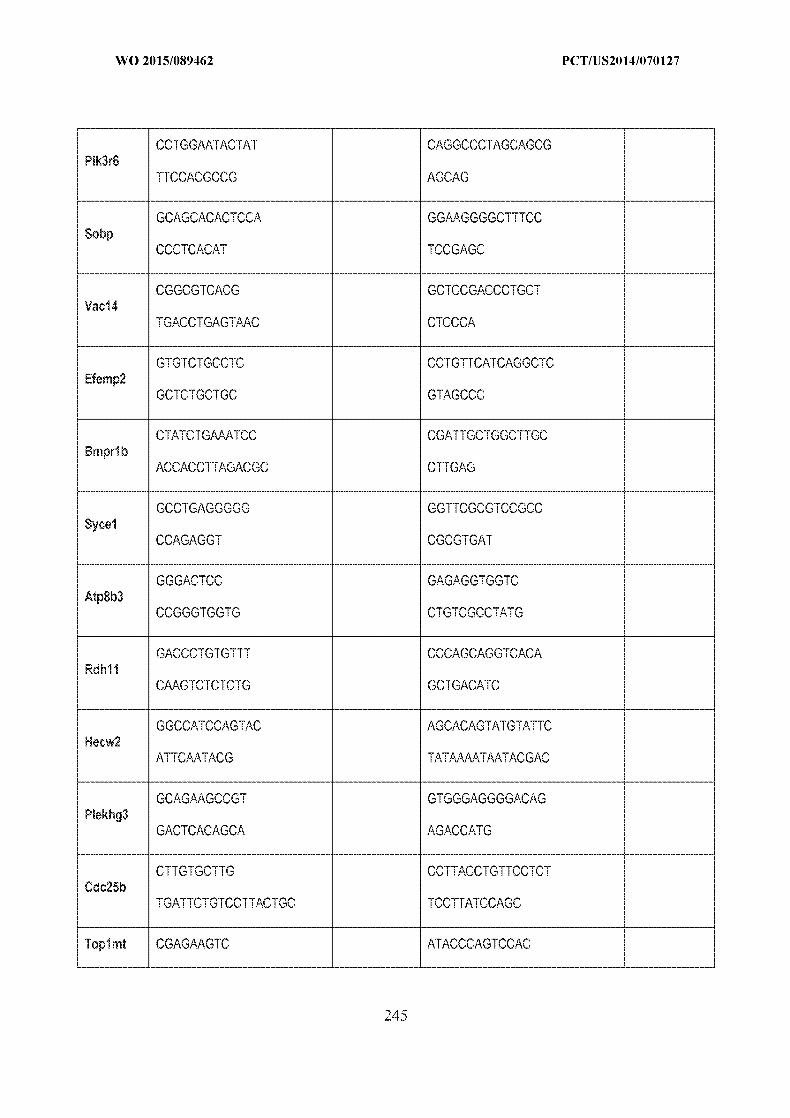
WO 2015/089462 PCT/US2014/070127
CCTGGAATACTAT CAGGCCCTAGCAGCG Pik3r6
TTCCACGCCG AGCAG
GCAGCACACTCCA GG.AAGGGGCTTTCC Sobp
CCCTCACAT TCCGAGC
CGGCCGTCACG C'CTCCC'ACCCTC Vac14
TGACCTGAGT.AAC CTCCCA
GTC-TCTGCCTC CCTGTTCATCAGGCCTC Efem p2
GCTCTGCTGC CGTAGCCC
CTATCTGAAATCC CGATTGCTGGCTTGC Bmprl b
ACCACCTTAGACGC CTTGAG
GCCTGAGGGGG GGTTCGCGTCCGCC Sycel
CCAGAGGT CGCGTGAT
GGGACTCC GAGAGGTGGTC Atp8b3
CCGGGTGGTG CTGTCGCCTATG
GACCCTGTGTTT CCCAGCAGGTCACA Rdhl I
C.AAGTCTCTCTG GCTGACATC
GGCCATCCAGTAC AGCACAGTATGTATTC Hecw2
ATTCAATACG TATAM%%,ATAATAC GAC
GCAGAAGCCGT GTGGGAGGGGACAG Plekhg3
GACTCACAGCA AGACCATG
CTTCGTGCTTG CCTTACCTGTTCCTCT Cdc25b
TC-ATTCTC-TCCTTACTGC TCCTTATCCACC
Topic rt CGAO-AAGTC ATACCCAGCCAC
-- ----- ------ ---- -- ----- ------ ------ ----- ------ ------ ------ ----- ------ ---------- ------ ------ ------ ----- ------ ------ ----2 4- ---- ------ ----

WO 2015/089462 PCT/US2014/070127
GATGCAGACACTTCIAA ATCCCTGCC
GCTGAAGACTGGC CCTCTGCATCTCCCTCAGGAAGT Sesn2 A
GAGCACAGCT ATT
GACCTGAATGTTG GCCTCCTGTC Ncan
TGGCTGAGAGTCC CCCAGGTCCC
CCCTCACGTTCC CACTAGGCTT Nacad
TGTCCAGCAA GGGCTGCCCTCT
Example 2: In vivo Retina Disease Mlodel (Mouse modelfor Retinitis Pignentosa)
[004411 Applicants demonstrate the in vivo effectiveness of a genome engineering approach
using Cas9 when packaged into AAV, and have successfully used it to modify endogenous
genome sequence in mammalian cells. Applicants use this system to demonstrate genetic
engineering potential in neurons, which represents one key group of post-mitotic cells in human
body. Applicants' studies underscore the therapeutic potential of this in vivo genome engineering
of somatic tissue through the correction of mutations associated with human disease Retinitis
Pigmentosa in the mouse model that bears mutation corresponding to the similar genetic defects
found in human patients. The mouse strain used for Applicants' studies is C57BL/6 strain
B6.129S6(Cg)-Rhotml.lKpal/J. This mouse strain was chosen because these mice carry a
nucleotide substitution at codon 23 in the mouse rhodopsin (Rho) gene, CCC to CAC. This
codon encodes the amino acid substitution of histidine for phenylalanine at position 23, P231-.
The P23H mutation is one of the most common causes of autosomal dominant retinitis
pigm entosa. The genomic location of the Rho gene is on mouse chromosome 6: 115,931,927
115,938,829. Applicants observed that mice that are homozygous for the targeted mutation are
viable and fertile. Both the mutant and wildtype gene product (mRNA) is detected by cDNA
sequencing chromatogram. 1'he phenotype in heterozygous mice mimics the retinopathy and
progressive retinal degeneration observed in patients with autosomal dominant retinitis
pigmentosa caused by the P231-I mutation. By 35 days of age, heterozygotes have a shorter rod
outer segment when compared to controls. At post natal day 63, heterozygotes have fewer rod
nuclei (half the number observed in wildtype mice), and decreased length of the rod outer
246

WO 2015/089462 PCT/US2014/070127
segment. Homozygous mice exhibit a more severe phenotype with thinner outer nuclear layer,
severe photoreceptor degeneration by post natal day 23 and loss of almost all photoreceptor cells
by post natal day 63. Glycosylation of the mutant P23H protein is severely diminished.
AA V delivery of Cas9 system to the retina neurons in the mouse strain B6. 129S6(Cg)
Rhotni .l1Kpal/:
[004421 Applicants chose to target the gene Rho that is critical for the normal function of
retina neurons, and induce homologous recombination on the target to correct the related disease
relevant mutations P23H using Adeno-associated virus (AAV) delivery of cas9 genome
engineering tools and a recombination template. Applicants designed three targets, P23H
mutation site labeled in orange with the single nucleotide mutation C-A labeled in red. (see Fig.
18A). This approach demonstrates that the correction of the genetic mutation in this mouse
model can rescue the disease-associated phenotypes and further demonstrates the feasibility of
performing genome modification in neurons of adult animals. Further. the study provides
information to assess the level of homologous recombination that could be induced in neuronal
cell types for genetic engineering purposes, using the retina neurons in this strain as a model
system. The experimental set up is as shown in the table below:
Gru proe njtin Num~ber of grup MieprIoa
N cirl. - saintV 1 o 3 tim ,, 5 15
con dios
[004431 In total, 25 mice are used to set up breeder pairs so in total = 100 mice for each strain
are used for this study.
Retina injection of vA V
[004441 Subretinal injection is used as the delivery route. Mice receive subretinal injection of
0.5-1 microliters of saline solution (0.9% sodium chloride) warmed to body temperature and
containing up to IE12 viral particles. These mice are more than 6 weeks old. The animal is kept
warm using a heating pad and/or lamp and is monitored. Animals are monitored daily for clinical
signs of procedure related complications. Up to two injection per mouse are administered.
Tissue collection:
247

WO 2015/089462 PCT/US2014/070127
[004451 After I to 4 weeks after injections of materials, mice are sacrificed by the CO 2
inhalation method prior to tissue sampling from mice. Tissues are analyzed for evidence of
successful gene transfection, genome alterations, or toxicity.
Example 3: In vivo therapeutic genome engineering approach/fbr Retinitis Pigmentosa
Guide selection and in vitro validation
[004461 First, guides for SaCas9 that target the gene RHO in human genome are designed as
shown below. It has been selected based on the locus of disease-causing mutation P231. The
design was generated through computational algorithm to maximize the potential of introducing
on-target cleavage that is closest to the disease mutation site to facilitate highest efficiency and
specificity of therapeutic gene correction. Guides were also screened for their DNaseI hyper
sensitivity (HS) assay results to maximize the accessibility of the genomnic region corresponding
to the target guide sequence, thereby increasing the expected efficiency of the guides following
delivery in vivo. More than one guides are selected and then screened using Surveyor assay in
vitro in cultured human cells (HEK 293FT) to measure their efficiency for inducing indel
formation at target genomic locus. The guide with highest efficiency in vitro is then selected for
virus production and downstream experiment in vivo. Figure 18A-B shows the guide design for
RHO locus, and in vitro guide screening results using the Surveyor assay.
flomnologous recombination template design and validation
[004471 Secondly, to induce homologous recombination on the target to correct the related
disease-relevant mutations P23H using homology-directed repair, a [HR vector is synthesized
based on the wild type (normal) genomic sequence of the unaffected human individual. This
vector bears the AAV packaging signal, and the normal version of genomic sequence with up to
5kb total homology arms: two homology arms, left and right, are on each side of the sequence
that sandwich the target mutation site P23H in the middle, as shown below. This vector is
validated in vitro by co-transfecting at 1:1, 1:3, 1:5 ratio with the corresponding vector encoding
the SaCas9 system with the best guide as measured in previous section, then the homologous
recombination (HR) efficiency at target P21 31- locus is measured using restriction fragment
length polymorphism assay (RFLP) assay and thus validate the optimal condition for the HDR
procedure.
1004481 Figure 19 shows an RHO HR AAV vector. The specific sequence for the part
responsible for serving as homologous recombination template is listed below.
248

WO 2015/089462 PCT/US2014/070127
>Rho HR AAV Vector Tern late Region
ACC2AGA (- IT r, I-, I I'AGCI' -. "CCTCCCACCC CCATT C C ATTT ArC C A C C AC A' n C T G GG C -CTTGGCACAC'ACAACI'CACALAC"TTPACCC G AD11 TCr-Cr" T"TC' --. C A'CTGAGTA.CCTC-TCTCC 'C CAC CC---G -TTCC -TrCTA-CTG7TC'A
C-CT-'GC*-CTCTrn? GAAGCC7Y-AA TAGCC"(-CTCAGTT-T(C('A GCCGATTAATAVJ-wITT ,'AACACr'~C 7AT C TCCCA~GATGCCT GATT CAGC CAGCCCT-AGCACG GACCTCI' T I T
AC GC ' IT, TC IT' -'' i 'CACAT TCCC 'CACCTCCTCCIC
IT'TCCACATCTT'C G A-C1 &CCACCCCCTC G CG C~ L CAACCCG r' ACNACCNA G"CA.A __"
TCl.CT .. ACrCCACAGTA.-Cr(-" CCTC l-A. .CCCA.TGC,-CACT-'- 'CA " CTG l-Cr C -CT , -ATCT ITC-CTGATC"-GTGCTC--GG'CTT.1-CCTAC'T C' C CTA( GT A'<C -TCA C7'A
A CAACC TCCCC"-ACCC"*CT CTCAAC-TACATCC'TCCT AAC TAG (s" 'T(OC '' AC-TI'TTmCATG( TAI 1 C' 'CC1CCJC,.CAC 7 CA'CC'C C T C ACC(-'TC _TC(-'T "'f CA "' A'~C TT "'C'C1CCC A
CACATCATC J -J' T'j 'j ACATCl
ITT 6'''' G' Gn n' (m - 6 r' T_C7 .7 T r, CCCCA C-. G~T GA (-.7 C_ CT T1- CCCSAC T CT CI-I
AG- A CTCTC~rCT-'---T TCCC-r AGT.T AC.AACCC.(C TAAACTC, -TAACAA
C C1 ,CT ,r TAACACGTC ACTA A AJ -GCACCAAACACCACCACCCACGG'",-)-A''CATT-AA TCGA'6 C"'
rAArCCCAr'CC
TCCATCCC-AACTC"-CG*-G'.-CTCCCTTCCCC-G TTTTTCTCC" AATCAO" T TAAAACAUC
TCCTT T TAAATA TT CC TIA-',- T TGCCTT AAG?l' CT CCA(-r'A T In C'7 n CATT G ~''AGG(T A C ,,,,T7'rA( T'TAACA'7'A" f01T' IT (' TC C CAG'C AC C 'A'f''AATCACA'"''G
TAACA A-1, Jl Jl 1 ,J
ATCTCCCCT T -7AG A T C TTCC(C7AAGCACCfC CAC-CAACArC, -CCC'CATTCI -I ' ,_1
C,-AC .C -ACr ACTC'ATCTCC,-CAA -CACC TICCCCACAT(TCCA-CTCT CTC-'T AACCAACA-TCACACATCACCCrAT AC A ACACAC('-AC A AAAACAC AGAC A
C, 1_7 - . 1_ CTTCCCCAACCCCT TCTAATCCCCCATCTCTCTTCC"'A~TA,-"CCACCTC"G'"G "AzC-'lA
AAAA'T'A- of Ias geom Inier ol n eoiia ~tmlt
GO49 C F ,-- ,Inaly, fo erpljn th Tdnsscae viru ((IV deTver of, Ias ge eninern tool and a-, reomiato ter aei vvbt vrssaerdcdwihsr,
'T T (' T.T rC,'A~,CA 'rC CA.""GN',".AT GN~''C-Ae G..I249e-Cr~(

WO 2015/089462 PCT/US2014/070127
AAV1, AAV2, AAV5, AAV7, AAV8 (all are effective), purified by gradient ultracentrifugation
or chromatography methods. The virus particles are then injected using the optimal condition as
determined in previous section through the subretinal route for delivery to the photoreceptor and
RPE cells.
Detailed injection protocol
[004501 Retina injection of AAV: subretinal injection is performed by injection of up to I
milliliter of saline solution (0.9% sodium chloride) containing different dose of AAV viral
particles warmed to body temperature. The two different AAV vector, one for SaCas9 and one
for HR template aree mixed at different ratios (e.g. SaCas9 vs. HR template:= 1:1, 1:3, or 1:5) to
determine the optimal condition. The dose used for the injection can be as low as 1.5 x 10E10
vector genomes total, or up to 1.5 x 10E I vector genomes total. The higher dose gives better
gene therapy effectiveness but may lead to higher chance of complications due to immune
response to the viral vector injection. Patients are monitored daily for clinical signs of procedure
related complications. This procedure essentially follows guidelines provided in the following
publications: Maguire A. M. et i. N Engl. J. Med. 2008 May 22;358(21):2240-8. doi:
10.1056/NE.Moa0802315. Epub 2008 Apr 27. Safety and efficacy of gene transfer for Leber's
congenital amaurosis. Simonelli F. et at. Mol. Ther. 2010 Mar;18(3):643-50. doi:
10.1038/mt.2009.277. Epub 2009 Dec 1. Gene therapy for Leber's congenital amaurosis is safe
and effective through 1.5 years after vector administration. Maguire A. M. et al. Lancet. 2009
Nov 7;374(9701):1597-605. doi: 10.1016/S0140-6736(09)61836-5. Epub 2009 Oct 23. Age
dependent effects of RPE65 gene therapy for Leber"s congenital amaurosis: a phase I dose
escalation trial.
Post in section procedures
[004511 Post injection, the patient is monitored for any immunological response or adverse
effect. After at least 4 weeks, restriction fragment length polymorphism assay (RFLP) is used to
assess the level of homologous recombination that could be induced in ocular cell types. And the
relief or recovery of RP phenotype is then also evaluated.
Example 4: In vivo therapeutic genonie engineeringfor Achroinatopsia
Guide selection and in vitro validation
1004521 First, guides for SaCas9 that target the gene CNGA3 and CNGB3 in human genome
are designed as shown below. It has been selected based on the locus of disease-causing
250
WO 2015/089462 PCT/US2014/070127
mutation, i.e. R277C and R283W for CNGA3 and 1148delC for CNGB3. The design was
generated through computational algorithm to maximize the potential of introducing on-target
cleavage that is closest to the disease mutation site to facilitate highest efficiency and specificity
of therapeutic gene correction. Guides were also screened for their DNaseI hyper-sensitivity
(-IS) assay results to maximize the accessibility of the genomic region corresponding to the
target guide sequence, thereby increasing the expected efficiency of the guides following
delivery in vivo. More than one guides are selected so that the most efficient guides can be tested
in vitro via Surveyor assay. The guide with highest efficiency in vitro is then selected for virus
production and downstream experiment in vivo. Figure 20A-B shows guide seletion for CNGA3
and CNGB3.
Homologous reconibination template design and validation
[004531 Second, to induce homologous recombination on the target to correct the related
disease-relevant mutations using homology-directed repair, a HR vector need to be synthesized
based on the wild type (normal) genomic sequence of the CNGA3 and CNGB3 genes from
unaffected human individual, respectively.
1004541 Figures 21 and 22 show maps of the CNGA3 [IR AAV vector and CNG33 [IR AAV
vector, respectively. These vectors bear the AAV packaging signal, and the normal version of
genomic sequence with up to 5 kb total homology arms: two homology arms, left and right, are
on each side of the sequence that sandwich the target mutation site in the middle, as shown
below. This vector is validated in vitro by co-transfecting at 1:1, 1:3, 1:5 ratio with the
corresponding vector encoding the SaCas9 system with the best guide as measured in previous
section, then the homologous recombination (HR) efficiency at target locus is measured using
restriction fragment length polymorphism assay (RFLP) assay and thus validate the optimal
condition for the HDR procedure.
[004551 The specific sequence for the part responsible fbr serving as homologous
recombination template is listed below.
>CNGA3 HR AAV Vector Template Region
CT GCTGCCTGCTCTGTCCCCTTTAAGTA T T GACAT CC T CAAAACCC CT TTGGAAAAAGCACA GGCCACAGATCTTACTGTGAC ThTGTGTT TCTTTCTCC T AGGTGNTACCTT CAACCTTGATAAAA AT AAACC T C TAAAT CAA T TGAGA T CT GCCT CCC T' CAC T T T T T T TI T T TTCAAAGAC T CAGAG T C TCACTCTGTTGCCCAGGCTGGAGTGTAGTGGTGCGATCTTGGCTCATTGCACCCTCCACCTCC TGGGTTCAAG TGGTTCTCGrTGCCTCAGCTT CCTGAGT AGCTGGGATT ACAGGGGTGCACCACC
251

WO 2015/089462 PCT/US2014/070127
ACATCTGGCTAATTTTTGTATTTTTAGTAGAGACAGGG'"TCACCATGTTGCCCAGGCTGGTC TCAAACCCTTGACCTCAGGTGATCCACCCGCCTCGGCCTCTCAAAGTACTGGGATTATAGGCA TGAGCCACGGCACCCGGCCCTCTGTCACTTTTTGATTTACAACATGTATCTCTAATTTTAAAG GATCCTTTTTTAAAATATGTATATAATTTCCATTTATCTTTTAAAATTTAATAATCATTCTTT
GTTATCATGTAATACCCAATTTATATTTAAATTTACTCAATCAACCTATGTTTTAAAAAAATT CAATAGAATAGAT T AGAACC T CA TAGAATAGAAAA TATCAGAGT GCAT TT CC T GT AGTAAT GG TAAGTGTTGTTTTTGAAATCATTTCTATTATATATGTATCACTGCATACTGTGTAGCCGTGAG GTAAAATATGTTTCTTTGTACTATGGTCAAAAAAAGTCAGCCTCTGTGATGCCCAATGACCTC:
CATCTTCTTCTTTAGGTTTTCTCGAGCAAGGCTTAATGGTCAGTGATACCAACAGGCTGTGGC AGCATTACAAGACGACCACGCAGTTCAAGCTGGATGTGTTGTCCCTGGTCCCCACCGACCTGG
CTTACTTAAAGG TGGGCACAAAC TACCCAGAAGT GAGGTTCAACCGCCTACTGAAGT T rT'TCCC GGC TCTT TGAA T 1T TTGACCGCACAGAGACAAGGACCAACTACCCCAATATGTTCAGGATTG
GGAACTTGGTCTTGTACATTCTCATCATCATCCACTGGAATGCCTGCATCTACTTTGCCATTT CCAAGTTCATTGGTTTTGGGACAGACTCCTGGGTCTACCCAAACATCTCAATCCCAGAGCATG GGCGCCTCTCCAGGAAGTACATTTACAGTCTCTACTGGTCCACCTTGACCCTTACCACCATTG GTGAGACCCCACCCCCCGTGAAAGATGAGGAGTATCTCTTTGTGGTCGTAGACTTCTTGGTGG GTGTTCTGATTTTTGCCACCATTGTGGGCAATGTGGGCTCCATGATCTCGAATATGAATGCCT
CACGGGCAGAG'TTCCAGGCCAAGA TTGA TTCCA T CAAGCAG TACA TGCAGTTCCGCAAGGTCA
CCAAGGACTTGGAGACGCGGGTTATCCGGTGGT"TTGACTACCTGTGGGCCAACAAGAAGACGG T GGA TGAGAAGGAGG T GC TCAAGAGCCT CCCAGACAAGC T GAAGGC TGAGATCGCCATCAACG TGCACCTGGACACGCTGAAGAAGGTTCGCATCTTCCAGGACTGTGAGGCAGGGCTGCTGGTGG AGCTGGTGCTGAAGCTGCGACCCACTGTGTTCAGCCCCTGGGGATTATATCTGCAAGAAGGGAG ATATTGGGAAGGAGATGTACATCAT CAACGAGGGCAAGCTGGCCGTGGTGGCTGATGA TGTGGGG TCACCCAGT TCGTGGTCCTCACG GCAGCTACTT CGGGGAGAT CAGCA TT CTGAACA T CA AG G G GAGCAAGTCGGGGAACCGCAGGACGGCCAACATCCGCAGCATTGGCTACTCAGACCTG T T C TGCCT C TCAAAGGACGA T CTCA TGGAGGCCC TCACCGAG TACCCCGAAGCCAAGAAGGCCC
TGGAGGAGAAAGGACGGCAGATCCTGATGAAAGACAACCT G ATCGATGAGGAGCTGGCCAGGGCGGGCGCGGACCCCAAGGACCTTGAGGAGAAAGTGGAGCAGCTGGGGTCCTCCCTGGACACCC T GCAGACCAGG TTTGCACGCCTCCTGGCTGAGTACAACGCCACCCAGATGAAGATGAAGCAGC GTCTCAGCCAACT GGAAAGCCAGGTGAAGGGTCGGTGGGGACAAGCCCCT GGC T GA TGGGGAAG T TCCCGGGGAT GC TACAAAAACAGAGCACAAAAAACAACAG T GAAAATi GCAGC T C AT GTC3 T CCT GC
TTCACAGGGTCGACTGTCAGGGTGACCGTATGTGGCCGCAGCTGTGTGGCATGGAACTTGGTC AGGGTTGAATTCCAGCTCTACTCACCCTTTGAAAGCTGTGTGACTGCCTGAGAGAACCTGTTT CTTCACCTAAAAAATGGGACTTTTTGTCTCAGTCCCAGTGAAGTGCCAGGTTTGATTGTGAAG TCCGCATGAAACACTGCACCAGGCAGGGCT T TGCAAAGTGCAA
>CNGB3 HR AAV Vector Template Region
GG TATTTTAGTCAATTT AA AA TAGTTGTTTAT
ATCAAAGTAAATAATTCACA T CTAAAGGGAATTATTTATTTAGTAAATT CTGGAAATTGA GTTTGGGTTTGTCC'ATCAGTGGTCCTTC'TGACTTTbAATr~TTTTAAAATCOT
TCTGGTTGTTATAATCCCT TATACATATCCAACTCACT C TAGGTAGTATGTAAT T T TGTAAGT TAT TTTCCTCTCTTTGCTCTAT CCTATAATTGCTCT CCAT CCCAAGGCTGCAG TGAGTTGCC CTT CAAAGTAATGCTGGGACCTGCTTTTTTTCCAGTTTGGACATTGCCT TAT TATATG TTCAA
TGTCATTTCACTGGAGCAGAAAGT TAGTGAAGTCAACTTTATGCCAGGT C TTTGTATTTTACC AAAAGGAAATTTCACTATTAAATAACCCAGTTGCCATTTCTGAGTCCTGATTCTACTGTTCTA AATTTTTCAAGTGATCTTTTTTTATTTCTGGGACACT TGCATACCTAATTGTCAAGTTTAATT
252

WO 2015/089462 PCT/US2014/070127
TATGATCCTCGIT fT-TAG ITG ' GI CAA
ATAAAACCCTTCA CTGGTAAACTA.ATTAGCCACTG(CACCTGCCCTTTAAATAATTAACATTGT TCATTACTAAAATC ' GGCATCGCAGTTAT T AAAAGTTACCTTA( (C TGCAGTTGTCTAGAGGC TTTCAAAC T T T T T TUAT CAT GAT CCAGAG T AAAAAA T GCAT T T CC AGG'C AAC T CGGAT AC ACA C A ('CAACACACAC /YCTCCCCTACTATCT ATC 'AGACTCT ' ATIA71 TTT"TATTCT' T 'TAT IT, C
TCTATCT -11' 1 . AT G TATTGACT'1TACT'7'7AAAGAGG. TTTTTCAG(C'TITAACA''T TATTTAGACCAATTCTGGGTACTCATGCAA" TTGTTACATGTATCAATCATATATC AA.GTCA.GGTTAGGTGTTTATTACTCGAG0TACAATACATG TTTTGAAACTATAGTCACC CTIACTCTGTTGCAAAC'GTGAATATA.TTCTTACTGTATGTTTGTATCCTTTAATCCACTTTTC T TTATGCGC-T CC TCTCCCCA,1CCAC TCACCCT TCC,---CAGT C TCT-G TTATC TT TCCAC TCTCTGT-C
TCTAT G CAAAAT I'T'AGCTCCCACATATATGT GAGAACGTG TGATAT T TAT CG TTTTG GGfCTCTGGCICTT.-A'TTTC,,(-ACT,.AAG'ATAATGA'CC'TITCAGITIC(-CCAT(CCAT",TT, TGGTCAAATG AC--AT--G u I3T, IT C ':1:' ATT., T TT'TATGGCCA AAAA CCT"GGC "T CAT A"AATT CA.TACAA T-AAACAGAATGTT AAAAATAATCAATAAAGGTT
TCTTTCAAAATCAGAA.C'TAC'CGTTTCCTTCCCCATCATACAC CCAm C'ATAG-TGCCAATT CT'TCA T AGTTTGAAGCC"AG T AATAAACACAGGCA TTAATGT GCAG AATAAACAGCAAGTAT C CTT GTCG AATAACT T GCAAAGATAGATGCAAAGT AAG A'ITCAT GT T G T CITA
AATACAGCC "T'ATTTTAACATTT T C CTTTTCCTTAAAGTC'ACCG' AC G ACCC CTTGCTTTGGATT TGTGAACTNrpGIT TAACTCTCA-.TTAGTl'AC A'GTA C AAAGCTG.A.T GGT"1'G'CCA.TT--GCrAT_,G 1"T'T' TTCTGATG GCAATGTCT IT G IACT GGGA T TGACAGAGTGT AAAGAAAAAAAAAGA A AAAGAAAAC'CCI"CCT C T
C -TTTTCAC'AGATGl-TTGTGAGTCAAC -TCCG(TGAAAGACATGCCTCAAAGGTCACTTCTTCAGTT
TAAGTCCCATAAAATACACTATGCTAATTTAACT GGATATCTCTGAAAAGCTCATGAGACTTT A TGC T ACGAT GAATGGCAACTAGA'GTTTCGGTGCAAGTAAAATT T AACAACAAACGAATG AAA T T CAGAT TAGGA AT GA AT T A ' CAT GAGAAAGG IT T TAAAG T 'T AAC 'T TG CAAAAGAG TAT GT TT TTjC TACT ""T T T TGACAG AGTCAGAT T C T AA
AT CT ATATAT T T T CAT ACT CT TAT T TCACA T TAAAATAT CCAC 'AT C' " C TACAACATAC TCCTA'"TTTATTAGCCA.AAGCTGAAATTA TATC~CTTTTTTTAAAGGGGTCATATCC
CT (CCAAATT CG T CTAAAATGTTGTACCAT GT TTTTCCCCTTCCCCCAAGTATACTGAGT TATAC'T7TTACCOTGTAGATATATG?CTTTGTCCATTATGACTCTAGGTGATGATTAAATTCAAA AAAT GAAGTGTACTAT'ATAGAAAAGCAAAAGAAATCCAAAAGCAT CTTAGTCTAAATATATA
TATTTAAATAAAACTATATGAAATAGATTTTATTACTGAAAAT
AA V delivery of Cas9) genone engineering tools and a recombination template
[00456] Finally for employing the Adeno-associated virus (AAV) delivery of cas9 genome
engineering tools and a recombination template in vivo, both viruses are produced with serotype
AAV1, AAV2, AAV5, AAV7, AAV8 (all are effective), purified by gradient ultracentrifugation
or chromatography methods. The virus particles are then injected using the optimal condition as
determined in previous section through the subretinal route for delivery to the photoreceptor and
RPE cells.
Detailed injection protocol
1004571 Retina injection of AAV: subretinal injection is performed by injection of up to 1
milliliter of saline solution (0.9% sodium chloride) containing different dose of AAV viral
253

WO 2015/089462 PCT/US2014/070127
particles warmed to body temperature. The two different AAV vector, one for SaCas9 and one
for HR template are mixed at different ratios (e.g. SaCas9 vs. HR template = 1:1, 1:3, or 1:5) to
determine the optimal condition. The dose used for the injection can be as low as 1.5 x IEI
vector genomes total, or up to 1.5 x I El vector genomes total. The higher dose may give
better gene therapy effectiveness but might lead to higher chance of complications due to
immune response to the viral vector injection. Patients are monitored daily for clinical signs of
procedure related complications.
Post injection procedures
[00458] Post injection, the patient is monitored for any immunological response or adverse
effect. After at least 4 weeks, restriction fragment length polymorphism assay (RFLP) is be used
to assess the level of homologous recombination that could be induced in ocular cell types. And
the relief or recovery of Achromatopsia phenotype is then also evaluated.
Example 5: In vivo therapeutic genome engineering approach jr age-related nacular
degeneration.
Guide selection and in vitro validation
1004591 First, guides for SaCas9 that target the genomic locus VEGFA in human genome are
designed as shown below. It has been selected within the first exon of the gene, but other parts of
the gene can also be targeted as well so that a wide range of target region can be screened to
determine the most effective guide design. All the designs were generated through computational
algorithm to maximize the potential of introducing on-target cleavage that is closest to the
transcriptional start site and located within the common region of different expressed transcripts
in retina. This will facilitate highest efficiency and specificity of therapeutic gene correction.
Guides were also screened for their DNasel hyper-sensitivitv (HS) assay results to maximize the
accessibility of the genomic region corresponding to the target guide sequence, thereby
increasing the expected efficiency of the guides following delivery in vivo. More than one guides
are selected so that the most efficient guides can be tested in vitro. Figure 23A-B shows guide
seletion for VEGFA.
[004601 To validate that the designed guides can effective repress the expression of VEGFA
gene in human cells and finding the optimal guides and conditions. The closed AAV vector
bearing the designed guides are delivered in vitro into human cells (e.g. HE1K293). The cells
were harvested 72-96 hours post delivery. RNAs and proteins are extracted from the cells. The
254
WO 2015/089462 PCT/US2014/070127
mRNA level of VEGF is measured via qRT-PCR method or other RNA measurement method,
whereas protein level of VEGF is also measured via ELISA or other protein measurement
method. The most critical criterion is the VEGF protein level because this is directly relevant for
the clinical effectiveness of the system. The guide with highest efficiency in vitro to lower the
expression level of VEGF is then selected for virus production and downstream experiment in
vivo.
1004611 Finally for employing the Adeno-associated virus (AAV) delivery of cas9 genome
engineering tools to disrupt VEGF expression in vivo, viruses are produced with serotype AAV2
and AAV8 (both are effective, other AAV serotype such as AAV1, AAV5, AAV7, AAV9, or
AAV-DJ might be used but will be less potent). The viral particles are all purified by gradient
ultracentrifugation or chromatography methods.
[004621 The AAV virus particles are then injected using the optimal condition as determined
in previous section through the intravitreal route, where AAV is injected in the vitreous humor of
the eye. This procedure is less invasive and can sustain persistent expression of the construct to
obtain sufficient disruption of VEGF expression to induce therapeutic corrective effects.
Detailed injection protocol
1004631 Retina injection of AAV: intravitreal injection is performed by injection of typically
100 ricroliter (or up to 1 milliliter but not recommended) of saline solution (0.9% sodium
chloride) containing different dose of AAV viral particles warmed to body temperature. The dose
used for the injection can be as low as I x 10E8 vector genomes in total, or up to 1 x 10111
vector genomes in total. The use of different dosage allows the determination of optimal dose.
[004641 The higher dose will give better gene therapy effectiveness but might lead to higher
chance of complications due to immune response to the viral vector injection. Patients will be
monitored daily for clinical signs of procedure related complications.
[004651 Reference for human injection parameter is available at Clinical Trials (dot) Gov - the
US Government website for information on clinical trials, having a web address of:
clinicaltrials. gov/ct2/show/N CT0 1024998.
Post injection procedures
[004661 Post injection, the patient are monitored for any immunological response or adverse
effect. After at least 4 weeks, ELISA or other protein measurement methods can be employed to
2'15 -5

WO 2015/089462 PCT/US2014/070127
assess the level of VEGF in the retina. And the relief or recovery of ARMD phenotype is then
evaluated.
[004671 Key safety measures to be followed in the patients include the maximum tolerated
dose of a single uniocular intravitreal injection, number of treatment emergent adverse events,
and the thickness of the retina post injection.
Example 6: dSaCas9 to stimulate A TOi1 expression to treat dealness or hearing impairments.
Guide selection and in vitro validation
[004681 First, guides for SaCas9 that target the gene ATOHI in human genome are designed
as shown below. It has been selected based on the optimal parameters to maximize the efficiency
of epigenetic modulation on target gene expression. Specifically, the design was generated
through computational algorithm to maximize the potential of introducing on-target binding to
facilitate highest efficiency and specificity of therapeutic gene therapy. Guides were also
screened for their DNaseI hyper-sensitivity (HS) assay results to maximize the accessibility of
the genomic region corresponding to the target guide sequence, thereby increasing the expected
efficiency of the guides following delivery in vivo. Moreover, transcription factor sites that
potentially might interfere with the binding were also considered during the selection of the
guides. More than one guides are selected and then screened in vitro in cultured human cells
(HEK 293FT) to measure their efficiency for inducing ATOHI1 gene expression. The guide with
highest efficiency in vitro is then selected for downstream experiment and therapeutic
intervention in vivo. The specific design of guides for ATO-1 is shown at Figure 25A-C.
Guide screening and validation
[004691 To validate that the designed guides can effective induce the expression of ATOH1
gene in human cells and finding the optimal guides and conditions, in vitro screening and
verification is performed with human cell lines. The system, which contains dSaCas9, fusion
effector, and optimal guide RNAs, is delivered in vitro into human cells (e.g. HEK293) (Figure
24). The cells were harvested 72-96 hours post delivery. RNAs and proteins are extracted from
the cells. The rnRNA level of ATOH1 is measured via qRT-PCR method or other RNA
measurement methods, whereas protein level of ATOH1 is measured via ELISA or other protein
measurement methods. The most critical criteria is the ATOH1 protein level because this is
directly relevant for the clinical effectiveness of the system. The guide with highest efficiency in
256

WO 2015/089462 PCT/US2014/070127
vitro to stimulate the expression of ATOH 1I is then selected for in vivo delivery and therapeutic
demonstration.
[004701 AAV or Ad viral particles or other delivery vehicle packaging the entire system are
then produced, and injected using the optimal conditions as determined experimentally. The most
critical parameter to optimize is the amount (i.e. dosage) of the delivery vehicle used for each
individual injection.
1004711 The higher dose will give better gene therapy effectiveness but might lead to higher
chance of complications due to immune response to the vehicle injection or the off-target effects
of the system. Patients are monitored daily for clinical signs of procedure related complications.
Post injection procedures
[004721 Post injection, the patient are monitored for any imnunological response or adverse
effect. Each week post injection, ELISA or other protein measurement methods can be employed
to assess the level of ATOH1 stimulation. New hair cell growth in the Cochleae can be
visualized through biopsy and imaging methods. And the relief or recovery of deafness and
hearing impairments is then evaluated through hearing tests on the human patients.
1004731 Key safety measures to be followed in the patients include the maximum tolerated
dose of a single injection into the human Cochleae, number of treatment emergent adverse events
post injection.
[004741 While preferred embodiments of tlie present invention have been shown and
described herein, it will be obvious to those skilled in the art that such embodiments are provided
by way of example only. Numerous variations, changes, and substitutions will now occur to
those skilled in the art without departing from the invention. It should be understood that various
alternatives to the embodiments of the invention described herein may be employed in practicing
the invention.
257

WO 2015/089462 PCT/US2014/070127
WHAT IS CLAIMED IS:
I. A method of modeling a disease associated with a genomic locus in a eukaryotic
organism or a non-human organism comprising manipulation of a target sequence within a
coding, non-coding or regulatory element of said genomic locus comprising delivering a non
naturally occurring or engineered composition comprising :
(A) - L a CRISPR-Cas system RNA polynucleotide sequence, wherein the
polynucleotide sequence comprises:
(a) a guide sequence capable of hybridizing to the target sequence,
(Ib) a tracr mate sequence, and
(c) a tracr sequence, and
I. a polvnucleotide sequence encoding Cas9, optionally comprising at
least one or more nuclear localization sequences,
wherein (a), (b) and (c) are arranged in a 5' to 3' orientation,
wherein when transcribed, the tracr mate sequence hybridizes to the tracr sequence and the guide
sequence directs sequence-specific binding of a CRISPR complex to the target sequence, and
wherein the CRISPR complex comprises Cas9 complexed with (1) the guide sequence that is
hybridized to the target sequence, and (2) the tracr mate sequence that is hybridized to the tracr
sequence and the polynucleotide sequence encoding Cas9 is DNA or RNA,
or
(B) . polynucleotides comprising:
(a) a guide sequence capable of hybridizing to the target sequence, and
(b) at least one or more tracr mate sequences,
IL a polynucleotide sequence encoding Cas9, and
IlL a polynucleotide sequence comprising a tracr sequence,
wherein when transcribed, the tracr mate sequence hybridizes to the tracr sequence and the guide
sequence directs sequence-specific binding of a CRISP3R complex to the target sequence, and
wherein the CRISPR complex comprises the Cas9 complexed with (1) the guide sequence that is
hybridized to the target sequence, and (2) the tracr mate sequence that is hybridized to the tracr
sequence, and the polynucleotide sequence encoding Cas9 is DNA or RNA.
2. The method of claim 1, wherein the Cas9 is SaCas9.
258

WO 2015/089462 PCT/US2014/070127
3. The method of claim I or 2, wherein the polynucleotides encoding the sequence
encoding Cas9, the guide sequence, tracr mate sequence or tracr sequence is/are RNA and are
delivered via liposomes, nanoparticles, cell penetrating peptides, exosomes, microvesicles, or a
gene-gun.
4. The method of any of claims I to 3, wherein the polynucleotides are comprised
within a vector system comprising one or more vectors.
5. A method of modeling a disease associated with a genomic locus in a eukaryotic
organism or a non-human organism comprising manipulation of a target sequence within a
coding, non-coding or regulatory element of said genomic locus comprising delivering a non
naturally occurring or engineered composition comprising a viral vector system comprising one
or more viral vectors operably encoding a composition for expression thereof, wherein the
composition comprises:
(A) a non-naturally occurring or engineered composition comprising a vector
system comprising one or more vectors comprising
. a first regulatory element operably linked to a CRISPR-Cas system RNA
polynucleotide sequence, wherein the polynucleotide sequence comprises
(a) a guide sequence capable of hybridizing to the target sequence,
(b) a tracr mate sequence, and
(c) a tracr sequence, and
IL a second regulatory element operably linked to an enzyme-coding
sequence encoding Cas9, optionally comprising at least one or more nuclear localization
sequences,
wherein (a), (b) and (c) are arranged in a 5' to 3' orientation,
wherein components I and II are located on the same or different vectors of the system,
wherein when transcribed, the tracr mate sequence hybridizes to the tracr sequence and the guide
sequence directs sequence-specific binding of a CRISPIR complex to the target sequence, and
wherein the CRISPR complex comprises the Cas9 complexed with (1) the guide sequence that is
hybridized to the target sequence, and (2) the tracr mate sequence that is hybridized to the tracr
sequence,
or
259

WO 2015/089462 PCT/US2014/070127
(B) a non-naturally occurring or engineered composition comprising a vector
system comprising one or more vectors comprising
1. a first regulatory element operably linked to
(a) a guide sequence capable of hybridizing to the target sequence, and
(b) at least one or more tracr mate sequences,
IL a second regulatory element operably linked to an enzyme-coding
sequence encoding Cas9, and
III. a third regulatory element operably linked to a tracr sequence,
wherein components I, II and III are located on the same or different vectors of the system,
wherein when transcribed, the tracr mate sequence hybridizes to the tracr sequence and the guide
sequence directs sequence-specific binding of a CRISPR complex to the target sequence, and
wherein the CRISPR complex comprises the Cas9 complexed with (1) the guide sequence that is
hybridized to the target sequence; (2 the tracr mate sequence that is hybridized to the tracr
sequence; and wherein the Cas9 is preferably SaCas9.
6. The method of claim 5, wherein one or more of the viral vectors are delivered via
liposomes, nanoparticles, exosomes, microvesicles, or a gene-gun.
7. A method of treating or inhibiting a condition or a disease caused by one or more
mutations in a oenomic locus in a eukaryotic oroanism or a non-human organism comprising
manipulation of a target sequence within a coding, non-coding or regulatory element of said
genomic locus in a target sequence in a subject or a non-human subject in need thereof
comprising modifying the subject or a non-human subject by manipulation of the target sequence
and wherein the condition or disease is susceptible to treatment or inhibition by manipulation of
the target sequence comprising providing treatment comprising:
delivering a non-naturally occurring or engineered composition comprising an AAV or
lentivirus vector system, comprising one or more AAV or lentivirus vectors operably encoding a
composition for expression thereof, wherein the target sequence is manipulated by the
composition when expressed, wherein the composition comprises:
(A) a non-naturally occurring or engineered composition comprising a vector
system comprising one or more vectors comprising
L a first regulatory element operably linked to a CRISPR-Cas system RNA
polynucleotide sequence, wherein the polynucleotide sequence comprises
260

WO 2015/089462 PCT/US2014/070127
(a) a guide sequence capable of hybridizing to the target sequence in a
eukaryotic cell,
(b) a tracr mate sequence, and
(c) a tracr sequence, and
II. a second regulatory element operably linked to an enzyme-coding
sequence encoding Cas9 comprising at least one or more nuclear localization sequences,
wherein (a), (b) and (c) are arranged in a 5' to 3' orientation,
wherein components I and II are located on the same or different vectors of the system,
wherein when transcribed, the tracr mate sequence hybridizes to the tracr sequence and the guide
sequence directs sequence-specific binding of a CRISP3R complex to the target sequence, and
wherein the CRISPR complex comprises the Cas9 comnplexed with (1) the guide sequence that is
hybridized to the arget sequence, and (2) the tracr mate sequence that is hybridized to the tracr
sequence,
or
(B) a non-naturally occurring or engineered composition comprising a vector
system comprising one or more vectors comprising
. a first regulatory element operably linked to
(a) a guide sequence capable of hybridizing to an target sequence in a
eukaryotic cell, and
(b) at least one or more tracr mate sequences,
Il, a second regulatory element operably linked to an enzyme-coding
sequence encoding Cas9, and
III. a third regulatory element operably linked to a tracr sequence,
wherein components I, II and III are located on the same or different vectors of the system,
wherein when transcribed, the tracr mate sequence hybridizes to the tracr sequence and the guide
sequence directs sequence-specific binding of a CRISPR complex to the target sequence,
wherein the CRISPR complex comprises Cas9 complexed with (1) the guide sequence that is
hybridized to the target sequence, and (2) the tracr mate sequence that is hybridized to the tracr
sequence; and wherein the Cas9 is preferably SaCas9.
8. The method of any preceding claim, wherein the method is carried out in vitro, ex
vivo or in vivo.
261

WO 2015/089462 PCT/US2014/070127
9. The method of any preceding claim including inducing expression.
10. The method of claim I wherein the condition or disease is an ocular disease.
11. The method of claim 10 where the ocular disease is retinitis pigmentosa or
achromatopsia.
12. The method of any of claims 4 to 8 wherein the viral vector is an AAV or
lentiviral vector.
13. A method of delivering the Cas9 of any preceding claim, comprising delivering to
a cell mRNA encoding the Cas9.
14. The method of any one of claims I to 13, wherein the polynucleotide or sequence
encoding the Cas9 is delivered to the cell by delivering mRNA encoding the Cas9 to the cell.
15. A method of preparing the AAV or lentivirus vector of claim 7 comprising
transfecting plasmid(s) containing or consisting essentially of nucleic acid molecule(s) coding
for the AAV or lentivirus into AAV-infected or lentivirus-infected cells, and supplying AAV
AAV or lentivirus rep and/or cap and/or helper nucleic acid molecules obligatory for replication
and packaging of the AAV or lentivirus.
16. A method of preparing an AAV or lentivirus vector for use in the method of claim
7, comprising transfecting plasmid(s) containing or consisting essentially of nucleic acid
molecule(s) coding for the AAV or lentivirus into AAV-infected or lentivirus-infected cells, and
supplying AAV AAV or lentivirus rep and/or cap and/or helper nucleic acid molecules
obligatory for replication and packaging of the AAV or lentivirus.
17. The method of claim 15 or 16 wherein the AAV or lentivirus rep and/or cap
obligatory for replication and packaging of the AAV or lentivirus are supplied by transfecting the
cells with helper plasmid(s) or helper virus(es).
18. The method of claim 17 wherein the helper virus is a poxvirus, adenovirus,
lentivirus, herpesvirus or baculovirus.
19. The method of claim 18 wherein the poxvirus is a vaccinia virus.
20. The method of any of cla ims 15 to 20 wherein the cells are mammalian cells.
21. The method of any of claims 15 to 20 wherein the cells are insect cells and the
helper virus (where present) is baculovirus.
22. The method of any preceeding claim wherein the Cas9 is a wild type, truncated or
a chimeric Cas9.
262

WO 2015/089462 PCT/US2014/070127
2.3. A composition as defined in any of claims 1-22 for use in medicine or in therapy.
24. A composition as defined in any of claims 1-22 for use in a method of modeling a
disease associated with a genetic locus in a eukaryotic organism or a non-human organism
comprising manipulation of a. target sequence within a. coding, non-coding or regulatory element
of said genetic locus.
25. Use of a composition as defined in any of claims 1-24 in ex vivo or in vivo gene or
genome editing.
26. Use of a composition as defined in any of claims 1-24 in the manufacture of a
medicament for in vitro, ex vivo or in vivo gene or genome editing or for use in a method of
modifying an organism or a non-human organism by manipulation of a target sequence in a
genomic locus associated with a disease or in a method of treating or inhibiting a condition or
disease caused by one or more mutations in a genomic locus in a eukaryotic organism or a non
human organism.
27. A composition comprising:
(A) - . a CRISPR-Cas system RNA polynucleotide sequence, wherein the
polynucleotide sequence comprises:
(a) a guide sequence capable of hybridizing to a target sequence in a
eukaryotic cell,
(b) a tracr mate sequence, and
(c) a tracr sequence, and
11 a polynucleotide sequence encoding Cas9, optionally comprising at least
one or more nuclear localization sequences,
wherein (a), (b) and (c) are arranged in a 5' to 3' orientation,
wherein when transcribed, the tracr mate sequence hybridizes to the tracr sequence and the guide
sequence directs sequence-specific binding of a CR ISPR complex to the target sequence, and
wherein the CRISPR complex comprises the Cas9 complexed with (1) the guide sequence that is
hybridized to the target sequence, and (2) the tracr mate sequence that is hybridized to the tracr
sequence and the polynucleotide sequence encoding SaCas9 is DNA or RNA,
or
(13) I. polynucleotides comprising:
263

WO 2015/089462 PCT/US2014/070127
(a) a guide sequence capable of hybridizing to an target sequence in a
eukaryotic cell, and
(b) at least one or more tracr mate sequences,
11. a polynucleotide sequence encoding Cas9, and
III. a polynucleotide sequence comprising a tracr sequence,
wherein when transcribed, the tracr mate sequence hybridizes to the tracr sequence and the guide
sequence directs sequence-specific binding of a CR] SPR complex to the target sequence, and
wherein the CRISPR complex comprises Cas9 complexed with (1) the guide sequence that is
hybridized to the target sequence, (2) the tracr mate sequence that is hybridized to the tracr
sequence, and the polynucleotide sequence encoding Cas9 is DNA or RNA; and the Cas9 is
preferably SaCas9.
for use in medicine or therapy; or for use in a method of modifying an organism or a non-human
organism by manipulation of a target sequence in a genomic locus associated with a disease or
disorder; or for use in a method of treating or inhibiting a condition caused by one or more
mutations in a genetic locus associated with a disease in a eukaryotic organism or a non-human
organism.; or for use in in vitro, ex vivo or in vivo gene or genome editing.
28. The composition of claim 27, wherein the polynucleotides are comprised within a
vector system comprising one or more vectors.
29. The method, use or composition of any of the preceding claims, wherein the
CRISPR-Cas system RNA is a chimeric RNA (chiRNA).
30. The method, use or composition of any of the preceding claims, wherein the
CRISPR-Cas system is a multiplexed SaCas9 enzyme system further comprising multiple
chimeras and/or multiple multiguide sequences and a single tracr sequence.
31. The method, use or composition according any of the preceding claims, wherein
theCas9 is a nuclease directing cleavage of both strands at the location of the target sequence.
32. The method, use or composition according to any of the preceding claims,
wherein the Cas9 comprises one or more mutations.
33. The method, use or composition according to claim 32, wherein the Cas9
comprises one or more mutations D1OA, E762A, H840A, N854A, N863A or D986A.
34. The method, use or composition according to claim 32 wherein the one or more
mutations is in a RuvC 1 domain of the Cas9.
264

WO 2015/089462 PCT/US2014/070127
35. The method, use or composition according to claim 30, wherein the Cas9 is a
nickase directing cleavage at the location of the target sequence.
36. The method, use or composition according to claim 35, wherein the nickase is a
double nickase.
37. The method, use or composition according to any preceding claim further
comprising at least two or more NLS.
38. The method, use or composition according to any preceding claim, wherein the
SaCas9 has one or more mutations in a catalytic domain, wherein when transcribed, the tracr
mate sequence hybridizes to the tracr sequence and the guide sequence directs sequence-specific
binding of a CRISPIR complex to the target sequence, and wherein the enzyme further comprises
a functional domain.
39. The method, use or composition according to claim 38, wherein the functional
domain is a transcriptional activation domain.
40. The method, use or composition according to claim 39, wherein the
transcriptional activation domain is VP64.
41. A therapeutic genome editing method for treating or inhibiting a condition or a
disease caused by one or more mutations in a genomic locus in a eukaryotic organism or a non
human orgamsrn comprising manipulation of a target sequence within a coding, non-coding or
regulatory element of said genomic locus in a target sequence in a subject or a non-human
subject in need thereof comprising modifying the subject or a non-human subject by
manipulation of the target sequence and wherein the condition or disease is susceptible to
treatment or inhibition by manipulation of the target sequence comprising providing treatment
comprising:
delivering a non-naturally occurring or engineered composition comprising an AAV or
lentivirus vector system, comprising one or more A-AV or lentivirus vectors operably encoding a
composition for expression thereof, wherein the target sequence is manipulated by the
composition when expressed, wherein the composition comprises:
(A) a non-naturally occurring or engineered composition comprising a vector system
comprising one or more vectors comprising
L a first regulatory element operably linked to a CRISPR-Cas system RNA
polynucleotide sequence, wherein the polynucleotide sequence comprises
2 6 5

WO 2015/089462 PCT/US2014/070127
(a) a guide sequence capable of hybridizing to the target sequence in a eukaryotic
cell,
(b) a tracr mate sequence, and
(c) a tracr sequence, and
II a second regulatory element operably linked to an enzyme-coding sequence
encoding Cas9 comprising at least one or more nuclear localization sequences,
wherein (a), (b) and (c) are arranged in a 5' to 3' orientation,
wherein components I and II are located on the same or different vectors of the system,
wherein when transcribed, the tracr mate sequence hybridizes to the tracr sequence and
the guide sequence directs sequence-specific binding of a CRISPR complex to the target
sequence, and
wherein the CRISPR complex comprises the Cas9 complexed with (1) the guide
sequence that is hybridized to the arget sequence, and (2) the tracr mate sequence that is
hybridized to the tracr sequence,
or
(B) a non-naturally occurring or engineered composition comprising a vector system
comprising one or more vectors comprising
I. a first regulatory element operably linked to
(a) a guide sequence capable of hybridizing to an target sequence in a eukaryotic cell,
and
(b) at least one or more tracr mate sequences,
IL a second regulatory element operably linked to an enzyme-coding sequence
encoding Cas9, and
III. a third regulatory element operably linked to a tracr sequence,
wherein components I, II and III are located on the same or different vectors of the
system,
wherein when transcribed, the tracr mate sequence hybridizes to the tracr sequence and
the guide sequence directs sequence-specific binding of a CRISPR complex to the targ-et
sequence, and
266

WO 2015/089462 PCT/US2014/070127
wherein the CRISPR complex comprises Cas9 complexed with (1) the guide sequence
that is hybridized to the target sequence, (2) the tracr mate sequence that is hybridized to the
tracr; and the Cas9 is preferably SaCas9.
42. The method of claim 41, wherein the condition or disease is retinitis pigentosa or
achromatopsia.
43. The method of claim 41, wherein the AAV is AAV1, AAV2, AAV5, AAV7, AAV8,
AAV DJ or any combination thereof.
44. A method of individualized or personalized treatment of a genetic disease in a
subject in need of such treatment comprising:
(a) introducing multiple mutations ex vivo in a tissue, organ or a cell line comprising
SaCas9-expressing eukaryotic cell(s), or in vivo in a transgenic non-human mamnmal having cells
that express SaCas9, comprising delivering to cell(s) of the tissue, organ, cell or mammal the
vector as herein-discussed, wherein the specific mutations or precise sequence substitutions are
or have been correlated to the genetic disease;
(b) testing treatment(s) for the genetic disease on the cells to which the vector has been
delivered that have the specific mutations or precise sequence substitutions correlated to the
genetic disease; and
(c) treating the subject based on results fiom the testing of treatments) of step (b).
45. The method of claim 44, wherein the genetic disease is an ocular disease.
46. The method of claim 45, wherein the ocular disease is retinitis pigmentosa or
achrom atopsia.
267