Transplants of embryonic brainstem containing the locus coeruleus into spinal cord enhance the...
12
Brain Research, 381 (1986) 225-236 225 Elsevier BRE 11954 Transplants of Embryonic Brainstem Containing the Locus Coeruleus into Spinal Cord Enhance the Hindlimb Flexion Reflex in Adult Rats JAMES T. BUCHANAN and HOWARD O. NORNES Department of Anatomy, Colorado State University, Fort Collins, CO 80523 (U.S.A.) (Accepted January 14th, 1986) Key words: neural transplant - - cell suspension - - brainstem - - locus coeruleus - - lumbar spinal cord - - catecholamine reinnervation - - 6-hydroxydopamine - - hindlimb flexion reflex Cell suspensions of embryonic brainstem containing the locus coeruleus were injected intervertebrally into the lumbar spinal cord of adult rats whose descending catecholamine (CA) fibers had been lesioned with intracisternal injection of 6-hydroxydopamine. Up to 1100 CA cells were found 2 and 4 months later, and these cells grew processes which produced histologically d,etectable reinnervation of the lumbar gray matter on the injected side of the cord. To assess the functional activity of the transplanted CA cells, the force of the hindlimb flexion reflex was measured in acute spinal rats. This reflex has been shown previously to be strongly enhanced by catechol- amines. The flexion reflexes were significantly stronger in the transplanted rats than in the controls. Further, the flexion reflexes were significantly reduced by phenoxybenzamine, an a-adrenergic blocker, in the transplanted rats while the reflexes of controls were not significantly changed. These results demonstrate that cell suspension transplants of embryonic brainstem containing the locus coeru- leus into the adult rat spinal cord survive, grow reinnervating catecholamine processes, and can affect the functional activity of the spi- nal cord. INTRODUCTION Transplants of embryonic central nervous systdm (CNS) tissue into adult CNS survive, develop in a do- nor-specific way, and establish synaptic connections with the host nervous system 8'22"26. These properties have allowed the use of embryonic CNS transplants to replace nerve cells lost due to mechanical or chem- ical lesions, congenital abnormalities or aging, and transplants have been shown to ameliorate functio- nal deficits in these animals 9'l°A4AS'21'31. Most studies have transplanted embryonic CNS tissue as solid pieces into the ventricles ls'21 or into surgically-prepared cavities 5'27'3°. Recently, success- ful transplants of cell suspensions of embryonic CNS have been reported 3'4'34. This technique offers the advantages of minimizing damage to the host tissue and of accurate placement of the transplant near the intended reinnervation site. The present study sought to transplant cell suspen- sions of embryonic locus coeruleus (LC) neurons into the lumbar spinal cord of adult rats. Norepinephrine- containing neurons within this brainstem nucleus have axon branches which descend to the lumbosa- cral spinal cord 2s. Norepinephrine has been impli- cated in several aspects of spinal cord function in- cluding nociception 32, sympathetic activity 6 and mo- tor functions such as the flexion reflex 1'2 and locomo- tion 12. Thus, depletion of descending norepinephrine fibers due to disease, aging or injury may contribute to spinal cord dysfunction. One strategy for correct- ing the loss of norepinephrine fibers is to replace them by transplanting norepinephrine nerve cells di- rectly into the spinal cord. In the present study, cell suspensions of embryonic brainstem containing the LC were injected into the lumbar spinal cord of adult rats. The hosts' spinal cords had been depleted of most descending cate- cholamine (CA) fibers by intracisternal injection of 6-hydroxydopamine (6-OHDA). The LC neurons survived, grew normal-appearing axonal processes which reinnervated the ipsilateral gray matter, and Correspondence: H.O. Nornes, Department of Anatomy, Colorado State University, Fort Collins, CO 80523, U.S.A. 0006-8993/86/$03.50 © 1986 Elsevier Science Publishers B.V. (Biomedical Division)
Transcript of Transplants of embryonic brainstem containing the locus coeruleus into spinal cord enhance the...

Brain Research, 381 (1986) 225-236 225 Elsevier
BRE 11954
Transplants of Embryonic Brainstem Containing the Locus Coeruleus into Spinal Cord Enhance the Hindlimb Flexion Reflex in Adult Rats
JAMES T. BUCHANAN and HOWARD O. NORNES
Department of Anatomy, Colorado State University, Fort Collins, CO 80523 (U.S.A.)
(Accepted January 14th, 1986)
Key words: neural transplant - - cell suspension - - brainstem - - locus coeruleus - - lumbar spinal cord - - catecholamine reinnervation - - 6-hydroxydopamine - - hindlimb flexion reflex
Cell suspensions of embryonic brainstem containing the locus coeruleus were injected intervertebrally into the lumbar spinal cord of adult rats whose descending catecholamine (CA) fibers had been lesioned with intracisternal injection of 6-hydroxydopamine. Up to 1100 CA cells were found 2 and 4 months later, and these cells grew processes which produced histologically d,etectable reinnervation of the lumbar gray matter on the injected side of the cord. To assess the functional activity of the transplanted CA cells, the force of the hindlimb flexion reflex was measured in acute spinal rats. This reflex has been shown previously to be strongly enhanced by catechol- amines. The flexion reflexes were significantly stronger in the transplanted rats than in the controls. Further, the flexion reflexes were significantly reduced by phenoxybenzamine, an a-adrenergic blocker, in the transplanted rats while the reflexes of controls were not significantly changed. These results demonstrate that cell suspension transplants of embryonic brainstem containing the locus coeru- leus into the adult rat spinal cord survive, grow reinnervating catecholamine processes, and can affect the functional activity of the spi- nal cord.
INTRODUCTION
Transplants of embryonic central nervous systdm
(CNS) tissue into adult CNS survive, develop in a do-
nor-specific way, and establish synaptic connections with the host nervous system 8'22"26. These properties
have allowed the use of embryonic CNS transplants
to replace nerve cells lost due to mechanical or chem-
ical lesions, congenital abnormalities or aging, and
transplants have been shown to ameliorate functio- nal deficits in these animals 9'l°A4AS'21'31.
Most studies have transplanted embryonic CNS tissue as solid pieces into the ventricles ls'21 or into
surgically-prepared cavities 5'27'3°. Recently, success-
ful transplants of cell suspensions of embryonic CNS have been reported 3'4'34. This technique offers the
advantages of minimizing damage to the host tissue
and of accurate placement of the transplant near the intended reinnervation site.
The present study sought to transplant cell suspen- sions of embryonic locus coeruleus (LC) neurons into
the lumbar spinal cord of adult rats. Norepinephrine-
containing neurons within this brainstem nucleus
have axon branches which descend to the lumbosa- cral spinal cord 2s. Norepinephrine has been impli-
cated in several aspects of spinal cord function in- cluding nociception 32, sympathetic activity 6 and mo-
tor functions such as the flexion reflex 1'2 and locomo-
tion 12. Thus, depletion of descending norepinephrine
fibers due to disease, aging or injury may contribute
to spinal cord dysfunction. One strategy for correct-
ing the loss of norepinephrine fibers is to replace
them by transplanting norepinephrine nerve cells di- rectly into the spinal cord.
In the present study, cell suspensions of embryonic
brainstem containing the LC were injected into the
lumbar spinal cord of adult rats. The hosts' spinal
cords had been depleted of most descending cate- cholamine (CA) fibers by intracisternal injection of 6-hydroxydopamine (6 -OHDA) . The LC neurons
survived, grew normal-appearing axonal processes which reinnervated the ipsilateral gray matter, and
Correspondence: H.O. Nornes, Department of Anatomy, Colorado State University, Fort Collins, CO 80523, U.S.A.
0006-8993/86/$03.50 © 1986 Elsevier Science Publishers B.V. (Biomedical Division)

226
appeared to be functional since the hindlimb flexion reflexes were significantly enhanced in the trans- planted animals.
MATERIALS AND METHODS
Subjects and overall design Ninety-four female Sprague-Dawley rats were
used in 4 groups: (1) normals (N); (2) 6-hydroxydo- pamine controls (OH) which received only an intra-
cisternal injection of 6-OHDA; (3) sham controls
(GS) which received intraspinal injections of a glu-
cose-saline solution 2 weeks after intracisternal 6- OHDA; and (4) locus coeruleus transplants (LC) which received intraspinal injections of LC cell sus-
pensions 2 weeks after intracisternal 6-OHDA. After a 2- or 4-month post-transplantation survival (PTS) time, the animals were tested functionally and then the spinal cords were examined with catecholamine histofluorescence.
6-OHDA procedure At 8 weeks of age the animals were anesthetized
with ether and then anesthesia was maintained with methoxyflurane (Metofane, Pitman-Moore). Twen-
ty/A of a 6 -OHDA solution were injected intracister- nally in 10 s from a syringe equipped with a micropi- pette (tip diameter = 50/~m). The 6-OHDA solution consisted of 250 ~g 6 -OHDA hydrobromide (Sigma)
(calculated as the free-base) and 4 ~g ascorbic acid per 20~1 of a 0.9% NaCl solution.
Transplantation procedure The preparation of the cell suspensions follows
the procedures described by Bj6rklund and his col- leagues 4. Cell suspensions were prepared from
Sprague-Dawley rat embryos (8.5-13 mm crown- rump length). The pregnant rats were palpated under ether anesthesia to determine if the embryos were of the desired size. The pregnant rats were then anes- thetized by an intramuscular (i,m.) injection of ket- amine (Vetalar, Parke-Davis, 87 mg/kg) and xyla- zine (Rompun, Haver-Lockhart, 13 mg/kg). Em- bryos were removed one at a time beginning at the distal end of the uterine horn and were dissected at room temperature using sterile instruments. The brainstem was removed under a dissecting micro- scope and placed onto a depression slide containing a
sterile solution of 0.9% NaCI and 0.6% glucose. l h c
region of the brainstem containing the anlagc of the locus coeruleus 34 was dissected bilaterally and placed
into a vial containing the glucose-saline solution,
Twelve to 16 embryos were dissected over 1-2 h.
With the LC anlagen and glucose-saline volume at 100 ~1, the tissue pieces were mechanically disso-
ciated by passing them in and out of the tips of 3 Pas-
teur pipettes with progressively smaller tip diameters (smallest diameter -- 0.5 mm). About 20 passes were used with each pipette. The result of this procedure
was a milky suspension of cells containing some lar-
ger pieces of tissue up to 0.5 mm in diameter. The suspension and pieces were allowed to settle to the bottom of the vial. The tip of a 5/zl Hamilton syringe (which had been ground to a 0.5 mm bevel) was in-
serted into the settled cells and several microliters were drawn into the syringe. Once prepared, the cell suspension was used over the next 2-5 h to inject sev- eral animals.
The host rats (which had been injected intracister- nally with 6-OHDA 2 weeks before) were anesthe- tized by an intraperitoneal (i.p.) injection of ket-
amine-xylazine. The lumbar spinal cord was exposed between the following vertebrae: L I - L 2 , T13-L1 and T12-T13 (spinal cord levels: L3-S1). The fol- lowing procedure was used for each of the 3 injection sites. A 0.5 mm longitudinal slit was made through
the meninges on the right side of the cord about 0.3 mm lateral to the midline. The tip of the syringe con- taining the cell suspension was inserted into the spi- nal cord through the slit using a micromanipulator.
The tip of the syringe was advanced to the ventral surface of the cord and then raised about 1 mm. Two
~1 of the cell suspension were injected over 2 min, and the tip was left in place for an additional 3 rain. The number of viable CA cells injected into each ani- mal is unknown because we injected the mix of cells from this region of the brainstem. The sham control animals received 3 2-~1 injections of the glucose-sa- line solution only.
Functional testing At 2- or 4-months PTS, the hindlimb flexion reflex
was tested after acute spinal transection. Each rat was initially anesthetized in ether and then anesthe- sia was maintained with methoxyflurane. A laminec- tomy was made on vertebra T9, and the exposed spi-
nal cord (T10) was transected using a suction pipette.
The wound was closed and a 2% lidocaine solution
was applied to the cut skin. The animal was secured in a harness, and its hindfeet were attached to a pair of force transducers (Grass FT03C) and extended with 25 g of tension. After a 75-min recovery period, the animals were alert and appeared to be comfort-
able. The flexion reflex was elicited by applying a clip
(125 g/mm 2) to a toe for about 0.3 s. The voltage sig-
nal was amplified and displayed on a chart recorder
(1 V = 60 g). The amplitude of the resulting flexion force was measured and found not to be dependent upon the duration of the pinch. Three toes were
pinched serially at 5-min intervals, alternating with
the toes of the opposite hindfoot. This sequence was repeated so that each of 3 toes was pinched twice at an interval of 15 min. The voltages (forces) of the 6 tests for each hindlimb were averaged. After this first set of measurements, the animal was injected i.p.
with phenoxybenzamine (PBZ; Smith, Kline and French; 25 mg/kg) and retested in the same manner
1 h later. There were no significant differences in the mean
weights of animals either between 2- and 4-month PTS animals or among the 4 control and experimen- tal groups. Also, no correlation was observed be- tween animal weight and strength of the reflex.
Histology Upon completion of the flexion reflex testing, the
animals were processed for catecholamine histofluo-
rescence. One hour after an i.p. injection of pargy- line (Sigma, 150 mg/kg), a monoamine oxidase in-
hibitor, the animals were anesthetized by pentobar- bital, i.p. (65 mg/kg). They were then perfused
through the heart with a cold aluminum-formalde- hyde solution at a pressure of 2 bar and processed ac- cording to the ALFA 24 method for catecholamine histochemistry. The paraffin-embedded cords were sectioned horizontally at 15/~m. Every twentieth sec- tion was stained with cresyl violet or hematoxylin- phloxine-safran (HPS). The number of catechol- amine-fluorescent nerve cells was estimated by counting cells with a nucleus in every tenth section. Since the average nuclear diameter was the same as the section thickness, each nucleus would, on the av- erage, be found in two sections. Therefore, the num-
227
ber of nuclei counted was divided by 2 to correct for
overcounting. This result was multiplied by 10.
Statistics The data were analyzed with A N O V A except for
the PBZ data which were analyzed by a modification
of the Wilcoxon rank sum test for dependent sam- ples 23.
RESULTS
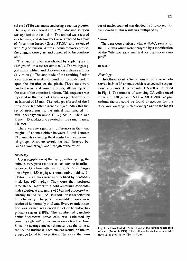
Histology Histofluorescent CA-containing cells were ob-
served in 34 of 36 animals which received cell-suspen-
sion transplants. A transplanted CA cell is illustrated in Fig. 1. The number of surviving CA cells ranged
from 0 to 1110 (mean + S.D. = 301 + 298). No pro- cedural factors could be found to account for the
wide survival range such as embryo age or the length
Fig. 1. A transplanted CA nerve cell in the lumbar spinal cord of a rat (2-month PTS). This cell was located near a needle track in the gray matter. Bar = 50gm.

DORSAL
of t ime f rom e m b r y o dissect ion to in jec t ion . Nor did
pos t - t ransplant survival t ime a p p e a r to be a fac tor
since 2- and 4 -mon th pos t - t r ansp lan t animals in jec ted
f rom the same cell suspension batch t ended to have
similar n u m b e r s of cells. The n u m b e r of viable C A
cells in jec ted into each animal is unknown.
o=
4
2
NT
- , z .
5
- 3
6
7
VENTRAL 5 Fig. 2. Tracings of horizontal sections of a lumbar spinal cord injected in 3 locations with cell suspensions of embryonic brainstem con- taining the nucleus locus coeruleus. Every twentieth section (15/~m thick) is shown. Fluorescent CA-containing nerve cells are labeled by solid circles and represent about 1/10 of all the CA cells present. Fluorescent CA fibers are sketched to represent relative fiber den- sities in the cord. Stippled regions (arrow, section 6) represent clusters of small cells, presumably gila. Note the presence of large solid- tissue implants (STI) in the ventral and dorsal fiber tracts (sections 1, 6 and 7). These implants contained many CA cells and a dense network of CA fibers. Due to post-transplant expansion, the implants dorsally displaced the right gray matter (section 4). Catechol- amine cells were also distributed along the injection needle tracks (NT, section 4). This animal survived 2 months after transplanta- tion. Outlined regions in sections 2, 4 and 6 show sites of photographs of Figs. 4, 3C, 3D, and 3A, 3B respectively. GM, gray matter; WM, white matter.

229
The CA cells were distributed in two locations: (1)
within solid-tissue implants; and (2) as single cells
within the host gray matter near the inject ion needle
tracks. Solid-tissue implants (Fig. 2, sections 6 and 7,
and Fig. 3A, B) had discrete boundaries and were
usually present within the ventral fiber tracts of the
host spinal cord at the ventral end of an injection
needle track. They were also observed in the dorsal
tracts (Fig. 2, section 1). Most of the CA cells were
found within these implants. Often more than one
implant would be present, abutt ing adjacent im-
plants, and each containing a dense growth of CA fi-
Fig. 3. The two locations of CA cells in the spinal cord after injection of cell suspensions. Fluorescent (A) and HPS (B) sections of a solid-tissue implant (STI) located in the ventral fiber tracts of the cord. The photographs were made from horizontal sections of the area indicated by the box in section 6 of Fig. 2. The implant was densely filled with CA fibers from CA cells within the implant (arrow in A). In B, note the numerous small cells, presumably glia, present within the implant (large arrow) and the lack of such dense clusters within the host white matter. Large cell bodies, presumably nerve cells, are also visible in B (small arrow). In C and D, a needle track (NT) is shown. Photographs were made from horizontal sections of the area indicated by the box in section 4 of Fig. 2. Glial cells are more numerous within the track. A fluorescent CA cell is present near the track in C (arrow). WM, white matter; GM, gray matter; CC, central canal; STI, solid-tissue implant; NT, needle track. Bar = 200gm.

230
bers (Figs. 2 and 3A). Catecholamine fibers were ob-
served distributed across hos t - implant boundaries
and thereby provided reinnervating CA fibers to the
host gray matter. The implants were variable in size
but could be as large as 1 × 3 mm in horizontal sec-
tions of the cord. Injected as small pieces of em-
bryonic tissue less than 0.5 mm in diameter, these im-
plants had expanded several times in size. As a result
of this expansion, the injected (right) side of the spi-
nal cord often showed some dorsal displacement
(Fig. 2, section 4). Al though the host tissue showed
little glial hyperplasia at hos t - implant borders,
dense clusters of small cells, presumably glia, were
usually observed within the implants (Fig. 3B). The second location of CA cells was in the host
gray matter near needle tracks. The tracks were
identifiable by the glial hyperplasia within and
around them (Figs, 2 and 3C, D). These CA cells
could be found at all dorsal to ventral levels of the
needle tracks and presumably were injected as single
cells which became lodged at various levels of the needle tracks.
The CA fiber density in the gray matter of LC ani-
mals was usually greatest near the CA cells: near the
solid-tissue implants or the injection needle tracks
(Fig. 2) and on the injected side of the cord (Fig. 4).
In one-third of the LC animals, the CA fiber density
in the ventral horn of the injected side was similar to
that of normal animals. The remaining two-thirds
had lower than normal CA fiber density. A normal
CA fiber density could not necessarily be attributed
to fiber outgrowth from transplanted CA cells since it
was observed in the 6 - O H D A control animals that
the extent of CA denervation was variable. There-
fore, the CA fiber density difference between the two sides of the cord was used as a relative measure
of fiber growth from the CA cells, with the non-in-
jected side serving as a control for the 6 - O H D A le-
sion. The CA fiber density in the lumbar ventral horns of each LC animal was subjectively assigned a value from 0 to 4 by one of the investigators without prior knowledge of the spinal cord's identity. (Nor-
mal density = 3; no fibers = 0.) In Fig. 5 the differ- ence in these values between the two sides of the cord (injected side - non-injected side) is plotted against the number of surviving CA cells. In general, more surviving CA cells resulted in a greater fiber density difference, and thus, a greater fiber outgrowth and
Fig. 4. Photograph of a horizontal CA-histofluorescent section (section 2 of Fig. 2) to illustrate the fiber density difference be- tween sides of the cord. More CA fibers were present on the in- jected (right) side than on the non-injected side. Most of thc CA fibers were confined to the gray matter. 1. GM, left gray matter; r. GM, right gray matter; WM, white matter. Bar = 2(RI um
reinnervation. Fig. 4 shows the fiber density differ- ence for the cord drawn in Fig. 3.
In animals with more than 300 CA cells, a density difference between the sides of the cord was always

231
observed. In these animals the fibers usually filled
the gray mat te r be tween the inject ion sites ( 5 - 6 mm
be tween sites) (Figs. 2 and 4). A difference in f iber
densi ty be tween the two sides of the cord could be de-
tec ted 10-12 mm rostral to the last inject ion site in
several animals, indicating that f iber growth f rom the
C A cells had ex tended that distance.
Ideal ly , the results of the functional test ing for
each animal should be compared to the C A fiber den-
sity in the spinal cord of that animal. However , our
assessment of f iber densi ty is only an approx imate
and subject ive measure , and therefore , cannot be
used for a quanti ta t ive compar ison in each animal.
Ins tead, the LC animals are divided into two groups
for compar ison of the functional data with the histo-
logical data: those animals with more than 300 C A
cells (mean = 600) and those with less than 300 C A
cells (mean = 110). The ra t ionale for this division is that in animals with more than 300 C A cells, C A fiber
outgrowth could always be de tec ted as a C A fiber
density difference be tween the two sides, whereas
C A fiber outgrowth was usually not observed in ani-
mals with less than 300 C A cells. The animals with
more than 300 C A cells had much grea ter C A fiber
outgrowth than those animals with less than 300 C A
cells as shown on the right in Fig. 5. Thus, if trans-
p lanted C A cells contr ibute to the flexion reflex,
there should be a difference in the mean reflex forces
be tween these two groups.
Function
Inject ion of a C A agonist or precursor enhances
the hindl imb flexion reflex in spinal cats 1'2 and
rats 17'29. Fig. 6 shows that in normal rats with acute
spinal t ransect ions, both the C A precursor L - D O P A
given 1 h after an inject ion of pargyl ine (monoamine
oxidase inhibitor) and the C A agonist clonidine in-
creased the pinch-el ici ted hindl imb flexion reflex
G) ¢.) C .o 4)
C 4~
,.D
U.
u
2.5
2.0
1.5
1.0
0.5'
• • Ii,, •
• ~ • •
o 8 ~ •
~ 2 14 • e ' ~ 0 ~ -'c 41)0 6 0 0 8 0 0 1000 12()0 <300 >30o
Number of CA Cells
Fig. 5. Plot of the differences in fiber density in the lumbar ventral horn between the transplanted and non-transplanted sides of each spinal cord. The fiber densities were estimated subjectively on a scale of 0-4 with 3 representing normal density and 0 representing the lack of fibers. The difference between these values (injected side - non-injected side) is plotted against the number of CA ceils for each LC animal. A general correlation between the number of CA cells and the magnitude of the difference in fiber density was found. Thus, with greater numbers of surviving CA cells, there was greater reinnervation of the gray matter and the reinnervation was con- fined mainly to the injected side. Every animal with more than 300 surviving CA cells had a difference in CA fiber density between the sides of the cord and thus, a detectable reinnervation of the injected side. Triangles, 2-month PTS animals; circles, 4-month PTS ani- mals. The mean fiber density differences for animals with less than 300 CA cells (mean number of CA cells = 110) and for animals with more than 300 CA cells (mean = 600) are shown to the right (+ S.E.M.). The number at the base of each line is the number of animals for that group.

~ 3 ~
ri-
LL 4~ ._E
"r-
8 . f f
6 .0 '
4 .0 '
2.0
Pre Post
Pargyline &
L-DOPA
t Pre Post
Clonidine
Fig. 6. Enhancement of the hindlimb flexion reflex by cate- cholamines in normal rats. The reflex was elicited in acutely transected animals by applying a clip to a toe as described in the Materials and Methods. The resulting force was measured with a force transducer and is shown in volts (1 V = 60 g). On the left, the mean pre- and postinjection reflex forces are shown for 6-month-old animals which received an i.p. injection of pargy- line (100 mg/kg) followed 1 h later by i.p. injection of e-DOPA (10 mg/kg). Within 10 rain of the L-DOPA injection, the reflex increased nearly 5-fold. Injection of either L-DOPA or pargy- line alone at these doses had little or no effect on the reflex. On the right, a similar response is shown in 4-month-old animals to i.p. injection of clonidine. Twenty minutes after 0.5 mg/kg clo- nidine injection, the force of the flexion reflex increased nearly 5-fold.
nearly 5-fold. Thus, the force of the hindl imb flexion
reflex should be an indicator of the degree of C A re-
lease and C A access to recep tor sites. Since there are
no endogenous C A cell bodies within the lumbar spi-
nal cord 7, the spinally t ransec ted normal and control
animals should have minimal resting C A release
from the isolated, descending terminals . Sponta-
neous activity 37 or synaptical ly driven activity of the
implanted C A cells should be de tec table as an en-
hanced hindl imb flexion reflex compared to the nor-
mal and control animals. The grea ter C A fiber den-
sity on the injected side might also result in a s t ronger
reflex on that side.
Fig. 7A summarizes the results of the hindl imb
flexion reflex testing. Several observat ions can be
made from this figure. First and most impor tant , the
reflexes of both the 2- and 4-month LC animals were
significantly s t ronger than the GS controls. The
mean right hindl imb reflexes were 200 and 101)~?; lar-
ger than the GS animals for the 2- and 4-month L(:
animals, respectively. Second, there were: no signifi-
cant differences be tween the right and the left hind-
limbs for any group al though the GS controls had
weaker reflexes on the side (right) which received
the glucose-saline injection. Third, there were no sig-
nificant differences among the 3 control groups al-
though, again, the right hindlimb reflexes of the GS
controls were slightly weaker. Four th , the reflexes of
the 2-month LC animals tended to be stronger than
those in the 4-month LC animals.
To de te rmine if the stronger flexion reflexes in the
LC animals were due to the t ransplanted CA cells,
two strategies were used. (1) The mean force of the
reflex in animals with greater than 300 CA cells was
compared to those animals with fewer than 300 C A
cells. This comparison indicates that C A cells cannot
be the only contr ibuting factor to the enhanced re-
flex. Fig. 7B shows that the reflex was only 2 6 - 3 3 ~
stronger in the right hindlimbs of animals with more
than 300 C A cells versus animals with fewer than 300
C A cells. This difference, however, was not signifi-
cant (P < 0.1). In addit ion, LC animals with fewer
than 300 C A cells, and thus, little or no detectable
C A fiber outgrowth, also had enhanced reflexes over
the GS controls. (2) The a -adrenerg ic blocker , phen-
oxybenzamine (PBZ) , was injected i.p. to de termine
the a -adrenerg ic receptor contr ibut ion to the reflex.
Fig. 8 illustrates the pre- and pos t -PBZ reflex forces.
The control animals had little change in their reflexes
1 h after PBZ (Fig. 8A), but the LC animals had a sig-
nificant reduction of about 30% (Fig. 8B). This re-
duction may be even greater since i.p. injection of sa-
line instead of PBZ resulted in a significant 15% in-
crease in the reflex. The decrease to PBZ was more
significant in animals with more than 300 C A cells
versus those with fewer than 300 C A cells (Fig. 8B2).
Thus, these data indicate that while the transplants
significantly increased the force of the hindl imb flex-
ion reflex, up to 200%, only about 30% of the in-
crease can be a t t r ibuted to the surviving C A cells.
DISCUSSION
The histological results of this study suppor t the
feasibility of in terver tebrai injections of embryonic
cell suspensions to re innervate the adult rat spinal

A B
~" 4.0,
.~ 3.0"
E
2 .0
"I"
"6
1.(>
LNR LOH
2rno-
< .001
• < .01 , ,
()
(b i ' " , ) ( ) ( ) •
< .01
), ,®
5-0
4mo.
4.0
o
a: 3.0.
~ 2.11'
Z
~ 1.0
I ¶ '
q) 7"
(
i P ~ H n 4 B 8 4 i L~ 22122 I 7 [7 15 15 1
L R L R L R L R L R L R L R L R GS LC N OH GS LC < 3 0 0 > 3 0 0 < 3 0 0 > 3 0 0
2too. 4too.
Fig. 7. A: the mean forces (_+ S.E.M.) of the hindlimb flexion reflex in the 4 groups of animals for the 2- and 4-month PTS times. The right and left hindlimbs are shown separately. The testing procedures are described in the Materials and Methods. No significant dif- ferences were found between left and right hindlimbs although in the sham controls (GS), the injected side (right) was depressed be- low the level of the non-injected side in both 2- and 4-month PTS. The LC reflexes are significantly larger than the GS control groups. B: comparison of mean reflex forces (+ S.E.M.) between LC animals with less than 300 CA cells with LC animals with more than 300 CA cells. Although the animals with more than 300 CA cells had larger reflexes, the differences were not significant (P < 0.1). Again, no differences were observed between left and right hindlimbs. N, normal; OH, 6-hydroxydopamine controls; GS, sham controls; LC, locus coeruleus transplants.
uI
O
4.0, n~
C 0
x 3.0, 0
U .
E --- 2,0' C
-r
1 . 0 ,
o
h
A
C o n t r o l T r a n s p l a n t s
N- sol(6) I ~ O H (14)
N (8)
GS(18)
B,
rl• sol(8) (10)
(io) . 2 Mo,
" ~ , , ~ 4 Mc (20)
B2
~ > 3 0 0 (12)
2 Mo. <500 (8 )
" ~ < 3 0 0 ( 2 6 ) 4 Mo. >300(14)
Pre Po'st P~:e Post P;'e Po'st ( I h r . ) ( I h r . ) ( l h r . )
Phenoxybenzamine Injection Fig. 8. The mean forces of the hindlimb flexion reflexes before and 1 h after injection of phenoxybenzamine (PBZ, i.p., 25 mg/kg). A: control groups. The mean reflex forces before (Pre) and 1 h after PBZ (Post) are connected by lines. The control groups showed no significant change in their reflexes after PBZ. However, if saline was injected into normal rats instead of PBZ (N-sal), there was a slight and significant increase in the reflex as indicated by the asterisks (*P < 0.01, **P < 0.001, modified Wilcoxon rank sum test for dependent samples). B 1 and B2: LC implanted animals. B]: the right and left hindlimb (r. and l.) are shown separately for the 2- and 4- month post-transplantation animals. There was a significant decrease in the animals' reflexes after PBZ injection. The level of signifi- cance is indicated by one or two asterisks near the line connecting the means. There was also a significant increase when the animals with transplants were injected with saline instead of PBZ (sal). B2: the 2- and 4-month post-transplantation animals are separated into those with more than 300 CA cells and those with less than 300 CA cells (>300 and <300). Those with more than 300 CA cells had more significant reductions of their reflexes after PBZ than those LC animals with less than 300 CA cells. Numbers in parentheses = n.
234
cord with CA fibers. When more than 300 CA cells
were present there was always detectable histologi- cal reinnervation of the lumbar spinal cord. The in-
jections were made unilaterally and the CA fiber
density was greater on the injected side.
The host tissue damage caused by the procedure was minimal. A slight gliosis occurred along the in-
jection needle track, and the growth of the solid-tis-
sue implants injected along with the single cells caused some dorsal displacement of the cord. Injec-
tion of suspensions of single cells without contamina-
tion by multicellular pieces of tissue would reduce this potential source of damage to the host spinal cord, but most of the CA cells were found within
these solid-tissue implants. The number of CA cells present was quite variable, but no factors in the transplant procedure such as embryo age, duration of dissection, or post-transplantation survival time
could be found to account for the differences in sur- vival numbers.
Previous transplants of embryonic locus coeruleus to the rat spinal cord have placed solid implants into surgically-prepared cavities 5"27"3°. These cavities re-
quire severing host axons and removal of host nerve cells. Dense glial scarring usually occurs at the site of lesion 33. Thus, the damage from the cell suspension
injection technique is much less than with the cavity technique. Use of the cell suspension technique in the hippocampus and striatum 4 and the spinal cord 3 has
yielded similar histological findings to those observed here. These include variable numbers of surviving
cells, distribution of cells in solid-tissue implants and as single cells along injecti'on tracks, dense CA fiber growth within the implants, and reinnervation of de- nervated gray matter.
Function It has been proposed that catecholamines in the
spinal cord modulate synaptic gain and adjust the lev- el of spinal cord excitability 11A6'36. The hindlimb flex- ion reflex has been shown (and was shown here in Fig. 6) to be strongly enhanced by CAs. Therefore, this reflex was used to determine if the transplanted CA cells were having a functional effect on the spinal cord, After spinal transection, those animals with cell suspensions had significantly larger reflexes than the control animals (200 and 160% for right hindlimbs of 2- and 4-month transplanted animals, respectively).
At least part of the enhancement can be attributed to the transplanted CA cells. The reflexes were signifi-
cantly reduced after i.p. injection of the ct-adrenergic blocker, phenoxybenzamine, indicating that about
30%, of the reflex enhancement was due to CA re- lease from transplanted CA cells and interaction with
ct-adrenergic receptors. The effects of L-DOPA on spine reflexes in the cat are reduced by ct-adrenergic
blockers, but not by fl-blockers t'2. A fl-adrenergic contribution cannot, however, be ruled out in the
present experiments. In addition, the animals with more than 300 CA cells had a mean reflex force of
26-33% stronger than those animals with fewer than 300 CA cells, but the differences were not statistical- ly significant. Denervation supersensitivity of CA re- ceptors may have minimized the reflex differences between these animals. It has been shown that the degree of CA denervation supersensitivity is related
to the degree of denervation of CA fibers in the rat spinal cord 2°'29. Thus, animals with few CA cells
would tend to have greater CA receptor sensitivity
than those animals with many CA cells. The CA re- ceptor sensitivity would be regulated to match the de- gree of CA innervation, thus tending to maintain an equal strength among animals with different numbers
of transplanted CA cells. This phenomenon might also contribute to the lack of difference between the reflex in left and right hindlimbs in spite of the great- er CA fiber density on the right side of the cords. Another possibility is that enhanced excitability due
to CA release is conveyed to the opposite side by commissural interneurons19.
The hindlimb reflexes of the 4-month LC animals were about 30% smaller than the reflexes of the 2- month LC animals although the difference was not
statistically significant. This difference is probably not related to the number of surviving CA cells in the two groups since the difference between the 2- and 4- month LC animals was present for animals with a few CA cells, as well as for animals with many CA cells (Fig. 7B). The 4-month GS control animals also had slightly weaker reflexes than the 2-month GS animals which may indicate that the weakening for both the GS and the LC animals is a progressive reaction to the injection of the glucose-saline or the cell suspen- sions into the cord.
Thus, the transplants of embryonic brainstem con- taining the locus coeruleus significantly strengthened

235
the hindl imb flexion reflex. Only par t of the increase,
however , can be a t t r ibuted to the release of catechol-
amines from the t ransplanted C A cells. Therefore ,
factors in addi t ion to C A cells must be contr ibut ing to
the enhancement of the flexion reflex. It has been
shown that chronic hemisect ion of the spinal cord re-
sults in long-lasting enhancement of both monosyn-
aptic and polysynapt ic reflexes in the lumbar cord of
cat TM and rat 25 by an unknown mechanism. The injec-
tion of the cell suspensions may have produced some
:non-specific damage to the cord (e.g. compress ion of
the cord by the expanding implants) result ing in en-
hanced reflexes by a mechanism similar to the chron-
ic hemisection. A n o t h e r possible factor is the injec-
tion of n o n - C A cells along with C A cells. The brain-
stem region dissected for the cell suspensions con-
tains o ther bulbospinal neurons which can affect spi-
nal cord activities 38. The n o n - C A cells may also sur-
vive and contr ibute to the enhancement of the flexion
reflex.
The present s tudy has shown the feasibili ty of re-
innervating the adul t spinal cord by intraspinal injec-
tions of embryonic cell suspensions. The trans-
p lanted C A cells survived and grew axon-l ike pro-
cesses which could form a normal -appear ing plexus
of C A fibers in the gray mat ter . Most impor tant ly ,
the t ransplanted C A cells affected the functional ac-
tivity of the spinal cord as assayed by the hindl imb
flexion reflex.
ACKNOWLEDGEMENTS
We are grateful for the quality technical assistance
of Els Cooper r ider . This work was suppor ted by a
grant from the Spinal Cord Society to H . O . N . and an
NIH Postdoctoral fel lowship to J .T.B.
REFERENCES
1 And6n, N.-E., Jukes, M.G.M. and Lundberg, A., The ef- fect of DOPA on the spinal cord. 2. A pharmacological analysis, Acta Physiol. Scand., 67 (1966) 387-397.
2 And6n, N.-E., Jukes, M.G.M., Lundberg, A. and Vyk- lick~, L., The effect of DOPA on the spinal cord. 1. Influ- ence on transmission from primary afferents, Acta Physiol. Scand., 67 (1966) 373-386.
3 Bj6rklund, A., Nornes, H., Dunnett, S.B., Gage, F.H. and Stenevi, U., Intracerebral and intraspinal implants of locus coeruleus cell suspensions: deleterious effect of trypsin in the suspension medium, Soc. Neurosci. Abstr., 9 (1983) 101.
4 Bj6rklund, A., Stenevi, U., Schmidt, R.H., Dunnett, S.B. and Gage, F.H., Intracerebral grafting of neuronal cell sus- pensions, Acta Physiol. Scand., Suppl. 522 (1983) 1-75.
5 Commissiong, J.W., Fetal locus coeruleus transplanted into the transected spinal cord of the adult rat, Brain Re- search, 271 (1983) 174-179.
6 Coote, J.H. and MacLeod, V.H., The influence of bulbo- spinal monoaminergic pathways on sympathetic nerve ac- tivity, J. Physiol. (London), 241 (1974) 453-475.
7 DahlstrOm, A. and Fuxe, K., Evidence for the existence of monoamine neurons in the central nervous system. II. Ex- perimentally induced changes in the intraneuronal amine levels of bulbospinal neuron systems, Acta Physiol. Scand., Suppl. 247, 64 (1965) 1-36.
8 Das, G.D. and Altman, J., Studies on the transplantation of developing neural tissue in the mammalian brain. I. Trans- plantation of cerebellar slabs into the cerebellum of neo- nate rats, Brain Research, 38 (1972) 233-249.
9 Dunnett, S.B., Bj6rklund, A., Stenevi, U. and Iversen, S.D., Grafts of embryonic substantia nigra reinnervating the ventrolateral striatum ameliorate sensorimotor impair-
merits and akinesia in rats with 6-OHDA lesions of the ni- grostriatal pathway, Brain Research, 229 (1981) 209-217.
10 Dunnett, S.B., Low, W.C., lversen, S.D., Stenevi, U. and Bj6rklund, A., Septal transplants restore maze learning in rats with fornix-fimbria lesions, Brain Research, 251 (1982) 335-348.
11 Foote, S.L., Bloom, F.E. and Aston-Jones, G., Nucleus lo- cus coeruleus: new evidence of anatomical and physiologi- cal specificity, Physiol. Rev., 63 (1983) 844-914.
12 Forssberg, H. and Grillner, S., The locomotion of the acute spinal cat injected with clonidine i.v., Brain Research, 50 (1973) 184-186.
13 Freed, W.J., Morihisa, J.M., Spoor, E., Hoffer, B.J., OI- son, L., Seiger, A. and Wyatt, R.J., Transplanted adrenal chromaffin cells in rat brain reduce lesion-induced rotation- al behavior, Nature (London), 292 (1981) 351-352.
14 Gage, F.H., Dunnett, S.B., Bj6rklund, A. and Stenevi, U., Aged rats: recovery of motor impairments by intrastriatal nigral grafts, Science, 221 (1983) 966-968.
15 Gash, D., Sladek, J.R., Jr. and Sladek, C.A., Functional development of grafted vasopressin neurons, Science, 210 (1980) 1367-1369.
16 Grillner, S., Control of locomotion in bipeds, tetrapods, and fish. In J.M. Brookhart, V.B. Mountcastle and V.B. Brooks (Eds.), The Nervous System, Vol. 2, American Physiological Society, Bethesda, MD, 1981, pp. 1179-1236.
17 Grossmann, W., Jurna, I. and Nell, T., The effect of reser- pine and DOPA on reflex activity in the rat spinal cord, Exp. Brain Res., 22 (1975) 351-361.
18 Hultborn, H. and Malmsten, J., Changes in segmental re- flexes following chronic spinal cord hemisection in the cat. I. Increased monosynaptic and polysynaptic ventral root discharges, Acta Physiol. Scand., 119 (1983) 405-422.
19 Jankowska, E., Jukes, M.G.M., Lund, S. and Lundberg,

236
A., The effect of DOPA on the spinal cord. 5. Reciprocal organization of pathways transmitting excitatory actions to alpha motoneurones of flexors and extensors, Acta Physiol. Scand., 70 (1967) 369-388.
20 Jones, D.J., Alcantara, O.F. and Ademe, R.M., Supersen- sitivity of the noradrenergic system in the spinal cord fol- lowing intracisternal injection of 6-hydroxydopamine. Neu- ropharmacology, 23 (1984) 431-438.
21 Krieger, D.T., Perlow, M.J., Gibson, M.J., Davies, T.F., Zimmerman, E.A., Ferin, M. and Charlton, H.M., Brain grafts reverse hypogonadism of gonadotropin releasing hormone deficiency, Nature (London), 298 (1982) 468-471.
22 Kromer, L.F., Bj6rklund, A. and Stenevi, U., Intracephal- ic embryonic neural implants in the adult rat brain. I. Growth and mature organization of brainstem, cerebellar, and hippocampal implants, J. Comp. Neurol., 218 (1983) 433-459.
23 Lam, F.C. and Longnecker, M.T., A modified Wilcoxon rank sum test for paired data, Biometrika, 70 (1983) 510-513.
24 Lor6n, I., Bjfrklund, A., Falck, B. and Lindvall, O., The aluminum-formaldehyde (ALFA) histofluorescence method for improved visualization of catecholamines and indoleamines. 1. A detailed account of the methodology for central nervous tissue using paraffin, cryostat or vibrotome sections, J. Neurosci. Meth., 2 (1980) 277-300.
25 Malmsten, J., Time course of segmental reflex changes af- ter chronic spinal cord hemisection in the rat, Acta Physiol. Scand., 119 (1983) 435-443.
26 McLoon, S.C. and Lund, R.D., Development of fetal reti- na, tectum, and cortex transplanted to the superior collicu- lus of adult rats, J. Comp. Neurol., 217 (1983) 376-389.
27 Nornes, H., Bj6rklund, A. and Stenevi, U., Reinnervation of the denervated adult spinal cord of rats by intraspinal transplants of embryonic brain stem neurons, Cell Tissue Res., 230 (1983) 15-35.
28 Nygren, L.-G. and Olson, L., A new major projection from locus coeruleus: the main source of noradrenergic nerve terminals in the ventral and dorsal columns of the spinal cord, Brain Research, 132 (1977) 85-93.
29 Nygren, L.-G. and Olson, L., On spinal noradrenaline re- ceptor supersensitivity: correlation between nerve terminal densities and flexor reflexes various times after mtracister- nal 6-hydroxydopamine. Brain Research, 1 E6 (1976) 455-470.
30 Nygren, L.-G., Olson, L. and Seiger, A., Monoamine re- innervation of the transected spinal cord by fetal brain grafts, Brain Research, 129 (1977) 227-235.
31 Perlow, M.J., Freed, W.J., Hoffer, B.3., Seiger, A., OI- son, L. and Wyatt, R.J., Brain grafts reduce motor abnor- malities produced by destruction of nigrostriatal dopamine system, Science, 204 (1979) 643-647.
32 Reddy, S.V.R. and Yaksh, T.L., Spinal noradrenergic ter- minal system mediates antinociception, Brain Research, 189 (1980) 391-404.
33 Reier, P.J., Stensaas, L.J. and Guth, L., The astrocytic scar as an impediment to regeneration in the central ner- vous system. In C.C. Kao, R.P. Bunge and P.J. Reier (Eds.), Spinal Cord Reconstruction, Raven Press, New York, 1983, pp. 163-195.
34 Schmidt, R.H., Bj6rklund, A. and Stenevi, U., lntracere- bral grafting of dissociated CNS tissue suspensions: a new approach for neuronal transplantation to deep brain sites, Brain Research, 218 (1981) 347-356.
35 Seiger, A. and Olson, L., Late prenatal ontogeny of central monoamine neurons in the rat: fluorescence histochemical observations, Z. Anat. Entwicklungsgesch., 140 (1973) 281-318.
36 White, S.R. and Neuman, R.S., Pharmacological antago- nism of facilitatory but not inhibitory effects of serotonin and norepinephrine on excitability of spinal motoneurons, Neuropharmacology, 22 (1983) 489-494.
37 Williams, J.T., North, R.A., Shefner, A., Nishi, S. and Egan, T.M., Membrane properties of rat locus coeruleus neurones, Neuroscience, 13 (1984) 137-156.
38 Wilson, V.J. and Peterson, B.W., Vestibulospinal and re- ticulospinal systems. In J.M. Brookhart, V.B. Mountcastle and V.B. Brooks (Eds.), The Nervous System, Vol. 2, American Physiological Society, Bethesda, MD, 1981, pp. 667-702.