The flexion synergy, mother of all synergies and father of new models of gait
9
REVIEW ARTICLE published: 13 March 2013 doi: 10.3389/fncom.2013.00014 The flexion synergy, mother of all synergies and father of new models of gait Jacques Duysens 1,2 *, Friedl De Groote 3 and Ilse Jonkers 1 1 Department of Kinesiology, KU Leuven, Heverlee, Belgium 2 Department of Research, Sint Maartenskliniek, Nijmegen, Netherlands 3 Department of Mechanical Engineering, KU Leuven, Heverlee, Belgium Edited by: Yuri P. Ivanenko, IRCCS Fondazione Santa Lucia, Italy Reviewed by: Yuri P. Ivanenko, IRCCS Fondazione Santa Lucia, Italy Francesca Sylos Labini, IRCCS Santa Lucia Foundation, Italy *Correspondence: Jacques Duysens, Department of Kinesiology, Tervuursevest 101 - bus 01500, BE-3001 Heverlee, Belgium. e-mail: jacques.duysens@ faber.kuleuven.be Recently there has been a growing interest in the modular organization of leg movements, in particular those related to locomotion. One of the basic modules involves the flexion of the leg during swing and it was shown that this module is already present in neonates (Dominici et al., 2011). In this paper, we question how these finding build upon the original work by Sherrington, who proposed that the flexor reflex is the basic building block of flexion during swing phase. Similarly, the relation between the flexor reflex and the withdrawal reflex modules of Schouenborg and Weng (1994) will be discussed. It will be argued that there is large overlap between these notions on modules and the older concepts of reflexes. In addition, it will be shown that there is a great flexibility in the expression of some of these modules during gait, thereby allowing for a phase-dependent modulation of the appropriate responses. In particular, the end of the stance phase is a period when the flexor synergy is facilitated. It is proposed that this is linked to the activation of circuitry that is responsible for the generation of locomotor patterns (CPG, “central pattern generator”). More specifically, it is suggested that the responses in that period relate to the activation of a flexor burst generator. The latter structure forms the core of a new asymmetric model of the CPG. This activation is controlled by afferent input (facilitation by a broad range of afferents, suppression by load afferent input). Meanwhile, many of these physiologic features have found their way in the control of very flexible walking bipedal robots. Keywords: flexion reflex, local sign, reflex modules, synergy, central pattern generator, gait, forward model INTRODUCTION One of the first authors to point out the modular organization of the motor control system was Sherrington (1910a,b). Reading his work, it is clear that for him the flexor reflex was the mother of all modules and synergies (in the broad sense, not in the sense of the mathematical synergies defined recently). He proposed that the flexor reflex is a basic building block of the central nervous system and that “the flexion reflex is in reality the reflex stepping of the limb” (pp. 69 in Sherrington, 1910b). In his view, stepping was basically a series of flexion reflexes, with extension occurring merely as the “rebound” following the flexion. The extension dur- ing the stance phase of gait could be provided as some type of “extensor thrust,” evoked by “the weight of the animal applied through the foot against the ground” (pp. 78 in Sherrington, 1910b). In the absence of support (air stepping), the rhythmic activity continues, which for Sherrington was an argument for stating that “the extensor thrust cannot therefore be an indispens- able factor in the reflex step” (pp. 79 in Sherrington, 1910b). This idea of a basic asymmetry in the control of locomotion has since lost terrain, mostly because of the powerful impact of the (sym- metrical) half-center model for the central pattern generation of locomotion (one half of this center inducing activity in flexors, the other in extensors). The latter model was described by Brown (1914) and is known as the “half-center” model. The first ideas in that direction were actually presented by Sherrington himself on the basis of work by Brown (1911, 1912). They are based on experiments showing that cats with a transected spinal cord and with cut dorsal roots still showed rhythmic alternating contrac- tions in ankle flexors and extensors. However, Sherrington did not necessarily propose a symmetrical organization. Instead, he and Brown proposed originally that gait was the result of a balance “between equal and opposite states of excitation” in flexors and extensors, while being well-aware that the origin of these states could be quite different. The latter notion seems to have been lost in later years. In addition, in many cases the discussion on the credits for the original ideas about a central basis for locomotion has been simplified considerably in many accounts (as explained elegantly in a review by Stuart and Hultborn, 2008). Both Sherrington (1910a) and Philippson (1905) have indeed emphasized the idea that during gait one phase induced automatically the next one (reflex chain) but this does not mean that these authors excluded a central origin for the rhythmic activity (for details see also Clarac, 2008). In particular, Philippson believed that the spinal control was due to a combination of central and reflex mech- anisms (Clarac, 2008). Hence it is a gross simplification to see this part of the history as a “victory” of Brown over his competi- tors (Sherrington and Philippson). Brown should be credited for Frontiers in Computational Neuroscience www.frontiersin.org March 2013 | Volume 7| Article 14 | 1 COMPUTATIONAL NEUROSCIENCE
-
Upload
independent -
Category
Documents
-
view
1 -
download
0
Transcript of The flexion synergy, mother of all synergies and father of new models of gait
REVIEW ARTICLEpublished: 13 March 2013
doi: 10.3389/fncom.2013.00014
The flexion synergy, mother of all synergies and father ofnew models of gaitJacques Duysens1,2*, Friedl De Groote3 and Ilse Jonkers 1
1 Department of Kinesiology, KU Leuven, Heverlee, Belgium2 Department of Research, Sint Maartenskliniek, Nijmegen, Netherlands3 Department of Mechanical Engineering, KU Leuven, Heverlee, Belgium
Edited by:
Yuri P. Ivanenko, IRCCS FondazioneSanta Lucia, Italy
Reviewed by:
Yuri P. Ivanenko, IRCCS FondazioneSanta Lucia, ItalyFrancesca Sylos Labini, IRCCS SantaLucia Foundation, Italy
*Correspondence:
Jacques Duysens, Department ofKinesiology, Tervuursevest 101 - bus01500, BE-3001 Heverlee, Belgium.e-mail: [email protected]
Recently there has been a growing interest in the modular organization of leg movements,in particular those related to locomotion. One of the basic modules involves the flexion ofthe leg during swing and it was shown that this module is already present in neonates(Dominici et al., 2011). In this paper, we question how these finding build upon theoriginal work by Sherrington, who proposed that the flexor reflex is the basic buildingblock of flexion during swing phase. Similarly, the relation between the flexor reflex andthe withdrawal reflex modules of Schouenborg and Weng (1994) will be discussed. It willbe argued that there is large overlap between these notions on modules and the olderconcepts of reflexes. In addition, it will be shown that there is a great flexibility in theexpression of some of these modules during gait, thereby allowing for a phase-dependentmodulation of the appropriate responses. In particular, the end of the stance phase isa period when the flexor synergy is facilitated. It is proposed that this is linked to theactivation of circuitry that is responsible for the generation of locomotor patterns (CPG,“central pattern generator”). More specifically, it is suggested that the responses in thatperiod relate to the activation of a flexor burst generator. The latter structure forms thecore of a new asymmetric model of the CPG. This activation is controlled by afferent input(facilitation by a broad range of afferents, suppression by load afferent input). Meanwhile,many of these physiologic features have found their way in the control of very flexiblewalking bipedal robots.
Keywords: flexion reflex, local sign, reflex modules, synergy, central pattern generator, gait, forward model
INTRODUCTIONOne of the first authors to point out the modular organization ofthe motor control system was Sherrington (1910a,b). Reading hiswork, it is clear that for him the flexor reflex was the mother ofall modules and synergies (in the broad sense, not in the sense ofthe mathematical synergies defined recently). He proposed thatthe flexor reflex is a basic building block of the central nervoussystem and that “the flexion reflex is in reality the reflex steppingof the limb” (pp. 69 in Sherrington, 1910b). In his view, steppingwas basically a series of flexion reflexes, with extension occurringmerely as the “rebound” following the flexion. The extension dur-ing the stance phase of gait could be provided as some type of“extensor thrust,” evoked by “the weight of the animal appliedthrough the foot against the ground” (pp. 78 in Sherrington,1910b). In the absence of support (air stepping), the rhythmicactivity continues, which for Sherrington was an argument forstating that “the extensor thrust cannot therefore be an indispens-able factor in the reflex step” (pp. 79 in Sherrington, 1910b). Thisidea of a basic asymmetry in the control of locomotion has sincelost terrain, mostly because of the powerful impact of the (sym-metrical) half-center model for the central pattern generation oflocomotion (one half of this center inducing activity in flexors,the other in extensors). The latter model was described by Brown(1914) and is known as the “half-center” model. The first ideas
in that direction were actually presented by Sherrington himselfon the basis of work by Brown (1911, 1912). They are based onexperiments showing that cats with a transected spinal cord andwith cut dorsal roots still showed rhythmic alternating contrac-tions in ankle flexors and extensors. However, Sherrington did notnecessarily propose a symmetrical organization. Instead, he andBrown proposed originally that gait was the result of a balance“between equal and opposite states of excitation” in flexors andextensors, while being well-aware that the origin of these statescould be quite different. The latter notion seems to have been lostin later years.
In addition, in many cases the discussion on the credits forthe original ideas about a central basis for locomotion has beensimplified considerably in many accounts (as explained elegantlyin a review by Stuart and Hultborn, 2008). Both Sherrington(1910a) and Philippson (1905) have indeed emphasized the ideathat during gait one phase induced automatically the next one(reflex chain) but this does not mean that these authors excludeda central origin for the rhythmic activity (for details see alsoClarac, 2008). In particular, Philippson believed that the spinalcontrol was due to a combination of central and reflex mech-anisms (Clarac, 2008). Hence it is a gross simplification to seethis part of the history as a “victory” of Brown over his competi-tors (Sherrington and Philippson). Brown should be credited for
Frontiers in Computational Neuroscience www.frontiersin.org March 2013 | Volume 7 | Article 14 | 1
COMPUTATIONAL NEUROSCIENCE
Duysens et al. Flexor synergies and models
having provided compelling evidence for the central spinal originof locomotor activity, while Sherrington and Philippson shouldbe remembered for their important insights on the importance ofafferent input for the control of gait.
The “half-center” model has helped us greatly in appreciat-ing the spinal origin of the central pattern generator (CPG) forlocomotion, but it may have led to the simplifying idea of sym-metry within the CPG (Jankowska et al., 1967a,b; Lundberg, 1981;Lafreniere-Roula and McCrea, 2005). In that way it has deterredour thinking away from the notion of a basic asymmetry of theneural organization of locomotion.
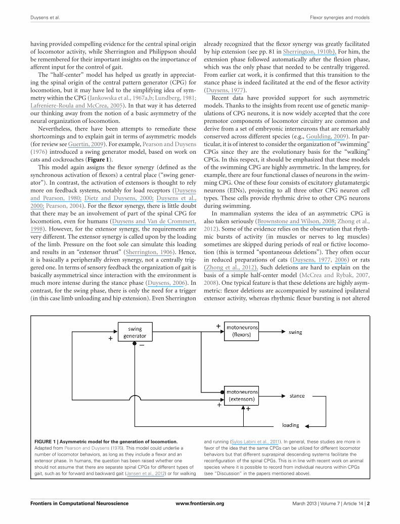
Nevertheless, there have been attempts to remediate theseshortcomings and to explain gait in terms of asymmetric models(for review see Guertin, 2009). For example, Pearson and Duysens(1976) introduced a swing generator model, based on work oncats and cockroaches (Figure 1).
This model again assigns the flexor synergy (defined as thesynchronous activation of flexors) a central place (“swing gener-ator”). In contrast, the activation of extensors is thought to relymore on feedback systems, notably for load receptors (Duysensand Pearson, 1980; Dietz and Duysens, 2000; Duysens et al.,2000; Pearson, 2004). For the flexor synergy, there is little doubtthat there may be an involvement of part of the spinal CPG forlocomotion, even for humans (Duysens and Van de Crommert,1998). However, for the extensor synergy, the requirements arevery different. The extensor synergy is called upon by the loadingof the limb. Pressure on the foot sole can simulate this loadingand results in an “extensor thrust” (Sherrington, 1906). Hence,it is basically a peripherally driven synergy, not a centrally trig-gered one. In terms of sensory feedback the organization of gait isbasically asymmetrical since interaction with the environment ismuch more intense during the stance phase (Duysens, 2006). Incontrast, for the swing phase, there is only the need for a trigger(in this case limb unloading and hip extension). Even Sherrington
already recognized that the flexor synergy was greatly facilitatedby hip extension (see pp. 81 in Sherrington, 1910b). For him, theextension phase followed automatically after the flexion phase,which was the only phase that needed to be centrally triggered.From earlier cat work, it is confirmed that this transition to thestance phase is indeed facilitated at the end of the flexor activity(Duysens, 1977).
Recent data have provided support for such asymmetricmodels. Thanks to the insights from recent use of genetic manip-ulations of CPG neurons, it is now widely accepted that the corepremotor components of locomotor circuitry are common andderive from a set of embryonic interneurons that are remarkablyconserved across different species (e.g., Goulding, 2009). In par-ticular, it is of interest to consider the organization of “swimming”CPGs since they are the evolutionary basis for the “walking”CPGs. In this respect, it should be emphasized that these modelsof the swimming CPG are highly asymmetric. In the lamprey, forexample, there are four functional classes of neurons in the swim-ming CPG. One of these four consists of excitatory glutamatergicneurons (EINs), projecting to all three other CPG neuron celltypes. These cells provide rhythmic drive to other CPG neuronsduring swimming.
In mammalian systems the idea of an asymmetric CPG isalso taken seriously (Brownstone and Wilson, 2008; Zhong et al.,2012). Some of the evidence relies on the observation that rhyth-mic bursts of activity (in muscles or nerves to leg muscles)sometimes are skipped during periods of real or fictive locomo-tion (this is termed “spontaneous deletions”). They often occurin reduced preparations of cats (Duysens, 1977, 2006) or rats(Zhong et al., 2012). Such deletions are hard to explain on thebasis of a simple half-center model (McCrea and Rybak, 2007,2008). One typical feature is that these deletions are highly asym-metric: flexor deletions are accompanied by sustained ipsilateralextensor activity, whereas rhythmic flexor bursting is not altered
FIGURE 1 | Asymmetric model for the generation of locomotion.
Adapted from Pearson and Duysens (1976). This model could underlie anumber of locomotor behaviors, as long as they include a flexor and anextensor phase. In humans, the question has been raised whether oneshould not assume that there are separate spinal CPGs for different types ofgait, such as for forward and backward gait (Jansen et al., 2012) or for walking
and running (Sylos Labini et al., 2011). In general, these studies are more infavor of the idea that the same CPGs can be utilized for different locomotorbehaviors but that different supraspinal descending systems facilitate thereconfiguration of the spinal CPGs. This is in line with recent work on animalspecies where it is possible to record from individual neurons within CPGs(see “Discussion” in the papers mentioned above).
Frontiers in Computational Neuroscience www.frontiersin.org March 2013 | Volume 7 | Article 14 | 2

Duysens et al. Flexor synergies and models
during extensor deletions. Such results are best explained by arhythm generator that provides direct input to a “swing” or“flexor burst” generator but not to the extensor part of the CPG(Rybak et al., 2006a,b; Zhong et al., 2012). Hence, it is basicallysimilar to the model proposed originally by Pearson and Duysens(1976) except that the swing generator is split up in a rhythmgenerator and a flexor center.
Some earlier cat modeling work had pointed toward asym-metry as well. For example, the model proposed by Prochazkaand Yakovenko (2007a) seems symmetrical at first sight but italready contains important elements of asymmetry. In particularit is argued that interneurons in the extensor timing element mayreceive less inputs generating persistent inward currents, there-fore as a network “they are not only set to have longer half-cycledurations, but also to be more sensitive to synaptic commands.”Interestingly, the model was only stable for extensor dominantphase-duration characteristics (where extension durations varymore than flexion durations; see also Prochazka and Yakovenko,2007a,b). This pattern is seen in the normal cat. The inverse(flexor dominant) was, however, not observed in the model whileit has been observed experimentally, but only in fictive loco-motion (rhythmic output of the spinal cord in paralyzed catpreparations). This is an important point as the existence of bothflexor- and extensor-dominated patterns has often been invokedto support the notion of a symmetrical CPG (McCrea and Rybak,2008). However, one may wonder whether the flexor-dominatedpattern is not simply an artifact of the preparation used, since theoutput observed is one from CPGs without interaction with affer-ent input. As pointed out above, the afferent input is crucial forthe automated phase transitions. Experimental work on cats hasclearly established that peripheral input from the paw (as occursduring touchdown) is very potent in terminating the flexor phaseand initiating the extensor phase (Duysens, 1977). Hence, in theabsence of such feedback it is not surprising to see flexor phasesof abnormally long duration.
This feature was not always recognized and perhaps for thisreason, the concept of an asymmetric model first met some resis-tance (McCrea and Rybak, 2007, 2008). However, due to morerecent data (Brownstone and Wilson, 2008; Zhong et al., 2012),the idea of an asymmetric pattern generator has reemerged andit is therefore worthwhile to reexamine the presumed basis ofthe swing generator, namely the flexor synergy, its adaptations(for example to stimulation of different skin areas on the leg, aphenomenon referred to as “local sign” in physiology) and itsintegration in the process of locomotion.
THE TASK TO WITHDRAW AND THE CORRESPONDINGFLEXOR SYNERGIES IN THE SPINAL CORD: A DEFENSE INFAVOR OF THE “LOCAL SIGN”For the flexor reflex, it is clear that the synergy (as described bySherrington) corresponds very well to the task of withdrawal.This protective reflex is so important that it is present at birthand can be elicited with about any type of stimulus to the foot.In neonates, the flexion reflex responses to innocuous stimula-tion are already present (Andrews and Fitzgerald, 1999). How dothese responses compare to the more recently described synergies(or “components”)? In the adult, the flexor reflex cannot simply
be related to just one component, although factor 5, as describedby Ivanenko et al. (2004), or P3 as described by Dominici et al.(2011), are close candidates. For example, the factor 5 of Ivanenkoet al. (2004) relies of strong activations of Sartorius and TibialisAnterior during the middle of the swing phase.
In neonates, the gait is explained (up to 89%) by just two pat-terns, one of which peaks at about 75% of the step cycle, hencein the swing phase. This “swing” pattern persists in the adultsand is seen in a wide variety of species. When one considersthe large input of flexors to these basic patterns, it is temptingto relate these components to the flexor synergy as described bySherrington (1910b). Furthermore, the appearance of these com-ponents in swing is nicely in line with the Sherrington proposal ofa common neural basis for the flexor reflex and the flexion phaseof stepping. Further experimental evidence for such common useof neural circuitry has been obtained in animal studies. For exam-ple, in the turtle, Berkowitz has described interneurons that areactive in both types of activity (flexion phase and flexor reflex;Berkowitz, 2007, 2010). In addition, in the same species it wasshown that often the same interneurons can be involved in vari-ous types of rhythmic behavior (swimming, scratching), therebysupporting the idea that basic synergies can be used in variousbehaviors (Berkowitz and Hao, 2011; see also Grillner, 1985).
During maturation in humans, the threshold for the reflexincreases and biceps femoris responses dominate (Andrews andFitzgerald, 1999). Furthermore, the pattern of the flexor reflexeschanges. The recruitment of specific flexor muscles dependsincreasingly more on the area of skin stimulated (“local sign”),thereby allowing a more efficient withdrawal when stimuli areapplied at various distinct locations on the limb. This has led sev-eral authors to propose the existence of a variety of reflex modulesboth in humans (Andersen et al., 1999, 2001; Sonnenborg et al.,2000, 2001) and in animals (Schouenborg and Weng, 1994; Treschet al., 1999).
There is no doubt that these new experiments have provideda wealth of very precise data but still the question arises whetherthis has basically altered our way of thinking. The idea of a “localsign” goes back to the early days of reflex physiology. Creedand Sherrington (1926) stated (pp. 265): “The term flexion-reflex . . . denotes strictly speaking a group of reflexes, all more orless alike . . . yet from one afferent to another differing in detaileddistribution of the motor units employed, while yet always con-forming to the general type flexion-reflex.” Especially this lastpoint is important as it is proposed originally that there remains abasic synergy (“flexion-reflex”) underlying all these different vari-ations. The data of Creed and Sherrington (1926) showed that,despite variations in some of the distal flexors, the hip and kneeflexors always participated in the various reflexes (see their tableon pp. 260). More recent work supports this, both in the frog(Tresch et al., 1999) and in the rat (Schouenborg and Kalliomaki,1990). However, this common element is often not emphasizedand the impression may arise that the recently defined “modules”and “synergies” are independent entities. This certainly differsfrom the view of Creed and Sherrington (1926), who viewed thedifferent versions of the flexor reflexes as expression or adapta-tions of one and the same basic flexor synergy. Hence the basicissue is whether the recently described reflex modules are also
Frontiers in Computational Neuroscience www.frontiersin.org March 2013 | Volume 7 | Article 14 | 3
Duysens et al. Flexor synergies and models
mostly variations of a basic synergy (the flexion reflex) or whetherthey really constitute separate entities.
In our opinion, there is no convincing evidence for the latter,at least when one considers the literature on withdrawal reflexes.Local cutaneous reflexes do exist, but they differ from with-drawal reflexes. For example, the selective activation of extensorreflexes such as the gastrocnemii was observed when stimulat-ing the skin that covered these muscles (Hagbarth, 1960). Forwithdrawal reflexes, in contrast, there is no data to show con-vincing evidence for neural pathways for separate types of flexorreflexes. They mainly show modifications of a basic flexor pat-tern. These modifications are likely to be due to changes inactivity in spinal dorsal horn cells (Schouenborg et al., 1995).During development, the withdrawal reflexes are “fine-tuned”by the spontaneous movements of the individual but the result-ing reflexes always have a component of hip and/or knee flexion(Holmberg and Schouenborg, 1996). Hence these studies providea substantial contribution to our knowledge on the “local sign”but they do not show that there is a conceptual deviation fromthe notion of “local sign,” as originally defined. In addition thesestudies underline the plasticity of reflexes. Synergies, as definedmore recently in mathematical terms, do not fully overlap withthese reflexes. Nevertheless, several authors have emphasized thatthese muscle synergies and modules may also be highly plasticand basically represent solutions for specific tasks at a given time(Latash, 1999; Ivanenko et al., 2013).
PATHOLOGYThe data provided by pathology further support the notion ofvariations in flexor reflexes rather than a set of separate mod-ules. As one might expect, the adjustments and fine-tuning of theflexor reflex relates to input from descending pathways. Hence,when a lesion occurs in these pathways, one should see a reversalto the more primitive state. This is exactly what happens.
In spinal cord injury (SCI) there is a loss of “local sign” and areturn to the simpler forms of flexor reflexes (Schmit et al., 2003;“an invariant flexion response pattern was produced regardlessof stimulus location”). In addition, in these patients there is alink between a normal flexor reflex and the ability to recover gait(Dietz et al., 2009). After some 6–12 months, this ability deterio-rates when the early flexor reflex (latency 60–120 ms) decreasesover time. Again, this illustrates the importance of the flexorreflex circuitry for the generation of gait. In stroke, a similarreturn to a more primitive synergy occurs after the insult andthis phenomenon is known as the Babinski sign (Babinski, 1922).Stimulation of the sole of the foot induces dorsiflexion of the bigtoe, by activating the extensor hallucis longus muscle, a flexor inthe physiological sense. Babinski pointed out that this reflex waspart of the flexion synergy of the lower limb and in fact clin-icians, still as of today, are advised to watch for flexion of thewhole limb as an obligatory concomitant of the reflex (Van Gijn,1978; Kumar, 2003). The whole reflex is a return to the con-dition of the neonate, where indeed a dorsiflexion Babinski isnormally present, usually in conjunction with a brisk flexion ofthe whole limb. Interestingly, in the neonate it is important notto stimulate too gently, because otherwise a grasp reflex occurs.This shows that actually what we know as the “normal plantar
reaction” (plantar flexion of the toes) may actually be a superpo-sition of two reflexes, with the grasp reflex dominating the flexorreflex. This makes sense in an evolutionary context since, forexample, grasping tree branches might have been more importantthan a “blind” withdrawal defense toward any type of stimulus.In complete SCI subjects, the occurrence of the Babinski signhas been described as well, although it can be absent in somepatients due to associated peripheral nerve damage (Petersenet al., 2010).
CAN THE REAL FLEXOR REFLEX PLEASE STAND UP!One problem in this research field is the confusion on the flexorreflex terminology. In humans, most studies do not use purenociceptive stimuli such as heat. Instead, electrical stimulationis used. However, it is impossible to activate nociceptive affer-ents in any nerve without coactivating large myelinated fibers.Therefore, in humans, the response to high intensity electricalstimuli typically has two components, an early (60–120 ms) anda late one (120–200 ms; Shahani and Young, 1971). The difficultyis to decide which afferents are responsible for a given compo-nent. If only high intensity stimuli are used, one is easily misledin thinking that the early response is a nociceptive one, whilein fact it often can be elicited by low intensity stimuli as well.The problem is aggravated by differences in definition. Hugon(1973) defined the early response (RII) as having a latency of40–60 ms and the late response (RIII) as having a latency of85–120 ms (for review see Sandrini et al., 2005). Hence, RIIIis really the equivalent of the “early” flexor reflex. In normalcontrol subjects, walking on a treadmill, one can easily evokeRIII responses in a variety of muscles with stimuli that are justabove perception threshold (Duysens et al., 1990). Nevertheless,some people label this component as “the flexor reflex” andin fact it was even claimed to be useful as an index of pain(Willer, 1977).
Part of the problem is that some of these reflexes are alsotask-dependent, needing stronger stimulation under unfavorableconditions. For example, the RIII component can be elicitedvery easily by stimulation of non-nociceptive low threshold affer-ents during gait while the same responses may be small orabsent in subjects at rest (Duysens et al., 1993; Komiyama et al.,2000). During gait, the RIII responses are especially promi-nent in muscles such as biceps femoris and tibialis anteriorboth in intact cats (Duysens and Loeb, 1980) and in intacthumans (Duysens et al., 1990; Yang and Stein, 1990; Zehret al., 1997). When cutaneous stimuli are given at the anklejust prior to the onset of the swing phase, they elicit responsesin these flexors, just as one would expect from Sherringtons’work (see Duysens et al., 2004). However, at end of swing thesame stimuli elicit facilitatory responses in extensor muscles(Duysens et al., 1990) while providing suppression to flexor mus-cles (Duysens et al., 1990; Yang and Stein, 1990). This has beentermed “reflex reversal” (in analogy with the use of this termin cat literature, Forssberg et al., 1975; Duysens and Pearson,1976). In later work it was shown that such reversal of EMGresponses resulted in a reversal of behavioral responses (flex-ion, extension) as well (Duysens et al., 1992; Zehr et al., 1997).Furthermore, the responses depended heavily on “local sign”
Frontiers in Computational Neuroscience www.frontiersin.org March 2013 | Volume 7 | Article 14 | 4

Duysens et al. Flexor synergies and models
(Van Wezel et al., 1997; Zehr et al., 1997, 1998; Nakajima et al.,2006).
These examples show that synergies are extremely flexible andtheir expression depends highly on the task and the phase ofthe movement (“phase-dependent modulation”). A given stim-ulus does not always elicit the same responses in the samemuscles. One way to interpret this type of results is by assum-ing that a given afferent input (or descending command) istranslated in the spinal cord into responses that are appropri-ate for the state of the interneurons related to a given phaseof the movement (Drew, 1991). This view differs from thecontention that reflexes or synergies are fixed building blocks.Instead it opens the way to the idea that they are highlyadaptable entities depending on the constraints of the envi-ronment and the state of the central nervous system (“time-varying muscle synergies,” d’Avella et al., 2003; Ivanenko et al.,2006b).
An important unresolved issue concerns the pathways of theflexor reflexes or synergies. Since both components of the flexorreflex persist in patients with a complete spinal cord lesion, it isevident that the minimal responsible pathways could go throughthe spinal cord (Shahani and Young, 1971). In fact, in recentliterature the first component is often simply labeled “spinalreflex” (Dietz et al., 2009; Bolliger et al., 2010; Dietz, 2010; Hubliet al., 2011, 2012). While this is entirely appropriate for SCIpatients, it can be questioned whether this can also be usedas a valid term when intact humans are tested since responseswith similar latencies have been related to circuits either throughbrainstem (spinobulbospinal “SBS” reflexes, Shimamura et al.,1980) as well as through cortex (Christensen et al., 1999). Hence,the responses at a given latency can arise from very differentsources.
ACTING AGAINST GRAVITY: EXTENSOR SYNERGIES IN THESPINAL CORDIn the interaction with the environment, one of the most cru-cial forces to deal with is gravity. This even applies to the flexorreflex. Indeed, it is often overlooked that the flexor reflex involvesnot only the activation of flexor muscles but also the suppres-sion of extensor activity. This could be particularly importantfor situations where the limb is loaded, for example during thestance phase of gait. In such cases it is crucial that a contactwith a nociceptive stimulus (a sharp object) can induce a fastunloading of the limb (Santos and Liu, 2007). However, in mostcases with normal ground surface, there is no need for unload-ing but instead there is a need to recruit additional extensoractivity as soon as the limb is loaded (early stance). In the lat-ter case, there is a need to suppress the flexor synergy. Workon cats has revealed that this is achieved through the activa-tion of load receptors in the extensor muscles (Duysens andPearson, 1980; Whelan, 1996; Duysens et al., 2000). Models,allowing reinforcing feedback from extensors during the stancephase of gait, have successfully simulated cat gait (Prochazka et al.,1997). In humans, the role of load feedback in shaping the exten-sor output during gait has been recognized as well (Dietz andDuysens, 2000). Under conditions of simulated reduced grav-ity, even minimal contact forces, and a very limited amount
of loading during the stance phase, have profound effects sinceit completely restores normal limb trajectory (Ivanenko et al.,2002).
In recent work, the activation of various extensors in thestance phase is identified as a synergy, based on a mathe-matical decomposition of the EMG data (factors 1 and 2 inIvanenko et al., 2004; see also Ivanenko et al., 2006a,b, 2007,2008). Consistent with the idea of combinations of synergies tosimplify motor control, the combination of these patterns withother synergies leads to the full process of walking, (d’Avellaet al., 2003; Lacquaniti et al., 2012). During maturation thereis a gradual transition from a two synergy state control of gait(flexor extensor, in neonates) to a four state synergies in tod-dlers (Dominici et al., 2011) This is consistent with the idea thatadditional tasks (such as equilibrium control) are achieved bythe addition of extra synergies. In this context, it is of interestthat the synergy approach has also been applied successfully instudies on balance and posture (Ting and Macpherson, 2005;Torres-Oviedo et al., 2006). For gait, these synergies are well-established (Ivanenko et al., 2005) and they have been shownto be robust in a wide variety of gait conditions (Ivanenkoet al., 2004, 2006a,b, 2007). In fact, it is now possible to usethese synergies to model human gait (see below) and in thefuture it is conceivable that these new notions enter the field ofrobotics, since there is increasing interest to incorporate physio-logical features in the design of walking robots (Klein and Lewis,2012).
INTRODUCING SYNERGIES IN MODELS OF HUMAN GAITThe question arises whether synergies can help to achieve a closercorrespondence between calculated and experimentally measuredmuscle activity in models of human gait. Although it is rec-ognized that muscle activity patterns underlying gait originatefrom a highly flexible modular system, this is largely ignored insimulation frameworks aiming to causally relate muscle actionto gait kinematics and kinetics. Due to the redundancy of themusculoskeletal system, a single motion can be obtained by dif-ferent muscle coordination strategies. Typically, a performancecriterion is optimized to predict the muscle coordination strat-egy underlying a given motion. Static optimization algorithmsminimize muscle activity while imposing that the correspond-ing muscle forces produce the net joint torques calculated usinginverse dynamics (Anderson and Pandy, 2001). Although suchoptimization approaches predict some basic features seen in themuscles’ EMG, other features are not well-predicted. Hence, inaddition to biomechanical constraints, it is important to takethe principles of neural control into account when estimat-ing muscle activations (Ting et al., 2012). Recently, simulatedgait motions based on modular activation patterns were suc-cessfully produced (Neptune et al., 2009; Allen and Neptune,2012; Sartori et al., 2012). Neptune et al. (2009) use five mus-cle activation modules identified from EMG and assigned eachmuscle to one module. They then used an optimization approachto find the magnitude and timing of the activation patternsthat minimized the tracking error in a forward simulation ofgait. They found that the five modules framework they pro-posed can successfully simulate 2D walking but that it does
Frontiers in Computational Neuroscience www.frontiersin.org March 2013 | Volume 7 | Article 14 | 5
Duysens et al. Flexor synergies and models
not provide all control needed for 3D walking. This addi-tional control is important since it may underlie the transi-tion from neonate to adult walking (Dominici et al., 2011; seeabove).
Alternatively, and in contrast to Neptune et al. (2009), aninverse approach can be used that allows each module to con-tribute to the activation pattern of all muscles, as described below.Ivanenko et al. (2006a) showed that during gait five Gaussiancomponents Gk(t) with a standard deviation of 6% of the gaitcycle duration and appropriate timing account for 90% of theEMG variation. This representation was used to model muscleactivation patterns underlying locomotion with each individualmuscle activation pattern am(t) described as a weighted sum ofGaussian components:
am (t) =∑
wmk Gk(t),
with wmk the weight of muscle m for component k. This descrip-tion of muscle activation patterns with a static optimizationapproach allows calculating muscle activations underlying a pre-viously measured gait motion. However, in this approach, the
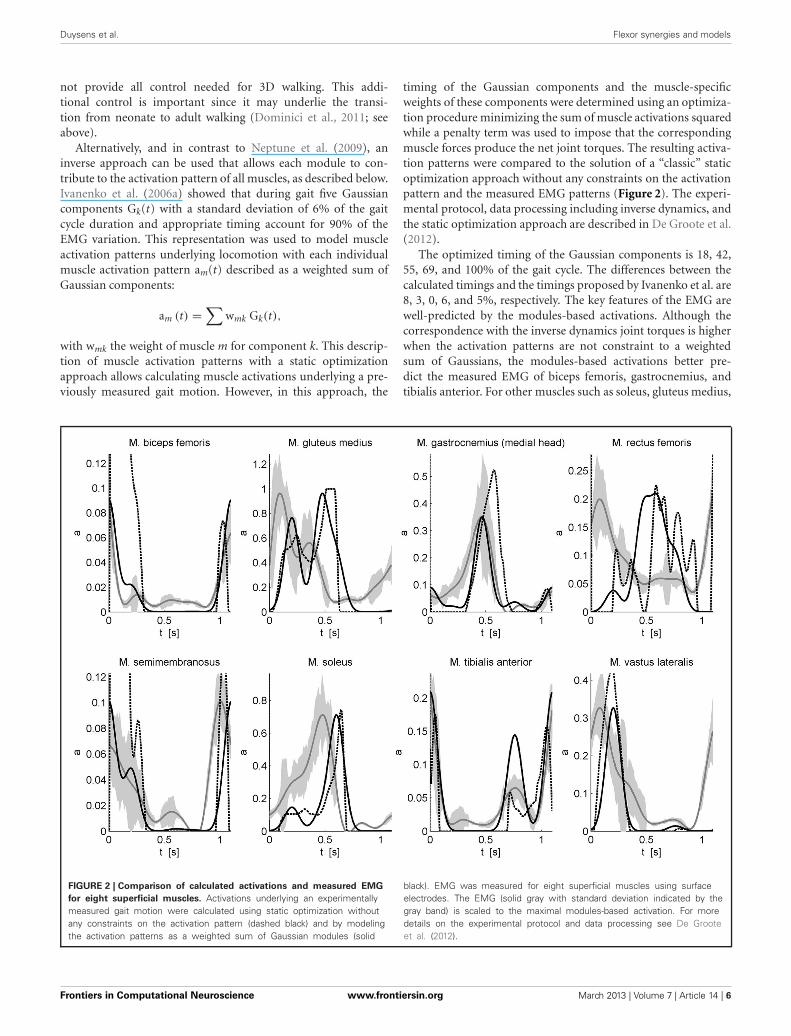
timing of the Gaussian components and the muscle-specificweights of these components were determined using an optimiza-tion procedure minimizing the sum of muscle activations squaredwhile a penalty term was used to impose that the correspondingmuscle forces produce the net joint torques. The resulting activa-tion patterns were compared to the solution of a “classic” staticoptimization approach without any constraints on the activationpattern and the measured EMG patterns (Figure 2). The experi-mental protocol, data processing including inverse dynamics, andthe static optimization approach are described in De Groote et al.(2012).
The optimized timing of the Gaussian components is 18, 42,55, 69, and 100% of the gait cycle. The differences between thecalculated timings and the timings proposed by Ivanenko et al. are8, 3, 0, 6, and 5%, respectively. The key features of the EMG arewell-predicted by the modules-based activations. Although thecorrespondence with the inverse dynamics joint torques is higherwhen the activation patterns are not constraint to a weightedsum of Gaussians, the modules-based activations better pre-dict the measured EMG of biceps femoris, gastrocnemius, andtibialis anterior. For other muscles such as soleus, gluteus medius,
FIGURE 2 | Comparison of calculated activations and measured EMG
for eight superficial muscles. Activations underlying an experimentallymeasured gait motion were calculated using static optimization withoutany constraints on the activation pattern (dashed black) and by modelingthe activation patterns as a weighted sum of Gaussian modules (solid
black). EMG was measured for eight superficial muscles using surfaceelectrodes. The EMG (solid gray with standard deviation indicated by thegray band) is scaled to the maximal modules-based activation. For moredetails on the experimental protocol and data processing see De Grooteet al. (2012).
Frontiers in Computational Neuroscience www.frontiersin.org March 2013 | Volume 7 | Article 14 | 6

Duysens et al. Flexor synergies and models
semimembranosus, and vastus lateralis there are still differencesin timing. Based on preliminary results we feel that this canbe improved by adding positive force feedback to the simula-tion. Finally for M. rectus femoris (RF) the fit is poor. The weakcorrespondence between measured EMG and calculated activa-tions for RF is seen in both activation patterns and may berelated to the notorious problem of cross-talk for surface EMGfor this muscle (Nene et al., 1999, 2004). In fact, it has beenrecognized that cross-talk can affect synergies as well, but onlyto the degree that weighting coefficient are altered (Ivanenkoet al., 2004). Therefore, some authors have insisted on using finewire EMG recordings (Ivanenko et al., 2004). Another reasonfor the difficulty of modeling RF is that this muscle presum-ably has activity which depends heavily on afferent input andreflexes. For example, in cats the activity in RF differed betweenfictive locomotion (i.e., in absence of reflexes) and normal for-ward level walking, indicating that afferent input helps shapingthe activity profile of this muscle during locomotor activity(Markin et al., 2012).
CONCLUSIONSIt is clear that the synergy approach is very fruitful and that itcan improve our understanding of human gait and its models,including new asymmetrical models of the CPG. Furthermore, itcan be helpful in providing the basis of new neuro-computationalapproaches, as was shown here, as a proof of principle, for inversedynamic calculation of muscle activations. Nevertheless, as con-cerns the popular notion of independent modules, a word ofcaution is in place since it is not fully appropriate to depict themodular organization as being a replacement of some of theolder theories (“local sign”), such as those put forward by earlyphysiologists (Creed and Sherrington, 1926).
ACKNOWLEDGMENTSJacques Duysens was supported by KU Leuven’s “BijzonderOnderzoeksfonds” (OT/08/034) and by the Research Foundation-Flanders (FWO grant G.0901.11). All authors received sup-port from KU Leuven’s Interdisciplinary Research Program(IDO/07/012).
REFERENCESAllen, J. L., and Neptune, R. R. (2012).
Three-dimensional modular con-trol of human walking. J. Biomech.45, 2157–2163.
Andersen, O. K., Sonnenborg, F. A., andArendt-Nielsen, L. (1999). Modularorganization of human leg with-drawal reflexes elicited by electricalstimulation of the foot sole. MuscleNerve 22, 1520–1530.
Andersen, O. K., Sonnenborg, F. A., andArendt-Nielsen, L. (2001). Reflexreceptive fields for human with-drawal reflexes elicited by non-painful and painful electrical stim-ulation of the foot sole. Clin.Neurophysiol. 112, 641–649.
Anderson, F. C., and Pandy, M. G.(2001). Static and dynamic opti-mization solutions for gait are prac-tically equivalent. J. Biomech. 34,153–161.
Andrews, K., and Fitzgerald, M. (1999).Cutaneous flexion reflex in humanneonates: a quantitative study ofthreshold and stimulus-responsecharacteristics after single andrepeated stimuli. Dev. Med. ChildNeurol. 41, 696–703.
Babinski, J. (1922). Réflexes de défense.Brain 45, 149–184.
Berkowitz, A. (2007). Spinal interneu-rons that are selectively activatedduring fictive flexion reflex.J. Neurosci. 27, 4634–4641.
Berkowitz, A. (2010). Multifunctionaland specialized spinal interneu-rons for turtle limb movements.Ann. N.Y. Acad. Sci. 1198,119–132.
Berkowitz, A., and Hao, Z. Z. (2011).Partly shared spinal cord networks
for locomotion and scratch-ing. Integr. Comp. Biol. 51,890–902.
Bolliger, M., Trepp, A., Zörner, B.,and Dietz, V. (2010). Modulationof spinal reflex by assisted loco-motion in humans with chroniccomplete spinal cord injury. Clin.Neurophysiol. 121, 2152–2158.
Brown, T. G. (1911). The intinsic fac-tors in the act of progression in themammal. Proc. R. Soc. Lond. B 84,308–319.
Brown, T. G. (1912). The factors inrhythmic activity of the nervoussystem. Proc. R. Soc. Lond. B 85,278–289.
Brown, T. G. (1914). On the nature ofthe fundamental activity of the ner-vous centres; together with an anal-ysis of the conditioning of rhythmicactivity in progression, and a the-ory of the evolution of function inthe nervous system. J. Physiol. 48,18–46.
Brownstone, R. M., and Wilson, J.M. (2008). Strategies for delin-eating spinal locomotor rhythm-generating networks and the pos-sible role of Hb9 interneurones inrhythmogenesis. Brain Res. Rev. 57,64–76.
Christensen, L. O., Morita, H., Petersen,N., and Nielsen, J. (1999). Evidencesuggesting that a transcortical reflexpathway contributes to cutaneousreflexes in the tibialis anterior mus-cle during walking in man. Exp.Brain Res. 124, 59–68.
Clarac, F. (2008). Some historicalreflections on the neural controlof locomotion. Brain Res. Rev. 57,13–21.
Creed, R. S., and Sherrington, C.(1926). Flexor muscles in the flex-ion reflex. Proc. R. Soc. Lond. B 100,258–267.
d’Avella, A., Saltiel, P., and Bizzi,E. (2003). Combinations of mus-cle synergies in the constructionof a natural motor behavior. Nat.Neurosci. 6, 300–308.
De Groote, F., Demeulenaere, B.,Swevers, J., De Schutter, J., andJonkers, I. (2012). A physiology-based inverse dynamic analysis ofhuman gait using sequential convexprogramming: a comparative study.Comput. Methods Biomech. Biomed.Engin. 15, 1093–1102.
Dietz, V. (2010). Behavior of spinalneurons deprived of supraspinalinput. Nat. Rev. Neurol. 6, 167–174.
Dietz, V., and Duysens, J. (2000).Significance of load receptor inputduring locomotion: a review. GaitPosture 11, 102–110.
Dietz, V., Grillner, S., Trepp, A.,Hubli, M., and Bolliger, M. (2009).Changes in spinal reflex andlocomotor activity after a completespinal cord injury: a commonmechanism? Brain 132(Pt 8),2196–2205.
Dominici, N., Ivanenko, Y. P.,Cappellini, G., d’Avella, A., Mondì,V., Cicchese, M., et al. (2011).Locomotor primitives in newbornbabies and their development.Science 334, 997–999.
Drew, T. (1991). Functional organiza-tion within the medullary reticu-lar formation of the intact unanes-thetized cat. III. Microstimulationduring locomotion. J. Neurophysiol.66, 919–938.
Duysens, J. (1977). Reflex control oflocomotion as revealed by stimula-tion of cutaneous afferents in spon-taneously walking premammillarycats. J. Neurophysiol. 40, 737–751.
Duysens, J. (2006). How deletions in amodel could help explain deletionsin the laboratory. J. Neurophysiol.95, 562–563; author reply: 563–565.
Duysens, J., Bastiaanse, C. M., Smits-Engelsman, B. C., and Dietz, V.(2004). Gait acts as a gate for reflexesfrom the foot. Can. J. Physiol.Pharmacol. 82, 715–722.
Duysens, J., Clarac, F., and Cruse,H. (2000). Load-regulating mecha-nisms in gait and posture: compara-tive aspects. Physiol. Rev. 80, 83–133.
Duysens, J., and Loeb, G. E. (1980).Modulation of ipsi- and contralat-eral reflex responses in unrestrainedwalking cats. J. Neurophysiol. 44,1024–1037.
Duysens, J., and Pearson, K. G. (1976).The role of cutaneous afferents fromthe distal hindlimb in the regulationof the step cycle of thalamic cats.Exp. Brain Res. 24, 245–255.
Duysens, J., and Pearson, K. G. (1980).Inhibition of flexor burst genera-tion by loading ankle extensor mus-cles in walking cats. Brain Res. 187,321–332.
Duysens, J., Tax, A. A., Trippel, M., andDietz, V. (1992). Phase-dependentreversal of reflexly induced move-ments during human gait. Exp.Brain Res. 90, 404–414.
Duysens, J., Tax, A. A., Trippel, M., andDietz, V. (1993). Increased ampli-tude of cutaneous reflexes duringhuman running as compared tostanding. Brain Res. 613, 230–238.
Frontiers in Computational Neuroscience www.frontiersin.org March 2013 | Volume 7 | Article 14 | 7

Duysens et al. Flexor synergies and models
Duysens, J., Trippel, M., Horstmann, G.A., and Dietz, V. (1990). Gating andreversal of reflexes in ankle musclesduring human walking. Exp. BrainRes. 82, 351–358.
Duysens, J., and Van de Crommert, H.W. (1998). Neural control of loco-motion; The central pattern gen-erator from cats to humans. GaitPosture 7, 131–141.
Forssberg, H., Grillner, S., andRossignol, S. (1975). Phase depen-dent reflex reversal during walkingin chronic spinal cats. Brain Res. 85,103–107.
Goulding, M. (2009). Circuits control-ling vertebrate locomotion: mov-ing in a new direction. Nat. Rev.Neurosci. 10, 507–518.
Grillner, S. (1985). Neurobiologicalbases of rhythmic motor acts in ver-tebrates. Science 228, 143–149.
Guertin, P. A. (2009). The mammaliancentral pattern generator for loco-motion. Brain Res. Rev. 62, 45–56.
Hagbarth, K. E. (1960). Spinal with-drawal reflexes in the humanlower limbs. J. Neurol. Neurosurg.Psychiatry 23, 222–227.
Holmberg, H., and Schouenborg, J.(1996). Postnatal development ofthe nociceptive withdrawal reflexesin the rat: a behavioural and elec-tromyographic study. J. Physiol.493(Pt 1), 239–252.
Hubli, M., Dietz, V., and Bolliger, M.(2011). Influence of spinal reflexeson the locomotor pattern afterspinal cord injury. Gait Posture 34,409–414.
Hubli, M., Dietz, V., and Bolliger,M. (2012). Spinal reflex activity:a marker for neuronal function-ality after spinal cord injury.Neurorehabil. Neural Repair 26,188–196.
Hugon, M. (1973). “Exteroceptivereflexes to stimulation of the suralnerve in normal man,” in NewDevelopments in Electromyography,Clinical Neurophysiology, Vol. 3,ed J. E. Desmedt (Basel: Karger),713–729.
Ivanenko, Y. P., Cappellini, G.,Dominici, N., Poppele, R. E., andLacquaniti, F. (2005). Coordinationof locomotion with voluntarymovements in humans. J. Neurosci.25, 7238–7253.
Ivanenko, Y. P., Cappellini, G.,Dominici, N., Poppele, R. E.,and Lacquaniti, F. (2007). Modularcontrol of limb movements duringhuman locomotion. J. Neurosci. 27,11149–11161.
Ivanenko, Y. P., Cappellini, G., Poppele,R. E., and Lacquaniti, F. (2008).Spatiotemporal organization ofalpha-motoneuron activity in the
human spinal cord during differentgaits and gait transitions. Eur. J.Neurosci. 27, 3351–3368.
Ivanenko, Y. P., Cappellini, G.,Solopova, I. A., Grishin, A. A.,MacLellen, M. J., Poppele, R. E.,et al. (2013). “Plasticity and differ-ent solutions to reorganize musclepatterns during gait,” in ConvergingClinical and Engineering Researchon Neurorehabilitation. Series:Biosystems and Biorobotics, Vol. 1,eds J. L. Pons, D. Torricelli, and M.Pajaro (Heidelberg: Springer), 732.
Ivanenko, Y. P., Grasso, R., Macellari, V.,and Lacquaniti, F. (2002). Controlof foot trajectory in human loco-motion: role of ground contactforces in simulated reduced gravity.J. Neurophysiol. 87, 3070–3089.
Ivanenko, Y. P., Poppele, R. E., andLacquaniti, F. (2004). Five basicmuscle activation patterns accountfor muscle activity during humanlocomotion. J. Physiol. 556(Pt 1),267–282.
Ivanenko, Y. P., Poppele, R. E., andLacquaniti, F. (2006a). Spinalcord maps of spatiotemporalalpha-motoneuron activation inhumans walking at different speeds.J. Neurophysiol. 95, 602–618.
Ivanenko, Y. P., Poppele, R. E., andLacquaniti, F. (2006b). Motorcontrol programs and walking.Neuroscientist 12, 339–348.
Jankowska, E., Jukes, M. G. M., Lund,S., and Lundberg, A. (1967a). Theeffect of DOPA on the spinal cord:V. Reciprocal organization of path-ways transmitting excitatory actionto alpha motoneurones of flexorsand extensors. Acta Physiol. Scand.70, 369–388.
Jankowska, E., Jukes, M. G. M., Lund,S., and Lundber, A. (1967b). Theeffect of DOPA on the spinal cord.VI. Half-centre organization ofinterneurones transmitting effectsfrom the flexor reflex afferents. ActaPhysiol. Scand. 70, 389–402.
Jansen, K., De Groote, F., Massaad,F., Meyns, P., Duysens, J., andJonkers, I. (2012). Similar musclescontribute to horizontal and verti-cal acceleration of center of massin forward and backward walk-ing: implications for neural control.J. Neurophysiol. 107, 3385–3396.
Klein, T. J., and Lewis, M. A. (2012).A physical model of sensorimo-tor interactions during locomo-tion. J. Neural Eng. 9:046011. doi:10.1088/1741-2560/9/4/046011
Komiyama, T., Zehr, E. P., and Stein, R.B. (2000). Absence of nerve speci-ficity in human cutaneous reflexesduring standing. Exp. Brain Res.133, 267–272.
Kumar, S. P. (2003). The Babinski sign-a critical review. J. Assoc. PhysiciansIndia 51, 53–57.
Lacquaniti, F., Ivanenko, Y. P., andZago, M. (2012). Patterned controlof human locomotion. J. Physiol.590(Pt 10), 2189–2199.
Lafreniere-Roula, M., and McCrea, D.A. (2005). Deletions of rhythmicmotoneuron activity during fic-tive locomotion and scratch pro-vide clues to the organization of themammalian central pattern genera-tor. J. Neurophysiol. 94, 1120–1132.
Latash, M. L. (1999). “On the evolu-tion of the notion of synergy,” inMotor Control, Today and Tomorrow,eds G. N. Gantchev, S. Mori,and J. Massion (Sofia: AcademicPublishing House) 181–196.
Lundberg, A. (1981). “Half-centresrevisited,” in Regulatory Functions ofthe CNS. Motion and OrganizationPrinciples, Advanced PhysiologicalScience Series, eds J. Szentagotheu,M. Palkovits, and J. Hamori(Budapest: Pergamon AkademiaiKiado), 155–167.
Markin, S. N., Lemay, M. A., Prilutsky,B. I., and Rybak, I. A. (2012).Motoneuronal and muscle synergiesinvolved in cat hindlimb controlduring fictive and real locomotion:a comparison study. J. Neurophysiol.107, 2057–2071.
McCrea, D. A., and Rybak, I. A. (2007).Modeling the mammalian locomo-tor CPG: insights from mistakes andperturbations. Prog. Brain Res. 165,235–253.
McCrea, D. A., and Rybak, I. A. (2008).Organization of mammalian loco-motor rhythm and pattern genera-tion. Brain Res. Rev. 57, 134–146.
Nakajima, T., Sakamoto, M., Endoh, T.,and Komiyama, T. (2006). Location-specific and task-dependent mod-ulation of cutaneous reflexes inintrinsic human hand muscles. Clin.Neurophysiol. 117, 420–429.
Nene, A., Byrne, C., and Hermens, H.(2004). Is rectus femoris really a partof quadriceps? Assessment of rec-tus femoris function during gait inable-bodied adults. Gait Posture 20,1–13.
Nene, A., Mayagoitia, R., and Veltink,P. (1999). Assessment of rectusfemoris function during initialswing phase. Gait Posture 9, 1–9.
Neptune, R. R., Clark, D. J., and Kautz,S. A. (2009). Modular control ofhuman walking: a simulation study.J. Biomech. 42, 1282–1287.
Pearson, K. G. (2004). Generating thewalking gait: role of sensory feed-back. Prog. Brain Res. 143, 123–129.
Pearson, K. G., and Duysens, J. (1976).“Function of segmental reflexes in
the control of stepping in cock-roaches and cats,” in Neural Controlof Locomotion, eds R. E. Herman, S.Grillner, P. S. Stein, and D. G. Stuart(New York, NY: Plenum Press),519–535.
Petersen, J. A., Schubert, M., and Dietz,V. (2010). The occurrence of theBabinski sign in complete spinalcord injury. J. Neurol. 257, 38–43.
Philippson, M. (1905). L’autonomie etla centralisation dans le systèmenerveux des animaux (Autonomyand centralization in the animalnervous system). Trav. Lab. Physiol.Inst. Solvay (Bruxelles) 7, 1–208.
Prochazka, A., and Yakovenko, S.(2007a). The neuromechanicaltuning hypothesis. Prog. Brain Res.165, 255–265.
Prochazka, A., and Yakovenko, S.(2007b). Predictive and reactivetuning of the locomotor CPG.Integr. Comp. Biol. 47, 474–481.
Prochazka, A., Gillard, D., and Bennett,D. J. (1997). The positive forcefeedback control of muscles.J. Neurophysiol. 77, 3226–3236.
Rybak, I. A., Shevtsova, N. A.,Lafreniere-Roula, M., and McCrea,D. A. (2006a). Modelling spinalcircuitry involved in locomotorpattern generation: insights fromdeletions during fictive locomotion.J. Physiol. 577(Pt 2), 617–639.
Rybak, I. A., Stecina, K., Shevtsova,N. A., and McCrea, D. A. (2006b).Modelling spinal circuitry involvedin locomotor pattern generation:insights from the effects of affer-ent stimulation. J. Physiol. 577(Pt 2),641–658.
Sandrini, G., Serrao, M., Rossi, P.,Romaniello, A., Cruccu, G., andWiller, J. C. (2005). The lowerlimb flexion reflex in humans. Prog.Neurobiol. 77, 353–395.
Santos, M. J., and Liu, W. (2007).Unloading reaction to electri-cal stimulation at neutral andsupinated ankle positions. GaitPosture 26, 106–112.
Sartori, M., Gizzi, L., and Farina, D.(2012). “Musculoskeletal modelingof human locomotion based onlow-dimensional impulsive acti-vation signals: perspectives forNeurotechnologies,” in ConvergingClinical and Engineering Researchon Neurorehabilitation. Series:Biosystems and Biorobotics, Vol. 1,eds J. L. Pons, D. Torricelli, and M.,Pajaro (Heidelberg: Springer), 732.
Schmit, B. D., Hornby, T. G., Tysseling-Mattiace, V. M., and Benz, E. N.(2003). Absence of local sign with-drawal in chronic human spinalcord injury. J. Neurophysiol. 90,3232–3241.
Frontiers in Computational Neuroscience www.frontiersin.org March 2013 | Volume 7 | Article 14 | 8

Duysens et al. Flexor synergies and models
Schouenborg, J., and Kalliomaki, J.(1990). Functional organization ofthe nociceptive withdrawal reflexes.I. Activation of hindlimb muscles inthe rat. Exp. Brain Res. 83, 67–78.
Schouenborg, J., and Weng, H. R.(1994). Sensorimotor transforma-tion in a spinal motor system. Exp.Brain Res. 100, 170–174.
Schouenborg, J., Weng, H. R.,Kalliomäki, J., and Holmberg,H. (1995). A survey of spinal dorsalhorn neurones encoding the spatialorganization of withdrawal reflexesin the rat. Exp. Brain Res. 106,19–27.
Shahani, B. T., and Young, R. R. (1971).Human flexor reflexes. J. Neurol.Neurosurg. Psychiatry 34, 616–627.
Sherrington, C. S. (1906). IntegrativeActions of the Nervous System. NewHaven, CT: Yale University Press,411.
Sherrington, C. S. (1910a). Remarks onthe reflex mechanism of the step.Brain 33, 1–25.
Sherrington, C. S. (1910b). Flexor-reflex of the limb, crossed exten-sion reflex, and reflex stepping andstanding (cat and dog) J. Physiol.(Lond.) 40, 28–121.
Shimamura, M., Kogure, I., and Wada,S. (1980). Three types of reticu-lar neurons involved in the spino-bulbo-spinal reflex of cats. BrainRes. 186, 99–113.
Sonnenborg, F. A., Andersen, O. K.,and Arendt-Nielsen, L. (2000).
Modular organization of exci-tatory and inhibitory reflexreceptive fields elicited by elec-trical stimulation of the foot solein man. Clin. Neurophysiol. 111,2160–2169.
Sonnenborg, F. A., Andersen, O. K.,Arendt-Nielsen, L., and Treede, R.D. (2001). Withdrawal reflexorgan-isation to electrical stimulation ofthe dorsal foot in humans. Exp.Brain Res. 136, 303–312.
Stuart, D. G., and Hultborn, H.(2008). Thomas Graham Brown(1882–1965), Anders Lundberg(1920-), and the neural control ofstepping. Brain Res. Rev. 59, 74–95.
Sylos Labini, F. S., Ivanenko, Y. P.,Cappellini, G., Gravano, S., andLacquaniti, F. (2011). Smoothchanges in the EMG patterns dur-ing gait transitions under bodyweight unloading. J. Neurophysiol.106, 1525–1536.
Ting, L. H., Chvatal, S. H., Safavynia,S. A., and McKay, J. L. (2012).Review and perspective: neurome-chanical considerations for predict-ing muscle activation patterns formovement. Int. J. Numer. MethodsBiomed. Engin. 28, 1003–1014.
Ting, L. H., and Macpherson, J. M.(2005). A limited set of muscle syn-ergies for force control during apostural task. J. Neurophysiol. 93,609–613.
Torres-Oviedo, G., Macpherson, J. M.,and Ting, L. H. (2006). Muscle
synergy organization is robustacross a variety of postural per-turbations. J. Neurophysiol. 96,1530–1546.
Tresch, M. C., Saltiel, P., and Bizzi, E.(1999). The construction of move-ment by the spinal cord. Nat.Neurosci. 2, 162–167.
Van Gijn, J. (1978). The Babinskisign and the pyramidal syndrome.J. Neurol. Neurosurg. Psychiatry 41,865–873.
Van Wezel, B. M., Ottenhoff, F. A., andDuysens, J. (1997). Dynamic con-trol of location-specific informationin tactile cutaneous reflexes fromthe foot during human walking.J. Neurosci. 17, 3804–3814.
Whelan, P. J. (1996). Control of loco-motion in the decerebrate cat. Prog.Neurobiol. 49, 481–515.
Willer, J. C. (1977). Comparative studyof perceived pain and nocicep-tive flexion reflex in man. Pain 3,69–80.
Yang, J. F., and Stein, R. B. (1990).Phase-dependent reflex reversalin human leg muscles duringwalking. J. Neurophysiol. 63,1109–1117.
Zehr, E. P., Komiyama, T., and Stein, R.B. (1997). Cutaneous reflexes dur-ing human gait: electromyographicand kinematic responses to electri-cal stimulation. J. Neurophysiol. 77,3311–3325.
Zehr, E. P., Stein, R. B., and Komiyama,T. (1998). Function of sural nerve
reflexes during human walking.J. Physiol. 507(Pt 1), 305–314.
Zhong, G., Shevtsova, N. A., Rybak,I. A., and Harris-Warrick, R. M.(2012). Neuronal activity in theisolated mouse spinal cord duringspontaneous deletions in fictivelocomotion: insights into loco-motor central pattern generatororganization. J. Physiol. 590(Pt 19),4735–4759.
Conflict of Interest Statement: Theauthors declare that the researchwas conducted in the absence of anycommercial or financial relationshipsthat could be construed as a potentialconflict of interest.
Received: 21 December 2012; accepted:20 February 2013; published online: 13March 2013.Citation: Duysens J, De Groote F andJonkers I (2013) The flexion synergy,mother of all synergies and father of newmodels of gait. Front. Comput. Neurosci.7:14. doi: 10.3389/fncom.2013.00014Copyright © 2013 Duysens, De Grooteand Jonkers. This is an open-accessarticle distributed under the terms of theCreative Commons Attribution License,which permits use, distribution andreproduction in other forums, providedthe original authors and source arecredited and subject to any copyrightnotices concerning any third-partygraphics etc.
Frontiers in Computational Neuroscience www.frontiersin.org March 2013 | Volume 7 | Article 14 | 9