Plaghki L, Bragard D, Le Bars D, Willer JC, Godfraind JM (1998) Facilitation of a nociceptive...
12
79:2557-2567, 1998. J Neurophysiol Jean-Marie Godfraind Léon Plaghki, Dominique Bragard, Daniel Le Bars, Jean-Claude Willer and Nonnoxious Radiant Heat Produced by a Laser Facilitation of a Nociceptive Flexion Reflex in Man by You might find this additional info useful... 79 articles, 26 of which can be accessed free at: This article cites http://jn.physiology.org/content/79/5/2557.full.html#ref-list-1 5 other HighWire hosted articles This article has been cited by [PDF] [Full Text] [Abstract] , February, 1 1999; 81 (2): 584-595. J Neurophysiol Cynthia G. Leung and Peggy Mason Physiological Properties of Raphe Magnus Neurons During Sleep and Waking [PDF] [Full Text] [Abstract] , December, 1 2001; 53 (4): 597-652. Pharmacol Rev Daniel Le Bars, Manuela Gozariu and Samuel W. Cadden Animal Models of Nociception [PDF] [Full Text] [Abstract] , December, 2002; 125 (12): 2766-2781. Brain Bernard Laurent and François Mauguière Luis Garcia?Larrea, Philippe Convers, Michel Magnin, Nathalie André?Obadia, Roland Peyron, and provoked pain Laser?evoked potential abnormalities in central pain patients: the influence of spontaneous [PDF] [Full Text] [Abstract] , March, 1 2003; 23 (5): 1933-1940. J. Neurosci. H. Foo and Peggy Mason by Repeated Laser Stimulation Discharge of Raphe Magnus on and offCells Is Predictive of the Motor Facilitation Evoked [PDF] [Full Text] [Abstract] , September, 2003; 23 (7): 534-540. Cephalalgia PD Drummond Stimulation of the Supraorbital Nerve The Effect of Trigeminal Nociceptive Stimulation on Blink Reflexes and Pain Evoked by including high resolution figures, can be found at: Updated information and services http://jn.physiology.org/content/79/5/2557.full.html can be found at: Journal of Neurophysiology about Additional material and information http://www.the-aps.org/publications/jn This infomation is current as of February 28, 2011. Physiological Society. ISSN: 0022-3077, ESSN: 1522-1598. Visit our website at http://www.the-aps.org/. by the American Physiological Society, 9650 Rockville Pike, Bethesda MD 20814-3991. Copyright © 1998 by the American publishes original articles on the function of the nervous system. It is published 12 times a year (monthly) Journal of Neurophysiology on February 28, 2011 jn.physiology.org Downloaded from
-
Upload
sorbonne-fr -
Category
Documents
-
view
0 -
download
0
Transcript of Plaghki L, Bragard D, Le Bars D, Willer JC, Godfraind JM (1998) Facilitation of a nociceptive...

79:2557-2567, 1998.J NeurophysiolJean-Marie GodfraindLéon Plaghki, Dominique Bragard, Daniel Le Bars, Jean-Claude Willer andNonnoxious Radiant Heat Produced by a LaserFacilitation of a Nociceptive Flexion Reflex in Man by
You might find this additional info useful...
79 articles, 26 of which can be accessed free at:This article cites http://jn.physiology.org/content/79/5/2557.full.html#ref-list-1
5 other HighWire hosted articlesThis article has been cited by
[PDF] [Full Text] [Abstract]
, February, 1 1999; 81 (2): 584-595.J NeurophysiolCynthia G. Leung and Peggy MasonPhysiological Properties of Raphe Magnus Neurons During Sleep and Waking
[PDF] [Full Text] [Abstract], December, 1 2001; 53 (4): 597-652.Pharmacol Rev
Daniel Le Bars, Manuela Gozariu and Samuel W. CaddenAnimal Models of Nociception
[PDF] [Full Text] [Abstract], December, 2002; 125 (12): 2766-2781.Brain
Bernard Laurent and François MauguièreLuis Garcia?Larrea, Philippe Convers, Michel Magnin, Nathalie André?Obadia, Roland Peyron,and provoked painLaser?evoked potential abnormalities in central pain patients: the influence of spontaneous
[PDF] [Full Text] [Abstract], March, 1 2003; 23 (5): 1933-1940.J. Neurosci.
H. Foo and Peggy Masonby Repeated Laser StimulationDischarge of Raphe Magnus on and offCells Is Predictive of the Motor Facilitation Evoked
[PDF] [Full Text] [Abstract], September, 2003; 23 (7): 534-540.Cephalalgia
PD DrummondStimulation of the Supraorbital NerveThe Effect of Trigeminal Nociceptive Stimulation on Blink Reflexes and Pain Evoked by
including high resolution figures, can be found at:Updated information and services http://jn.physiology.org/content/79/5/2557.full.html
can be found at:Journal of Neurophysiologyabout Additional material and information http://www.the-aps.org/publications/jn
This infomation is current as of February 28, 2011.
Physiological Society. ISSN: 0022-3077, ESSN: 1522-1598. Visit our website at http://www.the-aps.org/.by the American Physiological Society, 9650 Rockville Pike, Bethesda MD 20814-3991. Copyright © 1998 by the American
publishes original articles on the function of the nervous system. It is published 12 times a year (monthly)Journal of Neurophysiology
on February 28, 2011
jn.physiology.orgD
ownloaded from
Facilitation of a Nociceptive Flexion Reflex in Man by NonnoxiousRadiant Heat Produced by a Laser
LEON PLAGHKI,1 DOMINIQUE BRAGARD,1 DANIEL LE BARS,2 JEAN-CLAUDE WILLER,3
AND JEAN-MARIE GODFRAIND1
1Faculte de Medecine, Universite Catholique de Louvain, B-1200 Brussels, Belgium; 2Institut National de la Sante et dela Recherche Medicale U-161, 75014 Paris; and 3Faculte de Medecine Pitie-Salpetriere, Laboratoire deNeurophysiologie, 75634 Paris Cedex 13, France
Plaghki, Leon, Dominique Bragard, Daniel Le Bars, Jean- allowed clear interactions between the afferent signals to beClaude Willer, and Jean-Marie Godfraind. Facilitation of a no- seen. More recently, Steffens and Schomburg (1993)ciceptive flexion reflex in man by nonnoxious radiant heat produced showed that excitatory postsynaptic potentials evoked byby a laser. J. Neurophysiol. 79: 2557–2567, 1998. Electromyo- cutaneous and joint afferents clearly were facilitated by con-graphic recordings were made in healthy volunteers from the knee- ditioning radiant heat only if the stimulation temperaturesflexor biceps femoris muscle of the nociceptive RIII reflex elicited
were well above the pain threshold.by electrical stimulation of the cutaneous sural nerve. The stimulusIn animals, there are several lines of evidence that noci-intensity was adjusted to produce a moderate pricking-pain sensa-
ceptive flexor reflexes can be facilitated by homotopic nox-tion. The test responses were conditioned by a nonnoxious thermalious conditioning stimulation. Wall and Woolf (1984) re-(°407C) stimulus applied to the receptive field of the sural nerve.
This stimulus was delivered by a CO2 laser stimulator and consisted ported that in the decerebrated spinal unanesthetized rat, aof a 100-ms pulse of heat with a beam diameter of 20 mm. Its power conditioning procedure of applying 20 electrical stimuli towas 22.7 { 4.2 W (7.2 mJ/mm2), and it produced a sensation of the sural nerve at 1 Hz, resulted in a marked increase in thewarmth. The maximum surface temperature reached at the end of excitability of biceps femoris/semitendinosus motoneuronsthe period of stimulation was calculated to be 77C above the actual for 10 min. Only electrical conditioning stimuli at intensitiesreference temperature of the skin (327C). The interval between
that recruited afferent C fibers were effective in producingthe laser (conditioning) and electrical ( test) stimuli was varieda prolonged facilitation of the reflex. This observation wasfrom 50 to 3,000 ms in steps of 50 ms. It was found that theconfirmed repeatedly in the same preparation with nearlynociceptive flexion reflex was facilitated by the thermal stimulus;identical experimental protocols (Wiesenfeld-Hallin 1985;this modulation occurred with particular conditioning-test intervals,
which peaked at 500 and 1,100 ms with an additional late, long- Wiesenfeld-Hallin et al. 1990, 1991; Woolf and Wall 1986;lasting phase between 1,600 and 2,300 ms. It was calculated that Woolf and Wiesenfeld-Hallin 1986). It also has been ob-the conduction velocities of the cutaneous afferent fibers responsi- served in spinal rabbits during recordings of reflex responsesble for facilitating the RIII reflex, fell into three ranges: one corre- elicited in the nerves to the semitendinosus or gastrocnemiussponding to Ad fibers (3.2 m/s) and two in the C fiber range (1.3 muscle by stimulation of the sural nerve. In this last case,and 0.7 m/s) . It is concluded that information emanating from
the facilitation was seen after noxious stimulation of the heelwarm receptors and nociceptors converges. In this respect, theor the application of a train of electrical stimuli to the suralpresent data show, for the first time, that in man, conditioningnerve at an intensity that recruited Ad and C fibers (Catley etnonnociceptive warm thermoreceptive Ad and C fibers results in
an interaction at the spinal level with a nociceptive reflex. This al. 1984; Clarke et al. 1992). Long-lasting postconditioninginteraction may constitute a useful means whereby signals add depolarizations have been observed during recordings fromtogether to trigger flexion reflexes in defensive reactions and other motoneurons in the neonatal rat spinal cord in vitro afterbasic motor behaviors. It also may contribute to hyperalgesia in homosynaptic or heterosynaptic activation by repetitiveinflammatory processes. The methodology used in this study ap- stimulation of high-threshold afferent fibers (Sivilotti et al.pears to be a useful noninvasive tool for exploring the thermoalge-
1993; Thompson et al. 1993). Similarly, it has been reportedsic mechanisms in both experimental and clinical situations.that in the spinal unanesthetized rat, there is a decrease inthe latency for hindpaw withdrawal from noxious heat afternociceptive electrical conditioning of the sciatic nerve (Cle-I N T R O D U C T I O Nland et al. 1994). A decrease in the tail flick latency alsohas been seen after noxious thermal conditioning of the tailDirect investigation of neuronal convergence at the cellu-in pentobarbital /chloral, lightly anesthetized intact ratslar level is difficult in animals and impossible in man. How-(Cridland and Henry 1988; Yashpal et al. 1991, 1993).ever, it is possible to use an indirect approach that involves
Other studies of nociceptive reflexes have shown that nox-testing interactions elicited by the activation of differentious stimuli applied homotopically can be either facilitatorypathways. In this respect, spatial and temporal interactionsas mentioned earlier or inhibitory. Indeed, strong segmentalbetween afferent signals contributing to flexion reflexes haveinhibitory effects have been reported after high-intensitybeen studied in both animals and man (Lundberg 1979;conditioning applied ipsilaterally on or near to the sameSchomburg 1990). In all but one (Lundberg et al. 1977)dermatome (Catley et al. 1983; Chung et al. 1983; Clarkeof these studies, the nociceptive nature of the conditioning
stimulation appeared to be the common denominator that et al. 1988, 1989; Shin et al. 1986; Taylor et al. 1989–
25570022-3077/98 $5.00 Copyright q 1998 The American Physiological Society
J603-7/ 9k28$$my29 04-14-98 19:50:34 neupal LP-Neurophys
on February 28, 2011
jn.physiology.orgD
ownloaded from

PLAGHKI, BRAGARD, LE BARS, WILLER, AND GODFRAIND2558
over the submalleolar pathway of the right sural nerve (Fig. 1A) .1991). Interestingly, it was found that the effects foundThe stimulus consisted of a volley of eight rectangular constant-in spinal animals, whether facilitatory or inhibitory, can becurrent pulses, 0.5 ms in duration, delivered over 20 ms throughmodified drastically in intact nontransected animals wherean isolation device (Digitimer Model DS7) driven by a pulse-descending inhibitory processes are fully operationalgenerator (Master-8, AMPI). The interstimulus interval varied(Fleischman and Urca 1988; Gozariu et al. 1995).
It is a common observation that conditioning nonnocicep-tive afferents do not reveal interactions with afferent signalsproduced by a nociceptive test stimulus; nevertheless it isstill well accepted that fibers conveying nonnociceptive in-formation contribute to the pool of ‘‘flexor reflex afferents’’(FRA) (Eccles and Lundberg 1959). In view of the contri-bution of nonnociceptive afferents to FRA, the present studywas aimed at reinvestigating the possibility that nonnocicep-tive thermal signals may interact with nociceptive signals inman by recording a well-defined nociceptive flexion reflex,the RIII reflex, elicited by applying test stimuli to a cutaneousnerve (the sural nerve); using a CO2 laser stimulator toprovide nonnoxious thermal conditioning stimulation of thereceptive field of this nerve; and systematically varying thedelay between the conditioning and test stimuli.
The RIII reflex is elicited mainly by activation of Ad-afferent fibers and represents an objective physiological in-dex which is highly correlated with the sensation of painproduced by electrical stimulation of the sural nerve (Willer1977, 1984; Willer and Albe-Fessard 1983).
Laser stimulation allows precise control of stimulationparameters, elicits a well-synchronized afferent volley ofimpulses and can produce natural and selective thermal stim-ulation of low-thresholds afferents. Depending on theamount of energy delivered, the CO2 laser stimulator pro-duces warm and/or painful sensations by direct activationof cutaneous thermoreceptors and/or nociceptors connectedexclusively to Ad and C fibers (Bromm et al. 1984; Devoret al. 1982).
In this paper, it will be shown that conditioning nonnoci-ceptive afferents does facilitate the RIII flexion reflex at vari-ous conditioning-test intervals that, by use of a mathematicalmodel can be shown to correspond to the activation of pe-ripheral Ad and C fibers.
M E T H O D S
Subjects
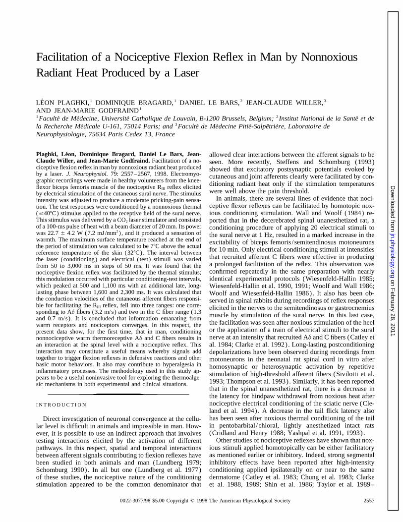
Experiments were carried out on 15 healthy volunteers: 7 womenand 8 men, mean age 26 { 5.7 yr (range 20–45). The rulesof the local ethics committee were followed. Subjects gave theirinformed consent after being carefully briefed on the experimentalprocedures and purposes. To ensure good muscle relaxation, they FIG. 1. Experimental setup and recording examples. A: experimental set-were installed comfortably in an armchair. They were familiarized up. Test RIII reflex was recorded from the ipsilateral biceps femoris afterwith the perception of electrical and laser stimuli and instructed in electrical stimulation delivered over the submalleolar path of the sural nerve
at a constant intensity of 1.25 times threshold. This test electromyographicall the usual manipulations performed during a session. They wore(EMG) response was or was not (controls) conditioned by a nonnoxious CO2protective goggles in line with the guidelines for control of laserlaser radiant heat pulse applied to the receptive field area of the sural nervehazards. To avoid any auditory cues that may have been associatedpreceding the conditioned stimulation at random interval of between 50 andwith the production of the stimuli, the subjects wore headphones3,000 ms. B: example of unconditioned and conditioned RIII reflex responses.(Ultramuffit, Racal Safety) .As shown below the recordings, the responses were full-wave rectified. C:full-wave rectified responses in the period 80–160 ms after the electrical teststimulus onset were used to provide an initial representation of the effects ofProcedure for the test RIII reflex the conditioning stimulus. Median of the 60 unconditioned responses then wascalculated, and the individual unconditioned response closest to that value was
The method for eliciting and recording the nociceptive RIII selected, as illustrated for 1 subject in Fig. 1C1 (h). Variations of amplitudeflexion reflex from a knee-flexor muscle was based on that de- (in V) of the EMG responses were built (C2) by subtracting from each bin, thescribed previously (Willer 1984). Briefly, bipolar electrical stimuli median rectified unconditioned EMG response (h in C1) from the individual
conditioned EMG response (j in C1).were applied via a pair of surface electrodes placed 20 mm apart
J603-7/ 9k28$$my29 04-14-98 19:50:34 neupal LP-Neurophys
on February 28, 2011
jn.physiology.orgD
ownloaded from
FACILITATION OF RIII REFLEX BY NONNOXIOUS RADIANT LASER HEAT 2559
from 5 to 11 s. Reflex activity evoked in the ipsilateral biceps responses (whether control or conditioned) measured in this fash-ion had a distribution that was skewed positively (Snedecor andfemoris was recorded through a pair of surface Ag-AgCl elec-
trodes (Meditrace model ECE1801) placed on the skin over the Cochran 1989). In accord with the normality hypothesis beingunfulfilled, medians rather than means were calculated.muscle, and connected to a Gould Universal Amplifier (Model 13
4615-58) with a band-pass of 10 Hz to 2 kHz and a gain of The median of the 60 control responses was calculated for eachof the 15 subjects. Then, the 60 conditioned responses, each corre-125,000. The upgoing edge of the electrical stimulus triggered
data acquisition by a small computer (Cambridge Electronic De- sponding to a given conditioning-test interval, and the 60 controlresponses were normalized to this median. This yielded for eachsign Model 1401) for a period of 500 ms, at 4,000 cps. The thresh-
old of the RIII reflex first was defined as the stimulus intensity conditioning-test interval, 15 normalized conditioned responsesand 15 normalized control responses, the median of which waseliciting liminal reflex responses with a probability of 80–90%.
The current intensity was then adjusted to 25% above this threshold calculated separately and taken as the grand median of the controlresponses (i.e., the median of the 15 1 60 control responses) .and was kept constant for the rest of the experiment. This intensity
[mean: 14.4 { 6.3 (SD) mA] produced supraliminal reflex re- Based on these normalized data, time series vectors (control andconditioned) of equally spaced points in time (Dt Å 50 ms; n Åsponses with minimal fluctuations from one stimulus to the next
(Fig. 1B) (Willer 1984) and a sensation of moderate pricking pain, 60) were obtained and submitted to a resistant nonlinear smoothingprocedure (3RSSH Tukey’s notation; STATGRAPHICS, STSC)which was well tolerated by the subjects at least for the duration
of an experimental session (20 min). (see Figs. 2B and 3). This procedure of nonlinear smoothing waschosen because it is based on a ‘‘running’’ median and is resistantto ‘‘isolated outliers.’’ Because this procedure has to find the me-Conditioning proceduredian for three values surrounding each point, the two first and twolast endpoints of the times series vectors were omitted in Fig. 3.The conditioning stimulus was delivered by a CO2 laser to the
To achieve a more comprehensible representation of data fromcutaneous area situated at the dorsum of the right foot and corre-a single subject (Fig. 2A) , the variations in amplitude of the EMGsponding to the receptive field of the sural nerve (Fig. 1A) . Itresponses were determined for the period 80–160 ms after the testconsisted of a 100-ms duration heat pulse with a beam diameterstimulus; this was done by subtracting from each bin, the medianof 20 mm and power rating of 22.7 { 4.2 W (7.2 mJ/mm2). Withvalue of the 60 rectified control responses from the individualsuch parameters, the maximum temperature reached at the end ofconditioned response (see Fig. 1C) .the stimulation period was 77C (or 97C with / SD of power)
above the reference skin temperature, i.e., 327C (for details of thesecomputations, see Haimi-Cohen et al. 1983). In these conditions, Theoretical calculationsthe cutaneous temperature should not exceed 407C in the superficialepidermis, and thus even the most superficial nociceptive endings In this section, an attempt will be made to identify the type ofshould not be activated (Bromm and Treede 1983). Indeed, the peripheral nerve fibers that were activated when cutaneous sensorystimuli were perceived as warm but never as painful. Before start- receptors were excited by the nonnociceptive thermal CO2 lasering the experimental procedure, it was confirmed that the condition- stimuli. This will be done by calculating the fibers’ conductioning stimulus when delivered alone, did not elicit an electromyo- velocities by taking account of the spatio-temporal interactions,graphic (EMG) response in the biceps femoris. The CO2 laser which occurred when changes in the excitability of the motoneuro-device was an improved version of the machine conceived and nal pool to the electrical test stimulus appeared after heat laserbuilt in the Department of Physics at the Universite catholique de stimulation. As already shown in Fig. 3, such changes occurredLouvain (Plaghki et al. 1989). The reproducibility of the laser only when the test response was timed properly after the condition-stimuli was accurate at 1%. A He-Ne laser beam (Hughes Aircraft; ing stimulus.wavelength 632.8 nm, power output 5 mW) was used to showvisually the site of stimulation. The laser equipment was kept in Initial equationan adjacent room to reduce noise and heat. The laser beam wasdeflected, through a hole in the wall, on to the foot of the subjects During any period of facilitation of the RIII reflex, convergenceby means of a remotely controlled gold-coated flat mirror. The and coincidence of information being conveyed by afferent fibersposition of the laser beam was checked via a closed circuit televi- activated by the electrical test stimulus and the laser stimulus mustsion video; it was displaced slightly after each laser stimulus to have occurred in the CNS.avoid any overheating of the skin. All parameters (output power, Thus for any timing of a facilitation, one can write the followingstimulus duration, beam diameter and target site) were controlled equationfrom the experimental room.
tpe / tae / tde / tcde / (Dt / Z ) Å tpl / tal / tdl / tcdl (1)
General experimental procedure where
During an experimental session, once the electrical stimulus tpe Å propagation time for the electrical current through the skinintensity had been determined (as defined above), a total of 120 RIII tae Å activation time of the nerve fibers by the electrical currentreflex responses were studied. Sixty of these were unconditioned tde Å delay resulting from conduction time of peripheral fibers(control responses, i.e., those associated with a sham stimulation) activated by the electrical currentand 60 were conditioned, i.e., preceded by a thermal stimulus. tcde Å central delay time of circuits activated by the electrical cur-All these responses were elicited in a completely random order rentdetermined by a computer program. The conditioning-test interval Dt Å time interval between the conditioning and the test stimulus(Dt) between the laser (conditioning) and electrical ( test) stimuli Z Å uncertainty parameter that affects Dtwas varied from 50 to 3,000 ms in steps of 50 ms. tpl Å propagation time of thermal energy through the superficial
layers of the skintal Å activation time of sensory receptors by thermal energyProcessing of datatdl Å delay resulting from conduction time of peripheral fibers
activated by the thermal laser stimulusThe EMG records containing the reflex responses was full-waverectified and integrated over a time window of 80 ms starting 80 tcdl Å central delay time of circuits activated by the thermal laser
stimulus.ms after the test stimulus onset (Fig. 1B) . The magnitudes of the
J603-7/ 9k28$$my29 04-14-98 19:50:34 neupal LP-Neurophys
on February 28, 2011
jn.physiology.orgD
ownloaded from

PLAGHKI, BRAGARD, LE BARS, WILLER, AND GODFRAIND2560
FIG. 2. Waterfall display of results obtained from 1subject. A : typical example of results obtained from 1 sub-ject (male, aged 29, stature 1.92 m) illustrating the influ-ence of the laser thermal stimulus on the RIII reflex. Ab-scissa indicates time (in ms) with 0 corresponding to theoccurrence of the test electrical stimulus; only the 80- to160-ms time window after the stimulus is shown. Ordinateindicates the variation in amplitude (in mV) of the EMGresponses by subtracting from each bin the median responseof the 60 rectified control EMG responses from the individ-ual conditioned EMG responses (see Fig. 1C) . Sixty condi-tioned responses of the biceps femoris muscle are presentedin a waterfall display. From bottom to top, each sweepcorresponds to a response conditioned by a laser stimulusand elicited by an electrical test stimulus delivered withprogressively increasing time intervals (Dt) in steps of 50ms. B : graph of the ratio of the responses integrated andnormalized to the median of the unconditioned responsesas a function of the time interval Dt between conditioningand test stimuli. RIII reflex response was noticeably en-hanced when intervals between the conditioning laser heatstimulus and the test electrical stimulus were Ç500, 1,000,and ú1,700 ms.
the motoneuronal pool and trigger activation of the biceps femorismuscle. After the peripheral processes elicited by laser stimulation,We will review all these parameters step by step. Because most
of them could reasonably be estimated, we will propose a final a central delay ( tcdl ) also is required for related information toreach the motoneuronal pool and influence its excitability. If theseEq. 2 giving a relationship between the experimentally controlled
parameter Dt and the conduction velocities (Vl ) of peripheral fibers central delays are equal, then these two parameters cancel eachother in Eq. 1. If the values of these parameters are not equal, theirin the sural nerve, which we presume were activated by the laser
heat stimulus. difference Dtd Å Étcde 0 tcdlÉ should be taken into account. Sucha difference could derive from the propagation of action potentials
Parameters related to peripheral processes elicited by through different neuronal networks, one of these possibly includ-ing additional synapses. We decided to take into account a greatlyelectrical stimulationoverestimated possible difference of 20 synapses. In fact, takingIt generally is accepted that electrical stimulation directly acti-into account a difference of °20 synapses, computational analysisvates peripheral nerve fibers. The propagation time of the electricalbased on Eq. 2 showed that this factor was of limited importancecurrent ( tpe ) through the skin and body fluids and the activationwith regards to the other parameters.time ( tae ) of the nerve fibers both may be regarded here as negligi-
ble values.Time elapsed between conditioning and test stimuliBased on the work of Willer and Albe-Fessard (1983) and the
source book of Liveson and Ma (1992), the peripheral fibers of theThe parameter that has been manipulated experimentally is Dt,sural nerve involved in the RIII reflex are known to conduct impulses
i.e., the time elapsed between application of conditioning and testat a velocity (Ve) Ç35 m/s. Because the distance of the peripheralstimuli. More particularly, Dt was the time elapsed between thepath (De) may be estimated easily by measuring the distance betweenbeginning of the 100-ms thermal stimulus and the beginning ofthe stimulation site and the spinal process of T12, the peripheralthe 20-ms train of electrical pulses. However, because the exactconduction time (tde) can be calculated as: tde Å De /Ve .timing of the start of nerve fiber excitation may have varied duringthe stimulating period, the time elapsed between the actual effectiveParameters related to peripheral processes elicited byconditioning and the test stimuli also may have varied. This uncer-laser stimulationtainty is represented by the parameter Z . According to the durations
The thermal energy sent by the laser stimulator reaches sensory of both thermal and electrical stimuli, the (Dt / Z ) value mayreceptors after a propagation time ( tpl ) through the superficial lay- vary from (Dt 0 100 ms) to (Dt / 20 ms).ers of the skin and activates the receptors after an activation time( tal ) . Their respective values presented in Table 1 are derived from Final equation and computationsthe work of Haimi-Cohen et al. (1983) and of Bromm and Treede(1983, 1984). By rearranging Eq. 1, and applying the simplifications mentioned
The peripheral path distance (Dl ) corresponds to the distance above, it may be rewrittenbetween the stimulation site and the spinal process of T12. Unlike
Vl Å Dl / (Dt / Z / tde 0 tpl 0 tal / Dtd ) (2)the electrical test stimulus, the thermal laser stimulation used inthe present study did not elicit a muscle reflex; therefore, neither Based on Eq. 2, the relation between Vl and Dt may be exam-the conduction velocity (Vl ) nor the peripheral conduction time ined for the three sets of parameter values listed in Table 1.( tdl ) could be measured in the present noninvasive experiments. These values were chosen on the basis of the above-mentionedHowever the relation tdl Å Vl /Dl will be used in the calculations reports in the literature and taking into account under- and over-described later. estimations.
Results of the computational analysis are presented in Fig. 4.Parameters related to central delays Three curves were calculated using underestimated, averaged, and
overestimated parameters, respectively, as shown in Table 1. ForAfter the peripheral processes elicited by electrical stimulation,a central delay ( tcde ) is required for related information to reach Dt values ú1 s, no significant differences were seen among the
J603-7/ 9k28$$my29 04-14-98 19:50:34 neupal LP-Neurophys
on February 28, 2011
jn.physiology.orgD
ownloaded from

FACILITATION OF RIII REFLEX BY NONNOXIOUS RADIANT LASER HEAT 2561
R E S U L T S
Experimental observations
In the 15 healthy volunteers, we examined whether non-noxious radiant heat produced by a laser could influence theRIII flexion reflex elicited by nociceptive electrical stimula-tion of the sural nerve (Fig. 1A) . The thermal stimulus wasperceived by the subjects as warm but never painful. Whenapplied alone, it never elicited an EMG response in thebiceps femoris.
As illustrated with an individual example (Fig. 1B) , theapplication of the laser stimulus with a 1,950-ms condition-ing-test interval resulted in a clear facilitation of the RIII
response (Fig. 1B2) as compared with the control response(Fig. 1B1) . Further processing of the data involved bothresponses being full-wave rectified (Fig. 1B, bottom) andconsidered within the period 80–160 ms after the test stimu-lus onset. The median of the 60 unconditioned responsesthen was calculated, and the individual unconditioned re-sponse closest to that value was selected, as illustrated for
FIG. 3. Modulation of RIII reflex responses by conditioning nonnoxious one subject in Fig. 1C1 (h) . Finally, the variations in ampli-heat stimulation. Influence of the conditioning laser thermal stimulus on tude of each conditioned EMG response were computed bythe nociceptive reflex RIII shown for data pooled from all 15 subjects. Curve subtracting from each bin, the value of the median rectifiedwith filled circles represents the data of the smoothed normalized EMG
unconditioned response from the individual rectified condi-evoked reflex activities (see METHODS) as a function of the time intervalDt between the conditioning laser stimulus and the test electrical stimulus. tioned response (Fig. 1C2) .Curve with open circles represents the smoothed normalized data computed Figure 2A shows a typical example of the result obtainedfrom the unconditioned responses, i.e., those associated with a sham stimu- in this way from one subject, illustrating the influence oflation. Two curves are clearly different. As compared with the controls, the
the laser heat stimulus on the RIII reflex. This figure wascurve obtained from the conditioned EMG responses shows 3 peaks at 500constructed by sequentially displaying the increased reflexms (with the fastest conditioning effect noticed Ç250 ms), 1,100 ms, and
a late, large peak between 1,600 and 2,300 ms. Mean conduction velocities activity (ordinate, D amplitude) elicited by the conditioningof any peripheral fibers excited by a conditioning stimulus when conver- thermal stimulus, with the time after the test stimulus run-gence and coincidence of the information conveyed by the afferent fibers ning from left to right (abscissa) and the conditioning-testactivated by the test and the conditioning stimulus occurred, were computed
intervals (Dt) increasing from front to back (z axis) . Theas in Fig. 4 ( top) . Dt values corresponding to the 3 peaks matched thecriteria defined by Eq. 2 and Fig. 4: 1 in the range of Ad fibers and 2 in facilitation of the 60 conditioned reflex responses are illus-the range of C fibers (see also Table 2). trated in perspective using a waterfall display presentation.
The data, initially acquired with conditioning-test intervalsthree curves (Fig. 4, front panel) . By contrast, for Dt valuesõ500 applied at random in the 50–3,000 ms range, were re-ms, and much more dramatically for Dt values õ250 ms, differ- arranged in such a way that each sweep of the figure corre-ences between the curves were greatly increased at decreasing
sponds to a test response conditioned by a laser stimulusvalues of Dt (Fig. 4, inset) . Thus Eq. 2 yields results that makewith progressively increasing conditioning-test intervalssense from a biological point of view only for (Dt / Z / tde 0from bottom to top in 50-ms steps. Note the facilitation oftpl 0 tal / Dtd ) @ 0. Indeed, the sum of the parameter valuesthe RIII reflex for some conditioning-test intervals, separated(Z / tde 0 tpl 0 tal / Dtd ) equal / 26, 0100, and 0180 ms,
respectively, for the under-, mean, and overestimated parameters by periods of ‘‘silence’’ during which individual EMG re-(Table 1). As a consequence, when using the mean and overesti- sponses were nearly identical to the control RIII reflex.mated parameter values respectively, computing the conduction The results obtained from this individual subject arevelocity for Dt lower than Ç150 and 250 ms yields inconsistent shown quantitatively in Fig. 2B. The results are expressedfigures. in terms of the ratio (ordinate) between each individual
For the Dt values corresponding to the peaks illustrated in Fig. response and the median unconditioned response as a func-3, i.e., corresponding to a facilitation of the electrically inducedtion of the conditioning-test interval (abscissa) . Both re-nociceptive reflex, conduction velocities of the peripheral fiberssponse values were calculated during the 80–160 ms afterexcited by the thermal laser stimulus can be computed from thethe test stimulus. The curves were obtained after a resistantthree curves shown in Fig. 4. The results are shown in Table 2. In
addition, the conduction velocities of the peripheral fibers excitedby the thermal laser stimulus, as estimated from the curve calcu- TABLE 1. Parameter values of the modellated with averaged parameters, are reported on the top abscissaof Fig. 3 for graphic examination. The facilitations elicited when Parameters Underestimated Average OverestimatedDt values were Ç500 ms (with the fastest conditioning effectstarting Ç250 ms), correspond to laser-induced activities in small- tpl , ms 20 35 50
tal , ms 20 35 50diameter myelinated fibers of the Ad type. The facilitations ob-Dl , mm 1,210 1,280 1,350served for Dt values Ç1,100 ms and those Ç1,600–2,300 ms,De , mm 1,080 1,150 1,220result from laser-induced activities in thin nonmyelinated nerveVe , m/s 30 35 40fibers of the C type; in addition, two separate C-fiber groups couldDtd , ms 10 0 010be defined: the first with conduction velocities of 1–1.5 m/s (C1),Z, ms 20 060 0100and the second with conduction velocities õ1 m/s (C2).
J603-7/ 9k28$$my29 04-14-98 19:50:34 neupal LP-Neurophys
on February 28, 2011
jn.physiology.orgD
ownloaded from

PLAGHKI, BRAGARD, LE BARS, WILLER, AND GODFRAIND2562
FIG. 4. Computation of the conduction velocity of pri-mary afferent fibers engaged in convergence. Theoreticalrelationship between the conditioning-test interval Dt andthe conduction velocity Vl of any peripheral fibers excitedby a conditioning stimulus when convergence and coinci-dence of the information conveyed by the afferent fibersactivated by the test and the conditioning stimulus oc-curred. By computation of Eq. 2, Vl Å Dl / (Dt / Z /tde 0 tpl 0 tal / Dtd ) , 3 curves were obtained using theunderestimated, averaged, and overestimated values of theparameters, respectively, as listed in Table 1. First part ofthe curves corresponding to the lower Dt values (0- to0.75-s range), are shown at the back with a higher magni-fication of the abscissa. Calculated values of Vl were addedas the top abscissa in Fig. 3.
nonlinear smoothing treatment. Note that the RIII reflex ripheral receptive field of the sural nerve. Such a modulationoccurred with definite time intervals between the condition-clearly was enhanced when the conditioning-test intervals
were Ç500 and 1,000 ms and when they were ú1,700 ms. ing and test stimuli, namely Ç500, Ç1,100 ms, and a late,long-lasting phase between 1,600 and 2,300 ms. The sim-The profile of this curve based on a single subject (Fig.
2B) was similar to that for data pooled from the 15 subjects. plest interpretation of our observations is that nociceptiveFigure 3 shows two curves obtained after the resistant non- and nonnociceptive information converges and interacts onlinear smoothing treatment of the normalized conditioned a common pool of interneurons. The occurrence of suchand unconditioned responses (see Processing data) . The interactions implies a temporal coincidence of informationcurve with filled circles represents the data of the smoothed emanating from the two areas of stimulation. This was obvi-normalized EMG evoked activities as a function of the inter- ous only with adequate time intervals between the condition-val between the conditioning laser stimulus and the test elec- ing and test stimuli. Because the RIII reflex is present intrical stimulus. The curve with open circles represents the patients with total sections of the spinal cord (Roby-Bramismoothed normalized data computed from the unconditioned et al. 1987; Willer and Bussel 1980), one can assume that itsresponses. The two curves are clearly different. The curve basic pathway is mainly spinal. Thus the most parsimoniousobtained from the conditioned EMG responses shows three interpretation of our present data is that the convergencepeaks at conditioning-test intervals of 500 and 1,100 ms and between noxious and nonnoxious signals occurred in thebetween 1,600 and 2,300 ms. These peak values, together spinal cord with the various peaks of facilitation being deter-with other parameters were used to compute the conduction mined mainly by the time required for the synchronizedvelocity of the cutaneous afferent fibers activated by the peripheral volleys elicited by the heat stimulus to reach theconditioning laser heat stimulus (see Theoretical calcula- common final pool of interneurons. Therefore the differenttions) . In Fig. 3, the top abscissa shows the computed con- facilitatory waves can be explained by the triggering of activ-duction velocity plotted against the corresponding EMG ity in fibers with different peripheral conduction velocities.data. Based on the computed conduction velocity, three dif- In spite of the parsimony of our interpretation of the pres-ferent groups of afferent fibers were recognized: one in the ent results, three other alternative interpretations could beAd-fiber range and two in the C-fiber range (see Table 2). put forward: sensitization, variation in central delays, and
oscillations of cord excitability. Sensitization (see referencesD I S C U S S I O N in Meyer et al. 1994) is unlikely to introduce a bias in the
present results for two reasons. First, the position of the laserIt was found that the nociceptive flexion reflex was facili-beam was moved slightly after each laser stimulus to avoidtated by nonnoxious thermal stimulation applied to the pe-any skin damage due to repetitive stimulation. Second, thestimulus intervals were applied randomly, and thus the rank
TABLE 2. Conduction velocities of peripheral nerve fibers order of the applied stimulus did not appear to be a signifi-cant source of variability. Variations in central delays mayUnder- Over- Fiberresult from additional synapses in segmental networksDt, s estimated Mean estimated Typeand/or in long loops. These variations appear to be of limited
0.50 2.3 3.2 4.2 Ad importance given that the introduction of an uncertainty fac-1.10 1.1 1.3 1.5 C tor as large as 20 ms in the mathematical model (see Z1.60–2.30 0.6 0.7 0.8 C
parameter in Theoretical calculations) had little impact onVelocities are in meters per second. the determination of the conduction velocities. Finally, it
J603-7/ 9k28$$my29 04-14-98 19:50:34 neupal LP-Neurophys
on February 28, 2011
jn.physiology.orgD
ownloaded from

FACILITATION OF RIII REFLEX BY NONNOXIOUS RADIANT LASER HEAT 2563
could be questioned whether the three peaks do reflect oscil- stimulation was provided by a Xenon lamp (visible light)and was noxious whereas in our study, a mild sensation oflations of cord excitability triggered by the conditioning laser
stimulus. Indeed mechanisms proposed to play a major role warmth was described by the subjects; and because the teststimulus was applied to the sole of the foot, whereas in ourduring spinal processing (convergence, temporal, and/or
spatial summation and assembly coding) may depend on study it was applied on the dorsum of the foot, i.e., withinthe receptive field of the sural nerve. All these differencesneuronal discharge synchronization. In this line, it has been
reported that oscillations arise locally in the dorsal horn of make comparison between the present study and that of An-dersen et al. (1994) quite difficult.the spinal cord and induce cooperative activity of spatially
distributed neuronal subpopulations (Eblen-Zajjur and Sand- In the second study (Willer and Albe-Fessard 1983), theRIII reflex was conditioned by painful electrical stimulikuhler 1997). Dorsal root stimulation also has been shown
to trigger oscillations in the motoneuron pool (Baranauskas (30-mA intensity, 1-ms duration) applied to the sural nerveterritory. Facilitations of the RIII reflex were induced by suchand Nistri 1995) with a power spectrum peaking at 8 Hz.
However, power spectrum of the present data peak õ2 Hz. conditioning stimuli and were attributed to the activation ofafferent fibers with conduction velocities in the 0.8–25As no such low-frequency oscillator has been observed, our
proposition seems more likely. Higher frequencies of oscilla- m/s range. In this case, however, the conditioning stimuluswas also nociceptive in nature, and facilitations were in thetions, if present, should have been observed in our data given
the Dt of sampling rate of 50 ms. Besides, radiant heat 150–200% range. By contrast, the moderate facilitations(15–20%) observed in the present study were elicited bystimulation is known to activate different types of peripheral
nerve fibers in the Ad and C range (references below). nonnoxious conditioning radiant heat. In any case, as far aswe know, the facilitation of nociceptive reflexes by non-The peak values in Fig. 3, together with the mathematical
modeling, helped us to compute the conduction velocities noxious thermal stimuli has not been reported to date.of the cutaneous afferent fibers activated by the conditioningthermal stimulus. Three different groups of afferent fibers Putative peripheral fibers involved in the facilitationwere identified: one in the Ad-fiber range (3.2 m/s) and twoin the C-fiber range (1.3 and 0.7 m/s) . This is consistent with It was possible to subdivide into three groups, the periph-
eral fibers which when activated facilitated the RIII reflexthe fact that the CO2 laser stimulator activates exclusively Adand C fibers (Bromm et al. 1984; Devor et al. 1982) and because of the time resolution resulting from the experimen-
tal design. However, as suggested in Fig. 3, the first peakevokes sensations through direct activation of cutaneousthermoreceptors and/or nociceptors, depending on the of activation might in fact be composed of two components,
both in the range of Ad-fiber conduction velocities. How-amount of energy delivered.The discussion will be organized into several sections as ever, before a definite conclusion can be made about this,
further experiments involving shorter time intervals (Dt)follows: facilitation of a flexion reflex by thermal stimuli;putative peripheral fibers involved in the facilitation; puta- between the test and conditioning stimuli will be required.
It should be emphasized that the three groups of fiberstive spinal neurons involved in the facilitation; and method-ological considerations. that we are proposing were involved in the modulation of
the RIII reflex, conveyed information subserving warm per-ception. This is supported not only by the verbal reports ofFacilitation of a flexion reflex by thermal nonnoxious orthe subjects but also by the computation of maximal temper-noxious stimuliatures reached in the center of the laser beam, which werealways õ407C (see Conditioning procedures) .To the best of our knowledge, there have been only two
studies that have used an experimental design close to that The responses of peripheral receptors and afferent fibersto thermal stimulation have been studied widely with micro-used in the present work and that merit consideration.
Andersen et al. (1994) reported that a reflex response neurographic techniques (e.g., Beck et al. 1974; Vallbo etal. 1979; Van Hees and Gybels 1972). Several classes offrom the tibialis anterior muscle was facilitated by a painful
thermal conditioning stimulus applied to the sole of the ipsi- cutaneous receptors that may respond to thermal stimuli areconnected to slowly conducting thin fibers, either myelinatedlateral foot with conditioning-test intervals from 600 to 3,000
ms. This effect was attributed to fibers with a broadband of (Ad) or unmyelinated (C). The afferent fibers that selec-tively respond to thermal stimuli generally are classified ei-conduction velocities within the C-fiber population being
activated by the conditioning stimulus. However, because ther as cold receptors, connected to Ad fibers or warm recep-tors, connected to C fibers. This dichotomy is based mainlythese authors used seven fixed conditioning-test intervals
(0, 300, 600, 800, 1,000, 1,200, and 3,000 ms), it was quite on the observation that warm sensibility is blocked earlierthan cold sensibility after local anesthesia (Fruhstorfer et al.impossible to differentiate with accuracy several peaks of
facilitatory effect as we did here by considering 60 condi- 1974; Sinclair and Hinshaw 1950). However, this separationbetween Ad cold and C warm receptors is not absolutetioning-test intervals between the laser and electrical stimuli.
Besides these differences in the resolution of conditioning- (Spray 1986) and, as discussed later, several types of periph-eral receptors might have contributed to the results reportedtest intervals, there were others between the present study
and that work, notably because in the latter EMG reflex herein.In the myelinated afferent fiber group, the Ad cold recep-activity was elicited from the tibialis anterior muscle by
electrical stimulation of the sole of the foot whereas we tors are known to operate optimally in the 15–307C range.In the monkey, the mean conduction velocity of these affer-recorded a well-defined nociceptive flexion reflex from the
biceps femoris muscle elicited by stimulation of Ad- fibers in ent fibers is 9.0 m/s with a large range from 1.7 to 16.7m/s (Dubner et al. 1975). These receptors present restinga cutaneous nerve, the sural nerve; because the conditioning
J603-7/ 9k28$$my29 04-14-98 19:50:34 neupal LP-Neurophys
on February 28, 2011
jn.physiology.orgD
ownloaded from

PLAGHKI, BRAGARD, LE BARS, WILLER, AND GODFRAIND2564
discharges at °11 Hz at the optimal temperature of 257C significantly to the thermally induced late facilitations of theRIII reflex.and disappear beyond 387C (Beitel et al. 1977; Darian-Smith
Although the distribution of C-fiber conduction velocitieset al. 1973; Sumino and Dubner 1981). Such a decreasedis most commonly said to be unimodal, it was found to beactivity of the Ad-cold receptors in the 25–387C rangebimodal in the present study. As far as we know, the exis-makes the participation of this class of neurons unlikely intence of two groups of conduction velocities of C fibers wasthe facilitation of the RIII reflex.reported by Douglass et Ritchie (1959). However, these CObservations of Ad-fiber activity associated with warmfibers were not sensitive to nonnoxious warm stimulation.perception have been reported but the proportion of suchIn a study of C-fiber warm receptors, Darian-Smith et al.afferent fibers is low compared with cold receptors (Darian-(1979) provided a histogram of the distribution of their con-Smith et al. 1973). In the monkey, facial warm receptorsduction velocities: it was clearly unimodal centered on 1.2connected to Ad and C fibers with conduction velocities inm/s. Two distinct classes of C-MH receptors were describedthe 0.9–6.7 m/s range have been reported (Sumino et al.in the monkey by Meyer and Campbell (1981) with either1973). Interestingly, the warm receptors described by Teras-quickly or slowly adapting responses. However these authorshima and Liang (1993) in man, are linked to Ad fibersdo not rule out the possibility that this difference dependedwith conduction velocities in the 3.8–11.2 m/s range. Theseon the depth below the skin surface at which the receptorconduction velocities are reasonably close to those computedwas located. Interestingly, latency distributions of C-fiber–in the present study (3.1–8.1 m/s); such fibers may haveevoked reflexes elicited by electrical stimulation of the suralcontributed to the early facilitation of the RIII reflex reportednerve in the rat are bimodal, suggesting existence of twoherein.populations of C fibers with different conduction velocitiesA priori, two other types of Ad receptors also might have(0.6 and 0.9 m/s) (see Falinower et al. 1994). Similar re-contributed to these effects. In the first subclass, the A-HTMsponse latencies with a bimodal distribution also have been(high-threshold mechanical) or type I A-MH (mechano-observed during recordings of rat dorsal horn convergentheat) , the Ad receptors respond to strong mechanical stimu-neurons after electrical stimulation of their receptive fieldslation and usually remain insensitive to noxious heat and(e.g., see Villanueva et al. 1986; this study).chemical stimulation. They have high thermal thresholds
(ú497C) and sometimes rapid conduction velocities (meanPutative spinal neurons involved in the facilitation31 m/s, range 5–53 m/s) (Campbell et al. 1979). In the
second subclass, the mechano-thermal receptors (MTR) orAs mentioned already, the convergence of afferent signalstype II A-MH, the Ad- receptors respond to strong mechani-
evoked by the nonnoxious thermal conditioning and the noci-cal stimuli, noxious heating, and algogenic chemicals. Theyceptive test stimuli most probably occurred in the spinalpresent lower thermal thresholds (range: 37–477C) andcord. For many reasons, such a convergence is not conceiv-slower conduction velocities (mean: 15.2 { 9.9 m/s)able at the level of the motoneuronal pool responsible for(Dubner et al. 1977). Although the former group is unlikelythe activation of the biceps femoris muscle. Radiant heatto have contributed to the early facilitation of the RIII reflex below the threshold for nociceptive afferent fibers did notreported herein, the latter might have contributed to some elicit synaptic effects in a motoneurons nor facilitation on
extent. reflex pathways from Group Ib or II muscle afferents orRegarding the unmyelinated fibers, two classes of re- mechanoreceptive cutaneous afferents (Behrends et al. 1983;
ceptors are concerned with warm stimuli. In the first, the Burke et al. 1971; Steffens and Schomburg 1993). BecauseC-warm receptors are included in afferent fibers that respond all Ad and C fibers terminate in the dorsal horn of the spinaloptimally to temperaturesÇ407C, i.e., temperatures that gen- cord, it follows that the functional site of convergence waserally are perceived as gentle warmth. Their conduction ve- located on interneurons situated upstream of the motoneuro-locities are reported to be between 0.6 and 2.4 m/s, with nal pool.mean values between 0.7 and 1.2 m/s (Darian-Smith et al. One subpopulation of dorsal horn neurons fulfills some1979; Duclaux and Kenshalo 1980; Hensel and Iggo 1971; of the properties required for being the locus of convergence,LaMotte and Campbell 1978); these are reasonably close to and several pieces of evidence suggest that they may wellthose computed in the present study (0.7–1.3 m/s) . Such belong to the reflex arc involved in the RIII reflex. Indeed, thereceptors were therefore probably involved in the late facili- RIII reflex recorded in humans and the convergent neuronstations of the RIII reflex reported herein. recorded in animals are modulated by common factors: both
In the second class, the C-mechano–heat nociceptors are inhibited by systemic or intrathecal morphine (see ref.(C-MHs), the unmyelinated afferent fibers respond to high- in Willer and Le Bars 1995); both are inhibited by transcuta-intensity mechanical and thermal stimuli. They were shown neous electrical nerve stimulation (Handwerker et al. 1975;to sometimes respond to chemical stimuli and thus also are Rosenberg et al. 1996); both are inhibited by heterotopictermed polymodal. Their thermal threshold ranges between noxious stimuli (see references in Willer et al. 1989) via38 and 497C with mean values Ç447C (Beitel and Dubner supraspinal structures located in the lower medulla (Bouhas-1976; Robinson et al. 1983). They show no spontaneous sira et al. 1995; De Broucker et al. 1990). A subset of suchactivity below 387C (Hallin et al. 1981; LaMotte and Camp- neurons, the so-called ‘‘warming/noxious heat-convergentbell 1978). In humans, the conduction velocity for this group units’’ have thresholds mainly õ427C, increase their dis-is reported to be in the 0.7–1.3 m/s range with mean values charge over a limited range of temperatures and most com-Ç0.8 m/s (Gybels et al. 1979; Van Hees and Gybels 1972). monly reach a plateau or even decline slightly when noxiousEven though their participation cannot be ruled out com- temperatures are achieved (Burton 1975; Kanui 1988; Le
Bars and Chitour 1983; Menetrey et al. 1979; Price andpletely, it is unlikely that such fibers would have contributed
J603-7/ 9k28$$my29 04-14-98 19:50:34 neupal LP-Neurophys
on February 28, 2011
jn.physiology.orgD
ownloaded from

FACILITATION OF RIII REFLEX BY NONNOXIOUS RADIANT LASER HEAT 2565
the Molecular Spectroscopy Laboratory (Universit catholique de Louvain)Browe 1975). This subset of neurons may well be a candi-for building the CO2 laser.date as the locus of convergence. However, involvement
This work was supported by Fonds National de la Recherce Scientifique/of ventral horn interneurons cannot be excluded because Fondation pour la Recherche Scientifique Medicale Belgium and l’Instituttemperature-sensitive neurons have been described within National de la Sante et de la Recherche Medicale.
Present address and address for reprint requests: L. Plaghki, Cliniqueslaminae VII and VIII (Burton 1975).Universitaires St. Luc, 10 Ave. Hippocrate, B-1200 Brussels, Belgium.
Received 23 July 1997; accepted in final form 21 January 1998.Methodological considerations
We believe that the present results provide and validate REFERENCESa potentially useful noninvasive method not only for further
ANDERSEN, O. K., JENSEN, L. M., BRENNUM, J., AND ARENDT-NIELSEN, L.studies concerned with central interactions among informa- Evidence for central summation of C and Ad nociceptive activity in man.tion from various origins but also for those studies that will Pain 59: 273–280, 1994.
BARANAUSKAS, G. AND NISTRI, A. Membrane potential oscillations of neo-deal with the indirect study of peripheral fibers in man. Itnatal rat spinal motoneurons evoked by electrical stimulation of dorsalis clear that a certain degree of convergence is implied withinroot fibres. Eur. J. Neurosci. 7: 2403–2408, 1995.the pathways underlying the RIII reflex. Admittedly, absence BECK, P. W., HANDWERKER, H. O., AND ZIMMERMANN, M. Nervous outflow
of facilitation by no means indicates absence of fiber activa- from the cat’s foot during noxious radiant heat stimulation. Brain Res.67: 373–386, 1974.tion by the conditioning stimulus; but occurrence of facilita-
BEHRENDS, T., SCHOMBURG, E. D., AND STEFFENS, H. Facilitatory interac-tion clearly indicates that a group of fibers actually havetion between cutaneous afferents from low threshold mechanoreceptorsbeen activated by the conditioning stimulus. As quoted byand noclceptors in segmental reflex pathways to alpha-motoneurons.
Schomburg (1990) ‘‘spatial facilitation depends on a distinct Brain Res. 260: 131–134, 1983.subliminal fringe around the discharge zone in the interneur- BEITEL, R. E. AND DUBNER, R. The response of unmyelinated (C) polymo-
dal nociceptors to thermal stimuli applied to the monkey’s face. J Neuro-onal pool shared by the two pathways.’’ Thus being awarephysiol. 39: 1160–1175, 1976.of the possible pitfalls of false negative results and consider-
BEITEL, R. E., DUBNER, R., HARRIS, R., AND SUMINO, R. Role of thermore-ing the difficulty of investigating, in man, peripheral fibers ceptive afferents in behavioral reaction times to warming temperaturewith slow conduction velocities, the experimental method shifts applied to the monkey’s face. Brain Res. 138: 329–346, 1977.
BROMM, B., JAHNKE, M. T., AND TREEDE, R. D. Responses of human cuta-used here may be a useful tool in both experimental andneous afferents to CO2 laser stimuli causing pain. Exp. Brain Res. 55:clinical situations. In this respect, it is important to note158–166, 1984.that the routine electrophysiological methods used in clinical
BROMM, B. AND TREEDE, R. D. CO2 laser radiant heat pulses activate Cdepartments investigate transmission only in afferent fibers nociceptors in man. Pflugers Arch. 399: 155–156, 1983.with the largest diameter and cannot explore the thermoalge- BROMM, B. AND TREEDE, R. D. Nerve fibre discharges, cerebral potentials
and sensations induced by CO2-laser stimulation. Hum. Neurobiol. 3:sic system (Noel and Desmedt 1975; Veilleux and Stevens33–40, 1984.1987).
BOUHASSIRA, D., CHITOUR, D., VILLANUEVA, L., AND LE BARS, D. TheWe will comment briefly on some possible functional im- spinal transmission of nociceptive information: modulation by the caudalplications of the present results. Under the term ‘‘erythral- medulla. Neuroscience 69: 931–938, 1995.
BURKE, R. E., RUDOMIN, P., VYCKLICKY, L., AND ZAJAC, F. E., III. Primarygia,’’ Lewis (1942) described temperature-dependent painafferent depolarisation and flexion reflexes produced by radiant heat stim-elicited by experimentally induced local redness and tender-ulation of the skin. J. Physiol. (Lond.) 207: 185–214, 1971.ness of the skin. Using an in vitro testis-superior spermatic
BURTON, H. Responses of spinal cord neurons to systematic changes innerve preparation, Kumazawa et al. (1987) provided con- hindlimb skin temperatures in cats and primates. J. Neurophysiol. 38:vincing evidence that such observations could be explained 1060–1079, 1975.
CAMPBELL, J. M., MEYER, R. A., AND LA MOTTE, R. H. Sensitization ofby a potentiation of the response of polymodal nociceptorsmyelinated nociceptive afferents that innervate monkey hand. J. Neuro-by increases of temperature in the nonnoxious 34–437Cphysiol. 49: 98–110, 1979.range (at least as far as the viscera are concerned). Because CATLEY, D. M., CLARKE, R. W., AND PASCOE, J. E. Naloxone enhancement
inflammation is associated with local increases in tempera- of spinal reflexes in the rabbit. J. Physiol. (Lond.) 339: 61–73, 1983.CATLEY, D. M, CLARKE, R. W., AND PASCOE, J. E. Post-tetanic depressionture, this may contribute to clinical hyperalgesia. However,
of spinal reflexes in the rabbit and the possible involvement of opioidas shown here for cutaneous stimuli, nonnoxious increasespeptides. J. Physiol. (Lond.) 352: 483–493, 1984.in temperature also could result in a central booster by means
CHUNG, J. M., FANG, Z. R., CARGILL, C. L., AND WILLIS, W. D. Prolonged,of convergence between information emanating from warm naloxone-reversible inhibition of the flexion reflex in the cat. Pain 15:and nociceptive receptors. Together with the effects of the 35–53, 1983.
CLARKE, R. W., FORD, T. W., HARRIS, J., AND TAYLOR, J. S. Thyrotropin‘‘inflammatory soup’’ (see reference in Dray 1994; Levinereleasing hormone, cholecystokinin and endogenous opioids in the modu-and Taiwo 1994), such factors potentially could contributelation of spinal reflexes in the rabbit. Neuropharmacology 27: 1279–to the hyperalgesia seen during inflammatory processes. 1284, 1988.
In conclusion, the present data suggest indirectly that CLARKE, R. W., FORD, T. W., AND TAYLOR, J. S. Activation by high inten-sity peripheral nerve stimulation of adrenergic and opioidergic inhibitionwarm nonnociceptive information can be conveyed by bothof a spinal reflex in the decerebrated rabbit. Brain Res. 505: 1–6, 1989.Ad- and C-afferent fibers and can modulate the RIII reflex
CLARKE, R. W., HARRIS, J., FORD, T. W., AND TAYLOR, J. S. Prolongedin man. Although these effects were moderate, in the 15–potentiation of transmission through a withdrawal reflex pathway after
20% range, they do suggest that such warm afferents may noxious stimulation of the heel in the rabbit. Pain 49: 65–70, 1992.contribute to some extent to the FRA and add a useful signal CLELAND, C. L., FOONG-YEN, L., AND GEBHART, G. F. Pentobarbital pre-
vents the development of C- fiber-induced hyperalgesia in the rat. Painto that of other information to trigger flexion reflexes in57: 31–43, 1994.defensive reactions and other basic motor behaviors.
CRIDLAND, R. A. AND HENRY, J. L. Facilitation of the tail-flick reflex bynoxious cutaneous stimulation in the rat: antagonism by a substance Panalogue. Brain Res. 462: 15–21, 1988.We thank Dr. S. W. Cadden for advice in the preparation of the manu-
script. We also thank Prof. A. Fayt and P. Stouffs (physicist engineer) of DARIAN-SMITH, I., JOHNSON, K. O., AND DYKES, R. ‘‘Cold’’ fiber population
J603-7/ 9k28$$my29 04-14-98 19:50:34 neupal LP-Neurophys
on February 28, 2011
jn.physiology.orgD
ownloaded from

PLAGHKI, BRAGARD, LE BARS, WILLER, AND GODFRAIND2566
innervating palmar and digital skin of the monkey: responses to cooling LIVESON, J. A., AND MA, D. M. Laboratory Reference for Clinical Neurpo-physiology . Philadelphia, PA: F. A. Davis, 1992.pulses. J. Neurophysiol. 36: 325–346, 1973.
DARIAN-SMITH, I., JOHNSON, K. O., LAMOTTE, C., KENINS, P., SHIGENAGA, LUNDBERG, A. Multisensory control of spinal reflex pathways. In: Progressin Brain Research. Reflex Control of Posture and Movement , edited byY., AND MING, V. C. Coding of incremental changes in skin temperature
by single warm fibers in the monkey. J. Neurophysiol. 42: 1316–1331, R. Granit and D. Pomeiano. Amsterdam: Elsevier, 1979, vol. 50, p. 11–28.1979.
DE BROUCKER, T., CESARO, P., WILLER, J. C., AND LE BARS, D. Diffuse LUNDBERG, A., MALMGREM, K., AND SCHOMBURG, E. D. Cutaneous facilita-tion of transmission in reflex pathways from Ib afferents to motoneurons.noxious inhibitory controls (DNIC) in man: involvement of the spinoreti-
cular tract. Brain 113: 1223–1234, 1990. J. Physiol. (Lond.) 265: 763–780, 1977.MENETREY, D., CHAOUCH, A., AND BESSON J. M. Responses of spinal cordDEVOR, M. CARMON, A., AND FROSTIG, R. Primary afferent and spinal
sensory neurones that respond to brief pulses of intense infra red laser dorsal horn neurones to non-noxious and noxious cutaneous temperaturechanges in the spinal rat. Pain 6: 163–282, 1979.radiation: a preliminary survey in rats. Exp. Neurol. 76: 483–494, 1982.
DOUGLAS, W. W. AND RITCHIE, J. M. The sensory functions of the non- MEYER, R. A. AND CAMPBELL, J. N. Evidence for two distinct classes ofunmyelinated nociceptive afferents in monkey. Brain Res. 224: 149–152,myelinated afferent nerve fibres from the skin, In: Pain and Itch: Nervous
Mechanisms, edited by G.E.W. Wolstenholme and M. O’Connor. Lon- 1981.don: Churchill, 1959, p. 26–40. MEYER, R. A., CAMPBELL, J. N., AND RAJA, S. N. Peripheral neural mecha-
nisms of nociception. In: Textbook of Pain, edited by P. D. Wall and R.DRAY, A. Tasting the inflammatory soup: the role of peripheral neurones.Pain Rev. 1: 153–171, 1994. Melzack. Edinburgh: Churchill Livingston, 1994, p. 13–44.
NOEL, P. AND DESMEDT, J. E. Somatosensory cerebral evoked potentialsDUBNER, R., PRICE D. D., BEITEL, R. E., AND HU, J. W. Peripheral neuralcorrelates of behavior in monkey and human related to sensory-discrimi- after vascular lesions of the brainstem and diencephalon. Brain 98: 113–
128, 1975.native aspects of pain. In: Pain in the Trigeminal Region , edited by D. J.Anderson and B. Matthews. Amsterdam: Elsevier, 1977, p. 57–66. PLAGHKI, L., DELISLE, D., FAYT, A., GODFRAIND, J. M., AND STOUFFS, P.
Algesimetry by brief pulses of CO2 radiation (Abstract). Arch. Int. Phys-DUBNER, R., SUMINO, R., AND WOOD, W. I. A peripheral ‘‘cold’’ fiberpopulation responsive to innocuous and noxious thermal stimuli applied iol. Biochem. 97: 82, 1989.to monkey’s face. J. Neurophysiol. 38: 1373–1389, 1975. PRICE, D. D. AND BROWE, A. C. Responses of spinal cord neurons to graded
noxious and non-noxious stimuli. Exp. Neurol. 48: 201–221, 1975.DUCLAUX, R. AND KENSHALO, D. R. Response characteristics of cutaneouswarm receptors in the monkey. J. Neurophysiol. 43: 1–15, 1980. ROBINSON, C. J., TOREBJRK, H. E., AND LA MOTTE, R. H. Psychophysical
detection and pain ratings of incremental thermal stimuli: a comparisonEBLEN-ZAJJUR, A. A. AND SANDKUHLER, J. Synchronicity of nociceptiveand non-nociceptive adjacent neurons in the spinal dorsal horn of the rat: with nociceptor responses in humans. Brain Res. 274: 87–106, 1983.stimulus-induced plasticity. Neuroscience 76: 39–54, 1997. ROBY-BRAMI, A., BUSSEL, B., WILLER, J.-C., AND LE BARS, D. An electro-
ECCLES, R. M. AND LUNDBERG, A. Synaptic actions in motoneurones by physiological investigation into the pain relieving effects of heterotopicafferents which may evoke the flexion reflex. Arch. Ital. Biol. 97: 199– nociceptive stimuli: probable involvement of a supraspinal loop. Brain221, 1959. 110: 1497–1508, 1987.
FALINOWER, S., WILLER, J. C., JUNIEN, J. L., AND LE BARS, D. A C-fiber ROSENBERG, S., DANZIGER, N., BOURGEOIS, P., CHARPENTIER, G., AND
reflex modulated by heterotopic noxious somatic stimuli in the rat. J. WILLER, J.-C. Inhibition of nociceptive reflexes by tens and piezo-electricNeurophysiol. 72: 194–213, 1994. currents. A neurophysiological investigation in normal human volunteers.
In: Abstracts 8th World Congress on Pain. Seattle: IASP Press, p. 85,FLEISHMAN, A. AND URCA, G. Different endogenous analgesia systems areactivated by noxious stimulation of different body regions. Brain Res. 1996.455: 49–57, 1988. SCHOMBURG, E. D. Spinal sensorimotor systems and their supraspinal con-
trol. Neurosci. Res. 7: 265–340, 1990.FRUHSTORFER, H., ZENZ, M., NOLTE, H., AND HENSEL, H. Dissociated lossof cold and warm sensibility during regional anaesthesia. Pflugers Arch. SHIN, H. K., KIM, J., AND CHUNG, J. N. Inhibition and excitation of the349: 73–82, 1974. nociceptive flexion reflex by conditioning stimulation of a peripheral
nerve in the cat. Exp. Neurol. 92: 335–348, 1986.GOZARIU, M., LE BARS, D., AND WILLER, J.-C. Temporal summations ofC-fibre inputs: competition between facilitatory and inhibitory effects on SINCLAIR, D. C. AND HINSHAW, J. R. A comparison of the sensory dissocia-a C-fibre reflex in the intact rat. Soc. Neurosci. Abstr. 21: 1408, 1995. tion produced by procaine and by limb compression. Brain 73: 480–498,
1950.GYBELS, J., HANDWERKER, H. O., AND VAN HEES, J. A comparison betweenthe discharges of human nociceptive nerve fibres and the subject’s ratings SIVILOTTI, L. G., THOMPSON, S.W.N., AND WOOLF, C. J. Rate of rise ofof his sensations. J. Physiol. (Lond.) 292: 193–206, 1979. the cumulative depolarization evoked by repetitive stimulation of small-
caliber afferents is a predictor of action potential windup in rat spinalHAIMI-COHEN, R., COHEN, A., AND CARMON, A. A model for the tempera-ture distribution in skin noxiously stimulated by a brief pulses of CO2 neurons in vitro. J. Neurophysiol. 69: 1621–1631, 1993.laser radiation. J. Neurosci. Methods 8: 127–137, 1983. SNEDECOR, G. W. AND COCHRAN, W. G. Statistical Methods. Iowa: Iowa
State Univ. Press, 1989.HALLIN, R. G., TOREBJRK, H. E., AND WIESENFELD, Z. Nociceptors andwarm receptors innervated by C fibres in human skin. J. Neurol. Neuro- SPRAY, D. C. Cutaneous temperature receptors. Annu. Rev. Physiol. 48:surg. Psychiatry 44: 313–319, 1981, 1981. 625–638, 1986, 1986.
HANDWERKER, H. O., IGGO, A., AND ZIMMERMANN, M. Segmental and su- STEFFENS, H. AND SCHOMBURG, E. D. Convergence in segmental reflexpraspinal actions on dorsal horn neurons responding to noxious and non- pathways from nociceptive and non-nociceptive afferents to alpha-moto-noxious skin stimuli. Pain 1: 147–165, 1975. neurones in the cat. J. Physiol. (Lond.) 466: 191–211, 1993.
HENSEL, H. AND IGGO, A. Analysis of cutaneous warm and cold fibres in SUMINO, R. AND DUBNER, R. Response characteristics of specific thermore-primates. Pflugers Arch. 329: 1–8, 1971. ceptive afferents innervating monkey facial skin and their relationship to
human thermal sensitivity. Brain Res. Rev. 3: 105–122, 1981.KANUI, T. I. Receptive field organisation and electrophysiological responsesof spinal cord thermoreactive neurones in the rat. Exp. Brain Res. 71: SUMINO, R., DUBNER, R., AND STARKMAN, S. Responses of small myelinated508–514, 1988. ‘‘warm’’ fibers to noxious heat stimuli applied to the monkey’s face.
Brain Res. 62: 260–263, 1973.KUMAZAWA, T., MIZUMURA, K., AND SATO, J. Thermally potentiated re-sponses to algesic substances of visceral nociceptors. Pain 28: 255–264, TAYLOR, J. S., NEAL, R. I., CLARKE, R. W., AND FORD, T. W. Supression1987. of a spinal reflex by intense stimulation of near and remote peripheral
nerves in the rabbit. Proc. Int. Union Physiol. Sci. 17: 94, 1989.LE BARS, D. AND CHITOUR, D. Do convergent neurones in the spinal dorsalhorn discriminate nociceptive from non-nociceptive information? Pain TAYLOR, J. S., NEAL, R. I., HARRIS, J., FORD, T. W., AND CLARKE, R. W.17: 1–19, 1983. Prolonged inhibition of a spinal reflex after intense stimulation of distant
peripheral nerves in the decerebrated rabbit. J. Physiol. (Lond.) 437: 71–LAMOTTE, R. H. AND CAMPBELL, J. N. Comparison of responses of warmand nociceptive C-fiber afferents in monkey with human judgments of 83, 1991.thermal pain. J. Neurophysiol. 41: 509–528, 1978. TAYLOR, J. S., PETTIT, J. S., HARRIS, J., FORD, T. W., AND CLARKE, R. W.
Noxious stimulation of the toes evokes long-lasting, naloxone-reversibleLEVINE, J. AND TAIWO, Y. Inflammatory pain. In: Textbook of Pain , editedby P. D. Wall and R. Melzack. Edinburgh: Churchill Livingstone, 1994, suppression of the sural-gastrocnemius reflex in the rabbit. Brain Res.
531: 263–268, 1990.p. 45–56.LEWIS, T. Pain. New York: Macmillan, 1942. TERASHIMA, S. AND LIANG, Y.-F. Modality difference in the physiological
J603-7/ 9k28$$my29 04-14-98 19:50:34 neupal LP-Neurophys
on February 28, 2011
jn.physiology.orgD
ownloaded from

FACILITATION OF RIII REFLEX BY NONNOXIOUS RADIANT LASER HEAT 2567
properties and morphological characteristics of the trigeminal sensory Correlates of Pain, edited by B. Bromm. Amsterdam: Elsevier, 1984, p.87–110.neurons. Jpn. J. Physiol. 43, Suppl. 1: S267–S274, 1993.
THOMPSON, S.W.N., WOOLF, C. J., AND SILVIOTTI, L. G. Small-caliber affer- WILLER, J.-C. AND ALBE-FESSARD, D. Further studies on the role of afferentinput from relatively large diameter fibers in transmission of nociceptiveent inputs produce a heterosynaptic facilitation of the synaptic responses
evoked by primary afferent A-fibers in the neonatal rat spinal cord in messages in humans. Brain Res. 278: 318–321, 1983.WILLER, J.-C. AND BUSSEL, B. Evidence for a direct spinal mechanism invitro. J. Neurophysiol. 69: 2116–2128, 1993.
VALLBO, A. B., HAGBARTH, K. E., TOREBJRK, H. E., AND WALLIN, B. G. morphine-induced inhibition of nociceptive reflexes in humans. BrainRes. 187: 212–215, 1980.Somatosensory, proprioceptive, and sympathetic activity in human pe-
ripheral nerves. Physiol. Rev. 59: 919–957, 1979. WILLER, J.-C., DE BROUCKER, T., AND LE BARS, D. Encoding of nociceptivethermal stimuli by diffuse noxious inhibitory controls in humans. J.VAN HEES, J. AND GYBELS, J. Pain related to single afferent C fibers from
human skin. Brain Res. 48: 397–400, 1972. Neurophysiol. 62: 1028–1038, 1989.WILLER, J.-C. AND LE BARS, D. Electrophysiological studies on morphineVEILLEUX, M. AND STEVENS, J. C. Syringomyelia: electrophysiological as-
pects. Muscle Nerve 10: 449–458, 1987. analgesia in man. In: Advances in Pain Research and Therapy, editedby B. Bromm and J. E. Desmedt. New York: Raven Press, 1995, vol.VILLANUEVA, L., CHITOUR, D., AND LE BARS, D. Involvement of the dorso-
lateral funiculus in the descending spinal projections responsible for dif- 22, p. 541–557.WILLER, J.-C., ROBY, A., AND LE BARS, D. Psychophysical and electrophys-fuse noxious inhibitory controls in the rat. J. Neurophysiol. 56: 1185–
1195, 1986. iological approaches to the pain-relieving effects of heterotopic nocicep-tive stimuli. Brain 107: 1095–1112, 1984.WALL, P. D. AND WOOLF, C. J. Muscle but not cutaneous C-afferent input
produces prolonged increases in the excitability of the flexion reflex in WOOLF, C. J. Long term alterations in the excitability of the flexion reflexproduced by peripheral tissue injury in the chronic decerebrate rat. Painthe rat. J. Physiol. (Lond.) 356: 443–458, 1984.
WIESENFELD-HALLIN, Z. Intrathecal somatostatin modulates spinal sensory 18: 325–343, 1984.WOOLF, C. J. AND WALL, P. D. Relative effectiveness of C primary afferentand reflex mechanisms: behavioral and electrophysiological studies in
the rat. Neurosci. Lett. 62: 69–74, 1985. fibres of different origins in evoking a prolonged facilitation of the flexorreflex in the rat. J. Neurosci. 6: 1433–1442, 1986.WIESENFELD-HALLIN, Z., XU, X.-J., HAKANSON, R., FENG, D.-M., AND
FOLKERS, K. The specific antagonistic effect of intrathecal spantide II on WOOLF, C. J. AND WIESENFELD-HALLIN, Z. Substance P and calcitonin gene-related peptide synergistically modulate the gain of the nociceptive flexorsubstance P- and C-fiber conditioning stimulation-induced facilitation of
the nociceptive flexor reflex in the rat. Brain Res. 526: 234–290, 1990. withdrawal reflex in the rat. Neurosci Lett. 66: 226–230, 1986.YASHPAL, K., RADHAKRISHNAN, V., AND HENRY, J. L. NMDA receptorWIESENFELD-HALLIN, Z., XU, X.-J., HAKANSON, R., FENG, D.-M., AND
FOLKERS, K. Tachykinins mediate changes in spinal reflexes after activa- antagonist blocks the facilitation of the tail flick reflex in the rat inducedby intrathecal administration of substance P and by noxious cutaneoustion of unmyelinated muscle afferents in the rat. Acta Physiol. Scan. 141:
57–61, 1991. stimulation. Neurosci. Lett. 128: 269–272, 1991.YASHPAL, K., RADHAKRISHNAN, V., CODERRE, T. J., AND HENRY, J. L. CP-WILLER, J.-C. Comparative study of perceived pain and nociceptive flexion
reflex in man. Pain 3: 69–80, 1977. 96,345, but not its stereoisomer, CP-96,344, blocks the nociceptive re-sponses to intrathecally administered substance P and to noxious thermalWILLER, J.-C. Nociceptive flexion reflex as a physiological correlate of pain
sensation in humans. In: Pain Measurement in Man. Neurophysiological and chemical stimuli in the rat. Neuroscience 52: 1039–1047, 1993.
J603-7/ 9k28$$my29 04-14-98 19:50:34 neupal LP-Neurophys
on February 28, 2011
jn.physiology.orgD
ownloaded from


![@aa SRT\d >R^ReR d]R^d 64 - Daily Pioneer](https://static.fdokumen.com/doc/165x107/632df348c95f46bf4c073a3c/aa-srtd-rrer-drd-64-daily-pioneer.jpg)