Transplantation of endothelial cells corrects the phenotype in hemophilia A mice
10
ORIGINAL ARTICLE Transplantation of endothelial cells corrects the phenotype in hemophilia A mice V. KUMARAN,* D. BENTEN,* A. FOLLENZI,* B. JOSEPH,* R. SARKAR and S. GUPTA* *Marion Bessin Liver Research Center, Departments of Medicine and Pathology, Albert Einstein College of Medicine, Bronx, NY; and Department of Genetics, University of Pennsylvania School of Medicine, Philadelphia, PA, USA To cite this article: Kumaran V, Benten D, Follenzi A, Joseph B, Sarkar R, Gupta S. Transplantation of endothelial cells corrects the phenotype in hemophilia A mice. J Thromb Haemost 2005; 3: 2022–31. Summary. Background: The deficiency of factor VIII, a co-factor in the intrinsic coagulation pathway results in hemophilia A. Although FVIII is synthesized largely in the liver, the specific liver cell type(s) responsible for FVIII production is controversial. Objective: This study aimed to determine the cellular origin of FVIII synthesis and release in mouse models. Methods: We transplanted cells into the peritoneal cavity of hemophilia A knockout mice. Plasma FVIII activity was measured using a Chromogenix assay 2–7 days after cell transplantation, and phenotypic correction was determined with tail-clip challenge 7 days following cell transplantation. Transplanted cells were identified by histologic and molecular assays. Results: Untreated hemophilia A mice, as well as mice treated with the hepatocyte-enriched fraction, showed extensive mortality following tail-clip challenge. In contrast, recipients of unfractionated liver cells (mixture of hepatocytes, liver sinusoidal endothelial cells (LSEC), Kupffer cells, and hepatic stellate cells) or of the cell fraction enriched in LSECs survived tail-clip challenge (P < 0.001). FVIII was secreted in the blood stream in recipients of unfractionated liver cells, LSECs and pancreatic islet-derived MILE SVEN 1 (MS1) endothelial cells. Although transplanted hepatocytes main- tained functional integrity in the peritoneal cavity, these cells did not produce detectable plasma FVIII activity. Conclusions: The assay of cell transplantation in the peritoneal cavity showed that endothelial cells but not hepatocytes produced phenotypic correction in hemophilia A mice. Therefore, endothelial cells should be suitable additional targets for cell and gene therapy in hemophilia A. Keywords: endothelial cell, hemophilia A, hepatocyte. Introduction The X-linked bleeding disorder, hemophilia A, afflicts one in 5000 males [1]. This disorder results from either absence of or abnormal forms of factor VIII, a non-enzymatic co-factor in the coagulation cascade for the serine protease, FIX. Main- tenance of FVIII levels in people with hemophilia A requires repeated administration of products derived from pooled plasma with inactivation of pathogens, although some risk of disease transmission remains, and use of recombinant FVIII is expensive. Although the gene therapy approach has shown promise in a FVIII knockout mouse model [2], progress in this area has been relatively slow [3–9]. To retarget therapeutic efforts, it will be critical to define which liver cell type produces FVIII. The liver being the major source of FVIII synthesis, normal levels of FVIII were restored following orthotopic liver transplantation in people with hemophilia A [10,11]. In contrast, transplantation of lymphatic tissue, lung or spleen was less effective [12–15]. However, identification of the liver cell type(s) responsible for FVIII synthesis and storage has been controversial. Previous reports demonstrated FVIII expression in hepatocytes, which constitute approximately 60% of the liver mass, as well as in less abundant cells, such as liver sinusoidal endothelial cells (LSEC) and Kupffer cells [16–19]. The ambiguity concerning the site of FVIII production seems unresolved, casting doubt over the role of hepatocytes, as hepatic failure with loss of parenchymal liver cells resulted in increased, as well as decreased, plasma FVIII levels [20,21]. Here, we attempted to demonstrate the type of cell responsible for FVIII synthesis and storage, with the hope that identification of appropriate liver cell types will enable cell therapy as a viable option for hemophilia A. Also, such cells could be targeted for FVIII gene delivery to pursue the gene therapy approach. Although FVIII assays have been used to demonstrate FVIII production in cultured mouse LSEC [18], such assays may not be informative in all cell types because it is known that gene expression is rapidly perturbed in cultured primary hepatocytes. In view of these limitations, we considered using a simple cell transplantation assay, where healthy cells are transplanted into the peritoneal cavity [22]. This permits monitoring of Correspondence: Sanjeev Gupta, Marion Bessin Liver Research Center, Albert Einstein College of Medicine, Ullmann 625, 1300 Morris Park Avenue, Bronx, NY 10461, USA. Tel.: 718-430-3309; fax: 718-430-8975; e-mail: [email protected] Received 5 August 2004, accepted 28 April 2005 Journal of Thrombosis and Haemostasis, 3: 2022–2031 ȑ 2005 International Society on Thrombosis and Haemostasis
Transcript of Transplantation of endothelial cells corrects the phenotype in hemophilia A mice

ORIGINAL ARTICLE
Transplantation of endothelial cells corrects the phenotypein hemophilia A mice
V. KUMARAN,* D . BENTEN ,* A . FOLLENZ I , * B . JOSEPH ,* R . SARKAR� and S . GUPTA**Marion Bessin Liver Research Center, Departments of Medicine and Pathology, Albert Einstein College of Medicine, Bronx, NY;
and �Department of Genetics, University of Pennsylvania School of Medicine, Philadelphia, PA, USA
To cite this article: Kumaran V, Benten D, Follenzi A, Joseph B, Sarkar R, Gupta S. Transplantation of endothelial cells corrects the phenotype in
hemophilia A mice. J Thromb Haemost 2005; 3: 2022–31.
Summary. Background: The deficiency of factor VIII, a
co-factor in the intrinsic coagulation pathway results in
hemophilia A. Although FVIII is synthesized largely in the
liver, the specific liver cell type(s) responsible for FVIII
production is controversial. Objective: This study aimed to
determine the cellular origin of FVIII synthesis and release in
mouse models. Methods: We transplanted cells into the
peritoneal cavity of hemophilia A knockout mice. Plasma
FVIII activity was measured using a Chromogenix assay
2–7 days after cell transplantation, and phenotypic correction
was determined with tail-clip challenge 7 days following cell
transplantation. Transplanted cells were identified by histologic
and molecular assays. Results: Untreated hemophilia A mice,
as well as mice treated with the hepatocyte-enriched fraction,
showed extensive mortality following tail-clip challenge. In
contrast, recipients of unfractionated liver cells (mixture of
hepatocytes, liver sinusoidal endothelial cells (LSEC), Kupffer
cells, and hepatic stellate cells) or of the cell fraction enriched in
LSECs survived tail-clip challenge (P < 0.001). FVIII was
secreted in the blood stream in recipients of unfractionated liver
cells, LSECs and pancreatic islet-derivedMILESVEN1 (MS1)
endothelial cells. Although transplanted hepatocytes main-
tained functional integrity in theperitoneal cavity, these cells did
not produce detectable plasma FVIII activity. Conclusions:
The assay of cell transplantation in the peritoneal cavity showed
that endothelial cells but not hepatocytes produced phenotypic
correction in hemophilia A mice. Therefore, endothelial cells
should be suitable additional targets for cell and gene therapy in
hemophilia A.
Keywords: endothelial cell, hemophilia A, hepatocyte.
Introduction
The X-linked bleeding disorder, hemophilia A, afflicts one in
5000 males [1]. This disorder results from either absence of or
abnormal forms of factor VIII, a non-enzymatic co-factor in
the coagulation cascade for the serine protease, FIX. Main-
tenance of FVIII levels in people with hemophilia A requires
repeated administration of products derived from pooled
plasma with inactivation of pathogens, although some risk of
disease transmission remains, and use of recombinant FVIII is
expensive. Although the gene therapy approach has shown
promise in a FVIII knockout mouse model [2], progress in this
area has been relatively slow [3–9].
To retarget therapeutic efforts, it will be critical to define
which liver cell type produces FVIII. The liver being the major
source of FVIII synthesis, normal levels of FVIII were restored
following orthotopic liver transplantation in people with
hemophilia A [10,11]. In contrast, transplantation of lymphatic
tissue, lung or spleen was less effective [12–15]. However,
identification of the liver cell type(s) responsible for FVIII
synthesis and storage has been controversial. Previous reports
demonstratedFVIII expression inhepatocytes,whichconstitute
approximately 60%of the livermass, aswell as in less abundant
cells, such as liver sinusoidal endothelial cells (LSEC) and
Kupffer cells [16–19]. The ambiguity concerning the site of
FVIII production seems unresolved, casting doubt over the role
of hepatocytes, as hepatic failure with loss of parenchymal liver
cells resulted in increased, as well as decreased, plasma FVIII
levels [20,21].
Here,weattemptedtodemonstrate the typeofcell responsible
forFVIII synthesis andstorage,with thehope that identification
of appropriate liver cell types will enable cell therapy as a viable
option for hemophilia A. Also, such cells could be targeted for
FVIII gene delivery to pursue the gene therapy approach.
Although FVIII assays have been used to demonstrate FVIII
production in cultured mouse LSEC [18], such assays may not
be informative in all cell types because it is known that gene
expression is rapidly perturbed in culturedprimary hepatocytes.
In view of these limitations, we considered using a simple cell
transplantation assay, where healthy cells are transplanted
into the peritoneal cavity [22]. This permits monitoring of
Correspondence: Sanjeev Gupta, Marion Bessin Liver Research
Center, Albert Einstein College of Medicine, Ullmann 625, 1300
Morris Park Avenue, Bronx, NY 10461, USA.
Tel.: 718-430-3309; fax: 718-430-8975; e-mail: [email protected]
Received 5 August 2004, accepted 28 April 2005
Journal of Thrombosis and Haemostasis, 3: 2022–2031
� 2005 International Society on Thrombosis and Haemostasis

transplanted cell survival, as well as release of secreted proteins
in the blood, which would help identify specific cell type(s) that
may serveas therapeutic targets forhemophiliaA.Weexamined
animal outcomes following transplantation of unfractionated
liver cells, and of cell fractions enriched in either hepatocytes or
LSEC.Moreover,wemeasuredplasmaFVIII activityproduced
by transplanted cells, including cell fractions enriched in
hepatocytes and endothelial cells. We found that transplanted
endothelial cells secreted FVIII and corrected the bleeding
phenotype in hemophilia A mice. In contrast, hepatocytes
engrafted in the peritoneal cavity but did not reconstitute FVIII
deficiency in these mice.
Materials and methods
Animals
TheAnimalCareandUseCommitteeatAlbertEinsteinCollege
of Medicine approved the studies. Hemophilia A mice were
originally producedby inserting aneomycin gene into the 3¢-endof the 16th exon of FVIII gene and were in the 129-C57BL/6J
background [2]. Donor C57BL/6J-TgN(MT-nLacZ)204Bri
mice and 129-Gt(ROSA)26 Sor mice were from Jackson Labs
(Bar Harbor, ME, USA) and wild-type C57BL/6 mice were
from the National Cancer Institute (Bethesda, MD, USA).
In some donor mice, Kupffer cells were labeled with carbon
12 h before cell isolation by intrasplenic injection of carbon
particles enriched from India ink No. 17 (Pelikan, Hannover,
Germany), as described previously [23].
For transplantation, cells were mixed with Cytodex 3
microcarriers (Amersham Pharmacia Biotech, Uppsala,
Sweden) in a ratio of 3 · 106 cells mL)1 rehydrated micro-
carriers and injected intraperitoneally using a 20-gauge needle.
The cells were suspended in RPMI 1640 culture medium
without any serum and transplanted within 1–2 h after
isolation following collagenase perfusion or cell fractionation.
Recipient animals were not treated with FVIII either prior to
or subsequent to cell transplantation. The tail-clip challenge
was designed to analyze hemophilia A mice 1 week after cell
transplantation by cutting 1 cm of the tail and monitoring
animal survival over 24 h [2].
Liver cell fractions
The liver was digested by antegrade perfusion via portal vein at
5 mL min)1 with 0.03% collagenase type-1 (Washington Bio-
chemical Corp., Lakewood, NJ, USA), essentially as described
[22]. To obtain the total liver cell fraction, the cell suspension
obtained from the collagenase-perfused liver was centrifuged
under 350 g for 10 min at 4 �C. To isolate the hepatocyte
fraction, cells were centrifuged under 50 g for 5 min at 16 �Cfollowed by two washes with plain Leffert’s buffer at 4 �C(SigmaChemical Co., St Louis,MO,USA). To obtain the non-
hepatocyte liver cell fraction, the supernatant remaining after
hepatocytes had been pelleted was centrifuged under 350 g for
10 min at 16 �C, washed with plain Leffert’s buffer once and
cells were resuspended in phosphate-buffered saline, pH 7.4
(PBS; Gibco, Grand Island, NY, USA). Finally, to isolate the
LSEC fraction, the non-hepatocyte cell fraction was resuspend-
ed in 2 mL PBS and processed withmodification of procedures
described previously [24,25]. Percoll (Sigma) was mixed with
10X PBS in 1:9 (v/v) ratio and this mixture was used to prepare
75% and 35% Percoll solutions by adding 1X PBS. Percoll
gradientswere preparedby layering 1.5 mLof 35%Percoll over
2 mL of 75% Percoll. Cells suspended in 1 mL PBS were then
layered above Percoll and centrifuged under 900 g for 20 min
without using brakes. Cells separated in two bands, with the
upper band-containing hepatic stellate cells and Kupffer cells,
and the lower band, LSEC. The LSEC layer was carefully
aspirated and resuspended in an equal volume of PBS followed
by centrifugation under 900 g for 10 min.All cell fractionswere
finally resuspended in RPMI 1640 culture medium without
serum.
MS1 cells
This cell line was originally from American Type Culture
Collection (CRL-2279, ATCC, Manassas, VA, USA). MILE
SVEN 1 (MS1) cells were isolated from C57BL/6 mouse
pancreatic islet endothelial cells and transduced with a
temperature-sensitive SV40 large T antigen construct con-
taining the neoR gene [26]. Resistant colonies were isolated
and screened for acetylated low-density lipoprotein (LDL)
uptake. MS1 cells are immortal with properties of endothelial
cells, including uptake of acetylated LDL, expression of
FVIII-related antigen, and low-level expression of vascular
endothelial growth factor (VEGF) and VEGF receptor. The
cells are not tumorigenic in vivo. To mark MS1 cells with a
green fluorescent protein (GFP) transgene, we obtained
vesicular stomatitis virus (VSV)-pseudotyped lentiviral stocks
by co-transfecting pCCL.PPT.hPGK.GFP.Wpre transfer
construct, the third generation packaging constructs
pMDLg/pRRE and pRSV-REV, and the pMD2.G envelope
into 293T cells, as described [27]. Incubation of MS1 cells
with GFP-lentivirus (using MOI of 2, as tittered on HeLa
cells by end-point titration) produced GFP expression in
>90% cells by flow cytometry and these GFP-MS1 cells
were expanded for studies.
Cell culture
Cell viability was analyzed by trypan blue dye exclusion.
Hepatocytes were cultured at 2.5 · 104 cells cm)2 in RPMI
1640 containing 10%fetal bovine serum (FBS), andLSECwere
plated at 5–10 · 104 cells cm)2 on dishes coated with type-1
collagen in 199 medium (Gibco) containing 15% FBS,
100 lg mL)1 of endothelial cell growth supplement (E2759,
Sigma), and antibiotics. MS1 cells were cultured in Dulbecco’s
modified Eagle’s medium with 10% FBS and antibiotics. To
determine FVIII activity, cells were cultured for 24 h and cell
culturemediumwas harvested for analysis with aChromogenix
assay (see below).
Cell therapy for hemophilia A 2023
� 2005 International Society on Thrombosis and Haemostasis

ICAM-1 and CD31 expression in LSEC
For immunostaining, cells were fixed in cold acetone for
10 min, washed with PBS and incubated in 2% FBS and 1%
bovine serum albumin (BSA). For flow cytometry, cells were
fixed in ethanol for 10 min. For intercellular adhesion
molecule-1 (ICAM-1) staining, cells were blocked in 0.05%
avidin and incubated with biotinylated anti-ICAM-1 (1:500,
BD Pharmingen, San Diego, CA, USA), for 1 h at room
temperature [18]. This was followed by incubation of cells for
1 h in the dark with avidin-fluorescein isothiocyanate (FITC;
1:300, Sigma) at room temperature. Cells were washed thrice
between procedures in PBS (hepatocytes, 50 g; LSEC, 350 g).
For CD31 staining, we incubated fixed cells with rat antimouse
CD31 (BD Biosciences, Mountain View, CA, USA) diluted
1:100 in 5% donkey serum followed by incubation with
Cy2-conjugated donkey antirat IgG (1:100; Jackson Immuno-
Research Labs, Inc., West Grove, PA, USA) for 1 h each at
room temperature. For fluorescence microscopy, cells were
counterstained with 4¢,6-diamidino-2-phenylindole (DAPI-
antifade) (Rainbow Scientific, Windsor, CT, USA).
Cytochemical and GFP stainings
Tissues were collected in either 4% paraformaldehyde in PBS
or frozen in methylbutane at )80 �C. Cryosections were
obtained. Histochemical stainings for glycogen, glucose-
6-phosphatase (G-6-P), and bacterial b-galactosidase (LacZ)
were as previously described [28]. For GFP immunostaining,
tissue samples were frozen in methylbutane at )80 �C and
7 lm cryosections fixed in paraformaldehyde were blocked
with 3% goat serum in PBS containing 0.1% Triton X-100
(PBS-T) for 1 h at room temperature. This was followed by
incubation with rabbit anti-GFP in PBS-T (1:300; Molecular
Probes, Eugene, OR, USA) for 1 h at room temperature.
After washing with PBS, sections were incubated with
FITC-conjugated antirabbit IgG (1:100; Sigma) for 1 h at
room temperature and counterstained with DAPI-antifade.
DNA PCR and RT–PCR analysis
Genomic DNA was extracted with the DNeasy kit (Qiagen,
Valencia, CA, USA) and RNA was extracted with the
Trizol reagent (Invitrogen Corp., Carlsbad, CA, USA).
Reverse transcription (RT) used the Omniscript RT kit
(Qiagen). The assays utilized 0.5–1 lg DNA and Hi-Fi
platinum Taq polymerase (Invitrogen). The primers for
the MT-nLacZ transgene were as described [29] with
DNA denaturing at 94 �C for 3 min, followed by 35 cycles
at 94 �C · 30 s, 55 �C · 1 min, 72 �C · 1 min, and
72 �C · 7 min for extension. RT–polymerase chain reaction
(PCR) for FVIII utilized published sequences with PCR for
40 cycles at 94 �C · 30 s, 51 �C · 30 s, 72 �C · 1 min, and
72 �C · 7 min [18]. RT–PCR for mouse GAPDH used 40
cycles at 94 �C · 30 s, 55 �C · 30 s, 72 �C · 1 min, and
72 �C · 7 min with the following primers: forward,
5¢-GGGTGGAGCCAAACGGGTC-3¢ and reverse,
5¢-GGAGTTGCTGTTGAAGTCGCA-3¢. The expected
product sizes for LacZ, FVIII cDNA and GAPDH were
400, 500, and 532 bp, respectively. PCR for FVIII mutation
in exon 16 knockout mice [2] used 40 cycles at 94 �C · 30 s,
52 �C · 45 s, 72 �C · 1 min, and 72 �C · 7 min with
primers A, 5¢-TGTGTCCCGCCCCTTCCTTT-3¢, B,
5¢-TGCAAGGCCTGGGCTTATTT-3¢, and C, 5¢-GAG-
CAAATTCCTGTACTGAC-3¢, where primer C is a reverse
primer that anneals in both normal and knockout mice,
primer B is a sense primer annealing in normal mouse to
generate a 680 bp product and primer A is a sense primer
that anneals only in the knockout mouse to amplify a
420 bp product.
FVIII assay
Cell culture mediumwas stored at )80 �C until analysis. Blood
samples were rapidly collected from the orbital sinus in tubes
prefilled with 3.8% sodium citrate to the necessary level for
obtaining the final sodium citrate concentration of 0.38% and
plasma was stored at )80 �C.Factor VIII activity was assayed using the Coatest C/4 kit
(Chromogenix, Milan, Italy). The samples were assayed
based on standard curves generated in the low range of
between 0.01 and 0.2 IU mL)1, which corresponded to
between 1% and 20% of normal plasma FVIII activity,
respectively. Standard curves were generated by mixing
pooled plasma from multiple healthy C57BL/6J mice (100%
FVIII activity) with plasma from multiple hemophilia A
mice (0% FVIII activity) to achieve the desired level of
FVIII activity. The assigned percentage values of the
standard dilutions were those obtained from normal plasma
containing 1 IU FVIII activity mL)1 (100%). Normal
mouse plasma was initially prediluted in hemophilia mouse
plasma in the ratios of 1:5 for 20%, 1:10 for 10%, 1:20 for
5%, 1:40 for 2.5%, and 1:100 for 1% content. This was
followed by final dilutions at 1:80 with 25 lL of the
prediluted standard in 2 mL buffer. As a negative control,
undiluted hemophilia mouse plasma was used for 0% of
normal FVIII activity. Coatest assay was carried out by the
acid-stopped method using 100 lL of the mixture (buffer/
plasma). Absorbance was measured at 405 nm. A linear
best-fit graph was generated with these standards to
establish linear relationship between the absorbance at
405 nm and FVIII activity within the range of the curve.
All test samples from mice (25 lL plasma) were diluted in
buffer at 1:80 and assayed with this procedure. Cell culture
medium was not diluted with buffer. The percentage of
FVIII activity for the corresponding absorbance was read
against the standard curve.
Statistics
Data are shown as mean ± SEM when in triplicate and as
mean ± SD otherwise. The significance of differences was
2024 V. Kumaran et al
� 2005 International Society on Thrombosis and Haemostasis
analyzed by t- and chi-square-tests using SIGMASTAT software
(Jandel Scientific, San Rafael, CA, USA). P-values of <0.05
were considered significant.
Results
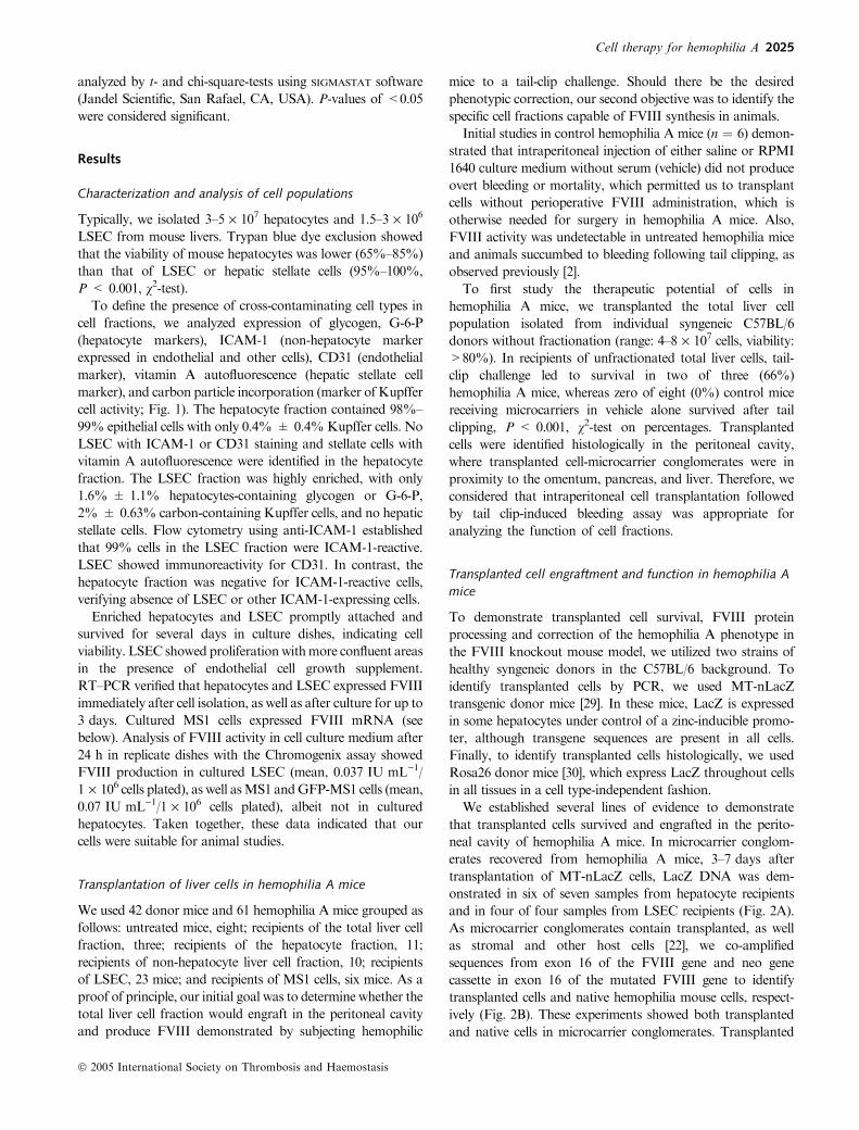
Characterization and analysis of cell populations
Typically, we isolated 3–5 · 107 hepatocytes and 1.5–3 · 106
LSEC from mouse livers. Trypan blue dye exclusion showed
that the viability of mouse hepatocytes was lower (65%–85%)
than that of LSEC or hepatic stellate cells (95%–100%,
P < 0.001, v2-test).To define the presence of cross-contaminating cell types in
cell fractions, we analyzed expression of glycogen, G-6-P
(hepatocyte markers), ICAM-1 (non-hepatocyte marker
expressed in endothelial and other cells), CD31 (endothelial
marker), vitamin A autofluorescence (hepatic stellate cell
marker), and carbon particle incorporation (marker of Kupffer
cell activity; Fig. 1). The hepatocyte fraction contained 98%–
99% epithelial cells with only 0.4% ± 0.4%Kupffer cells. No
LSEC with ICAM-1 or CD31 staining and stellate cells with
vitamin A autofluorescence were identified in the hepatocyte
fraction. The LSEC fraction was highly enriched, with only
1.6% ± 1.1% hepatocytes-containing glycogen or G-6-P,
2% ± 0.63% carbon-containing Kupffer cells, and no hepatic
stellate cells. Flow cytometry using anti-ICAM-1 established
that 99% cells in the LSEC fraction were ICAM-1-reactive.
LSEC showed immunoreactivity for CD31. In contrast, the
hepatocyte fraction was negative for ICAM-1-reactive cells,
verifying absence of LSEC or other ICAM-1-expressing cells.
Enriched hepatocytes and LSEC promptly attached and
survived for several days in culture dishes, indicating cell
viability. LSEC showed proliferationwithmore confluent areas
in the presence of endothelial cell growth supplement.
RT–PCR verified that hepatocytes and LSEC expressed FVIII
immediately after cell isolation, as well as after culture for up to
3 days. Cultured MS1 cells expressed FVIII mRNA (see
below). Analysis of FVIII activity in cell culture medium after
24 h in replicate dishes with the Chromogenix assay showed
FVIII production in cultured LSEC (mean, 0.037 IU mL)1/
1 · 106 cells plated), as well asMS1 andGFP-MS1 cells (mean,
0.07 IU mL)1/1 · 106 cells plated), albeit not in cultured
hepatocytes. Taken together, these data indicated that our
cells were suitable for animal studies.
Transplantation of liver cells in hemophilia A mice
We used 42 donor mice and 61 hemophilia A mice grouped as
follows: untreated mice, eight; recipients of the total liver cell
fraction, three; recipients of the hepatocyte fraction, 11;
recipients of non-hepatocyte liver cell fraction, 10; recipients
of LSEC, 23 mice; and recipients of MS1 cells, six mice. As a
proof of principle, our initial goal was to determine whether the
total liver cell fraction would engraft in the peritoneal cavity
and produce FVIII demonstrated by subjecting hemophilic
mice to a tail-clip challenge. Should there be the desired
phenotypic correction, our second objective was to identify the
specific cell fractions capable of FVIII synthesis in animals.
Initial studies in control hemophilia A mice (n ¼ 6) demon-
strated that intraperitoneal injection of either saline or RPMI
1640 culture medium without serum (vehicle) did not produce
overt bleeding or mortality, which permitted us to transplant
cells without perioperative FVIII administration, which is
otherwise needed for surgery in hemophilia A mice. Also,
FVIII activity was undetectable in untreated hemophilia mice
and animals succumbed to bleeding following tail clipping, as
observed previously [2].
To first study the therapeutic potential of cells in
hemophilia A mice, we transplanted the total liver cell
population isolated from individual syngeneic C57BL/6
donors without fractionation (range: 4–8 · 107 cells, viability:
>80%). In recipients of unfractionated total liver cells, tail-
clip challenge led to survival in two of three (66%)
hemophilia A mice, whereas zero of eight (0%) control mice
receiving microcarriers in vehicle alone survived after tail
clipping, P < 0.001, v2-test on percentages. Transplanted
cells were identified histologically in the peritoneal cavity,
where transplanted cell-microcarrier conglomerates were in
proximity to the omentum, pancreas, and liver. Therefore, we
considered that intraperitoneal cell transplantation followed
by tail clip-induced bleeding assay was appropriate for
analyzing the function of cell fractions.
Transplanted cell engraftment and function in hemophilia A
mice
To demonstrate transplanted cell survival, FVIII protein
processing and correction of the hemophilia A phenotype in
the FVIII knockout mouse model, we utilized two strains of
healthy syngeneic donors in the C57BL/6 background. To
identify transplanted cells by PCR, we used MT-nLacZ
transgenic donor mice [29]. In these mice, LacZ is expressed
in some hepatocytes under control of a zinc-inducible promo-
ter, although transgene sequences are present in all cells.
Finally, to identify transplanted cells histologically, we used
Rosa26 donor mice [30], which express LacZ throughout cells
in all tissues in a cell type-independent fashion.
We established several lines of evidence to demonstrate
that transplanted cells survived and engrafted in the perito-
neal cavity of hemophilia A mice. In microcarrier conglom-
erates recovered from hemophilia A mice, 3–7 days after
transplantation of MT-nLacZ cells, LacZ DNA was dem-
onstrated in six of seven samples from hepatocyte recipients
and in four of four samples from LSEC recipients (Fig. 2A).
As microcarrier conglomerates contain transplanted, as well
as stromal and other host cells [22], we co-amplified
sequences from exon 16 of the FVIII gene and neo gene
cassette in exon 16 of the mutated FVIII gene to identify
transplanted cells and native hemophilia mouse cells, respect-
ively (Fig. 2B). These experiments showed both transplanted
and native cells in microcarrier conglomerates. Transplanted
Cell therapy for hemophilia A 2025
� 2005 International Society on Thrombosis and Haemostasis

hepatocytes have previously been demonstrated to maintain
hepatic gene expression in the peritoneal cavity, including at
the mRNA and protein levels [22].
Histologic analysis of the recovered microcarriers provided
further evidence for transplanted cell survival (Fig. 3). Trans-
planted hepatocytes were readily identified on the basis of their
distinct morphology, with abundant eosinophilic cytoplasm,
rounded nuclei, and prominent nucleoli (Fig. 3A). Glycogen
staining provided additional morphologic evidence for engraft-
ment of transplanted hepatocytes (Fig. 3B). However, LSEC
are much smaller than hepatocytes (Fig. 1A,D), similar to
stromal cell types, and do not offer characteristic morphologic
features at the light microscopy level. However, use of Rosa26
cells was helpful in identifying transplanted cells in recipients of
both hepatocytes and LSEC (Fig. 3E,F). To verify that MS1
cells survived after transplantation in hemophilia A mice, we
G
g-a g-c
MW
mar
ker
Live
r
Hep
atoc
ytes
Non
-hep
atoc
ytes
Endo
thel
ial c
ells
PCR
mix
alo
ne
g-b g-d
0 200Forward scatter FITC fluorescence
Forward scatter
Sid
e sc
atte
rS
ide
scat
ter
Pro
pidi
um io
dide
Pro
pidi
um io
dide
Cel
l num
ber
FITC fluorescence
400 600 800 1000
0 200 400 600 800 1000 10 10 10 10 1043210
10 10 10 10 1043210
020
040
060
080
010
000
200
400
600
800
1000
H
1 2
PhaseICAM-1
3 4 5 6
100
101
102
103
104
100
101
102
103
104
A B C
D
LacZPhase Glycogen
E F
Fig. 1. Characterization of mouse liver cells. Panels A–C show primary hepatocytes and D–F primary liver sinusoidal endothelial cells (LSEC).
Hepatocytes showed larger size, contained glycogen andwere not contaminated with LSEC (A and B), as further verified by the absence of cells with CD31
staining. Rosa26 hepatocytes-expressing LacZ are shown after 3 days in culture (C). The LSEC fraction contained intercellular adhesion molecule
(ICAM-1-; D) and CD31 (E)-expressing cells, as seen under fluorescence microscopy with DAPI counterstaining of cells in panel (E). Inset in panel (D)
shows magnified view of LSEC with ICAM-1 staining. Panel (F) shows LSEC 3 days after culture with both single cells (arrowheads) and clusters of
proliferating cells (arrows). Flow cytometry for ICAM-1 in LSEC is shown in panel (G), where LSEC not incubated with ICAM-1 antibody (negative
controls) are at the top (G–A and G–C) and samples incubated with anti-ICAM-1 are at the bottom (G–B and G–D). Panels (G–A) and (G–C) show
forward scatter (cell size) and side scatter (cytoplasmic complexity) of the cell preparations, whichwere similar. Panels (G–G) and (G–D) show cell numbers
on the y-axis and fluorescein isothiocyanate (FITC) fluorescence, indicating intensity of ICAM-1 expression on the x-axis. Flow cytometry for the
hepatocyte fraction is not shown because these cells were negative for ICAM-1 staining, similar to panel (G–C). FVIII reverse transcription polymerase
chain reaction (RT–PCR) in cells immediately after isolation and intact liver is shown in panel (H) with a 500-bp size expected product. These data
verified that isolated cells were viable, enriched in hepatocytes and LSEC in respective fractions, and produced FVIII mRNA to proceed with analysis of
cell function in vivo. Original magnification: A–F, ·400; E is at higher magnification.
2026 V. Kumaran et al
� 2005 International Society on Thrombosis and Haemostasis

utilized GFP-transduced cells. The microcarrier conglomerates
recovered from hemophilia mice showed presence of trans-
planted MS1 cells with GFP expression (Fig. 3G–I). These
findings were in agreement with the survival and continued
function in transplanted hepatocytes and endothelial cells in the
peritoneal cavity.
Phenotypic correction in hemophilia A mice
We determined whether cell transplantation altered mortality
in hemophilia Amice following tail-clip challenge. As indicated
earlier, animals treated with onlymicrocarriers and vehicle died
within 6 h after tail clipping. Only one of six mice (17%) that
received hepatocytes from an entire liver, 3–5 · 107 viable cells
each, survived tail clipping (Table 1). In contrast, six of seven
animals (86%) transplanted with the non-hepatocyte cell
fraction, which contained LSEC, Kupffer cells, hepatic stellate
cells, and other cells, survived tail clipping (P ¼ 0.001).
Moreover, recipients of LSEC showed dose-dependent survival
following transplantation of 0.5 · 106 or more endothelial
cells. In some of these animals, plasma FVIII activity was
detected 7 days following cell transplantation, when animals
were subjected to tail-clip challenge, although sufficient plasma
samples were not available from all animals for FVIII assays.
To demonstrate whether transplanted cells secreted FVIII in
the blood stream, we studied additional animals 48 h following
cell transplantation (Fig. 4). In recipients of 3–4 · 107 hepato-
cytes (n ¼ 5), plasma FVIII activity was not detectable in any
of the mice. On the contrary, transplantation of the total non-
hepatocyte cell fraction showed FVIII activity in three of three
mice (1%, 2.4%, and 7.6%). Plasma FVIII activity was also
observed in recipients of 2 · 106 LSEC, where in three of five
mice, showed 1%, 2%, and 5.5% FVIII activity. In mice
receiving five times more MS1 cells (1 · 107 cells each), six of
six animals showed plasma FVIII activity in greater amounts
(6%, 8%, 8%, 10%, 12%, and 12% FVIII activity). In
contrast, FVIII activity was not detected in untreated hemo-
philia A mice serving as negative controls. Overall, these
findings were in agreement with the maintenance of secretory
function in endothelial cells and the ability of these cells to
replenish FVIII in hemophilia mice.
Discussion
In this study, we provide evidence regarding the liver cell type
that can produce and secrete FVIII in vivo. We used liver cell
fractions, including those enriched in either hepatocytes or
LSEC, to determine whether transplanted cells produced FVIII
protein in hemophilia mice. Donor cells with specific reporters
were used to demonstrate engraftment and function of
transplanted cells. Besides identifying LSEC, as well as MS1
cells derived from pancreatic islets, as capable of FVIII
synthesis, we established that primary LSEC corrected the
bleeding phenotype of hemophilia A mice, suggesting that
endothelial cells should be appropriate targets for FVIII cell
and gene therapy.
A
B
F8-/-
MT-
nLac
ZM
ix a
lone
1 2 3 4 5 6 87 9 10 11
1 2 3 4 5 6 87 9 10 11
400 bp
680 bp
420 bp
Hepatocyterecipients
Endothelical cellrecipients
F8-/-
F8+/
-
Mix
alo
ne
Hepatocyterecipients
Endothelical cellrecipients
Fig. 2. Transplanted cell survival in hemophilia A mice. Polymerase chain reaction (PCR) was used to identify transplanted cells with the LacZ transgene
(MT-nLacZ donors) (A) or wild-type FVIII sequences (B). Data are from animals 7 days after cell transplantation. Panel (A) shows a 400 bp band
expected in MT-nLacZ cells, lane 2; and its absence in hemophilia A mice (lane 1). LacZ sequences were present in DNA from recovered microcarrier-cell
conglomerates after transplantation of MT-nLacZ hepatocytes, lanes 4–7, and MT-nLacZ liver sinusoidal endothelial cells (LSEC), lanes 8–11. (B) PCR
for FVIII gene showed only mutant sequences (420 bp band) in FVIII)/) mouse, lane 1; and both mutant and wild-type bands in a FVIII+/)heterozygous mouse, lane 2. In MT-nLacZ cell recipients, both mutated and wild-type FVIII bands were amplified, indicating transplanted, as well as
native cells, in microcarrier conglomerates.
Cell therapy for hemophilia A 2027
� 2005 International Society on Thrombosis and Haemostasis

A
mc
B
D
mc
mc mc
F
mc
H
mc
mc
G
I
C
E
mc
J
1 2 3
MWMarker
-VeCtrl
MS1Cells
2028 V. Kumaran et al
� 2005 International Society on Thrombosis and Haemostasis

A major advantage of transplanting cells into the peritoneal
cavity was the capacity of this space to accommodate
functionally intact cells in large numbers. Previous studies
established that after transplantation of hepatocytes, secreted
proteins were detected within 30–60 min in the blood of
recipient mice [31]. Moreover, transplanted hepatocytes
engrafted in the peritoneal cavity in the presence of microcar-
riers, as shown here, and maintained liver gene expression, as
well as production of secreted proteins for more than 1 week,
as reported previously with rodent, as well as human hepato-
cytes [22,32,33]. These studies included in situ hybridization to
show maintenance of liver gene expression at the mRNA level,
as well as other assays to demonstrate protein expression in
transplanted hepatocytes (e.g. albumin, G-6-P, glycogen) in the
peritoneal cavity. Similarly, non-parenchymal liver cells have
been successfully transplanted into the peritoneal cavity ofmice
[34]. The characteristic morphology of hepatocytes and visu-
alization of liver gene expression permitted us to distinguish
between transplanted cells and native stromal cells in micro-
carrier conglomerates. In addition, our studies here with
LacZ-expressing Rosa26 cells and GFP-expressing MS1 cells
further verified that additional cell types engrafted in the
peritoneal cavity of recipient mice.
Our studies were restricted to the short-term because we first
wished to identify the most effective cell type capable of
producing FVIII. In the adult liver, hepatocytes constitute 60%
and LSEC 20%of the total liver cell mass, which approximates
100 million cells in the mouse [31]. Transplantation of hepato-
cytes in the native liver leads to engraftment of only 20% or
fewer transplanted cells, because of the requirement of
translocation from hepatic sinusoids into the liver plate
structure, with liver reconstitution by, at most, 1%–2%, after
one session of cell transplantation [31]. In contrast, cells
transplanted in the peritoneal cavity are not subject to
clearance because of the restriction imposed by the requirement
for entry into the liver parenchyma.
Transplantation of 20–35 million hepatocytes represented
reconstitution of 30%–60% of the hepatocyte mass in hemo-
philia mice. We observed sheets of transplanted hepatocytes in
the peritoneal cavity. These transplanted hepatocytes are
capable of secreting soluble proteins into the bloodstream, as
shown by previous studies using transgenic hepatitis B virus
140
120
100
80
60
40
20
0n 8 5 3 5 6
MS-1LSECAll livercells
Heps
FVIII activity present
FVIII activity absent
Cell-type transplanted
Frac
tion
of h
emop
hilia
A m
ice
with
FV
III a
ctiv
ity (
% tr
eate
d)
Vehiclealone
Fig. 4. Analysis of plasma FVIII activity in treated hemophilia A mice.
The data show the fraction of animals in various groups with reconstitu-
tion of plasma FVIII activity. Plasma FVIII activity was absent in animals
treated with either vehicle alone or the hepatocyte fraction. In contrast,
100% of the animals treated with all liver cells or MILE SVEN 1 (MS1)
cells showed plasma FVIII activity. In recipients of the liver sinusoidal
endothelial cells (LSEC) fraction, plasma FVIII activity was identified in
three of five animals. The animal number in various groups is indicated.
Asterisks represent P < 0.001, using percentages, v2-test.
Table 1 Phenotypic correction of hemophilia A mice
Intervention Number of mice Survival after tail clip, n (%) P-value (v2-test)�
Vehicle (RPMI 1640 culture medium) 8 0 NA
Total liver cell fraction (3–5 · 107 cells; 60%–65% hepatocytes,
10%–15% LSEC, 2%–4% hepatic stellate cells)
3 2 (67) NA
Hepatocyte fraction (2–3.5 · 107 cells) 6 1 (17) ns
Non-hepatocyte fraction (1–2 · 107 LSEC,
hepatic stellate cells, Kupffer cells, etc.)
7 6 (86) <0.001
LSEC fraction
0.25 · 106 LSEC 6 1 (17) ns
0.5–1 · 106 LSEC 12 8 (67) <0.001
ns, non-significant; NA, not applied; LSEC, liver sinusoidal endothelial cells.�Tested on percentages vs. vehicle alone.
Fig. 3. Identification of transplanted cells in tissues. Seven days after transplantation, transplanted hepatocytes are shown with characteristic morphology
(A), including glycogen (B) in areas adjacent to microcarriers (mc) in the peritoneal cavity of hemophilia mice. (C) LacZ staining of donor Rosa26 liver
demonstrating cytoplasmic blue staining of all cells, including sinusoidal cells (inset of boxed area). LacZ staining was absent in recipients of microcarriers
alone (D). In contrast, 3 days after transplantation of Rosa26 hepatocytes (E) or liver sinusoidal endothelial cells (LSEC; F), LacZ-positive cells (arrows)
were identified in proximity to microcarriers. Panels (G–I) show green fluorescent protein (GFP)-expressing MILE SVEN 1 (MS1) cells 2 days after
transplantationwithDAPI counterstain alone (G), GFP-immunostaining (H), andmerged view (I). Panel (J) shows reverse transcription polymerase chain
reaction (RT–PCR) indicating FVIIImRNAexpression in culturedMS1 cells. Originalmagnification: A, ·200; B–I, ·400; C–F, hematoxylin counterstain.
Cell therapy for hemophilia A 2029
� 2005 International Society on Thrombosis and Haemostasis

(HBV) hepatocytes [22,32], where secreted proteins rapidly
appeared in the circulation and persisted for a few weeks.
Nonetheless, despite transplantation of hepatocytes in large
numbers and their known capacity to release secreted proteins
in the bloodstream, we failed to demonstrate phenotypic
correction or plasma FVIII activity in hemophilia A mice.
Previous studies showed that blood loss and survival in
hemophilia A mice after tail-clip challenge correlates with
therapeutic reconstitution of plasma FVIII activity [35].
However, whether hepatocytes could produce FVIII in the
liver microenvironment compared with their inability to do this
in the peritoneal cavity will be best determined by studies of
liver repopulation in hemophilia mice. Such studies will be
facilitated by the development of effective strategies to repop-
ulate the healthy mouse liver, similar to the success in
repopulating the rat liver with transplanted hepatocytes after
genotoxic perturbations of the native liver [36]. Liver repop-
ulation studies will help further exclude whether hepatocytes
could possibly reconstitute FVIII activity. Similarly, long-term
or indefinite correction of FVIII deficiency in hemophilia A
mice will benefit from further strategies, e.g. transplanted
hepatocytes survive lifelong in the rat liver [37], and whether
transplanted endothelial cells could repopulate the liver should
be of interest.
Our studies with endothelial cells, especially MS1 cells,
established that cells transplanted into the peritoneal cavity
secreted FVIII into the bloodstream. Transplantation of
fractions containing all liver cells, mixture of non-hepatocyte
cells, and LSEC ameliorated the hemophilia A phenotype. The
level of plasma FVIII activity in recipients of 0.5–1 million
LSEC was commensurate with the cell number transplanted,
which represented 2.5%–5% of the mouse LSEC mass.
Although plasma FVIII activity in hemophilia mice treated
with primary LSEC was in the low range of detection by the
Chromogenix assay, whether unidentified differences in cell
viabilitymight have interfered with the engraftment or function
of transplanted primary LSEC was not excluded. These
limitations were avoided in our studies with MS1 cells, where
transplantation of 10 million MS1 cells reconstituted up to
12% plasma FVIII activity, which should again be reasonable,
because not all cells would necessarily have survived or
functioned equally in the peritoneal cavity after transplanta-
tion. Also, FVIII production in MS1 should be noteworthy,
considering their origin from pancreatic islets, and was in
agreement with the potential of extrahepatic endothelial cells.
Moreover, in our assays, endothelial cells were more effective
than hepatocytes in producing and secreting FVIII, despite
transplantation of the latter in much larger numbers.
Physiologically, LSEC offer advantages for FVIII expres-
sion, including presence of intracellular mechanisms that
facilitate FVIII synthesis, maturation, and release. For
instance, complexing of FVIII to von Willebrand factor
(VWF), which is produced in endothelial cells and megak-
aryocytes, with storage in Weibel-Palade bodies and
a-granules, respectively, helps concentrate FVIII in sites of
vascular injury and prevents premature proteolysis [38].
Alterations in the intracellular trafficking of VWF may
perturb subcellular FVIII localization [39]. Similarly, mul-
tiple extracellular stimuli, including inflammatory mediators
and vasoactive drugs, such as desmopressin, promote release
of FVIII and VWF from intracellular stores [40]. The
significant role of VWF in FVIII stabilization was further
verified in studies of cultured human hepatocytes, where
FVIII antigen expression was not detected without the
addition of VWF to the culture medium [41]. This is in
agreement with our findings here showing the inability of
transplanted mouse hepatocytes to reconstitute plasma
FVIII activity. Whether the additional provision of VWF
to transplanted hepatocytes could be effective for this
purpose will require suitable in vivo systems, e.g. examina-
tion of liver repopulation with transplanted hepatocytes,
where release of FVIII from transplanted cells and of VWF
released from LSEC within proximity to hepatocytes could
bring FVIII and VWF together.
The liver is clearly not an exclusive source of FVIII, as
endothelial cells derived from additional sources can also
produce FVIII [16,19,42], which will be in agreement with
the efficacy of MS1 cells, and possibly accounted for
relatively lower reconstitution of FVIII activity expected
from the mass of LSEC transplanted in our studies here. In
recent studies, endothelial cells isolated from lungs were
found to engraft in pulmonary tissues [43]. Similarly,
circulating endothelial cells engraft in diseased vessels [44].
Also, hematopoietic stem cells derived from mononuclear
blood cells can produce FVIII [42]. Moreover, hematopoi-
etic stem cells can differentiate into LSEC [45], which
should offer further opportunities for therapeutic targeting
of specific cell subsets in hemophilia A.
Acknowledgements
The authors thank Ms Chaoying Zhang for excellent technical
assistance. Supported in part by NIH grants R01 DK46952
and P30-DK-41296.
References
1 Bolton-Maggs PH, Pasi KJ. Hemophilias A and B. Lancet 2003; 361:
1801–9.
2 Bi L, Sarkar R, Naas T, Lawler AM, Pain J, Shumaker SL, Bedian V,
KazazianHH Jr. Further characterization of factor VIII-deficient mice
created by gene targeting: RNA and protein studies. Blood 1996; 88:
3446–50.
3 ChuahMK, SchiednerG,Thorrez L, BrownB, JohnstonM,GillijnsV,
Hertel S, Van Rooijen N, Lillicrap D, Collen D, VandenDriessche T,
Kochanek S. Therapeutic factor VIII levels and negligible toxicity in
mouse and dog models of hemophilia A following gene therapy with
high-capacity adenoviral vectors. Blood 2003; 101: 1734–43.
4 Kootstra NA, Matsumura R, Verma IM. Efficient production of
human FVIII in hemophilic mice using lentiviral vectors. Mol Ther
2003; 7: 623–31.
5 Chao H,Mansfield SG, Bartel RC, Hiriyanna S, Mitchell LG, Garcia-
BlancoMA,Walsh CE. Phenotype correction of hemophilia Amice by
spliceosome-mediated RNA trans-splicing. Nat Med 2003; 9: 1015–9.
2030 V. Kumaran et al
� 2005 International Society on Thrombosis and Haemostasis

6 Grimm D, Zhou S, Nakai H, Thomas CE, Storm TA, Fuess S,
Matsushita T, Allen J, Surosky R, Lochrie M, Meuse L, McClelland
A, Colosi P, Kay MA. Preclinical in vivo evaluation of pseudotyped
adeno-associated virus vectors for liver gene therapy. Blood 2003; 102:
2412–9.
7 Sarkar R, Xiao W, Kazazian HH Jr. A single adeno-associated virus
(AAV)-murine factor VIII vector partially corrects the hemophilia A
phenotype. J Thromb Haemost 2003; 1: 220–6.
8 MahC, Sarkar R, Zolotukhin I, SchleissingM, XiaoX, KazazianHH,
Byrne BJ. Dual vectors expressingmurine factorVIII result in sustained
correction of hemophilia A mice.Hum Gene Ther 2003; 14: 143–52.
9 Sarkar R, Tetreault R, Gao G, Wang L, Bell P, Chandler R, Wilson
JM, Kazazian HH Jr. Total correction of hemophilia A mice with
canine FVIII using an AAV 8 serotype. Blood 2004; 103: 1253–60.
10 Bontempo FA, Lewis JH, Gorenc TJ, Spero JA, Ragni MV, Scott JP,
Starzl TE. Liver transplantation in hemophilia A. Blood 1987; 69:
1721–4.
11 Gordon FH, Mistry PK, Sabin CA, Lee CA. Outcome of orthotopic
liver transplantation in patients with haemophilia.Gut 1998; 42: 744–9.
12 Groth CG, Hathaway WE, Gustafsson A, Geis WP, Putnam CW,
Bjorken C, Porter KA, Starzl TE. Correction of coagulation in the
hemophilic dog by transplantation of lymphatic tissue. Surgery 1974;
75: 725–33.
13 Veltkamp JJ, Asfaou E, van de Torren K, van der Does JA,
van Tilburg NH, Pauwels EK. Exrahepatic factor VII synthesis: lung
transplants in hemophilic dogs. Transplantation 1974; 18: 56–62.
14 Liu L, Xia S, Seifert J. Transplantation of spleen cells in patients with
hemophilia A: a report of 20 cases. Transplant Int 1994; 7: 201–6.
15 LiuL,XiaS,Tang J,QinX,LiuH.Allotransplantation of whole spleen
in patients with hepatic malignant tumors or hemophilia A. Operative
technique and preliminary results. Arch Surg 1995; 130: 33–9.
16 Wion KL, Kelly D, Summerfield JA, Tuddenham EGD, Lawn RM.
Distribution of factor VIII mRNA and antigen in human liver and
other tissues. Nature 1985; 317: 726–9.
17 Zelechowska MG, van Mourik JA, Brodniewicz-Proba T. Ultra-
structural localization of factor VIII procoagulant antigen in human
liver hepatocytes. Nature 1985; 317: 729–30.
18 Do H, Healey JF, Waller EK, Lollar P. Expression of factor VIII by
murine liver sinusoidal endothelial cells. J Biol Chem 1999; 274: 19587–
92.
19 HollestelleMJ, Thinnes T, Crain K, Stiko A, Kruijt JK, van Berkel TJ,
Loskutoff DJ, van Mourik JA. Tissue distribution of factor VIII gene
expression in vivo – a closer look. Thromb Haemost 2001; 86: 855–61.
20 Langley PG, Hughes RD, Williams R. Increased factor VIII complex
in fulminant hepatic failure. Thromb Hemost 1985; 54: 693–6.
21 Doering CB, Josephson CD, Craddock HN, Lollar P. Factor VIII
expression in azoxymethane-induced murine fulminant hepatic failure.
Blood 2002; 100: 143–7.
22 Gupta S, Vemuru RP, Lee C-D, Yerneni P, Aragona E, Burk RD.
Hepatocytes exhibit superior transgene expression after transplantation
into liver and spleen compared with peritoneal cavity or dorsal fat pad:
implications for hepatic gene therapy.HumGene Ther 1994; 5: 959–67.
23 Joseph B,Malhi H, Bhargava KK, Palestro CJ,McCuskey RS, Gupta
S. Kupffer cells participate in early clearance of syngeneic hepatocytes
transplanted in the rat liver. Gastroenterology 2002; 123: 1677–85.
24 Smedsrod B, Pertoft H, Eggertsen G, Sundstrom C. Functional and
morphological characterization of cultures ofKupffer cells prepared by
means of density separation in Percoll and selective substrate adher-
ence. Cell Tissue Res 1985; 241: 639–749.
25 Braet F, De Zanger R, Sasaoki T, BaekelandM, Janssens P, Smedsrod
B, Wisse E. Assessment of a method of isolation, purification and
cultivation of rat liver sinusoidal endothelial cells. Lab Invest 1994; 70:
944–52.
26 Arbiser JL, Moses MA, Fernandez CA, Ghiso N, Cao Y, Klauber N,
Frank D, Brownlee M, Flynn E, Parangi S, Byers HR, Folkman J.
Oncogenic H-ras stimulates tumor angiogenesis by two distinct path-
ways. Proc Natl Acad Sci U S A 1997; 94: 861–6.
27 Follenzi A, Naldini L. Generation of HIV-derived lentiviral vectors.
Methods Enzymol 2002; 346: 454–65.
28 Ott M, Rajvanshi P, Sokhi R, Alpini G, Aragona E, Dabeva M,
Shafritz DA, Gupta S. Differentiation-specific regulation of transgene
expression in a diploid epithelial cell line derived from the normal F344
rat liver. J Pathol 1999; 187: 365–73.
29 Braun KM, Sandgren EP. Cellular origin of regenerating parenchyma
in amouse model of severe hepatic injury.Am J Pathol 2000; 157: 561–
9.
30 Zambrowicz BP, Imamoto A, Fiering S, Herzenberg LA, Kerr WG,
Soriano P.Disruption of overlapping transcripts in the ROSAbeta geo
26 gene trap strain leads to widespread expression of beta-galactosi-
dase in mouse embryos and hematopoietic cells. Proc Natl Acad Sci
U S A 1997; 94: 3789–94.
31 Rajvanshi P, Kerr A, Bhargava KK, Burk RD, Gupta S. Studies of
liver repopulation using the dipeptidyl peptidase IV deficient rat and
other rodent recipients: cell size and structure relationships regulate
capacity for increased transplanted hepatocyte mass in the liver lobule.
Hepatology 1996; 23: 482–96.
32 Demetriou AA, Whiting JF, Feldman D, Levenson SM, Chowdhury
NR, Moscioni AD, Kram M, Chowdhury JR. Replacement of liver
function in rats by transplantation of microcarrier-attached hepato-
cytes. Science 1986; 233: 1190–2.
33 Cho J, Joseph B, Sappal BS, Giri RK, Wang R, Ludlow J, Susick R,
Gupta S. Analysis of the functional integrity of cryopreserved human
liver cells including xenografting in immunodeficient mice to address
suitability for clinical applications. Liver Int 2004; 24: 361–70.
34 Selden C, Calnan D, Morgan N, Wilcox H, Carr E, Hodgson HJ.
Histidinemia inmice: ametabolic defect treated using a novel approach
to hepatocellular transplantation.Hepatology 1995; 21: 1405–12.
35 Parker RT, Lollar P. A quantitative measure of the efficacy of factor
VIII in hemophilia A mice. Thromb Haemost 2003; 89: 480–5.
36 Gupta S, Inada M, Joseph B, Kumaran V, Benten D. Emerging
insights into liver-directed cell therapy for genetic and acquired
disorders. Transplant Immunol 2004; 12: 289–302.
37 Sokhi RP, Rajvanshi P, Gupta S. Transplanted reporter cells help in
defining onset of hepatocyte proliferation during the life of F344 rats.
Am J Physiol Gastrointest Liver Physiol 2000; 279: G631–40.
38 Wagner DD. The Weibel-Palade body: the storage granule for von
Willebrand factor and P-selectin. Thromb Haemost 1993; 70: 105–10.
39 Rosenberg JB, Foster PA, Kaufman RJ, Vokac EA, Moussalli M,
Kroner PA, Montgomery RR. Intracellular trafficking of factor VIII
to vonWillebrand factor storage granules. J Clin Invest 1998; 101: 613–
24.
40 Montgomery RR, Gill JC. Interactions between vonWillebrand factor
and factor VIII: where did they first meet? J Pediatr Hematol Oncol
2000; 22: 269–75.
41 Biron-Andreani C, Bezat-Bouchahda C, Raulet E, Pichard-Garcia L,
Fabre JM, Saric J, Baulieux J, Schved JF, Maurel P. Secretion of
functional plasma haemostasis proteins in long-term primary cultures
of human hepatocytes. Br J Haematol 2004; 125: 638–46.
42 Lin Y, Chang L, Solovey A, Healey JF, Lollar P, Hebbel RP. Use of
blood outgrowth endothelial cells for gene therapy for hemophilia A.
Blood 2002; 99: 457–62.
43 Ewing P, Wilke A, Brockhoff G, Andreesen R, Eissner G, Holler E,
Gerbitz A. Isolation and transplantation of allogeneic pulmonary
endothelium derived from GFP transgenic mice. J Immunol Methods
2003; 283: 307–15.
44 Griese DP, Ehsan A, Melo LG, Kong D, Zhang L, Mann MJ, Pratt
RE, Mulligan RC, Dzau VJ. Isolation and transplantation of autol-
ogous circulating endothelial cells into denuded vessels and prosthetic
grafts: implications for cell-based vascular therapy. Circulation 2003;
108: 2710–5.
45 Bailey AS, Jiang S, Afentoulis M, Baumann CI, Schroeder DA, Olson
SB,WongMH,FlemingWH. Transplanted adult hematopoietic stems
cells differentiate into functional endothelial cells. Blood 2004; 103:
13–9.
Cell therapy for hemophilia A 2031
� 2005 International Society on Thrombosis and Haemostasis