Computational investigation of epithelial cell dynamic phenotype in vitro
21
BioMed Central Page 1 of 21 (page number not for citation purposes) Theoretical Biology and Medical Modelling Open Access Research Computational investigation of epithelial cell dynamic phenotype in vitro Sean HJ Kim 1 , Sunwoo Park 2 , Keith Mostov 3 , Jayanta Debnath 4 and C Anthony Hunt* 1,2 Address: 1 UCSF/UC Berkeley Joint Graduate Group in Bioengineering, University of California, Berkeley, California 94720, USA, 2 Department of Bioengineering and Therapeutic Sciences, University of California, San Francisco, California 94143, USA, 3 Department of Anatomy, University of California, San Francisco, California 94143, USA and 4 Department of Pathology, University of California, San Francisco, California 94143, USA Email: Sean HJ Kim - [email protected]; Sunwoo Park - [email protected]; Keith Mostov - [email protected]; Jayanta Debnath - [email protected]; C Anthony Hunt* - [email protected] * Corresponding author Abstract Background: When grown in three-dimensional (3D) cultures, epithelial cells typically form cystic organoids that recapitulate cardinal features of in vivo epithelial structures. Characterizing essential cell actions and their roles, which constitute the system's dynamic phenotype, is critical to gaining deeper insight into the cystogenesis phenomena. Methods: Starting with an earlier in silico epithelial analogue (ISEA1) that validated for several Madin-Darby canine kidney (MDCK) epithelial cell culture attributes, we built a revised analogue (ISEA2) to increase overlap between analogue and cell culture traits. Both analogues used agent- based, discrete event methods. A set of axioms determined ISEA behaviors; together, they specified the analogue's operating principles. A new experimentation framework enabled tracking relative axiom use and roles during simulated cystogenesis along with establishment of the consequences of their disruption. Results: ISEA2 consistently produced convex cystic structures in a simulated embedded culture. Axiom use measures provided detailed descriptions of the analogue's dynamic phenotype. Dysregulating key cell death and division axioms led to disorganized structures. Adhering to either axiom less than 80% of the time caused ISEA1 to form easily identified morphological changes. ISEA2 was more robust to identical dysregulation. Both dysregulated analogues exhibited characteristics that resembled those associated with an in vitro model of early glandular epithelial cancer. Conclusion: We documented the causal chains of events, and their relative roles, responsible for simulated cystogenesis. The results stand as an early hypothesis–a theory–of how individual MDCK cell actions give rise to consistently roundish, cystic organoids. Published: 28 May 2009 Theoretical Biology and Medical Modelling 2009, 6:8 doi:10.1186/1742-4682-6-8 Received: 8 March 2009 Accepted: 28 May 2009 This article is available from: http://www.tbiomed.com/content/6/1/8 © 2009 Kim et al; licensee BioMed Central Ltd. This is an Open Access article distributed under the terms of the Creative Commons Attribution License (http://creativecommons.org/licenses/by/2.0 ), which permits unrestricted use, distribution, and reproduction in any medium, provided the original work is properly cited.
-
Upload
independent -
Category
Documents
-
view
0 -
download
0
Transcript of Computational investigation of epithelial cell dynamic phenotype in vitro

BioMed Central
Theoretical Biology and Medical Modelling
ss
Open AcceResearchComputational investigation of epithelial cell dynamic phenotype in vitroSean HJ Kim1, Sunwoo Park2, Keith Mostov3, Jayanta Debnath4 and C Anthony Hunt*1,2Address: 1UCSF/UC Berkeley Joint Graduate Group in Bioengineering, University of California, Berkeley, California 94720, USA, 2Department of Bioengineering and Therapeutic Sciences, University of California, San Francisco, California 94143, USA, 3Department of Anatomy, University of California, San Francisco, California 94143, USA and 4Department of Pathology, University of California, San Francisco, California 94143, USA
Email: Sean HJ Kim - [email protected]; Sunwoo Park - [email protected]; Keith Mostov - [email protected]; Jayanta Debnath - [email protected]; C Anthony Hunt* - [email protected]
* Corresponding author
AbstractBackground: When grown in three-dimensional (3D) cultures, epithelial cells typically form cysticorganoids that recapitulate cardinal features of in vivo epithelial structures. Characterizing essentialcell actions and their roles, which constitute the system's dynamic phenotype, is critical to gainingdeeper insight into the cystogenesis phenomena.
Methods: Starting with an earlier in silico epithelial analogue (ISEA1) that validated for severalMadin-Darby canine kidney (MDCK) epithelial cell culture attributes, we built a revised analogue(ISEA2) to increase overlap between analogue and cell culture traits. Both analogues used agent-based, discrete event methods. A set of axioms determined ISEA behaviors; together, they specifiedthe analogue's operating principles. A new experimentation framework enabled tracking relativeaxiom use and roles during simulated cystogenesis along with establishment of the consequencesof their disruption.
Results: ISEA2 consistently produced convex cystic structures in a simulated embedded culture.Axiom use measures provided detailed descriptions of the analogue's dynamic phenotype.Dysregulating key cell death and division axioms led to disorganized structures. Adhering to eitheraxiom less than 80% of the time caused ISEA1 to form easily identified morphological changes.ISEA2 was more robust to identical dysregulation. Both dysregulated analogues exhibitedcharacteristics that resembled those associated with an in vitro model of early glandular epithelialcancer.
Conclusion: We documented the causal chains of events, and their relative roles, responsible forsimulated cystogenesis. The results stand as an early hypothesis–a theory–of how individual MDCKcell actions give rise to consistently roundish, cystic organoids.
Published: 28 May 2009
Theoretical Biology and Medical Modelling 2009, 6:8 doi:10.1186/1742-4682-6-8
Received: 8 March 2009Accepted: 28 May 2009
This article is available from: http://www.tbiomed.com/content/6/1/8
© 2009 Kim et al; licensee BioMed Central Ltd. This is an Open Access article distributed under the terms of the Creative Commons Attribution License (http://creativecommons.org/licenses/by/2.0), which permits unrestricted use, distribution, and reproduction in any medium, provided the original work is properly cited.
Page 1 of 21(page number not for citation purposes)

Theoretical Biology and Medical Modelling 2009, 6:8 http://www.tbiomed.com/content/6/1/8
BackgroundHow single cells proliferate and organize into liquid filledcysts, or acini, is a central question in epithelial morpho-genesis and cancer research. Epithelial cells in tissuesengage an array of activities to attain acinar structures [1].The same is true in cultures. When grown embedded in3D culture, epithelial cells such as Madin-Darby caninekidney (MDCK) cells develop stereotypical cystic orga-noids by mechanisms that can differ depending on cultureconditions [2]. When manipulated or exposed to certainfactors, these organoids and composing cells can exhibitphenotypic attributes that are reminiscent of pre-cancer-ous or cancerous tissues [3]. While MDCK culture modelsare orders of magnitude simpler than epithelial cells in tis-sues, they provide an appropriate physiological environ-ment to study epithelial cyst development, function, andpathology. However, they too are complex dynamic sys-tems that have proven challenging to understand.
The emergence of stable organoid structures is the cumu-lative consequence of individual cell actions: the system'sdynamic phenotype. Disruption of one or more of theseactions can cause potentially pathologic changes. Little isknown about the varying cell mechanisms and activitiesthat engage in different stages of cystogenesis and howthey contribute to the process. A strategy to understandingthe phenomena must include classifying those essentialcell actions and tracing their relative use and roles as theprocess unfolds. With time-lapse, microscopy imagesalone, it can be difficult to ascertain what cell actions areresponsible for the observed structure transformations.
Computational methods detailed herein represent anadditional, synergistic approach to gain the much-neededinsight. The approach used [4-6] is an example of execut-able biology [7,8]. We used in silico epithelial analogues(ISEAs) that have undergone validation against a targetedset of MDCK epithelial cell attributes. As discussed in [9],the attributes targeted by the earlier analogue (ISEA1)were selected to reflect essential MDCK cell behaviors incultures but for simplicity, the list excluded other MDCKattributes. Our goal was to improve ISEA1 in stages toachieve increased phenotype overlap between the revisedanalogue (ISEA2) and MDCK cell cultures. To keepimprovement parsimonious, we expanded the original listby one additional attribute: all stable cyst structures musthave a convex contour without irregular margins or dim-ples. Unlike its referent, ISEA1 frequently produced cyststructures having irregular shapes. Through exploratorysimulations discussed below, we discovered and addedone new cell action to achieve the additional attribute.The mappings from in silico components, their spatialarrangement, their mechanisms of interactions, and sys-tem-level attributes to their in vitro counterparts (Figure1) improved following that refinement.
Cell biologists compare and contrast the growth charac-teristics of different, related epithelial cell lines in part tobetter understand how and where their behaviors differ orare similar. That knowledge can be used to make betterinferences about referent cell behaviors in vivo. A provenwet-lab approach is to design and conduct experiments totest hypotheses about cell line responses to interventions,such as blocking a signaling pathway or a cell surfacereceptor. Analogous methods must be used to study andcompare phenotypic attributes of in silico analogues, suchas ISEA1 and ISEA2. In addition, study of analogueresponses to interventions improves insight into MDCKmorphogenesis. Differences in morphological anddynamic phenotype, or lack thereof, between two ana-logues could shed additional insight on those of the refer-ent [10]. With that in mind, we compared ISEA1 andISEA2 behaviors to understand how specific mechanisticchanges alter their morphogenetic attributes.
ISEA1 and ISEA2 used sets of rules in the form of axiomsfor determining CELL action based on CELL neighbor typeand configuration. Each simulation cycle, each CELL
assessed the current arrangement of neighbors, selectedthe corresponding axiom, and then executed that axiom'saction. By adhering strictly to their axioms, both ana-logues achieved their respective set of targeted attributes.Are actions of MDCK cells in cultures (and epithelial cellsin general) so rigidly choreographed? How tightly mustISEA adhere to its operating principles before aspects ofphenotype become measurably abnormal? We gainedinsight into plausible answers by systematically relaxingtwo, key ISEA actions and exploring in detail the pheno-typic consequences. One action mapped to anoikis, a spe-cific category of cell death. The other involved directedplacement of an ISEA daughter cell, a form of oriented celldivision. The ISEA1 phenotype was quite sensitive to dys-regulating the two actions: engaging in either action lessthan 80% of the time caused easily detected phenotypicchanges. Interestingly, ISEA2 was more robust to identicaldisruptions. Both ISEA1 and ISEA2 exhibited phenotypesthat resembled those associated with an in vitro model ofearly glandular epithelial cancer. To the extent that the insilico-to-in vitro mappings in Figure 2 are valid, ISEA2'soperating principles and dynamic phenotype stand ashypotheses of their MDCK counterparts in cell culture.
MethodsIn vitro cell culture experimentsFull details of the original MDCK cell culture experimentsare provided in [11]. Briefly, MDCK cells were trituratedinto single-cell suspensions in type I collagen gel. Cellswere grown for 7–10 d until cysts with lumina formed. Forimmunofluorescence staining of cysts, samples were incu-bated with primary antibodies overnight, followed by anovernight incubation with fluorescent dye-labeled sec-
Page 2 of 21(page number not for citation purposes)

Theoretical Biology and Medical Modelling 2009, 6:8 http://www.tbiomed.com/content/6/1/8
Page 3 of 21(page number not for citation purposes)
Relationships between analogues and MDCK culturesFigure 1Relationships between analogues and MDCK cultures. To distinguish simulation components and characteristics from in vitro counterparts, we use small caps when referring to the former. An in silico epithelial analogue (ISEA) is comprised of autonomous CELL components interacting with adjacent CELLS and environment components. Interactions are governed by a set of axiomatic operating principles (rules). For each environment circumstance a CELL can encounter, there is a correspond-ing axiom. A clear mapping exists between ISEA components (CELL states and environment components) and in vitro counter-parts. Following execution, interacting components cause local and systemic behaviors. Measures of CELL and system behaviors (growth rates, structure type, etc.) are the in silico attributes. Validation occurs when a set of ISEA attributes is measurably similar to a corresponding, prespecified set of in vitro attributes. Upon validation, we can hypothesize that a semiquantitative mapping exists between ISEA events and in vitro events, and that the set of in silico operating principles has a biological coun-terpart.

Theoretical Biology and Medical Modelling 2009, 6:8 http://www.tbiomed.com/content/6/1/8
Figure 2 (see legend on next page)
Page 4 of 21(page number not for citation purposes)

Theoretical Biology and Medical Modelling 2009, 6:8 http://www.tbiomed.com/content/6/1/8
ondary antibodies. To quantitate cyst polarity, cysts werestained for gp135 (apical surface), β-catenin (basolateralsurface) and nuclei, and then visualized using a confocalmicroscope.
In silico experimentation frameworkISEA1 and ISEA2 are discrete event [12], agent-based [13]systems that comprise the core analogue and system-levelcomponents for experimentation and analysis (Figure 2).Because ISEA2 is based on ISEA1, both share a commondesign, and their experiment features overlap significantly(discussed below). Before moving forward with modelrefinement and experimentation, implementation redun-dancies of ISEA1 and ISEA2 were removed. We revised theexisting framework to enable simulation of multiple,somewhat different CELL analogue types. ISEA1 wasported and revalidated within the new framework prior toISEA2 development. To clearly distinguish ISEA compo-nents and processes from their in vitro counterparts, here-after we use small caps when referring the former.
We created system-level components including EXPERI-
MENT MANAGER, OBSERVER, and CULTURE graphical userinterface (GUI) to enable semi-automated experimenta-tion and analysis. EXPERIMENT MANAGER, the top-level sys-tem component, is an agent that provides experimentprotocol functions and specifications. The specificationsdefine the mode of experimentation and the system'sparameter vector. Experiments can be conducted indefault, visual, or batch modes. Batch mode enables auto-matic construction and execution of multiple experi-ments, as well as processing and analysis of recordedmeasurements. Based on user-defined specifications,EXPERIMENT MANAGER automatically generates a set ofparameter files and executes a batch of experiments, eachcorresponding to a different parameter file. After comple-tion of all experiments, basic analytic operations collectand summarize data. OBSERVER is responsible primarilyfor recording measurements. At the end of every simula-tion cycle, OBSERVER scans the CULTURE internals and per-forms measurements. The measurements are recorded as
time series vectors. At simulation's end, data are written toa set of files for analytic processing by EXPERIMENT MAN-
AGER. CULTURE GUI provides a visualization console,which can be used interactively to start or pause a simula-tion and to access live states of CULTURE grid content.Using CULTURE GUI functionalities, OBSERVER can capturetime-lapse CULTURE images and store them in multiple for-mats for post-processing.
ISEA1 and ISEA2 designs are agent-based and object-orientedDetailed descriptions of ISEA1 design features, and devel-opment methods, are available in [9]. ISEA2 design usessimilar features, which have been refined to meet studyrequirements. An abridged description follows. The refer-ent in vitro cell culture was conceptually abstracted intofour components: cells, media containing matrix (matrixhereafter), matrix-free media (free space hereafter), and aspace to contain them. Discrete software objects witheponymous names represent those four essential cell cul-ture components: CELL, MATRIX, FREE SPACE, and CULTURE.MATRIX and FREE SPACE are passive objects. A MATRIX objectmaps to a cell-sized volume of extracellular matrix (ECM).A FREE SPACE object maps to a similarly sized volume ofmaterial that is essentially free of cells and matrix ele-ments. FREE SPACE also represents luminal space and non-matrix material in pockets enclosed by cells. The latter arecalled LUMINAL SPACE when distinction from FREE SPACE isuseful. CELLS are quasi-autonomous agents (as agents,they can schedule their own events; they follow their ownagenda). They use a set of rules or decision logic to interactwith their local environment. A CULTURE is an agent thatmaps abstractly to a cell culture within one well of a multi-well culture plate. The CULTURE uses a standard two-dimensional (2D) hexagonal grid to provide the space inwhich its objects reside. The grid has toroidal topologies.For simplicity, each grid position is occupied by oneobject. That condition can be changed when the needarises.
ISEA components and system architectureFigure 2 (see previous page)ISEA components and system architecture. The in silico system consists of a CULTURE and framework components. MDCK cell cultures and ISEAs are both composite systems. A CULTURE represents one in vitro cell culture. It is a composite of three object types: CELLS, MATRIX, and FREE SPACE. A hexagonal grid provides the space (CULTURE space) within which compo-nents interact. CELLS are quasi-autonomous agents whose actions are driven by their internal logic and a set of axiomatic oper-ating principles. MATRIX maps to extracellular matrix, and FREE SPACE maps to aqueous material (e.g., cyst lumen) devoid of cells and matrix. Both are passive objects. ISEA1 [9] validated for basic, target attributes of four different cell culture types: embed-ded, suspension, surface, and overlay. ISEA1 was revised to ISEA2, which validated for an expanded set of target attributes. The framework provides components and methods to enable semi-automatic experimentation and analysis. EXPERIMENT MANAGER is the experiment control agent. It prepares parameter files, manages experiments, and processes data. OBSERVER is a module that automatically conducts and records measurements on CULTURE. CULTURE GUI provides a graphical interface to visualize and interactively probe CULTURE during execution.
Page 5 of 21(page number not for citation purposes)

Theoretical Biology and Medical Modelling 2009, 6:8 http://www.tbiomed.com/content/6/1/8
There is a direct link between the choice of level ofdetail—granularity—and the list of targeted attributes.Granularity is the extent to which a larger entity is subdi-vided. There is also a direct link between required mecha-nistic detail and granularity. We can discover that a cellalways (or almost always) executes a particular movewhen confronted with a specific situation without know-ing (or needing to represent) details of how the move wasaccomplished. Our goal has been to first discover plausi-ble cell-level mechanistic details that account for a varietyof targeted attributes; cell size is thus a logical granularitylevel. We can then explore more detailed (fine-grained)explanations for how a particular mechanistic detail wasenabled, because a coarse-grained component can bereplaced by a finer-grained component when that isneeded. A more coarse-grained mechanism that canaccount for targeted attributes is preferred over a moredetailed mechanism because the coarse-grained mecha-nism is simpler. The parsimony guideline is to prefer thesimpler explanation of the facts (the targeted attributes).
ISEA execution protocolA CULTURE has base methods that are called automaticallyat a simulation's start and end. The start function initial-izes the grid and CULTURE components, CELLS, MATRIX, andFREE SPACE. Simulation starts upon completion of thatprocess. As execution advances, the event schedule isstepped for a number of simulation cycles or until a stopsignal is produced. At simulation's end, the CULTURE finishfunction closes open files and clears the system.
Simulation time advances discretely, and is maintained bya master event schedule. Event ordering within a simula-tion cycle is pseudo-random. Having objects updatepseudo-randomly simulates the parallel operation of cellsin culture and the nondeterminism fundamental to livingsystems, while building in a controllable degree of uncer-tainty. Within a simulation cycle, each CELL in pseudo-ran-dom order is given an opportunity to interact withadjacent objects in its environment and, if required,undertake an action. Every CELL uses the same step func-tion. A set of CELL axioms (Figure 3) determines all CELL
actions. A CELL selects just one axiom and correspondingaction during each simulation cycle.
Axiomatic operating principlesAn agent has rules and protocols for interacting with exter-nal components. Rules can take any form. We elected tohave all rules take the form of axioms. We use the term"axiom" to reinforce an idea that our computationalmodel is a mathematical, formal system and that ana-logue execution is a form of deduction from the originalaxioms or assumptions explicitly programmed into themodel. An axiom specifies a precondition and corre-sponding action. We specified what we judged to be a
minimal set of action options: replace an adjacent non-CELL object with a CELL copy, DIE (vanish) and leavebehind a LUMINAL SPACE, create MATRIX, destroy an adjacentnon-CELL object and move to that location leaving behinda LUMINAL SPACE, POLARIZE, DEPOLARIZE, and do nothing.For any precondition, only one action option was exe-cuted.
ISEA1 had eleven axiomatic operating principles that ena-bled the analogue to validate against its initial targetedattributes. For convenience, the final ISEA1 axioms aresummarized as follows. The precondition applies to thesix objects adjacent to each CELL.
1. All neighbors are CELLS: DIE (delete self) and leavebehind a LUMINAL SPACE.
2. All neighbors are LUMINAL SPACE: DIE and leave behind aLUMINAL SPACE.
3. All neighbors are MATRIX: replace a randomly selectedMATRIX with a CELL copy.
4. Neighbors comprise one CELL and LUMINAL SPACES: addMATRIX between self and the adjoining CELL.
5. Neighbors comprise at least two CELLS and LUMINAL
SPACES, but no MATRIX: DIE (undergo ANOIKIS) and leavebehind a LUMINAL SPACE.
6. Neighbors comprise at least one CELL and MATRIX: createa CELL copy; the copy replaces any MATRIX that maximizesits number of CELL neighbors.
7. Neighbors comprise at least two LUMINAL SPACES andMATRIX: create a CELL copy; the copy replaces any LUMINAL
SPACE that adjoins MATRIX.
8. Neighbors comprise CELLS, MATRIX, and at least twoadjacent LUMINAL SPACES: create a CELL copy; the copyreplaces any LUMINAL SPACE neighbor that adjoins MATRIX
and LUMINAL SPACE.
9. Two CELL neighbors are separated on one side by MATRIX
and on the other side by LUMINAL SPACE: POLARIZE.
10. A POLARIZED CELL has noncontiguous MATRIX neigh-bors: revert to NONPOLARIZED CELL state.
11. None of the preceding preconditions has been met: donothing; CELL mandates achieved.
Detailed descriptions of supporting biological evidenceand assumptions made for ISEA1 CELL axioms are pro-vided in [9]. Briefly, CELL DEATH axioms (Axioms 1, 2, and
Page 6 of 21(page number not for citation purposes)

Theoretical Biology and Medical Modelling 2009, 6:8 http://www.tbiomed.com/content/6/1/8
5) were based on a general biological principle that cells,such as epithelial cells, undergo a process of cell deathwithin some interval after detaching from ECM [14,15].That behavior is observed in MDCK cell cultures [2,16].Axiom 4, which dictates MATRIX deposition between twoadjacent CELLS, was specified based on observations thatsome matrix is produced de novo between two adheringMDCK cells in suspension culture [17]. A CELL DIVISION
axiom, Axiom 3, follows from experimental observationsthat, when embedded in matrix, single MDCK cells prolif-erate [11,16]. Other CELL DIVISION axioms, Axioms 6, 7,and 8, follow from a similar, general principle that epithe-lial cells proliferate when they adhere to ECM and tend do
so in arrangements that maximize intercellular contact[18,19]. CELL POLARIZATION axioms, Axioms 9 and 10,reflect in vitro observations on MDCK cell polarity [2,18].Axiom 11 applied when the CELL achieved mandates thatmap to the three-surfaces principle articulated in [1,18].
Starting with the ISEA1 axioms, we devised, tested, anditeratively refined candidate axioms to enable the CELLS toconsistently develop CYSTS with smooth margins and aconvex shape (in the hexagonal grid representation),while validating for the targeted attributes described in[9]. At each step, variations of an axiom were tested, andthose that moved the analogue closer to validation were
ISEA2 CELL decision logic and axiomatic operating principlesFigure 3ISEA2 CELL decision logic and axiomatic operating principles. Simulation time advances by simulation cycles. During a simulation cycle, every CULTURE component is given an opportunity to update. Every CELL in a pseudo-random order decides what action to take based on its internal state (POLARIZED or UNPOLARIZED) and the composition of its adjacent neighborhood. Actions available to UNPOLARIZED CELLS are: DIE, create a new CELL, produce MATRIX, POLARIZE, and do nothing. POLARIZED CELLS have three options: DEPOLARIZE, reposition, or do nothing. At every decision point, the CELL uses the diagrammed logic to select and execute just one action. We iteratively refined ISEA1 to ISEA2. It consistently produced convex, cystic structures in addition to achieving the original set of targeted attributes.
Page 7 of 21(page number not for citation purposes)

Theoretical Biology and Medical Modelling 2009, 6:8 http://www.tbiomed.com/content/6/1/8
selected for further refinement. In its validated form,ISEA2 used Axioms 1–10 from ISEA1 without change.However, ISEA1's Axiom 11 was replaced by the followingtwo axioms.
11. Neither the preceding nor the following preconditionshave been met: do nothing; CELL mandates achieved.
12. A POLARIZED CELL confirms that Axiom 9 preconditionis met and has only one MATRIX neighbor: the POLARIZED
CELL deletes the adjacent MATRIX, moves to its location,and leaves behind a LUMINAL SPACE.
The revised axioms diagrammed in Figure 3 and summa-rized in Table 1 represent what we determined as a mini-mal change that was required for final validation.Revisions that were more elaborate also enabled thoseISEAs to achieve the target attributes. However, they wererejected because they were not parsimonious. The final
validation required that > 98% of the CYSTS formed during50 simulation cycles in 100 Monte Carlo simulationsmust have a roundish, convex shape (visually inspected).We determined by visual inspection that convex CYSTS hadno dimples or irregular margins. Manual inspection of theISEA CYSTS sufficed for this study's purposes. However, wewill need algorithmic metrics to expedite and automateanalyzing and quantifying CYST convexity.
Operational disruption of ISEA CELL axiomsWe implemented a method to disrupt selectively the oper-ation of individual CELL axioms. We added a parameter, p,for each axiom. It controlled the probability of the deci-sion-making CELL electing to follow the axiom when itsprecondition applied. Parameter values ranged from 0 to1 inclusively. A parameter value = 1 corresponded to100% adherence. Setting it to zero completely blocked theprescribed action and, as specified, dictated an alternateaction. An additional control was added to allow the CELL
Table 1: ISEA CELL axioms and consequences of dysregulated CELL actions.
Axiom Precondition Action Dysregulated action Observed morphological changes
1 CELLS only DIE Do nothing None (p > 0); unchecked growth (p = 0)
2 LUMINAL SPACE only DIE Do nothing None (p ≥ 0)
3 MATRIX only DIVIDE non-directionally Do nothing None (p ≥ 0)
4 1 CELL and LUMINAL SPACES; no MATRIX
Produce and deposit MATRIX Do nothing None (p ≥ 0)
5 ≥ 2 CELLS and LUMINAL SPACE; no MATRIX
DIE Do nothing Increased CELL population; nested CELL CLUSTERS in CYST LUMEN (p < 1)
6 ≥ 1 CELL and MATRIX; no LUMINAL
SPACE
DIVIDE directionally DIVIDE in a random direction Increased CELL population; nested CELL CLUSTERS in CYST LUMEN (p < 1)
7 MATRIX and ≥ 2 LUMINAL SPACES; no CELLS
DIVIDE directionally DIVIDE in a random direction None (p ≥ 0)
8 CELLS, MATRIX, and ≥ 2 adjacent LUMINAL SPACES
DIVIDE directionally N/A N/A
9 2 CELLS, MATRIX, and LUMINAL SPACE; POLARIZING condition*
POLARIZE N/A N/A
10 DEPOLARIZING condition† DEPOLARIZE N/A N/A
11 All other configurations Do nothing N/A N/A
12 POLARIZING condition; 1 MATRIX Move and replace the neighboring MATRIX
Do nothing Frequently irregular, nonconvex CYST shape (p < 1)
Each axiom's precondition takes into consideration the six objects adjacent to the decision-making CELL. In dysregulated state, the normal CELL action applies with probability p; the dysregulated CELL action applies with probability 1 - p. Exploration of Axioms 8–11 was beyond the scope of this study. Axiom 12 is used only by ISEA2.* Two CELL neighbors separate MATRIX on one side from LUMINAL SPACE on the other side. †A POLARIZED CELL has noncontiguous MATRIX neighbors.
Page 8 of 21(page number not for citation purposes)

Theoretical Biology and Medical Modelling 2009, 6:8 http://www.tbiomed.com/content/6/1/8
to draw a pseudo-random number (PRN) from the stand-ard uniform distribution at each decision point. Theaxiom's prescribed action was followed only when thePRN was ≤ the probability threshold set by its parameter.
We considered, and used when applicable, alternativeactions that map to plausible in vitro cell actions occur-ring in a dysregulated state (Table 1). Axioms 1, 2, and 5governed CELL DEATH; a reasonable alternative was toremain ALIVE (i.e., do nothing). Axiom 3 dictated non-directional CELL DIVISION; its alternate action was to donothing (i.e., prevent REPLICATION). We also assigned thealternate action of 'do nothing' to Axiom 4 (MATRIX pro-duction). Several dysregulated action options were availa-ble for Axiom 6 (directed CELL DIVISION). One was to donothing, effectively suppressing CELL DIVISION. Anotherwas DISORIENTED CELL DIVISION, positing the CELL copy in arandom direction without regard for the number of CELL
neighbors. We elected to use the latter, for which ade-quate, supportive biological information is available [20-23]. Axiom 7, which dictated CELL DIVISION, had availablethe same alternative action options. Axiom 8 (CELL DIVI-
SION or POLARIZATION) had a precondition comprising allthree component types (CELL, MATRIX, and LUMINAL SPACE),which presented many plausible action options. Oneoption was preventing CELL DIVISION; another was to allowthe CELL to DIVIDE non-directionally as described above.Another option was to initiate POLARIZATION. The remain-ing axioms, Axioms 9–12, posed a similar problem of hav-ing many plausible action options. Because no wet-labexperimental insight was available to narrow the options,we elected to defer investigation of those axioms untilmore information becomes available.
Simulation experiment designThe following describes design and execution of ISEA1and ISEA2 simulation experiments. First, the top-level sys-tem component, EXPERIMENT MANAGER, was initialized.Next, EXPERIMENT MANAGER created a new CULTURE andfilled its grid with MATRIX. The grid width and height wereset to 100. CULTURE initialized a PRN generator with a seedset to the system's clock. A new seed was used to initializethe CULTURE'S PRN generator at the start of each simula-tion. Pseudo-random seeds were generated from the CUL-
TURE'S PRN generator to initialize those used by CELLS.Following CULTURE grid setup, one CELL was placed at thecenter of the CULTURE grid, replacing an existing MATRIX
object. The simulation started when the initialization ofthe CULTURE contents was completed. Each simulationexperiment comprised 100 Monte Carlo (MC) runs. EachMC run was executed for 50 simulation cycles. At simula-tion's end, the recorded measurements were written tofiles and the CULTURE was destroyed. A new CULTURE wascreated for each repetition.
Implementation toolsThe model framework was implemented using MASON, amulti-agent, discrete event simulation library, coded inJava [24]. Batch simulation experiments were performedon a small-scale Beowulf cluster system. For model devel-opment, testing, and analysis, we used personal comput-ers. Computer codes and project files are available athttp://biosystems.ucsf.edu/research_epimorph.html.
ResultsTo validate against the targeted attributes, a single CELL
was placed in CULTURE space, surrounded by MATRIX. Assimulation progressed, the CELL underwent repeatedrounds of REPLICATION, followed by LUMINAL SPACE forma-tion and CYST maturation. The LUMINAL SPACE grew as CELLS
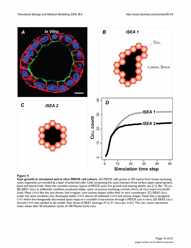
in the inner region DIED (and vanished) or moved out-ward. Growth characteristics were similar to thoseobserved in MDCK embedded cultures (Figure 4A). CUL-
TURES always formed stable CYSTS bordered by POLARIZED
CELLS (Figure 4B, C). Most ISEA1 CYSTS had irregularshapes. ISEA2 consistently produced CYSTS having aroundish, convex shape (Figure 4C). CYSTS in ISEA2 CUL-
TURES stabilized with fewer CELLS (Figure 4D) than didISEA1.
For dysregulation experiments, we focused on two criticalCELL axioms, Axioms 5 and 6. Axioms 2, 3, 4, and 7, werenot critical to CYST formation in EMBEDDED CULTURE (theywere critical in other CULTURE conditions, such as monol-ayer), and were infrequently used, so they were excludedfrom detailed analysis. Although not essential for EMBED-
DED CULTURE, Axiom 4 proved to be an important yet rareevent axiom, as discussed below. Disrupting Axiom 8 isnot straightforward: if the axiom is not applied, somealternative action must follow from its precondition, andthere are many plausible options. We elected not to pur-sue disruption of Axiom 8 until further insight from wet-lab studies becomes available to narrow options. Disrupt-ing Axiom 1 was straightforward, but the results (notshown) offered no significant insight: CLUSTERS eitherdeveloped normally into CYSTS for p > 0 or grewunchecked as a solid mass when p = 0. We expected thatoutcome because Axiom 1 was required for initial LUMINAL
SPACE creation but became nonessential thereafter. On theother hand, Axioms 5 and 6 were essential to CYST forma-tion. Anoikis is a form of cell death that epithelial cellsundergo when they lose direct matrix contact [14]. Axiom5 dictates ANOIKIS. It is the most frequently used CELL
DEATH axiom in both ISEA1 and ISEA2. Axiom 6 dictatesdirected CELL creation (the event maps to selective place-ment of a daughter cell), and accounts for most of the CELL
creation events in both analogues. The in vitro counter-parts of Axioms 5 and 6 are centrally implicated in epithe-lial morphogenesis and carcinogenesis, and have been
Page 9 of 21(page number not for citation purposes)
Theoretical Biology and Medical Modelling 2009, 6:8 http://www.tbiomed.com/content/6/1/8
Page 10 of 21(page number not for citation purposes)
Cyst growth in simulated and in vitro MDCK cell cultureFigure 4Cyst growth in simulated and in vitro MDCK cell culture. (A) MDCK cells grown in 3D matrix form lumen-enclosing cystic organoids surrounded by a layer of polarized cells. Cells composing the cysts maintain three surface types: apical (green), basal and lateral (red). Note the roundish contour typical of MDCK cysts. For growth and staining details, see [11]. Bar: 10 μm. (B) ISEA1 CELLS in EMBEDDED condition produced stable, cystic structures enclosing LUMINAL SPACE; all CELLS were POLARIZED (red). Many CYSTS like the one shown, had irregular, non-convex shapes unlike their in vitro counterpart. (C) ISEA2 CELLS under the same condition also developed stable CYSTS; almost all stabilized CYSTS had convex shapes. Note that a hexagonal CYST within the hexagonally discretized space maps to a roundish cross-section through a MDCK cyst in vitro. (D) ISEA2 CELLS formed CYSTS that tended to be smaller than those of ISEA1 (average 27 vs 31 CELLS per CYST). The CELL count represents mean values after 50 simulation cycles of 100 Monte Carlo runs.

Theoretical Biology and Medical Modelling 2009, 6:8 http://www.tbiomed.com/content/6/1/8
shown to be important in the context of in vitro cell cul-tures.
Dysregulation of Axiom 5 (ANOIKIS)In MDCK cultures, apoptosis contributes centrally tolumen formation [2]. Cells in the inner region of thedeveloping structure undergo anoikis. We speculated thatif ISEA CELL actions have MDCK counterparts, then thetwo analogues would exhibit (predict) LUMEN filling whenANOIKIS is compromised. We simulated the condition bydisrupting application of Axiom 5. So doing caused aber-rant growth (Figure 5) and changed CELL activity patterns(Figure 6). Growth rates increased nonlinearly withincreasing dysregulation. ISEA1 was more sensitive to dys-regulation at mid-range p of 0.4 and 0.6 than was ISEA2.No marked differences were noted at other tested levels.CELL population measurements after 50 simulation cyclesreflected changes in growth (Figure 5C). ISEA2 (vs ISEA1)produced structures having fewer CELLS.
Visual assessment of sample images showed that the CUL-
TURE morphology became irregular with increased dysreg-ulation (Figure 7). Relative to ISEA2, irregularities weremore pronounced when ISEA1's Axiom 5 was dysregu-lated. When CELL creation events outpaced DEATH, small,inverted CYSTS formed and stabilized (through POLARIZA-
TION) within LUMENS. As dysregulation increased, surfaceirregularities postponed POLARIZATION enabling furtherCELL creation events and surface expansion. For ISEA2,other factors contributed to LUMEN clearing. The convexitydrive (Axiom 12) enabled surface CELLS to POLARIZE
sooner. It also retarded inverted CYST formation by CELLS
trapped within LUMENS. Trapped CELLS were thus morelikely to satisfy the precondition of Axiom 5, even whenAxiom 5 was partially dysregulated.
Figure 6 shows how changes in CELL activity patternsaccompanied morphology changes for two levels ofAxiom 5 dysregulation. ANOIKIS dysregulation changedthe occurrence frequencies of axiom preconditions. Thatchange resulted in increased CELL creation events for bothISEA1 and ISEA2. Interestingly, for p = 0.8 and 0.6, thosechanges led to a net increase in CELL DEATH events. ForISEA1, many of the additional CELL creation eventsoccurred along the CYST's outer edge, whereas for ISEA2,many of the additional CELL creation and DEATH eventsoccurred within the LUMEN. The CELL creation eventswithin LUMENS were enabled by the Axiom 4 action: createMATRIX between two CELLS. Blocking Axiom 4 use blocksalmost all CELL creation events within LUMENS and pro-motes LUMEN clearance (not shown).
Dysregulation of Axiom 6 (oriented CELL creation)Oriented cell division is central to multicellular morpho-genesis [25-27]. Matrix contact and cell adhesions play animportant role in determining the orientation of the divi-sion axis in vitro [28,29]. Similar to its in vitro counter-part, CELL creation from Axiom 6 was oriented (notrandom). We dysregulated Axiom 6 by allowing the deci-sion-making CELL to place a new CELL in a randomlyselected MATRIX location, rather than selecting one thatmaximizes CELL contact.
Dysregulation of Axiom 5 (ANOIKIS) and its effect on ISEA growth and morphologyFigure 5Dysregulation of Axiom 5 (ANOIKIS) and its effect on ISEA growth and morphology. Axiom 5 dictates CELL DEATH when the decision-making CELL has in its neighborhood at least two CELLS and LUMINAL SPACE but no MATRIX. CELLS followed Axiom 5 with a parameter-controlled probability, p. Otherwise, the Axiom 5 precondition produced no CELL DEATH. Evasion of Axiom 5 changed ISEA1 and ISEA2 growth and structural characteristics in EMBEDDED CULTURES. (A-B) CELL counts at six levels of dysregulation are shown. Values are means of 100 Monte Carlo runs. CELL count increased monotonically with the severity of dysregulation. For ISEA2, the effects were less dramatic for larger p. (C) Dysregulation caused a nonlinear increase in both ISEA1 and ISEA2 CELL count measured after 50 simulation cycles.
Page 11 of 21(page number not for citation purposes)

Theoretical Biology and Medical Modelling 2009, 6:8 http://www.tbiomed.com/content/6/1/8
Page 12 of 21(page number not for citation purposes)
CELL DEATH and creation events by ISEA1 and ISEA2 with and without dysregulation (two levels) of Axioms 5 and 6Figure 6CELL DEATH and creation events by ISEA1 and ISEA2 with and without dysregulation (two levels) of Axioms 5 and 6. Values in circles are axiom p values. Left panels (A–D): ISEA1 events. Right panels (E-H): ISEA2 events. Top four panels: CELL creation events. Bottom four panels: CELL DEATH events. Event values are occurrences per simulation averaged over 100 Monte Carlo runs.

Theoretical Biology and Medical Modelling 2009, 6:8 http://www.tbiomed.com/content/6/1/8
Page 13 of 21(page number not for citation purposes)
Typical structures formed by ISEA1 and ISEA2 when Axiom 5 or 6 was dysregulatedFigure 7Typical structures formed by ISEA1 and ISEA2 when Axiom 5 or 6 was dysregulated. Shown are images of struc-tures formed after 50 simulation cycles for p = 0.8 and 0.6. Note that a regular hexagon in 2D hexagonal space maps to a circle in 2D continuous space. Objects: POLARIZED CELL (red), UNPOLARIZED CELL (gray), MATRIX (white), and LUMINAL SPACE (black). (A-B) Shown are examples of structures formed when ISEA1 was dysregulated. (C-D) Shown are examples of structures formed when ISEA2 was dysregulated.

Theoretical Biology and Medical Modelling 2009, 6:8 http://www.tbiomed.com/content/6/1/8
We ran simulations with Axiom 6's p ranging from 0 to 1,and recorded changes in CULTURE growth and morphologyalong with CELL activity patterns. The overall results areshown in Figure 8. CULTURE growth rate and CELL countafter 50 simulation cycles increased monotonically withAxiom 6 dysregulation. The changes were less dramaticthan those observed following Axiom 5 dysregulation,and there were marked differences between dysregulatedISEA1 and ISEA2 CULTURE growth. ISEA2 was less suscep-tible to disoriented placement of a newly created CELL.Mean CELL count in ISEA2 CULTURES was always smallerthan that for ISEA1 at every tested dysregulation level.
Dysregulating Axiom 6 using p = 0.8 and 0.6 increasedCELL DEATH and CELL PROLIFERATION activities of ISEA2 lessthan ISEA1 (Figure 6). CELL DEATH events were offset by anapproximately equal number of CELL creation events, andthat was consistent with the observation that LUMEN-entrapped CELLS underwent cycles of CELL creation andDEATH.
Inspection of Figure 7C, D shows that the morphologicalirregularities resulting from a given degree of Axiom 6 dys-regulation were less pronounced than from a correspond-ing degree of Axiom 5 dysregulation. For ISEA1, themorphology change produced by a degree of Axiom 6 dys-regulation was very similar to that caused by a lesserdegree of Axiom 5 dysregulation. ISEA1 structures pro-duced using dysregulated Axiom 6 contained a larger frac-tion of POLARIZED CELLS than did corresponding Axiom 5dysregulated structures, and so the former changed more
slowly as simulations progressed. For ISEA2, because allCELL DEATH axioms were always followed, there was lessLUMEN filling when Axiom 6 was dysregulated, comparedto when Axiom 5 was disrupted to the same degree. Asnoted above, ISEA2 LUMEN filling was enabled by Axiom4. Blocking it severely restrained and often eliminated for-mation of INTRALUMINAL CELL CLUSTERS.
Dynamic phenotypeFigure 9 presents dynamic phenotype: the normalized fre-quency of axiom use by both ISEA1 and ISEA2. TheCYSTOGENESIS mechanism at any stage in the process is theset of all events occurring within that interval. It is clearfrom Figure 9 that there is no specific CYSTOGENESIS mech-anism. From start to the end of a simulation or until a sta-ble structure forms, the mechanism evolves. How itevolves is a feature of that analogue's dynamic phenotype.Use patterns were similar for those axioms common toboth analogues and that were used most frequently (1, 3,5, 6, 9, and 11). Major differences were evident only forthe less frequently used axioms (2, 4, 7, 8, and 10). Asnoted earlier, enabling CELL movement (Axiom 12) hadan unanticipated consequence: it enabled the occasionalformation of long-lived, small islands of CELLS within aLUMEN. Once a unit of MATRIX was formed, CELLS within aLUMEN could move and that gave rise to preconditions forcreation of new CELLS as well as CELL DEATH. The processcan continue for an extended interval and that accountsfor the very low frequency of use of Axioms 2, 4, 7, and 8by ISEA2. Note that when CELLS are trapped within an oth-erwise stable CYST, those INTRALUMINAL events are the only
Dysregulation of Axiom 6 and its effect on ISEA CULTURE growthFigure 8Dysregulation of Axiom 6 and its effect on ISEA CULTURE growth. Axiom 6 dictates oriented placement of a newly created CELL. It is placed at an adjacent MATRIX position that maximizes its number of CELL neighbors. CELLS followed Axiom 6 with a parameter-controlled probability, p. Otherwise, the CELL copy replaced a randomly selected MATRIX neighbor without regard for CELL neighbor number. Doing so changed ISEA growth and structural characteristics. (A-B) CELL count increased monotonically with the severity of dysregulation. Compared to ISEA1 growth (A), ISEA2 growth was affected less for every dysregulation level. (C) CELL count after 50 simulation cycles showed marked differences between ISEA1 and ISEA2 that increased with the severity of dysregulation.
Page 14 of 21(page number not for citation purposes)

Theoretical Biology and Medical Modelling 2009, 6:8 http://www.tbiomed.com/content/6/1/8
Page 15 of 21(page number not for citation purposes)
Dynamic phenotype: axiom usage by ISEA1 and ISEA2Figure 9Dynamic phenotype: axiom usage by ISEA1 and ISEA2. Normalized axiom use frequencies are plotted versus simula-tion cycle. Left panels (A-C): ISEA1 use frequencies. Right panels (D-F): ISEA2 use frequencies. Top: axioms used most fre-quently. Middle: moderate use events. Bottom: Rare axiom use events. Axiom numbers in circles: the curves are normalized use frequencies averaged over 100 Monte Carlo runs. Axiom numbers in pentagons: the variance in average use frequency for the rarely used axioms was large; for clarity, trend lines are shown. In B and E, trend lines for Axiom 8 usage are magnified by a factor of 5. Raw data are provided in additional file 1: Supplemental Material. As simulations progressed and CYSTS matured, Axiom 11 (do nothing) was executed most frequently.

Theoretical Biology and Medical Modelling 2009, 6:8 http://www.tbiomed.com/content/6/1/8
events. For that simulation, their relative use frequenciesare large, and it is those values that are averaged with thevalues from other simulations, which are typically zero.
If nutrient levels within lumens are less than outside thecyst, then intraluminal cell division may not be sustaina-ble. Furthermore, under 3D culture conditions, there is nodirect evidence of matrix production by MDCK cellstrapped within early-stage lumens during cystogenesis. Itis noteworthy that by simulation cycle 50, when Axiom 4is blocked, ISEA2's use frequency of axioms 2, 7, 8 and 10drops to zero (not shown): ISEA2's axiom frequency ofuse pattern becomes similar to that of ISEA1.
Axiom dysregulation changed dynamic phenotype. Addi-tional records for dysregulating Axioms 5 and 6 are pro-vided in additional file 1: Supplemental Material for bothISEA1 and ISEA2. Because trends are similar for ISEA1 andISEA2, we present in Figures 10 and 11 selected results forISEA2. Figure 10 shows ISEA2 axiom use frequencies forAxiom 5 p = 0.8 and 0.6. The major consequence wasreduction in Axiom 11 usage (do nothing: mandatesachieved). That decline was mirrored by the rise in Axiom5* (dysregulated action) usage, which remained relativelyconstant after five simulation cycles. In parallel, the usepatterns for all other axioms changed relative to their p =1 patterns. Even though only Axiom 5 was disrupted occa-sionally, all ISEA2 operating principles were impacted tosome extent: the entire dynamic phenotype changed.However, the morphological consequences for p = 0.8were difficult to detect: except for a tendency to be larger,most stabilized CYSTS were indistinguishable from thoseformed when p = 1. The potential morphological conse-quences of relaxing Axiom 5's p by 20% were thwarted bysmall shifts in the use frequencies of all other axioms. Thisobservation suggests that the networked nature of ISEA2axiom usage acts to buffer the consequences of small dis-ruptions of any one operating principle.
Both the morphological and dynamic phenotypic conse-quences of Axiom 6 dysregulation were less dramatic thanthose of Axiom 5. They were also less dramatic in ISEA2than in ISEA1. Reducing p led to larger structures thateventually stabilized (Figure 8B) and to more CELLS beingtrapped within occasional LUMENS (Figure 7D). Compari-son of Figures 10 and 11 reveals that the influence ofAxiom 6 disruption was also less significant than that ofdisrupting Axiom 5 to the same degree. For p = 0.8 and0.6, the activities of CELLS trapped in LUMENS were prima-rily responsible for increased axiom use after about 20simulation cycles. When Axiom 4 was blocked (notshown), those axiom use frequencies diminished consid-erably making an increased CYST size the primary conse-quence of Axiom 6 disruption.
DiscussionWe detailed a computational approach to build and testplausible hypotheses of in vitro dynamic phenotype. Thenewly developed framework enabled MDCK cell-mimeticanalogues to function as autonomously as feasible forsoftware agents. Axiomatic operating principles enabledISEA2 CELLS to consistently produce convex CYSTS undersimulated 3D embedded culture condition. Measures ofaxiom use during CYSTOGENESIS provided a detaileddescription of ISEA2 dynamic phenotype. Dysregulatingkey CELL DEATH and DIVISION axioms led to disorganizedcystic forms that were reminiscent of the in vitro tumorreconstruction phenotype. Unexpectedly, ISEA2's drivefor convexity made it less susceptible to, or more robustagainst, the dysregulation of either axiom when comparedto its predecessor, ISEA1. It will be interesting to learn ifthe mechanisms underlying epithelial cyst convexity incultures contribute to robustness against comparableinterventions. In addition, occasional disruption of oneactivity in a minority of CELLS, as in Figures 10 and 11, hadconsequences for the system (e.g., altered CYST morphol-ogy) and for all other normal behaving CELLS. The averageaxiom use patterns of all other CELLS changed. Uponreflection, the observation could be expected. The actionsof all CELLS in a CLUSTER transforming into a CYST are net-worked in space and time. An action of one CELL can affectthe action options of a nearby CELL at a future time. If aCELL occasionally malfunctions, it has measurable conse-quences, as shown in Figures 10 and 11. To the extent thatthe mappings in Figure 1 are accepted as valid, we canextend such observations to MDCK epithelial cells under-going morphogenesis.
The results reaffirm that Axioms 5 and 6 play critical,dominant roles in determining the CYSTOGENESIS pheno-type. Also, as noted in Results, Axiom 1 was essential forinitial LUMINAL SPACE creation, and completely blocking itsuse had a detrimental effect on CULTURE morphology. Onthe other hand, Axioms 2–4 and 7 were nonessential forCYSTOGENESIS in EMBEDDED CULTURE. Dysregulating or sim-ply deleting the axioms did not patently alter theCYSTOGENESIS phenotype. However, that does not meanthat the axioms were not parsimonious: they were essen-tial to achieving targeted attributes of the other CULTURE
types—SUSPENSION, SURFACE, and OVERLAY—from [9].Whether a similar relationship holds true for their biolog-ical counterpart is unknown. However, it is clear thatMDCK cells under different culture conditions use some-what different cell mechanisms depending on the specificculture condition, which leads to different culture pheno-types [2,30-32].
While reasonable mappings can be established from ISEAto MDCK and MCF-10A mammary epithelial cell pheno-types [16], ISEA axioms may not map well to other epithe-
Page 16 of 21(page number not for citation purposes)

Theoretical Biology and Medical Modelling 2009, 6:8 http://www.tbiomed.com/content/6/1/8
Page 17 of 21(page number not for citation purposes)
Axiom usage by ISEA2 during partial Axiom 5 dysregulationFigure 10Axiom usage by ISEA2 during partial Axiom 5 dysregulation. Normalized axiom use frequencies are plotted versus simulation cycle as in Figure 9. Axiom numbers in circles are shown for each curve. *: dysregulated action. Left panels (A-C): p = 0.8. Right panels (D-F): p = 0.6. Top: axioms used most frequently. Middle: moderate use events. Bottom: Rare axiom use events. The curves are normalized use frequencies averaged over 100 Monte Carlo runs. In A and B, Axiom 5* usage frequen-cies are magnified by a factor of 5.

Theoretical Biology and Medical Modelling 2009, 6:8 http://www.tbiomed.com/content/6/1/8
Page 18 of 21(page number not for citation purposes)
Axiom usage by ISEA2 during partial Axiom 6 dysregulationFigure 11Axiom usage by ISEA2 during partial Axiom 6 dysregulation. Normalized axiom use frequencies are plotted versus simulation cycle as in Figures 9 and 10. Axiom numbers in circles are shown for each curve. *: dysregulated action. Left panels (A-C): p = 0.8. Right panels (D-F): p = 0.6. Top: axioms used most frequently. Middle: moderate use events. Bottom: Rare axiom use events. The curves are normalized use frequencies averaged over 100 Monte Carlo runs.

Theoretical Biology and Medical Modelling 2009, 6:8 http://www.tbiomed.com/content/6/1/8
lial cell types and culture systems. For example, in AT IIcell cultures, cyst structures develop by a mechanism thatinvolves neither cell death nor proliferation [33]. Alveo-lar-like cysts form by cell migration and aggregation, incontrast to how cysts typically develop in MDCK cell cul-tures. Those differences are mirrored in validated CELL
axiom specifications of the ISEAs and AT II analogues.Unlike the ISEA CELLS, the AT II analogue [6] lacks CELL
DEATH and PROLIFERATION action options. They form CYSTS
exclusively by spatial rearrangement. Notwithstandingthose differences, their stable form similarities suggestcommon mandates. For instance, ISEA and AT II ana-logues do exhibit a common, essential feature: CELLS striveto achieve and maintain lateral CELL-CELL contacts. Addi-tional insight is anticipated when 2D simulations areexpanded to 3D.
Cell processes work together in ways that give rise to effec-tive mandates that normal epithelial cells appear to fol-low. Each mandate is assumed a consequence of theinteroperation of genetics and environmental factors.How specific cell actions contribute to these mandates isunclear. However, tracing CELL activities during ISEA2simulations makes clear how their mandates, the targetedattributes, are achieved. That clarity provides insight intoand plausible explanations of MDCK's morphogenic phe-nomena. Because ISEA components and mechanisms arecoarse-grained, one ISEA2 axiom may map to many fine-grain MDCK processes. Iterative refinement of ISEA2 sothat it achieves an expanded set of MDCK attributes willimprove and concretize the mappings from analogue toMDCK cultures, potentially creating new knowledge.Mappings from specifics of MDCK cultures (complex) toanalogue (simplified), however, will always be ambigu-ous, a property of all referent-model pairs.
Moving forward, we suggest the following iterative refine-ment protocol. It was used successfully herein and in pre-vious studies [4-6,9,34]. The protocol supports adheringto the guideline of parsimony which is important whenbuilding a complex model. It is straightforward and so canbe used for refinement of any mechanistically focused,agent-based biomimetic analogue. Basic steps are: 1) startwith a small but diverse set of in vitro attributes, static anddynamic. They are the initial targeted attribute list. 2)Posit coarse-grained, discrete mechanisms, requiring asfew components as is reasonable, that may generate anal-ogous phenomena. 3) Instantiate (represent an abstrac-tion by a concrete software instance) analoguecomponents and mechanisms. 4) Conduct experiments tomeasure a variety of phenomena generated during execu-tion. So doing establishes the degree of in silico-in vitrophenotype overlap, and lack thereof. 5) Achieve a degreeof validation by satisfying a prespecified level of similaritybetween in silico and targeted in vitro attributes. 6) Add
one or more new attributes (measurable phenomena) tothe targeted list until the analogue in step 5 is falsified.Added attributes need to be at a similar level to and suffi-ciently close to those already present so that it seems fea-sible to achieve the expanded attribute list with as littlecomponent reengineering as possible. Once the analoguein step 5 is falsified, return to step 2.
The nature and organization of software componentswithin the ISEA framework, as illustrated in Figure 2, weredesigned to facilitate iterative refinement of everything onthe right side of Figure 1. That process can concretize eachof the mappings from ISEA to MDCK counterparts. As theprocess continues, following each round of validation,more of what we know or think we know becomes instan-tiated in the analogue. After many such rounds, the ana-logue will mature as instantiated, working hypotheses ofhow MDCK cystogenesis and pathologic transformationsoccur. At that stage, it will have become an extensible,interactive instantiation of available biological knowledgeabout mechanisms and processes. It will have become anexecutable knowledge embodiment. To achieve thatvision, it is essential that biomimetic components func-tion (quasi-) autonomously, all or part of the time. That iswhy CELLS are agents. Everything that a CELL needs to func-tion (in a specified software environment) is containedwithin its code. Absent that property, the mappings fromISEA to MDCK cystogenesis mechanisms are not concre-tizable, and so the mappings from ISEA to MDCK operat-ing principles are forced to remain conceptual.
Finally, axiom use results show that at the same time, dif-ferent CELLS within the same CULTURE are engaged in quitedifferent activities. The same is true in vitro; one MDCKcell can be moving actively relative to its attached neigh-bors while another is undergoing anoikis, and yet anotheris initiating division. Simultaneously, polarized cells thathave achieved their mandates may begin downregulatingprocesses used earlier. It follows that the ensemble ofmolecular biology details, such as gene and proteinexpression levels, which enable those different activitieswill themselves be different. Patterns detected in gene andprotein expression data averaged over all cells in an activecyst may have little scientific value in answering suchquestions as these. When and how does an epithelial cellchoose to switch from one activity to another? Why doesit choose one action rather than another? Are severalaction options always available to each cell? Obtainingplausible answers to these and related questions is essen-tial to achieving deeper insight into epithelial morpho-genesis and early cancer progression. As demonstrated,the class of models presented herein provides a rigorousplatform to hypothesize, challenge, and refine plausibleanswers. The causal chain of events responsible for mostsimulated behaviors can be explored in detail, and assess-
Page 19 of 21(page number not for citation purposes)

Theoretical Biology and Medical Modelling 2009, 6:8 http://www.tbiomed.com/content/6/1/8
ments made as to whether critical events are biotic (sup-portable by in vitro evidence) or not.
ConclusionThe approach described herein provided for a hypothe-sis—a theory—of how the collective consequences ofindividual MDCK cell actions might give rise to systemicin vitro phenotype. The causal chain of events responsiblefor most ISEA behaviors could be explored in detail, andassessments could be made of their relative roles duringsimulation. Having that capability enabled us to developa detailed dynamic ISEA phenotype. The MDCK embed-ded culture counterpart is problematic to obtain usingstate-of-the-art in vitro methods. We expect future roundsof model refinement and validation will strengthen in sil-ico-to-in vitro mappings, thus providing a viable strategyto gain deeper insight into the mechanistic basis of epithe-lial cystogenesis, morphogenesis, and in vitro transforma-tions.
Competing interestsThe authors declare that they have no competing interests.
Authors' contributionsSK and CH conceived the idea. SK designed and per-formed the experiments. SP participated in the design andimplementation. SK, KM, JD, and CH analyzed the exper-iment results. SK and CH wrote the paper with input fromcoauthors. All authors read and approved the final manu-script.
Additional material
AcknowledgementsWe thank Wei Yu, Mark Grant, Glen Ropella, Jesse Engelberg, Jon Tang, Teddy Lam, Shahab Sheikh-Bahaei, and members of the BioSystems group for helpful discussions and suggestions. This research was supported in part by the CDH Research Foundation, a graduate fellowship to SHJK from the International Foundation for Ethical Research, NIH grants R01 DK067153 and R01 DK074398 to KM, and the Culpeper Scholar Award (Partnership For Cures) to JD. The funding bodies had no role in study design; in the col-lection, analysis, and interpretation of data; in the writing of the manuscript; and in the decision to submit the manuscript for publication.
References1. Bryant DM, Mostov KE: From cells to organs: building polarized
tissue. Nat Rev Mol Cell Biol 2008, 9:887-901.2. Martín-Belmonte F, Yu W, Rodríguez-Fraticelli AE, Ewald AJ, Werb Z,
Alonso MA, Mostov K: Cell-polarity dynamics controls the
mechanism of lumen formation in epithelial morphogenesis.Curr Biol 2008, 18:507-513.
3. Debnath J, Brugge JS: Modelling glandular epithelial cancers inthree-dimensional cultures. Nat Rev Cancer 2005, 5:675-688.
4. Tang J, Ley KF, Hunt CA: Dynamics of in silico leukocyte rolling,activation, and adhesion. BMC Syst Biol 2007, 1:14.
5. Engelberg JA, Ropella GE, Hunt CA: Essential operating princi-ples for tumor spheroid growth. BMC Syst Biol 2008, 2:110.
6. Kim SHJ, Yu W, Mostov K, Matthay MA, Hunt CA: A computa-tional approach to understand in vitro alveolar morphogen-esis. PLoS ONE 2009, 4:e4819.
7. Fisher J, Henzinger TA: Executable cell biology. Nat Biotechnol2007, 25:1239-1249.
8. Hunt CA, Ropella GE, Park S, Engelberg JA: Dichotomies betweencomputational and mathematical models. Nat Biotechnol 2008,26:737-738.
9. Grant MR, Mostov KE, Tlsty TD, Hunt CA: Simulating propertiesof in vitro epithelial cell morphogenesis. PLoS Comput Biol 2006,2:e129.
10. Park S, Ropella GE, Kim SHJ, Roberts MS, Hunt CA: Computationalstrategies unravel and trace how liver disease changeshepatic drug disposition. J Pharmacol Exp Ther 2009,328(1):294-305.
11. Yu W, Datta A, Leroy P, O'Brien LE, Mak G, Jou TS, Matlin KS, Mos-tov KE, Zegers MM: Beta1-integrin orients epithelial polarityvia Rac1 and laminin. Mol Biol Cell 2005, 16:433-445.
12. Zeigler BP, Kim TG, Praehofer H: Theory of modeling and simulation:integrating discrete event and continuous complex dynamic systems SanDiego, USA: Academic Press; 2000.
13. Thorne BC, Bailey AM, Peirce SM: Combining experiments withmulti-cell agent-based modeling to study biological tissuepatterning. Brief Bioinform 2007, 8:245-257.
14. Frisch SM, Screaton RA: Anoikis mechanisms. Curr Opin Cell Biol2001, 13:555-562.
15. Chiarugi P, Giannoni E: Anoikis: a necessary death program foranchorage-dependent cells. Biochem Pharmacol 2008,76:1352-1364.
16. Debnath J, Mills KR, Collins NL, Reginato MJ, Muthuswamy SK,Brugge JS: The role of apoptosis in creating and maintainingluminal space within normal and oncogene-expressing mam-mary acini. Cell 2002, 111:29-40.
17. Nelson WJ: Epithelial cell polarity from the outside looking in.News Physiol Sci 2003, 18:143-146.
18. O'Brien LE, Zegers MM, Mostov KE: Building epithelial architec-ture: insights from three-dimensional culture models. NatRev Mol Cell Biol 2002, 3:531-537.
19. Kroschewski R: Molecular mechanisms of epithelial polarity:about shapes, forces, and orientation problems. News PhysiolSci 2004, 19:61-66.
20. Chen JG, Ullah H, Young JC, Sussman MR, Jones AM: ABP1 isrequired for organized cell elongation and division in Arabi-dopsis embryogenesis. Genes Dev 2001, 15:902-911.
21. Gonzalez C: Spindle orientation, asymmetric division andtumour suppression in Drosophila stem cells. Nat Rev Genet2007, 8:462-472.
22. Lee M, Vasioukhin V: Cell polarity and cancer–cell and tissuepolarity as a non-canonical tumor suppressor. J Cell Sci 2008,121:1141-1150.
23. Harris PC, Torres VE: Polycystic kidney disease. Annu Rev Med2009, 60:321-337.
24. Luke S, Cioffi-Revilla C, Panait L, Sullivan K, Balan G: MASON: amultiagent simulation environment. SIMULATION 2005,81:517-527.
25. Sausedo RA, Smith JL, Schoenwolf GC: Role of nonrandomly ori-ented cell division in shaping and bending of the neural plate.J Comp Neurol 1997, 381:473-488.
26. Gong Y, Mo C, Fraser SE: Planar cell polarity signalling controlscell division orientation during zebrafish gastrulation. Nature2004, 430:689-693.
27. Baena-López LA, Baonza A, García-Bellido A: The orientation ofcell divisions determines the shape of Drosophila organs.Curr Biol 2005, 15:1640-1644.
28. Théry M, Racine V, Pépin A, Piel M, Chen Y, Sibarita JB, Bornens M:The extracellular matrix guides the orientation of the celldivision axis. Nat Cell Biol 2005, 7:947-953.
Additional File 1Supplemental Material. Provided are complete, raw axiom usage data.Click here for file[http://www.biomedcentral.com/content/supplementary/1742-4682-6-8-S1.pdf]
Page 20 of 21(page number not for citation purposes)
http://www.ncbi.nlm.nih.gov/entrez/query.fcgi?cmd=Retrieve&db=PubMed&dopt=Abstract&list_uids=9136804

Theoretical Biology and Medical Modelling 2009, 6:8 http://www.tbiomed.com/content/6/1/8
Publish with BioMed Central and every scientist can read your work free of charge
"BioMed Central will be the most significant development for disseminating the results of biomedical research in our lifetime."
Sir Paul Nurse, Cancer Research UK
Your research papers will be:
available free of charge to the entire biomedical community
peer reviewed and published immediately upon acceptance
cited in PubMed and archived on PubMed Central
yours — you keep the copyright
Submit your manuscript here:http://www.biomedcentral.com/info/publishing_adv.asp
BioMedcentral
29. Théry M, Jiménez-Dalmaroni A, Racine V, Bornens M, Jülicher F:Experimental and theoretical study of mitotic spindle orien-tation. Nature 2007, 447:493-496.
30. Hall HG, Farson DA, Bissell MJ: Lumen formation by epithelialcell lines in response to collagen overlay: a morphogeneticmodel in culture. Proc Natl Acad Sci USA 1982, 79:4672-4676.
31. Wang AZ, Ojakian GK, Nelson WJ: Steps in the morphogenesisof a polarized epithelium. II. Disassembly and assembly ofplasma membrane domains during reversal of epithelial cellpolarity in multicellular epithelial (MDCK) cysts. J Cell Sci1990, 95:153-165.
32. Nelson CM, Jean RP, Tan JL, Liu WF, Sniadecki NJ, Spector AA, ChenCS: Emergent patterns of growth controlled by multicellularform and mechanics. Proc Natl Acad Sci USA 2005,102:11594-11599.
33. Yu W, Fang X, Ewald A, Wong K, Hunt CA, Werb Z, Matthay MA,Mostov K: Formation of cysts by alveolar type II cells in three-dimensional culture reveals a novel mechanism for epithelialmorphogenesis. Mol Biol Cell 2007, 18:1693-1700.
34. Lam TN, Hunt CA: Discovering plausible mechanistic details ofhepatic drug interactions. Drug Metab Dispos 2009, 37:237-246.
Page 21 of 21(page number not for citation purposes)
http://www.ncbi.nlm.nih.gov/entrez/query.fcgi?cmd=Retrieve&db=PubMed&dopt=Abstract&list_uids=6956885
http://www.ncbi.nlm.nih.gov/entrez/query.fcgi?cmd=Retrieve&db=PubMed&dopt=Abstract&list_uids=6956885
http://www.ncbi.nlm.nih.gov/entrez/query.fcgi?cmd=Retrieve&db=PubMed&dopt=Abstract&list_uids=6956885
http://www.ncbi.nlm.nih.gov/entrez/query.fcgi?cmd=Retrieve&db=PubMed&dopt=Abstract&list_uids=2351700
http://www.ncbi.nlm.nih.gov/entrez/query.fcgi?cmd=Retrieve&db=PubMed&dopt=Abstract&list_uids=2351700