Transcriptional regulation by Pax3 and TGFbeta2 signaling: a potential gene regulatory network in...
12
Transcriptional regulation by Pax3 and TGFβ2 signaling: a potential gene regulatory network in neural crest development HIROMICHI NAKAZAKI 1,a,dˇ , YUEH-WEI SHEN 1,d , BETH YUN 1,b , ANVESH REDDY 1,c , VARUN KHANNA 1 , BARBARA MANIA-FARNELL 2 , SHUNSUKE ICHI 1 , DAVID G. MCLONE 1 , TADANORI TOMITA 1 and C. SHEKHAR K. MAYANIL* ,1 1 Laboratory of Neural Tube Research, Department of Pediatric Neurosurgery, Children’s Memorial Research Center and Northwestern University Feinberg School of Medicine, Chicago, IL and 2 Department of Biology, Purdue University at Calumet, Hammond, IN, USA ABSTRACT Pax3 regulates neural crest cell migration and is critical during neural crest develop- ment. TGFβs modify neural crest cell migration and differentiation. TGFβ2 nullizygous embryos (TGFβ2 -/- Pax3 +/+ ) display open neural tube and bifid spine, whereas in wild type embryos the neural tube is closed. In previous work, we have demonstrated that Pax3 regulates TGFβ2 by directly binding to cis -regulatory elements on its promoter. In this study, we found that the TGFβ2 nullizygous phenotype can be reversed to the wild type phenotype by down-regulating one allele of Pax3, as in TGFβ2 -/- Pax3 +/- embryos obtained through breeding TGFβ2 +/- Pax3 +/- mice. The data in this paper suggest that Pax3 and TGFβ2 interact in a coordinated gene regulatory network, linked by common downstream effector genes, to bring about this phenotypic reversal. Down- stream effectors may include Hes1, Ngn2 and Sox9, as well as other genes involved in neuronal differentiation. KEY WORDS: Hes1, Ngn2, Sox9, epithelial-to-mesenchymal transformation Int. J. Dev. Biol. 53: 69-79 (2009) doi: 10.1387/ijdb.082682hn THE INTERNATIONAL JOURNAL OF DEVELOPMENTAL BIOLOGY www.intjdevbiol.com *Address correspondence to: C. S. K. Mayanil. Laboratory of Neural Tube Research, Developmental Biology Program, Childrens Memorial Research Center, Box # 204, 2430 N Halsted Street, Chicago, IL 60614. Fax: 773-755-6385. e-mail address: [email protected] Present addresses: a Department of Neurosurgery, Jikei University School of Medicine, Tokyo, Japan; b Department of Neurology, Northwestern University, Feinberg School of Medicine, Chicago, IL 60614, USA; c The University of Tennessee Health Science Center, College of Medicine, Memphis, TN 38163, USA. Note d: These authors contributed equally to the work. Accepted: 5 June 2008. Published online: 5 December 2008. Edited by: Angie Rizzino. ISSN: Online 1696-3547, Print 0214-6282 © 2008 UBC Press Printed in Spain Abbreviations used in this paper: NCC, neural crest cell; ngn, neurogenin. Introduction In 1954, Auerbach observed that developmental defects in homozygous Splotch embryos are associated with neural crest cell deficiency. In the homozygous condition the Splotch (Sp) mutation is lethal by embryonic day 13.5. These embryos display defects in neural tissue, neural crest derivatives, and limb muscu- lature. Splotch heterozygotes exhibit defects in neural crest derivatives, which result in white patches on their bellies caused by defective development of neural crest derived melanocytes (Foy et al., 1990). Chromosome localization in conjunction with the spatio-temporal tissue distribution of Pax3 transcripts and tissues affected in Sp mutants led to direct experimental analysis demonstrating that Splotch and Pax3 are the same gene (Epstein et al., 1991; Goulding et al., 1993) and the homozygous Splotch condition is Pax3 -/- . These studies show that Pax3 has a regula- tory role in neural crest development. Other studies also indicate that Pax3 has a role in regulating neurogenesis in neural crest derived precursor cells. Goulding et al. (1991) demonstrated Pax3 expression in a population of migrating neural crest cells (NCCs) that contribute to the dorsal root ganglia and cephalic mesenchyme. In a separate study Li et al. (1999) found that Pax3 replacement rescued neural tube closure and other neural crest related anomalies in Sp mutants. This group produced transgenic mice that over expressed Pax3 in neural tube and neural crest. They then took Pax3 over- expressing transgenic lines and crossed them with heterozygous Splotch mice. Transgenic heterozygous Splotch mice were iden- tified, backcrossed to Splotch heterozygotes and examined at E13.5. All transgenic Splotch homozygous embryos had normal closed neural tubes in comparison to non-transgenic Splotch homozygous embryos, all of which displayed neural tube defects in the lumbrosacral region and in some cases exencephaly. More
-
Upload
independent -
Category
Documents
-
view
3 -
download
0
Transcript of Transcriptional regulation by Pax3 and TGFbeta2 signaling: a potential gene regulatory network in...

Transcriptional regulation by Pax3 and TGFβ2 signaling:
a potential gene regulatory network in neural crest development
HIROMICHI NAKAZAKI1,a,dˇ, YUEH-WEI SHEN1,d, BETH YUN1,b, ANVESH REDDY1,c, VARUN KHANNA1,BARBARA MANIA-FARNELL2, SHUNSUKE ICHI1, DAVID G. MCLONE1, TADANORI TOMITA1
and C. SHEKHAR K. MAYANIL*,1
1Laboratory of Neural Tube Research, Department of Pediatric Neurosurgery, Children’s Memorial Research Center and NorthwesternUniversity Feinberg School of Medicine, Chicago, IL and 2Department of Biology, Purdue University at Calumet, Hammond, IN, USA
ABSTRACT Pax3 regulates neural crest cell migration and is critical during neural crest develop-
ment. TGFβs modify neural crest cell migration and differentiation. TGFβ2 nullizygous embryos
(TGFβ2 -/-Pax3+/+) display open neural tube and bifid spine, whereas in wild type embryos the
neural tube is closed. In previous work, we have demonstrated that Pax3 regulates TGFβ2 by
directly binding to cis -regulatory elements on its promoter. In this study, we found that the TGFβ2
nullizygous phenotype can be reversed to the wild type phenotype by down-regulating one allele
of Pax3, as in TGFβ2 -/-Pax3 +/- embryos obtained through breeding TGFβ2 +/-Pax3 +/- mice. The data
in this paper suggest that Pax3 and TGFβ2 interact in a coordinated gene regulatory network,
linked by common downstream effector genes, to bring about this phenotypic reversal. Down-
stream effectors may include Hes1, Ngn2 and Sox9, as well as other genes involved in neuronal
differentiation.
KEY WORDS: Hes1, Ngn2, Sox9, epithelial-to-mesenchymal transformation
Int. J. Dev. Biol. 53: 69-79 (2009)doi: 10.1387/ijdb.082682hn
THE INTERNATIONAL JOURNAL OF
DEVELOPMENTAL
BIOLOGYwww.intjdevbiol.com
*Address correspondence to: C. S. K. Mayanil. Laboratory of Neural Tube Research, Developmental Biology Program, Childrens Memorial Research Center,Box # 204, 2430 N Halsted Street, Chicago, IL 60614. Fax: 773-755-6385. e-mail address: [email protected]
Present addresses: a Department of Neurosurgery, Jikei University School of Medicine, Tokyo, Japan; b Department of Neurology, Northwestern University,Feinberg School of Medicine, Chicago, IL 60614, USA; c The University of Tennessee Health Science Center, College of Medicine, Memphis, TN 38163, USA.
Note d: These authors contributed equally to the work.
Accepted: 5 June 2008. Published online: 5 December 2008. Edited by: Angie Rizzino.
ISSN: Online 1696-3547, Print 0214-6282© 2008 UBC PressPrinted in Spain
Abbreviations used in this paper: NCC, neural crest cell; ngn, neurogenin.
Introduction
In 1954, Auerbach observed that developmental defects inhomozygous Splotch embryos are associated with neural crestcell deficiency. In the homozygous condition the Splotch (Sp)mutation is lethal by embryonic day 13.5. These embryos displaydefects in neural tissue, neural crest derivatives, and limb muscu-lature. Splotch heterozygotes exhibit defects in neural crestderivatives, which result in white patches on their bellies causedby defective development of neural crest derived melanocytes(Foy et al., 1990). Chromosome localization in conjunction withthe spatio-temporal tissue distribution of Pax3 transcripts andtissues affected in Sp mutants led to direct experimental analysisdemonstrating that Splotch and Pax3 are the same gene (Epsteinet al., 1991; Goulding et al., 1993) and the homozygous Splotchcondition is Pax3-/-. These studies show that Pax3 has a regula-tory role in neural crest development.
Other studies also indicate that Pax3 has a role in regulating
neurogenesis in neural crest derived precursor cells. Goulding etal. (1991) demonstrated Pax3 expression in a population ofmigrating neural crest cells (NCCs) that contribute to the dorsalroot ganglia and cephalic mesenchyme. In a separate study Li etal. (1999) found that Pax3 replacement rescued neural tubeclosure and other neural crest related anomalies in Sp mutants.This group produced transgenic mice that over expressed Pax3in neural tube and neural crest. They then took Pax3 over-expressing transgenic lines and crossed them with heterozygousSplotch mice. Transgenic heterozygous Splotch mice were iden-tified, backcrossed to Splotch heterozygotes and examined atE13.5. All transgenic Splotch homozygous embryos had normalclosed neural tubes in comparison to non-transgenic Splotchhomozygous embryos, all of which displayed neural tube defectsin the lumbrosacral region and in some cases exencephaly. More

70 H. Nakazaki et al.
recently, our lab (Nakazaki et al., 2008) investigated the mecha-nism by which Pax3 regulates neurogenesis in neural crestderived precursor cells. We found that Pax3 regulates two bHLHtranscription factor genes, Hairy and enhancer of split homolog-1 (Hes1) and Neurogenin2 (Ngn2). Hes1 and other Hes genesprevent premature neurogenesis (Hirata et al., 2001) and Ngn2plays a critical role in sensory neurogenesis (Lo et al., 2002).
TGFβs are essential for normal neural crest development (Nieet al., 2008). Loss of TGFβ signaling causes decreased chondro-cyte proliferation and premature differentiation of cartilage tobone (Hosokawa et al., 2007). Expression of Msx2, a critical factorin the formation of the dorso-ventral axis, is diminished in Tgfbr2mutants (Hosokawa et al., 2007). The TGFβ intracellular effectorSmad3 regulates neuronal differentiation and cell fate specifica-tion in the developing spinal cord. Additionally, Smad3 –mediatedTGFβ activity promotes neurogenesis, cell-cycle exit and lateralmigration of neuroepithelial progenitor cells (García-Campmanyand Martí, 2007). Nie et al. (2008) investigated the role of Smad4,a TGFβ/BMP signaling intermediate, in the development of NCCs.A Cre/loxP system was used to specifically disrupt Smad4 inNCCs. Expression of multiple genes, including Msx1, 2, Ap-2α,Pax3, and Sox9, which play critical roles in NCC development,was down regulated by Smad4 disruption. Mutant mice died atmid-gestation due to severe molecular defects.
TGFβ2-null embryos show open caudal neural tube with un-fused spine (Sanford et al., 1997), similar to Pax3-/- embryos.Work from our lab established a link between Pax3 and TGFβ2(Mayanil et al., 2006). Pax3 regulates the TGFβ2 promoter bybinding to its cis-regulatory elements, and TGFβ2 expression isdiminished in Pax3-/- embryos. Thus a regulatory relationshipexists between Pax3 and TGFβ2 (Mayanil et al., 2006).
In the present study we further examined the relationshipbetween Pax3 and TGFβ2 in neural crest development. We foundthat the open neural tube and bifid spine observed in TGFβ-/-
Pax3+/+ embryos was significantly reversed by down-regulatingone allele of Pax3 as in TGFβ2-/-Pax3+/- embryos. This reversalmay be due to Pax3 and TGFβ2 working together in a regulatedfashion to affect the actions of downstream target genes impor-tant in neural crest development.
Results
Incidence of neural tube defects in embryos bred from TGFβββββ2+/
-Pax3+/- double heterozygous mice: phenotypic reversal inTGFβββββ2 null embryos by Pax3 down-regulation
In order to investigate how Pax3 and TGFβ interact in neuralcrest development we first bred TGFβ2+/-Pax3+/- double heterozy-gous mice and examined the phenotypes of resulting offspring.Table 1 compares Mendelian projections for breeding TGFβ2+/-
Pax3+/- double heterozygotes, with live births at post natal day 0(P0). Live births were seen in all genotypes where there was atleast one copy of TGFβ2 and Pax3. The table also shows theobserved frequency of the open neural tube phenotype in E10.0embryos bred from TGFβ2+/-Pax3+/- double heterozygotes. Ap-proximately 4% (10/254 embryos) of the wild type TGFβ2+/
+Pax3+/+ embryos showed neural tube defects (NTDs), which wasnot statistically significant. No NTDs were observed in TGFβ2+/-
Pax3+/+ embryos. Open NTDs were seen in 85% (24 out of 28) ofthe TGFβ2-/-Pax3+/+ embryos, however only 36% (9 out of 25) of
the TGFβ2-/-Pax3+/- embryos exhibited open NTDs. The differ-ence seen between the latter two groups is statistically significant(p=0.0002) as analyzed by Fisher’s Exact test (Prism v.4.0software). The closed neural tube phenotype in TGFβ2-/-Pax3+/-
embryos can be seen in Fig. 1. E10.0 TGFβ2-/-Pax3+/+ andTGFβ2-/-Pax3+/- embryos did not exhibit additional external ab-normalities. However Sanford et al.,1997) have shown that TGFβ2null mice exhibit perinatal mortality and a wide range of develop-mental defects. These include cardiac, lung, craniofacial, limb,spinal column, eye, inner ear and urogenital defects. The devel-opmental processes commonly involved in the affected tissuesinclude epithelial-mesenchymal interactions, cell growth, extra-cellular matrix production and tissue remodeling. In addition,many affected tissues have neural crest-derived components andsimulate neural crest deficiencies (Sanford et al., 1997).
In the type of study done here there is a possibility of a partialrescue of the TGFβ2 null phenotype due to a mixed genetic
Fig. 1. Phenotypic recovery in TGFβββββ2 nullizygous embryos by Pax3
down-regulation. (A) TGFβ2+/+Pax3-/- embryo with open cranial andcaudal neural tube (arrows). (B)TGFβ2-/-Pax3+/+ embryo with open caudalneural tube (arrow). Enlarged version of the open neural tube in (B) on theright. (C)TGFβ2-/-Pax3+/- embryo with no neural tube defect; the pheno-type here is comparable to wild type (TGFβ+/+Pax3+/+).
A
B
C

Pax3 and TGFβ2 constitute a gene regulatory network 71
(Fode et al., 1998). Additionally, down-regulation of Hes1 expres-sion, which induces neural progenitor differentiation, leads tosustained up-regulation of Ngn2 (Shimojo et al., 2008). Sox9 waschosen because it regulates neural crest development (Cheungand Briscoe, 2003), and it is up-regulated by TGFβ (Zavadil et al.,2001) and down-regulated by Pax3 over-expression (Mayanil etal., 2001).
Whole mount in situ hybridization was done on E10.0 (30somite stage) embryos, using digoxigenin labeled Hes1 (Fig. 2A),Ngn2 (Fig. 2B) and Sox9 riboprobes (Fig. 2C). Wild type (TGFβ2+/
+Pax3+/+) embryos expressed Hes1 in dorsal neural tube (redarrow) and in migrating neural crest cells (yellow arrows) (Fig.
2Ai). Semi-quantification (Fig. 3) of our in situ hybridization resultswith densitometry (total density 8 bit gray) showed a slight butinsignificant increase in Hes1 levels in TGFβ2-/-Pax3+/+ embryosin both the dorsal neural tube and migrating neural crest cells (Fig.2A-ii) as compared with wild type embryos (Fig. 2A-i). Hes1expression was down-regulated in both expression domains inTGFβ2+/+Pax3-/- (* p<0.001) (Fig. 2A-iii) and in TGFβ2++Pax3+/-
(** p<0.0005) embryos (Fig. 2A-iv). Expression was close to wild-type in TGFβ2+/-Pax3+/- (Fig. 2A-v), and TGFβ2-/-Pax3+/- embryos(Fig. 2A-vi). These studies suggest that both Pax3 and TGFβ2play a role in regulating Hes1 expression in the dorsal neural tubeand migrating NCCs during embryonic development.
Fig. 2. Whole mount in situ hybridization. E10.0 (30 somite) embryos with thefollowing genotypes: (i) TGFβ2+/+Pax3+/+, (ii) TGFβ2-/-Pax3+/+, (iii)TGFβ2+/+Pax3-/-, (iv)TGFβ2+/+Pax3+/-, (v) TGFβ2+/-Pax3+/- and (vi) TGFβ2-/-Pax3+/-, were used for in situhybridization with digoxigenin-labeled murine Hes1, Ngn2 and Sox9 riboprobes. (A)
Hes1 mRNA expression in embryonic NCCs. Yellow and red arrows represent Hes1positive staining in migrating NCCs and neural tube respectively. (B) Ngn2 mRNAexpression showing dorsal and ventral neural tube staining of Ngn2 transcripts.Yellow arrows point to dorsal and ventral neural tube regions. (C) Sox9 mRNAexpression in embryonic NCCs. Yellow arrows indicate Sox9 positive migrating NCCs.In situ hybridization experiments for each genotype were done in quadruplicate,representative data is shown here. In situ hybridization using digoxigenin-labeledsense Hes1, Ngn2 and Sox9 riboprobes did not show staining (data not shown).
B
C
Abackground. To rule out possible contributions of back-ground effects on the partial rescue of the TGFβ2 nullphenotype, we took TGFβ2+/-Pax3+/- double heterozy-gous females from the F1 generation of the originalcross and back-crossed these with black Swiss3T3males for 6 generations. The results demonstrated thateven in a mixed background, TGFβ2-/-Pax3+/+ embryosshowed open neural tube phenotype which was re-versed in TGFβ2-/-Pax3+/- embryos. These results showthat: (a) Pax3 down regulation decreases the incidenceof open neural tube in TGFβ2 null embryos; (b) Down-regulation of Pax3 in TGFβ2 null embryos does nottotally rescue these embryos, as no live TGFβ2-/-
Pax3+/- births were observed. The lack of live births isnot surprising as TGFβ2 null genotype produces a widerange of developmental defects, in addition to opencaudal neural tube with unfused spine (Sanford et al.,1997); (c) Down-regulating TGFβ2 does not the rescuethe open neural tube phenotype in Pax3-/- embryos.
Hes1, Ngn2 and Sox9 expression levels in vivoBased on the above observations and earlier work
from our laboratory showing Pax3 regulation of TGFβ2in migrating NCCs (Mayanil et al., 2006) we surmisedthat Pax3 and TGFβ2 regulated common effecter genesinvolved in neural crest development. The significantreversal from the open neural tube phenotype seen inTGFβ2-/-Pax3+/+ embryos to closed neural tubes seenin TGFβ2-/-Pax3+/- embryos suggested a possible inter-play between Pax3 and TGFβ2, where these two mayact in a reciprocal fashion to maintain levels of somecommon downstream genes in proper balance.
To begin elucidating the relationship between Pax3and TGFβ2 we investigated Hes1, Ngn2 and Sox9expression levels in embryos with the following 6genotypes: TGFβ2+/+Pax3+/+, TGFβ2-/-Pax3+/+, TGFβ2+/
+Pax3-/-, TGFβ2+/+Pax3+/-, TGFβ2+/-Pax3+/- and TGFβ2-
/-Pax3+/-. We choose Hes1 and Ngn2 because thesemolecules are critical in neurogenesis and Pax3 regu-lates Hes1 and Ngn2 by directly binding to cis-regula-tory elements on their promoters (Nakazaki et al.,2008). Moreover, Hes1 is down-regulated by TGFβ(Zavadil et al., 2001; Blokzijl et al., 2003) and itsexpression oscillates with 2 hour periodicity duringdevelopment (Hirata et al., 2002). Ngn2 expression iscoincident with Pax3 during neural tube closure andNgn2 knockout embryos display neural tube defects

72 H. Nakazaki et al.
In situ hybridization (Fig. 2B) using digoxigenin labeled Ngn2riboprobe in TGFβ2+/+Pax3+/+ embryos (Fig. 2B-i) showed dorsaland ventral expression patterns consistent with earlier reportsthat“Ngn2 expression in dorsal and ventral spinal neural tubeprogenitor cells is regulated by distinct enhancers (Simmons etal., 2001). Semi-quantification (Fig. 3) of the in situ hybridizationresults with densitometry (total density 8 bit gray) showed asignificant decrease (§p<0.0001) in Ngn2 expression in the dorsalneural tube, but a significant increase in Ngn2 expression(§§p<0.002) in the ventral neural tube of TGFβ2-/-Pax3+/+ embryos
(Fig. 2B-ii) compared to wild type. TGFβ2+/+Pax3-/- embryosshowed reduced Ngn2 expression (Fig. 2B-iii) in the dorsalregion. Ngn2 expression was significantly reduced (§p<0.0001) inTGFβ2+/+Pax3+/- embryos as compared to wild type in both ex-pression domains (Fig. 2B-iv). In double heterozygous embryos(TGFβ2+/-Pax3+/-) Ngn2 expression was reduced (§§§p<0.005) inthe dorsal region (Fig. 2B-v) as compared with wild type embryos.In TGFβ2-/-Pax3+/- embryos (Fig. 2B-vi) Ngn2 levels in the ventralregion were similar to those seen in TGFβ2-/-Pax3+/+ embryos, thedorsal region however showed a tendency to recover towardlevels seen in wild type. These observations indicate that Pax3and TGFβ2 both play a role in regulating Ngn2 expression.
When digoxigenin labeled Sox9 riboprobe (Fig. 2C) was usedfor whole mount in situ hybridization, Sox9 positive staining wasseen in migrating NCCs in TGFβ2+/+Pax3+/+ embryos (Fig. 2C-i).Semi-quantification (Fig. 3) of the in situ hybridization results withdensitometry (total density 8 bit gray) showed a significant de-crease (¥ p<0.001) in Sox9 expression in TGFβ2-/-Pax3+/+ (Fig.2C-ii) embryos as compared to wild type controls. TGFβ2+/+Pax3-
/- embryos showed an increase (¥¥ p<0.005) in Sox9 expression(Fig. 2C-iii) compared with wild type. TGFβ2+/+Pax3+/- embryosshowed a small insignificant increase in Sox9 expression (Fig.2C-iv). Sox9 expression was significantly decreased in TGFβ2+/
-Pax3+/- embryos (¥¥¥ p<0.05) (2C-v). Staining in TGFβ2-/-Pax3+/
- embryos (Fig. 2C-vi) was similar to the pattern seen in wild-type(TGFβ2+/+Pax3+/+). These data suggest that Sox9 expression is
Genotypes TGFβ2+/+ Pax3+/+
TGFβ2+/- Pax3+/+
TGFβ2-/- Pax3+/+
TGFβ2+/+ Pax3+/-
TGFβ2+/- Pax3+/-
TGFβ2-/- Pax3+/-
TGFβ2+/+ Pax3-/-
TGFβ2+/- Pax3-/-
TGFβ2-/- Pax3-/-
Mendelian Projection
6.25% 12.50% 6.25% 12.50% 25% 12.50% 6.25% 12.50% 6.25%
Number of Live BIRTHS
235 live births 110 live births No live births 88 live births 125 live births No live births No live births No live births No live births
Frequency of open neural tube in
E10.0 embryos
10 out of 245 embryos had NTD
(4%)
4 out of 190 embryos had NTD
(2%)
24 out of 28 embryos had NTD
(85%)
6 out of 188 embryos had NTD
(3%)
10 out of 55 embryos had NTD
(18%)
9 out of 25 embryos had NTD
(36%) (p=0.0002)
28 out of 28 embryos had NTD
(100%)
32 out of 32 embryos had NTD
(100%)
5 out of 5 embryos were resorped
(100%)
p=0.0002
TABLE 1
PHENOTYPIC RECOVERY OF TGFβββββ2 NULLIZYGOUS EMBRYOS BY DOWN-REGULATING ONE ALLELE OF PAX3
This table compares Mendelian projections and live births in populations bred from double heterozygous TGFβ2+/-Pax3+/- mice. Live births were seen for the following genotypes TGFβ+/+Pax3+/+,TGFβ+/-Pax3+/+, TGFβ+/+Pax3+/-, TGFβ+/-Pax3+/-. There were no live births for TGFβ-/-Pax3+/+, TGFβ-/-Pax3+/-, TGFβ+/+Pax3-/-, TGFβ+/-Pax3-/-, and TGFβ-/-Pax3-/- due to major developmental problems.The table also depicts the frequency of neural tube defects observed at E10.0 (30 somite stage). Neural tube defects were not present in TGFβ+/-Pax3+/+ and TGFβ+/+Pax3+/- embryos. The numberof TGFβ+/+Pax3+/+ embryos with neural tube defects was not significant. Two different populations of mice were used to obtain these results, one in which the embryos went full term and one in whichthey were removed from the dam at E 10.0. The data for E10.0 was analyzed with Fisher’s Exact test (Prism v. 4.0 software), the difference in neural tube defect, 85% in TGFβ2-/-Pax3+/+ and 36%in TGFβ2-/-Pax3+/- is statistically significant (p=0.0002).
Fig. 3. Semi-quantification of digoxigenin-labeled Hes1, Ngn2 and
Sox9 riboprobe staining using densitometry scanning (Open Lab
program -Leica). Total density was determined from images scanned in256 gray scale. The data is represented as the total density (8 bit gray),which is defined as the area multiplied by the mean of the relativedensitometry units in the embryonic region under investigation. ForHes1, the data is represented as total density (8 bit gray) of Hes1 positivestaining in dorsal neural tube and in migrating NCCs; (* p<0.001; **p<0.0005, Student T test). For Ngn2, the data is represented as totaldensity (8 bit gray) of the dorsal and ventral Ngn2 positive staining; (§
p<0.0001;§§ p<0.002, §§§ p<0.005, Student T test). For Sox9, the data isrepresented as total density (8 bit gray) of the Sox9 positive staining inmigrating NCCs; (¥ p<0.001; ¥¥ p<0.005; ¥ ¥¥ p<0.05, Student T test). Thevalues represent the average + SEM of 4 embryos for each genotype.

Pax3 and TGFβ2 constitute a gene regulatory network 73
regulated by Pax3 and TGFβ2 during embryonic development.
TGFβββββ2 effect on Pax3 regulation of Hes1, Ngn2 and Sox9promoter activity
The TGFβ2 intracellular effector phospho-Smad3 regulatesneuronal differentiation and cell fate specification in the devel-oping spinal cord (García-Campmany and Martí, 2007). Tofurther examine the relationship between Pax3 and TGFβ2 weco-transfected DAOY cells with pcDNA3 or pcDNA3-Pax3 ex-pression constructs, Hes1-luciferase (Fig. 4 A,B), Ngn2-lu-ciferase (Fig. 5 A,B) or Sox9-luciferase (Fig. 6 A,B) promoterconstructs, and Flag-Smad3 or Flag-DN-Smad3 (dominantnegative Smad3) expression vectors. Transfected cells werethen untreated or treated with 10ng/ml human recombinantTGFβ2 (10 ng/ml) for 6 hours. Luciferase activity was measured
48 hours after transfection.Hes1-promoter-luciferase and mutant constructs are fully de-
scribed in our previous paper (Nakazaki et al., 2008). When pGL3control vector, Hes-1-luc, Hes1 mut #1 or Hes1 mut #2 constructswere co-transfected with pcDNA3 in DAOY cells, neither Smad3nor TGFβ2 affected promoter activity (Fig. 4A). When Hes1-lucwas co-transfected with pcDNA3-Pax3, with DN-Smad3 minusTGFβ2, there was a 7-8 fold increase in Hes1 promoter activity incomparison to pcDNA3 transfection (Fig. 4B). Promoter activitydid not increase significantly when Hes1 mut1 or Hes1 mut2 wereco-transfected with pcDNA3-Pax3. These results agree with ourprevious work (Nakazaki et al., 2008). Pax3 mediated Hes1promoter activity (DN-Smad3 minus TGFβ2) decreased signifi-cantly (p<0.05) in the presence of Smad3 and TGFβ2. Smad3alone or TGFβ2 and DN-Smad3 did not decrease the Pax3
Fig. 4 (Left). Effect of Pax3 and TGFβββββ2 on Hes1-promoter luciferase activity. DAOY cells were co-transfected with (A) pcDNA3 or (B) pcDNA3-Pax3 construct, Hes1-promoter-luciferase or mutant constructs and wild type Flag-Smad3 or dominant negative Flag DN-Smad3. pGL3 served as acontrol vector. Transfected cells were treated or not treated with TGFβ, and luciferase activity was measured. Renilla luciferase (pRL-null) was usedas a transfection control and luciferase activities are expressed as a ratio of Firefly/Renilla luciferase. Each experiment was done in quadruplicate, witheach data point in triplicate. Promoter activity in the presence of Smad3 plusTGFβ2 was compared with activity in the presence of DN-Smad3 minusTGFβ2, significance was determined with the Student T test (*p<0.05).
Fig. 5 (Right). Effect of Pax3 and TGFβββββ2 on Ngn2-promoter luciferase activity. DAOY cells were co-transfected with (A) pcDNA3 or (B) pcDNA3-Pax3 construct, full length (FL) Ngn2-luciferase or truncated mutant constructs and wild type Flag-Smad3 or dominant negative Flag DN-Smad3. pGL3served as a control vector. Transfected cells were treated or not treated with TGFβ2 and luciferase activity was measured. Renilla luciferase (pRL-null) was used as a transfection control and luciferase activities are expressed as a ratio of Firefly/Renilla luciferase. Each experiment was done 4 timesand each data point was in triplicate. Promoter activity in the presence of Smad3 plusTGFβ2 was compared with activity in the presence of DN-Smad3minus TGFβ2, significance was determined with the Student T test (*p<0.05; **p<0.001). Key, same as for Fig. 4.
0 1 2 3
pGL3
Hes1-luc
Hes1 mut 1
Hes1 mut 2
Hes1 promoter
DAOY cells transfected with pcDNA3
Firefly/Renilla Luciferase Ratio 1 90 180 270 360 bps
0 2 4 6 8 10 12 14
pGL3
Hes1-luc
Hes1 mut 1
Hes1 mut 2
1 90 180 270 360 bps
Hes1 promoter
Firefly/Renilla Luciferase Ratio
*
DAOY cells transfected with pcDNA3-Pax3
B
A DAOY cells transfected with pcDNA3 Firefly/Renilla Luciferase Ratio
0 1 2 3 5498 7254 5932 6800 bps
Ngn2 promoter 1.2 kb pGL3
FL Ngn2-luc
mut1
mut2
mut3
mut4
mut5
mut5 gttcc
6366
5498 7254 5932 6366 6800 bps
Ngn2 promoter 1.2 kb pGL3
FL Ngn2-luc
mut1
mut2
mut3
mut4
mut5
mut5 gttcc
0 2 4 6 8 10 12 14 16
DAOY cells transfected with pcDNA3-Pax3 Firefly/Renilla Luciferase Ratio
**
**
*
B
A
Smad3 (+ TGFbeta2) Smad3 (- TGFbeta2) DN-Smad3 (+TGFbeta2) DN Smad3 (-TGFbeta2)

74 H. Nakazaki et al.
Fig. 6. Effect of Pax3 and TGFβββββ2 on Sox9-promoter luciferase activ-
ity. DAOY cells were co-transfected with (A) pcDNA3 or (B) pcDNA3-Pax3 construct and Sox9-luciferase or truncated mutant constructs,along with wild type Flag-Smad3 or dominant negative Flag DN-Smad3.pGL.b-luc served as a vector. Transfected cells were treated or nottreated with TGFβ2 and luciferase activity was measured. Renilla lu-ciferase (pRL-null) was used as a transfection control and luciferaseactivities are expressed as a ratio of Firefly/Renilla luciferase. Eachexperiment was done in quadruplicate, with each data point in triplicate.Promoter activity in the presence of Smad3 plusTGFβ2 was comparedwith activity in the presence of DN-Smad3 minus TGFβ2, significancewas determined with the Student T test (**p<0.001; ***p<0.0001).
S1
DAOY cells transfected with pcDNA3
1 kb
S1 E E H Kp
S
Nh Sm
pGL/5.2-luc
pE4.0-luc
pE2.4-luc
pSc-luc
pNh-luc
pSm-luc
pGL.b-luc
Sox9 promoter
DAOY cells transfected with pcDNA3-Pax3
1 kb E E H Kp
Sc
Nh Sm
pGL/5.2-luc
pE4.0-luc
pE2.4-luc
pSc-luc
pNh-luc
pSm-luc
pGL.b-luc
Sox9 promoter
0 5 10 15
Firefly/Renilla Luciferase Ratio
**
Smad3 (+ TGFbeta2) Smad3 (- TGFbeta2) DN-Smad3 (+TGFbeta2) DN Smad3 (-TGFbeta2)
0 2 4 6 8 20 40
Firefly/Renilla Luciferase Ratio
***
B
A
mediated increase in Hes1 promoter activity. These observationssuggest that Hes1 promoter activity is itself not affected byTGFβ2, but TGFβ2 decreases Pax3 induced Hes1 activity.
Ngn2-promoter-luciferase and mutant constructs are fully de-scribed in our previous paper (Nakazaki et al., 2008). When pGL3control vector, FLNgn2-luc (1.2 kb), mut1, mut2, mut4, mut5,mut5 ∆ gttcc constructs were co-transfected with pcDNA3 inDAOY cells, neither Smad3 nor TGFβ2 affected promoter activity(Fig. 5A). When FLNgn2-luc was co-transfected with pcDNA3-Pax3, with DN-Smad3 minus TGFβ2, there was a 2 fold increasein promoter activity (Fig. 5B). Pax3 mediated promoter activitywas also observed with mut1 (5 fold increase), mut2 (11-12 fold
increase) and mut5 (12-14 fold increase) in the presence of DN-Smad3 minus TGFβ2. These results agree with our previous work(Nakazaki et al., 2008). In the presence of Smad3 plus TGFβ2,Pax3 mediated promoter activity was decreased in FLNgn2-luc,mut1 (*p<0.05), mut2 (**p<0.001) and mut5 (**p<0.001). Smad3alone or TGFβ2 with DN-Smad3 did not decrease Pax3 mediatedNgn2 promoter activity. These observations suggest that Ngn2promoter activity is itself not affected by TGFβ2, but TGFβ2decreases Pax3 induced Ngn2 activity.
Sox9-luciferase constructs are described in Kanai and Koopman(1999). When pGL.b-luc (control vector), pGL/5.2-luc, pE4.0-luc,pE2.4-luc, pSc-luc, pNh-luc and pSm-luc Sox9 promoter con-structs were co-transfected with pcDNA3 (Fig. 6A), there was asignificant (p<0.0001) ~ 40 fold increase in Sox9 promoter activitywith pSc-luc, minimal essential promoter. This increase was notseen with the other three treatment conditions. When Sox9promoter constructs were co-transfected with pcDNA3-Pax3 (Fig.6B), with DN-Smad3 minus TGFβ2, pE2.4-luc, pSc-luc, and pNh-luc constructs showed an insignificant increase in promoteractivity in comparison to the vector control, suggesting that Pax3does not have a direct effect on Sox9 promoter activity. Sox9promoter may be regulated by genes downstream to Pax3 or co-regulators may be required for Sox9 promoter regulation. pSc-lucSox9 promoter activity increased ~12 fold (p<0.001) as opposedto ~40 fold when co-transfected with pcDNA3 under the sameexperimental conditions (Smad3 plus TGFβ2). These studiessuggest that although the minimum essential Sox9 promoter isnot directly regulated by Pax3, it is directly up-regulated by Smad3plus TGFβ2. Pax3 may negatively affect this increase.
The data presented in this section suggest Pax3 and TGFβ2may reciprocally regulate Hes1, Ngn2 and Sox9 promoters duringdevelopment. These downstream effector genes together withPax3 and TGFβ2 may be part of a coordinated gene regulatorynetwork present during neural crest development.
Discussion
In summary, (a) In TGFβ2 nullizygous embryos, down-regulat-ing one allele of Pax3 (as in TGFβ2-/- Pax3+/-) can bring aboutphenotypic reversal of open neural tube. 85% of TGFβ2-/-Pax3+/
+ E10.0 embryos, but only 36% of TGFβ2-/-Pax3+/- E10.0 embryosexhibit open neural tube, representing an approximate 60%success rate in reversing the open neural tube defect. (b) Pheno-typic reversal seen in 60% of TGFβ2-/-Pax3+/- embryos is mostlikely due to differential gene changes, with some genes, possiblyincluding Hes1, Ngn2 and Sox9, affected by Pax3 and TGFβ2 inan opposing fashion (Fig. 7). Not all genes will be regulated bythese molecules in an opposing fashion, as we did not observe a100% phenotypic recovery of the open neural tube defect in E10.0TGFβ2-/-Pax3+/- embryos and no phenotypic recovery in TGFβ2+/
-Pax3-/- embryos.Previous data from our lab on Pax3 regulation of genes
(Mayanil et al., 2001) and TGFβ regulation of genes from Zavadilet al. (2001) supports the fact that these two developmentalregulators may act on certain genes in an opposing fashion. Table2 shows genes that are up-regulated by Pax3 and down-regulatedby TGFβ2, or vice versa. Genes selected for this table from theMayanil et al. (2001) paper showed at least a 2-fold change inresponse to Pax3 over-expression in DAOY cells as measured by
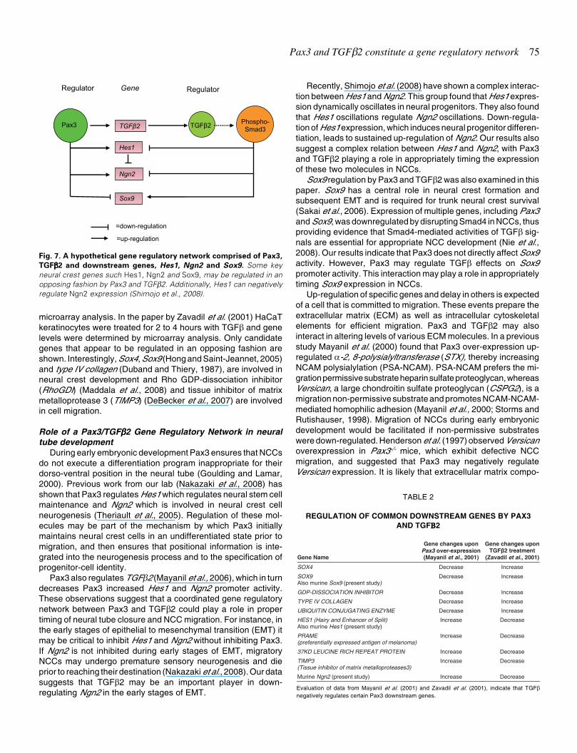
Pax3 and TGFβ2 constitute a gene regulatory network 75
microarray analysis. In the paper by Zavadil et al. (2001) HaCaTkeratinocytes were treated for 2 to 4 hours with TGFβ and genelevels were determined by microarray analysis. Only candidategenes that appear to be regulated in an opposing fashion areshown. Interestingly, Sox4, Sox9 (Hong and Saint-Jeannet, 2005)and type IV collagen (Duband and Thiery, 1987), are involved inneural crest development and Rho GDP-dissociation inhibitor(RhoGDI) (Maddala et al., 2008) and tissue inhibitor of matrixmetalloprotease 3 (TIMP3) (DeBecker et al., 2007) are involvedin cell migration.
Role of a Pax3/TGFβββββ2 Gene Regulatory Network in neuraltube development
During early embryonic development Pax3 ensures that NCCsdo not execute a differentiation program inappropriate for theirdorso-ventral position in the neural tube (Goulding and Lamar,2000). Previous work from our lab (Nakazaki et al., 2008) hasshown that Pax3 regulates Hes1 which regulates neural stem cellmaintenance and Ngn2 which is involved in neural crest cellneurogenesis (Theriault et al., 2005). Regulation of these mol-ecules may be part of the mechanism by which Pax3 initiallymaintains neural crest cells in an undifferentiated state prior tomigration, and then ensures that positional information is inte-grated into the neurogenesis process and to the specification ofprogenitor-cell identity.
Pax3 also regulates TGFβ2 (Mayanil et al., 2006), which in turndecreases Pax3 increased Hes1 and Ngn2 promoter activity.These observations suggest that a coordinated gene regulatorynetwork between Pax3 and TGFβ2 could play a role in propertiming of neural tube closure and NCC migration. For instance, inthe early stages of epithelial to mesenchymal transition (EMT) itmay be critical to inhibit Hes1 and Ngn2 without inhibiting Pax3.If Ngn2 is not inhibited during early stages of EMT, migratoryNCCs may undergo premature sensory neurogenesis and dieprior to reaching their destination (Nakazaki et al., 2008). Our datasuggests that TGFβ2 may be an important player in down-regulating Ngn2 in the early stages of EMT.
Recently, Shimojo et al. (2008) have shown a complex interac-tion between Hes1 and Ngn2. This group found that Hes1 expres-sion dynamically oscillates in neural progenitors. They also foundthat Hes1 oscillations regulate Ngn2 oscillations. Down-regula-tion of Hes1 expression, which induces neural progenitor differen-tiation, leads to sustained up-regulation of Ngn2. Our results alsosuggest a complex relation between Hes1 and Ngn2, with Pax3and TGFβ2 playing a role in appropriately timing the expressionof these two molecules in NCCs.
Sox9 regulation by Pax3 and TGFβ2 was also examined in thispaper. Sox9 has a central role in neural crest formation andsubsequent EMT and is required for trunk neural crest survival(Sakai et al., 2006). Expression of multiple genes, including Pax3and Sox9, was downregulated by disrupting Smad4 in NCCs, thusproviding evidence that Smad4-mediated activities of TGFβ sig-nals are essential for appropriate NCC development (Nie et al.,2008). Our results indicate that Pax3 does not directly affect Sox9activity. However, Pax3 may regulate TGFβ effects on Sox9promoter activity. This interaction may play a role in appropriatelytiming Sox9 expression in NCCs.
Up-regulation of specific genes and delay in others is expectedof a cell that is committed to migration. These events prepare theextracellular matrix (ECM) as well as intracellular cytoskeletalelements for efficient migration. Pax3 and TGFβ2 may alsointeract in altering levels of various ECM molecules. In a previousstudy Mayanil et al. (2000) found that Pax3 over-expression up-regulated α-2, 8-polysialyltransferase (STX), thereby increasingNCAM polysialylation (PSA-NCAM). PSA-NCAM prefers the mi-gration permissive substrate heparin sulfate proteoglycan, whereasVersican, a large chondroitin sulfate proteoglycan (CSPG2), is amigration non-permissive substrate and promotes NCAM-NCAM-mediated homophilic adhesion (Mayanil et al., 2000; Storms andRutishauser, 1998). Migration of NCCs during early embryonicdevelopment would be facilitated if non-permissive substrateswere down-regulated. Henderson et al. (1997) observed Versicanoverexpression in Pax3-/- mice, which exhibit defective NCCmigration, and suggested that Pax3 may negatively regulateVersican expression. It is likely that extracellular matrix compo-
Pax3 TGFβ2
Hes1
Ngn2
Sox9
Regulator Gene Regulator
=down-regulation
=up-regulation
TGFβ2 Phospho-Smad3
Fig. 7. A hypothetical gene regulatory network comprised of Pax3,
TGFβββββ2 and downstream genes, Hes1, Ngn2 and Sox9. Some keyneural crest genes such Hes1, Ngn2 and Sox9, may be regulated in anopposing fashion by Pax3 and TGFβ2. Additionally, Hes1 can negativelyregulate Ngn2 expression (Shimojo et al., 2008).
Gene Name
Gene changes upon Pax3 over-expression (Mayanil et al., 2001)
Gene changes upon TGFβ2 treatment
(Zavadil et al., 2001)
SOX4 Decrease Increase
SOX9 Also murine Sox9 (present study)
Decrease Increase
GDP-DISSOCIATION INHIBITOR Decrease Increase
TYPE IV COLLAGEN Decrease Increase
UBIQUITIN CONJUGATING ENZYME Decrease Increase
HES1 (Hairy and Enhancer of Split) Also murine Hes1 (present study)
Increase Decrease
PRAME (preferentially expressed antigen of melanoma)
Increase Decrease
37KD LEUCINE RICH REPEAT PROTEIN Increase Decrease
TIMP3 (Tissue inhibitor of matrix metalloproteases3)
Increase Decrease
Murine Ngn2 (present study) Increase Decrease
TABLE 2
REGULATION OF COMMON DOWNSTREAM GENES BY PAX3AND TGFΒΒΒΒΒ2
Evaluation of data from Mayanil et al. (2001) and Zavadil et al. (2001), indicate that TGFβnegatively regulates certain Pax3 downstream genes.

76 H. Nakazaki et al.
nents such as Versican V3 are regulated by TGFβ2, given thefact that several extracellular components that affect cell-matrix interactions are remodeled by TGFβ for efficient EMT(Zavadil et al., 2001). An unpublished observation from our labshows that Pax3 down-regulation of Versican V3 (Mayanil etal., 2001) is partially reversed by TGFβ2 treatment.
A final point in relation to extracellular matrix components isthat they could play a key role in making mature TGFβ availableat an appropriate time to induce EMT by negatively regulatingPax3 downstream genes which keep NCCs in a non-migratorystate. ECM components and membrane bound forms of TGFβco-receptors determine bioavailability of TGFβ to NCCs (Dijkeand Arthur, 2007). Thus TGFβ2 may be secreted by NCCs, butmay not be accessible to interact with its receptors on themembrane because of covalent association with the ECM vialatent TGFβ-binding protein (LTBP) (Mangasser-Stephan andGressner, 1999). However at the critical time, ECM bound largelatent TGFβ complex may be released by matr ixmetalloproteinases. The released complex may be targeted tothe cell surface, and proteolytically activated. Interestingly theexpression of tissue inhibitor of matrix metalloprotease 3(TIMP3), which assists in EMT, is increased by Pax3 over-expression and decreased by TGFβ treatment (Table 2).
In this paper we have begun to examine the regulatoryinteraction between Pax3 and TGFβ2. Pax3 and TGFβ2 mayact as a gene regulatory network in neural tube developmentand NCC migration. The role of this type of network is to a
specify sets of genes that must be expressed in specific spatio-temporal patterns (Meulemans and Bronner-Fraser, 2004).This regulatory network, in turn, is most likely a small part of alarger network involved in the highly complex behavior of neuralcrest cells. Fig. 8 is an adaptation of a diagram from Lee et al.(2002). We have modified this diagram based on publisheddata to show different types of networks that may exist indeveloping NCCs. Each of these regulatory motifs in turn, maybe building blocks that can be combined into larger networkstructures.
Further work needs to be done to elucidate the complexrelationship between Pax3 and TGFβ2 in regulating geneexpression levels in NCCs. Regulation of genes downstream ofPax3 and TGFβ2 will involve binding of transcription factors,histones, and other transcriptional regulators to the gene’sregulatory region. The regulatory region may serve as anintegration point, performing a logical computation of transcrip-tion factors to determine resulting gene expression levels(Siggia, 2005). Transcriptional regulatory proteins, which re-cruit and regulate chromatin modifying complexes and compo-nents of the transcription apparatus, will recognize specificpromoter sequences and in turn affect gene expression pro-grams. For instance, Blumenberg et al. (2007) reported thatTGFβ-directed EMT is accompanied by chromatin structureregulation involving Polycomb group epigenetic silencers andhistone-lysine methyl transferase EZH1 and EZH2. To add tothe complexity, Zavadil et al. (2007) reported TGFβ-directed
Fig. 8 . Gene regulatory networks (network motif diagram adapted from Lee et al., 2002, modified for neural crest development). Different networkmotifs may exist in developing neural crest. (A) Auto-regulation: transcription factor Egr4 acts as an auto-regulatory transcription factor (Zipfel et al.,1997). (B) Feed forward loop: Pax3 regulating MyoD (Tajbakhsh et al., 1997) and MyoD regulating Slug (Zhao et al., 2002). Pax3 also regulating Slug(Monsoro-Burq et al., 2005). (C) Multi-component loop: Slug regulating Sox9 and Sox9 regulating Slug (Meulemans and Bronner-Fraser, 2004). (D)
Single input: Slug regulating expression levels of Sox9, Sox10 and FoxD3 (Meulemans and Bronner-Fraser, 2004). (E) Multi-input: seen in neural creststem cell specifiers: Pax3 and Sox10 regulate c-ret (Lang and Epstein, 2003) and Mitf (Lang et al., 2005). Zic1 and Pax3 regulate Foxd3 and Slug(Meulemans and Bronner-Fraser, 2004), and Smad3 and NICD regulate Hes1 (Blokzijl et al., 2003). (F) Regulatory chain: Tead2 regulating Pax3(Milewski et al., 2004), Pax3 regulating MyoD among others (Tajbakhsh et al., 1997); MyoD regulating Slug and Calpain 6 (Zhao et al., 2002).Additionally, a regulatory network based on TGFβ2 regulation of Slug (Romano and Runyan, 2000) can be constructed by overlapping some of themotifs depicted here.
Egr4
MyoD
Slug
Sox9
Slug Sox10
C-Ret Mitf Foxd3
Slug Hes1
Pax3 MyoD
Slug
Calpain 6
FoxD3 Sox9
Autoregulation Feed-forward loop Multi-component loop Single input motif
Multi-input motif Regulatory chain motif
TGFβ2
Ngn2
Hes1
Egr4 Pax3
MyoD
Slug
Sox9
Slug
Zic1 Pax3 Sox10 Msx1 NICD Smad3
Tead2 Pax3 MyoD
A B C D
E F

Pax3 and TGFβ2 constitute a gene regulatory network 77
microRNA: mRNA regulatory circuits in EMT. Future studieswill examine epigenetic features on promoters of common Pax3and TGFβ2 downstream targets, as well as gene expression.
Materials and Methods
Double heterozygous mouse breeding and genotypingThe C57BL/6J-Pax3Sp/J (stock = 2469) male and C57BL/6J breed-
ing pairs were obtained from The Jackson Laboratory. Genotyping ofPax3+/+(wild-type), Pax3+/-heterozygous, and Pax3-/- homozygousembryos was performed on isolated genomic DNA from embryonicmembranes using PCR with a Pax3+/+ (wild-type) or (Pax3-/-) homozy-gous specific reverse primer and a common forward primer in separatePCRs as described by Epstein et al. (1996). TGFβ2 heterozygous andhomozygous genotypes were detected with PCR using the exon 6primers,p5 (AATGTGCAGGATAATTGCTGC) andp3 (AACTCCATAGATATGGGGATGC). The amplification conditionswere 35 cycles at 94°C for 30 seconds, 57°C for 30 seconds and 72°Cfor 90 seconds (Sanford et al., 1997). TGFβ2 heterozygous breedingpairs were kindly provided by Dr. T. Doetschman (Sanford et al., 1997)and were used to maintain the TGFβ2 heterozygous colony and toobtain TGFβ2 nullizygous embryos. Double heterozygous males andfemales were produced by mating TGFβ2+/- heterozygous females withPax3+/- heterozygous males. The double heterozygous males andfemales were back-crossed with wild type Swiss black females andmales respectively for 6 generations. The resulting double heterozy-gous males and females were than used to generate the TGFβ2-/-
Pax3+/- genotypes and other genotypes as per Mendelian projection.
Whole mount in situ hybridizationWhole mount in situ hybridization was done as described earlier
(Mayanil et al., 2006). Washes were performed at 65°C. The antisensedigoxigenin labeled mouse Hes1 riboprobe was created by linearizingwith EcoR1 and synthesized using T7 polymerase. The Hes1 senseriboprobe was made by, linearizing with BamH1 and synthesized usingT3 polymerase. The antisense digoxigenin labeled mouse Ngn2riboprobe was created by linearizing with BamH1 and synthesizedusing T7 polymerase, the Ngn2 sense probe was made by linearizingwith EcoR1 and synthesized using SP6 polymerase. The antisensedigoxigenin labeled mouse Sox9 riboprobe was created by linearizingwith Hind III and synthesized using T7 polymerase. The Sox9 senseriboprobe was made by linearizing with EcoR1 and synthesized usingT3 polymerase. Embryos were stained and photographed with a SpotCamera (Diagnostic Instruments Inc.).
Densitometry was performed on appropriate stained areas, usingthe Open Lab program (Leica). Areas examined were the dorsal neuraltube andÄmigratory neural crest cells in Hes1 riboprobe stained em-bryos, dorsal and ventral tube regions in Ngn2 riboprobe stainedembryos and migratory NCCs in Sox9 riboprobe stained embryos.Staining was quantified from the thoracic/lumbar area downward toinclude the entire caudal region. On an average, 14-15 somites werecounted per embryo of a given genotype. Total density was determinedfrom images scanned in 256 gray scale. The data is represented as thetotal density (8 bit gray), which is defined as the area multiplied by themean of the relative densitometry units in the field under investigation.Total density of stained NCCs or dorsal or ventral regions of the neuraltube were measured, and then the total density of each genotype wasaveraged. The data is expressed as average density of all embryos (+/- SEM) in each genotype category (n= 4 per genotype). Differencesbetween wild type embryos and embryos with other genotypes werecompared by subjecting the data to unpaired T-Test with two-tailed Pvalue (Graph Pad PRISM 4). The data is expressed as the averagedensity of all embryos (n=4 per genotype).
Analysis of Hes1, Ngn2 and Sox9 promoter activity in the presenceand absence of TGFβββββ2
Constructs were kindly provided by: Hes1- promoter luciferaseconstruct, Dr. R. Kageyama, Ngn2-promoter (AF303001) promoterluciferase constructs, Dr. J. Johnson, Sox9-promoter luciferase con-structs Dr. P. Koopman. Hes1- and Ngn2-luciferase and their mutantconstructs are fully described in our earlier work (Nakazaki et al.,2008). Sox9-luciferase and its mutant constructs are described inKanai and Koopman (1999). Each of these constructs (0.2 µg) weretransiently co-transfected into DAOY cells with Pax3-pcDNA3 or pcDNA3vector control and Flag-Smad3 wild type (0.2 µg) or dominant negativeFlag-DN-Smad3 (0.2 µg) cloned in HSV-TK-pEXL vector (kindly pro-vided by Dr. H. F. Lodish, MIT). DAOY cells were used to maintainconsistency with previous experiments done in our laboratory (Mayanilet al., 2000; 2001; 2006 and Nakazaki et al., 2008). 24 hrs post-transfection, transfected DAOY cells were treated or not treated with10ng/ml human recombinant TGFβ2 for 6 hrs. Luciferase activity wasmeasured 48 hrs post-transfection using the Dual Luciferase Kit fromPromega, as previously described (Mayanil et al., 2006). Transfectedcells not treated with TGFβ2 served as the negative control.
Comparison of common downstream genes regulated by Pax3 orTGFβββββ2
Microarray data from Mayanil et al. (2001) and Zavadil et al. (2001)were compared. Examination of 270 genes altered by Pax3, as exam-ined in Pax3 stably transfected DAOY cells (Mayanil et al., 2001), and728 genes, in HaCaT keratinocytes, altered by 2 to 4 hour treatmentwith TGFβ (Zavadil et al., 2001), yielded 58 genes that were altered inboth studies. To increase stringency 2 fold or higher increase ordecrease in expression levels was used as a cut off. This resulted in atotal of 22 genes altered by both Pax3 overexpression and TGFβtreatment. From these 22, 10 candidate genes, 5 up-regulated and 5down-regulated by Pax3 (Mayanil et al., 2001) and oppositely regu-lated by TGFβ treatment (Zavadil et al., 2001) are shown in Table 2. Outof these 10 genes, we used three for our in situ hybridization andpromoter-luciferase studies. Several genes not listed in Table 2 didshow up-regulation or down-regulation in response to both Pax3overexpression and TGFβ treatment.
AcknowledgementsThe authors would like to thank Dr. P. Gruss Max Planck Institute,
Gottingen, Germany, for the pBH3.2 plasmid containing Pax3; Dr. N. LBrown, Division of Developmental Biology, Departments of Pediatricsand Ophthalmology, University of Cincinnati College of Medicine,Cincinnati, OH, USA, for pBluescriptHes1 and pGEM3Ngn2 plasmids;Dr. de Crombrugghe, University of Texas, MD Anderson CancerCenter, and Department of Molecular Genetics, Houston Texas formurine Sox9 cDNA probe. TGFβ2+/- heterozygous breeding pairs wereprovided by Dr. T. Doetschman, Department of Molecular Genetics,Biochemistry and Microbiology, University of Cincinnati, Ohio. Theauthors also wish to thank Dr. R. Kageyama, Institute for Virus Re-search, Kyoto University for Hes1-promoter luciferase constructs; Dr.J. Johnson, Department of Neuroscience, University of Texas South-western Medical Center, Dallas, TX for Ngn2-promoter-luciferase con-structs; and Dr. P. Koopman, Division of Molecular Genetics andDevelopment, Institute for Molecular Bioscience, The University ofQueensland, Brisbane, Australia for Sox9-promoter-luciferase con-structs. The Flag-Smad3 and Flag-DN-Smad3 constructs were giftsfrom Dr H.F. Lodish, Whitehead Institute for Biomedical Research,Department of Biology, Massachusetts Institute of Technology. Theauthors also wish to thank Dr. J. A. Kessler, Feinberg School ofMedicine, Northwestern University, and Dr. S. Ahlgren DevelopmentalBiology Program, Childrens Memorial Research Center, Chicago forvaluable discussions, Dr H. Chung for statistical assistance and BillGoossens for microscope assistance. This work was supported by the

78 H. Nakazaki et al.
State of Illinois Excellence in Academic Medicine award (CSKM) anda Grant from the Spastic Paralysis Research Foundation of Illinois-Eastern Iowa District of Kiwanis (CSKM and DGM). The earlier prelimi-nary part of the work described in this paper was done in the Neurobi-ology Program at Childrens Memorial Research Center, Chicago, IL60614.
References
AUERBACH, R. (1954). Analysis of the developmental defects of a lethal mutationin the house mouse. J. Exp. Zool. 127: 305-329.
BLOKZIJL, A., DAHLQVIST, C., REISSMANN, E., FALK, A., MOLINER, A.,LENDHAL, U. and IBANEZ, C. F. (2003). Cross-talk between Notch and TGFβ2signaling pathways mediated by interaction of Notch intracellular domain withSmad3. J. Cell. Biol. 163: 723-728.
BLUMENBERG, M., GAO, S., DICKMAN, K., GROLLMAN, A. P., BOTTINGER, E.P. and ZAVADIL, J. (2007). Chromatin structure regulation in transforminggrowth factor-beta-directed epithelial-mesenchymal transition. Cells TissueOrgans 185:162-174.
CHEUNG, M. and BRISCOE, J. (2003). Neural crest development is regulated bythe transcription factor Sox9. Development 130:5681-5693.
DEBECKER, A., VAN HUMMELEN, P., BAKKUS, M., VANDE BROEK, I., DEWEVER, J., DE WAELE, M., VAN RIET, I. (2007). Migration of culture-expanded human mesenchymal stem cells through bone marrow endotheliumis regulated by matrix metalloproteinase-2 and tissue inhibitor ofmetalloproteinase-3. Haematologica 92: 440-449.
DIJKE P. T. and ARTHUR, H. M. (2007). Extracellular control of TGFβ signaling invascular development and disease. Nature Rev Mol. Cell. Biol. 7: 857-869.
DUBAND, J. L. and THIERY, J. P. (1987). Distribution of laminin and collagensduring avian neural crest development. Development 101: 461-478.
EPSTEIN, D. J. VEKEMANS, M. and GROS, P. (1991). Splotch (Sp2H), a mutationaffecting development of the mouse neural tube, shows a deletion within thepaired homeodomain of Pax-3. Cell 67: 767-774.
EPSTEIN, J. A., SHAPIRO, D. N., CHENG, J., LAM, P. Y. P. and MAAS, R. L.(1996). Pax3 modulates expression of the c-Met receptor during limb muscledevelopment. J. Biol. Chem. 93: 4213-4218.
FODE, C., GRADWOHL, G., MORIN, X., DIERICH, A., LEMEUR, M., GORIDIS, C.and GUILLEMOT, F. (1998). The bHLH protein NEUROGENIN2 is a determi-nation factor for epibranchial placode-derived sensory neurons. Neuron 20:483-494.
FOY, C., NEWTON, V., WELLESLEY, D., HARRIS, R. and READ, A. P. (1990).Assignment of the locus for Waardenburg syndrome type I to human chromo-some 2q37 and possible homology to the Splotch mouse. Am. J. Hum. Genet.46:1017-1023.
GARCÍA-CAMPMANY, L. and MARTÍ, M. (2007). The TGFβ intracellular effectorSmad3 regulates neuronal differentiation and cell fate specification in thedeveloping spinal cord. Development 134: 65-75.
GOULDING, M. D., CHALEPAKIS, G., DEUTSCH, U., ERSELIUS, J. R. andGRUSS, P. (1991). Pax-3, a novel murine DNA binding protein expressedduring early neurogenesis. EMBO J. 10: 1135-1147.
GOULDING, M. D., STERRER, S., FLEMING, J., BALLING, R., NADEAU, J.,MOORE, K. J., BROWN, S., STEEL, K. P. and GRUSS, P. (1993). Analysis ofthe Pax-3 gene in the mouse mutant splotch. Genomics 17: 355-363.
GOULDING, M. and LAMAR, E. (2000). Neuronal patterning: Making stripes in thespinal cord. Curr. Biol. 10:R565-R658.
HENDERSON, D. J., YBOT-GONZALEZ, P. and COPP, A. J. (1997). Over-expression of the chondroitin sulphate proteoglycan Versican is associated withdefective neural crest migration in the Pax3 mutant mouse (splotch). Mech. Dev.69: 39-51.
HIRATA, H., TOMITA, K., BESSHO, Y. and KAGEYAMA, R. (2001). Hes1 and Hes3regulate maintenance of the isthmic organizer and development of the mid/hindbrain. EMBO J. 20: 4454-4466.
HIRATA, H., YOSHIURA, S., OHTSUKA, T., BESSHO, Y., HARADA, T.,YOSHIKAWA, K. and KAGEYAMA, R. (2002). Oscillatory expression of thebHLH factor Hes1 regulated by a negative feedback loop. Science 298: 840-
843.
HONG, C. S., and SAINT-JEANNET, J. P. (2005). Sox proteins and neural crestdevelopment. Seminars in Cell and Dev. Biol. 16: 694-703.
HOSOKAWA, R., URATA, M., HAN, J., ZEHNALY, A., BRINGAS, P JR, NONAKA,K., and CHAI, Y. (2007). TGF-beta mediated Msx2 expression controls occipitalsomites-derived caudal region of skull development. Dev. Biol. 310: 140-153.
KANAI, Y., and KOOPMAN, P. (1999). Structural and functional characterization ofthe mouse Sox9 promoter: implications for campomelic dysplasia. Hum. Mol.Genet. 8: 691-696.
LANG, D. and EPSTEIN, J. A. (2003). Sox10 and Pax3 physically interact tomediate activation of a conserved c-RET enhancer. Hum. Mol. Genet. 12: 937-945.
LANG, D., LU, M. M., HUANG, L., ENGLEKA, K. A., ZHANG, M., CHU, E. Y.,LIPNER, S., SKOULTCHI, A., MILLAR, S. E. and EPSTEIN, J. A. (2005). Pax3functions at the nodal point in melanocyte stem cell differentiation. Nature 433:884-887.
LEE, T. I., RINALDI, N. J., ROBERT, F., ODOM, D. T., BAR-JOSEPH, Z., GERBER,G. K., HANNETT, N. M., SIMAON, I., ZEITLINGER, J., JENNINGS, E. G.,MURRAY, H. L., GORDON, D.B., REN, B., WYRICK, J. J., TAGNE, J-B.,VOLKERT, T. L., FRAENKEL, E., GIFFORD, D. K. and YOUNG, R. A. (2002).Transcriptional Regulatory networks in Saccharomyces cerevisiae. Science298: 799-804.
LI, J., LIU, K. C., JIN, F., LU, M. M. and EPSTEIN, J. A. (1999). Transgenic rescueof congenital heart disease and spina bifida in Splotch mice. Development 126:2495-503.
LO, L., DORMAND, E., GREENWOOD, A., and ANDERSON, D. J. (2002). Com-parison of the generic neuronal differentiation and neuron subtype specificationfunctions of mammalian achaete-scute and atonal homologs in cultured neuralprogenitor cells. Development 29: 1553-1567.
MADDALA, R., RENEKER, L. W., PENDURTHI, B., and RAO, P. V. (2008). RhoGDP dissociation inhibitor-mediated disruption of Rho GTPase activity impairslens fiber cell migration, elongation and survival. Dev. Biol. 315: 217-231.
MANGASSER-STEPHAN, K. and GRESSNER, A. M. (1999). Molecular andfunctional aspects of latent transforming growth factor-beta binding protein: justa masking protein? Cell Tissue Res. 297: 363-370.
MAYANIL, C. S. K., GEORGE, D., MANIA-FARNELL, B., BREMER, C. L., MCLONE,D. G. and BREMER, E. G. (2000). Overexpression of murine Pax3 increasesNCAM polysialylation in a human medulloblastoma cell line. J. Biol. Chem. 275:23259-23266.
MAYANIL, C. S. K., GEORGE, D., FREILICH, L., MILJAN, E. J., MANIA-FARNELL.,MCLONE, D. G. and BREMER, E. G. (2001). Microarray analysis detects novelPax3 downstream target genes. J. Biol. Chem. 52: 49299-49309.
MAYANIL, C. S. K., POOL, A., NAKAZAKI, H., REDDY, A., MANIA-FARNELL, B.,YUN, B., GEORGE, D., MCLONE, D. G. and BREMER, E. G. (2006). Regulationof murine TGFβ2 by Pax3 during early embryonic development. J. Biol. Chem.281: 24544–24552.
MEULEMANS, D. and BRONNER-FRASER, M. (2004). Gene-regulatory Interac-tions in neural crest evolution and development. Developmental Cell 7: 291-299.
MILEWSKI, R. C, CHI, N. C., LI, J., BROWN, C., LU, M. M. and EPSTEIN, J. A.(2004). Identification of minimal enhancer elements sufficient for Pax3 expres-sion in neural crest and implication of Tead2 as a regulator of Pax3. Develop-ment 131:829-837.
MONSORO-BURQ, A-H., WANG, E. and HARLAND, R. (2005). Msx1 and Pax3cooperate to mediate FGF8 and Wnt signals during Xenopus neural crestinduction. Developmental Cell 8: 167-178.
NAKAZAKI, H., REDDY, A., MANIA-FARNELL, B., YUEH-WEI, S., MCCABE C.,ICHI, S., GEORGE, D., MCLONE, D. G., TOMITA, T. and MAYANIL, C. S. K.(2008). Key basic helix loop helix transcription factor genes Hes1 and Ngn2 areregulated by Pax3 during mouse embryonic development. Dev. Biol. 316: 510-523.
NIE, X., DENG, C. X., WANG, Q. and JIAO, K. (2008). Disruption of Smad4 in neuralcrest cells leads to mid-gestation death with pharyngeal arch, craniofacial andcardiac defects. Dev. Biol. 316: 417-430.
ROMANO, L. and RUNYAN, R. B. (2000). Slug is an essential target of TGFβ2signaling in the developing chicken heart. Dev. Biol. 223: 91-102.

Pax3 and TGFβ2 constitute a gene regulatory network 79
SAKAI, D., SUZUKI, T., OSUMI, N., and WAKAMATSU, Y. (2006). Cooperativeaction of Sox9, Snail2 and PKA signaling in early neural crest development.Development 133:1323-1333.
SANFORD, L. P., ORMSBY, I., GITTENBERGER-DE GROOT A. C., SARIOLA, H.,FRIEDMAN, R., BOIVIN, G. P., CARDELL, E. L. and DOETSCHMAN, T. (1997).TGFbeta2 knockout mice have multiple developmental defects that are non-overlapping with other TGFbeta knockout phenotypes. Development 124:2659-2670.
SHIMOJO, H., OHTSUKA, T. and KAGEYAMA, R. (2008). Oscillations in NotchSignaling Regulate Maintenance of Neural Progenitors. Neuron 58: 52-64.
SIGGIA, E. D. (2005). Computational methods for transcriptional regulation. Curr.Opin. Genet. Dev. 15: 214-221.
SIMMONS, A. D., HORTON, S., ABNEY, A. L. and JOHNSON, J. E. (2001).Neurogenin2 expression in ventral and dorsal spinal neural tube progenitor cellsis regulated by distinct enhancers. Dev. Biol. 229: 327-339.
STORMS, S. D. and RUTISHAUSER, U. (1998). A role for polysialic acid in neuralcell adhesion molecule heterophilic binding to proteoglycans. J. Biol. Chem.273: 27124-27149.
TAJBAKHSH, S., ROCANCOURT, D., COSSU, G. and BUCKINGHAM, M. (1997).
Redefining the genetic hierarchies controlling skeletal myogenesis: Pax-3 andMyf-5 act upstream of MyoD. Cell 89: 127-138.
THERIAULT, F. M., NUTHALL, H. N., DONG, Z., LO, R., BARNABE-HEIDER, F.,MILLER, F. D. and STIFANI, S. (2005). Role for Runx1 in the proliferation andneuronal differentiation of selected progenitor cells in the mammalian nervoussystem. J. Neurosci. 25: 2050-2061.
ZAVADIL, J., BITZER, M., LANG, D., YANG, Y-C., MASSIMI, A., KNEITZ, S., PIEK,E. AND BOTTINGER, E.P. (2001). Genetic programs of epithelial cell plasticitydirected by transforming growth factor-β. Proc. Natl. Acad. Sci. (USA) 98: 6686-6691.
ZAVADIL, J., NARASIMHAN, M., BLUMENBERG, M. and SCHNEIDER, R. J.(2007). Transforming growth factor-beta and microRNA: mRNA regulatorynetworks in epithelial plasticity. Cells Tissues Organs 185: 157-161.
ZHAO, P., IEZZI, S., CAVER, E., DRESSMAN, D., GRIDLEY, T., SARTORELLI, V.and HOFFMAN, E. P. (2002). Slug is a novel downstream target of MyoD:Temporal profiling in muscle regeneration. J. Biol. Chem. 277: 30091-30101.
ZIPFEL, P. F., DECKER, E. L., HOLST, C. and SKERKA, C. (1997). The human zincfinger protein EGR-4 acts as auto regulatory transcriptional repressor. Biochim.Biophys. Acta 1354: 134-144.

80 H. Nakazaki et al.
Further Related Reading, published previously in the Int. J. Dev. Biol.
See our Special Issue The Nogent Institute in honor of Nicole Le Douarin and edited by Francoise Dieterlen at:http://www.ijdb.ehu.es/web/contents.php?vol=49&issue=2-3
See our recent Special Issue Fertilization, in honor of David L. Garbers and edited by Paul M. Wassarman and Victor D. Vacquier at:http://www.ijdb.ehu.es/web/contents.php?vol=52&issue=5-6
Epithelial-Mesenchymal Transitions in development and disease: old views and new perspectivesM. Angela NietoInt. J. Dev. Biol. In Press doi: 10.1387/ijdb.072410mn
Perichondrial-mediated TGF-beta regulation of cartilage growth in avian long bone developmentMarsha L. Crochiere, James K. Kubilus and Thomas F. LinsenmayerInt. J. Dev. Biol. (2008) 52: 63-70
Equivalent genetic regulatory networks in different contexts recover contrasting spatial cell patterns that resemble those inArabidopsis root and leaf epidermis: a dynamic modelMariana Benítez, Carlos Espinosa-Soto, Pablo Padilla-Longoria, José Díaz and Elena R. Alvarez-BuyllaInt. J. Dev. Biol. (2007) 51: 139-155
Comparative expression analysis of Pax3 and Pax7 during mouse myogenesisDavid Horst, Svetlana Ustanina, Consolato Sergi, Gregor Mikuz, Herbert Juergens, Thomas Braun and Eugene Vorobyov
2006 ISI **Impact Factor = 3.577**
Int. J. Dev. Biol. (2006) 50: 47-54
Genetic interaction between Lef1 and Alx4 is required for early embryonicdevelopmentKata Boras-Granic, Rudolf Grosschedl and Paul A. HamelInt. J. Dev. Biol. (2006) 50: 601-610
Long-range signalling in plant reproductive developmentPaula Suárez-LópezInt. J. Dev. Biol. (2005) 49: 761-771
Gene network analysis in plant development by genomic technologiesFrank Wellmer and José Luis RiechmannInt. J. Dev. Biol. (2005) 49: 745-759
Historical perspectives on plant developmental biologyMieke Van Lijsebettens and Marc Van MontaguInt. J. Dev. Biol. (2005) 49: 453-465
Contributions by members of the TGFbeta superfamily to lens developmentDavid Beebe, Claudia Garcia, Xiaohui Wang, Ramya Rajagopal, Mary Feldmeier, Ji-Young Kim, Anna Chytil, Harold Moses, Ruth Ashery-Padan, and Michael RauchmanInt. J. Dev. Biol. (2004) 48: 845-856
Developmental gene network analysis.Roger Revilla-i-Domingo and Eric H DavidsonInt. J. Dev. Biol. (2003) 47: 695-703
Transcriptional regulation and the evolution of development.Gregory A WrayInt. J. Dev. Biol. (2003) 47: 675-684
Evolution of cis-regulatory regions versus codifying regions.Francisco Rodríguez-Trelles, Rosa Tarrío and Francisco J AyalaInt. J. Dev. Biol. (2003) 47: 665-673
Developmental basis for vein pattern variations in insect wings.José F De Celis and Fernando J Diaz-BenjumeaInt. J. Dev. Biol. (2003) 47: 653-663