Transcriptional regulation and immunity in mycobacteriophage Bxb1: Mycobacteriophage Bxb1 gene...
15
Molecular Microbiology (2000) 38(5), 971–985 Transcriptional regulation and immunity in mycobacteriophage Bxb1 Shruti Jain and Graham F. Hatfull* Department of Biological Sciences, University of Pittsburgh, Pittsburgh, PA 15260, USA. Summary Mycobacteriophage Bxb1 is a temperate phage of Mycobacterium smegmatis that shares a similar genome organization to mycobacteriophage L5, although the two phages are heteroimmune. We have investigated the regulatory circuitry of Bxb1 and found that it encodes a repressor, gp69, which regulates at least two promoters, an early lytic promoter, P left , and the divergent promoter, P right . Bxb1 gp69 is 41% identical to the L5 repressor (gp71) and binds to repressor binding sites that conform to a similar, but distinct, 13 bp asymmetric consensus sequence to that for the L5 gp71 binding sites. The two phage repressors have a strong preference for their cognate binding sites, thus accounting for their immunity phenotypes. The Bxb1 genome contains 34 putative repressor binding sites located throughout the genome, but situated within short intergenic spaces and orientated in only one direction relative to the direction of transcription. Comparison with the locations of repressor binding sites within the L5 genome provides insights into how these unusual regulatory systems evolve. Introduction Temperate bacteriophages can pursue two alternative developmental routes upon infection of a bacterial host cell: lytic growth, which results in the generation and release of progeny phage particles and cell death; and lysogeny, in which the lytic functions are switched off and the phage DNA (usually, although not always) integrates into the host chromosome. In the well-studied group of lambda-like phages, maintenance of the lysogenic state requires the expression of a repressor that binds to two tripartite operator sites to downregulate the activity of the early lytic promoters P L and P R (Ptashne, 1987) and to regulate its own synthesis (Ptashne et al., 1976). The repressor also acts to confer immunity to superinfection by homoimmune phage particles by binding to cognate operator sites present on the genome of superinfecting phages. However, the regulation of lysogenic mainte- nance and superinfection immunity by repressors is a common theme among a diverse array of temperate bacteriophages (Scott et al., 1978; Dhaese et al., 1985; Ladero et al., 1998; Salmi et al., 1998; Kameyama et al., 1999; Nesper et al., 1999). Mycobacteriophage L5 is a temperate phage of the mycobacteria that uses a very different immunity system from that of the lambda-like phages (Donnelly-Wu et al., 1993; Hatfull, 1994; 1999; Nesbit et al., 1995; Brown et al., 1997). The L5 repressor (gp71) is a 183-amino-acid protein that contains a putative helix–turn–helix DNA binding motif near its N-terminus and recognizes a 13 bp asymmetric binding site (Donnelly-Wu et al., 1993; Brown et al., 1997). Only one early lytic promoter has been identified in L5 (P left ), and this is under direct negative regulation by gp71 (Nesbit et al., 1995). However, there is just a single gp71 binding site overlapping the 235 region of this promoter, and this operator appears to be sufficient for regulation of P left by L5 gp71 (Brown et al., 1997). An unusual feature of the regulatory system in L5 is that the genome contains a large number of repressor binding sites (Brown et al., 1997). There are at least 29 additional sites with close sequence similarity to the P left operator, and gp71 binds to at least 24 of the 30 sites. These sites conform closely to a 13 bp consensus sequence (5 0 - GGTGGc/aTGTCAAG), and all the 24 sites contain the consensus nucleotide in eight of the 13 positions (Brown et al., 1997). These sites are found throughout the L5 genome, are located within short intergenic regions and are situated in only one orientation relative to the direction of transcription (Brown et al., 1997). They are referred to as ‘stoperators’ because, when the gp71 repressor is bound, the elongation of transcription complexes is inhibited; the strong orientation dependence of this effect and the rather modest affinity of L5 gp71 for its binding sites (K d < 5 10 28 M) suggests an active mechan- ism rather than a simple passive block. Mycobacterioph- age D29, a close and homoimmune relative of L5, contains a similar set of sites, which conform to the same consensus sequence and appear to be functionally identical (Ford et al., 1998). The Streptomyces phage fC31 also has multiple repressor binding sites, most of which are also present in intergenic regions, although their specific role in lysogenic maintenance is not clear Q 2000 Blackwell Science Ltd Accepted 8 September, 2000. *For correspondence. E-mail gfh@pitt. edu; Tel. (11) 412 624 6975; Fax (11) 412 624 4870.
-
Upload
independent -
Category
Documents
-
view
1 -
download
0
Transcript of Transcriptional regulation and immunity in mycobacteriophage Bxb1: Mycobacteriophage Bxb1 gene...

Molecular Microbiology (2000) 38(5), 971±985
Transcriptional regulation and immunity inmycobacteriophage Bxb1
Shruti Jain and Graham F. Hatfull*
Department of Biological Sciences, University of
Pittsburgh, Pittsburgh, PA 15260, USA.
Summary
Mycobacteriophage Bxb1 is a temperate phage of
Mycobacterium smegmatis that shares a similar
genome organization to mycobacteriophage L5,
although the two phages are heteroimmune. We
have investigated the regulatory circuitry of Bxb1
and found that it encodes a repressor, gp69, which
regulates at least two promoters, an early lytic
promoter, Pleft, and the divergent promoter, Pright.
Bxb1 gp69 is 41% identical to the L5 repressor (gp71)
and binds to repressor binding sites that conform to
a similar, but distinct, 13 bp asymmetric consensus
sequence to that for the L5 gp71 binding sites. The
two phage repressors have a strong preference for
their cognate binding sites, thus accounting for their
immunity phenotypes. The Bxb1 genome contains 34
putative repressor binding sites located throughout
the genome, but situated within short intergenic
spaces and orientated in only one direction relative
to the direction of transcription. Comparison with the
locations of repressor binding sites within the L5
genome provides insights into how these unusual
regulatory systems evolve.
Introduction
Temperate bacteriophages can pursue two alternative
developmental routes upon infection of a bacterial host
cell: lytic growth, which results in the generation and
release of progeny phage particles and cell death; and
lysogeny, in which the lytic functions are switched off and
the phage DNA (usually, although not always) integrates
into the host chromosome. In the well-studied group of
lambda-like phages, maintenance of the lysogenic state
requires the expression of a repressor that binds to two
tripartite operator sites to downregulate the activity of the
early lytic promoters PL and PR (Ptashne, 1987) and to
regulate its own synthesis (Ptashne et al., 1976). The
repressor also acts to confer immunity to superinfection
by homoimmune phage particles by binding to cognate
operator sites present on the genome of superinfecting
phages. However, the regulation of lysogenic mainte-
nance and superinfection immunity by repressors is a
common theme among a diverse array of temperate
bacteriophages (Scott et al., 1978; Dhaese et al., 1985;
Ladero et al., 1998; Salmi et al., 1998; Kameyama et al.,
1999; Nesper et al., 1999).
Mycobacteriophage L5 is a temperate phage of the
mycobacteria that uses a very different immunity system
from that of the lambda-like phages (Donnelly-Wu et al.,
1993; Hatfull, 1994; 1999; Nesbit et al., 1995; Brown et al.,
1997). The L5 repressor (gp71) is a 183-amino-acid
protein that contains a putative helix±turn±helix DNA
binding motif near its N-terminus and recognizes a 13 bp
asymmetric binding site (Donnelly-Wu et al., 1993; Brown
et al., 1997). Only one early lytic promoter has been
identified in L5 (Pleft), and this is under direct negative
regulation by gp71 (Nesbit et al., 1995). However, there is
just a single gp71 binding site overlapping the 235 region
of this promoter, and this operator appears to be sufficient
for regulation of Pleft by L5 gp71 (Brown et al., 1997).
An unusual feature of the regulatory system in L5 is that
the genome contains a large number of repressor binding
sites (Brown et al., 1997). There are at least 29 additional
sites with close sequence similarity to the Pleft operator,
and gp71 binds to at least 24 of the 30 sites. These sites
conform closely to a 13 bp consensus sequence (5 0-GGTGGc/aTGTCAAG), and all the 24 sites contain the
consensus nucleotide in eight of the 13 positions (Brown
et al., 1997). These sites are found throughout the L5
genome, are located within short intergenic regions and
are situated in only one orientation relative to the direction
of transcription (Brown et al., 1997). They are referred
to as `stoperators' because, when the gp71 repressor
is bound, the elongation of transcription complexes is
inhibited; the strong orientation dependence of this effect
and the rather modest affinity of L5 gp71 for its binding
sites (Kd � < 5 � 1028 M) suggests an active mechan-
ism rather than a simple passive block. Mycobacterioph-
age D29, a close and homoimmune relative of L5,
contains a similar set of sites, which conform to the
same consensus sequence and appear to be functionally
identical (Ford et al., 1998). The Streptomyces phage
fC31 also has multiple repressor binding sites, most of
which are also present in intergenic regions, although
their specific role in lysogenic maintenance is not clear
Q 2000 Blackwell Science Ltd
Accepted 8 September, 2000. *For correspondence. E-mail [email protected]; Tel. (11) 412 624 6975; Fax (11) 412 624 4870.

(Ingham et al., 1994; Wilson et al., 1995; Smith et al.,
1999).
The biological role of the stoperator sites is uncertain,
although we presume that they act to silence the
prophage genome in lysogeny. In this respect, we note
that lambda and its relatives contain a large number of
rho-dependent and rho-independent terminators that are
not only present in the regulatory regions, but are found
throughout the genome (Juhala et al., 2000). These
terminators presumably act, at least in part, to maintain a
low transcriptional state of the prophage genome,
although they do not present an impediment to lytic
growth because of the N- and Q-mediated antitermination
systems (Ptashne, 1987). Although the L5 genome does
contain some recognizable factor-independent termina-
tors (see Mediavilla et al., 2000), these are all located
around the ends of the early and late operons and are not
found within them (Hatfull and Sarkis, 1993). Mycobacter-
iophage antitermination systems have not been studied,
but there is no obvious regulatory necessity for their
inclusion in these genomes. It therefore seems likely that
many of the lambda terminators act like L5 stoperators to
confer a low level of expression of phage genes from
prophage genomes.
In this paper, we describe the immunity system of
mycobacteriophage Bxb1, a phage that is related to L5
but shares little DNA sequence similarity (Barletta et al.,
1992). Bxb1 and L5 are heteroimmune as a result of the
specificity of their respective repressors, which bind with a
strong preference to their cognate binding sites. Bxb1
contains even more putative binding sites than L5 but, as
in L5, they are located within intergenic regions and in
only one orientation relative to the direction of transcrip-
tion. A comparison of the Bxb1 regulatory circuitry and the
location and nature of the repressor binding sites relative
to those in L5 provides insights into these regulatory
systems and how they may evolve.
Results
Identification of the Bxb1 repressor protein
From the analysis of the Bxb1 genome sequence, it was
found that gene 69 encodes a protein that shares 41%
identity with the L5 repressor gp69 (Mediavilla et al.,
2000). Bxb1 gp69 is 170 amino acids long and can be
aligned with L5 gp71 without gaps except for the extreme
N- and C-termini (Fig. 1). In L5 gp71, there is a putative
helix±turn±helix motif close to the N-terminus, which is
presumably involved in DNA recognition, as amino acid
substitutions in it lead to a loss of DNA binding (K. Brown
and G. F. Hatfull, unpublished observations). A related
sequence is present in Bxb1 gp69, with over half the
residues being identical, including the three amino acids
in the `turn'. The most notable difference between the two
motifs is the first position of the second helix, which is
usually associated with making direct contact with DNA at
the recognition site (Harrison and Aggarwal, 1990). In L5
gp71, this position is an arginine, whereas in Bxb1 gp69, it
is a proline residue, an unusual amino acid to find in this
position, although its presence is not unprecedented
(Laughon and Scott, 1984; Harrison and Aggarwal, 1990).
These differences are likely to contribute to changes in
sequence recognition by these proteins. We noted
previously that the C-terminal part of L5 gp71 has an
abundance of acidic amino acids (Donnelly-Wu et al.,
1993), but this feature is much less evident in Bxb1 gp69,
suggesting that this may not be an important aspect of the
function of these repressors.
Bxb1 gene 69 was inserted into the T7 overexpression
vector pET21a, and the resulting plasmid (pSJ6) was
used to overexpress Bxb1 gp69. Good levels of gp69
synthesis were observed after induction with IPTG,
although the protein migrates with an apparent molecular
mass of 27 kDa, somewhat greater than its predicted
mass of 18.7 kDa, a property that is shared with L5 gp71
(K. Brown and G. F. Hatfull, unpublished observations).
Bxb1 gp69 was purified to . 90% homogeneity by
chromatography using a BioCAD SPRINT system (data
not shown).
Identification of the Bxb1 Pleft promoter
In L5, there is one major promoter, Pleft, which is regulated
directly by the gp71 repressor (Nesbit et al., 1995), and it
seems likely that Bxb1 would have a similar promoter that
is responsible for early lytic gene expression. In L5, the
Pleft promoter is located upstream of gene 89, the first
open reading frame (ORF) in the leftwards operon, at co-
ordinate 51, 672 (Fig. 2A). However, an analogous Bxb1
promoter cannot easily be identified by sequence com-
parison of the genomes, as these segments are not
closely related and the genome organizations of L5 and
Bxb1 are somewhat different at the righthand ends
(Fig. 2A; see Mediavilla et al., 2000). In particular, there
are three small genes transcribed in the rightwards
direction at the extreme right end of the Bxb1 genome
for which there are no obvious counterparts in L5.
Although these genes are small (encoding putative
protein products of 7.2 kDa, 5.6 kDa and 6.8 kDa), the
veracity of the assignments is supported by the observa-
tion that Bxb1 gp84 has significant sequence similarity
(76% identity) to L5 gp79. It therefore seems probable
that a promoter for leftwards transcription of the early
genes (analogous to L5 Pleft) is located between the
divergently transcribed genes 83 and 84.
A DNA segment containing this intergenic region (co-
ordinates 48 657±48 888) was amplified by polymerase
972 S. Jain and G. F. Hatfull
Q 2000 Blackwell Science Ltd, Molecular Microbiology, 38, 971±985

chain reaction (PCR) and inserted into the integration-
proficient shuttle vector pDK16 (Table 1), which contains
a promoterless lacZ reporter gene (Fig. 2B). The resulting
plasmid (pSJ9) efficiently transforms Mycobacterium
smegmatis to give colonies with a blue colour on Xgal
plates, and b-galactosidase assays indicate that it has a
modest level of promoter activity (Fig. 2B). However,
when pSJ9 was introduced into a Bxb1 lysogen, the
colonies were only pale blue on Xgal plates and had . 30-
fold less b-galactosidase activity than the non-lysogen
(Fig. 2B). To determine whether this reduction in activity
was caused by the action of the Bxb1 repressor, we
inserted a copy of Bxb1 gene 69 into pSJ9 (to give pSJ15)
and introduced this plasmid into M. smegmatis. The
resulting colonies were also pale blue on Xgal plates
and have only low levels of b-galactosidase activity. We
conclude that this 232 bp segment of the Bxb1 genome
contains a leftwards-facing promoter that is directly
regulated by the Bxb1 repressor.
The same segment of the Bxb1 genome was also
Fig. 2. Identification of the Bxb1 Pleft
promoter.A. Organization of the right ends of the L5and Bxb1 genomes. Segments of < 4 kbp ofthe L5 and Bxb1 genomes are shown with co-ordinates above each genome. The positionsof putative genes are shown as grey boxes.Lightly shaded genes below the genome aretranscribed leftwards, and darkly shaded onesabove are transcribed rightwards. Thepositions of the Pleft promoters are shown.B. Regulation of the Bxb1 Pleft promoter.Plasmids containing various segments ofBxb1 DNA were introduced into eitherM. smegmatis mc2155 or a Bxb1 lysogen[mc2155 (Bxb1)] and the b-galactosidaseactivities determined. Plasmids pSJ23 andpSJ22 contain 232 bp and 126 bp fragmentsof Bxb1 DNA, respectively, fused to lacZ inthe extrachromosomal vector pSD5B. PlasmidpSJ9 contains the same 232 bp fragment asin pSJ23 but fused to lacZ in the integratingvector pDK16. Plasmids pSJ24 and pSJ15are similar to pSJ23 and pSJ9, respectively,but also contain a copy of the Bxb1 repressorgene 69. The b-galactosidase activities areaverages from three individual transformants,except for those indicated (*), in which therange of activities is reported. b-Galactosidase activities were determined asdescribed in Experimental procedures and areexpressed as nmol min21 mg21 total protein.ND denotes the assays not done.
Fig. 1. Sequence alignment of the repressorproteins of mycobacteriophage Bxb1 (gp69)and L5 (gp71). Amino acid identities andsimilarities are indicated by colons and dotsrespectively. Dashes indicate gaps insertedfor optimal alignment of the sequences. Thetwo helices of the putative helix±turn±helixDNA-binding motifs are underlined.
Mycobacteriophage Bxb1 gene expression 973
Q 2000 Blackwell Science Ltd, Molecular Microbiology, 38, 971±985

Table 1. Plasmids used in this study.
Plasmid Description Source or reference
pET21a E. coli expression vector for inducible expression of genes NovagenpSD5B lacZ-based promoter probe shuttle vector for mycobacteria with XbaI and SphI as promoter cloning sites upstream of lacZ Jain et al. (1997)pDK10 pGEM5Zf(1) containing the mycobacteriophage L5 `attP±int' DNA fragment DasGupta et al. (1998)pDK16 lacZ-based promoter probe integrative vector for mycobacteria with NcoI, SpeI and NotI as promoter cloning sites upstream of lacZ Kaushal and Tyagi (unpublished)pSJ6 Bxb1 gene 69 in NdeI±NotI sites of pET21a This studypSJ7 232 bp Pright amplicon in XbaI upstream of lacZ in pSD5B This studypSJ8 232 bp Pleft/right in SmaI site of pUC118 This studypSJ9 232 bp Pleft in NcoI site upstream of lacZ in pDK16 This studypSJ10 735 bp amplicon with gene 69 and its upstream region containing the promoter in NheI site of pSJ7 This studypSJ11 735 bp amplicon (as in pSJ10) in XbaI upstream of lacZ in pSD5B This studypSJ12B 735 bp amplicon (as in pSJ10) in SmaI site of pUC118 This studypSJ14 293 bp KpnI±EcoRV fragment from pSJ12B containing promoter of gene 69 in XbaI upstream of lacZ in pSD5B This studypSJ15 735 bp amplicon (as in pSJ10) in ScaI site of pSJ9 This studypSJ18 401 bp KpnI fragment from pSJ12B (as in pSJ16) in ScaI site of pSJ9 This studypSJ19 293 bp KpnI±EcoRV fragment from pSJ12B (as in pSJ14) in ScaI of pSJ9 This studypSJ21 126 bp amplicon with Bxb1 Pright in XbaI upstream of lacZ in pSD5B This studypSJ22 126 bp amplicon with Bxb1 Pleft in XbaI upstream of lacZ in pSD5B This studypSJ23 232 bp amplicon with Pleft in XbaI upstream of lacZ in pSD5B This studypSJ24 735 bp amplicon (as in pSJ10) in NheI site of pSJ23 This studypSJ26 232 bp Bxb1 Pright upstream of the promoterless lacZ in the integration vector This study
The details of the primers used for obtaining the PCR amplicons are mentioned in Experimental procedures. Pleft and Pright are the divergently placed promoters within the 232 bp and 126 bpamplicons obtained from the right end of the Bxb1 genome. pET21a, pDK10 and pSJ6 carry an ampicillin resistance gene marker for selection of transformants. All other plasmids carry the gene forresistance to kanamycin.
974
S.
Jain
and
G.
F.
Hatfu
ll
Q2000
Bla
ckw
ell
Scie
nce
Ltd
,M
ole
cula
rM
icro
bio
log
y,
38,
971
±985

inserted into the extrachromosomal lacZ reporter plasmid
pSD5B (Table 1). This plasmid (pSJ23) efficiently trans-
forms a Bxb1 lysogen to give pale blue colonies and
confers low levels of reporter gene activity. However,
when attempting to introduce pSJ23 into a non-lysogen,
transformants were obtained at a low frequency, grew
very slowly and had b-galactosidase activities that varied
greatly. A plasmid (pSJ22) containing a smaller segment
of the Bxb1 genome (co-ordinates 48 763±48 888) has a
similar phenotype (Fig. 2B). A simple explanation for this
phenomenon is that the wild-type Pleft±lacZ fusion is not
tolerated because of a high level of promoter activity, and
those transformants that do arise have mutations that
reduce the level of expression or lower the plasmid copy
number. Introduction of the repressor gene into pSJ23
(to give pSJ24) enabled efficient transformation into an
M. smegmatis non-lysogen with normal growth rates and
a consistently low level of b-galactosidase activity.
Presumably, the inability of pSJ23 to transform non-
lysogenic M. smegmatis efficiently results from the high
activity of a repressor-regulated promoter even though the
level of reporter gene activity in the integration-proficient
vector seems relatively modest. We have also observed
that the Pleft promoter of L5, which is very active when
fused to the FFlux reporter gene in an integrating vector
(Brown et al., 1997), also gives only modest activity when
fused to lacZ (L. Bibb and G. F. Hatfull, unpublished
observations). The mediocre levels of b-galactosidase
activity may therefore reflect, at least in part, a feature of
the reporter gene rather than the promoter per se.
The transcriptional start site of the promoter in pSJ9
was identified by primer extension analysis, which
revealed a major start site at co-ordinate 48 802
(Fig. 3), within the smaller fragment inserted into the
lacZ vector pSJ22 (Fig. 2B). Immediately upstream of the
start site are putative 210 and 235 regions, and this
small region exhibits a reasonable similarity to the Pleft
promoter of L5 with about 50% nucleotide identity
(Fig. 3B). The 235 region is identical to that in L5 (5 0-TTGACA), and the 210 segment differs by a single
nucleotide (5 0-CATACT in Bxb1, 5 0-CATTCT in L5); the
spacing between the 210 and 235 regions is also
different (18 bp in L5 and 17 bp in Bxb1). By analogy
with L5, we propose to refer to this as the Bxb1 Pleft
promoter.
The Pleft promoter in L5 is known to be regulated by the
L5 repressor, and a 13 bp repressor binding site overlaps
the 235 region (Fig. 3B). In Bxb1, a related sequence is
present that also overlaps the 235 region (where it is
identical) but differs from the L5 sequence in four
positions (Fig. 3B). The previous analysis of repressor
binding sites in L5 suggests strongly that this Bxb1
sequence would not be bound by the L5 repressor (Brown
et al., 1997). This therefore raises the question as to
whether there are multiple occurrences of this sequence
in the Bxb1 genome (as there are L5 repressor binding
sites; Brown et al., 1997) and whether this represents a
recognition sequence for the Bxb1 repressor.
Repressor binding sites in the Bxb1 genome
The Bxb1 genome was searched for the presence of
sequences closely related to the 13 bp segment that
overlaps the 235 region of the Bxb1 Pleft promoter. This
search revealed the presence of at least 34, 13 bp
segments that correspond to a consensus sequence 5 0-GTTACGt/ag/aTCAAG, which, like the L5 consensus
sequence is quite asymmetric (Fig. 4). The assignation
of these as repressor binding sites is strengthened by two
other properties that are shared with the analogous sites
Fig. 3. Mapping the Pleft transcription start site.A. Total RNA was isolated from M. smegmatis mc2155 transformedwith pSJ9 and reverse transcribed with the end-labelled primerSJ98-4 as described in Experimental procedures. Dideoxysequencing reactions (lanes G, A, T and C) were carried out withpSJ9 DNA using the same primer. The primer extension productcorresponding to the transcription start site is indicated by anarrow.B. Alignment of the Pleft promoters of L5 and Bxb1. The putative210 and 235 promoter regions are underlined, and the start sitesare indicated by arrows. The putative repressor binding sites areboxed.
Mycobacteriophage Bxb1 gene expression 975
Q 2000 Blackwell Science Ltd, Molecular Microbiology, 38, 971±985

in L5. First, they are predominantly located within non-
coding regions, short intergenic spaces or very close to
initiation or termination codons (Fig. 4; see Mediavilla
et al., 2000). Only two of the 34 putative sites are within
the middles of genes (in genes 12 and 49), whereas eight
overlap start or stop codons. Secondly, the orientation of
the sites correlates tightly with the direction of transcrip-
tion. For example, in the rightwards-transcribed structural
operon, there are 11 sites, all of which are in the
designated `±' orientation. In contrast, within the leftwards
transcribed genes (36±83), there are 18 sites, all of which
lie in the `1' orientation. There are also three sites at the
extreme right end of the genome that are in the `±'
orientation, but these are positioned around the three
rightwards-transcribed genes, 84±86. The only site that
does not appear to follow this pattern is one of the two
sites located in the 33±34 intergenic region, which is in
the `1' orientation (normally associated with leftwards
transcription).
The Bxb1 consensus sequence is similar to, but distinct
from, that of the L5 binding sites (Fig. 4). The sites are
similar in that they share seven positions, in which the
consensus base is the same and that are very highly
conserved in both genomes (positions 1, 3 and 9±13).
However, there are three positions that are very highly
conserved in both but with different consensus nucleotides
Fig. 4. Repressor binding sites in the Bxb1genome. Putative Bxb1 repressor bindingsites were identified by searching the Bxb1genome for sequences similar to the 13 bpsequence overlapping the 235 promoter(Fig. 3). A total of 34 sites was found andthey are aligned (shown in red) with therepresentation of bases in each positionshown below, along with a consensussequence. The base representation andconsensus sequence for the L5 gp71repressor binding sites are also shown. Theorientation of each site in the Bxb1 genome isindicated as well as its position relative toBxb1 genes. See Mediavilla et al. (2000) for arepresentation of sites in the Bxb1 genome.
976 S. Jain and G. F. Hatfull
Q 2000 Blackwell Science Ltd, Molecular Microbiology, 38, 971±985
(positions 2, 4 and 5). The bases in these positions are
therefore excellent candidates for conferring specificity
of repressor binding and, ultimately, heteroimmunity
between L5 and Bxb1. Three positions (6±8) are more
complex, as each of these is well conserved in one phage,
but much less so in the other. However, in positions 7 and
8, the bases that are highly conserved in L5 represent the
preferred bases in Bxb1, which could be accounted for by
supposing that these positions are important for the
evolutionary transition of specificity through relaxed as
opposed to switched specificity.
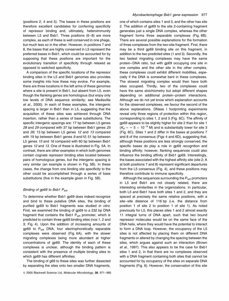
A comparison of the specific locations of the repressor
binding sites in the L5 and Bxb1 genomes also provides
some insights into how these may evolve. For example,
there are three locations in the left arms of these genomes
where a site is present in Bxb1, but absent from L5, even
though the flanking genes are homologous (but enjoy only
low levels of DNA sequence similarity; see Mediavilla
et al., 2000). In each of these examples, the intergenic
spacing is larger in Bxb1 than in L5, suggesting that the
acquisition of these sites was achieved through DNA
insertion, rather than a series of base substitutions. The
specific intergenic spacings are: 17 bp between L5 genes
28 and 29 compared with 37 bp between Bxb1 genes 25
and 26; 13 bp between L5 genes 12 and 13 compared
with 19 bp between Bxb1 genes 9 and10; 51 bp between
L5 genes 15 and 16 compared with 60 bp between Bxb1
genes 12 and 13. One of these is illustrated in Fig. 5A. In
contrast, there are other examples in which both genomes
contain cognate repressor binding sites located between
pairs of homologous genes, but the intergenic spacing is
very similar (an example is shown in Fig. 5B). In these
cases, the change from one binding site specificity to the
other could be accomplished through a series of base
substitutions (five in the example given in Fig. 5B).
Binding of gp69 to Bxb1 Pleft
To determine whether Bxb1 gp69 does indeed recognize
and bind to these putative DNA sites, the binding of
purified gp69 to Bxb1 fragments was studied in vitro.
First, we examined the binding of gp69 to a 232 bp DNA
fragment that contains the Bxb1 Pleft promoter, which is
predicted to contain three gp69 binding sites (nos 1, 2 and
3; Fig. 4). Upon the addition of increasing amounts of
gp69 to Pleft DNA, four electrophoretically separable
complexes were observed (Fig. 6A), with the slower
migrating complexes being more abundant at higher
concentrations of gp69. The identity of each of these
complexes is unclear, although the binding pattern is
consistent with the presence of multiple binding sites to
which gp69 has different affinities.
The binding of g69 to these sites was further dissected
by separating the sites onto two smaller DNA fragments,
one of which contains sites 1 and 3, and the other has site
2. The addition of gp69 to the site 2-containing fragment
generates just a single DNA complex, whereas the other
fragment forms three separable complexes (Fig. 6B).
There are several possible explanations for the formation
of three complexes from the two-site fragment. First, there
may be a third gp69 binding site on this fragment, in
addition to the two predicted sites (1 and 3). Secondly, the
two fastest migrating complexes may have the same
protein±DNA ratio, but with gp69 occupying one site in
one complex and the other site in the other complex,
these complexes could exhibit different mobilities, espe-
cially if the DNA is somewhat bent in these complexes.
The slowest migrating complex would then have both
sites occupied. Thirdly, two of the complexes could
have the same stoichiometry but adopt different shapes
depending on additional protein±protein interactions.
Although we do not yet know which explanation accounts
for the observed complexes, we favour the second of the
above explanations. DNase I footprinting experiments
reveal only three regions of protection within this region,
corresponding to sites 1, 2 and 3 (Fig. 6C). The affinity of
gp69 appears to be slightly higher for site 2 than for site 1
(Kd � < 5 � 1028 M) and is substantially lower for site 3
(Fig. 6C). Sites 1 and 2 differ in the bases at positions 7
and 8 of the consensus (Figs 4 and 6D), suggesting that,
although these positions are less strongly conserved, the
specific bases do play a role in gp69 recognition and
binding affinity; however, flanking sequences could also
influence the binding affinity of Bxb1 gp69. Interestingly,
the bases associated with the highest affinity site (site 2; A
at both positions 7 and 8) represent significant departures
from the L5 consensus (Fig. 4), and these positions may
therefore contribute to immune specificity.
Although the sequences surrounding the Pleft promoters
in L5 and Bxb1 are not closely related, there are
interesting similarities in the organizations. In particular,
both L5 and Bxb1 have both sites 1 and 2, and they are
spaced at precisely the same relative positions, with a
site±site distance of 116 bp (i.e. the distance from
position 1 of site 2 to position 1 of site 1). As noted
previously for L5, this places sites 1 and 2 almost exactly
11 integral turns of DNA apart, such that two bound
repressor molecules would be on the same face of the
DNA helix, where they would have the potential to interact
to form a DNA loop. However, the occupancy of the L5
sites is not affected by placing them on different DNA
fragments or altered by changing the spacing between the
sites, which argues against such an interaction (Brown
et al., 1997). This also appears to be the case for Bxb1
sites 1 and 2, in that there are no complexes observed
with a DNA fragment containing both sites that cannot be
accounted for by occupancy of the sites on separate DNA
fragments (Fig. 6). However, the conservation of this site
Mycobacteriophage Bxb1 gene expression 977
Q 2000 Blackwell Science Ltd, Molecular Microbiology, 38, 971±985
spacing, regardless of the low degree of DNA sequence
similarity between these regions, raises the possibility that
such protein±protein interactions may be of importance,
even though they are not detected with the assays and
DNA substrates that have been used.
The gp69 binding site designated as site 3 is situated in
the opposite orientation to sites 1 and 2 and has no
counterpart in L5. In L5, there are two binding sites to the
right of site 1, one is 59 bp upstream (to the right) of site 1
and is in the same orientation as it, whereas the second
site is 233 bp to the right of site 1 and is in the opposite
orientation. No ORFs have been identified between L5
Pleft and the right end of the genome, although we cannot
rule out a rightwards-facing promoter in this part of the
genome. In contrast, in Bxb1, there are three rightwards-
transcribed genes (84±86), and a promoter must be
located between genes 83 and 84. Site 3 is thus a
candidate operator site for such a promoter.
Identification of the Pright promoter
To determine whether rightwards transcription initiates
from within the gene 83±84 region, total RNA isolated
from a Bxb1 lysogen was used as a template for primer
extension (Fig. 7A). A primer extension product was
observed that corresponds to transcription initiation at
position 48 875, although only a weak signal could be
detected; some additional primer extension end-points
were also observed, but these all mapped within the
ORF for gene 84. Immediately upstream of this start site
are putative 210 (5 0-TAACCT) and 235 (5 0-TTGATC)
recognition sequences, separated by a 17 bp spacer
(see Fig. 6D). We have designated this putative promoter
as Pright. The gp69 binding site 3 coincides with this
promoter, but overlaps the 210 region rather than the
235 motif observed with site 1 and the Pleft promoter.
The observation that gp69 binding site 3 overlaps the
putative 210 region of the Pright promoter suggests that
transcription from Pright is under repressor control. To test
this, we constructed reporter gene fusion plasmids in
which the Pright promoter is fused to the lacZ gene, using
both extrachromosomal and integration-proficient vectors.
However, the extrachromosomal version of the recombi-
nant plasmid failed to transform M. smegmatis, unless the
strain was lysogenic for Bxb1. This could the result of poor
tolerance of either the Pright promoter or the Pleft promoter,
which is also present on this plasmid; as reported above,
plasmids with the Pleft promoter fused to lacZ also
transform non-lysogen strains poorly. However, the
integration-proficient plasmids did transform, and b-
galactosidase assays indicated that the promoter was
downregulated about 10-fold in the Bxb1 lysogen, most
Fig. 5. Origins of repressor binding sites.A. Acquisition of sites by insertion. Comparison of the L5 and Bxb1 genomes reveals at least three positions in which a repressor binding siteis located in one genome, but absent from the homologous location in the other genome (see text for further details). In all three cases, theintergenic distance is significantly larger in the genome that contains the site, consistent with the site being acquired by DNA insertion ratherthan by base substitution. The sequences of the intergenic spaces in one example are shown, in which a repressor binding site is locatedbetween Bxb1 genes 9 and 10, but is absent from the space between the homologous L5 genes 12 and 13.B. Acquisition of sites by substitution. There are also several examples in which the spacing between flanking genes is similar, and both L5and Bxb1 contain repressor binding sites. The example shown compares the junctions of Bxb1 genes 22 and 23 and L5 genes 26 and 27. Therepressor binding sites are in similar locations, and the switch in repressor binding specificity presumably arises from base substitutions. In (A)and (B), the 13 bp repressor binding sites are boxed, and nucleotide and amino acid residues that are identical in the two phages are shownin red. Ribosome binding sites are underlined. The co-ordinates of the leftmost nucleotides are shown.
978 S. Jain and G. F. Hatfull
Q 2000 Blackwell Science Ltd, Molecular Microbiology, 38, 971±985

probably because of the action of the gp69 repressor
(Fig. 7B). The level of gp69 regulation is less than that
seen for the Pleft promoter and presumably reflects the
differences in the affinity of the gp69 repressor for binding
sites 1 and 3 (Fig. 6).
Expression of the Bxb1 repressor gene
Examination of the Bxb1 genome map (see Fig. 4; Mediavilla
et al., 2000) shows that there is a small non-coding gap of
261 bp immediately upstream (i.e. to the right) of gene 69.
Presumably, there is at least one promoter in this region
that is responsible for the expression of gene 69. We note
that, in L5, there is a similar non-coding space upstream
of gene 71, within which there are three promoters
(designated P1, P2 and P3). However, there is no obvious
DNA sequence similarity between the two intergenic
regions, and it is not possible to predict the locations of
putative promoters.
To determine whether the Bxb1 region upstream of
Fig. 6. DNA binding of the Bxb1 gp69 repressor to Pleft DNA.A. Mobility shift assays for the binding of Bxb1 gp69 and L5 gp71 repressor proteins to Bxb1 and L5 Pleft promoters. Bxb1 gp69 or L5 gp71was added at decreasing concentrations (threefold serial dilutions; as indicated) to a 203 bp DNA fragment containing the Bxb1 Pleft promoter(top) or a 219 bp fragment containing the L5 Pleft promoter (bottom), and the protein±DNA complexes were separated from free DNA byelectrophoresis through a native 4% polyacrylamide gel. The amounts of Bxb1 gp69 present in each reaction (from left to right) areapproximately 26, 8, 2.6, 0.8, 0.26 and 0.08 pmol.B. Bxb1 gp69 binding to DNA subfragments. The DNA present in the Bxb1 Pleft fragment shown in (A) was amplified as two separate DNAfragments, one of 95 bp containing site 2 (probe 2) and the other of 107 bp containing sites 1 and 3 (probe 1). Binding of gp69 was as in (A).The amounts of Bxb1 gp69 in each reaction (from right to left) are approximately 8, 2.6, 0.8, 0.26 and 0.08pmol. The fastest moving complexwith probe 1 is likely to be a site 1 complex, as this is a higher affinity site than site 3; see (C).C. DNase I footprinting of Bxb1 Pleft DNA±gp69 complexes. Bxb1 gp69±DNA complexes were formed as in (A), cleaved with DNase I and theproducts separated by denaturing PAGE. Bxb1 gp69 was added in threefold serial dilutions, and lane `D' contained no gp69. The positions ofprotection at sites 1, 2 and 3 are indicated. The amounts of Bxb1 gp69 added to each reaction are (from right to left) 26, 8, 2.6, 0.8 and0.26 pmol.D. Sequence of the Bxb1 gene 83±84 intergenic region. The locations of repressor binding sites 1, 2 and 3 are boxed, and the 235, 210 andstart site positions of the Pleft and Pright promoters are shown. The sequences to the left of the mark (I) are present in the 95 bp fragment,probe 2 (B), and those to the right are in the 107 bp fragment, probe 1 (B). Sequences not present in the probes are shown in italics.
Mycobacteriophage Bxb1 gene expression 979
Q 2000 Blackwell Science Ltd, Molecular Microbiology, 38, 971±985

gene 69 is promoter active, a 293 bp fragment (containing
Bxb1 DNA corresponding to co-ordinates 44 679±
44 969) was inserted upstream of the lacZ reporter
gene in a shuttle plasmid vector and introduced into M.
smegmatis (Fig. 8A). This plasmid exhibited considerable
b-galactosidase activity of a magnitude similar to that of
the reporter for a similar fusion with the region upstream
of L5 gene 71 (Nesbit et al., 1995). When the Bxb1
repressor gene was also present (plasmid pSJ11), there
was a modest downregulation (< twofold) of the activity,
suggesting that Bxb1 gp69 regulates its own synthesis (as
also seen in L5).
The positions of transcription initiation from this
segment of the Bxb1 genome were determined by primer
extension, using RNA isolated from an M. smegmatis
strain carrying either pSJ11 or pSJ14 (Fig. 8B). Two
initiation sites were observed (at co-ordinates 44 778 and
44 886), suggesting that there are at least two promoters
in this region; we will refer to these as P1 and P2. Putative
210 and 235 regions for both promoters can be identified
and are shown in Fig. 8C. However, there is only one
possible gp69 binding site in this region, site 28 (Fig. 4),
which overlaps the putative 235 region of the P1
promoter. DNA-binding studies show that gp69 does
bind to this DNA fragment but predominantly forms just a
single complex, consistent with the presence of a single
binding site (Fig. 8D). The affinity for this site is con-
siderably lower than that for sites 1 and 2 at Pleft (Fig. 8D),
which presumably accounts for the low level of gp69
repression.
Discussion
We have described here some central aspects of
immunity regulation in Bxb1 and how they compare with
the related system in L5. As phages for comparative
studies, L5 and Bxb1 are a convenient pair, as they are
not particularly closely related at the nucleotide level and
yet share similar genome architectures, virion structures
and systems for genetic regulation. In other aspects, such
as host range and halo formation, they are clearly different
(Mediavilla et al., 2000). A comparative approach is
particularly useful for understanding the genetic circuitry in
these phages, in part because of the novel immunity system
in phage L5 that involves an unusual repressor and multiple
binding sites. The study of Bxb1 immunity regulation not
only validates the interpretations of the L5 studies but
offers insights into how these systems have evolved.
The observation that L5 and Bxb1 are heteroimmune
can be readily explained in view of the differences
between the phage repressors and their binding sites.
L5 gp71 and Bxb1 gp69 are only 41% identical, and the
amino acid difference at the first position of the second
helix of the putative helix±turn±helix motif is an obvious
candidate for being more directly involved in determining
DNA-binding specificity. The proline at this position in
Bxb1 gp69 is unusual, but not unprecedented (Harrison
and Aggarwal, 1990). The consensus sequences for the
sites are also quite distinct, in that three of the consensus
positions (positions 2, 4 and 5) that are tightly conserved
are different in the two phages (T, A and C in Bxb1 versus
G at all three positions in L5). Nevertheless, there is also a
lot of similarity between the Bxb1 and L5 consensus
sequences, and seven of the positions are both highly
conserved and identical in the two phages. Moreover, the
DNA-binding studies show that the Bxb1 repressor binds
with a much greater preference to its cognate sites than
those in L5, and the reciprocal behaviour is seen with L5
gp71. The difference in affinities is < 1000-fold in each
case.
Fig. 7. Identification of the Bxb1 Pright promoter.A. Identification of the Pright transcription start site by primer extension. Total RNA extracted from a Bxb1 lysogen of M. smegmatis mc2155was used as template for extension from primer SJ99-5. The same primer was used for sequencing reactions. The position of the majorprimer extension product is shown by an arrow.B. Activities of a Pright±lacZ fusion plasmid. Plasmid pSJ26 was constructed by inserting a Pright promoter fragment into the integrating lacZreporter vector and transformed into both M. smegmatis mc2155 and a Bxb1 lysogen. The b-galactosidase activities are presented asnmol min21 mg21 total protein.
980 S. Jain and G. F. Hatfull
Q 2000 Blackwell Science Ltd, Molecular Microbiology, 38, 971±985

A closer examination of the L5 and Bxb1 repressor
binding site sequences is very informative. In L5 positions
7±12, the sequence 5 0-TGTCAA is very well conserved,
and this segment corresponds to the location of the 235
motif (i.e. 5 0-TTGACA on the other strand) within the Pleft
promoter (Fig. 4). This would indicate that this particular
235 sequence is present at each of the stoperator sites in
the L5 genome. However, the Bxb1 sites show that this
may not be an important feature, as positions 7 and 8 are
not as well conserved and, at both positions, there is
considerable variation. In Bxb1, the Pleft operator (site 1)
is only one of 12 sites that has the 5 0-TGTCAA motif,
whereas 23 of the 24 l5 binding sites have this sequence.
Nevertheless, it is noteworthy that the major differences in
the sites (those that are expected to alter repressor
binding specificity) are located away from this 235 region
at positions 2, 4 and 5.
The extreme right end of the Bxb1 genome represents
one of the greatest departures from the organization of
the L5 genome. Here, there is divergent transcription
originating from a 213 bp intergenic region that contains
the Pleft and Pright promoters, both of which are under
repressor control. The Bxb1 Pleft promoter is analogous to
the L5 Pleft promoter, being located upstream of the first
of the identified ORFs in the leftwards operons in the
right arm and with almost identical 210 and 235 RNA
polymerase recognition sequences. No counterpart to the
Bxb1 Pright promoter has been identified in L5, but nor
Fig. 8. Expression and autoregulation of the Bxb1 repressor gene 69.A. b-Galactosidase activities of gene 69± lacZ reporter plasmids. Plasmid pSJ14, containing a DNA fragment with the putative gene 69promoter fused to lacZ, and pSJ11, which contains a similar fragment but with the entire gene 69 ORF, were introduced into M. smegmatismc2155, and b-galactosidase activities were determined as described.B. Primer extension analysis of the Bxb1 gene 69 promoter. Total RNA isolated from M. smegmatis carrying either plasmid pSJ14 or plasmidpSJ11 (shown in lanes P14 and P11 respectively) was used as template for extension with primer SJ99-4. The major transcription start sites(P1 and P2) are shown by arrows.C. Location of the gene 69 promoters. The sequence from the region upstream of gene 69 (co-ordinates 44 928±44 741) is shown with the P1and P2 transcription start sites as indicated. Putative 210 and 235 sequence motifs are underlined by a dashed line. The amino acidsequence of the start of gp69 and the gene 69 ribosome binding site are indicated. The position of a putative repressor binding site (28) isboxed.D. Binding of Bxb1 gp69 to the gene 69 promoter region. Serial dilutions of Bxb1 gp69 were incubated with a 309 bp DNA fragment containingthe gene 69 upstream region and a 203 bp fragment containing the Pleft promoter. Protein±DNA complexes were separated from free DNA byelectrophoresis through a 4% native polyacrylamide gel. The amounts of Bxb1 gp69 added to each reaction (from right to left) areapproximately 2.6, 0.8, 0.26 and 0.08 pmol.
Mycobacteriophage Bxb1 gene expression 981
Q 2000 Blackwell Science Ltd, Molecular Microbiology, 38, 971±985

have any rightwards-transcribed ORFs at the right end of
the genome. We note that there is a repressor binding site
at the extreme right end of the L5 genome that is oriented
in such a way as to regulate rightwards transcription, and
the possibility that there is a rightwards promoter located
near this site cannot be eliminated.
The function of the rightwards-facing genes 84, 85 and
86 in Bxb1 is unclear. The Pright promoter located
immediately upstream of these is only rather modestly
downregulated (<10-fold), and transcription initiation
could be detected using RNA isolated from a Bxb1
lysogen. As there is also a putative terminator located
between genes 85 and 86, the segment containing the
promoter, genes 84 and 85 and the terminator are
reminiscent of the morons described in the lambda-like
phages (Juhala et al., 2000). However, we also cannot
rule out the possibility that Pright plays a role in late lytic
growth, either as a result of inefficient termination or
perhaps through the use of an antiterminator, and could
thus be responsible for synthesis of the late genes. It is
also unclear how gene 86 is expressed and whether there
is an additional promoter between genes 85 and 86.
Autoregulation of repressor synthesis appears to be a
common theme among temperate bacteriophages. In
phage lambda, this is accomplished through the action of
the tripartite operator OR, in which the binding of the cI
repressor to the highest affinity site, OR1, results in the
activation of cI synthesis (Meyer et al., 1975); at higher
concentrations, the repressor binds to the lower affinity
site OR3, resulting in downregulation of cI expression
(Hochschild et al., 1986; Ptashne, 1987). In Bxb1 and L5,
the repressor gene promoters do not appear to require an
activator, but both are modestly downregulated by their
repressor (two- to threefold). Presumably, this acts to
maintain the required intracellular concentration of the
repressor.
It is very common, especially within phage genomes, to
find genes encoding DNA-binding proteins situated close
to the site of action of the gene product. This is seen
for phage recombination systems (Campbell, 1981),
packaging systems (Catalano et al., 1995) as well as for
most phage immunity systems, and is presumed to result
from the evolutionary advantage of the cis- and trans-
acting components to co-evolve (e.g. by moving together
from one genome to another). However, evolution of
the mycobacteriophage immunity systems described here
must be more complex, as the binding sites are both
numerous and distributed across the phage genomes.
Comparison of the Bxb1 and L5 genomes provides some
insights into how this may occur. First, it is noteworthy that
the switch in immune specificity appears to be quite
complete, and the Bxb1 genome is devoid of L5 repressor
binding sites and vice versa. Secondly, the change in
specificity appears to be achieved through mutations in
the repressor gene and acquisition of cognate repressor
binding sites. Comparison of the genomes suggests that
they can be acquired de novo through DNA insertion or by
base substitution of existing binding sites. However, there
are presumably quite strong evolutionary pressures to
obtain these sites, as a large number of them are present
and they generally conform tightly to a consensus
sequence.
Experimental procedures
Bacterial strains and plasmids
Escherichia coli DH5a was used for all cloning purposes.M. smegmatis mc2155 is a high-efficiency transformationstrain (Snapper et al., 1990). mc2155(L5) and mc2155(Bxb1)are the mycobacteriophage L5 and Bxb1 lysogens of M.smegmatis mc2155 respectively. M. smegmatis mc2155 andmc2155(L5) were grown in Middlebrook 7H9 broth (Difco)supplemented with ADC enrichment and 0.2% Tween 80 withshaking at 378C. mc2155(Bxb1) was grown similarly at 308C.Plasmids were introduced into M. smegmatis strains byelectroporation using a Bio-Rad gene pulser with minormodifications of the method described previously (Snapperet al., 1990). Plasmid pET21a was procured from Novagen.Plasmids pSD5B, pDK10 and pDK16 were kind gifts from DrAnil K. Tyagi, University of Delhi South Campus, India. DNAmanipulations were carried out as described previously(Sambrook et al., 1989). Mycobacteriophage Bxb1 infections,purification of phage and isolation of its genomic DNA werecarried out as described for L5 (Sarkis and Hatfull, 1998).
Cloning, expression and purification of Bxb1 repressor
protein (gp69)
Bxb1 gene 69 coding for the repressor protein gp69 wasamplified by PCR using primers SJ98-2 (5 0-CAAGGAGGACATATGAGAACCACC; co-ordinates 44 774±44 751) andSJ98-3 (5 0-AGGGGCGGGTGGCCATCAGGG; co-ordinates44 235±44 255) to obtain a product of 540 bp (the nucleo-tides that were modified to create restriction enzyme sites areunderlined in the primer sequences). The amplicon wasdigested with NdeI and EaeI and cloned into NdeI±NotI-digested pET21a vector to obtain pSJ6. E. coli BL21(DE3)-pLysS transformed with pSJ6 was grown in Luria±Bertani(LB) medium containing carbenicillin (50 mg ml21) andchloramphenicol (34 mg ml21) to an A600 of 0.8±1.0 at 378Cwith shaking. Expression of gene 69 was induced by theaddition of IPTG to a final concentration of 0.3 mM, and theincubation was continued for an additional 90 min. Next,the culture was chilled on ice, and cells were harvested bycentrifugation at maximum speed at 48C for 10 min. For theanalysis of gene expression, the cell pellet from a 3 ml culturewas boiled in 200 ml of 1 � SDS buffer (60 mM Tris-Cl,pH 6.8, 1% SDS, 350 mM b-mercaptoethanol, 10% glycerol)for 10 min, and proteins were resolved by 10% SDS±PAGEanalysis. For purification of gp69, cells from a 250 ml culturewere suspended in 5 ml of buffer containing 12.5 mMHEPES-MES buffer, pH 7.94, 1 mM dithiothreitol (DTT),
982 S. Jain and G. F. Hatfull
Q 2000 Blackwell Science Ltd, Molecular Microbiology, 38, 971±985

1 mM EDTA, 1 mM phenylmethylsulphonyl fluoride (PMSF)and lysed by freeze thawing in liquid nitrogen. Polyethyle-neimine (PEI) at pH 8.0 was added to the cell-free extract toa final concentration of 0.7% and left on ice for 45 min. Afterclarification, ammonium sulphate was added to the super-natant to a final concentration of 40%. The suspension wascentrifuged at 10 000 g for 15 min at 48C, and the super-natant was loaded on a POROS-PE column using a BioCADSPRINT system (PE Biosystems). Proteins were eluted fromthe column using a gradient of 40±0% ammonium sulphate in12.5 mM HEPES-MES buffer, pH 7.94. Various fractionswere analysed by 10% SDS±PAGE, and those containingpurified gp69 were pooled and dialysed against 4 l of12.5 mM HEPES-MES buffer, pH 7.94, at 48C with fourchanges. Bxb1 gp69 precipitated out of solution and wassolubilized in buffer containing 10 mM Tris-Cl, pH 8.0, 1 mMEDTA, 200 mM NaCl and 50% glycerol for future studies.
DNA-binding studies
The DNA fragments were PCR amplified using Bxb1 genomicDNA as a template with Pfu DNA polymerase (Stratagene).A 232 bp fragment containing the Bxb1 Pleft promoter regionand encompassing the gp69 binding sites 1 and 2 wasamplified using primers SJ98-4 (5 0-CCGGACCGAGTCGACGATGCG; co-ordinates 48 657±48 677) and SJ98-5 (5 0-CGGTGTACGTGTGCTCTAGAGG; co-ordinates 48 888±48 867)(underlined nucleotides indicate the base positions that weremodified to create restriction enzyme sites). Primers SJ98-5and SJ99-2 (5 0-CCACCGGGATGAAGCTGATGATG, position48 763±48 785) amplified the 126 bp product containing thegp69 binding site 1 that overlaps with the Pleft promoter. The106 bp amplicon containing gp69 binding site 2 was obtainedwith primers SJ98-4 and SJ99-1 (5 0-TGCTCGCGGTGATCGCGGGC; co-ordinates 48 762±48 743). Purified ampliconswere digested with appropriate restriction enzymes (232 bpwith SalI and XbaI; 126 bp with XbaI and 106 bp with Sal I)and radiolabelled using [a-32P]-dATP (NEN Life ScienceProducts) with Klenow fragment. Bxb1 gp69-binding assayswere performed with the purified protein essentially asdescribed for L5 gp71 (Brown et al., 1997). In brief, dilutionsof purified gp69 were incubated with the radiolabelled DNAfragments in a 10 ml reaction volume comprising 2 mg of calfthymus DNA, 10 mg of bovine serum albumin (BSA), 30 mMNaCl, 1 mM Tris-Cl, pH 7.9, and 3 mM EDTA for 15 min onice. Ten � sucrose loading dye (3 ml; 60% sucrose,bromophenol blue, xylene cyanol) was added to eachreaction, and the sample was immediately loaded on aprerun native polyacrylamide gel (4% acrylamide, 2.5%glycerol, 3.3 mM NaOAc, pH 7, 6.75 mM Tris-Cl, pH 8,1 mM EDTA) in TEA buffer (6.7 mM Tris-Cl, pH 7.9,3.3 mM NaOAc, pH 7, 1 mM EDTA). Electrophoresis wascarried out at 58C at 200 V.
For DNase I footprinting, pSJ8 (Table 1) was linearizedwith EcoRI and radiolabelled as described above. It wasdigested further with HindIII, and the DNA fragments wereresolved on a 1.5% agarose gel. The gel piece containing the287 bp fragment of the Bxb1 Pleft promoter was cut out, andthe DNA was eluted using a QiaexII gel extraction kit(Qiagen). Binding of gp69 to this DNA fragment wasperformed as mentioned above except that the reaction
was incubated at 378C. Two units of DNase I (10 U ml21;Stratagene) were added to each reaction, and incubation wascontinued at 378C for 12 s. The reaction was stopped byadding 100 ml of DNase I stop buffer (30 mg of tRNA, 0.3 mMNaOAc, pH 7, 10 mM EDTA, 0.01% SDS). Next, thereactions were extracted with buffer-saturated phenol(pH 8.0) once and precipitated with three volumes of chilledethanol. The samples were suspended in 1.2 ml of water and1.8 ml of sequencing stop dye (95% formamide, bromophenolblue, xylene cyanol) and heated at 908C for 5 min. DNAbands were resolved on a 6% denaturing polyacrylamide gel.A G1A sequencing ladder was prepared by Maxam±Gilbertchemical sequencing reactions as described previously(Ausubel et al., 1996).
Construction of reporter fusion plasmids
To investigate the transcriptional capabilities of the diver-gently located Pleft and Pright promoters of mycobacterioph-age Bxb1, a 232 bp DNA fragment carrying these promoterswas amplified using primers SJ98-4 and SJ98-5 (described inthe previous section) and kinased at its 5 0 ends using T4polynucleotide kinase (Boehringer Mannheim). It was thencloned upstream of the promoterless lacZ gene in bothorientations in the promoter probe vector pSD5B (Jain et al.,1997), which had been digested with XbaI and end repairedwith T4 DNA polymerase (New England Biolabs) to obtainpSJ23 and pSJ7. In these plasmids, lacZ was transcribed byPleft in pSJ23 and by Pright in pSJ7. In addition, the 232 bppromoter fragment was cloned in the NcoI site of theintegration-proficient vector pDK16 after filling its stickyends with Klenow fragment (New England Biolabs) and inthe SmaI site of pUC118. The recombinants resulting fromthese ligations were termed pSJ9 and pSJ8, respectively,and were selected such that the Pleft promoter expressed thedownstream lacZ gene in each case. To construct pSJ21 andpSJ22, the 126 bp amplification product containing the gp69binding sites 1 and 3 was obtained with the primers SJ98-5and SJ99-2 (described in the previous section), kinased andcloned in both orientations in pSD5B at its XbaI site after endrepair with Klenow fragment. The orientation of the clonedDNA fragment in the recombinants in which Pleft transcribedthe downstream lacZ reporter gene was designated pSJ22,and the opposite orientation in which Pright expressed lacZwas termed pSJ21. Finally, to construct pSJ26, pDK10(DasGupta et al., 1998) was linearized with EcoRV. It wasthen cloned into the large fragment obtained after thedigestion of pSJ7 with NheI and MscI followed by end repairwith T4 DNA polymerase. This manipulation replaced themycobacterial origin of replication in pSJ7 with mycobacter-iophage L5-based integration signals derived from pDK10.
To study the gene regulation in Bxb1 by the product ofgene 69 encoding the repressor protein, the 735 bp DNAfragment containing the Bxb1 gene 69 along with itsupstream sequences was amplified using primers SJ98-3(5 0-AGGGGCGGGTGGCCATCAGGG; co-ordinates 44 235±44 255) and SJ99-3 (5 0-GGTTGTGACGATGCCGGTTCAGC;co-ordinates 44 969±44 947) and kinased at its 5 0 end.Subsequently, it was cloned into pSJ7 and pSJ23 at theirNheI sites, which were end repaired with Klenow fragment toobtain pSJ10 and pSJ24, respectively, and also in the ScaI
Mycobacteriophage Bxb1 gene expression 983
Q 2000 Blackwell Science Ltd, Molecular Microbiology, 38, 971±985

site of pSJ9 to construct pSJ15. pSJ12B was made byinserting the 735 bp amplicon in the SmaI site of pUC118.To identify the promoter region of gene 69, the 735 bpamplicon and the T4 DNA polymerase-treated 293 bpKpnI±EcoRV restriction fragment from pSJ12B (containingthe gene 69 upstream sequences only) were cloned into theend-repaired XbaI site of pSD5B in the correct orientation toobtain pSJ11 and pSJ14 respectively. Finally, pSJ18 andpSJ19 were constructed by inserting the 401 bp KpnI and293 bp KpnI±EcoRV fragments from pSJ12B, after theirtreatment with T4 DNA polymerase into the ScaI site ofpSJ9 respectively.
b -Galactosidase assays
M. smegmatis transformed with the individual plasmids wasgrown in Middlebrook 7H9 medium supplemented with ADC,Tween 80 and kanamycin (20 mg ml21) to an A600 of 2±2.5.The cells were harvested by centrifugation and suspended inone-fifth of the culture volume of buffer containing 100 mMTris-Cl, pH 8.0, 1 mM EDTA, 1 mM DTT and 1 mM PMSF.Cells were disrupted by sonication and centrifuged at12 000 g at 48C for 15 min. The cell-free extracts wereused for b-galactosidase assays as described previously(Miller, 1972; Jain et al., 1997) using o-nitrophenyl b-D-galactopyranoside (ONPG) as substrate. The enzyme activitywas expressed as nmol of ONPG converted to o-nitrophenolmin21 mg21 protein.
RNA isolation and primer extension analysis
M. smegmatis transformed with the appropriate plasmid wasgrown to an A600 of 2±2.5 in 10 ml of Middlebrook 7H9 mediumsupplemented with ADC and kanamycin (20 mg ml21). Thecells were suspended in 100 ml of TE containing 16 mg ml21
lysozyme and incubated at 378C for 30 min. The cell suspen-sion was transferred to a 1.5 ml microfuge tube containing400 ml of 0.5 mm Zirconia beads (Biospec Products) and350 ml of RLT buffer (RNeasy kit; Qiagen). The tube wasvortexed five times by giving 30 s pulses with chilling in betweenthe pulses. This was followed by centrifugation in the cold for5 min. The supernatant was collected in a fresh tube, andtotal RNA was purified with the RNeasy kit according to themanufacturer's instructions. RNA (10 mg) was digested with10 U of DNase I (RNase free; Stratagene) at 378C for 15 minand purified using the RNeasy kit.
For primer extension reactions, primers were end labelledwith [a32P]-ATP (NEN Life Science Products), and 10 mg ofthe total RNA was reverse transcribed using Superscript II(Life Technologies) according to the manufacturer's instruc-tions. Sequencing reactions were carried out using aThermoSequenase cycle sequencing kit (Amersham Phar-macia Biotech). Reactions were resolved on a 6% denatur-ing polyacrylamide gel before autoradiography. PrimersSJ98-4 (described in the previous section), SJ99-4 (5 0-GGGGAGCTGTTCTCTGGTGG; co-ordinates 44 736±44 755) and SJ99-5 (5 0-GAACGACGACTGATCGAACG;co-ordinates 49 025±49 006) were used for the determina-tion of transcription start sites of Pleft, gene 69 promoter andPright respectively.
Acknowledgements
We are grateful to Dr Anil K. Tyagi, India, for providing themycobacterial promoter probe vectors pSD5B, pDK10 andpDK16. We thank Aisha Mitchell for excellent technicalassistance, and Lori Bibb for valuable comments on themanuscript. This work was supported by grant AI28927 fromthe National Institutes of Health.
References
Ausubel, F.M., Brent, R., Kingston, R.E., Moore, D.D.,Seidman, J.G., Smith, J.A., and Struhl, K. (1996). CurrentProtocols in Molecular Biology. New York: Wiley Inter-sciences.
Barletta, R.G., Kim, D.D., Snapper, S.B., Bloom, B.R., andJacobs, W.R., Jr (1992) Identification of expression signalsof the mycobacteriophages Bxb1, L1 and TM4 using theEscherichia±Mycobacterium shuttle plasmids pYUB75 andpYUB76 designed to create translational fusions to the lacZgene. J Gen Microbiol 138: 23±30.
Brown, K.L., Sarkis, G.J., Wadsworth, C., and Hatfull, G.F.(1997) Transcriptional silencing by the mycobacteriophageL5 repressor. EMBO J 16: 5914±5921.
Campbell, A. (1981) Evolutionary significance of accessoryDNA elements in bacteria. Annu Rev Microbiol 35: 55±83.
Catalano, C.E., Cue, D., and Feiss, M. (1995) Virus DNApackaging: the strategy used by phage lambda. MolMicrobiol 16: 1075±1086.
DasGupta, S.K., Jain, S., Kaushal, D., and Tyagi, A.K. (1998)Expression systems for study of mycobacterial generegulation and development of recombinant BCG vaccines.Biochem Biophys Res Commun 246: 797±804.
Dhaese, P., Seurinck, J., De Smet, B., and Van Montagu, M.(1985) Nucleotide sequence and mutational analysis of animmunity repressor gene from Bacillus subtilis temperatephage phi 105. Nucleic Acids Res 13: 5441±5455.
Donnelly-Wu, M.K., Jacobs, W.R., Jr, and Hatfull, G.F.(1993) Superinfection immunity of mycobacteriophage L5:applications for genetic transformation of mycobacteria.Mol Microbiol 7: 407±417.
Ford, M.E., Sarkis, G.J., Belanger, A.E., Hendrix, R.W., andHatfull, G.F. (1998) Genome structure of mycobacterioph-age D29: implications for phage evolution. J Mol Biol 279:143±164.
Harrison, S.C., and Aggarwal, A.K. (1990) DNA recognitionby proteins with the helix±turn±helix motif. Annu RevBiochem 59: 933±969.
Hatfull, G.F. (1994) Mycobacteriophage L5: a toolbox fortuberculosis. Am Soc Microbiol News 60: 255±260.
Hatfull, G.F. (1999) Mycobacteriophages. In Mycobacteria:Molecular Biology and Virulence. Ratledge, C., and Dale, J.(eds). London: Chapman & Hall, pp. 38±58.
Hatfull, G.F., and Sarkis, G.J. (1993) DNA sequence,structure and gene expression of mycobacteriophage L5:a phage system for mycobacterial genetics. Mol Microbiol7: 395±405.
Hochschild, A., Douhan, J.D., and Ptashne, M. (1986) Howlambda repressor and lambda Cro distinguish betweenOR1 and OR3. Cell 47: 807±816.
Ingham, C.J., Owen, C.E., Wilson, S.E., Hunter, I.S., and
984 S. Jain and G. F. Hatfull
Q 2000 Blackwell Science Ltd, Molecular Microbiology, 38, 971±985
Smith, M.C. (1994) An operator associated with auto-regulation of the repressor gene in actinophage phiC31 isfound in highly conserved copies in intergenic regions inthe phage genome. Nucleic Acids Res 22: 821±827.
Jain, S., Kaushal, D., DasGupta, S.K., and Tyagi, A.K. (1997)Construction of shuttle vectors for genetic manipulationand molecular analysis of mycobacteria. Gene 190: 37±44.
Juhala, R.J., Ford, M.E., Duda, R.L., Youton, A., Hatfull,G.F., and Hendrix, R.W. (2000) Genomic sequences ofbacteriophages HK97 and HK022: pervasive geneticmosaicism in the lambdoid bacteriophages. J Mol Biology299: 27±51.
Kameyama, L., Fernandez, L., Calderon, J., Ortiz-Rojas, A.,and Patterson, T.A. (1999) Characterization of wildlambdoid bacteriophages: detection of a wide distributionof phage immunity groups and identification of a nus-dependent, nonlambdoid phage group. Virology 263: 100±111.
Ladero, V., Garcia, P., Bascaran, V., Herrero, M., Alvarez,M.A., and Suarez, J.E. (1998) Identification of therepressor-encoding gene of the Lactobacillus bacterio-phage A2. J Bacteriol 180: 3474±3476.
Laughon, A., and Scott, M.P. (1984) Sequence of aDrosophila segmentation gene: protein structure homologywith DNA-binding proteins. Nature 310: 25±31.
Mediavilla, J., Jain, S., Kriakov, J., Ford, M.E., Duda, R.L.,Jacobs, W.R., Jr, et al. (2000) Genome organization andcharacterization of mycobacteriophage Bxb1. Mol Microbiol38: 955±970.
Meyer, B.J., Kleid, D.G., and Ptashne, M. (1975) Lambdarepressor turns off transcription of its own gene. Proc NatlAcad Sci USA 72: 4785±4789.
Miller, J.H. (1972) Experiments in Molecular Genetics. ColdSpring Harbor, NY: Cold Spring Harbor Laboratory Press.
Nesbit, C.E., Levin, M.E., Donnelly-Wu, M.K., and Hatfull, G.F.(1995) Transcriptional regulation of repressor synthesis inmycobacteriophage L5. Mol Microbiol 17: 1045±1056.
Nesper, J., Blass, J., Fountoulakis, M., and Reidl, J. (1999)Characterization of the major control region of Vibriocholerae bacteriophage K139: immunity, exclusion, andintegration. J Bacteriol 181: 2902±2913.
Ptashne, M. (1987) A Genetic Switch. Oxford/Cambridge:Blackwell Science and Cell Press.
Ptashne, M., Backman, K., Humayun, M.Z., Jeffrey, A.,Maurer, R., Meyer, B., and Sauer, R.T. (1976) Autoregula-tion and function of a repressor in bacteriophage lambda.Science 194: 156±161.
Salmi, D., Magrini, V., Hartzell, P.L., and Youderian, P.(1998) Genetic determinants of immunity and integration oftemperate Myxococcus xanthus phage Mx8. J Bacteriol180: 614±621.
Sambrook, J., Fritsch, E.F., and Maniatis, T. (1989)Molecular Cloning: a Laboratory Manual. Cold SpringHarbor, NY: Cold Spring Harbor Laboratory Press.
Sarkis, G.J., and Hatfull, G.F. (1998) Mycobacteriophages.Methods Mol Biol 101: 145±173.
Scott, J.R., West, B.W., and Laping, J.L. (1978) Super-infection immunity and prophage repression in phage P1.IV. The c1 repressor bypass function and the role of c4repressor in immunity. Virology 85: 587±600.
Smith, M.C., Burns, R.N., Wilson, S.E., and Gregory, M.A.(1999) The complete genome sequence of the Strepto-myces temperate phage straight phiC31: evolutionaryrelationships to other viruses. Nucleic Acids Res 27:2145±2155.
Snapper, S.B., Melton, R.E., Mustafa, S., Kieser, T., andJacobs, W.R., Jr (1990) Isolation and characterization ofefficient plasmid transformation mutants of Mycobacteriumsmegmatis. Mol Microbiol 4: 1911±1919.
Wilson, S.E., Ingham, C.J., Hunter, I.S., and Smith, M.C.(1995) Control of lytic development in the Streptomycestemperate phage phi C31. Mol Microbiol 16: 131±143.
Mycobacteriophage Bxb1 gene expression 985
Q 2000 Blackwell Science Ltd, Molecular Microbiology, 38, 971±985