Structural insight into gene transcriptional regulation and effector binding by the Lrp/AsnC family
11
Structural insight into gene transcriptional regulation and effector binding by the Lrp/AsnC family Paul Thaw, Svetlana E. Sedelnikova, Tatyana Muranova, Sebastian Wiese, Sylvia Ayora 1,2 , Juan C. Alonso 2 , Arie B. Brinkman 3 , Jasper Akerboom 4 , John van der Oost 4 and John B. Rafferty* Department of Molecular Biology and Biotechnology, Krebs Institute, University of Sheffield, Western Bank, Sheffield S10 2TN, UK, 1 Departamento de Biologia Molecular, Universidad Autonoma de Madrid, Cantoblanco, 28049 Madrid, Spain, 2 Departamento de Biotecnologia Microbiana, Centro Nacional de Biotecnologia, CSIC, Campus Universidad Autonoma de Madrid, Cantoblanco, 28049 Madrid, Spain, 3 Department of Molecular Biology, NCMLS M850/3.79, Geert Grooteplein 30, 6525 GA, Nijmegen, The Netherlands and 4 Laboratory of Microbiology, Wageningen University, Hesselink van Suchtelenweg 4, 6307 CT Wageningen, The Netherlands Received November 21, 2005; Revised and Accepted February 10, 2006 ABSTRACT The Lrp/AsnC family of transcriptional regulatory proteins is found in both archaea and bacteria. Members of the family influence cellular metabolism in both a global (Lrp) and specific (AsnC) manner, often in response to exogenous amino acid effectors. In the present study we have determined both the first bacterial and the highest resolution structures for members of the family. Escherichia coli AsnC is a specific gene regulator whose activity is triggered by asparagine binding. Bacillus subtilis LrpC is a global regulator involved in chromosome condensa- tion. Our AsnC-asparagine structure is the first for a regulator–effector complex and is revealed as an octameric disc. Key ligand recognition residues are identified together with a route for ligand access. The LrpC structure reveals a stable octamer support- ive of a topological role in dynamic DNA packaging. The structures yield significant clues to the func- tionality of Lrp/AsnC-type regulators with respect to ligand binding and oligomerization states as well as to their role in specific and global DNA regulation. INTRODUCTION Proteins belonging to the Lrp/AsnC family of global or specific transcriptional regulators are widely distributed in numerous prokaryotes, including bacteria and archaea (1,2). At least one Lrp-like homologue can be identified in 45 and 94% of the currently available bacterial and archaeal genomes, respectively. To date, there are no confirmed homologues in the available eukaryal genomes, indicating that the family is probably restricted to prokaryotes (3). Members of the Lrp/ AsnC family typically have a molecular mass of 15 kDa but populate a range of multimeric species in solution, which include dimers, tetramers, octamers and hexadecamers (4–8). In addition to their well-studied role in specific and global regulation of gene expression, it has been suggested that some bacterial Lrp homologues may play a role in (i) chromosome structure and organization (9) based upon observations of high copy number (between 1300 and 3200 dimers per cell) (10), (ii) DNA bending (11) and (iii) condensation of DNA into globular nucleoprotein-like structures (6). Several homologues from Archaea have also been characterized recently. These studies concerned regulators that block one or more binding sites of the general transcription initiation machinery, and as such repress the expression of the downstream gene [e.g. Pyrococcus furiosus LrpA (7) and Sulfolobus solfataricus LrpB (12)]. An addi- tional homologue from S.solfataricus (LysM) appears to be a lysine-dependent transcription activator (13). The best- documented case of an archaeal Lrp-like transcription activ- ator is Methanocaldococcus jannaschii Ptr2 that enhances the expression of several genes in a ligand-independent manner (14,15). S.solfataricus Lrs14, a relatively abundant protein (2,16), may correspond to a chromosome organizer, as has been suggested for some bacterial Lrp homologues and for Pyrococcus OT3 FL11, where glutamine triggers the binding and wrapping of DNA (17). The best-characterized member of the family is Escherichia coli Lrp, which controls a global regulon encompassing at least 10% of all E.coli genes (18). The genes that belong to the Lrp-regulon encode proteins that are involved in transport, *To whom correspondence should be addressed. Tel: +44 (114) 222 2809; Fax: +44 (114) 222 2800; Email: [email protected] Ó The Author 2006. Published by Oxford University Press. All rights reserved. The online version of this article has been published under an open access model. Users are entitled to use, reproduce, disseminate, or display the open access version of this article for non-commercial purposes provided that: the original authorship is properly and fully attributed; the Journal and Oxford University Press are attributed as the original place of publication with the correct citation details given; if an article is subsequently reproduced or disseminated not in its entirety but only in part or as a derivative work this must be clearly indicated. For commercial re-use, please contact [email protected] Nucleic Acids Research, 2006, Vol. 34, No. 5 1439–1449 doi:10.1093/nar/gkl009 Published online March 9, 2006 by guest on February 25, 2014 http://nar.oxfordjournals.org/ Downloaded from
Transcript of Structural insight into gene transcriptional regulation and effector binding by the Lrp/AsnC family
Structural insight into gene transcriptional regulationand effector binding by the Lrp/AsnC familyPaul Thaw, Svetlana E. Sedelnikova, Tatyana Muranova, Sebastian Wiese, Sylvia Ayora1,2,
Juan C. Alonso2, Arie B. Brinkman3, Jasper Akerboom4, John van der Oost4 and
John B. Rafferty*
Department of Molecular Biology and Biotechnology, Krebs Institute, University of Sheffield, Western Bank,Sheffield S10 2TN, UK, 1Departamento de Biologia Molecular, Universidad Autonoma de Madrid, Cantoblanco,28049 Madrid, Spain, 2Departamento de Biotecnologia Microbiana, Centro Nacional de Biotecnologia, CSIC, CampusUniversidad Autonoma de Madrid, Cantoblanco, 28049 Madrid, Spain, 3Department of Molecular Biology,NCMLS M850/3.79, Geert Grooteplein 30, 6525 GA, Nijmegen, The Netherlands and 4Laboratory of Microbiology,Wageningen University, Hesselink van Suchtelenweg 4, 6307 CT Wageningen, The Netherlands
Received November 21, 2005; Revised and Accepted February 10, 2006
ABSTRACT
The Lrp/AsnC family of transcriptional regulatoryproteins is found in both archaea and bacteria.Members of the family influence cellular metabolismin both a global (Lrp) and specific (AsnC) manner,often in response to exogenous amino acid effectors.In the present study we have determined both the firstbacterial and the highest resolution structures formembers of the family. Escherichia coli AsnC is aspecific gene regulator whose activity is triggeredby asparagine binding. Bacillus subtilis LrpC is aglobal regulator involved in chromosome condensa-tion. Our AsnC-asparagine structure is the first for aregulator–effector complex and is revealed as anoctameric disc. Key ligand recognition residuesare identified together with a route for ligand access.The LrpC structure reveals a stable octamer support-ive of a topological role in dynamic DNA packaging.The structures yield significant clues to the func-tionality of Lrp/AsnC-type regulators with respectto ligand binding and oligomerization states as wellas to their role in specific and global DNA regulation.
INTRODUCTION
Proteins belonging to the Lrp/AsnC family of global orspecific transcriptional regulators are widely distributed innumerous prokaryotes, including bacteria and archaea (1,2).At least one Lrp-like homologue can be identified in 45 and94% of the currently available bacterial and archaeal genomes,
respectively. To date, there are no confirmed homologues inthe available eukaryal genomes, indicating that the family isprobably restricted to prokaryotes (3). Members of the Lrp/AsnC family typically have a molecular mass of �15 kDabut populate a range of multimeric species in solution,which include dimers, tetramers, octamers and hexadecamers(4–8). In addition to their well-studied role in specific andglobal regulation of gene expression, it has been suggestedthat some bacterial Lrp homologues may play a role in(i) chromosome structure and organization (9) based uponobservations of high copy number (between 1300 and3200 dimers per cell) (10), (ii) DNA bending (11) and(iii) condensation of DNA into globular nucleoprotein-likestructures (6). Several homologues from Archaea have alsobeen characterized recently. These studies concernedregulators that block one or more binding sites of the generaltranscription initiation machinery, and as such repress theexpression of the downstream gene [e.g. Pyrococcus furiosusLrpA (7) and Sulfolobus solfataricus LrpB (12)]. An addi-tional homologue from S.solfataricus (LysM) appears to bea lysine-dependent transcription activator (13). The best-documented case of an archaeal Lrp-like transcription activ-ator is Methanocaldococcus jannaschii Ptr2 that enhances theexpression of several genes in a ligand-independent manner(14,15). S.solfataricus Lrs14, a relatively abundant protein(2,16), may correspond to a chromosome organizer, as hasbeen suggested for some bacterial Lrp homologues and forPyrococcus OT3 FL11, where glutamine triggers the bindingand wrapping of DNA (17).
The best-characterized member of the family is Escherichiacoli Lrp, which controls a global regulon encompassing atleast 10% of all E.coli genes (18). The genes that belong tothe Lrp-regulon encode proteins that are involved in transport,
*To whom correspondence should be addressed. Tel: +44 (114) 222 2809; Fax: +44 (114) 222 2800; Email: [email protected]
� The Author 2006. Published by Oxford University Press. All rights reserved.
The online version of this article has been published under an open access model. Users are entitled to use, reproduce, disseminate, or display the open accessversion of this article for non-commercial purposes provided that: the original authorship is properly and fully attributed; the Journal and Oxford University Pressare attributed as the original place of publication with the correct citation details given; if an article is subsequently reproduced or disseminated not in its entirety butonly in part or as a derivative work this must be clearly indicated. For commercial re-use, please contact [email protected]
Nucleic Acids Research, 2006, Vol. 34, No. 5 1439–1449doi:10.1093/nar/gkl009
Published online March 9, 2006 by guest on February 25, 2014
http://nar.oxfordjournals.org/D
ownloaded from

degradation and biosynthesis of amino acids, as well as a smallnumber of proteins involved in the production of pili, porins,sugar transporters and nucleotide transhydrogenases (19,20).E.coli Lrp utilizes the binding of L-leucine to trigger eitheractivation or repression of some target promoters, althoughin the majority of cases control by Lrp is leucine-independent,e.g. negative autoregulation of its own lrp gene.
E.coli AsnC shows notable sequence identity (25%) withLrp (Figure 1), which has resulted in them being classified asa distinct evolutionary protein family (21). In contrast to Lrp,AsnC has only been shown to exert specific control of its owngene and that of asnA. The latter gene codes for asparaginesynthase, and is regulated by AsnC in an asparagine-dependentfashion. Increasing levels of exogenous L-asparaginereduces asnA transcription leading to decreased cellular levelsof asparagine synthase, consistent with a classical negativefeedback mechanism (22,23). AsnC has also been demon-strated to autoregulate its own expression in an asparagine-independent manner.
Bacillus subtilis LrpC was one of seven genes encodingproteins belonging to the Lrp/AsnC family identified in its
genomic sequence. It shares 34 and 25% identity withE.coli Lrp and AsnC, respectively, and has reported function-ality in both sporulation and amino acid metabolism (24).LrpC has been shown to bind multiple sites in the upstreamregion of its own gene, resulting in slight positive autoregu-lation, in contrast to the negative autoregulation observed forother family members (25). Gel filtration studies indicatedthat LrpC forms a tetramer in solution and DNA bindinghas been reported to proceed cooperatively in a sequenceindependent manner, with LrpC preferentially recognizingintrinsically curved regions of DNA (26). Interestingly, elec-tron microscopy studies have demonstrated that LrpC is able toform nucleoprotein complexes capable of wrapping DNA intoa right-handed super-helix to form structures resembling nuc-leosomes (27) Furthermore, LrpC has been demonstrated toconstrain DNA supercoils, implying it may also have a roleakin to bacterial chromatin (26).
Prior to this study, structures of family members existedonly for the archaeal proteins, Pyrococcus furiosus LrpA (28)and Pyrococcus OT3 FL11 (17). These revealed an N-terminalDNA-binding domain, containing a helix–turn–helix (HtH)
Figure 1. Sequence alignment of the Lrp/AsnC family. A structure-based multiple sequence alignment of the AsnC/Lrp family is shown and the residues forming theligand-binding site are identified. Secondary structure elements are indicated as red for a-helices and green for b-strands. Residues are coloured as blue forhydrophobic, red for charged and green for polar. The location of the G37 to E37 mutation in our AsnC construct is marked with a yellow box. Residues found to beimportant in the formation of the ligand-binding site are indicated by closed boxes. The positions of DNA binding, activation and leucine response mutants identifiedin E.coli Lrp are indicated by the symbols plus, asterisk and hash, respectively. The five C-terminal residues of BkdR (MTLRE) have been omitted from thealignment. The figure was produced using the INDONESIA alignment package (D. Madsen, P. Johansson and G.J. Kleywegt manuscript in preparation). Speciesabbreviations are as follows: Ecoli, E.coli; Bsubt, B.subtilis; Pfuri, P.furiosus; POT3, Pyrococcus OT3; AgrTu, A.tumefaciens; PsePu, P.putida; MycTu, Myco-bacterium tuberculosis; SulSo, S.solfataricus.
1440 Nucleic Acids Research, 2006, Vol. 34, No. 5
by guest on February 25, 2014http://nar.oxfordjournals.org/
Dow
nloaded from
motif coupled via a linker region to a C-terminal effectordomain. The latter domain was found to be reminiscent ofthe ACT family of small molecule binding domains; however,because of significant structural and functional differences ithas been termed the RAM domain (regulation of amino acidmetabolism) (29). The main structural unit of the protein con-sists of a homo-dimer, held together mainly by interactionsbetween the anti-parallel b-sheets of the C-terminal domain.Analysis of sequence comparisons and mapping of mutationaldata (30) onto these structures revealed no clear ligand-binding site, only that it was most likely present in the inter-face between the dimers (28). On the basis of an alignment ofRAM domains, certain conserved residues (equivalent to L95,M101, A134, I135 and I136 of P.furiosus LrpA) have beenpredicted to be involved in ligand binding (29).
We have determined the crystal structures of both E.coliAsnC and B.subtilis LrpC to 2.4 s resolution. The AsnCstructure is seen to be an octamer with the L-asparagine ligandbound in a cleft at the interface between dimers. Analysis ofthis structure with respect to biochemical and mutational datahas implications for the oligomerization state of the proteinin vivo and its subsequent DNA binding. The structure of LrpCis also revealed to be octameric and yields clues as to hownucleosome-like structures might be formed.
MATERIALS AND METHODS
A full account of the purification, crystallization and datacollection details for both proteins will be published elsewhere(P. Thaw, S. Sedelnikova, S. Ayora, J. Van der Oost andJ.B. Rafferty, manuscript in preparation). In brief, a glycineresidue 37 to glutamate variant (G37E) of AsnC was over-expressed in the methionine auxotroph E.coli B834(DE3)strain using a pLUW634 vector (a derivative of pET24dcarrying the asnC gene of E.coli in which the unintentionalG37E mutation was introduced during PCR amplification).A two-step chromatographic purification was then applied,firstly utilizing ion-exchange chromatography on DEAE-Sepharose (Amersham Biotech), followed by gel filtrationon a Superdex S-200 column (Amersham Biotech). Samplepurity was assessed by SDS–PAGE, prior to concentrating theprotein to 10 mg/ml for hanging drop vapour diffusion trialsat 17�C. Initial crystals were obtained in 2.2 M (NH4)2SO4
in 0.1 M Bicine, pH 8.7, plus 5 mM L-asparagine (Table 1).Crystal optimization resulted in square plate-like crystals up todimensions of 0.3 · 0.3 · 0.05 mm in 2 days. Crystals werefound to belong to space group P4 and contain two copies ofthe AsnC monomer in the asymmetric unit. Crystals of aselenomethionine-incorporated form of the protein werealso grown at 14 mg/ml protein in 8% PEG 8K, 200 mMNH4I, in 0.1 M MES, pH 5.6. These crystals were found tobelong to the space group I222, with 10 copies of AsnC in theasymmetric unit. The selenium substructure of these lattercrystals was determined using SHELXD (31) from a multi-wavelength anomalous dispersion (MAD) experiment (32) onstation BM14 at the European Synchrotron Radiation Facility(ESRF). Data were processed using the MOSFLM package(33) and scaled in SCALA (34). An initial model was built inTURBO-FRODO (35) using maps and phases calculated inboth SHARP/SOLOMON (36,37) and the CCP4 suite (38) to a
resolution of 3.0 s. The 10 copies of the protein in the asym-metric unit comprised an octamer and a dimer (which formedan additional octamer by the application of crystallographicsymmetry). Crystallographic refinement of these data provedunsuccessful, largely due to significant anisotropy. Thus thismodel was used in the molecular replacement program MOL-REP (39) to solve a dataset of the other crystal form in spacegroup P4 at 2.4 s resolution obtained from station 14.1 at theSRS Daresbury.
B.subtilis LrpC was overexpressed in E.coli BL21(DE3)pLysS using a pET3b vector. Purification was achieved viaan (NH4)2SO4 cut at 2.2 M, followed by gel filtration on aHi-Load Superdex 200 column. Samples were assessed forpurity by SDS–PAGE and concentrated to 10 mg/ml for hang-ing drop crystallization trials. Crystal optimization resulted inlarge triangular shaped crystals (0.3 · 0.25 · 0.15 mm) in2.2 M (NH4)2SO4 0.1 M Bicine pH 8.7. Crystals werefound to belong to the space group C2221 suggesting an octa-mer in the asymmetric unit. The structure was determinedvia MOLREP using an octameric model of P.furiosus LrpAin combination with selenomethionine derivative data collec-ted at the ESRF. A set of initial phases was generated using theCCP4 suite, prior to solvent flattening and phase extension inDM (40) to produce a 2.4 s map.
The final AsnC and LrpC models were built using theprogram COOT (41) and refined in REFMAC (42) with ste-reochemical monitoring carried out using the program PRO-CHECK (43). AsnC and LrpC were both refined at 2.4 s tocrystallographic R-factors of 20.6% (Rfree ¼ 27.2%) and22.9% (Rfree ¼ 26.7%), respectively. The solvent structuresfor both AsnC and LrpC have been modelled using wARP (44)and contain 143 and 240 water molecules, respectively.
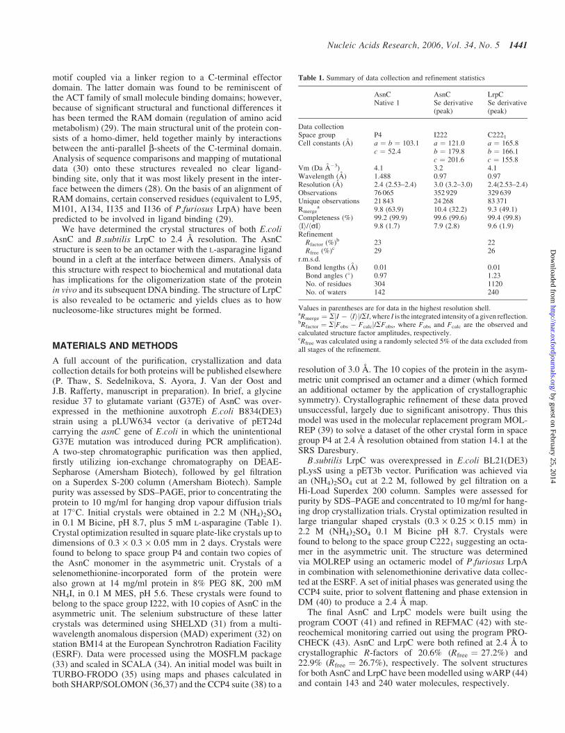
Table 1. Summary of data collection and refinement statistics
AsnC AsnC LrpCNative 1 Se derivative
(peak)Se derivative(peak)
Data collectionSpace group P4 I222 C2221
Cell constants (A) a ¼ b ¼ 103.1 a ¼ 121.0 a ¼ 165.8c ¼ 52.4 b ¼ 179.8 b ¼ 166.1
c ¼ 201.6 c ¼ 155.8Vm (Da A�3) 4.1 3.2 4.1Wavelength (A) 1.488 0.97 0.97Resolution (A) 2.4 (2.53–2.4) 3.0 (3.2–3.0) 2.4(2.53–2.4)Observations 76 065 352 929 329 639Unique observations 21 843 24 268 83 371Rmerge
a 9.8 (63.9) 10.4 (32.2) 9.3 (49.1)Completeness (%) 99.2 (99.9) 99.6 (99.6) 99.4 (99.8)hIi/hsIi 9.8 (1.7) 7.9 (2.8) 9.6 (1.9)Refinement
Rfactor (%)b 23 22Rfree (%)c 29 26
r.m.s.d.Bond lengths (A) 0.01 0.01Bond angles (�) 0.97 1.23No. of residues 304 1120No. of waters 142 240
Values in parentheses are for data in the highest resolution shell.aRmerge ¼ SjI � hIij/SI, where I is the integrated intensity of a given reflection.bRfactor ¼ SjFobs � Fcalcj/SFobs, where Fobs and Fcalc are the observed andcalculated structure factor amplitudes, respectively.cRfree was calculated using a randomly selected 5% of the data excluded fromall stages of the refinement.
Nucleic Acids Research, 2006, Vol. 34, No. 5 1441
by guest on February 25, 2014http://nar.oxfordjournals.org/
Dow
nloaded from

All figures were produced using PyMOL (http://www.pymol.org) with the exception of the electrostatic potentialsurfaces, which were produced in GRASP (45). The atomiccoordinates and structure factors for AsnC and LrpC aredeposited at the protein data bank with codes 2cg4 and2cfx, respectively.
RESULTS
Overall structure
The monomer structure of both AsnC and LrpC comprises twodomains. An N-terminal DNA-binding domain consisting of3 a-helices (aA–aC), with aB–aC forming a simple 2 helixmotif (HtH) (46) stabilized by a hydrophobic core protectedby aA (Figure 2a–d). The G37 to E37 mutation in our AsnCconstruct (Materials and Methods) lies on the surface at the N-terminal end of helix aC. This represents a significant changein both size and charge and whilst its location suggests it mightimpair DNA binding, it should have no other detrimentaleffect to the overall fold of the protein. Crystallization trialswith the wild-type protein produced only very poorly diffract-ing crystals whose space group could not be determined. TheG37E variant yielded diffracting crystals suitable for crystal-lographic studies and analysis of the packing reveals E37 insome subunits does contribute to the crystal lattice contacts.The packing does not resemble the helical arrangement seenwith Pyrococcus OT3 FL11 (17). The relative overall orienta-tion of the N- and C-terminal domains is essentially the samein both AsnC and LrpC. However there is a shift of 20� in theposition of helix aC relative to strand b1 between AsnC andLrpC (Figure 2). The inter-helix spacing between helices aBand aC within the N-terminal domain is maintained betweenthe two structures by increased curvature of helix aC in LrpC.This DNA-binding domain is linked by a single b-strand (b1)to a C-terminal effector binding or RAM domain (29) formedby a four-stranded anti-parallel b-sheet (b2–b5) flanked on oneface by two a-helices (aD–aE).
A dimer representing one of the functional units in AsnC orLrpC is stabilized via the formation of a hydrophobic corebetween strands b2–b5 of each monomer. The dimer is furtherstabilized via direct hydrogen bonding interactions betweenresidues on b1 of each monomer, which come together toform a 2-stranded anti-parallel b-ribbon. Predominantlyhydrophobic interactions between the C-terminus of onemonomer and strand b3 and helix aD of another also contrib-ute to dimer stability.
The P4 symmetry of the AsnC crystal lattice allows theformation of an octamer, consisting of a tetramer of dimers�52 · 52 · 118 s in size (Figure 2e and g). The asymmetricunit of the LrpC crystal contains an octamer measuring56 · 56 · 118 A in size. The dimer–dimer interface consistsmainly of hydrophobic contacts, with the main interactionsforming between helix aE and strand b5 of one dimer pair,with residues leading into helix aD and those in a loopbetween strands b3 and b4 of another. Analysis using AREA-IMOL (47) with a search probe radius of 1.4 s has providedmeasurements of buried surface area upon dimer and octamerformation (Table 2). A comparison of these values with thoseof other multimeric proteins (48) suggests that these values fall
within the normal range for cytosolic proteins from mesophilicsources.
Asparagine binding in AsnC
In the refined structure of the AsnC octamer, clear additionaldensity was observed in the cleft between the turn from strandb3 to b4 of one monomer and strand b5 of another. Thisadditional density was identified as bound L-asparagine,which had been present at a concentration of 5 mM duringcrystal growth. Eight molecules of asparagine were identifiedin each octamer. Loop residues Y100 to S106 from one dimerpair form one side of a binding pocket, with the side chain ofthe Y100 pointing inwards together with the backbone car-bonyls of T101 and G103 (Figure 3a). Residue G103 adopts apositive phi angle of �90� to create a sharp turn that allowsthe use of the backbone carbonyls of T101 and G103 in bind-ing the effector. The remainder of the pocket is constructedfrom residues contributed by a second dimer pair, Q128 andL123 emanating from helix aE, and residues T136 to T138 ofstrand b5 (Figure 3b). The asparagine is positioned such thatthe generic carboxyl and amino groups of the amino aciddisplay hydrogen bonding potential to the backbone amidesor carbonyls of G103, T138 and Y105. The side chaincarboxyamide of the asparagine is most likely stabilizedthrough interactions with the side chains of Y100, Q128and the backbone of T101 and S106. AsnC discriminatesbetween asparagine and aspartate and an analysis of the bind-ing pocket suggests that unfavourable interactions would arisebetween the charged sidechain of the effector molecule andresidue E128. The formation of a stable octamer in the pres-ence or absence of asparagine is observed by gel filtration(data not shown) suggesting that the ligand is not an absoluterequirement for octamer formation, which is consistent withthe previously obtained effector-free structure of P.furiosusLrpA (28), as well as with reported observations of effector-free octamers in studies on E.coli Lrp (49). An analysis of themolecular surfaces of the octameric state of AsnC using theprogram GRASP (45) reveals channels that allow the diffusionof asparagine into the protein (Figure 4). Residues H104 toS106 and I75 to L76 of one dimer pair, together with a con-tribution from S135 to E137 from an adjacent dimer, shape thechannel entrance. The eight channels branch off the centralcavity of the octamer and would permit diffusion of the ligandin and out of the protein, allowing for a rapid response toexogenous asparagine levels.
DISCUSSION
Mutational and sequence analysis
Effector-binding region. The structural data available for fourmembers of the family, together with our ligand-binding dataallow us to study the likely effects of mutations both oneffector binding and the oligomeric state of the protein.Previous work on E.coli Lrp (50) identified subgroups of resi-dues which influenced either DNA binding or its interactionwith leucine. In Lrp, variants found to be insensitive to leucinecarried mutations in one of seven positions in the C-terminaldomain (residues L107, D114, M124, L136, Y147, V148 andV149). In addition, a thorough comparative analysis of RAM
1442 Nucleic Acids Research, 2006, Vol. 34, No. 5
by guest on February 25, 2014http://nar.oxfordjournals.org/
Dow
nloaded from
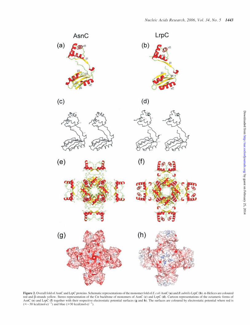
Figure 2. Overall fold of AsnC and LrpC proteins. Schematic representations of the monomer fold of E.coli AsnC (a) and B.subtilis LrpC (b).a-Helices are colouredred and b-strands yellow. Stereo representation of the Ca backbone of monomers of AsnC (c) and LrpC (d). Cartoon representations of the octameric forms ofAsnC (e) and LrpC (f) together with their respective electrostatic potential surfaces (g and h). The surfaces are coloured by electrostatic potential where red is(<�30 kcal(mol·e)�1) and blue (>30 kcal(mol·e)�1).
Nucleic Acids Research, 2006, Vol. 34, No. 5 1443
by guest on February 25, 2014http://nar.oxfordjournals.org/
Dow
nloaded from
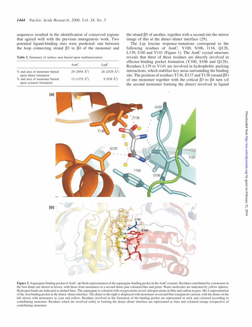
sequences resulted in the identification of conserved regionsthat agreed well with the previous mutagenesis work. Twopotential ligand-binding sites were predicted: one betweenthe loop connecting strand b3 to b4 of the monomer and
the strand b5 of another, together with a second site the mirrorimage of this at the dimer–dimer interface (29).
The Lrp leucine response-mutations correspond to thefollowing residues of AsnC: Y100, S106, I116, Q128,L139, I140 and V141 (Figure 1). The AsnC crystal structurereveals that three of these residues are directly involved ineffector-binding pocket formation (Y100, S106 and Q128).Residues L139 to V141 are involved in hydrophobic packinginteractions, which stabilize key areas surrounding the bindingsite. The position of residues T136, E137 and T138 (strand b5)of one monomer together with the critical b3 to b4 turn (ofthe second monomer forming the dimer) involved in ligand
Table 2. Summary of surface area buried upon multimerization
AsnC LrpC
% and area of monomer buriedupon dimer formation
29 (2954 s2) 26 (2529 s
2)
% and area of monomer buriedupon octamer formation
13 (1335 s2) 9 (938 s
2)
(a)
(b)
Figure 3. Asparagine binding pocket of AsnC. (a) Stick representation of the asparagine-binding pocket in the AsnC octamer. Residues contributed by a monomer inthe first dimer are shown in brown, with those from monomers in a second dimer pair coloured blue and green. Water molecules are indicated by yellow spheres.Hydrogen bonds are indicated as dashed lines. The asparagine is coloured with oxygen atoms in red, nitrogen atoms in blue and carbon in grey. (b) A representationof the Asn binding pocket at the dimer–dimer interface. The dimer to the right is displayed with monomers in red and blue transparent cartoon, with the dimer on theleft shown with monomers in cyan and yellow. Residues involved in the formation of the binding pocket are represented in stick and coloured according tocontributing monomer. Residues which are involved solely in forming the dimer–dimer interface are represented as lines and coloured orange irrespective ofcontributing monomer.
1444 Nucleic Acids Research, 2006, Vol. 34, No. 5
by guest on February 25, 2014http://nar.oxfordjournals.org/
Dow
nloaded from

binding are heavily dependent on the spatial positioning ofL139 to V141. Residues T136 and T138 are highly conservedand their roles in effector binding suggest they constitute animportant marker of effector regulation. It is likely that the b5to aE dimer–dimer contacts would be perturbed by mutationsto these residues and would have a significant impact oneffector binding by the octamer. The reason for the effectof an I116 mutation (equivalent to M124 in Lrp) is lessclear as it is situated 12 s away from the effector-bindingsite. However, its location at the dimer–dimer interface and itsproximity to sheet b5 suggest it might exert a direct effectupon octamer stability or an indirect longer-range effect oneffector binding.
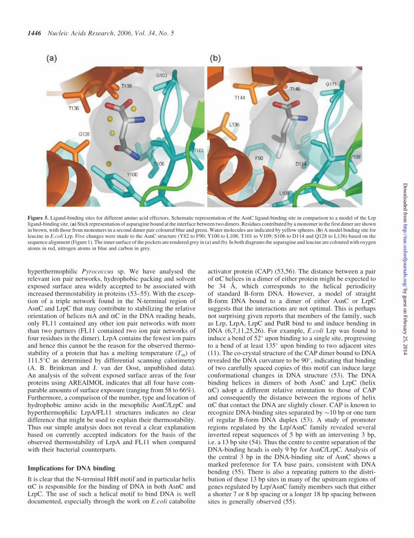
Using a structure-based sequence alignment, mapping ofequivalent Lrp residues onto the AsnC structure resultsin one end of the binding pocket becoming predominantlyhydrophobic. The substitution of residues Y82, Y100 andQ128 in AsnC by a phenylalanine and two leucines, respect-ively, in Lrp, changes the size and nature of the pocket suchthat it would be ideally suited to binding the aliphatic sidechain of leucine as opposed to the carboxyamide group ofasparagine (Figure 5). The remainder of the pocket islargely unchanged with the AsnC TET motif (residues 136–138) mimicked by TRT (residues 144–146) in Lrp. AlthoughE137 in AsnC becomes R145 in Lrp, there is a compensatorychange in an interacting residue of an H for a D at a positionequivalent to residue 104 of AsnC (Figure 1). Furthermore,residue G103 in the turn between b3 and b4 (equivalent toG111 in Lrp) that is critical to the interaction with the genericamino group of the effector is totally conserved across allknown sequences (29) and is the only glycine in the structureto possess a positive phi angle (�90�). Lrp does respond toexogenous alanine and this could clearly be incorporated intothe binding pocket. However Lrp does not show a response toIle or Val and our model would indicate that binding would bedisfavoured through possible clashes with the sidechainhydroxyl of T136 and also the backbone carbonyl of T101.
It is also possible to map the pocket residues for the othermembers of the Lrp/AsnC family for which effector moleculeshave been identified: FL11 (Pyrococcus OT3) binds glutamine(17); PutR (Agrobacterium tumefaciens) binds proline (51);BkdR (Pseudomonas putida) preferentially binds valine (52);LysM (S.solfataricus) exclusively binds lysine (13). In each ofthese cases the residue changes result in a binding pocketwhose steric characteristics and charge distribution fit theproposed ligand. A study of the equivalent region of LrpAfinds the binding pocket obstructed by residues M101 andP133 (equivalent to S106 and T138 of AsnC) and is consistentwith its reported effector-independent activity. In LrpC, arestructuring of the pocket is achieved via changes to 8 ofthe 11 residues local to the binding site. The most notable ofthese are a change in the side chain rotamer of Y78 (equivalentto Y82 in AsnC) and the substitution of Y100 for R96 andQ128 for S123. The resultant binding pocket is smaller in sizethan that of AsnC, mainly due to the presence of R96, althoughit still possesses a channel into the central cavity of theoctamer. The majority of contacts required to bind the genericcarboxyl and amino groups of an amino acid are in place.However, the small size of the pocket precludes the bindingof any amino acid much larger than alanine in the absenceof any restructuring upon effector binding. The addition ofleucine to LrpC does not prevent it binding to its own promoter(25,26) and biochemical studies indicate that LrpC binding toDNA is not influenced by alanine (S. Ayora, unpublished data)and in all likelihood it has no effector. The structure ofLrpC contains four water molecules within each of thepockets at the dimer–dimer interface. Changes in the localsize and shape of the binding pocket can be modelled basedupon our current structural data. However, ligand bindingis likely to have larger structural consequences e.g. tertiaryand/or quaternary conformational changes, which influencefunction but cannot be reliably modelled with the availablestructures.
DNA binding region
Mapping of residues identified in E.coli Lrp as being involvedin DNA binding [residues D13, L34, L40, S41, P44, L46,R48, Y61, L65 and L70, (50)] onto the structures of AsnCand LrpC reveals two main clusters. Most of the first group ofDNA-binding mutants are located in recognition helix aC(Figure 1) and seem likely to be involved in direct contactwith the DNA. The possible exceptions are R48 (equivalent toAsnC R42 and LrpC R39), which forms an ion pair networkwith two totally conserved aspartates in helix aA and L34(equivalent to AsnC L28 and LrpC L25). These residues mapto the inner surface of helix aB and contribute to the core ofthe domain. The second group of DNA-binding mutants arefrom strand b1 and are involved in the positioning of helix aAfrom a second monomer in a dimer pair. Thus these mutationsmay exert their effect by perturbation of the relative geometryof the helices making up the HtH motif.
Thermostability
The structural elucidation of AsnC and LrpC allows usto investigate potential factors effecting protein stability bycomparing them to the structures of LrpA and FL11 from the
Figure 4. Channels into the asparagines-binding pocket The channelsleading to the ligand-binding pocket of E.coli AsnC are illustrated via a sectionthrough the molecular surface of the protein with underlying secondary struc-ture elements shown in cartoon representation. The channels into the bindingsites from the central cavity are indicated by white dotted lines. The view islooking down the 4-fold axis of the octamer (as in Figure 2e). The asparagineligands are coloured as carbon atoms in yellow, oxygen atoms in red andnitrogen atoms in blue with the protein surface coloured grey.
Nucleic Acids Research, 2006, Vol. 34, No. 5 1445
by guest on February 25, 2014http://nar.oxfordjournals.org/
Dow
nloaded from
hyperthermophilic Pyrococcus sp. We have analysed therelevant ion pair networks, hydrophobic packing and solventexposed surface area widely accepted to be associated withincreased thermostability in proteins (53–55). With the excep-tion of a triple network found in the N-terminal region ofAsnC and LrpC that may contribute to stabilizing the relativeorientation of helices aA and aC in the DNA reading heads,only FL11 contained any other ion pair networks with morethan two partners (FL11 contained two ion pair networks offour residues in the dimer). LrpA contains the fewest ion pairsand hence this cannot be the reason for the observed thermo-stability of a protein that has a melting temperature (Tm) of111.5�C as determined by differential scanning calorimetry(A. B. Brinkman and J. van der Oost, unpublished data).An analysis of the solvent exposed surface areas of the fourproteins using AREAIMOL indicates that all four have com-parable amounts of surface exposure (ranging from 58 to 66%).Furthermore, a comparison of the number, type and location ofhydrophobic amino acids in the mesophilic AsnC/LrpC andhyperthermophilic LrpA/FL11 structures indicates no cleardifference that might be used to explain their thermostability.Thus our simple analysis does not reveal a clear explanationbased on currently accepted indicators for the basis of theobserved thermostability of LrpA and FL11 when comparedwith their bacterial counterparts.
Implications for DNA binding
It is clear that the N-terminal HtH motif and in particular helixaC is responsible for the binding of DNA in both AsnC andLrpC. The use of such a helical motif to bind DNA is welldocumented, especially through the work on E.coli catabolite
activator protein (CAP) (53,56). The distance between a pairof aC helices in a dimer of either protein might be expected tobe 34 s, which corresponds to the helical periodicityof standard B-form DNA. However, a model of straightB-form DNA bound to a dimer of either AsnC or LrpCsuggests that the interactions are not optimal. This is perhapsnot surprising given reports that members of the family, suchas Lrp, LrpA, LrpC and PutR bind to and induce bending inDNA (6,7,11,25,26). For example, E.coli Lrp was found toinduce a bend of 52� upon binding to a single site, progressingto a bend of at least 135� upon binding to two adjacent sites(11). The co-crystal structure of the CAP dimer bound to DNArevealed the DNA curvature to be 90�, indicating that bindingof two carefully spaced copies of this motif can induce largeconformational changes in DNA structure (53). The DNAbinding helices in dimers of both AsnC and LrpC (helixaC) adopt a different relative orientation to those of CAPand consequently the distance between the regions of helixaC that contact the DNA are slightly closer. CAP is known torecognize DNA-binding sites separated by �10 bp or one turnof regular B-form DNA duplex (53). A study of promoterregions regulated by the Lrp/AsnC family revealed severalinverted repeat sequences of 5 bp with an intervening 3 bp,i.e. a 13 bp site (54). Thus the centre to centre separation of theDNA-binding heads is only 9 bp for AsnC/LrpC. Analysis ofthe central 3 bp in the DNA-binding site of AsnC shows amarked preference for TA base pairs, consistent with DNAbending (55). There is also a repeating pattern to the distri-bution of these 13 bp sites in many of the upstream regions ofgenes regulated by Lrp/AsnC family members such that eithera shorter 7 or 8 bp spacing or a longer 18 bp spacing betweensites is generally observed (55).
Figure 5. Ligand-binding sites for different amino acid effectors. Schematic representation of the AsnC ligand-binding site in comparison to a model of the Lrpligand-binding site. (a) Stick representation of asparagine bound at the interface between two dimers. Residues contributed by a monomer in the first dimer are shownin brown, with those from monomers in a second dimer pair coloured blue and green. Water molecules are indicated by yellow spheres. (b) A model binding site forleucine in E.coli Lrp. Five changes were made to the AsnC structure (Y82 to F90; Y100 to L108; T101 to V109; S106 to D114 and Q128 to L136) based on thesequence alignment (Figure 1). The inner surface of the pockets are rendered grey in (a) and (b). In both diagrams the asparagine and leucine are coloured with oxygenatoms in red, nitrogen atoms in blue and carbon in grey.
1446 Nucleic Acids Research, 2006, Vol. 34, No. 5
by guest on February 25, 2014http://nar.oxfordjournals.org/
Dow
nloaded from

Modelling of DNA interactions
Modelling of the AsnC octamer bound to a curved piece ofDNA (based on PDB code 1CGP) indicates that the pairs ofDNA-binding N-terminal domains of individual dimers mustbe reoriented relative to the effector-binding C-terminaldomains to enable binding of an octamer simultaneously tomultiple 13 bp sites with 18 bp spacings (Figure 6a). Thisseems plausible because there are relatively few contactsbetween the N- and C-terminal domains. Alternatively bindingof multiple DNA sites by an array of adjacent octamers mightallow the introduction of varying degrees of curvature or eventhe wrapping of DNA in a solenoid form (Figure 6b). BothAsnC and LrpC have similar diameters of �118 s, whereas incontrast LrpA has a diameter of �107 s, which makes itdifficult to envisage how LrpA could wrap the DNAaround itself to accommodate simultaneous binding of a singleoctamer to more than one site. The majority of the increase indiameter of AsnC and LrpC arises from the packing of theC-terminal residues into the space between elements of strandsb3 and b4 and the helical turn around residues P61 toL64 (numbering as for AsnC). This moves helix aA (andconsequently the entire N-terminal domain) 3.5 s furtheraway from the C-terminal core. There is notable sequence con-servation in the C-termini of enterobacterial Lrp proteinsand this may support a critical role for the C-termini in trans-mitting the effector-bound status of the protein. There is littledifference in the relative orientation of the three helices mak-ing up the N-terminal domain, with >94% of Ca atoms super-imposing with a root mean square deviation (r.m.s.d.) of 1.4 s
or better over the four known structures. Whilst the exactnature of the interaction of these proteins with their targetDNA awaits elucidation, it does appear that the conformation
of the C-terminal residues may be important in orienting theposition of the HtH domain and thus plays a role in switchingpreference between differently spaced DNA-binding sites.The importance of such a small shift in structure issupported by work on an E.coli Lrp variant lacking thefinal 11 C-terminal residues which does not bind to ilvIHoperator DNA, despite having a CD trace comparable tothat of the wild-type protein (49).
The effector switch
Like Lrp, AsnC binds DNA and regulates gene expressionin the absence or presence of effector. It appears likely thatthe presence of the effector selects for one possible conformerof the protein rendering it suitable for acting at a subset oftarget DNA sites but unsuitable for others, presumably byaltering the relative orientation and spacing of the N- andC-terminal domains and associated bound DNA. This notionis supported by previous biochemical studies on E.coli Lrpand P.putida BkdR, which noted a decrease in the intrinsicfluorescence upon their binding of their amino acid effectorsto high affinity sites, suggesting a conformational change inboth proteins (49,52). Furthermore, slight inter-domainrearrangements have been observed upon effector bindingin the structurally analogous ACT domain containing proteins,which are also subject to allosteric regulation by smallmolecules, typically amino acids [reviewed in (29)]. Theexact mechanism by which such a switch could be madeis difficult to ascertain in the absence of an effector-free struc-ture of AsnC and awaits elucidation. In addition, in certaincases the presence of the effector may favour an increase in thelevel of an octameric species of the protein in vivo such thata shift in its oligomeric state equilibrium would encourage
Figure 6. Models of AsnC and LrpC binding to DNA. (a) Cartoon representation of AsnC binding to DNA with a curved piece of B-form DNA based on the 22 bpfragment from CRP (53) shown as a surface representation. Dimer-binding sites (labelled 1–3) of 13 bp in length are coloured green and separated by 18 bp of non-conserved DNA (coloured grey) to mimic a promoter region. Binding of DNA reading heads to sites 1 and 3 required a hinge movement around residue 60 to allow asmall degree of flexibility between the N- and C-terminal domains of the protein. (b) Model of how LrpC could wrap DNA in a nucleosome-like structure.Cooperative binding of LrpC to the DNA forms a right-handed super-helix, which constrains the positive supercoils. Two octamers of the protein are shown in cyanand green. Modelled DNA is shown as a grey surface and based on existing crystal structures of wrapped DNA (PDB code 1AOI) (57) and electron microscopy studiesof LrpC (27).
Nucleic Acids Research, 2006, Vol. 34, No. 5 1447
by guest on February 25, 2014http://nar.oxfordjournals.org/
Dow
nloaded from
cooperative DNA binding to sites optimized for interactionwith an octamer.
DNA binding of the AsnC/Lrp family
Thus it appears that DNA binding in the Lrp/AsnC family isgoverned by a complex interplay between the oligomericstates populated by the protein and both the numberand nucleotide base sequence of mirrored repeats in theupstream regions of the genes concerned. Fine tuningof promoter selection is likely conveyed through subtlechanges in structure due to effector binding and/or the packingof the C-terminal residues close to the HtH motif. LrpCreportedly binds DNA in a non-sequence specific manner,recognizing intrinsically curved regions containing phasedA-tracts (27). Its stable octameric structure (dimers ofthe protein were not observed under our solution conditions)indicates that DNA could indeed be wrapped around it to yielda nucleosome-like structure or to influence promoter geometry(Figure 6b), as observed in electron microscopy experiments(27). An extension of the model can be made in which twooctamers form a hexadecamer and bind multiple DNA sites, asobserved for E.coli Lrp (51). This requires a small movementof �5 s and rotation of �5� for each of the N-terminal DNA-binding heads relative to the experimentally observed octamerstructure such that a bound DNA solenoid structure is formed.The spacing between binding sites is not altered in our modelupon the passage of the DNA from one octamer to the next inthe hexadecamer.
ACKNOWLEDGEMENTS
We would like to thank the staff on stations ID 14.4 and ID 29 atthe E.S.R.F Grenoble, and also station 14.1 at the SRSDaresbury, for their support and assistance with data collec-tion. This work was supported by grants from the WellcomeTrust and BBSRC together with collaborative grantsBMC2003-00150, BMC2003-01969 from DGICYT.Funding to pay the Open Access publication charges forthis article was provided by the Wellcome Trust.
Conflict of interest statement. None declared.
REFERENCES
1. Charlier,D., Roovers,M., Thia-Toong,T.L., Durbecq,V. andGlansdorff,N. (1997) Cloning and identification of the Sulfolobussolfataricus lrp gene encoding an archaeal homologue of theeubacterial leucine responsive global transcriptional regulator Lrp.Gene, 201, 63–68.
2. Napoli,A., Van der Oost,J., Sensen,C.W., Charlebois,R.L., Rossi,M. andCiaramella,M. (1999) An Lrp-like protein of the hyperthermophilicarchaeon Sulfolobus solfataricus which binds to its own promoter.J. Bacteriol., 181, 1474–1480.
3. Brinkman,A.B., Ettema,T.J.G., de Vos,W.M. and van der Oost,J. (2003)The Lrp family of transcriptional regulators. Mol. Microbiol., 48,287–294.
4. Willins,D.A., Ryan,C.W., Platko,J.V. and Calvo,J.M. (1991)Characterization of Lrp, an Escherichia coli regulatoryprotein that mediates a global response to leucine. J. Biol. Chem., 266,10768–10774.
5. Madhusudhan,K.T., Huang,N. and Sokatch,J.R. (1995) Characterizationof BkdR-DNA binding in the expression of the bkd operon ofPseudomonas putida. J. Bacteriol., 177, 636–641.
6. Brinkman,A.B., Dahlke,I., Tuininga,J.E., Lammers,T., Dumay,V., deHeus,E., Lebbink,J.H.G., Thomm,M., de Vos,W.M. and van der Oost,J.(2000) An Lrp-like transcriptional regulator from the archaeonPyrocococcus furiosus is negatively autoregulated.J. Biol. Chem., 275, 38160–38169.
7. Jafri,S., Evoy,S., Cho,K.Y., Craighead,H.G. and Winans,S.C. (1999)An Lrp-type transcriptional regulator from Agrobacterium tumefacienscondenses more than 100 nucleotides of DNA into globular nucleoproteincomplexes. J. Mol. Biol., 288, 811–824.
8. Chen,S., Rosner,M.H. and Calvo,J.M. (2001) Leucine-regulated self-association of leucine-responsive regulatory protein (Lrp) fromEscherichia coli. J. Mol. Biol., 312, 625–635.
9. D’Ari,R., Lin,R.T. and Newman,E.B. (1993) The leucine-responsiveregulatory protein—more than a regulator? Trends Biochem. Sci., 18,260–263.
10. Azam,T.A., Iwata,A., Nishimura,A., Ueda,S. and Ishihama,A. (1999)Growth phase-dependent variation in protein composition of theEscherichia coli nucleoid. J. Bacteriol., 181, 6361–6370.
11. Wang,Q. and Calvo,J.M. (1993) Lrp, a major regulatory protein inEscherichia coli, bends DNA and can organize the assembly of ahigher-order nucleoprotein structure. EMBO J., 12,2495–2501.
12. Peeters,E., Thia-Toong,T.L., Gigot,D., Maes,D. and Charlier,D. (2004)Ss-LrpB, a novel Lrp-like regulator of Sulfolobus solfataricus P2, bindscooperatively to three conserved targets in its own control region.Mol. Microbiol., 54, 321–336.
13. Brinkman,A.B., Bell,S.D.,Lebbink,R.J., de Vos,W.M.and vander Oost,J.(2002) The Sulfolobus solfataricus Lrp-like protein LysM regulates lysinebiosynthesis in response to lysine availability. J. Biol. Chem., 277,29537–29549.
14. Ouhammouch,M., Dewhurst,R.E., Hausner,W., Thomm,M. andGeiduschek,E.P. (2003) Activation of archaeal transcription byrecruitment of the TATA-binding protein. Proc. Natl Acad. Sci. USA,100, 5097–5102.
15. Ouhammouch,M., Langham,G.E., Hausner,W., Simpson,A.J., El-Sayed,N.M.A. and Geiduschek,E.P. (2005) Promoter architecture andresponse to a positive regulator of archaeal transcription.Mol. Microbiol., 56, 625–637.
16. Bell,S.D. and Jackson,S.P. (2001) Mechanism and regulation oftranscription in archaea. Curr. Opin. Microbiol., 4, 208–213.
17. Koike,H., Ishijima,S.A., Clowney,L. and Suzuki,M. (2004) The archaealfeast/famine regulatory protein: potential roles of its assembly formsfor regulating transcription. Proc. Natl Acad. Sci. USA,101, 2840–2845.
18. Tani,T.H., Khodursky,A., Blumenthal,R.M., Brown,P.O. andMatthews,R.G. (2002) Adaptation to famine: a family of stationary-phasegenes revealed by microarray analysis. Proc. Natl Acad. Sci. USA,99, 13471–13476.
19. Calvo,J.M. and Matthews,R.G. (1994) The leucine responsive regulatoryprotein, a global regulator of metabolism in Escherichia coli. Microbiol.Rev., 58, 466–490.
20. Newman,E.B. and Lin,R.T. (1995) Leucine-responsive regulatoryprotein – a global regulator of gene expression in Escherichia coli. Ann.Rev. Microbiol., 49, 747–775.
21. Willins,D.A., Ryan,C.W., Platko,J.V. and Calvo,J.M. (1991)Characterization of Lrp, an Escherichia coli regulatory proteinthat mediates a global response to leucine. J. Biol. Chem., 266,10768–10774.
22. Kolling,R. and Lother,H. (1985) AsnC—an autogenously regulatedactivator of asparagine synthetase A transcription in Escherichia coli.J. Bacteriol., 164, 310–315.
23. de Wind,N., de Jong,M., Meijer,M. and Stuitje,A.R. (1985) Site directedmutagenesis of the Escherichia coli chromosome near oriC: identificationand characterization of asnC, a regulatory element in E.coli asparaginemetabolism. Nucleic Acids Res., 13, 8797–8811.
24. Beloin,C., Ayora,S., Exley,R., Hirschbein,L., Ogasawara,N.,Kasahara,Y., Alonso,J.C. and LeHegarat,F. (1997)Characterization of an lrp-like (lrpC) gene from Bacillus subtilis.Mol. Gen. Genet., 256, 63–71.
25. Beloin,C., Exley,R., Mahe,A.L., Zouine,M., Cubasch,S. and LeHegarat,F. (2000) Characterization of LrpC DNA-binding properties andregulation of Bacillus subtilis lrpC gene expression. J. Bacteriol.,182, 4414–4424.
1448 Nucleic Acids Research, 2006, Vol. 34, No. 5
by guest on February 25, 2014http://nar.oxfordjournals.org/
Dow
nloaded from
26. Tapias,A., Lopez,G. and Ayora,S. (2000) Bacillus subtilis LrpC is asequence-independent DNA-binding and DNA-bending protein whichbridges DNA. Nucleic Acids Res., 28, 552–559.
27. Beloin,C., Jeusset,J., Revet,B., Mirambeau,G., Le Hegarat,F. and LeCam,E. (2003) Contribution of DNA conformation and topology in right-handed DNA wrapping by the Bacillus subtilis LrpC protein.J. Biol. Chem., 278, 5333–5342.
28. Leonard,P.M., Smits,S.H.J., Sedelnikova,S.E., Brinkman,A.B., deVos,W.M., van der Oost,J., Rice,D.W. and Rafferty,J.B. (2001) Crystalstructure of the Lrp-like transcriptional regulator from the archaeonPyrococcus furiosus. EMBO J., 20, 990–997.
29. Ettema,T.J.G., Brinkman,A.B., Tani,T.H., Rafferty,J.B. and van derOost,J. (2002) A novel ligand-binding domain involved in regulation ofamino acid metabolism in prokaryotes. J. Biol. Chem., 277, 37464–37468.
30. Platko,J.V. and Calvo,J.M. (1993) Mutations affecting the ability ofEscherichia coli Lrp to bind DNA, activate transcription, or respond toleucine. J. Bacteriol., 175, 1110–1117.
31. Schneider,T.R. and Sheldrick,G.M. (2002) Substructure solution withSHELXD. Acta Crystallogr. D, 58, 1772–1779.
32. Hendrickson,W.A. (1991) Determination of macromolecular structuresfrom anomalous diffractionof synchrotron radiation. Science, 254, 51–58.
33. Leslie,A.G.W. (1992) Recent changes to the MOSFLM package forprocessing film and image plate data. Joint CCP4 and ESF-EAMCBNewsletter on Protein Crystallography, 26.
34. Evans,P.R. Scaling of MAD data. In Proceedings of the CCP4 StudyWeekend. Recent Advances in Phasing, 97–102.
35. Roussel,A., Fontecilla-Camps,J.C. and Cambillau,C. (1990)TURBO-FRODO: a new program for protein crystallography andmodelling. XV IUCr Congress Abstracts, 66–67.
36. Bricogne,G., Vonrhein,C., Flensburg,C., Schiltz,M. and Paciorek,W.(2003) Generation, representation and flow of phase information instructure determination: recent developments in and around SHARP 2.0.Acta Crystallogr. D, 59, 2023–2030.
37. Abrahams,J.P. and Leslie,A.G.W. (1996) Methods used in the structuredetermination of bovine mitochondrial F-1 ATPase. Acta Crystallogr. D,52, 30–42.
38. Collaborative Computational Project, Number 4. (1994), The CCP4suite: programs for protein crystallography. Acta Crystallogr. D, 50,760–763.
39. Vagin,A. and Teplyakov,A. (2000) An approach to multi-copy search inmolecular replacement. Acta Crystallogr. D, 56, 1622–1624.
40. Cowtan,K.D. (1994) An automated procedure for phase improvement bydensity modification. Joint CCP4 and ESF-EAMCB Newsletter onProtein Crystallography, 31, 34–38.
41. Emsley,P. and Cowtan,K. (2004) Coot: model-building tools formolecular graphics. Acta Crystallogr. D, 60, 2126–2132.
42. Murshudov,G.N., Vagin,A.A. and Dodson,E.J. (1997) Refinement ofmacromolecular structures by the maximum-likelihood method.Acta Crystallogr. D, 53, 240–255.
43. Laskowski,R.A., Macarthur,M.W., Moss,D.S. and Thornton,J.M. (1993)PROCHECK—a program to check the stereochemical quality of proteinstructures. J. Appl. Crystallogr., 26, 283–291.
44. Perrakis,A., Morris,R. and Lamzin,V.S. (1999) Automated protein modelbuilding combined with iterative structure refinement. Nature Struct.Biol., 6, 458–463.
45. Nicholls,A., Bharadwaj,R. and Honig,B. (1993) GRASP—graphicalrepresentation and analysis of surface-properties. Biophys. J., 64,A166–A166.
46. Aravind,L., Anantharaman,V., Balaji,S., Babu,M.M. and Iyer,L.M.(2005) The many faces of the helix-turn-helix domain: Transcriptionregulation and beyond. FEMS Microbiol. Rev., 29, 231–262.
47. Lee,B. and Richards,F.M. (1971) The interpretation of protein structures:estimation of static accessibility. J. Mol. Biol., 55, 379–400.
48. Jones,S. and Thornton,J.M. (1995) Protein–protein interactions—areview of protein dimer structures. Prog. Biophys. Mol. Biol., 63, 31–65.
49. Chen,S. and Calvo,J.M. (2002) Leucine-induced dissociation ofEscherichia coli Lrp hexadecamers to octamers. J. Mol. Biol.,318, 1031–1042.
50. Platko,J.V. and Calvo,J.M. (1993) Mutations affecting the ability ofEscherichia coli Lrp to bind DNA, activate transcription, or respond toleucine. J. Bacteriol., 175, 1110–1117.
51. Cho,K.Y. and Winans,S.C. (1996) The putA gene of Agrobacteriumtumefaciens is transcriptionally activated in response to prolineby an Lrp-like protein and is not autoregulated. Mol. Microbiol.,22, 1025–1033.
52. Madhusudhan,K.T., Huang,N., Braswell,E.H. and Sokatch,J.R. (1997)Binding of L-branched-chain amino acids causes a conformationalchange in BkdR. J. Bacteriol., 179, 276–279.
53. Schultz,S.C., Shields,G.C. and Steitz,T.A. (1991) Crystal structure of aCAP-DNA complex: the DNA is bent by 90 degrees. Science, 253,1001–1007.
54. Suzuki,M. (2003) The DNA-binding specificity of eubacterial andarchaeal FFRPs. Proc. Jpn. Acad. Ser. B Phys. Biol. Sci.,, 79,213–222.
55. Koo,H.S., Wu,H.M. and Crothers,D.M. (1986) DNA bending atadenine:thymine tracts. Nature, 320, 501–506.
56. McKay,D.B. and Steitz,T.A. (1981) Structure of catabolite geneactivator protein at 2.9s resolution suggests binding toleft-handed B-DNA. Nature, 290, 744–749.
57. Luger,K., Mader,A.W., Richmond,R.K., Sargent,D.F. and Richmond,T.J.(1997) Crystal structure of the nucleosome core particle at 2.8sresolution. Nature, 389, 251–260.
Nucleic Acids Research, 2006, Vol. 34, No. 5 1449
by guest on February 25, 2014http://nar.oxfordjournals.org/
Dow
nloaded from