Transcriptional landscape of bone marrow-derived very small embryonic-like stem cells during hypoxia
11
RESEARCH Open Access Transcriptional landscape of bone marrow- derived very small embryonic-like stem cells during hypoxia Sina A Gharib 1† , Abdelnaby Khalyfa 2,3† , Magdalena J Kucia 4 , Ehab A Dayyat 2 , Jinkwan Kim 2,3 , Heather B Clair 2 and David Gozal 2,3* Abstract Background: Hypoxia is a ubiquitous feature of many lung diseases and elicits cell-specific responses. While the effects of hypoxia on stem cells have been examined under in vitro conditions, the consequences of in vivo oxygen deprivation have not been studied. Methods: We investigated the effects of in vivo hypoxia on a recently characterized population of pluripotent stem cells known as very small embryonic-like stem cells (VSELs) by whole-genome expression profiling and measuring peripheral blood stem cell chemokine levels. Results: We found that exposure to hypoxia in mice mobilized VSELs from the bone marrow to peripheral blood, and induced a distinct genome-wide transcriptional signature. Applying a computationally-intensive methodology, we identified a hypoxia-induced gene interaction network that was functionally enriched in a diverse array of programs including organ-specific development, stress response, and wound repair. Topographic analysis of the network highlighted a number of densely connected hubs that may represent key controllers of stem cell response during hypoxia and, therefore, serve as putative targets for altering the pathophysiologic consequences of hypoxic burden. Conclusions: A brief exposure to hypoxia recruits pluripotent stem cells to the peripheral circulation and actives diverse transcriptional programs that are orchestrated by a selective number of key genes. Background Hypoxia is a pathophysiologic condition commonly seen in lung diseases and in many other disorders such as tissue ischemia and cancer [1]. In the developing embryo expo- sure to global reduction in tissue oxygen is a critical factor in recruitment and selective differentiation of embryonic stem cells that orchestrate organogenesis [2]. The presence of hypoxic microenvironments in adult tissues, as seen for example in solid tumors or during ischemic injury, can also lead to activation and recruitment of stem cells from specific tissue niches, including the bone marrow (BM) [3]. Under such local hypoxic circumstances, resident BM stem cells have been shown to become mobilized and facilitate the structural and functional repair of injured organs [4-6]. The effects of global hypoxia on stem cell activation and recruitment remain poorly understood despite being a common feature of many pulmonary disor- ders, and increasing evidence that BM-derived circulating progenitor cells may play a causative role in acute lung injury, pulmonary hypertension, pulmonary fibrosis and COPD [7-10]. Recently, Ratajczak et al isolated a homogenous popula- tion of rare, small stem cells from adult murine BM mononuclear cells that expressed embryonic lineage mar- kers, and were thus named very small embryonic-like stem cells (VSELs) [11]. Pluripotency of these Sca-1 + Lin - CD45 - stem cells was established by demonstrating their ability to differentiate into cells representing all three germ layers [12]. Bone marrow-resident VSELs can be mobilized to the peripheral blood (PB) in response to specific chemoat- tractant gradients, including stromal derived factor-1 (SDF-1), hepatocyte growth factor (HGF), and leukemia * Correspondence: [email protected] † Contributed equally 2 Department of Pediatrics, University of Louisville, Louisville, KY, USA Full list of author information is available at the end of the article Gharib et al. Respiratory Research 2011, 12:63 http://respiratory-research.com/content/12/1/63 © 2011 Gharib et al; licensee BioMed Central Ltd. This is an Open Access article distributed under the terms of the Creative Commons Attribution License (http://creativecommons.org/licenses/by/2.0), which permits unrestricted use, distribution, and reproduction in any medium, provided the original work is properly cited.
-
Upload
washington -
Category
Documents
-
view
0 -
download
0
Transcript of Transcriptional landscape of bone marrow-derived very small embryonic-like stem cells during hypoxia
RESEARCH Open Access
Transcriptional landscape of bone marrow-derived very small embryonic-like stem cellsduring hypoxiaSina A Gharib1†, Abdelnaby Khalyfa2,3†, Magdalena J Kucia4, Ehab A Dayyat2, Jinkwan Kim2,3, Heather B Clair2 andDavid Gozal2,3*
Abstract
Background: Hypoxia is a ubiquitous feature of many lung diseases and elicits cell-specific responses. While theeffects of hypoxia on stem cells have been examined under in vitro conditions, the consequences of in vivooxygen deprivation have not been studied.
Methods: We investigated the effects of in vivo hypoxia on a recently characterized population of pluripotent stemcells known as very small embryonic-like stem cells (VSELs) by whole-genome expression profiling and measuringperipheral blood stem cell chemokine levels.
Results: We found that exposure to hypoxia in mice mobilized VSELs from the bone marrow to peripheral blood, andinduced a distinct genome-wide transcriptional signature. Applying a computationally-intensive methodology, weidentified a hypoxia-induced gene interaction network that was functionally enriched in a diverse array of programsincluding organ-specific development, stress response, and wound repair. Topographic analysis of the networkhighlighted a number of densely connected hubs that may represent key controllers of stem cell response duringhypoxia and, therefore, serve as putative targets for altering the pathophysiologic consequences of hypoxic burden.
Conclusions: A brief exposure to hypoxia recruits pluripotent stem cells to the peripheral circulation and activesdiverse transcriptional programs that are orchestrated by a selective number of key genes.
BackgroundHypoxia is a pathophysiologic condition commonly seenin lung diseases and in many other disorders such as tissueischemia and cancer [1]. In the developing embryo expo-sure to global reduction in tissue oxygen is a critical factorin recruitment and selective differentiation of embryonicstem cells that orchestrate organogenesis [2]. The presenceof hypoxic microenvironments in adult tissues, as seen forexample in solid tumors or during ischemic injury, canalso lead to activation and recruitment of stem cells fromspecific tissue niches, including the bone marrow (BM)[3]. Under such local hypoxic circumstances, resident BMstem cells have been shown to become mobilized andfacilitate the structural and functional repair of injured
organs [4-6]. The effects of global hypoxia on stem cellactivation and recruitment remain poorly understooddespite being a common feature of many pulmonary disor-ders, and increasing evidence that BM-derived circulatingprogenitor cells may play a causative role in acute lunginjury, pulmonary hypertension, pulmonary fibrosis andCOPD [7-10].Recently, Ratajczak et al isolated a homogenous popula-
tion of rare, small stem cells from adult murine BMmononuclear cells that expressed embryonic lineage mar-kers, and were thus named very small embryonic-like stemcells (VSELs) [11]. Pluripotency of these Sca-1+ Lin- CD45-
stem cells was established by demonstrating their ability todifferentiate into cells representing all three germ layers[12]. Bone marrow-resident VSELs can be mobilized tothe peripheral blood (PB) in response to specific chemoat-tractant gradients, including stromal derived factor-1(SDF-1), hepatocyte growth factor (HGF), and leukemia
* Correspondence: [email protected]† Contributed equally2Department of Pediatrics, University of Louisville, Louisville, KY, USAFull list of author information is available at the end of the article
Gharib et al. Respiratory Research 2011, 12:63http://respiratory-research.com/content/12/1/63
© 2011 Gharib et al; licensee BioMed Central Ltd. This is an Open Access article distributed under the terms of the Creative CommonsAttribution License (http://creativecommons.org/licenses/by/2.0), which permits unrestricted use, distribution, and reproduction inany medium, provided the original work is properly cited.
inhibitory factor (LIF) [12,13]. Since SDF-1 is upregulatedin response to hypoxia [14], it seemed plausible that che-mokine-dependent recruitment of VSELs can occur duringtissue injury characterized by hypoxia. Indeed, recentreports have demonstrated that these pluripotent stemcells are mobilized from BM to PB following organdamage associated with significant hypoxic burden, includ-ing ischemic stroke [15] and myocardial infarction [16].Exposure to reduced oxygen tension causes widespread
perturbations in cellular transcription [17] that is con-trolled by a number of critical regulators, most promi-nently hypoxia inducible factors (HIFs) [18]. While geneexpression profiling of stem cells under various differen-tiating conditions has been undertaken [19], the tran-scriptional consequences of hypoxia in these cells is lessstudied. Recently, Westfall et al reported on the effects of4% vs. 20% oxygen on gene expression of culturedhuman embryonic stem cells, and concluded that loweroxygen tension (4%) in vitro more accurately capturesthe true “physiologic” oxygen exposure in vivo by preser-ving the pluripotent property of these cells [20]. Indeed, asignificant difficulty with all in vitro studies of hypoxicexposure is establishing the physiologically relevant oxy-gen tension during exposure [3,21] and retaining thein vivo undifferentiated state of stem cells [22].To overcome these limitations, we utilized an in vivo
murine model to systematically identify the transcriptionalresponse of BM-derived VSELs to hypoxia. We hypothe-sized that exposing the animals to hypoxia will mobilizeVSELs from their BM to peripheral circulation in a stem-cell specific chemokine gradient, and activate a spectrumof transcriptional programs that captures the diverse, plur-ipotent potential of these cells.
MethodsAnimalsAll animal experiments were performed according toprotocols approved by the Institutional Animal Care andUse Committee (IACUC) of the University of Louisville.Eight-week-old, adult, male, C57BL/J6 mice were pur-chased from Jackson Laboratory (Bar Harbor, ME) andhoused in a specific pathogen free environment.
Hypoxic exposureMice were placed in identical commercially designedchambers (Oxycycler model A44XO; Biospherix, Red-field, NY, USA) that were operated under a 12:12-h light-dark cycle (6:00 a.m to 6:00 p.m.). Gas was circulatedaround each of the chambers, attached tubing, and otherunits at 10 l/min to maintain low ambient CO2 levels. AnO2 analyzer measured the O2 concentration continuouslyand deviations from the desired concentration were cor-rected by addition of N2 or O2 through solenoid valvessuch that the moment-to-moment desired oxygen
concentration of the chamber was programmed andadjusted automatically. Ambient CO2 in the chamber wasmonitored periodically and maintained at < 0.01% byadjusting overall chamber basal ventilation. Humiditywas measured and maintained at 40-50% by circulatingthe gas through a freezer and silica gel. Ambient tem-perature was kept at 22-24°C. Hypoxic mice wereexposed to FiO2 of 8%, a level that is associated withreproducible oxyhemoglobin saturations in the 75-80%range. Control animals were exposed to circulating roomair (FiO2: 21%). Animals were exposed to hypoxia or nor-moxia for a period of 24 h and sacrificed immediatelyafterwards.
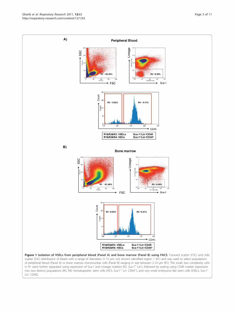
Isolation of VSELs from bone marrow and peripheralbloodVSELs were isolated from murine bone marrow andperipheral blood nucleated cells using multicolor fluor-escence-activated cell sorting (FACS) as previouslydescribed [4,12,23]. Briefly, the population of totalnucleated cells (TNCs) from murine PB and BM wasobtained after lysis of red blood cells using 1× BDPharm Lyse Buffer (BD Pharmingen, San Jose, CA,USA). TNCs were subsequently stained for hematopoie-tic lineage markers (Lineage - lin), for Sca-1 and CD45antigens, for 30 minutes in medium containing 2% fetalbovine serum. The following anti-mouse antibodies (BDPharmingen, San Jose, CA, USA) were used for staining:anti-CD45R/B220 (phycoerythrin - PE; clone RA3-6B2),anti-Gr-1 (PE; clone RB6-8C5), anti-T-cell receptor-ab(PE; clone H57-597), anti-T-cell receptor-gδ (PE; cloneGL3), anti-CD11b (PE; clone M1/70), anti-Ter119 (PE;clone TER-119), and anti-Ly-6A/E (Sca-1) (biotin; cloneE13-161.7, with streptavidin-conjugated to PE-Cy5) andrat anti-CD45 (allophycocyanin-Cy7; clone 30-F11). AllmAbs were added at saturating concentrations. Cellswere then washed, re-suspended in RPMI 1640 mediumwith 10% fetal bovine serum, and sorted by MoFlo cellsorter (Dako, Carpinteria, CA, USA). The Sca-1+Lin-CD45- cells (VSELs) and control Sca-1+Lin-CD45+ cells(HSCs) were isolated according to the gating and sortingstrategy described before and outlined in Figure 1. Foreach sample run, at least 106 events were acquired, withVSELs accounting for 0.02-0.03% of the total cells (BMor PB). Given the scarcity of VSELs in peripheral bloodand bone marrow, we performed six independent mea-surements per exposure condition (hypoxia, normoxia),each based on pooled samples from 10 mice, for a totalof 120 animals.
Measurement of plasma chemokinesMouse blood was collected from the vena cava in vacu-tainer tubes containing EDTA (Becton Dickinson,Franklin Lakes, NJ, USA). The collected fresh blood was
Gharib et al. Respiratory Research 2011, 12:63http://respiratory-research.com/content/12/1/63
Page 2 of 11
A)
B)
Figure 1 Isolation of VSELs from peripheral blood (Panel A) and bone marrow (Panel B) using FACS. Forward scatter (FSC) and sidescatter (SSC) distribution of beads with a range of diameters (1-15 μm, not shown) identified region 1 (R1) and was used to select populationsof peripheral blood (Panel A) or bone marrow mononuclear cells (Panel B) ranging in size between 2-10 μm (R1). The small, low complexity cellsin R1 were further separated using expression of Sca-1 and Lineage markers (R2: Sca-1+ Lin-), followed by sorting using CD45 marker expressioninto two distinct populations (R3, R4): hematopoietic stem cells (HCS, Sca-1+ Lin- CD45+), and very small embryonic-like stem cells (VSELs, Sca-1+
Lin- CD45).
Gharib et al. Respiratory Research 2011, 12:63http://respiratory-research.com/content/12/1/63
Page 3 of 11
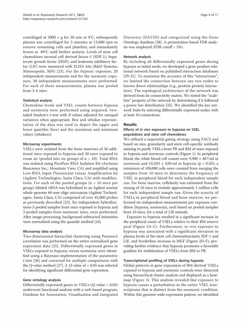
centrifuged at 2000 × g for 20 min at 4°C, subsequentlyplasma was centrifuged for 5 minutes at 13,000 rpm toremove remaining cells and platelets, and immediatelyfrozen at -80°C until further analysis. Levels of stem cellchemokines stromal cell derived factor-1 (SDF-1), hepa-tocyte growth factor (HGF), and leukemia inhibitory fac-tor (LIF) were measured with ELISA kits (R&D Systems,Minneapolis, MN) [23]. For the hypoxic exposure, 20independent measurements and for the normoxic expo-sure, 30 independent measurements were performed.For each of these measurements, plasma was pooledfrom 3-4 mice.
Statistical analysisChemokine levels and VSEL counts between hypoxiaand normoxia were performed using unpaired, two-tailed Student’s t-test with P-values adjusted for unequalvariances when appropriate. Box and whisker represen-tation of the data was used to depict the upper andlower quartiles (box) and the maximum and minimumvalues (whiskers).
Microarray experimentsVSELs were isolated from the bone marrows of 30 addi-tional mice exposed to hypoxia and 30 mice exposed toroom air (pooled into six groups of n = 10). Total RNAwas isolated using PicoPure RNA Isolation Kit (ArcturusBioscience Inc., Mountain View, CA) and amplified usingLow-RNA Input Fluorescent Linear Amplification kit(Agilent Technologies, Santa Clara, CA) with modifica-tions. For each of the pooled groups (n = 10 mice pergroup), labeled cRNA was hybridized to an Agilent murinewhole-genome 60-mer oligo microarray (Agilent Technol-ogies, Santa Clara, CA) comprised of over 45,000 probesas previously described [23]. Six independent hybridiza-tions-3 pooled samples from mice exposed to hypoxia and3 pooled samples from normoxic mice, were performed.After image processing, background-subtracted intensitieswere normalized using the quantile method [24].
Microarray data analysisTwo-dimensional hierarchal clustering using Pearson’scorrelation was performed on the entire normalized geneexpression data [25]. Differentially expressed genes inVSELs exposed to hypoxia versus normoxia were identi-fied using a Bayesian implementation of the parametrict-test [26] and corrected for multiple comparisons withthe Q-value method [27]. A Q-value of < 0.05 was selectedfor identifying significant differential gene expression.
Gene ontology analysisDifferentially expressed genes in VSELs (Q-value < 0.05)underwent functional analysis with a web-based program,Database for Annotation, Visualization and Integrated
Discovery (DAVID) and categorized using the GeneOntology database [28]. A permutation-based FDR analy-sis was employed (FDR cutoff < 5%).
Network analysisBy including all differentially expressed genes duringhypoxia as initial seeds, we developed a gene product rela-tional network based on published interaction databases[29-31]. To maximize the accuracy of this “interactome”,we limited the connection between any two nodes toknown direct relationships (e.g., protein-protein interac-tion). The topological architecture of the network wasderived from its connectivity matrix. We tested the “scale-free” property of the network by determining if it followeda power law distribution [32]. We identified the key net-work hubs by selecting differentially expressed nodes withat least 10 connections.
ResultsEffects of in vivo exposure to hypoxia on VSELpopulations and stem cell chemokinesWe utilized a sequential gating strategy using FACS andbased on size, granularity and stem cell-specific antibodystaining to purify VSELs from PB and BM of mice exposedto hypoxia and normoxic controls (Figure 1). In peripheralblood, the white blood cell counts were 9,980 ± 467/ml innormoxia and 10,503 ± 420/ml in hypoxia (p > 0.05); aminimum of 100,000 cells were counted from merging thesamples from 10 mice to determine the frequency ofVSEL in peripheral blood for each independent samplerun. For bone marrow, cellularity was estimated from themixing of 10 mice to include approximately 1 million cellsfor each independent sample run. Given the scarcity ofVSELs in peripheral blood and bone marrow, we per-formed six independent measurements per exposure con-dition (hypoxia, normoxia), each based on pooled samplesfrom 10 mice, for a total of 120 animals.Exposure to hypoxia resulted in a significant increase in
the peripheral count of VSELs relative to their BM reservepool (Figure 2A-C). Furthermore, in vivo exposure tohypoxia was associated with a significant elevation inplasma levels of the stem cell chemoattractants SDF-1 andLIF, and borderline increase in HGF (Figure 2D-F), pro-viding further evidence that hypoxia promotes a favorablegradient for mobilization of VSELs from BM to PB.
Transcriptional profiling of VSELs during hypoxiaGlobal patterns in gene expression of BM-derived VSELsexposed to hypoxia and normoxic controls were dissectedusing hierarchical cluster analysis and displayed as a heat-map (Figure 3). This analysis revealed that exposure tohypoxia causes a perturbation in the entire VSEL tran-scriptome that is distinct from the normoxic condition.Within this genome-wide expression pattern, we identified
Gharib et al. Respiratory Research 2011, 12:63http://respiratory-research.com/content/12/1/63
Page 4 of 11
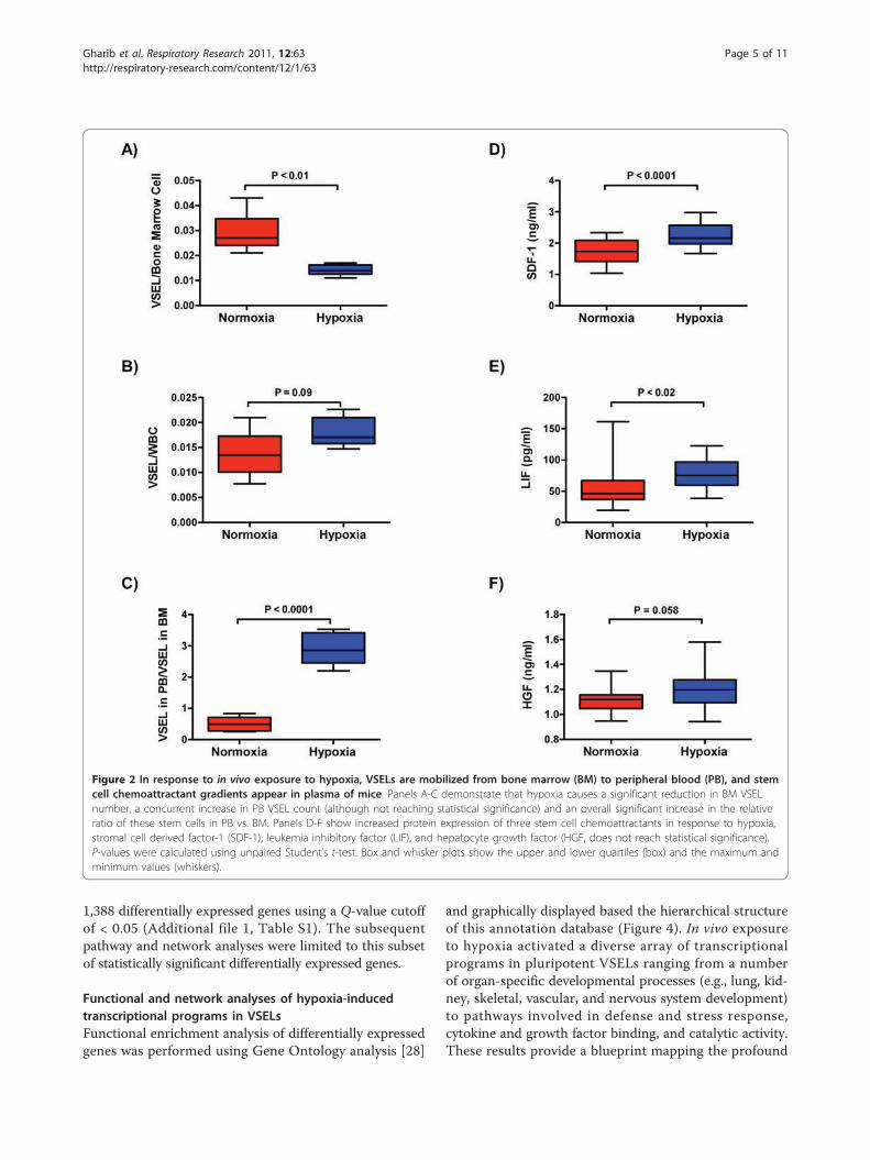
1,388 differentially expressed genes using a Q-value cutoffof < 0.05 (Additional file 1, Table S1). The subsequentpathway and network analyses were limited to this subsetof statistically significant differentially expressed genes.
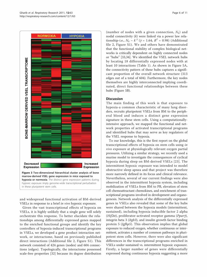
Functional and network analyses of hypoxia-inducedtranscriptional programs in VSELsFunctional enrichment analysis of differentially expressedgenes was performed using Gene Ontology analysis [28]
and graphically displayed based the hierarchical structureof this annotation database (Figure 4). In vivo exposureto hypoxia activated a diverse array of transcriptionalprograms in pluripotent VSELs ranging from a numberof organ-specific developmental processes (e.g., lung, kid-ney, skeletal, vascular, and nervous system development)to pathways involved in defense and stress response,cytokine and growth factor binding, and catalytic activity.These results provide a blueprint mapping the profound
Figure 2 In response to in vivo exposure to hypoxia, VSELs are mobilized from bone marrow (BM) to peripheral blood (PB), and stemcell chemoattractant gradients appear in plasma of mice. Panels A-C demonstrate that hypoxia causes a significant reduction in BM VSELnumber, a concurrent increase in PB VSEL count (although not reaching statistical significance) and an overall significant increase in the relativeratio of these stem cells in PB vs. BM. Panels D-F show increased protein expression of three stem cell chemoattractants in response to hypoxia,stromal cell derived factor-1 (SDF-1), leukemia inhibitory factor (LIF), and hepatocyte growth factor (HGF, does not reach statistical significance).P-values were calculated using unpaired Student’s t-test. Box and whisker plots show the upper and lower quartiles (box) and the maximum andminimum values (whiskers).
Gharib et al. Respiratory Research 2011, 12:63http://respiratory-research.com/content/12/1/63
Page 5 of 11
and widespread functional activation of BM-derivedVSELs in response to a brief in vivo hypoxic exposure.Given the vast transcriptional effects of hypoxia on
VSELs, it is highly unlikely that a single gene will solelyorchestrate this response. To better elucidate the rela-tionships among differentially expressed genes mappedto the enriched functional groups and identify the keycontrollers of hypoxia-induced transcriptional programsin VSELs, we developed a gene product interaction net-work, or interactome, based on previously publisheddirect interactions (Additional file 2, Figure S1). Thisnetwork consisted of 424 genes (nodes) and 604 connec-tions (edges). Topologically, the interactome possessedscale-free properties [32] because its degree distribution
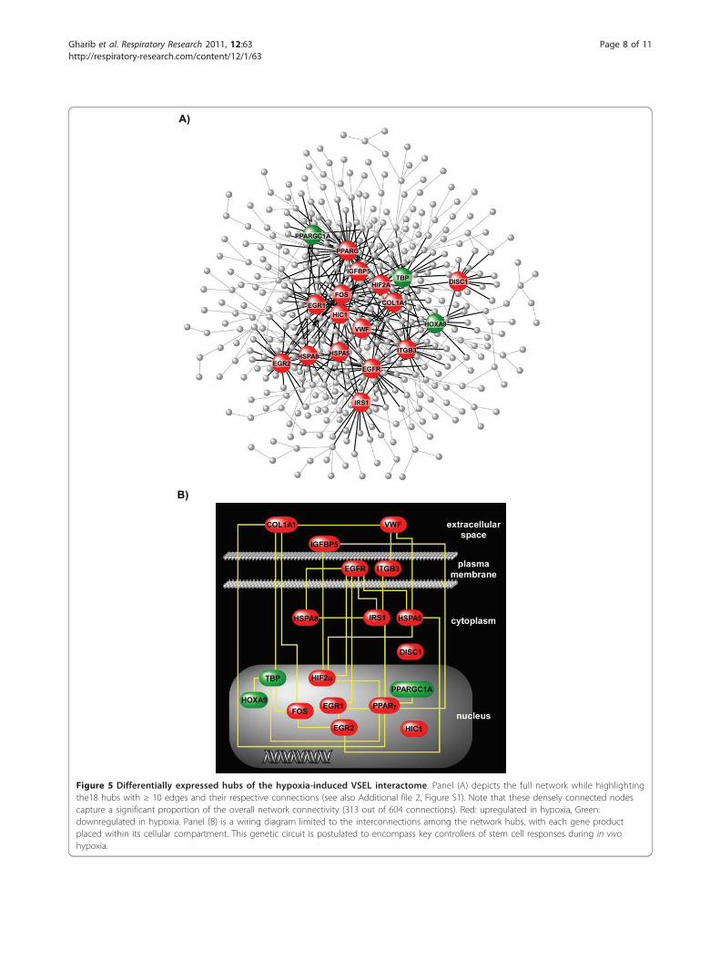
(number of nodes with a given connection, Nk) andnodal connectivity (k) were linked via a power law rela-tionship-i.e., Nk ~ k-g (g = 1.64, R2 = 0.98) (Additionalfile 2, Figure S1). We and others have demonstratedthat the functional stability of complex biological net-works is critically dependent on highly connected nodesor “hubs” [33,34]. We identified the VSEL network hubsby locating 18 differentially expressed nodes with atleast 10 interactions (Table 1). As shown in Figure 5A,the connectivity pattern of these hubs captures a signifi-cant proportion of the overall network structure (313edges out of a total of 604). Furthermore, the key nodesthemselves are highly interconnected implying coordi-nated, direct functional relationships between thesehubs (Figure 5B).
DiscussionThe main finding of this work is that exposure tohypoxia-a common characteristic of many lung disor-ders, recruits pluripotent VSELs from BM to the periph-eral blood and induces a distinct gene expressionsignature in these stem cells. Using a computationally-intensive approach, we mapped the functional and net-work properties of activated transcriptional programsand identified hubs that may serve as key regulators ofthe VSEL response to hypoxia.To our knowledge, this is the first report on the global
transcriptional effects of hypoxia on stem cells using invivo exposure at physiologically relevant oxygen partialpressures. Utilizing a similar strategy, we recently used amurine model to investigate the consequences of cyclicalhypoxia during sleep on BM-derived VSELs [23]. Theintermittent hypoxic exposure was intended to modelobstructive sleep apnea and that project was thereforemore narrowly defined in its focus and clinical relevance.Nevertheless, several of our current findings were alsoobserved in the intermittent hypoxia system, includingmobilization of VSELs from BM to PB, elevation of stemcell chemoattractant chemokines, and enrichment of tran-scriptional programs involved in development and organo-genesis. Network analysis of the differentially expressedgenes in VSELs also revealed that some of the key hubswere shared between the hypoxia models (intermittent,sustained), including hypoxia inducible factor 2 alpha(Hif2a), proliferator-activated receptor gamma (Pparg),integrin beta 3 (Itgb3), and insulin growth factor bindingprotein 5 (Igfbp5). This observation implies that globalexposure to reduced oxygen, whether continuous or inter-mittent, activates a number of common pathways in pluri-potent stem cells. However, there were also significantdifferences in the transcriptional programs enriched inVSELs under sustained vs. intermittent hypoxic exposure.Firstly, a larger number of genes were differentiallyexpressed during continuous hypoxia suggesting a more
Figure 3 Two-dimensional hierarchical cluster analysis of bonemarrow-derived VSEL gene expression in mice exposed tohypoxia or normoxia. The distinct gene expression patterns duringhypoxic exposure imply genome-wide transcriptional perturbationin these pluripotent stem cells.
Gharib et al. Respiratory Research 2011, 12:63http://respiratory-research.com/content/12/1/63
Page 6 of 11
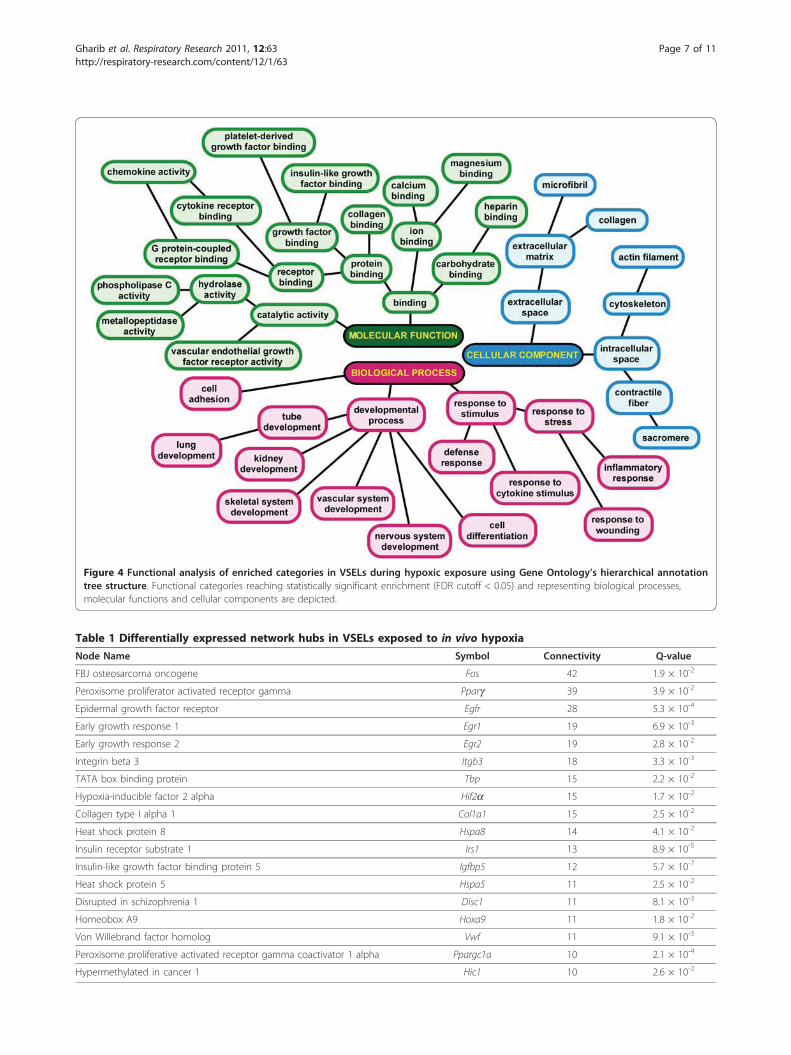
Figure 4 Functional analysis of enriched categories in VSELs during hypoxic exposure using Gene Ontology’s hierarchical annotationtree structure. Functional categories reaching statistically significant enrichment (FDR cutoff < 0.05) and representing biological processes,molecular functions and cellular components are depicted.
Table 1 Differentially expressed network hubs in VSELs exposed to in vivo hypoxia
Node Name Symbol Connectivity Q-value
FBJ osteosarcoma oncogene Fos 42 1.9 × 10⁻²
Peroxisome proliferator activated receptor gamma Pparg 39 3.9 × 10⁻²
Epidermal growth factor receptor Egfr 28 5.3 × 10⁻⁴
Early growth response 1 Egr1 19 6.9 × 10⁻³
Early growth response 2 Egr2 19 2.8 × 10⁻²
Integrin beta 3 Itgb3 18 3.3 × 10⁻³
TATA box binding protein Tbp 15 2.2 × 10⁻²
Hypoxia-inducible factor 2 alpha Hif2a 15 1.7 × 10⁻²
Collagen type I alpha 1 Col1a1 15 2.5 × 10⁻²
Heat shock protein 8 Hspa8 14 4.1 × 10⁻²
Insulin receptor substrate 1 Irs1 13 8.9 × 10⁻⁵
Insulin-like growth factor binding protein 5 Igfbp5 12 5.7 × 10⁻⁷
Heat shock protein 5 Hspa5 11 2.5 × 10⁻²
Disrupted in schizophrenia 1 Disc1 11 8.1 × 10⁻³
Homeobox A9 Hoxa9 11 1.8 × 10⁻²
Von Willebrand factor homolog Vwf 11 9.1 × 10⁻³
Peroxisome proliferative activated receptor gamma coactivator 1 alpha Ppargc1a 10 2.1 × 10⁻⁴
Hypermethylated in cancer 1 Hic1 10 2.6 × 10⁻²
Gharib et al. Respiratory Research 2011, 12:63http://respiratory-research.com/content/12/1/63
Page 7 of 11
A)
B)
Figure 5 Differentially expressed hubs of the hypoxia-induced VSEL interactome. Panel (A) depicts the full network while highlightingthe18 hubs with ≥ 10 edges and their respective connections (see also Additional file 2, Figure S1). Note that these densely connected nodescapture a significant proportion of the overall network connectivity (313 out of 604 connections). Red: upregulated in hypoxia, Green:downregulated in hypoxia. Panel (B) is a wiring diagram limited to the interconnections among the network hubs, with each gene productplaced within its cellular compartment. This genetic circuit is postulated to encompass key controllers of stem cell responses during in vivohypoxia.
Gharib et al. Respiratory Research 2011, 12:63http://respiratory-research.com/content/12/1/63
Page 8 of 11
profound perturbation. Secondly, a much broader spec-trum of pathways was enriched in sustained compared tointermittent hypoxia that extended beyond developmentalprograms and included processes involved in defense andstress response, wound healing, chemokine activity, celladhesion and structure (Figure 4). Thirdly, many of thenetwork hubs identified during exposure to continuoushypoxia were not differentially expressed during cyclicalhypoxia (Figure 5, Table 1), including the most denselyconnected node, Fos (FBJ osteosarcoma oncogene), andseveral other key transcriptional regulators such as earlygrowth response 1 and 2 (Egr1, Egr2), and TATA boxbinding protein (Tbp).Fos and Egr1 are prototypic master regulators known
as immediate early-genes (IEG) that orchestrate wide-spread activation of cellular gene expression in responseto specific perturbations, including hypoxia, ischemia,atherosclerosis, angiogenesis, and neuronal survival[35-37]. Egr2 has been shown to govern the develop-ment and segmentation of the mouse hindbrain [38],and mutations in this gene have been associated withcongenital hypomyelinating neuropathies in humans[39]. Intriguingly, a recent study demonstrated that apopulation of Egr2-expressing precursor cells developinto the first embryonic respiratory rhythm-generatingneuronal circuit [40], and Egr2-/- transgenic mice diefrom breathing irregularities resulting in severe hypoxia[41].Two members of the heat shock protein 70 group,
Hspa8 and Hspa5, were also densely connected networknodes whose expression increased significantly duringhypoxia. Products of these genes play crucial roles inprotecting cells from environmental stressors includinghigh temperature, oxidative stress, ischemia and hypoxia[42]. More specifically, targeting Hspa8 and Hspa5 hasbeen demonstrated to protect cardiomyocytes [43] andmesenchymal stem cells from hypoxia-induced apoptosis[44], whereas delivering these heat shock proteins intotransplanted mesenchymal stem cells rescues heart func-tion after myocardial infarction in rats [44].Another differentially upregulated hub within the
interactome, Hif2a, is a member of the HIF family ofbasic helix-loop-helix transcription factors that controlthe global cellular response to oxygen deprivation. Hif2aoverexpression is seen many solid tumors characterizedwith significant hypoxic burden including renal cell car-cinoma, non small cell lung cancer and meduloblastoma,and is associated with poor outcome in these cancers[45]. Recent reports suggest that Hif2a is a critical acti-vator of a subpopulation of cancer cells with stem cell-like properties [46,47], and pharmacologic and genetictargeting of this gene affects differentiation of progenitorstem cells [48,49]. Interestingly, a single nucleotide poly-morphism in Hif2a was recently demonstrated to be
highly associated with adaptability to high altitudehypoxia in ethnic Tibetans [50].The mobilization of VSELs from BM to peripheral cir-
culation in response to oxygen deprivation implies theactivation of organ-specific transcriptional programsunder regulatory controls. Our network analysis pro-vided an overview of the transcriptional landscape ofthese pluripotent stem cells during hypoxia. Since thefunctional integrity of such scale-free biologic networksis dependent on densely connected nodes [33], we pos-tulate that these hubs comprise key regulators ofhypoxia-induced gene expression in VSELs. As depictedin Figure 5, the hubs not only form the structural foun-dation of the interactome, but are themselves highlyinterconnected via direct interactions, implying a highdegree of functional coordination in orchestrating thehypoxia-induced transcriptional response of stem cells.To explore whether this hypoxia-induced transcrip-
tional signature is unique to VSELs or common amongleukocyte subpopulations, we compared our results toexpression profiles of mononuclear cells (MNCs) andmesenchymal stem cells (MSCs) under hypoxia asreported by Onishi et al [51]. While there are substan-tial differences between the two studies in terms ofexperimental protocols, hypoxic exposures, and microar-ray platforms, less than 1% of the differentially expressedgenes in VSELs were significantly altered in MSCs orMNCs, suggesting that many of the hypoxia-inducedgenes in VSELs are exclusive to this subpopulation ofcells. However, from a functional standpoint, differen-tially expressed genes in VSELs and MSCs were bothenriched in several developmental processes, whereasMNCs were highly enriched in defense/immuneresponses and inflammation. Therefore, VSELs appearto possess transcriptional programs in common withboth MSCs and MNCs-a finding that is consistent withtheir pluripotent properties.Several limitations in the current study deserve men-
tion. We have only explored the effects of hypoxia ongene expression and not assessed the post-transcrip-tional modifications of gene products activated duringhypoxic exposure. The duration of hypoxia was short-the long term consequences of chronic oxygen depriva-tion on VSELs were not investigated in this study andrepresent a future research goal. Although bone marrowis the primary depot of VSELs, our results do not pre-clude the possibility that some of these stem cells werealso mobilized from other tissue niches during exposureto hypoxia. Nevertheless, our expression profilingexperiments were focused on BM-derived VSELs. Wehave not identified the tissue source of hypoxia-inducedelevation in stem cells chemokines, but given the extre-mely low number of circulating VSELs, we believe it isunlikely that VSELs are the primary source. Although
Gharib et al. Respiratory Research 2011, 12:63http://respiratory-research.com/content/12/1/63
Page 9 of 11
the bone marrow is a known source of these chemoat-tractants, hypoxic injury to various tissues can lead tothe release of the chemokines into peripheral blood[5,14,16]. Finally, we did not follow the fate of mobilizedVSELs to identify their target end organs, althoughgiven the global effects of hypoxia and the diversity ofenriched developmental programs, we expect that thesestem cells will be recruited to multiple sites andundergo tissue-specific differentiation.
ConclusionsCollectively, our results demonstrate that in vivo expo-sure to hypoxia in mice elicits chemoattractant gradientsthat promote the mobilization of pluripotent very smallembryonic-like stem cells from the bone marrow to per-ipheral blood. A computationally-driven analysis of thetranscriptome of these cells highlighted the activation ofdiverse transcriptional programs that appeared to becoordinated by a select number of highly connected net-work nodes. These hubs may represent critical targetsfor modulating the functional consequences of hypoxicburden on stem cells. Our approach provides a generalframework for the systematic study of stem cell biologyunder clinically relevant pathophysiologic perturbations.
Additional material
Additional file 1: Tabular list of differentially expressed genes inVSELs after hypoxic exposure. This file contains the list of differentiallyexpressed genes (based on a Q-value cutoff less than 0.05) in VSELsexposed to hypoxia. The list includes the official gene symbols, EntrezGene IDs, Q-values, and Log2[expression in hypoxia/expression innormoxia].
Additional file 2: Gene product interaction network of differentiallyexpressed genes in VSELs. This file contains two figures depicting agene product interaction network of differentially expressed genes inVSELs after in vivo exposure to hypoxia. Panel (A) highlights up anddownregulated genes (red and green respectively). This network has 424genes (nodes) and 604 connections (edges). Note that each connectionrepresents a known direct interaction between two gene products. Panel(B) demonstrates that the topology of this network is scale-free becauseit follows a power law distribution. Nk, degree distribution; k, nodalconnectivity.
AcknowledgementsThis work was supported in part by the National Institutes of HealthHL065270 and HL086662 (DG), and American Sleep Medicine FoundationJunior Faculty Research Award (SAG).
Author details1Center for Lung Biology and Division of Pulmonary and Critical CareMedicine, Department of Medicine, University of Washington, Seattle, WA,USA. 2Department of Pediatrics, University of Louisville, Louisville, KY, USA.3Department of Pediatrics, University of Chicago, Chicago, IL, USA. 4Stem CellBiology Institute, University of Louisville, Louisville, KY, USA.
Authors’ contributionsSAG analyzed the data and wrote the manuscript. DG conceived the study,participated in its design and draft. AK participated in designing the study
and collected the stem cell data and performed the transcriptional profilingexperiments. MK assisted with FACS-based stem cell isolation and collectionof stem cell data. EAD, JK, and HBC assisted with the stem cell isolation,animal experiments, and stem cell data measurements. All authors read andapproved the final manuscript.
Competing interestsThe authors declare that they have no competing interests.
Received: 11 February 2011 Accepted: 10 May 2011Published: 10 May 2011
References1. Semenza GL: Hypoxia and human disease-and the Journal of Molecular
Medicine. J Mol Med 2007, 85(12):1293-1294.2. Simon MC, Keith B: The role of oxygen availability in embryonic
development and stem cell function. Nat Rev Mol Cell Biol 2008,9(4):285-296.
3. Mohyeldin A, Garzon-Muvdi T, Quinones-Hinojosa A: Oxygen in stem cellbiology: a critical component of the stem cell niche. Cell Stem Cell 2010,7(2):150-161.
4. Kucia MJ, Wysoczynski M, Wu W, Zuba-Surma EK, Ratajczak J, Ratajczak MZ:Evidence that very small embryonic-like stem cells are mobilized intoperipheral blood. Stem Cells 2008, 26(8):2083-2092.
5. Kucia M, Zhang YP, Reca R, Wysoczynski M, Machalinski B, Majka M,Ildstad ST, Ratajczak J, Shields CB, Ratajczak MZ: Cells enriched in markersof neural tissue-committed stem cells reside in the bone marrow andare mobilized into the peripheral blood following stroke. Leukemia 2006,20(1):18-28.
6. Schachinger V, Erbs S, Elsasser A, Haberbosch W, Hambrecht R,Holschermann H, Yu J, Corti R, Mathey DG, Hamm CW, et al: Intracoronarybone marrow-derived progenitor cells in acute myocardial infarction. NEngl J Med 2006, 355(12):1210-1221.
7. Burnham EL, Taylor WR, Quyyumi AA, Rojas M, Brigham KL, Moss M:Increased circulating endothelial progenitor cells are associated withsurvival in acute lung injury. Am J Respir Crit Care Med 2005,172(7):854-860.
8. Fadini GP, Schiavon M, Cantini M, Baesso I, Facco M, Miorin M, Tassinato M,de Kreutzenberg SV, Avogaro A, Agostini C: Circulating progenitor cellsare reduced in patients with severe lung disease. Stem Cells 2006,24(7):1806-1813.
9. Fadini GP, Schiavon M, Rea F, Avogaro A, Agostini C: Depletion ofendothelial progenitor cells may link pulmonary fibrosis and pulmonaryhypertension. Am J Respir Crit Care Med 2007, 176(7):724-725, author reply725.
10. Palange P, Testa U, Huertas A, Calabro L, Antonucci R, Petrucci E, Pelosi E,Pasquini L, Satta A, Morici G, et al: Circulating haemopoietic andendothelial progenitor cells are decreased in COPD. Eur Respir J 2006,27(3):529-541.
11. Kucia M, Wysoczynski M, Ratajczak J, Ratajczak MZ: Identification of verysmall embryonic like (VSEL) stem cells in bone marrow. Cell Tissue Res2008, 331(1):125-134.
12. Kucia M, Reca R, Campbell FR, Zuba-Surma E, Majka M, Ratajczak J,Ratajczak MZ: A population of very small embryonic-like (VSEL) CXCR4(+)SSEA-1(+)Oct-4+ stem cells identified in adult bone marrow. Leukemia2006, 20(5):857-869.
13. Kucia M, Wojakowski W, Reca R, Machalinski B, Gozdzik J, Majka M, Baran J,Ratajczak J, Ratajczak MZ: The migration of bone marrow-derived non-hematopoietic tissue-committed stem cells is regulated in an SDF-1-,HGF-, and LIF-dependent manner. Arch Immunol Ther Exp (Warsz) 2006,54(2):121-135.
14. Ceradini DJ, Kulkarni AR, Callaghan MJ, Tepper OM, Bastidas N,Kleinman ME, Capla JM, Galiano RD, Levine JP, Gurtner GC: Progenitor celltrafficking is regulated by hypoxic gradients through HIF-1 induction ofSDF-1. Nat Med 2004, 10(8):858-864.
15. Paczkowska E, Kucia M, Koziarska D, Halasa M, Safranow K, Masiuk M,Karbicka A, Nowik M, Nowacki P, Ratajczak MZ, et al: Clinical evidence thatvery small embryonic-like stem cells are mobilized into peripheral bloodin patients after stroke. Stroke 2009, 40(4):1237-1244.
16. Kucia M, Dawn B, Hunt G, Guo Y, Wysoczynski M, Majka M, Ratajczak J,Rezzoug F, Ildstad ST, Bolli R, et al: Cells expressing early cardiac markers
Gharib et al. Respiratory Research 2011, 12:63http://respiratory-research.com/content/12/1/63
Page 10 of 11
reside in the bone marrow and are mobilized into the peripheral bloodafter myocardial infarction. Circ Res 2004, 95(12):1191-1199.
17. Chi JT, Wang Z, Nuyten DS, Rodriguez EH, Schaner ME, Salim A, Wang Y,Kristensen GB, Helland A, Borresen-Dale AL, et al: Gene expressionprograms in response to hypoxia: cell type specificity and prognosticsignificance in human cancers. PLoS Med 2006, 3(3):e47.
18. Wang GL, Jiang BH, Rue EA, Semenza GL: Hypoxia-inducible factor 1 is abasic-helix-loop-helix-PAS heterodimer regulated by cellular O2 tension.Proc Natl Acad Sci USA 1995, 92(12):5510-5514.
19. Menicanin D, Bartold PM, Zannettino AC, Gronthos S: Genomic profiling ofmesenchymal stem cells. Stem Cell Rev 2009, 5(1):36-50.
20. Westfall SD, Sachdev S, Das P, Hearne LB, Hannink M, Roberts RM, Ezashi T:Identification of oxygen-sensitive transcriptional programs in humanembryonic stem cells. Stem Cells Dev 2008, 17(5):869-881.
21. Ivanovic Z: Hypoxia or in situ normoxia: The stem cell paradigm. J CellPhysiol 2009, 219(2):271-275.
22. Millman JR, Tan JH, Colton CK: The effects of low oxygen on self-renewaland differentiation of embryonic stem cells. Curr Opin Organ Transplant2009, 14(6):694-700.
23. Gharib SA, Dayyat E, Khalyfa A, Kim J, Clair HB, Kucia M, Gozal D:Intermittent Hypoxia Mobilizes Bone Marrow-Derived Very SmallEmbryonic-Like Stem Cells and Activates Developmental TranscriptionalPrograms in Mice. Sleep 2010, 33(11):1439-1446.
24. Bolstad BM, Irizarry RA, Astrand M, Speed TP: A comparison ofnormalization methods for high density oligonucleotide array databased on variance and bias. Bioinformatics 2003, 19(2):185-193.
25. Saeed AI, Sharov V, White J, Li J, Liang W, Bhagabati N, Braisted J, Klapa M,Currier T, Thiagarajan M, et al: TM4: a free, open-source system formicroarray data management and analysis. Biotechniques 2003,34(2):374-378.
26. Baldi P, Long AD: A Bayesian framework for the analysis of microarrayexpression data: regularized t-test and statistical inferences of genechanges. Bioinformatics 2001, 17(6):509-519.
27. Storey JD, Tibshirani R: Statistical significance for genomewide studies.Proc Natl Acad Sci USA 2003, 100(16):9440-9445.
28. Dennis G Jr, Sherman BT, Hosack DA, Yang J, Gao W, Lane HC, Lempicki RA:DAVID: Database for Annotation, Visualization, and Integrated Discovery.Genome Biol 2003, 4(5):P3.
29. Aranda B, Achuthan P, Alam-Faruque Y, Armean I, Bridge A, Derow C,Feuermann M, Ghanbarian AT, Kerrien S, Khadake J, et al: The IntActmolecular interaction database in 2010. Nucleic Acids Res 2009, , 38Database: D525-531.
30. Ceol A, Chatr Aryamontri A, Licata L, Peluso D, Briganti L, Perfetto L,Castagnoli L, Cesareni G: MINT, the molecular interaction database: 2009update. Nucleic Acids Res 2009, , 38 Database: D532-539.
31. Calvano SE, Xiao W, Richards DR, Felciano RM, Baker HV, Cho RJ, Chen RO,Brownstein BH, Cobb JP, Tschoeke SK, et al: A network-based analysis ofsystemic inflammation in humans. Nature 2005, 437(7061):1032-1037.
32. Barabasi AL, Albert R: Emergence of scaling in random networks. Science1999, 286(5439):509-512.
33. Luscombe NM, Babu MM, Yu H, Snyder M, Teichmann SA, Gerstein M:Genomic analysis of regulatory network dynamics reveals largetopological changes. Nature 2004, 431(7006):308-312.
34. Khalyfa A, Gharib SA, Kim J, Dayyat E, Snow AB, Bhattacharjee R,Kheirandish-Gozal L, Goldman JL, Gozal D: Transcriptomic analysisidentifies phosphatases as novel targets for adenotonsillar hypertrophyof pediatric obstructive sleep apnea. Am J Respir Crit Care Med 2010,181(10):1114-1120.
35. Rybnikova E, Glushchenko T, Tyulkova E, Baranova K, Samoilov M: Mildhypobaric hypoxia preconditioning up-regulates expression oftranscription factors c-Fos and NGFI-A in rat neocortex andhippocampus. Neurosci Res 2009, 65(4):360-366.
36. Khachigian LM: Early growth response-1 in cardiovascular pathobiology.Circ Res 2006, 98(2):186-191.
37. Zhang J, Zhang D, McQuade JS, Behbehani M, Tsien JZ, Xu M: c-fosregulates neuronal excitability and survival. Nat Genet 2002,30(4):416-420.
38. Schneider-Maunoury S, Topilko P, Seitandou T, Levi G, Cohen-Tannoudji M,Pournin S, Babinet C, Charnay P: Disruption of Krox-20 results in alterationof rhombomeres 3 and 5 in the developing hindbrain. Cell 1993,75(6):1199-1214.
39. Warner LE, Mancias P, Butler IJ, McDonald CM, Keppen L, Koob KG,Lupski JR: Mutations in the early growth response 2 (EGR2) gene areassociated with hereditary myelinopathies. Nat Genet 1998, 18(4):382-384.
40. Thoby-Brisson M, Karlen M, Wu N, Charnay P, Champagnat J, Fortin G:Genetic identification of an embryonic parafacial oscillator coupling tothe preBotzinger complex. Nat Neurosci 2009, 12(8):1028-1035.
41. Jacquin TD, Borday V, Schneider-Maunoury S, Topilko P, Ghilini G, Kato F,Charnay P, Champagnat J: Reorganization of pontine rhythmogenicneuronal networks in Krox-20 knockout mice. Neuron 1996, 17(4):747-758.
42. Daugaard M, Rohde M, Jaattela M: The heat shock protein 70 family:Highly homologous proteins with overlapping and distinct functions.FEBS Lett 2007, 581(19):3702-3710.
43. Hardy B, Raiter A: Peptide-binding heat shock protein GRP78 protectscardiomyocytes from hypoxia-induced apoptosis. J Mol Med 2010.
44. Chang W, Song BW, Lim S, Song H, Shim CY, Cha MJ, Ahn DH, Jung YG,Lee DH, Chung JH, et al: Mesenchymal stem cells pretreated withdelivered Hph-1-Hsp70 protein are protected from hypoxia-mediatedcell death and rescue heart functions from myocardial injury. Stem Cells2009, 27(9):2283-2292.
45. Qing G, Simon MC: Hypoxia inducible factor-2alpha: a critical mediator ofaggressive tumor phenotypes. Curr Opin Genet Dev 2009, 19(1):60-66.
46. Li Z, Bao S, Wu Q, Wang H, Eyler C, Sathornsumetee S, Shi Q, Cao Y,Lathia J, McLendon RE, et al: Hypoxia-inducible factors regulatetumorigenic capacity of glioma stem cells. Cancer Cell 2009, 15(6):501-513.
47. Seidel S, Garvalov BK, Wirta V, von Stechow L, Schanzer A, Meletis K,Wolter M, Sommerlad D, Henze AT, Nister M, et al: A hypoxic nicheregulates glioblastoma stem cells through hypoxia inducible factor 2alpha. Brain 2010, 133(Pt 4):983-995.
48. Moreno-Manzano V, Rodriguez-Jimenez FJ, Acena-Bonilla JL, Fustero-Lardies S, Erceg S, Dopazo J, Montaner D, Stojkovic M, Sanchez-Puelles JM:FM19G11, a new hypoxia-inducible factor (HIF) modulator, affects stemcell differentiation status. J Biol Chem 2010, 285(2):1333-1342.
49. Covello KL, Kehler J, Yu H, Gordan JD, Arsham AM, Hu CJ, Labosky PA,Simon MC, Keith B: HIF-2alpha regulates Oct-4: effects of hypoxia onstem cell function, embryonic development, and tumor growth. GenesDev 2006, 20(5):557-570.
50. Yi X, Liang Y, Huerta-Sanchez E, Jin X, Cuo ZX, Pool JE, Xu X, Jiang H,Vinckenbosch N, Korneliussen TS, et al: Sequencing of 50 human exomesreveals adaptation to high altitude. Science 2010, 329(5987):75-78.
51. Ohnishi S, Yasuda T, Kitamura S, Nagaya N: Effect of hypoxia on geneexpression of bone marrow-derived mesenchymal stem cells andmononuclear cells. Stem Cells 2007, 25(5):1166-1177.
doi:10.1186/1465-9921-12-63Cite this article as: Gharib et al.: Transcriptional landscape of bonemarrow-derived very small embryonic-like stem cells during hypoxia.Respiratory Research 2011 12:63.
Submit your next manuscript to BioMed Centraland take full advantage of:
• Convenient online submission
• Thorough peer review
• No space constraints or color figure charges
• Immediate publication on acceptance
• Inclusion in PubMed, CAS, Scopus and Google Scholar
• Research which is freely available for redistribution
Submit your manuscript at www.biomedcentral.com/submit
Gharib et al. Respiratory Research 2011, 12:63http://respiratory-research.com/content/12/1/63
Page 11 of 11