Thyroid Hormones within the Normal Range and Cardiac ...
11
Clinical Thyroidology / Research Article Eur Thyroid J 2021;10:150–160 Thyroid Hormones within the Normal Range and Cardiac Function in the General Population: The EPIPorto Study João Sérgio Neves a, b Ricardo Fontes-Carvalho a, c Marta Borges-Canha a, b Ana Rita Leite a Sandra Martins d, f Ana Oliveira b João Tiago Guimarães d–f Davide Carvalho b, h Adelino Leite-Moreira a Ana Azevedo f, g a Unidade de Investigação Cardiovascular, Departamento de Cirurgia e Fisiologia, Faculdade de Medicina da Universidade do Porto, Porto, Portugal; b Department of Endocrinology, Diabetes and Metabolism, Centro Hospitalar Universitário de São João, Porto, Portugal; c Department of Cardiology, Centro Hospitalar Gaia/Espinho, Vila Nova de Gaia, Portugal; d Department of Clinical Pathology, Centro Hospitalar Universitário de São João, Porto, Portugal; e Department of Biomedicine, Faculdade de Medicina da Universidade do Porto, Porto, Portugal; f EPIUnit – Instituto de Saúde Pública, Universidade do Porto, Porto, Portugal; g Departamento de Ciências da Saúde Pública e Forenses e Educação Médica, Faculdade de Medicina da Universidade do Porto, Porto, Portugal; h Instituto de Investigação e Inovação em Saúde (i3S), Universidade do Porto, Porto, Portugal Received: November 2, 2019 Accepted: May 4, 2020 Published online: July 8, 2020 João Sérgio Neves Unidade de Investigação Cardiovascular, Departamento de Cirurgia e Fisiologia Faculdade de Medicina da Universidade do Porto Alameda Professor Hernâni Monteiro, PT–4200-319 Porto (Portugal) jsneves @med.up.pt © 2020 European Thyroid Association Published by S. Karger AG, Basel [email protected] www.karger.com/etj DOI: 10.1159/000508407 Keywords Thyroid hormones · Cardiac function · Cardiac structure · Thyroid stimulating hormone · Free T4 · Free T3 Abstract Background: Hypothyroidism and hyperthyroidism are as- sociated with marked changes in cardiac structure and func- tion. However, the association of thyroid function within the normal range with cardiac structure and function in the general population remains uncertain. Methods: Eight hun- dred thirty-five subjects aged ≥45 years from the EPIPorto cohort (evaluation between 2006 and 2008) were cross-sec- tionally analyzed. We excluded participants with TSH, free T4 (FT4), or free T3 (FT3) outside of the reference range or with self-reported cardiovascular or thyroid disease. Cardiac structure and function were evaluated by echocardiogra- phy. We used linear regression models unadjusted and ad- justed for sex and age (model 1), and sex, age, BMI, diabetes, hypertension, and smoking (model 2). Nonlinear associa- tions were assessed using restricted cubic splines. Results: The mean age was 61.5 years (SD 10.5); 61.1% of the patients were women. In the adjusted model 2, heart rate was posi- tively associated with FT3; diastolic blood pressure was pos- itively associated with TSH; LV end-diastolic and end-systol- ic volumes were inversely associated with TSH, and ejection fraction was nonlinearly associated with FT3, with higher ejection fractions near the limits of the reference range. Left ventricle (LV) posterior wall thickness was nonlinearly asso- ciated with FT4 in the adjusted model 1, with a greater thick- ness near the limits of the reference range. Regarding dia- stolic function, no significant associations were observed in adjusted models. Conclusions: Thyroid function within the reference range was associated with heart rate, blood pres-
-
Upload
khangminh22 -
Category
Documents
-
view
3 -
download
0
Transcript of Thyroid Hormones within the Normal Range and Cardiac ...
Clinical Thyroidology / Research Article
Eur Thyroid J 2021;10:150–160
Thyroid Hormones within the Normal Range and Cardiac Function in the General Population: The EPIPorto Study
João Sérgio Neves
a, b Ricardo Fontes-Carvalho
a, c Marta Borges-Canha
a, b
Ana Rita Leite
a Sandra Martins
d, f Ana Oliveira
b João Tiago Guimarães
d–f
Davide Carvalho
b, h Adelino Leite-Moreira
a Ana Azevedo
f, g a
Unidade de Investigação Cardiovascular, Departamento de Cirurgia e Fisiologia, Faculdade de Medicina da Universidade do Porto, Porto, Portugal; b Department of Endocrinology, Diabetes and Metabolism, Centro Hospitalar Universitário de São João, Porto, Portugal; c Department of Cardiology, Centro Hospitalar Gaia/Espinho, Vila Nova de Gaia, Portugal; d Department of Clinical Pathology, Centro Hospitalar Universitário de São João, Porto, Portugal; e Department of Biomedicine, Faculdade de Medicina da Universidade do Porto, Porto, Portugal; f
EPIUnit – Instituto de Saúde Pública, Universidade do Porto, Porto, Portugal; g Departamento de Ciências da Saúde Pública e Forenses e Educação Médica, Faculdade de Medicina da Universidade do Porto, Porto, Portugal; h
Instituto de Investigação e Inovação em Saúde (i3S), Universidade do Porto, Porto, Portugal
Received: November 2, 2019Accepted: May 4, 2020Published online: July 8, 2020
João Sérgio NevesUnidade de Investigação Cardiovascular, Departamento de Cirurgia e FisiologiaFaculdade de Medicina da Universidade do PortoAlameda Professor Hernâni Monteiro, PT–4200-319 Porto (Portugal)jsneves @ med.up.pt
© 2020 European Thyroid AssociationPublished by S. Karger AG, Basel
DOI: 10.1159/000508407
KeywordsThyroid hormones · Cardiac function · Cardiac structure · Thyroid stimulating hormone · Free T4 · Free T3
AbstractBackground: Hypothyroidism and hyperthyroidism are as-sociated with marked changes in cardiac structure and func-tion. However, the association of thyroid function within the normal range with cardiac structure and function in the general population remains uncertain. Methods: Eight hun-dred thirty-five subjects aged ≥45 years from the EPIPorto cohort (evaluation between 2006 and 2008) were cross-sec-tionally analyzed. We excluded participants with TSH, free T4 (FT4), or free T3 (FT3) outside of the reference range or with self-reported cardiovascular or thyroid disease. Cardiac structure and function were evaluated by echocardiogra-
phy. We used linear regression models unadjusted and ad-justed for sex and age (model 1), and sex, age, BMI, diabetes, hypertension, and smoking (model 2). Nonlinear associa-tions were assessed using restricted cubic splines. Results: The mean age was 61.5 years (SD 10.5); 61.1% of the patients were women. In the adjusted model 2, heart rate was posi-tively associated with FT3; diastolic blood pressure was pos-itively associated with TSH; LV end-diastolic and end-systol-ic volumes were inversely associated with TSH, and ejection fraction was nonlinearly associated with FT3, with higher ejection fractions near the limits of the reference range. Left ventricle (LV) posterior wall thickness was nonlinearly asso-ciated with FT4 in the adjusted model 1, with a greater thick-ness near the limits of the reference range. Regarding dia-stolic function, no significant associations were observed in adjusted models. Conclusions: Thyroid function within the reference range was associated with heart rate, blood pres-
Thyroid Hormones and Cardiac Function 151Eur Thyroid J 2021;10:150–160DOI: 10.1159/000508407
sure, cardiac structure, and function. Increasing thyroid function (lower TSH, higher FT4, or higher FT3) was associ-ated with a higher heart rate, a lower diastolic blood pres-sure, and larger LV volumes. LV wall thickness and ejection fraction had a U-shaped association with thyroid hormones.
© 2020 European Thyroid AssociationPublished by S. Karger AG, Basel
Introduction
Cardiovascular disease is the leading cause of death worldwide. Traditional risk factors, including diabetes, dyslipidemia, and hypertension, contribute significantly to the development of cardiovascular disease [1]. Inten-sive treatment of these risk factors is associated with an important reduction of the risk of cardiovascular events [2–4]. However, even after correcting these risk factors, many patients still have a significant residual risk, high-lighting the importance of understanding other mecha-nisms of cardiovascular dysfunction [5, 6].
Thyroid hormones play a critical role in cardiovascu-lar system development and homeostasis [7]. Both overt hypothyroidism and hyperthyroidism contribute to a high risk of atherosclerotic disease and heart failure [8–10]. Even subclinical thyroid dysfunction is known to in-crease the cardiovascular risk, with both subclinical hy-pothyroidism and hyperthyroidism being associated with heart failure and atherosclerotic disease [11–15]. Further-more, subclinical hyperthyroidism has been associated with an increased incidence of cardiac arrhythmias, par-ticularly atrial fibrillation [15, 16], and subclinical hypo-thyroidism has been linked to an increased risk of hyper-tension [17, 18].
While the effects of overt and subclinical thyroid dys-function on cardiac function have been well characterized [19, 20], the association of thyroid hormone levels within the reference range with cardiac function remains uncer-tain. Therefore, we aimed to evaluate the association be-tween thyroid function within the reference range and cardiac function in the general population.
Materials and Methods
Study Design and ParticipantsThe EPIPorto study is a population-based cohort study [21], on-
going for > 20 years, with the main aim of assessing the determinants of health in the adult population of Porto, Portugal. For the baseline evaluation (1999–2003), 2,485 persons were randomly selected and have been repeatedly evaluated over time. Between October 2006 and July 2008, participants aged 45 years or older were eligible to
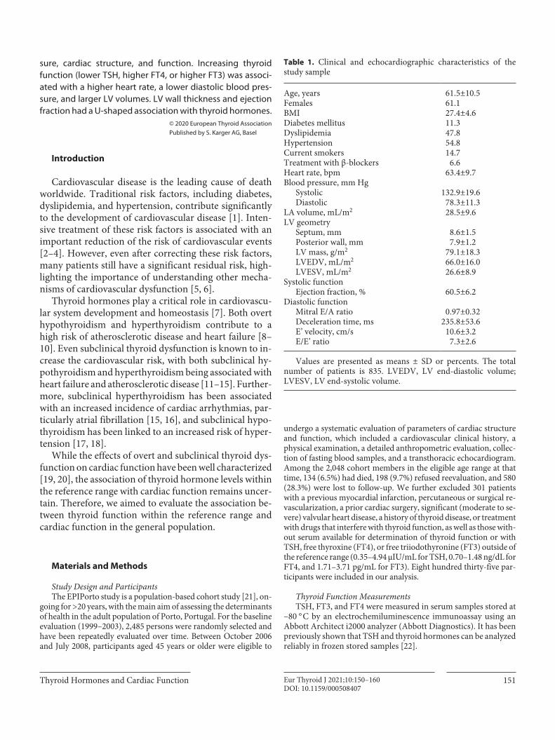
undergo a systematic evaluation of parameters of cardiac structure and function, which included a cardiovascular clinical history, a physical examination, a detailed anthropometric evaluation, collec-tion of fasting blood samples, and a transthoracic echocardiogram. Among the 2,048 cohort members in the eligible age range at that time, 134 (6.5%) had died, 198 (9.7%) refused reevaluation, and 580 (28.3%) were lost to follow-up. We further excluded 301 patients with a previous myocardial infarction, percutaneous or surgical re-vascularization, a prior cardiac surgery, significant (moderate to se-vere) valvular heart disease, a history of thyroid disease, or treatment with drugs that interfere with thyroid function, as well as those with-out serum available for determination of thyroid function or with TSH, free thyroxine (FT4), or free triiodothyronine (FT3) outside of the reference range (0.35–4.94 μIU/mL for TSH, 0.70–1.48 ng/dL for FT4, and 1.71–3.71 pg/mL for FT3). Eight hundred thirty-five par-ticipants were included in our analysis.
Thyroid Function MeasurementsTSH, FT3, and FT4 were measured in serum samples stored at
–80 ° C by an electrochemiluminescence immunoassay using an Abbott Architect i2000 analyzer (Abbott Diagnostics). It has been previously shown that TSH and thyroid hormones can be analyzed reliably in frozen stored samples [22].
Table 1. Clinical and echocardiographic characteristics of the study sample
Age, years 61.5±10.5Females 61.1BMI 27.4±4.6Diabetes mellitus 11.3Dyslipidemia 47.8Hypertension 54.8Current smokers 14.7Treatment with β-blockers 6.6Heart rate, bpm 63.4±9.7Blood pressure, mm Hg
Systolic 132.9±19.6Diastolic 78.3±11.3
LA volume, mL/m2 28.5±9.6LV geometry
Septum, mm 8.6±1.5Posterior wall, mm 7.9±1.2LV mass, g/m2 79.1±18.3LVEDV, mL/m2 66.0±16.0LVESV, mL/m2 26.6±8.9
Systolic functionEjection fraction, % 60.5±6.2
Diastolic functionMitral E/A ratio 0.97±0.32Deceleration time, ms 235.8±53.6E’ velocity, cm/s 10.6±3.2E/E’ ratio 7.3±2.6
Values are presented as means ± SD or percents. The total number of patients is 835. LVEDV, LV end-diastolic volume; LVESV, LV end-systolic volume.
Neves et al.Eur Thyroid J 2021;10:150–160152DOI: 10.1159/000508407
Echocardiographic Evaluation and Blood Pressure EvaluationAll echocardiography studies were acquired by 1 of 4 cardiolo-
gists, using the same equipment (Hewlett-Packard Sonos 5500). Images were stored for later offline analysis by 2 experienced car-diologists who were blinded to clinical data. Cardiac chamber di-mensions and volumes and left ventricular (LV) mass were in-dexed to body surface area. Diastolic function was assessed by measuring mitral inflow velocities (E-wave, A wave, and E/A ratio) and the E-wave deceleration time and the isovolumetric relaxation time using pulsed-wave Doppler in the apical 4-chamber view. Ve-locities were recorded at end expiration and averaged over 3 con-secutive cardiac cycles. Pulsed-wave tissue Doppler velocities were acquired at end expiration, in the apical 4-chamber view, on the lateral side of the mitral annulus, measuring early diastolic (E’) and late diastolic (A’) velocities and estimating the E/E’ ratio accord-ingly. Systolic function was evaluated by ejection fraction calcula-tion using the modified biplane Simpson rule.
Blood pressure was determined by 2 measurements separated by at least 5 min after a 10-min rest. When the difference between measurements was larger than 5 mm Hg for systolic or diastolic blood pressure, a third measurement was taken and the mean of the 2 closest values was considered.
Comorbidities and Clinical DefinitionsBMI was calculated as weight in kilograms divided by the
square of height in meters. Hypertension was defined as a systolic blood pressure ≥140 mm Hg or a diastolic blood pressure ≥90 mm Hg at the time of the visit [23] or as use of antihypertensive drugs. Diabetes was defined as a fasting blood glucose level ≥126 mg/dL [24], a self-reported history of diabetes, or use of diabetes medica-tions. Dyslipidemia was defined as low-density lipoprotein choles-terol ≥160 mg/dL, triglycerides ≥200 mg/dL, high-density lipo-protein cholesterol < 40 mg/dL [25], or use of lipid-lowering drugs.
Statistical AnalysisWe used linear regression models to evaluate the associations
of TSH, FT4, and FT3 (independent variables) with heart rate, blood pressure, cardiac structure, and cardiac function parameters (dependent variables). These associations were evaluated in unad-justed models and adjusted models for sex and age (model 1) and for sex, age, BMI, diabetes, hypertension, and current smoking (model 2). These variables were included as potential confounders due to their known effects on thyroid function [26, 27] and car-diac function [28]. Participants treated with β-blockers were ex-cluded from analyses of heart rate, blood pressure, and cardiac function. As previous studies have shown some similar cardiac manifestations in participants with hypothyroidism and hyperthy-roidism [29], we also assessed nonlinear associations using re-stricted cubic splines with 3 knots at the 10th, 50th, and 90th per-centiles [30]. This approach allows the identification of changes in cardiac structure or function that occur with both increasing and decreasing thyroid function within the normal range.
Continuous variables are presented as means (±SD). Categori-cal variables are presented as percentages. Comparisons between groups were made using t tests and the χ2 test. Two-sided p < 0.05 was considered statistically significant. Statistical analyses were performed using Stata software, version 14.2 (StataCorp).
Results
Study PopulationA total of 835 participants (510 women and 325 men)
were included in our analysis after applying the inclu-sion and exclusion criteria. Participants who were ex-
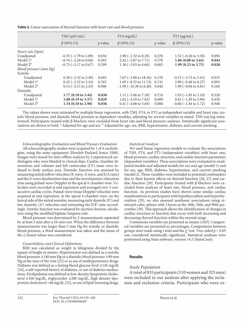
Table 2. Linear association of thyroid function with heart rate and blood pressure
TSH (μIU/mL) FT4 (ng/dL) FT3 (pg/mL)
β (95% CI) p value β (95% CI) p value β (95% CI) p value
Heart rate (bpm)Unadjusted –0.35 (–1.79 to 1.09) 0.634 2.98 (–2.32 to 8.28) 0.270 1.52 (–0.26 to 3.30) 0.094Model 1a –0.76 (–2.20 to 0.68) 0.303 2.42 (–2.87 to 7.71) 0.370 1.86 (0.08 to 3.64) 0.041Model 2b –0.75 (–2.17 to 0.67) 0.299 1.38 (–3.83 to 6.60) 0.603 1.99 (0.23 to 3.75) 0.026Blood pressure (mm Hg)Systolic
Unadjusted 0.58 (–2.32 to 3.49) 0.693 7.67 (–3.00 to 18.36) 0.159 –0.15 (–3.73 to 3.43) 0.935Model 1a 0.42 (–2.32 to 3.16) 0.763 1.69 (–8.35 to 11.74) 0.741 2.90 (–0.48 to 6.27) 0.093Model 2b 0.14 (–2.15 to 2.43) 0.906 –1.99 (–10.38 to 6.40) 0.642 1.99 (–0.84 to 4.81) 0.168
DiastolicUnadjusted 1.77 (0.10 to 3.44) 0.038 1.11 (–5.06 to 7.28) 0.724 1.03 (–1.03 to 3.10) 0.328Model 1a 1.88 (0.19 to 3.57) 0.029 1.62 (–4.59 to 7.83) 0.609 0.81 (–1.29 to 2.90) 0.450Model 2b 1.54 (0.10 to 2.98) 0.036 0.41 (–4.88 to 5.69) 0.880 –0.06 (–1.84 to 1.72) 0.948
The values shown were estimated by multiple linear regression, with TSH, FT4, or FT3 as independent variable and heart rate, sys-tolic blood pressure, and diastolic blood pressure as dependent variables, adjusting for several variables as stated. TSH was log-trans-formed. Participants treated with β-blockers were excluded from heart rate and blood pressure analyses. Statistically significant asso-ciations are shown in bold. a Adjusted for age and sex. b Adjusted for age, sex, BMI, hypertension, diabetes, and current smoking.
Thyroid Hormones and Cardiac Function 153Eur Thyroid J 2021;10:150–160DOI: 10.1159/000508407
cluded or lost to follow-up did not differ from those included at baseline regarding sex, BMI, or smoking status, but they were older and had a higher prevalence of comorbidities (online suppl. Table 1, see www. karger.com/doi/10.1159/000508407). The general char-acteristics and echocardiographic findings of the in-cluded population are shown in Table 1. The mean age was 61.5 years (SD 10.5) years. Eleven percent of the participants had diabetes mellitus, nearly half had dys-lipidemia (47.8%), and more than half had hyperten-sion (54.8%).
Association of Thyroid Function with Heart Rate and Blood PressureThe associations of thyroid function with heart rate
and blood pressure in univariable and multivariable anal-ysis are shown in Table 2. We observed that heart rate
increased significantly with increasing FT3 levels after adjustment for age and sex (model 1) and further adjust-ment for potential confounding factors of BMI, hyperten-sion, diabetes, and smoking (model 2). Regarding blood pressure, mean diastolic blood pressure was positively as-sociated with TSH levels both in univariate and in multi-variate regression models.
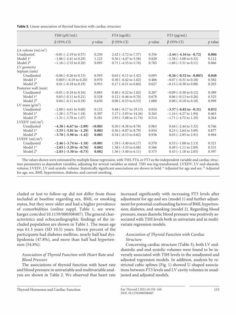
Association of Thyroid Function with Cardiac StructureConcerning cardiac structure (Table 3), both LV end-
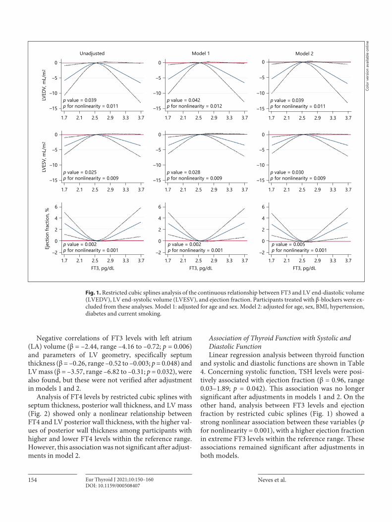
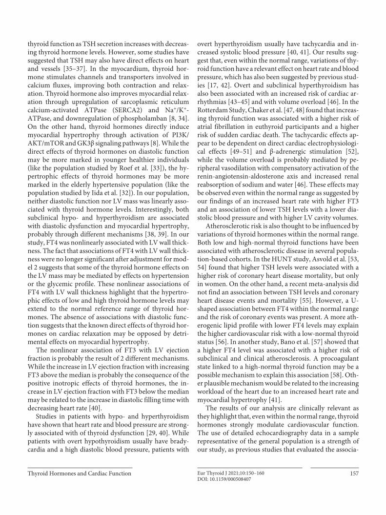
diastolic and end-systolic volumes were found to be in-versely associated with TSH levels in the unadjusted and adjusted regression models. In addition, analysis by re-stricted cubic splines (Fig. 1) showed U-shaped associa-tions between FT3 levels and LV cavity volumes in unad-justed and adjusted models.
Table 3. Linear association of thyroid function with cardiac structure
TSH (μIU/mL) FT4 (ng/dL) FT3 (pg/mL)
β (95% CI) p value β (95% CI) p value β (95% CI) p value
LA volume (mL/m2)Unadjusted –0.81 (–2.19 to 0.57) 0.250 2.42 (–2.72 to 7.57) 0.356 –2.44 (–4.16 to –0.72) 0.006Model 1a –1.06 (–2.41 to 0.29) 1.123 0.56 (–4.47 to 5.58) 0.828 –1.38 (–3.08 to 0.32) 0.112Model 2b –1.16 (–2.52 to 0.20) 0.095 0.71 (–4.35 to 5.76) 0.783 –1.60 (–3.31 to 0.11) 0.066LV geometrySeptum (mm)
Unadjusted –0.06 (–0.26 to 0.15) 0.593 0.65 (–0.11 to 1.42) 0.095 –0.26 (–0.52 to –0.003) 0.048Model 1a 0.003 (–0.19 to 0.20) 0.970 0.30 (–0.42 to 1.02) 0.406 –0.07 (–0.31 to 0.18) 0.582Model 2b 0.01 (–0.18 to 0.19) 0.953 0.17 (–0.51 to 0.84) 0.627 –0.15 (–0.38 to 0.08) 0.203
Posterior wall (mm)Unadjusted –0.01 (–0.18 to 0.16) 0.883 0.40 (–0.22 to 1.02) 0.207 –0.09 (–0.30 to 0.12) 0.389Model 1a 0.05 (–0.11 to 0.21) 0.528 0.12 (–0.46 to 0.70) 0.678 0.06 (–0.13 to 0.26) 0.525Model 2b 0.04 (–0.11 to 0.18) 0.630 0.00 (–0.53 to 0.53) 1.000 0.00 (–0.18 to 0.18) 0.999
LV mass (g/m2)Unadjusted –2.00 (–4.61 to 0.60) 0.132 9.48 (–0.17 to 19.13) 0.054 –3.57 (–6.82 to –0.31) 0.032Model 1a –1.28 (–3.75 to 1.18) 0.307 5.17 (–3.93 to 14.26) 0.265 –1.16 (–4.27 to 1.94) 0.463Model 2b –1.31 (–3.70 to 1.07) 0.281 2.93 (–5.88 to 11.74) 0.514 –1.71 (–4.72 to 1.29) 0.264
LVEDV (mL/m2)Unadjusted –4.38 (–6.67 to –2.09) <0.001 0.20 (–8.38 to 8.78) 0.963 0.44 (–2.44 to 3.32) 0.766Model 1a –3.55 (–5.81 to –1.29) 0.002 0.36 (–8.07 to 8.79) 0.934 0.23 (–2.64 to 3.09) 0.877Model 2b –3.70 (–5.98 to –1.42) 0.001 0.34 (–8.13 to 8.82) 0.936 0.03 (–2.85 to 2.91) 0.984
LVESV (mL/m2)Unadjusted –2.46 (–3.74 to –1.18) <0.001 1.39 (–3.40 to 6.17) 0.570 0.53 (–1.08 to 2.13) 0.521Model 1a –2.03 (–3.29 to –0.76) 0.002 1.38 (–3.33 to 6.08) 0.566 0.49 (–1.11 to 2.09) 0.551Model 2b –2.03 (–3.30 to –0.75) 0.002 1.36 (–3.40 to 6.11) 0.575 0.43 (–1.18 to 2.05) 0.597
The values shown were estimated by multiple linear regression, with TSH, FT4, or FT3 as the independent variable and cardiac struc-ture parameters as dependent variables, adjusting for several variables as stated. TSH was log-transformed. LVEDV, LV end-diastolic volume; LVESV, LV end-systolic volume. Statistically significant associations are shown in bold. a Adjusted for age and sex. b Adjusted for age, sex, BMI, hypertension, diabetes, and current smoking.
Neves et al.Eur Thyroid J 2021;10:150–160154DOI: 10.1159/000508407
Negative correlations of FT3 levels with left atrium (LA) volume (β = –2.44, range –4.16 to –0.72; p = 0.006) and parameters of LV geometry, specifically septum thickness (β = –0.26, range –0.52 to –0.003; p = 0.048) and LV mass (β = –3.57, range –6.82 to –0.31; p = 0.032), were also found, but these were not verified after adjustment in models 1 and 2.
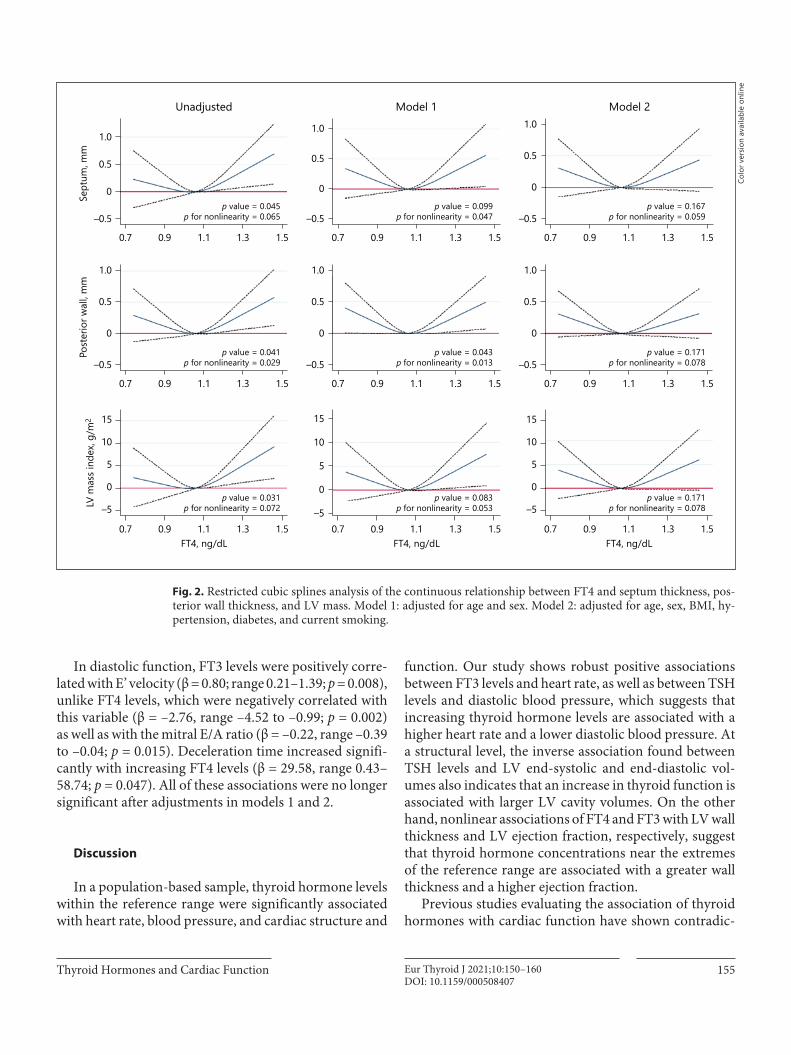
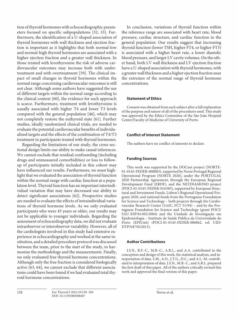
Analysis of FT4 levels by restricted cubic splines with septum thickness, posterior wall thickness, and LV mass (Fig. 2) showed only a nonlinear relationship between FT4 and LV posterior wall thickness, with the higher val-ues of posterior wall thickness among participants with higher and lower FT4 levels within the reference range. However, this association was not significant after adjust-ments in model 2.
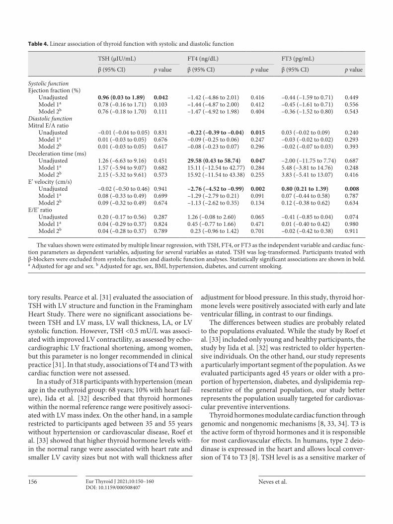
Association of Thyroid Function with Systolic and Diastolic FunctionLinear regression analysis between thyroid function
and systolic and diastolic functions are shown in Table 4. Concerning systolic function, TSH levels were posi-tively associated with ejection fraction (β = 0.96, range 0.03–1.89; p = 0.042). This association was no longer significant after adjustments in models 1 and 2. On the other hand, analysis between FT3 levels and ejection fraction by restricted cubic splines (Fig. 1) showed a strong nonlinear association between these variables (p for nonlinearity = 0.001), with a higher ejection fraction in extreme FT3 levels within the reference range. These associations remained significant after adjustments in both models.
Unadjusted Model 1 Model 2
p value = 0.039p for nonlinearity = 0.011
1.7 2.1 2.5 2.9 3.3 3.7
0
–5
–10
–15p value = 0.042p for nonlinearity = 0.012
1.7 2.1 2.5 2.9 3.3 3.7
0
–5
–10
–15p value = 0.039p for nonlinearity = 0.011
1.7 2.1 2.5 2.9 3.3 3.7
0
–5
–10
–15
LVED
V, m
L/m
2
p value = 0.030p for nonlinearity = 0.009
1.7 2.1 2.5 2.9 3.3 3.7
0
–5
–10
–15p value = 0.028p for nonlinearity = 0.009
1.7 2.1 2.5 2.9 3.3 3.7
0
–5
–10
–15p value = 0.025p for nonlinearity = 0.009
1.7 2.1 2.5 2.9 3.3 3.7
0
–5
–10
–15
LVES
V, m
L/m
2
p value = 0.005p for nonlinearity = 0.001
FT3, pg/dL1.7 2.1 2.5 2.9 3.3 3.7
p value = 0.002p for nonlinearity = 0.001
FT3, pg/dL1.7 2.1 2.5 2.9 3.3 3.7
p value = 0.002p for nonlinearity = 0.001
FT3, pg/dL1.7 2.1 2.5 2.9 3.3 3.7
6
4
0
2
–2
6
4
0
2
–2
6
4
0
2
–2
Ejec
tion
fract
ion,
%
Fig. 1. Restricted cubic splines analysis of the continuous relationship between FT3 and LV end-diastolic volume (LVEDV), LV end-systolic volume (LVESV), and ejection fraction. Participants treated with β-blockers were ex-cluded from these analyses. Model 1: adjusted for age and sex. Model 2: adjusted for age, sex, BMI, hypertension, diabetes and current smoking.
Colo
r ver
sion
avai
labl
e on
line
Thyroid Hormones and Cardiac Function 155Eur Thyroid J 2021;10:150–160DOI: 10.1159/000508407
In diastolic function, FT3 levels were positively corre-lated with E’ velocity (β = 0.80; range 0.21–1.39; p = 0.008), unlike FT4 levels, which were negatively correlated with this variable (β = –2.76, range –4.52 to –0.99; p = 0.002) as well as with the mitral E/A ratio (β = –0.22, range –0.39 to –0.04; p = 0.015). Deceleration time increased signifi-cantly with increasing FT4 levels (β = 29.58, range 0.43–58.74; p = 0.047). All of these associations were no longer significant after adjustments in models 1 and 2.
Discussion
In a population-based sample, thyroid hormone levels within the reference range were significantly associated with heart rate, blood pressure, and cardiac structure and
function. Our study shows robust positive associations between FT3 levels and heart rate, as well as between TSH levels and diastolic blood pressure, which suggests that increasing thyroid hormone levels are associated with a higher heart rate and a lower diastolic blood pressure. At a structural level, the inverse association found between TSH levels and LV end-systolic and end-diastolic vol-umes also indicates that an increase in thyroid function is associated with larger LV cavity volumes. On the other hand, nonlinear associations of FT4 and FT3 with LV wall thickness and LV ejection fraction, respectively, suggest that thyroid hormone concentrations near the extremes of the reference range are associated with a greater wall thickness and a higher ejection fraction.
Previous studies evaluating the association of thyroid hormones with cardiac function have shown contradic-
1.0
0.5
0
p value = 0.043p for nonlinearity = 0.013
0.7 0.9 1.1 1.3 1.5
–0.5p value = 0.041
p for nonlinearity = 0.029
1.0
0.5
0
–0.5
0.7 0.9 1.1 1.3 1.5
Post
erio
r wal
l, m
m
1.0
0.5
0
p value = 0.171p for nonlinearity = 0.078–0.5
0.7 0.9 1.1 1.3 1.5
15
10
5
0
–5p value = 0.083
p for nonlinearity = 0.053
0.7 0.9 1.1 1.3 1.5FT4, ng/dL
15
10
5
0
–5p value = 0.031
p for nonlinearity = 0.072
0.7 0.9 1.1 1.3 1.5FT4, ng/dL
LV m
ass i
ndex
, g/m
2 15
10
5
0
–5
0.7 0.9 1.1 1.3 1.5
p value = 0.171p for nonlinearity = 0.078
FT4, ng/dL
0.7 0.9 1.1
p value = 0.099p for nonlinearity = 0.047
1.3 1.5
1.0
0.5
0
–0.5
0.7 0.9 1.1 1.3 1.5
1.0
0.5
0
–0.5p value = 0.045
p for nonlinearity = 0.065
Sept
um, m
m
0.7 0.9 1.1
p value = 0.167p for nonlinearity = 0.059
1.3 1.5
1.0
0.5
0
–0.5
Unadjusted Model 1 Model 2
Fig. 2. Restricted cubic splines analysis of the continuous relationship between FT4 and septum thickness, pos-terior wall thickness, and LV mass. Model 1: adjusted for age and sex. Model 2: adjusted for age, sex, BMI, hy-pertension, diabetes, and current smoking.
Colo
r ver
sion
avai
labl
e on
line
Neves et al.Eur Thyroid J 2021;10:150–160156DOI: 10.1159/000508407
tory results. Pearce et al. [31] evaluated the association of TSH with LV structure and function in the Framingham Heart Study. There were no significant associations be-tween TSH and LV mass, LV wall thickness, LA, or LV systolic function. However, TSH < 0.5 mU/L was associ-ated with improved LV contractility, as assessed by echo-cardiographic LV fractional shortening, among women, but this parameter is no longer recommended in clinical practice [31]. In that study, associations of T4 and T3 with cardiac function were not assessed.
In a study of 318 participants with hypertension (mean age in the euthyroid group: 68 years; 10% with heart fail-ure), Iida et al. [32] described that thyroid hormones within the normal reference range were positively associ-ated with LV mass index. On the other hand, in a sample restricted to participants aged between 35 and 55 years without hypertension or cardiovascular disease, Roef et al. [33] showed that higher thyroid hormone levels with-in the normal range were associated with heart rate and smaller LV cavity sizes but not with wall thickness after
adjustment for blood pressure. In this study, thyroid hor-mone levels were positively associated with early and late ventricular filling, in contrast to our findings.
The differences between studies are probably related to the populations evaluated. While the study by Roef et al. [33] included only young and healthy participants, the study by Iida et al. [32] was restricted to older hyperten-sive individuals. On the other hand, our study represents a particularly important segment of the population. As we evaluated participants aged 45 years or older with a pro-portion of hypertension, diabetes, and dyslipidemia rep-resentative of the general population, our study better represents the population usually targeted for cardiovas-cular preventive interventions.
Thyroid hormones modulate cardiac function through genomic and nongenomic mechanisms [8, 33, 34]. T3 is the active form of thyroid hormones and it is responsible for most cardiovascular effects. In humans, type 2 deio-dinase is expressed in the heart and allows local conver-sion of T4 to T3 [8]. TSH level is as a sensitive marker of
Table 4. Linear association of thyroid function with systolic and diastolic function
TSH (μIU/mL) FT4 (ng/dL) FT3 (pg/mL)
β (95% CI) p value β (95% CI) p value β (95% CI) p value
Systolic functionEjection fraction (%)
Unadjusted 0.96 (0.03 to 1.89) 0.042 –1.42 (–4.86 to 2.01) 0.416 –0.44 (–1.59 to 0.71) 0.449Model 1a 0.78 (–0.16 to 1.71) 0.103 –1.44 (–4.87 to 2.00) 0.412 –0.45 (–1.61 to 0.71) 0.556Model 2b 0.76 (–0.18 to 1.70) 0.111 –1.47 (–4.92 to 1.98) 0.404 –0.36 (–1.52 to 0.80) 0.543
Diastolic functionMitral E/A ratio
Unadjusted –0.01 (–0.04 to 0.05) 0.831 –0.22 (–0.39 to –0.04) 0.015 0.03 (–0.02 to 0.09) 0.240Model 1a 0.01 (–0.03 to 0.05) 0.676 –0.09 (–0.25 to 0.06) 0.247 –0.03 (–0.02 to 0.02) 0.293Model 2b 0.01 (–0.03 to 0.05) 0.617 –0.08 (–0.23 to 0.07) 0.296 –0.02 (–0.07 to 0.03) 0.393
Deceleration time (ms)Unadjusted 1.26 (–6.63 to 9.16) 0.451 29.58 (0.43 to 58.74) 0.047 –2.00 (–11.75 to 7.74) 0.687Model 1a 1.57 (–5.94 to 9.07) 0.682 15.11 (–12.54 to 42.77) 0.284 5.48 (–3.81 to 14.76) 0.248Model 2b 2.15 (–5.32 to 9.61) 0.573 15.92 (–11.54 to 43.38) 0.255 3.83 (–5.41 to 13.07) 0.416
E’ velocity (cm/s)Unadjusted –0.02 (–0.50 to 0.46) 0.941 –2.76 (–4.52 to –0.99) 0.002 0.80 (0.21 to 1.39) 0.008Model 1a 0.08 (–0.33 to 0.49) 0.699 –1.29 (–2.79 to 0.21) 0.091 0.07 (–0.44 to 0.58) 0.787Model 2b 0.09 (–0.32 to 0.49) 0.674 –1.13 (–2.62 to 0.35) 0.134 0.12 (–0.38 to 0.62) 0.634
E/E’ ratioUnadjusted 0.20 (–0.17 to 0.56) 0.287 1.26 (–0.08 to 2.60) 0.065 –0.41 (–0.85 to 0.04) 0.074Model 1a 0.04 (–0.29 to 0.37) 0.824 0.45 (–0.77 to 1.66) 0.471 0.01 (–0.40 to 0.42) 0.980Model 2b 0.04 (–0.28 to 0.37) 0.789 0.23 (–0.96 to 1.42) 0.701 –0.02 (–0.42 to 0.38) 0.911
The values shown were estimated by multiple linear regression, with TSH, FT4, or FT3 as the independent variable and cardiac func-tion parameters as dependent variables, adjusting for several variables as stated. TSH was log-transformed. Participants treated with β-blockers were excluded from systolic function and diastolic function analyses. Statistically significant associations are shown in bold. a Adjusted for age and sex. b Adjusted for age, sex, BMI, hypertension, diabetes, and current smoking.
Thyroid Hormones and Cardiac Function 157Eur Thyroid J 2021;10:150–160DOI: 10.1159/000508407
thyroid function as TSH secretion increases with decreas-ing thyroid hormone levels. However, some studies have suggested that TSH may also have direct effects on heart and vessels [35–37]. In the myocardium, thyroid hor-mone stimulates channels and transporters involved in calcium fluxes, improving both contraction and relax-ation. Thyroid hormone also improves myocardial relax-ation through upregulation of sarcoplasmic reticulum calcium-activated ATPase (SERCA2) and Na+/K+-ATPase, and downregulation of phospholamban [8, 34]. On the other hand, thyroid hormones directly induce myocardial hypertrophy through activation of PI3K/AKT/mTOR and GK3β signaling pathways [8]. While the direct effects of thyroid hormones on diastolic function may be more marked in younger healthier individuals (like the population studied by Roef et al. [33]), the hy-pertrophic effects of thyroid hormones may be more marked in the elderly hypertensive population (like the population studied by Iida et al. [32]). In our population, neither diastolic function nor LV mass was linearly asso-ciated with thyroid hormone levels. Interestingly, both subclinical hypo- and hyperthyroidism are associated with diastolic dysfunction and myocardial hypertrophy, probably through different mechanisms [38, 39]. In our study, FT4 was nonlinearly associated with LV wall thick-ness. The fact that associations of FT4 with LV wall thick-ness were no longer significant after adjustment for mod-el 2 suggests that some of the thyroid hormone effects on the LV mass may be mediated by effects on hypertension or the glycemic profile. These nonlinear associations of FT4 with LV wall thickness highlight that the hypertro-phic effects of low and high thyroid hormone levels may extend to the normal reference range of thyroid hor-mones. The absence of associations with diastolic func-tion suggests that the known direct effects of thyroid hor-mones on cardiac relaxation may be opposed by detri-mental effects on myocardial hypertrophy.
The nonlinear association of FT3 with LV ejection fraction is probably the result of 2 different mechanisms. While the increase in LV ejection fraction with increasing FT3 above the median is probably the consequence of the positive inotropic effects of thyroid hormones, the in-crease in LV ejection fraction with FT3 below the median may be related to the increase in diastolic filling time with decreasing heart rate [40].
Studies in patients with hypo- and hyperthyroidism have shown that heart rate and blood pressure are strong-ly associated with of thyroid dysfunction [29, 40]. While patients with overt hypothyroidism usually have brady-cardia and a high diastolic blood pressure, patients with
overt hyperthyroidism usually have tachycardia and in-creased systolic blood pressure [40, 41]. Our results sug-gest that, even within the normal range, variations of thy-roid function have a relevant effect on heart rate and blood pressure, which has also been suggested by previous stud-ies [17, 42]. Overt and subclinical hyperthyroidism has also been associated with an increased risk of cardiac ar-rhythmias [43–45] and with volume overload [46]. In the Rotterdam Study, Chaker et al. [47, 48] found that increas-ing thyroid function was associated with a higher risk of atrial fibrillation in euthyroid participants and a higher risk of sudden cardiac death. The tachycardic effects ap-pear to be dependent on direct cardiac electrophysiologi-cal effects [49–51] and β-adrenergic stimulation [52], while the volume overload is probably mediated by pe-ripheral vasodilation with compensatory activation of the renin-angiotensin-aldosterone axis and increased renal reabsorption of sodium and water [46]. These effects may be observed even within the normal range as suggested by our findings of an increased heart rate with higher FT3 and an association of lower TSH levels with a lower dia-stolic blood pressure and with higher LV cavity volumes.
Atherosclerotic risk is also thought to be influenced by variations of thyroid hormones within the normal range. Both low and high-normal thyroid functions have been associated with atherosclerotic disease in several popula-tion-based cohorts. In the HUNT study, Asvold et al. [53, 54] found that higher TSH levels were associated with a higher risk of coronary heart disease mortality, but only in women. On the other hand, a recent meta-analysis did not find an association between TSH levels and coronary heart disease events and mortality [55]. However, a U-shaped association between FT4 within the normal range and the risk of coronary events was present. A more ath-erogenic lipid profile with lower FT4 levels may explain the higher cardiovascular risk with a low-normal thyroid status [56]. In another study, Bano et al. [57] showed that a higher FT4 level was associated with a higher risk of subclinical and clinical atherosclerosis. A procoagulant state linked to a high-normal thyroid function may be a possible mechanism to explain this association [58]. Oth-er plausible mechanism would be related to the increasing workload of the heart due to an increased heart rate and myocardial hypertrophy [41].
The results of our analysis are clinically relevant as they highlight that, even within the normal range, thyroid hormones strongly modulate cardiovascular function. The use of detailed echocardiography data in a sample representative of the general population is a strength of our study, as previous studies that evaluated the associa-
Neves et al.Eur Thyroid J 2021;10:150–160158DOI: 10.1159/000508407
tion of thyroid hormones with echocardiographic param-eters focused on specific subpopulations [32, 33]. Fur-thermore, the identification of a U-shaped association of thyroid hormones with wall thickness and ejection frac-tion is important as it highlights that both normal-low and normal-high thyroid hormones are associated with a higher ejection fraction and a greater wall thickness. In those treated with levothyroxine the risk of adverse car-diovascular outcomes may increase both with under-treatment and with overtreatment [59]. The clinical im-pact of small changes in thyroid hormones within the normal range concerning cardiovascular outcomes is still not clear. Although some authors have suggested the use of different targets within the normal range according to the clinical context [60], the evidence for such approach is scarce. Furthermore, treatment with levothyroxine is usually associated with higher T4 and lower T3 levels compared with the general population [46], which may not completely restore the euthyroid state [61]. Further studies, ideally randomized clinical trials, are needed to evaluate the potential cardiovascular benefits of individu-alized targets and the effects of the combination of T4/T3 treatment in participants treated with thyroid hormones.
Regarding the limitations of our study, the cross-sec-tional design limits our ability to make causal inferences. We cannot exclude that residual confounding (including drugs and unmeasured comorbidities) or loss to follow-up of participants initially included in this cohort may have influenced our results. Furthermore, we must high-light that we evaluated the association of thyroid function within the normal range with cardiac function at a popu-lation level. Thyroid function has an important interindi-vidual variation that may have decreased our ability to detect significant associations [62]. Prospective studies are needed to evaluate the effects of intraindividual varia-tions of thyroid hormone levels. As we only evaluated participants who were 45 years or older, our results may not be applicable to younger individuals. Regarding the assessment of echocardiography data, we did not evaluate intraobserver or interobserver variability. However, all of the cardiologists involved in this study had extensive ex-perience in echocardiography and worked at the same in-stitution, and a detailed procedure protocol was discussed between the team, prior to the start of the study, to har-monize the methodology and the measurements. Finally, we only evaluated free thyroid hormone concentrations. Although only the free fraction is considered biologically active [63, 64], we cannot exclude that different associa-tions could have been found if we had evaluated total thy-roid hormone concentrations.
In conclusion, variations of thyroid function within the reference range are associated with heart rate, blood pressure, cardiac structure, and cardiac function in the general population. Our results suggest that increasing thyroid function (lower TSH, higher FT4, or higher FT3) is associated with a higher heart rate, a lower diastolic blood pressure, and larger LV cavity volumes. On the oth-er hand, both LV wall thickness and LV ejection fraction have a U-shaped association with thyroid hormones, with a greater wall thickness and a higher ejection fraction near the extremes of the normal range of thyroid hormone concentrations.
Statement of Ethics
Consent was obtained from each subject after a full explanation of the purpose and nature of all of the procedures used. This study was approved by the Ethics Committee of the São João Hospital Center/Faculty of Medicine of University of Porto.
Conflict of Interest Statement
The authors have no conflict of interests to declare.
Funding Sources
This work was supported by the DOCnet project (NORTE-01-0145-FEDER-000003), supported by Norte Portugal Regional Operational Program (NORTE 2020), under the PORTUGAL 2020 Partnership Agreement, through the European Regional Development Fund (ERDF), and the NETDIAMOND project (POCI-01-0145-FEDER-016385), supported by European Struc-tural and Investment Funds, Lisbon’s Regional Operational Pro-gram 2020, and national funds from the Portuguese Foundation for Science and Technology – both projects through the Cardio-vascular Research Center (UnIC, FCT 51/94) – and by the Por-tuguese Foundation for Science and Technology (grant POCI/SAU-ESP/61492/2004) and the Unidade de Investigação em Epidemiologia – Instituto de Saúde Pública da Universidade do Porto (EPIUnit) (POCI-01-0145-FEDER-006862, ref. UID/DTP/04750/2013).
Author Contributions
J.S.N., R.F.-C., M.B.-C., A.R.L., and A.A. contributed to the conception and design of this work, the statistical analysis, and in-terpretation of data. S.M., A.O., J.T.G., D.C., and A.L.-M. contrib-uted to interpretation of data. J.S.N., M.B.-C., and A.R.L. prepared the first draft of this paper. All of the authors critically revised this work and approved the final version of this paper.
Thyroid Hormones and Cardiac Function 159Eur Thyroid J 2021;10:150–160DOI: 10.1159/000508407
References
1 Yusuf S, Hawken S, Ounpuu S, Dans T, Ave-zum A, Lanas F, et al.; INTERHEART Study Investigators. Effect of potentially modifiable risk factors associated with myocardial infarc-tion in 52 countries (the INTERHEART study): case-control study. Lancet. 2004 Sep;
364(9438): 937–52. 2 Gaede P, Lund-Andersen H, Parving HH,
Pedersen O. Effect of a multifactorial inter-vention on mortality in type 2 diabetes. N Engl J Med. 2008 Feb; 358(6): 580–91.
3 Baigent C, Blackwell L, Emberson J, Holland LE, Reith C, Bhala N, et al.; Cholesterol Treat-ment Trialists’ (CTT) Collaboration. Efficacy and safety of more intensive lowering of LDL cholesterol: a meta-analysis of data from 170,000 participants in 26 randomised trials. Lancet. 2010 Nov; 376(9753): 1670–81.
4 Wright JT Jr, Williamson JD, Whelton PK, Snyder JK, Sink KM, Rocco MV, et al.; SPRINT Research Group. A Randomized Tri-al of Intensive versus Standard Blood-Pres-sure Control. N Engl J Med. 2015 Nov;
373(22): 2103–16. 5 Lieb W, Enserro DM, Sullivan LM, Vasan RS.
Residual Cardiovascular Risk in Individuals on Blood Pressure-Lowering Treatment. J Am Heart Assoc. 2015 Nov; 4(11):e002155.
6 Dash S, Leiter LA. Residual cardiovascular risk among people with diabetes. Diabetes Obes Metab. 2019 Apr; 21(Suppl 1): 28–38.
7 Cooper DS, Biondi B. Subclinical thyroid dis-ease. Lancet. 2012 Mar; 379(9821): 1142–54.
8 Jabbar A, Pingitore A, Pearce SH, Zaman A, Iervasi G, Razvi S. Thyroid hormones and car-diovascular disease. Nat Rev Cardiol. 2017 Jan; 14(1): 39–55.
9 Chaker L, Bianco AC, Jonklaas J, Peeters RP. Hypothyroidism. Lancet. 2017 Sep; 390 (10101): 1550–62.
10 De Leo S, Lee SY, Braverman LE. Hyperthy-roidism. Lancet. 2016 Aug; 388(10047): 906–18.
11 Hak AE, Pols HA, Visser TJ, Drexhage HA, Hofman A, Witteman JC. Subclinical hypo-thyroidism is an independent risk factor for atherosclerosis and myocardial infarction in elderly women: the Rotterdam Study. Ann In-tern Med. 2000 Feb; 132(4): 270–8.
12 Selmer C, Olesen JB, Hansen ML, von Kap-pelgaard LM, Madsen JC, Hansen PR, et al. Subclinical and overt thyroid dysfunction and risk of all-cause mortality and cardiovascular events: a large population study. J Clin Endo-crinol Metab. 2014 Jul; 99(7): 2372–82.
13 Gencer B, Collet TH, Virgini V, Bauer DC, Gussekloo J, Cappola AR, et al.; Thyroid Stud-ies Collaboration. Subclinical thyroid dys-function and the risk of heart failure events: an individual participant data analysis from 6 prospective cohorts. Circulation. 2012 Aug;
126(9): 1040–9.14 Rodondi N, den Elzen WP, Bauer DC, Cap-
pola AR, Razvi S, Walsh JP, et al.; Thyroid Studies Collaboration. Subclinical hypothy-
roidism and the risk of coronary heart disease and mortality. JAMA. 2010 Sep; 304(12):
1365–74.15 Collet TH, Gussekloo J, Bauer DC, den Elzen
WP, Cappola AR, Balmer P, et al.; Thyroid Studies Collaboration. Subclinical hyperthy-roidism and the risk of coronary heart disease and mortality. Arch Intern Med. 2012 May;
172(10): 799–809.16 Sawin CT, Geller A, Wolf PA, Belanger AJ,
Baker E, Bacharach P, et al. Low serum thyro-tropin concentrations as a risk factor for atri-al fibrillation in older persons. N Engl J Med. 1994 Nov; 331(19): 1249–52.
17 Asvold BO, Bjøro T, Nilsen TI, Vatten LJ. As-sociation between blood pressure and serum thyroid-stimulating hormone concentration within the reference range: a population-based study. J Clin Endocrinol Metab. 2007 Mar; 92(3): 841–5.
18 Ittermann T, Thamm M, Wallaschofski H, Rettig R, Völzke H. Serum thyroid-stimulat-ing hormone levels are associated with blood pressure in children and adolescents. J Clin Endocrinol Metab. 2012 Mar; 97(3): 828–34.
19 Vale C, Neves JS, von Hafe M, Borges-Canha M, Leite-Moreira A. The Role of Thyroid Hormones in Heart Failure. Cardiovasc Drugs Ther. 2019 Apr; 33(2): 179–88.
20 von Hafe M, Neves JS, Vale C, Borges-Canha M, Leite-Moreira A. The impact of thyroid hormone dysfunction on ischemic heart dis-ease. Endocr Connect. 2019 May; 8(5):R76–90.
21 Santos AC, Ebrahim S, Barros H. Alcohol in-take, smoking, sleeping hours, physical activ-ity and the metabolic syndrome. Prev Med. 2007 Apr; 44(4): 328–34.
22 Männistö T, Surcel HM, Bloigu A, Ruokonen A, Hartikainen AL, Järvelin MR, et al. The ef-fect of freezing, thawing, and short- and long-term storage on serum thyrotropin, thyroid hormones, and thyroid autoantibodies: im-plications for analyzing samples stored in se-rum banks. Clin Chem. 2007 Nov; 53(11):
1986–7.23 Williams B, Mancia G, Spiering W, Agabiti
Rosei E, Azizi M, Burnier M, et al.; ESC Scien-tific Document Group. 2018 ESC/ESH Guide-lines for the management of arterial hyper-tension. Eur Heart J. 2018 Sep; 39(33): 3021–104.
24 American Diabetes Association. 2. Classifica-tion and Diagnosis of Diabetes: Standards of Medical Care in Diabetes-2019. Diabetes Care. 2019 Jan; 42 Suppl 1:S13–28.
25 Expert Panel on Detection, Evaluation, and Treatment of High Blood Cholesterol in Adults. Executive Summary of The Third Report of The National Cholesterol Educa-tion Program (NCEP) Expert Panel on De-tection, Evaluation, And Treatment of High Blood Cholesterol In Adults (Adult Treat-ment Panel III). JAMA. 2001 May; 285(19):
2486–97.
26 Reinehr T. Obesity and thyroid function. Mol Cell Endocrinol. 2010 Mar; 316(2): 165–71.
27 Laclaustra M, Moreno-Franco B, Lou-Bonafonte JM, Mateo-Gallego R, Casasnovas JA, Guallar-Castillon P, et al. Impaired Sensi-tivity to Thyroid Hormones Is Associated With Diabetes and Metabolic Syndrome. Dia-betes Care. 2019 Feb; 42(2): 303–10.
28 Ziaeian B, Fonarow GC. Epidemiology and aetiology of heart failure. Nat Rev Cardiol. 2016 Jun; 13(6): 368–78.
29 Razvi S, Jabbar A, Pingitore A, Danzi S, Bion-di B, Klein I, et al. Thyroid Hormones and Cardiovascular Function and Diseases. J Am Coll Cardiol. 2018 Apr; 71(16): 1781–96.
30 Harrell FE Jr, Lee KL, Pollock BG. Regression models in clinical studies: determining rela-tionships between predictors and response. J Natl Cancer Inst. 1988 Oct; 80(15): 1198–202.
31 Pearce EN, Yang Q, Benjamin EJ, Aragam J, Vasan RS. Thyroid function and left ventricu-lar structure and function in the Framingham Heart Study. Thyroid. 2010 Apr; 20(4): 369–73.
32 Iida M, Yamamoto M, Ishiguro Y, Yamazaki M, Honjo H, Kamiya K. Thyroid hormone within the normal range is associated with left ventricular mass in patients with hyperten-sion. J Am Soc Hypertens. 2012 Jul-Aug; 6(4):
261–9.33 Roef GL, Taes YE, Kaufman JM, Van Daele
CM, De Buyzere ML, Gillebert TC, et al. Thy-roid hormone levels within reference range are associated with heart rate, cardiac struc-ture, and function in middle-aged men and women. Thyroid. 2013 Aug; 23(8): 947–54.
34 Vargas-Uricoechea H, Sierra-Torres CH. Thyroid hormones and the heart. Horm Mol Biol Clin Investig. 2014 Apr; 18(1): 15–26.
35 Dong J, Gao C, Liu J, Cao Y, Tian L. TSH in-hibits SERCA2a and the PKA/PLN pathway in rat cardiomyocytes. Oncotarget. 2016 Jun;
7(26): 39207–15.36 Fernandez-Ruocco J, Gallego M, Rodriguez-
de-Yurre A, Zayas-Arrabal J, Echeazarra L, Alquiza A, et al. High Thyrotropin Is Critical for Cardiac Electrical Remodeling and Ar-rhythmia Vulnerability in Hypothyroidism. Thyroid. 2019 Jul; 29(7): 934–45.
37 Tian L, Ni J, Guo T, Liu J, Dang Y, Guo Q, et al. TSH stimulates the proliferation of vascu-lar smooth muscle cells. Endocrine. 2014 Aug;
46(3): 651–8.38 Monzani F, Di Bello V, Caraccio N, Bertini A,
Giorgi D, Giusti C, et al. Effect of levothyrox-ine on cardiac function and structure in sub-clinical hypothyroidism: a double blind, pla-cebo-controlled study. J Clin Endocrinol Metab. 2001 Mar; 86(3): 1110–5.
39 Yue WS, Chong BH, Zhang XH, Liao SY, Jim MH, Kung AW, et al. Hyperthyroidism-in-duced left ventricular diastolic dysfunction: implication in hyperthyroidism-related heart failure. Clin Endocrinol (Oxf). 2011 May;
74(5): 636–43.
Neves et al.Eur Thyroid J 2021;10:150–160160DOI: 10.1159/000508407
40 Biondi B, Palmieri EA, Lombardi G, Fazio S. Effects of thyroid hormone on cardiac func-tion: the relative importance of heart rate, loading conditions, and myocardial contrac-tility in the regulation of cardiac performance in human hyperthyroidism. J Clin Endocrinol Metab. 2002 Mar; 87(3): 968–74.
41 Klein I, Danzi S. Thyroid disease and the heart. Circulation. 2007 Oct; 116(15): 1725–35.
42 Asvold BO, Bjøro T, Vatten LJ. Associations of TSH levels within the reference range with future blood pressure and lipid concentra-tions: 11-year follow-up of the HUNT study. Eur J Endocrinol. 2013 Jun; 169(1): 73–82.
43 Frost L, Vestergaard P, Mosekilde L. Hyper-thyroidism and risk of atrial fibrillation or flutter: a population-based study. Arch Intern Med. 2004 Aug; 164(15): 1675–8.
44 Sgarbi JA, Villaça FG, Garbeline B, Villar HE, Romaldini JH. The effects of early antithyroid therapy for endogenous subclinical hyperthy-roidism in clinical and heart abnormalities. J Clin Endocrinol Metab. 2003 Apr; 88(4):
1672–7.45 Kaminski G, Dziuk M, Szczepanek-Parulska
E, Zybek-Kocik A, Ruchala M. Electrocardio-graphic and scintigraphic evaluation of pa-tients with subclinical hyperthyroidism dur-ing workout. Endocrine. 2016 Aug; 53(2):
512–9.46 Biondi B. Mechanisms in endocrinology:
heart failure and thyroid dysfunction. Eur J Endocrinol. 2012 Nov; 167(5): 609–18.
47 Chaker L, Heeringa J, Dehghan A, Medici M, Visser WE, Baumgartner C, et al. Normal Thyroid Function and the Risk of Atrial Fi-brillation: the Rotterdam Study. J Clin Endo-crinol Metab. 2015 Oct; 100(10): 3718–24.
48 Chaker L, van den Berg ME, Niemeijer MN, Franco OH, Dehghan A, Hofman A, et al.
Thyroid Function and Sudden Cardiac Death: A Prospective Population-Based Cohort Study. Circulation. 2016 Sep; 134(10): 713–22.
49 Renaudon B, Lenfant J, Decressac S, Bois P. Thyroid hormone increases the conductance density of f-channels in rabbit sino-atrial node cells. Receptors Channels. 2000; 7(1): 1–8.
50 Sun ZQ, Ojamaa K, Nakamura TY, Artman M, Klein I, Coetzee WA. Thyroid hormone increases pacemaker activity in rat neonatal atrial myocytes. J Mol Cell Cardiol. 2001 Apr;
33(4): 811–24.51 Sun ZQ, Ojamaa K, Coetzee WA, Artman M,
Klein I. Effects of thyroid hormone on action potential and repolarizing currents in rat ven-tricular myocytes. Am J Physiol Endocrinol Metab. 2000 Feb; 278(2):E302–7.
52 Silva JE, Bianco SD. Thyroid-adrenergic in-teractions: physiological and clinical implica-tions. Thyroid. 2008 Feb; 18(2): 157–65.
53 Asvold BO, Bjøro T, Nilsen TI, Gunnell D, Vatten LJ. Thyrotropin levels and risk of fatal coronary heart disease: the HUNT study. Arch Intern Med. 2008 Apr; 168(8): 855–60.
54 Asvold BO, Bjøro T, Platou C, Vatten LJ. Thy-roid function and the risk of coronary heart disease: 12-year follow-up of the HUNT study in Norway. Clin Endocrinol (Oxf). 2012 Dec;
77(6): 911–7.55 Åsvold BO, Vatten LJ, Bjøro T, Bauer DC,
Bremner A, Cappola AR, et al.; Thyroid Stud-ies Collaboration. Thyroid function within the normal range and risk of coronary heart disease: an individual participant data analy-sis of 14 cohorts. JAMA Intern Med. 2015 Jun;
175(6): 1037–47.56 Roos A, Bakker SJ, Links TP, Gans RO,
Wolffenbuttel BH. Thyroid function is associ-ated with components of the metabolic syn-drome in euthyroid subjects. J Clin Endocri-nol Metab. 2007 Feb; 92(2): 491–6.
57 Bano A, Chaker L, Mattace-Raso FU, van der Lugt A, Ikram MA, Franco OH, et al. Thyroid Function and the Risk of Atherosclerotic Car-diovascular Morbidity and Mortality: the Rot-terdam Study. Circ Res. 2017 Dec; 121(12):
1392–400.58 Bano A, Chaker L, de Maat MP, Atiq F, Ka-
vousi M, Franco OH, et al. Thyroid Function and Cardiovascular Disease: The Mediating Role of Coagulation Factors. J Clin Endocri-nol Metab. 2019 Aug; 104(8): 3203–12.
59 Thayakaran R, Adderley NJ, Sainsbury C, Torlinska B, Boelaert K, Šumilo D, et al. Thy-roid replacement therapy, thyroid stimulat-ing hormone concentrations, and long term health outcomes in patients with hypothy-roidism: longitudinal study. BMJ. 2019 Sep;
366:l4892.60 Hoermann R, Midgley JE, Larisch R, Dietrich
JW. Homeostatic Control of the Thyroid-Pi-tuitary Axis: Perspectives for Diagnosis and Treatment. Front Endocrinol (Lausanne). 2015 Nov; 6: 177.
61 Peterson SJ, McAninch EA, Bianco AC. Is a Normal TSH Synonymous With “Euthyroid-ism” in Levothyroxine Monotherapy? J Clin Endocrinol Metab. 2016 Dec; 101(12): 4964–73.
62 Andersen S, Pedersen KM, Bruun NH, Laur-berg P. Narrow individual variations in serum T(4) and T(3) in normal subjects: a clue to the understanding of subclinical thyroid disease. J Clin Endocrinol Metab. 2002 Mar; 87(3):
1068–72.63 Faix JD. Principles and pitfalls of free hor-
mone measurements. Best Pract Res Clin En-docrinol Metab. 2013 Oct; 27(5): 631–45.
64 Thienpont LM, Van Uytfanghe K, Poppe K, Velkeniers B. Determination of free thyroid hormones. Best Pract Res Clin Endocrinol Metab. 2013 Oct; 27(5): 689–700.