Automated Cardiac Chamber Size and Cardiac Physiology ...
19
Citation: Saputra, F.; Farhan, A.; Suryanto, M.E.; Kurnia, K.A.; Chen, K.H.-C.; Vasquez, R.D.; Roldan, M.J.M.; Huang, J.-C.; Lin, Y.-K.; Hsiao, C.-D. Automated Cardiac Chamber Size and Cardiac Physiology Measurement in Water Fleas by U-Net and Mask RCNN Convolutional Networks. Animals 2022, 12, 1670. https://doi.org/ 10.3390/ani12131670 Academic Editors: Ismael Jerez-Cepa, Ignacio Ruiz-Jarabo and Leandro Rodríguez-Viera Received: 29 May 2022 Accepted: 27 June 2022 Published: 29 June 2022 Publisher’s Note: MDPI stays neutral with regard to jurisdictional claims in published maps and institutional affil- iations. Copyright: © 2022 by the authors. Licensee MDPI, Basel, Switzerland. This article is an open access article distributed under the terms and conditions of the Creative Commons Attribution (CC BY) license (https:// creativecommons.org/licenses/by/ 4.0/). animals Article Automated Cardiac Chamber Size and Cardiac Physiology Measurement in Water Fleas by U-Net and Mask RCNN Convolutional Networks Ferry Saputra 1,2,† , Ali Farhan 1,2,† , Michael Edbert Suryanto 1,2 , Kevin Adi Kurnia 1,2 , Kelvin H.-C. Chen 3 , Ross D. Vasquez 4 , Marri Jmelou M. Roldan 5 , Jong-Chin Huang 3, * , Yih-Kai Lin 6, * and Chung-Der Hsiao 1,2,7,8, * 1 Department of Chemistry, Chung Yuan Christian University, Chung-Li 320314, Taiwan; [email protected] (F.S.); [email protected] (A.F.); [email protected] (M.E.S.); [email protected] (K.A.K.) 2 Department of Bioscience Technology, Chung Yuan Christian University, Chung-Li 320314, Taiwan 3 Department of Applied Chemistry, National Pingtung University, Pingtung 90003, Taiwan; [email protected] 4 Department of Pharmacy, Research Center for Natural and Applied Sciences, University of Santo Tomas, Manila 1008, Philippines; [email protected] 5 Faculty of Pharmacy, The Graduate School, University of Santo Tomas, Manila 1008, Philippines; [email protected] 6 Department of Computer Science, National Pingtung University, Pingtung 90003, Taiwan 7 Center for Nanotechnology, Chung Yuan Christian University, Chung-Li 320314, Taiwan 8 Research Center for Aquatic Toxicology and Pharmacology, Chung Yuan Christian University, Chung-Li 320314, Taiwan * Correspondence: [email protected] (J.-C.H.); [email protected] (Y.-K.L.); [email protected] (C.-D.H.) † These authors contributed equally to this work. Simple Summary: With the rapid development of technology, artificial intelligent become a major breakthrough that can help human with laborious job. Previously cardiac imaging in Daphnia was also suffer from laborious and tedious process to extract some information from it. Thus the aim of this study was to develop a simple artificial intelligent based method to help anyone in this field to perform analysis in fast, reliable, and less tedious manner. In this study, we compare U-Net and Mask RCNN and found out that Mask RCNN was perform better than U-Net in cardiac chamber area estimation. From this data, several parameter like heart rhythm, stroke volume, ejection fraction, fractional shortening, and cardiac output can be extracted. The validation was done by comparing the normal and Roundup exposed group and it show that Roundup can increase the stroke volume, cardiac output, and the shortening fraction of Daphnia magna. Abstract: Water fleas are an important lower invertebrate model that are usually used for ecotoxicity studies. Contrary to mammals, the heart of a water flea has a single chamber, which is relatively big in size and with fast-beating properties. Previous cardiac chamber volume measurement methods are primarily based on ImageJ manual counting at systolic and diastolic phases which suffer from low efficiency, high variation, and tedious operation. This study provides an automated and robust pipeline for cardiac chamber size estimation by a deep learning approach. Image segmentation analysis was performed using U-Net and Mask RCNN convolutional networks on several different species of water fleas such as Moina sp., Daphnia magna, and Daphnia pulex. The results show that Mask RCNN performs better than U-Net at the segmentation of water fleas’ heart chamber in every parameter tested. The predictive model generated by Mask RCNN was further analyzed with the Cv2.fitEllipse function in OpenCV to perform a cardiac physiology assessment of Daphnia magna after challenging with the herbicide of Roundup. Significant increase in normalized stroke volume, cardiac output, and the shortening fraction was observed after Roundup exposure which suggests the possibility of heart chamber alteration after roundup exposure. Overall, the predictive Mask RCNN Animals 2022, 12, 1670. https://doi.org/10.3390/ani12131670 https://www.mdpi.com/journal/animals
-
Upload
khangminh22 -
Category
Documents
-
view
4 -
download
0
Transcript of Automated Cardiac Chamber Size and Cardiac Physiology ...

Citation: Saputra, F.; Farhan, A.;
Suryanto, M.E.; Kurnia, K.A.; Chen,
K.H.-C.; Vasquez, R.D.; Roldan,
M.J.M.; Huang, J.-C.; Lin, Y.-K.;
Hsiao, C.-D. Automated Cardiac
Chamber Size and Cardiac
Physiology Measurement in Water
Fleas by U-Net and Mask RCNN
Convolutional Networks. Animals
2022, 12, 1670. https://doi.org/
10.3390/ani12131670
Academic Editors: Ismael Jerez-Cepa,
Ignacio Ruiz-Jarabo and
Leandro Rodríguez-Viera
Received: 29 May 2022
Accepted: 27 June 2022
Published: 29 June 2022
Publisher’s Note: MDPI stays neutral
with regard to jurisdictional claims in
published maps and institutional affil-
iations.
Copyright: © 2022 by the authors.
Licensee MDPI, Basel, Switzerland.
This article is an open access article
distributed under the terms and
conditions of the Creative Commons
Attribution (CC BY) license (https://
creativecommons.org/licenses/by/
4.0/).
animals
Article
Automated Cardiac Chamber Size and Cardiac PhysiologyMeasurement in Water Fleas by U-Net and Mask RCNNConvolutional NetworksFerry Saputra 1,2,† , Ali Farhan 1,2,† , Michael Edbert Suryanto 1,2 , Kevin Adi Kurnia 1,2 ,Kelvin H.-C. Chen 3 , Ross D. Vasquez 4 , Marri Jmelou M. Roldan 5 , Jong-Chin Huang 3,* ,Yih-Kai Lin 6,* and Chung-Der Hsiao 1,2,7,8,*
1 Department of Chemistry, Chung Yuan Christian University, Chung-Li 320314, Taiwan;[email protected] (F.S.); [email protected] (A.F.); [email protected] (M.E.S.);[email protected] (K.A.K.)
2 Department of Bioscience Technology, Chung Yuan Christian University, Chung-Li 320314, Taiwan3 Department of Applied Chemistry, National Pingtung University, Pingtung 90003, Taiwan;
[email protected] Department of Pharmacy, Research Center for Natural and Applied Sciences, University of Santo Tomas,
Manila 1008, Philippines; [email protected] Faculty of Pharmacy, The Graduate School, University of Santo Tomas, Manila 1008, Philippines;
[email protected] Department of Computer Science, National Pingtung University, Pingtung 90003, Taiwan7 Center for Nanotechnology, Chung Yuan Christian University,
Chung-Li 320314, Taiwan8 Research Center for Aquatic Toxicology and Pharmacology, Chung Yuan Christian University,
Chung-Li 320314, Taiwan* Correspondence: [email protected] (J.-C.H.); [email protected] (Y.-K.L.);
[email protected] (C.-D.H.)† These authors contributed equally to this work.
Simple Summary: With the rapid development of technology, artificial intelligent become a majorbreakthrough that can help human with laborious job. Previously cardiac imaging in Daphnia wasalso suffer from laborious and tedious process to extract some information from it. Thus the aim ofthis study was to develop a simple artificial intelligent based method to help anyone in this fieldto perform analysis in fast, reliable, and less tedious manner. In this study, we compare U-Net andMask RCNN and found out that Mask RCNN was perform better than U-Net in cardiac chamberarea estimation. From this data, several parameter like heart rhythm, stroke volume, ejection fraction,fractional shortening, and cardiac output can be extracted. The validation was done by comparingthe normal and Roundup exposed group and it show that Roundup can increase the stroke volume,cardiac output, and the shortening fraction of Daphnia magna.
Abstract: Water fleas are an important lower invertebrate model that are usually used for ecotoxicitystudies. Contrary to mammals, the heart of a water flea has a single chamber, which is relatively bigin size and with fast-beating properties. Previous cardiac chamber volume measurement methodsare primarily based on ImageJ manual counting at systolic and diastolic phases which suffer fromlow efficiency, high variation, and tedious operation. This study provides an automated and robustpipeline for cardiac chamber size estimation by a deep learning approach. Image segmentationanalysis was performed using U-Net and Mask RCNN convolutional networks on several differentspecies of water fleas such as Moina sp., Daphnia magna, and Daphnia pulex. The results show thatMask RCNN performs better than U-Net at the segmentation of water fleas’ heart chamber in everyparameter tested. The predictive model generated by Mask RCNN was further analyzed with theCv2.fitEllipse function in OpenCV to perform a cardiac physiology assessment of Daphnia magnaafter challenging with the herbicide of Roundup. Significant increase in normalized stroke volume,cardiac output, and the shortening fraction was observed after Roundup exposure which suggests thepossibility of heart chamber alteration after roundup exposure. Overall, the predictive Mask RCNN
Animals 2022, 12, 1670. https://doi.org/10.3390/ani12131670 https://www.mdpi.com/journal/animals

Animals 2022, 12, 1670 2 of 19
model established in this study provides a convenient and robust approach for cardiac chamber sizeand cardiac physiology measurement in water fleas for the first time. This innovative tool can offermany benefits to other research using water fleas for ecotoxicity studies.
Keywords: water flea; deep learning; cardiac physiology; U-Net; Mask RCNN
1. Introduction
Cladocera, commonly known as water fleas, belong to the subclass Phyllopoda ofthe class Crustacea [1]. They are lower invertebrate animals often shaped like flat diskswith small sizes ranging from 0.2 to 3 mm in length. They are widely distributed in theglobal freshwater ecosystem and have a vital role in the aquatic food chain as one of fish’sprimary natural food sources [2]. Cladocera is an essential component of microcrustaceanzooplankton, which are sensitive indicators of environmental changes. They provide earlywarnings by demonstrating a prompt response to environmental changes [3].
Most of the order Cladocera belongs to the suborder Anomopoda, which is principallycomprised of the families Daphniidae (the Genus Daphnia) and Moinidae (Genus Moina) [1],which are the common water flea crustacea used for ecotoxicological testing [4–7]. Withsufficient food and aeration, they are easily cultured in the laboratory. High availability,rapid reproduction, and economic feasibility are the reasons for selecting this animalmodel for a toxicity test [8]. They have high sensitivity toward various chemical pollutantsfound in the environment, such as metals, pesticides, and pharmaceuticals [9–12]. TheOrganization for Economic Cooperation and Development (OECD) has described theinternational guidelines of standard bioassays using freshwater cladocerans to determinethe toxicity of chemicals and pollutants [13]. The ecotoxicological assessment is based oneasily measurable endpoints such as lethality, growth, reproduction, immobilization, orbehavior [14]. Another important parameter, cardiovascular function, is also usually usedas an indicator of toxicity evaluation. A previous study displayed that the water flea’sheartbeat, cardiac output, and heartbeat regularity are significantly reduced when exposedto the pesticide imidacloprid [15]. It is plausible to detect and measure cardiovascularperformance with their body transparency, which is suitable for ecotoxicity assay [16,17].
Digital imaging processing (DIP) is a well-known subject for analyzing image datasets [18].Unlike task-based methods, deep learning (DL) comprises machine learning data process-ing [19]. It has efficient results, especially on larger datasets. The big data world is usingDL for many of its applications. It has been considered that neural networks are sig-nificantly designed to converge computer vision programs with DL applications. Thetraditional approaches in DL are used for video sequencing and image processing. Digitaldata acquisition is a common practice in medical informatics with the help of DL. Heartchamber size measurement and volumetric behaviors are established using the DL models.Two-dimensional convolutional neural networks (CNN) are the principle neural networkextension that converges pixel-based instant segmentation. Kernel slides with 2D networksconvolve the layer to the previous with height and width, which is used to extract thefeatures of the image. The Deep Convolutional Neural Networks (DCNNS) are integratedwith high-level performance in image classification with soaring heights array [20].
Good classification and segmentation algorithms always help researchers find accuratetest cases in the desired image. In the field of computer vision, several different methodssuch as U-Net [21,22], Mask RCNN [23], and YOLO [24,25] have been developed in thepast decade, which put the image classification world in more conducive manners [26]. Inprevious studies conducted by Karatzas et al. [27], the Mask RCNN method has been testedto detect heart malformation in Daphnia. Zhao et al. also utilized the U-Net architectureto segment cardiac chamber in magnetic resonance images [28]. A further study by Donget al. showed that U-Net could also be used for video segmentation for cardiac MRI video

Animals 2022, 12, 1670 3 of 19
with state-of-the-art performance [29]. All those studies have proven that the deep learningapproach was beneficial for cardiac chamber segmentation.
Previous studies measured the cardiac performance endpoints in water fleas basedon image-based methods (Table 1). Multiple endpoints such as heart rate, cardiac output,ejection fraction, fractional shortening, and heartbeat regularity can be extracted. Heartsize, contraction capacity, and heart shape malformation due to toxicants have also beenstudied [17]. All these endpoints were achieved based on the manual analysis of heartimages and calculating the cardiac chamber size during heart relaxation (diastole) and con-traction (systole) [30]. Therefore, cardiac physiology, as well as morphology observation, isfundamental in cardiac performance evaluation. Both quantitative and qualitative methodsare essential to detect possible alterations of heart function in this animal model due toexposure to hazardous or toxic substances within the ecotoxicology field. Based on Table 1,we learned that most of the previous cardiac physiology measurement methods wereconducted using manual counting or ImageJ-based methods. However, slow and complexoperation processing is the major drawback that makes previous methods unsuitable forhigh-throughput toxicity assessment. Thus, the incorporation of deep learning will reallybenefit everyone in this feld of study as a fast and reliable performance can be achievedwith help of machine learning.
Table 1. Comparison of previous cardiac physiology measurement methods in Cladocera.
Reference Recording Instrument Software/Tools Animal model Obtainable Result
This studyHigh-speed CCD
camera mounted to aninverted microscope
U-Net and MaskRCNN convolutional
Networks
D. magna, D. pulex, andMoina sp.
Cross sectional area change,heart rate, stroke volume,ejection fraction, fraction
shortening, cardiac output,and heartbeat regularity
[31]Spencer microscope
devised withstroboscope
Stroboscope orstopwatch for manual
counting with thenaked eye
D. magna Heart rate
[32]
Inverted microscope,digital video camera,
and videotape recorderassembled to computer
Echocardiography D. magna
Irregularity of cardiacrhythm, cardiac area in
systole/diastole, and beatsper min.
[33] Digital camera attachedto a microscope Manual counting D. pulex Heart rate
[34] Panasonic DMC-LZ8camera
Movie maker was usedto play the recordingvideo in slow motion,then manual counting
(beats/min) wasconducted
Simocephalus vetulus Heart rate
[35]a digital camera Nikon
D3100 mountedon a microscope.
Tracker®software D. magnaHeart rate, diastole/systole
heart area ratio, durationof diastole
[36]microscope (CKX41SF,Olympus) equippedwith a digital camera
GOM player andImageJ software D. magna Heart size, contraction
capacity, and heart rate

Animals 2022, 12, 1670 4 of 19
Table 1. Cont.
Reference Recording Instrument Software/Tools Animal model Obtainable Result
[27]
Nikonstereomicroscope,
model SMZ800 DigitalSight, fitted with a
D5-Fi2 camera
Image capture byNIS-Elements softwareand image analysis by
machine learning(R-CNN)
D. magna Heart malformationdetection
[15]High-speed CCD
camera mounted to aninverted microscope
ImageJ Time SeriesAnalyzer plug-in
D. magna, D. similis,and Moina sp.
Heart rate, blood flow rate,stroke volume, ejection
fraction, fractionalshortening, cardiac output,
and heartbeat regularity
[37]High-speed CCD
camera mounted to aninverted microscope
ImageJ Kymographplug-in D. magna
Heart rate, stroke volume,ejection fraction, fraction
shortening, cardiac output,and heartbeat regularity
[38]High-speed CCD
camera mounted to aninverted microscope
OpenCV D. magna Heart rate andheartbeat regularity
As mentioned above, the drawback of the previous assessment method was the slowand complex operation. In addition, it is also prone to human error as most of the processwas conducted by human operator. Thus, the main focus of this study was to develop amachine learning based tool to undertake the assessment of cardiac physiology in waterfleas. This study will compare the performance of U-Net and Mask RCNN on cardiacimage segmentation for water fleas after being trained with a video of water fleas’ heartchamber. Later the optimized convolutional networks will be used to predict the cardiacchamber size of water fleas, and from this, data several cardiac physiology parameters canbe obtained. The successful establishment of this novel approach can significantly boost thethroughput for cardiac physiology and toxicity studies in water fleas while also reducingthe possibility of error caused by human mistake.
2. Materials and Methods2.1. Water Flea Culture
This study used three water flea species Daphnia magna, Daphnia pulex, and Moina sp.Cultures of D. magna and D. pulex were obtained from the National Chiayi University stockcenter, and Moina sp. was obtained from the National Pingtung University stock center.The water fleas were kept in 10 L plastic tanks and supplied with baker yeast as food, andthe temperature was maintained at ±24 ◦C. To maintain the healthy culture conditions,half of the old culture water was removed weekly and replaced with freshwater.
2.2. Chemical Exposure
The roundup (41% w/v) was purchased from Yih Fong Chemical Corp. (Taichung,Taiwan) and then diluted into 1000 ppm stock concentration using ddH2O and kept at4 ◦C until the time of exposure. At the time of exposure, D. magna was placed into a 5 cmpetri dish. The stock solution was further diluted using Daphnia culture water until theconcentration of 5 ppm. The exposure was performed for 24 h. The Daphnia culture waterwas used to reduce the shock due to sudden water temperature change caused by thedifferent water used for the experiment. All protocols and procedures involving Daphniawere approved by the Committee for Animal Experimentation of the Chung Yuan ChristianUniversity (Approval No. 109001, issue date 15 January 2020).

Animals 2022, 12, 1670 5 of 19
2.3. Video Acquisition
For cardiac chamber size estimation, the videos of water fleas were captured usinga high-speed charged coupled device (CCD) camera (AZ Instrument, Yuyao, Taiwan),mounted onto an inverted microscope (Sunny Optical Technology, Yuyao, China). TheLPlan objective lens with 20× magnification was used to capture the video at high quality.Videos were recorded for 10 s at a frame rate of 200 frames per second (fps) and laterconverted to 30 fps to create slow-motion videos with a total of 2000 frames using HighBestViewer software (AZ Instrument, Taichung City, Taiwan). For the mounting solution, 3%methylcellulose was used to immobilize water fleas before recording the cardiac chamber.
2.4. Training Dataset Preparation
After recording, the video format was converted into .avi format with VirtualDubsoftware (Available online: http://www.virtualdub.org/, accessed on 26 June 2022) andthen processed using ImageJ [39]. A total of ten frames were selected from the video asthe dataset and outputted in .png format at an interval of 200 frames for each video. Thetraining dataset was prepared by marking the image extracted from the video using ImageJ.The border of the heart chamber was marked with pencil tools using ImageJ, and themarked images were saved in .png format (Figure A1). When the image set was not bigenough, image augmentation was used to increase the size of the training image [40,41].The image augmentation methods performed included cropping, flipping, grid distortion,elastic transform, optical distortion, and brightness contrasting [42]. In total, 600 imageswere included as a training dataset, and manual labeling was performed on the givendataset. The training dataset was used separately to train both Mask-RCNN and U-Netmodels. The code used for
2.5. Performance Validation
The four following parameters were used for deep learning performance evaluation:Dice coefficient, Intersection over Union (IOU), sensitivity, and specificity. Given thenumber of true positives (TP), false positives (FP), and false negatives (FN) in the pixel-wiseclassification of the predicted mask, the Dice coefficient is defined as Dice = 2TP
2TP+FP+FN .The IOU is defined as IOU = TP
TP+FP+FN . The sensitivity (also called recall) is defined assensitivity = TP
TP+FN . The specificity is defined as specificity = TNTN+FP . Those four important
parameters were collected and measured for the U-Net and Mask RCNN methods.
2.6. Volumetric Estimation of Water Flea’s Heart
In calculating the volume of the water flea heart chamber, the Open source ComputerVision (OpenCV) library was used for ellipse fitting and point detection of the long andshort axis of the water flea’s heart chamber [43]. The black-white frame-by-frame imageexported from Mask-RCNN was used as the basis for volumetric assessment to reduce thebackground noise present in the original image. The basic concept was initiated by findingcontour boundaries using Python’s fit ellipse function [44]. Contour is defined as the linesurrounding the water flea’s heart chamber. Contour can be presented in multiple ways aspolygons and Freeman chain codes [45]. In OpenCV, contours are mostly computed usingbinary images as it was easier to define contrast in respective images. Once contour wascomputed, the next step was to fit an ellipse with a diameter range of the water flea’s heartchamber. Cv2.fitEllipse function in OpenCV was used to estimate the x and y coordinatesof the ellipse diameter [46]. Finally, the short and long axes that fit the heart chamberarea were extracted and exported into .csv format automatically in the respective folder(Figure A2).
2.7. Computer Hardware Requirement
The proposed experimental design was implemented using the deep learning libraryPytorch [47] on a desktop computer running the Linux operating system with AMD Ryzen9 5900X Computer Processing Unit (CPU), 128- gigabyte Random Access Memory (RAM),

Animals 2022, 12, 1670 6 of 19
64 Terrabyte hard disk storage, and RTX3090 24G VRAM Graphic Processing Unit (GPU).Although a lower computer specification could also be used for the study, a high-speedGPU card is necessary to complete the design and testing of the model faster.
2.8. Cardiac Performance Analysis
In validating this newly developed method in detecting the cardiac rhythm, the heartrate and the heart rate variability were calculated and then compared with the previouslypublished ImageJ method [15]. In calculating the heart rate using the newly developedmethod, the size of the heart chamber from time to time was exported and then processedusing OriginPro 2019 software (Originlab Corporation, Northampton, MA, USA). Thetiming of the heart muscle relaxation was extracted using the OriginPro 2019 software,and the average time interval between each relaxation was calculated. Later the heart rateper minute was calculated by dividing 60 with the average time interval. Calculating theheart rate using the method was performed by detecting the difference in brightness at theRegion of Interest (ROI). In this case, the heart chamber using Time Series Analyzer V3Plugin is available in ImageJ software (https://imagej.nih.gov/ij/plugins/time-series.html,accessed on 26 June 2022). Later the data were also processed similarly by using OriginPro2019 software to get the timing of heart muscle relaxation. The Poincare Plot Plugin onOriginPro 2019 software was used to calculate the heart rate variability, and the sd1 andsd2 generated were noted and compared statistically. In calculating the cross-section areachange, the following formula was used: EDV area−ESV area
EDV area × 100%. (EDV, end diastolicvolume; ESV, end systolic volume). Other cardiac physiology parameters such as strokevolume, cardiac output, shortening fraction, and ejection fraction were calculated using thesame concept and formulas described in the previous study [15].
2.9. Statistical Calculation
Statistical analysis was performed using GraphPad Prism (GraphPad Inc., La Jolla, CA,USA). Depending on the data distribution, a paired t-test or Wilcoxon test was performedto calculate the statistical significance between the method used, and a student t-test and aMann–Whitney test was performed to calculate the significance between the control andthe treatment group [48,49].
3. Results3.1. Overview of Experimental Design and Training Dataset Preparation
In this study, the collection of water flea heartbeat videos was captured by usinghigh-speed CCD. The high-speed CCD setup with 200 fps frame rate can capture superfastheartbeat in water fleas with less information loss [15]. Later, images were frame-by-frameoutputted from the video, and the heart’s outlooking was manually labeled as a trainingdataset. Two predictive models of mrcnn_predict.py (source code is provided in Supple-mentary File S1) and unet_predict.py (source code is provided in Supplementary File S2)were established after extensively training by Mask RCNN or U-net methods, respectively.The performance of both predictive models was tested to predict the heart size (area) of thewater flea. Once we got the heart area prediction data, the major and minor axis lengthcould be extracted using the Cv2.fitEllipse function (axis.py, source code is provided inSupplementary File S3) in OpenCV. Finally, cardiac physiology endpoints such as cardiacrhythm, heartbeat regularity, heart 2D/3D area, cardiac output, stroke volume, fractionalshortening, and ejection fraction, could be obtained by using the mathematic calculationfunction in excel (the entire experimental design is summarized in Figure 1).

Animals 2022, 12, 1670 7 of 19
Animals 2022, 12, x FOR PEER REVIEW 7 of 19
Figure 1. Experimental workflow for water flea heart size prediction by using a deep learning approach. Two different convolutional networks of U-Net and Mask RCNN were used for comparison.
3.2. Training and Validation Performance We evaluated deep learning’s performance by comparing the dice and loss curves in
the training and validation processes. In order to train our U-Net and Mask RCNN models, we prepared 540 D. magna images and their ground truth segmentation images as the dataset. In the training stage, 60 of the 540 images in the dataset were used as the validation set, and the remaining 480 images were used as the training set. Each image in the validation and training set was augmented into 15 images using image processing methods such as affine transform, rotation, flip, and deformation. Therefore, there are 7200 images for training and 900 images for verification. For training by the U-Net method, the dice coefficient increased exponentially from the first steps and reached the maximal plateau after about 4300 steps. The loss curve reached the minimal level after about 3000 steps. The dice coefficient reached the maximal plateau after about 3600 steps for validation. Similarly, the loss curve showed a minimal level of about 3600 steps (Figure 2A). For validation by the Mask RCNN method, the dice coefficient gradually increased starting from the first step and reaching the maximal plateau after about 25,000 steps. The loss curve also appeared from the first step and reached the minimal level after 40,000 steps (Figure 2B). The high dice coefficient for both training (Figure 2C) and validation (Figure 2D) processes suggests that the trained U-Net and Mask RCNN might predict heart size for D. magna with high accuracy.
Figure 1. Experimental workflow for water flea heart size prediction by using a deep learning ap-proach. Two different convolutional networks of U-Net and Mask RCNN were used for comparison.
3.2. Training and Validation Performance
We evaluated deep learning’s performance by comparing the dice and loss curvesin the training and validation processes. In order to train our U-Net and Mask RCNNmodels, we prepared 540 D. magna images and their ground truth segmentation imagesas the dataset. In the training stage, 60 of the 540 images in the dataset were used as thevalidation set, and the remaining 480 images were used as the training set. Each image in thevalidation and training set was augmented into 15 images using image processing methodssuch as affine transform, rotation, flip, and deformation. Therefore, there are 7200 imagesfor training and 900 images for verification. For training by the U-Net method, the dicecoefficient increased exponentially from the first steps and reached the maximal plateauafter about 4300 steps. The loss curve reached the minimal level after about 3000 steps. Thedice coefficient reached the maximal plateau after about 3600 steps for validation. Similarly,the loss curve showed a minimal level of about 3600 steps (Figure 2A). For validationby the Mask RCNN method, the dice coefficient gradually increased starting from thefirst step and reaching the maximal plateau after about 25,000 steps. The loss curve alsoappeared from the first step and reached the minimal level after 40,000 steps (Figure 2B).The high dice coefficient for both training (Figure 2C) and validation (Figure 2D) processessuggests that the trained U-Net and Mask RCNN might predict heart size for D. magnawith high accuracy.

Animals 2022, 12, 1670 8 of 19
Animals 2022, 12, x FOR PEER REVIEW 8 of 19
Figure 2. The Dice and loss curves for the U-net and Mask RCNN method for D. magna heart size prediction. The Dice and loss curve for the U-Net method at either training (A) or validation (B) process. The Dice and loss curve for the Mask RCNN method at either training (C) or validation (D) process.
3.3. Testing Process To test whether the optimized training networks for either U-Net or Mask RCNN are
suitable for heart size prediction in water fleas, we initially used 60 images from D. magna as a testing dataset. We also determined whether the optimized networks have broad application utility to predict heart chamber size for other water flea species. Four parameters in terms of Dice coefficient, IOU, sensitivity, and specificity were used for performance evaluation, given the number of true positives (TP), false positives (FP), and false negatives (FN) in the pixel-wise classification of the predicted mask. Figure 3 shows a comparison of the prediction power between Mask RCNN (Figure 3A) and U-Net (Figure 3B). The prediction results were consistent with the ground truth in three tested water fleas using Mask RCNN. On the contrary, only D. magna showed consistent results between prediction and ground truth by the U-Net method. The other two closely related water fleas of D. pulex and Moina sp. displayed inconsistent ground truth and prediction results. Therefore, Mask RCNN’s performance is better than U-Net for heart size prediction in water fleas.
Figure 2. The Dice and loss curves for the U-net and Mask RCNN method for D. magna heart size pre-diction. The Dice and loss curve for the U-Net method at either training (A) or validation (B) process.The Dice and loss curve for the Mask RCNN method at either training (C) or validation (D) process.
3.3. Testing Process
To test whether the optimized training networks for either U-Net or Mask RCNN aresuitable for heart size prediction in water fleas, we initially used 60 images from D. magnaas a testing dataset. We also determined whether the optimized networks have broad ap-plication utility to predict heart chamber size for other water flea species. Four parametersin terms of Dice coefficient, IOU, sensitivity, and specificity were used for performanceevaluation, given the number of true positives (TP), false positives (FP), and false negatives(FN) in the pixel-wise classification of the predicted mask. Figure 3 shows a comparisonof the prediction power between Mask RCNN (Figure 3A) and U-Net (Figure 3B). Theprediction results were consistent with the ground truth in three tested water fleas usingMask RCNN. On the contrary, only D. magna showed consistent results between predictionand ground truth by the U-Net method. The other two closely related water fleas of D.pulex and Moina sp. displayed inconsistent ground truth and prediction results. Therefore,Mask RCNN’s performance is better than U-Net for heart size prediction in water fleas.

Animals 2022, 12, 1670 9 of 19
Animals 2022, 12, x FOR PEER REVIEW 9 of 19
Figure 3. Image segmentation done using Mask RCNN (A) and U-Net (B) to predict heart size in water fleas. The white area show the predicted position of heart chamber. Three water flea species of D. magna, D. pulex, and Moina sp. were tested and the heart size for ground truth, and prediction is shown for comparison.
Next, we conducted a detailed quantitative comparison of the heart size prediction performance between Mask RCNN and U-Net among three water fleas in terms of Dice coefficient, IOU, sensitivity, and specificity. Results showed that the optimized Mask RCNN model trained by D. magna performed well and predicted heart chamber size with good performance for D. pulex and Moina sp. based on the high score obtained from Dice coefficient, IOU, sensitivity, and specificity (Table 2). On the contrary, U-net only displayed a qualified prediction power on D. magna, and relatively low prediction power on D. pulex and Moina sp. For example, based on IOU, Mask RCNN maintained relatively high scores of 0.940 ± 0.015, 0.919 ± 0.020, and 0.874 ± 0.083 for D. magna, D. pulex, and Moina sp., respectively. For U-Net, the IOU scores sharply declined from 0.872 ± 0.070 for D. magna to 0.707 ± 0.161 for D. pulex and 0.526 ± 0.228 for Moina sp. Therefore, the optimized Mask RCNN model established in this study can be applied to water flea’s heart size prediction with high accuracy which is supported by statistical analysis.
Table 2. Comparison of prediction power of U-Net and Mask RCNN for cardiac size prediction in different water flea species.
Dice Coefficient IOU Sensitivity Specificity N U-Net
D. magna 0.930 ± 0.042 0.872 ± 0.070 0.946 ± 0.084 0.987 ± 0.009 60 D. pulex 0.817 ± 0.124 0.707 ± 0.161 0.804 ± 0.173 0.989 ± 0.006 100
Moina sp. 0.659 ± 0.209 0.526 ± 0.228 0.732 ± 0.207 0.970 ± 0.029 100 Mask RCNN
D. magna 0.969 ± 0.008 0.940 ± 0.015 0.967 ± 0.019 0.995 ± 0.005 60 D. pulex 0.958 ± 0.011 0.919 ± 0.020 0.945 ± 0.025 0.998 ± 0.001 100
Moina sp. 0.930 ± 0.054 0.874 ± 0.083 0.961 ± 0.032 0.994 ± 0.009 100
3.4. Analysis of Heart Cardiac Size Change over Time in Three Water Fleas by Mask RCNN Since the trained Mask RCNN networks displayed good performance on heart size
prediction in three tested water fleas of D. magna, D. pulex, and Moina sp., we evaluated whether it can be applied to measure cardiac rhythm and cardiac size change over time. We first outputted a 10- sec high-speed video at 200 fps frame-by-frame into the image
Figure 3. Image segmentation done using Mask RCNN (A) and U-Net (B) to predict heart size inwater fleas. The white area show the predicted position of heart chamber. Three water flea species ofD. magna, D. pulex, and Moina sp. were tested and the heart size for ground truth, and prediction isshown for comparison.
Next, we conducted a detailed quantitative comparison of the heart size predictionperformance between Mask RCNN and U-Net among three water fleas in terms of Dicecoefficient, IOU, sensitivity, and specificity. Results showed that the optimized MaskRCNN model trained by D. magna performed well and predicted heart chamber size withgood performance for D. pulex and Moina sp. based on the high score obtained from Dicecoefficient, IOU, sensitivity, and specificity (Table 2). On the contrary, U-net only displayeda qualified prediction power on D. magna, and relatively low prediction power on D. pulexand Moina sp. For example, based on IOU, Mask RCNN maintained relatively high scoresof 0.940 ± 0.015, 0.919 ± 0.020, and 0.874 ± 0.083 for D. magna, D. pulex, and Moina sp.,respectively. For U-Net, the IOU scores sharply declined from 0.872 ± 0.070 for D. magna to0.707 ± 0.161 for D. pulex and 0.526 ± 0.228 for Moina sp. Therefore, the optimized MaskRCNN model established in this study can be applied to water flea’s heart size predictionwith high accuracy which is supported by statistical analysis.
Table 2. Comparison of prediction power of U-Net and Mask RCNN for cardiac size prediction indifferent water flea species.
Dice Coefficient IOU Sensitivity Specificity N
U-Net
D. magna 0.930 ± 0.042 0.872 ± 0.070 0.946 ± 0.084 0.987 ± 0.009 60
D. pulex 0.817 ± 0.124 0.707 ± 0.161 0.804 ± 0.173 0.989 ± 0.006 100
Moina sp. 0.659 ± 0.209 0.526 ± 0.228 0.732 ± 0.207 0.970 ± 0.029 100
Mask RCNN
D. magna 0.969 ± 0.008 0.940 ± 0.015 0.967 ± 0.019 0.995 ± 0.005 60
D. pulex 0.958 ± 0.011 0.919 ± 0.020 0.945 ± 0.025 0.998 ± 0.001 100
Moina sp. 0.930 ± 0.054 0.874 ± 0.083 0.961 ± 0.032 0.994 ± 0.009 100
3.4. Analysis of Heart Cardiac Size Change over Time in Three Water Fleas by Mask RCNN
Since the trained Mask RCNN networks displayed good performance on heart sizeprediction in three tested water fleas of D. magna, D. pulex, and Moina sp., we evaluatedwhether it can be applied to measure cardiac rhythm and cardiac size change over time.

Animals 2022, 12, 1670 10 of 19
We first outputted a 10 s high-speed video at 200 fps frame-by-frame into the image seriesto reach this goal. Later, 2000 frames were output as a testing dataset to perform heart sizeprediction using Mask RCNN. Finally, the cardiac rhythm plot was generated by plottingheart size as the y-axis and time as the x-axis. Using this approach, we found that thetrained Mask RCNN network works as a universal tool for heart size prediction and cardiacrhythm detection for all three water flea species tested in this study (Figure 4). By analyzingthe heart chamber size variation over time, it was able to recapitulate heartbeat rhythmfor three tested water fleas. For example, D. magna (455 bpm, pink color) and D. pulex(460 bpm, green color) were found with a similar level of heartbeat rate which was fasterthan that detected in Moina sp. (208 bpm, blue color) (Figure 4). Videos for heart chamberprediction of D. magna, D. pulex, and Moina sp. can be found in Videos S1–S6.
Animals 2022, 12, x FOR PEER REVIEW 10 of 19
series to reach this goal. Later, 2000 frames were output as a testing dataset to perform heart size prediction using Mask RCNN. Finally, the cardiac rhythm plot was generated by plotting heart size as the y-axis and time as the x-axis. Using this approach, we found that the trained Mask RCNN network works as a universal tool for heart size prediction and cardiac rhythm detection for all three water flea species tested in this study (Figure 4). By analyzing the heart chamber size variation over time, it was able to recapitulate heartbeat rhythm for three tested water fleas. For example, D. magna (455 bpm, pink color) and D. pulex (460 bpm, green color) were found with a similar level of heartbeat rate which was faster than that detected in Moina sp. (208 bpm, blue color) (Figure 4). Videos for heart chamber prediction of D. magna, D. pulex, and Moina sp. can be found in Video S1–S6.
Figure 4. Use Mask RCNN to study cardiac rhythm in water fleas. Three water flea species of D. magna (pink color), D. pulex (green color), and Moina sp. (blue color) were tested, and the cardiac rhythm and heart size change dynamic can be elucidated by the Mask RCNN method.
3.5. Validation of Cardiac Physiology Alterations in D. magna after Herbicide Exposure We performed data validation for our newly developed deep learning method and
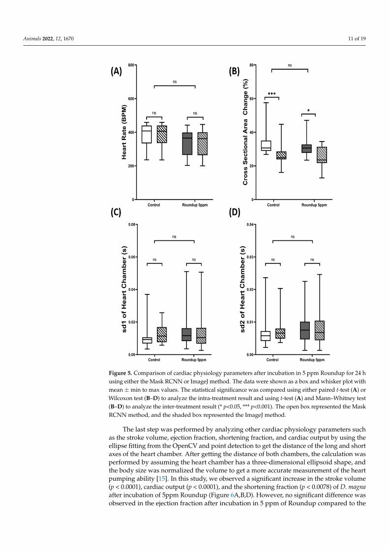
previously reported ImageJ method for the next step. The result showed that no significant difference in heart rate was found in both the control (p = 0.9677) and roundup (p = 0.4982) treatment between deep learning and ImageJ-based methods (Figure 5A). However, after comparing the cross-sectional area change using our developed method and manual counting using ImageJ, we found that the deep learning method predicted heart size was significantly larger than the manual method conducted by ImageJ for both the control (p = 0.003) and Roundup treatment (p = 0.0125) (Figure 5B). We estimated they were around 6–8% bigger when the deep learning method was conducted. In the case of heart rate variability, no significant different was found in the sd1 (Control p = 0.2026 & Roundup p = 0.5144) (Figure 5C) and sd2 (Control p = 0.4458 & Roundup p = 0.3119) (Figure 5D) in both the treatment groups. These results strongly suggest that the newly developed deep learning method can give similar results to the already established ImageJ method except in the cross-sectional area change.
Figure 4. Use Mask RCNN to study cardiac rhythm in water fleas. Three water flea species ofD. magna (pink color), D. pulex (green color), and Moina sp. (blue color) were tested, and the cardiacrhythm and heart size change dynamic can be elucidated by the Mask RCNN method.
3.5. Validation of Cardiac Physiology Alterations in D. magna after Herbicide Exposure
We performed data validation for our newly developed deep learning method andpreviously reported ImageJ method for the next step. The result showed that no significantdifference in heart rate was found in both the control (p = 0.9677) and roundup (p = 0.4982)treatment between deep learning and ImageJ-based methods (Figure 5A). However, aftercomparing the cross-sectional area change using our developed method and manualcounting using ImageJ, we found that the deep learning method predicted heart size wassignificantly larger than the manual method conducted by ImageJ for both the control(p = 0.003) and Roundup treatment (p = 0.0125) (Figure 5B). We estimated they were around6–8% bigger when the deep learning method was conducted. In the case of heart ratevariability, no significant different was found in the sd1 (Control p = 0.2026 & Roundupp = 0.5144) (Figure 5C) and sd2 (Control p = 0.4458 & Roundup p = 0.3119) (Figure 5D) inboth the treatment groups. These results strongly suggest that the newly developed deeplearning method can give similar results to the already established ImageJ method exceptin the cross-sectional area change.
Animals 2022, 12, 1670 11 of 19Animals 2022, 12, x FOR PEER REVIEW 11 of 19
Figure 5. Comparison of cardiac physiology parameters after incubation in 5 ppm Roundup for 24 h using either the Mask RCNN or ImageJ method. The data were shown as a box and whisker plot with mean ± min to max values. The statistical significance was compared using either paired t-test (A) or Wilcoxon test (B–D) to analyze the intra-treatment result and using t-test (A) and Mann–Whitney test (B–D) to analyze the inter-treatment result (* p<0.05, *** p<0.001). The open box represented the Mask RCNN method, and the shaded box represented the ImageJ method.
The last step was performed by analyzing other cardiac physiology parameters such as the stroke volume, ejection fraction, shortening fraction, and cardiac output by using the ellipse fitting from the OpenCV and point detection to get the distance of the long and short axes of the heart chamber. After getting the distance of both chambers, the calculation was performed by assuming the heart chamber has a three-dimensional ellipsoid shape, and the body size was normalized the volume to get a more accurate measurement of the heart pumping ability [15]. In this study, we observed a significant increase in the stroke volume (p < 0.0001), cardiac output (p < 0.0001), and the shortening fraction (p < 0.0078) of D. magna after incubation of 5ppm Roundup (Figure 6A,B,D). However, no significant difference was observed in the ejection fraction after incubation
Figure 5. Comparison of cardiac physiology parameters after incubation in 5 ppm Roundup for 24 husing either the Mask RCNN or ImageJ method. The data were shown as a box and whisker plot withmean ± min to max values. The statistical significance was compared using either paired t-test (A) orWilcoxon test (B–D) to analyze the intra-treatment result and using t-test (A) and Mann–Whitney test(B–D) to analyze the inter-treatment result (* p<0.05, *** p<0.001). The open box represented the MaskRCNN method, and the shaded box represented the ImageJ method.
The last step was performed by analyzing other cardiac physiology parameters suchas the stroke volume, ejection fraction, shortening fraction, and cardiac output by using theellipse fitting from the OpenCV and point detection to get the distance of the long and shortaxes of the heart chamber. After getting the distance of both chambers, the calculation wasperformed by assuming the heart chamber has a three-dimensional ellipsoid shape, andthe body size was normalized the volume to get a more accurate measurement of the heartpumping ability [15]. In this study, we observed a significant increase in the stroke volume(p < 0.0001), cardiac output (p < 0.0001), and the shortening fraction (p < 0.0078) of D. magnaafter incubation of 5ppm Roundup (Figure 6A,B,D). However, no significant difference wasobserved in the ejection fraction after incubation in 5 ppm of Roundup compared to the

Animals 2022, 12, 1670 12 of 19
control, which suggests that at 5 ppm concentration, Roundup can increase the heart sizeof D. magna without significantly changing the pumping capability of the heart chamber.
Animals 2022, 12, x FOR PEER REVIEW 12 of 19
in 5 ppm of Roundup compared to the control, which suggests that at 5 ppm concentration, Roundup can increase the heart size of D. magna without significantly changing the pumping capability of the heart chamber.
Figure 6. Comparison of cardiac physiology parameters after incubation in 5 ppm of Roundup for 24 h. The heart size was first predicted by Mask RCNN, and later the long and short axes of the cardiac chamber was predicted by OpenCV. Next, we used long and short axis lengths to assess cardiac physiology by measuring stroke volume (A), cardiac output (B), shortening fraction (C), and ejection fraction (D). The data were shown as mean ± SD, and the statistical significance was compared using an unpaired student t-test (** p<0.01, **** p<0.0001).
Figure 6. Comparison of cardiac physiology parameters after incubation in 5 ppm of Roundup for24 h. The heart size was first predicted by Mask RCNN, and later the long and short axes of thecardiac chamber was predicted by OpenCV. Next, we used long and short axis lengths to assesscardiac physiology by measuring stroke volume (A), cardiac output (B), shortening fraction (C),and ejection fraction (D). The data were shown as mean ± SD, and the statistical significance wascompared using an unpaired student t-test (** p < 0.01, **** p < 0.0001).
4. Discussion
Several methods currently available in the literature may be suitable for marking theedge of the heart chamber. In this study, U-Net [21,22] and Mask RCNN [23] methods

Animals 2022, 12, 1670 13 of 19
were adopted to conduct cardiac size prediction in water fleas as those two tools have beensuccessfully reported to perform heart segmentation in humans [50–53]. We discovered thatthe heart chamber detection in water fleas using Mask RCNN shows superior performancein comparison to U-Net. U-net is a generic deep-learning solution for image detectionand segmentation and can be used for biomedical image data analysis. U-Net uses theU-shaped network structure first to capture the features of the images and reconstruct therequired partitions based on these features [22]. The U-Net architecture contains a U shapepath. The image enters one end of the U shape network to go through the encoding partof the network (also called the contracting path). The encoding part is used to capturethe features of the input images, and during this step, the spatial information is reducedwhile the feature information is increased. Then the information of the input image is sentto the decoding part (also called the expansive path). At the expansive path, the featurecollected during the contracting step was combined through a high-resolution sequence ofup-convolutions and concatenations to construct the segmentation image [22]. Therefore,U-Net’s method of fusing low-level and high-level image features may have a chance tomark the heart chamber’s edge successfully.
Unlike U-Net, Mask RCNN was used to solve instance segmentation problems incomputer vision. Mask RCNN consists of two stages. The first step is to propose theregion which contains the object based on the input image. Then in the second stage, it willpredict the class of object, make the boundaries of the object, and generate a mask at pixellevel based on the proposal from the first stage [54]. Compared to U-Net, which performsbetter on semantic segmentation, Mask RCNN performs better than U-net, for instance, insegmentation [55]. Several studies suggest that the combination of Mask RCNN and U-Netcan outperform a single component only, which could be explored more to increase theaccuracy of the image segmentation [56–58].
Our training result suggests that the Mask-RCNN model in this study performs betterthan the U-Net model in defining the heart chamber boundary. A similar case happenedwhen both models were challenged to perform nuclei segmentation, which shows thatMask-RCNN has better precision than U-Net [59]. Another study also reported that U-Netyielded more false-positive results when detecting the immunofluorescence images of kidneybiopsies from lupus nephritis patients than Mask-RCNN [60]. Although U-net has higheraccuracy, the limitation of U-Net observed in the study was the obstruction of detection whenlabeled data has edge noise, thus yielding false-positive segmentation [59,61]. However, itcan be said that both U-Net and Mask-RCNN models might have similar outputs if thetraining dataset has more viable point boundary determination. In conclusion, the accuracyand effectiveness of each model depend upon the targeted images that are being classifiedand segmented.
Another thing that could be observed in this study is the significant difference betweenmanual counting using ImageJ and the Mask RCNN-based method to calculate the cross-section area change of the cardiac chamber. This might be because by using Mask RCNN,the calculation of the cross-sectional area change was based on the maximum and minimumcross-sectional area of the heart chamber from the whole cardiac cycle recorded in the video.On the contrary, the calculation was only based on randomly selected images from a fewcardiac cycles for manual counting. Thus, the calculation using manual counting has alower cross-sectional area change than the automated Mask RCNN method.
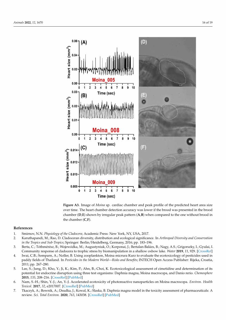
It is also worth noting that some limitations were noticed in this deep learning-basedstudy. As the developed tool was based on detecting the edge of the heart chamber, theclarity of the heart chamber edge plays a crucial role in the accuracy of cardiac chamberprediction. In the case of Daphnia and Moina sp., the brood chamber was located rightbeside the heart chamber. Thus if the samples are in the breeding period, the accuracy ofthe tools would be compromised when the brood was positioned overlapping the heartchamber (Figure A4 & Video S7) [62]. Another type of limitation is the presence of someparasites near the heart chamber. This problem could also reduce the clarity of the heartchamber, making it harder for deep learning to analyze the heart chamber [63]. Compared

Animals 2022, 12, 1670 14 of 19
to the ImageJ-based method in which the selection of ROI can be made anywhere as longas it has distinct dynamic pixel change, our tool only works accurately if the whole heartchamber can be detected. However, this problem can be overcome by selecting the waterfleas with an empty brood chamber or those which are parasite free, ensuring that heartchamber edge is undisturbed.
5. Conclusions
In conclusion, it can be said that Mask-RCNN performed better than U-Net in esti-mating D. magna cardiac chamber size. Higher Dice coefficient, specificity, and sensitivityshow that the Mask-RCNN model was superior in Daphnia cardiac chamber segmentation.Furthermore, the segmentation result could also be used to calculate several cardiac perfor-mance parameters such as heart rate, heart rate variability, and cross-sectional area change,while with the addition of the ellipse fitting function from OpenCV, more parameters suchas stroke volume, cardiac output, shortening fraction, and ejection fraction could also becalculated. Several limitations such as the dependency on the heart chamber clarity andthe presence of obstruction nearby the heart chamber could decrease the accuracy of thesegmentation, but both problems could be solved by selecting and sorting the Daphniabefore recording the heart chamber. Overall, this study suggests that deep learning couldhelp analyze cardiac physiological parameters in Daphnia. Although in this study, fullyautomated analysis was not yet achieved, in the future, fully automated high throughputanalysis could be achieved by incorporating essential function in several software by usingour trained network as the foundation.
Supplementary Materials: The following supporting information can be downloaded at: https://www.mdpi.com/article/10.3390/ani12131670/s1. File S1. Python script used to calculate heartsize of water fleas by Mask-RCNN. File S2. Python script used to calculate heart size of water fleas byU-Net. File S3. Python script used to calculate the length of major and minor axis of heart in waterfleas. Video S1. D. magna heartbeat video deposited at YouTube: https://www.youtube.com/watch?v=NwB-Hyf47qo (accessed on 29 July 2021). Video S2. D. pulex heartbeat video deposited at YouTube:https://www.youtube.com/watch?v=jTCGQ8yzZbM (accessed on 29 July 2021). Video S3. Moina sp.heartbeat video deposited at YouTube: https://www.youtube.com/watch?v=LjX1xQXG38I (accessedon 29 July 2021). Video S4. D. magna heartbeat video tracked with Mask RCNN deposited at YouTube:https://www.youtube.com/watch?v=DBuLshjuMTU (accessed on 29 July 2021), Video S5. D. pulexheartbeat video tracked with Mask RCNN deposited at YouTube https://www.youtube.com/watch?v=RGf5naSgcVE (accessed on 29 July 2021), Video S6. Moina sp. heartbeat video tracked with MaskRCNN deposited at YouTube https://www.youtube.com/watch?v=RVRKXvpFq74 (accessed on 29July 2021). Video S7. Bias for heart size prediction in Moina sp. by Mask RCNN. In this video, theheart size prediction power will be interfered by embryos in the breeding chamber due to loss ofsharp edge of heart. Video has been deposited at YouTube https://www.youtube.com/watch?v=MoRyMSYCNNY (accessed on 29 July 2021).
Author Contributions: Conceptualization, F.S., J.-C-H., Y.-K.L. and C.-D.H.; methodology, A.F.,M.E.S. and K.A.K.; software, Y.-K.L.; validation, M.E.S., K.A.K., R.D.V. and M.J.M.R.; formal analysis,K.H.-C.C. and J.-C.H.; investigation, F.S.; resources, K.H.-C.C.; writing—original draft preparation,F.S. and C.-D.H.; visualization, C.-D.H.; supervision and funding acquisition, J.-C.H. and C.-D.H. Allauthors have read and agreed to the published version of the manuscript.
Funding: This research was funded by the Ministry of Science Technology, Taiwan, grant numbersMOST107-2622-B-033-001-CC2 and MOST108-2622-B-033-001-CC2 to C.-D.H.
Institutional Review Board Statement: All protocols and procedures involving Daphnia were ap-proved by the Committee for Animal Experimentation of the Chung Yuan Christian University(Approval No. 109001, issue date 15 January 2020).
Informed Consent Statement: Not applicable.
Data Availability Statement: The data presented in this study are available on request from thecorresponding author.

Animals 2022, 12, 1670 15 of 19
Conflicts of Interest: The authors declare no conflict of interest. The funders had no role in the designof the study; in the collection, analyses, or interpretation of data, in the writing of the manuscript, orin the decision to publish the results.
Appendix A
Animals 2022, 12, x FOR PEER REVIEW 15 of 19
Data Availability Statement: The data presented in this study are available on request from the corresponding author.
Conflicts of Interest: The authors declare no conflict of interest. The funders had no role in the design of the study; in the collection, analyses, or interpretation of data, in the writing of the manuscript, or in the decision to publish the results.
Appendix A
Figure A1. The image of the heart chamber of D. magna before (A) and after (B) manual marked with red color line.
Figure A2. Detection of the short (minor) and long (major) axes of the heart chamber using fit ellipse function in OpenCV. The heart images at either diastolic or systolic phases were used for long and short axis length extraction. The binary images before (left panel) and after (right panel) performing
Figure A1. The image of the heart chamber of D. magna before (A) and after (B) manual marked withred color line.
Animals 2022, 12, x FOR PEER REVIEW 15 of 19
Data Availability Statement: The data presented in this study are available on request from the corresponding author.
Conflicts of Interest: The authors declare no conflict of interest. The funders had no role in the design of the study; in the collection, analyses, or interpretation of data, in the writing of the manuscript, or in the decision to publish the results.
Appendix A
Figure A1. The image of the heart chamber of D. magna before (A) and after (B) manual marked with red color line.
Figure A2. Detection of the short (minor) and long (major) axes of the heart chamber using fit ellipse function in OpenCV. The heart images at either diastolic or systolic phases were used for long and short axis length extraction. The binary images before (left panel) and after (right panel) performing
Figure A2. Detection of the short (minor) and long (major) axes of the heart chamber using fitellipse function in OpenCV. The heart images at either diastolic or systolic phases were used forlong and short axis length extraction. The binary images before (left panel) and after (right panel)performing the Cv2.fitEllipse function in OpenCV were listed for side-by-side comparison. Scale barwas provided for size estimation.
Animals 2022, 12, 1670 16 of 19
Animals 2022, 12, x FOR PEER REVIEW 16 of 19
the Cv2.fitEllipse function in OpenCV were listed for side-by-side comparison. Scale bar was provided for size estimation.
Figure A3. The image of Moina sp. shows the limitation of the tool. The heart chamber detection accuracy was lower if the brood was presented in the brood chamber (A,B) compared to the one without brood in the chamber (C), (D,E,F) brood chamber.
References 1. Smirnov, N.N. Physiology of the Cladocera; Academic Press: New York, NY, USA, 2017. 2. Karuthapandi, M.; Rao, D. Cladoceran diversity, distribution and ecological significance. In Arthropod Diversity and Conservation
in the Tropics and Sub-Tropics, Springer: Berlin/Heidelberg, Germany, 2016; pp. 183–196. 3. Berta, C.; Tóthmérész, B.; Wojewódka, M.; Augustyniuk, O.; Korponai, J.; Bertalan-Balázs, B.; Nagy, A.S.; Grigorszky, I.; Gyulai,
I. Community response of cladocera to trophic stress by biomanipulation in a shallow oxbow lake. Water 2019, 11, 929. 4. Iwai, C.B.; Somparn, A.; Noller, B. Using zooplankton, Moina micrura Kurz to evaluate the ecotoxicology of pesticides used in
paddy fields of Thailand. In Pesticides in the Modern World—Risks and Benefits; INTECH Open Access Publisher: Rijeka, Croatia, 2011; pp. 267–280.
5. Lee, S.; Jung, D.; Kho, Y.; Ji, K.; Kim, P.; Ahn, B.; Choi, K. Ecotoxicological assessment of cimetidine and determination of its potential for endocrine disruption using three test organisms: Daphnia magna, Moina macrocopa, and Danio rerio. Chemosphere 2015, 135, 208–216.
Figure A3. Image of Moina sp. cardiac chamber and peak profile of the predicted heart area sizeover time. The heart chamber detection accuracy was lower if the brood was presented in the broodchamber (D,E) shown by irregular peak pattern (A,B) when compared to the one without brood inthe chamber (C,F).
References1. Smirnov, N.N. Physiology of the Cladocera; Academic Press: New York, NY, USA, 2017.2. Karuthapandi, M.; Rao, D. Cladoceran diversity, distribution and ecological significance. In Arthropod Diversity and Conservation
in the Tropics and Sub-Tropics; Springer: Berlin/Heidelberg, Germany, 2016; pp. 183–196.3. Berta, C.; Tóthmérész, B.; Wojewódka, M.; Augustyniuk, O.; Korponai, J.; Bertalan-Balázs, B.; Nagy, A.S.; Grigorszky, I.; Gyulai, I.
Community response of cladocera to trophic stress by biomanipulation in a shallow oxbow lake. Water 2019, 11, 929. [CrossRef]4. Iwai, C.B.; Somparn, A.; Noller, B. Using zooplankton, Moina micrura Kurz to evaluate the ecotoxicology of pesticides used in
paddy fields of Thailand. In Pesticides in the Modern World—Risks and Benefits; INTECH Open Access Publisher: Rijeka, Croatia,2011; pp. 267–280.
5. Lee, S.; Jung, D.; Kho, Y.; Ji, K.; Kim, P.; Ahn, B.; Choi, K. Ecotoxicological assessment of cimetidine and determination of itspotential for endocrine disruption using three test organisms: Daphnia magna, Moina macrocopa, and Danio rerio. Chemosphere2015, 135, 208–216. [CrossRef] [PubMed]
6. Nam, S.-H.; Shin, Y.-J.; An, Y.-J. Accelerated ecotoxicity of photoreactive nanoparticles on Moina macrocopa. Environ. HealthToxicol. 2017, 32, e2017007. [CrossRef] [PubMed]
7. Tkaczyk, A.; Bownik, A.; Dsudka, J.; Kowal, K.; Slaska, B. Daphnia magna model in the toxicity assessment of pharmaceuticals: Areview. Sci. Total Environ. 2020, 763, 143038. [CrossRef] [PubMed]

Animals 2022, 12, 1670 17 of 19
8. Harris, K.D.; Bartlett, N.J.; Lloyd, V.K. Daphnia as an emerging epigenetic model organism. Genet. Res. Int. 2012, 2012, 147892.[CrossRef]
9. de Oliveira, L.L.D.; Antunes, S.C.; Gonçalves, F.; Rocha, O.; Nunes, B. Acute and chronic ecotoxicological effects of fourpharmaceuticals drugs on cladoceran Daphnia magna. Drug Chem. Toxicol. 2016, 39, 13–21. [CrossRef]
10. Pociecha, A.; Wojtal, A.Z.; Szarek-Gwiazda, E.; Cieplok, A.; Ciszewski, D.; Kownacki, A. Response of Cladocera fauna to heavymetal pollution, based on sediments from subsidence ponds downstream of a mine discharge (S. Poland). Water 2019, 11, 810.[CrossRef]
11. Sarma, S.; Nandini, S. Review of recent ecotoxicological studies on cladocerans. J. Environ. Sci. Health Part B 2006, 41, 1417–1430.[CrossRef]
12. Suhett, A.L.; Santangelo, J.M.; Bozelli, R.L.; Steinberg, C.E.W.; Farjalla, V.F. An overview of the contribution of studies withcladocerans to environmental stress research. Acta Limnol. Bras. 2015, 27, 145–159. [CrossRef]
13. [OECD] Organization for Economic Co-operation and Development. Guideline for testing of chemicals. Daphnia sp., Acuteimmobilisation test. In OECD Guidel. No 202; OECD: Paris, France, 2004.
14. Peake, B.M.; Braund, R.; Tong, A.Y.; Tremblay, L.A. Impact of pharmaceuticals on the environment. In The Life-Cycle ofPharmaceuticals in the Environment; Peake, B.M., Braund, R., Tong, A.Y.C., Tremblay, L.A., Eds.; Woodhead Publishing: Sawston,UK, 2016; pp. 109–152.
15. Santoso, F.; Krylov, V.V.; Castillo, A.L.; Saputra, F.; Chen, H.-M.; Lai, H.-T.; Hsiao, C.-D. Cardiovascular performance measurementin water fleas by utilizing high-speed videography and imagej software and its application for pesticide toxicity assessment.Animals 2020, 10, 1587. [CrossRef]
16. Bekker, J.M.; Krijgsman, B. Physiological investigations into the heart function of Daphnia. J. Physiol. 1951, 115, 249. [CrossRef][PubMed]
17. Bownik, A. Physiological endpoints in daphnid acute toxicity tests. Sci. Total Environ. 2020, 700, 134400. [CrossRef] [PubMed]18. Ahmed, B.; Gulliver, T.A.; Alzahir, S. Image splicing detection using mask-RCNN. Signal Image Video Process. 2020, 14, 1035–1042.
[CrossRef]19. Pu, Y.; Gan, Z.; Henao, R.; Yuan, X.; Li, C.; Stevens, A.; Carin, L. Variational autoencoder for deep learning of images, labels and
captions. Adv. Neural Inf. Process. Syst. 2016, 29, 2352–2360.20. Chen, L.-C.; Papandreou, G.; Kokkinos, I.; Murphy, K.; Yuille, A.L. Deeplab: Semantic image segmentation with deep convolu-
tional nets, atrous convolution, and fully connected crfs. IEEE Trans. Pattern Anal. Mach. Intell. 2017, 40, 834–848. [CrossRef]21. Falk, T.; Mai, D.; Bensch, R.; Çiçek, Ö.; Abdulkadir, A.; Marrakchi, Y.; Böhm, A.; Deubner, J.; Jäckel, Z.; Seiwald, K. U-Net: Deep
learning for cell counting, detection, and morphometry. Nat. Methods 2019, 16, 67–70. [CrossRef]22. Ronneberger, O.; Fischer, P.; Brox, T. U-net: Convolutional networks for biomedical image segmentation. In International Conference
on Medical image Computing and Computer-Assisted Intervention, Proceedings of the 18th International Conference, Munich, Germany, 5–9October 2015; Springer: Cham, Switzerland, 2015; pp. 234–241.
23. Kido, S.; Hirano, Y.; Hashimoto, N. Detection and classification of lung abnormalities by use of convolutional neural network(CNN) and regions with CNN features (R-CNN). In Proceedings of the 2018 International Workshop on Advanced ImageTechnology (IWAIT), Chiang Mai, Thailand, 7–9 January 2018; pp. 1–4.
24. Chang, Y.; Song, B.; Jung, C.; Huang, L. Automatic segmentation and cardiopathy classification in cardiac mri images based ondeep neural networks. In Proceedings of the 2018 IEEE International Conference on Acoustics, Speech and Signal Processing(ICASSP), Calgary, AB, Canada, 15–20 April 2018; pp. 1020–1024.
25. Ünver, H.M.; Ayan, E. Skin lesion segmentation in dermoscopic images with combination of YOLO and grabcut algorithm.Diagnostics 2019, 9, 72. [CrossRef]
26. He, P.; Zuo, L.; Zhang, C.; Zhang, Z. A value recognition algorithm for pointer meter based on improved Mask-RCNN. InProceedings of the 2019 9th International Conference on Information Science and Technology (ICIST), Hulunbuir, China, 2–5August 2019; pp. 108–113.
27. Karatzas, P.; Melagraki, G.; Ellis, L.J.A.; Lynch, I.; Varsou, D.D.; Afantitis, A.; Tsoumanis, A.; Doganis, P.; Sarimveis, H.Development of deep learning models for predicting the effects of exposure to engineered nanomaterials on Daphnia Magna.Small 2020, 16, 2001080. [CrossRef]
28. Zhao, M.; Wei, Y.; Lu, Y.; Wong, K.K. A novel U-Net approach to segment the cardiac chamber in magnetic resonance images withghost artifacts. Comput. Methods Programs Biomed. 2020, 196, 105623. [CrossRef]
29. Dong, S.; Zhao, J.; Zhang, M.; Shi, Z.; Deng, J.; Shi, Y.; Tian, M.; Zhuo, C. DeU-Net: Deformable U-Net for 3D Cardiac MRI VideoSegmentation. In International Conference on Medical Image Computing and Computer-Assisted Intervention, Proceedings of the 23rdInternational Conference, Lima, Peru, 4–8 October 2020; Springer: Cham, Switzerland, 2020; pp. 98–107.
30. Silva, C.E.S.; Ferreira, L.D.C.; Peixoto, L.B.; Monaco, C.G.; Gil, M.A.; Ortiz, J. Study of the myocardial contraction and relaxationvelocities through Doppler tissue imaging echocardiography: A new alternative in the assessment of the segmental ventricularfunction. Arq. Bras. Cardiol. 2002, 78, 206–211. [CrossRef]
31. Baylor, E. Cardiac pharmacology of the cladoceran, Daphnia. Biol. Bull. 1942, 83, 165–172. [CrossRef]32. Villegas-Navarro, A.; Rosas, L.E.; Reyes, J.L. The heart of Daphnia magna: Effects of four cardioactive drugs. Comp. Biochem.
Physiol. Part C Toxicol. Pharmacol. 2003, 136, 127–134. [CrossRef]

Animals 2022, 12, 1670 18 of 19
33. FernáNdez-GonzáLez, M.A.; Gonzalez-Barrientos, J.; Carter, M.J.; Ramos-Jiliberto, R. Parent-to-offspring transfer of sublethaleffects of copper exposure: Metabolic rate and life-history traits of Daphnia. Rev. Chil. Hist. Nat. 2011, 84, 195–201. [CrossRef]
34. Mishra, A.; Shukla, S.; Chopra, A. Physiological responses of heart of tailless fresh water flea Simocephalus vetulus (Crustacea-cladocera) under copper sulphate stress. CIBTech J. Zool 2016, 5, 52–59.
35. Bownik, A. Protective effects of ectoine on physiological parameters of Daphnia magna subjected to clove oil-induced anaesthesia.Turk. J. Fish. Aquat. Sci. 2016, 16, 691–701. [CrossRef]
36. Jeong, T.-Y.; Asselman, J.; De Schamphelaere, K.A.; Van Nieuwerburgh, F.; Deforce, D.; Kim, S.D. Effect of β-adrenergic receptoragents on cardiac structure and function and whole-body gene expression in Daphnia magna. Environ. Pollut. 2018, 241, 869–878.[CrossRef]
37. Kurnia, K.A.; Saputra, F.; Roldan, M.J.M.; Castillo, A.L.; Huang, J.-C.; Chen, K.H.-C.; Lai, H.-T.; Hsiao, C.-D. Measurement ofMultiple Cardiac Performance Endpoints in Daphnia and Zebrafish by Kymograph. Inventions 2021, 6, 8. [CrossRef]
38. Farhan, A.; Kurnia, K.A.; Saputra, F.; Chen, K.H.-C.; Huang, J.-C.; Roldan, M.J.M.; Lai, Y.-H.; Hsiao, C.-D. An OpenCV-BasedApproach for Automated Cardiac Rhythm Measurement in Zebrafish from Video Datasets. Biomolecules 2021, 11, 1476. [CrossRef]
39. Rueden, C.T.; Schindelin, J.; Hiner, M.C.; DeZonia, B.E.; Walter, A.E.; Arena, E.T.; Eliceiri, K.W. ImageJ2: ImageJ for the nextgeneration of scientific image data. BMC Bioinform. 2017, 18, 529. [CrossRef]
40. Frid-Adar, M.; Klang, E.; Amitai, M.; Goldberger, J.; Greenspan, H. Synthetic data augmentation using GAN for improvedliver lesion classification. In Proceedings of the 2018 IEEE 15th International Symposium on Biomedical Imaging (ISBI 2018),Washington, DC, USA, 4–7 April 2018; pp. 289–293.
41. Shorten, C.; Khoshgoftaar, T.M. A survey on image data augmentation for deep learning. J. Big Data 2019, 6, 60. [CrossRef]42. Buslaev, A.; Iglovikov, V.I.; Khvedchenya, E.; Parinov, A.; Druzhinin, M.; Kalinin, A.A. Albumentations: Fast and flexible image
augmentations. Information 2020, 11, 125. [CrossRef]43. Jin, S.; Zedong, H.; Yuan, L. Software implementation of corn grain morphology detection based on OpenCV. In Proceedings of
the 2017 13th IEEE International Conference on Electronic Measurement & Instruments (ICEMI), Yangzhou, China, 20–22 October2017; pp. 412–415.
44. Hart, K.A.; Rimoli, J.J. MicroStructPy: A statistical microstructure mesh generator in Python. SoftwareX 2020, 12, 100595.[CrossRef]
45. Marengoni, M.; Stringhini, D. High level computer vision using opencv. In Proceedings of the 2011 24th SIBGRAPI Conference onGraphics, Patterns, and Images Tutorials, Alagoas, Brazil, 28–30 August 2011; pp. 11–24.
46. Zhang, Z.; Ryoo, D.; Balusek, C.; Acharya, A.; Rydmark, M.O.; Linke, D.; Gumbart, J.C. Inward-facing glycine residues createsharp turns in β-barrel membrane proteins. Biochim. Biophys. Acta (BBA)-Biomembr. 2021, 1863, 183662. [CrossRef] [PubMed]
47. Paszke, A.; Gross, S.; Chintala, S.; Chanan, G.; Yang, E.; DeVito, Z.; Lin, Z.; Desmaison, A.; Antiga, L.; Lerer, A. AutomaticDifferentiation in Pytorch. Available online: https://openreview.net/pdf?id=BJJsrmfCZ (accessed on 28 June 2022).
48. MacFarland, T.W.; Yates, J.M. Introduction to Nonparametric Statistics for the Biological Sciences Using R; Springer InternationalPublishing: Cham, Switzerland, 2016.
49. Watt, T.A.; McCleery, R.H.; Hart, T. Introduction to Statistics for Biology; CRC Press: Boca Raton, FL, USA, 2007.50. Liu, T.; Tian, Y.; Zhao, S.; Huang, X.; Wang, Q. Automatic whole heart segmentation using a two-stage u-net framework and an
adaptive threshold window. IEEE Access 2019, 7, 83628–83636. [CrossRef]51. Shu, J.-H.; Nian, F.-D.; Yu, M.-H.; Li, X. An improved mask R-CNN model for multiorgan segmentation. Math. Probl. Eng. 2020,
2020, 8351725. [CrossRef]52. Tong, Q.; Ning, M.; Si, W.; Liao, X.; Qin, J. 3D deeply-supervised U-net based whole heart segmentation. In International Workshop
on Statistical Atlases and Computational Models of the Heart, Proceedings of the 8th International Workshop, STACOM 2017, Held inConjunction with MICCAI 2017, Quebec City, Canada, 10–14 September 2017; Springer: Cham, Switzerland, 2017; pp. 224–232.
53. Zhang, J.; Du, J.; Liu, H.; Hou, X.; Zhao, Y.; Ding, M. LU-NET: An Improved U-Net for ventricular segmentation. IEEE Access2019, 7, 92539–92546. [CrossRef]
54. He, K.; Gkioxari, G.; Dollár, P.; Girshick, R. Mask r-cnn. In Proceedings of the 2017 IEEE International Conference on ComputerVision, Venice, Italy, 22–29 October 2017; pp. 2961–2969.
55. Zhang, X.; An, G.; Liu, Y. Mask R-CNN with feature pyramid attention for instance segmentation. In Proceedings of the 2018 14thIEEE International Conference on Signal Processing (ICSP), Beijing, China, 12–16 August 2018; pp. 1194–1197.
56. Dogan, R.O.; Dogan, H.; Bayrak, C.; Kayikcioglu, T. A Two-Phase Approach using Mask R-CNN and 3D U-Net for High-AccuracyAutomatic Segmentation of Pancreas in CT Imaging. Comput. Methods Programs Biomed. 2021, 207, 106141. [CrossRef]
57. Konopczynski, T.; Heiman, R.; Woznicki, P.; Gniewek, P.; Duvernoy, M.-C.; Hallatschek, O.; Hesser, J. Instance Segmentation ofDensely Packed Cells Using a Hybrid Model of U-Net and Mask R-CNN. In International Conference on Artificial Intelligence andSoft Computing, Proceedings of the19th International Conference, ICAISC 2020, Zakopane, Poland, 12–14 October 2020; Springer: Cham,Switzerland, 2020; pp. 626–635.
58. Xu, Z.; Wu, Z.; Feng, J. CFUN: Combining faster R-CNN and U-net network for efficient whole heart segmentation. arXiv 2018,arXiv:1812.04914.
59. Vuola, A.O.; Akram, S.U.; Kannala, J. Mask-RCNN and U-net ensembled for nuclei segmentation. In Proceedings of the 2019IEEE 16th International Symposium on Biomedical Imaging (ISBI 2019), Venice, Italy, 8–11 April 2019; pp. 208–212.

Animals 2022, 12, 1670 19 of 19
60. Durkee, M.S.; Abraham, R.; Ai, J.; Fuhrman, J.D.; Clark, M.R.; Giger, M.L. Comparing Mask R-CNN and U-Net architectures forrobust automatic segmentation of immune cells in immunofluorescence images of Lupus Nephritis biopsies. In Proceedingsof the Imaging, Manipulation, and Analysis of Biomolecules, Cells, and Tissues XIX, Online Only, CA, USA, 6–12 March2021; p. 116470X.
61. Quoc, T.T.P.; Linh, T.T.; Minh, T.N.T. Comparing U-Net Convolutional Network with Mask R-CNN in Agricultural AreaSegmentation on Satellite Images. In Proceedings of the 2020 7th NAFOSTED Conference on Information and Computer Science(NICS), Ho Chi Minh City, Vietnam, 26–27 November 2020; pp. 124–129.
62. Campbell, A.K.; Wann, K.T.; Matthews, S.B. Lactose causes heart arrhythmia in the water flea Daphnia pulex. Comp. Biochem.Physiol. Part B Biochem. Mol. Biol. 2004, 139, 225–234. [CrossRef]
63. Ebert, D. Ecology, Epidemiology, and Evolution of Parasitism in Daphnia; National Library of Medicine: Bethesda, MD, USA, 2005.