Thromboxane prostanoid receptor stimulation induces shedding of the transmembrane chemokine CX 3 CL1...
12
Thromboxane prostanoid receptor stimulation induces shedding of the transmembrane chemokine CX 3 CL1 yet enhances CX 3 CL1-dependent leukocyte adhesion Soumitra Tole,* Anne M. Durkan,* Yi-Wei Huang, Guang Ying Liu, Alexander Leung, Laura L. Jones, Jasmine A. Taylor, and Lisa A. Robinson The Hospital for Sick Children Research Institute, Toronto, Ontario, Canada Submitted 21 August 2009; accepted in final form 12 March 2010 Tole S, Durkan AM, Huang YW, Liu GY, Leung A, Jones LL, Taylor JA, Robinson LA. Thromboxane prostanoid receptor stimu- lation induces shedding of the transmembrane chemokine CX3 CL1 yet enhances CX3 CL1-dependent leukocyte adhesion. Am J Physiol Cell Physiol 298: C1469 –C1480, 2010. First published March 17, 2010; doi:10.1152/ajpcell.00380.2009.—In atherosclerosis, chemo- kines recruit circulating mononuclear leukocytes to the vascular wall. A key factor is CX3 CL1, a chemokine with soluble and transmem- brane species that acts as both a chemoattractant and an adhesion molecule. Thromboxane A2 and its receptor, TP, are also critical to atherogenesis by promoting vascular inflammation and consequent leukocyte recruitment. We examined the effects of TP stimulation on processing and function of CX3 CL1, using CX 3 CL1-expressing hu- man ECV-304 cells and primary human vascular endothelial cells. TP agonists promoted rapid shedding of cell surface CX3 CL1, which was inhibited by pharmacological inhibitors or specific small interfering RNA targeting tumor necrosis factor--converting enzyme (TACE). Because it reduced cell surface CX3 CL1, we predicted that TP stimulation would inhibit adhesion of leukocytes expressing the CX3 CL1 cognate receptor but, paradoxically, saw enhanced adhesion. We questioned whether the enhanced ability of the remaining mem- brane-associated CX3 CL1 to bind targets was caused by changes in its lateral mobility. Using fluorescence recovery after photobleaching, we found that plasmalemmal CX3 CL1 was initially tethered but ulti- mately mobilized by TP agonists. TP stimulation provoked clustering of transmembrane CX3 CL1 at sites of contact with adherent leuko- cytes. These data demonstrate that TP stimulation induces two distinct effects: a rapid cleavage of surface CX3 CL1, thereby releasing the soluble chemoattractant, plus mobilization of the remaining trans- membrane CX3 CL1 to enhance the avidity of interactions with ad- herent leukocytes. The dual effect of TP allows CX3 CL1 to recruit leukocytes to sites of vascular inflammation while enhancing their adhesion once recruited. leukocytes; inflammation; endothelial cells ATHEROSCLEROSIS is a chronic inflammatory disease involving the vascular wall, leukocytes, and platelets. Thromboxane A 2 (TXA 2 ), a prostanoid formed from the metabolism of arachi- donic acid via the cyclooxygenase (COX) pathway, has long been implicated in the pathogenesis of atherosclerosis and induces platelet aggregation, vasoconstriction, and vascular smooth muscle cell (VSMC) proliferation (42). It is produced mainly by platelets but also by monocytes/macrophages, VSMC, and endothelial cells (37, 41, 42). TXA 2 mediates its effects through the thromboxane prostanoid (TP) receptor, a G protein-coupled receptor expressed in diverse hematopoietic cell types, including platelets and monocytes, as well as in endothelial cells, fibroblasts, and VSMC (15, 41). COX inhib- itors, which decrease TXA 2 production, protect against vascu- lar inflammation and acute vascular events in patients at high risk (3). Mice deficient in TP or treated with a TP antagonist show markedly decreased leukocyte accumulation within the vascular wall (5, 22). In atherogenesis, chemokines recruit circulating leukocytes and other cells to the injured vascular wall. The chemokine CX 3 CL1 (fractalkine) plays a key role by recruiting mono- cytes, T lymphocytes, and other inflammatory cells (6, 24, 28, 29, 32, 40). Most chemokines are small, secreted proteins. CX 3 CL1 is one of only two chemokines that have soluble and membrane-anchored species (2, 10, 27). Soluble CX 3 CL1 consists of a chemokine domain and mucin stalk and is re- leased from the plasma membrane following cleavage of the full-length protein by ADAM (a disintegrin and metallopro- tease)-10 and ADAM17/TACE (tumor necrosis factor--con- verting enzyme) metalloproteases (12, 17, 43). The soluble species thus generated is chemoattractant for monocytes/mac- rophages, natural killer cells, T lymphocytes, and VSMC, all of which express CX 3 CR1, the unique receptor for CX 3 CL1 (10, 18, 26). CX 3 CL1-CX 3 CR1-mediated cellular traffic is critically im- plicated in coronary atherosclerosis and stroke. In affected coronary arteries, CX 3 CL1 protein expression is increased throughout the vascular wall in endothelium, intima, media, and adventitia (26, 45, 47). CX 3 CL1 is also expressed in macrophages, foam cells, and VSMC and acts to recruit mono- cytes, memory T lymphocytes, and additional VSMC to the injured arterial wall (26, 45, 47). Thus, within the atheroscle- rotic lesion, CX 3 CL1 is expressed by and can recruit cells capable of secreting large amounts of extracellular matrix proteins, thereby favoring formation of stable atherosclerotic plaques (10, 18, 47). By recruiting monocytes to the injured vessels, CX 3 CL1 promotes formation of macrophage-rich, un- stable plaques. Genetic targeting of CX 3 CL1 or CX 3 CR1 in atherosclerosis-prone mice highly protects them from vascular disease (6, 24, 40). In humans, gene polymorphisms in CX 3 CR1 that impair leukocyte chemotaxis and binding of the receptor to cell surface CX 3 CL1 protect against myocardial infarction, unstable angina, cardiac death, and stroke (13, 28, 32). It is widely recognized that important links exist between the thrombotic events and the vascular inflammation that define atherosclerosis. The precise nature of these links has not yet been well elucidated. TXA 2 , acting through TP, potently in- * S. Tole and A. M. Durkan contributed equally to this work. Address for reprint requests and other correspondence: L. A. Robinson, 555 Univ. Ave., Rm. 5264, Toronto, Ontario, Canada M5G 1X8 (e-mail: [email protected]). Am J Physiol Cell Physiol 298: C1469–C1480, 2010. First published March 17, 2010; doi:10.1152/ajpcell.00380.2009. 0363-6143/10 Copyright © 2010 the American Physiological Society http://www.ajpcell.org C1469
-
Upload
independent -
Category
Documents
-
view
0 -
download
0
Transcript of Thromboxane prostanoid receptor stimulation induces shedding of the transmembrane chemokine CX 3 CL1...

Thromboxane prostanoid receptor stimulation induces shedding of thetransmembrane chemokine CX3CL1 yet enhances CX3CL1-dependentleukocyte adhesion
Soumitra Tole,* Anne M. Durkan,* Yi-Wei Huang, Guang Ying Liu, Alexander Leung, Laura L. Jones,Jasmine A. Taylor, and Lisa A. RobinsonThe Hospital for Sick Children Research Institute, Toronto, Ontario, Canada
Submitted 21 August 2009; accepted in final form 12 March 2010
Tole S, Durkan AM, Huang YW, Liu GY, Leung A, Jones LL,Taylor JA, Robinson LA. Thromboxane prostanoid receptor stimu-lation induces shedding of the transmembrane chemokine CX3CL1yet enhances CX3CL1-dependent leukocyte adhesion. Am J PhysiolCell Physiol 298: C1469–C1480, 2010. First published March 17,2010; doi:10.1152/ajpcell.00380.2009.—In atherosclerosis, chemo-kines recruit circulating mononuclear leukocytes to the vascular wall.A key factor is CX3CL1, a chemokine with soluble and transmem-brane species that acts as both a chemoattractant and an adhesionmolecule. Thromboxane A2 and its receptor, TP, are also critical toatherogenesis by promoting vascular inflammation and consequentleukocyte recruitment. We examined the effects of TP stimulation onprocessing and function of CX3CL1, using CX3CL1-expressing hu-man ECV-304 cells and primary human vascular endothelial cells. TPagonists promoted rapid shedding of cell surface CX3CL1, which wasinhibited by pharmacological inhibitors or specific small interferingRNA targeting tumor necrosis factor-�-converting enzyme (TACE).Because it reduced cell surface CX3CL1, we predicted that TPstimulation would inhibit adhesion of leukocytes expressing theCX3CL1 cognate receptor but, paradoxically, saw enhanced adhesion.We questioned whether the enhanced ability of the remaining mem-brane-associated CX3CL1 to bind targets was caused by changes in itslateral mobility. Using fluorescence recovery after photobleaching, wefound that plasmalemmal CX3CL1 was initially tethered but ulti-mately mobilized by TP agonists. TP stimulation provoked clusteringof transmembrane CX3CL1 at sites of contact with adherent leuko-cytes. These data demonstrate that TP stimulation induces two distincteffects: a rapid cleavage of surface CX3CL1, thereby releasing thesoluble chemoattractant, plus mobilization of the remaining trans-membrane CX3CL1 to enhance the avidity of interactions with ad-herent leukocytes. The dual effect of TP allows CX3CL1 to recruitleukocytes to sites of vascular inflammation while enhancing theiradhesion once recruited.
leukocytes; inflammation; endothelial cells
ATHEROSCLEROSIS is a chronic inflammatory disease involvingthe vascular wall, leukocytes, and platelets. Thromboxane A2
(TXA2), a prostanoid formed from the metabolism of arachi-donic acid via the cyclooxygenase (COX) pathway, has longbeen implicated in the pathogenesis of atherosclerosis andinduces platelet aggregation, vasoconstriction, and vascularsmooth muscle cell (VSMC) proliferation (42). It is producedmainly by platelets but also by monocytes/macrophages,VSMC, and endothelial cells (37, 41, 42). TXA2 mediates itseffects through the thromboxane prostanoid (TP) receptor, a G
protein-coupled receptor expressed in diverse hematopoieticcell types, including platelets and monocytes, as well as inendothelial cells, fibroblasts, and VSMC (15, 41). COX inhib-itors, which decrease TXA2 production, protect against vascu-lar inflammation and acute vascular events in patients at highrisk (3). Mice deficient in TP or treated with a TP antagonistshow markedly decreased leukocyte accumulation within thevascular wall (5, 22).
In atherogenesis, chemokines recruit circulating leukocytesand other cells to the injured vascular wall. The chemokineCX3CL1 (fractalkine) plays a key role by recruiting mono-cytes, T lymphocytes, and other inflammatory cells (6, 24, 28,29, 32, 40). Most chemokines are small, secreted proteins.CX3CL1 is one of only two chemokines that have soluble andmembrane-anchored species (2, 10, 27). Soluble CX3CL1consists of a chemokine domain and mucin stalk and is re-leased from the plasma membrane following cleavage of thefull-length protein by ADAM (a disintegrin and metallopro-tease)-10 and ADAM17/TACE (tumor necrosis factor-�-con-verting enzyme) metalloproteases (12, 17, 43). The solublespecies thus generated is chemoattractant for monocytes/mac-rophages, natural killer cells, T lymphocytes, and VSMC, all ofwhich express CX3CR1, the unique receptor for CX3CL1 (10,18, 26).
CX3CL1-CX3CR1-mediated cellular traffic is critically im-plicated in coronary atherosclerosis and stroke. In affectedcoronary arteries, CX3CL1 protein expression is increasedthroughout the vascular wall in endothelium, intima, media,and adventitia (26, 45, 47). CX3CL1 is also expressed inmacrophages, foam cells, and VSMC and acts to recruit mono-cytes, memory T lymphocytes, and additional VSMC to theinjured arterial wall (26, 45, 47). Thus, within the atheroscle-rotic lesion, CX3CL1 is expressed by and can recruit cellscapable of secreting large amounts of extracellular matrixproteins, thereby favoring formation of stable atheroscleroticplaques (10, 18, 47). By recruiting monocytes to the injuredvessels, CX3CL1 promotes formation of macrophage-rich, un-stable plaques. Genetic targeting of CX3CL1 or CX3CR1 inatherosclerosis-prone mice highly protects them from vasculardisease (6, 24, 40). In humans, gene polymorphisms inCX3CR1 that impair leukocyte chemotaxis and binding of thereceptor to cell surface CX3CL1 protect against myocardialinfarction, unstable angina, cardiac death, and stroke (13,28, 32).
It is widely recognized that important links exist between thethrombotic events and the vascular inflammation that defineatherosclerosis. The precise nature of these links has not yetbeen well elucidated. TXA2, acting through TP, potently in-
* S. Tole and A. M. Durkan contributed equally to this work.Address for reprint requests and other correspondence: L. A. Robinson, 555
Univ. Ave., Rm. 5264, Toronto, Ontario, Canada M5G 1X8 (e-mail:[email protected]).
Am J Physiol Cell Physiol 298: C1469–C1480, 2010.First published March 17, 2010; doi:10.1152/ajpcell.00380.2009.
0363-6143/10 Copyright © 2010 the American Physiological Societyhttp://www.ajpcell.org C1469

duces vascular inflammation, promoting leukocyte recruitmentto the injured vascular wall. We studied whether TP mediatesthese effects by enhancing the exposure and activity of thechemokine CX3CL1. We found that TP stimulation causesrapid shedding of CX3CL1 from the cell surface yet, unexpect-edly, promotes enhanced CX3CL1-dependent leukocyte adhe-sion. TP stimulation mobilizes remaining membrane-associ-ated CX3CL1, allowing it to cluster at sites of contact withadherent leukocytes. The potential implications of these dualmechanisms of regulation of CX3CL1 function by TP for thedevelopment of vascular inflammation are discussed.
MATERIALS AND METHODS
Cell culture. ECV-304 cells were obtained from the AmericanType Culture Collection (Manassas, VA), and the generation of cellsexpressing green fluorescent protein-tagged CX3CL1 (ECV-CX3CL1-GFP) or untagged CX3CL1 (ECV-CX3CL1) has been describedpreviously (8, 25). A cDNA construct encoding mCherry-taggedCX3CL1 was generated by PCR amplification of full-length CX3CL1with EcoR1 and BamH1 sites at the NH2 and COOH terminus,respectively, and subcloning into the corresponding restriction sites ofpmCherry-N1 vector (Clontech Laboratories, Mountain View, CA).The resulting CX3CL1-mCherry cDNA construct was verified bysequencing and was expressed in ECV-304 cells using FuGene HDtransfection reagent according to the manufacturer’s specifications(Roche Diagnsotics, Laval, PQ, Canada). Cells were grown in DMEMand Ham’s F12 (Wisent, St-Bruno, QC, Canada) supplemented with5% fetal calf serum. Human K562 erythroleukemia cells that stablyexpress CX3CR1 (K562-CX3CR1) were cultured in RPMI supple-mented with HEPES and 10% fetal calf serum (8, 10). Primary humanumbilical vein endothelial cells (HUVEC) were obtained from Clo-netics (La Jolla, CA) and cultured in endothelial cell growth medium(Clonetics) (10). Peripheral blood mononuclear cells (PBMC) wereisolated from the blood of healthy volunteers as previously described (8).
Antibodies and reagents. The following primary antibodies wereused: goat anti-human CX3CL1 (R&D Systems, Minneapolis, MN),rabbit anti-Erk (extracellular signal-related kinase) and rabbit anti-phospho-Erk (Cell Signaling Technology, Danvers, MA), mouseanti-actin (Stressgen, Victoria, BC, Canada), mouse anti-GAPDH(Chemicon, Temecula, CA), and mouse anti-Bcr (Santa Cruz Biotech-nology, Santa Cruz, CA). Rabbit anti-phospho-c-Raf, rabbit anti-phospho-MEK1/2, and rabbit anti-MEK1/2 were all obtained fromCell Signaling Technology. Rabbit anti-c-Raf was obtained fromSanta Cruz Biotechnology. Mouse monoclonal antibody (Ab) directedagainst human TACE was a kind gift of Dr. Roy Black (Amgen,Sherman Oaks, CA). The following secondary antibodies were used:horseradish peroxidase-conjugated anti-mouse, anti-goat and anti-rabbit IgG, Cy3- and Cy5-conjugated anti-goat IgG, Cy5-conjugatedanti-mouse IgG, and phycoerythrin-conjugated anti-goat IgG (JacksonImmunoResearch Laboratories, Bar Harbor, ME), as well asAlexa488-conjugated anti-mouse IgG (Invitrogen, Burlington, ON,Canada). TP agonists 9,11-dideoxy-11�,9�-epoxymethanoprostag-landin F2� (U46619) and [1S-1�,2�(5Z),3�(1E,3R*),4�]-7-[3-(3-hydroxy-4-(4=-iodophenoxy)-1-butenyl)-7-oxabicyclo-[2.2.1]heptan-2-yl]-5-heptenoic acid (IBOP), as well as the specific TP antagonistSQ29548 were obtained from Biomol (Plymouth Meeting, PA). TheErk inhibitor U0126 was obtained from LC Laboratories (Woburn,MA). The metalloprotease inhibitors TAPI-2 and GM6001 wereobtained from Peptides International (Louisville, KY) and ChemiconInternational (Temecula, CA), respectively. DNA expression con-structs encoding GFP-tagged glycosylphosphatidylinositol (GPI-GFP)and Fc�RIIa receptor (Fc�RIIa-GFP) were a gift from Dr. SergioGrinstein (The Hospital for Sick Children Research Institute, Toronto,ON, Canada), and cells were transfected using FuGene (Roche Diag-nostics).
Immunoblotting. SDS-PAGE and immunoblotting were performed,as previously described, using anti-CX3CL1 Ab (0.2 �g/ml) andanti-Erk (0.01 �g/ml) or anti-phospho-Erk Ab (0.2 �g/ml) (8, 25).Anti-actin or anti-GAPDH Ab was used to control for protein loading.Immunoreactive bands were visualized by enhanced chemilumines-cence (Amersham Biosciences, Little Chalfont, UK) recorded onX-ray film.
Flow cytometry. ECV-CX3CL1-GFP cells were grown in 10-cmdishes and treated with the TP agonists IBOP or U46619 for varioustime periods. Cells were lifted by briefly incubating with 5% EDTAat 4°C, washed, and fixed in 4% paraformaldehyde. Cells wereincubated with 5% donkey serum followed by anti-CX3CL1 Ab for 1h at room temperature. After further washing, cells were incubatedwith phycoerythrin-conjugated anti-goat IgG for 1 h at room temper-ature. Cells were suspended in PBS and analyzed using a fluorescence-activated cell scanner (FACScan; BD Biosciences, San Jose, CA).
Immunofluorescence staining. ECV-CX3CL1-GFP cells were grownon glass coverslips, fixed using 4% paraformaldehyde, washed, andblocked with 5% donkey serum at room temperature for 1 h. Cells wereincubated with anti-CX3CL1 Ab (2.5 �g/ml) and anti-TACE Ab (2.7�g/ml) at room temperature for 1 h followed by Cy3-conjugated anti-goatIgG and Cy5-conjugated anti-mouse IgG for 1 h. Cells were mountedonto glass slides and visualized using a spinning disk DMIRE2 confocalmicroscope (Leica Microsystems, Toronto, ON, Canada) equipped witha Hamamatsu backthinned charge-coupled device (EM-CCD) camera.Images were acquired using �100 oil immersion and appropriate filters(8, 25).
Fluorescence recovery after photobleaching. Fluorescence recov-ery after photobleaching (FRAP) was performed as previously de-scribed (8, 25). Briefly ECV-CX3CL1-GFP cells or ECV-CX3CL1cells transfected with GPI-GFP or Fc�RIIa-GFP cDNA were grownon 25-mm coverslips, mounted into Attafluor chambers, and incu-bated in RPMI containing 5% fetal calf serum and 25 mM HEPES at37°C (8, 25). Live cells were analyzed by confocal microscopy. A30-mW LASOS argon laser was used to irreversibly photobleach theentire juxtanuclear CX3CL1-containing compartment (8, 25). To pre-vent potential shedding of cell surface CX3CL1, cells were treatedwith the metalloprotease inhibitor GM6001 for 4 h before assays wereperformed. In other experiments, a 2-�m region of the plasma mem-brane was irreversibly photobleached. Recovery of fluorescence wasmeasured over time. Control, nonbleached areas of the plasma mem-brane of the same cell were also serially monitored to quantify theamount of bleaching caused by repeated image acquisition. Resultsare expressed as the mean (�SE) normalized fluorescence recovery.
Acid-stripping experiments. The rate of delivery of internalizedCX3CL1 back to the plasma membrane was assessed as previouslydescribed (25, 44). To prevent potential shedding of cell surfaceCX3CL1, cells were pretreated with the metalloprotease inhibitorGM6001 before incubation with anti-CX3CL1 Ab for 1 h (43).Membrane-associated Ab was removed by acid wash (0.15M NaCl,50 mM glycine, 0.1% BSA, pH 2.5) on ice for 2 min and then allowedto recover for various time periods in the presence of TP agonist. Cellswere washed, fixed, and incubated with Cy3-conjugated secondaryAb. Images were captured using a Leica DMIRE2 microscope andOpenLab software (Improvision, Lexington, MA). Cell surface im-munofluorescence intensity was measured using Volocity imagingsoftware (Improvision, Waltham, MA).
Adhesion assays. Stamper-Woodruff assays were performed asdescribed elsewhere with minor modifications (8, 35, 36). Briefly,ECV-304, ECV-CX3CL1-GFP, or HUVEC cells were grown to con-fluence on 25-mm coverslips and treated with the TP agonist IBOP for30 min. In some experiments, HUVEC were incubated with TNF-�(100 U/ml) for 4 h, followed by incubation with function-blockinganti-CX3CL1 Ab (1 �g/ml) for 30 min before addition of leukocytes.Control human K562 or K562-CX3CR1 cells were labeled withAlexa555-conjugated cholera toxin to easily visualize them. Cells(5 � 105) were incubated with either ECV-304 or ECV-CX3CL1-GFP
C1470 REGULATION OF CX3CL1 BY TP
AJP-Cell Physiol • VOL 298 • JUNE 2010 • www.ajpcell.org

Fig. 1. Thromboxane prostanoid (TP) receptor stimulation decreases surface and total levels of CX3CL1. A: ECV-304 cells expressing green fluorescent protein-taggedCX3CL1 (ECV-CX3CL1-GFP) were incubated with U46619 (20 �M) for the indicated time periods. Cell lysates were harvested, and immunoblotting was performedusing anti-CX3CL1 antibody (Ab; 0.2 �g/ml) and horseradish (HRP)-conjugated secondary Ab. To control for protein loading, blots were stripped and reprobed withanti-GAPDH Ab and HRP-conjugated secondary Ab. B: cells were incubated with IBOP (0.1 �g/ml), and immunoblotting was performed as described in A. To control for proteinloading, blots were stripped and reprobed with anti-actin Ab and HRP-conjugated secondary Ab. C: experiments were performed as described in B. Band intensities for CX3CL1were quantified and normalized to actin. Values are means � SE for 4 separate experiments. *P � 0.05; †P � 0.01 vs. untreated. D: ECV-CX3CL1 cells were incubated withIBOP (0.1 �g/ml) for 30 min, and immunoblotting was performed as described in B. E: experiments were performed as described in D. Band intensities for CX3CL1 werequantified and normalized to actin. Values are means � SE for 3 separate experiments. *P � 0.01. F: ECV-CX3CL1-GFP cells were incubated with U46619 (20 �M) for theindicated time periods, fixed, and labeled with anti-CX3CL1 Ab (2.5 �g/ml) and PE-conjugated secondary Ab, and cell surface CX3CL1 expression was measured using flowcytometry. Results are means � SE expressed as a percentage of control fluorescence for 5 separate experiments. ‡P � 0.05. G: cells were incubated with IBOP (0.1 �g/ml)for 30 min after preincubation with the specific TP antagonist SQ29548 (10 �M for 15 min), and immunoblotting was performed as described in B. H: experiments wereperformed as described in G. Band intensities for CX3CL1 were quantified and normalized to actin. Values are means � SE for 3 separate experiments. ¶P � 0.05.
C1471REGULATION OF CX3CL1 BY TP
AJP-Cell Physiol • VOL 298 • JUNE 2010 • www.ajpcell.org

cells using gentle rocking at 10 cycles/min. Nonadherent cells werewashed away, and remaining cells were fixed and mounted onto slides.Using a Leica deconvolution microscope, we examined at least 50high-power fields (�63) to count the number of adherent cells. Resultsrepresent the mean values (�SE) and were compared using Student’s t-test.
To examine localized changes in CX3CL1 expression at the pointof contact with adherent leukocytes, ECV-304 cells were plated insix-well plates and grown to 50% confluence. Cells were transfectedwith CX3CL1-mCherry cDNA (1.0 �g) using FuGene HD (RocheDiagnostics). After 36 h, adhesion assays were performed by incu-
C1472 REGULATION OF CX3CL1 BY TP
AJP-Cell Physiol • VOL 298 • JUNE 2010 • www.ajpcell.org

bating ECV-CX3CL1-mCherry cells with K562-CX3CR1 cells (5 �105 cells/well) in the presence or absence of IBOP (2.5 �g/ml). Toprevent inadvertent shedding of CX3CL1 from the plasma membrane,ECV-CX3CL1-mCherry cells in some wells were incubated with themetalloprotease inhibitor GM6001 for 4 h before experiments wereperformed. To rule out any inadvertent effects of TP stimulation oncell surface expression and mobility of the CX3CL1 receptorCX3CR1, K562-CX3CR1 cells were fixed using 4% paraformalde-hyde and washed with glycine (100 mM) in PBS just before adhesionassays were performed. K562-CX3CR1 cells were allowed to adherefor 30 min at 37°C under gentle rocking conditions and then gentlywashed twice with PBS to remove nonadherent cells. Cells were fixedand permeabilized using 0.1% Triton, and K562-CX3CR1 cells werelabeled by incubation with anti-Bcr Ab (2 �g/ml) followed by Alexa-Fluor 488-conjugated anti-mouse IgG secondary Ab (23). Spinningdisk confocal microscopy was performed, and Z-stack images werecaptured. XY or YZ dimensions of the Z-stack images were used tocompare the localized fluorescence intensity CX3CL1-mCherry signalon transfected ECV-304 cells at the point of interface with adherentK562-CX3CR1 cells with the CX3CL1-mCherry fluorescent signal inan adjacent region of similar size in the plasma membrane, where noleukocyte was attached. Fluorescent signals were quantified usingImageJ software (National Institutes of Health), and the ratio of signalintensities between site of contact and site of no contact was measuredfor 24 randomly chosen cells for each treatment condition. Experi-ments were performed three separate times.
CX3CL1 shedding experiments. Small interfering RNA (siRNA)directed against human ADAM10, human TACE, and control nontar-geting siRNA were purchased from Santa Cruz Biotechnology. ECV-CX3CL1 cells were electroporated with siRNA by nucleofection(Amaxa, Gaithersburg, MD) on day 0 and day 2. After the secondelectroporation, cells were plated in six-well tissue culture plates at adensity of 1 � 105 cells/well and cultured for 48 h. Cells wereincubated with IBOP (2.5 �g/ml) for 30 min at 37°C, and conditionedmedium was collected. Cells were washed once with PBS and lysedby adding 0.5 ml of RIPA buffer. Protease inhibitor cocktail (Sigma)was added (1:100) to both harvested conditioned medium and celllysis buffer to prevent protein degradation. Supernatants and celllysates were cleared by centrifugation at 13,000 rpm at 4°C for 10min. With the use of fresh samples, CX3CL1 was detected in condi-tioned medium and cell lysates using a CX3CL1 ELISA kit (R&DSystems, Minneapolis, MN) according to the manufacturer’s specifi-cations. Three independent experiments were performed, and the dataare expressed as the mean (�SE) percentage of soluble CX3CL1
released into the medium in relation to the total amount of CX3CL1(soluble and cell associated).
Statistical analyses. The data were analyzed by comparing the meanvalues using Student’s t-test. A value of P � 0.05 was consideredsignificant.
RESULTS
Stimulation of the TP receptor decreases surface and totallevels of CX3CL1. When ECV-CX3CL1-GFP cells were treatedwith two distinct TP agonists, total levels of CX3CL1, mea-sured by immunoblotting, fell after just 10 min, with the nadirobserved at 30 min (Fig. 1, A–C). When experiments wereperformed using cells that express untagged CX3CL1, TPstimulation provoked a similar fall in the levels of CX3CL1(Fig. 1, D and E; P � 0.02). Cell surface CX3CL1, measuredby flow cytometry, also rapidly fell, reaching minimum levelsby 30 min (Fig. 1F; P � 0.05). Although TP stimulationpromoted a significant decrease in both total and cell surfaceCX3CL1, the magnitude of the changes was not identical,presumably because the inherent differences in the two meth-odologies used resulted in a differential ability to detectCX3CL1. We further found that the specific TP antagonistSQ29548 prevented loss of CX3CL1, verifying the specificityof the response (Fig. 1, G and H). Thus TP activation promotesreduction of surface and total levels of CX3CL1.
TP stimulation induces cleavage of CX3CL1 by TACE. Wequestioned whether TP stimulation results in shedding ofCX3CL1 from the plasma membrane, a process mediated byADAM10 and TACE (12, 17, 43). Consistent with this notion,the metalloprotease inhibitors TAPI-2 and GM6001, whichinhibit both ADAM10 and TACE, prevented TP-induced lossof CX3CL1 (Fig. 2A, P � 0.02; data not shown for GM6001).To verify these findings, we performed immunoblotting usingan antibody directed against GFP, conjugated to the cytoplas-mic domain of CX3CL1 (Fig. 2B). TP stimulation resulted inloss of full-length CX3CL1 (P � 0.01) and appearance of asmall cell-associated species of CX3CL1 (P � 0.01) aftershedding of the extracellular domain of the chemokine (Fig.2B). These events were prevented by TAPI-2 (Fig. 2B). Todetermine whether ADAM10 and/or TACE induced the ob-
Fig. 2. TP stimulation induces cleavage of CX3CL1 by tumor necrosis factor-�-converting enzyme (TACE). A, top: ECV-CX3CL1 cells were incubated withmetalloprotease inhibitor TAPI-2 (10 �M for 2 h) and then with IBOP (0.1 �g/ml for 30 min), and immunoblotting was performed using anti-CX3CL1 Ab. Blotswere stripped and reprobed with anti-actin Ab. Bottom, CX3CL1 band intensities normalized to actin. Values are means � SE for 4 separate experiments.¶P � 0.02. B, top: experiments were performed as described in A, and immunoblotting was performed using anti-GFP Ab. Bottom, proportion of full-length andcleaved CX3CL1 detected for each experimental condition. Values are means � SE for 3 separate experiments. For full-length and cleaved CX3CL1: P � 0.01,untreated vs. IBOP; P � 0.0001, IBOP vs. IBOP TAPI-2. C: cells were electroporated with TACE small interfering RNA (siRNA), ADAM10 siRNA, orcontrol, nontargeting (scrambled) siRNA on days 0 and 2. After 48 h, cell lysates were harvested, and immunoblotting was performed using anti-TACE andanti-ADAM10 Ab. D: cells were electroporated with the indicated siRNA and incubated with IBOP (30 min), conditioned medium was collected, and cell lysateswere harvested. Protease inhibitor cocktail was added to conditioned medium and to cell lysis buffer to prevent protein degradation. Supernatants and cell lysateswere cleared by centrifugation. With the use of fresh samples, CX3CL1 was measured using a CX3CL1 ELISA kit (R&D Systems). Values are means � SE,expressed as the percentage of soluble CX3CL1 released into the conditioned medium relative to the total amount of CX3CL1 (soluble and cell associated), for3 separate experiments. *P � 0.03. E: cells were incubated with IBOP (5 min), and cell surface CX3CL1 and TACE were immunofluorescently labeled. Imagesat right show a magnified view of the indicated areas. Colocalization between CX3CL1 and TACE was assessed using Volocity software. Representativephotographs show 16 cells from 3 independent experiments. F: cells were incubated with PMA (1 �M), U46619, or IBOP for 30 min, cell lysates were harvested,and immunoblotting was performed using anti-phospho-Erk Ab. To control for protein loading, blots were stripped and reprobed with anti-Erk Ab and anti-actinAb. Phospho-Erk band intensities were normalized to total Erk. Values are means � SE for 3 separate experiments. *P � 0.01 vs. control. G: cells were incubatedwith PMA or IBOP, and immunoblotting was performed using anti-phospho-MEK1/2 and anti-phospho-c-Raf Ab. Blots were stripped and reprobed withanti-MEK1/2 Ab and anti-c-Raf Ab. Phospho-MEK1/2 and phospho-c-Raf band intensities were normalized to total MEK1/2 and total c-Raf, respectively. Valuesare means � SE for 4 separate experiments. *P � 0.01; **P � 0.05 vs. control. H: cells were incubated with U46619 after pretreatment with the Erk inhibitorU0126 (5 �M for 30 min). Cell surface CX3CL1 was detected by flow cytometry. Values are means � SE, expressed as the percentage of control fluorescenceintensity, for 4 separate experiments. †P � 0.05.
C1473REGULATION OF CX3CL1 BY TP
AJP-Cell Physiol • VOL 298 • JUNE 2010 • www.ajpcell.org

served proteolysis of CX3CL1, we used siRNA to selectivelyknock down expression of each protease (Fig. 2C) and mea-sured soluble levels of CX3CL1 generated. In the presence ofnontargeting siRNA or ADAM10 siRNA, TP stimulation pro-moted release of soluble CX3CL1 (Fig. 2D, P � 0.03). In theabsence of TACE, TP failed to promote shedding of CX3CL1(Fig. 2D). In keeping with these observations, CX3CL1 andTACE, which did not colocalize on the cell surface under basalconditions, coassociated after 5 min of TP stimulation, yieldinga Pearson’s correlation coefficient (r) greater than 0.5 (Fig. 2E;basal: r � 0.429 � 0.042, P 0.05; IBOP: r � 0.645 � 0.031,P � 0.01). After TP stimulation, the increase in solubleCX3CL1 detected in the medium was not the same as thedecrease in the amount of cell-associated chemokine. Althoughthe magnitude of the individual changes is statistically signif-icant, there is likely differential sensitivity of the two assaysused to detect soluble and cell-associated CX3CL1 present. It isfurther possible that some of the soluble CX3CL1 released byproteolytic shedding could be bound to the surface of cells orto the underlying extracellular matrix, resulting in a failure todetect it by ELISA.
Since TP can activate a number of signaling pathways, westudied the intracellular mechanisms whereby TP causes shed-ding of CX3CL1. TP promoted rapid phosphorylation of Erk(Fig. 2F, P � 0.01) and its upstream effectors, MEK1/2 (Fig.2G, P � 0.01) and c-Raf (Fig. 2G, P � 0.05) (7). Erk inhibitionprevented TP-induced loss of surface CX3CL1 (Fig. 2H, P �0.05). Overall, TP stimulation causes cleavage of plasmale-mmal CX3CL1 by TACE, rather than by ADAM10.
TP stimulation does not affect recycling of CX3CL1. Be-cause CX3CL1 recycles between the plasma membrane and anendocytic compartment, we questioned whether TP stimulationmight redistribute the chemokine from the superficial to theintracellular pool (25). To prevent inadvertent shedding ofCX3CL1 from the cell surface, cells were preincubated withGM6001 and endocytosis was examined by FRAP, irreversiblyphotobleaching the entire CX3CL1-containing intracellularcompartment, as previously described (25, 43). Recovery offluorescence, an index of internalization of CX3CL1 from theplasma membrane, was recorded over time (25). Fluorescenceof the plasma membrane pool remained largely unaffected bythe bleaching procedure and was also monitored. Figure 3depicts a cell before, immediately after, and 5 min afterbleaching. Note that the plasmalemmal fluorescence decreasedas the juxtanuclear fluorescence recovered, consistent withdelivery of unbleached CX3CL1-GFP from the former to thelatter compartment. The juxtanuclear CX3CL1-GFP compart-ment recovered to 21.5 � 3.1% of its original fluorescence by170 s and was unchanged by TP stimulation (Fig. 3D).
To test whether TP stimulation affects delivery of internal-ized CX3CL1 back to the plasma membrane, shedding of thechemokine was inhibited using GM6001, and the endomem-brane pool was loaded by incubation with CX3CL1 Ab at37°C. Ab bound to the surface was stripped by a short exposureto an acidic solution. Cells were allowed to recover, andreinsertion of CX3CL1 in the plasma membrane was deter-mined by quantifying the reappearance of Ab (25). Fluores-cence at the cell surface increased progressively after acidstripping, confirming that CX3CL1 within the endomembranecompartment traffics back to the plasma membrane (Fig. 4,
A–E, P � 0.02). TP stimulation did not affect this traffic(compare Fig. 4, C and D; Fig. 4E).
TP stimulation enhances, not inhibits, CX3CL1-dependentleukocyte adhesion. We predicted that TP stimulation, whichinduces rapid loss of CX3CL1 from the cell surface, wouldimpair adhesion of CX3CR1-expressing leukocytes. Minimalbinding of K562 cells, which do not express CX3CR1, to eitherECV-304 or ECV-CX3CL1-GFP cells was seen (data notshown). Likewise, there was very little adhesion of K562-CX3CR1 cells to ECV-304 cells, which do not expressCX3CL1 (Fig. 5A) (10). K562-CX3CR1 leukocytes effectivelyadhered to ECV-CX3CL1-GFP cells (Fig. 5A). The presence ofplasmalemmal CX3CL1 increased leukocyte adhesion fourfold(Fig. 5A, P � 0.0000001). Unexpectedly, TP stimulationincreased rather than decreased adhesion of K562-CX3CR1leukocytes to ECV-CX3CL1-GFP cells (Fig. 5A, P � 0.05). TPstimulation did not increase adhesion of K562-CX3CR1 cellsto untransfected ECV-304 cells, ruling out upregulated levelsof other adhesion molecules as the cause of the enhancedleukocyte binding observed (Fig. 5A) (10).
To verify that the observed responses were not an idiosyn-cratic feature of the transformed cell lines, we performedexperiments performed using primary human vascular endo-thelial cells. Upon TNF-� stimulation, endogenous expressionof CX3CL1 increased fivefold (Fig. 5B, P � 0.01). TP stimu-lation caused a rapid fall in total levels of CX3CL1 (Fig. 5B,
Fig. 3. TP stimulation does not alter endocytosis of CX3CL1. ECV-CX3CL1-GFP cells were grown on glass coverslips and incubated for 4 h with themetalloprotease inhibitor GM6001 (20 �M) to prevent inadvertent shedding ofCX3CL1 from the cell surface. Cells were incubated with U46619 (20 �M) for30 min, and with an argon laser and scanning confocal microscope, the entirejuxtanuclear CX3CL1-containing compartment was irreversibly photo-bleached, and recovery of fluorescence was determined at 5- to 200-s intervals.To control for inadvertent photobleaching associated with serial image acqui-sition, nonbleached areas in the plasma membrane were serially observed inparallel. The same cell is shown before bleaching (A), immediately afterphotobleaching (B), and 5 min later (C). D: at each time point, the ratio of thefluorescence intensity of the internal compartment to the mean control mem-brane fluorescence intensity was calculated. Each point represents the mean �SE for at least 6 separate experiments. Scale bar, 14 �m.
C1474 REGULATION OF CX3CL1 BY TP
AJP-Cell Physiol • VOL 298 • JUNE 2010 • www.ajpcell.org

P � 0.05). In keeping with these results, TP stimulationsignificantly enhanced proteolytic release of soluble CX3CL1(P � 0.005, Fig. 5C). The enhanced shedding of CX3CL1 wasprevented when activation of TACE was blocked (P � 0.005,Fig. 5C). When HUVEC were incubated with TNF-�, adhesionof K562-CX3CR1 cells increased by 30% (Fig. 5D; P �0.005). In this instance, preincubation of endothelial cells withspecific function-blocking anti-CX3CL1 Ab significantly de-creased the number of bound leukocytes (Fig. 5D, P � 0.05).Similarly, TNF-� activation increased binding of primaryPBMC 10-fold (Fig. 5E, P � 0.001). Although TP stimulationresulted in loss of cell-associated CX3CL1, K562-CX3CR1 celladhesion increased by 70% (Fig. 5D, P � 0.005) and PBMCadhesion increased by 50% (Fig. 5E, P � 0.001). Preincuba-tion of endothelial cells with anti-CX3CL1 Ab prevented TP-induced leukocyte adhesion, confirming the observed increasein binding was CX3CL1 dependent (Fig. 5D, P � 0.01; Fig.5E, P � 0.001). Using a specific antagonist of TP, SQ29548,we verified that the observed increase in leukocyte adhesionwas directly related to TP stimulation (Fig. 5F, P � 0.005).Blocking activation of TACE using the Erk inhibitor U0126also prevented TP-induced increase in leukocyte adhesion (Fig.5F, P � 0.001). Together, these data demonstrate that TPstimulation increases leukocyte adhesion to cell surfaceCX3CL1 despite reducing the overall amount of the chemokinepresent on the plasma membrane.
TP stimulation mobilizes plasmalemmal CX3CL1 and in-duces clustering of CX3CL1 at sites of contact with adherentleukocytes. In principle, increased leukocyte binding toCX3CL1 could occur despite reduced CX3CL1 levels ifTP stimulation mobilized and redistributed plasmalemmal
CX3CL1, allowing the chemokine to move to sites of contactwith CX3CR1 on leukocytes. TP stimulation could therebyincrease the overall avidity of ligand-receptor interactions,strengthening adhesion between CX3CR1-expressing leuko-cytes and CX3CL1-expressing adherent cells.
To test this notion, an area of CX3CL1-GFP in the plasmamembrane was irreversibly photobleached and recovery offluorescence was serially observed (Fig. 6A, prebleaching; Fig. 6B,immediately after bleaching; Fig. 6C, 5 min after bleaching).As a control, CX3CL1-expressing cells were transfected withcDNA encoding freely mobile GPI-GFP, and recovery offluorescence was similarly observed (Fig. 6, D–F). CX3CL1-GFP fluorescence recovered poorly compared with GPI-GFP(Fig. 6, G and H). For CX3CL1-GFP, only 25.8 � 2.6% offluorescence of the bleached area recovered after 60 s, com-pared with 85.0 � 2.9% of GPI-GFP (Fig. 6H). As anothercontrol, ECV-CX3CL1 cells were transfected with DNA en-coding the single transmembrane-spanning protein, Fc�RIIA,conjugated to GFP (Fc�RIIA-GFP). Fc�RIIA receptors werehighly mobile, as indicated by the rapid and near completerecovery of fluorescence after photobleaching (Fig. 6H). Thesedata demonstrate that lateral diffusion of transmembrane CX3CL1within the membrane is highly impeded, rendering the chemokinelargely immobile.
IBOP treatment significantly enhanced mobility of plasmale-mmal CX3CL1, resulting in fluorescence recovery of 48.2 � 6.2% at55 s postbleaching, compared with 26.3 � 2.5% recovery inthe absence of IBOP (Fig. 6G, P � 0.05). After TP-inducedectodomain shedding of CX3CL1, we reasoned that it mightnot be possible to distinguish between reappearance of GFPfluorescence due to movement of full-length CX3CL1 or to
Fig. 4. TP stimulation does not affect traffic ofinternalized CX3CL1 to the plasma mem-brane. ECV-CX3CL1 cells were incubatedwith anti-CX3CL1 Ab (2.5 �g/ml) at 37°C for1 h, and then membrane-associated Ab wasremoved by acid wash (pH 2.5) at 4°C for 2min. Cells were allowed to recover for 30 minin the presence or absence of IBOP (0.1�g/ml) and were then washed, fixed, andstained with Cy3-conjugated secondary Ab.In all experiments, cells were preincubatedwith the metalloprotease inhibitor GM6001(20 �M for 4 h) to prevent cleavage ofCX3CL1 from the cell surface. Immunofluo-rescent images show cells before acid strip-ping (A), immediately afterward (B), and 30min after recovering from acid shock in thepresence (C) or absence (D). E: cell surfaceimmunofluorescence was quantified usingVolocity software. Values are means � SE ofat least 30 fields per condition from 3 separateexperiments. *P � 0.001. †P � 0.03. ¶P �0.02.
C1475REGULATION OF CX3CL1 BY TP
AJP-Cell Physiol • VOL 298 • JUNE 2010 • www.ajpcell.org

movement of any COOH-terminal cleavage fragments retainedwithin the plasma membrane. To prevent potential shedding ofCX3CL1, cells were pretreated with the metalloprotease inhib-itor GM6001. Accordingly, GM6001 did not affect basal mo-bility of CX3CL1 (Fig. 6H). In the presence of GM6001, TP
similarly mobilized CX3CL1. The bleached area recovered to55.5 � 3.5% by 1 min, significantly greater than the 27.7 �2.5% recovery observed in the absence of TP stimulation (Fig.6H, P � 0.001). At all time points after 20 s, TP facilitatedrecovery of fluorescence of plasmalemmal CX3CL1 (Fig. 6H,
C1476 REGULATION OF CX3CL1 BY TP
AJP-Cell Physiol • VOL 298 • JUNE 2010 • www.ajpcell.org
P � 0.03 at each point). Thus TP stimulation enhances lateralmobility of CX3CL1, which is normally tethered within theplasma membrane.
We directly examined whether TP stimulation promotesclustering of CX3CL1 at sites of contact with CX3CR1 onadherent leukocytes, by comparing the fluorescent signal ofCX3CL1 at these contact points to that of CX3CL1 at adjacentsites where no leukocyte was tethered. We reasoned that TPstimulation could conceivably affect mobility of CX3CR1receptor present on the surface of leukocytes, resulting inenhanced adhesion between these cells and CX3CL1-express-ing cells. To selectively study the effects of TP stimulation onclustering of plasmalemmal CX3CL1, leukocytes were pre-fixed, effectively converting them to “beads” coated withCX3CR1 (4). After TP stimulation, the ratio of contact siteCX3CL1 to non-contact site CX3CL1 was greater than inunstimulated cells (Fig. 6, I and J; control: 1.1 � 0.1 vs. TPstimulated: 1.7 � 0.2; Fig. 6K, P � 0.005). A ratio greater thanone was seen in approximately one-half (46%) of unstimulatedcells. After TP stimulation, 83% had a ratio greater than one,indicating clustering of CX3CL1. Collectively, these resultsdemonstrate that TP stimulation promotes rapid shedding ofplasmalemmal CX3CL1 but that despite decreased cell surfacelevels of the chemokine, CX3CL1-dependent adhesion of leu-kocytes is enhanced due to mobilization and clustering of thechemokine at contact sites with adherent leukocytes. In thismanner, although net cell surface exposure of CX3CL1 isdecreased, the overall avidity of interactions between leuko-cytes and CX3CL1 on the surface of adherent cells is actuallystrengthened.
DISCUSSION
The principal aim of this study was to examine regulation ofCX3CL1 by TP. For these studies, we used CX3CL1-express-ing ECV-304 cells and primary human vascular endothelialcells. Although derived from bladder epithelial carcinomacells, ECV-304 cells have many endothelial features, includingthe propensity to grow in monolayers of flattened cells, forma-tion of moderately tight intercellular junctions, and expressionof the endothelial markers Flt-1 and von Willebrand factor (14,16, 39). Since the identification of CX3CL1, ECV-304 cells
have been used extensively to study distribution, cleavage, andfunction of the chemokine, because the surface levels ofCX3CL1 mirror those seen in cytokine-activated primary vas-cular endothelial cells and because expression of other cellularadhesion molecules is minimal (10, 17, 25, 43). We previouslyshowed that addition of a GFP tag alters neither distributionnor subcellular traffic of CX3CL1 (25). To confirm the keyfindings of the current study, assays were performed usingprimary human vascular endothelial cells.
Overall exposure of CX3CL1 at the cell surface reflects thebalance between shedding and recycling. Thus, in principle, theTP-induced decrease in CX3CL1 could result from 1) in-creased endocytosis, 2) decreased traffic of internalizedCX3CL1 back to the plasma membrane, and/or 3) increasedshedding from the cell surface. TP stimulation did not acutelyalter recycling of the chemokine. Sustained TP stimulation(4–24 h) has been shown to augment expression of the vascularadhesion molecules, intercellular adhesion molecule-1 (ICAM-1), vascular adhesion molecule-1 (VCAM-1), and E-selectin(19, 31). The resultant increase reflected enhanced nuclearfactor (NF)-�B-dependent transcription and translation (19,31). ICAM-1 expression is significantly reduced in geneticallytargeted mice lacking TP, again illustrating the ability of TP toincrease adhesion molecule expression over time (22). Like-wise, prolonged TP stimulation enhances expression of thechemokine CCL2 (monocyte chemoattractant protein-1, MCP-1), also through protein kinase C and NF-�B (20). To ourknowledge, ours is the first study to examine how TP activationposttranslationally regulates cell surface exposure of an endo-thelial adhesion molecule or chemokine.
Blocking TACE, but not ADAM10, attenuated TP-inducedshedding of CX3CL1 from the cell surface. Erk inhibitionsimilarly blocked CX3CL1 shedding, consistent with studiesdemonstrating that TP engagement promotes protein kinase Cactivation and Erk phosphorylation (11, 30). Erk, in turn,phosphorylates the cytoplasmic domain of TACE at Thr735 andSer819, thereby activating the protease (7, 9). Blocking thisphosphorylation prevents activation of TACE and ectodomainshedding of its biological substrates (7, 9). Overall, our resultsdemonstrate that TP stimulation causes rapid loss of CX3CL1from the cell surface by promoting TACE-induced, but not
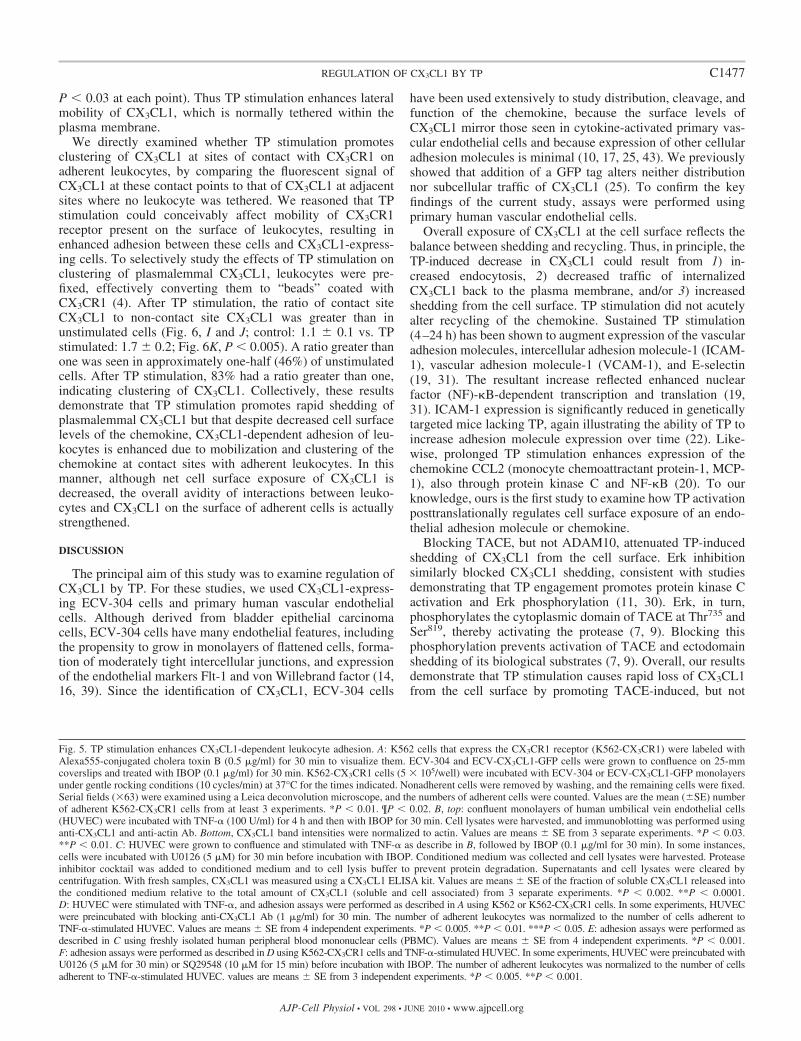
Fig. 5. TP stimulation enhances CX3CL1-dependent leukocyte adhesion. A: K562 cells that express the CX3CR1 receptor (K562-CX3CR1) were labeled withAlexa555-conjugated cholera toxin B (0.5 �g/ml) for 30 min to visualize them. ECV-304 and ECV-CX3CL1-GFP cells were grown to confluence on 25-mmcoverslips and treated with IBOP (0.1 �g/ml) for 30 min. K562-CX3CR1 cells (5 � 105/well) were incubated with ECV-304 or ECV-CX3CL1-GFP monolayersunder gentle rocking conditions (10 cycles/min) at 37°C for the times indicated. Nonadherent cells were removed by washing, and the remaining cells were fixed.Serial fields (�63) were examined using a Leica deconvolution microscope, and the numbers of adherent cells were counted. Values are the mean (�SE) numberof adherent K562-CX3CR1 cells from at least 3 experiments. *P � 0.01. ¶P � 0.02. B, top: confluent monolayers of human umbilical vein endothelial cells(HUVEC) were incubated with TNF-� (100 U/ml) for 4 h and then with IBOP for 30 min. Cell lysates were harvested, and immunoblotting was performed usinganti-CX3CL1 and anti-actin Ab. Bottom, CX3CL1 band intensities were normalized to actin. Values are means � SE from 3 separate experiments. *P � 0.03.**P � 0.01. C: HUVEC were grown to confluence and stimulated with TNF-� as describe in B, followed by IBOP (0.1 �g/ml for 30 min). In some instances,cells were incubated with U0126 (5 �M) for 30 min before incubation with IBOP. Conditioned medium was collected and cell lysates were harvested. Proteaseinhibitor cocktail was added to conditioned medium and to cell lysis buffer to prevent protein degradation. Supernatants and cell lysates were cleared bycentrifugation. With fresh samples, CX3CL1 was measured using a CX3CL1 ELISA kit. Values are means � SE of the fraction of soluble CX3CL1 released intothe conditioned medium relative to the total amount of CX3CL1 (soluble and cell associated) from 3 separate experiments. *P � 0.002. **P � 0.0001.D: HUVEC were stimulated with TNF-�, and adhesion assays were performed as described in A using K562 or K562-CX3CR1 cells. In some experiments, HUVECwere preincubated with blocking anti-CX3CL1 Ab (1 �g/ml) for 30 min. The number of adherent leukocytes was normalized to the number of cells adherent toTNF-�-stimulated HUVEC. Values are means � SE from 4 independent experiments. *P � 0.005. **P � 0.01. ***P � 0.05. E: adhesion assays were performed asdescribed in C using freshly isolated human peripheral blood mononuclear cells (PBMC). Values are means � SE from 4 independent experiments. *P � 0.001.F: adhesion assays were performed as described in D using K562-CX3CR1 cells and TNF-�-stimulated HUVEC. In some experiments, HUVEC were preincubated withU0126 (5 �M for 30 min) or SQ29548 (10 �M for 15 min) before incubation with IBOP. The number of adherent leukocytes was normalized to the number of cellsadherent to TNF-�-stimulated HUVEC. values are means � SE from 3 independent experiments. *P � 0.005. **P � 0.001.
C1477REGULATION OF CX3CL1 BY TP
AJP-Cell Physiol • VOL 298 • JUNE 2010 • www.ajpcell.org

ADAM10-induced, shedding of CX3CL1, rather than by inter-fering with recycling of the chemokine.
Despite inducing proteolysis of approximately one-half theCX3CL1 present, TP augmented CX3CL1-dependent leuko-cyte adhesion. Although unexpected, this is not inconceivable,
since the overall binding of CX3CR1 receptor on the leukocyteis determined by two distinct factors, namely, the density ofCX3CL1 ligand present and the mobility of CX3CL1 within theplasma membrane. We hypothesized that TP stimulation mustincrease the avidity of remaining CX3CL1 for its receptor. In
Fig. 6. TP stimulation mobilizes plasmalemmal CX3CL1 and induces clustering of CX3CL1 at sites of contact with adherent leukocytes. With the use ofECV-CX3CL1-GFP cells, fluorescence recovery after photobleaching (FRAP) was performed as described in MATERIALS AND METHODS. A: confocal image ofsingle cell (�100). B: a region within the plasma membrane was irreversibly photobleached (see arrow). C: same cell 5 min after photobleaching. D–F: as acontrol, the membrane mobility of glycosylphosphatidylinositol (GPI)-linked GFP in transfected ECV-CX3CL1 cells was determined in the same manner. G: cellswere incubated with IBOP (0.1 �g/ml) for 30 min, and fluorescence recovery of the photobleached area was monitored over time. Values are means � SE from9 separate experiments per condition. P � 0.05, untreated vs. IBOP at all time points after 11 s. H: recovery of fluorescence of the photobleached area wasmeasured over time. In some experiments, ECV-CX3CL1-GFP cells were preincubated with GM6001 (20 �M for 2 h), to inhibit cleavage of CX3CL1 from thecell surface, and then incubated with IBOP (0.1 �g/ml) for 30 min. In some experiments, ECV-CX3CL1 cells were transfected with cDNA encodingGFP-conjugated Fc�RIIA. Recovery of fluorescence after photobleaching of untreated ECV-CX3CL1-GFP, GM6001-treated ECV-CX3CL1-GFP, and GM6001-treated ECV-CX3CL1-GFP stimulated with IBOP, GPI-GFP, or Fc�RIIA-GFP was measured. At each time point, fluorescence intensity in the bleached regionwas normalized to the mean control unbleached membrane fluorescence intensity. Values are means � SE from 5–9 separate experiments per condition. I andJ: ECV-304 cells were transfected with CX3CL1-mCherry cDNA. Adhesion assays were performed by incubating ECV-CX3CL1-mCherry cells for 30 min withK562-CX3CR1 cells (5 � 105/well) in the presence (J) or absence (I) of IBOP. To prevent unwanted shedding of CX3CL1, in some wells ECV-CX3CL1-mCherrycells were preincubated with GM6001. To rule out potential effects of TP stimulation on cell surface clustering of CX3CR1, K562-CX3CR1 cells were first fixedusing 4% paraformaldehyde and then washed with 100 mM glycine. At the end of the adhesion assay, cells were fixed and permeabilized, and K562-CX3CR1cells were visualized by labeling with anti-BCR Ab and Alexa488-conjugated secondary Ab. Z-stack images are from spinning disk confocal microscopy (�63).K: experiments were performed as described in I and J. XY or YZ dimensions of the Z-stack images were used to compare the localized fluorescence intensityof CX3CL1-mCherry at sites of interface with adherent K562-CX3CR1 cells with the signal in an adjacent, similar-size region in the plasma membrane, whereno leukocyte was attached. With the use of ImageJ software, the ratio of fluorescent signal intensities between the site of contact and site of no contact wasmeasured for 24 randomly chosen cells for each treatment condition. Values are means � SE from 3 separate experiments. *P � 0.005 vs. control.
C1478 REGULATION OF CX3CL1 BY TP
AJP-Cell Physiol • VOL 298 • JUNE 2010 • www.ajpcell.org

principle, this could occur through mobilization of plasmale-mmal CX3CL1 and subsequent clustering of the chemokine atpoints of contact with adherent leukocytes. Accordingly, plas-malemmal CX3CL1 was relatively immobile under basal con-ditions but became highly mobile following TP stimulation.We previously reported that plasmalemmal CX3CL1 is alsoimmobile in other cell types under basal conditions (8). TPstimulation promoted clustering of CX3CL1 at contact inter-faces with bound leukocytes. Similarly, Kobayashi et al. (22)reported less leukocyte rolling and adherence in mice lackingTP, perhaps due to the absence of TP-induced clustering ofCX3CL1 on the surface of the vascular wall. Our results arereminiscent of a study demonstrating that isoprostane iPF2�-III, a known agonist of TP, enhances neutrophil adhesion toendothelial cells despite no increase in expression of endothe-lial adhesion molecules (1, 46). Although the authors postu-lated that release of an unknown endothelial activating factorwas responsible, isoprostane activation might also increasemobility of plasmalemmal adhesion molecules, therebystrengthening the avidity of interactions between neutrophilsand endothelial cells.
Our results differ somewhat from those of Hundhausen et al.(17), who reported that the synthetic phorbol ester PMAinhibited adhesion of THP-1 monocytic cells to CX3CL1-expressing adherent cells. Several possible explanations forthis apparent discrepancy exist. First, THP-1 cells expressother adhesion receptors whose levels and function may bemodulated by PMA stimulation. In our study, we purposelyused CX3CR1-expressing K562 cells, which do not expressadhesion molecules such as L-selectin (10). Thus any adhesionobserved was presumably attributable to interactions betweenleukocyte CX3CR1 and CX3CL1 on the surface of adherentcells. Stimulation by PMA and TP may have differentialeffects on the cell. TP receptors couple to Gq/G11, with mostfunctioning attributed to signaling via the �- rather than ��-subunits (21). Thus stimulation by PMA or TP can activatephospholipase C, generating inositol triphosphate (IP3) anddiacylglycerol (DAG), increasing intracellular calcium concen-tration, and activating protein kinase C (21). In primary humancells, TP also couples to Gi and to G12/13 and is linked to cAMP(15, 21). Thus the TP receptor has the potential to utilize atleast four distinct intracellular signaling pathways. Mobiliza-tion of CX3CL1 by TP may reflect activation of these path-ways. In our study, TP activation consistently increasedCX3CL1-dependent leukocyte adhesion, even to primary hu-man vascular endothelial cells expressing endogenousCX3CL1, highlighting the physiologic relevance of these ob-servations. This is the first description of regulation ofCX3CL1 exposure and mobility by a G protein-coupled recep-tor.
An intriguing question is how CX3CL1 is anchored to theplasma membrane in the first place. Previously, we reportedthat CX3CL1 does not directly interact with the actin cytoskel-eton (8). Recent work has demonstrated an integral role fordendritic cell (DC) CX3CL1 in the accumulation of lipid raftsat DC-NK cell synapses (34). However, we found that inadherent cells, plasmalemmal CX3CL1 does not associate withlipid rafts and, accordingly, that cholesterol depletion does notalter the mobility of the chemokine (8). Future studies areneeded to investigate 1) whether CX3CL1 indirectly associateswith the actin cytoskeleton by one or more Triton-soluble
adaptor proteins, 2) whether CX3CL1 is “fenced” into discreteregions of the membrane, and 3) precisely how TP uncouplesCX3CL1 from its membrane anchor (33, 38).
TP plays a complex role in vascular inflammation. Given thetimeframe of events that we investigated, it appears that earlyin the inflammatory cascade, TP stimulation induces sheddingof endothelial CX3CL1 to release the soluble chemoattractantspecies, thereby recruiting circulating leukocytes. Slightlylater, the balance is subtly altered to facilitate adhesion ofleukocytes thus recruited to the area of vascular injury. Regu-lation of CX3CL1 by TP in this manner may represent a link,heretofore unexplored, between the events accompanying theplatelet aggregation and activation and those accompanying thevascular inflammation that, collectively, contribute to athero-genesis. The dynamic interplay among these individual eventsin vivo remains to be elucidated.
ACKNOWLEDGMENTS
We thank Drs. Mansoor Husain and Sergio Grinstein for helpful discus-sions.
GRANTS
This work was supported by a Grant-in-Aid from the Heart and StrokeFoundation of Canada (to L. A. Robinson). L. A. Robinson is the recipient ofa Canada Research Chair (Tier 2) Award.
DISCLOSURES
A. M. Durkan is the recipient of a Fellowship Award jointly sponsored byNorth American Pediatric Renal Trials and Collaborative Studies (NAPRTCS)and Roche.
REFERENCES
1. Audoly LP, Rocca B, Fabre JE, Koller BH, Thomas D, Loeb AL,Coffman TM, FitzGerald GA. Cardiovascular responses to the isopros-tanes iPF2�-III and iPE2-III are mediated via the thromboxane A2 receptorin vivo. Circulation 101: 2833–2840, 2000.
2. Baan JF, Bacon KB, Hardiman G, Wang W, Soo K, Rossi D, GreavesDR, Zlotnick A, Schall TJ. A new class of membrane-bound chemokinewith a CX3C motif. Nature 385: 640–644, 1997.
3. Becker RC. Antithrombotic therapy after myocardial infarction. N Engl JMed 347: 1019–1022, 2002.
4. Chan JR, Hyduk SJ, Cybulsky MI. Detecting rapid and transientupregulation of leukocyte integrin affinity induced by chemokines andchemoattractants. J Immunol Methods 273: 43–52, 2003.
5. Cheng Y, Austin SC, Rocca B, Koller BH, Coffman TM, Grosser T,Lawson JA, FitzGerald GA. Role of prostacyclin in the cardiovascularresponse to thromboxane A2. Science 296: 539–541, 2002.
6. Combadiere C, Potteaux S, Gao JL, Esposito B, Casanova S, Lee EJ,Debre P, Tedgui A, Murphy PM, Mallat Z. Decreased atheroscleroticlesion formation in CX3CR1/apolipoprotein E double knockout mice.Circulation 107: 1009–1016, 2003.
7. Diaz-Rodriguez E, Montero JC, Esparis-Ogando A, Yuste L, Pan-diella A. Extracellular signal-related kinase phosphorylates tumor necrosisfactor alpha-converting enzyme at threonine 735: a potential role inregulated shedding. Mol Biol Cell 13: 2031–2044, 2002.
8. Durkan AM, Alexander RT, Liu GY, Rui M, Femia G, Robinson LA.Expression and targeting of CX3CL1 (fractalkine) in renal tubular epithe-lial cells. J Am Soc Nephrol 18: 74–83, 2007.
9. Fan H, Turck CW, Derynk R. Characterization of growth factor-inducedserine phosphorylation of tumor necrosis factor-alpha converting enzymeand of an alternatively translated polypeptide. J Biol Chem 278: 18617–18627, 2003.
10. Fong AM, Robinson LA, Steeber DA, Tedder TF, Yoshie O, Imai T,Patel DD. Fractalkine and CX3CR1 mediate a novel mechanism ofleukocyte capture, firm adhesion, and activation under physiologic flow. JExp Med 188: 1413–1419, 1998.
11. Gao Y, Tang S, Zhou S, Ware JA. The thromboxane A2 receptoractivates mitogen-activated protein kinase via protein kinase C-dependent
C1479REGULATION OF CX3CL1 BY TP
AJP-Cell Physiol • VOL 298 • JUNE 2010 • www.ajpcell.org

Gi coupling and Src-dependent phosphorylation of the epidermal growthfactor receptor. J Pharmacol Exp Ther 296: 426–433, 2001.
12. Garton KJ, Gough PJ, Blobel CP, Murphy G, Greaves DR, DempseyPJ, Raines EW. Tumor necrosis factor-�-converting enzyme (ADAM17)mediates the cleavage and shedding of fractalkine (CX3CL1). J Biol Chem276: 37993–38001, 2001.
13. Ghilardi G, Biondi ML, Turri O, Guagnellini E, Scorza R. Internalcarotid artery occlusive disease and polymorphisms of fractalkine receptorCX3CR1: a genetic risk factor. Stroke 35: 1276–1279, 2004.
14. Haller C, Kiessling F, Kubler W. Polarized expression of heterologousmembrane proteins transfected in a human endothelial-derived cell line.Eur J Cell Biol 75: 353–361, 1997.
15. Hirata T, Ushikubi F, Kakizuka A, Okuma M, Narumiya S. Twothromboxane A2 receptor isoforms in human platelets. J Clin Invest 97:949–956, 1996.
16. Hughes SE. Functional characterization of the spontaneously transformedhuman umbilical vein endothelial cell line ECV304: use in an in vitromodel of angiogenesis. Exp Cell Res 225: 171–185, 1996.
17. Hundhausen C, Misztela D, Berkhout TA, Broadway N, Saftig P,Reiss K, Hartmann D, Fahrenholz F, Postina R, Matthews V, KallenKJ, Rose-John S, Ludwig A. The disintegrin-like metalloproteinaseADAM-10 is involved in constitutive cleavage of CX3CL1 (fractalkine)and regulates CX3CL1-mediated cell-cell adhesion. Blood 102: 1186–1195, 2003.
18. Imai T, Hieshima K, Haskell CA, Baba M, Nagira M, Nishimura M,Kakizaki M, Takagi S, Nomiyama H, Schall TJ, Yoshie O. Identifica-tion and molecular characterization of fractalkine receptor, CX3CR1,which mediates both leukocyte migration and adhesion. Cell 91: 521–530,1997.
19. Ishizuka T, Kawakami M, Hidaka T, Matsuki Y, Takamizawa M,Suzuki K, Kurita A, Nakamura H. Stimulation with thromboxane A2
(TXA2) receptor agonist enhances ICAM-1, VCAM-1 or ELAM-1 expres-sion by human vascular endothelial cells. Clin Exp Immunol 112: 464–470, 1998.
20. Ishizuka T, Sawada S, Sugama K, Kurita A. Thromboxane A2 (TXA2)receptor blockade suppresses monocyte chemoattractant protein-1 (MCP-1)expression by stimulated vascular endothelial cells. Clin Exp Immunol 120:71–78, 2000.
21. Kinsella BT. Thromboxane A2 signalling in humans: a “tail” of tworeceptors. Biochem Soc Trans 29: 641–654, 2001.
22. Kobayashi T, Tahara Y, Matsumoto M, Iguchi M, Sano H, MurayamaT, Arai H, Oida H, Yurugi-Kobayashi T, Yamashita JK, Katagiri H,Majima M, Yokode M, Kita T, Narumiya S. Roles of thromboxane A2
and prostacyclin in the development of atherosclerosis in apoE-deficientmice. J Clin Invest 114: 784–794, 2004.
23. Konopska JB, Watanabe SM, Witte ON. An alteration of the humanc-abl protein in K562 leukemia cells unmasks associated tyrosine kinaseactivity. Cell 37: 1035–1042, 1984.
24. Lesnik P, Haskell CA, Charo IF. Decreased atherosclerosis in CX3CR1 �/�mice reveals a role for fractalkine in atherogenesis. J Clin Invest 111: 333–340,2003.
25. Liu GY, Kulasingam V, Alexander RT, Touret N, Fong AM, Patel DD,Robinson LA. Recycling of the membrane-anchored chemokine, CX3CL1.J Biol Chem 280: 19858–19866, 2005.
26. Lucas AD, Bursill C, Guzik TJ, Sadowski J, Channon KM, GreavesDR. Smooth muscle cells in human atherosclerotic plaques express thefractalkine receptor CX3CR1 and undergo chemotaxis to the CX3C che-mokine fractalkine (CX3CL1). Circulation 108: 2498–2504, 2003.
27. Matloubian M, David A, Engel S, Ryan JE, Cyster JG. A transmem-brane CXC chemokine is a ligand for HIV-coreceptor Bonzo. Nat Immu-nol 1: 298–304, 2000.
28. McDermott DH, Fong AM, Yang Q, Sechler JM, Cupples LA, MerrellMN, Wilson PW, D’Agostino RB, O’Donnell CJ, Patel DD. Chemokinereceptor mutant CX3CR1-M280 has impaired adhesive function and cor-relates with protection from cardiovascular disease in humans. J ClinInvest 111: 1241–1250, 2003.
29. McDermott DH, Halcox JP, Schenke WH, Waclawiw MA, MerrellMN, Epstein N, Quyyumi AA, Murphy PM. Association between
polymorphism in the chemokine receptor CX3CR1 and coronary vascularendothelial dysfunction and atherosclerosis. Circ Res 89: 401–407, 2001.
30. Miggin SM, Kinsella BT. Thromboxane A2 receptor-mediated activationof the mitogen activated protein kinase cascades in human uterine smoothmuscle cells. Biochim Biophys Acta 1539: 147–162, 2001.
31. Mitsuhashi M, Tanaka A, Fujisawa C, Kawamoto K, Itakura A,Takaku M, Hironaka T, Sawada S, Matsuda H. Necessity of throm-boxane A2 for initiation of platelet-mediated contact sensitivity: dualactivation of platelets and vascular endothelial cells. J Immunol 166:617–623, 2001.
32. Moatti D, Faure S, Fumeron F, Amara MW, Seknadji P, McDermottDH, Debre P, Aumont MC, Murphy PM, de Prost D, Combadiere C.Polymorphism in the fractalkine receptor CX3CR1 as a genetic risk forcoronary artery disease. Blood 97: 1925–1928, 2001.
33. Neame SJ, Uff CR, Sheikh H, Wheatley SC, Isacke CM. CD44 exhibitsa cell type dependent interaction with triton X-100 insoluble, lipid rich,plasma membrane domains. J Cell Sci 108: 3127–3135, 1995.
34. Pallandre JR, Krzewski K, Bedel R, Ryffel B, Caignard A, RohrlichPS, Pivot P, Tiberghien P, Zitvogel L, Strominger JL, Borg C.Dendritic cell and natural killer cell cross-talk: a pivotal role of CX3CL1in NK cytoskeleton organization and activation. Blood 112: 4420–4424,2008.
35. Robinson LA, Nataraj C, Thomas DW, Howell DN, Griffiths R,Bautch VL, Patel DD, Feng L, Coffman TM. A role for fractalkine andits receptor (CX3CR1) in cardiac allograft rejection. J Immunol 165:6067–6072, 2000.
36. Robinson LA, Tu L, Steeber DA, Preis O, Platt JL, Tedder TF. Therole of adhesion molecules in human leukocyte attachment to porcinevascular endothelium: implications for xenotransplantation. J Immunol161: 6931–6938, 1998.
37. Shiokoshi T, Ohsaki Y, Kawabe J, Fujino T, Kikuchi K. Downregu-lation of nitric oxide accumulation by cyclooxygenase-2 induction andthromboxane A2 production in interleukin-1�-stimulated rat aortic smoothmuscle cells. J Hypertens 20: 455–461, 2002.
38. Suzuki K, Ritchie K, Kajikawa E, Fujiwara T, Kusumi A. Rapid hopdiffusion of a G-protein-coupled receptor in the plasma membrane asrevealed by single-molecule techniques. Biophys J 88: 3659–3680, 2005.
39. Takahashi K, Sawasaki Y. Rare spontaneously transformed humanendothelial cell line provides useful research tool. In Vitro Cell Dev Biol28A: 380–382, 1992.
40. Teupser D, Pavlides S, Tan M, Gutierrez-Ramos JC, Kolbeck R,Breslow JL. Major reduction of atherosclerosis in fractalkine (CX3CL1)-deficient mice is at the brachiocephalic artery, not the aortic root. ProcNatl Acad Sci USA 101: 17795–17800, 2004.
41. Thomas DW, Rocha PN, Nataraj C, Robinson LA, Spurney RF,Koller BH, Coffman TM. Proinflammatory actions of thromboxanereceptors to enhance cellular immune responses. J Immunol 171: 6389–6395, 2003.
42. Tilley SL, Coffman TM, Koller BH. Mixed messages: modulation ofinflammation and immune responses by prostaglandins and thromboxanes.J Clin Invest 108: 15–23, 2001.
43. Tsou CL, Haskell CA, Charo IF. Tumor necrosis factor-�-convertingenzyme mediates the inducible cleavage of fractalkine. J Biol Chem 276:44622–44626, 2001.
44. van Kerkhof P, Sachse M, Klumperman J, Strous GJ. Growth hormonereceptor ubiquitination coincides with recruitment to clathrin-coated mem-brane domains. J Biol Chem 276: 3778–3784, 2001.
45. Wong BW, Wong D, McManus BM. Characterization of fractalkine(CX3CL1) and CX3CR1 in human coronary arteries with native athero-sclerosis, diabetes mellitus, and transplant vascular disease. CardiovascPathol 11: 332–338, 2002.
46. Zahler S, Becker BF. Indirect enhancement of neutrophil activity andadhesion to cultured human umbilical vein endothelial cells by isopros-tanes (iPF2�-III and iPE2-III). Prostaglandins Other Lipid Mediat 57:319–331, 1999.
47. Zeiffer U, Schober A, Lietz M, Liehn EA, Erl W, Emans N, Tan Z,Weber C. Neointimal smooth muscle cells display a proinflammatoryphenotype resulting in increased leukocyte recruitment mediated by P-selectin and chemokines. Circ Res 94: 776–784, 2004.
C1480 REGULATION OF CX3CL1 BY TP
AJP-Cell Physiol • VOL 298 • JUNE 2010 • www.ajpcell.org