thesis.pdf - KI Open Archive
70
Thesis for doctoral degree (Ph.D.) 2007 Dan Gryth Thesis for doctoral degree (Ph.D.) 2007 Dan Gryth Hemodynamic, Respiratory and Neurophysiological Reactions after High-Velocity Behind Armor Blunt Trauma Hemodynamic, Respiratory and Neurophysiological Reactions after High-Velocity Behind Armor Blunt Trauma
-
Upload
khangminh22 -
Category
Documents
-
view
2 -
download
0
Transcript of thesis.pdf - KI Open Archive
Thesis for doctoral degree (Ph.D.)2007
Dan Gryth
Thesis for doctoral degree (Ph.D.) 2007
Dan G
ryth
Hemodynamic, Respiratory and Neurophysiological Reactions after High-Velocity Behind Armor Blunt Trauma
Hem
odynamic, R
espiratory and Neurophysiological R
eactions after High-Velocity B
ehind Arm
or Blunt Traum
a
1
From the Department of Clinical Science and Education, Södersjukhuset, Karolinska Institutet, Stockholm, Sweden
Hemodynamic, Respiratory and Neurophysiological Reactions after
High-Velocity Behind Armor Blunt Trauma
Dan Gryth
M.D.
Stockholm 2007

2 Dan Gryth
All previously published papers were reproduced with permission from the publisher.
Printed by Larserics Digital Print ABLay-out Ringvor Hägglöf
Published by:© Dan Gryth, 2007ISBN 978-91-7357-355-9
3
“The pen is mightier than the sword”
Edward Bulwer-Lytton
To my family

4 Dan Gryth
5
Abstract
This thesis is addressing Behind Armor Blunt Trauma (BABT), defined as the non-
penetrating injury resulting from a ballistic impact on personal body armor. The
protective vest may impede the projectile, but some of the kinetic energy is transferred
to the body, causing effects such as pulmonary contusion, apnea, hypotension and
occasionally death.
Our aims of these studies have been to investigate physiological responses after high-
velocity BABT, including EEG (study I). Furthermore, the safety criterion of 44 mm
for protective vests (study II), effects of vagotomy (study III), and fluid resuscitation
(study IV) has been evaluated.
Anaesthetized pigs, wearing body armor on the right side of thorax, were shot with
a standard 7.62 mm assault rifle (velocity approx. 800 m/s). We used body armors
corresponding to 28 mm impression in clay placed behind the vest (study I and III), 34
mm and 40 mm (study II), and 42 mm (study IV). Several physiological parameters
were thereafter monitored during two hours after the shot. Experimental protocol was
similar in all studies, except from study III (in which one group received bilateral
cervical vagotomy) and study IV, in which 2 groups received Ringer´s acetate (RA)
or hypertonic saline with dextrane (HSD).
In all studies we observed an immediate drop of blood pressure, desaturation, increased
pressure in the lung circulation, suppressed EEG-pattern and pulmonary contusion.
In study II and IV, severe hyperkalemia was seen early after the trauma and several
animals had serious arrhythmias. Our observed EEG-changes indicate that high-
velocity BABT induces brain dysfunction, for at least a couple of minutes. Based
on our results, the safety criteria of 44 mm should be considered insufficient when a
vest is exposed to high-velocity bullets. Our results show that apnea after BABT is
a vagally mediated reflex, that can be inhibited by vagotomy. Fluid resuscitation has
limited effects on physiological parameters in our model, although HSD induces less
edema formation and a tendency to improved saturation compared to RA.

6 Dan Gryth

7
List of publications
This thesis is based on the following papers, which will be referred to in the text by their Roman numerals:
I. Dan Drobin,* Dan Gryth,* Jonas KE Persson, David Rocksén, Ulf P Arborelius, Lars-Gunnar Olsson, Jenny Bursell, B Thomas Kjellström. * These authors contributed equally to the study. Electroencephalogram, circulation and lung function after high-velocity Behind Armor Blunt Trauma (BABT). J Trauma. 2007. 63:405-13. II. Dan Gryth, David Rocksén, Jonas KE Persson, Ulf P Arborelius, Dan Drobin, Jenny Bursell, Lars-Gunnar Olsson, Thomas B Kjellström. Severe lung contusion and death after high-velocity Behind-Armor Blunt Trauma (BABT) – relation to protection level. Military Medicine: International Journal of AMSUS 2007. 172(10):1110-16.
III. Dan Gryth, David Rocksén, Ulf P Arborelius, Dan Drobin, Jonas KE Persson, Anders Sondén, Jenny Bursell, Lars-Gunnar Olsson, B Thomas Kjellström. Bilateral vagotomy inhibits apnea and attenuates other physiological responses after blunt chest trauma. J Trauma (in press)
IV. Dan Gryth, David Rocksén, Dan Drobin, Henrik Druid, Eddie Weitzberg, Jenny Bursell, Lars-Gunnar Olsson, Ulf P Arborelius. Effects of fluid resuscitation with hypertonic saline-dextran or Ringer acetate after pulmonary contusion and shock. Submitted August 2007

8 Dan Gryth
9
Contents
Abbrevations……………….......... …………………………………............. 11
Introduction…………………………………………..…….........………….. 13 The history of body armors……………………….....................……... 13 Body armor design……………………………………..............……... 15 Biomechanical aspects of behind armor blunt trauma…………....…... 17 Experimental models of behind armor blunt trauma……………...….. 17 Safety criteria for body armor…………………………………......….. 17 Pathophysiology of behind armor blunt trauma………………......….. 18 Incidence of behind armor blunt trauma……………..……..........…… 19 Diagnos and treatment of pulmonary contusion....………........……… 21
Aims of the thesis……………………………………………………............ 25
Materials and methods…………………………………….....……...........… 27 Measurement of back-face deformation………................................… 27 Shooting procedure in ballistic plasticine………....................….. 27 Body armors………………………………………....................... 28 Animals………………………………………………..……................ 29 Anesthesia and ventilation……………………………......................... 30 Catheterization…………………………………………....................... 31 Recording of circulatory parameters………………………............….. 31 Calculated variables……………………………………..........………. 32 Recording of electroencephalogram (EEG)………………....…........... 32 Blood sampling……………………………………………….............. 34 Analysis of Endothelin-1, TNF-α, IL-6 and S-100…………...........… 34 Armor and shooting procedure in the animal experiments…................ 35 Post mortal examination……………………………………........…… 35
Histology…………………………………………..........…....….. 35 Methods specific to studies I-IV……..……………………….....….... 36
The specific methodology in study I………………...........…....… 36 The specific methodology in study II………………..........…........ 36 The specific methodology in study III……………...........……...... 36 The specific methodology in study IV………………..................... 36 Statistical methods…………………………………………........…..... 38
10 Dan Gryth
Results & Discussion……………………………………………….....…… 39 Injuries………………………………………………………........….. 39 Mortality………………………………………………………........… 40 Respiration……………………………………………………............ 40 Hemodynamics………………………………………………........….. 42 Cerebral effects……….…………………………………....……….... 45 EEG…………………………………………………….................. 45 S-100………………………………………………...............…...... 48 Blood samples…………………………………………….......……… 48 Lactate……………………………………………................…….. 48 Potassium………………………………………............................. 49 Hemoglobin…………………………………………….................. 50 Glucose…………………………………………….....................… 51 Inflammatory response………………………….............................. 52Conclusions………………………………………........................................ 55
Acknowledgements........................................................................................ 57
References...................................................................................................... 61
Papers I-IV
11
Abbreviations
ARDS Acute respiratory distress syndromeATP Adenosine triphosphateBABT Behind armor blunt traumaBE Base excessCO Cardiac output CPR Cardio-pulmonary resuscitation CT Computer tomographyCVP Central venous pressureDAB 3,3’- DiaminobenzidineDO
2 Oxygen delivery
ECG ElectrocardiogramEDTA Ethylenediamine tetraacetic acidEEG ElectroencephalogramELISA Enzyme-linked immunosorbent assayET-1 Endothelin-1Hb HemoglobinHSD Hypertonic saline with dextranICU Intensive care unit IL- 6 Interleukin-6i.v. IntravenouslyMAP Mean arterial blood pressureMPAP Mean pulmonary artery pressureNATO North Atlantic Treaty OrganizationNIJ National Institute of Justice (USA)NO Nitric oxidePA Pulmonary artery PaCO
2 Partial pressure of carbon dioxide in arterial blood
PaO2
Partial pressure of oxygen in arterial bloodPEEP Positive end-expiratory pressure RA Ringer’s acetate solutionS-100 S-100 proteinSaO
2 Arterial oxygen saturation
SvO2
Mixed venous oxygen saturation SVR Systemic vascular resistanceTNF-α Tumour necrosis factor-alphaVF Ventricular fibrillation
Abbreviations

12 Dan Gryth
13
Introduction “Wisdom is a treasure
that follows its owner everywhere”
Chinese proverb
The history of body armors
This thesis focuses on Behind Armor Blunt Trauma (BABT), which is defined as the non-penetrating injury resulting from a ballistic impact into personal body armor1.
The development of modern body armors, which began during the First World War, initially involved soft armors made from silk and other natural fibers.
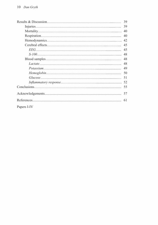
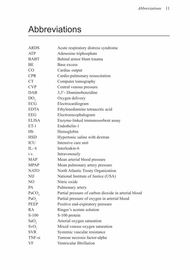
Furthermore, rigid armors containing one large or several small metal plates were produced. Some of these were heavy and bulky and could weigh as much as 12 kg 2
(figure 1 and 2).
Figure 1. British Dayfield body shield (front and rear view) (reproduced from “Modern body armour and helmets: An introduction”, with permission from Michael Iremonger).
Introduction
14 Dan Gryth
Figure 2. US Brewster shield (reproduced from “Modern body armour and helmets: An introduction”, with permission from Michael Iremonger).
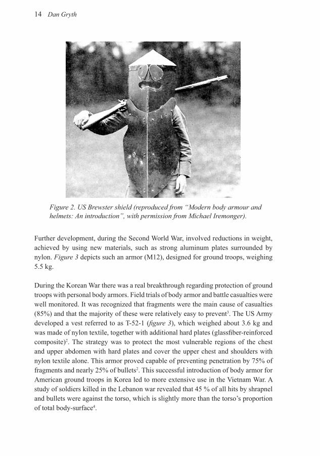
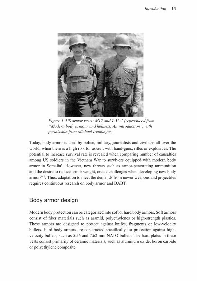
Further development, during the Second World War, involved reductions in weight, achieved by using new materials, such as strong aluminum plates surrounded by nylon. Figure 3 depicts such an armor (M12), designed for ground troops, weighing 5.5 kg.
During the Korean War there was a real breakthrough regarding protection of ground troops with personal body armors. Field trials of body armor and battle casualties were well monitored. It was recognized that fragments were the main cause of casualties (85%) and that the majority of these were relatively easy to prevent3. The US Army developed a vest referred to as T-52-1 (figure 3), which weighed about 3.6 kg and was made of nylon textile, together with additional hard plates (glassfiber-reinforced composite)2. The strategy was to protect the most vulnerable regions of the chest and upper abdomen with hard plates and cover the upper chest and shoulders with nylon textile alone. This armor proved capable of preventing penetration by 75% of fragments and nearly 25% of bullets2. This successful introduction of body armor for American ground troops in Korea led to more extensive use in the Vietnam War. A study of soldiers killed in the Lebanon war revealed that 45 % of all hits by shrapnel and bullets were against the torso, which is slightly more than the torso’s proportion of total body-surface4.
15
Figure 3. US armor vests: M12 and T-52-1 (reproduced from “Modern body armour and helmets: An introduction”, with permission from Michael Iremonger).
Today, body armor is used by police, military, journalists and civilians all over the world, when there is a high risk for assault with hand-guns, rifles or explosives. The potential to increase survival rate is revealed when comparing number of causalties among US soldiers in the Vietnam War to survivors equipped with modern body armor in Somalia5. However, new threats such as armor-penetrating ammunition and the desire to reduce armor weight, create challenges when developing new body armors6, 7. Thus, adaptation to meet the demands from newer weapons and projectiles requires continuous research on body armor and BABT.
Body armor design
Modern body protection can be categorized into soft or hard body armors. Soft armors consist of fiber materials such as aramid, polyethylenes or high-strength plastics. These armors are designed to protect against knifes, fragments or low-velocity bullets. Hard body armors are constructed specifically for protection against high-velocity bullets, such as 5.56 and 7.62 mm NATO bullets. The hard plates in these vests consist primarily of ceramic materials, such as aluminum oxide, boron carbide or polyethylene composite.
Introduction
16 Dan Gryth
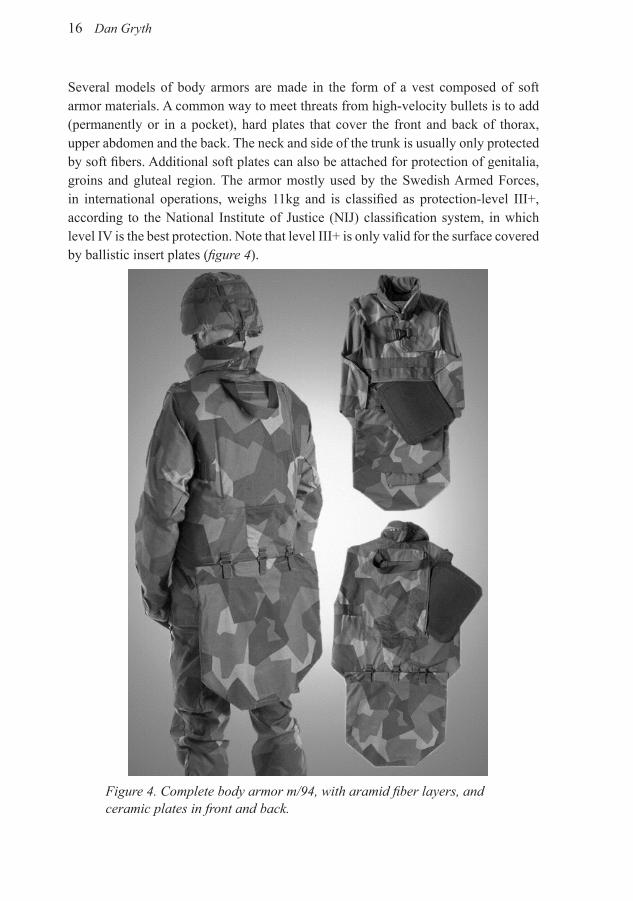
Figure 4. Complete body armor m/94, with aramid fiber layers, and ceramic plates in front and back.
Several models of body armors are made in the form of a vest composed of soft armor materials. A common way to meet threats from high-velocity bullets is to add (permanently or in a pocket), hard plates that cover the front and back of thorax, upper abdomen and the back. The neck and side of the trunk is usually only protected by soft fibers. Additional soft plates can also be attached for protection of genitalia, groins and gluteal region. The armor mostly used by the Swedish Armed Forces, in international operations, weighs 11kg and is classified as protection-level III+, according to the National Institute of Justice (NIJ) classification system, in which level IV is the best protection. Note that level III+ is only valid for the surface covered by ballistic insert plates (figure 4).
17
Biomechanical aspects of behind armor blunt trauma
Deformation of the projectile and of the armor itself is a part of the retardation and energy absorbing process that captures the projectile1. In the process of stopping the missile, the backside of the armor will be deformed to a certain degree, with the temporary deformation being generally more extensive than the permanent. The kinetic energy causes rapid deflection of the chest wall and sends shock-waves throughout the body, causing acceleration, shear deformation and retardation of tissues in the body wall and underlying organs, resulting in internal injuries and occasionally death1, 8-11.
Energy transferred to the body of the individual wearing the protective vest is considered to be linked to the degree of injury sustained8, 10-12. To investigate the relation between energy and resulting injury, tests has been conducted in a chest model, using an injury criterion originally developed to assess thoracic impacts within the automobile industry. This criterion is called the “Viscous criterion”, and is dependent on the amount of chest deflection, but also the rate at which it occurs13.
Experimental models of behind armor blunt trauma
In order to increase knowledge regarding BABT, research on animals is crucial. During the 1970´s goats were used routinely in these kind of experiments. But today, swine are more commonly used, due to their ready availability, similarities to human anatomy and physiology.
In addition to animal experiments, there exist other ways to study BABT, including human corpses. Although not frequently used, corpses can provide certain information about structural injuries14, but no data on pathophysiological effects. Computerized simulation models are also utilized15, 16, but such models are no better than their input data, and it is obvious that the existing knowledge about the mechanisms is too weak, to permit reliable descriptions of the relationship between applied violence and resulting injury. Therefore, these models are mostly used to investigate mechanistic characteristics of different impacts.
Safety criteria for body armors
Protection efficiency of body armors is categorized into 4 levels (I-IV), based on the type of ammunition that can be defeated. Level IV is the highest protection level and can resist bullets with calibers as large as 12.7 mm.
Introduction
18 Dan Gryth
At present, the most commonly used safety criterion for body armors is the so-called ”44 mm-criterion”, originating from the USA. The criterion was established after BABT studies on goats17, protected by a soft body armor (aramide) and exposed to handgun ammunition (caliber 0.22 and 0.38). Physiological results were compared to the impression made in a block of ballistic plasticine placed behind the armor. It was concluded that the injury sustained is tolerable if the impact does not generate an impression deeper than 44 mm.
The recommended method for testing whether body armor fulfills this criterion is described in the report “NIJ Standard-0101.44, Ballistic Resistance of Personal Armor”. The body armor should be fixed to the front of a block of ballistic plasticine (Roma Plasteline No.1), and the projectile of interest should be fired from a distance of 10 m. If the impression in clay is deeper than 44 mm, the body armor should be considered inadequate16.
This criterion was originally validated for hand-gun bullets or fragments, but has been extended far beyond the original limited bullet energy levels, without proper consideration of the energy delivered to the tissue, the projectile speed, body mass, gender or the location at which the body is hit13, 16, 18.
Pathophysiology of behind armor blunt trauma
As mentioned above, experimental animals and human corpses have been used to evaluate biomechanical aspects and physiological consequences of BABT. Projectiles aimed at the lungs, heart or spine cause subcutaneous hematomas, rib fractures, pulmonary contusions, heart contusions and spinal fractures. Physiological effects have only been studied in animal models, for obvious reasons. The results have shown decreased systolic blood pressure, slightly slower heart rate and arrhythmias, as well as apnea and decreased saturation of the blood19, 20.
The triad of apnea, bradycardia and hypotension has also been described in a number of studies investigating primary blast injury to the chest21-25. The pathophysiological effects has been shown to be a reflex mediated via the vagus nerve22. Activation of C-fiber receptors in the lung parenchyma are involved in that reflex. These receptors are located in the alveolar interstitial space, close to the pulmonary capillaries 26, 27, and are triggered by various stimuli, for example by elevations in pulmonary capillary pressure28, increase in the lung interstitial pressure29 and intravenous injections of certain chemical substances30. The afferent pathways from the lung stretch-receptors and the C-fibers are conducted through the vagus nerve to the respiratory center in the brain stem. Consequently, bilateral transection of the vagus nerve might prevent apnea and other pathophysiological effects, resulting from BABT24, 30, 31.
19
Brain function might also be affected by pressure waves propagated through the body after high-velocity trauma. Thus, Göransson et al. demonstrated suppressed activity of electroencephalogram (EEG) directly following impact of a high-velocity bullet on the hind leg of pigs32. Pronounced depression of EEG has also been described in pigs after blast trauma33. However, no data have been published previously, concerning the effects of high-velocity chest impact on EEG.
BABT and its pathophysiological consequences have similarities to civilian blunt chest trauma. Pulmonary contusion, one of the most significant effects of BABT, is also a common finding in trauma patients, especially after traffic accidents and fall from heights, occurring in nearly 20% of all individuals suffering multiple injuries (i.e with an Injury severity score > 15) 1, 34. However, in contrast to these examples, impact velocity is higher and the area of impact smaller in our studies, although peak velocity of the chest wall movement should not be considered equal to bullet velocity. In warfare and in connection with terrorist attacks, pulmonary contusions are also observed after shock waves produced by explosions35. Pulmonary damage caused by such trauma is characterized by disruption of microvasculature, resulting in local flooding of alveoli and interstitial extravasation of red cells and plasma36.
Incidence of behind armor blunt trauma
The first modern clinical report on BABT was written by Carrol et al.19788. This report described five cases of policemen who were hit by handgun bullets (caliber 0.38 and 0.22). The policemen were carrying soft body armors, made by 7- 18 layers of Kevlar®, and were hit at different regions of the chest. They were hospitalized for 2 to 3 days, due to bruises and superficial bleedings, but no subject had any signs of pulmonary or cardiac contusion.
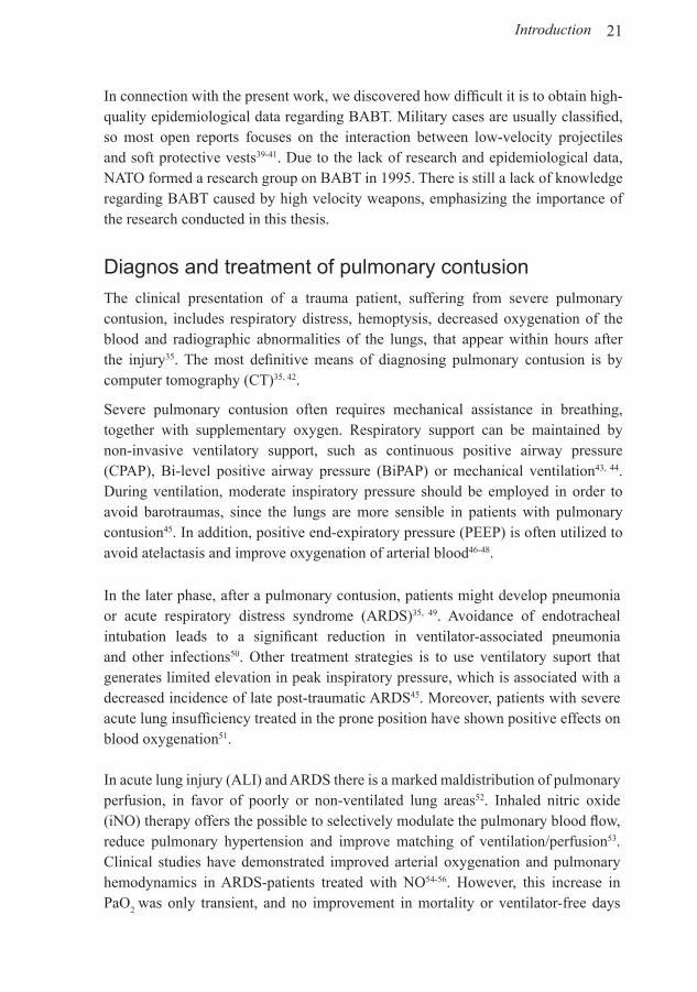
Figure 5 depicts a subcutaneous hematoma resulting from BABT, but the circumstances and injuries of the subject are unknown to us. IACP/DuPont Kevlar Survivors’ Club is a voluntary association for policemen who survived assault due to protection by body armor. To date, 207 persons have survived hits from different types of ammunition and are members in this association. The injuries described are often bruises and hematomas, but some suffered more serious damage. It is difficult to draw conclusions from the material, because different weapons, distances, angles and target areas were involved. Bir and coworkers made an attempt to evaluate policemen in the Survivors’ Club, with the aim to determine whether there is any gender difference in susceptibility to injury from BABT. In the report only 3 women and 8 men protected by soft body armor met the inclusion criteria, e.g. known weapon and distance. Described injuries were bruises, rib fractures, pulmonary contusions
Introduction
20 Dan Gryth
Figure 5. A soldier with subcutaneous hematoma resulting from BABT.
and hemoptyse, but all patients survived after hospital care37. In that study, it was difficult to determine whether any gender difference exists. However, it seems likely that gender (or mass) is a parameter that might affect outcome after BABT. Beside reports about soft armors, there exists some reports concerning BABT occurring behind rigid armors, after impact of high-velocity bullets. The earliest reported case of lethal BABT, due to a high-velocity bullet, was described 1969 by Shepard et al.38. During the Vietnam War, they described a US sergeant that was accidentally shot with a M-16 from close range. This report provides no description of his body armor, but the bullet did not penetrate the pleural cavity. After a short period of respiratory and hemodynamic stability the patient rapidly deteriorated, and died within 45 minutes after admission to the hospital.
In 1995 the French military reported that during the war in former Yugoslavia, a civilian voluntary worker had worn body armor (with a ceramic plate), that defeated a 14.5 mm bullet. This individual survived with large areas of myocutaneous necrosis beneath the plate and minor haemothorax1.
21
In connection with the present work, we discovered how difficult it is to obtain high-quality epidemiological data regarding BABT. Military cases are usually classified, so most open reports focuses on the interaction between low-velocity projectiles and soft protective vests39-41. Due to the lack of research and epidemiological data, NATO formed a research group on BABT in 1995. There is still a lack of knowledge regarding BABT caused by high velocity weapons, emphasizing the importance of the research conducted in this thesis.
Diagnos and treatment of pulmonary contusion
The clinical presentation of a trauma patient, suffering from severe pulmonary contusion, includes respiratory distress, hemoptysis, decreased oxygenation of the blood and radiographic abnormalities of the lungs, that appear within hours after the injury35. The most definitive means of diagnosing pulmonary contusion is by computer tomography (CT)35, 42.
Severe pulmonary contusion often requires mechanical assistance in breathing, together with supplementary oxygen. Respiratory support can be maintained by non-invasive ventilatory support, such as continuous positive airway pressure (CPAP), Bi-level positive airway pressure (BiPAP) or mechanical ventilation43, 44. During ventilation, moderate inspiratory pressure should be employed in order to avoid barotraumas, since the lungs are more sensible in patients with pulmonary contusion45. In addition, positive end-expiratory pressure (PEEP) is often utilized to avoid atelactasis and improve oxygenation of arterial blood46-48.
In the later phase, after a pulmonary contusion, patients might develop pneumonia or acute respiratory distress syndrome (ARDS)35, 49. Avoidance of endotracheal intubation leads to a significant reduction in ventilator-associated pneumonia and other infections50. Other treatment strategies is to use ventilatory suport that generates limited elevation in peak inspiratory pressure, which is associated with a decreased incidence of late post-traumatic ARDS45. Moreover, patients with severe acute lung insufficiency treated in the prone position have shown positive effects on blood oxygenation51.
In acute lung injury (ALI) and ARDS there is a marked maldistribution of pulmonary perfusion, in favor of poorly or non-ventilated lung areas52. Inhaled nitric oxide (iNO) therapy offers the possible to selectively modulate the pulmonary blood flow, reduce pulmonary hypertension and improve matching of ventilation/perfusion53. Clinical studies have demonstrated improved arterial oxygenation and pulmonary hemodynamics in ARDS-patients treated with NO54-56. However, this increase in PaO
2 was only transient, and no improvement in mortality or ventilator-free days
Introduction
22 Dan Gryth
were found57. Nevertheless, the European expert group for inhaled nitric oxide therapy, recommend to use iNO as a rescue treatment in adults with severe refractory ALI/ARDS58.
Many investigators have proposed a linkage between ARDS and the “two-hit” theory, suggesting that one injury alters cell and organ function, so that the response to a second injury is exacerbated59, 60. To avoid that, some studies recommend preventive treatment with corticosteroids61, although evidence concerning effectiveness of this approach is insubstantial62, 63. In the last decade, corticosteroids has not been recommended in the management of pulmonary contusion35, 64.
In some cases the pulmonary contusion is accompanied by other injuries, for example hemorrhage, requiring fluid treatment. However, infusion of fluids should be performed with caution34, 35, 45, 65, since the lungs are more sensible after a contusion66. Lung edema can occur when fluids are administrated to patients sustaining multitrauma and lung contusion, leading to decreased saturation67. Negative effects of fluid management in patients with lung contusion have been discussed since World War II68. During the Vietnam War it was described as “shock lung” or “DaNang lung” 69. Certain clinical findings support these observations70, whereas other studies conclude that fluids could be administrated safely36, 71. Prehospital resuscitation during trauma treatment until recently consisted of early and aggressive fluid administration, to stabilize the patient and normalize the blood pressure before arrival at the hospital72. In recent years this strategy has been challenged, and clinical and experimental models have shown possible deleterious effects if fluids is administrated before hemorrhage is under control. Emphasis has shifted from aggressive fluid administration, to early hemorrhage control73, 74 together with fast admission to trauma hospital75. Furthermore, timing of the infusion must be considered76.
Fluids commonly used for resuscitation are isotonic crystalloid solutions, such as Ringer’s acetate (RA) or lactated Ringer’s (LR) solution. These fluids are distributed throughout the entire extracellular space, with no preference for the intravascular space. Consequently, these solutions can occasionally cause tissue edema, which in turn impairs oxygenation42, 77. It has been proposed that hypertonic saline solutions containing colloids, such as hypertonic saline with dextrane (HSD), can be used to increase the blood pressure, cardiac output and peripheral tissue perfusion, without causing edema formation78, 79. The hyper-osmotic effects of such solutions have been shown to expand plasma volume three to four times the volume infused, by influx of extravascular fluid80. Resuscitation with HSD has shown a tendency to improve survival rate in severely injured patients81 and improve outcome in patients with severe head trauma82. In contrast, Bunn et al. concluded that hypertonic fluid
23
resuscitation in critically ill patients was not more efficient than resuscitation with isotonic crystalloid fluids83. However, hypertonic saline has been shown to reduce the inflammatory response after severe hemorrhagic shock in animal models84-86. HSD has also other advantages, particularly in pre-hospital and military circumstances, as it is easy to carry and store79. One might also speculate that infusion of a small volume HSD decreases the risk that hypothermia will develop, or be aggravated, in a cold environment.
Introduction

24 Dan Gryth
25
Aims of the thesis
General aim: To develop an animal model for high-velocity BABT, and in this experimental set-up investigate pathophysiological effects and mechanisms.
Specific aims:• Study I: Evaluate physiological effects after high-velocity BABT in pigs
wearing “complete protection” (M94), with particular focus on EEG-changes.
• Study II: To determine whether the 44 mm-criterion provides sufficient protection when body armors is exposed to high-velocity ammunition.
• Study III: To examine whether the vagus nerve is involved in apnea and other pathophysiological effects caused by BABT.
• Study IV: To compare fluid resuscitation with Hypertonic saline-Dextran (HSD) or Ringer’s acetate (RA) solution, for treatment of pulmonary contusion accompanied by shock.
Aims of the thesis

26 Dan Gryth

27
Material and methods
Detailed descriptions regarding methods used in this thesis is included in papers I-IV. Only a brief summary of methodological principles and considerations is given here. Most aspects of methodology were identical in the four studies and the experimental setting is displayed in figure 6. Specific differences between the studies are described in the section “Methods specific for studies I-IV”.
Figure 6. An illustration of our experimental set-up.
Measurement of back-face deformation
These tests were performed prior to the animal experiments, to evaluate the deformation behind the body armor (measured as impression in ballistic plasticine), following the standard developed by National Institute of Justice in USA (Standard 0101.04). The same procedure was used in study I-IV and only one shot was fired at each armor.
Shooting procedure in ballistic plasticineA standard assault rifle (Swedish Armed Forces Mark AK4) equipped with a laser aiming device (Diode laser type S 1889) was used in all experiments. The weapon was
Material and methods
28 Dan Gryth
mounted on a gun-carriage placed 10 meters in front of the target. All ammunition was of Danish issue, NATO type, 7.62 × 51 mm (M/94). This is standard “ball” ammunition with a lead core and a bullet weight of 9.5 g. Projectile velocity was measured with an optical shutter device (Chronograph Beta model). The recorded bullet velocities, during all reported shootings, were very close to 800 m/s (range 787-811). This velocity is associated with a kinetic energy of 3kJ, which was reduced to 0 within approximately 25 microseconds after impact.
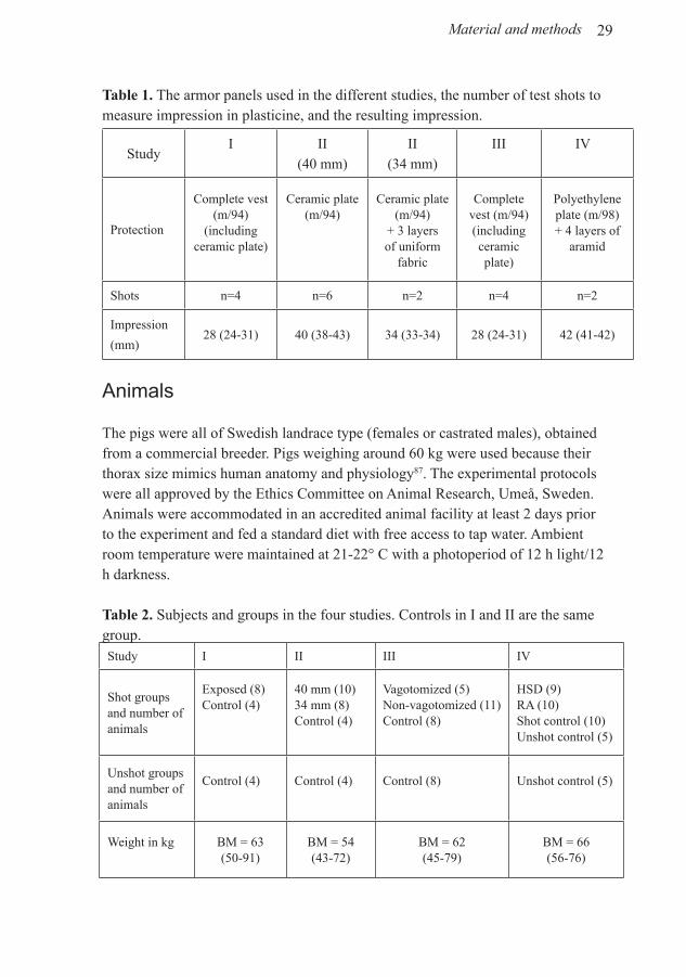
Body armorsIn all studies a layer of cotton fabric was placed tight between the block of plasticine and the armor, to simulate a field shirt. The manner in which the various components of protection were used in the four studies is summarized in table 1, where the resulting impression in plasticine is also given.
In study I, we used a specially manufactured vest segment, corresponding to the Swedish Armed Forces standard issue, m/94 consisting of three layers of uniform fabric, a ceramic ballistic insert plate (Aluminum oxide, 255 x 300 mm), and behind the plate 14 layers of aramid fabric (Kevlar®). This body armor is normally used by the Swedish Army during international operations and consists of a vest, covering the trunk, containing the aramid pack. On the front and back of the trunk there are pockets for the additional ballistic insert plates.
In study II the aim was to evaluate the “44 mm-criterion”. In order to obtain an impression with a depth as close to 44 mm as possible, the components of the “complete protection” were successively stripped away. 40 mm deep impression was achieved by using only the ceramic plate. This group was compared to the 34 mm-group, where the impression was slightly less (34 mm). This change was accomplished by adding three layers of uniform fabric.
In study III the same complete body armor as in study I (m/94) was used.
In study IV a ballistic insert plate (size 300 x 255 mm), made of polyethylene, (Swedish m/98) was used with 4 additional layers of aramid fabric at the backside, resulting in an impression of 42 mm. The goal with this set-up was a more severe BABT-injury than in earlier studies (I-III).
29
Table 1. The armor panels used in the different studies, the number of test shots to measure impression in plasticine, and the resulting impression.
StudyI II
(40 mm)II
(34 mm)III IV
Protection
Complete vest (m/94)
(including ceramic plate)
Ceramic plate(m/94)
Ceramic plate(m/94)
+ 3 layers of uniform
fabric
Complete vest (m/94)(including ceramic plate)
Polyethylene plate (m/98)+ 4 layers of
aramid
Shots n=4 n=6 n=2 n=4 n=2
Impression
(mm)28 (24-31) 40 (38-43) 34 (33-34) 28 (24-31) 42 (41-42)
Animals
The pigs were all of Swedish landrace type (females or castrated males), obtained from a commercial breeder. Pigs weighing around 60 kg were used because their thorax size mimics human anatomy and physiology87. The experimental protocols were all approved by the Ethics Committee on Animal Research, Umeå, Sweden. Animals were accommodated in an accredited animal facility at least 2 days prior to the experiment and fed a standard diet with free access to tap water. Ambient room temperature were maintained at 21-22° C with a photoperiod of 12 h light/12 h darkness.
Table 2. Subjects and groups in the four studies. Controls in I and II are the same group. Study I II III IV
Shot groups and number of animals
Exposed (8)Control (4)
40 mm (10)34 mm (8)Control (4)
Vagotomized (5)Non-vagotomized (11)Control (8)
HSD (9)RA (10)Shot control (10)Unshot control (5)
Unshot groups and number of animals
Control (4) Control (4) Control (8) Unshot control (5)
Weight in kg BM = 63 (50-91)
BM = 54 (43-72)
BM = 62 (45-79)
BM = 66 (56-76)
Material and methods
30 Dan Gryth
Anesthesia and ventilation
All animals got premedication before further handling and transport to the laboratory. A solution consisting of tiletamin 25 mg/ml, zolazepam 25 mg/ml and medetomidinhydrochlorid 1 mg/ml, was used. A dose of approximately 0.06 ml/kg was administered intramuscularly in the dorsum of the neck.
The general anesthesia was started with pentobarbital sodium 6 mg/kg, and 0.5 mg atropine sulphate intravenously (i.v.). The anesthesia was maintained with intrave-nous infusion of ketamine hydrochloride 50 mg/ml, and pethidin hydrochloride 50 mg/ml. 1 ml pethidin was added to every 30 ml of ketamine, and the infusion rate was 0.5 ml / (kg h).
In study III and IV, the pethidin was excluded to minimize deprivation of the respiratory drive, since these animals were spontaneously breathing during the whole, or part, of the experimental course.
In general anesthesia the animals were tracheotomized and mechanically ventilated in a volume controlled mode with room air (Siemens Servo Ventilator 900C), at a rate of 20 breaths per minute. The tidal volume was adjusted to achieve normoventilation. In all four studies the ventilation started in the same mode.
In study I and II, the ventilation was continued in the same mode as described above, through the whole experiment (120 min).
In study III, after the preparation, the ventilator was switched to spontaneously breathing (30 minutes before the shot), and was maintained so throughout the whole experiment. A small pressure support was set to compensate for the air flow resistance in the tubes. The length of the apnea period was visually registrated.
In study IV, the ventilator was switched to spontaneous breathing mode 30 minutes before the shot, but 1 minute before the shot the ventilator was disconnected until 5 minutes after the shot. After that they were connected again to the ventilator in a volume controlled mode. This ventilatory strategy was performed since our aim was to mimic field conditions, where an exposed person would receive assisted ventilation 5 min after impact. The length of the apnea period was visually registrated.
31
Catheterization
One catheter was introduced in an ear margin vein and used for induction of general
anesthesia. Through a paramedian left neck incision, a polyethylene catheter was
introduced into the left external jugular vein, to be used for continuous infusion of
the ketamine. The same incision, as for the tracheotomy, was used for introduction
of a polyethylene catheter into the left common carotid artery, for blood sampling
and mean arterial blood pressure (MAP) monitoring. Trough a right paramedian
neck incision, an optical pulmonary thermo dilution catheter (Opticath, Abbot) was
inserted into the right external jugular vein, for measurements of central venous
pressure (CVP), mean pulmonary artery pressure (MPAP), cardiac output (CO),
mixed venous saturation (SvO2) and body core temperature. The cardiac output was
measured using thermo dilution technique.
After the preparation was completed, animals were allowed 30 minutes rest to
achieve steady state.
Recording of circulatory parameters
Electrocardiography (ECG), heart rate (HR), mean arterial pressure (MAP), central
venous pressure (CVP) and mean pulmonary artery pressure (MPAP) were measured
and monitored with a Sirecust 960 (Siemens Medical Electronics). Cardiac output
(CO), mixed venous saturation (SvO2) and core temperature were measured and
monitored with an Oximetrix 3 (Abbott Critical Care Systems). CO was measured
by duplicate injection of 10 ml of 0.9 % saline with ambient room temperature.
Measurements were accepted if both injections agreed within 10 %, otherwise
additional measurements were obtained. The mean of the two accepted measurements
was registered.
Material and methods
32 Dan Gryth
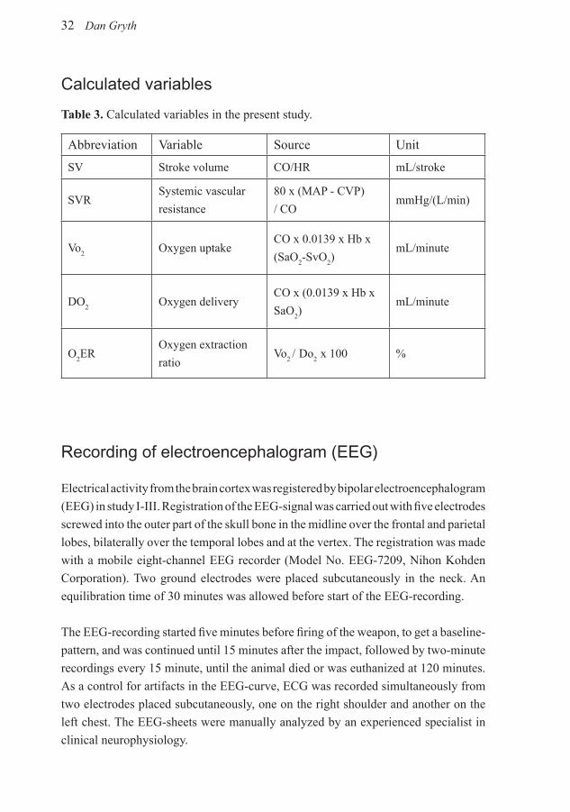
Abbreviation Variable Source Unit
SV Stroke volume CO/HR mL/stroke
SVRSystemic vascular
resistance
80 x (MAP - CVP)
/ COmmHg/(L/min)
Vo2
Oxygen uptakeCO x 0.0139 x Hb x
(SaO2-SvO
2)
mL/minute
DO2
Oxygen deliveryCO x (0.0139 x Hb x
SaO2)
mL/minute
O2ER
Oxygen extraction
ratioVo
2 / Do
2 x 100 %
Calculated variables
Table 3. Calculated variables in the present study.
Recording of electroencephalogram (EEG)
Electrical activity from the brain cortex was registered by bipolar electroencephalogram
(EEG) in study I-III. Registration of the EEG-signal was carried out with five electrodes
screwed into the outer part of the skull bone in the midline over the frontal and parietal
lobes, bilaterally over the temporal lobes and at the vertex. The registration was made
with a mobile eight-channel EEG recorder (Model No. EEG-7209, Nihon Kohden
Corporation). Two ground electrodes were placed subcutaneously in the neck. An
equilibration time of 30 minutes was allowed before start of the EEG-recording.
The EEG-recording started five minutes before firing of the weapon, to get a baseline-
pattern, and was continued until 15 minutes after the impact, followed by two-minute
recordings every 15 minute, until the animal died or was euthanized at 120 minutes.
As a control for artifacts in the EEG-curve, ECG was recorded simultaneously from
two electrodes placed subcutaneously, one on the right shoulder and another on the
left chest. The EEG-sheets were manually analyzed by an experienced specialist in
clinical neurophysiology.
33
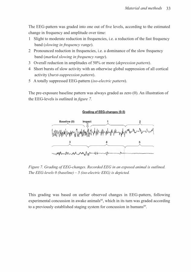
The EEG-pattern was graded into one out of five levels, according to the estimated
change in frequency and amplitude over time:
1 Slight to moderate reduction in frequencies, i.e. a reduction of the fast frequency
band (slowing in frequency range).
2 Pronounced reduction in frequencies, i.e. a dominance of the slow frequency
band (marked slowing in frequency range).
3 Overall reduction in amplitudes of 50% or more (depression pattern).
4 Short bursts of slow activity with an otherwise global suppression of all cortical
activity (burst-suppression pattern).
5 A totally suppressed EEG-pattern (iso-electric pattern).
The pre-exposure baseline pattern was always graded as zero (0). An illustration of
the EEG-levels is outlined in figure 7.
This grading was based on earlier observed changes in EEG-pattern, following
experimental concussion in awake animals88, which in its turn was graded according
to a previously established staging system for concussion in humans89.
Figure 7. Grading of EEG-changes. Recorded EEG in an exposed animal is outlined. The EEG-levels 0 (baseline) – 5 (iso-electric EEG) is depicted.
Material and methods

34 Dan Gryth
Blood sampling
Blood samples were obtained from the arterial line for analysis of SaO2, PaO
2,
PaCO2, Na+, K+, Ca2+, pH, base excess (BE) (GEM Premier Plus analyzer), lactate
(Miniphotometer, 8), blood glucose (B-Glucose analyzer, Hemocue AB, Ängelholm,
Sweden), and whole blood hemoglobin (Hb) (Hemoglobin Photometer, Electrolux).
In study IV we used a new blood gas machine (GEM Premier 3000), which could
measure all the samples mentioned above except blood hemoglobin, for which we
used the same method as previously.
These parameters and physiological data were recorded at baseline, 1, 5, 10 and
15 minutes following impact, and thereafter every 15 minutes until the end of the
experiment at 120 minutes. However, in study IV, due to practical reasons, no blood
sample was taken 1 min after impact.
Analysis of Endothelin-1, TNF-α, IL-6 and S-100
These measurements were only performed in study IV. For analysis of endothelin-1,
blood was drawn separately from the arterial line and the PA line at baseline,
7, 30, 60 and 120 minutes after shot. Collected blood samples were mixed with
EDTA (final concentration 10 mM) and kept on ice until centrifuged at + 4°C.
Plasma was stored at -20°C for analysis of endothelin-1 as described earlier90.
Venous blood samples for TNF-α and IL-6 determinations were taken at baseline
and 120 minutes after the shot, and were centrifuged at 15,000 rpm for 20 minutes
at 4°C. Serum was thereafter kept at -70°C until final processing. Cytokine levels
were determined using commercial enzyme-linked immunosorbent assay (ELISA)
kits (Quantiqine M Immunoassay), specific for murine TNF-α and IL-6. All assays
were performed in duplicate.
Blood samples for the determination of S-100 were drawn from the arterial line at 7,
30, 60 and 120 minutes after shot and allowed to clot for 30 minutes at room temper-
ature. Tubes were then centrifuged in 3,000g for 10 min. The supernatant was stored
in aliquots at - 70°C until analysis. Serum concentrations of S-100 were measured
with a commercially available, automated immunoluminometric assay (Byk-Sangtec
Diagnostica).
35
Armor and shooting procedure in the animal experiments
The same specially made armor panels used for measurements of back-face deformation were used for the animal experiments. In all four studies the weapon, ammunition and shooting distance (10 m) were the same and, hence, also the incoming energy. Consequently, the actual energy transferred to the swine thorax, as well as other loading characteristics in the different studies, were determined by the characteristics of the used armor panel. The armor was firmly attached to the right side of thorax with two 3 cm broad girdles. Care was taken not to cause restriction of the ventilation.
The conditions were kept identical to the ones of the plasticine impact tests, and agreed in general with those determined by the NATO task group BABT, as standard for these kind of experiments87.
The control animals were subjected to a rifle shot with the same weapon but using blank ammunition. The amount of gunpowder in the blanks was adjusted to produce a similar sound level as the live ammunition.
The animals were randomized, immediately before the shooting, to live ammunition or to blank ammunition, except for the vagotomized group in study III, which was exposed only to live ammunition. The target point was over the eighth rib at the right side of thorax, generating an adequate exposure of the right lung. The firing of the rifle was synchronized to the endpoint of the inspiratory phase.
After two hours observation time, the animals were euthanized with pentobarbital 60 mg/ml, 70 ml i.v. or more until the ECG became iso-electric.
Post mortal examination
Directly following death, an autopsy was performed in all studies (I-IV), in which
the swine were examined for gross pathology. The thorax wall, lungs, heart, liver and
bowels of the upper part of the trunk were examined.
Histology
In study IV, histology was performed on the lung, heart and kidney. Small pieces,
averaging 1 x 1 x 0.5 cm, were collected from both lungs, the myocardium and
from the right kidney and put into phosphate-buffered formalin. Paraffin sections
Material and methods
36 Dan Gryth
were stained with Hematoxylin-eosin, and with anti-myeloperoxidase antibodies
(myoclonal, 1:200, Chemicon, Stockholm, Sweden), followed by a goat-anti-mouse
secondary antibody, labeled with horseradish peroxidase, and a DAB detection kit.
Neurophils in lungs were counted in five fields at 600x magnification, whereas other
morphometry was based on semiquantitative grading.
Methods specific to studies I-IV
Below are descriptions of methods that are specific for the individual studies (I-IV),
and differences from the general methodology, described above.
The specific methodology in study I
Study I followed the previously described methodology. The aim of this study was to
test our new experimental model and evaluate the effects of BABT on physiological
parameters, including EEG. The shot animals were protected by the complete body
armor (vest + insert plate) and were compared to unshot animals.
The specific methodology in study II
Study II followed the previously described methodology. The aim of this study was to
test if the 44-mm criterion is valid for protection against high-velocity bullets. Unshot
controls (same as in study I) were compared to two groups who had protection panels
that were stripped down, in order to obtain deeper impression behind the armor (34
and 40 mm).
The specific methodology in study III
Study III had basically the same design as study I, except that all animals were
spontaneously breathing and that pethidin was excluded from the medication. Since
the aim of study III was to investigate the role of the vagus nerve, in causing apnea, one
of three groups received bilateral cervical vagotomy. This preparation was performed
on five animals. The nerves were accessed through the same midline incision that was
used for tracheotomy.
The specific methodology in study IV
As in study III, in this study the animals were spontaneously breathing and the
pethidin was excluded from the medication. There were four groups receiving
different treatment (see below and table 2). This was the only study where we used a
37
polyethylene ballistic insert plate. Four aramid layers where added to get a protection
allowing approximately 50 % lethality after BABT (42 mm).
Exposed animals were shot with live ammunition and divided into three groups:
• Receiving Hypertonic saline containing Dextran (HSD group, n=9).
• Receiving Ringer’s acetate solution (RA group, n=10).
• Shot control group, receiving no fluid (SC, n= 10).
Blank ammunition and no fluid were used in the unshot control group (C, n=5).
The treatment protocol was chosen to mimic the earliest possible initial medical
care during battlefield conditions. One minute after shot the animal was positioned,
lying on the right side, to prevent blood from flooding the unharmed lung. Suction
of the airways began after three minutes and was repeated when required. After five
minutes the pig was connected to the ventilator, in volume control mode at the same
adjusting as earlier. Fluid treatment started 10 minutes after impact. In the HSD-
group, 250 ml (~4ml/kg) was infused intravenously during 5 minutes, which is a
recommended volume and infusion rate78.
In the RA-group, 2000 ml (~32ml/kg) was infused intravenously during 30 minutes,
following the recommended administration of 2 L bolus of intravenous fluid for
trauma patients in shock91.
Fluid treatment with HSD results in an initial plasma expansion of 3-4 times the
volume of infused fluid80, which is roughly equivalent to the plasma expansion
caused by 2 L of isotonic solution69.
In study IV, wet/dry weight weight ratio of the injured lung was examined by the
following method: A small piece of lung tissue was taken from the middle lobe, 3
cm from the border of the hemorrhagic edge of the pulmonary contusion. From the
left lung was also a small piece taken from the similar location. These samples were
weighed and dried in an incubator at 45° C, until a constant weight was achieved92.
The amount of water in the lung was calculated by the relation between wet and dry
weight (wet/dry weight).
Material and methods
38 Dan Gryth
Statistical methods
All groups of animals were followed over time and measurements were performed
at 12 pre-defined time-points. The data were analyzed using a linear mixed-effects
model93, because some data were missing at the late phase of the experiment in
study I, and due to animals dying in study II-IV. PROC MIXED in the statistical
package SAS was used for the analysis. In text and figures, the least square
means are depicted inside 95% confidence intervals. p values of 0.05 or less were
considered significant.
39
Results and Discussion
Injuries
The injuries and physiological responses of the animals were well related to body
armor protection in study I-IV. Beneath the point of impact at the body armor,
an almost circular subcutaneous hematoma was seen in all studies. With the best
protection (study I and III), the mean diameter of the hematoma was 6 cm. The
weakest protection in study IV resulted in a mean diameter of 7 cm. We also observed
fractures of one to three ribs and a lung contusion in all four studies. The largest lung
contusions were seen in study IV (figure 8), with a mean size of 13 cm in length, 8
cm broad and 6 cm deep. Compared to full protection (study I and III), the mean
size was 8 cm in length, 7 cm broad and 4.5 cm deep. In study IV, a minor lung
laceration was seen in five of twenty-nine animals and eight had slight traumatic
emphysema, compared to study III (full protection) where one of sixteen animals had
a lung laceration and only one had emphysema. Cut sections of the lung contusion
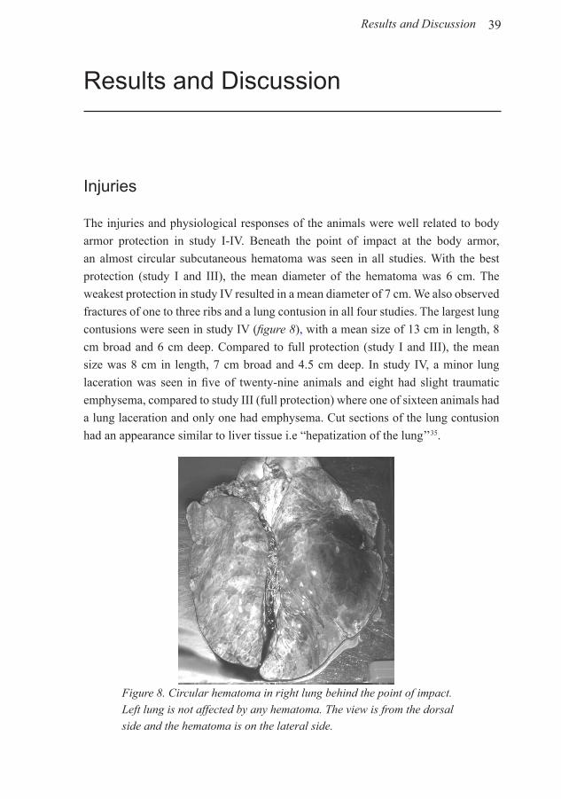
had an appearance similar to liver tissue i.e “hepatization of the lung’’35.
Figure 8. Circular hematoma in right lung behind the point of impact. Left lung is not affected by any hematoma. The view is from the dorsal side and the hematoma is on the lateral side.
Results and Discussion
40 Dan Gryth
In study II and IV some exposed animals demonstrated dense cardiac tissue, in
other studies referred to as a “stone heart”94. This change was only seen in deceased
animals that suffered from low PaO2 for a longer period during the experiment. In
study II, dense cardiac tissue was observed in 3 of 18 exposed animals, and in study
IV in 11 of 29 exposed animals. In the control groups and in study I and III, there
were no observations of dense cardiac tissue.
Myocardial stunning has also been described after global myocardial ischemia95. The
cause of myocardial stunning is probably myocardial hypoxia leading to depletion of
adenosine triphosphate (ATP), making actin-myosin binding irreversible. Myocardial
stunning has been shown to occur in vivo with as little as 50% depletion of ATP95,
96. Therefore, one might hypothesize that dense cardiac tissue could be an important
tool for forensic scientists, to evaluate if a person suffered from grave hypoxia before
cardiac arrest. Hypothetically, the dense heart and death can perhaps be prevented by
early ventilation with extra oxygen content.
Mortality
Mortality caused by BABT was documented in study II and IV (worst injury), and in
study III in which one animal died. In study II, 2/8 (25 %) of animals in the 34mm-
group and 5/10 (50 %) in the 40mm-group died due to the trauma. In study IV, 13
of 29 (45 %) exposed animals died due to the trauma. Fluid resuscitation did not
improve survival rate in study IV. The animals who died from the injuries, except
three, died within 60 minutes.
Respiration
Apnea was only detectable in study III and IV, since the respiration of animals in
study I and II where regulated by a ventilator. This experimental set-up resulted in
an apnea of 22 (6-44) seconds in study III and 146 (46-210) seconds in study (IV).
More energy was transferred to the lung tissue in study IV, indicating that more
stretching of the lung and the C-fibers give a longer apnea period. This has also been
published by Adams et al. 1987 and Jaffin et al. 1987, showing that the apnea period
was proportional to the load of hyperinflation or blast distance30, 97.
In this thesis, vagotomized animals in study III were protected from apnea, clearly

41
showing that apnea during BABT is a vagally mediated reflex. Based on the results
from our studies, it is a fair assumption that early supported ventilation might reduce
other pathophysiological effects of BABT. This hypothesis is supported by the
observation that desaturation was more pronounced in groups suffering from apnea. In
study IV all animals had apnea, but some started the breathing with gasping by means
of very low respiratory rate, until the animals were supported by the ventilator. 13 of
29 animals in study IV died during the experiment and showed immediately before
death yet again a breathing pattern of gasping. Gasping is an universal phenomenon
in mammals (also known as agonal respirations), originating in the medulla of the
central nervous system, and is the terminal breathing pattern that occurs after anoxia
or ischemia98. In humans, gasping is prevalent during cardiac arrest and has been
recognized in 30-40 % of witnessed episodes of cardiac arrest in adults99. Thus, when
gasping occur, it is an important sign for bystanders to start resuscitation99.
Arterial oxygen saturation (SaO2) decreased directly after the trauma in all studies,
a result well related to body protection and size of lung contusion. A contributing
factor for the desaturation was probably increased “dead space ventilation”, due
to the lung contusion. Furthermore, bleeding inside airways resulted in flooding
of non-injured parts of the lungs, which probably caused occlusion of the alveoli
(shunt). Such ventilation-perfusion changes have recently been reported in another
experimental pulmonary contusion model42. In our studies, hemoptysis was only
observed in exposed animals in study II (n=10/18) and IV (n=29/29). In the ventilated
studies (I and II), the lowest SaO2 was observed after 30 minutes, measuring 91% and
60%, respectively. For animals in study III and IV, SaO2 was most depressed after
1 minute, measuring 83% (non-vagotomized animals in study III) and 35% (study
IV). SaO2 was improving over time in surviving animals in all four experiments. An
explanation might be that in the later phase ventilation/perfusion (V/Q) developed a
better matching. In contrast, Mosely et al. 1970 observed decreasing partial pressure
of oxygen in arterial blood (PaO2) until 2 hours after the lung contusion19, possibly
because the animals were ventilated by 100% oxygen which can create further
atelectasis and increase the shunt100. However, during routine induction of general
anesthesia, 80% oxygen content caused minimal increase of atelectasis100.
It should be mentioned that the animals that died in study II and IV demonstrated
more severe arterail oxygenation desaturation than surviving animals, an observation
indicating that low SaO2 is an important cause of death in our studies.
PaO2 and mixed venous oxygen saturation (SvO
2) showed similar pattern as SaO
2 in
Results and Discussion

42 Dan Gryth
all four studies, although data for these parameters is shown only in study I (SvO2)
and IV (PaO2). In study IV there was a marked decrease of PaO
2 after the trauma in
all three exposed groups. Between 30 and 120 min post impact, HSD-treated animals
displayed significantly better recovery of PaO2, compared to the shot control group,
and a tendency (p = 0.09) toward better recovery compared to the RA group.
Although the evidence are not clear-cut, study IV indicate that it is possible that
edema formation contribute to the desaturation after BABT. Our study showed that
the group treated with RA significantly increased the relative water content of the
injured lung, when compared to the HSD, shot control and unshot control group. This
difference between the RA and HSD groups is an important finding in study IV, since
it shows that fluid resuscitation with RA might increase edema after a pulmonary
contusion, compared to treatment with HSD. Previous studies show conflicting data
regarding the effectiveness of HSD in preventing lung edema92, 101. It should also
be mentioned that there were no significant differences between the groups, in the
amount of lung water in the corresponding left unharmed lung, indicating that the
fluids did not induce edema in non-injured tissue.
Oxygen delivery (DO2) and oxygen extraction (O
2ER) were reported in study I.
These parameters are influenced by changes in SaO2 and CO. The SaO
2 and CO was
deteriorated due to the trauma, leading to a marked drop in DO2 early after impact in
study I. An increased O
2ER compensated for the reduced DO
2, but this compensation
did not seem to be sufficient, since the blood lactate level increased, as a sign of
peripheral anaerobic metabolism.
Hemodynamics
Mean arterial pressure (MAP) decreased immediately after the trauma in all four
studies. In study II and IV, MAP declined about 50% during the first minute. With
full protection (study I and III), the decrease of MAP was only about 25%. At 5
min post impact, exposed animals in study II-IV had almost recovered to baseline
level, followed by a subsequent decrease. Animals in study I differed from the other
animals, since MAP was stable after the 5 minute period.
MPAP increased directly after the hit in all four studies. In study IV (worst injury),
43
it increased from 22 to 32 mmHg, in exposed groups. After 15-30 minutes MPAP
had decreased back to baseline level in all studies. The rationale for the decrease is
probably redistribution of lung circulation, i.e increased blood flow in parts of the
lung not affected by the trauma. Furthermore, connections between lung arteries and
lung veins (von Hayeks vessels), has been suggested to open if MPAP is markedly
elevated 102.
Cardiac output (CO) was also affected in all four studies, decreasing in study II from
approximately 5.5 to 3.5 L/min after 1 minute. In this study, CO did not recover to
baseline for the rest of the experiment. In study II and IV, systemic vascular resistance
(SVR) showed a similar pattern as MAP, with an initial drop (approximately from
2000 to 1500 mmHg/L/min), recovery, and a subsequent slow decrease (data not
shown).
In study IV, we observed a striking tackycardia, from approximately 90 to 150
beats/minute, 10 minutes after the hit. The tackycardia was maintained for the rest
of the experiment in the shot control group, but there was a significant decrease
of HR in groups receiving fluids. We monitored heart frequency directly (0-1 min)
after the trauma only in study III. This was done using the ECG-recordings, since
we considered it necessary to compare our data to other studies showing decreased
heart rate early after trauma19, 20. We observed a slight decrease the first half minute,
although only in the non-vagotomized group in study III.
In our studies we have undoubtly shown that the circulation has been affected by the
trauma. One explanation might be pulmonary vascular occlusion, due to the lung
contusion, leading to derange of lung circulation. This hypothesis is supported by the
increased MPAP directly after impact, as well as the decrease in MAP and CO. These
changes has also been shown after blast trauma against the right thorax103. Furthermore,
our manual examination of lung tissue showed coagulation and occlusion of the lung
circulation, as well as disrupted vessels in the area of the pulmonary contusion.
Another possible explanation to the fast circulatory failure is a neurological reflex
after the trauma, inducing bradycardia and rapid relaxing of the vascular tonus. It has
been previously suggested that the vagus nerve is involved in the hypotension after
blast trauma22, 24, 104. An early study by Clemedson et al. showed that hypotension
after blast trauma could be prevented by a combination of bilateral vagotomy and
blocking of the carotid sinus region, with local anesthesia105. Furthermore, Daly
Results and Discussion
44 Dan Gryth
and Kirkman showed that stimulation of C-fibers results in a symphatho-inhibition,
leading to decreased peripheral vascular resistance and hypotension106. In our studies,
we noted a fast decrease of MAP, and a slight, but significant, decrease in heart rate
during the first minute in study III. In this study, the vagotomy prevented animals
from decreased heart rate, but had no effect on MAP or MPAP.
A third theoretical contribution to the circulatory effects is release of inflammatory
mediators after trauma. Cell destruction after a lung contusion can induce an out-
flow of stored mediators, for example endothelin-1. This is a potent vasoconstrictive
and pro-inflammatory peptide, which is produced in the vascular endothelium. The
plasma half-life of endothelin-1 is only approximately 2 min in vivo, but the effects
persist up to 60 min after administration in humans107. It has been demonstrated that
the production of endothelin-1 is increased after trauma108 and sepsis90. In study IV,
we found a rapid and short-lived increase of endothelin-1 in two out of three groups 7
min after impact. Hypothetically, the short half-life of endothelin in vivo suggests that
these levels could have been even higher earlier after impact. In agreement with this
finding, a previous study of experimental cardiac ischemia showed that the highest
levels of endothelin could be measured already after 2 min109. Albeit the short eleva-
tion in plasma, the effects of endothelin may have persisted for a longer time-period,
thus contributing to the pulmonary hypertension.
A fourth contributing mechanism to increased MPAP can be hypoxic pulmonary va-
soconstriction (HPV)110, 111.
In study II-IV we also noted a subsequent, slower decrease of MAP and CO, fol-
lowing the first recovery. Hypothetically, the decrease in this second phase could be
an effect of ATP-deficiency, caused by low oxygen in the blood. Deficiency in ATP
causes decreased constrictive effect in vessels and reduced contractile ability of heart
muscles, leading to lower CO and MAP. Furthermore, severe hypoxia increase the
adenosine in blood and can stimulate the adenosine-receptors A2A
and A2B
in extreme
conditions, leading to vasodilatation112. These theories is supported by study I, where
SaO2 was only modestly affected, with no subsequent decrease of MAP.
In contrast to other studies19, 34, 92, fluid treatment in study IV had no effect on MAP,
CO or survival rate in our shock model. The explanation is probably that our lung
contusion was more severe, inducing more severe hypoxia that caused failure of
circulation in the delayed, second phase. Supported ventilation with extra oxygen
would most likely have led to a positive effect on circulatory parameters.
45
Cerebral effects
EEG
Electroencephalogram (EEG) was recorded before and after impact in study I-III. To
our knowledge was study I the first scientific article that demonstrates EEG-changes
after chest BABT. An immediate slowing of the EEG-activity has been previously de-
scribed in pigs following blast wave exposure caused by detonation of explosives33,
and after high energy missile trauma in the hind limb of pigs32.
In all our three studies the EEG became affected as early as 15-30 seconds after
impact. These changes from the baseline pattern, indicates that the trauma almost
instantly induced a global cerebral dysfunction. The changes were in most cases
temporary, but some of the most affected animals never recovered from the isoelec-
tric pattern. Other showed a depressed EEG-activity that gradually demonstrated a
further decrease down to iso-electric pattern.
When comparing EEG-changes in study I, against study III (equal protection), the
non-vagotomized group in study III was more affected by the trauma (table 4).
The methodological differences between study I and III were that in study I the
respiration was controlled by a ventilator, and in study III the animals were breathing
spontaneously. Therefore, non-vagotomized animals in study III suffered from
apnea, resulting in lower oxygenation of the blood, and consequently generating a
slightly negative influence on EEG, compared to study I. In contrast, the bilaterally
vagotomized group in study III was protected from apnea. These vagotomized
animals showed slightly reduced EEG-changes after trauma compared to the other
two groups (table 4).
One likely reason for the reduced EEG-changes in vagotomized animals might be
that this group were protected from apnea. Another, or contributing, possibility is
that the EEG-changes are mediated through the vagus nerve. Support for this hy-
pothesis is the fact that vagal afferent nerve fibres is linked to several different parts
of the brain28, 113, 114 and can therefore affect EEG. Balzamo et. al showed that electri-
cal and chemical stimulation of afferent vagal C-fibres in cats causes an immediate
depression of the background EEG-rhythms, unrelated to changes in cardiovascular
parameters113, 114. Bilateral vagotomy markedly reduced these EEG-changes, despite
a persistence of cardiovascular effects, indicating that vagal afferent signals might
directly affect the EEG-rhythm.
Results and Discussion
46 Dan Gryth
That the EEG-changes in our study is caused solely by direct influence from a shock
wave transmitted from the area of impact to the brain, either through the blood ves-
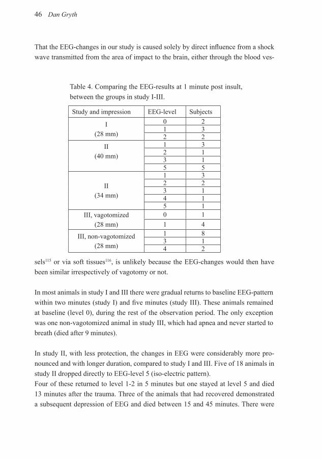
Study and impression EEG-level Subjects
I(28 mm)
0 21 32 2
II(40 mm)
1 32 13 15 5
II(34 mm)
1 32 23 14 15 1
III, vagotomized(28 mm)
0 1
1 4
III, non-vagotomized(28 mm)
1 83 14 2
Table 4. Comparing the EEG-results at 1 minute post insult,
between the groups in study I-III.
sels115 or via soft tissues116, is unlikely because the EEG-changes would then have
been similar irrespectively of vagotomy or not.
In most animals in study I and III there were gradual returns to baseline EEG-pattern
within two minutes (study I) and five minutes (study III). These animals remained
at baseline (level 0), during the rest of the observation period. The only exception
was one non-vagotomized animal in study III, which had apnea and never started to
breath (died after 9 minutes).
In study II, with less protection, the changes in EEG were considerably more pro-
nounced and with longer duration, compared to study I and III. Five of 18 animals in
study II dropped directly to EEG-level 5 (iso-electric pattern).
Four of these returned to level 1-2 in 5 minutes but one stayed at level 5 and died
13 minutes after the trauma. Three of the animals that had recovered demonstrated
a subsequent depression of EEG and died between 15 and 45 minutes. There were
47
also two other animals that showed only minor changes initially, but demonstrated
a slow subsequent decrease down to iso-electric pattern (died at 62 and 103 minutes
respectively). The first change in EEG was seen as early as 15 seconds after trauma.
After only 15 seconds the blood was not desaturated, so it seems likely that the main
cause of EEG changes in study II is the fast drop of MAP or/and a direct nerve reflex
to CNS.
As previously mentioned, we also observed a subsequent depression of EEG in study
II, after the first recovery. This change in EEG was seen in both deceased and surviv-
ing animals, but more frequently among the deceased animals. During this phase of
the experimental course, blood pressure and desaturation decreased slowly. Serious
hypotension or hypoxia did not affect EEG separetely, but a combination of the two
parameters seems to induce EEG-depression.
There were no EEG-changes in two of the exposed pigs in study I and one in study
III. Although our experimental protocol is standardized, small differences in body
weight or where the impact was on the torso must be taken into account. Biological
variation in sensitivity to trauma might also cause some deviation in our results.
In conclusion, our results indicate that an early effect on EEG is probably from vagal
influence on CNS and/or the fast hypotension. It seems likely that these changes
can be aggravated by hypoxia. The results from study III support that hypothesis,
since vagotomy provided some protection from the depression of EEG, either as a
direct effect on CNS or indirectly by preventing apnea. The delayed effect on EEG
in the second phase is probably due to influence of serious hypotension and hypoxia,
especially in combination of these.
The EEG-pattern is registered from the brain cortex, but since the cortical activity
is dependent on input from subcortical brain structures, the observed changes
could reflect a direct effect on vital centers in the brainstem, such as the centre for
wakefulness. The fact that the EEG-changes were general could be an indication that
the signals were elicited from deep brain structures, and consequently not being signs
of local processes from more superficial structures of the hemispheres.
Our opinion is that a human suffering from the observed EEG changes would be
incapacitated for a number of minutes, due to the cerebral effects only. The initial
Results and Discussion
48 Dan Gryth
incapacitation can be lethal, if this person lose the ability to return fire or move to
shelter. These results clearly demonstrate the importance of studying a possible
influence on the cerebral function also in trauma not directed to the head.
In situations where the victims is conscious (but have apnea), there is no need for
ventilatory support, because they will soon start to breathe spontaneously and there
is no risk for sever hypoxia. However, EEG-changes of level 3-5 should probably
result in unconsciousness and such a victim might have problems with obstructed
airways if they begin to breathe before they are conscious. Our results in this
thesis emphasize the importance of creating free airways if unconscious persons
seems to have breathing difficulties. Furthermore, supported ventilation should be
administrated to unconscious persons with apnea.
S-100
In study I we hypothesized that our observed negative effects on EEG might induce
brain injury. A marker for brain injury is S-100, a protein mainly produced in the
central nervous system by neurons and/or glial cells117. It has been suggested that
S-100 can be measured in blood as a biomarker for traumatic brain damage and
leakage of the blood-brain barrier118, 119. We sampled S-100 in study IV, where we
had the most severe shock and hypoxia. About 50% of the animals died due to the
trauma, but we detected no increase of S-100 in any of the animals. We therefore
conclude that no leakage of the blood-brain barrier occurs in our experimental set-
up, even though we observed severe EEG-changes.
Blood samples
Lactate
Blood lactate increased after impact and was reported in study I, II and IV. Lactate
increases when the blood flow decreases in the microcirculation, leading to impaired
oxygen delivery and the generation of lactic acid, via anaerobic glycolysis120. Serum
lactate measurement is recommended as a test to estimate and monitor shock75, but
also as a prognostic parameter in septic shock121.
Baseline values of blood lactate in our studies was approximately 1 mmol/L.
Blood lactate increased in study I to 2 mmol/L at 120 min. Study II showed an
increase to approximately 4 mmol/L, for both exposed groups, at 60 minutes. In
49
study IV blood lactate increased significantly in all exposed groups, compared to the
unshot control group. The shot control group reached the level of 9 mmol/L at the
end of the experiment, the HSD group 5.5 mmol/L and the RA group 4 mmol/L. It
should be mentioned that these values in study IV are very high and reflect the low
oxygenation and deteriorated circulation in that study. It was somewhat surprising
that blood lactate was higher in the HSD group, compared to the RA group, since
HSD has been shown to improve the microcirculation in other studies78.
Potassium
Potassium levels in serum was reported in study II and IV. In study II, both exposed
groups showed a transient increase of serum potassium after the trauma, with the peak
value at 5 minutes (the 40 mm-group increased from 4.2 to 5.0 mmol/L). Study IV
showed the same pattern, but the peak was much higher with the three exposed groups
increasing from 4.1 mmol/L to 7.7 mmol/L. Several animals had serum potassium
close to 9 mmol/L and the highest level was 11.4 mmol/L. We believe that massive
cell destruction in lung tissue induced the elevated S-potassium 5 min after the hit.
Several animals showed elevated T-waves, widened QRS-complexes and flattened
P-waves, on the ECG-recordings, which is typical for hyperkalemia122. The high S-
potassium levels observed in our studies should be considered life-threatening, since
it may lead to lethal arrhythmias123. The most serious arrhythmias we noticed on ECG-
recordings were ventricular fibrillation (VF) and asystoli. In study (IV) there were
two animals with VF 3 minutes after the hit and one with asystole. These arrhythmias
sustained approximately one minute and converted spontaneously to sinus rhythm,
which is rare according to previous studies124, 125. Two other animals died before 10
minutes and were excluded from the study (one had VF and the other asystole). In
study II, one animal died after 13 minutes, due to VF, with a serum potassium level
of 8.6 mmol/L at 5 minutes, and 6.7 mmol/L at 10 minutes.
In study I and III (full protection), the pulmonary contusion were much less extensive
than in study II and IV. Potassium in serum only increased from 3.9 mmol/L to
4.2 mmol/L (study I) and from 4.1 mmol/L to 4.3 mmol/L in study III. The results
indicate that serum potassium is related to the size of pulmonary injury.
Our observation of massive hyperkalemia shortly after a lung contusion seems to be
a new finding. It has been shown previously that some patients with crush injuries,
as well as other injuries, have developed hyperkalemia126, but not to the level we
observed in study IV. The short-lived peak observed in our studies have never been
documented in authentic trauma cases, which is not surprising, since it is usually
Results and Discussion

50 Dan Gryth
not possible to collect blood samples immediately after an accident. Human victims
who sustain high-energy blunt chest trauma (traffic accidents or fall from heights)
would probably get similar levels of hyperkalemia in the blood, and this might hypo-
thetically be the explanation to some unclear deaths in human trauma victims. These
findings tell us that it might be beneficial to perform cardio-pulmonary resuscitation
(CPR) early after isolated blunt chest trauma, if there is no pulse. This may be effi-
cient since the potassium decrease very fast, so the possibility exists that the arrhyth-
mias spontaneously convert to sinus rhythm (as we observed in study IV), while CPR
protects from hypoxic brain damage in the meantime.
Hemoglobin
In study IV we observed a marked increase of hemoglobin in all exposed groups,
which was a surprising finding. As an example, hemoglobin increased in the RA
group from 113 mg/L to 133 mg/L after 5 minutes, but was back at baseline level at
30 minutes. We also observed increased hematocrite which is in agreement with the
results from hemoglobin.
Surprisingly, Hb did not increase in study II although the trauma was nearly the same.
One difference was that the respiration in study II was controlled by a ventilator,
compared to study IV where the animals were breathing spontaneously. After 5
minutes, PaO2
was 6 kPa in the 40 mm-group in study II, and 4 kPa in study IV.
MAP was approximately 55 mmHg in both studies. Our hypothesis is that the pigs
have a compensatory mechanism to counteract hypoxia and fast circulatory failure
by releasing red blood cells from the liver and/or spleen. The purpose of such a
mechanism could be to increase oxygen transporting capacity in life-threatening
situations. Later in the experimental course of both study II and IV, MAP and PaO2
decreased slowly to similar levels, but no increase of Hb was observed. Therefore,
our hypothesis is that the fast drop of MAP and PaO2, early in the experimental
course of study IV, is an important factor for the increase of Hb in that study.
It has previously been shown that stimulation of sympathetic nerves mobilizes blood
from the liver127. Perhaps a fast and potent drop of MAP and PaO2 stimulates the
sympathetic activation to mobilize blood from the liver and/or the spleen. A pure
reflex is not likely because the traumatic impact was powerful in both studies (II and
IV), but Hb was only increased in study IV.
To our knowledge, a similar fast increase of blood hemoglobin after trauma has not
been described previously. It has been shown that hemolysis occurs after blast trauma,
particularly after fast shock waves128. However, it is unlikely that the early increase
51
of hemoglobin in our study is due to hemolysis of red blood cells, since we measured
both intracellular and free hemoglobin. Another, or supplementary, explanation is
that the trauma induces rapid leakage of fluid through the endothelium and therefore
results in a higher concentration of red blood cells. It would be very interesting to
prevent circulation from the liver/spleen to investigate if the red blood cells are
recruited from these organs in our experimental model. Another interesting question
for the future is if a sympathetic acting drug as epinephrine given intravenously can
increase Hb.
Glucose
Only in study II and IV, we observed significantly increased levels of glucose in
blood. Between 5 to 30 minutes after impact, all exposed groups in study IV and
the 40 mm-group in study II had significantly higher blood glucose, compared to
the unshot control group. Both the HSD and the RA groups in study IV had lower
B-glucose, compared to the shot control group, and the reason might be dilution of
B-glucose after resuscitation.
Circulatory shock and stress release catecholamines, glucocorticoid and glucagon
which all contribute to release glucose from the liver. Moreover, it has been shown
that catecholamines can induce insulin resistance, thus potentiating the concentration
of glucose129. However, glucose can also be metabolized via biosynthesis pathways,
leading to the synthesis of O-linked beta-N-acetyl-glucosamine (O-GlcNAc). It
has been suggested that increased levels of O-GlcNAc improves cell survival after
ischemia/perfusion injury, and improves cardiac function, organ perfusion and
attenuates the inflammatory response in rodent models of trauma-hemorrhage130.
Trauma patients submitted to the intensive care unit (ICU) often present hyperglycemia
and hyperinsulinemia, suggesting overall insulin resistance and possibly impaired
compensatory response of ß-cells131. Two large, prospective, randomized, controlled
clinical studies of critically ill patients, have demonstrated that strict maintenance of
normoglycemia with insulin infusion, for at least a few days, reduce morbidity and
mortality132, 133. Although these studies indicated that persisting hyperglycemia exert
negative effects on patient outcome, it is tempting to speculate that hyperglycemia
generate a positive effect on morbidity and mortality in the short perspective, early
after a life-threating trauma.
Inflammatory responseWhen planning study IV, we hypothesized that we could detect an inflammatory
Results and Discussion

52 Dan Gryth
response after the lung contusion. Furthermore, the HSD-solution has been shown to
cause anti-inflammatory effects84, 86, 134, so our aim was also to investigate if we could
observe such an effect in our resuscitation study. Blood was taken after two hours
and analyzed for cytokines, and lung tissue was dissected post mortem to investigate
recruitment of neutrophils. TNF-α was significantly increased in the shot control
group, compared to the unshot control group. The RA and HSD groups displayed
also increased TNF-α, although not significantly, compared to unshot controls. This
indicates that the chest trauma in our model induced an inflammatory response, that
could be detected in the systemic circulation. We can only speculate if the lower
cytokine level in groups receiving fluid was an effect of dilution or if fluid treatment
down-regulate the inflammatory response after BABT. Resuscitation with lactated
Ringer’s solution after hemorrhagic shock has been shown to induce an upregulation
of various neutrophil and endothelial adhesion molecules135. However, in contrast to
some previous publications136, we can conclude that fluid treatment did not induce
an augmented systemic inflammatory response in the systemic circulation, when
measured 2 hours after impact.
IL-6 was not increased in any of the groups and no neutrophil accumulation was
observed in the lungs. Most likely is a 2 hours surveillance period too short to
discover any rise in IL-6 or neutrophil accumulation. This assumption is supported
by a previous experiment in rats, studying isolated bilateral lung contusion. After
12 hours they did not observe any increased IL-6 in BAL, but noted a significant
elevation after 24 hours137. In a similar experiment, Raghavendran et al. found
significantly increased number of leukocytes in BAL after 24 hours138.
It is well known that severely injured trauma patients have activated neutrophils135.
TNF-α is a key mediator in the early inflammatory response, suggesting that
activation of neutrophiles were induced at least in the shot control group in study IV.
It is also likely that a similar response was induced in the other studies, especially in
study II (with similar protection as study IV).
Microscopic examination of lung tissue showed no significant differences between
the exposed groups regarding the degree of congestion, edema or atelectasis in
the specimens from right lungs. In the kidneys, no fibrin deposition could be
demonstrated, and there were no signs of tubular obstruction or necrosis. Biopsies
from the left heart ventricle demonstrated microscopically scarce, focal contraction
band necrosis, but no reactive inflammatory changes. With this staining we could not
53
find any evidence, why some heart had dense contracted tissue in study II and IV.
We probably need other staining methods to investigate this interesting phenomenon,
and this could hopefully be done in future studies.
Results and Discussion

54 Dan Gryth
IndividTime 45 60MAP 53 43SaO
261 69
EEG 4 4Time 60 75MAP 71 76SaO
247 47
EEG 0 0Time 105 120MAP 54 52
55
Conclusions
This thesis is based on a model of behind armor blunt trauma (BABT), that was originally developed by the NATO Task Group on BABT. Our group was the first to set up this model of lung contusion in Sweden. Besides the parameters measured by the NATO task group, we have further developed the model and included EEG, insertment of an optical pulmonary thermo dilution catheter to measure central venous pressure (CVP), mean pulmonary artery pressure (MPAP), cardiac output (CO) and mixed venous saturation (SvO
2). Moreover, sampling of blood to analyze
endothelin-1, TNF- α, IL- 6 and S-100 have not been done before, when studying BABT. We have also performed bilateral cervical vagotomy in pigs exposed to BABT, and explored the physiological response (study III). In study IV, we have used our model to investigate effects of fluid resuscitation after a pulmonary contusion, accompanied by shock. Other findings in this thesis include some previously unknown physiological responses after BABT, which has given us better understanding of body reactions after severe injuries.
In all studies included in this thesis, the pulmonary contusion has been the central injury. Our opinion is that the observed injuries and physiological responses after the trauma are accurately related to level of protection in study I-IV. Our physiological results, especially when monitoring severe BABT, showed apnea for several minutes with serious desaturation, immediate hypotension, decreased CO, increased MPAP and depressed EEG-activity. The circulatory system did some recovery after trauma, but showed a subsequent decrease, probably due to the lack of oxygen.
Early after the lung contusion, the damage of cells caused a short peak of hyperkalemia, causing in some cases lethal arrhythmias. This short-lived peak of potassium, following cell damage, seems to be a new finding and has to our knowledge never been documented before.
Animals that suffered from severe hypoxia for a longer period and then died, demonstrated dense contracted cardiac tissue during autopsy. This is a surprising finding that should be investigated in further studies.
Blood hemoglobin showed an early peak after 5 min, hypothetically as a defense mechanism in a life-threatening situation. To our knowledge this is also a new finding.
Conclusions
56 Dan Gryth
This thesis has also been the first to document EEG-changes after BABT. EEG has been graded from 0 (normal EEG) to 5 (isoelectric EEG). We can conclude that even with “full protection”, spontaneously breathing pigs showed EEG-pattern that was depressed to level 4 in some animals. Our data indicate that the trauma almost instantly elicited a global cerebral dysfunction. This is probably caused by a fast decrease of blood pressure and/or a nerve reflex. Desaturation is likely contributing to the depressed EEG, especially in combination with low MAP late in the experimental course. It should be mentioned that it is complex to transfer the results from anaesthesised pigs directly to awake human subjects. However, it seems that a human suffering from the observed EEG-changes would be incapacitated for at least a number of minutes.
From study I, combined with study II, we have documented an effect on
pathophysiology and mortality in conformity with the level of body armor protection.
In these studies the impression in clay, placed behind the different body armors, were
28 mm (study I), 34 mm and 40 mm (study II). The mortality was 0% (28 mm), 25%
(34 mm) and 50% (40 mm). Our data is clearly demonstrating that the safety criterion
of 44 mm impression for body armor is insufficient when they are exposed to high
velocity projectiles.
The bilateral vagotomy in study III prevented apnea, and induced a slight, positive
effect on the EEG-depression. We can conclude that the apnea after BABT is a vagally
mediated reflex.
None of the fluids given in study IV had any significant effect on MAP or CO in this
type of shock. The HSD-group displayed significantly improved PaO2 compared to
the shot control group, and a tendency compared to the RA-group. Moreover, the
group treated with RA significantly increased the lung wet/dry weight in the injured
lung, when compared to the other groups. We also observed lower TNF-a release
and decreased HR in animals receiving fluid. However, given the fact that we did
not observe striking effects, it seems like fluid resuscitation has limited therapeutic
effects for treatment of BABT.
Who dares wins!
SAS - Special forces
57
Acknowledgements
Jag vill från djupet av mitt hjärta tacka dessa personer, som på ett eller annat sätt
hjälpt mig att genomföra denna avhandling:
Thomas B Kjellström, min förste huvudhandledare, som visade vägen in i forskningen
och med sitt skarpa intellekt var en god förebild. Vi jobbade hårt och hade kul, men
vi hade även tid att ta en och annan öl. Inför halvtiden förberedde han mig väl trots
att han knappt orkade ta sig till jobbet p.g.a. sin sjukdom. Men vi klarade det, och vi
gjorde det tillsammans vilket det gladde oss mycket. Strax därefter lämnade han oss,
sommaren 2006. Thomas var även en god skeppare, och därför känns det nu så skönt
att föra disputationen i hamn. Tack.
Ulf P Arborelius, min huvudhandledare efter Thomas bortgång, som förnämligt har
fortsatt att utbilda och utveckla mina kunskaper inom forskningen. Jag har uppskattat
att Ulf är en god lyssnare av idéer och teorier och att han alltid backat upp med de
resurser som har funnits. Sedan har han alltid på lut, en liten vits med knorr.
David Rocksén, min handledare som kom in i matchen efter att Thomas blev sjuk.
Det är sällan man träffar på en person med en sådan skärpa och förmåga att inta ny
information på kort tid. Vi har kämpat i vått och torrt, och fyllt många papperskorgar
med RedBull för att hålla oss vakna på småtimmarna. En god vän som alltid finns
till hands.
Dan Drobin, min handledare som lärde mig grunderna i prepareringen, och var ett
stöd i utvecklingen av modellen och metoderna. Dan är en idéspruta och uppfinnare
som har stimulerat och berikat mitt sätt att fundera kring forskningen.
Professor Sten G E Lindahl, min tidigare chef på anestesi- och intensivvårdsklinken
vid Karolinska Universitetssjukhuset, som alltid har stöttat och trott på mig, även när
jag var ny anestesiolog på kliniken. Sten har lagt grunden till ett gynnsamt och trevlig
forskningsklimat på kliniken. Jag är mycket tacksam för att han förde in mig i denna
värld och initierade min forskning på FOI.
Acknowledgements
58 Dan Gryth
Generalläkare Ann-Marie Göransson, som skapade forskningstid och möjligheten
att forska på FOI.
Lars Irestedt samt Professor Claes Frostell, min tidigare respektive nuvarande
chef på anestesi- och intensivvårdsklinken vid Karolinska Universitetssjukhuset.
De har fortsatt att hålla en trevlig stämning på kliniken och en stimulerande
forskningsmiljö. Utan deras stöd hade detta inte varit möjligt.
Professor Lars I Eriksson, min handledare under ST-utbildningen, som alltid har
funnits i bakgrunden för att stötta både kliniskt och inom forskning. Lars har en
entusiasm för forskning och trauma som smittar av sig på ett härligt sätt.
Jenny Bursell, för ett bra och skickligt arbete på laboratoriet, för all hjälp med datainsamling och figurer. För all den mentala support och uppmuntran du har gett, vilket har varit av stort värde.
Lars-Gunnar Olsson för teknisk skicklighet och bra diskussioner när fysik och biologi skall vävas samman. 40 års erfarenhet inom forskningen är inte att underskatta. Det är alltid en positiv stämning när L-G är med och den blir inte sämre av alla goda bakverk som han har med till fikat.
Jonas Persson, medförfattare och specialist inom neurologi som på ett förtjänstfullt sätt bidragit till arbetena I-III. Vi har analyserat och diskuterat resultaten på en akademisk nivå och det är då forskningen är som bäst.
Professor Eddie Weitzberg, medförfattare med sin stora erfarenhet av bl.a. endothelin-1. Eddie är en inspirationskälla till att forska och i hans grupp finns det mycket värme. -”Det finns inget häftigare än att forska, det enda som skulle kunna konkurrera vore att vara en stor rockartist”.
Henrik Druid, medförfattare till histologi avsnittet. En bra forskare ut i fingerspetsarna, och ett energiknippe som får Duracellkaninen att se ut som om den rör sig i slowmotion. En person som man blir inspirerad av och gärna samarbetar mera med.
Anders Sondén, medförfattare till arbete III, en god kirurg och forskare. Anders har varit en stor tillgång vid de vetenskapliga diskussionerna och under experimenten.
59
Elisabeth Malm, som alltid utför ett bra arbete i laboratoriet och den trevliga atmosfären som hon alltid skapar.
Ulf Berg, Mårten Risling, Lena Nordin, Maria Angéria, Inga-Lisa Larsson, Mattias Sköld, Louis Riddez, Thomas Landegren, Ola Eiken, Kaj Olmegard, Melker Skoglund, mina vänner och kollegor på FOI som har gjort arbetsplatsen till en stimulerande, trivsam och givande miljö.
Sari Ponzer, Christer Svensén och Anita Stålsäter Pettersson, för det goda administrativa forskningsstöd som jag haft från SÖS.
Kerstin Larsson, Kerstin Staffansson och Christina Nordh-Claesson, för att ni alltid är positiva och underlättar med administrativ support.
Magnus Backheden, för väl utförda statistiska analyser och bra stöd vid tolkningen av dessa.
Janet Holmén och Professor Joe De Pierre, för utmärkt språkgranskning.
Ringvor Hägglöf, för fint arbete med lay-out och alltid positivt bemötande.
Leif Lindblad, min gamle mentor som hjälpte mig vid uppstarten till doktorandregistreringen. Med all sin erfarenhet om livet och kirurgiska kunskaper har han lärt mig mycket, inte minst från Kenya när vi bl.a. opererade krokodilbett.
Anders Jakobsson, som har stöttat och kommit med goda kommentarer till skrivarbete i studie IV. Även för god vänskap i mörkaste Afrika.
Hans G Hägglund, en av mina allra bästa vänner, för alla givande diskussioner vi har haft om forskning och livet, i löparspåret, på skridskoturerna eller i bastun. En god kamrat och en förebild för hur man får familjen, klinik/forskning och fritiden att gå ihop.
Till alla mina arbetskamrater på anestesi och intensivvårdskliniken vid Karolinska Universitetssjukhuset, för att de gör kliniken så trivsam och stimulerande.
Acknowledgements
60 Dan Gryth
Till alla i släkterna Gryth och Grip för att de alltid finns där i glädje & sorg.
Till mina föräldrar Ingegerd och Tage som alltid kärleksfullt stöttat mig i min framfart genom livet.
Till mina kära barn Olivia, Fredrik och Jonathan som ger mig glädje, och lärt mig vad som är viktigast i livet.
Annette, min älskade fru som alltid har stöttat mig och varit förstående trots att jag ofta fysiskt eller mentalt varit i en annan värld. För att du alltid är kärleksfull och med mental spänst stimulerar alla mina sinnen. Du är min kärlek.
Denna avhandling har finansierat med hjälp från:
Försvarsmakten (FM)
Anestesi-, Operations- och Intensivvårdskliniken,Karolinska Universitetssjukhuset, Solna, Stockholm
Försvarets Materielverk (FMV)
Totalförsvarets Forskningsinstitut (FOI)

61
References
1. Cannon L. Behind armour blunt trauma--an emerging problem. J R Army Med Corps 2001;147(1):87-96.
2. Laurence BT, Iremonger MJ. Modern body armour and helmets: An introduction. Canberra, Australia: Argo press; 2006.
3. Iremonger MJ, Gotts P. Practical personal ballistic protection: past present and future. In: PASS Symp; 2002; The Hauge, The Netherlands; 2002. p. 19-30.
4. Gofrit ON, Kovalski N, Leibovici D, Shemer J, O’Hana A, Shapira SC. Accurate anatomical location of war injuries: analysis of the Lebanon war fatal casualties and the proposition of new principles for the design of military personal armour system. Injury 1996;27(8):577-81.
5. Mabry RL, Holcomb JB, Baker AM, et al. United States Army Rangers in Somalia: an analysis of combat casualties on an urban battlefield. J Trauma 2000;49(3):515-28; discussion 28-9.
6. Champion HR, Bellamy RF, Roberts CP, Leppaniemi A. A profile of combat injury. J Trauma 2003;54(5 Suppl):S13-9.
7. Knudsen P. NATO Specialist team on body armor. In: PASS Symp; 1996; 1996. p. 213-7.
8. Carroll AW, Soderstrom CA. A new nonpenetrating ballistic injury. Ann Surg 1978;188(6):753-7.
9. Cooper GJ, Taylor DE. Biophysics of impact injury to the chest and abdomen. J R Army Med Corps 1989;135(2):58-67.
10. Liden E, Berlin R, Janzon B, Schantz B, Seeman T. Some observations relating to behind-body armour blunt trauma effects caused by ballistic impact. J Trauma 1988;28(1 Suppl):S145-8.
11. Missliwetz J, Denk W, Wieser I. Study on the wound ballistics of fragmentation protective vests following penetration by handgun and assault rifle bullets. J Forensic Sci 1995;40:582-4.
12. Grimal Q, Naili S, Watzky A. A high-frequency lung injury mechanism in blunt thoracic impact. J Biomech 2005;38(6):1247-54.
13. Bir C, Viano DC. Design and injury assessment criteria for blunt ballistic impacts. J Trauma 2004;57(6):1218-24.
14. Bass CR SR, Davis M, et al Injury risk in behind armor blunt thoracic trauma. In: PASS Symp; 2004; The Hague; 2004. p. 115-20.
References
62 Dan Gryth
15. Roberts JC, O’Connor JV, Ward EE. Modeling the effect of non-penetrating ballistic impact as a means of detecting behind armor blunt trauma. J Trauma 2005;58(6):1241-51.
16. Roberts JC, Ward EE, Merkle AC, O’Connor JV. Assessing behind armor blunt trauma in accordance with the National Institute of Justice Standard for Personal Body Armor Protection using finite element modeling. J Trauma 2007;62(5):1127-33.
17. Clare VR LJ, Mickiewicz AP, Sturdivan LM. Body Armor-Blunt Trauma Data. Edgewood Arsenal: Aberdeen Proving Ground; 1975.
18. Stuhmiller JH, Shen W, Niu E. Modeling for military operational medicine scientific and tecnical objectives. San Diego, CA: Jaycor; 2003.
19. Moseley RV, Vernick JJ, Doty DB. Response to blunt chest injury: a new experimental model. J Trauma 1970;10(8):673-83.
20. Sarron J, Destombe C, Lonjon T, Cuncha J, Vassout P, Magnan P. Rewiew of behind armor blunt trauma studies since 1999. In: RTO AVT/HFM Specialist’ Meeting on ‘’ Equipment for Personal Protection (AVT-097)’’ and ‘’Personal Protection: Bio-Mechanical Issue and Associated Physio-Pathological Risks (HFM-102)’’; 2003 19-23 May; Koblenz, Germany; 2003. p. 1-14.
21. Guy RJ, Kirkman E, Watkins PE, Cooper GJ. Physiologic responses to primary blast. J Trauma 1998;45(6):983-7.
22. Irwin RJ, Lerner MR, Bealer JF, Mantor PC, Brackett DJ, Tuggle DW. Shock after blast wave injury is caused by a vagally mediated reflex. J Trauma 1999;47(1):105-10.
23. Mayorga MA. The pathology of primary blast overpressure injury. Toxicology 1997;121(1):17-28.
24. Ohnishi M, Kirkman E, Guy RJ, Watkins PE. Reflex nature of the cardiorespiratory response to primary thoracic blast injury in the anaesthetised rat. Exp Physiol 2001;86(3):357-64.
25. Singer P, Cohen JD, Stein M. Conventional terrorism and critical care. Crit Care Med 2005;33(1 Suppl):S61-5.
26. Coleridge JC, Coleridge HM. Afferent vagal C fibre innervation of the lungs and airways and its functional significance. Rev Physiol Biochem Pharmacol 1984;99:1-110.
27. Lee LY, Pisarri TE. Afferent properties and reflex functions of bronchopulmonary C-fibers. Respir Physiol 2001;125(1-2):47-65.
28. Rutecki P. Anatomical, physiological, and theoretical basis for the antiepileptic effect of vagus nerve stimulation. Epilepsia 1990;31 Suppl 2:S1-6.
29. Roberts AM, Bhattacharya J, Schultz HD, Coleridge HM, Coleridge JC. Stimulation of pulmonary vagal afferent C-fibers by lung edema in dogs. Circ Res 1986;58(4):512-22.
30. Adams L, Schneider DA, Schertel ER, Strong EB, Green JF. Respiratory reflexes in the anesthetized miniature swine. Respir Physiol 1987;70(3):343-57.
63
31. Harris MB, St -John WM. Tonic pulmonary stretch receptor feedback modulates both eupnea and gasping in an in situ rat preparation. Am J Physiol Regul Integr Comp Physiol 2003;285(1):R215-21.
32. Goransson AM, Ingvar DH, Kutyna F. Remote cerebral effects on EEG in high-energy missile trauma. J Trauma 1988;28(1 Suppl):S204-5.
33. Axelsson H, Hjelmqvist H, Medin A, Persson JK, Suneson A. Physiological changes in pigs exposed to a blast wave from a detonating high-explosive charge. Mil Med 2000;165(2):119-26.
34. Cohn SM, Zieg PM. Experimental pulmonary contusion: review of the literature and description of a new porcine model. J Trauma 1996;41(3):565-71.
35. Cohn SM. Pulmonary contusion: review of the clinical entity. J Trauma 1997;42(5):973-9.
36. Bongard FS, Lewis FR. Crystalloid resuscitation of patients with pulmonary contusion. Am J Surg 1984;148(1):145-51.
37. Bir CA, Wilhelm M. Female body armor assessment: current methods and future techniques. In: PASS Symp; 2004 September 6-10; The Hague, The Netherlands 2004. p. 139-49.
38. Shepard GH, Ferguson JL, Foster JH. Pulmonary contusion. Ann Thorac Surg 1969;7(2):110-9.
39. Cooper GJ, Pearce BP, Stainer MC, Maynard RL. The biomechanical response of the thorax to nonpenetrating impact with particular reference to cardiac injuries. J Trauma 1982;22(12):994-1008.
40. Goldfarb MA, Ciurey TF, Weinstein MA, Metker LR. A Method for Soft Body Armor Evaluation: Medical Assessment. Aberdeen Prooving Ground, MD: Edgewood Arsenal; 1975. Report No.: EB-TR-74073.
41. Soderstrom CA, Carrol AW, Hawkins CL. The Medical Assessment of a New Soft Body Armor. Aberdeen Proving Ground, MD: Chemical Systems Laboratory; 1978. Report No.: ARCSL-TR-77057.
42. Batchinsky AI, Weiss WB, Jordan BS, Dick EJ, Jr., Cancelada DA, Cancio LC. Ventilation-perfusion relationships following experimental pulmonary contusion. J Appl Physiol 2007;103(3):895-902.
43. Sutyak JP, Wohltmann CD, Larson J. Pulmonary contusions and critical care management in thoracic trauma. Thorac Surg Clin 2007;17(1):11-23, v.
44. Vidhani K, Kause J, Parr M. Should we follow ATLS guidelines for the management of traumatic pulmonary contusion: the role of non-invasive ventilatory support. Resuscitation 2002;52(3):265-8.
45. Plurad D, Martin M, Green D, et al. The decreasing incidence of late posttraumatic acute respiratory distress syndrome: the potential role of lung protective ventilation and conservative transfusion practice. J Trauma 2007;63(1):1-7; discussion 8.
46. LaFollette R, Hojnowski K, Norton J, DiRocco J, Carney D, Nieman G. Using pressure-volume curves to set proper PEEP in acute lung injury. Nurs Crit Care 2007;12(5):231-41.
64 Dan Gryth
47. Schreiter D, Reske A, Stichert B, et al. Alveolar recruitment in combination with sufficient positive end-expiratory pressure increases oxygenation and lung aeration in patients with severe chest trauma. Crit Care Med 2004;32(4):968-75.
48. Spinella PC, Priestley MA. Damage control mechanical ventilation: ventilator induced lung injury and lung protective strategies in children. J Trauma 2007;62(6 Suppl):S82-3.
49. Derdak S. Acute respiratory distress syndrome in trauma patients. J Trauma 2007;62(6 Suppl):S58.
50. Antonelli M, Conti G, Esquinas A, et al. A multiple-center survey on the use in clinical practice of noninvasive ventilation as a first-line intervention for acute respiratory distress syndrome. Crit Care Med 2007;35(1):18-25.
51. Mure M, Martling CR, Lindahl SG. Dramatic effect on oxygenation in patients with severe acute lung insufficiency treated in the prone position. Crit Care Med 1997;25(9):1539-44.
52. Rossaint R, Falke KJ, Lopez F, Slama K, Pison U, Zapol WM. Inhaled nitric oxide for the adult respiratory distress syndrome. N Engl J Med 1993;328(6):399-405.
53. Hambraeus-Jonzon K, Bindslev L, Frostell C, Hedenstierna G. Individual lung blood flow during unilateral hypoxia: effects of inhaled nitric oxide. Eur Respir J 1998;11(3):565-70.
54. Johannigman JA, Davis K, Jr., Campbell RS, Luchette F, Hurst JM, Branson RD. Inhaled nitric oxide in acute respiratory distress syndrome. J Trauma 1997;43(6):904-9; discussion 9-10.
55. Papazian L, Bregeon F, Gaillat F, et al. Respective and combined effects of prone position and inhaled nitric oxide in patients with acute respiratory distress syndrome. Am J Respir Crit Care Med 1998;157(2):580-5.
56. Puybasset L, Rouby JJ, Mourgeon E, et al. Factors influencing cardiopulmonary effects of inhaled nitric oxide in acute respiratory failure. Am J Respir Crit Care Med 1995;152(1):318-28.
57. Lundin S, Mang H, Smithies M, Stenqvist O, Frostell C. Inhalation of nitric oxide in acute lung injury: results of a European multicentre study. The European Study Group of Inhaled Nitric Oxide. Intensive Care Med 1999;25(9):911-9.
58. Germann P, Braschi A, Della Rocca G, et al. Inhaled nitric oxide therapy in adults: European expert recommendations. Intensive Care Med 2005;31(8):1029-41.
59. Pascual JL, Khwaja KA, Ferri LE, et al. Hypertonic saline resuscitation attenuates neutrophil lung sequestration and transmigration by diminishing leukocyte-endothelial interactions in a two-hit model of hemorrhagic shock and infection. J Trauma 2003;54(1):121-30; discussion 30-2.
60. Rocksen D, Koch B, Sandstrom T, Bucht A. Lung effects during a generalized Shwartzman reaction and therapeutic intervention with dexamethasone or vitamin E. Shock 2004;22(5):482-90.
65
61. Franz JL, Richardson JD, Grover FL, Trinkle JK. Effect of methylprednisolone sodium succinate on experimental pulmonary contusion. J Thorac Cardiovasc Surg 1974;68(5):842-4.
62. Svennevig JL, Bugge-Asperheim B, Geiran O, et al. High-dose corticosteroids in thoracic trauma. Acta Chir Scand Suppl 1985;526:110-9.
63. Thompson BT. Glucocorticoids and acute lung injury. Crit Care Med 2003;31(4 Suppl):S253-7.
64. Agarwal R, Nath A, Aggarwal AN, Gupta D. Do glucocorticoids decrease mortality in acute respiratory distress syndrome? A meta-analysis. Respirology 2007;12(4):585-90.
65. Wiedemann HP, Wheeler AP, Bernard GR, et al. Comparison of two fluid-management strategies in acute lung injury. N Engl J Med 2006;354(24):2564-75.
66. Tranbaugh RF, Elings VB, Christensen J, Lewis FR. Determinants of pulmonary interstitial fluid accumulation after trauma. J Trauma 1982;22(10):820-6.
67. Trinkle JK, Furman RW, Hinshaw MA, Bryant LR, Griffen WO. Pulmonary contusion. Pathogenesis and effect of various resuscitative measures. Ann Thorac Surg 1973;16(6):568-73.
68. Fulton RL, Peter ET. Compositional and histologic effects of fluid therapy following pulmonary contusion. J Trauma 1974;14(9):783-90.
69. Rhee P, Koustova E, Alam HB. Searching for the optimal resuscitation method: recommendations for the initial fluid resuscitation of combat casualties. J Trauma 2003;54(5 Suppl):S52-62.
70. Collins JA, James PM, Bredenberg CE, Anderson RW, Heisterkamp CA, Simmons RL. The relationship between transfusion and hypoxemia in combat casualties. Ann Surg 1978;188(4):513-20.
71. Johnson JA, Cogbill TH, Winga ER. Determinants of outcome after pulmonary contusion. J Trauma 1986;26(8):695-7.
72. Battlefield advanced trauma life support (BATLS). J R Army Med Corps 2001;147(2):187-94.
73. Battlefield advanced trauma life support. J R Army Med Corps 2003;149(1):58-63.
74. Riddez L, Drobin D, Sjostrand F, Svensen C, Hahn RG. Lower dose of hypertonic saline dextran reduces the risk of lethal rebleeding in uncontrolled hemorrhage. Shock 2002;17(5):377-82.
75. Spahn DR, Cerny V, Coats TJ, et al. Management of bleeding following major trauma: a European guideline. Crit Care 2007;11(1):R17.
76. Olssen J, Svensén C. The use of hypertonic solution in prehospital care in Scandinavia. Scand J Trauma Resusc Emerg Med 2004;12:78-85.
77. Twigley AJ, Hillman KM. The end of the crystalloid era? A new approach to peri-operative fluid administration. Anesthesia 1985;40(9):860-71.
78. Kramer GC. Hypertonic resuscitation: physiologic mechanisms and recommendations for trauma care. J Trauma 2003;54(5 Suppl):S89-99.
79. Smith JE, Hall MJ. Hypertonic saline. J R Army Med Corps 2004;150(4):239-43.
66 Dan Gryth
80. Dubick MA, Wade CE. A review of the efficacy and safety of 7.5% NaCl/6% dextran 70 in experimental animals and in humans. J Trauma 1994;36(3):323-30.
81. Holcroft JW, Vassar MJ, Turner JE, Derlet RW, Kramer GC. 3% NaCl and 7.5% NaCl/dextran 70 in the resuscitation of severely injured patients. Ann Surg 1987;206(3):279-88.
82. Simma B, Burger R, Falk M, Sacher P, Fanconi S. A prospective, randomized, and controlled study of fluid management in children with severe head injury: lactated Ringer’s solution versus hypertonic saline. Crit Care Med 1998;26(7):1265-70.
83. Bunn F, Roberts I, Tasker R, Akpa E. Hypertonic versus near isotonic crystalloid for fluid resuscitation in critically ill patients. Cochrane Database Syst Rev 2004(3):CD002045.
84. Angle N, Hoyt DB, Coimbra R, et al. Hypertonic saline resuscitation diminishes lung injury by suppressing neutrophil activation after hemorrhagic shock. Shock 1998;9(3):164-70.
85. Bahrami S, Zimmermann K, Szelenyi Z, et al. Small-volume fluid resuscitation with hypertonic saline prevents inflammation but not mortality in a rat model of hemorrhagic shock. Shock 2006;25(3):283-9.
86. Shukla A, Hashiguchi N, Chen Y, Coimbra R, Hoyt DB, Junger WG. Osmotic regulation of cell function and possible clinical applications. Shock 2004;21(5):391-400.
87. Anderson IB, Knudsen PT, Sarron JC, van Bree J, Gotts P, Waclawik S. A review of the work of the Task group on behind armour blunt trauma. In: PASS symp; 2002 November 18-22; The Hague, The Netherlands; 2002. p. 87-94.
88. West M, Parkinson D, Havlicek V. Spectral analysis of the electroencephalographic response in experimental concussion in the rat. Electroencephalogr Clin Neurophysiol 1982;53(2):192-200.
89. Parkinson D, West M, Pathiraja T. Concussion: comparison of humans and rats. Neurosurgery 1978;3(2):176-80.
90. Weitzberg E, Lundberg JM, Rudehill A. Elevated plasma levels of endothelin in patients with sepsis syndrome. Circ Shock 1991;33(4):222-7.
91. ATLS. Shock. In: Advanced trauma life support for doctors. 6th ed. Chicago: American college of surgeons; 1997:99-117.
92. Cohn SM, Fisher BT, Rosenfield AT, Cross JH. Resuscitation of pulmonary contusion: hypertonic saline is not beneficial. Shock 1997;8(4):292-9.
93. Brown H, Prescott R. Applied Mixed Models in Medicine. Chichester, West Sussex, England: John Wiley & sons; 1996.
94. Klouche K, Weil MH, Sun S, et al. Evolution of the stone heart after prolonged cardiac arrest. Chest 2002;122(3):1006-11.
95. Neumar RW, Brown CG, Van Ligten P, Hoekstra J, Altschuld RA, Baker P. Estimation of myocardial ischemic injury during ventricular fibrillation with total circulatory arrest using high-energy phosphates and lactate as metabolic markers. Ann Emerg Med 1991;20(3):222-9.
67
96. Choong YS, Gavin JB, Armiger LC. Effects of glutamic acid on cardiac function and energy metabolism of rat heart during ischaemia and reperfusion. J Mol Cell Cardiol 1988;20(11):1043-51.
97. Jaffin JH, McKinney L, Kinney RC, et al. A laboratory model for studying blast overpressure injury. J Trauma 1987;27(4):349-56.
98. Manole MD, Hickey RW. Preterminal gasping and effects on the cardiac function. Crit Care Med 2006;34(12 Suppl):S438-41.
99. Bohm K, Rosenqvist M, Hollenberg J, Biber B, Engerstrom L, Svensson L. Dispatcher-assisted telephone-guided cardiopulmonary resuscitation: an underused lifesaving system. Eur J Emerg Med 2007;14(5):256-9.
100. Edmark L, Kostova-Aherdan K, Enlund M, Hedenstierna G. Optimal oxygen concentration during induction of general anesthesia. Anesthesiology 2003;98(1):28-33.
101. Murao Y, Hoyt DB, Loomis W, et al. Does the timing of hypertonic saline resuscitation affect its potential to prevent lung damage? Shock 2000;14(1):18-23.
102. Vollmerhaus B, Roos H, Troger U. [The pulmonary arteries and veins of miniature swine, together with a common view of their supportive and stabilizing functions in the lung]. Anat Histol Embryol 1999;28(3):143-9.
103. Dodd KT, Mundie TG, Lagutchik MS, Morris JR. Cardiopulmonary effects of high-impulse noise exposure. J Trauma 1997;43(4):656-66.
104. Clemedson CJ. Blast injury. Physiol Rev 1956;36(3):336-54.105. Clemedson CJ, Pettersson H. Genesis of respiratory and circulatory changes in
blast injury. Am J Physiol 1953;174(2):316-20.106. Daly MD, Kirkman E. Cardiovascular responses to stimulation of pulmonary
C fibres in the cat: their modulation by changes in respiration. J Physiol 1988;402:43-63.
107. Hollenberg SM, Tong W, Shelhamer JH, Lawrence M, Cunnion RE. Eicosanoid production by human aortic endothelial cells in response to endothelin. Am J Physiol 1994;267(6 Pt 2):H2290-6.
108. Lai X, Liu Y, Wang J, Li S, Chen L, Guan Z. Injury to vascular endothelial cells and the change of plasma endothelin level in dogs with gunshot wounds. J Trauma 1996;40(3 Suppl):S60-2.
109. Vallance P, Collier J, Moncada S. Effects of endothelium-derived nitric oxide on peripheral arteriolar tone in man. Lancet 1989;2(8670):997-1000.
110. Hambraeus-Jonzon K, Bindslev L, Mellgard AJ, Hedenstierna G. Hypoxic pulmonary vasoconstriction in human lungs. A stimulus-response study. Anesthesiology 1997;86(2):308-15.
111. Starr IR, Lamm WJ, Neradilek B, Polissar N, Glenny RW, Hlastala MP. Regional hypoxic pulmonary vasoconstriction in prone pigs. J Appl Physiol 2005;99(1):363-70.
112. Fredholm BB. Adenosine, an endogenous distress signal, modulates tissue damage and repair. Cell Death Differ 2007;14(7):1315-23.

68 Dan Gryth
113. Balzamo E, Gayan-Ramirez G, Jammes Y. Pulmonary vagal sensory afferents and spontaneous EEG rhythms in the cat sensorimotor cortex. J Auton Nerv Syst 1990;30(2):149-57.
114. Balzamo E, Jammes Y. Vagal afferents and EEG rhythms in the Sl area in anesthetized cats: similarities between responses to electrical and chemical (phenyldiguanide) stimulations. Arch Int Physiol Biochim 1989;97(6):483-92.
115. Young MW. War medicine: The American Medical Association; 1945.116. Clemedson CJ. Shock wave transmission to the central nervous system. Acta
Physiol Scand 1956;37(2-3):204-14.117. Kleindienst A, Hesse F, Bullock MR, Buchfelder M. The neurotrophic protein
S100B: value as a marker of brain damage and possible therapeutic implications. Prog Brain Res 2007;161:317-25.
118. Ingebrigtsen T, Waterloo K, Jacobsen EA, Langbakk B, Romner B. Traumatic brain damage in minor head injury: relation of serum S-100 protein measurements to magnetic resonance imaging and neurobehavioral outcome. Neurosurgery 1999;45(3):468-75; discussion 75-6.
119. Korfias S, Stranjalis G, Papadimitriou A, et al. Serum S-100B protein as a biochemical marker of brain injury: a review of current concepts. Curr Med Chem 2006;13(30):3719-31.
120. Rixen D, Raum M, Holzgraefe B, et al. Local lactate and histamine changes in small bowel circulation measured by microdialysis in pig hemorrhagic shock. Shock 2002;18(4):355-9.
121. Phua J, Koay ES, Lee KH. Lactate, Procalcitonin, and Amino-Terminal Pro-B-Type Natriuretic Peptide Versus Cytokine Measurements and Clinical Severity Scores for Prognostication in Septic Shock. Shock 2007;Publish Ahead of Print.
122. Littmann L, Brearley WD, Jr., Taylor L, 3rd, Monroe MH. Double counting of heart rate by interpretation software: a new electrocardiographic sign of severe hyperkalemia. Am J Emerg Med 2007;25(5):584-6.
123. Perkins RM, Aboudara MC, Abbott KC, Holcomb JB. Resuscitative hyperkalemia in noncrush trauma: a prospective, observational study. Clin J Am Soc Nephrol 2007;2(2):313-9.
124. Arad M, Mahler Y, Rogel S, Uretzky G. The effect of calcium concentration on spontaneous ventricular defibrillation and VF threshold. Basic Res Cardiol 1987;82(4):405-10.
125. Interian A, Jr., Trohman RG, Castellanos A, Cox M, Zaman L, Myerburg RJ. Spontaneous conversion of ventricular fibrillation in cardiogenic shock from acute myocardial infarction. Am J Cardiol 1987;59(12):1200-1.
126. Sever MS, Erek E, Vanholder R, et al. Serum potassium in the crush syndrome victims of the Marmara disaster. Clin Nephrol 2003;59(5):326-33.
127. Noble BJ, Drinkhill MJ, Myers DS, Hainsworth R. Blood mobilization from the liver of the anaesthetized dog. Exp Physiol 1998;83(4):513-22.
69
128. Delius M, Jordan M, Liebich HG, Brendel W. Biological effects of shock waves: effect of shock waves on the liver and gallbladder wall of dogs--administration rate dependence. Ultrasound Med Biol 1990;16(5):459-66.
129. Skowronski GA. Circulatory Shock. In Oh TE (ed): Intensive care manual. 4th ed. Bath, Great Britain: Butterworth-Heinemann, 1997,pp 139-145.
130. Chatham JC, Not LG, Fulop N, Marchase RB. Hexosamine Biosynthesis and Protein O-Glycosylation: the First Line of Defense against Stress, Ischemia, and Trauma. Shock 2007.
131. Langouche L, Vander Perre S, Wouters PJ, D’Hoore A, Hansen TK, Van den Berghe G. Effect of intensive insulin therapy on insulin sensitivity in the critically ill. J Clin Endocrinol Metab 2007;92(10):3890-7.
132. Van den Berghe G, Wilmer A, Hermans G, et al. Intensive insulin therapy in the medical ICU. N Engl J Med 2006;354(5):449-61.
133. van den Berghe G, Wouters P, Weekers F, et al. Intensive insulin therapy in the critically ill patients. N Engl J Med 2001;345(19):1359-67.
134. Yip L, Cheung CW, Corriden R, Chen Y, Insel PA, Junger WG. Hypertonic Stress Regulates T-Cell Function by the Opposing Actions of Extracellular Adenosine Triphosphate and Adenosine. Shock 2007;27(3):242-50.
135. Rhee P, Burris D, Kaufmann C, et al. Lactated Ringer’s solution resuscitation causes neutrophil activation after hemorrhagic shock. J Trauma 1998;44(2):313-9.
136. Meyers JR, Meyer JS, Baue AE. Does hemorrhagic shock damage the lung? J Trauma 1973;13(6):509-19.
137. Raghavendran K, Davidson BA, Woytash JA, et al. The evolution of isolated bilateral lung contusion from blunt chest trauma in rats: cellular and cytokine responses. Shock 2005;24(2):132-8.
138. Raghavendran K, Davidson BA, Helinski JD, et al. A rat model for isolated bilateral lung contusion from blunt chest trauma. Anesth Analg 2005;101(5):1482-9.