high-intensity interval training in - KI Open Archive Home
84
From THE DEPARTMENT OF PHYSIOLOGY & PHARMACOLOGY Karolinska Institutet, Stockholm, Sweden HIGH-INTENSITY INTERVAL TRAINING IN COMBINATION WITH AEROBIC OR RESISTANCE TRAINING FOR PATIENTS WITH BREAST CANCER - A HIIT TO COUNTERACT DETRIMENTAL EFFECTS OF CHEMOTHERAPY Sara Mijwel Stockholm 2018
-
Upload
khangminh22 -
Category
Documents
-
view
0 -
download
0
Transcript of high-intensity interval training in - KI Open Archive Home

From THE DEPARTMENT OF PHYSIOLOGY &
PHARMACOLOGY
Karolinska Institutet, Stockholm, Sweden
HIGH-INTENSITY INTERVAL TRAINING IN
COMBINATION WITH AEROBIC OR
RESISTANCE TRAINING FOR PATIENTS WITH
BREAST CANCER
- A HIIT TO COUNTERACT DETRIMENTAL
EFFECTS OF CHEMOTHERAPY
Sara Mijwel
Stockholm 2018

All previously published papers were reproduced with permission from the publisher.
Published by Karolinska Institutet.
Printed by Eprint AB 2018
Cover illustration: Mattias Karlén
© Sara Mijwel, 2018
ISBN 978-91-7549-978-9

High-intensity interval training in combination with aerobic or resistance training for patients with breast cancer - a HIIT to counteract detrimental effects of chemotherapy THESIS FOR DOCTORAL DEGREE (Ph.D.)
By
Sara Mijwel
Principal Supervisor:
Professor Yvonne Wengström
Karolinska Institutet
Department of Neurobiology, Care Science and
Society
Division of Nursing
Co-supervisor(s):
PhD Helene Rundqvist
Karolinska Institutet
Department of Cell and Molecular Biology
Professor Carl Johan Sundberg
Karolinska Institutet
Department of Physiology and Pharmacology
Division of molecular exercise physiology
PhD Jessica Norrbom
Karolinska Institutet
Department of Physiology and Pharmacology
Division of molecular exercise physiology
Opponent:
Professor Karen Steindorf
National Center for Tumor Diseases
Division of Physical activity, Prevention and
Cancer
Examination Board:
Docent Thomas Hatschek
Karolinska Institutet
Department of Oncology and Pathology
Docent Örjan Ekblom
The Swedish School of Sport and Health Sciences
Division of Physical Activity and Health
Professor Sussanne Börjesson
Linköping University
Department of Medical and Health Sciences
Division of Nursing


To all of those who have been afflicted by breast cancer

“I have a personal trainer twice a week that drives me to exercise on a high level … more a
focus on exercise and not a focus on illness and treatment”
Participant from the OptiTrain study

ABSTRACT
With the increasing number of individuals that enter cancer survivorship there is a growing
need for interventions that can alleviate treatment-related adverse effects such as fatigue, and
that can improve and restore physical functioning and health-related quality of life during and
after treatment. After a breast cancer diagnosis, patients with breast cancer show significantly
reduced physical activity levels, especially during the adjuvant treatment phase. Today, there
is evidence supporting the efficacy of exercise, particularly combined resistance and aerobic
training, to improve physiological and health-related outcomes in patients with breast cancer.
However, most evidence is based on findings from studies conducted after chemotherapy. The
training modality high-intensity interval training (HIIT) has been proven time effective and
beneficial for various physiological outcomes. Despite this, data on the effects of HIIT during
chemotherapy is limited. Moreover, few of the trials that have assessed exercise training during
chemotherapy have included longer follow-ups, especially on objectively measured
physiological outcomes, and no trials for patients with breast cancer during chemotherapy have
evaluated the effects of exercise on objectively measured pain sensitivity or molecular
outcomes in skeletal muscle. To meet the needs that follow the increasingly aggressive
treatments for patients with breast cancer, equally progressive countermeasures are needed.
Therefore, assessment of the influence of HIIT is highly justified. In this thesis, the aim was to
examine and compare the effects of two different supervised exercise programs that included
the training modality HIIT with focus on objectively measured physiological and self-reported
health-related outcomes over the course of chemotherapy, and 12 months into survivorship.
Two hundred and forty women with early stage breast cancer were randomized to 16 weeks of
twice weekly supervised resistance combined with high-intensity interval training (RT-HIIT),
moderate-intensity continuous aerobic training (AT-HIIT), or usual care (UC).
The results of this thesis showed that compared to the deteriorations found in the UC group
over the 16-week intervention, RT-HIIT was effective to counteract fatigue and pain sensitivity
and showed improvements in muscle strength, while both exercise groups were effective to
prevent declines in cardiorespiratory fitness, gains in body mass, and were effective to alleviate
symptom burden. Moreover, exercise-induced specific morphological and functional
improvements were found in skeletal muscle. At 12 months, both exercise groups were
effective to counteract fatigue, and displayed improved muscle strength compared to
deteriorations in the UC group. Additionally, AT-HIIT was effective to maintain body mass,
and displayed lower symptom burden, as well as lower sick leave rates compared to the UC
group.
Taken together, this thesis shows that adding high-intensity interval training to resistance or
aerobic training during chemotherapy in women with breast cancer was feasible, and was
shown to be a powerful strategy to manage or prevent many of the short-and long-term adverse
effects of treatment and inactivity, as well as to potentially minimize significant societal costs
associated with high sick leave rates.

SVENSK SAMMANFATTNING
Cytostatika är ofta en viktig del av behandlingen vid bröstcancer, dock kan behandlingen ha
biverkningar som negativt påverkar en persons fysiska och psykiska funktion, och kan leda till
lägre nivåer av fysisk aktivitet. Det finns ett behov av interventioner som positivt påverkar
patienters funktionsförmåga, livskvalitet, hälsostatus och återhämtning efter en behandling.
Högintensiv träning har visat sig vara en effektiv träningsmodell hos friska individer och hos
individer med hjärt- och kärlsjukdom. Inom cancervården råder en brist på träningsstudier
under pågående cytostatikabehandling, och studier som jämför effekten av olika
träningsprogram, som kan ge kunskap om både kort- och långvariga effekter av högintensiv
träning på fysisk funktion och livskvalitet hos patienter med bröstcancer.
Huvudsyftet med denna randomiserade kontrollerade studie var att, hos patienter med
bröstcancer undersöka effekten av två träningsprogram innehållande högintensiv konditions-
och styrketräning vad gäller, påverkan på trötthet, fysisk kapacitet, cancerspecifika symptom,
fysisk funktion, muskelstruktur och muskelfunktion både före och direkt efter behandlingen
och 12 månader senare.
I OptiTrain studien randomiserades 240 patienter med bröstcancer till en av tre grupper. En
grupp deltog under den 16 veckor långa cytostatikabehandlingen i ett strukturerat
träningsprogram som kombinerade styrketräning med högintensiv intervallträning. Den andra
gruppen genomförde ett strukturerat träningsprogram som innehöll måttligt ansträngande
konditionsträning samt högintensiv intervallträning. Kontrollgruppen fick standardråd om
fysisk aktivitet.
Resultaten visade att kvinnorna som tränade kombinerad styrke- och högintensiv
intervallträning blev mindre trötta, mindre smärtkänsliga och starkare. Båda
träningsprogrammen medförde positiva effekter på kondition, kroppsvikt, symptombörda och
medförde träningsspecifika anpassningar på muskelnivå jämfört med generella försämringar
hos kontrollgruppen. Resultaten från uppföljningsstudien vid 12 månader visade att båda
träningsgrupperna minskade den cancerrelaterade tröttheten, och förbättrade muskelstyrkan
jämfört med en fortsatt försämrad muskelstyrka hos kontrollgruppen. Gruppen som endast
tränade konditionsträning hade en fortsatt bibehållen kroppsvikt och en minskad
symptombörda. Dessutom var denna grupp mindre sjukskrivna jämfört med kontrollgruppen
12 månader efter cytostatikabehandlingen.
Sammantaget visar denna avhandling att det kan vara av stort värde av att inom cancervården
implementera strukturerad, övervakad fysisk träning för patienter med bröstcancer under
pågående cytostatikabehandling för att motverka många av de negativa effekterna av
cytostatika och inaktivitet på både muskelstyrka, muskelfunktion, samt för att förbättra
livskvaliteten.

LIST OF SCIENTIFIC PAPERS
This thesis is based on the following papers:
I. Mijwel S, Backman M, Bolam KA, Olofsson E, Norrbom J, Bergh J,
Sundberg CJ, Wengström Y, Rundqvist H. Highly favorable physiological
responses to concurrent resistance and high-intensity interval training during
chemotherapy: the OptiTrain breast cancer trial. Breast Cancer Res Treat.
2018;169(1):93-103.
II. Mijwel S, Cardinale D, Ekblom-Bak E, Sundberg CJ, Wengström Y,
Rundqvist H. Validation of 2 Submaximal Cardiorespiratory Fitness Tests in
Patients With Breast Cancer Undergoing Chemotherapy. Rehabil Onc
(American Physical Therapy Association Oncology Section). 2016;34(4):137-
43.
III. Mijwel S*, Cardinale DA*, Norrbom J, Chapman M, Ivarsson N, Wengström
Y, Sundberg CJ, Rundqvist H. Exercise training during chemotherapy
preserves skeletal muscle fiber area, capillarization, and mitochondrial
content in patients with breast cancer. Faseb j. 2018:fj201700968R.
IV. Mijwel S, Jervaeus A, Bolam KA, Norrbom J, Bergh J, Rundqvist H,
Wengström Y. High-intensity exercise during chemotherapy induces
beneficial effects on fatigue, muscle strength, and return to work 12 months
into breast cancer survivorship. Manuscript submitted.
*Equal contributors

LIST OF SCIENTIFIC PAPERS NOT INCLUDED IN
THIS THESIS
Mijwel S, Backman M, Bolam KA, Jervaeus A, Sundberg CJ, Margolin S,
Browall M, Rundqvist H, Wengström Y. Adding high-intensity interval
training to conventional training modalities: optimizing health-related
outcomes during chemotherapy for breast cancer: the OptiTrain randomized
controlled trial. Breast Cancer Res Treat. 2018;168(1):79-93.
Wengström Y, Bolam KA*, Mijwel S*, Sundberg CJ, Backman M, Browall
M, Norrbom J, Rundqvist H. Optitrain: a randomised controlled exercise
trial for women with breast cancer undergoing chemotherapy. BMC Cancer.
2017;17(1):100.
Browall M, Mijwel S, Rundqvist H, Wengström Y. Physical Activity During
and After Adjuvant Treatment for Breast Cancer. Integr Cancer Ther.
2016:1534735416683807.
Siebenmann C, Keramidas ME, Rundqvist H, Mijwel S, Cowburn AS,
Johnson RS, Eiken O. Cutaneous exposure to hypoxia does not affect skin
perfusion in humans. Acta Physiol (Oxf). 2017;220(3):361-9.
Rundqvist H, Augsten M, Strömberg A, Rullman E, Mijwel S, Kharaziha P,
Panaretakis T, Gustafsson T, Östman A. Effect of acute exercise on prostate
cancer cell growth. PLoS One. 2013;8(7):e67579.
*Equal contributors

TABLE OF CONTENTS
1 BACKGROUND ............................................................................................................ 1
1.1 Breast cancer incidence ......................................................................................... 1
1.2 Breast cancer treatment ......................................................................................... 2
1.2.1 Chemotherapy for breast cancer ............................................................... 2
1.3 Immediate, long-term, and late adverse effects of chemotherapy ....................... 3
1.3.1 Cancer-related fatigue and pain ................................................................ 3
1.3.2 Health-related quality of life and symptom burden ................................. 4
1.3.3 The cardiovascular system ........................................................................ 4
1.3.4 Skeletal muscle .......................................................................................... 5
1.3.5 Body mass ................................................................................................. 6
1.4 Physical inactivity ................................................................................................. 6
1.5 Defining cancer survivorship ................................................................................ 6
1.6 Benefits of exercise training .................................................................................. 7
1.6.1 Exercise for patients with breast cancer ................................................... 8
1.7 Exercise recommendations for individuals with cancer ....................................... 9
2 Hypotheses and aims ..................................................................................................... 10
3 Results and discussion ................................................................................................... 11
3.1 Attendance to the exercise intervention and patient characteristics .................. 11
3.2 Exercise training durng chemotherapy is effective to counteract increases
in cancer-related fatigue ...................................................................................... 14
3.3 Pressure-pain threshold and pain ........................................................................ 18
3.4 Validation of submaximal exercise tests for assessment of
cardiorespiratory fitness in patients with breast cancer ...................................... 20
3.5 Mitochondrial content and capillarization may explain maintained
cardiorespiratory fitness with exercise despite declines in blood
hemoglobin concentrations ................................................................................. 22
3.6 Declines in Skeletal muscle strength, cross-sectional area, and satellite
cells were counteracted by exercise .................................................................... 27
3.7 A shift in muscle fiber type – toxicity or disuse? ............................................... 31
3.8 Body mass gains were counteracted with aerobic training ................................ 34
3.9 Acute and persistent symptom burden was alleviated with exercise ................. 36
3.10 Effects of exercise on return to work .................................................................. 37
3.11 Associations between physiological outcomes and fatigue ............................... 39
3.12 Statistical considerations ..................................................................................... 42
3.13 Study design considerations ................................................................................ 43
4 Conclusions ................................................................................................................... 44
5 Implications, future perspectives and recommendations ............................................. 45
5.1.1 The importance of supervised exercise .................................................. 45
5.1.2 Is exercise during chemotherapy important? ......................................... 45
5.1.3 Are we treating physical inactivity or chemotherapy-related
symptoms? ............................................................................................... 46

6 Acknowledgments ......................................................................................................... 48
7 References ..................................................................................................................... 50

LIST OF ABBREVIATIONS
UC Usual care
AT-HIIT Moderate-intensity aerobic combined with high-intensity
interval training
RT-HIIT
ITT
Resistance- combined with high-intensity interval training
Intention to treat
VO2peak Peak oxygen uptake
IMTP Isometric mid-thigh pull
PPT Pressure-pain threshold
CRF
HRQoL
QoL
Cancer-related fatigue
Health-related quality of life
Quality of life
PFS Piper fatigue scale
EORTC-QLQ-C30 European Organization for Research and Treatment of
Cancer quality of life questionnaire
MSAS Memorial symptom assessment scale
E-B Ekblom-Bak
A-R
HR
Bpm
Åstrand-Rhyming
Heart rate
Beats per minute
ES Effect size
CS Citrate synthase
CSA Cross-sectional area
MHC Myosin heavy chain
PA
RPE
ER+
PR+
HER2+
ANCOVA
LMM
EM
ES
Physical activity
Rating of perceived exertion
Estrogen receptor positive
Progesterone receptor positive
Human epidermal growth factor receptor 2 positive
Analysis of covariance
Linear mixed models
Expectation maximization
Effect size

1
1 BACKGROUND
With the increasing number of individuals that enter cancer survivorship there is a growing
need for interventions that alleviate treatment-related adverse effects such as fatigue and that
can improve and restore physical functioning during and after treatment. Moreover, after a
breast cancer diagnosis, patients with breast cancer show significantly reduced physical activity
(PA) levels, especially during the adjuvant treatment phase. Today, there is evidence
supporting the efficacy of exercise, particularly combined resistance and aerobic training, to
improve physiological and health-related outcomes; although, most evidence is based on
findings from studies conducted after chemotherapy (Furmaniak et al., 2016). Moreover,
supervised exercise trials with sufficient sample sizes are few and the number of trials that have
compared two exercise programs with an appropriate control group is limited.
The training modality high-intensity interval training (HIIT) has been proven time effective
and beneficial for various physiological outcomes (MacInnis and Gibala, 2017). Despite this,
data on the effects of HIIT during chemotherapy is limited to one pilot study with a mixed
cancer population (Schulz et al., 2017). Few trials that have assessed exercise during
chemotherapy have included longer follow-ups, especially on objectively measured
physiological outcomes, and no trials for patients with breast cancer during chemotherapy have
evaluated the effects of exercise on objectively measured pain sensitivity or molecular
outcomes in skeletal muscle. To meet the needs that follow the increasingly aggressive
treatments for patients with breast cancer, equally progressive countermeasures are needed;
therefore, assessment of the incorporation of HIIT is highly justified. In this thesis, the effects
of two different supervised exercise programs that included the training modality HIIT were
investigated with focus on objectively measured physiological and self-reported health-related
outcomes over the course of chemotherapy, and 12 months into survivorship. An effort was
also made to support the participants to maintain physical exercise after completion of the
intervention. To gain benefits from both resistance and aerobic training in the most time-
efficient manner, HIIT was combined with resistance training in one exercise session (RT-
HIIT). The second exercise program consisted of endurance training only, where HIIT was
combined with moderate-intensity continuous aerobic training (AT-HIIT) in an effort to target
and counteract the detrimental effects of chemotherapy.
1.1 BREAST CANCER INCIDENCE
Breast cancer is the most commonly diagnosed type of cancer among women. In 2015, 9 382
new cases of breast cancer were reported in Sweden and 1 431 individuals died from breast
cancer the same year (Nationellt vårdprogram, 2018). This is a remarkable increase since 1960
when 2 000 individuals were diagnosed with breast cancer in Sweden, and 641 000 individuals
worldwide, in comparison to 2.4 million in 2015 (Fitzmaurice et al., 2017). The risk of getting
breast cancer before the age of 75 is 10.3%. Besides genetic factors, there is strong evidence
for lifestyle related factors such as being overweight, poor diet, alcohol consumption, smoking,
and physical inactivity being major risk factors associated with breast cancer
(Cancerfondsrapporten 2018).

2
1.2 BREAST CANCER TREATMENT
Advances in cancer therapy have contributed to improvements in breast cancer survival
(Anampa et al., 2015). The combination of surgery and adjuvant treatment for early stage breast
cancer is associated with a 90% cure rate, defined as 5-year survival (Cancerfondsrapporten
2018). Most women with breast cancer in stages I to III receive drug therapy as part of their
treatment which may include: chemotherapy, hormone therapy, human epidermal growth
factor receptor 2 protein (HER2) targeted drugs, such as trastuzumab (Herceptin), or a
combination of these (Nationellt vårdprogram, 2018).
1.2.1 Chemotherapy for breast cancer
The use of chemotherapy depends on a variety of factors including overall health, menopausal
status, tolerance for specific medications, and tumor receptor status (Nationellt vårdprogram,
2018). Approximately 80% of breast cancers are estrogen-positive (ER+) and progesterone-
positive (PR+) cancers, while 15% are human epidermal growth factor receptor 2 protein
positive (HER2+) breast cancers. The type of chemotherapy being administered depends on
the classification of the hormone receptor type. A summary of current adjuvant therapy options
for four intrinsic subtypes summarized based on ER, PR, and HER2 expression can be found
in the table below:
Table 1. “Systemic adjuvant therapy options for operable breast cancer”. Reprinted from Anampa et al. (2015)
licensed under CC BY 4.0.
The most commonly used chemotherapeutic agents used to inhibit cancer cell proliferation
include anthracyclines and taxanes, which often are given in cycles with a recovery period in
between (Andreopoulou and Sparano, 2013). The main cytotoxic action of anthracyclines
involve inhibition of DNA, RNA, and protein synthesis, ultimately leading to cell death
(Mordente et al., 2012, Szulawska and Czyz, 2006). The mechanism by which cellular uptake
of anthracyclines occurs is not well understood, but is believed to occur through passive
diffusion (Mordente et al., 2012). The sequential addition of taxane cycles to anthracycline
cycles has been found to improve survival (Peto et al., 2012). Taxanes inhibit tumor growth
through targeting microtubule function and thus promoting apoptosis resulting in nonfunctional
microtubule synthesis (Abal et al., 2003).
Systemic adjuvant therapy options for operable breast cancer
Hormone receptors HER2 overexpresion
+ - Luminal A or B Yes No Yes (if high risk)
+ + Lumonal B or HER2 enriched Yes Yes Yes
- - Basal No No Yes
- + HER2 enriched No Yes Yes
Breast cancer subtype/classification Adjuvant systemic therapy
Endocrine
therapy
Anti-HER2
therapyChemotherapyPhenotypic subtype Intrinsic subtype
3
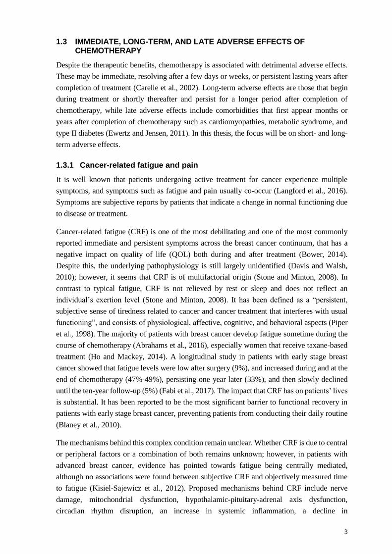
1.3 IMMEDIATE, LONG-TERM, AND LATE ADVERSE EFFECTS OF CHEMOTHERAPY
Despite the therapeutic benefits, chemotherapy is associated with detrimental adverse effects.
These may be immediate, resolving after a few days or weeks, or persistent lasting years after
completion of treatment (Carelle et al., 2002). Long-term adverse effects are those that begin
during treatment or shortly thereafter and persist for a longer period after completion of
chemotherapy, while late adverse effects include comorbidities that first appear months or
years after completion of chemotherapy such as cardiomyopathies, metabolic syndrome, and
type II diabetes (Ewertz and Jensen, 2011). In this thesis, the focus will be on short- and long-
term adverse effects.
1.3.1 Cancer-related fatigue and pain
It is well known that patients undergoing active treatment for cancer experience multiple
symptoms, and symptoms such as fatigue and pain usually co-occur (Langford et al., 2016).
Symptoms are subjective reports by patients that indicate a change in normal functioning due
to disease or treatment.
Cancer‐related fatigue (CRF) is one of the most debilitating and one of the most commonly
reported immediate and persistent symptoms across the breast cancer continuum, that has a
negative impact on quality of life (QOL) both during and after treatment (Bower, 2014).
Despite this, the underlying pathophysiology is still largely unidentified (Davis and Walsh,
2010); however, it seems that CRF is of multifactorial origin (Stone and Minton, 2008). In
contrast to typical fatigue, CRF is not relieved by rest or sleep and does not reflect an
individual’s exertion level (Stone and Minton, 2008). It has been defined as a “persistent,
subjective sense of tiredness related to cancer and cancer treatment that interferes with usual
functioning”, and consists of physiological, affective, cognitive, and behavioral aspects (Piper
et al., 1998). The majority of patients with breast cancer develop fatigue sometime during the
course of chemotherapy (Abrahams et al., 2016), especially women that receive taxane-based
treatment (Ho and Mackey, 2014). A longitudinal study in patients with early stage breast
cancer showed that fatigue levels were low after surgery (9%), and increased during and at the
end of chemotherapy (47%-49%), persisting one year later (33%), and then slowly declined
until the ten-year follow-up (5%) (Fabi et al., 2017). The impact that CRF has on patients’ lives
is substantial. It has been reported to be the most significant barrier to functional recovery in
patients with early stage breast cancer, preventing patients from conducting their daily routine
(Blaney et al., 2010).
The mechanisms behind this complex condition remain unclear. Whether CRF is due to central
or peripheral factors or a combination of both remains unknown; however, in patients with
advanced breast cancer, evidence has pointed towards fatigue being centrally mediated,
although no associations were found between subjective CRF and objectively measured time
to fatigue (Kisiel-Sajewicz et al., 2012). Proposed mechanisms behind CRF include nerve
damage, mitochondrial dysfunction, hypothalamic-pituitary-adrenal axis dysfunction,
circadian rhythm disruption, an increase in systemic inflammation, a decline in

4
cardiorespiratory fitness, anemia, and pain (LaVoy et al., 2016). Several of the same proposed
mechanisms behind CRF have also been shown to be associated with pain such as neural
damage and systemic inflammation which can further induce peripheral neuropathy, and pain
has been shown to be a predictor of long-term CRF (Schmidt et al., 2015a). Moreover, patients
with breast cancer display hypersensitivity to pressure pain compared with healthy individuals
(Caro-Moran et al., 2016).
1.3.2 Health-related quality of life and symptom burden
The term health-related quality of life (HRQoL), being distinct from QoL which is a broader
concept covering all aspects of life including non-health related features of life, has a focus on
the effects of illness and specifically on the impact treatment may have on a person’s well-
being. HRQoL is therefore thought of as a person’s subjective perception of the impact the
illness has on physical, psychological and social functioning (Karimi and Brazier, 2016).
Patients with cancer often experience multiple symptoms related to the disease and its treatment
(Gapstur, 2007). In contrast to HRQOL which provides information about the impact of
symptoms on general aspects of an individual’s health, symptom burden is a more disease-
specific measurement and is an indicator of the severity of the symptoms that are most
associated with a disease or treatment (Kenne Sarenmalm et al., 2014). This self-reported
measure has been shown to be more sensitive to the changes in symptoms experienced by
individuals with cancer (Burkett and Cleeland, 2007), and includes the presence, frequency,
and severity of multiple symptoms, and the level of distress caused by symptoms (Molassiotis
et al., 2010). A measure of symptom burden can enhance the understanding of a patients’
physiological and psychological functioning during diagnosis and treatment of cancer and is
therefore of major relevance for practice and clinical research (Burkett and Cleeland, 2007).
Figure 1. “Symptoms are not synonymous with HRQoL”. Symptom burden (circled) is closest to the disease and
treatment processes. General HRQOL includes more general aspects of an individual’s perception of health-related
well-being which is not directly related to treatment, while QoL is the individual’s perception of overall well-
being. Reprinted with modifications from Burkett and Cleeland (2007) with permission from Springer Nature.
1.3.3 The cardiovascular system
Anthracyclines primarily accumulate in the cell nucleus and mitochondria, which can lead to
negative consequences for noncancerous tissues. This chemotherapeutic agent group is known
to cause cardiotoxicities including cardiac muscle damage, tachycardia and atrial fibrillation
(late effects) (Florescu et al., 2013), and anthracycline-containing chemotherapy can lead to a
decline in cardiorespiratory fitness (Peel et al., 2014) (short- and long-term effects). Peak
oxygen consumption (VO2peak), which is measured from analysis of expired respiratory gases
during a graded exercise test to maximal effort (Hill and Lupton, 1923), is an inversely related

5
predictor of mortality in the general population (Kodama et al., 2009). It has been shown that
patients with breast cancer have a marked impairment in cardiorespiratory fitness across the
disease and treatment continuum, with the most pronounced effects in patients that undergo
chemotherapy (Klassen et al., 2014). The reductions in VO2peak for patients with breast cancer
undergoing chemotherapy typically range from 5% to 10%, and has been found to be
approximately 25% lower compared VO2peak values to those of healthy sedentary women after
completion of chemotherapy (Peel et al., 2014). Given that cardiovascular disease exceeds
breast cancer as the leading cause of death in older women 9 years after their breast cancer
diagnosis (Patnaik et al., 2011), and considering that with each metabolic equivalent (resting
metabolic rate: 1 MET=3.5 ml/kg/min) increase of cardiorespiratory fitness there is a 24%
lower risk of all-cause mortality, and a 24% lower risk of cardiovascular disease mortality in
women (Zhang et al., 2017), strategies to maintain VO2peak cannot be emphasized enough.
Moreover, poor cardiorespiratory fitness has direct consequences on HRQoL since
performance of daily activities is adversely affected (Herrero et al., 2006).
1.3.4 Skeletal muscle
Chemotherapeutic agents have been shown to induce muscle weakness in cancer patients
regardless of disease stage or nutritional status (Christensen et al., 2014), as reflected by a
slower chair-rise test time (Galvao et al., 2009) and lower handgrip strength (Hayes et al., 2005,
Cantarero-Villanueva et al., 2012) compared to healthy controls. In a cross-sectional study,
both isokinetic and isometric strength were impaired throughout the breast cancer continuum,
with the most detrimental effects for patients who had received chemotherapy (Klassen et al.,
2017). Muscle dysfunction and muscle weakness is associated with poorer disease prognosis
and is believed to accelerate morbidity and mortality (Dobek et al., 2013).
Preclinical findings in rodents demonstrate that anthracycline-containing chemotherapy
induces long-term impairments in muscular mitochondrial respiration and increased release of
reactive oxygen species (Gouspillou et al., 2015). In contrast, taxane treatment has, unlike
anthracycline-effects, not been shown to impair muscle force production in rodents (Chaillou
et al., 2017). Moreover, chemotherapy for breast cancer is often accompanied by prolonged
high-dose corticosteroid treatment which can lead to skeletal muscle atrophy and mitochondrial
dysfunction (Batchelor et al., 1997). Interest in muscle function has traditionally been confined
to cancer cachexia (Christensen et al., 2014), a condition associated with severe body weight,
fat, and muscle loss due to underlying disease (Muscaritoli et al., 2010). Loss of skeletal muscle
mass with concomitant gains in adiposity (sarcopenic obesity) has clinical implications since
it has been linked to increased toxicity (i.e. poor tolerance to chemotherapy) leading to a poorer
prognosis (Bozzetti, 2017), indicating a need to also assess muscle function in non-cachectic
cancer populations. Cytokines have been suggested to play a vital role in promoting skeletal
muscle atrophy/sarcopenia (Zhou et al., 2016). With chemotherapy, circulating cytokines are
significantly upregulated (van Vulpen et al., 2018), and it is therefore crucial to implement an
intervention capable of maintaining or improve muscle mass and reducing the systemic
inflammation caused by chemotherapy and physical inactivity.

6
1.3.5 Body mass
Evidence shows that overweight women and women who gain weight after a breast cancer
diagnosis have a doubled risk of recurrence and death from breast cancer at 5 years, as well as
60% higher risk of death over 10 years, when compared to non-overweight breast cancer
survivors (Soares Falcetta et al., 2018). Weight gain occurs in most women after breast cancer
treatment, especially in those who are younger (Nissen et al., 2011, Harris et al., 2009) and who
have been treated with chemotherapy (Demark-Wahnefried et al., 2001). Women with breast
cancer gain up to 5 kg body weight during chemotherapy (Makari-Judson et al., 2014), and few
return to their pre-diagnosis weight (Irwin et al., 2015). Increases in adiposity may be caused
by alterations in metabolic pathways as a result of chemotherapy or drugs taken in combination
with chemotherapy such as glucocorticoids which have been shown to induce weight gain
(Wung et al., 2008). Moreover, weight gain has an impact on a woman’s self-image and QoL
(McInnes and Knobf, 2001).
1.4 PHYSICAL INACTIVITY
As defined by the World Health Organization (WHO), “physical activity is any bodily
movement produced by skeletal muscle that requires energy expenditure”. Physical inactivity,
defined as achieving less than 30 minutes of moderate-intensity physical activity per week
(World Health Organization 2018) has been shown to be a primary cause of most chronic
diseases (Booth et al., 2012), and is estimated to be the main cause for about 21–25% of breast
cancers (World Health Organization 2018).
After a breast cancer diagnosis, patients show significantly reduced PA levels, especially
during the adjuvant treatment phase, with difficulties returning to pre-diagnosis PA levels one
year into survivorship (Huy et al., 2012). Personal and cancer-related characteristics such as
age, stage of cancer, and time since treatment have been proposed to be related to physical
inactivity (Andrykowski et al., 2007). However, more recent findings have pointed towards
fatigue being a more important predictor than the above mentioned factors (Brunet et al.,
2013). This is also supported by qualitative study findings showing that fatigue is a significant
barrier to maintaining PA levels among active women after treatment (Browall et al., 2016).
However, it is not known whether fatigue contributes to and/or is the result of inactivity.
Considering that physical inactivity places adults with a history of cancer at greater risk for
morbidity, poorer health outcomes, and mortality (Lynch et al., 2013), women that are
consistently inactive is an important target group for exercise interventions.
1.5 DEFINING CANCER SURVIVORSHIP
The Institute of Medicine (IOM) has defined the survivorship time period as “the period
following first diagnosis and treatment and prior to the development of a recurrence of cancer
or death.”(Institute of Medicine, 2006). However, this phase of cancer control continuum has
typically not been well described. Courneya and Friedenreich (2007) have proposed a Physical
Activity and Cancer Control framework with six cancer-related time periods: two pre-diagnosis

7
(pre-screening and screening) and four post-diagnosis (pre-treatment, treatment, survivorship,
and end of life). In this thesis, the term “survivorship” will be used when referring to the time
period after completion of chemotherapy. More than 97 000 women in Sweden today have
survived breast cancer (Cancerfondsrapporten 2018), and the aging of the population will
contribute to an even larger cohort of cancer survivors in the future. As a result, there is an
increasing emphasis on providing better support for long-term health promotion and
prevention.
Figure 2. “Physical activity and cancer control framework.” Reprinted from Courneya and Friedenreich (2007)
with permission from Elsevier.
1.6 BENEFITS OF EXERCISE TRAINING
Exercise training is structured, repetitive, and has as an objective to improve or maintain
physical fitness (Caspersen et al., 1985). The biological stress induced by an exercise stimulus,
through the perturbation of cellular and systemic environments, results in an adaptation of the
body that subsequently withstands future similar challenges (Kraemer, 1988, Goldspink et al.,
2002). The pleiotropic effects that exercise induces on the human body are well established in
the healthy population (Garber et al., 2011). Endurance training has been shown to result in
improved cardiac output (Ekblom et al., 1968), mitochondrial biogenesis (Gollnick et al.,
1972), and increased capillarization (Schantz et al., 1983). These adaptations lead to an
increased ability to transport and use oxygen to generate energy (Gollnick et al., 1972). The
mitochondrion is the main organelle for energy conversion from energy-rich substrates through
generation of adenosine triphosphate (ATP) via the electron transport chain. The electron
transport chain consists of five protein complexes (complex I, II, III, IV, and V) embedded in
the inner mitochondrial membrane, in which the first four complexes transfer electrons by
using substrates generated through the tricarboxylic acid (TCA) cycle (Mitchell, 1961).
Resistance training on the other hand leads to increases in neural adaptations (Aagaard and
Mayer, 2007), muscle size, improved muscle strength (Mangine et al., 2015), and an increase
in satellite cell number (Kadi et al., 2005). These classic and well established adaptations to
both endurance- and resistance training have been shown in both young and old individuals.

8
An emerging exercise modality, high-intensity interval training (HIIT), which consists of short
bursts of high-intensity exercise interspersed with recovery periods, has been found to be a
potent treatment strategy for improving cardiorespiratory fitness (MacInnis and Gibala, 2017,
Milanovic et al., 2015) and to induce adaptations of mitochondrial markers (Robinson et al.,
2017) in a time efficient manner. The beneficial effects of HIIT are not limited to these
physiological factors but have also been shown to induce improvements on HRQoL
(Jaureguizar et al., 2016), mood state (Ouerghi et al., 2016), cognitive health (Drigny et al.,
2014), systemic inflammation (Munk et al., 2011), neuromuscular adaptations (Buchheit and
Laursen, 2013), increases endorphin release in brain areas associated with controlling emotion
and pain (Saanijoki et al., 2017), and has also been reported to be a more enjoyable exercise
modality compared to low- or moderate-intensity endurance training (Thum et al., 2017).
1.6.1 Exercise for patients with breast cancer
There is a large body of epidemiological evidence showing that individuals that exercise on
average have a 25% lower risk of breast cancer, with a stronger effect for moderate to vigorous
PA (Friedenreich et al., 2010). Emerging observational data also suggest that regular exercise
is associated with a 10-50% reduction in risk of recurrence and cancer specific mortality
(Friedenreich et al., 2016). Recently, two randomized controlled trials showed that women that
conducted regular aerobic or resistance exercise training had favourable, although non-
significant, effects compared to the control group on overall survival (Courneya et al., 2014b,
Hayes et al., 2018).
Exercise trials for individuals with breast cancer have primarily been performed in breast
cancer survivors after completion of chemotherapy and have shown beneficial effects on
cardiorespiratory fitness, muscle strength, HRQoL (Cramp et al., 2010), and fatigue (Cramp
and Byron-Daniel, 2012, Meneses-Echavez et al., 2015). Few supervised exercise trials have
been conducted during chemotherapy, and most of these have small sample sizes (Juvet et al.,
2017). A recent systematic review by Furmaniak et al. (2016) showed that both aerobic and
resistance training alone or in combination were effective to attenuate fatigue. Aerobic exercise
has been shown to have the most positive effect on cardiorespiratory fitness although effect
sizes have on average been small, while resistance training has been shown to induce
improvements in muscle strength, lean body mass, and self-esteem. The effects of aerobic
and/or resistance training on HRQoL are less convincing showing no or small effects
(Furmaniak et al., 2016). Studies have also assessed home based walking interventions;
however, there is a lack of intention-to-treat analyses in these studies, and the more recent
home-based walking intervention studies have shown negligible to no effects on CRF and
VO2peak (Fairman et al., 2016).
HIIT alone in breast cancer survivors after completion of chemotherapy has been shown to be
safe and tolerable (Dolan et al., 2016, De Backer et al., 2007b, Kampshoff et al., 2015),
inducing slightly more beneficial effects compared to moderate-intensity endurance exercise
on cardiorespiratory fitness and cancer-related fatigue (Kampshoff et al., 2015). In patients
with breast cancer undergoing chemotherapy, the incorporation of HIIT is limited to one pilot

9
study in which HIIT was combined with resistance training and included a mix of cancer
diagnoses (Schulz et al., 2017). Findings from this pilot study demonstrated favorable effects
on cardiorespiratory fitness, muscle strength, and QoL.
1.7 EXERCISE RECOMMENDATIONS FOR INDIVIDUALS WITH CANCER
The American College of Sports Medicine (ACSM) organized a roundtable on exercise
prescription guidelines in 2010 (Schmitz et al., 2010). They reported that few randomized
controlled trials existed, mainly with small sample sizes and showed contradictory results.
However, findings regarding safety was consistent across studies. Since then, an increasing
number of trials have been published showing that exercise induces favourable effects on
physical function and self-reported outcomes, although the larger bulk of studies included
cancer survivors rather than women undergoing chemotherapy, as previously mentioned. The
Swedish up-to date clinical handbook on how to prevent and treat diagnoses using PA includes
a chapter on PA guidelines for individuals with cancer (Johnsson et al., 2017). These guidelines
suggest to engage in both aerobic and resistance based exercise. Recommendations for aerobic
exercise are the same as for guidelines for the general population: at least 150 min of moderate
intensity (3-7 days/week), at least 75 min of vigorous intensity aerobic exercise (3-5
days/week), or a combination of these. For resistance exercise, the Swedish guidelines
recommend including 12 exercises, performed at a moderate intensity at least 1x8-12
repetitions performed 2-3 times/week. Caution is advised for those during chemotherapy: the
intensity should be based on the day-to-day state which can be influenced by various degrees
of symptoms.

10
2 HYPOTHESES AND AIMS
It was hypothesized that adding high-intensity interval training (HIIT) to conventional
resistance training (RT) or aerobic training (AT) during chemotherapy for patients with breast
cancer would be increasingly beneficial in counteracting increases in fatigue and symptom
burden, as well as declines in skeletal muscle structural and functional properties. At 12
months, compared to baseline, it was hypothesized that both exercise groups would display
sustained levels for physiological and self-reported health-related outcomes compared to
deteriorated levels in the control group.
The specific aims of this thesis were to examine and compare the effects of a 16-week RT-
HIIT and AT-HIIT intervention during chemotherapy for patients with breast cancer over the
intervention period, and 12 months following commencement of chemotherapy compared to
usual care (UC) on:
1) Physiological outcomes: cardiorespiratory fitness, muscle strength, body mass,
hemoglobin concentration, pain sensitivity, muscle morphology and mitochondrial
markers
2) Self-reported health-related outcomes; cancer-related fatigue, pain, symptom burden,
and sick leave rates

11
3 RESULTS AND DISCUSSION
3.1 ATTENDANCE TO THE EXERCISE INTERVENTION AND PATIENT CHARACTERISTICS
The four studies that are included in this thesis are based on the OptiTrain trial (see Wengström
et al. (2017) for the study protocol). From March 2013 to July 2016, two hundred and forty
participants were included in the OptiTrain trial (papers I-IV). In July 2017 the 12-month
follow-up was completed. The participant flow is shown in Figure 3 (papers I, III, & IV).
Figure 3. CONSORT diagram of the participant flow (papers I, III, and IV). RT–HIIT, resistance and high-
intensity interval training; AT–HIIT, moderate-intensity aerobic and high-intensity interval training; UC, usual
care.
In the initial 16-week intervention study (paper I), attendance (calculated as the mean of the
individual percentages: attended exercise sessions divided by the total number of sessions) to
the exercise sessions was 68% in RT-HIIT and 63% in AT-HIIT groups, while adherence to
intensity (calculated as the number of patients who successfully completed 90% of the exercise

12
sessions according to plan, i.e., intensity and duration, divided by the total number of patients
in the intervention groups) was 83% in RT-HIIT and 75% in AT-HIIT. No adverse events
occurred during exercise sessions. Moreover, no significant differences were found in
attendance rates between those receiving taxane and not receiving taxane treatment. These
attendance rates are comparable with other exercise interventions for patients with breast
cancer, and a certain amount of contamination and imperfect adherence is to be expected in
exercise trials for patients with breast cancer (Furmaniak et al., 2016). Of note, 59% of the
participants that dropped out immediately following the randomization were those unwilling
to donate a muscle biopsy, indicating that the invasive method rather than unwillingness to
participate in the exercise intervention was the reason for this initial dropout.
Patient characteristics from the initial study (paper I) are shown in Table 2. No significant
differences were found in patient characteristics between groups, and no significant differences
were found between those that dropped out after baseline assessment and those who completed
baseline and 16-week post assessments, indicating no systematic drop out during the
intervention. Preliminary data analysis (unpublished data) does however suggest that those that
dropped out during the study period had a lower sense of coherence, i.e. coping worse with
stressful situations to maintain and develop health (Olsson et al., 2006).
When assessing participants with high attendance versus low attendance (cutoff for high
attendance was set as over 65%), there was a significant difference in level of education as
assessed by an exact χ2 test (p=0.003), but no differences in other factors such as chemotherapy
regimen, menopausal status, PA level, age, or BMI. This is in line with a recent meta-analysis
Table 2. Participant characteristics at baseline
RT-HIIT
n=74
AT-HIIT
n=72
UC
n=60
mean±SD mean±SD mean±SD
Age (years) 52.7±10.3 54.4±10.3 52.6±10.2 Body mass (kg) 68.7±11.3 67.7±13.0 69.1±11.0 Height 165.7±6.7 165.3±6.6 166.4±7.0 SED (% of daily wear time) 63.7±7.7 65.6±6.2 66.6±7.2 MVPA (% of daily wear time) 9.6±2.8 8.3±2.8 8.5±4.3 n (%) n (%) n (%) Married or partnered 60.6 59.7 69.5
University completed 67.6 64.7 66.0
Current smokers 4.3 5.9 5.2
Employed 74.6 86.8 79.7
Postmenopausal 51.4 63.9 61.7
Tumour profile
Triple negative 14.9 11.0 16.7
HER2+, ER+/- 21.6 30.2 20.0
HER2-, ER+ 62.2 58.9 61.6
HER2-, ER- 1.4 0.0 1.7
Anthracycline based therapy 40.6 37.0 41.7
Taxane based therapy 59.4 63.0 58.3 SD, standard deviation; RT-HIIT, resistance and high-intensity interval training; AT-HIIT, moderate-intensity aerobic and
high-intensity interval training; UC, usual care, MVPA objectively measured moderate- to vigorous intensity physical activity,
SED objectively measured sedentary behavior.

13
that suggested an important role for socio-demographic factors, including level of education,
in predicting adherence and attendance to exercise in patients with breast cancer (Ormel et al.,
2018).
Among the 12-month follow-up completers, no significant differences were found in baseline
characteristics between groups. Baseline differences between those that declined to come in for
in-clinic follow-up included less moderate-vigorous PA min/day (objectively measured)
compared to those that completed 12-month in clinic assessments (p=0.027). Moreover a
significantly higher proportion (72%) of participants that did not participate in the in-clinic
assessment follow-up had lower attendance levels to the exercise sessions compared to
completers (43%) (p=0.005) suggesting a potential selection bias. However no differences
were found between RT-HIIT, AT-HIIT and UC groups.
Self-reported PA was assessed through a question whether participants met the PA
recommendations of 75 min of vigorous PA or 150 min of moderate intensity PA/week or not.
Over the intervention, these results showed a significant decline in the proportion of
participants meeting exercise recommendations in the UC group, being significantly different
compared to RT-HIIT and AT-HIIT. At the 12–month follow-up, a significantly higher
proportion of participants in the UC group met exercise recommendations than at baseline, with
no between-group differences (Figure 4). This may be explained by the selection of participants
(as indicated by baseline differences in PA levels in the follow-up sample) at the 12-month
follow-up being slightly more active compared to those at baseline and at 16 weeks. Similar to
our findings, a follow-up study found that patients with breast or colon cancer who previously
participated in 18 weeks of combined aerobic and resistance training displayed significantly
higher levels of objectively measured moderate-to-vigorous PA levels at 4 years compared to
baseline (Witlox et al., 2018). In contrast, a longitudinal study showed significantly reduced
levels of PA during cancer treatment, which did not return to baseline PA levels one year later
(Huy et al., 2012). Moreover, previous studies have shown that 42% of participants did not
meet exercise recommendation guidelines six months after an intervention, regardless if they
had taken part in exercise or not (Courneya et al., 2009). Similar findings were also reported
by Schmidt et al. (2017).
One limitation in the current study was that objectively measured PA was only measured at
baseline, due to practicality issues. It has been shown that in studies using self-reported
measures of PA, subjectively assessed PA activity levels are typically higher than objectively
measured PA levels (Prince et al., 2008). A preliminary analysis (unpublished data) showed
discrepancies with 62% of the participants rating their PA levels according to measured PA.
Nevertheless, objectively measured PA will be obtained at the two-year follow-up of the
OptiTrain trial which will provide valuable information regarding the participants’ PA
behavior.

14
B a s e lin e 1 6 w e e k s 1 2 m o n t h s
0
2 0
4 0
6 0
8 0
1 0 0
M eetin g ex erc ise reco m m en da tio n gu id e lines
Pa
rtic
ipa
nts
(%
)
R T -H IIT
A T -H IIT
U C
*† †
*
*
**
*
Figure 4. Percentage of participants in each group reporting moderate/high physical activity at baseline, 16 weeks-
and 12 months post-baseline. RT-HIIT, resistance and high-intensity interval training group; AT-HIIT, moderate-
intensity aerobic and high-intensity interval training group; UC, usual care group; *p<0.05 vs baseline, †p<0.05 vs
UC.
When conducting exercise trials, there is a risk of selection bias in which there is a likelihood
that a more active and/or healthier sample of patients with breast cancer may accept
participation. In the OptiTrain trial, this may also help to explain why a high proportion of
participants in the UC group dropped out immediately following randomization. However,
compared to objectively measured PA in 508 cancer survivors in the National Health and
Nutrition Examination Survey (NHANES) (Thraen-Borowski et al., 2017), as well as 486
healthy middle-aged women from the Swedish CArdioPulmonary bioImage Study (SCAPIS)
(Ekblom-Bak et al., 2015), the baseline objectively measured PA data in the OptiTrain trial
indicated similar PA patterns as in those studies. Cancer survivors in the NHANES study spent
62% in sedentary time, and 2% in moderate to vigorous PA, while those in the SCAPIS study
spent 58% in sedentary time and 4% in moderate to vigorous PA, compared to the OptiTrain
participants that spent 63% in sedentary behavior and 9% in moderate to vigorous PA. The
slightly higher amount of time spent in moderate to vigorous PA in our sample may be due to
the inclusion of younger participants compared to the NHANES and SCAPIS studies, as well
as due to the mixed cancer diagnoses in the NHANES study.
3.2 EXERCISE TRAINING DURNG CHEMOTHERAPY IS EFFECTIVE TO COUNTERACT INCREASES IN CANCER-RELATED FATIGUE
Cancer-related fatigue (CRF) has been reported to be one of the most prevalent and debilitating
symptoms experienced by patients with breast cancer during chemotherapy and can persist for
several years after completion of treatment (Patrick et al., 2003). Besides psychological
interventions, exercise training is the only intervention that has the potential to counteract CRF
(Mustian et al., 2017). Supervised exercise interventions during chemotherapy have been
shown to be effective in relieving CRF (Furmaniak et al., 2016), particularly interventions
including both resistance and aerobic training (Meneses-Echavez et al., 2015). However, there

15
is limited data from studies with sufficient sample sizes that have examined the multiple and
different dimensions of fatigue.
In the OptiTrain trial, CRF was the primary outcome and was measured by the validated
Swedish version of the 22-item Piper fatigue scale (PFS) (Jakobsson et al., 2013). Data was
obtained at baseline, 16 weeks, and 12 months post-intervention. The PFS measures several
dimensions of CRF: 1) Behavioral/daily life CRF, 2) Affective/Emotional CRF, 3)
Physical/sensory CRF, and 4) Cognitive CRF. The effect of the initial 16-week exercise
intervention on CRF have been published in detail elsewhere (findings not included in the
thesis) (Mijwel et al., 2018). Findings from this study showed that RT-HIIT counteracted total
fatigue (ES=-0.51; p=0.02), behavior/daily life fatigue (ES=-0.62; p<0.001), and physical
fatigue (ES=-0.47; p=0.03) compared to the UC group. Moreover, no significant effects on
CRF were found in the AT-HIIT group when assessed by the PFS. These findings are similar
to those reported by Travier et al. (2015), who also trialed a program of combined aerobic and
resistance training, although with lower effect sizes (-0.20), while another trial of resistance
training only (BEATE study) showed no beneficial effects on fatigue (Schmidt et al., 2015b).
Those findings and the results from this thesis indicate the importance of combined exercise
modalities to induce more beneficial effects on CRF. In the current study, fatigue measured by
the European Organization for Research and Treatment of Cancer quality of life questionnaire
(EORTC-QLQ-C30), showed similar results with the exception that using this assessment tool
AT-HIIT also showed favorable effects for countering fatigue compared to UC (ES=-0.47;
p=0.01). The EORTC-QLQ-C30 has been shown to be sensitive to change for patients with
cancer during chemotherapy (Uwer et al., 2011), but the responsiveness of the PFS has not
previously been established (Jakobsson et al., 2013). Preliminary analyzes by our research
group to assess the ability of the PFS to detect change over the course of chemotherapy
demonstrated that the instrument is indeed sensitive to changes over time in patients with breast
cancer (unpublished data). The reason for the different findings between the two assessment
tools is unclear. It has been found that for patients with advanced cancer, there is a ceiling effect
for assessing fatigue with the EORTC-QLQ-C30 scale. When assessing individual values and
histograms for the two scales, there was a normal distribution for the EORTC-QLQ-C30 scale
but not for PFS, showing a right skewness of the data. Thus, further analyzes of the PFS will
be undertaken.
At 12 months (paper IV), both the RT-HIIT and AT-HIIT groups reported a significant
attenuation of total CRF (ES=-0.34; ES=-0.10), behavioral/daily life CRF (ES=-0.76; ES=-
0.50), and affective/emotional CRF (ES=-0.60; ES=-0.39), which was significantly different to
the UC group. Moreover, cognitive fatigue was also counteracted in the AT-HIIT group (ES=-
0.13) (Figure 5A). No significant differences between groups were found for fatigue with the
EORTC-QLQ-C30 symptom scale. This is not surprising since the EORTC-QLQ-C30 mainly
reflects the physical component of fatigue, and results from this scale align more with results
from the physical CRF measured by the PFS (i.e. no significant differences between groups).
At 12 months, 56% of participants in the UC group were experiencing moderate (score 4-7 out
of 10) to severe fatigue (score 7-10 out of 10), while the proportion of participants that

16
experienced moderate to severe fatigue were 33% and 37% in the RT-HIIT and AT-HIIT
groups respectively (Figure 5B-D).
0
1
2
3
4
5
1 0
T o ta l C R F
To
tal
fati
gu
e (
a.u
.)
R T -H IIT
A T -H IIT
U C
B a s e l in e 1 6 w e e k s 1 2 m o n th s
‡‡ †
No f
at i
gu
e
Mil
d f
at i
gu
e
Mod
e r ate
fat i
gu
e
Se v
e r e fat i
gu
e
0
2 0
4 0
6 0
T o ta l C R F a t b a s e lin e
Pa
rtic
ipa
nts
(%
)
U C
R T -H IIT
A T -H IIT
No f
at i
gu
e
Mil
d f
at i
gu
e
Mod
e r ate
fat i
gu
e
Se v
e r e fat i
gu
e
0
1 0
2 0
3 0
4 0
5 0
T o ta l C R F a t 1 6 w e e k s
Pa
rtic
ipa
nts
(%
)
No f
at i
gu
e
Mil
d f
at i
gu
e
Mod
e r ate
fat i
gu
e
Se v
e r e fat i
gu
e
0
1 0
2 0
3 0
4 0
5 0
T o ta l C R F a t 1 2 m o n th sP
art
icip
an
ts (
%)
A B
C D
Figure 5. Cancer-related fatigue (CRF) in RT-HIIT (resistance combined with high-intensity interval training),
AT-HIIT (moderate-intensity aerobic combined with high-intensity interval training), and UC (usual care) groups
A) from baseline to 16 weeks post-intervention and from baseline to 12 months, and Total CRF fatigue levels
divided into categories: no fatigue, mild fatigue (score>0), moderate fatigue (score≥4), or severe fatigue (score≥7)
at B) baseline, C) 16 weeks- and D) 12 months post-baseline. ‡p<0.05 between RT-HIIT and UC, †p<0.05 between
AT-HIIT and UC.
Previous follow-up studies after completion of a supervised exercise intervention during
chemotherapy did not find lasting beneficial effects for fatigue either at 4-6 month (Travier et
al., 2015, Courneya et al., 2007a, Mutrie et al., 2007) or at 12-month follow-up (Schmidt et al.,
2018a). Similar findings, to those from supervised studies, were found for follow-up studies of
two home-based exercise interventions during chemotherapy (Eakin et al., 2012, Haines et al.,
2010). In the supervised PACT (Travier et al., 2015) and START combined resistance and
aerobic exercise trials (Courneya et al., 2007a), the control group had recovered from an
increase in CRF to a similar extent as the exercise groups. The reason for the recovery in the
control groups was not explained, but could potentially be due to the fact that the control groups
in those trials received various types of interventions such as muscle relaxation during

17
chemotherapy in the PACT trial and a 1-month supervised exercise program post-intervention
in the START trial, while in the OptiTrain trial the UC group is strictly treated as a control
group until the 5-year follow-up. Alternatively, the control sample in those studies may have
been a more motivated group to stay physically active and therefore not being representative
of the broader population.
In the current trial, moderate to large effect sizes were found for several dimensions of fatigue,
both at 16 weeks and at 12 months following commencement of chemotherapy. Typically small
effects sizes have been reported for exercise trials during chemotherapy (-0.18 to -0.35) (van
Vulpen et al., 2016). The systematic review conducted by van Vulpen et al. (2016) showed that
exercise had the strongest potential to reduce physical fatigue, and the smallest effect on
affective and cognitive fatigue. This was also the case for OptiTrain results over the 16-week
intervention but not at the 12-month follow-up, where exercise groups were able to counteract
affective and cognitive fatigue. The results at 16 weeks and 12 months for the different domains
of CRF may be attributable to the fact that the manifestation of acute fatigue during
chemotherapy and long-term persisting fatigue have been shown to be predicted by different
factors (Schmidt et al., 2015a). Acute fatigue has been found to be caused by chemotherapy,
especially physical fatigue, but has not been found to be associated with long‐term persisting
fatigue, which instead appears to be associated with preexisting depressive or psychological
disorders, pain medication, and pre-diagnosis physical inactivity (Schmidt et al., 2015a,
Goedendorp et al., 2013). Given that exercise has the potential to alleviate depressive
symptoms in patients with breast cancer (Courneya et al., 2007b, Mutrie et al., 2007), as well
as in other populations with depressive symptoms (Craft and Perna, 2004), it may have been
crucial for the participants that they were able to exercise during chemotherapy to counter the
psychological distress associated with finding out about a cancer diagnosis and going through
cancer treatment. However, it is unclear whether preexisting depressive disorders were the
cause of persistent fatigue in the current trial. Affective fatigue has been shown to be
significantly associated with poor social support (Schmidt et al., 2018a). Therefore, in the
present study it cannot be ruled out that the motivational support and attention to uphold
physical exercise received by the exercise groups after completion of the intervention, although
attendance was very low (20%), resulted in favorable effects to relieve affective and/or
cognitive fatigue, rather than the exercise per se. Nevertheless, in support of exercise inducing
these beneficial effects, the BEATE trial by Schmidt et al. (2015b) showed that despite the
control group receiving support in the form of a muscle relaxation intervention during
chemotherapy, a tendency towards favorable effects were found for the resistance training
group to counteract affective fatigue.
There was a significant difference in baseline behavioral/daily life CRF between RT-HIIT and
AT-HIIT. The reason for this difference may be attributable to the (non-significant) relatively
higher proportion of participants in RT-HIIT being pre-menopausal (47.3%), compared to
36.1% in AT-HIIT and 38.3% in UC. It has been shown that lower age can be a predictor of
increased fatigue (Winters-Stone et al., 2008), and this may be due to the distressing premature
menopausal symptoms. While these experiences might be more normalized for women in their

18
50s, for younger women these changes can be exceedingly stressful and can negatively affect
QoL (Rosenberg and Partridge, 2013). The blocked randomization did not result in any other
differences in patient population characteristics; however, since there is no guarantee of
balanced groups with this type of randomization in randomized controlled trials, stratification
for important variables may have been able to prevent this randomization issue.
3.3 PRESSURE-PAIN THRESHOLD AND PAIN
Pain is one of the most debilitating symptoms in patients with breast cancer that have
undergone chemotherapy and can result in impairments in activities of daily living (Patrick et
al., 2003).
Pressure-pain threshold (PPT) is the minimum intensity at which a physical stimulus is
perceived as being painful, and is measured and quantified by using algometry (Kinser et al.,
2009). A higher PPT has been associated with a higher fitness level (Lemming et al., 2015), as
well with higher self-reported PA levels (Andrzejewski et al., 2010). Moreover, resistance
exercise training has proven to increase PPT in patients with chronic neck pain (Li et al., 2017),
whereas aerobic training has been shown to improve pain tolerance, but not pain threshold in
healthy individuals (Jones et al., 2014). To date, no trials have assessed whether exercise has
an effect on pressure-pain threshold for patients with cancer.
In paper I, PPT was measured at baseline and at 16 weeks by using a handheld electronic
algometer pressed on the mid-trapezius and mid-gluteus muscle bilaterally. RT-HIIT
significantly counteracted the hyperalgesia that was found in the UC group for both gluteus
(ES=0.53) and trapezius muscles (ES=0.46), and was also superior to AT-HIIT for trapezius
PPT (ES=0.30).
Different chemotherapy drugs have been shown to induce pain to various degrees in patients
with breast cancer with taxanes being more neurotoxic and associated with higher levels of
self-reported pain compared to non-taxane treatments (Majithia et al., 2016). Of note, when
performing a subgroup analysis in the current trial, we found that the patients receiving taxanes
displayed no hyperalgesia over time and no between group differences. It may be that taxanes
cause increased numbness in the acute phase leading to a blunted PPT response. In line with
this speculation, taxane-therapies have been shown to cause a loss of vibratory perception
(Hilkens et al., 1997). PPT was not assessed at the 12-month follow-up; however, results from
the two-year follow up will provide valuable insights into long-term effects of chemotherapy
on PPT.
Interestingly, self-reported pain assessed by the EORTC-QLQ-C30 showed that the AT-HIIT
group managed to counteract the increase in pain found in the UC group over the intervention
(Mijwel et al., 2018). At 12 months, no differences were found for self-reported pain between
groups (Figure 6). It has been shown that aerobic training can be more effective compared to
resistance training in alleviating pain symptoms over the course of chemotherapy (Schmidt et
al., 2015c), while others have found no differences between combined resistance and aerobic
training compared to usual care (Travier et al., 2015). Pain symptoms have been shown to

19
persist one year following completion of chemotherapy (Ganz et al., 2011), and pain has been
shown to be one of the major symptoms to impact HRQoL 5 years into survivorship (Schmidt
et al., 2018b). A score threshold of 25 for clinically important pain problems has been
established for pain for the EORTC-QLQ-C30 questionnaire (Giesinger et al., 2016). This
threshold was reached only by the UC group 16 weeks post-intervention. However, a subgroup
analysis assessing the proportions of participants below and above the clinically important pain
threshold, showed no statistical differences between groups, as assessed by a χ2 test.
B a s e lin e 1 6 w e e k s 1 2 m o n th s
0
1 0
2 0
3 0
4 0
1 0 0
PP
T (
kP
a)
S e l f - r e p o r t e d p a in
B a s e lin e 1 6 w e e k s
0
2 5 0
3 0 0
3 5 0
4 0 0
4 5 0
5 0 0
PP
T (
kP
a)
P r e s s u r e - p a in th r e s h o ld G lu te u s
‡U C
R T - H I I T
A T - H I I T
H e a l th y
re f e r e n c e
A B
Figure 6. Changes in A) self-reported pain from baseline to 16 weeks (post-intervention) and from baseline to 12
months, and B), pressure-pain threshold (PPT) from baseline to 16 weeks in RT-HIIT (resistance combined with
high-intensity interval training), AT-HIIT (moderate-intensity aerobic combined with high-intensity interval
training) and UC (usual care) groups. Healthy reference values were obtained from Michelson et al. (2000).
‡p<0.05 between RT-HIIT and UC.
The conflicting findings regarding PPT and self-reported pain could be explained by the lack
of relationship between participants’ self-report of pain and participants’ actual pain threshold
(PPT gluteus and self-reported pain, r=-0.02; PPT trapezius and self-reported pain r=0.03).
This is in line with a previous study in healthy individuals (Edwards and Fillingim, 2007). In
those individuals, it was shown that those with higher self-reported pain were more anxious
than those with lower self-reported pain scores, while objectively measured pain thresholds did
not follow the same pattern. Another study showed that depression predicted self-reported pain
but not cold pain tolerance in patients with opioid dependence (Tsui et al., 2013). Given that a
breast cancer diagnosis can be a major psychological stress, associated with depression, fear,
and anxiety (Baqutayan, 2012), it may be speculated that the participants were influenced by
negative feelings that might have influenced their subjective feeling of pain; in part helping to
explain the lack of association with PPT. Indeed, when assessing the association with anxiety
from the Memorial Symptom Assessment Scale in the current trial (unpublished data), there
was a significant association between self-reported pain and anxiety (r=0.3, p=0.002), but not
between measured PPT and anxiety (r=-0.03, p=0.749) at 16 weeks. The implications of self-
reported pain and its potential association with important prospective endpoints such as the
development of clinical pain conditions remains to be explored.
Evidence suggests an association between muscle strength and PPT in healthy and diseased
populations (Henriksen et al., 2013, Hooten et al., 2013). In line with this, we found significant,
but weak associations between pre- to post changes in PPT at the gluteus (r=0.16, p<0.05) and

20
trapezius muscles (r=0.17, p<0.05) and pre- to post changes in lower-limb muscle strength as
well as between pre-to post changes in PPT at the trapezius muscle and changes in handgrip
strength (r=0.17, p<0.05). These findings could indicate that resistance training may be of
particular importance in inducing analgesic effects. In healthy individuals, Henriksen et al.
(2013), showed that PPT muscle function was only associated with the pressure pain sensitivity
in the local muscle. Therefore, it may be speculated that measuring PPT on more relevant
anatomical sites (i.e. vastus lateralis) than those measured may have resulted in stronger
associations with lower-limb muscle strength in the current study. Despite this, in elderly
women, no association between vastus lateralis PPT and lower-limb muscle strength (as
measured by a chair-stand test) was found (Alfieri et al., 2017). The conflicting findings may
be due to differences in populations and differences in assessment techniques of muscle
strength as well as anatomical sites of PPT measurement.
PPT reproducibility has been found to be good for the gluteal site (Maquet et al., 2004), and
the mean of the bilateral measurements on the same anatomical site have been shown to be
valid (Lacourt et al., 2017). However, a limitation here is that only one measurement was
performed at each site. To examine the reliability and the absolute agreement of three repeated
PPT measurements, a test-retest was performed on 9 individuals by our research group. The
findings revealed an excellent reliability of repeated measures (intraclass correlation
coefficient=0.83); therefore, any possible error of single measurement is expected to be
negligible.
3.4 VALIDATION OF SUBMAXIMAL EXERCISE TESTS FOR ASSESSMENT OF CARDIORESPIRATORY FITNESS IN PATIENTS WITH BREAST CANCER
Patients with breast cancer have considerable impairments in cardiorespiratory fitness that, in
part, have been linked to the toxic effects of anticancer therapy. According to a recent
systematic review that assessed psychometric properties, safety, and clinical utility of
cardiorespiratory fitness in patients with cancer, the “gold standard” for measurement of
aerobic fitness, VO2peak (Hill and Lupton, 1923), was not recommended for patients with breast
cancer due to clinical efficiency and safety concerns (Drouin and Morris, 2015). Instead,
submaximal exercise tests for estimation of cardiorespiratory fitness provide a safer alternative
to maximal exercise testing (Drouin and Morris, 2015). There are several valid equations
available to estimate VO2peak calculated based on the results of submaximal testing for healthy
individuals (Noonan and Dean, 2000). The indirect submaximal exercise tests, the Åstrand-
Rhyming (A-R) and Ekblom-Bak (E-B) prediction models have been shown to be valid in
healthy individuals (Ekblom-Bak et al., 2014, Åstrand, 1960). Unlike performing a direct
VO2peak test, such tests are relatively simple to perform and do not require a maximal effort or
as advanced equipment in a laboratory setting (Noonan and Dean, 2000, Drouin and Morris,
2015). The usefulness and simplicity of submaximal exercise tests encourages further research
in the field to establish a valid, reliable, and sensitive test for assessment of cardiorespiratory
fitness in patients with breast cancer. Furthermore, knowledge of a patient’s cardiorespiratory
fitness can assist clinicians in accurately and correctly providing an exercise prescription. In

21
paper II, a validation of A-R and E-B submaximal exercise tests were performed on 8
participants that were part of the larger OptiTrain trial.
All 8 participants underwent a submaximal exercise test on a cycle ergometer prior to an
incremental VO2peak test on a cycle ergometer. Findings from the study showed that the single
workload stage test A-R was a valid test for patients with breast cancer during chemotherapy.
The A-R prediction model overestimated VO2peak by 6%, with a correlation coefficient of 0.9
(p<0.05) between A-R estimated VO2peak and measured VO2peak, and a coefficient of variation
of 7%. The E-B prediction model overestimated VO2peak by 42%, and showed a very weak,
non-significant correlation with measured VO2peak (r=0.2), and the coefficient of variation was
21%. The individual variation was quite high, although no outliers were found (Figure 7).
It may be speculated that directly measured VO2peak was not reached in all instances during the
graded maximal test; however, when assessing the criteria for reaching VO2peak, all participants
reached a plateau and the mean respiratory exchange ratio was 1.31.
Figure 7. Correlations between measured versus estimated VO2peak for: A) the Åstrand-Rhyming prediction model
(A-R) and B) the Ekblom-Bak prediction model (E-B). This work is licensed under CC BY 4.0.
It has been suggested that conducting only one submaximal exercise test, without a
familiarization test, is likely to result in an underestimation of VO2peak due to anxiety leading
to a higher HR (Åstrand and Ryhming, 1954). However, this was not the case in the current
validation study, where both the A-R and E-B tests overestimated VO2peak; thus, one can
potentially rule out any anxiety factors that may have influenced the outcomes. An
overestimation of both A-R (Siconolfi et al., 1982, Zwiren et al., 1991) and E-B (Ekblom-Bak
et al., 2014) has previously been reported in the female population, although the overestimation
of E-B was substantially greater in the current study. This may partly be attributed to the fact
that VO2peak was performed on a treadmill as opposed to on a cycle ergometer as in the original
validation of the E-B test (Ekblom-Bak et al., 2014). VO2peak obtained from treadmill exercise
can be 5% to 10% higher. However, given that elderly patients and those with treatment-
associated ataxia or peripheral neuropathy were included, cycle ergometry was favored for
safety reasons.

22
The large overestimation of VO2peak by the E-B prediction model seemed to be due to an
unusually elevated HR, especially at the lower submaximal workload (30W), likely as a result
of chemotherapy making the E-B estimated VO2peak test potentially inappropriate for use in
patients with breast cancer while undergoing chemotherapy. Moreover, variations in HR are
typically higher at lower intensities compared to higher intensities (Åstrand and Ryhming,
1954). Furthermore, an elevated resting HR was found in women with breast cancer during
chemotherapy compared with women with breast cancer who had not yet received
chemotherapy or women who had already completed oncologic treatment (Jones et al., 2012).
The influence of chemotherapy on HR has previously been suggested to be attributed to
chemotherapy-induced autonomic dysfunction in women with early breast cancer treated with
chemotherapy, which has been explained by an increased sympathetic activity and decreased
cardiac vagal tone (Scott et al., 2014).
As indicated in the current validation study, a single stage test at approximately 70% of the
maximal HR seems to accurately predict VO2peak. A study by Evans et al. (2009) showed that
women with breast cancer who had completed chemotherapy displayed no difference in HR
response at an exercise intensity of 70% of maximal HR compared with healthy women, which
may explain why a single stage test at a similar workload generated valid results to predict
VO2peak.
The A-R submaximal VO2peak test was valid in patients with breast cancer during
chemotherapy. However, sensitivity of the test still remains to be established in this population,
as well as in healthy individuals (Noonan and Dean, 2000). Reliability of the A-R prediction
model in patients with breast cancer also needs to be further explored. Two previous studies
(May et al., 2010, De Backer et al., 2007a) on cancer survivors have established a good test-
retest reliability for a submaximal cycle ergometer test, a submaximal test comparable to the
A-R predictions model (10 min test at 50% of peak power output). The same studies suggested
that the submaximal exercise tests assessed in those studies lacked the ability to detect changes
in cardiorespiratory fitness over time. Although, in the study by May et al. (2010), a subgroup
analysis revealed that a HR above 140 beats per min (bpm) was associated with changes in
directly measured VO2peak, indicating that higher submaximal loads are required during a
submaximal cycle test to find reflections of changes in VO2peak.
Besides the importance of establishing an accurate test for evaluation of cardiorespiratory
fitness in patients with breast cancer, these findings also supported the use of the A-R prediction
model in the larger OptiTrain trial.
3.5 MITOCHONDRIAL CONTENT AND CAPILLARIZATION MAY EXPLAIN MAINTAINED CARDIORESPIRATORY FITNESS WITH EXERCISE DESPITE DECLINES IN BLOOD HEMOGLOBIN CONCENTRATIONS
Current breast cancer treatments can have a negative impact on cardiovascular health (Jones et
al., 2012). Exercise for patients with breast cancer has shown short-term beneficial effects on
cardiorespiratory fitness, although the long-term effects are less known. Moreover, the effect
of exercise during chemotherapy on skeletal muscle mitochondrial content and morphological

23
adaptations has not been established in patients with breast cancer undergoing chemotherapy.
Preclinical reports suggest that anthracyclines can inhibit the complexes of the electron
transport chain in skeletal muscle (Gilliam et al., 2013), and reduce mitochondrial respiration
(Gouspillou et al., 2015).
Over the intervention (paper I), both RT-HIIT and AT-HIIT showed maintained levels of
estimated VO2peak, significantly different from the declines found in the UC group (Figure 8).
Similar results were found when only assessing participants with higher attendance levels (over
80% attendance). Interestingly, cardiorespiratory fitness in the RT-HIIT group was maintained
despite only a total of 9 min of HIIT per session. This finding contradicts that by Courneya et
al. (2013), where only the higher dose (duration) aerobic training at a submaximal constant
exercise intensity (50-60 min, 3 days/week, 60-75% of VO2peak) resulted in a maintained
cardiorespiratory fitness. This suggests that HIIT provides an effective and time-efficient
training strategy resulting in the same beneficial maintenance in cardiorespiratory fitness as
high-volume aerobic training, similar to findings in healthy individuals (MacInnis and Gibala,
2017, Milanovic et al., 2015).
1 .8
2 .0
2 .2
2 .4
2 .6
E stim a te d V O 2 p e a k
VO
2p
ea
k (
L/m
in)
U C
R T -H IIT
A T -H IIT
1 6 w e e k s 1 2 m o n t h sB a s e l in e
H e a lth y
r e f e r e n c e
‡ †
Figure 8. Changes in estimated VO2peak from baseline to 16 weeks and from baseline to 12 months in RT-HIIT
(resistance combined with high-intensity interval training), AT-HIIT (moderate-intensity aerobic combined with
high-intensity interval training) and UC (usual care) groups. Healthy reference values are obtained from Aspenes
et al. (2011). ‡p<0.05 between RT-HIIT and UC, †p<0.05 between AT-HIIT and UC.
In healthy individuals, aerobic training typically results in dose-response improvements in
cardiorespiratory fitness over time. Here, the maintained levels of estimated VO2peak in the
exercise groups are in line with recent exercise trials (Travier et al., 2015, Schmidt et al., 2015b,
Courneya et al., 2013, Dolan et al., 2010). A general increase in the use of taxane-based
treatments (Peto et al., 2012) has been suggested to be the cause for the non-improvements in
cardiorespiratory fitness (Møller et al., 2015). In paper I, a subgroup analysis was performed
on those who had received taxanes compared to those who had not received taxanes. This
analysis showed no influence of chemotherapy regimen on cardiorespiratory fitness in the
exercise groups. However, the decline in estimated VO2peak in the UC group showed slightly
more unfavorable effects for the participants in the UC group that received taxanes (-14%)
compared to those who had not received taxanes (-7%), suggesting slightly more toxic effects

24
of taxanes on the cardiovascular system. It may be that the high-intensity nature of the exercise
regimens in the current trial were more effective compared to previous trials to counteract the
toxic effects of taxanes on cardiorespiratory fitness.
To assess local skeletal muscle adaptations, muscle biopsies from 23 participants were assessed
for muscle morphology and mitochondrial content at baseline and at 16 weeks (paper III). Here,
participants in the AT-HIIT group showed significant improvements compared to the declines
found in the UC group in capillarization (+33% vs -18%, ES=1.9), citrate synthase activity - a
marker for mitochondrial content) (+20% vs -25%, ES=2.2), and protein levels of complex IV
(+20% vs -9%, ES=1.3) (Figure 9). These are adaptations that typically take place with aerobic
training in healthy individuals (Schantz et al., 1983) showing a trainability in patients with
breast cancer receiving chemotherapy.
U C A T -H I I T R T -H I I T
0
2 0
4 0
6 0
8 0
C S a c t iv ity
CS
ac
tiv
ity
(n
M
min
-1
mg
-1)
†
* †
U C A T -H I I T R T -H I I T
0
1
2
3
C o m p lex IV
Pr
ote
in e
xp
re
ss
ion
(a
.u.)
P r e
P o s t† §
*
U C A T -H I I T R T -H I I T
0
1
2
3
4
C a p illa r ie s
CD
31
po
sit
ive
ce
lls
/fib
er
(n
)
†*
*†
A B
C
Figure 9. Local skeletal muscle adaptations for A) Citrate synthase activity (CS) (nM·min-1·mg-1), B) protein
levels of complex IV, and C) immunohistochemical labeling in skeletal muscle for capillaries per fiber pre- to
post-intervention in usual care (UC), moderate-intensity aerobic and high-intensity interval training (AT-HIIT),
and resistance and high-intensity interval training (RT-HIIT) groups. *p<0.05 at post versus pre-measurement;
†p<0.05 compared to UC; §p<0.05 between exercise groups. Image shows immunofluorescent staining of CD31
for capillary analysis stained in green and myonuclei stained in blue. Reprinted with permission from the Faseb
Journal.

25
The direct effects of surgery, chemotherapy, and radiotherapy, as well as the secondary effect
of these treatments (deconditioning), may cause impairments in one or several steps within the
oxygen cascade that consequently leads to a reduction in oxygen delivery (Koelwyn et al.,
2014). Despite the declines found in blood hemoglobin concentration in all three groups over
the intervention, which plays an important role in the oxygen cascade (Calbet et al., 2006),
local skeletal muscle adaptations still took place in the exercise groups in the current trial.
Although it cannot be ruled out that the declines in hemoglobin concentration in the exercise
groups were partly due to hemodilution through an increase in plasma volume, a training
adaptation that usually occurs in healthy individuals (Bonne et al., 2014), it is highly likely that
chemotherapy significantly affected blood cell counts negatively despite exercise. This is in
line with previous exercise trials that have been conducted during chemotherapy (Dolan et al.,
2010).
Mitochondrial oxidative capacity has been reported to be in excess of oxygen delivery in
healthy individuals (Boushel et al., 2011). However, recent findings showed that in untrained
individuals this mitochondrial oxidative capacity was fully utilized in contrast to the ≈35%
excess found in well-trained individuals, which would indicate that an increase in
mitochondrial oxidative capacity would lead to an increase in VO2peak only in untrained, but
not well-trained individuals (Gifford et al., 2016). In patients with heart failure skeletal muscle
diffusion was shown to be a step in the oxygen cascade with the most detrimental effect on
VO2peak (Houstis et al., 2018). Although it is largely unknown which steps of the oxygen
cascade are primarily affected in patients with breast cancer undergoing chemotherapy
(Koelwyn et al., 2014), it can be speculated that an increased oxygen extraction took place
through increases in mitochondrial content and capillarization found in the exercise groups
compared to the declines in the UC group. This may explain why exercise groups were able to
counteract a decline in estimated VO2peak despite declines in oxygen delivery (i.e. hemoglobin
concentrations). In support of this hypothesis, a recent study showed that the functional role of
possessing a mitochondrial oxidative excess capacity was to lower mitochondrial activation
while maintaining a high mitochondrial oxygen affinity; thus, optimizing oxygen extraction
capacity (Cardinale et al., 2018). However, this remains speculative since the various steps of
the oxygen cascade such as total hemoglobin mass, blood volume, and cardiac output were not
measured in the current study.
Several associations were found between the various biochemical methods that were used.
Outcomes such as changes in capillarization, assessed through immunohistochemistry were
associated with changes in CS activity which was assessed spectrophotometrically (r=0.6).
Moreover, an association was found between changes in CS activity and changes in complex
IV protein levels assessed through immunoblotting (r=0.5), indicating the robustness of the
methods despite the low sample size (Figure 10).

26
-1 .0 -0 .5 0 .0 0 .5 1 .0
-4 0
-2 0
0
2 0
4 0
C h a n g e in c a p il la r ie s p e r f ib e r (n )Ch
an
ge
in
CS
ac
tiv
ity
(n
M
min
-1
mg
-1
)
r = 0 .6
p = 0 .0 0 4
A
-4 0 -2 0 0 2 0 4 0
-1 .0
-0 .5
0 .0
0 .5
1 .0
C h a n g e in c a p il la r ie s p e r f ib e r (n )Ch
an
ge
in
Co
mp
lex
IV
pr
ote
in l
ev
els
(a
.u)
r = 0 .5
p = 0 .0 2 4
U C
A T -H IIT
R T -H IIT
B
Figure 10. Correlations between changes in A) capillarization and CS activity, and B) capillarization and complex
IV protein levels in usual care (UC), moderate-intensity aerobic and high-intensity interval training (AT-HIIT),
and resistance and high-intensity interval training (RT-HIIT) groups. p, significance level; r, correlation
coefficient; CS, citrate synthase.
At 12 months (paper IV), all groups had improved estimated VO2peak to a similar extent
compared to baseline values (Figure 8). These findings are in line with findings from a previous
trial showing a similar recovery at 6 months in all groups versus baseline (van Waart et al.,
2015). The self-reported PA data (Figure 4) indicate that the UC group had increased their
activity levels during survivorship (from 16 weeks to 12 months), and a higher proportion
reported being active compared to baseline. Therefore, it may be that the increase in activity
may have protected these individuals from the decline in VO2peak that is commonly found in
patients with breast cancer that have undergone chemotherapy (Klassen et al., 2014).
Alternatively, the estimated submaximal cardiorespiratory fitness test was not able to detect
changes over time (Drouin and Morris, 2015). As discussed previously, a study by May et al.
(2010) showed that HR above 140 bpm can provide valid results on changes in VO2peak. To
evaluate those findings further, a subgroup analysis was performed to assess whether those
reaching 140 bpm or more compared to those below 140 bpm at the higher workload during
the A-R test (unpublished data). From pre to post-intervention, the subgroup analysis showed
that for those above 140 bpm (50% of the participants), there were significant differences
between both RT-HIIT and AT-HIIT versus UC, as demonstrated in the larger OptiTrain trial,
while no changes between groups were found for those below 140 bpm (Figure 11). At 12
months, no differences were found between groups for neither above or below 140 bpm
subgroup analysis. However, it must be noted that at 12 months, only 30% of the participants
reached a HR of at least 140 bpm. In support of previous findings in breast cancer survivors
(May et al., 2010), these subgroup analyzes indicate the importance of reaching a higher HR to
be able to detect changes in VO2peak. The reason why this was not performed in the current
study was due to the workload being initially determined based on rating of perceived exertion
(RPE).

27
UC
RT
-HII
T
AT
-HII
T
-1 .0
-0 .5
0 .0
0 .5
1 .0
1 .5
VO
2p
ea
k f
rom
ba
se
lin
e t
o 1
6 w
ee
ks
(L
/min
)
C h a n g e s in V O 2 p e a k (b p m o v e r 1 4 0 )
*
*
UC
RT
-HII
T
AT
-HII
T
-1 .0
-0 .5
0 .0
0 .5
1 .0
1 .5
V
O2
pe
ak f
rom
ba
se
lin
e t
o 1
6 w
ee
ks
(L
/min
)
C h a n g e s in V O 2 p e a k (b p m u n d e r 1 4 0 )
UC
RT
-HII
T
AT
-HII
T
-1 .0
-0 .5
0 .0
0 .5
1 .0
1 .5
V
O2
pe
ak f
rom
ba
se
lin
e t
o 1
2 m
on
ths
(L
/min
)
C h a n g e s in V O 2 p e a k (b p m o v e r 1 4 0 )
UC
RT
-HII
T
AT
-HII
T
-1 .0
-0 .5
0 .0
0 .5
1 .0
1 .5
V
O2
pe
ak f
rom
ba
se
lin
e t
o 1
2 m
on
ths
(L
/min
)
C h a n g e s in V O 2 p e a k (b p m u n d e r 1 4 0 )
A B
C D
Figure 11. Changes in VO2peak from baseline to 16 weeks for participants with a heart rate A) over 140 beats per
min (bpm) and B) under 140 bpm, as well as changes from baseline to 12 months in participants with a heart rate
C) over 140 beats per min (bpm) and D) under 140 bpm, in RT-HIIT (resistance combined with high-intensity
interval training), AT-HIIT (moderate-intensity aerobic combined with high-intensity interval training) and UC
(usual care) groups.
3.6 DECLINES IN SKELETAL MUSCLE STRENGTH, CROSS-SECTIONAL AREA, AND SATELLITE CELLS WERE COUNTERACTED BY EXERCISE
Muscle strength is inversely and independently associated with all-cause mortality (Ruiz et al.,
2008), and a reduction in muscle strength has implications for activities of daily living (Hairi
Noran et al., 2010). Although most trials have focused on aerobic training for patients with
breast cancer, an increasing number of recent trials have incorporated resistance training as a
single exercise modality (Schmidt et al., 2015b) or in combination with aerobic training
(Travier et al., 2015, Schmidt et al., 2015c), and have shown beneficial effects of resistance
training on muscle strength. However, less is known about whether these changes in muscle
strength remain into survivorship. Despite observed declines in muscle strength, little is known
regarding the effects of chemotherapy per se on skeletal muscle in non-cachectic cancer
patients. In preclinical models, chemotherapy has been suggested to cause skeletal muscle
dysfunction and chemotherapy-induced skeletal muscle atrophy (Gilliam et al., 2009). Muscle
fiber CSA and satellite cells have been assessed in a previous exercise trial for patients with

28
B a s e lin e 1 6 w e e k s 1 2 m o n th s
0
2 5
2 6
2 7
2 8
2 9
3 0
3 1
Fo
rc
e (
kg
)
H a n d g r ip s t r e n g th s u r g e r y s id e
‡ §‡ †
B a s e lin e 1 6 w e e k s 1 2 m o n th s
0
2 5
2 6
2 7
2 8
2 9
3 0
3 1
Fo
rc
e (
kg
)
U C
R T -H IIT
A T -H IIT
H a n d g r ip s t r e n g th n o n - s u r g e r y s id e
‡ §
H e a lth y
r e f e r e n c e
‡ †
B a s e lin e 1 6 w e e k s 1 2 m o n th s
0
7 0
8 0
9 0
1 0 0
1 1 0
Fo
rc
e (
kg
)
I s o m e t r ic m id - t h ig h p u l l
‡ †‡ †
A B
C
germ cell cancer undergoing chemotherapy (Christensen et al., 2016), but not in patients with
breast cancer.
Muscle strength was measured at baseline, 16 weeks, and at 12 months (papers I and IV).
Handgrip strength was measured using a hand dynamometer and lower-limb muscle strength
through isometric mid-thigh pull. Following the 16-week intervention (paper I) both RT-HIIT
and AT-HIIT improved lower-limb muscle strength significantly in comparison to the UC
group (ES=0.66, ES=0.48). Similar results were found when analyzing lower-limb muscle
strength relative to body mass. Significant handgrip strength gains were found in RT-HIIT
compared to both declines in the UC group and maintained levels in the AT-HIIT group
(surgery side: RT-HIIT vs. UC: ES=0.41, RT-HIIT vs. AT-HIIT: ES=0.28; non-surgery side:
RT-HIIT vs. UC: ES=0.35, RT-HIIT vs. AT-HIIT: ES=0.22) (Figure 12).
The gains in muscle strength over the intervention are in agreement with previous resistance-
training interventions for patients with breast cancer (Schwartz et al., 2007, Courneya et al.,
2007b, Schmidt et al., 2015b) and mixed cancer diagnoses (Adamsen et al., 2009). Importantly,
lower-limb muscle strength also improved in the AT-HIIT group, not a common finding from
a conventional aerobic exercise program, possibly indicating a role for HIIT in inducing
neuromuscular adaptations. Despite this unexpected finding, a previous trial found that HIIT
was able to induce strength improvements in cancer survivors (Dolan et al., 2016). The
improvement in handgrip strength that was displayed by the RT-HIIT group offers a prognostic
value and is an important correlate of health in survivors of breast cancer (Cantarero-
Villanueva et al., 2012).
Figure 12. Changes in A) handgrip strength surgery-side, B)
handgrip strength non surgery-side, and C) isometric mid-thigh
pull (lower-limb muscle strength) from baseline to 16 weeks
post-intervention, and to the 12-month follow-up in RT-HIIT
(resistance combined with high-intensity interval training), AT-
HIIT (moderate-intensity aerobic combined with high-intensity
interval training) and UC (usual care) groups. Reference values
are obtained from Leong et al. (2015). ‡p<0.05 between RT-
HIIT and UC, †p<0.05 between AT-HIIT and UC, §p<0.05
between exercise groups.

29
In the current study, strength gains were found with RT-HIIT regardless of chemotherapy
regimen, and even slightly greater within-group improvements for those receiving taxane-
treatment for lower-limb muscle strength (+18.3%) compared to those on taxane-free treatment
(+14.2%). In contrast, Courneya et al. (2014a) showed that participants on a taxane-based
regimen showed less pronounced strength gains compared to those not receiving taxane-based
regimens after combined resistance and aerobic training. This disagreement is unclear;
however, despite that taxanes typically induce higher rates of myalgia, arthralgia, and
neurosensory effects (Ho and Mackey, 2014), a recent preclinical study, showed no
impairments in muscle function in response to taxanes (Chaillou et al., 2017).
In paper III, muscle fiber CSA and satellite cells were measured in skeletal muscle biopsies
through immunohistochemistry labelled with laminin and paired-box protein-7 (Pax-7)
respectively, at baseline and at 16 weeks (Figure 13). Both RT-HIIT (+29%, ES=2.3) and AT-
HIIT (+3%, ES=1.2) significantly counteracted the decline in muscle fiber CSA, found in the
UC group (-34%) for type I muscle fibers. Only RT-HIIT significantly counteracted declines
in type IIA (+33%) muscle fiber CSA compared to UC (-28%, ES=3.0). In line with this, type
I muscle fiber CSA has been shown to increase after HIIT (Simoneau et al., 1985), while
resistance training has been shown to induce hypertrophy of type IIA muscle fibers in elderly
men (Nilwik et al., 2013) and in men with prostate cancer (Nilsen et al., 2016).
The magnitude of increase in type IIA fibers (34%) is comparable to increases found previously
showing a 45% increase in vastus lateralis muscle of women after 20 weeks of high-load
resistance training (Staron et al., 1994), as well as in healthy elderly after 16 weeks of resistance
training (Hikida et al., 2000), indicating a trainability comparable to that seen in healthy
individuals.
The detection of satellite cells were performed by analyzing Pax-7 positive cells. The life-long
maintenance of muscle tissue is mediated by satellite cells, lying in close proximity to the
muscle fibers (Kadi et al., 2005). Pax-7 has been shown to be frequently expressed in human
muscle fibers and has been found to be a reliable marker of human satellite cells (Boldrin and
Morgan, 2012). The changes in satellite cell count were significantly associated with changes
in muscle CSA and with muscle strength (Figure 14), indicating that the increase in Pax7-
positive cells in the RT-HIIT group reflects an activation of skeletal muscle regeneration
processes. This increase in satellite cell count was significantly different versus UC (+16% vs
-4%, ES=0.9) (Figure 13). Though, it must be noted that the satellite cell acute response has
been shown to increase at 24 h and peaking at 72 h following an exercise bout, and thereafter
decline (McKay et al., 2009). Therefore, the timing of the biopsies (48-72 h after the last
exercise session) may have reflected an acute activation of satellite cells and not chronic
training adaptations. In the UC group, the unchanged satellite cell count following the
intervention is in accordance with a study conducted on healthy individuals where muscle
disuse atrophy did not seem to be accompanied by a decline in satellite cell content (Snijders
et al., 2014).

30
U C A T -H I I T R T -H I I T
0
2 0 0 0
4 0 0 0
6 0 0 0
8 0 0 0
C S A T y p e I
CS
A (
m2
)P r e
P o s t
*
†
*
†
U C A T -H I I T R T -H I I T
0
2 0 0 0
4 0 0 0
6 0 0 0
8 0 0 0
C S A T y p e II
CS
A (
m2
)
*
† §
U C A T -H I I T R T -H I I T
0 .0
0 .1
0 .2
0 .3
0 .4
S a te ll it e c e lls
Pa
x7
po
sit
ive
ce
lls
/fib
er
(n
)
† §
A
B
C
Figure 13. Immunohistochemical labeling in skeletal muscle for A) type I muscle fiber CSA (cross-sectional area),
B) type IIA muscle fiber CSA, and C) satellite cells per fiber. *p<0.05 at post versus pre-measurement; †p<0.05
compared to UC; §p<0.05 between exercise groups. Top image shows immunofluorescent staining of myosin type
I stained in green, myosin type IIA stained in gray, and basement membrane (laminin) stained in red, and bottom
image shows immunofluorescent staining of Pax-7 for satellite cell analysis stained in green and myonuclei stained
in blue. Reprinted with permission from the Faseb Journal.
It has been suggested that an impaired recovery of skeletal muscle in older adults following
exercise may be due to a decline in capillary density, which may result in a greater distance
between satellite cells and capillaries. Moreover, a recent study conducted in older men
concluded that capillarization was necessary for a muscle hypertrophic response (Snijders et
al., 2017). In line with this, in the current study the association between muscle fiber CSA and
capillaries per fiber was significant between changes in type I muscle fiber CSA and
capillarization (r=0.45, p=0.045), but no associations were found between type II muscle fiber
CSA and capillarization.

31
-4 0 -2 0 0 2 0 4 0 6 0
-4 0 0 0
-2 0 0 0
0
2 0 0 0
4 0 0 0
C h a n g e in m u s c le s t r e n g t h ( k g )
Ch
an
ge
in
mu
sc
le C
SA
(
m2
)
r = 0 .5
p = 0 .0 3 3
-4 0 -2 0 0 2 0 4 0 6 0
-0 .1
0 .0
0 .1
0 .2
C h a n g e in m u s c le s t r e n g t h ( k g )
Ch
an
ge
in
Sa
te
llit
e c
ell
s/f
ibe
r (
n)
r = 0 .6
p = 0 .0 0 3
R T -H IIT
U C
A T -H IIT
A B
Figure 14. Correlations between changes in A) lower-limb muscle strength and muscle CSA (cross sectional area),
and B) lower-limb muscle strength and satellite cell count in usual care (UC), moderate-intensity aerobic and high-
intensity interval training (AT-HIIT), and resistance and high-intensity interval training (RT-HIIT) groups. p,
significance level; r, correlation coefficient.
At 12 months (paper IV), both RT-HIIT and AT-HIIT improved lower-limb muscle strength
(ES=0.73, ES=1.03) and handgrip strength (surgery side, ES=0.70; ES=0.71; non-surgery side,
ES=0.57, ES=0.59) compared to the declines in the UC group. These findings are in contrast
with other studies that have included a follow-up after completion of the exercise trial during
chemotherapy showing no differences between exercise and control groups for handgrip
strength (van Waart et al., 2015, Travier et al., 2015) and other strength measures (van Waart
et al., 2015). In the trial by Travier et al. (2015), small effect sizes were found for changes in
lower-limb muscle strength favoring exercise 6 months after start of chemotherapy. In those
follow-up studies, for many of the muscle strength measures, both the control and exercise
groups remained stable/recovered back to baseline levels to the same extent. Similarly, a
supervised exercise trial for cancer survivors after chemotherapy showed maintained muscle
strength from baseline to the 6-month follow-up in the control group (Rogers et al., 2009).
However, it is likely that those individuals had already deteriorated in muscle strength when
entering the study showing no further declines at follow-up. In the current study, the
motivational support and PA prescriptions for the exercise groups could explain the more
positive direction, while the non-existent support for the UC group could explain the continued
deteriorations. Moreover, it is unknown whether skeletal muscle atrophy was reversed or not
at the 12 month follow-up; however, given the association between muscle strength and muscle
CSA in paper III, the declines in muscle strength found at 12 months was likely accompanied
by muscle atrophy. Taken together, these findings highlight the importance of the inclusion of
high-intensity exercise during chemotherapy to counter the progressive downward spiral in
muscle weakness, given its association with therapy complications, lower QoL (Christensen et
al., 2014), and its independent association with all-cause mortality (Ruiz et al., 2008).
3.7 A SHIFT IN MUSCLE FIBER TYPE – TOXICITY OR DISUSE?
Skeletal muscle is composed of functionally diverse fiber types (Pette and Staron, 2001). These
muscle fiber types are broadly classified as slow-twitch type I fibers, and fast-twitch type II
fibers (type IIA and type IIX) (Schiaffino and Reggiani, 2011). Muscle fiber types are capable

32
of responding to altered functional demands, and changes in the phenotypic profile are
dependent on various conditions (Pette and Staron, 2001). With muscle disuse/denervation a
slow-to-fast fiber type shift takes place (Jansson et al., 1978, Bagley et al., 2012), while a fast-
to-slow muscle fiber shift is evident as a result of cancer cachexia and aging (Ciciliot et al.,
2013). No exercise studies in patients with breast cancer have assessed changes is myosin
heavy chain (MHC) isoforms; furthermore, the effects of exercise in patients with cancer on
MHC isoforms need to be established.
In paper III, separation of MHC isoforms was performed by using a mini-gel electrophoresis
system. The results indicated that in the UC group there was a significant reduction in MHC
isoform type I (-30%) with a concomitant non-significant increase in type IIA (+25%) and IIX
(+24%), indicating a slow-to-fast shift in MHC proportions (Figure 15).
Figure 15. Myosin heavy chain distribution (%) of A) type I, B) type IIa C) type IIx muscle fibers, and D)
representative blots pre- to post- intervention in usual care (UC), moderate-intensity aerobic and high-intensity
interval training (AT-HIIT), and resistance and high-intensity interval training (RT-HIIT) groups. *p<0.05 at post
versus pre-measurement; †p<0.05 compared to UC. Reprinted with permission from the Faseb Journal.
These results suggest a muscle disuse phenotype due to loss of mechanical loading and/or
neuronal loss, which is typically accompanied by preferential atrophy of type 1 fibers (Wang
and Pessin, 2013, Ciciliot et al., 2013). It may be speculated that the significantly lower
proportion of participants that reported to meet exercise recommendations (20% in the UC
group compared to 80% in exercise groups) was the underlying cause for this finding.
Preclinical findings show that cancer cachexia, a muscle wasting condition associated with
severe body weight, fat and muscle loss due to underlying disease (Muscaritoli et al., 2010), is
associated with fast-to-slow fiber type shift (Ciciliot et al., 2013). However, human data on
MHC isoforms suggest that both cachectic and weight stable patients receiving cancer
treatment showed a slow- to fast shift in MHC isoforms as well as a reduction in muscle CSA

33
from the vastus lateralis muscle (Toth et al., 2016). Given that muscle CSA was significantly
reduced in both type I and type II fibers in the UC group, the shift in proportions did not seem
to take place due to alterations in fiber size. A point-biserial correlation analysis (unpublished
data) indicated that the participants’ self-reported PA levels were significantly associated with
the changes in both MHC type I (r=0.6) and MHC type II X (r=-0.6) (Figure 16). These
findings support the muscle disuse phenotype shift showing that participants with lower PA
levels displayed a reduction in proportions of the oxidative MHC type I muscle fibers, while
concomitantly displaying an increase in proportions of MHC type IIX muscle fibers.
-4 0
-2 0
0
2 0
4 0
Ch
an
ge
in
MH
C t
yp
e I
pro
po
rtio
ns
(%
)
Sed
enta
r y/l
ow
ac ti
vit
y
Mod
e rate
/hig
h a
c t ivit
y
r = 0 .5 5
p = 0 .0 0 9
P A a n d c h a n g e in M H C ty p e I
-2 0
-1 0
0
1 0
2 0
3 0
4 0
Ch
an
ge
in
MH
C t
yp
e I
IX p
rop
ort
ion
s (
%)
Sed
enta
r y/l
ow
ac ti
vit
y
Mod
e rate
/hig
h a
c t ivit
y
r = - 0 .5 6
p = 0 .0 0 7
P A a n d c h a n g e in M H C ty p e I IXA B
Figure 16. Associations between PA (physical activity) levels at 16 weeks post-intervention and changes in A)
type I MHC (myosin heavy chain) isoforms and B) type II MHC isoforms over the 16 week intervention. p,
significance level; r, correlation coefficient.
In addition to denervation, some hormones also appear to affect fiber type composition in a
slow-to-fast direction such as thyroid hormone, catecholamines, and insulin (Salvatore et al.,
2014, Izumo et al., 1986). Of all the hormones, thyroid hormones appear to have the greatest
effect on muscle fiber phenotypes (Pette and Staron, 2001). Of note, thyroid hormones have
been shown to increase during chemotherapy in patients with breast cancer (de Groot et al.,
2015); thus, it may be speculated that in addition to physical inactivity, alterations in thyroid
hormones caused by chemotherapy may have had implications for the altered fiber type
proportions in the UC group in the present study. Alternatively it can be speculated that the
lower proportion of MHC type I, which are rich in mitochondria, may have been more
susceptible to mitochondrial DNA damage caused by anthracyclines (Sorensen et al., 2016).
However, a preclinical study suggested otherwise, where no preferential accumulation of
anthracyclines was found in any specific fiber type (Fabris and MacLean, 2015).
In paper III, no changes were found in fiber type proportions in participants in either exercise
group. These findings are comparable to findings from bedrest studies, which show that
resistance and high-intensity aerobic training prevent a deloading-induced slow-to-fast fiber
type shift (Bagley et al., 2012, Trappe et al., 2004). In healthy individuals, whether a fiber type

34
shift takes place with exercise training or not remains disputed (Wilson et al., 2012). However,
numerous studies have shown that high load resistance training elicit a maintenance of MHC
I, increase in MHC IIa, and decrease in MHC IIx, while HIIT has been shown to lead to an
increase in MHC type I and a decrease in MHC type IIX (Simoneau et al., 1985). It may be
that lengthier training durations induce increases in MHC I proportions as their transition may
take longer to be apparent (Bagley et al., 2012). A limitation with the method used in paper III
is that it cannot distinguish between pure and hybrid fibers (I/IIA, IIA/IIX), which are known
to be highly plastic; thus, a fiber type shift that was undetected may have occurred with
exercise.
3.8 BODY MASS GAINS WERE COUNTERACTED WITH AEROBIC TRAINING
Weight gain related to chemotherapy, steroid medication, hormonal treatment, and physical
inactivity is common following a diagnosis of breast cancer (Soares Falcetta et al., 2018), and
has been shown to persist several months following chemotherapy (Ganz et al., 2011, McInnes
and Knobf, 2001). This can have detrimental implications for developing comorbidities such
as diabetes or coronary heart disease (Gross et al., 2015). PA has been shown to be a stronger
predictor of weight change during chemotherapy than energy intake (Nissen et al., 2011),
indicating the importance of exercise training during chemotherapy for patients with breast
cancer.
In papers I and IV, body mass was measured on a scale at baseline, at 16 weeks and at 12
months. From pre to 16 weeks post-intervention (paper I) both RT-HIIT (ES=-0.16) and AT-
HIIT (ES=-0.16) did not experience body mass gains that the UC group did. At 12 months, the
difference in body mass between AT-HIIT and UC still remained significant (ES=-0.24).
Previous findings have shown that 62.5% of cancer survivors that had undergone
chemotherapy showed a gain in body mass one year following the start of treatment (McInnes
and Knobf, 2001). Similarly, at 12 months (paper IV), 70% of the women in the UC group in
the current study displayed gains in body mass, while 56% in the RT-HIIT group and 43% in
the AT-HIIT group had gained body weight (Figure 17).
In healthy individuals, exercise has been shown to generally have a negligible impact on weight
loss (Swift et al., 2014), although a tendency toward aerobic exercise being more effective
compared to other exercise modalities in reducing total body mass exists, while resistance
exercise is beneficial for increasing lean body mass (Willis et al., 2012). Although a recent
systematic review concluded that exercise during survivorship (after chemotherapy) led to an
overall weight reduction of 1.4 kg (Soares Falcetta et al., 2018), it remains largely unknown
whether aerobic exercise interventions can curb increases in body mass in patients with breast
cancer during chemotherapy. A previous trial in patients with breast cancer found a similar
follow-up weight gain (+1.6 kg) in both the combined resistance and aerobic training and
control group (Travier et al., 2015). Moreover, Haines et al. (2010) did not detect any
difference on body composition with combined resistance and aerobic training. No follow-up
trials have reported on the effects of aerobic training alone on body mass or body composition
outcomes. In the present study, the reason for the effectiveness of the AT-HIIT intervention to

35
induce a maintenance of participants’ body mass directly after chemotherapy and several
months later is unclear. A recent systematic review indicated that HIIT was effective in
decreasing whole-body fat mass (Maillard et al., 2018). A study by Demark-Wahnefried et al.
(2001) suggested that the development of sarcopenic obesity may underlie the weight gain
found in patients with breast cancer undergoing chemotherapy. It is difficult to know how much
of the reduced body mass in AT-HIIT was fat mass since no measurements of body
composition were performed. However, given that skeletal muscle is a major site of insulin-
induced glucose uptake, the alterations found in proportions of fiber types in the present study,
as well as declines in mitochondrial content indicating a reduced oxidative capacity (as
discussed in a previous section), may have had implications for the regulation of metabolic
processes, including basal metabolic rate. This may in turn have affected the short and long-
term weight gain found in the UC group, and proposing a role for exercise in limiting gains in
body mass.
A limitation in the current trial was that there were no measures of body composition. Given
that sarcopenia and an increase in total adipose tissue (sarcopenic obesity) have been shown to
occur simultaneously in over one third of newly diagnosed patients with early stage breast
cancer, body composition measurements provide significant prognostic information that
outperform measures of BMI (Caan et al., 2018).
6 0
6 5
7 0
7 5
B o d y m a ss
Bo
dy
ma
ss
(k
g)
U C
R T -H IIT
A T -H IIT
1 6 w e e k s 1 2 m o n th sB a s e l in e
† ‡ † ‡
A
U C R T - H I I T A T - H I I T
- 2 0
- 1 0
0
1 0
2 0
3 0
Ch
an
ge
fro
m b
as
eli
ne
to
12
mo
nth
s (
kg
)
BB o d y m a s s in d iv id u a l c h a n g e s
Figure 17. Changes in body mass A) from baseline to 16 weeks post-intervention and to the 12 month follow-up,
and B) from baseline to the 12-month follow-up for individual data. ‡p<0.05 between RT-HIIT and UC, †p<0.05
between AT-HIIT and UC.
Moreover, in the current study, there were indirect implications of weight gain on
cardiorespiratory fitness. Despite no differences between exercise groups and the UC group in
absolute levels of VO2peak (L/min), the UC group displayed 3.8 ml/kg/min lower values
compared to a healthy reference group (Loe et al., 2016), while the exercise groups had similar
or even slightly higher values compared to this reference group at the follow up. Thus, the
weight gain found in the UC group may have clinically meaningful implications since a decline
in 1 MET (1 metabolic equivalent=3.5 ml/kg/min) is associated with a significantly increased
risk of all-cause and cardiovascular mortality (Barlow et al., 2012).

36
3.9 ACUTE AND PERSISTENT SYMPTOM BURDEN WAS ALLEVIATED WITH EXERCISE
Symptom burden provides a more comprehensive snapshot of the specific impact of the illness
and its treatment than measures of individual symptoms or general HRQoL measures (Burkett
and Cleeland, 2007), and can persist despite improvements in HRQoL (Ganz et al., 1998).
Given that 1 out of 4 patients with breast cancer report a high symptom burden 12 months after
completion of therapy (Molassiotis et al., 2010), finding ways to prevent and alleviate
symptoms is of major importance to improve function and decrease the distress experienced
by the disease. No randomized controlled exercise trials have measured symptom burden using
a symptom specific scale that incorporates both frequency, distress, and burden of breast cancer
specific symptoms.
In paper IV, symptom burden was measured by the MSAS which consists of the frequency,
severity, and distress of 32 symptoms (Browall et al., 2012, Portenoy et al., 1994). Specifically,
findings from the MSAS global distress index subscale (GDI) was reported here. GDI is a
measure of overall symptom distress and consists of 10 items: the frequency of 4 psychological
symptoms (feeling sad, worrying, feeling irritable, and feeling nervous), and the distress of 6
physical symptoms (lack of appetite, lack of energy, pain, feeling drowsy, constipation, dry
mouth).
Importantly, following the intervention period, symptom burden decreased in both the RT-
HIIT (ES=-0.43) and AT-HIIT groups (ES=-0.42) compared to increases in the control group
(Mijwel et al., 2018). From baseline to 12 months (paper IV), women in the AT-HIIT group
reported significantly reduced symptom burden compared to the unchanged levels UC group
(ES=-0.46) (Figure 18).
0 .0
0 .5
1 .0
1 .5
A T -H IIT
S y m p to m b u rd en
Sc
ore
(a
.u.)
U C
R T -H IIT
C u to f f s c o r e
s y m p to m b u rd e n
B a s e l in e 1 6 w e e k s 1 2 m o n t h s
‡ †
†
Figure 18. Symptom burden (global distress index scale) from baseline to 16 weeks and to the 12-month follow-
up in usual care (UC), moderate-intensity aerobic and high-intensity interval training (AT-HIIT), and resistance
and high-intensity interval training (RT-HIIT) groups. The cutoff score is based on a threshold level defining low
occurrence in combination with low levels of distress and severity (Browall et al., 2017). ‡p<0.05 between RT-
HIIT and UC, †p<0.05 between AT-HIIT and UC.
In the UC group, symptom burden gradually declined following completion of chemotherapy.
This is in accordance with a previous longitudinal study showing that the persistence of

37
symptom burden declined over time in patients with cancer (Deshields et al., 2014). However,
the levels did not decline below the “negligible impact threshold levels”, indicating that the
women in the UC group still suffered from the consequences of elevated chemotherapy-
specific symptoms which have implications for survivorship and the patients’ QoL (Gapstur,
2007).
The above mentioned results demonstrate that exercise training during chemotherapy was
effective to minimize both acute and persistent effects of symptoms related to chemotherapy,
indicating that the perception of the illness and limitations by the women in the exercise groups
had changed. The current findings are comparable to a study by Mutrie et al. (2007) showing
that at the 6-month follow-up, breast cancer symptoms, assessed by the a different
questionnaire (FACT-B), had recovered in both exercise and control group, although to a
greater extent in the combined aerobic/resistance training group, while a follow-up study of
home-based exercise during chemotherapy showed no differences in breast cancer symptoms
as assessed by the EORTC-BR23, between exercise and control groups at 6 months (Haines et
al., 2010). In the present study, only the AT-HIIT group was effective to induce long-term
beneficial effects, reporting levels of symptoms that have a negligible impact on the
individuals’ well-being (Browall et al., 2017). Interestingly, AT-HIIT was also the only group
to significantly counteract cognitive CRF, suggesting that a psychological component was
affected in this group. In healthy individuals, both aerobic and resistance training have been
found to have positive effects on psychological distress and anxiety, although slightly favoring
aerobic training (LeBouthillier and Asmundson, 2017, Khorvash et al., 2012). However, given
the increase in muscle strength in the AT-HIIT group following the intervention, this group
may have engaged in resistance training following the intervention. Moreover, PA levels
indicated that a similar proportion in both RT-HIIT and AT-HIIT reported that they met PA
recommendations at 12 months; thus, it is unlikely that the AT-HIIT group was more active.
Moreover no significant differences in subject characteristics were found that could explain the
more favorable effect in AT-HIIT for alleviating psychological components. Though, as was
noted for CRF, participants in the RT-HIIT reported slightly higher symptom burden at
baseline. As mentioned previously, there was a slightly higher proportion of participants being
premenopausal in the RT-HIIT group. Although this difference was not statistically significant,
this may have resulted in the less favorable outcomes for symptom burden at the follow-up.
Given that a majority of the women received hormonal therapy after completion of
chemotherapy, the symptoms of menopause that tamoxifen and aromatase inhibitors can induce
(Garreau et al., 2006) may have exacerbated the experience of symptoms in the premenopausal
women at the follow-up.
3.10 EFFECTS OF EXERCISE ON RETURN TO WORK
Studies show that most cancer survivors are able to return to work (de Boer et al., 2009),
although a considerable proportion of survivors report a reduced ability to work (Torp et al.,
2012a), with cancer survivors 1.37 times more likely to be unemployed than healthy
participants (de Boer et al., 2008). The growing number of breast cancer survivors means that

38
more women may be on sick leave after treatment for breast cancer, ever increasing the burden
on the individual and associated societal costs. Emerging evidence highlights the potential for
exercise as a potential strategy to decrease sick leave rates (White et al., 2016). However, few
trials have investigated if exercise during chemotherapy has an effect on return to work among
cancer survivors.
In paper IV, self-reported sick leave rates indicated that a higher proportion of participants in
the AT-HIIT group returned to work (from 100% sick leave to 75%, 50%, 25%, or 0%)
compared to the UC group (91% vs 69%, respectively; p=0.02) 12 months following the
initiation of chemotherapy. In the RT-HIIT group 82% had returned to work at 12 months.
Our findings are in line with a previous exercise trial conducted in cancer survivors showing
that those who had performed high-intensity exercise had an increased ability to work (Thijs et
al., 2012, van Waart et al., 2015). Additionally, another trial that included a combination of
aerobic and resistance training combined with counselling for patients with cancer during
chemotherapy showed that 86% of the participants had returned to work at their 12-month
follow-up; however, a comparison group was lacking making it difficult to draw any
conclusions regarding the effectiveness of their intervention (Leensen et al., 2017). Predictors
of a diminished return to work after cancer treatment include fatigue and cognitive symptoms
(de Boer et al., 2008). Interestingly, in the current trial, cognitive fatigue in the UC group was
still higher than baseline levels at 12 months, while being significantly counteracted in the AT-
HIIT group, indicating a possible link between cognitive fatigue and sick leave. In this thesis,
a point-biserial correlation analysis was performed to assess whether an association existed
between return to work and psychological factors (unpublished data). Results showed that those
who had not returned to work had a higher symptom burden (r=0.4) as well as higher cognitive
fatigue (r=0.5) at 12 months (Figure 19). However, the causal direction remains unclear. Those
that did not return to work may be more psychologically distressed, and since more participants
in the AT-HIIT group had returned to work, this may have resulted in a lower psychological
burden for these individuals.
0
1
2
3
4
Sy
mp
tom
bu
rde
n (
a.u
.)
R e tu r n e d to w o r k D id n o t r e tu r n to w o r k
r = 0 .4
p < 0 .0 0 1
S y m p to m b u rd e n a n d R T W a t 1 2 m o n th s
0
5
1 0
Co
gn
itiv
e f
ati
gu
e (
a.u
.)
R e tu r n e d to w o r k D id n o t r e tu r n to w o r k
r = 0 .5
p < 0 .0 0 1
F a tig u e a n d R T W a t 1 2 m o n th s
A B
Figure 19. Associations between return to work (RTW) and A) symptom burden B) cognitive fatigue at 12 months
after initiation of chemotherapy. p, significance level; r, correlation coefficient.

39
Similarly to the sick leave rates in the UC group, a Dutch longitudinal study showed that 37%
of women treated for breast cancer were on sick leave for longer than 1 year (Torp et al.,
2012b). The same study showed that 12% were on sick leave for longer than 2 years, suggesting
the importance of implementing effective countermeasures as early as possible after a cancer
diagnosis. The findings from the current study are promising and suggests a role for exercise
in improving long-term adverse effects of chemotherapy that affect the ability to work.
3.11 ASSOCIATIONS BETWEEN PHYSIOLOGICAL OUTCOMES AND FATIGUE
As mentioned previously, cancer‐related fatigue is one of the most prevalent symptoms
experienced by patients with cancer, both during and after treatment. Several mechanisms are
proposed for fatigue, both central and peripheral. Among these, reduced physical activity
(Goedendorp et al., 2013, Winters-Stone et al., 2008) and loss of muscle mass (Kilgour et al.,
2010) or muscle strength (Winters-Stone et al., 2008) may contribute to fatigue. However,
although advances are being made, the etiology of CRF is not yet fully understood.
In the OptiTrain study, the change in fatigue from EORTC-QLQ-C30 was used for measuring
associations with physiological outcomes. This scale has previously been shown to be
responsive to change over the course of chemotherapy and reflects the physical component of
fatigue.
A weak inverse association between change in self-reported CRF and change in lower-limb
muscle strength was found both over the intervention (r=-0.3; p<0.001) (paper I), and from
baseline to 12 months (EORTC-QLQ-C30: r=-0.25; p=0.008) (paper IV). Despite being weak,
the sustained association with fatigue at 12 months suggests muscle strength as an important
underlying component of the multifactorial symptom CRF. This is in line with a study by
Winters-Stone et al. (2008) showing that fatigue was significantly associated with lower-limb
muscle strength in breast cancer survivors at least one year after completion of chemotherapy.
In patients with advanced breast cancer, evidence points in the direction that CRF is centrally
mediated through an inability to voluntarily recruit and activate as much muscle as that in
healthy controls (Kisiel-Sajewicz et al., 2012). The reason for the motor unit recruitment and
activation dysfunction is unclear. It may be speculated that chemotherapy induces both
peripheral and central nerve damage (Dietrich, 2010), which may lead to muscle denervation.
Resistance training may be vital to induce central neuromuscular adaptations through increased
activation of motor neurons by increasing firing frequencies (Aagaard and Mayer, 2007), as
indicated by the more pronounced muscle strength gains in RT-HIIT over the intervention and
in the AT-HIIT group following the intervention with subsequent beneficial effects on CRF.
Studies of patients with advanced cancer have shown a weak inverse but significant association
between self-reported CRF and handgrip strength (Schvartsman et al., 2017). Moreover, in
breast cancer survivors, weak negative associations were found between handgrip strength and
self-reported CRF (Cantarero-Villanueva et al., 2012), as well as between changes in 12-min
walk test and CRF in patients with breast cancer during chemotherapy (Mock et al., 2005).
However, no associations were found between handgrip strength and cardiorespiratory fitness

40
measures with changes in self-reported CRF, either from baseline to 16 weeks, or from baseline
to 12 months. In line with this, no associations were found in a study by Thorsen et al. (2005)
between cardiorespiratory fitness (also assessed by a submaximal cycle test) and self-reported
CRF. It can be speculated that stronger associations would be found between time to fatigue
assessments and CRF than maximal strength assessments. A recent study that measured
fatigability by repeated handgrip contractions showed a significant strong association with
several dimensions of self-reported fatigue (Veni et al., 2018).
It may seem prudent to assume that the association between muscle strength and CRF, as
evident from both the current findings and findings by others, would be accompanied by
associations between CRF and muscle fiber size. However, in paper III, no significant
association was found between changes in CRF and muscle fiber CSA (r=-0.4) (Figure 20).
Previous cross-sectional studies in patients with advanced cancer showed a strong negative
association with muscle mass in men but not in women (Neefjes et al., 2017, Kilgour et al.,
2010), suggesting a sex-difference with regards to the possible influence of muscle mass on
fatigue.
Mitochondria have an essential role in energy conversion through the process of oxidative
phosphorylation, and powers most of the cells' activities (Mitchell, 1961). Given that fatigue is
a hallmark symptom of mitochondrial disease (Filler et al., 2014), it may be that physical
inactivity, in combination with chemotherapy, is detrimental to the function of mitochondria,
as evident in paper III, and as a result induces or aggravates symptoms of fatigue. In paper III,
a significant and strong association was found between changes in CS activity and changes in
fatigue as measured by the EORTC-QLQ-C30 (r=0.7) (Figure 20).
-4 0 -2 0 0 2 0 4 0 6 0
-4 0
-2 0
0
2 0
4 0
C h a n g e in C R F (a .u )Ch
an
ge
in
CS
ac
tiv
ity
(n
M
min
-1
mg
-1
)
r = -0 .7
p < 0 .0 0 1
-4 0 -2 0 0 2 0 4 0 6 0
-4 0 0 0
-2 0 0 0
0
2 0 0 0
4 0 0 0
C h a n g e in C R F (a .u )
Ch
an
ge
in
mu
sc
le C
SA
(
m2
)
r = - 0 .4
p = 0 .0 8 6
R T -H IIT
U C
A T -H IIT
A B
Figure 20. Associations between changes in cancer-related fatigue and A) CS (citrate synthase) activity, B) muscle
CSA (cross-sectional area) in usual care (UC), moderate-intensity aerobic and high-intensity interval training (AT-
HIIT), and resistance and high-intensity interval training (RT-HIIT) groups. p, significance level; r, correlation
coefficient. Reprinted with permission from the Faseb journal.
These findings are in line with findings in individuals with chronic fatigue syndrome showing
an impaired oxidative phosphorylation compared with healthy individuals (Filler et al., 2014).
Chronic fatigue syndrome which displays many similarities with CRF, is a syndrome
speculated to have both central peripheral explanations for the origin of fatigue. In the current
study, the strong association between CS activity and CRF indicates a peripheral component

41
of the physical domain of CRF; however the reduction in CS activity may be an indirect effect
of physical inactivity or an originally centrally mediated mechanism.
The physical inactivity in the UC group, as reflected by declines in oxidative capacity, was
described in a previous section, with a significant association between PA levels and MHC
type I and MHC type IIX. The same association was found between changes in PA levels and
changes in CS activity (r=0.7; p<0.001) (unpublished data). Moreover, at 16 weeks and at 12
months those reporting being sufficiently physically active displayed lower fatigue levels
compared to those that did not meet PA recommendations (p<0.001), emphasizing the
important role of PA to counteract CRF directly or indirectly. The causal direction is unclear,
but given the numerous detrimental consequences of physical inactivity, a negative spin-off
effect may be taking place as a consequence leading to exacerbated fatigue. Both psychological
distress (depression and anxiety) that can occur when learning of a cancer diagnosis
(Schumacher et al., 2013), and insomnia have been shown to be linked to fatigue (Abrahams
et al., 2018), which could lead to reduced PA, even prior to chemotherapy. Further reductions
in PA may take place after initiation of chemotherapy through indirect factors such as social
barriers (e.g. encouragement to rest and not to be active during a cancer treatment and lack of
support from health care professionals) (Browall et al., 2016), and symptom burden. A decline
in muscle function, as reflected by muscle weakness and contractile dysfunction may take place
resulting from either chemotherapy-induced nerve damage and physical inactivity leading to
further increases in fatigue. This could in turn lead to reductions in oxidative capacity as
demonstrated by a reduction in type I fibers and reduced mitochondrial content, and a decline
in cardiorespiratory fitness (Figure 21).
Figure 21. Illustration of
the potential links
between cancer
treatment, physical
inactivity, and the
complex multifactorial
syndrome fatigue, and
the role of exercise to
directly or indirectly
counteract the adverse
effects of physical
inactivity and
chemotherapy on
fatigue.

42
3.12 STATISTICAL CONSIDERATIONS
The strengths of the statistical analysis include the ITT analysis, as well as the adjustment for
the pre-test score, both when performing the analysis of covariance (ANCOVA) and linear
mixed model (LMM) analysis in order to be certain that any post-test differences truly result
from the treatment (Zhang et al., 2014).
In paper I, missing data were imputed using the expectation maximization (EM) method. The
EM which is based on a likelihood function has been shown to provide unbiased estimates
when missing values do not exceed 10% (Rubin et al., 2007, Dong and Peng, 2013), as was the
case in paper I. LMM was chosen in paper IV due to the strengths of LMM in longitudinal
designs, and its ability to account for a larger number of missing values (Baayen et al., 2008).
Adjusting for baseline covariates improves the precision of the outcome. However, these
covariates need to be carefully chosen and should predict the outcome of interest
(Steingrimsson et al., 2017). In paper IV, adjustment for menopausal status and tumor receptor
status was performed for the primary outcome. Both of these variables have been shown to
influence fatigue levels. Since both chemotherapy and hormonal therapy are typically chosen
based on tumor receptor status, and previous findings have shown that fatigue levels can differ
depending on these treatment regimens, tumor receptor status was chosen as an appropriate
baseline covariate, while menopausal status was chosen as the second appropriate factor given
that pre- versus postmenopausal women experience fatigue differently as previously discussed.
Both p-values and effect sizes (ES) were reported for results from paper I and paper IV.
Reporting the ES, rather than only the statistical significance, provides information regarding
the magnitude of difference, and more “true” information about the significance of the
difference (Sullivan and Feinn, 2012). In this thesis, the effect size was calculated as the mean
pre-post change in the treatment group minus the mean pre-post change in the control group,
divided by the pooled pretest standard deviation (Morris, 2007). The reason for pooling only
the pretest standard deviation, as opposed to the change in standard deviation which is likely
to yield an inflation in ES, was to provide an unbiased estimate of the effect size since pre- and
posttest scores are not independent (Morris, 2007).
In paper I, the subgroup analyzes that were performed to assess whether differences in
chemotherapy regimen (taxane vs no-taxane) had an impact on physiological outcomes, should
be interpreted with caution. Subgroup analyses have been shown to be prone to over
interpretation, and although these can provide valuable information, such analyzes are typically
underpowered (Assmann et al., 2000).
The correlation analyzes that were performed provided an indication of the relationship
between two variables, but does not indicate that one variable causes the other. In several cases
a spurious relationship may be present, in which the relationship may be induced by a third
unseen factor. Therefore, despite providing indications of a link between two sets of variables,
those analyzes must be interpreted cautiously.

43
3.13 STUDY DESIGN CONSIDERATIONS
The rationale for choosing the training modality HIIT in both exercise groups was because of
its proven effectiveness in the healthy population and in its ability to provide beneficial effects
in a time efficient manner (MacInnis and Gibala, 2017). Given that exercise adherence in
patients with cancer is typically low (Furmaniak et al., 2016), the addition of HIIT was
hypothesized to maximize health benefits. Resistance exercise was combined with HIIT to
target the preservation of muscle mass, while moderate-intensity continuous aerobic training
was combined with HIIT (i.e. endurance/aerobic exercise only) to match the training duration
of RT-HIIT. Concerns have been raised regarding the safety HIIT in clinical populations
(Colberg et al., 2010). It can be speculated that its induction of exercise stress could
compromise the immune system, especially considering the periods of increased risk of
infection (neutropenic periods). However, in healthy individuals, despite that HIIT results in
acutely elevated circulating stress hormones such as catecholamines and cortisol, immunity has
not been shown to be compromised (Fisher et al., 2011), and HIIT has in sedentary individuals
recently been shown to improve neutrophil and monocyte function (Bartlett et al., 2017).
Whether this is the case in patients with breast cancer is not known; however, here, the HIIT
regimens appeared safe and no adverse events were reported as a result of training sessions. A
limitation is the lack of comparison with continuous aerobic training, resistance training, or
HIIT alone, which should be addressed in future studies.
The use of rating perceived exertion (RPE) to control exercise intensity has been found to be a
valid measure in healthy individuals (Borg, 1982). Data indicate that patients with breast cancer
underreport RPE (Drouin et al., 2015); so, at an expected higher RPE these women reported a
lower RPE, suggesting that the intensity in the current trial was likely not lower than the
intended intensity.
The timing of the baseline assessment, after one chemotherapy session was necessary due to
practicality reasons as there was limited time to perform both ECG and baseline measurements
before the first chemotherapy session. Although, this does provide some benefit by excluding
acute effects of chemotherapy from analyses comparing measurements over the intervention,
for the 12 month follow-up, it is not optimal with a baseline that is influenced by chemotherapy.
This may have led to an overestimation of the outcome effects at 12 months. However, this
was the case for all three groups, and, since this is an issue in several other trials, findings from
the current study are still comparable.
In the current study, there was an issue of detection bias – the assessors were not blinded to
group allocation. Randomization after baseline assessment would have been optimal. Another
aspect that could be improved was the randomization process used to randomize participants
to donate muscle biopsies. Given that muscle biopsy sampling is an invasive method, a high
dropout rate was expected; thus the nature of the randomization process (1 out of 5 randomized
to donate a muscle biopsy) did not allow for the intended number of participants for the
biochemical analyzes.

44
4 CONCLUSIONS
Adding high-intensity interval training to resistance or aerobic training was shown to be
feasible and proved to be effective in counteracting fatigue, symptom burden and
physiological declines in women with breast cancer undergoing chemotherapy. Participants
performing RT-HIIT or AT-HIIT had maintained, or even improved, markers of skeletal
muscle function and morphology, which was significantly different from the declines found
in the group undergoing usual care. Both high-intensity exercise programs resulted in
beneficial effects on fatigue and muscle strength at 12 months, with additional effects for
AT-HIIT on symptom burden, body mass, and return to work. Taken together, this thesis
shows that it is possible to conduct supervised high-intensity exercise programs for patients
with breast cancer during chemotherapy and that this may be a powerful strategy to manage
or prevent many of the short-and long-term adverse effects of treatment and inactivity, as
well as to potentially minimize significant societal costs associated with high sick leave
rates in women with breast cancer.

45
5 IMPLICATIONS, FUTURE PERSPECTIVES AND RECOMMENDATIONS
5.1.1 The importance of supervised exercise
Here, it was shown that high-intensity exercise training during chemotherapy was effective
within a clinical setting. The beneficial effects on several physiological and self-reported health
outcomes that were found over the intervention lasting into survivorship justifies high-intensity
supervised exercise for patients with breast cancer. In support of this, several studies have
demonstrated the importance of supervised exercise not only to provide motivational support
to perform exercise (Segal et al., 2017) but also to provide an educational component, which
in turn can induce self-efficacy and a sense of mastery (Eakin et al., 2012) and can induce long-
term effects on self-esteem (Courneya et al., 2007a). In the current study, the higher self-
reported PA levels in the UC group at the follow-up was not enough to counteract the
deteriorations found over the course of chemotherapy. Participants in the exercise groups,
through probable gains in self-esteem and sense of mastery, were potentially able to carry out
exercise at higher intensities after completion of the intervention which led to beneficial effects.
5.1.2 Is exercise during chemotherapy important?
An intriguing question is whether exercise is needed during chemotherapy or if patients are too
fatigued to engage in a supervised exercise program, particularly HIIT, during chemotherapy.
In the current study, the participants experienced increases in fatigue, symptoms, and body
mass, declines in muscle strength accompanied by muscle atrophy and a fiber type shift,
declines in cardiorespiratory fitness accompanied by mitochondrial dysfunction and a decline
in capillarization in the UC group, with many of these decrements still present in survivorship.
This supports the notion of initiating a structured exercise program as soon as possible after a
cancer diagnosis, in order for the women to enter the treatment period better equipped to
counteract detrimental adverse effects of chemotherapy. Although supervised exercise
conducted after completion of chemotherapy tends to show larger improvements in fatigue and
physiological outcomes than exercise conducted during chemotherapy (Juvet et al., 2017), this
is likely due to the detraining that may have taken place since the time from diagnosis as well
as there being no acute influence of chemotherapy.
In healthy and several clinical populations, HIIT has been suggested as a successful strategy
that results in quick physiological adaptations and improved health outcomes with benefits for
public health (MacInnis and Gibala, 2017). In patients with breast cancer, there is increasing
evidence to suggest higher intensity exercise as a more effective training strategy to counteract
the adverse effects of chemotherapy. Since there is no innate drive to be physically active,
sustaining PA is, and will continue to be a major challenge (Biddle and Batterham, 2015).
Therefore, when implementing exercise, focus should lie on types of exercise that will induce
the greatest health benefits in a time-efficient manner. Also, given that treatment for patients
with breast cancer has become increasingly aggressive, HIIT may be effective as an equally
progressive countermeasure to counter the adverse effects of chemotherapy. For certain

46
individuals that may not be willing or able to perform HIIT at the intended intensity,
adjustments in the exercise regimen and intensity is crucial, highlighting the important role of
competent staff capable of modifying exercise programs for these individuals. However, given
the high adherence to intensity in the current trial, many women with early stage breast cancer
should be physically capable of performing HIIT and high-load resistance training, and may
only need educational and motivational support initially.
As with all exercise studies, those participating in this exercise study may not be entirely
representative of the general breast cancer population. Given that those dropping out from the
exercise trial had a lower sense of coherence, highlights the need to target these individuals, as
well as those who were eligible but did not choose to participate in the first place. A recent
study found a significant difference in socio-economic status between those accepting
participation and those that did not in an exercise trial for patients with prostate cancer (Koonj
Beharry et al., 2018). Although such information is not available regarding those that declined
participation immediately after randomization, the high proportion of highly educated
participants in the OptiTrain study adds weight to this issue. Another factor that may
significantly influence participation is clinician recruitment, with factors reported to be time
constraints, a lack of interest, and not having a strong belief in the efficacy of the trial (Ross et
al., 1999). Moreover, recent findings have shown that one factor that affects health-care
professionals’ promotion of physical exercise is the belief that patients with cancer might suffer
from a physical overexertion by doing intense exercise (Haussmann et al., 2018). These factors
should be considered and addressed in order to ensure that as many individuals as possible are
encouraged to enter exercise intervention trials. In support of this, it has been found that unfit
patients can benefit more from exercise than fit patients (Wiskemann et al., 2014). Therefore,
it is important to find ways to inform clinicians and health care practitioners of the efficacy and
safety of exercise, especially more strenuous exercise, since they play a crucial “gateway” role
in providing information on implementation and benefits of physical exercise. To overcome
the selective sample in exercise trials, in an ongoing study (Umbrella Fit), patients will be asked
if they want to participate in future studies and give consent for collection of patient
information, providing self-reported outcomes, and randomization into future intervention
studies (Gal et al., 2017). The participants will not know whether they have been part of a
control group or not. These types of studies can be a solution to the potential selection bias in
randomized controlled trials.
5.1.3 Are we treating physical inactivity or chemotherapy-related symptoms?
The marked deterioration of the individual’s physical function, and reduced HRQoL after a
cancer diagnosis have been suggested to be due to direct effects of chemotherapy and/or general
reductions of PA levels (Patrick et al., 2003).
A challenge is to design studies that can isolate the effects of cancer and its treatment from
physical inactivity at baseline as well as increased physical inactivity during treatment.
Findings from the OptiTrain trial (not part of this thesis) showed that those who were less

47
physically active reported greater fatigue and symptoms at baseline, also were those who
gained the greatest benefit from exercise (Mijwel et al., 2018). There is a possibility that these
women were not physically active because of experiencing fatigue; as discussed previously,
the causal direction cannot be established. It may be that exercise has the potential to induce
direct beneficial effects on chemotherapy-related symptoms such as fatigue and symptom
burden. Keeping symptoms in check could prevent further declines in PA. Findings from paper
III indicated that a muscle fiber shift took place that resembled a muscle disuse phenotype,
which was accompanied by lower PA levels following the intervention. Therefore, it can be
speculated that physiological deteriorations is a secondary cause of chemotherapy which
induces increased fatigue and symptoms leading to reduced PA levels.
Another great challenge is to maintain patients’ motivation to exercise after the completion of
an in-clinic supervised exercise program. Although HIIT seems to be a favorable exercise
modality to counter adverse effects of chemotherapy, the effectiveness of unsupervised HIIT
in a cancer population after completion of supervised exercise interventions still needs to be
established. Future studies also need to delineate if all individuals benefit from these regimens
(i.e. individual response), and to examine the optimal exercise frequency and duration.
Future studies should aim to identify strategies to motivate patients to engage in physical
exercise and maintain lifestyle changes throughout the cancer treatment and survivorship
continuum, and to introduce exercise as an integral and continuous part of standard care.

48
6 ACKNOWLEDGMENTS
I wish to express my sincere gratitude to everyone that contributed to this thesis, you all mean
so much more to me than what can be expressed in a few sentences.
First of all, I would like to thank the participants in the OptiTrain trial for the immense
contribution to the research project during such a challenging period in life. Without them this
study would not have been possible.
Thanks to Karolinska Institutet for providing me with a supportive research environment
during these 4 years. The studies included in this thesis were supported by grants from the
Swedish Cancer Society, the Cancer Society of Stockholm, the Swedish Cancer and Traffic
Accident Foundation, and the Swedish Society for Medical Research.
Special thanks to my supervisors;
Yvonne Wengström, my main supervisor, for introducing me to the clinical world, for being
a great leader, for believing in me and for letting me be part of the OptiTrain trial. I couldn’t
wish for a better supervisor, group, and project to be part of.
Helene Rundqvist, for introducing me to exercise oncology and preclinical research already
in 2012, that made me want to undertake a PhD in this field. Thanks for sharing your intellectual
ideas and opinions, for believing in my abilities, and for always being there since day one.
Carl Johan Sundberg, for welcoming me to the Molecular Muscle Physiology Lab, and for
sharing your vast knowledge, for interesting discussions and your support during these years.
Jessica Norrbom, for your endless support, and for the positive energy you bring to the group.
My mentor; Eva Jansson, for sharing your insightful knowledge about exercise physiology,
and for your advice on how to cope in the research world.
Other members of the OptiTrain group; Malin Backman, for all the hard, but fun years
collecting all the OptiTrain data together; Kate Bolam, for bringing new ideas to the group,
for your touch of Australian humor that always cheers people up, and most of all for all of your
support; Anna Jervaeus, for your scientific and real life advice, and for always being
supportive.
Other people that have been involved in the OptiTrain study; The Clinical Trial Unit at
Karolinska University Hospital (KPE), for all the assistance with randomization and blood
sampling during the OptiTrain study period; the Oncological rehabilitation center (ORC),
for the use of gym facilities; Jonas Bergh, Maria Browall, Anouk Hiensch and Anne May,
for your inspiring and fruitful collaboration.
Present and former colleagues at;
The Molecular Muscle Physiology Lab, especially my co-author and colleague Mark
Chapman; my colleagues Stefan Reitzner, Charlotte Schönbeck, Malene Lindholm, Eva

49
Karin Gidlund, Mia Ydfors, Eric Emanuelsson, for all scientific contributions, interesting
discussions, and joyful times both during and after working hours.
The Department of Physiology and Pharmacology, especially Håkan Westerblad, Irene
Lund, Sofia Pettersson, Eva Lindgren and Manizheh Izadi, for the support during my PhD;
my co-author Niklas Ivarsson, for the collaboration; Michaela Sundberg, Maja Schlittler,
Arthur Cheng, and Rocio Paublete, for making work and non-work time into such a joy.
The Division of Clinical Physiology, especially Thomas Gustafsson, for giving me the
opportunity to work in your lab on the Metapredict study; Rodrigo Fernando Gonzalo and
Tommy Lundberg, KI’s real exercise physiologists, for interesting discussions about science
and life; Seher Alam, for your kindness and making me feel welcome in the lab; Helene
Fischer, for always being helpful and for taking time to teach me about cell culture.
The Department of Oncology and Pathology, especially, Arne Östman, for giving me the
opportunity to work in your lab on a project that led to my first publication, and for sharing
your profound knowledge.
Christoph Siebenmann, for involving me in the hypoxic sensing paper.
Fawzi Kadi, and Jeffrey Moffitt, for sharing your knowledge and for inspiring me to pursue
a research career in exercise physiology.
All my friends, especially Pharah, Tina & Aram, Therese, Melissa & Ken, Dijana &
Nicolas, Sophia, Mahmoud & Lisa, Tom & Saara, Fredrik & Marielle, Dmitry, Gregory
& Mirjam, for making life such a joy, and for sharing great moments in life from travels,
weddings, parties, sports, to discussions about life.
The Cardinale family, Sanja and Alessandro, for always being there, and for sharing all joys
in life; Antonietta and Franco for the amazing son that you brought into this life, and for the
support and hospitality.
My grandmother, for always being an inspiration and having a positive attitude in life.
My aunt, for caring and for sharing memorable moments in life.
My sister, for being my best friend, for always being there for me, and for all the great
memories that we share.
My mother, for your love and support. Your positive attitude towards life and your strength
has made me believe that I can do anything I pursue in life.
Daniele, my soulmate, husband, best friend, colleague, and co-author, for the joy and energy
you bring into my life. Thank you for always standing by my side and for making the world a
more beautiful place. We started this PhD journey and we will end it together. I can’t wait to
move on to the next stage in life and explore the rest of the world with you.

50
7 REFERENCES
Cancerfondsrapporten 2018. https://static-
files.cancerfonden.se/Cancerfondsrapporten2018_webb_(2)_1521607903.pdf:
Cancerfonden. [Accessed July 6th 2018].
Nationellt vårdprogram 2018. Version 2.0 ed. Regionala Cancercentrum i samverkan:
Regionalt cancercentrum Stockholm Gotland.
World Health Organization 2018. Global Strategy on Diet, Physical Activity and Health
[Online]. http://www.who.int/dietphysicalactivity/pa/en/. [Accessed June 29th 2018].
AAGAARD, P. & MAYER, F. 2007. Neuronal adaptations to strength training. Deutsche
zeitschrift für sportmedizin, 58, 50-53.
ABAL, M., ANDREU, J. M. & BARASOAIN, I. 2003. Taxanes: microtubule and
centrosome targets, and cell cycle dependent mechanisms of action. Curr Cancer
Drug Targets, 3, 193-203.
ABRAHAMS, H. J. G., GIELISSEN, M. F. M., SCHMITS, I. C., VERHAGEN, C. A. H. H.
V. M., ROVERS, M. M. & KNOOP, H. 2016. Risk factors, prevalence, and course of
severe fatigue after breast cancer treatment: a meta-analysis involving 12 327 breast
cancer survivors. Annals of Oncology, 27, 965-974.
ABRAHAMS, H. J. G., GIELISSEN, M. F. M., VERHAGEN, C. & KNOOP, H. 2018. The
relationship of fatigue in breast cancer survivors with quality of life and factors to
address in psychological interventions: A systematic review. Clin Psychol Rev, 63, 1-
11.
ADAMSEN, L., QUIST, M., ANDERSEN, C., MOLLER, T., HERRSTEDT, J.,
KRONBORG, D., BAADSGAARD, M. T., VISTISEN, K., MIDTGAARD, J.,
CHRISTIANSEN, B., STAGE, M., KRONBORG, M. T. & RORTH, M. 2009. Effect
of a multimodal high intensity exercise intervention in cancer patients undergoing
chemotherapy: randomised controlled trial. Bmj, 339, b3410.
ALFIERI, F. M., LIMA, A. R. S., OLIVEIRA, N. C. & PORTES, L. A. 2017. The influence
of physical fitness on pressure pain threshold of elderly women. J Bodyw Mov Ther,
21, 599-604.
ANAMPA, J., MAKOWER, D. & SPARANO, J. A. 2015. Progress in adjuvant
chemotherapy for breast cancer: an overview. BMC Medicine, 13, 195.
ANDREOPOULOU, E. & SPARANO, J. A. 2013. Chemotherapy in Patients with
Anthracycline- and Taxane-Pretreated Metastatic Breast Cancer: An Overview. Curr
Breast Cancer Rep, 5, 42-50.
ANDRYKOWSKI, M. A., BEACHAM, A. O. & JACOBSEN, P. B. 2007. Prospective,
Longitudinal Study of Leisure-Time Exercise in Women with Early-Stage Breast
Cancer. Cancer Epidemiology Biomarkers &amp; Prevention, 16, 430.
ANDRZEJEWSKI, W., KASSOLIK, K., BRZOZOWSKI, M. & CYMER, K. 2010. The
influence of age and physical activity on the pressure sensitivity of soft tissues of the
musculoskeletal system. J Bodyw Mov Ther, 14, 382-90.
ASPENES, S. T., NILSEN, T. I., SKAUG, E. A., BERTHEUSSEN, G. F., ELLINGSEN, O.,
VATTEN, L. & WISLOFF, U. 2011. Peak oxygen uptake and cardiovascular risk
factors in 4631 healthy women and men. Med Sci Sports Exerc, 43, 1465-73.

51
ASSMANN, S. F., POCOCK, S. J., ENOS, L. E. & KASTEN, L. E. 2000. Subgroup analysis
and other (mis)uses of baseline data in clinical trials. Lancet, 355, 1064-9.
BAAYEN, R. H., DAVIDSON, D. J. & BATES, D. M. 2008. Mixed-effects modeling with
crossed random effects for subjects and items. Journal of Memory and Language, 59,
390-412.
BAGLEY, J., MURACH, K. & TRAPPE, S. 2012. Microgravity-induced fiber type shift in
human skeletal muscle. Gravitational and Space Research, 26, 34-40.
BAQUTAYAN, S. M. S. 2012. The Effect of Anxiety on Breast Cancer Patients. Indian
Journal of Psychological Medicine, 34, 119-123.
BARLOW, C. E., DEFINA, L. F., RADFORD, N. B., BERRY, J. D., COOPER, K. H.,
HASKELL, W. L., JONES, L. W. & LAKOSKI, S. G. 2012. Cardiorespiratory
fitness and long-term survival in "low-risk" adults. J Am Heart Assoc, 1, e001354.
BARTLETT, D. B., SHEPHERD, S. O., WILSON, O. J., ADLAN, A. M.,
WAGENMAKERS, A. J. M., SHAW, C. S. & LORD, J. M. 2017. Neutrophil and
Monocyte Bactericidal Responses to 10 Weeks of Low-Volume High-Intensity
Interval or Moderate-Intensity Continuous Training in Sedentary Adults. Oxidative
Medicine and Cellular Longevity, 2017, 8148742.
BATCHELOR, T. T., TAYLOR, L. P., THALER, H. T., POSNER, J. B. & DEANGELIS, L.
M. 1997. Steroid myopathy in cancer patients. Neurology, 48, 1234-8.
BIDDLE, S. J. H. & BATTERHAM, A. M. 2015. High-intensity interval exercise training for
public health: a big HIT or shall we HIT it on the head? International Journal of
Behavioral Nutrition and Physical Activity, 12, 95.
BLANEY, J., LOWE-STRONG, A., RANKIN, J., CAMPBELL, A., ALLEN, J. &
GRACEY, J. 2010. The Cancer Rehabilitation Journey: Barriers to and Facilitators of
Exercise Among Patients With Cancer-Related Fatigue. Physical Therapy, 90, 1135-
1147.
BOLDRIN, L. & MORGAN, J. E. 2012. Human satellite cells: identification on human
muscle fibres. PLoS Currents, 3, RRN1294.
BONNE, T. C., DOUCENDE, G., FLUCK, D., JACOBS, R. A., NORDSBORG, N. B.,
ROBACH, P., WALTHER, G. & LUNDBY, C. 2014. Phlebotomy eliminates the
maximal cardiac output response to six weeks of exercise training. Am J Physiol
Regul Integr Comp Physiol, 306, R752-60.
BOOTH, F. W., ROBERTS, C. K. & LAYE, M. J. 2012. Lack of exercise is a major cause of
chronic diseases. Compr Physiol, 2, 1143-211.
BORG, G. A. 1982. Psychophysical bases of perceived exertion. Med Sci Sports Exerc, 14,
377-81.
BOUSHEL, R., GNAIGER, E., CALBET, J. A., GONZALEZ-ALONSO, J., WRIGHT-
PARADIS, C., SONDERGAARD, H., ARA, I., HELGE, J. W. & SALTIN, B. 2011.
Muscle mitochondrial capacity exceeds maximal oxygen delivery in humans.
Mitochondrion, 11, 303-7.
BOWER, J. E. 2014. Cancer-related fatigue--mechanisms, risk factors, and treatments. Nat
Rev Clin Oncol, 11, 597-609.

52
BOZZETTI, F. 2017. Forcing the vicious circle: sarcopenia increases toxicity, decreases
response to chemotherapy and worsens with chemotherapy. Ann Oncol, 28, 2107-
2118.
BROWALL, M., BRANDBERG, Y., NASIC, S., RYDBERG, P., BERGH, J., RYDEN, A.,
XIE, H., ERIKSSON, I. & WENGSTROM, Y. 2017. A prospective exploration of
symptom burden clusters in women with breast cancer during chemotherapy
treatment. Support Care Cancer, 25, 1423-1429.
BROWALL, M., MIJWEL, S., RUNDQVIST, H. & WENGSTRÖM, Y. 2016. Physical
Activity During and After Adjuvant Treatment for Breast Cancer. Integr Cancer
Ther, 1534735416683807.
BROWALL, M., SARENMALM, E. K., NASIC, S., WENGSTROM, Y. & GASTON-
JOHANSSON, F. 2012. Validity and Reliability of the Swedish Version of the
Memorial Symptom Assessment Scale (MSAS): An Instrument for the Evaluation of
Symptom Prevalence, Characteristics, and Distress. J Pain Symptom Manage.
BRUNET, J., TARAN, S., BURKE, S. & SABISTON, C. M. 2013. A qualitative exploration
of barriers and motivators to physical activity participation in women treated for
breast cancer. Disabil Rehabil, 35, 2038-45.
BUCHHEIT, M. & LAURSEN, P. B. 2013. High-intensity interval training, solutions to the
programming puzzle. Part II: anaerobic energy, neuromuscular load and practical
applications. Sports Med, 43, 927-54.
BURKETT, V. S. & CLEELAND, C. S. 2007. Symptom burden in cancer survivorship.
Journal of Cancer Survivorship, 1, 167-175.
CAAN, B. J., CESPEDES FELICIANO, E. M., PRADO, C. M., ALEXEEFF, S.,
KROENKE, C. H., BRADSHAW, P., QUESENBERRY, C. P., WELTZIEN, E. K.,
CASTILLO, A. L., OLOBATUYI, T. A. & CHEN, W. Y. 2018. Association of
Muscle and Adiposity Measured by Computed Tomography With Survival in Patients
With Nonmetastatic Breast Cancer. JAMA Oncol.
CALBET, J. A., LUNDBY, C., KOSKOLOU, M. & BOUSHEL, R. 2006. Importance of
hemoglobin concentration to exercise: acute manipulations. Respir Physiol Neurobiol,
151, 132-40.
CANTARERO-VILLANUEVA, I., FERNANDEZ-LAO, C., DIAZ-RODRIGUEZ, L.,
FERNANDEZ-DE-LAS-PENAS, C., RUIZ, J. R. & ARROYO-MORALES, M.
2012. The handgrip strength test as a measure of function in breast cancer survivors:
relationship to cancer-related symptoms and physical and physiologic parameters. Am
J Phys Med Rehabil, 91, 774-82.
CARDINALE, D. A., LARSEN, F. J., JENSEN-URSTAD, M., RULLMAN, E.,
SONDERGAARD, H., MORALES-ALAMO, D., EKBLOM, B., CALBET, J. A. L.
& BOUSHEL, R. 2018. Muscle mass and inspired oxygen influence oxygen
extraction at maximal exercise: Role of mitochondrial oxygen affinity. Acta Physiol
(Oxf), e13110.
CARELLE, N., PIOTTO, E., BELLANGER, A., GERMANAUD, J., THUILLIER, A. &
KHAYAT, D. 2002. Changing patient perceptions of the side effects of cancer
chemotherapy. Cancer, 95, 155-63.
CARO-MORAN, E., FERNANDEZ-LAO, C., DIAZ-RODRIGUEZ, L., CANTARERO-
VILLANUEVA, I., MADELEINE, P. & ARROYO-MORALES, M. 2016. Pressure

53
Pain Sensitivity Maps of the Neck-Shoulder Region in Breast Cancer Survivors. Pain
Med, 17, 1942-1952.
CASPERSEN, C. J., POWELL, K. E. & CHRISTENSON, G. M. 1985. Physical activity,
exercise, and physical fitness: definitions and distinctions for health-related research.
Public Health Reports, 100, 126-131.
CHAILLOU, T., MCPEEK, A. & LANNER, J. T. 2017. Docetaxel does not impair skeletal
muscle force production in a murine model of cancer chemotherapy. Physiological
Reports, 5, e13261.
CHRISTENSEN, J. F., JONES, L. W., ANDERSEN, J. L., DAUGAARD, G., RORTH, M.
& HOJMAN, P. 2014. Muscle dysfunction in cancer patients. Ann Oncol, 25, 947-58.
CHRISTENSEN, J. F., SCHJERLING, P., ANDERSEN, J. L., DAUGAARD, G., RORTH,
M. & MACKEY, A. L. 2016. Muscle satellite cell content and mRNA signaling in
germ cell cancer patients - effects of chemotherapy and resistance training. Acta
Oncol, 55, 1246-1250.
CICILIOT, S., ROSSI, A. C., DYAR, K. A., BLAAUW, B. & SCHIAFFINO, S. 2013.
Muscle type and fiber type specificity in muscle wasting. Int J Biochem Cell Biol, 45,
2191-9.
COLBERG, S. R., SIGAL, R. J., FERNHALL, B., REGENSTEINER, J. G., BLISSMER, B.
J., RUBIN, R. R., CHASAN-TABER, L., ALBRIGHT, A. L. & BRAUN, B. 2010.
Exercise and type 2 diabetes: the American College of Sports Medicine and the
American Diabetes Association: joint position statement. Diabetes Care, 33, e147-67.
COURNEYA, K. S. & FRIEDENREICH, C. M. 2007. Physical activity and cancer control.
Semin Oncol Nurs, 23, 242-52.
COURNEYA, K. S., FRIEDENREICH, C. M., REID, R. D., GELMON, K., MACKEY, J.
R., LADHA, A. B., PROULX, C., VALLANCE, J. K. & SEGAL, R. J. 2009.
Predictors of follow-up exercise behavior 6 months after a randomized trial of
exercise training during breast cancer chemotherapy. Breast Cancer Res Treat, 114,
179-87.
COURNEYA, K. S., MCKENZIE, D. C., MACKEY, J. R., GELMON, K.,
FRIEDENREICH, C. M., YASUI, Y., REID, R. D., COOK, D., JESPERSEN, D.,
PROULX, C., DOLAN, L. B., FORBES, C. C., WOODING, E., TRINH, L. &
SEGAL, R. J. 2013. Effects of Exercise Dose and Type During Breast Cancer
Chemotherapy: Multicenter Randomized Trial. JNCI Journal of the National Cancer
Institute, 105, 1821-1832.
COURNEYA, K. S., MCKENZIE, D. C., MACKEY, J. R., GELMON, K.,
FRIEDENREICH, C. M., YASUI, Y., REID, R. D., VALLERAND, J. R., ADAMS,
S. C., PROULX, C., DOLAN, L. B., WOODING, E. & SEGAL, R. J. 2014a.
Subgroup effects in a randomised trial of different types and doses of exercise during
breast cancer chemotherapy. Br J Cancer, 111, 1718-25.
COURNEYA, K. S., SEGAL, R. J., GELMON, K., REID, R. D., MACKEY, J. R.,
FRIEDENREICH, C. M., PROULX, C., LANE, K., LADHA, A. B., VALLANCE, J.
K., LIU, Q., YASUI, Y. & MCKENZIE, D. C. 2007a. Six-month follow-up of
patient-rated outcomes in a randomized controlled trial of exercise training during
breast cancer chemotherapy. Cancer Epidemiol Biomarkers Prev, 16, 2572-8.

54
COURNEYA, K. S., SEGAL, R. J., MACKEY, J. R., GELMON, K., REID, R. D.,
FRIEDENREICH, C. M., LADHA, A. B., PROULX, C., VALLANCE, J. K., LANE,
K., YASUI, Y. & MCKENZIE, D. C. 2007b. Effects of aerobic and resistance
exercise in breast cancer patients receiving adjuvant chemotherapy: a multicenter
randomized controlled trial. J Clin Oncol, 25, 4396-404.
COURNEYA, K. S., SEGAL, R. J., MCKENZIE, D. C., DONG, H., GELMON, K.,
FRIEDENREICH, C. M., YASUI, Y., REID, R. D., CRAWFORD, J. J. &
MACKEY, J. R. 2014b. Effects of exercise during adjuvant chemotherapy on breast
cancer outcomes. Med Sci Sports Exerc, 46, 1744-51.
CRAFT, L. L. & PERNA, F. M. 2004. The Benefits of Exercise for the Clinically Depressed.
Primary Care Companion to The Journal of Clinical Psychiatry, 6, 104-111.
CRAMP, F. & BYRON-DANIEL, J. 2012. Exercise for the management of cancer-related
fatigue in adults. Cochrane Database Syst Rev, 11, CD006145.
CRAMP, F., JAMES, A. & LAMBERT, J. 2010. The effects of resistance training on quality
of life in cancer: a systematic literature review and meta-analysis. Support Care
Cancer, 18, 1367-76.
DAVIS, M. P. & WALSH, D. 2010. Mechanisms of fatigue. J Support Oncol, 8, 164-74.
DE BACKER, I. C., SCHEP, G., HOOGEVEEN, A., VREUGDENHIL, G., KESTER, A. D.
& VAN BREDA, E. 2007a. Exercise testing and training in a cancer rehabilitation
program: the advantage of the steep ramp test. Archives of Physical Medicine &
Rehabilitation, 88, 610-616.
DE BACKER, I. C., VAN BREDA, E., VREUGDENHIL, A., NIJZIEL, M. R., KESTER, A.
D. & SCHEP, G. 2007b. High-intensity strength training improves quality of life in
cancer survivors. Acta Oncol, 46, 1143-51.
DE BOER, A. G. E. M., VERBEEK, J. H. A. M., SPELTEN, E. R., UITTERHOEVE, A. L.
J., ANSINK, A. C., DE REIJKE, T. M., KAMMEIJER, M., SPRANGERS, M. A. G.
& VAN DIJK, F. J. H. 2008. Work ability and return-to-work in cancer patients.
British Journal Of Cancer, 98, 1342.
DE BOER, A. M., TASKILA, T., OJAJÄRVI, A., VAN DIJK, F. H. & VERBEEK, J. M.
2009. Cancer survivors and unemployment: A meta-analysis and meta-regression.
JAMA, 301, 753-762.
DE GROOT, S., JANSSEN, L. G. M., CHAREHBILI, A., DIJKGRAAF, E. M., SMIT, V. T.
H. B. M., KESSELS, L. W., VAN BOCHOVE, A., VAN LAARHOVEN, H. W. M.,
MEERSHOEK-KLEIN KRANENBARG, E., VAN LEEUWEN-STOK, A. E., VAN
DE VELDE, C. J. H., PUTTER, H., NORTIER, J. W. R., VAN DER HOEVEN, J. J.
M., PIJL, H. & KROEP, J. R. 2015. Thyroid function alters during neoadjuvant
chemotherapy in breast cancer patients: results from the NEOZOTAC trial (BOOG
2010-01). Breast Cancer Research and Treatment, 149, 461-466.
DEMARK-WAHNEFRIED, W., PETERSON, B. L., WINER, E. P., MARKS, L., AZIZ, N.,
MARCOM, P. K., BLACKWELL, K. & RIMER, B. K. 2001. Changes in Weight,
Body Composition, and Factors Influencing Energy Balance Among Premenopausal
Breast Cancer Patients Receiving Adjuvant Chemotherapy. Journal of Clinical
Oncology, 19, 2381-2389.

55
DESHIELDS, T. L., POTTER, P., OLSEN, S. & LIU, J. 2014. The persistence of symptom
burden: symptom experience and quality of life of cancer patients across one year.
Support Care Cancer, 22, 1089-96.
DIETRICH, J. 2010. Chemotherapy associated central nervous system damage. Adv Exp Med
Biol, 678, 77-85.
DOBEK, J., WINTERS-STONE, K. M., BENNETT, J. A. & NAIL, L. 2013.
Musculoskeletal changes after 1 year of exercise in older breast cancer survivors. J
Cancer Surviv.
DOLAN, L. B., CAMPBELL, K., GELMON, K., NEIL-SZTRAMKO, S., HOLMES, D. &
MCKENZIE, D. C. 2016. Interval versus continuous aerobic exercise training in
breast cancer survivors--a pilot RCT. Support Care Cancer, 24, 119-27.
DOLAN, L. B., GELMON, K., COURNEYA, K. S., MACKEY, J. R., SEGAL, R. J., LANE,
K., REID, R. D. & MCKENZIE, D. C. 2010. Hemoglobin and aerobic fitness changes
with supervised exercise training in breast cancer patients receiving chemotherapy.
Cancer Epidemiol Biomarkers Prev, 19, 2826-32.
DONG, Y. & PENG, C.-Y. J. 2013. Principled missing data methods for researchers.
SpringerPlus, 2, 222.
DRIGNY, J., GREMEAUX, V., DUPUY, O., GAYDA, M., BHERER, L., JUNEAU, M. &
NIGAM, A. 2014. Effect of interval training on cognitive functioning and cerebral
oxygenation in obese patients: a pilot study. J Rehabil Med, 46, 1050-4.
DROUIN, J. S. & MORRIS, S. G. 2015. Oncology Section EDGE Task Force Breast Cancer
Outcomes: A Systematic Review of Clinical Measures of Cardiorespiratory Fitness
Tests. Rehabil Onc, 33, 24-51.
DROUIN, J. S., SERENO, K. & REESMAN, K. H. 2015. Evaluation of ratings of perceived
exertion during maximal exercise tests among women undergoing breast cancer
treatment: a measurement focused study. Physiotherapy, 101, e332.
EAKIN, E. G., LAWLER, S. P., WINKLER, E. A. & HAYES, S. C. 2012. A randomized
trial of a telephone-delivered exercise intervention for non-urban dwelling women
newly diagnosed with breast cancer: exercise for health. Ann Behav Med, 43, 229-38.
EDWARDS, R. R. & FILLINGIM, R. B. 2007. Self-reported pain sensitivity: Lack of
correlation with pain threshold and tolerance. European journal of pain (London,
England), 11, 594-598.
EKBLOM-BAK, E., BJÖRKMAN, F., HELLENIUS, M. L. & EKBLOM, B. 2014. A new
submaximal cycle ergometer test for prediction of VO2max. Scand J Med Sci Sports,
24, 319-26.
EKBLOM-BAK, E., OLSSON, G., EKBLOM, Ö., EKBLOM, B., BERGSTRÖM, G. &
BÖRJESSON, M. 2015. The Daily Movement Pattern and Fulfilment of Physical
Activity Recommendations in Swedish Middle-Aged Adults: The SCAPIS Pilot
Study. PLoS ONE, 10, e0126336.
EKBLOM, B., ASTRAND, P. O., SALTIN, B., STENBERG, J. & WALLSTROM, B. 1968.
Effect of training on circulatory response to exercise. J Appl Physiol, 24, 518-28.
EVANS, E. S., BATTAGLINI, C. L., GROFF, D. G. & HACKNEY, A. C. 2009. Aerobic
exercise intensity in breast cancer patients: a preliminary investigation. Integr Cancer
Ther, 8, 139-47.

56
EWERTZ, M. & JENSEN, A. B. 2011. Late effects of breast cancer treatment and potentials
for rehabilitation. Acta Oncologica, 50, 187-193.
FABI, A., FALCICCHIO, C., GIANNARELLI, D., MAGGI, G., COGNETTI, F. &
PUGLIESE, P. 2017. The course of cancer related fatigue up to ten years in early
breast cancer patients: What impact in clinical practice? The Breast, 34, 44-52.
FABRIS, S. & MACLEAN, D. A. 2015. Skeletal Muscle an Active Compartment in the
Sequestering and Metabolism of Doxorubicin Chemotherapy. PLoS One, 10,
e0139070.
FAIRMAN, C. M., FOCHT, B. C., LUCAS, A. R. & LUSTBERG, M. B. 2016. Effects of
exercise interventions during different treatments in breast cancer. J Community
Support Oncol, 14, 200-9.
FILLER, K., LYON, D., BENNETT, J., MCCAIN, N., ELSWICK, R., LUKKAHATAI, N.
& SALIGAN, L. N. 2014. Association of mitochondrial dysfunction and fatigue: A
review of the literature. BBA Clinical, 1, 12-23.
FISHER, G., SCHWARTZ, D. D., QUINDRY, J., BARBERIO, M. D., FOSTER, E. B.,
JONES, K. W. & PASCOE, D. D. 2011. Lymphocyte enzymatic antioxidant
responses to oxidative stress following high-intensity interval exercise. J Appl Physiol
(1985), 110, 730-7.
FITZMAURICE, C., ALLEN, C., BARBER, R. M., BARREGARD, L., BHUTTA, Z. A.,
BRENNER, H., DICKER, D. J., CHIMED-ORCHIR, O., DANDONA, R.,
DANDONA, L., FLEMING, T., FOROUZANFAR, M. H., HANCOCK, J., HAY, R.
J., HUNTER-MERRILL, R., HUYNH, C., HOSGOOD, H. D., JOHNSON, C. O.,
JONAS, J. B., KHUBCHANDANI, J., KUMAR, G. A., KUTZ, M., LAN, Q.,
LARSON, H. J., LIANG, X., LIM, S. S., LOPEZ, A. D., MACINTYRE, M. F.,
MARCZAK, L., MARQUEZ, N., MOKDAD, A. H., PINHO, C., POURMALEK, F.,
SALOMON, J. A., SANABRIA, J. R., SANDAR, L., SARTORIUS, B.,
SCHWARTZ, S. M., SHACKELFORD, K. A., SHIBUYA, K., STANAWAY, J.,
STEINER, C., SUN, J., TAKAHASHI, K., VOLLSET, S. E., VOS, T., WAGNER, J.
A., WANG, H., WESTERMAN, R., ZEEB, H., ZOECKLER, L., ABD-ALLAH, F.,
AHMED, M. B., ALABED, S., ALAM, N. K., ALDHAHRI, S. F., ALEM, G.,
ALEMAYOHU, M. A., ALI, R., AL-RADDADI, R., AMARE, A., AMOAKO, Y.,
ARTAMAN, A., ASAYESH, H., ATNAFU, N., AWASTHI, A., SALEEM, H. B.,
BARAC, A., BEDI, N., BENSENOR, I., BERHANE, A., BERNABE, E., BETSU,
B., BINAGWAHO, A., BONEYA, D., CAMPOS-NONATO, I., CASTANEDA-
ORJUELA, C., CATALA-LOPEZ, F., CHIANG, P., CHIBUEZE, C., CHITHEER,
A., CHOI, J. Y., COWIE, B., DAMTEW, S., DAS NEVES, J., DEY, S.,
DHARMARATNE, S., DHILLON, P., DING, E., DRISCOLL, T., EKWUEME, D.,
ENDRIES, A. Y., FARVID, M., FARZADFAR, F., FERNANDES, J., FISCHER, F.,
TT, G. H., GEBRU, A., GOPALANI, S., HAILU, A., et al. 2017. Global, Regional,
and National Cancer Incidence, Mortality, Years of Life Lost, Years Lived With
Disability, and Disability-Adjusted Life-years for 32 Cancer Groups, 1990 to 2015: A
Systematic Analysis for the Global Burden of Disease Study. JAMA Oncol, 3, 524-
548.
FLORESCU, M., CINTEZA, M. & VINEREANU, D. 2013. Chemotherapy-induced
Cardiotoxicity. Maedica (Buchar), 8, 59-67.
FRIEDENREICH, C. M., NEILSON, H. K., FARRIS, M. S. & COURNEYA, K. S. 2016.
Physical Activity and Cancer Outcomes: A Precision Medicine Approach. Clin
Cancer Res.

57
FRIEDENREICH, C. M., NEILSON, H. K. & LYNCH, B. M. 2010. State of the
epidemiological evidence on physical activity and cancer prevention. Eur J Cancer,
46, 2593-604.
FURMANIAK, A. C., MENIG, M. & MARKES, M. H. 2016. Exercise for women receiving
adjuvant therapy for breast cancer. Cochrane Database Syst Rev, 9, Cd005001.
GAL, R., MONNINKHOF, E. M., GROENWOLD, R. H. H., VAN GILS, C. H., VAN DEN
BONGARD, D. H. J. G., PEETERS, P. H. M., VERKOOIJEN, H. M. & MAY, A. M.
2017. The effects of exercise on the quality of life of patients with breast cancer (the
UMBRELLA Fit study): study protocol for a randomized controlled trial. Trials, 18,
504.
GANZ, P. A., KWAN, L., STANTON, A. L., BOWER, J. E. & BELIN, T. R. 2011. Physical
and psychosocial recovery in the year after primary treatment of breast cancer. J Clin
Oncol, 29, 1101-9.
GANZ, P. A., ROWLAND, J. H., MEYEROWITZ, B. E. & DESMOND, K. A. 1998. Impact
of different adjuvant therapy strategies on quality of life in breast cancer survivors.
Recent Results Cancer Res, 152, 396-411.
GAPSTUR, R. L. 2007. Symptom burden: a concept analysis and implications for oncology
nurses. Oncol Nurs Forum, 34, 673-80.
GARBER, C. E., BLISSMER, B., DESCHENES, M. R., FRANKLIN, B. A., LAMONTE,
M. J., LEE, I. M., NIEMAN, D. C. & SWAIN, D. P. 2011. American College of
Sports Medicine position stand. Quantity and quality of exercise for developing and
maintaining cardiorespiratory, musculoskeletal, and neuromotor fitness in apparently
healthy adults: guidance for prescribing exercise. Med Sci Sports Exerc, 43, 1334-59.
GARREAU, J. R., DELAMELENA, T., WALTS, D., KARAMLOU, K. & JOHNSON, N.
2006. Side effects of aromatase inhibitors versus tamoxifen: the patients' perspective.
Am J Surg, 192, 496-8.
GIESINGER, J. M., KUIJPERS, W., YOUNG, T., TOMASZEWSKI, K. A., FRIEND, E.,
ZABERNIGG, A., HOLZNER, B. & AARONSON, N. K. 2016. Thresholds for
clinical importance for four key domains of the EORTC QLQ-C30: physical
functioning, emotional functioning, fatigue and pain. Health and Quality of Life
Outcomes, 14, 87.
GIFFORD, J. R., GARTEN, R. S., NELSON, A. D., TRINITY, J. D., LAYEC, G.,
WITMAN, M. A., WEAVIL, J. C., MANGUM, T., HART, C., ETHEREDGE, C.,
JESSOP, J., BLEDSOE, A., MORGAN, D. E., WRAY, D. W., ROSSMAN, M. J. &
RICHARDSON, R. S. 2016. Symmorphosis and skeletal muscle VO2 max : in vivo
and in vitro measures reveal differing constraints in the exercise-trained and untrained
human. J Physiol, 594, 1741-51.
GILLIAM, L. A. A., FERREIRA, L. F., BRUTON, J. D., MOYLAN, J. S., WESTERBLAD,
H., ST. CLAIR, D. K. & REID, M. B. 2009. Doxorubicin acts through tumor necrosis
factor receptor subtype 1 to cause dysfunction of murine skeletal muscle. Journal of
Applied Physiology, 107, 1935-1942.
GILLIAM, L. A. A., FISHER-WELLMAN, K. H., LIN, C.-T., MAPLES, J. M., CATHEY,
B. L. & NEUFER, P. D. 2013. The anticancer agent doxorubicin disrupts
mitochondrial energy metabolism and redox balance in skeletal muscle. Free radical
biology & medicine, 65, 10.1016/j.freeradbiomed.2013.08.191.

58
GOEDENDORP, M. M., GIELISSEN, M. F. M., VERHAGEN, C. A. H. H. V. M. &
BLEIJENBERG, G. 2013. Development of Fatigue in Cancer Survivors: A
Prospective Follow-Up Study From Diagnosis Into the Year After Treatment. Journal
of Pain and Symptom Management, 45, 213-222.
GOLDSPINK, G., WILLIAMS, P. & SIMPSON, H. 2002. Gene expression in response to
muscle stretch. Clin Orthop Relat Res, S146-52.
GOLLNICK, P. D., ARMSTRONG, R. B., SAUBERT, C. W. T., PIEHL, K. & SALTIN, B.
1972. Enzyme activity and fiber composition in skeletal muscle of untrained and
trained men. J Appl Physiol, 33, 312-9.
GOUSPILLOU, G., SCHEEDE-BERGDAHL, C., SPENDIFF, S., VUDA, M., MEEHAN,
B., MLYNARSKI, H., ARCHER-LAHLOU, E., SGARIOTO, N., PURVES-SMITH,
F. M., KONOKHOVA, Y., RAK, J., CHEVALIER, S., TAIVASSALO, T.,
HEPPLE, R. T. & JAGOE, R. T. 2015. Anthracycline-containing chemotherapy
causes long-term impairment of mitochondrial respiration and increased reactive
oxygen species release in skeletal muscle. Sci Rep, 5, 8717.
GROSS, A. L., MAY, B. J., AXILBUND, J. E., ARMSTRONG, D. K., RODEN, R. B. S. &
VISVANATHAN, K. 2015. Weight change in breast cancer survivors compared to
cancer-free women: a prospective study in women at familial risk of breast cancer.
Cancer epidemiology, biomarkers & prevention : a publication of the American
Association for Cancer Research, cosponsored by the American Society of Preventive
Oncology, 24, 1262-1269.
HAINES, T. P., SINNAMON, P., WETZIG, N. G., LEHMAN, M., WALPOLE, E., PRATT,
T. & SMITH, A. 2010. Multimodal exercise improves quality of life of women being
treated for breast cancer, but at what cost? Randomized trial with economic
evaluation. Breast Cancer Res Treat, 124, 163-75.
HAIRI NORAN, N., CUMMING ROBERT, G., NAGANATHAN, V., HANDELSMAN
DAVID, J., LE COUTEUR DAVID, G., CREASEY, H., WAITE LOUISE, M.,
SEIBEL MARKUS, J. & SAMBROOK PHILIP, N. 2010. Loss of Muscle Strength,
Mass (Sarcopenia), and Quality (Specific Force) and Its Relationship with Functional
Limitation and Physical Disability: The Concord Health and Ageing in Men Project.
Journal of the American Geriatrics Society, 58, 2055-2062.
HARRIS, B. M., BROXSON, A. C., ANDERSON, L. A., ENGELBRINK, J. G.,
ZALEWSKI, M. A., BROGLIO, K. R., HORTOBAGYI, G. N. & GREEN, M. C.
2009. Weight changes in breast cancer survivors who received adjuvant antiestrogen
therapy. Journal of Clinical Oncology, 27, 6588-6588.
HAUSSMANN, A., GABRIAN, M., UNGAR, N., JOOSS, S., WISKEMANN, J.,
SIEVERDING, M. & STEINDORF, K. 2018. What hinders healthcare professionals
in promoting physical activity towards cancer patients? The influencing role of
healthcare professionals' concerns, perceived patient characteristics and perceived
structural factors. Eur J Cancer Care (Engl), e12853.
HAYES, S., BATTISTUTTA, D. & NEWMAN, B. 2005. Objective and subjective upper
body function six months following diagnosis of breast cancer. Breast Cancer Res
Treat, 94, 1-10.
HAYES, S. C., STEELE, M. L., SPENCE, R. R., GORDON, L., BATTISTUTTA, D.,
BASHFORD, J., PYKE, C., SAUNDERS, C. & EAKIN, E. 2018. Exercise following
breast cancer: exploratory survival analyses of two randomised, controlled trials.
Breast Cancer Res Treat, 167, 505-514.

59
HENRIKSEN, M., KLOKKER, L., BARTHOLDY, C., GRAVEN-NIELSEN, T. &
BLIDDAL, H. 2013. The Associations between Pain Sensitivity and Knee Muscle
Strength in Healthy Volunteers: A Cross-Sectional Study. Pain Research and
Treatment, 2013, 787054.
HERRERO, F., BALMER, J., SAN JUAN, A. F., FOSTER, C., FLECK, S. J., PEREZ, M.,
CANETE, S., EARNEST, C. P. & LUCIA, A. 2006. Is cardiorespiratory fitness
related to quality of life in survivors of breast cancer? J Strength Cond Res, 20, 535-
40.
HIKIDA, R. S., STARON, R. S., HAGERMAN, F. C., WALSH, S., KAISER, E., SHELL, S.
& HERVEY, S. 2000. Effects of High-Intensity Resistance Training on Untrained
Older Men. II. Muscle Fiber Characteristics and Nucleo-Cytoplasmic Relationships.
The Journals of Gerontology: Series A, 55, B347-B354.
HILKENS, P. H., VERWEIJ, J., VECHT, C. J., STOTER, G. & VAN DEN BENT, M. J.
1997. Clinical characteristics of severe peripheral neuropathy induced by docetaxel
(Taxotere). Ann Oncol, 8, 187-90.
HILL, A. V. & LUPTON, H. 1923. Muscular Exercise, Lactic Acid, and the Supply and
Utilization of Oxygen. QJM, os-16, 135-171.
HO, M. Y. & MACKEY, J. R. 2014. Presentation and management of docetaxel-related
adverse effects in patients with breast cancer. Cancer Manag Res, 6, 253-9.
HOOTEN, W. M., ROSENBERG, C. J., ELDRIGE, J. S. & QU, W. 2013. Knee Extensor
Strength Is Associated with Pressure Pain Thresholds in Adults with Fibromyalgia.
PLoS ONE, 8, e59930.
HOUSTIS, N. E., EISMAN, A. S., PAPPAGIANOPOULOS, P. P., WOOSTER, L.,
BAILEY, C. S., WAGNER, P. D. & LEWIS, G. D. 2018. Exercise Intolerance in
Heart Failure With Preserved Ejection Fraction: Diagnosing and Ranking Its Causes
Using Personalized O2 Pathway Analysis. Circulation, 137, 148-161.
HUY, C., SCHMIDT, M. E., VRIELING, A., CHANG-CLAUDE, J. & STEINDORF, K.
2012. Physical activity in a German breast cancer patient cohort: one-year trends and
characteristics associated with change in activity level. Eur J Cancer, 48, 297-304.
IRWIN, M. L., FABIAN, C. & MCTIERNAN, A. 2015. Risk Reduction from Weight
Management and Physical Activity Interventions. Adv Exp Med Biol, 862, 193-212.
IZUMO, S., NADAL-GINARD, B. & MAHDAVI, V. 1986. All members of the MHC
multigene family respond to thyroid hormone in a highly tissue-specific manner.
Science, 231, 597.
JAKOBSSON, S., TAFT, C., OSTLUND, U. & AHLBERG, K. 2013. Performance of the
Swedish version of the Revised Piper Fatigue Scale. Eur J Oncol Nurs, 17, 808-13.
JANSSON, E., SJODIN, B. & TESCH, P. 1978. Changes in muscle fibre type distribution in
man after physical training. A sign of fibre type transformation? Acta Physiol Scand,
104, 235-7.
JAUREGUIZAR, K. V., VICENTE-CAMPOS, D., BAUTISTA, L. R., DE LA PENA, C. H.,
GOMEZ, M. J., RUEDA, M. J. & FERNANDEZ MAHILLO, I. 2016. Effect of
High-Intensity Interval Versus Continuous Exercise Training on Functional Capacity
and Quality of Life in Patients With Coronary Artery Disease: A RANDOMIZED
CLINICAL TRIAL. J Cardiopulm Rehabil Prev, 36, 96-105.

60
JOHNSSON, A., RUNDQVIST, H. & WENGSTRÖM, Y. 2017. Fysisk aktivitet vid cancer
Physical Activity in the Prevention and Treatment of Disease (FYSS).
http://www.fyss.se/wp-content/uploads/2017/09/Cancer.pdf.
JONES, L. W., COURNEYA, K. S., MACKEY, J. R., MUSS, H. B., PITUSKIN, E. N.,
SCOTT, J. M., HORNSBY, W. E., COAN, A. D., HERNDON, J. E., 2ND,
DOUGLAS, P. S. & HAYKOWSKY, M. 2012. Cardiopulmonary function and age-
related decline across the breast cancer survivorship continuum. J Clin Oncol, 30,
2530-7.
JONES, M. D., BOOTH, J., TAYLOR, J. L. & BARRY, B. K. 2014. Aerobic training
increases pain tolerance in healthy individuals. Med Sci Sports Exerc, 46, 1640-7.
JUVET, L. K., THUNE, I., ELVSAAS, I. K. O., FORS, E. A., LUNDGREN, S.,
BERTHEUSSEN, G., LEIVSETH, G. & OLDERVOLL, L. M. 2017. The effect of
exercise on fatigue and physical functioning in breast cancer patients during and after
treatment and at 6 months follow-up: A meta-analysis. Breast, 33, 166-177.
KADI, F., CHARIFI, N., DENIS, C., LEXELL, J., ANDERSEN, J. L., SCHJERLING, P.,
OLSEN, S. & KJAER, M. 2005. The behaviour of satellite cells in response to
exercise: what have we learned from human studies? Pflugers Arch, 451, 319-27.
KAMPSHOFF, C. S., CHINAPAW, M. J., BRUG, J., TWISK, J. W., SCHEP, G., NIJZIEL,
M. R., VAN MECHELEN, W. & BUFFART, L. M. 2015. Randomized controlled
trial of the effects of high intensity and low-to-moderate intensity exercise on physical
fitness and fatigue in cancer survivors: results of the Resistance and Endurance
exercise After ChemoTherapy (REACT) study. BMC Med, 13, 275.
KARIMI, M. & BRAZIER, J. 2016. Health, Health-Related Quality of Life, and Quality of
Life: What is the Difference? Pharmacoeconomics, 34, 645-9.
KENNE SARENMALM, E., BROWALL, M. & GASTON-JOHANSSON, F. 2014.
Symptom burden clusters: a challenge for targeted symptom management. A
longitudinal study examining symptom burden clusters in breast cancer. J Pain
Symptom Manage, 47, 731-41.
KHORVASH, M., ASKARI, A., RAFIEMANZELAT, F., BOTSHEKAN, M. &
KHORVASH, F. 2012. An investigation on the effect of strength and endurance
training on depression, anxiety, and C-reactive protein's inflammatory biomarker
changes. Journal of Research in Medical Sciences : The Official Journal of Isfahan
University of Medical Sciences, 17, 1072-1076.
KILGOUR, R. D., VIGANO, A., TRUTSCHNIGG, B., HORNBY, L., LUCAR, E.,
BACON, S. L. & MORAIS, J. A. 2010. Cancer-related fatigue: the impact of skeletal
muscle mass and strength in patients with advanced cancer. Journal of Cachexia,
Sarcopenia and Muscle, 1, 177-185.
KINSER, A. M., SANDS, W. A. & STONE, M. H. 2009. Reliability and validity of a
pressure algometer. J Strength Cond Res, 23, 312-4.
KISIEL-SAJEWICZ, K., DAVIS, M. P., SIEMIONOW, V., SEYIDOVA-KHOSHKNABI,
D., WYANT, A., WALSH, D., HOU, J. & YUE, G. H. 2012. Lack of muscle
contractile property changes at the time of perceived physical exhaustion suggests
central mechanisms contributing to early motor task failure in patients with cancer-
related fatigue. J Pain Symptom Manage, 44, 351-61.

61
KLASSEN, O., SCHMIDT, M. E., SCHARHAG-ROSENBERGER, F., SORKIN, M.,
ULRICH, C. M., SCHNEEWEISS, A., POTTHOFF, K., STEINDORF, K. &
WISKEMANN, J. 2014. Cardiorespiratory fitness in breast cancer patients
undergoing adjuvant therapy. Acta Oncol, 53, 1356-65.
KLASSEN, O., SCHMIDT, M. E., ULRICH, C. M., SCHNEEWEISS, A., POTTHOFF, K.,
STEINDORF, K. & WISKEMANN, J. 2017. Muscle strength in breast cancer
patients receiving different treatment regimes. Journal of Cachexia, Sarcopenia and
Muscle, 8, 305-316.
KODAMA, S., SAITO, K., TANAKA, S., MAKI, M., YACHI, Y., ASUMI, M.,
SUGAWARA, A., TOTSUKA, K., SHIMANO, H., OHASHI, Y., YAMADA, N. &
SONE, H. 2009. Cardiorespiratory fitness as a quantitative predictor of all-cause
mortality and cardiovascular events in healthy men and women: a meta-analysis.
Jama, 301, 2024-35.
KOELWYN, G. J., JONES, L. W. & MOSLEHI, J. 2014. Unravelling the Causes of
Reduced Peak Oxygen Consumption in Patients With Cancer: Complex, Timely, and
Necessary. J Am Coll Cardiol, 64, 1320-2.
KOONJ BEHARRY, B., CRAIKE, M., BOLTON, D., LIVINGSTON, P. M. &
SENGUPTA, S. 2018. Research correspondence: differences between participants
and non participants in a randomized controlled trial - lessons learnt from the engage
study. BJU Int.
KRAEMER, W. J. 1988. Endocrine responses to resistance exercise. Med Sci Sports Exerc,
20, S152-7.
LACOURT, T. E., HOUTVEEN, J. H. & VAN DOORNEN, L. J. P. 2017. Experimental
pressure-pain assessments: Test-retest reliability, convergence and dimensionality.
Scand J Pain, 3, 31-37.
LANGFORD, D. J., PAUL, S. M., COOPER, B., KOBER, K. M., MASTICK, J.,
MELISKO, M., LEVINE, J. D., WRIGHT, F., HAMMER, M. J., CARTWRIGHT,
F., LEE, K. A., AOUIZERAT, B. E. & MIASKOWSKI, C. 2016. Comparison of
subgroups of breast cancer patients on pain and co-occurring symptoms following
chemotherapy. Support Care Cancer, 24, 605-614.
LAVOY, E. C. P., FAGUNDES, C. P. & DANTZER, R. 2016. Exercise, inflammation, and
fatigue in cancer survivors. Exercise immunology review, 22, 82-93.
LEBOUTHILLIER, D. M. & ASMUNDSON, G. J. G. 2017. The efficacy of aerobic exercise
and resistance training as transdiagnostic interventions for anxiety-related disorders
and constructs: A randomized controlled trial. J Anxiety Disord, 52, 43-52.
LEENSEN, M. C. J., GROENEVELD, I. F., HEIDE, I. V. D., REJDA, T., VAN
VELDHOVEN, P. L. J., BERKEL, S. V., SNOEK, A., HARTEN, W. V., FRINGS-
DRESEN, M. H. W. & DE BOER, A. G. E. M. 2017. Return to work of cancer
patients after a multidisciplinary intervention including occupational counselling and
physical exercise in cancer patients: a prospective study in the Netherlands. BMJ
Open, 7.
LEMMING, D., BORSBO, B., SJORS, A., LIND, E. B., ARENDT-NIELSEN, L.,
GRAVEN-NIELSEN, T. & GERDLE, B. 2015. Single-point but not tonic cuff
pressure pain sensitivity is associated with level of physical fitness--a study of non-
athletic healthy subjects. PLoS One, 10, e0125432.

62
LEONG, D. P., TEO, K. K., RANGARAJAN, S., LOPEZ-JARAMILLO, P., AVEZUM, A.,
JR., ORLANDINI, A., SERON, P., AHMED, S. H., ROSENGREN, A.,
KELISHADI, R., RAHMAN, O., SWAMINATHAN, S., IQBAL, R., GUPTA, R.,
LEAR, S. A., OGUZ, A., YUSOFF, K., ZATONSKA, K., CHIFAMBA, J.,
IGUMBOR, E., MOHAN, V., ANJANA, R. M., GU, H., LI, W. & YUSUF, S. 2015.
Prognostic value of grip strength: findings from the Prospective Urban Rural
Epidemiology (PURE) study. Lancet, 386, 266-73.
LI, X., LIN, C., LIU, C., KE, S., WAN, Q., LUO, H., HUANG, Z., XIN, W., MA, C. & WU,
S. 2017. Comparison of the effectiveness of resistance training in women with
chronic computer-related neck pain: a randomized controlled study. Int Arch Occup
Environ Health, 90, 673-683.
LOE, H., NES, B. M. & WISLOFF, U. 2016. Predicting VO2peak from Submaximal- and
Peak Exercise Models: The HUNT 3 Fitness Study, Norway. PLoS One, 11,
e0144873.
LYNCH, B., DUNSTAN, D., VALLANCE, J. & OWEN, N. 2013. Don't take cancer sitting
down. Cancer, 119, 1928-1935.
MACINNIS, M. J. & GIBALA, M. J. 2017. Physiological adaptations to interval training and
the role of exercise intensity. J Physiol, 595, 2915-2930.
MAILLARD, F., PEREIRA, B. & BOISSEAU, N. 2018. Effect of High-Intensity Interval
Training on Total, Abdominal and Visceral Fat Mass: A Meta-Analysis. Sports Med,
48, 269-288.
MAJITHIA, N., LOPRINZI, C. L. & SMITH, T. J. 2016. New Practical Approaches to
Chemotherapy-Induced Neuropathic Pain: Prevention, Assessment, and Treatment.
Oncology (Williston Park), 30, 1020-9.
MAKARI-JUDSON, G., BRAUN, B., JERRY, D. J. & MERTENS, W. C. 2014. Weight
gain following breast cancer diagnosis: Implication and proposed mechanisms. World
J Clin Oncol, 5, 272-82.
MANGINE, G. T., HOFFMAN, J. R., GONZALEZ, A. M., TOWNSEND, J. R., WELLS, A.
J., JAJTNER, A. R., BEYER, K. S., BOONE, C. H., MIRAMONTI, A. A., WANG,
R., LAMONICA, M. B., FUKUDA, D. H., RATAMESS, N. A. & STOUT, J. R.
2015. The effect of training volume and intensity on improvements in muscular
strength and size in resistance-trained men. Physiological Reports, 3, e12472.
MAQUET, D., CROISIER, J. L., DEMOULIN, C. & CRIELAARD, J. M. 2004. Pressure
pain thresholds of tender point sites in patients with fibromyalgia and in healthy
controls. Eur J Pain, 8, 111-7.
MAY, A. M., VAN WEERT, E., KORSTJENS, I., HOEKSTRA-WEEBERS, J. E., VAN
DER SCHANS, C. P., ZONDERLAND, M. L., MESTERS, I., VAN DEN BORNE,
B. & ROS, W. J. 2010. Monitoring training progress during exercise training in
cancer survivors: a submaximal exercise test as an alternative for a maximal exercise
test? Arch Phys Med Rehabil, 91, 351-7.
MCINNES, J. A. & KNOBF, M. T. 2001. Weight gain and quality of life in women treated
with adjuvant chemotherapy for early-stage breast cancer. Oncol Nurs Forum, 28,
675-84.
MCKAY, B. R., DE LISIO, M., JOHNSTON, A. P., O'REILLY, C. E., PHILLIPS, S. M.,
TARNOPOLSKY, M. A. & PARISE, G. 2009. Association of interleukin-6

63
signalling with the muscle stem cell response following muscle-lengthening
contractions in humans. PLoS One, 4, e6027.
INSTITUTE OF MEDICINE, 2006. From Cancer Patient to Cancer Survivor: Lost in
Transition: An American Society of Clinical Oncology and Institute of Medicine
Symposium, Washington, DC, The National Academies Press.
MENESES-ECHAVEZ, J. F., GONZALEZ-JIMENEZ, E. & RAMIREZ-VELEZ, R. 2015.
Supervised exercise reduces cancer-related fatigue: a systematic review. J Physiother,
61, 3-9.
MICHELSON, H., BOLUND, C., NILSSON, B. & BRANDBERG, Y. 2000. Health-related
quality of life measured by the EORTC QLQ-C30--reference values from a large
sample of Swedish population. Acta Oncol, 39, 477-84.
MIJWEL, S., BACKMAN, M., BOLAM, K. A., JERVAEUS, A., SUNDBERG, C. J.,
MARGOLIN, S., BROWALL, M., RUNDQVIST, H. & WENGSTRÖM, Y. 2018.
Adding high-intensity interval training to conventional training modalities: optimizing
health-related outcomes during chemotherapy for breast cancer: the OptiTrain
randomized controlled trial. Breast Cancer Res Treat, 168, 79-93.
MILANOVIC, Z., SPORIS, G. & WESTON, M. 2015. Effectiveness of High-Intensity
Interval Training (HIT) and Continuous Endurance Training for VO2max
Improvements: A Systematic Review and Meta-Analysis of Controlled Trials. Sports
Med, 45, 1469-81.
MITCHELL, P. 1961. Coupling of phosphorylation to electron and hydrogen transfer by a
chemi-osmotic type of mechanism. Nature, 191, 144-8.
MOCK, V., FRANGAKIS, C., DAVIDSON, N. E., ROPKA, M. E., PICKETT, M.,
PONIATOWSKI, B., STEWART, K. J., CAMERON, L., ZAWACKI, K.,
PODEWILS, L. J., COHEN, G. & MCCORKLE, R. 2005. Exercise manages fatigue
during breast cancer treatment: a randomized controlled trial. Psychooncology, 14,
464-77.
MOLASSIOTIS, A., WENGSTRÖM, Y. & KEARNEY, N. 2010. Symptom Cluster Patterns
During the First Year After Diagnosis with Cancer. Journal of Pain and Symptom
Management, 39, 847-858.
MORDENTE, A., MEUCCI, E., SILVESTRINI, A., MARTORANA, G. E. & GIARDINA,
B. 2012. Anthracyclines and mitochondria. Adv Exp Med Biol, 942, 385-419.
MORRIS, S. B. 2007. Estimating Effect Sizes From Pretest-Posttest-Control Group Designs.
Organizational Research Methods, 11, 364-386.
MUNK, P. S., BRELAND, U. M., AUKRUST, P., UELAND, T., KVALOY, J. T. &
LARSEN, A. I. 2011. High intensity interval training reduces systemic inflammation
in post-PCI patients. Eur J Cardiovasc Prev Rehabil, 18, 850-7.
MUSCARITOLI, M., ANKER, S. D., ARGILES, J., AVERSA, Z., BAUER, J. M., BIOLO,
G., BOIRIE, Y., BOSAEUS, I., CEDERHOLM, T., COSTELLI, P., FEARON, K. C.,
LAVIANO, A., MAGGIO, M., ROSSI FANELLI, F., SCHNEIDER, S. M.,
SCHOLS, A. & SIEBER, C. C. 2010. Consensus definition of sarcopenia, cachexia
and pre-cachexia: joint document elaborated by Special Interest Groups (SIG)
"cachexia-anorexia in chronic wasting diseases" and "nutrition in geriatrics". Clin
Nutr, 29, 154-9.

64
MUSTIAN, K. M., ALFANO, C. M., HECKLER, C. & ET AL. 2017. Comparison of
pharmaceutical, psychological, and exercise treatments for cancer-related fatigue: A
meta-analysis. JAMA Oncology, 3, 961-968.
MUTRIE, N., CAMPBELL, A. M., WHYTE, F., MCCONNACHIE, A., EMSLIE, C., LEE,
L., KEARNEY, N., WALKER, A. & RITCHIE, D. 2007. Benefits of supervised
group exercise programme for women being treated for early stage breast cancer:
pragmatic randomised controlled trial. Bmj, 334, 517.
MØLLER, T., LILLELUND, C., ANDERSEN, C., BLOOMQUIST, K., CHRISTENSEN,
K. B., EJLERTSEN, B., NØRGAARD, L., WIEDENBEIN, L., OTURAI, P.,
BREITENSTEIN, U. & ADAMSEN, L. 2015. The challenge of preserving
cardiorespiratory fitness in physically inactive patients with colon or breast cancer
during adjuvant chemotherapy: a randomised feasibility study. BMJ Open Sport &
Exercise Medicine, 1, e000021.
NEEFJES, E. C. W., HURK, R. M., BLAUWHOFF-BUSKERMOLEN, S., VORST, M. J.
D. L., BECKER-COMMISSARIS, A., SCHUEREN, M. A. E., BUFFART, L. M. &
VERHEUL, H. M. W. 2017. Muscle mass as a target to reduce fatigue in patients
with advanced cancer. Journal of Cachexia, Sarcopenia and Muscle, 8, 623-629.
NILSEN, T. S., THORSEN, L., FOSSA, S. D., WIIG, M., KIRKEGAARD, C.,
SKOVLUND, E., BENESTAD, H. B. & RAASTAD, T. 2016. Effects of strength
training on muscle cellular outcomes in prostate cancer patients on androgen
deprivation therapy. Scand J Med Sci Sports, 26, 1026-35.
NILWIK, R., SNIJDERS, T., LEENDERS, M., GROEN, B. B., VAN KRANENBURG, J.,
VERDIJK, L. B. & VAN LOON, L. J. 2013. The decline in skeletal muscle mass
with aging is mainly attributed to a reduction in type II muscle fiber size. Exp
Gerontol, 48, 492-8.
NISSEN, M. J., SHAPIRO, A. & SWENSON, K. K. 2011. Changes in weight and body
composition in women receiving chemotherapy for breast cancer. Clin Breast Cancer,
11, 52-60.
NOONAN, V. & DEAN, E. 2000. Submaximal exercise testing: clinical application and
interpretation. Phys Ther, 80, 782-807.
OLSSON, M., HANSSON, K., LUNDBLAD, A.-M. & CEDERBLAD, M. 2006. Sense of
coherence: definition and explanation. International Journal of Social Welfare, 15,
219-229.
ORMEL, H. L., VAN DER SCHOOT, G. G. F., SLUITER, W. J., JALVING, M.,
GIETEMA, J. A. & WALENKAMP, A. M. E. 2018. Predictors of adherence to
exercise interventions during and after cancer treatment: A systematic review.
Psycho-Oncology, 27, 713-724.
OUERGHI, N., SELMI, O., BEN KHALIFA, W., BEN FRADJ, M. K., FEKI, M.,
KAABACHI, N. & BOUASSIDA, A. 2016. Effect of High-intensity Intermittent
Training Program on Mood State in Overweight/Obese Young Men. Iranian Journal
of Public Health, 45, 951-952.
PATNAIK, J. L., BYERS, T., DIGUISEPPI, C., DABELEA, D. & DENBERG, T. D. 2011.
Cardiovascular disease competes with breast cancer as the leading cause of death for
older females diagnosed with breast cancer: a retrospective cohort study. Breast
Cancer Res, 13, R64.

65
PATRICK, D. L., FERKETICH, S. L., FRAME, P. S., HARRIS, J. J., HENDRICKS, C. B.,
LEVIN, B., LINK, M. P., LUSTIG, C., MCLAUGHLIN, J., RIED, L. D., TURRISI,
A. T., 3RD, UNUTZER, J. & VERNON, S. W. 2003. National Institutes of Health
State-of-the-Science Conference Statement: Symptom Management in Cancer: Pain,
Depression, and Fatigue, July 15-17, 2002. J Natl Cancer Inst, 95, 1110-7.
PEEL, A. B., THOMAS, S. M., DITTUS, K., JONES, L. W. & LAKOSKI, S. G. 2014.
Cardiorespiratory fitness in breast cancer patients: a call for normative values. J Am
Heart Assoc, 3, e000432.
PETO, R., DAVIES, C., GODWIN, J., GRAY, R., PAN, H. C., CLARKE, M., CUTTER, D.,
DARBY, S., MCGALE, P., TAYLOR, C., WANG, Y. C., BERGH, J., DI LEO, A.,
ALBAIN, K., SWAIN, S., PICCART, M. & PRITCHARD, K. 2012. Comparisons
between different polychemotherapy regimens for early breast cancer: meta-analyses
of long-term outcome among 100,000 women in 123 randomised trials. Lancet, 379,
432-44.
PETTE, D. & STARON, R. S. 2001. Transitions of muscle fiber phenotypic profiles.
Histochem Cell Biol, 115, 359-72.
PIPER, B. F., DIBBLE, S. L., DODD, M. J., WEISS, M. C., SLAUGHTER, R. E. & PAUL,
S. M. 1998. The revised Piper Fatigue Scale: psychometric evaluation in women with
breast cancer. Oncology nursing forum, 25, 677-684.
PORTENOY, R. K., THALER, H. T., KORNBLITH, A. B., LEPORE, J. M.,
FRIEDLANDER-KLAR, H., KIYASU, E., SOBEL, K., COYLE, N., KEMENY, N.,
NORTON, L. & ET AL. 1994. The Memorial Symptom Assessment Scale: an
instrument for the evaluation of symptom prevalence, characteristics and distress. Eur
J Cancer, 30A, 1326-36.
PRINCE, S. A., ADAMO, K. B., HAMEL, M. E., HARDT, J., GORBER, S. C. &
TREMBLAY, M. 2008. A comparison of direct versus self-report measures for
assessing physical activity in adults: a systematic review. The International Journal of
Behavioral Nutrition and Physical Activity, 5, 56-56.
ROBINSON, M. M., DASARI, S., KONOPKA, A. R., JOHNSON, M. L., MANJUNATHA,
S., ESPONDA, R. R., CARTER, R. E., LANZA, I. R. & NAIR, K. S. 2017.
Enhanced Protein Translation Underlies Improved Metabolic and Physical
Adaptations to Different Exercise Training Modes in Young and Old Humans. Cell
Metab, 25, 581-592.
ROGERS, L. Q., HOPKINS-PRICE, P., VICARI, S., MARKWELL, S., PAMENTER, R.,
COURNEYA, K. S., HOELZER, K., NARITOKU, C., EDSON, B., JONES, L.,
DUNNINGTON, G. & VERHULST, S. 2009. Physical activity and health outcomes
three months after completing a physical activity behavior change intervention:
persistent and delayed effects. Cancer Epidemiol Biomarkers Prev, 18, 1410-8.
ROSENBERG, S. M. & PARTRIDGE, A. H. 2013. Premature menopause in young breast
cancer: effects on quality of life and treatment interventions. Journal of Thoracic
Disease, 5, S55-S61.
ROSS, S., GRANT, A., COUNSELL, C., GILLESPIE, W., RUSSELL, I. & PRESCOTT, R.
1999. Barriers to participation in randomised controlled trials: a systematic review. J
Clin Epidemiol, 52, 1143-56.

66
RUBIN, L. H., WITKIEWITZ, K., ANDRE, J. S. & REILLY, S. 2007. Methods for
Handling Missing Data in the Behavioral Neurosciences: Don't Throw the Baby Rat
out with the Bath Water. J Undergrad Neurosci Educ, 5, A71-7.
RUIZ, J. R., SUI, X., LOBELO, F., MORROW, J. R., JR., JACKSON, A. W., SJOSTROM,
M. & BLAIR, S. N. 2008. Association between muscular strength and mortality in
men: prospective cohort study. Bmj, 337, a439.
SAANIJOKI, T., TUOMINEN, L., TUULARI, J. J., NUMMENMAA, L., ARPONEN, E.,
KALLIOKOSKI, K. & HIRVONEN, J. 2017. Opioid Release after High-Intensity
Interval Training in Healthy Human Subjects. Neuropsychopharmacology.
SALVATORE, D., SIMONIDES, W. S., DENTICE, M., ZAVACKI, A. M. & LARSEN, P.
R. 2014. Thyroid hormones and skeletal muscle — new insights and potential
implications. Nature reviews. Endocrinology, 10, 206-214.
SCHANTZ, P., HENRIKSSON, J. & JANSSON, E. 1983. Adaptation of human skeletal
muscle to endurance training of long duration. Clin Physiol, 3, 141-51.
SCHIAFFINO, S. & REGGIANI, C. 2011. Fiber types in mammalian skeletal muscles.
Physiol Rev, 91, 1447-531.
SCHMIDT, M. E., CHANG-CLAUDE, J., SEIBOLD, P., VRIELING, A., HEINZ, J.,
FLESCH-JANYS, D. & STEINDORF, K. 2015a. Determinants of long-term fatigue
in breast cancer survivors: results of a prospective patient cohort study.
Psychooncology, 24, 40-6.
SCHMIDT, M. E., WISKEMANN, J., ARMBRUST, P., SCHNEEWEISS, A., ULRICH, C.
M. & STEINDORF, K. 2015b. Effects of resistance exercise on fatigue and quality of
life in breast cancer patients undergoing adjuvant chemotherapy: A randomized
controlled trial. Int J Cancer, 137, 471-80.
SCHMIDT, M. E., WISKEMANN, J., SCHNEEWEISS, A., POTTHOFF, K., ULRICH, C.
M. & STEINDORF, K. 2018a. Determinants of physical, affective, and cognitive
fatigue during breast cancer therapy and 12 months follow-up. Int J Cancer, 142,
1148-1157.
SCHMIDT, M. E., WISKEMANN, J. & STEINDORF, K. 2018b. Quality of life, problems,
and needs of disease-free breast cancer survivors 5 years after diagnosis. Qual Life
Res.
SCHMIDT, M. E., WISKEMANN, J., ULRICH, C. M., SCHNEEWEISS, A. &
STEINDORF, K. 2017. Self-reported physical activity behavior of breast cancer
survivors during and after adjuvant therapy: 12 months follow-up of two randomized
exercise intervention trials. Acta Oncol, 56, 618-627.
SCHMIDT, T., WEISSER, B., DURKOP, J., JONAT, W., VAN MACKELENBERGH, M.,
ROCKEN, C. & MUNDHENKE, C. 2015c. Comparing Endurance and Resistance
Training with Standard Care during Chemotherapy for Patients with Primary Breast
Cancer. Anticancer Res, 35, 5623-9.
SCHMITZ, K. H., COURNEYA, K. S., MATTHEWS, C., DEMARK-WAHNEFRIED, W.,
GALVÃO, D. A., PINTO, B. M., IRWIN, M. L., WOLIN, K. Y., SEGAL, R. J.,
LUCIA, A., SCHNEIDER, C. M., VON GRUENIGEN, V. E., SCHWARTZ, A. L. &
OF MEDICINE, A. 2010. American College of Sports Medicine roundtable on
exercise guidelines for cancer survivors. Medicine and science in sports and exercise,
42, 1409-1426.

67
SCHULZ, S. V. W., LASZLO, R., OTTO, S., PROKOPCHUK, D., SCHUMANN, U.,
EBNER, F., HUOBER, J. & STEINACKER, J. M. 2017. Feasibility and effects of a
combined adjuvant high-intensity interval/strength training in breast cancer patients: a
single-center pilot study. Disabil Rehabil, 1-8.
SCHUMACHER, J. R., PALTA, M., LOCONTE, N. K., TRENTHAM-DIETZ, A., WITT,
W. P., HEIDRICH, S. M. & SMITH, M. A. 2013. Characterizing the Psychological
Distress Response Before and After a Cancer Diagnosis. Journal of behavioral
medicine, 36, 10.1007/s10865-012-9453-x.
SCHVARTSMAN, G., PARK, M., LIU, D. D., YENNU, S., BRUERA, E. & HUI, D. 2017.
Could Objective Tests Be Used to Measure Fatigue in Patients With Advanced
Cancer? J Pain Symptom Manage, 54, 237-244.
SCHWARTZ, A. L., WINTERS-STONE, K. & GALLUCCI, B. 2007. Exercise effects on
bone mineral density in women with breast cancer receiving adjuvant chemotherapy.
Oncol Nurs Forum, 34, 627-33.
SCOTT, J. M., JONES, L. W., HORNSBY, W. E., KOELWYN, G. J., KHOURI, M. G.,
JOY, A. A., DOUGLAS, P. S. & LAKOSKI, S. G. 2014. Cancer therapy-induced
autonomic dysfunction in early breast cancer: implications for aerobic exercise
training. Int J Cardiol, 171, e50-1.
SEGAL, R., ZWAAL, C., GREEN, E., TOMASONE, J. R., LOBLAW, A. & PETRELLA,
T. 2017. Exercise for people with cancer: a systematic review. Current Oncology, 24,
e290-e315.
SICONOLFI, S. F., CULLINANE, E. M., CARLETON, R. A. & THOMPSON, P. D. 1982.
Assessing VO2max in epidemiologic studies: modification of the Astrand-Rhyming
test. Med Sci Sports Exerc, 14, 335-8.
SIMONEAU, J. A., LORTIE, G., BOULAY, M. R., MARCOTTE, M., THIBAULT, M. C.
& BOUCHARD, C. 1985. Human skeletal muscle fiber type alteration with high-
intensity intermittent training. Eur J Appl Physiol Occup Physiol, 54, 250-3.
SNIJDERS, T., NEDERVEEN, J. P., JOANISSE, S., LEENDERS, M., VERDIJK, L. B.,
VAN LOON, L. J. C. & PARISE, G. 2017. Muscle fibre capillarization is a critical
factor in muscle fibre hypertrophy during resistance exercise training in older men.
Journal of Cachexia, Sarcopenia and Muscle, 8, 267-276.
SNIJDERS, T., WALL, B. T., DIRKS, M. L., SENDEN, J. M., HARTGENS, F.,
DOLMANS, J., LOSEN, M., VERDIJK, L. B. & VAN LOON, L. J. 2014. Muscle
disuse atrophy is not accompanied by changes in skeletal muscle satellite cell content.
Clin Sci (Lond), 126, 557-66.
SOARES FALCETTA, F., DE ARAUJO VIANNA TRASEL, H., DE ALMEIDA, F. K.,
RANGEL RIBEIRO FALCETTA, M., FALAVIGNA, M. & DORNELLES ROSA,
D. 2018. Effects of physical exercise after treatment of early breast cancer: systematic
review and meta-analysis. Breast Cancer Res Treat, 170, 455-476.
SORENSEN, J. C., CHEREGI, B. D., TIMPANI, C. A., NURGALI, K., HAYES, A. &
RYBALKA, E. 2016. Mitochondria: Inadvertent targets in chemotherapy-induced
skeletal muscle toxicity and wasting? Cancer Chemother Pharmacol, 78, 673-83.
STARON, R. S., KARAPONDO, D. L., KRAEMER, W. J., FRY, A. C., GORDON, S. E.,
FALKEL, J. E., HAGERMAN, F. C. & HIKIDA, R. S. 1994. Skeletal muscle

68
adaptations during early phase of heavy-resistance training in men and women.
Journal of Applied Physiology, 76, 1247-1255.
STEINGRIMSSON, J. A., HANLEY, D. F. & ROSENBLUM, M. 2017. Improving precision
by adjusting for prognostic baseline variables in randomized trials with binary
outcomes, without regression model assumptions. Contemp Clin Trials, 54, 18-24.
STONE, P. C. & MINTON, O. 2008. Cancer-related fatigue. Eur J Cancer, 44, 1097-104.
SULLIVAN, G. M. & FEINN, R. 2012. Using Effect Size-or Why the P Value Is Not
Enough. J Grad Med Educ, 4, 279-82.
SWIFT, D. L., JOHANNSEN, N. M., LAVIE, C. J., EARNEST, C. P. & CHURCH, T. S.
2014. The Role of Exercise and Physical Activity in Weight Loss and Maintenance.
Progress in cardiovascular diseases, 56, 441-447.
SZULAWSKA, A. & CZYZ, M. 2006. [Molecular mechanisms of anthracyclines action].
Postepy Hig Med Dosw (Online), 60, 78-100.
THIJS, K. M., DE BOER, A. G., VREUGDENHIL, G., VAN DE WOUW, A. J.,
HOUTERMAN, S. & SCHEP, G. 2012. Rehabilitation using high-intensity physical
training and long-term return-to-work in cancer survivors. J Occup Rehabil, 22, 220-
9.
THORSEN, L., SKOVLUND, E., STRØMME, S. B., HORNSLIEN, K., DAHL, A. A. &
FOSSÅ, S. D. 2005. Effectiveness of Physical Activity on Cardiorespiratory Fitness
and Health-Related Quality of Life in Young and Middle-Aged Cancer Patients
Shortly After Chemotherapy. Journal of Clinical Oncology, 23, 2378-2388.
THRAEN-BOROWSKI, K. M., GENNUSO, K. P. & CADMUS-BERTRAM, L. 2017.
Accelerometer-derived physical activity and sedentary time by cancer type in the
United States. PLoS One, 12, e0182554.
THUM, J. S., PARSONS, G., WHITTLE, T. & ASTORINO, T. A. 2017. High-Intensity
Interval Training Elicits Higher Enjoyment than Moderate Intensity Continuous
Exercise. PLoS One, 12, e0166299.
TORP, S., NIELSEN, R. A., GUDBERGSSON, S. B. & DAHL, A. A. 2012a. Worksite
adjustments and work ability among employed cancer survivors. Support Care
Cancer, 20, 2149-56.
TORP, S., NIELSEN, R. A., GUDBERGSSON, S. B., FOSSA, S. D. & DAHL, A. A. 2012b.
Sick leave patterns among 5-year cancer survivors: a registry-based retrospective
cohort study. J Cancer Surviv, 6, 315-23.
TOTH, M. J., CALLAHAN, D. M., MILLER, M. S., TOURVILLE, T. W., HACKETT, S.
B., COUCH, M. E. & DITTUS, K. 2016. Skeletal muscle fiber size and fiber type
distribution in human cancer: Effects of weight loss and relationship to physical
function. Clin Nutr, 35, 1359-1365.
TRAPPE, S., TRAPPE, T., GALLAGHER, P., HARBER, M., ALKNER, B. & TESCH, P.
2004. Human single muscle fibre function with 84 day bed-rest and resistance
exercise. The Journal of Physiology, 557, 501-513.
TRAVIER, N., VELTHUIS, M. J., STEINS BISSCHOP, C. N., VAN DEN BUIJS, B.,
MONNINKHOF, E. M., BACKX, F., LOS, M., ERDKAMP, F., BLOEMENDAL,
H. J., RODENHUIS, C., DE ROOS, M. A., VERHAAR, M., TEN BOKKEL
HUININK, D., VAN DER WALL, E., PEETERS, P. H. & MAY, A. M. 2015.

69
Effects of an 18-week exercise programme started early during breast cancer
treatment: a randomised controlled trial. BMC Med, 13, 121.
TSUI, J., SAMET, J., ALCORN, M., MAO, J. & EDWARDS, R. 2013. Differential
predictors of self-reported pain and experimental pain tolerance among treated opioid
addicts. The Journal of Pain, 14, S21.
UWER, L., ROTONDA, C., GUILLEMIN, F., MINY, J., KAMINSKY, M. C., MERCIER,
M., TOURNIER-RANGEARD, L., LEONARD, I., MONTCUQUET, P., RAUCH,
P. & CONROY, T. 2011. Responsiveness of EORTC QLQ-C30, QLQ-CR38 and
FACT-C quality of life questionnaires in patients with colorectal cancer. Health Qual
Life Outcomes, 9, 70.
VAN WAART, H., STUIVER, M. M., VAN HARTEN, W. H., GELEIJN, E., KIEFFER, J.
M., BUFFART, L. M., DE MAAKER-BERKHOF, M., BOVEN, E., SCHRAMA, J.,
GEENEN, M. M., MEERUM TERWOGT, J. M., VAN BOCHOVE, A., LUSTIG,
V., VAN DEN HEILIGENBERG, S. M., SMORENBURG, C. H.,
HELLENDOORN-VAN VREESWIJK, J. A., SONKE, G. S. & AARONSON, N. K.
2015. Effect of Low-Intensity Physical Activity and Moderate- to High-Intensity
Physical Exercise During Adjuvant Chemotherapy on Physical Fitness, Fatigue, and
Chemotherapy Completion Rates: Results of the PACES Randomized Clinical Trial.
J Clin Oncol, 33, 1918-27.
VAN VULPEN, J. K., PEETERS, P. H., VELTHUIS, M. J., VAN DER WALL, E. & MAY,
A. M. 2016. Effects of physical exercise during adjuvant breast cancer treatment on
physical and psychosocial dimensions of cancer-related fatigue: A meta-analysis.
Maturitas, 85, 104-11.
VAN VULPEN, J. K., SCHMIDT, M. E., VELTHUIS, M. J., WISKEMANN, J.,
SCHNEEWEISS, A., VERMEULEN, R. C. H., HABERMANN, N., ULRICH, C.
M., PEETERS, P. H. M., VAN DER WALL, E., MAY, A. M. & STEINDORF, K.
2018. Effects of physical exercise on markers of inflammation in breast cancer
patients during adjuvant chemotherapy. Breast Cancer Res Treat, 168, 421-431.
VENI, T., BOYAS, S., BEAUNE, B., BOURGEOIS, H., RAHMANI, A., LANDRY, S.,
BOCHEREAU, A., DURAND, S. & MOREL, B. 2018. Handgrip fatiguing exercise
can provide objective assessment of cancer-related fatigue: a pilot study. Support
Care Cancer.
WANG, Y. & PESSIN, J. E. 2013. Mechanisms for fiber-type specificity of skeletal muscle
atrophy. Curr Opin Clin Nutr Metab Care, 16, 243-50.
WENGSTRÖM, Y., BOLAM, K. A., MIJWEL, S., SUNDBERG, C. J., BACKMAN, M.,
BROWALL, M., NORRBOM, J. & RUNDQVIST, H. 2017. Optitrain: a randomised
controlled exercise trial for women with breast cancer undergoing chemotherapy.
BMC Cancer, 17, 100.
WHITE, M. I., DIONNE, C. E., WARJE, O., KOEHOORN, M., WAGNER, S. L.,
SCHULTZ, I. Z., KOEHN, C., WILLIAMS-WHITT, K., HARDER, H. G., PASCA,
R., HSU, V., MCGUIRE, L., SCHULZ, W., KUBE, D. & WRIGHT, M. D. 2016.
Physical Activity and Exercise Interventions in the Workplace Impacting Work
Outcomes: A Stakeholder-Centered Best Evidence Synthesis of Systematic Reviews.
Int J Occup Environ Med, 7, 61-74.
WILLIS, L. H., SLENTZ, C. A., BATEMAN, L. A., SHIELDS, A. T., PINER, L. W.,
BALES, C. W., HOUMARD, J. A. & KRAUS, W. E. 2012. Effects of aerobic and/or

70
resistance training on body mass and fat mass in overweight or obese adults. Journal
of Applied Physiology, 113, 1831-1837.
WILSON, J. M., LOENNEKE, J. P., JO, E., WILSON, G. J., ZOURDOS, M. C. & KIM, J.
S. 2012. The effects of endurance, strength, and power training on muscle fiber type
shifting. J Strength Cond Res, 26, 1724-9.
WINTERS-STONE, K. M., BENNETT, J. A., NAIL, L. & SCHWARTZ, A. 2008. Strength,
physical activity, and age predict fatigue in older breast cancer survivors. Oncol Nurs
Forum, 35, 815-21.
WISKEMANN, J., KUEHL, R., DREGER, P., SCHWERDTFEGER, R., HUBER, G.,
ULRICH, C. M., JAEGER, D. & BOHUS, M. 2014. Efficacy of exercise training in
SCT patients--who benefits most? Bone Marrow Transplant, 49, 443-8.
WITLOX, L., HIENSCH, A. E., VELTHUIS, M. J., STEINS BISSCHOP, C. N., LOS, M.,
ERDKAMP, F. L. G., BLOEMENDAL, H. J., VERHAAR, M., TEN BOKKEL
HUININK, D., VAN DER WALL, E., PEETERS, P. H. M. & MAY, A. M. 2018.
Four-year effects of exercise on fatigue and physical activity in patients with cancer.
BMC Med, 16, 86.
WUNG, P. K., ANDERSON, T., FONTAINE, K. R., HOFFMAN, G. S., SPECKS, U.,
MERKEL, P. A., SPIERA, R., DAVIS, J. C., ST CLAIR, E. W., MCCUNE, W. J. &
STONE, J. H. 2008. Effects of glucocorticoids on weight change during the treatment
of Wegener's granulomatosis. Arthritis Rheum, 59, 746-53.
ZHANG, S., PAUL, J., NANTHA-AREE, M., BUCKLEY, N., SHAHZAD, U., CHENG, J.,
DEBEER, J., WINEMAKER, M., WISMER, D., PUNTHAKEE, D., AVRAM, V. &
THABANE, L. 2014. Empirical comparison of four baseline covariate adjustment
methods in analysis of continuous outcomes in randomized controlled trials. Clinical
Epidemiology, 6, 227-235.
ZHANG, Y., ZHANG, J., ZHOU, J., ERNSTSEN, L., LAVIE, C. J., HOOKER, S. P. & SUI,
X. 2017. Nonexercise Estimated Cardiorespiratory Fitness and Mortality Due to All
Causes and Cardiovascular Disease: The NHANES III Study. Mayo Clinic
Proceedings: Innovations, Quality & Outcomes, 1, 16-25.
ZHOU, J., LIU, B., LIANG, C., LI, Y. & SONG, Y.-H. 2016. Cytokine Signaling in Skeletal
Muscle Wasting. Trends in Endocrinology & Metabolism, 27, 335-347.
ZWIREN, L. D., FREEDSON, P. S., WARD, A., WILKE, S. & RIPPE, J. M. 1991.
Estimation of VO2max: a comparative analysis of five exercise tests. Res Q Exerc
Sport, 62, 73-8.
ÅSTRAND, I. 1960. Aerobic work capacity in men and women with special reference to age.
Acta Physiol Scand Suppl, 49, 1-92.
ÅSTRAND, P.-O. & RYHMING, I. 1954. A Nomogram for Calculation of Aerobic Capacity
(Physical Fitness) From Pulse Rate During Submaximal Work. Journal of Applied
Physiology, 7, 218-221.