experimental magnetic resonance - KI Open Archive Home
46
From the Department of Neurobiology, Care Sciences and Society Karolinska Institutet, Stockholm, Sweden EXPERIMENTAL MAGNETIC RESONANCE IMAGING MODALITIES AS TOOLS TO EVALUATE BRAIN FUNCTION, STRUCTURE AND NETWORKS: EXERCISE AND COCAINE INDUCED ADAPTATIONS Adam Sierakowiak Stockholm 2015
-
Upload
khangminh22 -
Category
Documents
-
view
1 -
download
0
Transcript of experimental magnetic resonance - KI Open Archive Home

From the Department of Neurobiology, Care Sciences and Society Karolinska Institutet, Stockholm, Sweden
EXPERIMENTAL MAGNETIC RESONANCE IMAGING MODALITIES AS TOOLS TO
EVALUATE BRAIN FUNCTION, STRUCTURE AND NETWORKS:
EXERCISE AND COCAINE INDUCED ADAPTATIONS
Adam Sierakowiak
Stockholm 2015

All previously published papers were reproduced with permission from the publisher. Published by Karolinska Institutet. Printed by AJ E-print AB © Adam Sierakowiak, 2015 ISBN 978-91-7549-976-5

Experimental Magnetic Resonance Imaging Modalities as Tools to Evaluate Brain Function, Structure and Networks: Exercise and Cocaine Induced Adaptations
THESIS FOR DOCTORAL DEGREE (Ph.D.)
By
Adam Sierakowiak
Principal Supervisor: Associate Professor Stefan Brené Karolinska Institutet Department of Neurobiology, Care Sciences and Society Division of Clinical Geriatrics Co-supervisors: Professor Gilberto Fisone Karolinska Institutet Department of Neuroscience Professor Sergi Ferré National Institute on Drug Abuse Molecular Targets and Medications Discovery Branch Section of Integrative Neurobiology
Opponent: Associate Professor David Engblom Linköping University Faculty of Health Sciences Department of Clinical and Experimental Medicine, IKE Division of Cell Biology Examination Board: Professor Torkel Klingberg Karolinska Institutet Department of Neuroscience Klingberg Lab – Developmental Cognitive Neuroscience Professor Göran Engberg Karolinska Institutet Department of Physiology and Pharmacology Professor Fred Nyberg Uppsala University Department of Pharmaceutical Biosciences Division of Biological Research on Drug Dependence


“With horses you check the teeth; with a human you check the brains.”
”בַײ ֿפערד קוקט מען אויף די ציין; בַײ ַא מענטשן אויֿפן ׂשכל.”
Yiddish proverb
To my family

1
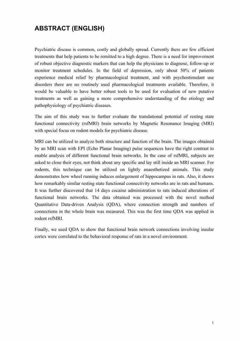
ABSTRACT (ENGLISH)
Psychiatric disease is common, costly and globally spread. Currently there are few efficient treatments that help patients to be remitted to a high degree. There is a need for improvement of robust objective diagnostic markers that can help the physicians to diagnose, follow-up or monitor treatment schedules. In the field of depression, only about 50% of patients experience medical relief by pharmacological treatment, and with psychostimulant use disorders there are no routinely used pharmacological treatments available. Therefore, it would be valuable to have better robust tools to be used for evaluation of new putative treatments as well as gaining a more comprehensive understanding of the etiology and pathophysiology of psychiatric diseases.
The aim of this study was to further evaluate the translational potential of resting state functional connectivity (rsfMRI) brain networks by Magnetic Resonance Imaging (MRI) with special focus on rodent models for psychiatric disease.
MRI can be utilized to analyze both structure and function of the brain. The images obtained by an MRI scan with EPI (Echo Planar Imaging) pulse sequences have the right contrast to enable analysis of different functional brain networks. In the case of rsfMRI, subjects are asked to close their eyes, not think about any specific and lay still inside an MRI scanner. For rodents, this technique can be utilized on lightly anaesthetized animals. This study demonstrates how wheel running induces enlargement of hippocampus in rats. Also, it shows how remarkably similar resting state functional connectivity networks are in rats and humans. It was further discovered that 14 days cocaine administration to rats induced alterations of functional brain networks. The data obtained was processed with the novel method Quantitative Data-driven Analysis (QDA), where connection strength and numbers of connections in the whole brain was measured. This was the first time QDA was applied in rodent rsfMRI.
Finally, we used QDA to show that functional brain network connections involving insular cortex were correlated to the behavioral response of rats in a novel environment.

2
POPULÄRVETENSKAPLIG SAMMANFATTNING
Psykisk sjukdom är ett vanligt, kostsamt och globalt problem. I dagsläget finns det få effektiva behandlingar som patienterna mår bättre av. Därför finns det ett behov av bättre objektiv diagnostik, som kan hjälpa läkare att identifiera sjukdomen samt följa upp och kontrollera behandlingar.
När det gäller depression uppfattar endast hälften av patienterna att de mår bättre av medicinering. Vid missbruk av psykostimulantia så finns det inga rutinmässigt använda läkemedel. Därför skulle det vara värdefullt med objektiva och mer tillförlitliga verktyg för utvärdering av nya behandlingsmöjligheter samt för en utökad förståelse för orsaker och bakomliggande mekanismer till psykiska sjukdomar.
Syftet med detta arbete var att utvärdera användningen av bildtagning med magnetresonanskamera, MR, för att kartlägga hjärnans aktivitet vid vila. Fokus lades på psykiska sjukdomar.
MR kan användas för att analysera både hjärnans struktur och funktion. MR-bilder som tas med hjälp av en typ av magnetiska pulser kallade EPI, Echo Planar Imaging, gör det möjligt att analysera olika funktionella nätverk i hjärnan. Med funktionella nätverk menas nätverk av regioner som samspelar men inte nödvändigtvis är fysiskt kopplade till varandra. Vid kartläggning av hjärnans aktivitet vid vila ombeds försökspersoner att blunda och inte tänka på något särskilt medan de ligger stilla i en MR-kamera. Vid användning av experimentella modeller kan en lätt nedsövning användas under bildtagningen.
Detta arbete visar hur träning, i detta fall i springhjul, leder till en förstoring av hjärnregionen hippocampus hos in vivo-modeller. Det visar även att det finns en stark likhet mellan modellerna och människor när det gäller hjärnans aktivitet vid vila. Dessutom framkom det att en daglig dos av den psykostimulerande drogen kokain i 14 dagars tid förändrade de funktionella nätverken i hjärnan. Informationen analyserades med hjälp av metoden Quantitative Data-driven Analysis, QDA, som användes för första gången vid analys av experimentella modeller.

3
LIST OF SCIENTIFIC PAPERS
I. Hippocampal morphology in a rat model of depression: the effects of physical activity. By: Sierakowiak, A., A. Mattsson, M. Gomez-Galan, T. Femenía, L. Graae, S. N. Aski, P. Damberg, M. Lindskog, S. Brené and E. Åberg (2014). Open Neuroimaging Journal 9: 1-6.
II. Default Mode Network, Motor Network, Dorsal and Ventral Basal Ganglia Networks in the Rat Brain: Comparison to Human Networks Using Resting State-fMRI. By: Sierakowiak, A., C. Monnot, S. N. Aski, M. Uppman, T. Q. Li, P. Damberg and S. Brené (2015). PLoS One 10(3): e0120345.
III. Cocaine Increases and Decreases Functional Coupling of the Rodent Brain in Prelimbic Cortex and Dorsal Striatum Respectively. By: Adam Sierakowiak, Tie-Qiang Li, Cyril Monnot, Josefine Klein-Dahlgren, Sahar Nikkhou Aski, Peter Damberg, Stefan Brené. (manuscript)
IV. Resting State Functional Coupling in the Rat Insular Cortex is Correlated to Behavior in a Novel Environment. By: Adam Sierakowiak, Tie-Qiang Li, Cyril Monnot, Teresa Femenía, Sahar Nikkhou Aski, Peter Damberg, Sarah Holst, Stefan Brené. (manuscript)

4
INDEX 1 Abbreviations ...................................................................................................................... 5
2 Introduction ........................................................................................................................ 6 2.1 MRI, fMRI and resting state functional connectivity .......................................................... 6 2.2 Depression ................................................................................................................................. 7
2.2.1 The serotonin hypothesis .................................................................................................... 8 2.2.2 The cytokine hypothesis ...................................................................................................... 9 2.2.3 Flinders Sensitive Line (FSL) Rats ................................................................................... 10
2.3 Psychostimulant Addiction ................................................................................................... 10 2.3.1 Cocaine .............................................................................................................................. 12
2.4 Translational Research in depression and addiction ......................................................... 13
Aims .......................................................................................................................................... 14
3 Materials and Methods .................................................................................................... 15 3.1 Animals and Ethics ................................................................................................................ 15 3.2 MRI Preparations .................................................................................................................. 15 3.3 MRI Scanning Protocols ........................................................................................................ 16
3.3.1 Spin Echo Multi Slice (SEMS) Volume and Bias Field Correction ................................ 16 3.3.2 High Resolution SEMS ..................................................................................................... 17 3.3.3 Echo Planar Imaging ......................................................................................................... 17 3.3.4 Arterial Spin Labeling ....................................................................................................... 18
3.4 MRI Data Analysis ................................................................................................................. 19 3.4.1 Volume Measurement Analysis ........................................................................................ 19 3.4.2 Seed Based Analysis ......................................................................................................... 19 3.4.3 Quantitative Data-driven Analysis (QDA) ....................................................................... 19
3.5 Study Design (Paper IV) ........................................................................................................ 20
4 Results and Discussion ..................................................................................................... 21 4.1 The effect of Wheel-Running on Hippocampus Size (Paper I) ......................................... 21 4.2 Similarities and Differences in rsfMRI Networks in Humans and Rats (Paper II) ....... 21 4.3 Cocaine Administration and Arterial Spin Labeling (Paper III) ..................................... 22 4.4 Insular Cortex in Relation to Both Cocaine and Behavior (Papers II-IV) ...................... 23 4.5 The Translational Aspect of rsfMRI in Addiction and Depression (Papers II-IV) ........ 24
5 General Conclusions ......................................................................................................... 25
6 Future perspectives .......................................................................................................... 26 6.1 Depression ............................................................................................................................... 26 6.2 Addiction ................................................................................................................................. 26 6.3 Insular cortex .......................................................................................................................... 26
7 Acknowledgements ........................................................................................................... 27
8 References .......................................................................................................................... 34
5
1 ABBREVIATIONS 5HT – Serotonin
ACC – Anterior Cingulate Cortex
ASL – Arterial Spin Labeling
BOLD – Blood Oxygen Level Dependent
CBF – Cerebral Blood Flow
CCI – Connectivity Counter Index
CSI – Connectivity Strength Index
DFP – diisopropyl fluorophosphate
DMN – Default Mode Network
ECS – Electroconvulsive Seizures
EPI – Echo Planar Imaging
fMRI – functional Magnetic Resonance Imaging
FRL – Flinders Resistant Line rats
FSL – Flinders Sensitive Line rats
IL-6 – Interleukin-6
MDD – Major Depressive Disorder
mPFC – medial Prefrontal Cortex
MRI – Magnetic Resonance Imaging
NMDA – N-methyl-d-aspartate
OFC – Orbitofrontal Cortex
PCA – Principal Component Analysis
PET – Positron Emission Tomography
PFC – Prefrontal Cortex
QDA – Quantitative Data-driven Analysis
rsfMRI – resting state functional connectivity
SEMS – Spin Echo Multi Slice
SSRI – Selective Serotonin Reuptake Inhibitors
TNF-α – Tumor Necrosis Factor alpha
VOI – Volume of Interest
VTA – Ventral Tegmental Area
6
2 INTRODUCTION
2.1 MRI, FMRI AND RESTING STATE FUNCTIONAL CONNECTIVITY
Magnetic Resonance Imaging (MRI) technology was put in clinical use in the mid 1980’s (Bandettini, 2012). The original idea of magnetic resonance was developed by Isidor Rabi and was awarded by a Nobel price in 1944 (Rabi, Zacharias, Millman, & Kusch, 1992). However, after the principle of MRI was discovered, many different discoveries (Pake, 1993) were crucial for the development of the MRI as we have it today. It would take until 1973 before the first magnetic resonance image was produced (Lauterbur, 1973).
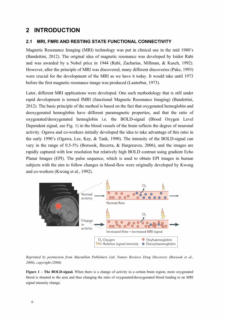
Later, different MRI applications were developed. One such methodology that is still under rapid development is termed fMRI (functional Magnetic Resonance Imaging) (Bandettini, 2012). The basic principle of the method is based on the fact that oxygenated hemoglobin and deoxygenated hemoglobin have different paramagnetic properties, and that the ratio of oxygenated/deoxygenated hemoglobin i.e. the BOLD-signal (Blood Oxygen Level Dependent signal, see Fig. 1) in the blood vessels of the brain reflects the degree of neuronal activity. Ogawa and co-workers initially developed the idea to take advantage of this ratio in the early 1990’s (Ogawa, Lee, Kay, & Tank, 1990). The intensity of the BOLD-signal can vary in the range of 0.5-5% (Borsook, Becerra, & Hargreaves, 2006), and the images are rapidly captured with low resolution but relatively high BOLD contrast using gradient Echo Planar Images (EPI). The pulse sequence, which is used to obtain EPI images in human subjects with the aim to follow changes in blood-flow were originally developed by Kwong and co-workers (Kwong et al., 1992).
Reprinted by permission from Macmillan Publishers Ltd: Nature Reviews Drug Discovery (Borsook et al., 2006), copyright (2006)
Figure 1 – The BOLD-signal. When there is a change of activity in a certain brain region, more oxygenated blood is shunted to the area and thus changing the ratio of oxygenated/deoxygenated blood leading to an MRI signal intensity change.

7
After the discovery and development of the BOLD signal and neuronal activity, researchers have used the technology for various applications. One example is the detection of different resting state functional brain networks, which are detected when people are resting, having closed eyes, and not thinking about anything specific. Those networks differ from the functional brain networks that are detected during specific tasks (Beckmann, DeLuca, Devlin, & Smith, 2005; B. Biswal, Yetkin, Haughton, & Hyde, 1995; B. B. Biswal, VanKylen, & Hyde, 1997; Fransson, 2005; Raichle et al., 2001). This lead to the discovery of resting state functional connectivity (rsfMRI) brain networks with synchronized blood flow that do not have to be directly anatomically linked. One such network is called the DMN (Default Mode Network), and was first characterized by Raichle and co-workers in 2001, who hypothesized that the brain has a network of higher activity nodes when it is at rest (Raichle et al., 2001). It was also compared to PET (Positron Emission Tomography) data using O15-radiopharmaceuticals to calculate the Oxygen Extraction Fraction, which is the ratio of oxygen used by the brain in relation to the oxygen delivered by the blood. It was thereby concluded that there in fact is an active network in the brain while it is resting.
Over the last twenty years, the technical development has enabled new MRI scanners with strong magnetic fields that are optimal for studying small subjects such as mice or rats (Jonckers, Van Audekerke, De Visscher, Van der Linden, & Verhoye, 2011; Pawela et al., 2009; A. Schwarz et al., 2013; Silva, Zhang, Williams, & Koretsky, 1995). In parallel the development of novel resonators, scanning protocols, statistical analysis methods, and other hardware for scanning preclinical subjects is still ongoing (Anderson et al., 2011; Fox, Zhang, Snyder, & Raichle, 2009; Kalthoff, Po, Wiedermann, & Hoehn, 2013; X. Liu, Zhu, Zhang, & Chen, 2013; Wang & Li, 2013).
Thus, in the light of the fast development in different fields the fMRI technology has the potential to be used for example to study complex diseases within the field of psychiatry. Classical pharmacological biomarkers of psychiatric disease, treatments, and pathophysiology are difficult to develop and are yet to be further investigated (Maes et al., 2009). At the same time, there is a need for more robust methods to assess severeness, follow-up treatments and elucidate the pathophysiology as well as the mechanism of disease.
2.2 DEPRESSION
Depression is a major psychiatric disease that impacts people worldwide. It is estimated to be the fourth most common disability that has an annual estimated cost of up to 118 billion Euro (2004) in Europe alone. Due to the complexity, extreme variability and heterogeneity of this disease, treatments and understanding of the underlying mechanism have not yet been very successful (Maes et al., 2009).
Major Depressive Disorder (MDD) is a disease where the subjects can have daily feelings of depressed mood, experience worthlessness, close to no feelings of pleasure out of any activity during the day, has drastic changes in weight, suicidal thoughts, and problems sleeping (APA, 2013). Some of these symptoms are evaluated on a subjective basis, making it difficult

8
to assess in comparison to other subjects of similar feelings. Women are more prevalent among the depressed patients compared to men with a relationship 2:1 (Nestler et al., 2002).
There are several treatments for depression such as pharmacotherapy, electroconvulsive seizures (ECS), and psychotherapy (Nestler et al., 2002). However, far from all individuals respond to treatment and also there is a delayed response. It takes weeks or even months before an antidepressant effect can be manifested. Because of the delayed response of the different treatments it is difficult to understand and pin point specific adaptations in the brain that can underlie the antidepressant effect.
In the field of imaging with a focus on depression, there is for example strong documentation on the shrinkage of hippocampus in depressed subjects (Bremner et al., 2000; Wisse et al., 2015). In fact time with depression is correlated to the degree of shrinkage (Sheline, Sanghavi, Mintun, & Gado, 1999). In addition to these findings, the hippocampal volume decrease is also detected in post-mortem analysis of depressed patents, which also indicated decreased number of synapses as well as lower glial cell density (Russo & Nestler, 2013).
Recently, resting state functional MRI was used to analyze brain networks in depressed patients and controls (Tang et al., 2013). The methodology could with 94% certainty (Zeng et al., 2012) distinguish clinically depressed patients from healthy controls. Thus, rsfMRI methodology encompasses a different level of network complexity compared to other well-established pharmacological aspects of disease.
2.2.1 The serotonin hypothesis
The serotonin hypothesis of depression has so far been the most investigated theory of depression from the pharmaceutical industry since most anti-depressant treatments mainly target the serotonergic signaling system (Maes et al., 2009). The hypothesis states that there are altered relationships of serotonin receptors in patients suffering from depression, and that there are lower levels of serotonin (5HT) in depressed patients (Maes M, HY, Bloom, & Kupfer, 1995). For example, levels of 5HT in post-mortem studies of suicide victims are generally lower in the brain stem (Mann, Arango, Marzuk, Theccanat, & Reis, 1989).
Initially, tricyclic antidepressants and monoamine oxidase inhibitors were found to exert antidepressive effects, and the mechanism of action was inhibition of serotonin and norepinephrine reuptake as well as inhibition of monoamine oxidase (Nestler et al., 2002).
Later, development of second-generation antidepressants lead to the introduction of Serotonin-Selective Reuptake Inhibitors (SSRI’s) and norepinephrine-selective reuptake inhibitors were considered to be more selective than the first-generation drugs and are still in routine use today. However, time has shown that the mechanism of action by these drugs is more complex. They are not as selective as it was initially thought.

9
However, it takes several weeks before patients get any effect by SSRI’s and only about 50-60% of the patients experience remission indicating that 5HT is not the single signaling substance involved in the pathophysiology of the disease (Maes et al., 2009; Nestler et al., 2002).
2.2.2 The cytokine hypothesis
Cytokines are small endogenous molecules that are involved in inflammatory response, secreted by immune cells. They act on the surroundings as well as the cells secreting them (Venes, 2013).
Both immune cell activation and an increase of proinflammatory cytokines have been linked to depression in patients. Inflammatory markers such as lower serum zinc levels have also been detected among depressed subjects hinting an inflammatory response correlated with depression (Maes et al., 2009). In a meta-analysis of depressed patients the levels of serum cytokines showed a significant increase in Tumor Necrosis Factor (TNF)-α and Interleukin(IL)-6 compared to controls (Dowlati et al., 2010).
Other observations made in patients with different inflammatory diseases such as type-2 diabetes, cardiovascular disease, and rheumatoid arthritis is that it often coincides with depression. Moreover, the symptoms of sickness behavior that are fatigue, anhedonia, reduction in locomotion, loss of appetite, and cognitive malfunction are similar to the symptoms of depression (Dantzer, O'Connor, Freund, Johnson, & Kelley, 2008).
Interestingly, in depressed patients lower levels of L-tryptophan and 5HT is noticed (Maes et al., 1993). This indicates that tryptophan is catabolized into kynurenic acid due to induction of indoleamine 2–3-dioxygenase by cytokines (Babcock & Carlin, 2000). Elevated levels of kynurenic acid has previously been associated with schizophrenia (Erhardt, Olsson, & Engberg, 2009) and bipolar disorder (Olsson et al., 2010).
One theory states that the induced level of cytokines in depressed patents increases permeability of the gut allowing bacteria to colonize the outside of the gut protective mucosal layer and allowing lipopolysaccharide to “leak” into the blood known as a condition called leaky gut. The condition will then potentiate the immune system and lead to increased inflammatory response in the periphery (Maes et al., 2009).
Additionally, TNF-α contributes to the neurotoxic effects of glutamate in the hippocampus through a synergetic effect by inhibiting glutamate re-uptake by transporters on astrocytes leading to an increase of glutamate in the synaptic cleft and in the extrasynaptic compartment. The glutamate in the extrasynaptic environment mediates a postsynaptic neurotoxic effect by stimulating extrasynaptic N-methyl-d-aspartate (NMDA) receptors (Zou & Crews, 2005).

10
2.2.3 Flinders Sensitive Line (FSL) Rats
Several different rodent models for depression exist and one of the best characterized is the Flinder’s Sensitive Line (FSL) rat strain. The FSL rat strain was originally developed to be resistant to diisopropyl fluorophosphate (DFP) (Overstreet & Wegener, 2013), which is an acetyl choline esterase inhibitor (Millard et al., 1999). However, the strain turned out to be more sensitive instead of resistant to DFP and showed depression-like symptoms (Overstreet & Wegener, 2013). The hallmark features of depression that are observed in the FSL rat model are; psychomotor retardation, reduced appetite, sleep abnormalities, as well as an altered immune system (Overstreet, Friedman, Mathe, & Yadid, 2005). However, one main symptom of depression, anhedonia, was not evident in the FSL rat strain when testing for sucrose preference, which is a test that is suggested to monitor anhedonia in rats. The rats show no decrease of sucrose consumption compared to controls (Overstreet & Wegener, 2013).
The FSL rats also respond to antidepressant treatments and physical activity (wheel running). All SSRI’s and tricyclic drugs with antidepressant effects in humans that have been tested on FSL rats also demonstrate antidepressant effects in the rat model (Bjornebekk, Mathe, & Brene, 2005, 2006; Overstreet & Wegener, 2013).
Moreover, FSL rats have lower levels of D-serine in hippocampus, a decrease of glutamate transporters on the astrocytes, simultaneously higher synaptic glutamate activity as well as decreased synaptic plasticity (Gomez-Galan, De Bundel, Van Eeckhaut, Smolders, & Lindskog, 2013).
The FSL and its’ often used control strain FRL strain have also been analyzed with fMRI. Interestingly, when presented with an odor of a predator (Huang et al., 2011) awake FSL rats have differences in BOLD signal in brain regions, which are suggested to be affected in depression such as insular cortex, prefrontal cortex, and amygdala when compared to the FRL (Flinders Resistant Line rats) strain.
2.3 PSYCHOSTIMULANT ADDICTION
Addiction is a complex disorder that can be subdivided in many stages. The two longitudinal stages of the disease are habitual usage, and withdrawal.
Stimulant use disorder is a psychiatric disease defined as when a person takes a drug and use it over longer periods of time than intended, is unable to control the amount of intake, experience craving, and uses the drug despite knowledge of its hazardous properties (APA, 2013).
Stimulant withdrawal on the other hand manifests many clinical features similar to major depressive disorder, for example: dysphoria, issues sleeping, increased appetite, and psychomotor retardation (APA, 2013).

11
Currently, there are no successful pharmacological treatments routinely used for psychostimulant addiction. There is a great need for more research on novel therapeutic approaches, and the main rehabilitation strategy is behavioral therapy (Phillips, Epstein, & Preston, 2014).
There are multiple anatomical regions involved in the different stages of addiction that has great resemblance to the brain regions affected in depression (see Fig. 2). The prefrontal cortex (PFC) and orbitofrontal cortex (OFC) are important for motivation/drive and impulse control (Volkow, Fowler, & Wang, 2003). Ventral tegmental area (VTA) is the site for the dopamine cells that project to nucleus accumbens, which is rich in postsynaptic dopamine receptors. It is an essential part of the mesolimbic dopamine pathway (Nestler, 2005). Virtually all addictive drugs are shown to acutely elevate dopamine levels in nucleus accumbens and caudatus putamen (Di Chiara & Imperato, 1988). There are also studies showing that glutamate is important for different aspects of addiction and persistent changes of VTA dopaminergic cells occur after cocaine self-administration (Rodriguez Parkitna & Engblom, 2012). Amygdala is important for emotional processing and hippocampus in spatial learning.
©Science by American Association for the Advancement of Science Reproduced with permission of AMERICAN ASSOCIATION FOR THE ADVANCEMENT OF SCIENCE in the format Republish in a thesis/dissertation via Copyright Clearance Center.
Figure 2 – Brain Regions Involved in Addiction. Dopamine has a central role in addiction, and projections from the ventral tegmental area (VTA) assert effects on brain regions such as nucleus accumbens, and the frontal cortical areas. (Holden, 2001)

12
When drug abuse is established among recreational drug users, and drugs are taken more as a ‘habit’ rather than due to the ‘rewarding effects’, a shift in which anatomical regions that are affected has been observed (Everitt & Robbins, 2005, 2013). Preclinical studies show that when drugs of abuse are taken initially, parts of the nucleus accumbens shell as well as mediodorsal parts of the caudatus putamen are activated. By time when the substance abuse has entered a habitual phase, a transition has occurred and now nucleus accumbens core and laterodorsal parts of caudatus putamen is more involved (Everitt & Robbins, 2005, 2013).
Many imaging studies of drug abuse show altered blood flows and changes in resting state functional connectivity networks in both humans (Gu et al., 2010; Volkow et al., 2003; Volkow, Fowler, & Wang, 2004) and rats (Gozzi et al., 2011; Marota et al., 2000; Schwarz et al., 2004).
An intriguing matter in addiction biology is the question of what is essential for developing addiction. There are many studies showing on drug dependence induced adaptations of the brain, but there is also evidence for inherited biological abnormalities in drug dependent subjects (Volkow & Baler, 2012).
In order to answer the question on whether the addictive drugs induce the structural and functional changes as addiction develops in humans, analysis of the individuals before and after addiction is manifested is necessary. Recently, a study showed that there are similar alterations in brain structure and impulse control in addicts and their non-addicted siblings when compared to healthy controls (Ersche et al., 2012). The data suggests that it is not the drugs per se that induce structural and functional changes that are noted in drug addicts. However, when researching the underlying pathology of addiction in a clinical setting, with MRI technology, there is a fundamental ethical problem with longitudinal studies examining e.g. the functional networks before and after prolonged abuse of drugs in the same individual. In contrast, MRI examination of these networks before and after prolonged drug abuse in rodent models can be preformed.
2.3.1 Cocaine
The main mechanism of cocaine is blocking dopamine reuptake via the dopamine transporter (DAT), which is situated on the presynaptic dopamine terminal, This results in increased dopamine levels in the synaptic cleft and prolonged exposure of dopamine on dopamine receptors on the postsynaptic terminal (Hyman, Malenka, & Nestler, 2006).
A study of cocaine addicts in withdrawal showed that there were persistent lower uptake of a PET-ligand called [18F]N-methylspiroperidol, that is used to study dopamine receptor 2 availability, in the basal ganglia one month after cocaine abuse cessation. The decreased availability is still very evident even 4 months after cocaine abuse cessation compared to healthy controls (Volkow et al., 1993). Additional studies of early withdrawal showed generally higher glucose metabolism in the whole brain compared to control subjects, but the longer time after discontinuation of cocaine, the lower glucose metabolism in orbitofrontal cortex (Volkow et al., 1991).
13
An analysis of rsfMRI networks among active cocaine-addicted subjects compared to healthy controls show differences in connectivity from VTA to thalamus/nucleus accumbens, from amygdala to mPFC (medial Prefrontal Cortex)/rostral ACC (Anterior Cingulate Cortex), as well as from rostral ACC to amygdala/hippocampus after (Gu et al., 2010). Interestingly, these are all regions (described previously) where dopamine is a prevalent neurotransmitter.
There are also preclinical fMRI studies of withdrawal and rats on 10 days withdrawal after prolonged cocaine self-administration have differences in basal cerebral blood volume in rostral ACC, prelimbic cortex, and OFC compared to drug naïve rats. The cocaine group showed significantly lower relative cerebral blood volume response in caudatus putamen when challenged with amphetamine while in the MRI-scanner (Gozzi et al., 2011). Interestingly, and in line with the rodent imaging study mentioned above, multiple daily cocaine injections over time induce lower baseline dopaminergic tone in dorsolateral and ventromedial striatum when cocaine is not present (Schlussman, Nyberg, & Kreek, 2002).
2.4 TRANSLATIONAL RESEARCH IN DEPRESSION AND ADDICTION
There are a few different aspects in validation of animal models in neuropsycho-pharmacology; predictive validity, face validity, and construct validity. Predictive validity is when a treatment has similar effect in a human and a model animal i.e. the outcome of the treatment is the same. Face validity is when an animal model shows the same symptoms as in the clinical aspect of the disease. Construct validity is when the underlying cause e.g. the disease pathophysiology or the etiology is the same or as similar as possible as in humans (Belzung & Lemoine, 2011).
In the field of addiction, rats can be trained to self-administer in principle all drugs that are addictive to humans (Shippenberg & Koob, 2002) and there is vast literature describing molecular (Dworkin, Co, & Smith, 1995; Self, Choi, Simmons, Walker, & Smagula, 2004; Self & Nestler, 1995), and behavioral (Ahmed & Koob, 1998; Sanchis-Segura & Spanagel, 2006) aspects of self-administration.
There are for example imaging studies of cocaine addiction investigating both acute effects (Schwarz et al., 2004) on blood flows in the brain as well as withdrawal effects (Gozzi et al., 2011). However, only very few imaging studies have focused on translational networks in both animals and humans, but mainly explored the DMN (Lu et al., 2012; A. J. Schwarz et al., 2013; Smucny, Wylie, & Tregellas, 2014).
14
AIMS
1. Investigate whether experimental MRI can detect structural adaptations in hippocampus, in an exercising rat model of depression.
i. Can the hippocampus size be altered by physical activity?
2. Analyze if humans and rats have common resting state brain networks.
i. Elucidate whether resting state fMRI can be used as a translational tool when validating animal models of brain disorders.
3. Investigate whether cocaine alters resting state functional connectivity in drug naïve rats.
4. Explore how behavior in a novel environment is correlated to specific brain regions using resting state fMRI.
5. Suggest how MRI methodology can be further developed for translational and diagnostic purposes.

15
3 MATERIALS AND METHODS The material and methods are described in detail in the manuscripts in the thesis. Here, the different methods are discussed from the perspective of what was learnt along the way of the studies.
3.1 ANIMALS AND ETHICS
All animals used in the studies were approved by the Stockholm North Committee on Ethics on Animal Experimentation and followed the ethical guidelines of the Swedish Legislation on Animal Experimentation (Animal Welfare Act SFS1998:56) as well as the European Communities Council Directive (86/609/EEC). In paper I, a rat model of depression was used to investigate a novel therapy in depression: exercise. This preclinical data can be used to motivate future clinical studies on how exercise can be used in treatment or in combination with already existing treatments in patients suffering from depression. There are many animal models of psychiatric disease available. To be able to evaluate how the brain networks are altered in these models, it is of great importance to understand the normal/healthy animal and human as was done in paper II. Moreover, better understanding of evaluated animal models can lead to a reduction in use of animal models that do not correspond to either normal or disease symptoms seen in a clinical setting. In study III, cocaine was given to naïve rats and MRI data was collected before and after 14 days with cocaine. It would not be ethically justifiable to do these experiments in human subjects, yet the rat experiments provides vital data for the understanding of the pathophysiology of cocaine addiction and the alteration of the resting state functional connectivity. Paper IV describes how a behavioral trait can be linked to a brain networks in the same individual. Thus, combining behavioral analysis with brain network analysis (preclinical imaging and behavioral neuroscience) on the same individuals can reduce total number of animal used.
3.2 MRI PREPARATIONS
A crucial step in MRI of small animals such as rodents is the animal preparation before the scanning session and also to keep the animal in a stable and reproducible physiological state during the whole MRI scan-time. There are mainly two schools of thought each with their benefits and disadvantages. One could (like in the studies of this thesis) lightly anaesthetize the animals during the MRI scanning or otherwise habituate the animals to the scanner setting while they are awake as has been done by others (Becerra, Pendse, Chang, Bishop, & Borsook, 2011; Huang et al., 2011; Upadhyay et al., 2011). The disadvantage with the anesthesia is that there might be pharmacological interference with the study. Awake animals have a greater tendency not to lie still and thus move more than the anaesthetized counterpart, additionally the scanning time has to be reduced so longer experiments are not an option. Not only does the study of awake rodents bring ethical considerations, but it also makes it harder to compare to other studies of e.g. acute pharmacological treatments or electrical stimulation while in the MRI.

16
As the influence of anesthesia is inevitable, there are studies (Kalthoff et al., 2013; Zhao, Zhao, Zhou, Wu, & Hu, 2008) suggesting light medetomidine as the primary choice of anesthesia.
Here, isoflurane was used to induce anesthesia, and a subcutaneous catheter for transition to subcutaneous medetomidine was placed. After the animal’s temperature had stabilized at 37°C, a bolus dose of medetomidine was given, the isoflurane was discontinued, and consecutively, medetomidine was infused continuously.
During the MRI scans, physiological parameters such as breathing rate, temperature, and pulse were monitored. One way to see the transition of isoflurane to medetomidine was by the decreased heart rate and slower breathing.
3.3 MRI SCANNING PROTOCOLS
Due the effects of anesthesia, the scanning time for each individual is potentially much greater so more different types of images can be collected. In a single scan session that could take up to four hours, it was possible to obtain high-resolution anatomical images, Echo Planar Imaging (EPI) images, as well as Arterial Spin Labeling (ASL) images.
3.3.1 Spin Echo Multi Slice (SEMS) Volume and Bias Field Correction
This method was used in paper I, to obtain volumetric data of the entire rat brain for further analysis. The transmitting coil was a an actively tuned 72 mm inner diameter birdcage (volume coil). Receiving the signal is a 4-channel phased array coil (surface coil). The surface coil is cinched tight around the head of the rat to minimize the length between the coil elements and the brain. Obtaining images with the surface coil will give a signal intensity that is higher proximal to the coil and thus creating bias fields that are a gradient decay of signal intensity from the most proximal to the distal point from the surface coil, i.e. from the dorsal to the ventral part of the brain when the animal is in prone position (see Fig. 3). In paper III, a decision was made to try to correct for this intensity change and a bias field correction was made by obtaining low resolution images of the whole volume, from both the surface coil and the volume coil, smoothing them and calculating new images according to this formula:
𝑙𝑜𝑤 𝑟𝑒𝑠𝑜𝑙𝑢𝑡𝑖𝑜𝑛 𝑠𝑚𝑜𝑜𝑡ℎ𝑒𝑑 𝑣𝑜𝑙𝑢𝑚𝑒 𝑖𝑚𝑎𝑔𝑒𝑠𝑙𝑜𝑤 𝑟𝑒𝑠𝑜𝑙𝑢𝑡𝑖𝑜𝑛 𝑠𝑚𝑜𝑜𝑡ℎ𝑒𝑑 𝑠𝑢𝑟𝑓𝑎𝑐𝑒 𝑖𝑚𝑎𝑔𝑒𝑠
×𝑠𝑎𝑚𝑝𝑙𝑒𝑑 ℎ𝑖𝑔ℎ 𝑟𝑒𝑠𝑜𝑙𝑢𝑡𝑖𝑜𝑛 𝑠𝑢𝑟𝑓𝑎𝑐𝑒 𝑖𝑚𝑎𝑔𝑒𝑠
This evened out the gradient intensity and resulted in easier manual skull stripping in paper II (see Fig. 4). Future studies intended for volumetric measuring could benefit from this intensity correction.

17
Figure 3 – Profile Cross-section Illustration of Rat in Prone Position with Volume Coil, Surface Coil and ASL Coil.
Figure 4 – Bias Fields and Correction Overview.
3.3.2 High Resolution SEMS
The advantage with this scanning protocol is that very high-resolution images can be obtained on the cost of scanning time. It takes around 20 minutes to generate 11 slices that are 1 mm thick. In this case, anesthetized animals are preferred, due to the long scanning time, where slightest movement could destroy the entire scan.
3.3.3 Echo Planar Imaging
EPI proved to be very good in generating BOLD-images. However, there are problems regarding the cost of the rapid image collection such as resolution and geometric distortions. The time to collect the entire volume was only 1 sec, and the BOLD-signal could be detected even if the time was set to 2 sec, and that might allow for better resolution (as was done in the human fMRI scanning in paper II).

18
However, one of the main problems with the protocol utilized was that the paramagnetic effects of the air in the ear-channels contributed with large artifacts on the brain proximal to the ears. This could be corrected if one were to apply a parallel imaging paradigm (similar to the ipat=2 in the human fMRI data collection in paper II).
3.3.4 Arterial Spin Labeling
The method of Arterial Spin Labeling (ASL) generates blood quantitative perfusion maps of the brain.
Perfusion measurements were performed using an echo planar imaging sequence with continuous labeling of the arterial spins, TR=6 s, TE=6.18, matrix size 64 × 64 and FOV= 32mm, slice thickness=1mm, 11 consecutive slices, 15 repetitions, and 3 shots in k-space. Labeled images were acquired by labeling the arterial spins during TR before the phase encoding step using RF power sent to the neck coil together with 1 Gauss/cm gradient. The labeling slice was located 24 mm away from the most caudal slice in the imaging slice packet. For acquiring control images no RF power was sent to the labeling coil.
Perfusion maps are calculated according to:
P=
ρ = Tissue density, λ=0.9 is the blood:tissue partition coefficient for water, = T1 relaxation time for the blood in the brain tissue Mc= The intensity of the non-labeled slice ML= The intensity of the labeled slice α = the Labeling Efficiency P = Perfusion
And since φ=ρ·P (perfusion rate = tissue density · Perfusion) we can calculate Cerebral Blood Flow maps (CBF-maps) over the brain.
3.3.4.1 Labeling Efficiency
To measure labeling efficiency, a gradient echo sequence was used with TR=7.45 ms, TE=3.74 ms, flip angle=40°, 1 average and slice thickness= 1.0 mm. The labeling slice was placed upstream from the bifurcation of the carotid. Labeling was performed using a RF pulse on the neck coil during a gradient of 1Gauss/cm. The labeling efficiency was calibrated to obtain a 75% labeling efficiency by varying the RF pulse amplitude and measuring the arterial spin magnetization in the presence (MT) and absence (Mo) of RF irradiation. The labeling efficiency is then calculated as α=(MT- Mo)/(2 Mo).
ρλT1!
MC −ML
2αMC
T1!

19
3.4 MRI DATA ANALYSIS
3.4.1 Volume Measurement Analysis
There are different approaches of data analysis in MRI volumetric data. Manual segmentation of a volume of interest (VOI) could be preformed, semi manual segmentation, or automatic segmentation.
Manual segmentation is the most exact segmentation strategy where the researcher manually outlines the VOI in each slice of a 3d volume, hence the most time consuming. Semi-manual segmentation is when the researcher manually segments some slices of the entire VOI and later interpolates between them. Automatic segmentation is the fastest process yet the least careful and with the possibility of committing greater mistakes, however consistently. In paper I, the method chosen was the manual segmentation due to the strength in accuracy of the segmentation outlining.
3.4.2 Seed Based Analysis
The seed based analysis of rsfMRI data is a hypothesis driven method. I.e. the method is based on seed regions placed according to the hypothesis in the brain areas of choice according to the hypothesis. This method was used in paper II where robust and already known networks were analyzed and paper III, where prior clinical as well as preclinical data existed. This is an excellent method to use when e.g. a specific network, a region of interest, or prior knowledge is obtained to analyze differences related to that specific seed.
3.4.3 Quantitative Data-driven Analysis (QDA)
To our knowledge, a suitable method, which did not require any prior assumptions and that could be used to correlate a set of behavioral traits with rsfMRI data (paper IV), did not exist. Therefore the development of a new hypothesis generating method was encouraged and welcomed. The strength of this method is that one can visualize, and do statistics on the entire brain volume with respect to changes in correlation strength (CSI – Connection Strength Index) as well as changes in number of connections (CCI – Connection Counter Index) from each point in the volume to the entire brain. This method cannot be utilized to visualize where the connections go (directionality) or which specific connections have decreased or increased in strength.

20
3.5 STUDY DESIGN (PAPER IV)
In the cocaine study, a longitudinal design (see Fig. 5) was selected to ensure the quality of the data and push the limits of what the fMRI method can preform. Not only does this design allow intra-group changes (where each individual is its own control), but also inter-group differences can be evaluated. This design is more complex than only analyzing differences after treatments, but it can also handle factors such as the unavoidable anesthesia by taking the changes of the control-group (ΔSal) into account.
Figure 5 – A schematic view of the longitudinal design in paper IV. Twenty naïve rats were scanned in the MRI camera and then randomly divided into two different treatment groups with ten individuals in each group (Coc1 and Sal1). One group received a daily dose of 20mg/kg cocaine and the other group received saline for 14 consecutive days. The post treatment names of the two groups were Coc2 and Sal2. All subjects in the groups underwent another MRI scan 24 hours after the last injection received.

21
4 RESULTS AND DISCUSSION
4.1 THE EFFECT OF WHEEL-RUNNING ON HIPPOCAMPUS SIZE (PAPER I)
Here, adult depressed rats that were single housed with free access to a running-wheel were investigated to see whether they could increase hippocampal size by voluntary exercise.
It was concluded that the FSL rats with free access to running-wheels had increased hippocampus size compared to FSL rats that were single-housed without running-wheels. Clinical studies have shown that hippocampus size is correlated with the level of exercise in both children (Chaddock et al., 2010) and in elderly people (Erickson et al., 2011). It has also been concluded that both humans (Spalding et al., 2013) and rats (Bjornebekk et al., 2005) have ongoing neurogenesis in hippocampus. The effect of neurogenesis driven by exercise has already proven to be antidepressant (Bjornebekk et al., 2006; Brene et al., 2007).
However, one limitation of imaging studies is that it is impossible to distinguish different cell types in a voxel of the size that was used in study I. because the resolution and contrast is too poor to separate cell layers in such small volumes. New techniques of high-resolution MRI can already produce 120µm slices of the human brain (Marques, van der Zwaag, Granziera, Krueger, & Gruetter, 2010), so visualizing different cell-types might not be too far in the future.
Apart from volumetric analysis of hippocampus, network changes in FSL rats and depressed patients can be computed and compared to each other as well as compared to healthy controls, thus exploring the face and construct validity of the model. The FSL strain can also be monitored and followed up during treatments with e.g. SSRI’s and compared to placebo as well as human data thereby investigating the predictive validity.
4.2 SIMILARITIES AND DIFFERENCES IN RSFMRI NETWORKS IN HUMANS AND RATS (PAPER II)
Well-established rsfMRI networks in rats as well as humans were thoroughly investigated to better comprehend the differences and similarities between the species.
Remarkable similarities were found and the networks showed to be highly evolutionary conserved. One of the main targets with the study was to analyze the data from each species as similarly as possible. However, since the MRI-apparatus for humans differ from the rats, size differences are prevalent, and pulse-protocols are slightly different, the data had to be treated as such. Apart from the set-up differences, also analysis was slightly different due to brain structural aberrations between species. It is therefore not surprising that there were some deviations in the network magnitude and characteristics. E.g. white-matter regression where partial volume effects were taken into account in the human brain was not applied in the rat brain. Instead, binary masks made on the basis of the standard brain template for the white matter were applied. It can then be concluded that it is inevitable not to regress out some of the gray matter signal in the rat brains, since the white matter is only 1-2 voxels in

22
size. This can be a part of the explanation why the rat brain has more anti-correlated networks than the human brain. However, there are studies on human rsfMRI where anti-correlated networks are detected as well (Anderson et al., 2011).
An interesting finding in the study was that the ROI-ROI correlation matrices were very similar in rats and humans. I.e. the correlation strength between different parts of the networks and other areas showed that there was very strong correlation between the motor cortex and the somatosensory cortex, the different components of the DMN-network, and the basal ganglia (nucleus accumbens and caudatus putamen in rats/putamen in humans) in both species.
This study implies that a logic step in translational psychiatry is to further analyze and characterize animal models of psychiatric disease. Face validity, construct validity, and predictive validity can all be examined using rsfMRI in rodent models and compared to humans.
4.3 COCAINE ADMINISTRATION AND ARTERIAL SPIN LABELING (PAPER III)
The aim in paper III was to test if two weeks repeated cocaine administration could manifest changes detectable by rsfMRI. It has been shown that siblings to stimulant addicts have changes in brain region densities as well as inhibitory control similar to their addicted sibling and different to healthy controls (Ersche et al., 2012). This implies that genetics and developmental biology are important for development of addiction. However, due to ethical considerations, studies of before and after stimulant use disorder have not been performed in human subjects. The findings in paper III also suggest that repeated cocaine administration contributes to changes in brain networks as well leading to the conclusion that perhaps both the underlying brain alterations in stimulant addicts and the induced changes by stimulant drugs are important for developing addiction.
In addition to the resting state functional connectivity, ASL was preformed and CBF-maps were calculated (see Fig. 6). Due to technical issues and method development, too few maps were generated to draw conclusions on cocaine-induced changes in blood-flow.
Figure 6 – Arterial Spin Labeling (ASL) of a rat brain after 14 consecutive days of 30mg/kg cocaine.

23
4.4 INSULAR CORTEX IN RELATION TO BOTH COCAINE AND BEHAVIOR (PAPERS II-IV)
The aim of study IV was to integrate behavioral testing in animal models with state-of-the-art imaging. This resulted in detection of a key brain region involved in both depression and addiction: the insular cortex. In paper IV, rats were placed in a novel cage and scored on their behavioral characteristics. These scores were later plotted in a Principal Component Analysis (PCA), and the two components describing most of the data were later used as regressors in the analysis of rsfMRI data using QDA. This resulted in bilateral correlation of the insular cortex in the rats that were linearly correlated to the behavioral components from the PCA. Thus, the data reveals a strong relationship between behavior in a novel environment and the insular cortex.
The insular cortex is separated in different sub-sections that have been mainly divided in cardiovascular, cognitive, pain, social, somatosensory, and taste functions in humans, and auditory, cardiovascular, pain, somatosensory, taste functions in rats (Moraga-Amaro & Stehberg, 2012). The networks examined in paper II showed striking similarities between the different species. Humans have additional sub-sections (that are not characterized in the rat insular cortex), which are functionally associated to essential functional features that should be important to both humans and rats. Where in the rat brain would these two sub-sections be placed? Yet, the most logic assumption is that the rat insula also have these sub-sections but possibly more integrated or less subdivided. This can be found elsewhere in the rat brain, where the caudatus putamen is one integrated region, but divided in two distinct regions in the human brain.
Figure 7 – Insular Cortex Network (paper IV) and the ΔCoc-ΔSal Connection Counter Index (CCI), paper III. Note the similarities in the T-map of the network distribution of the insular cortex and the increased number of connections in the cocaine subjects when subtracted from the control group. The white arrows show the seed-placement where the insular cortex network is derived from.
It is very interesting to note the striking similarities in the distribution of the dorsal insular cortex network and the areas with increased numbers of connections after chronic treatment of cocaine i.e. rats undergoing withdrawal of cocaine. Strong evidence for the important role of insular cortex in addiction is that nicotine smokers that have had had strokes in the area of

24
insular cortex have ceased smoking (Naqvi, Rudrauf, Damasio, & Bechara, 2007). Increasing research show that the insular cortex is important for the maintenance of addiction as well as withdrawal (Naqvi & Bechara, 2009). Insular cortex is also affected in depression (C. H. Liu et al., 2015; Sprengelmeyer et al., 2011; Takahashi et al., 2010).
The DSM-5 criteria for depression and stimulant use disorder withdrawal are very similar and can very well be linked to the insular cortex network found in paper IV with a high similarity to the number of connections change between cocaine and healthy controls in paper III.
4.5 THE TRANSLATIONAL ASPECT OF RSFMRI IN ADDICTION AND DEPRESSION (PAPERS II-IV)
The translational aspect of fMRI studies is analyzed in paper II where it was found that there are remarkable similarities between the networks in humans and rats. Paper III then investigates the alteration in the resting state functional connectivity data including the networks described in paper II, and how these are influenced by repeated cocaine administration. In addition to the cocaine study, paper IV evaluates resting state functional connectivity in a model for depression and how it is correlated to behavioral traits.
Ultimately, the translational aspects in paper III, and IV are in line with previous findings showing that there are interhemispheric functional disruptions in cocaine addicts (Kelly et al., 2011), and studies emphasizing the importance of insular cortex in depression (C. H. Liu et al., 2015).
Psychiatric diseases such as depression are heterogeneous, complex and characterized by a multitude of symptoms (Nestler et al., 2002). It is therefore reasonable to believe that finding a single biomarker e.g. a protein in order to diagnose such a diverse syndrome will not be feasible. However, with the current developments within the field of imaging, it would not be unreasonable that a biomarker based on complex network structures and internal relationships will prevail within the field of psychiatry. After all, there are already studies that aim to find such diagnostic markers (Zeng et al., 2012). One reason for why it might be difficult to use such methodology is that psychiatric diseases are multifaceted and currently the trend in the diagnostic guidelines are to put many symptoms together based on treatment success (APA, 2013). It would perhaps be more beneficial to the psychiatric research including diagnostics if the diseases would split up in sub-profiles within each psychiatric disorder even though the treatment for them would be similar.

25
5 GENERAL CONCLUSIONS
The physical activity of wheel running in single-housed FSL rats lead to an increased hippocampus size compared with single-housed controls that could be detected by volumetric analysis by MRI.
Humans and rats have striking similarities in well-established and robust resting state functional networks. This implies that one can further characterize and validate animal models of disease with the help of rsfMRI.
Repeated cocaine administration significantly altered the number of connections as well as the average strength in the cingulate cortex, caudatus putamen, and the primary motor cortex.
By making a regression analysis of the two major Principal Components of the behavior analysis in a novel environment, and the individual fMRI-data sets it is possible to detect significant correlations with the caudal bilateral insular cortices.
The insular cortex network has a key role when aiming at understanding the mechanism of psychiatric conditions at an individual level.
Analysis of fMRI data holds great potential in understanding the networks and regions in the brain. Here we introduce a novel method (QDA) that gives new insights in fMRI data analysis. QDA has the potential to be an important part in novel method development for both translational and diagnostic purposes.

26
6 FUTURE PERSPECTIVES
6.1 DEPRESSION
An interesting project in depression would be to evaluate how anti depressant treatments affect the rsfMRI connectivity analyzed with the QDA-method in both FSL rats and depressed humans before and after treatment as well as compared to matched controls.
Furthermore, a project that would be interesting to pursue, is deeper evaluation of QDA and the translational nature of this method. For example, by collecting rsfMRI data from more FSL subjects, as well as depressed patients and healthy controls, analysis could be preformed in the same way and compared between species to evaluate the FSL model.
6.2 ADDICTION
Since there are very few ASL data sets of the cocaine subjects, it would be interesting to collect more such data before, as well as after repeated cocaine treatment or even self-administration. It would then be possible to monitor how the blood-flow is altered, and which areas that are involved in the different stages of addiction over time. Also, rsfMRI data can be collected at the same occasion and further investigation on how the different stages of addiction manifests in resting state fMRI data can be evaluated.
Another project of interest would be to examine the different treatments of addiction and how they influence the QDA analysis and the rsfMRI networks.
6.3 INSULAR CORTEX
Insular cortex (and the network associated with insula) is very interesting for both depression and addiction. It would be interesting to evaluate this network with more extensive behavior analysis, as well as collect clinical data and investigate the insular cortex and its role in psychiatric disease.

27
7 ACKNOWLEDGEMENTS
First and foremost, I have to extend my eternal gratitude to Stefan Brené, my supervisor, coach, mentor, and amazing role model. Thank you for showing me the world of academic research, guiding me through hardships, and always been encouraging me in times of need. After spending more than 5 years together, I will have a hard time not keeping in touch on a daily basis. You have made me believe, and proven many times over that it is possible to move mountains and excel any quest, if only you put your mind and soul into it! It can not be left unsaid that your entire family has played an immense role in this work as well and showed a lot of patience even though we have had many late night talks, work during vacations, late meetings, and sometimes even destroyed dinner plans. So, my gratitude extends to Eva, Erik, Ylva and Björn as well! Thank you for everything, and remember – this is only the beginning.
Gilberto Fisone – my co-supervisor, thank you for guiding me on my scientific path and for following up with me during all the years. Our discussions have been very valuable to me in terms of both scientific progress and venting my issues. It has been a great pleasure working on projects together with you and your lab members. Not only have you given me multiple opportunities to practice presentations, but you have also given me a lot of infrastructural support in terms of materials and methods to enable my work.
Sergi Ferré – my co-supervisor, I will be forever grateful for the experience you gave me. Thank you for giving me the opportunity to work in your lab, showing me a new aspect of neuroscience, as well as teaching me a whole new technique. The life experience I got in the USA and the NIH was amazing. What I learned during my year at NIDA is invaluable to me, and I will always look up to your networking skills and your ability to initiate and carry through very important collaborations.
Cesar Qiroz-Molina – my colleague and day-to-day operations manager at NIDA, I would like to thank you very much for all the time you put into teaching and instructing me. You have taught me many things in the lab and explained how things work which I will have a lot of use for in the future! I also appreciate your patience with all my questions and the time you spent on making me understand what we were working on.
Matti Nikkola – my mentor, thank you for guiding me and giving me career-path advice all these years. It means a lot to me and you have taught me very important lessons along the way. You have expanded my horizons in a way only a mentor can, also the advice you have given me has always turned out to be extremely good, even with me not realizing it at the moment (!), and I will always be inspired by your ideas.
28
Cyril Monnot – you have been one of the most influential people in my life for the last few years and I would have gotten nowhere without you. I really can’t thank you enough… Working with you has been an amazing experience, all the discussions, the zombie-apocalypse preparations, the political issues we have turned up side down and inside out, the insight in the world of physics mathematics… I could go on forever, and as stated, I don’t know where I would be without your input to my work!!! Thank you and please let me help you whenever I can!
Sahar Nikkhou Aski – it has been nearly five years since we started working together, and it only gets better by the day! I am so thankful for your invaluable input to my experiments and the design of many complicated pulse sequences. I would also like to especially thank you for all the help with carrying the experiments out, for the fun discussions, and the very valuable scientific explanations. My thesis would not be even close to being finalized by now if it wasn’t for you. Thank you so much for everything and please let me know if I can ever help you out as much as you have helped me.
Peter Damberg – I can’t thank you enough for all the input you have given me throughout the years. Your advice is always to the point, interesting, and fundamentally thought-through. Thank you for all your contribution to our experiments, the calculations, the calibrations and the development of many methods. I really look up to and appreciate your intellectual appetite and way of asking me to describe things with my own words. Thank you for all the hard work, enthusiasm, and good advice!
Elin Åberg – as you have been instrumental for my thesis to be finalized, I have to express my deep gratitude for all you have done for me. Thank you for inspiring me, helping me progress as a PhD-student, and dealing with Stefan J. I have always looked up to you and greatly appreciated your advice and support in times of great need. I wish that you will continue to be a source of inspiration to more people and that we will keep contact in the future. Thank you for everything! This thesis would not be as strong as it is if it wasn’t for you…
Professor Tie-Qiang Li – as you might know by now, during my life I have met a lot of people, but never have I met anyone as multisided as you! You are most creative, knowledgeable, nice, helpful, and understanding person I know. My work would not be even half as good as it is without you. You have inspired me in ways not imaginable, hence the calls late evenings starting with: “Hi, I just got an amazing idea, what do you think about…?” I wish to keep you as my close friend as well as colleague forever.
Sarah Holst – thank you for the nice time we spent this period of my life. Your contributions to my research project are invaluable to me. I am so happy for all the debates, discussions, and great times we had!
29
Hideaki Yano – I wish to express my gratitude for all the things you did for me: all the late nights in the lab, all discussions, your career advice, cheering me up in times of hardships etc. I can’t list all things you have done for me here, because the list would be too long J. I am so happy I got to know you during the time we worked together in the lab. I wish that you will be the same type of role model for future students or people that you will meet in your career path as you were for me.
Jordi Bonaventura, Marta Sánchez-Soto, Ning-Sheng Cai, William Rea, and Xavier Guitart – my co-workers at NIH, thank you for the time we spent together at NIDA, all the nice lunches, all scientific discussions, and all things you taught me about USA, Cataluña, and China. I will never forget the time I spent with you!
Josefine Klein-Dahlgren – my first student, it was such a pleasant coincidence to meet you further up in our life-paths again. Teaching you has also thought me a lot. Not only material-wise, and through your excellent questions, but also how to teach from the other side of the Atlantic! The work you helped me preform only got to such standards due to your hard-working mentality and your interest in the subject, but also because we had so much fun! Don’t ever forget to keep in touch!
Ilana Selli – my student at NIH, you have taught me a lot with your good questions and for that, I am very grateful. You managed to do a great job in the lab and learn many complicated methods. I am still very impressed on how you managed to combined dancing, theater, and neuroscience! That will always be a mystery to me… Thank you for the time together in the lab.
Lisette Graae – you were my only peer-PhD student in Sweden, and it has been great getting to know you during these years, hearing about your adventures, and spending time together. I hope that we will stay in touch and continue all our discussions forever.
Anna Mattsson – it was a lot of fun working with you and developing the volume segmentation technique. Thank you for teaching me your secrets in the world of spectroscopy shimming!
Sophia Savage – thank you for the time we worked together. Maybe the projects didn’t turn out as we had expected, but at least we got to spend many long days as well as late nights at the MRI center. And from that experience, I got to know you very well, we had many interesting topics that we went over, a lot of fun, and we even managed to learn a lot of science by all experiments J.
The Gilberto Fisone lab members – Alessandra, Emilie, and Mike, thank you for the discussions, all the good questions, the help with the analysis of data, the instructions, and help with carrying out the experiments.

30
The Lars Olson lab members – Lars, Eva, Karin, Karin, Tobias, Anna Jo., Anna An. et al. – Thank you for giving me the opportunity to join the lab meetings, present, discuss science and attend the journal club. I would also like to thank you for all the help with my projects, writing grant applications, and sharing all lab equipment and resources with me during the years.
Lars-Olof Whalund, Eric Westman, Anette Eidehall, Annette Karlsson, Farshad Falahati – Thank you for all financial support, administrative support, good advice and help with all kinds of problems during all these years.
The Mia Lindskog lab – Mia, Teresa, Marta, Salvatore – thank you for the great collaboration, the sharing of materials, exchange of data, and the help with the experiments.
Thank you Ann-Christine Eklöf and Ann-Christin Sandberg Nordqvist for letting me use the KERIC facility and other AKM facilities for conducting my PhD research.
I would also like to thank all AKM staff including Leo J., Pellina J., Kristina (Kicki) E., Cecilia B., Björn, Jossan, and Magnus that have all helped me in different ways during my time as a PhD student.
Allen Counter, Cecilia Engmér Berglin – I had a great time working with you on different analysis approaches and having interesting conversations about adventures. Thank you so much for making my time so much more enjoyable at work.
Rita Almeida – I am eternally grateful for all the time you spent on helping me to try to understand my data. Without your statistical expertise, I would not have had such nice results, so thank you once again.
Tanzil Khan – I don’t know what we would have done without you! The first paper in this thesis was made possible by your engineering skills, and for that I am very grateful. Not only have you helped me with my research, but also with my wedding and a lot of other things as well. It was great to meet up with you during the time I lived in the USA, in fact, you were the first friend that came and visited me from Sweden during that time and it meant a whole bunch to me. I am happy to have such an amazing friend as you! Meanwhile I would also like to express my gratitude to your wife, Ammara, and your son, Thomas, that have both suffered from your absence when you stayed in the lab long hours in order to help us.
Milind – thank you for all this time! Since we met in Baltimore, NIH, we started off on the right foot immediately. I think we almost met every day for half a year so obviously we shared a great amount of experiences and great moments! Thank you for making my every day life in the US so much easier by explaining the differences and similarities from life in Sweden. Thank you for all these years and the great times that I am sure will continue for a long time…

31
KERIC and CMM friends – Gilad, Nasrinne, Philip, Fabian, Richard, Sara, Kalman, Zsolt, Susu, Attila, Yahor, Anton, Jiangning, – Thank you for making my work-day so much better and for all the great times we spent together lunching, having ‘fika’, learning Swedish, discussing cultural differences, and just having a great time! I will miss the bake-offs, the lunch discussions, and all the fun stuff we did together.
All my other Karolinska friends – Devesh Mishra & Marika Brislöv, Martin Benka-Wallén, Martin Uppman, Sanja Kurdija, Hannane Mhamdi, Sonja Abele, Jianren Song, Young-Keun (Daeho) Kwak, Daniel Reyes, Patricia Colque-Navarro, Nubia Ramos, Lina Marcela, Joanna Kritikou, Eva Karlöf, Hazhar Karim, Rita Ivanchenko, Pavla Cermakova, Agnieszka Sowinska, Hannah Aucott, Arash Hellysaz, Giuseppe Coppotelli – thank you for making my time at Karolinska Institutet so much more enjoyable and nice in one way or another!
The KI-NIH friends – Stal, TJ, Raymond, Jamie, Pavitra, Kelly, Tracy, Konrad – thank you for the nice times together in both Stockholm and in Bethesda/Baltimore. I hope to see you around for a long time and to keep in touch in the future as well.
Michael Baumann – thank you for taking me in as an informal mentee. I really appreciate the time you took to explain things for me, teach me about pharmacology, and come up with creative solutions to complex problems. I look forward to all future collaborations we are planning!
Yannis, Yuko, Juliana, Mary, Irene, Paula, Joshua, and the rest of the FN - UN in Swedish ;)! Thank you for all the trivia nights, extra curricular activities, and all the fun nights out. You really helped me get out of the lab and gain a social life in the US, and you know that you will always have a spot in my heart and you are always invited to come and visit wherever I am.
Florence, Evandro, Erez, Lu, Chinmoyee, Kenneth, and José – thank you for making my time in Baltimore extra special! I loved all the interesting science discussions, events, courses, dinners, gatherings and European football games we played. I will miss you a lot!
Ariane, Davide, Tom, Nora, Helena, and Mike – only a few selected people experience such exciting trips to western MD!!! Thank you for the amazing ski-trip that I am sure we will all remember for a very long time to come. You taught me that I have to be very careful with where I place my car-keys, how to drive in snow-terrain and who to travel with! It was a wonderful experience, at least for me J and I hope that we will be able to go on many more trips to come!
Robert, Patryk, Saaraa, Greg, and Marisha – thank you for helping me with my living arrangements, introducing me to Polish Baltimore, showing me around in Baltimore and explaining how stuff works in the USA.

32
Josh and Limor – thank you for all dinners, discussions, mingles and nice evenings we spent together. You treated me like a relative and I whish to return the favor whenever I get a chance. You have standing dinner invitations and are more than welcome to visit us here in Sweden J!
Robert Csikasz and Richard Shore – I would like to thank you for all the fun, all discussions, and career talk we have had during the last few years. I promise to join you and go to the beer-festival next time it is here!
Andy and Espen – you have been very important to me the last few years. Thank you so much for all the regular day stuff, the lunches, the encouragement, the coffee machine evaluations etc. My days would not be as joyful if it wasn’t for you, and for that I am eternally grateful!
Janos och Johan – Tack för allt ni hjälpt mig med i alla år, samt för den tid jag jobbade extra för er. Jag skulle även vilja tacka för alla roliga historier, ryskalektioner och skämt. Nu blir jag doktor på riktigt!!!
Gustav & Åsa, Ron & Sarit, Ron & Nok, Martin & Sascha, Katya, Keeha & Patrik, Nina & Johan, Ahmet, Sihan, Sara Eriksson, Alam S., Denise M., Paras, Marcin, Ann Du, Mary P., Ben G., Benjamin G., Jonathan J., Stefan S., Eyad, Michelle, Goldi, Jen, Ilit, Avraham, Assaf, Julka, Gisela, Shimon B, Dan-Mikael & Hannah, Irene P., Andrew & Nava, Tanya – thank you for your continuous support of my PhD, all the jokes you have made about it, and being so understanding with my time-constraints during the time of my PhD-thesis that led to me not being able to meet you as much as I wish.
Karin, Katarina, Lasse, Haidi, Linda, Micke, Stefan, Victoria, Carolin, Ludwig, Oskar, Gustav, Henry, Lisa, Noelia, Melia – Tack alla medlemmar i min nya familj! Ni har verkligen haft tålamod med mitt jobbiga arbetsschema, oregelbundna tider samt bidragit med många ljusglimtar under tiden jag doktorerat… Och nu kan vi umgås ännu mer än tidigare!
Lior Suchoy and Adi Hoffman – I was so happy that you came to America and made my social life full of great experiences (apart from the “fishing-trip”)… You really elevated my extra curricular schedule and made it full of nice memories of trips, vineyard visits, movie-nights etc. So thank you so much for contributing to such a nice atmosphere during my doctoral study time.
My father’s cousins and their families: Lynn, Elaine, Jeff, Alison, Jessica, Lauren, Dorit, Larry, Amanda, Jason, Julia, Millie, Matt, Gaby, Margalit, Eitan, Hila, Maya – Thank you all for opening your houses for me as well as Erika and for letting me/us stay with you during all holidays, and vacations. I hope that I can return the favor, and you are always welcome to us wherever in the world we will be. I love you all very much, and thank you so much for alleviating a lot of stress during such hectic times.

33
My American cousins and their families: Nathan, Danielle, Karin, Shimon, and Ariella – I don’t know what I would do without such a nice and amazing people as you in my life. I will carry many memories from the times we spent together in the USA. You are always welcome to come and stay with us whenever you feel like.
The Klinghoffer’s: Eva, Frèdèric, Ylva, Bianca, Ania, Theodor, Vincent, Elina, Alexandra “Sanna”, Juan, Simon, Elias – after years of speculation, and not knowing for sure, we managed to reconnect our families after generations. I am so happy to have you all in my life from now on…
Barbara and Marek Brandys – my family in the USA. You have both been my adoptive parents in America and treated me as a son. I can’t thank you enough for what you have done for me, helped me with, showed me, and fixed for me. Not a day passes without me thinking of how amazing you made my stay in America and how grateful I am to have such lovely family. So from the bottom of my heart – thank you for everything!
I would like to extend a special thank you to my mother, Karin Sandek, who was the first person introducing me to biomedical sciences, and inspired me to become a medical scientist. Not only did you help me with data analysis for a paper that is going to be published in the future (unfortunately not included in the thesis) but you also gave me a lot of advice and compassion along the way. Thank you also for all Shabbat-dinners at home during all these years where you have shopped, planned and cooked as well as cleaned.
Martin Sierakowiak (my father), Debora Sierakowiak (my sister) and her fiancé Avi Cohen. Thank you for never being upset with me for not being able to commit to time-plans during the time of my PhD. Thank you for waiting patiently for me many Fridays even when you were hungry and tired, without complaining.
My wife, Erika Lindbom Sierakowiak, without you, I wouldn’t have been able to finish my thesis project. You have taken the hardest hit of my work; me being gone in the USA for a year, all late nights working from home and staying at work, me not being able to contribute to keeping it so nice at home, washing, cooking, and everything. Without you I would not have had breakfast for the entire time, never woken up in time for experiments or being able to come in time for important meetings. So thank you for your entire contribution that has taken over your life as well during this time to help me.
34
8 REFERENCES
Ahmed, S. H., & Koob, G. F. (1998). Transition from moderate to excessive drug intake: Change in hedonic set point. Science, 282(5387), 298-‐300. doi: 10.1126/science.282.5387.298
Anderson, J. S., Druzgal, T. J., Lopez-‐Larson, M., Jeong, E. K., Desai, K., & Yurgelun-‐Todd, D. (2011). Network Anticorrelations, Global Regression, and Phase-‐Shifted Soft Tissue Correction. Human Brain Mapping, 32(6), 919-‐934. doi: 10.1002/hbm.21079
APA. (2013). Introduction -‐ Diagnostic and statistical manual of mental disorders DSM-‐5 (A. American Psychiatric & D. S. M. T. F. American Psychiatric Association Eds. 5th ed. ed.). Arlington, VA: Arlington, VA : American Psychiatric Association.
Babcock, T. A., & Carlin, J. M. (2000). Transcriptional activation of indoleamine dioxygenase by interleukin 1 and tumor necrosis factor alpha in interferon-‐treated epithelial cells. Cytokine, 12(6), 588-‐594. doi: 10.1006/cyto.1999.0661
Bandettini, P. A. (2012). Twenty years of functional MRI: the science and the stories. Neuroimage, 62(2), 575-‐588. doi: 10.1016/j.neuroimage.2012.04.026
Becerra, L., Pendse, G., Chang, P. C., Bishop, J., & Borsook, D. (2011). Robust Reproducible Resting State Networks in the Awake Rodent Brain. PLoS One, 6(10), 10. doi: 10.1371/journal.pone.0025701
Beckmann, C. F., DeLuca, M., Devlin, J. T., & Smith, S. M. (2005). Investigations into resting-‐state connectivity using independent component analysis. Philosophical Transactions of the Royal Society B-‐Biological Sciences, 360(1457), 1001-‐1013. doi: 10.1098/rstb.2005.1634
Belzung, C., & Lemoine, M. (2011). Criteria of validity for animal models of psychiatric disorders: focus on anxiety disorders and depression. Biol Mood Anxiety Disord, 1(1), 9. doi: 10.1186/2045-‐5380-‐1-‐9
Biswal, B., Yetkin, F. Z., Haughton, V. M., & Hyde, J. S. (1995). FUNCTIONAL CONNECTIVITY IN THE MOTOR CORTEX OF RESTING HUMAN BRAIN USING ECHO-‐PLANAR MRI. Magnetic Resonance in Medicine, 34(4), 537-‐541. doi: 10.1002/mrm.1910340409
Biswal, B. B., VanKylen, J., & Hyde, J. S. (1997). Simultaneous assessment of flow and BOLD signals in resting-‐state functional connectivity maps. Nmr in Biomedicine, 10(4-‐5), 165-‐170. doi: 10.1002/(sici)1099-‐1492(199706/08)10:4/5<165::aid-‐nbm454>3.0.co;2-‐7
Bjornebekk, A., Mathe, A. A., & Brene, S. (2005). The antidepressant effect of running is associated with increased hippocampal cell proliferation. Int J Neuropsychopharmacol, 8(3), 357-‐368. doi: 10.1017/S1461145705005122
Bjornebekk, A., Mathe, A. A., & Brene, S. (2006). Running has differential effects on NPY, opiates, and cell proliferation in an animal model of depression and controls. Neuropsychopharmacology, 31(2), 256-‐264. doi: 10.1038/sj.npp.1300820
Borsook, D., Becerra, L., & Hargreaves, R. (2006). A role for fMRI in optimizing CNS drug development. Nat Rev Drug Discov, 5(5), 411-‐424. doi: 10.1038/nrd2027
Bremner, J. D., Narayan, M., Anderson, E. R., Staib, L. H., Miller, H. L., & Charney, D. S. (2000). Hippocampal volume reduction in major depression. American Journal of Psychiatry, 157(1), 115-‐117.
35
Brene, S., Bjornebekk, A., Aberg, E., Mathe, A. A., Olson, L., & Werme, M. (2007). Running is rewarding and antidepressive. Physiol Behav, 92(1-‐2), 136-‐140. doi: 10.1016/j.physbeh.2007.05.015
Chaddock, L., Erickson, K. I., Prakash, R. S., Kim, J. S., Voss, M. W., VanPatter, M., . . . Kramer, A. F. (2010). A neuroimaging investigation of the association between aerobic fitness, hippocampal volume, and memory performance in preadolescent children. Brain Research, 1358, 172-‐183. doi: 10.1016/j.brainres.2010.08.049
Dantzer, R., O'Connor, J. C., Freund, G. G., Johnson, R. W., & Kelley, K. W. (2008). From inflammation to sickness and depression: when the immune system subjugates the brain. Nat Rev Neurosci, 9(1), 46-‐56. doi: 10.1038/nrn2297
Di Chiara, G., & Imperato, A. (1988). Drugs abused by humans preferentially increase synaptic dopamine concentrations in the mesolimbic system of freely moving rats. Proc Natl Acad Sci U S A, 85(14), 5274-‐5278.
Dowlati, Y., Herrmann, N., Swardfager, W., Liu, H., Sham, L., Reim, E. K., & Lanctot, K. L. (2010). A Meta-‐Analysis of Cytokines in Major Depression. Biological Psychiatry, 67(5), 446-‐457. doi: 10.1016/j.biopsych.2009.09.033
Dworkin, S. I., Co, C., & Smith, J. E. (1995). Rat brain neurotransmitter turnover rates altered during withdrawal from chronic cocaine administration. Brain Res, 682(1-‐2), 116-‐126.
Erhardt, S., Olsson, S. K., & Engberg, G. (2009). Pharmacological manipulation of kynurenic acid: potential in the treatment of psychiatric disorders. CNS Drugs, 23(2), 91-‐101. doi: 10.2165/00023210-‐200923020-‐00001
Erickson, K. I., Voss, M. W., Prakash, R. S., Basak, C., Szabo, A., Chaddock, L., . . . Kramer, A. F. (2011). Exercise training increases size of hippocampus and improves memory. Proceedings of the National Academy of Sciences of the United States of America, 108(7), 3017-‐3022. doi: 10.1073/pnas.1015950108
Ersche, K. D., Jones, P. S., Williams, G. B., Turton, A. J., Robbins, T. W., & Bullmore, E. T. (2012). Abnormal brain structure implicated in stimulant drug addiction. Science, 335(6068), 601-‐604. doi: 10.1126/science.1214463
Everitt, B. J., & Robbins, T. W. (2005). Neural systems of reinforcement for drug addiction: from actions to habits to compulsion. Nat Neurosci, 8(11), 1481-‐1489. doi: 10.1038/nn1579
Everitt, B. J., & Robbins, T. W. (2013). From the ventral to the dorsal striatum: devolving views of their roles in drug addiction. Neurosci Biobehav Rev, 37(9 Pt A), 1946-‐1954. doi: 10.1016/j.neubiorev.2013.02.010
Fox, M. D., Zhang, D. Y., Snyder, A. Z., & Raichle, M. E. (2009). The Global Signal and Observed Anticorrelated Resting State Brain Networks. Journal of Neurophysiology, 101(6), 3270-‐3283. doi: 10.1152/jn.90777.2008
Fransson, P. (2005). Spontaneous low-‐frequency BOLD signal fluctuations: An fMRI investigation of the resting-‐state default mode of brain function hypothesis. Human Brain Mapping, 26(1), 15-‐29. doi: 10.1002/hbm.20113
Gomez-‐Galan, M., De Bundel, D., Van Eeckhaut, A., Smolders, I., & Lindskog, M. (2013). Dysfunctional astrocytic regulation of glutamate transmission in a rat model of depression. Mol Psychiatry, 18(5), 582-‐594. doi: 10.1038/mp.2012.10
Gozzi, A., Tessari, M., Dacome, L., Agosta, F., Lepore, S., Lanzoni, A., . . . Bifone, A. (2011). Neuroimaging evidence of altered fronto-‐cortical and striatal function after prolonged cocaine self-‐administration in the rat. Neuropsychopharmacology, 36(12), 2431-‐2440. doi: 10.1038/npp.2011.129
Gu, H., Salmeron, B. J., Ross, T. J., Geng, X., Zhan, W., Stein, E. A., & Yang, Y. (2010). Mesocorticolimbic circuits are impaired in chronic cocaine users as

36
demonstrated by resting-‐state functional connectivity. Neuroimage, 53(2), 593-‐601. doi: 10.1016/j.neuroimage.2010.06.066
Holden, C. (2001). 'Behavioral' addictions: do they exist? Science, 294(5544), 980-‐982. doi: 10.1126/science.294.5544.980
Huang, W., Heffernan, M. E., Li, Z., Zhang, N., Overstreet, D. H., & King, J. A. (2011). Fear induced neuronal alterations in a genetic model of depression: an fMRI study on awake animals. Neurosci Lett, 489(2), 74-‐78. doi: 10.1016/j.neulet.2010.11.069
Hyman, S. E., Malenka, R. C., & Nestler, E. J. (2006). Neural mechanisms of addiction: the role of reward-‐related learning and memory. Annu Rev Neurosci, 29, 565-‐598. doi: 10.1146/annurev.neuro.29.051605.113009
Jonckers, E., Van Audekerke, J., De Visscher, G., Van der Linden, A., & Verhoye, M. (2011). Functional Connectivity fMRI of the Rodent Brain: Comparison of Functional Connectivity Networks in Rat and Mouse. PLoS One, 6(4), 9. doi: 10.1371/journal.pone.0018876
Kalthoff, D., Po, C., Wiedermann, D., & Hoehn, M. (2013). Reliability and spatial specificity of rat brain sensorimotor functional connectivity networks are superior under sedation compared with general anesthesia. Nmr in Biomedicine, 26(6), 638-‐650. doi: 10.1002/nbm.2908
Kelly, C., Zuo, X.-‐N., Gotimer, K., Cox, C. L., Lynch, L., Brock, D., . . . Milham, M. P. (2011). Reduced Interhemispheric Resting State Functional Connectivity in Cocaine Addiction. Biological Psychiatry, 69(7), 684-‐692. doi: 10.1016/j.biopsych.2010.11.022
Kwong, K. K., Belliveau, J. W., Chesler, D. A., Goldberg, I. E., Weisskoff, R. M., Poncelet, B. P., . . . et al. (1992). Dynamic magnetic resonance imaging of human brain activity during primary sensory stimulation. Proc Natl Acad Sci U S A, 89(12), 5675-‐5679.
Lauterbur, P. C. (1973). IMAGE FORMATION BY INDUCED LOCAL INTERACTIONS -‐ EXAMPLES EMPLOYING NUCLEAR MAGNETIC-‐RESONANCE. Nature, 242(5394), 190-‐191. doi: 10.1038/242190a0
Liu, C. H., Ma, X., Song, L. P., Fan, J., Wang, W. D., Lv, X. Y., . . . Wang, C. Y. (2015). Abnormal spontaneous neural activity in the anterior insular and anterior cingulate cortices in anxious depression. Behavioural Brain Research, 281, 339-‐347. doi: 10.1016/j.bbr.2014.11.047
Liu, X., Zhu, X. H., Zhang, Y., & Chen, W. (2013). The Change of Functional Connectivity Specificity in Rats Under Various Anesthesia Levels and its Neural Origin. Brain Topography, 26(3), 363-‐377. doi: 10.1007/s10548-‐012-‐0267-‐5
Lu, H. B., Zou, Q. H., Gu, H., Raichle, M. E., Stein, E. A., & Yang, Y. H. (2012). Rat brains also have a default mode network. Proceedings of the National Academy of Sciences of the United States of America, 109(10), 3979-‐3984. doi: Doi 10.1073/Pnas.1200506109
Maes M, HY, M., Bloom, F. E., & Kupfer, D. J. (1995). The Serotonin Hypothesis of major depression. Psychopharmacology, The Fourth Generation of Progress. New York: New York : Raven Press.
Maes, M., Meltzer, H. Y., Scharpe, S., Bosmans, E., Suy, E., De Meester, I., . . . Cosyns, P. (1993). Relationships between lower plasma L-‐tryptophan levels and immune-‐inflammatory variables in depression. Psychiatry Res, 49(2), 151-‐165.
Maes, M., Yirmyia, R., Noraberg, J., Brene, S., Hibbeln, J., Perini, G., . . . Maj, M. (2009). The inflammatory & neurodegenerative (I&ND) hypothesis of depression: leads for future research and new drug developments in depression. Metab Brain Dis, 24(1), 27-‐53. doi: 10.1007/s11011-‐008-‐9118-‐1

37
Mann, J. J., Arango, V., Marzuk, P. M., Theccanat, S., & Reis, D. J. (1989). Evidence for the 5-‐HT hypothesis of suicide. A review of post-‐mortem studies. Br J Psychiatry Suppl(8), 7-‐14.
Marota, J. J. A., Mandeville, J. B., Weisskoff, R. M., Moskowitz, M. A., Rosen, B. R., & Kosofsky, B. E. (2000). Cocaine activation discriminates dopaminergic projections by temporal response: An fMRI study in rat. Neuroimage, 11(1), 13-‐23. doi: 10.1006/nimg.1999.0520
Marques, J. P., van der Zwaag, W., Granziera, C., Krueger, G., & Gruetter, R. (2010). Cerebellar cortical layers: in vivo visualization with structural high-‐field-‐strength MR imaging. Radiology, 254(3), 942-‐948. doi: 10.1148/radiol.09091136
Millard, C. B., Kryger, G., Ordentlich, A., Greenblatt, H. M., Harel, M., Raves, M. L., . . . Sussman, J. L. (1999). Crystal structures of aged phosphonylated acetylcholinesterase: nerve agent reaction products at the atomic level. Biochemistry, 38(22), 7032-‐7039. doi: 10.1021/bi982678l
Moraga-‐Amaro, R., & Stehberg, J. (2012). The Insular Cortex and the Amygdala: Shared Functions and Interactions.
Naqvi, N. H., & Bechara, A. (2009). The hidden island of addiction: the insula. Trends Neurosci, 32(1), 56-‐67. doi: 10.1016/j.tins.2008.09.009
Naqvi, N. H., Rudrauf, D., Damasio, H., & Bechara, A. (2007). Damage to the insula disrupts addiction to cigarette smoking. Science, 315(5811), 531-‐534. doi: 10.1126/science.1135926
Nestler, E. J. (2005). Is there a common molecular pathway for addiction? Nat Neurosci, 8(11), 1445-‐1449. doi: 10.1038/nn1578
Nestler, E. J., Barrot, M., DiLeone, R. J., Eisch, A. J., Gold, S. J., & Monteggia, L. M. (2002). Neurobiology of depression. Neuron, 34(1), 13-‐25. doi: 10.1016/s0896-‐6273(02)00653-‐0
Ogawa, S., Lee, T. M., Kay, A. R., & Tank, D. W. (1990). Brain magnetic resonance imaging with contrast dependent on blood oxygenation. Proc Natl Acad Sci U S A, 87(24), 9868-‐9872.
Olsson, S. K., Samuelsson, M., Saetre, P., Lindstrom, L., Jonsson, E. G., Nordin, C., . . . Landen, M. (2010). Elevated levels of kynurenic acid in the cerebrospinal fluid of patients with bipolar disorder. J Psychiatry Neurosci, 35(3), 195-‐199.
Overstreet, D. H., Friedman, E., Mathe, A. A., & Yadid, G. (2005). The Flinders Sensitive Line rat: a selectively bred putative animal model of depression. Neurosci Biobehav Rev, 29(4-‐5), 739-‐759. doi: 10.1016/j.neubiorev.2005.03.015
Overstreet, D. H., & Wegener, G. (2013). The flinders sensitive line rat model of depression-‐-‐25 years and still producing. Pharmacol Rev, 65(1), 143-‐155. doi: 10.1124/pr.111.005397
Pake, G. E. (1993). NUCLEAR-‐MAGNETIC-‐RESONANCE IN BULK MATTER. Physics Today, 46(10), 46-‐50. doi: 10.1063/1.881383
Pawela, C. P., Biswal, B. B., Hudetz, A. G., Schulte, M. L., Li, R. P., Jones, S. R., . . . Hyde, J. S. (2009). A protocol for use of medetomidine anesthesia in rats for extended studies using task-‐induced BOLD contrast and resting-‐state functional connectivity. Neuroimage, 46(4), 1137-‐1147. doi: 10.1016/j.neuroimage.2009.03.004
Phillips, K. A., Epstein, D. H., & Preston, K. L. (2014). Psychostimulant addiction treatment. Neuropharmacology, 87, 150-‐160. doi: 10.1016/j.neuropharm.2014.04.002

38
Rabi, II, Zacharias, J. R., Millman, S., & Kusch, P. (1992). Milestones in magnetic resonance: 'a new method of measuring nuclear magnetic moment' . 1938. J Magn Reson Imaging, 2(2), 131-‐133.
Raichle, M. E., MacLeod, A. M., Snyder, A. Z., Powers, W. J., Gusnard, D. A., & Shulman, G. L. (2001). A default mode of brain function. Proceedings of the National Academy of Sciences of the United States of America, 98(2), 676-‐682. doi: 10.1073/pnas.98.2.676
Rodriguez Parkitna, J., & Engblom, D. (2012). Addictive drugs and plasticity of glutamatergic synapses on dopaminergic neurons: what have we learned from genetic mouse models? Front Mol Neurosci, 5, 89. doi: 10.3389/fnmol.2012.00089
Russo, S. J., & Nestler, E. J. (2013). The brain reward circuitry in mood disorders. Nat Rev Neurosci, 14(9), 609-‐625. doi: 10.1038/nrn3381
Sanchis-‐Segura, C., & Spanagel, R. (2006). Behavioural assessment of drug reinforcement and addictive features in rodents: an overview. Addict Biol, 11(1), 2-‐38. doi: 10.1111/j.1355-‐6215.2006.00012.x
Schlussman, S. D., Nyberg, F., & Kreek, M. J. (2002). The effects of drug abuse on the stress responsive hypothalamic-‐pituitary-‐adrenal axis and the dopaminergic and endogenous opioid systems. Acta Psychiatr Scand Suppl(412), 121-‐124.
Schwarz, A., Gass, N., Sartorius, A., Risterucci, C., Spedding, M., Schenker, E., . . . Weber-‐Fahr, W. (2013). Anti-‐correlated cortical networks of intrinsic connectivity in the rat brain. Brain Connect. doi: 10.1089/brain.2013.0168
Schwarz, A. J., Gass, N., Sartorius, A., Zheng, L., Spedding, M., Schenker, E., . . . Weber-‐Fahr, W. (2013). The Low-‐Frequency Blood Oxygenation Level-‐Dependent Functional Connectivity Signature of the Hippocampal-‐Prefrontal Network in the Rat Brain. Neuroscience, 228, 243-‐258. doi: Doi 10.1016/J.Neuroscience.2012.10.032
Schwarz, A. J., Zocchi, A., Reese, T., Gozzi, A., Garzotti, M., Varnier, G., . . . Bifone, A. (2004). Concurrent pharmacological MRI and in situ microdialysis of cocaine reveal a complex relationship between the central hemodynamic response and local dopamine concentration. Neuroimage, 23(1), 296-‐304. doi: 10.1016/j.neuroimage.2004.05.001
Self, D. W., Choi, K. H., Simmons, D., Walker, J. R., & Smagula, C. S. (2004). Extinction training regulates neuroadaptive responses to withdrawal from chronic cocaine self-‐administration. Learning & Memory, 11(5), 648-‐657. doi: 10.1101/lm.81404
Self, D. W., & Nestler, E. J. (1995). MOLECULAR MECHANISMS OF DRUG REINFORCEMENT AND ADDICTION. Annual Review of Neuroscience, 18, 463-‐495. doi: 10.1146/annurev.neuro.18.1.463
Sheline, Y. I., Sanghavi, M., Mintun, M. A., & Gado, M. H. (1999). Depression duration but not age predicts hippocampal volume loss in medically healthy women with recurrent major depression. Journal of Neuroscience, 19(12), 5034-‐5043.
Shippenberg, T. S., & Koob, G. F. (2002). Chapter 97: Recent Advances in Animal Models of Drug Addiction in Neuropsychopharmacology: The Fifth Generation of Progress: Lippincott Williams & Wilkins.
Silva, A. C., Zhang, W. G., Williams, D. S., & Koretsky, A. P. (1995). MULTISLICE MRI OF RAT-‐BRAIN PERFUSION DURING AMPHETAMINE STIMULATION USING ARTERIAL SPIN-‐LABELING. Magnetic Resonance in Medicine, 33(2), 209-‐214. doi: 10.1002/mrm.1910330210

39
Smucny, J., Wylie, K. P., & Tregellas, J. R. (2014). Functional magnetic resonance imaging of intrinsic brain networks for translational drug discovery. Trends Pharmacol Sci. doi: 10.1016/j.tips.2014.05.001
Spalding, K. L., Bergmann, O., Alkass, K., Bernard, S., Salehpour, M., Huttner, H. B., . . . Frisen, J. (2013). Dynamics of hippocampal neurogenesis in adult humans. Cell, 153(6), 1219-‐1227. doi: 10.1016/j.cell.2013.05.002
Sprengelmeyer, R., Steele, J. D., Mwangi, B., Kumar, P., Christmas, D., Milders, M., & Matthews, K. (2011). The insular cortex and the neuroanatomy of major depression. Journal of Affective Disorders, 133(1-‐2), 120-‐127. doi: 10.1016/j.jad.2011.04.004
Takahashi, T., Yucel, M., Lorenzetti, V., Tanino, R., Whittle, S., Suzuki, M., . . . Allen, N. B. (2010). Volumetric MRI study of the insular cortex in individuals with current and past major depression. Journal of Affective Disorders, 121(3), 231-‐238. doi: 10.1016/j.jad.2009.06.003
Tang, Y., Kong, L., Wu, F., Womer, F., Jiang, W., Cao, Y., . . . Wang, F. (2013). Decreased functional connectivity between the amygdala and the left ventral prefrontal cortex in treatment-‐naive patients with major depressive disorder: a resting-‐state functional magnetic resonance imaging study. Psychol Med, 43(9), 1921-‐1927. doi: 10.1017/s0033291712002759
Upadhyay, J., Baker, S. J., Chandran, P., Miller, L., Lee, Y., Marek, G. J., . . . Day, M. (2011). Default-‐Mode-‐Like Network Activation in Awake Rodents. PLoS One, 6(11), 8. doi: 10.1371/journal.pone.0027839
Venes, D. (2013). "Cytokines" Taber's Cyclopedic Medical Dictionary Retrieved from http://KI.eblib.com/patron/FullRecord.aspx?p=1115679
Volkow, N. D., & Baler, R. D. (2012). Neuroscience. To stop or not to stop? Science, 335(6068), 546-‐548. doi: 10.1126/science.1218170
Volkow, N. D., Fowler, J. S., & Wang, G. J. (2003). The addicted human brain: insights from imaging studies. J Clin Invest, 111(10), 1444-‐1451. doi: 10.1172/JCI18533
Volkow, N. D., Fowler, J. S., & Wang, G. J. (2004). The addicted human brain viewed in the light of imaging studies: brain circuits and treatment strategies. Neuropharmacology, 47 Suppl 1, 3-‐13. doi: 10.1016/j.neuropharm.2004.07.019
Volkow, N. D., Fowler, J. S., Wang, G. J., Hitzemann, R., Logan, J., Schlyer, D. J., . . . Wolf, A. P. (1993). Decreased dopamine D2 receptor availability is associated with reduced frontal metabolism in cocaine abusers. Synapse, 14(2), 169-‐177. doi: 10.1002/syn.890140210
Volkow, N. D., Fowler, J. S., Wolf, A. P., Hitzemann, R., Dewey, S., Bendriem, B., . . . Hoff, A. (1991). Changes in brain glucose metabolism in cocaine dependence and withdrawal. Am J Psychiatry, 148(5), 621-‐626.
Wang, Y., & Li, T. Q. (2013). Analysis of whole-‐brain resting-‐state FMRI data using hierarchical clustering approach. PLoS One, 8(10), e76315. doi: 10.1371/journal.pone.0076315
Wisse, L. E. M., Biessels, G. J., Stegenga, B. T., Kooistra, M., van der Veen, P. H., Zwanenburg, J. J. M., . . . Geerlings, M. I. (2015). Major depressive episodes over the course of 7 years and hippocampal subfield volumes at 7 tesla MRI: The PREDICT-‐MR study. Journal of Affective Disorders, 175, 1-‐7. doi: 10.1016/j.jad.2014.12.052
Zeng, L. L., Shen, H., Liu, L., Wang, L., Li, B., Fang, P., . . . Hu, D. (2012). Identifying major depression using whole-‐brain functional connectivity: a multivariate pattern analysis. Brain, 135(Pt 5), 1498-‐1507. doi: 10.1093/brain/aws059

40
Zhao, F., Zhao, T., Zhou, L., Wu, Q., & Hu, X. (2008). BOLD study of stimulation-‐induced neural activity and resting-‐state connectivity in medetomidine-‐sedated rat. Neuroimage, 39(1), 248-‐260. doi: 10.1016/j.neuroimage.2007.07.063
Zou, J. Y., & Crews, F. T. (2005). TNF alpha potentiates glutamate neurotoxicity by inhibiting glutamate uptake in organotypic brain slice cultures: neuroprotection by NF kappa B inhibition. Brain Res, 1034(1-‐2), 11-‐24. doi: 10.1016/j.brainres.2004.11.014