Thermal stability of isocitrate dehydrogenase from Archaeoglobus fulgidus studied by crystal...
13
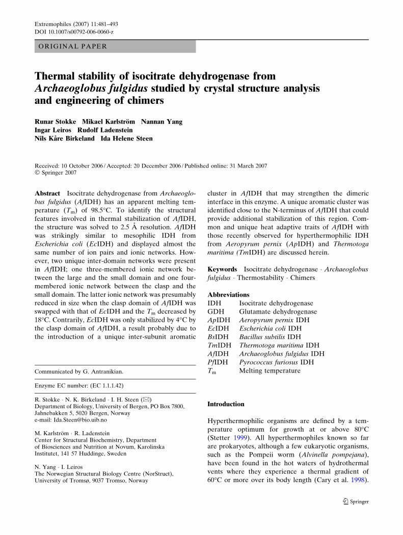
ORIGINAL PAPER Thermal stability of isocitrate dehydrogenase from Archaeoglobus fulgidus studied by crystal structure analysis and engineering of chimers Runar Stokke Mikael Karlstro ¨m Nannan Yang Ingar Leiros Rudolf Ladenstein Nils Ka ˚ re Birkeland Ida Helene Steen Received: 10 October 2006 / Accepted: 20 December 2006 / Published online: 31 March 2007 ȑ Springer 2007 Abstract Isocitrate dehydrogenase from Archaeoglo- bus fulgidus (AfIDH) has an apparent melting tem- perature (T m ) of 98.5ŶC. To identify the structural features involved in thermal stabilization of AfIDH, the structure was solved to 2.5 A ˚ resolution. AfIDH was strikingly similar to mesophilic IDH from Escherichia coli (EcIDH) and displayed almost the same number of ion pairs and ionic networks. How- ever, two unique inter-domain networks were present in AfIDH; one three-membered ionic network be- tween the large and the small domain and one four- membered ionic network between the clasp and the small domain. The latter ionic network was presumably reduced in size when the clasp domain of AfIDH was swapped with that of EcIDH and the T m decreased by 18ŶC. Contrarily, EcIDH was only stabilized by 4ŶC by the clasp domain of AfIDH, a result probably due to the introduction of a unique inter-subunit aromatic cluster in AfIDH that may strengthen the dimeric interface in this enzyme. A unique aromatic cluster was identified close to the N-terminus of AfIDH that could provide additional stabilization of this region. Com- mon and unique heat adaptive traits of AfIDH with those recently observed for hyperthermophilic IDH from Aeropyrum pernix (ApIDH) and Thermotoga maritima (TmIDH) are discussed herein. Keywords Isocitrate dehydrogenase Á Archaeoglobus fulgidus Á Thermostability Á Chimers Abbreviations IDH Isocitrate dehydrogenase GDH Glutamate dehydrogenase ApIDH Aeropyrum pernix IDH EcIDH Escherichia coli IDH BsIDH Bacillus subtilis IDH TmIDH Thermotoga maritima IDH AfIDH Archaeoglobus fulgidus IDH PfIDH Pyrococcus furiosus IDH T m Melting temperature Introduction Hyperthermophilic organisms are defined by a tem- perature optimum for growth at or above 80ŶC (Stetter 1999). All hyperthermophiles known so far are prokaryotes, although a few eukaryotic organisms, such as the Pompeii worm (Alvinella pompejana), have been found in the hot waters of hydrothermal vents where they experience a thermal gradient of 60ŶC or more over its body length (Cary et al. 1998). Communicated by G. Antranikian. Enzyme EC number: (EC 1.1.1.42) R. Stokke Á N. K. Birkeland Á I. H. Steen (&) Department of Biology, University of Bergen, PO Box 7800, Jahnebakken 5, 5020 Bergen, Norway e-mail: [email protected] M. Karlstro ¨m Á R. Ladenstein Center for Structural Biochemistry, Department of Biosciences and Nutrition at Novum, Karolinska Institutet, 141 57 Huddinge, Sweden N. Yang Á I. Leiros The Norwegian Structural Biology Centre (NorStruct), University of Tromsø, 9037 Tromso, Norway 123 Extremophiles (2007) 11:481–493 DOI 10.1007/s00792-006-0060-z
-
Upload
independent -
Category
Documents
-
view
0 -
download
0
Transcript of Thermal stability of isocitrate dehydrogenase from Archaeoglobus fulgidus studied by crystal...
ORIGINAL PAPER
Thermal stability of isocitrate dehydrogenase fromArchaeoglobus fulgidus studied by crystal structure analysisand engineering of chimers
Runar Stokke Æ Mikael Karlstrom Æ Nannan Yang ÆIngar Leiros Æ Rudolf Ladenstein ÆNils Kare Birkeland Æ Ida Helene Steen
Received: 10 October 2006 / Accepted: 20 December 2006 / Published online: 31 March 2007� Springer 2007
Abstract Isocitrate dehydrogenase from Archaeoglo-
bus fulgidus (AfIDH) has an apparent melting tem-
perature (Tm) of 98.5�C. To identify the structural
features involved in thermal stabilization of AfIDH,
the structure was solved to 2.5 A resolution. AfIDH
was strikingly similar to mesophilic IDH from
Escherichia coli (EcIDH) and displayed almost the
same number of ion pairs and ionic networks. How-
ever, two unique inter-domain networks were present
in AfIDH; one three-membered ionic network be-
tween the large and the small domain and one four-
membered ionic network between the clasp and the
small domain. The latter ionic network was presumably
reduced in size when the clasp domain of AfIDH was
swapped with that of EcIDH and the Tm decreased by
18�C. Contrarily, EcIDH was only stabilized by 4�C by
the clasp domain of AfIDH, a result probably due to
the introduction of a unique inter-subunit aromatic
cluster in AfIDH that may strengthen the dimeric
interface in this enzyme. A unique aromatic cluster was
identified close to the N-terminus of AfIDH that could
provide additional stabilization of this region. Com-
mon and unique heat adaptive traits of AfIDH with
those recently observed for hyperthermophilic IDH
from Aeropyrum pernix (ApIDH) and Thermotoga
maritima (TmIDH) are discussed herein.
Keywords Isocitrate dehydrogenase � Archaeoglobus
fulgidus � Thermostability � Chimers
Abbreviations
IDH Isocitrate dehydrogenase
GDH Glutamate dehydrogenase
ApIDH Aeropyrum pernix IDH
EcIDH Escherichia coli IDH
BsIDH Bacillus subtilis IDH
TmIDH Thermotoga maritima IDH
AfIDH Archaeoglobus fulgidus IDH
PfIDH Pyrococcus furiosus IDH
Tm Melting temperature
Introduction
Hyperthermophilic organisms are defined by a tem-
perature optimum for growth at or above 80�C
(Stetter 1999). All hyperthermophiles known so far
are prokaryotes, although a few eukaryotic organisms,
such as the Pompeii worm (Alvinella pompejana),
have been found in the hot waters of hydrothermal
vents where they experience a thermal gradient of
60�C or more over its body length (Cary et al. 1998).
Communicated by G. Antranikian.
Enzyme EC number: (EC 1.1.1.42)
R. Stokke � N. K. Birkeland � I. H. Steen (&)Department of Biology, University of Bergen, PO Box 7800,Jahnebakken 5, 5020 Bergen, Norwaye-mail: [email protected]
M. Karlstrom � R. LadensteinCenter for Structural Biochemistry, Departmentof Biosciences and Nutrition at Novum, KarolinskaInstitutet, 141 57 Huddinge, Sweden
N. Yang � I. LeirosThe Norwegian Structural Biology Centre (NorStruct),University of Tromsø, 9037 Tromso, Norway
123
Extremophiles (2007) 11:481–493
DOI 10.1007/s00792-006-0060-z
Most of the hyperthermophilic microorganisms belong
to the archaeal domain although hyperthermophiles
have been found in the two bacterial orders, Therm-
otogales and Aquificales (Blochl et al. 1995). Enzymes
synthesized by (hyper)thermophiles are typically
thermostable, or resistant to irreversible inactivation
at high temperatures, and thermophilic, i.e. optimally
active at elevated temperatures between 60 and 125�C
(Vieille and Zeikus 2001). In order to reveal main
adaptive strategies used for protein stabilization,
numerous three-dimensional structures of hyper-
thermophilic proteins have been obtained. Compara-
tive studies between hyperthermophilic and
mesophilic enzymes have demonstrated that interac-
tions such as hydrogen bonds, disulphide bonds, ion
pairs, salt bridges, hydrophobic interactions and
compactness are of importance for stability (Scan-
durra et al. 2000; Vieille and Zeikus 2001). No uni-
versal basis of stability has been recognized and the
major reason for this is the relatively small free en-
ergy difference between the folded and the unfolded
state and the complex way in which the small number
of weak forces, determining protein stability, interplay
with each other (Karlstrom et al. 2005). However, the
most common determinants for increased thermal
stability are in the first line a statistical prevalence of
ionic interactions at the protein surface, increased
formation of large ionic networks, electrostatic opti-
mization and the reduction of repulsive charge–charge
interactions (Spassov et al. 1997; Vetriani et al. 1998;
Karshikoff and Ladenstein 2001; Karlstrom et al.
2005). Despite a great availability of data on ther-
mophiles and thermophilic proteins, most of the
information generated on how proteins from hyper-
thermophilic organisms have adapted in their natural
environments are based on case studies (Karshikoff
and Ladenstein 2001) or structural and biochemical
comparisons of single mesophilic/hyperthermophilic
enzyme pairs.
We have chosen the enzyme isocitrate dehydroge-
nase (IDH) as a model enzyme for studying environ-
mental adaptations of proteins to extreme
temperatures. IDH catalyses the oxidative decarbox-
ylation of D-isocitrate to 2-oxoglutarate and CO2 with
NAD+ (EC 1.1.1.41) or NADP+ (EC 1.1.1.42) as co-
factor and comprises a diverse enzyme family with
regard to cofactor specificity, primary structure, and
oligomeric state.
Aeropyrum pernix (Ap), Pyrococcus furiosus (Pf),
Archaeoglobus fulgidus (Af) and Thermotoga maritima
(Tm) are hyperthermophilic microorganisms growing
optimally at 95, 100, 83 and 80�C, respectively (Fiala
and Stetter 1986; Huber et al. 1986; Sako et al. 1996).
We have previously estimated the thermal stability of
Ap-, Pf-, Af- and TmIDH by determination of the
apparent melting temperatures (Tm) using differential
scanning calorimetry (DSC) (Steen et al. 2001). It was
found that ApIDH has highest thermal stability with a
Tm of 109.9�C. Pf, Af and TmIDH were less stable with
a Tm of 103.7, 98.5, and 98.3�C, respectively. However,
each of the hyperthermophilic IDHs showed a signifi-
cantly higher Tm than the one determined for IDH
from Escherichia coli (Tm = 52.6�C) (EcIDH) (Kar-
lstrom et al. 2005) and pig (Tm = 59�C) (Karlstrom
et al. 2006). Recently, the three-dimensional structures
of ApIDH (Karlstrom et al. 2005) and TmIDH (Kar-
lstrom et al. 2006) have been resolved and the struc-
tural properties important for their high thermal
stability have been identified.
Here we report the crystallization and structure
determination of the hyperthermophilic AfIDH
including a comparative structural study with other
known IDH homologs. Furthermore, chimers between
the hyperthermophilic AfIDH and the mesophilic
EcIDH were constructed to investigate the contribu-
tion of the clasp domain to the thermal stability of
wild-type AfIDH.
Materials and methods
Strains
Escherichia coli strain EB106 (icd-11 dadR1 trpA62
trpE61 tna-5 lambda–) was originally obtained from the
E. coli Genetic Stock Centre, MCD Biology Depart-
ment, Yale University, in courtesy of Dr. Mary K.B.
Berlyn. In order to express proteins in this strain,
E. coli EB106 was lysogenized using the kDE3
Lysogenization Kit from NOVAGEN.
Crystallization and data collection
AfIDH was crystallized using the hanging drop vapour
diffusion technique with reservoir solution consisting
of 0.6 M ZnSO4 and 0.1 M Na Cacodylate, pH 6.3,
which are optimized from the Nextal EasyXtal Cations
suite condition number 60 (QIAGEN GmbH, Hilden,
Germany). Crystals were grown by mixing equal vol-
umes (2 ll) of a 9 mg/ml protein solution with the
reservoir solution. The drops were equilibrated at 8�C.
20% (v/v) glycerol added to the reservoir solution
sufficed as a cryo-protectant for flash-cooling the
crystals in liquid nitrogen. A data set (see Table 1) was
collected at the macromolecular crystallography
beamline X06SA at the Swiss Light Source (SLS). The
482 Extremophiles (2007) 11:481–493
123
data were collected with a MAR225 CCD detector and
allowed the determination of the crystal structure using
molecular replacement techniques.
Structure determination and refinement
The collected data set was indexed and integrated
using MOSFLM (Leslie 1992). The crystals were
monoclinic, with unit cell parameters of a = 81.6 A,
b = 65.4 A, c = 87.2 A, b = 95.28�. The data were
scaled, merged and the intensities were converted into
structure factors using the CCP4 programs SCALA
and TRUNCATE (CCP4 1994). A summary of the
data collection statistics is presented in Table 1. The
systematic absences in the collected data set indicated
the presence of a twofold screw axis along the b-axis,
with the only possible space group being P21. The
solvent content was estimated to be around 52%, with
a Matthews Coefficient of 2.6 A3 Da–1, assuming two
protein molecules per asymmetric unit. The crystal
structure of AfIDH was determined by molecular
replacement methods using MOLREP (CCP4 1994).
The crystal structure of ApIDH was used as search
model and the automated program functions in
MOLREP were applied in order to create the model
that presumably had the best fit to the sequence of
AfIDH. Reflections up to a high-resolution limit of
3.5 A were used. One well-resolved solution for the
two molecules in the asymmetric unit could be found,
having a correlation coefficient of 0.459 and an Rfactor
of 45.1%. A rigid-body fitting of the model using a
high-resolution cut-off at 2.5 A resulted in Rwork of
44.5% (Rfree of 46.0%). After a manual intervention
using O (Jones et al. 1991), the model was refined in
REFMAC5 (Murshudov et al. 1999), resulting in R-
factors of 24.0 and 29.1% for the working and test sets
of reflections, respectively. Subsequent cycles of
refinement interspersed with manual rebuilding, gave
final Rwork and Rfree values of 19.6 and 25.4%,
respectively, with acceptable protein geometry. In
general, water molecules were added using the
embedded functions found in REFMAC and manually
checked using O. The four residues found in disallowed
regions of the Ramachandran plot are all located in
flexible loops.
Structure analysis
Sequence alignment was performed using the program
STAMP (Russell and Barton 1992) based on the Ca-
atom coordinates and secondary structural assignments
using the program DSSP (Kabsch and Sander 1983).
Potential salt-bridge formation and ionic networks
were analysed using the program CONTACT (CCP4
1994) and IONSTAT (W. Meining, unpublished) with
varying maximum distances from 3.5 to 8.0 A. Acces-
sible surface areas were calculated using CNS with Ala,
Ile, Leu, Met, Phe, Pro, Trp, and Val defined as
hydrophobic residues, Asn, Gln, Ser, Thr, Tyr, Cys and
Gly regarded as polar residues and Asp, Glu, Arg, Lys
and His as charged residues. The water probe radius
was set to 1.4 A and the accuracy of the numerical
integration was set to 0.12. Water and ions were
excluded from the model.
Figure preparations
Figures 1, 3, 4 and 5 were made using the program
PYMOL (PyMol 2005) and Fig. 2 was prepared using
the program ESPript2.2 (Gouet et al. 1999).
Cloning and engineering of chimers
The wild-type Ec idh gene was amplified from E. coli
K12 (DSMZ - Literature Reference No. 4684) genomic
DNA by PCR using the primers P1-P2 (Table 2) and
ligated into pET101/D-TOPO (Invitrogen Corporation,
Carlsbad, CA 92008, USA). Domain-swapping of the
clasp domain from EcIDH with the clasp domain from
AfIDH was performed by using the ExSiteTMPCR-
Based Site-Directed Mutagenesis Kit (Stratagene, La
Jolla, CA, USA) with the pET101/D-TOPO-Ec idh as
Table 1 Data collection and refinement statistics for AfIDH
Data collectionWavelength (A) 0.92Space group P21
Unit cell parameters (A, �) 81.6, 65.4, 87.2, 90, 95.28, 90Resolution range (A) 40–2.5 (2.64–2.5)No. observed reflections 130993 (19123)No. unique reflections 31867 (4612)Redundancy 4.1 (4.1)Completeness (%) 99.8 (100.0)Mean I/rI 11.1 (3.5)Rmerge (%) 11.4 (47.6)
RefinementNon-hydrogen atoms 6553Metal ions 9Zn/3ClNo. solvent molecules 94Overall B-factor (A2) 16.80Rcryst (%) 19.6Rfree (%) 25.4
Deviation from ideal geometryBond lengths (A) 0.016Bond angles (�) 1.785
Ramachandran plotMost favoured region (%) 87.7Allowed regions (%) 11.7Disallowed regions (%) 0.6
Extremophiles (2007) 11:481–493 483
123
template. Mutagenesis was performed as described in
the manufacturer protocol. Because of the large size of
the mutagenic primers the mutagenesis was performed
in a two-step reaction with primers P3-P6 (Table 2).
Swapping of the clasp domain from AfIDH with the
clasp domain from EcIDH (AfIDH/EcIDHclasp) was
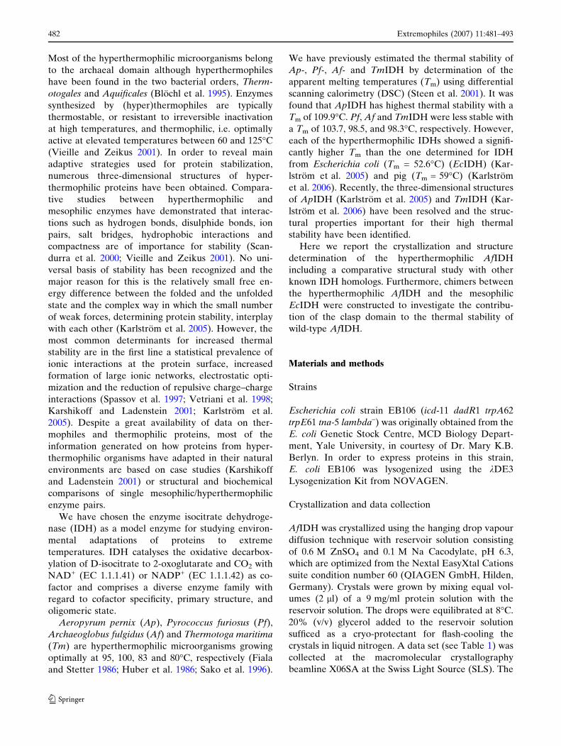
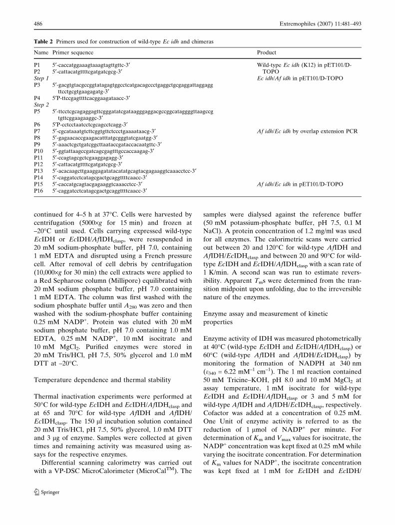
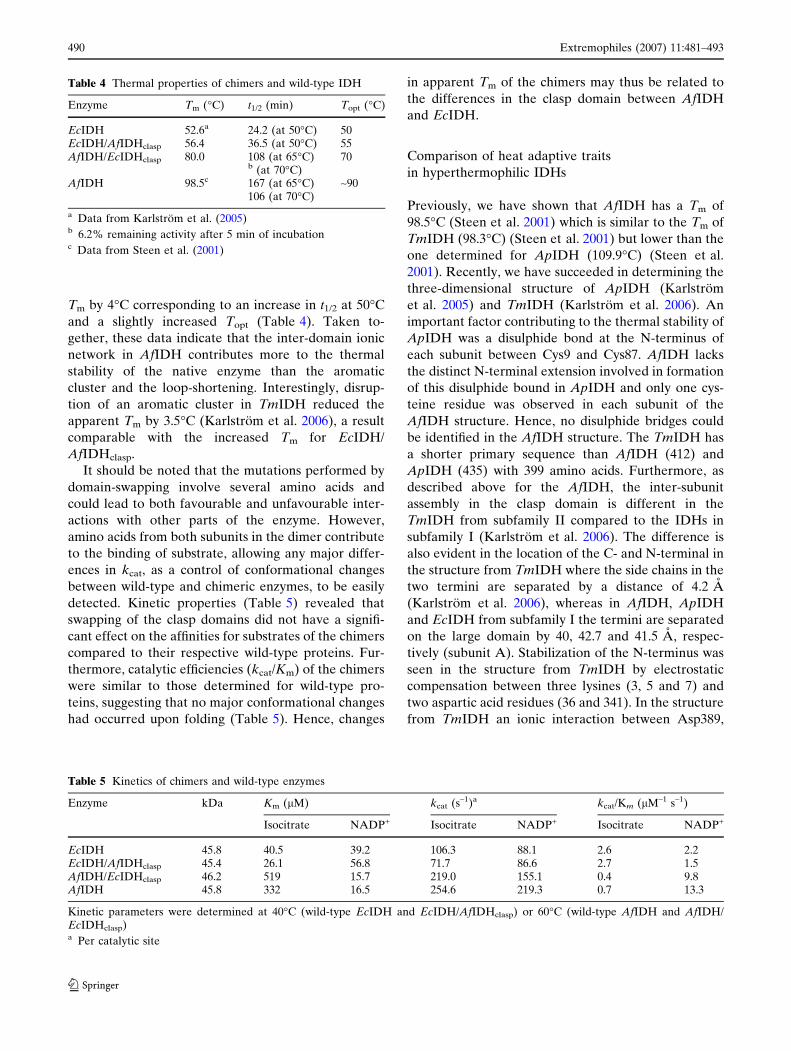
Fig. 1 The structure ofAfIDH was crystallized andresolved without substrateand cofactor in the active site,however, a high concentrationof zinc was present in thecrystallization condition. Azinc ion was found tightlybound to Asp301, Asp305,Asp277¢ in the active site ofsubunit A (a) and subunit B(b) of AfIDH. Amino acidsfrom subunit A are colouredblue and amino acids fromsubunit B are coloured red.Amino acids from bothsubunits are involved inbinding of zinc
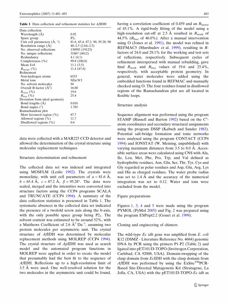
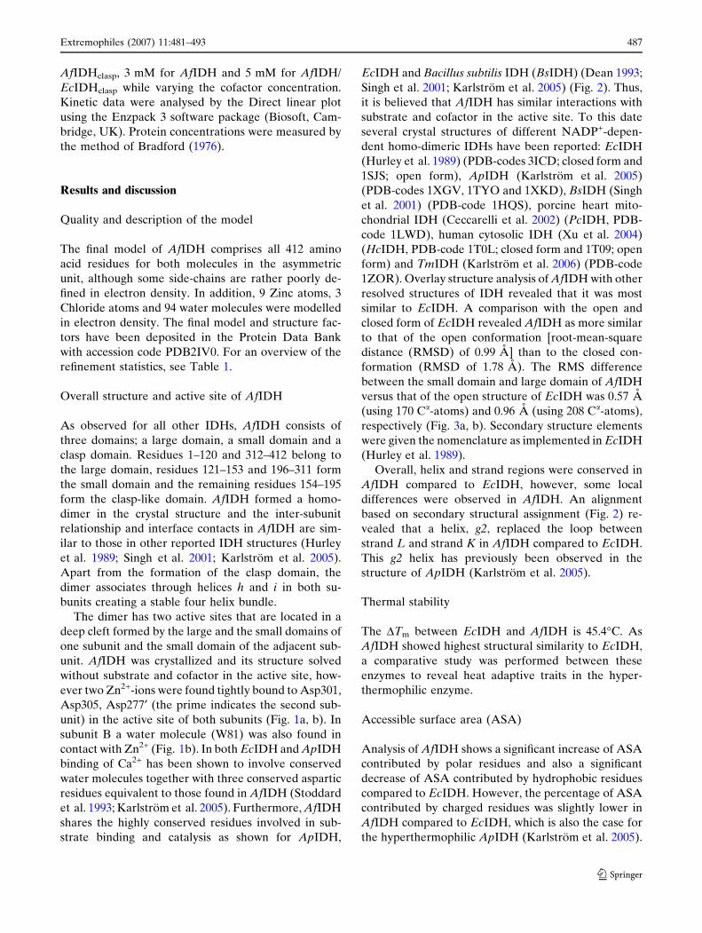
Fig. 2 Sequence alignment ofAfIDH, ApIDH (PDB code1TYO), BsIDH (PDB code1HQS) and EcIDH (PDBcode 1SJS) with thesecondary structure elementsof AfIDH (top) and EcIDH(bottom). Identical andsimilar residues are boxed.The secondary structureelements were given thenomenclature asimplemented in EcIDH(Hurley et al. 1989) and thefigure was made in ESPript2.2(Gouet et al. 1999)
484 Extremophiles (2007) 11:481–493
123
performed by overlap extension PCR (Warrens et al.
1997) on the template pET-11-a/Af idh (Steen et al.
2001) using primers P7-P14 (Table 2). For expression,
the chimer construct was sub-cloned into pET101/D-
TOPO vector using primers P15-P16 (Table 2) to
amplify the gene.
Expression and purification
Expression and purification of wild-type AfIDH and
AfIDH/EcIDHclasp were performed as previously
described for wild-type AfIDH (Steen et al. 2001).
Expression of wild-type EcIDH and EcIDH/
AfIDHclasp was performed at 37�C in E. coli strain
EB106 (DE3) in LB broth containing ampicillin
(100 lg/ml). Isopropyl-b-thiogalactopyranoside was
added to 1.0 mM concentration to induce expres-
sion at A600 nm = 0.7–0.8, and the incubation was
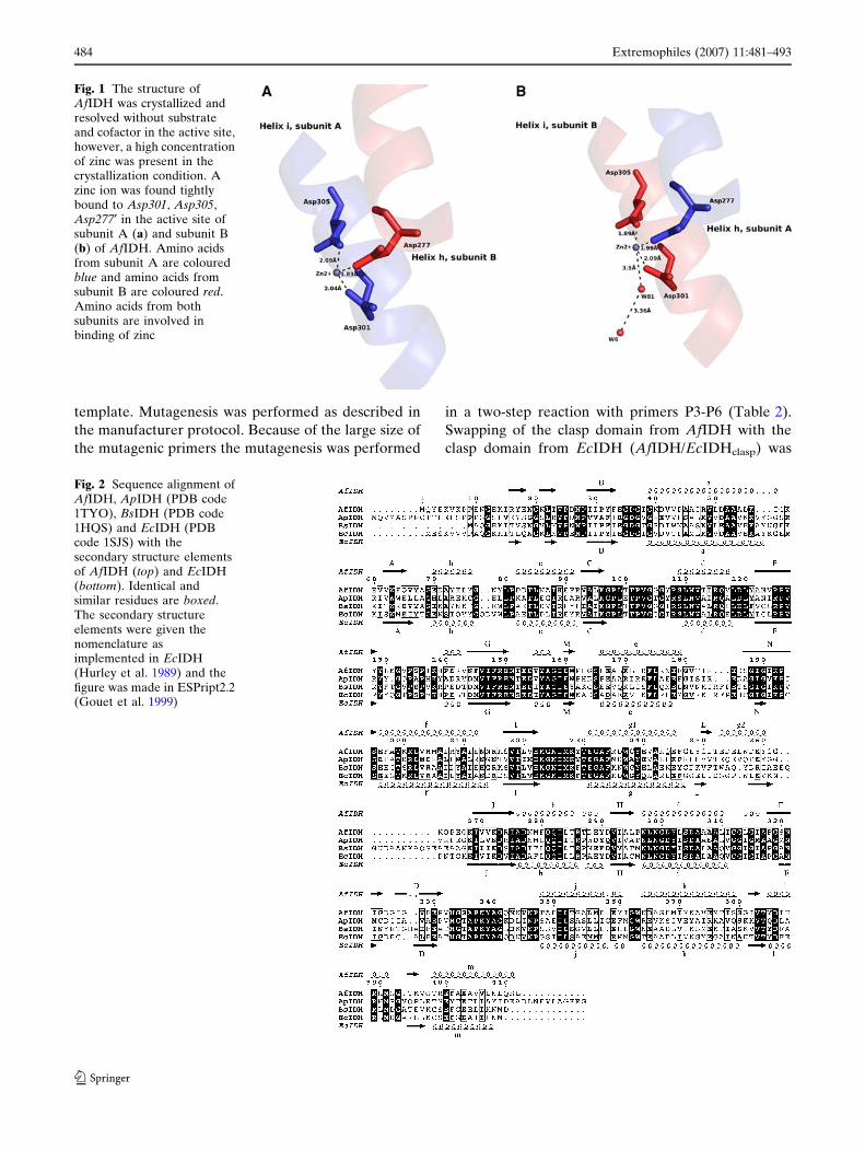
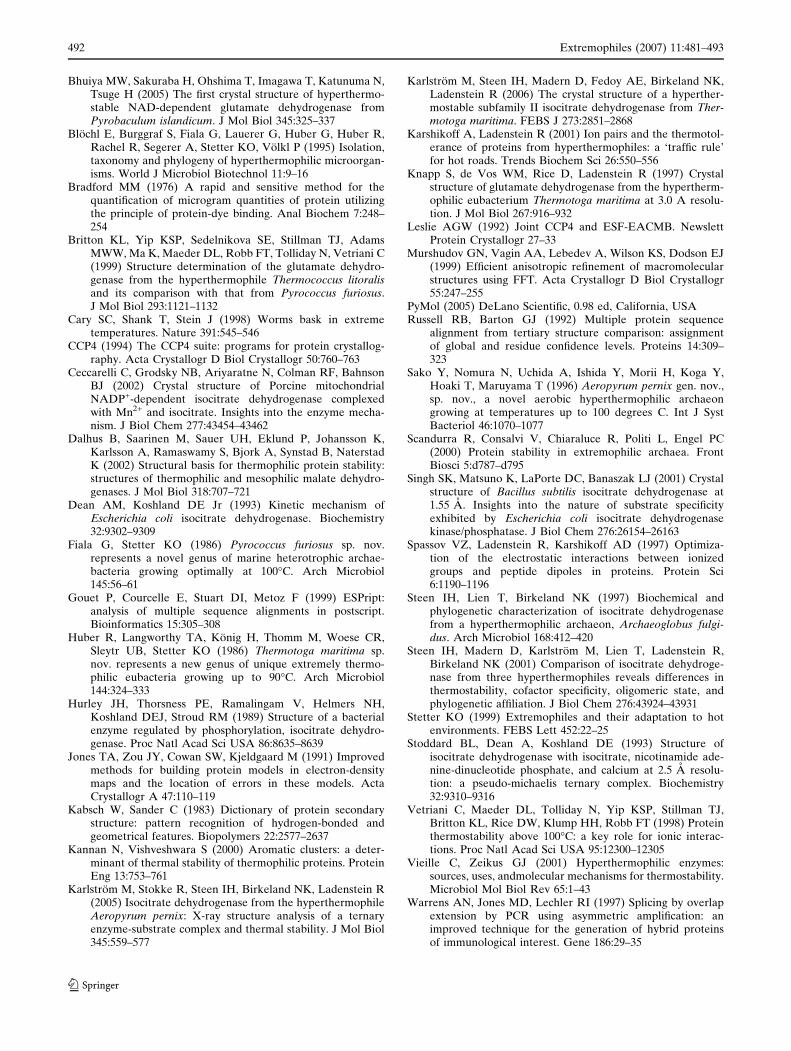
Fig. 3 Overlay of the small(a) and large (b) domains ofAfIDH and EcIDHopen
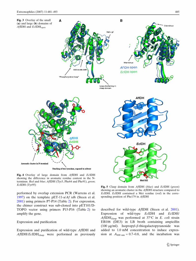
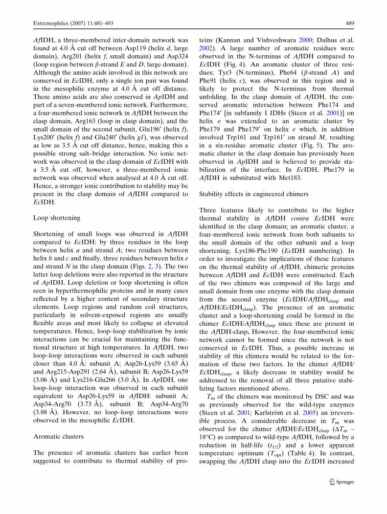
Fig. 4 Overlay of large domain from AfIDH and EcIDHshowing the difference in aromatic residue content in the N-terminus. Red and blue; AfIDH (Tyr3, Phe64 and Phe91), green;EcIDH (Tyr95)
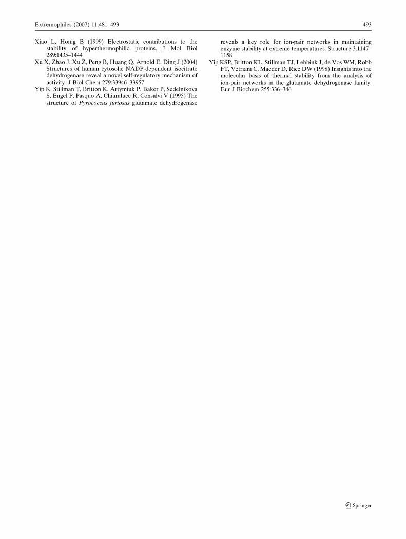
Fig. 5 Clasp domain from AfIDH (blue) and EcIDH (green)showing an aromatic cluster in the AfIDH structure compared toEcIDH. EcIDH contained a Met residue (red) in the corre-sponding position of Phe179 in AfIDH
Extremophiles (2007) 11:481–493 485
123
continued for 4–5 h at 37�C. Cells were harvested by
centrifugation (5000·g for 15 min) and frozen at
–20�C until used. Cells carrying expressed wild-type
EcIDH or EcIDH/AfIDHclasp, were resuspended in
20 mM sodium-phosphate buffer, pH 7.0, containing
1 mM EDTA and disrupted using a French pressure
cell. After removal of cell debris by centrifugation
(10,000·g for 30 min) the cell extracts were applied to
a Red Sepharose column (Millipore) equilibrated with
20 mM sodium phosphate buffer, pH 7.0 containing
1 mM EDTA. The column was first washed with the
sodium phosphate buffer until A280 was zero and then
washed with the sodium-phosphate buffer containing
0.25 mM NADP+. Protein was eluted with 20 mM
sodium phosphate buffer, pH 7.0 containing 1.0 mM
EDTA, 0.25 mM NADP+, 10 mM isocitrate and
10 mM MgCl2. Purified enzymes were stored in
20 mM Tris/HCl, pH 7.5, 50% glycerol and 1.0 mM
DTT at –20�C.
Temperature dependence and thermal stability
Thermal inactivation experiments were performed at
50�C for wild-type EcIDH and EcIDH/AfIDHclasp and
at 65 and 70�C for wild-type AfIDH and AfIDH/
EcIDHclasp. The 150 ll incubation solution contained
20 mM Tris/HCl, pH 7.5, 50% glycerol, 1.0 mM DTT
and 3 lg of enzyme. Samples were collected at given
times and remaining activity was measured using as-
says for the respective enzymes.
Differential scanning calorimetry was carried out
with a VP-DSC MicroCalorimeter (MicroCalTM). The
samples were dialysed against the reference buffer
(50 mM potassium-phosphate buffer, pH 7.5, 0.1 M
NaCl). A protein concentration of 1.2 mg/ml was used
for all enzymes. The calorimetric scans were carried
out between 20 and 120�C for wild-type AfIDH and
AfIDH/EcIDHclasp and between 20 and 90�C for wild-
type EcIDH and EcIDH/AfIDHclasp with a scan rate of
1 K/min. A second scan was run to estimate revers-
ibility. Apparent Tms were determined from the tran-
sition midpoint upon unfolding, due to the irreversible
nature of the enzymes.
Enzyme assay and measurement of kinetic
properties
Enzyme activity of IDH was measured photometrically
at 40�C (wild-type EcIDH and EcIDH/AfIDHclasp) or
60�C (wild-type AfIDH and AfIDH/EcIDHclasp) by
monitoring the formation of NADPH at 340 nm
(e340 = 6.22 mM–1 cm–1). The 1 ml reaction contained
50 mM Tricine–KOH, pH 8.0 and 10 mM MgCl2 at
assay temperature, 1 mM isocitrate for wild-type
EcIDH and EcIDH/AfIDHclasp or 3 and 5 mM for
wild-type AfIDH and AfIDH/EcIDHclasp, respectively.
Cofactor was added at a concentration of 0.25 mM.
One Unit of enzyme activity is referred to as the
reduction of 1 lmol of NADP+ per minute. For
determination of Km and Vmax values for isocitrate, the
NADP+ concentration was kept fixed at 0.25 mM while
varying the isocitrate concentration. For determination
of Km values for NADP+, the isocitrate concentration
was kept fixed at 1 mM for EcIDH and EcIDH/
Table 2 Primers used for construction of wild-type Ec idh and chimeras
Name Primer sequence Product
P1 5¢-caccatggaaagtaaagtagttgttc-3¢ Wild-type Ec idh (K12) in pET101/D-TOPOP2 5¢-cattacatgttttcgatgatcgcg-3¢
Step 1 Ec idh/Af idh in pET101/D-TOPOP3 5¢-gacgtgtacgccggtatagagtggcctcatgacagccctgaggctgcgaggattaggagg
ttcctgcgtgaagagatg-3¢P4 5¢P-ttccgagttttcacggaagataacc-3¢Step 2P5 5¢-ttcctcgcagaggagttcgggatatcgataagggaggacgccggcataggggttaagccg
tgttcggaagaaggc-3¢P6 5¢P-cctcctaatcctcgcagcctcagg-3¢P7 5¢-cgcataaatgtcttcggtgttctccctgaaaataacg-3¢ Af idh/Ec idh by overlap extension PCRP8 5¢-gagaacaccgaagacatttatgcgggtatcgaatgg-3¢P9 5¢-aaactcgctgatcggcttaataccgataccacaatgttc-3¢P10 5¢-ggtattaagccgatcagcgagtttgccaccaagag-3¢P11 5¢-ccagtagcgctcgaaggagagg-3¢P12 5¢-cattacatgttttcgatgatcgcg-3¢P13 5¢-acacaagcttgaaggagatatacatatgcagtacgagaaggtcaaacctcc-3¢P14 5¢-caggatcctcatagcgactgcaggttttcaacc-3¢P15 5¢-caccatgcagtacgagaaggtcaaacctcc-3¢ Af idh/Ec idh in pET101/D-TOPOP16 5¢-caggatcctcatagcgactgcaggttttcaacc-3¢
486 Extremophiles (2007) 11:481–493
123
AfIDHclasp, 3 mM for AfIDH and 5 mM for AfIDH/
EcIDHclasp while varying the cofactor concentration.
Kinetic data were analysed by the Direct linear plot
using the Enzpack 3 software package (Biosoft, Cam-
bridge, UK). Protein concentrations were measured by
the method of Bradford (1976).
Results and discussion
Quality and description of the model
The final model of AfIDH comprises all 412 amino
acid residues for both molecules in the asymmetric
unit, although some side-chains are rather poorly de-
fined in electron density. In addition, 9 Zinc atoms, 3
Chloride atoms and 94 water molecules were modelled
in electron density. The final model and structure fac-
tors have been deposited in the Protein Data Bank
with accession code PDB2IV0. For an overview of the
refinement statistics, see Table 1.
Overall structure and active site of AfIDH
As observed for all other IDHs, AfIDH consists of
three domains; a large domain, a small domain and a
clasp domain. Residues 1–120 and 312–412 belong to
the large domain, residues 121–153 and 196–311 form
the small domain and the remaining residues 154–195
form the clasp-like domain. AfIDH formed a homo-
dimer in the crystal structure and the inter-subunit
relationship and interface contacts in AfIDH are sim-
ilar to those in other reported IDH structures (Hurley
et al. 1989; Singh et al. 2001; Karlstrom et al. 2005).
Apart from the formation of the clasp domain, the
dimer associates through helices h and i in both su-
bunits creating a stable four helix bundle.
The dimer has two active sites that are located in a
deep cleft formed by the large and the small domains of
one subunit and the small domain of the adjacent sub-
unit. AfIDH was crystallized and its structure solved
without substrate and cofactor in the active site, how-
ever two Zn2+-ions were found tightly bound to Asp301,
Asp305, Asp277¢ (the prime indicates the second sub-
unit) in the active site of both subunits (Fig. 1a, b). In
subunit B a water molecule (W81) was also found in
contact with Zn2+ (Fig. 1b). In both EcIDH and ApIDH
binding of Ca2+ has been shown to involve conserved
water molecules together with three conserved aspartic
residues equivalent to those found in AfIDH (Stoddard
et al. 1993; Karlstrom et al. 2005). Furthermore, AfIDH
shares the highly conserved residues involved in sub-
strate binding and catalysis as shown for ApIDH,
EcIDH and Bacillus subtilis IDH (BsIDH) (Dean 1993;
Singh et al. 2001; Karlstrom et al. 2005) (Fig. 2). Thus,
it is believed that AfIDH has similar interactions with
substrate and cofactor in the active site. To this date
several crystal structures of different NADP+-depen-
dent homo-dimeric IDHs have been reported: EcIDH
(Hurley et al. 1989) (PDB-codes 3ICD; closed form and
1SJS; open form), ApIDH (Karlstrom et al. 2005)
(PDB-codes 1XGV, 1TYO and 1XKD), BsIDH (Singh
et al. 2001) (PDB-code 1HQS), porcine heart mito-
chondrial IDH (Ceccarelli et al. 2002) (PcIDH, PDB-
code 1LWD), human cytosolic IDH (Xu et al. 2004)
(HcIDH, PDB-code 1T0L; closed form and 1T09; open
form) and TmIDH (Karlstrom et al. 2006) (PDB-code
1ZOR). Overlay structure analysis of AfIDH with other
resolved structures of IDH revealed that it was most
similar to EcIDH. A comparison with the open and
closed form of EcIDH revealed AfIDH as more similar
to that of the open conformation [root-mean-square
distance (RMSD) of 0.99 A] than to the closed con-
formation (RMSD of 1.78 A). The RMS difference
between the small domain and large domain of AfIDH
versus that of the open structure of EcIDH was 0.57 A
(using 170 Ca-atoms) and 0.96 A (using 208 Ca-atoms),
respectively (Fig. 3a, b). Secondary structure elements
were given the nomenclature as implemented in EcIDH
(Hurley et al. 1989).
Overall, helix and strand regions were conserved in
AfIDH compared to EcIDH, however, some local
differences were observed in AfIDH. An alignment
based on secondary structural assignment (Fig. 2) re-
vealed that a helix, g2, replaced the loop between
strand L and strand K in AfIDH compared to EcIDH.
This g2 helix has previously been observed in the
structure of ApIDH (Karlstrom et al. 2005).
Thermal stability
The DTm between EcIDH and AfIDH is 45.4�C. As
AfIDH showed highest structural similarity to EcIDH,
a comparative study was performed between these
enzymes to reveal heat adaptive traits in the hyper-
thermophilic enzyme.
Accessible surface area (ASA)
Analysis of AfIDH shows a significant increase of ASA
contributed by polar residues and also a significant
decrease of ASA contributed by hydrophobic residues
compared to EcIDH. However, the percentage of ASA
contributed by charged residues was slightly lower in
AfIDH compared to EcIDH, which is also the case for
the hyperthermophilic ApIDH (Karlstrom et al. 2005).
Extremophiles (2007) 11:481–493 487
123
The distribution of hydrophobic, polar and charged
surface area of AfIDH and EcIDH is shown in Table 3.
The dimer interface of AfIDH buried 5,564 A2 of the
37,970 A2 molecular surface area (MSA) of the dimer,
giving a solvent-accessible surface area of 32,406 A2
for the dimer. In contrast, the EcIDH dimer buries
6,020 A2 of the 38,547 A2 of the dimer with a total
solvent-accessible surface of 32,527 A2, i.e the buried
inter-subunit surface comprise 14.7 and 15.6% of
AfIDH and EcIDH, respectively.
Amino acid composition
The AfIDH subunit is made up of 412 amino acids of
which 30.1% are polar residues, 27.2% charged resi-
dues and 42.7% hydrophobic residues. Compared to
EcIDH, the distribution of amino acids was similar in
the two proteins with a slightly higher fraction of polar
residues in AfIDH and a slightly higher fraction of
hydrophobic residues in EcIDH (Table 3). However,
AfIDH has a significantly higher fraction of aromatic
residues of 11.2% compared to 8.7% in EcIDH. It has
previously been shown that AfIDH and other archaeal
hyperthermophilic IDHs have a decreased number of
Cys residues compared with EcIDH, 0.20 and 1.40%,
respectively (Steen et al. 1997, 2001), hence, following
the trend observed for thermophilic proteins whereby
Cys residues tend to be avoided.
Ionic interactions
Comparative studies between hyperthermophilic and
mesophilic homologs have shown a clear tendency for
the total number of ion pairs and large ionic networks
to increase with the optimal growth temperature of the
organisms as well as with the Tm of the proteins (Yip
et al. 1995, 1998; Knapp et al. 1997). Surprisingly, few
differences were found between the mesophilic EcIDH
and the hyperthermophilic AfIDH in the total amount
of charges and ionic interactions. The number of ionic
networks at different cut off distances, are summarized
in Table 3. Overall, the number and size of ionic net-
works did not differ significantly between AfIDH and
EcIDH; however, the location of the ionic networks
seems to play an important role for the increased
thermal stability of the hyperthermphilic enzyme. In
Table 3 Characteristics of AfIDH and EcIDH
AfIDH EcIDHopen
PDB code 2IVO 1SJSTm (�C) 98.5 52.6a
Resolution (A) 2.5 2.4RMSD (A) Ca-atoms small domain/large domain/overall 0.57/0.96/0.99 (170 atoms/208 atoms/378 atoms)RMSD (A) Ca-atoms of clasp-domain 4.42 (total domain)/0.69 (without loop) (42 atoms/40
atoms)No. residues per subunit 412 416Charged residues (%)b 27.2 27.2Polar residues (%)c 30.1 28.6Hydrophobic residues (%)d 42.7 44.9Aromatic residues (%)e 11.2 8.7No. ion pairs per dimer (4/6/8 A)f 53/104/175 58/110/181No. ion pairs per residue in dimer (4/6/8 A) 0.129/0.252/0.425 0.139/0.264/0.435Volume (·104 A3) 8.5 8.4Accessible surface area of dimer (A2) 32406 32527Buried inter-subunit surface (% of MSA) 14.7 15.6No. residues forming two ion pairs (4/6/8 A) 14/41/60 18/50/75No. residues forming three ion pairs (4/6/8 A) 0/10/42 0/6/22No. 2/3/4 membered intra-subunit networks (4 A) 27/8/0 24/12/0No. 2/3/4 membered inter-subunit networks (4 A) 0/2/2 0/2/2% of charged residues forming ion pairs (4/6/8 A) 46/78/92 45/76/89Distribution of hydrophobic/polar/charged residues at accessible surface (%) 20.7/30.1/49.3 25.6/23.1/51.3Distribution of hydrophobic/polar/charged residues at interface (%) 47.0/27.3/25.7 50.4/19.7/29.9
MSA molecular surface areaa Data from Karlstrom et al. (2005)b Charged residues: R, K, H, D, Ec Polar residues: G, S, T, Y, N, Q, Cd Hydrophobic residues: A, V, L, I, W, F, P, Me Aromatic residues: W, F, Y, Hf A cutoff
488 Extremophiles (2007) 11:481–493
123
AfIDH, a three-membered inter-domain network was
found at 4.0 A cut off between Asp119 (helix d, large
domain), Arg201 (helix f, small domain) and Asp324
(loop region between b-strand E and D, large domain).
Although the amino acids involved in this network are
conserved in EcIDH, only a single ion pair was found
in the mesophilic enzyme at 4.0 A cut off distance.
These amino acids are also conserved in ApIDH and
part of a seven-membered ionic network. Furthermore,
a four-membered ionic network in AfIDH between the
clasp domain, Arg163 (loop in clasp domain), and the
small domain of the second subunit, Glu196¢ (helix f),
Lys200¢ (helix f) and Glu240¢ (helix g1), was observed
as low as 3.5 A cut off distance, hence, making this a
possible strong salt–bridge interaction. No ionic net-
work was observed in the clasp domain of EcIDH with
a 3.5 A cut off, however, a three-membered ionic
network was observed when analysed at 4.0 A cut off.
Hence, a stronger ionic contribution to stability may be
present in the clasp domain of AfIDH compared to
EcIDH.
Loop shortening
Shortening of small loops was observed in AfIDH
compared to EcIDH: by three residues in the loop
between helix a and strand A; two residues between
helix b and c and finally, three residues between helix e
and strand N in the clasp domain (Figs. 2, 3). The two
latter loop deletions were also reported in the structure
of ApIDH. Loop deletion or loop shortening is often
seen in hyperthermophilic proteins and in many cases
reflected by a higher content of secondary structure
elements. Loop regions and random coil structures,
particularly in solvent-exposed regions are usually
flexible areas and most likely to collapse at elevated
temperatures. Hence, loop–loop stabilization by ionic
interactions can be crucial for maintaining the func-
tional structure at high temperatures. In AfIDH, two
loop–loop interactions were observed in each subunit
closer than 4.0 A: subunit A; Asp26-Lys59 (3.65 A)
and Arg215-Asp291 (2.64 A), subunit B; Asp26-Lys59
(3.06 A) and Lys216-Glu266 (3.0 A). In ApIDH, one
loop–loop interaction was observed in each subunit
equivalent to Asp26-Lys59 in AfIDH: subunit A;
Asp34-Arg70 (3.73 A), subunit B; Asp34-Arg70
(3.88 A). However, no loop–loop interactions were
observed in the mesophilic EcIDH.
Aromatic clusters
The presence of aromatic clusters has earlier been
suggested to contribute to thermal stability of pro-
teins (Kannan and Vishveshwara 2000; Dalhus et al.
2002). A large number of aromatic residues were
observed in the N-terminus of AfIDH compared to
EcIDH (Fig. 4). An aromatic cluster of three resi-
dues; Tyr3 (N-terminus), Phe64 (b-strand A) and
Phe91 (helix c), was observed in this region and is
likely to protect the N-terminus from thermal
unfolding. In the clasp domain of AfIDH, the con-
served aromatic interaction between Phe174 and
Phe174¢ [in subfamily I IDHs (Steen et al. 2001)] on
helix e was extended to an aromatic cluster by
Phe179 and Phe179¢ on helix e which, in addition
involved Trp161 and Trp161¢ on strand M, resulting
in a six-residue aromatic cluster (Fig. 5). The aro-
matic cluster in the clasp domain has previously been
observed in ApIDH and is believed to provide sta-
bilization of the interface. In EcIDH, Phe179 in
AfIDH is substituted with Met183.
Stability effects in engineered chimers
Three features likely to contribute to the higher
thermal stability in AfIDH contra EcIDH were
identified in the clasp domain; an aromatic cluster, a
four-membered ionic network from both subunits to
the small domain of the other subunit and a loop
shortening; Lys186-Phe190 (EcIDH numbering). In
order to investigate the implications of these features
on the thermal stability of AfIDH, chimeric proteins
between AfIDH and EcIDH were constructed. Each
of the two chimers was composed of the large and
small domain from one enzyme with the clasp domain
from the second enzyme (EcIDH/AfIDHclasp and
AfIDH/EcIDHclasp). The presence of an aromatic
cluster and a loop-shortening could be formed in the
chimer EcIDH/AfIDHclasp since these are present in
the AfIDH-clasp. However, the four-membered ionic
network cannot be formed since the network is not
conserved in EcIDH. Thus, a possible increase in
stability of this chimera would be related to the for-
mation of these two factors. In the chimer AfIDH/
EcIDHclasp, a likely decrease in stability would be
addressed to the removal of all three putative stabi-
lizing factors mentioned above.
Tm of the chimers was monitored by DSC and was
as previously observed for the wild-type enzymes
(Steen et al. 2001; Karlstrom et al. 2005) an irrevers-
ible process. A considerable decrease in Tm was
observed for the chimer AfIDH/EcIDHclasp (DTm –
18�C) as compared to wild-type AfIDH, followed by a
reduction in half-life (t1/2) and a lower apparent
temperature optimum (Topt) (Table 4). In contrast,
swapping the AfIDH clasp into the EcIDH increased
Extremophiles (2007) 11:481–493 489
123
Tm by 4�C corresponding to an increase in t1/2 at 50�C
and a slightly increased Topt (Table 4). Taken to-
gether, these data indicate that the inter-domain ionic
network in AfIDH contributes more to the thermal
stability of the native enzyme than the aromatic
cluster and the loop-shortening. Interestingly, disrup-
tion of an aromatic cluster in TmIDH reduced the
apparent Tm by 3.5�C (Karlstrom et al. 2006), a result
comparable with the increased Tm for EcIDH/
AfIDHclasp.
It should be noted that the mutations performed by
domain-swapping involve several amino acids and
could lead to both favourable and unfavourable inter-
actions with other parts of the enzyme. However,
amino acids from both subunits in the dimer contribute
to the binding of substrate, allowing any major differ-
ences in kcat, as a control of conformational changes
between wild-type and chimeric enzymes, to be easily
detected. Kinetic properties (Table 5) revealed that
swapping of the clasp domains did not have a signifi-
cant effect on the affinities for substrates of the chimers
compared to their respective wild-type proteins. Fur-
thermore, catalytic efficiencies (kcat/Km) of the chimers
were similar to those determined for wild-type pro-
teins, suggesting that no major conformational changes
had occurred upon folding (Table 5). Hence, changes
in apparent Tm of the chimers may thus be related to
the differences in the clasp domain between AfIDH
and EcIDH.
Comparison of heat adaptive traits
in hyperthermophilic IDHs
Previously, we have shown that AfIDH has a Tm of
98.5�C (Steen et al. 2001) which is similar to the Tm of
TmIDH (98.3�C) (Steen et al. 2001) but lower than the
one determined for ApIDH (109.9�C) (Steen et al.
2001). Recently, we have succeeded in determining the
three-dimensional structure of ApIDH (Karlstrom
et al. 2005) and TmIDH (Karlstrom et al. 2006). An
important factor contributing to the thermal stability of
ApIDH was a disulphide bond at the N-terminus of
each subunit between Cys9 and Cys87. AfIDH lacks
the distinct N-terminal extension involved in formation
of this disulphide bound in ApIDH and only one cys-
teine residue was observed in each subunit of the
AfIDH structure. Hence, no disulphide bridges could
be identified in the AfIDH structure. The TmIDH has
a shorter primary sequence than AfIDH (412) and
ApIDH (435) with 399 amino acids. Furthermore, as
described above for the AfIDH, the inter-subunit
assembly in the clasp domain is different in the
TmIDH from subfamily II compared to the IDHs in
subfamily I (Karlstrom et al. 2006). The difference is
also evident in the location of the C- and N-terminal in
the structure from TmIDH where the side chains in the
two termini are separated by a distance of 4.2 A
(Karlstrom et al. 2006), whereas in AfIDH, ApIDH
and EcIDH from subfamily I the termini are separated
on the large domain by 40, 42.7 and 41.5 A, respec-
tively (subunit A). Stabilization of the N-terminus was
seen in the structure from TmIDH by electrostatic
compensation between three lysines (3, 5 and 7) and
two aspartic acid residues (36 and 341). In the structure
from TmIDH an ionic interaction between Asp389,
Table 4 Thermal properties of chimers and wild-type IDH
Enzyme Tm (�C) t1/2 (min) Topt (�C)
EcIDH 52.6a 24.2 (at 50�C) 50EcIDH/AfIDHclasp 56.4 36.5 (at 50�C) 55AfIDH/EcIDHclasp 80.0 108 (at 65�C) 70
b (at 70�C)AfIDH 98.5c 167 (at 65�C) ~90
106 (at 70�C)
a Data from Karlstrom et al. (2005)b 6.2% remaining activity after 5 min of incubationc Data from Steen et al. (2001)
Table 5 Kinetics of chimers and wild-type enzymes
Enzyme kDa Km (lM) kcat (s–1)a kcat/Km (lM–1 s–1)
Isocitrate NADP+ Isocitrate NADP+ Isocitrate NADP+
EcIDH 45.8 40.5 39.2 106.3 88.1 2.6 2.2EcIDH/AfIDHclasp 45.4 26.1 56.8 71.7 86.6 2.7 1.5AfIDH/EcIDHclasp 46.2 519 15.7 219.0 155.1 0.4 9.8AfIDH 45.8 332 16.5 254.6 219.3 0.7 13.3
Kinetic parameters were determined at 40�C (wild-type EcIDH and EcIDH/AfIDHclasp) or 60�C (wild-type AfIDH and AfIDH/EcIDHclasp)a Per catalytic site
490 Extremophiles (2007) 11:481–493
123
located on a helix in the C-terminus, and Lys29 (close
to N-terminus), was found to be important for the
stability of the protein. Site-directed mutagenesis re-
sulted in a considerable decrease in apparent Tm of
almost 22�C compared to the recombinant wild-type of
TmIDH (Karlstrom et al. 2006). The ionic interaction
was also shown to be conserved in PcIDH and HcIDH
and due to the high impact of the mutation in TmIDH,
believed to be involved in the protection of both ter-
mini from thermal unfolding. As described above, an
aromatic cluster was observed in the N-terminal region
of AfIDH and is most likely an alternative strategy for
this protein to protect the flexible region of the N-
terminus from heat degradation. Furthermore, an ionic
interaction was observed in the N-terminus of AfIDH
between Lys14 and Glu90 (helix c), however, this
interaction was also found to be conserved in EcIDH.
In the C-terminus no ionic interaction was observed in
AfIDH or EcIDH at 4.0 A cut off, however, at 6.0 A a
four-membered ionic network was observed in AfIDH
involving Arg48 (helix a), Glu403 (helix m), Arg399,
(helix m) and Glu400 (helix m) (subunit B). Ionic
interactions in the N-and C-terminus to prevent heat
denaturation have also been observed in studies from
other hyperthermophilic proteins, in particular the
extensive work performed on citrate synthase (Bell
et al. 2002).
In ApIDH, more ion pairs and larger ionic net-
works were present in the protein contra the meso-
philic EcIDH homolog. A major determinant
conferring the increased thermal stability in ApIDH,
confirmed by mutational studies was a seven-mem-
bered inter-domain ionic network with many neigh-
bouring charged residues extending the network to 23
members if a cut off of 6 A instead of 4.2 A was used.
In AfIDH only three of these seven amino acid resi-
dues were conserved. In the structure of TmIDH no
inter-domain ionic networks were identified
(Karlstrom et al. 2006).
As previously found for ApIDH, AfIDH contained
a small number of inter-subunit ion pairs. At 4.0 A
distance cut off, only four inter-subunit ion pairs were
found in AfIDH (between Arg163 in the clasp domain
and Glu196 from the small domain from both
subunits, and the conserved interaction between
Lys233 and Asp301 in each active site). There were no
differences in the number of inter-subunit ion pairs
when compared to the mesophilic EcIDH, which also
contained four inter-subunit ion pairs at 4.0 A.
Increasing the cut off to 6.0 A did not reveal any
major differences in the number of inter-subunit ion
pairs in the hyperthermophilic ApIDH and AfIDH
compared to the mesophilic EcIDH (12, 11 and 10 ion
pairs, respectively). In contrast, TmIDH was found to
have more inter subunit ion pairs than its mesophilic
counterparts (Karlstrom et al. 2006).
In conclusion, the structural comparison between
the three hyperthermophilic IDH homologs has re-
vealed the importance of stabilization of the N-termi-
nus, although different strategies have been employed
by the different enzymes. The size and positioning of
ionic networks differs among the model enzymes, a
result in common with previous observations for
hyperthermophilic glutamate dehydrogenases (GDH)
(Britton et al. 1999; Bhuiya et al. 2005). As may be
expected from the exceptional high Tm of ApIDH, this
enzyme has larger networks compared with both
TmIDH and AfIDH. The importance of electrostatic
contribution to the stability of hyperthermophilic pro-
teins has previously been discussed by Xiao and Honig
(1999). Their study revealed that in all instances the
electrostatic interactions were more favourable in the
hyperthermophilic proteins compared to mesophilic
homologs. Furthermore, the electrostatic free energy
was found not to be correlated with the number of
ionisable amino acids, ion pairs or ion pair networks in
a protein structure, but rather the location of these
groups within the structure. It has previously been
noted that each of the hyperthermophilic IDHs under
investigation have net charges towards zero as opposed
to mesophilic EcIDH and porcine IDH. Furthermore,
when the cut off was increased to 6 and 8 A the ionic
networks were substantially increased in ApIDH,
indicating an electrostatic optimization of the surface
(Karlstrom et al. 2005). In common with TmIDH this
was not observed for AfIDH, indicating a less opti-
mized surface in these enzymes. It should be noted that
the Tm of both TmIDH and AfIDH is ~10�C lower
than that of ApIDH.
Acknowledgments This work was supported by the NorwegianResearch Council (Project no. 153774/420). The NorwegianStructural Biology Centre (NorStruct) is supported by the na-tional Functional Genomics Programme (FUGE) of the Re-search Council of Norway. Provision of beamtime at the SwissLight Source is gratefully acknowledged. We are grateful to Prof.Aurora Martinez, Department of Biomedicine, University ofBergen, for access to her laboratory facilities and expertise in useof differential scanning calorimetry. The excellent laboratoryskills of Lisbeth Glærum and Marit Steine Madsen are also muchappreciated.
References
Bell GS, Russell RJM, Connaris H, Hough DW, Danson MJ,Taylor GL (2002) Stepwise adaptations of citrate synthaseto survival at life’s extremes. From psychrophile to hyper-thermophile. Eur J Biochem 269:6250–6260
Extremophiles (2007) 11:481–493 491
123
Bhuiya MW, Sakuraba H, Ohshima T, Imagawa T, Katunuma N,Tsuge H (2005) The first crystal structure of hyperthermo-stable NAD-dependent glutamate dehydrogenase fromPyrobaculum islandicum. J Mol Biol 345:325–337
Blochl E, Burggraf S, Fiala G, Lauerer G, Huber G, Huber R,Rachel R, Segerer A, Stetter KO, Volkl P (1995) Isolation,taxonomy and phylogeny of hyperthermophilic microorgan-isms. World J Microbiol Biotechnol 11:9–16
Bradford MM (1976) A rapid and sensitive method for thequantification of microgram quantities of protein utilizingthe principle of protein-dye binding. Anal Biochem 7:248–254
Britton KL, Yip KSP, Sedelnikova SE, Stillman TJ, AdamsMWW, Ma K, Maeder DL, Robb FT, Tolliday N, Vetriani C(1999) Structure determination of the glutamate dehydro-genase from the hyperthermophile Thermococcus litoralisand its comparison with that from Pyrococcus furiosus.J Mol Biol 293:1121–1132
Cary SC, Shank T, Stein J (1998) Worms bask in extremetemperatures. Nature 391:545–546
CCP4 (1994) The CCP4 suite: programs for protein crystallog-raphy. Acta Crystallogr D Biol Crystallogr 50:760–763
Ceccarelli C, Grodsky NB, Ariyaratne N, Colman RF, BahnsonBJ (2002) Crystal structure of Porcine mitochondrialNADP+-dependent isocitrate dehydrogenase complexedwith Mn2+ and isocitrate. Insights into the enzyme mecha-nism. J Biol Chem 277:43454–43462
Dalhus B, Saarinen M, Sauer UH, Eklund P, Johansson K,Karlsson A, Ramaswamy S, Bjork A, Synstad B, NaterstadK (2002) Structural basis for thermophilic protein stability:structures of thermophilic and mesophilic malate dehydro-genases. J Mol Biol 318:707–721
Dean AM, Koshland DE Jr (1993) Kinetic mechanism ofEscherichia coli isocitrate dehydrogenase. Biochemistry32:9302–9309
Fiala G, Stetter KO (1986) Pyrococcus furiosus sp. nov.represents a novel genus of marine heterotrophic archae-bacteria growing optimally at 100�C. Arch Microbiol145:56–61
Gouet P, Courcelle E, Stuart DI, Metoz F (1999) ESPript:analysis of multiple sequence alignments in postscript.Bioinformatics 15:305–308
Huber R, Langworthy TA, Konig H, Thomm M, Woese CR,Sleytr UB, Stetter KO (1986) Thermotoga maritima sp.nov. represents a new genus of unique extremely thermo-philic eubacteria growing up to 90�C. Arch Microbiol144:324–333
Hurley JH, Thorsness PE, Ramalingam V, Helmers NH,Koshland DEJ, Stroud RM (1989) Structure of a bacterialenzyme regulated by phosphorylation, isocitrate dehydro-genase. Proc Natl Acad Sci USA 86:8635–8639
Jones TA, Zou JY, Cowan SW, Kjeldgaard M (1991) Improvedmethods for building protein models in electron-densitymaps and the location of errors in these models. ActaCrystallogr A 47:110–119
Kabsch W, Sander C (1983) Dictionary of protein secondarystructure: pattern recognition of hydrogen-bonded andgeometrical features. Biopolymers 22:2577–2637
Kannan N, Vishveshwara S (2000) Aromatic clusters: a deter-minant of thermal stability of thermophilic proteins. ProteinEng 13:753–761
Karlstrom M, Stokke R, Steen IH, Birkeland NK, Ladenstein R(2005) Isocitrate dehydrogenase from the hyperthermophileAeropyrum pernix: X-ray structure analysis of a ternaryenzyme-substrate complex and thermal stability. J Mol Biol345:559–577
Karlstrom M, Steen IH, Madern D, Fedoy AE, Birkeland NK,Ladenstein R (2006) The crystal structure of a hyperther-mostable subfamily II isocitrate dehydrogenase from Ther-motoga maritima. FEBS J 273:2851–2868
Karshikoff A, Ladenstein R (2001) Ion pairs and the thermotol-erance of proteins from hyperthermophiles: a ‘traffic rule’for hot roads. Trends Biochem Sci 26:550–556
Knapp S, de Vos WM, Rice D, Ladenstein R (1997) Crystalstructure of glutamate dehydrogenase from the hypertherm-ophilic eubacterium Thermotoga maritima at 3.0 A resolu-tion. J Mol Biol 267:916–932
Leslie AGW (1992) Joint CCP4 and ESF-EACMB. NewslettProtein Crystallogr 27–33
Murshudov GN, Vagin AA, Lebedev A, Wilson KS, Dodson EJ(1999) Efficient anisotropic refinement of macromolecularstructures using FFT. Acta Crystallogr D Biol Crystallogr55:247–255
PyMol (2005) DeLano Scientific, 0.98 ed, California, USARussell RB, Barton GJ (1992) Multiple protein sequence
alignment from tertiary structure comparison: assignmentof global and residue confidence levels. Proteins 14:309–323
Sako Y, Nomura N, Uchida A, Ishida Y, Morii H, Koga Y,Hoaki T, Maruyama T (1996) Aeropyrum pernix gen. nov.,sp. nov., a novel aerobic hyperthermophilic archaeongrowing at temperatures up to 100 degrees C. Int J SystBacteriol 46:1070–1077
Scandurra R, Consalvi V, Chiaraluce R, Politi L, Engel PC(2000) Protein stability in extremophilic archaea. FrontBiosci 5:d787–d795
Singh SK, Matsuno K, LaPorte DC, Banaszak LJ (2001) Crystalstructure of Bacillus subtilis isocitrate dehydrogenase at1.55 A. Insights into the nature of substrate specificityexhibited by Escherichia coli isocitrate dehydrogenasekinase/phosphatase. J Biol Chem 276:26154–26163
Spassov VZ, Ladenstein R, Karshikoff AD (1997) Optimiza-tion of the electrostatic interactions between ionizedgroups and peptide dipoles in proteins. Protein Sci6:1190–1196
Steen IH, Lien T, Birkeland NK (1997) Biochemical andphylogenetic characterization of isocitrate dehydrogenasefrom a hyperthermophilic archaeon, Archaeoglobus fulgi-dus. Arch Microbiol 168:412–420
Steen IH, Madern D, Karlstrom M, Lien T, Ladenstein R,Birkeland NK (2001) Comparison of isocitrate dehydroge-nase from three hyperthermophiles reveals differences inthermostability, cofactor specificity, oligomeric state, andphylogenetic affiliation. J Biol Chem 276:43924–43931
Stetter KO (1999) Extremophiles and their adaptation to hotenvironments. FEBS Lett 452:22–25
Stoddard BL, Dean A, Koshland DE (1993) Structure ofisocitrate dehydrogenase with isocitrate, nicotinamide ade-nine-dinucleotide phosphate, and calcium at 2.5 A resolu-tion: a pseudo-michaelis ternary complex. Biochemistry32:9310–9316
Vetriani C, Maeder DL, Tolliday N, Yip KSP, Stillman TJ,Britton KL, Rice DW, Klump HH, Robb FT (1998) Proteinthermostability above 100�C: a key role for ionic interac-tions. Proc Natl Acad Sci USA 95:12300–12305
Vieille C, Zeikus GJ (2001) Hyperthermophilic enzymes:sources, uses, andmolecular mechanisms for thermostability.Microbiol Mol Biol Rev 65:1–43
Warrens AN, Jones MD, Lechler RI (1997) Splicing by overlapextension by PCR using asymmetric amplification: animproved technique for the generation of hybrid proteinsof immunological interest. Gene 186:29–35
492 Extremophiles (2007) 11:481–493
123
Xiao L, Honig B (1999) Electrostatic contributions to thestability of hyperthermophilic proteins. J Mol Biol289:1435–1444
Xu X, Zhao J, Xu Z, Peng B, Huang Q, Arnold E, Ding J (2004)Structures of human cytosolic NADP-dependent isocitratedehydrogenase reveal a novel self-regulatory mechanism ofactivity. J Biol Chem 279:33946–33957
Yip K, Stillman T, Britton K, Artymiuk P, Baker P, SedelnikovaS, Engel P, Pasquo A, Chiaraluce R, Consalvi V (1995) Thestructure of Pyrococcus furiosus glutamate dehydrogenase
reveals a key role for ion-pair networks in maintainingenzyme stability at extreme temperatures. Structure 3:1147–1158
Yip KSP, Britton KL, Stillman TJ, Lebbink J, de Vos WM, RobbFT, Vetriani C, Maeder D, Rice DW (1998) Insights into themolecular basis of thermal stability from the analysis ofion-pair networks in the glutamate dehydrogenase family.Eur J Biochem 255:336–346
Extremophiles (2007) 11:481–493 493
123