18 Electrostatic-Accelerator Free-Electron Lasers - Inspire HEP
Upload
independentCategory
view
0download
0
RSC Advances
PAPER
Publ
ishe
d on
17
Febr
uary
201
5. D
ownl
oade
d by
DT
U L
ibra
ry o
n 04
/03/
2015
17:
13:5
6.
View Article OnlineView Journal | View Issue
Theoretical and e
aDepartment of Applied Chemistry, India
Jharkhand, India. E-mail: sahu.s.ac@ismd
Fax: +91-326-2307772; Tel: +91-326-223593bImmunology and Microbiology Laboratory
Community Health, Vidyasagar University,
† Electronic supplementary informa10.1039/c4ra16785f
Cite this: RSC Adv., 2015, 5, 21515
Received 21st December 2014Accepted 17th February 2015
DOI: 10.1039/c4ra16785f
www.rsc.org/advances
This journal is © The Royal Society of C
xperimental study of folic acidconjugated silver nanoparticles throughelectrostatic interaction for enhance antibacterialactivity†
Angshuman Ray Chowdhuri,a Satyajit Tripathy,b Chanchal Haldar,a Soumen Chandra,a
Balaram Das,b Somenath Royb and Sumanta Kumar Sahu*a
In this paper, folic acid conjugated silver nanoparticles (Ag NPs) are developed for enhancing antibacterial
activity. Here triethylamine is used as a capping agent as well as reducing agent during the synthesis of silver
nanoparticles. Folic acid is conjugated on the surface of the functionalized silver nanoparticles through
electrostatic interaction. The folic acid conjugated silver nanoparticles are characterized in terms of size
and morphology by transmission electron microscopy (TEM) and field emission scanning electron
microscopy (FESEM) respectively. The phase formation and surface functional groups of nanoparticles
are analyzed by X-ray diffraction (XRD) and Fourier transform infrared (FTIR) spectroscopy respectively.
Minimum inhibitory concentration study, minimum bactericidal concentration, growth pattern analysis
and fluorescence carbon dot tagged nanoparticles uptake study reveal that the folic acid conjugated
silver nanoparticles show good prospects against both Gram-negative (Escherichia coli) and Gram-
positive (Staphylococcus aureus) bacteria.
1. Introduction
In recent years, there has been a phenomenal impetus for thedevelopment of novel, multifunctional materials for medi-cine.1–3 Among them antibacterial materials play signicantroles in treating infectious diseases caused by pathogenicbacteria.4–6 Nanoscale materials have emerged as novel anti-microbial agents. Some of these agents were found to be cyto-toxic against bacteria but not against mammalian cells making,antibacterial drugs. The use of inorganic nanoparticles hasattracted a lot of interest because of their reliable antimicrobialactivities found to be effective at low concentrations.7
Drawbacks for conventional antimicrobial agents are notonly the development of multiple drug resistance, but alsoadverse side effects. Drug resistance enforces high doseadministration of antibiotics, oen generating intolerabletoxicity. This has prompted the development of alternativestrategies to treat bacterial diseases.8 Especially, several classesof nanoparticles have proven their effectiveness for treatinginfectious diseases.9 Therefore, currently the rapid use of
n School of Mines, Dhanbad 826004,
hanbad.ac.in; [email protected];
6
, Department of Human Physiology with
Midnapore-721102, India
tion (ESI) available. See DOI:
hemistry 2015
nanotechnology for medical applications opens up a newprospect in antibacterial research. Till now number of nano-materials are reported as antibacterial agents.10 Among themsilver nanoparticles have sparked signicant interest. Theeffectiveness of silver nanoparticles as an antibacterial agenthas been known for a long time.11–13 To improve further anti-bacterial effects, we have introduced here folic acid conjugatedsilver nanoparticles.
Folic acid has been extensively used as a targeting ligandtowards cancer cells due to its high binding affinity for thefolate receptors.14,15 Folic acid is conjugated with nanoparticlesthrough the chemical bond by multistep synthetic route. Rogeret al. synthesized folic acid functionalized poly (d, L-lactide-co-glycolide) nanoparticles loaded with paclitaxel to enhance theoral absorption of drugs with poor oral bioavailability.16 Linet al. designed folic acid conjugated Pluronic F127 magneticnanoparticle clusters for combined targeting, diagnosis, andtherapy applications.17 We have also synthesized folic acidfunctionalized different nanoparticles for potential tumourtargeting.18,19 But in this work folic acid is conjugated with silvernanoparticles without any chemical bond for enhancement ofthe antibacterial activity.
It is possible that folic acid functionalized silver nano-particles enhance the interactions with the cell membrane ofthe bacterial cell. In this context, the presence of folic acid on Agnanoparticles plays a crucial role, as this ligand may essentialnutrient for nucleotide synthesis of the bacteria, helps to
RSC Adv., 2015, 5, 21515–21524 | 21515
RSC Advances Paper
Publ
ishe
d on
17
Febr
uary
201
5. D
ownl
oade
d by
DT
U L
ibra
ry o
n 04
/03/
2015
17:
13:5
6.
View Article Online
transport the nanoparticle through endocytosis across theplasma membrane into the cytoplasm.20,21 This explanation isalso consistent with the ndings of our previously publishedreport in which vancomycin loaded folic acid functionalizedchitosan nanoparticles were used for anti VRSA agent.22 Till nowa number of investigations have dealt with the design, fabrica-tion and antibacterial property of silver nanoparticles but thefundamental point is the conjugation of folic acid inuencesthe antibacterial effect of nanoparticles still remainsunaddressed.
In this study, a convenient physical interaction has beendeveloped to enhance the antibacterial property. Here an envi-ronmentally friendly and one step method is developed for thepreparation of Ag nanoparticles using triethylamine as areducing agent as well as capping agent. Then folic acid hasbeen attached with triethylamine functionalized silver nano-particles by physical interaction to increase the bacterialtoxicity. To prove the increment of attachment of nanoparticleswith the bacterial cell, a highly orescent carbon dot is attachedwith the synthesized nanoparticles by electrostatic attraction.
2. Experimental method2.1. Material
Silver nitrate (AgNO3) was obtained from RANKEM, triethyl-amine (TEA), dimethyl sulfoxide (DMSO), ethanol anddichloromethane was purchased from MERCK, folic acid (FA)and EDC–HCl was taken from SPECTROCHEM, orange juicefrom local market. All chemicals were used without furtherpurication. Nutrient broth and Luria broth were purchasedfrom Himedia, India. Alcohol and other chemicals wereprocured from Merck Ltd., SRL Pvt. Ltd. Mumbai India.
2.2. Synthesis of silver nanoparticle (Ag@TEA)
Silver nanoparticles were synthesised by previously reportedmethod with slight modication.23 Briey, 0.16 g of AgNO3 wasdissolved in 10 ml of Millipore water in a RB ask to prepare a 1M solution. To this solution, 1.37 ml TEA was added drop wisein an argon atmosphere at 45 �C temperature. Colour of thesolution turns black aer addition of TEA indicates nano-particles were formed and stirring was continued for 360 min.The nanoparticles were washed several times with ethanol,followed by centrifugation (5000 rpm, 10 min), to removeunbounded TEA. The nanoparticles were then dried at 40 �Ctemperature under vacuum for 24 h.
2.3. Preparation of folic acid functionalised silvernanoparticle (Ag@TEA@FA)
To prepare folic acid conjugated silver nanoparticle, 176.5 mg offolic acid and 76.6 mg EDC were dissolved in 40 ml DMSO-watermedium (1 : 1) at room temperature by 3 h. Then 130 mg Agnanoparticles were added to that mixture solution withcontinuous stirring at room temperature for 12 h. The nano-particles were collected by centrifugation at 5000 rpm. Theproduct was dried in vacuum at room temperature. The func-tionalised material was yellowish in colour.
21516 | RSC Adv., 2015, 5, 21515–21524
2.4. Synthesis of carbon dots (CDs) attached Ag@TEA andAg@TEA@FA
Firstly, carbon dots were synthesized by previously reportedmethod with slight modication.24 Briey, 20 ml of orange juice(absolutely pulp-free) was mixed with 15 ml ethanol, and thenthe mixture was transferred into a 50 ml Teon-lined stainless-steel autoclave and heated at 125 �C for 150 min (1 �C min�1).Aer the reaction is over, the autoclave was cooled down natu-rally. The resulted dark brown solution was washed withdichloromethane to remove the unreached organic moieties.The aqueous solution was centrifuged at 4000 rpm for 10 min toseparate the less-uorescent deposit. Aer that in carbon dotssolution, EDC–HCl was added to enrich the reactivity of surfaceCOOH group of carbon dots. Then, Ag@TEA and Ag@TEA@FAwere added separately to the above carbon dots solution andstirred the reaction for 12 h. The carbon dot attached Ag@TEAand Ag@TEA@FA was collected by centrifugation at 5000 rpm.
2.5. Instrumentation
Attachment of surface functional groups was investigated byFTIR spectroscopy (Thermo Nicolet Nexux FTIR model 870) andTGA. TGA measurement was performed by TGA 2850 thermog-ravimetric analyzer (TA instruments) under N2 atmosphere. Thephase formation and crystallographic state of silver nano-particles were determined by XRD with an Expert Pro (Phillips)X-ray diffractometer using Cu Ka. The particle size and micro-structure were studied by high-resolution transmission electronmicroscopy in a JEOL 3010, Japan operating at 200 keV. FESEManalysis was performed by Supra 55 with air lock chamber forscientic research. Growth curves of bacterial cell cultures wereattained through repeated measures of the optical density(O.D.) at 600 nm by UV-vis spectroscopy were taken at 25 �C witha Perkin-Elmer Lambda-20 spectrometer. The uorescenceintensity of each sample was analyzed under uorescentmicroscope for detection of uorescence intensity in bacterialcells.
2.6. Computational study
All type of interactions between triethylamine functionalizedsilver nanoparticle with folic acid and full geometry optimiza-tion of triethylamine and folic acid were performed withGaussian 03 (ref. 25) program package. For visualization wehave used GaussView 5.0.26 To reduce the computational loadwhile maintain the real picture of interactions as much prac-tical as possible we used Hartree–Fock (HF) method. All theHartree–Fock (HF) calculations are done for isolated moleculesin gaseous phase without any symmetry constrain. Ground stategeometry of folic acid optimized by using HF/6-311g (d,p) levelof theory. HF/LANL2MB basis set with an effective core potentialfor Ag was used in its restricted and unrestricted form, and a fewcombination of basis set were tested for getting the bestcomputational model which produce the best geometric struc-ture. For sake of simplicity we have considered one silver atomas a model for silver nanoparticle. We have performed vibra-tional frequency calculations at the same level of theory, lack of
This journal is © The Royal Society of Chemistry 2015
Paper RSC Advances
Publ
ishe
d on
17
Febr
uary
201
5. D
ownl
oade
d by
DT
U L
ibra
ry o
n 04
/03/
2015
17:
13:5
6.
View Article Online
imaginary frequencies ensures that the optimized geometryrepresents the local minima and that there are only positiveEigen value.
2.7. Preparation of bacterial suspension
The bacterial strain of Gram-positive bacteria (Staphylococcusaureus) and Gram-negative bacteria (Escheria coli) were grown at37 �C overnight in nutrient agar broth. The bacterial culture wascentrifuged at 15 000 rpm for 15 minutes. The pallets weresuspended and washed with sterile phosphate buffer saline(PBS). The bacterial suspension was adjusted by serial dilutionin PBS to nal concentration of approximately 5 � 106 in 100 mlby using a UV-spectrophotometer (Schimadzu, USA) at anabsorbance of 620 nm, the viable bacterial count was adjustedto approximately 1.0 � 109 colony forming units (CFU) per ml.
2.8. Determination of minimum inhibitory concentration(MIC)
The minimum inhibitory concentration (MIC), concentration ofthe Ag@TEA and folic acid conjugated silver nanoparticle(Ag@TEA@FA) where no visible growth appeared in the brothtube, were determined against Gram-positive bacteria (S.aureus) and Gram-negative bacteria (E. coli) by a slight modi-cation of the described method of Wang et al.27
2.9. Determination of minimum bactericidal concentration(MBC)
Theminimum bactericidal concentration (MBC) of the Ag@TEAand Ag@TEA@FA were evaluated against S. aureus and E. coli byslight modication of the method described by Ericsson andSherris.28 Aer the MIC test, bacteria from each test tube werechecked overnight at 37 �C. The MBC value was that concen-tration of the nanoparticles where no visible growth appearedon the agar plate.
2.10. Disc agar diffusion (DAD) method
The disc diffusion method29 was used to evaluate the antimi-crobial activity of Ag@TEA and Ag@TEA@FA at MIC concen-tration respectively against Gram-positive (Staphylococcusaureus) and Gram-negative bacteria (Escheria coli). This methodwas performed in Luria Bertani (LB) medium solid agar Petridish. Briey, 6 mm sterile paper discs were impregnated withnanoparticles and placed on Staphylococcus aureus and E. colicultured agar plate. Agar plate was then incubated for 24 h at37 �C and inhibition zone was monitored. Aer incubation thepresence of bacterial growth inhibition around the sampleswere absorbed.
2.11. Determining the growth curves of bacterial cellsexposed to MIC concentrations of nanoparticles
To examine the growth patterns, the Gram-positive bacterialcells were charged by Ag@TEA and Ag@TEA@FA nano-particles at MIC concentrations (100 and 10 mg ml�1)respectively, and the Gram-negative bacterial cells alsotreated by Ag@TEA and Ag@TEA@FA, at MIC concentrations,
This journal is © The Royal Society of Chemistry 2015
(250 mg ml�1 and 10 mg ml�1) respectively aer adjustment ofbacterial concentration to 106 CFU ml�1. Each culture wasincubated in a shaking incubator at 37 �C and optical densityat 600 nm was measured at different time interval.30
2.12. Nanoparticle uptake efficacy
To examine the nanoparticles incorporation into the bacterialcells, carbon dots (CDs) were attached with Ag@TEA andAg@TEA@FA nanoparticles in MIC concentrations. The bacte-rial concentration was adjusted to 106 CFU ml�1. Each culturewas incubated in a shaking incubator at 37 �C for overnight. Thecells were washed twice with phosphate buffered saline toremove any unutilized CDs attached nanoparticles. The uo-rescence intensity of each sample was analyzed under uores-cent microscope for detection of uorescence intensity inbacterial cells.
2.13. Data analysis
The statistical analysis was performed by using a statisticalpackage, Origin 6.1, Northampton, MA 01060, USA withstudent's t tests, p < 0.05 as a limit of signicance.
3. Result and discussion
In the last decade increasing interest has been devoted to thedesign of silver nanoparticles for their potential antimicrobialactivities. Nevertheless, various studies have also reported thatfunctionalised Ag NPs may induce signicant cytotoxicity bothin vitro and in vivo.31 The mechanism of Ag NPs induced cyto-toxicity is not completely understood. Many recent investiga-tions have attributed the toxicity to the generation of ROS oroxidative stress or Ag+ ion release from the nanosizedmaterial.32–34
3.1. XRD analysis
Powder XRD analysis exhibits the characteristics of the crys-tallographic structure and physical properties of the materials,shown in Fig. 1(a). The Ag@TEA and Ag@TEA@FA NPs showidentical characteristic diffraction peaks at 2Theta ¼ 38.14�,44.30�, 64.53�, 77.36� which corresponds to the reection planeindices of (1 1 1), (2 0 0), (2 2 0), (3 1 1), respectively. All detecteddiffraction peaks could be attributed to the characteristic peaksof silver nanoparticles (JCPDS card no. 04-0783). The meancrystallite size was found to be around 14 nm. This reveals thatthe surface modication and conjugation of the Ag nanoparticleby folic acid do not lead to their phase change.
3.2. FTIR analysis
The conjugation of silver nanoparticles with folic acid is char-acterized by FTIR analysis. FTIR spectrum of the Ag nano-particles synthesized using triethylamine and folic acidconjugation of the same was shown in Fig. 1(b). TheAg@TEA@FA was characterized to obtain detailed informationabout conjugation of folic acid for the peaks at 3000, 2337, 2367,1600, 1510, 1408, 1350, 1030 cm�1. The characteristic band at
RSC Adv., 2015, 5, 21515–21524 | 21517
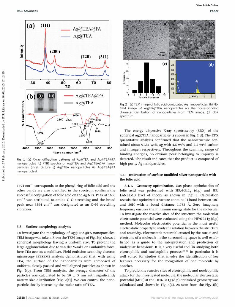
Fig. 1 (a) X-ray diffraction patterns of Ag@TEA and Ag@TEA@FAnanoparticles (b) FTIR spectra of Ag@TEA and Ag@TEA@FA nano-particles (inset picture (i) Ag@TEA nanoparticles (ii) Ag@TEA@FAnanoparticles).
Fig. 2 (a) TEM image of folic acid conjugated Ag nanoparticles. (b) FE-SEM image of Ag@FA@TEA nanoparticles (c) the correspondingdiameter distribution of nanoparticles from TEM image. (d) EDXspectrum.
RSC Advances Paper
Publ
ishe
d on
17
Febr
uary
201
5. D
ownl
oade
d by
DT
U L
ibra
ry o
n 04
/03/
2015
17:
13:5
6.
View Article Online
1494 cm�1 corresponds to the phenyl ring of folic acid and theother bands are also identied in the spectrum conrms thesuccessful conjugation of folic acid on the Ag NPs. Peak at 1600cm�1 was attributed to amide C–O stretching and the broadpeak near 3394 cm�1 was designated as an O–H stretchingvibration.
3.3. Surface morphology analysis
To investigate the morphology of Ag@TEA@FA nanoparticles,TEM image was taken. From the TEM image of Fig. 2(a) shows aspherical morphology having a uniform size. To prevent thelarge agglomeration due to van der Waal's or Coulomb's force,here TEA acts as a stabiliser. Field emission scanning electronmicroscopy (FESEM) analysis demonstrated that, with usingTEA, the surface of the nanoparticles were composed ofuniform, closely packed and well-aligned particles as shown inFig. 2(b). From TEM analysis, the average diameter of theparticles was calculated to be 10 � 5 nm with signicantlynarrow size distribution [Fig. 2(c)]. We can control the nano-particle size by increasing the molar ratio of TEA.
21518 | RSC Adv., 2015, 5, 21515–21524
The energy dispersive X-ray spectroscopy (EDX) of thespherical Ag@TEA nanoparticles is shown in Fig. 2(d). The EDXquantitative analysis conrmed that the nanostructure con-tained about 91.51 wt% Ag with 4.5 wt% and 2.5 wt% carbonand nitrogen respectively. Throughout the scanning range ofbinding energies, no obvious peak belonging to impurity isdetected. The result indicates that the product is composed ofhigh purity Ag nanoparticles.
3.4. Interaction of surface modied silver nanoparticle withthe folic acid
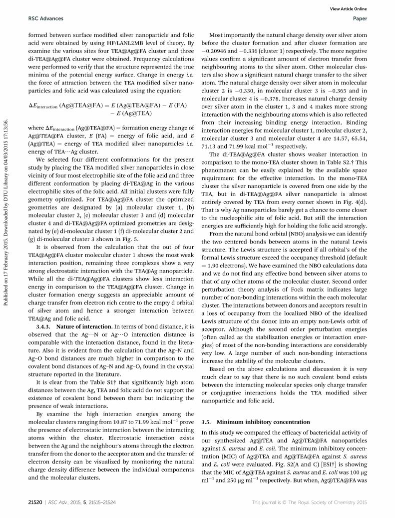
3.4.1. Geometry optimization. Gas phase optimization offolic acid was performed with HF/6-311g (d,p) and HF/LANL2MB level of theory as shown in Fig. 3. Calculationreveals that optimized structure contains H-bond between 10Oand 50H with a bond distance 1.783 A. Zero imaginaryfrequency ensures the minimum energy state for the molecule.To investigate the reactive sites of the structure the molecularelectrostatic potential were evaluated using the HF/6-311g (d,p)method. Molecular electrostatic potential is the most usefulelectrostatic property to study the relation between the structureand reactivity. Electrostatic potential created by the nuclei andelectrons of a molecule in the surrounding space is well estab-lished as a guide to the interpretation and prediction ofmolecular behaviour. It is a very useful tool in studying bothelectrophilic and nucleophilic process.35–39 In particular it iswell suited for studies that involve the identication of keyfeatures necessary for the recognition of one molecule byanother.
To predict the reactive sites of electrophilic and nucleophilicattack for the investigated molecule, the molecular electrostaticpotential (MEP) at the HF/6-311g (d,p) optimized geometry wascalculated and shown in Fig. 4(a). As seen from the Fig. 4(b)
This journal is © The Royal Society of Chemistry 2015
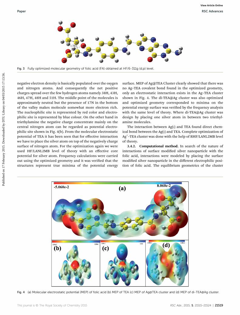
Fig. 3 Fully optimized molecular geometry of folic acid (FA) obtained at HF/6-311g (d,p) level.
Paper RSC Advances
Publ
ishe
d on
17
Febr
uary
201
5. D
ownl
oade
d by
DT
U L
ibra
ry o
n 04
/03/
2015
17:
13:5
6.
View Article Online
negative electron density is basically populated over the oxygenand nitrogen atoms. And consequently the net positivecharges spread over the few hydrogen atoms namely 38H, 43H,46H, 47H, 48H and 51H. The middle point of the molecules isapproximately neutral but the presence of 17N in the bottomof the valley makes molecule somewhat more electron rich.The nucleophilic site is represented by red color and electro-philic site is represented by blue colour. On the other hand intriethylamine the negative charge concentrate mainly on thecentral nitrogen atom can be regarded as potential electro-philic site shown in Fig. 4(b). From the molecular electrostaticpotential of TEA it has been seen that for effective interactionwe have to place the silver atom on top of the negatively chargesurface of nitrogen atom. For the optimization again we wereused HF/LANL2MB level of theory with an effective corepotential for silver atom. Frequency calculations were carriedout using the optimized geometry and it was veried that thestructures represent true minima of the potential energy
Fig. 4 (a) Molecular electrostatic potential (MEP) of folic acid (b) MEP o
This journal is © The Royal Society of Chemistry 2015
surface. MEP of Ag@TEA Cluster clearly showed that there wasno Ag–TEA covalent bond found in the optimized geometry,only an electrostatic interaction exists in the Ag–TEA clustershown in Fig. 4. The di-TEA@Ag cluster was also optimizedand optimized geometry corresponded to minima on thepotential energy surface was veried by the frequency analysiswith the same level of theory. Where di-TEA@Ag cluster wasdesign by placing one silver atom in between two triethyl-amine molecules.
The interaction between Ag(I) and TEA found direct chem-ical bond between the Ag(I) and TEA. Complete optimization ofAg+–TEA cluster was done with the help of RHF/LANL2MB levelof theory.
3.4.2. Computational method. In search of the nature ofinteractions of surface modied silver nanoparticle with thefolic acid, interactions were modeled by placing the surfacemodied silver nanoparticle in the different electrophilic posi-tion of folic acid. The equilibrium geometrics of the cluster
f TEA (c) MEP of Ag@TEA cluster and (d) MEP of di-TEA@Ag cluster.
RSC Adv., 2015, 5, 21515–21524 | 21519
RSC Advances Paper
Publ
ishe
d on
17
Febr
uary
201
5. D
ownl
oade
d by
DT
U L
ibra
ry o
n 04
/03/
2015
17:
13:5
6.
View Article Online
formed between surface modied silver nanoparticle and folicacid were obtained by using HF/LANL2MB level of theory. Byexamine the various sites four TEA@Ag@FA cluster and threedi-TEA@Ag@FA cluster were obtained. Frequency calculationswere performed to verify that the structure represented the trueminima of the potential energy surface. Change in energy i.e.the force of attraction between the TEA modied silver nano-particles and folic acid was calculated using the equation:
DEinteraction (Ag@TEA@FA) ¼ E (Ag@TEA@FA) � E (FA)
� E (Ag@TEA)
where DEinteraction (Ag@TEA@FA) ¼ formation energy change ofAg@TEA@FA cluster, E (FA) ¼ energy of folic acid, and E(Ag@TEA) ¼ energy of TEA modied silver nanoparticles i.e.energy of TEA/Ag cluster.
We selected four different conformations for the presentstudy by placing the TEA modied silver nanoparticles in closevicinity of four most electrophilic site of the folic acid and threedifferent conformation by placing di-TEA@Ag in the variouselectrophilic sites of the folic acid. All initial clusters were fullygeometry optimized. For TEA@Ag@FA cluster the optimizedgeometries are designated by (a) molecular cluster 1, (b)molecular cluster 2, (c) molecular cluster 3 and (d) molecularcluster 4 and di-TEA@Ag@FA optimized geometries are desig-nated by (e) di-molecular cluster 1 (f) di-molecular cluster 2 and(g) di-molecular cluster 3 shown in Fig. 5.
It is observed from the calculation that the out of fourTEA@Ag@FA cluster molecular cluster 1 shows the most weakinteraction position, remaining three complexes show a verystrong electrostatic interaction with the TEA@Ag nanoparticle.While all the di-TEA@Ag@FA clusters show less interactionenergy in comparison to the TEA@Ag@FA cluster. Change incluster formation energy suggests an appreciable amount ofcharge transfer from electron rich centre to the empty d orbitalof silver atom and hence a stronger interaction betweenTEA@Ag and folic acid.
3.4.3. Nature of interaction. In terms of bond distance, it isobserved that the Ag/N or Ag/O interaction distance iscomparable with the interaction distance, found in the litera-ture. Also it is evident from the calculation that the Ag–N andAg–O bond distances are much higher in comparison to thecovalent bond distances of Ag–N and Ag–O, found in the crystalstructure reported in the literature.
It is clear from the Table S1† that signicantly high atomdistances between the Ag, TEA and folic acid do not support theexistence of covalent bond between them but indicating thepresence of weak interactions.
By examine the high interaction energies among themolecular clusters ranging from 10.87 to 71.99 kcal mol�1 provethe presence of electrostatic interaction between the interactingatoms within the cluster. Electrostatic interaction existsbetween the Ag and the neighbour's atoms through the electrontransfer from the donor to the acceptor atom and the transfer ofelectron density can be visualized by monitoring the naturalcharge density difference between the individual componentsand the molecular clusters.
21520 | RSC Adv., 2015, 5, 21515–21524
Most importantly the natural charge density over silver atombefore the cluster formation and aer cluster formation are�0.20946 and�0.336 (cluster 1) respectively. The more negativevalues conrm a signicant amount of electron transfer fromneighbouring atoms to the silver atom. Other molecular clus-ters also show a signicant natural charge transfer to the silveratom. The natural charge density over silver atom in molecularcluster 2 is �0.330, in molecular cluster 3 is �0.365 and inmolecular cluster 4 is �0.378. Increases natural charge densityover silver atom in the cluster 1, 3 and 4 makes more stronginteraction with the neighbouring atoms which is also reectedfrom their increasing binding energy interaction. Bindinginteraction energies for molecular cluster 1, molecular cluster 2,molecular cluster 3 and molecular cluster 4 are 14.57, 65.54,71.13 and 71.99 kcal mol�1 respectively.
The di-TEA@Ag@FA cluster shows weaker interaction incomparison to the mono-TEA cluster shown in Table S2.† Thisphenomenon can be easily explained by the available spacerequirement for the effective interaction. In the mono-TEAcluster the silver nanoparticle is covered from one side by theTEA, but in di-TEA@Ag@FA silver nanoparticle is almostentirely covered by TEA from every corner shown in Fig. 4(d).That is why Ag nanoparticles barely get a chance to come closerto the nucleophilic site of folic acid. But still the interactionenergies are sufficiently high for holding the folic acid strongly.
From the natural bond orbital (NBO) analysis we can identifythe two centered bonds between atoms in the natural Lewisstructure. The Lewis structure is accepted if all orbital's of theformal Lewis structure exceed the occupancy threshold (default¼ 1.90 electrons). We have examined the NBO calculations dataand we do not nd any effective bond between silver atoms tothat of any other atoms of the molecular cluster. Second orderperturbation theory analysis of Fock matrix indicates largenumber of non-bonding interactions within the each molecularcluster. The interactions between donors and acceptors result ina loss of occupancy from the localized NBO of the idealizedLewis structure of the donor into an empty non-Lewis orbit ofacceptor. Although the second order perturbation energies(oen called as the stabilization energies or interaction ener-gies) of most of the non-bonding interactions are considerablyvery low. A large number of such non-bonding interactionsincrease the stability of the molecular clusters.
Based on the above calculations and discussion it is verymuch clear to say that there is no such covalent bond existsbetween the interacting molecular species only charge transferor conjugative interactions holds the TEA modied silvernanoparticle and folic acid.
3.5. Minimum inhibitory concentration
In this study we compared the efficacy of bactericidal activity ofour synthesized Ag@TEA and Ag@TEA@FA nanoparticlesagainst S. aureus and E. coli. The minimum inhibitory concen-tration (MIC) of Ag@TEA and Ag@TEA@FA against S. aureusand E. coli were evaluated. Fig. S2(A and C) [ESI†] is showingthat the MIC of Ag@TEA against S. aureus and E. coli was 100 mgml�1 and 250 mg ml�1 respectively. But when, Ag@TEA@FA was
This journal is © The Royal Society of Chemistry 2015

Fig. 5 Conformers of TEA@Ag@FA and di-TEA@Ag@FA clusters. (a) Molecular cluster 1, (b) molecular cluster 2, (c) molecular cluster 3, (d)molecular cluster 4, (e) di-molecular cluster 1, (f) di-molecular cluster 2, (g) di-molecular cluster 3.
Paper RSC Advances
Publ
ishe
d on
17
Febr
uary
201
5. D
ownl
oade
d by
DT
U L
ibra
ry o
n 04
/03/
2015
17:
13:5
6.
View Article Online
charged against the bacteria the MIC concentration decreasedat 10 mg ml�1 in both, shown in Fig. S2(B and D).† Though theantibacterial activities of the Ag@TEA@FA against the Gram-positive S. aureus and Gram-negative E. coli were identical butmore bactericidal against E. coli than only Ag@TEA.
3.6. Minimum bactericidal concentration
To be conrming about the bactericidal activity of Ag@TEA@FAwe followed the MBC study. The particular particle concentra-tion was noted for each strain where no visible growth appearson the agar plate, both in the case of Ag@TEA andAg@TEA@FA. In the case of Ag@TEA, charged against the
This journal is © The Royal Society of Chemistry 2015
Gram-positive strain and Gram-negative strain, the MBC valuewas found at 500 mgml�1 in both (gure not shown) whereas theMBC value was found at 25 mg ml�1 in both strain againstAg@TEA@FA (Fig. 6).
3.7. Disc agar diffusion (DAD) method
DAD study was followed for visual comparison between theAg@TEA and Ag@TEA@FA. Fig. 6 exhibits the inhibition zoneof the two particles. The result showed that Ag@TEA@FA (i) ismore effective than Ag@TEA (ii) against Gram-positive andGram-negative both.
RSC Adv., 2015, 5, 21515–21524 | 21521
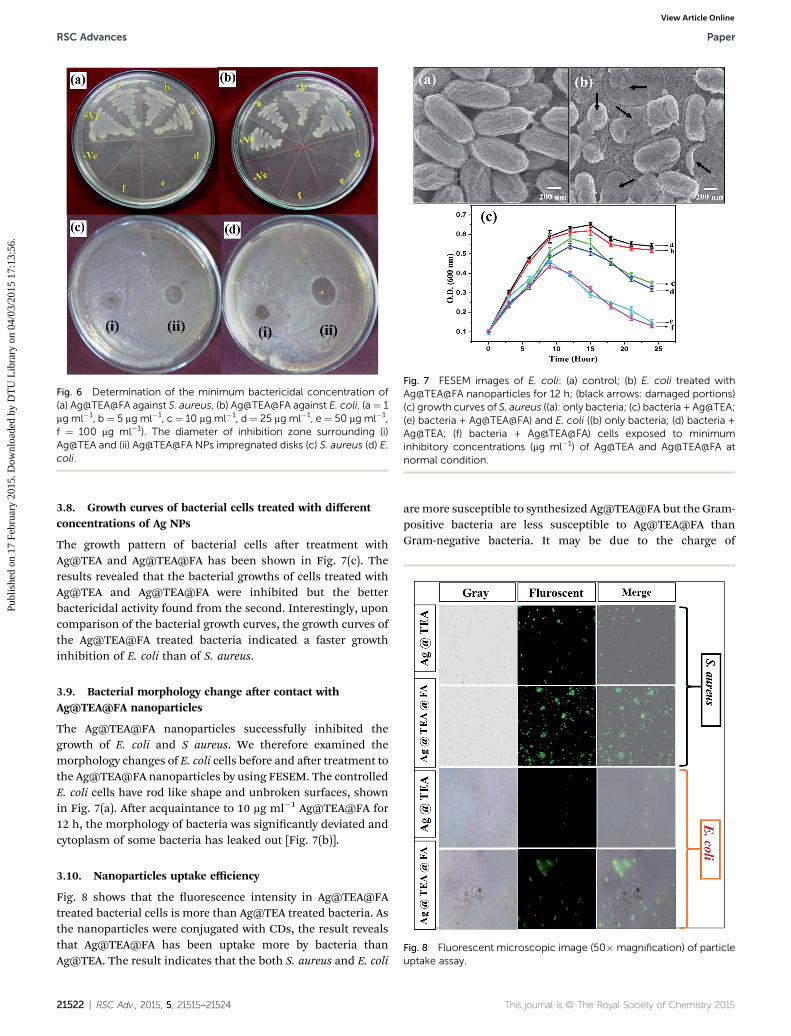
Fig. 6 Determination of the minimum bactericidal concentration of(a) Ag@TEA@FA against S. aureus, (b) Ag@TEA@FA against E. coli, (a¼ 1mg ml�1, b ¼ 5 mg ml�1, c¼ 10 mg ml�1, d¼ 25 mg ml�1, e¼ 50 mg ml�1,f ¼ 100 mg ml�1). The diameter of inhibition zone surrounding (i)Ag@TEA and (ii) Ag@TEA@FA NPs impregnated disks (c) S. aureus (d) E.coli.
Fig. 7 FESEM images of E. coli: (a) control; (b) E. coli treated withAg@TEA@FA nanoparticles for 12 h; (black arrows: damaged portions)(c) growth curves of S. aureus ((a): only bacteria; (c) bacteria + Ag@TEA;(e) bacteria + Ag@TEA@FA) and E. coli ((b) only bacteria; (d) bacteria +Ag@TEA; (f) bacteria + Ag@TEA@FA) cells exposed to minimuminhibitory concentrations (mg ml�1) of Ag@TEA and Ag@TEA@FA atnormal condition.
RSC Advances Paper
Publ
ishe
d on
17
Febr
uary
201
5. D
ownl
oade
d by
DT
U L
ibra
ry o
n 04
/03/
2015
17:
13:5
6.
View Article Online
3.8. Growth curves of bacterial cells treated with differentconcentrations of Ag NPs
The growth pattern of bacterial cells aer treatment withAg@TEA and Ag@TEA@FA has been shown in Fig. 7(c). Theresults revealed that the bacterial growths of cells treated withAg@TEA and Ag@TEA@FA were inhibited but the betterbactericidal activity found from the second. Interestingly, uponcomparison of the bacterial growth curves, the growth curves ofthe Ag@TEA@FA treated bacteria indicated a faster growthinhibition of E. coli than of S. aureus.
3.9. Bacterial morphology change aer contact withAg@TEA@FA nanoparticles
The Ag@TEA@FA nanoparticles successfully inhibited thegrowth of E. coli and S aureus. We therefore examined themorphology changes of E. coli cells before and aer treatment tothe Ag@TEA@FA nanoparticles by using FESEM. The controlledE. coli cells have rod like shape and unbroken surfaces, shownin Fig. 7(a). Aer acquaintance to 10 mg ml�1 Ag@TEA@FA for12 h, the morphology of bacteria was signicantly deviated andcytoplasm of some bacteria has leaked out [Fig. 7(b)].
Fig. 8 Fluorescent microscopic image (50� magnification) of particleuptake assay.
3.10. Nanoparticles uptake efficiency
Fig. 8 shows that the uorescence intensity in Ag@TEA@FAtreated bacterial cells is more than Ag@TEA treated bacteria. Asthe nanoparticles were conjugated with CDs, the result revealsthat Ag@TEA@FA has been uptake more by bacteria thanAg@TEA. The result indicates that the both S. aureus and E. coli
21522 | RSC Adv., 2015, 5, 21515–21524
are more susceptible to synthesized Ag@TEA@FA but the Gram-positive bacteria are less susceptible to Ag@TEA@FA thanGram-negative bacteria. It may be due to the charge of
This journal is © The Royal Society of Chemistry 2015

Paper RSC Advances
Publ
ishe
d on
17
Febr
uary
201
5. D
ownl
oade
d by
DT
U L
ibra
ry o
n 04
/03/
2015
17:
13:5
6.
View Article Online
peptidoglycan molecules in the bacterial cell wall. Gram-positive bacteria have more peptidoglycan than Gram-negativebacteria because of their thicker cell walls and as peptido-glycan is negatively charged and silver ions are positivelycharged, more silver may get trapped by peptidoglycan in Gram-positive bacteria than in Gram-negative bacteria.40
4. Conclusion
In this paper, a novel technique has been developed tosynthesize folic acid conjugated silver nanoparticles to enhancethe antibacterial activity in comparison to pure silver nano-particles. From the FTIR and TGA results, it is conrmed thatthe folic acid is conjugated with silver nanoparticles throughelectrostatic interaction. Moreover, theoretical simulationsfurther elucidate the interactions of folic acid and silver nano-particles are electrostatic in nature. Fluorescent carbon dots aresuccessfully conjugated with silver nanoparticles for uptakestudy in bacteria. This folic acid conjugated silver nanoparticleshave lowered the level of the MIC and MBC values and act aseffective antibacterial agents against both Gram-positive andGram-negative bacteria. From this study it is concluded thatfolic acid can be conjugated with other nanoparticles throughelectrostatic interaction for anticancer drug delivery.
References
1 X. Dai, Z. Fan, Y. Lu and P. C. Ray, ACS Appl. Mater. Interfaces,2013, 5, 11348–11354.
2 S. M. Lee, H. J. Kim, Y. J. Ha, Y. N. Park, S. K. Lee, Y. B. Parkand K. H. Yoo, ACS Nano, 2013, 7(1), 50–57.
3 F. Jia, X. Liu, L. Li, S. Mallapragada, B. Narasimhan andQ. Wang, J. Controlled Release, 2013, 172, 1020–1034.
4 D. M. Tobaldi, C. Piccirillo, R. C. Pullar, A. F. Gualtieri,M. P. Seabra, P. M. L. Castro and J. A. Labrincha, J. Phys.Chem. C, 2014, 118, 4751–4766.
5 T. Angelova, N. Rangelova, R. Yuryev, N. Georgieva andR. Mulle, Mater. Sci. Eng., C, 2012, 32, 1241–1246.
6 S. P. Chakraborty, S. K. Mahapatra, S. Chattopadhyay,P. Pramanik and S. Roy, Asian Pac. J. Trop. Biomed., 2011,1(2), 102–109.
7 J. Vidic, S. Stankic, F. Haque, D. Ciric, R. L. Goffic, A. Vidy,J. Jupille and B. Delmas, J. Nanopart. Res., 2013, 15, 1595–1605.
8 C. Baker Austin, M. S. Wright, R. Stepanauskas andJ. V. McArthur, Trends Microbiol., 2006, 14, 176–182.
9 A. J. Huh and Y. Kwon, J. Controlled Release, 2011, 156, 128–145.
10 S. M. Dizaj, F. Lotpour, M. B. Jalali, M. H. Zarrintana andK. Adibkia, Mater. Sci. Eng., C, 2014, 44, 278–284.
11 Y. H. Kim, C. W. Kim, H. G. Cha, D. K. Lee, B. K. Jo,G. W. Ahn, E. S. Hong, J. C. Kim and Y. S. Kang, J. Phys.Chem. C, 2009, 113, 5105–5110.
12 S. K. Gogoi, P. Gopinath, A. Paul, A. Ramesh, S. S. Ghosh andA. Chattopadhyay, Langmuir, 2006, 22(22), 9322–9328.
This journal is © The Royal Society of Chemistry 2015
13 A. T. Le, L. T. Tama, P. D. Tama, P. T. Huy, T. Q. Huy,N. V. Hieuc, A. A. Kudrinskiy and Y. A. Krutyakov, Mater.Sci. Eng., C, 2010, 30, 910–916.
14 M. Wang, H. Hu, Y. Sun, L. Qiu, J. Zhang, G. Guan, X. Zhao,M. Qiao, L. Cheng, L. Cheng and D. Chen, Biomaterials, 2013,34, 10120–10132.
15 D. Chen, P. Song, F. Jiang, X. Meng, W. Sui, C. Shu andL. J. Wan, J. Phys. Chem. B, 2013, 117, 1261–1268.
16 E. Roger, S. Kalscheuer, A. Kirtane, B. R. Guru, A. E. Grill,J. Whittum-Hudson and J. Panyam, Mol. Pharmaceutics,2012, 9, 2103–2110.
17 J. J. Lin, J. S. Chen, S. J. Huang, J. H. Ko, Y. M. Wang,T. L. Chen and L. F. Wang, Biomaterials, 2009, 30, 5114–5124.
18 S. K. Sahu, S. Maiti, A. Pramanik, S. K. Ghosh andP. Pramanik, Carbohydr. Polym., 2012, 87, 2593–2604.
19 S. K. Sahu, S. Maiti, T. K. Maiti, S. K. Ghosh and P. Pramanik,Macromol. Biosci., 2011, 11(2), 285–295.
20 S. J. Yang, F. H. Lin, H. M. Tsai, C. F. Lin, H. C. Chin,J. M. Wong and M. J. Shieh, Biomaterials, 2011, 32, 2174–2182.
21 A. Sulistio, J. Lowenthal, A. Blencowe, M. N. Bongiovanni,L. Ong, S. L. Gras, X. Zhang and G. G. Qiao,Biomacromolecules, 2011, 12, 3469–3477.
22 S. P. Chakraborty, S. K. Sahu, S. Kar Mahapatra, S. Santra,M. Bal, S. Roy and P. Pramanik, Nanotechnology, 2010, 21,105103–105112.
23 J. T. Wu and L. C. Hsu Steve, J. Nanopart. Res., 2011, 13,3877–3883.
24 S. Sahu, B. Behera, T. K. Maiti and S. Mohapatra, Chem.Commun., 2012, 48, 8835–8837.
25 M. J. Frisch, et al., Gaussian 03, Gaussian Inc., Pittsburgh,PA, 2003.
26 A. Frisch, I. I. R. Dennington, T. Keith, J. Millam,A. B. Nielsen, A. J. Holder and J. Hiscocks, Reference,Version 5.0, Gaussian Inc., Pittsburgh, 2007.
27 J. X. Wang, L. X. Wen, Z. H. Wang and J. F. Chen, Mater.Chem. Phys., 2006, 96, 90–97.
28 H. M. Ericsson and J. C. Sherris, Acta Pathol. Microbiol.Scand., Sect. B, Suppl., 1971, 217, 1–10.
29 A. Z. Melaiye, K. Sun, A. Milsted, D. Ely, D. H. Reneker,C. A. Tessier and W. J. Youngs, J. Am. Chem. Soc., 2005,127, 2285–2291.
30 J. Y. Kim, K. Sungeun, J. Kim, L. Jongchan and J. Yoon, J.Korean Soc. Environ. Eng., 2005, 27, 771–776.
31 G. Jin, H. Qin, H. Cao, S. Qian, Y. Zhao, X. Peng, X. Zhang,X. Liu and P. K. Chu, Biomaterials, 2014, 35(27), 7699–7713.
32 P. Pallavicini, A. Taglietti, G. Dacarro, Y. A. Diaz-Fernandez,M. Galli, P. Grisoli, M. Patrini, G. S. De Magistris andR. Zanoni, J. Colloid Interface Sci., 2010, 350, 110–116.
33 H. L. Sua, C. C. Choub, D. J. Hunga, S. H. Lina, I. C. Paoa,J. H. Linc, F. L. Huang, R. X. Dong and J. J. Linb,Biomaterials, 2009, 30, 5979–5987.
34 J. Shi, X. Sun, Y. Lin, X. Zou, Z. Li, Y. Liao, M. Du andH. Zhang, Biomaterials, 2014, 35, 6657–6666.
35 P. Kolandaivel, G. Praveen and P. Selvarengan, J. Chem. Sci.,2005, 117, 591–598.
RSC Adv., 2015, 5, 21515–21524 | 21523

RSC Advances Paper
Publ
ishe
d on
17
Febr
uary
201
5. D
ownl
oade
d by
DT
U L
ibra
ry o
n 04
/03/
2015
17:
13:5
6.
View Article Online
36 F. J. Luque, J. M. Lopez and M. Orazco, Theor. Chem. Acc.,2000, 103, 343–345.
37 N. Okulik and A. H. Jubert, Internet Electron. J. Mol. Des.,2005, 4, 17–30.
38 L. Zhang, T. Ren, L. Zhou, J. Tian and X. Li, Comput. Theor.Chem., 2013, 1019, 1–10.
21524 | RSC Adv., 2015, 5, 21515–21524
39 W. Sun and R. D. Felice, J. Phys. Chem. C, 2012, 116, 24954–24961.
40 K. Kawahara, K. Tsuruda, M. Morishita and M. Uchida, Dent.Mater., 2000, 16, 452–455.
This journal is © The Royal Society of Chemistry 2015
Copyright © 2022 FDOKUMEN