The rat trkC locus encodes multiple neurogenic receptors that exhibit differential response to...
16
Neuron, Vol. 10, 975-990,May, 1993, Copyright© 1993by Cell Press The Rat trkC Locus Encodes Multiple Neurogenic Receptors That Exhibit Differential Response to Neurotrophin-3 in PC12 Cells Pantelis Tsoulfas,* Dan Soppet,* Enrique Escandon,+ Lino Tessarollo,* Jos~Luis Mendoza-Ramirez, tt Arnon Rosenthal,t Karoly Nikolics,+ and Luis F. Parada* Molecular Embryology Section *NCI-Frederick Cancer Research and Development Center ABL-Basic Research Program Frederick, Maryland 21702-1201 tGenentech 460 Point San Bruno Boulevard South San Francisco, California 94080 Summary Members of the Trk tyrosine kinase family have recently been identified as functional receptors of the NG F family of neurotrophins. Here we show the rat trkC locus to be complex, encoding at least four distinct polypeptides. Three of the encoded polypeptides are full-length recep- tor tyrosine kinases that differ by novel amino acid inser- tions in the kinase domain. A fourth protein is a trun- cated receptor that lacks the catalytic domain. Tyrosine phosphorylation, cross-linking, and ligand binding assays indicate that TrkC receptors interact with NT-3 and not with the related neurotrophins NGF, BDNF, xNT-4, or hNT-5. Furthermore, high and low affinity NT-3-binding sites are associated with the TrkC receptors. Stable and transient expression of TrkC receptors in PC 12 cells indi- cates that the neurite outgrowth response elicited by NT-3 is dramatic in receptors lacking the novel kinase insert (gp15@'*c) but absent in receptors containing the 14 amino acid insert in the kinase domain (gp15@'*c14). These data suggestthat the trkC locus encodes receptors that may be capable of mediating different biological responses within the cell. This could have important im- plications in understanding the role of neurotrophins in the development of the vertebrate nervous system. Introduction The vertebrate nervous system comprises a broad va- riety of differentiated neuronal populations. The sur- vival, functional maintenance, and phenotype of these varied cell types often depend on the presence of specific polypeptides (for review see Barde, 1989). 13-Nerve growth factor (NGF) is the prototypical exam- ple of a target-derived neurotrophic factor that is es- sential for development and survival of peripheral sympathetic and neural crest-derived sensory neu- rons (for reviews, see Barde, 1989; Levi-Montalcini, 1987). In the CNS, NGF supports the survival of cholin- ergic neurons in the basal forebrain (for reviews, see tPermanent address: Instituto de Fisiologia Celular, UN AM, Mexico, DF.04510. Thoenen et al., 1987; Barde, 1989; Ebendal, 1989). In recent years, it has been shown that NGF is but one member of a family of structurally related polypep- tides that support the survival of vertebrate embry- onic neurons (for review see Barde, 1991). Proteins of this gene family include brain-derived neurotrophic factor (BDNF; Leibrock et al., 1989), neurotrophin-3 (NT-3; Ernfors et al., 1990; Hohn et al., 1990; Jones and Reichardt 1990; Kaisho et al., 1990; Maisonpierre et al., 1990a; Rosenthal et al., 1990), Xenopus neurotrophin-4 (xNT-4; Hallb66k et al., 1991; Ip et al., 1992), and human neurotrophin-5 (h NT-5; Berkemeier et al., 1991). These factors, like NGF, are expressed in many tissues, in- cluding the CNS (Goedert et al., 1986; Korsching et al., 1985; Shelton and Reichardt, 1986; Whittemore et al., 1986; Ernfors et al., 1990; Hofer et al., 1990; Maison- pierre et al. , 1990b; Phillips et al., 1990). The NGF- related neurotrophins display trophic activity on dis- tinct as well as overlapping neuronal populations within the CNS and PNS (for review see Barde, 1991). For example, like NGF, BDNF affects the survival of neural crest-derived sensory neurons (Kalcheim et al., 1987), but unlike NGF, BDNF has no apparent activity in sympathetic neurons (Davies et al., 1986; Lindsay et al., 1985). This theme is repeated in various neuronal subpopulations. Characterization of the receptors for NGF, BDNF, and NT-3, demonstrates two classes of receptors in sensory ganglion explants (Sutter et al., 1979; Rodri- guez-T6bar and Barde, 1988; Rodriguez-T6bar et al., 1992) and in neuronally derived cell lines (Landreth and Shooter, 1980; Schechter and Bothwell, 1981; Son- nefeld and Ishii, 1985). Several laboratories have ob- served high affinity binding sites with an affinity con- stant (Ko) of -10 -11 M and low affinity binding sites with a Ko of - 10-9 M. A low affinity receptor (gp75 NCFR) has been isolated from several organisms (Johnson et al., 1986; Radeke et al., 1987; Large et al., 1989); this site binds NGF, BDNF, NT-3, and xNT-4 with a similar low KD(HallbO6k et al., 1991; Radeke et al., 1987; Rodri- guez-T~bar et al., 1990, 1992). However, the character- istic biological responses of neuronal survival or neu- rite outgrowth are thought to be mediated through the presence of high affinity receptors for both NGF (Green et al., 1986) and BDNF (Rodriguez-T6bar and Barde, 1988). While the preceding studies with NGF and related molecules elucidate some aspects of neuronal devel- opment and survival, the mechanism of action and the precise role for each neurotrophin in vivo remain to be determined. The finding that gp140pr°t°trk, a 140 kd receptor tyrosine kinase (RTK) encoded by the trk locus (Martin-Zanca et al., 1986), can bind NGF and become catalytically activated in response to this in- teraction (Kaplan et al., 1991a, 1991b; Hempstead et al., 1991; Klein et al., 1991) has provided new insights into the mechanism of neurotrophin action. Loeb et
Transcript of The rat trkC locus encodes multiple neurogenic receptors that exhibit differential response to...

Neuron, Vol. 10, 975-990, May, 1993, Copyright © 1993 by Cell Press
The Rat trkC Locus Encodes Multiple Neurogenic Receptors That Exhibit Differential Response to Neurotrophin-3 in PC12 Cells Pantelis Tsoulfas,* Dan Soppet,* Enrique Escandon,+ Lino Tessarollo,* Jos~Luis Mendoza-Ramirez, tt Arnon Rosenthal, t Karoly Nikolics,+ and Luis F. Parada* Molecular Embryology Section *NCI-Frederick Cancer Research and Development
Center ABL-Basic Research Program Frederick, Maryland 21702-1201 tGenentech 460 Point San Bruno Boulevard South San Francisco, California 94080
Summary
Members of the Trk tyrosine kinase family have recently been identified as functional receptors of the NG F family of neurotrophins. Here we show the rat trkC locus to be complex, encoding at least four distinct polypeptides. Three of the encoded polypeptides are full-length recep- tor tyrosine kinases that differ by novel amino acid inser- tions in the kinase domain. A fourth protein is a trun- cated receptor that lacks the catalytic domain. Tyrosine phosphorylation, cross-linking, and ligand binding assays indicate that TrkC receptors interact with NT-3 and not with the related neurotrophins NGF, BDNF, xNT-4, or hNT-5. Furthermore, high and low affinity NT-3-binding sites are associated with the TrkC receptors. Stable and transient expression of TrkC receptors in PC 12 cells indi- cates that the neurite outgrowth response elicited by NT-3 is dramatic in receptors lacking the novel kinase insert (gp15@ '*c) but absent in receptors containing the 14 amino acid insert in the kinase domain (gp15@'*c14). These data suggest that the trkC locus encodes receptors that may be capable of mediating different biological responses within the cell. This could have important im- plications in understanding the role of neurotrophins in the development of the vertebrate nervous system.
Introduction
The vertebrate nervous system comprises a broad va- riety of differentiated neuronal populations. The sur- vival, functional maintenance, and phenotype of these varied cell types often depend on the presence of specific polypeptides (for review see Barde, 1989). 13-Nerve growth factor (NGF) is the prototypical exam- ple of a target-derived neurotrophic factor that is es- sential for development and survival of peripheral sympathetic and neural crest-derived sensory neu- rons (for reviews, see Barde, 1989; Levi-Montalcini, 1987). In the CNS, NGF supports the survival of cholin- ergic neurons in the basal forebrain (for reviews, see
tPermanent address: Instituto de Fisiologia Celular, UN AM, Mexico, DF.04510.
Thoenen et al., 1987; Barde, 1989; Ebendal, 1989). In recent years, it has been shown that NGF is but one member of a family of structurally related polypep- tides that support the survival of vertebrate embry- onic neurons (for review see Barde, 1991). Proteins of this gene family include brain-derived neurotrophic factor (BDNF; Leibrock et al., 1989), neurotrophin-3 (NT-3; Ernfors et al., 1990; Hohn et al., 1990; Jones and Reichardt 1990; Kaisho et al., 1990; Maisonpierre et al., 1990a; Rosenthal et al., 1990), Xenopus neurotrophin-4 (xNT-4; Hallb66k et al., 1991; Ip et al., 1992), and human neurotrophin-5 (h NT-5; Berkemeier et al., 1991). These factors, like NGF, are expressed in many tissues, in- cluding the CNS (Goedert et al., 1986; Korsching et al., 1985; Shelton and Reichardt, 1986; Whittemore et al., 1986; Ernfors et al., 1990; Hofer et al., 1990; Maison- pierre et al. , 1990b; Phillips et al., 1990). The NGF- related neurotrophins display trophic activity on dis- tinct as well as overlapping neuronal populations within the CNS and PNS (for review see Barde, 1991). For example, like NGF, BDNF affects the survival of neural crest-derived sensory neurons (Kalcheim et al., 1987), but unlike NGF, BDNF has no apparent activity in sympathetic neurons (Davies et al., 1986; Lindsay et al., 1985). This theme is repeated in various neuronal subpopulations.
Characterization of the receptors for NGF, BDNF, and NT-3, demonstrates two classes of receptors in sensory ganglion explants (Sutter et al., 1979; Rodri- guez-T6bar and Barde, 1988; Rodriguez-T6bar et al., 1992) and in neuronally derived cell lines (Landreth and Shooter, 1980; Schechter and Bothwell, 1981; Son- nefeld and Ishii, 1985). Several laboratories have ob- served high affinity binding sites with an affinity con- stant (Ko) of -10 -11 M and low affinity binding sites with a Ko of - 10 -9 M. A low affinity receptor (gp75 NCFR) has been isolated from several organisms (Johnson et al., 1986; Radeke et al., 1987; Large et al., 1989); this site binds NGF, BDNF, NT-3, and xNT-4 with a similar low KD (HallbO6k et al., 1991; Radeke et al., 1987; Rodri- guez-T~bar et al., 1990, 1992). However, the character- istic biological responses of neuronal survival or neu- rite outgrowth are thought to be mediated through the presence of high affinity receptors for both NGF (Green et al., 1986) and BDNF (Rodriguez-T6bar and Barde, 1988).
While the preceding studies with NGF and related molecules elucidate some aspects of neuronal devel- opment and survival, the mechanism of action and the precise role for each neurotrophin in vivo remain to be determined. The finding that gp140p r°t°trk, a 140 kd receptor tyrosine kinase (RTK) encoded by the trk locus (Martin-Zanca et al., 1986), can bind NGF and become catalytically activated in response to this in- teraction (Kaplan et al., 1991a, 1991b; Hempstead et al., 1991; Klein et al., 1991) has provided new insights into the mechanism of neurotrophin action. Loeb et

Neuron 976
A
trkC
1 MDVSLCPAKCSFWRIFLLGSVWLDYVGSVLACPANCVCSKTEINCRRPDDGNLFPLLEGQDSGNSNGNASINITDISRNITSIHIENWRGLHTLNAVDME
i01 LYTGLQKLTIKNSGLRNIQ•RAFAKN•HLRYINLSSNRLTTL•WQLFQTLSLRELRLEQNFFNCSCDIRWMQL•QEQGEARLDSQSLYCISADGSQL•LF
201 RMNISQCDLPEISVSHVNLTVREGDNAVITCNGSGSPLPDVDWIvTGLQSINTHQTNLNWTNVHAINLTLVNVTSEDNGFTLTCIAENVVGMSNASVALT
301 VYYPPRVVSLVEPEVRLEHCIEFVvRGNPTPTLHWLYNGQPLRESKIIHMDYYQEGEVSEGCLLFNKPTHYNNGNYTLIAKNALGTANQTINGHFLKEPF
401 PESTDFFDFESDAS~TPPIT~THKPEEDTFGvS~AvGLAAFACVLLV~IMINKYGRRSKFGMKGPVAVISGEEDSASPLHHINHGITT~SSLDAGPDT
501 VVIG•TRIPVIEN•Q•FRQGHNCHKPDTYVQHIKRRD•VLKRELGEGAFGKVFLAECYNLS•TKDK•LVAV•ALKDPTLAARKDFQREAELLTNLQHEHI
601 VKFYGvCGDGDPLIMVFEYMKHGDLNKFLRAHGPDAMILVDGQPRQAKGELGLSQMLHIASQIASGMVYLASQHFVHRDLATRNcLVGANLLVKIGDFGM
701 ~7
SRDVYSTDYYRVGGHTMLP I RWMPPE S I MYRKFTTESDVWSFGVI LWE I F TYGKQPWFQLSNTEVI EC I T QGRVLERPRVCPKEVYDVML GCWQREPQQR
801 LNIKEIYKILHALGKATPIYLDILG*
trkCl4
711 RLFNPSGNDFCIWCEV
trkC25
711 REGPYQKGPFSVSWQQQRLAASAASTV
trkCtk-
529 WvFSNIDNHGILNLKDNRDHLVPSTHYIYEEPEVQSGDVSY•RSHGFREIMLNPISLSGHSKpLNHGIYVEDVNVYFSKGRHGF*
B trkCl4
trkC25
SP EX TM JM I I I I I ~ @ ' @ @ / X XT////////////~ I • COOH NH 2
TK
5P EX TM JM TR NH 2 IIIIl~F_,f///~" xF////////////~lll l l l l l l l l l COON
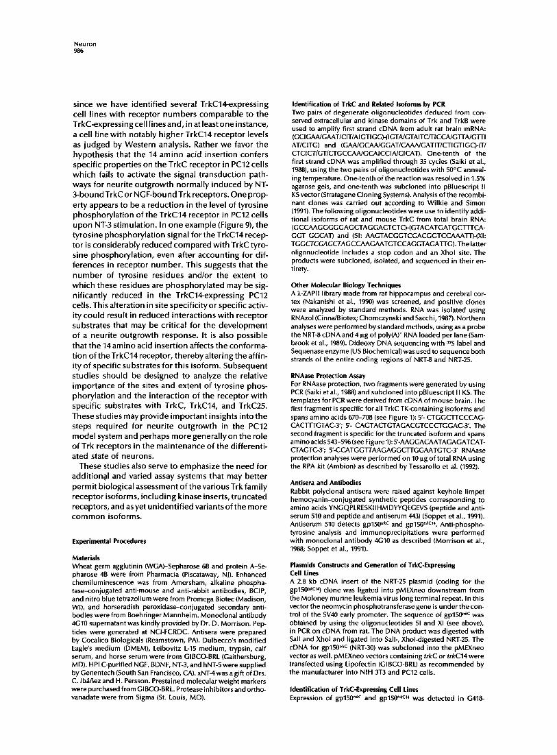
Figure 1. Predicted Amino Acid Sequence and Schematic Representation of the TrkC Isoforms (A) Complete amino acid sequence of TrkC. The putative signal sequence is in italics, and the transmernbrane domain is in bold. The kinase doff~ain is double underlined. The vertical arrow over amino acid 528 indicates the position of frame change for TrkCTK-. The inverted triangle between the amino acids 711 and 712 (in bold) indicates where the 14 and 25 amino acids are inserted to generate the TrkC14 and TrkC2S isoforrns, respectively. An asterisk indicates a stop codon. (B) The different isoforrns are represented schematically: SP, signal peptide; EX, extracellular domain; TM, transmernbrane domain; JM, juxtarnernbrane domain; TK, tyrosine kinase; and TR, truncated peptide. The thick vertical line across the TK domain indicates the site of insertion for the TrkC14 and TrkC25 peptides.
al. (1991)further demonstrated that the trk gene prod- uct can reconstitute the classic biological response of di f ferent iat ion and neuri te outgrowth in a PC12 derivat ive cell l ine (nnrS) that has greatly reduced ex- pression of trk. Taken together, these data implicate the Trk receptor as a requisite component of the high af f in i ty and funct ional NGF receptors in neurons.
A natural extension of the above experiments has been the search for fur ther relat ionships among trk- related genes and NGF-related neurotrophins. Sev- eral groups have shown that a second trk-related gene, trkB (Klein et al., 1989, 1990; Middlemas et al.,
1991), encodes a RTK that binds the neurot roph ins BDNF (Squinto et al., 1991; Soppet et al., 1991), NT-3 (Squinto et al., 1991; Soppet et al., 1991), NT-4 (Ip et al., 1992), and hNT-5 (Berkemeier et al., 1991). To isolate addit ional receptors for the trk gene family, we have employed polymerase chain reaction (PCR) strategies. In the present study, we report the characterizat ion of a th i rd t rkfamilygene f rom the rat. This locus encodes mult ip le receptors that are h ighly homologous to the recently described gene isolated from porcine cDNA and designated trkC (Lamballe et al., 1991). We f ind the rat trkC locus to be complex, encoding mul t ip le

trkC Encodes Multiple TK Receptors 977
transcripts and novel polypeptide isoforms, which in the rat PC12 pheochromocytoma cell line respond differentially to NT-3.
Results
Identification and Sequence of Two Isoforms for the New Trk-Related Receptor A PCR strategywas used to isolate novel cDNAs for the Trk neu rotrophin receptor gene family by am plifying first strand eDNA derived from chicken, mouse, and rat brain RNA using oligonucleotide primers that showed the highest conservation between Trk and TrkB (see Experimental Procedures). DNA sequencing of the amplified products confirmed the isolation of novel trk-related sequences (data not shown). The novel probes were in turn used to screen a rat brain cDNA library (Nakanishi et al., 1990), from which sev- eral independent clones were obtained. Two clones (NRT-8 and NRT-25), containing inserts of -2 .0 kb and -2.8 kb, respectively, were sequenced. Translation of these cDNA sequences predicts polypeptides of 611 amino acids (molecular mass of 79,336 daltons) and 839 amino acids (molecular mass of 109,480 dal- tons), respectively (Figure 1A). Both proteins contain an identical N-terminal hydrophobic signal peptide (yon Heijne, 1985) and a single predicted hydrophobic transmembrane domain located between amino acids 430 and 453. The identical sequence encoding the ex- tracellular domain of both clones displays homology with the corresponding domains of rat Trk (Meakin et al., 1992) and rat TrkB (Middlemas et al., 1991), with an amino acid identity of 37% and 40%, respectively. The predicted extracellular domain contains 14 poten- tial N-linked glycosylation sites. The identical juxta- membrane domains are characteristic of the trk gene family (with an amino acid identity of 44% for rat Trk and 68% for rat TrkB), including the consensus se- quence NPXY, which is implicated in receptor inter- nalization by endocytosis in coated pits (Chen et al., 1990; Ktistakis et al., 1990). The protein encoded by the longer clone, NRT-25 (Figure 1), is colinear with the Trk receptor and has a catalytic tyrosine kinase (l-K) domain that has 76% sequence identity with rat Trk and 83% with rat TrkB. Like all Trk receptors de- scribed to date, rat TrkC contains a short C-terminal tail of 11 amino acids that is extremely well conserved among all TK domain-containing members of this gene family (Martin-Zanca et al., 1989; Klein et al., 1989).
Unexpectedly, the catalytic domain of the deduced protein contains an in-frame insertion of 14 amino acids (between amino acids 711 and 712) that splits the central core of the TK domain between subdo- mains VII and VIII (Hanks et al., 1988; Hanks, 1991; see Figure 1A). This is a novel insertion site that has not been reported for other members of the trk gene family or for other RTKs. The 14 amino acid insertion
is not present in the sequence reported for porcine TrkC (Lamballe et al., 1991).
The homology between the two clones (NRT-8 and NRT-25) diverges at amino acid residue 528, where the NRT-8 sequence extends 84 amino acids before encountering a stop codon. Thus, the protein prod- ucts are colinear through the ext racellular, transmem- brane, and juxtamembrane domains, but the shorter clone (NRT-8) does not encode a TK catalytic domain (see Figure 1; and below) reminiscent of a truncated TrkB product (Klein et al., 1991; Middlemas et al., 1991; Soppet et al., 1991). The C-terminal 84 amino acid resi- dues present in NRT-8 do not exhibit homology with any protein sequences available in the data base. We designate this novel isoform of rat TrkC lacking a TK catalytic domain (NRT-8) as TrkCTK-.
Identification of Additional TrkC Isoforms Several independently derived TrkC-encoding cDNA clones were analyzed from the same cDNA library. Among those clones that encoded a TK catalytic do- main, all contained the novel 14 amino acid insertion. To determine whether this insertion represented a species difference or whether the NRT-25 clone was a variant trkC transcript, a PCR strategy was devised to search for additional trkC transcripts. Oligonucleo- tide primers were designed to span the 14 amino acid insertion site present in NRT-25 from amino acid 647 to amino acid 724 (Figure 1A) and was used to amplify by PCR first strand cDNA from mouse and rat brain (see Experimental Procedures). The products of the reaction revealed the existence of at least three tran- scripts differing in this region (data not shown). We next isolated longer versions of the three variant ca- talytic domains by PCR amplification of the region extending from the juxtamembrane region to the translational termination coclon (see Experimental Procedures). Sequence analysis revealed that the pre- dominant proportion (-70%) of both mouse and rat PCR products spanning the insert region lacked the 14 amino acids found in NRT-25.
Approximately 5% of the clones isolated from the insert-spanning PCR analysis exhibited a third novel form of nucleotide insertion encoding 25 additional amino acids, in frame, at the site identical to that of the 14 codon insert present in NRT-25. The 14 and 25 amino acid variants have been designated TrkC14 and TrkC25, respectively (Figure I).
trkC RNA and Protein Products To determine the expression pattern of the trkC gene, mRNA and protein levels were examined in a variety of neonatal and adult tissues. Poly(A) + mRNA was pre- pared from a variety of organs from 1-week-old mice and analyzed by Northern blot hybridization using trkC probes. As shown in Figure 2, hybridizing tran- scripts of 4.4 and 4.7 kb were identified. The highest mRNA levels were detected in the CNS, while a weak

Neuron 978
A
! ~ 4.7 Kb ~ 6
~4.4Kb ~ '.
~-28 S
"'- 18 S
B ~ 2.0 Kb
~ 1.6 Kb
Figure 2. Northern Blot Analysis of trkC Transcripts in 1-Week- Old Mouse Tissues (A) Poly(A) + selected RNAs (-4 pg) were analyzed as described in Experimental Procedures using the cDNA from clone NRT-8 as probe (B). The same filter was hybridized to a I)-actin probe to determine the relative amounts of RNA loaded in each lane.
signal was seen in thymus, lung, kidney, stomach, and testis. Upon long exposure, transcripts could be detected in heart and intestine (data not shown).
To obtain greater sensitiv.ity of detection, RNAase protection assays were next performed. These data permitted the distinction between transcripts encod- ing full-length receptors (i.e., TrkC, TrkC14, and TrkC25) and transcripts encoding the truncated TrkCTK iso- forms. Figure 3 shows the presence of TK ÷ and TK- transcripts normalized against a [[3-actin control. These data confirm the presence of transcripts encoding full-length and truncated receptors in brain at consid- erably greater concentrations than in other tissues. These protection assays have revealed the presence of a TK + protected doublet in brain, thymus, intestine, kidney, and muscle, whereas only one of the bands can be detected in heart (Figure 3C). The nature of this unexpected doublet remains uncertain, since the probe used in this study would be expected to protect a single stretch of 114 nucleotides in all cases. It is possible that alternative splicing occurs in this region, leading to subtle transcript and protein differences. Since this result is reproducible, we are currently in- vestigating the identity of the unexpected band.
We next studied the protein distribution of the vari- ous TrkC isoforms by Western blot analysis of adult mouse tissues. Regions of the brain and several or- gans were dissected and analyzed for TrkC protein (see Experimental Procedures). In adult brain, two pro- tein bands migrating at apparent molecular masses of a 150 kd and 120 kd were observed (Figure 4A). Thus, at the present level of resolution, the protein data are in agreement with our Northern analysis, detecting
the most abundant levels of trkC transcripts and pro- teins in the CNS. It is l ikelythat the 150 kd band repre- sents a combination of the three receptor isoforms (gp150 trkc, gp150 trkc14 and gp150trkc2s), although confir- mation of this interpretation must await the use of antibodies directed against the 14 and 25 amino acid inserts. We conclude that the 120 kd band in Figure 4 corresponds to a TrkCTK- isoform, as deduced from its size and reactivity with antibodies generated against the extracellular domain, but not with anti- bodies directed against the C-terminus of the full- length receptor (Figure 4B).
gp150 t~c and gp15@ '*C14 Are Similarly Activated by NT-3 To test whether the various trkC transcripts encode functional proteins, we generated full-length cDNAs (see Experimental Procedures). trkC and trkC14 were subcloned into eukaryotic expression vectors, trans- fected, and stably expressed in NIH 3T3 fibroblasts, which do not normally express any trk gene family proteins (Kaplan et al., 1991 a; Soppet et al., 1991). Mul- tiple, independently derived cell lines that expressed gp150 trkc o r gp150 trkC14 were identified by screening cell lysates on Western blots with anti-TrkC peptide antiserum 510, directed against an extracellular do- main sequence (see Experimental Procedures). The results obtained from studies with representative cell lines, trkC-23 (gp150 t'kc) and trkC14-5 (gp150trkc14), are described below.
Our previous studies with Trk and TrkB have dem- onstrated the interaction of these receptors with neu-
A
WW% W.W
213 -
~ ' l l l m " " mid ~ a ' Qm, I , Wm p-Acti= 2 3 4 -
B 157- I ~ TK -
C i 114 O TK,
Figure 3. Expression of the Truncated Transcript (trkCTK-) and the TK Domain-Containing Transcripts (trkC, trkC14, and trkC25) in Several Organs Tissues from 1-week-old mice were analyzed by RNAase protec- tion assay using specific probes (see Experimental Procedures) for I~-actin (A), trkCTK- (B); and the different trkC forms encoding the tyrosine kinase domain (C). The tissues are indicated above, and molecular weight standards (bases) are on the left side. All probes were included in the same hybridization mixture and resolved in a 10% sequencing gel (Tessarollo et al., 1992).

trkC Encodes Multiple TK Receptors 979
A
2 4 0 - -
117 - -
B ~¢~ Cor tex
~ R m 4 - -
240 - - ~::
117-- b
Figure 4. TrkC Receptor Distribution in Mouse Tissues (A) trkC gene product expression in mouse tissues. Glycoproteins were concentrated from NP-40 lysates of mouse tissues with WGA-Sepharose, resolved by 7% SDS-polyacrylamide gel electrophoresis (SDS-PAGE), transferred to nitrocellulose, and detected with antiserum 510, which is directed to the extracellular domain of the TrkC protein. The tissues assayed are indicated at the top. The positions of the 150 kd full-length TrkC protein and 120 kd TrkC truncated protein are indicated on the right side of the figure with arrows. Apparent molecular weights are indicated on the left. (B) Characterization of the two forms of TrkC in mouse cortex. NP-40 lysates of mouse cortex were precipitated with antiserum 443 (443) or with WGA. The precipitates were resolved on 7% SDS-PAGE, transferred to nitrocellulose, and detected with the anti-TrkC antiserum. The upper arrow indicates the position of the full-length TrkC receptor apparent in both the 443 and the WGA lanes. The lower arrow indicates the position of the TrkC truncated form, which is present only in the WGA lane.
rotrophins of the NGF family (Kaplan et al., 1991a, 1991b; Hempstead et al., 1991; Soppet et al., 1991). We therefore wanted to determine whether the rat TrkC molecules would also interact with neurotrophins. Cell lines trkC-23 and trkC14-5 were exposed to excess (nanomolar) amounts of high-pressure liquid chroma- tography (HPLC)-purified, biologically active (Escan- don et al., 1993) neurotrophins (50 ng/ml NGF, BDNF, NT-3, and hNT-5) and xNT-4 supernatants for 5 rain. The cells were lysed, and lysates were immunoprecipi- tated with rabbit antiserum 443 (Kaplan et al., 1991a; Soppet et al., 1991; see Experimental Procedures), which recognizes the highly conserved C-terminus of all Trk family receptors. After gel electrophoresis and transfer to nitrocellulose, the filters were probed with monoclonal antibody4G10, which is specific for phos- phorylated tyrosine residues (Kaplan et al., 1991a). Ty- rosine phosphorylation of TrkC residues, indicating catalytic activation (Kaplan et al., 1991a, 1991b), was observed only in the NT-3-treated cultures (Figure 5A). gp150 t'kc and gp150 t'kcl" were equally responsive to NT-3 in the phosphorylation assay. A variety of other non-NGF-related polypeptides, including platelet- derived growth factor (PDGF, 100 ng/ml), epidermal growth factor (EGF, 100 ng/ml), insulin (100 ng/ml), and basic fibroblast growth factor (bFGF, 100 ng/ml), were incapable of inducing phosphorylation (data not shown). These results confirm the specificity of the gp150 t'kc and gp150 trkc14 response to NT-3 in our assays and are in agreement with the findings described for porcine TrkC (Lamballe et al., 1991).
We next tested the range of response to NT-3 by exposing cell lines trkC-23 and trkC14-5 to varying con- centrations of neurotrophin (Figure 5B). These data further confirm the similarity of response for both TrkC receptors, as they are both induced to maximal phosphorylation levels by similar doses of NT-3 (25 ng/ml). The half-maximal response observed was in the range of 5-10 ng/ml (200-400 pM)for both receptor forms (Figure 5B).
We have previously determined that the tyrosine phosphorylation of gp140P r°t°t'k and gp145 trkB receptors appears rapidly (1 min) upon exposure to the appro- priate ligand (Kaplan et al., 1991a; Soppet et al., 1991). Time course experiments with the TrkC-expressing cell lines indicate that the phosphorylation of gp150 t'kc14 and gp150 trkc also occurs rapidly, within 1 min after addition of NT-3 (Figu re 5C). Thus, the specificity, dose dependence, and time course of activation for the two TrkC isoforms in this study are similar to those of gp140P r°t°trk response to NGF (Kaplan et al., 1991) and the gp145 t'kB response to BDNF and NT-3 (Soppet et al., 1991).
Affinity Cross-Linking of NT-3 to gp150 t~c and gp150 t~c14 The rapid tyrosine phosphorylation of gp150 t'*c and gp15@ rkc14 in response to NT-3 strongly indicated a receptor-ligand interaction. To determine whether NT-3 binds to both TrkC isoforms, we performed chemical cross-linking experiments. Membranes were prepared from the trkC-23 and trkC14-5 cell lines, incu-

Neuron 98O
A
240 - -
117 - -
B
F
trk C
l m * -
NT-3 (ng/ml) I
0 0.1 1 5 10 25
trk Cl 4 m I l ~ -
C NT-3 treatment (min)
l 1 0 1 5 15 120
trk C OP ~ i
trkC14 411) m ~I 1
Figure 5. TrkC Is Phosphorylated on Tyrosine Residues in Re- sponse to NT-3 (A) NIH 3T3 cells expressing TrkC were exposed to polypeptide factors at 50 ng/ml for 5 min at 37°C and assayed as described in Experimental Procedures. The lanes include untreated TrkC- expressing cells (-) as well as cells treated with NGF, BDN F, NT-3, xNT-4, or hNT-5. The apparent molecular weights of standards are indicated on the left side, and the position of the trkC- encoded protein is indicated by the arrow on the right. (B) Dose-response of TrkC isoforms to NT-3. TrkC-expressing NIH 3T3 cells (upper panel) or TrkC14-expressing NIH 3T3 cells (lower panel) were treated with 0, 0.1, 1, 5, 10, or 25 ng/ml NT-3 for 5 min at 37°C. The positions of the phosphorylated trkC or trkC14 gene products are indicated with arrows. (C) Time course of TrkC and TrkC14 phosphorylation. TrkC- expressing N IH 3T3 cells (upper panel) or TrkC14-expressing NIH 3T3 cells (lower panel) were treated with 25 ng/ml NT-3 for 0, 1, 5, or 15 rain.
bated wi th 1 nM ~2Sl-labeled NGF, BDNF, NT-3, or NT-5, and treated wi th the chemical cross-l inking agent EDAC as described in Experimental Procedures. Fig- ure 6 indicates the presence of two cross-linked prod- ucts of approximately 170 kd and 120-130 kd on ly when 12Sl-labeled NT-3 was used (lanes 1, 5, and 7). Iodinated NGF, BDNF, and NT-5 failed to aff ini ty label e i ther TrkC isoform. The specif ic i ty of NT-3 b ind ing was demonstrated by the complete prevent ion of 12Sl-labeled NT-3 cross-l inked products when the reac- t ion was done in the presence of 100 nM unlabeled NT-3 (Figure 6, lane 2) and by the absence of cross- l inked products when NIH 3T3 control membranes were used (data not shown). We observed no differ-
ences in the intensi ty or molecular weight of the EDA C cross-linked species ident i f ied in cells expressing gp15@ rkc and gp150 trkc14 isoforms. However, if the cross- l inking procedure was fo l lowed by immunoprecip i ta- t ion wi th anti-TrkC ant ibodies (antiserum 443), only the 170 kd receptor complex was present (Figure 6, lanes 10 and 11). A similar EDAC cross-linked receptor product of approximately 160 kd is found in mem- branes of avian and rodent brain incubated wi th iodin- ated NT-3, fo l lowed by immunoprec ip i ta t ion wi th anti-TrkC ant ibodies (Soppet et al., 1991; Escandon et al., 1993). These results indicate that, in addi t ion to responding equivalent ly in a phosphotyros ine assay, the TrkC and TrkC14 receptors bind NT-3 at physiolog- ically active concentrat ions.
TrkC Receptors Bind NT-3 with High and Low Affinities To assess the l igand b ind ing kinetics of cells express- ing gp15@ rkc, and gp15@ 'kC14, we next performed equi- l ibr ium b ind ing exper iments w i th radioioclinated NT-3 as ligand. NT-3 protein was produced by recom- binant expression of the ful l - length precursor in CHO cells and pu rif led by a series of chromatograph ic steps as described in detail for NGF (Schmelzer et al., 1992). The NT-3 preparation used for radio iodinat ion and all neurot rophin preparations used for d isplacement were greater than 95% purew i th an intact N-terminus, as assessed by analytical HPLC, mass spectrometry,
trkC
Z 4=
+
z z m z kDa
205 --
116-- i
trkC14 IP I I I I
z z z m z z z
1 2 3 4 5 6 7 8 9 10 11
Figure 6. Cross-Linking of 12SI-Labeled Neurotrophins to TrkC Isoforms NIH 3T3 fibroblast cell lines expressing recombinant gp150 trkc (TrkC-23) (lanes 1 and 2, 1 x 10⁶ cells per assay; lanes 3-6 and 10, 3 x 10⁶ cells per assay) and gp150 trkc14 (1rkC14-5) (lanes 7-9 and 11, 3 x 10⁶ cells per assay) were incubated in the presence of 1 nM 12Sl-labeled neurotrophins as indicated. Lane 2, NT-3 indicates excess unlabeled NT-3; lanes 10 and 11, NT-3 followed by immunoprecipitation (IP) with antiserum 443. After EDAC cross-linking and immunoprecipitation, the samples were resus- pended in SDS sample buffer and subjected to SDS-PAGE on a 6% polyacrylamide-SDS gel. The fixed gel was exposed for 5 days.

trkC Encodes Multiple TK Receptors 981
A
120
E 100 :3 E "~ 80 ¢U E
60
03
40
20
¢ trkC -- trkC 14
~ ~ n e
~ o 0 5 O o
6 (p~]
0.1 1 10 100 N T - 3 [pM]
B
110
E 100
E 9O m E 8O
70 o~
. 60 m 03
5O
40
¢ trkC -- trkC 14
o P - - o 20 ,+=E.+.p ao ~oo
, ,o, . . . . . . . .
100 1000 1 04 N T - 3 [ p M ]
C 3200
2800
2400
= 2000
1600
1200
800
40O
0
!0 , , , , r , , , , I , , , , I , , , ,
50 100 150 200 NT-3 [pM]
Figure 7. Displacement and Saturation Analysis of lzSI-Labeled NT-3 Binding to TrkC-Expressing Cells For high affinity binding conditions (A) NIH 3T3 cells expressing gplS0 t'kc (closed circles) or gplS0 t'kc14 (open squares) were incu- bated with 25 pM iodinated NT-3 in the presence of different concentrations (0.125-300 pM) of unlabeled NT-3. Each point was assayed in triplicate. Bars represent standard deviations. Scatch- ard transformations of the displacement data (inserts) for both TrkC isoforms (TrkC and TrkC14) are shown. Identical symbols with the displacement curves were used for the Scatchard plots.
and protein sequence determination. NT-3 was la- beled with radioiodine in the presence of the Enzymo- bead (matrix-bound lactoperoxidase) reagent'as de- scribed (Escandon et al., 1993). All neurotrophins have been extensively tested in in vitro peripheral and cen- tral neuronal survival assays and have been found to be fully biologically active as compared with data re- ported in the literature (data not shown; for detailed analyses see Henderson et al., 1993). The radioiodin- ated protein had full biological activity in peripheral neuronal survival assays (Escandon et al., 1993).
In displacement experiments using fixed concentra- tions of iodinated ligand and membrane preparation, we found that unlabeled NT-3 displaced the labeled ligand at two different concentrations representing high and low affinity inhibit ion constants (Figures 7A and 7B). The IC50 values of the two affinities were ap- proximately 15 pM and 1 nM, respectively. In more than 50 independent experiments, the range of the high affinity displacement was between 8 and 30 pM, whereas in 4 independent experiments, the range of the low affinity displacement was between 0.2 and 1.4 nM. Nearly identical high and low aff inity ICs0 values were obtained with cellular membranes from cells expressing TrkC and TrkC14 receptors (Figures 7A and 7B). In the experiments shown in Figure 7A, the ICs0 value for TrkC was 9.6 pM and that for TrkC14 was 14.9 pM. This difference, however, was not statistically significant in 5 independent comparisons of TrkC and TrkC14 binding displacement experiments. Scatchard analysis of the displacement experiments showed a single, high affinity KD value, in agreement with the ICs0 values (Figure 7#,, inserts). The Ko values calcu- lated by Scatchard analysis were between 5 and 24 pM in more than 10 independent experiments, with no significant difference between TrkC and TrkC14 i s o f o r m s .
Saturation binding analysis of NT-3 to TrkC and TrkC14 was also performed as an independent means of assessing binding affinities. When increasing con- centrations of the labeled ligand were incubated with membranes from cells expressing the two different forms of TrkC, results consistent with the displace- ment experiments were obtained, i.e., high and low affinity KD values were obtained (Table 1).
For detection of the low affinity binding component (B), cells expressing gplS0 ~,kc (closed circles) or gp150 "kc14 (open squares) were incubated with S0 and 200 pM iodinated NT-3, respectively, in the presence of higher concentrations (39-20,000 pM) of unla- beled NT-3. Inserts show the Scatchard transformations of the displacement curves for each isoform as described above (in- serts). For the steady-state binding analysis (C), cells express- ing gp150 trkc14 were incubated with various concentrations of 12Sl-labeled NT-3. Each point was assayed in triplicate. Specific binding was determined by subtracting nonspecific binding (measured in the presence of 100 nM unlabeled NT-3). The Scatchard transformation of the saturation curve is shown (in- sert). All the incubations were done overnight at 4°C.

Neuron 982
Table 1. Binding Characteristics
Receptor IC~0 (4-Param) KD (Displ. Scat) KD (Saturation Scat)
High affinity binding characteristic Trk C 9.65:1:3.59 7.91 + 4.11 TrkC14 14.86 + 4.51 11.97 £ 5.27
Low affinity binding characteristics TrkC 959 + 121 892 + 137 TrkC14 974 + 136 911 + 203
15.39 + 5.18 19.72 + 6.91
Binding affinities as determined by displacement (ICs0) or by saturation (KD) experiments, respectively, of TrkC and TrkC14 receptors. Experiments and data analyses were carried out as described in Experimental Procedures.
The results combined f rom displacement analysis and saturation analysis indicated clearly that the two di f ferent forms of TrkC have the abil i ty to bind NT-3 wi th high and low affinities. The NT-3-TrkC interac- t ions have aff inity constants very similar to those re- ported earlier for NGF and other neurotrophins wi th their receptors (Bernd and Greene, 1984; Rodriguez- T~bar et al., 1990, 1992; Schechter and Bothwell, 1981; Sutter et al., 1979).
In our experiments, the expression of TrkC recep- tors in NIH 3T3 fibroblasts is sufficient to consti tute high aff inity b inding sites for NT-3. Approximately 4300 high affinity receptor sites per cell were found based on these binding experiments in TrkC-ex- pressing cells, and 3700 in TrkC14-expressing cells. The high affinity component represented approxi- mately10%-15% of the to ta l number of sites in 3differ- ent experiments. The b ind ing affinity observed was specific for NT-3. NGF;BDNF, and NT-5 could not dis-
place NT-3 b inding to TrkC or TrkC14 receptors at concentrations up to 5 nM (data not shown).
gp150 "~c and gp150 t'kc14 Expressed in PC12 Cells Respond Differently to NT-3 Having established that NT-3 specifically binds and induces tyrosine autophosphory lat ion of TrkC and TrkC14, we wanted to evaluate the possible funct ion of these receptors in a cellular context that more closely resembles neural cells than do NIH 3T3 fibro- blasts, trkC and trkC14 expression vectors and a con- trol pMEXneo backbone vector were used in transient transfection assays of PC12 cells (see Loeb et al., 1991; see Experimental Procedures for details). One day later NT-3 (10 ng/ml) was added, and after 3 days, the culture dishes were scanned for neurite outgrowth. The appearance of neurites in response to added NT-3 was observed only in cultures that were transfected with the trkC plasmid and not with trkC14 or back-
25- A
+ (O O)
20- z.
O Z
15-
_ w
10- ~ o ¢J O +
o ~ ~ ¢ 5 - G X X • ,.1 W W
U. 0 ,
Treatment
Z +
o
X UJ
Figure 8. PC12 Transient Transfection Assay Cells were plated at 10%-20% density on 10 cm collagen-coated dishes and transfected with pMEXneo vector alone, pMEX- neotrkC, or pMEXneotrkC14. Between 12 and 24 hr later, cells were treated with medium containing 10 ng/ml of NT-3 (pMEX- neo + NT-3), (pMEXneotrkC + NT-3, pMEXneotrkC14 + NT-3). As a control pMEXneotrkC cells were fed with medium alone. The frequency of cells with neurites is expressed as the ratio of neu- rite-bearing cells per total number fields counted, multiplied by 100. More than 1000 fields were counted in a total of 5 indepen- dent experiments.
240 - -
117 - -
I -- +
/ / II II I -- + -- +
: !:~+ i
Figure 9. NT-3 Stimulates Tyrosine Phosphorylation of TrkC14 and TrkC in PC12 Cells PC12, trkC14-PC12, or trkC-PC12 cells were treated with me- dium alone (-) or medium containing 25 ng/ml NT-3 (+) for 5 min. The ceils were washed and lysed, and Trk receptors were immunoprecipitated with antiserum 443 and protein A-Sepha° rose. Immunoprecipitates from equivalent numbers of cells were boiled in sample buffer, resolved on 7% SDS-PAGE, and probed for phosphotyrosine with monoclonal antibody 4G10.

trkC Encodes Mul t ip le TK Receptors 983
No neutrotrophin 50 ng/ml NT-3 50 ng/ml NG F
r~ I,_
¢J a.
¢J I._
O a.
o
O O O
Figure 10. Neurite Outgrowth Assay PC12 cell lines expressing TrkC (A, B, and C) or TrkC14 (D, E, and F) were plated on collagen-coated dishes. The cells were treated with PC12 medium alone (A and D), PC12 medium supplemented with 50 ng/ml NT-3 (B and E), or PC12 medium with 50 ng/ml NGF (C and F). Representative fields were photographed 3 days after treatment.
bone vector plasmids (Figure 8). Next, to enable bio- chemical analysis of the TrkC and TrkC14 receptors, trkC and trkC14 expression vectors were transfected into PC12 cells and stable G418-resistant colonies were isolated. The isolated colonies were tested for receptor expression verified by screening lysates with antiserum directed against the extracellular domain of TrkC. Five independent trkC-PC12 lines and eight trkC14-PC12 lines were isolated. Among the cell lines analyzed, the receptors levels varied by 3- to 5-fold, as determined by Western analysis (data not shown).
In NIH 3T3 cells, exogenously expressed TrkC and TrkC14 receptors are phosphorylated on tyrosine resi- dues upon binding of NT-3. To determine whether these receptors are also phosphorylated in stably transfected PC12 cells upon exposure to ligand, we treated cells with 25 ng/ml NT-3 and performed anti- phosphotyrosine Western analysis on the immuno- precipitates. These data indicate that both gp150 trkc
and gp150 trkc14 become phosphorylated on tyrosine residues in response to NT-3 (Figure 9), whereas the endogenous gp140p r°t°~rk receptor did not become phosphorylated (data not shown). Although TrkC14 autophosphorylation is robust, it is substantially re- duced when compared with TrkC phosphotyrosine levels.
Finally, the stably transfected trkC and trkC14 cell lines were assayed for neu rite outgrowth (Figure 10). Stimulation of these cells with NGF via the Trk recep- tor indicated that the neurite outgrowth pathwaywas intact in all clones tested. However, while NT-3 elic- ited neurite outgrowth in trk-PC12 cells over a broad range of ligand concentrations, with a half-maximal concentration of 50 pg/ml, trkC14-PC12 cells were completely unresponsive in this assay, even at NT-3 levels that were 1000-fold higher than the saturation concentration for the TrkC receptor (100 ng/ml; Fig- u res 10A-10F). Taken together, these data indicate that

Neuron 984
the 14 amino acid insertion present in TrkC14 renders this receptor inactive in the neurite outgrowth assay, eve n though NT-3 can sti m u late tyrosine phosphoryla- tion in these cells.
Discussion
In this study we describe the identification of an addi- tional member of the gene family encoding receptors for the NGF family of neurotrophins. Rat trkC cDNAs that encode four distinct isoforms were isolated, and the deduced amino acid sequence revealed extensive homology with the sequences of the rat Trk and rat TrkB tyrosine kinases (Meakin et al., 1992; Middlemas et al., 1991). Recently, RTKs bearing homology with the TK domain of the trk gene family have been iso- lated from Drosophila melanogaster (Pulido et al., 1992) and Caenorhabditis elegans (Koga and Oh- shima, personal communication), indicating the exis- tence of a progenitor of the vertebrate trk gene family in the animal kingdom before the divergence of bilat- eralia. The trkC gene is highly conserved among verte- brates. This is indicated by sequence comparison of rat, porcine (Lamballe et al., 1991), chicken, and zebra- fish sequences (P. T. and L. F. P., unpublished data), suggesting an important function for this gene in ver- tebrates.
TrkC Expression TrkC is widely expressed in adult tissues and in the developing embryo (Tessarollo et al., 1993). Expres- sion is generally low, although we have been able to document the presence of transcripts in the majority of tissu es assayed by Northern analysis. The more sen- sitive RNAase protection assay has confirmed these data. We observed trkC expression in areas of the nervous system that do not appear to express other trk family members, such as the enteric nervous system (Tessarollo et al., 1993). We can detect the presence of trkC transcripts in Schwann cells, and the TrkC receptor may be expressed in other nonneural cell types, suggesting the possibility that TrkC functions in other cell systems (Tessarollo et al., 1993).
Antibodies directed against a TrkC extracellular do- main peptide allowed detection of the receptor in dif- ferent tissues. With these reagents we confirmed the presence of receptors in the adult nervous systems. Our attempts to detect TrkC receptors in nonneural organs and tissues have failed. This failure is likely due to the decreased sensitivity of our Western analysis relative to transcript detection methods. Neverthe- less, on the basis of our results, we estimate a reduc- tion in protein levels of greater than 25-fold in tissues outside the nervous system.
Structural Features of the TrkC Extracellular Domain Two principal structural motifs are present in the ex- tracellular domains of vertebrate Trk family proteins
(O'Bryan et al., 1991; Ku ma et al., 1991; Schneider et al., 1991; Schneider and Schweiger, 1991). The N-terminus exhibits three tandem repeats of the 15 amino acid leuci ne-rich region motif flan ked by cysteine-rich mo- tifs (H ic key et al., 1989; Rothberg et al., 1990). Although the function of leucine-rich region motifs is not known, it has been proposed that they form amphi- path ic I~ structures (Krantz et al., 1991) and cou Id fu nc- tion in protein-protein interactions (Titani et al., 1987; Lopez et al., 1987, 1988) or could mediate interactions between the protein and membranes (Takahashi et al., 1985; Kataoka et al., 1985).
In TrkC, the N-terminal leucine-rich region repeats are followed by two tandem sequences that can be aligned with motifs characteristic of the immunoglo- bulin superfamily of extracellular domains (immuno- globulin type C2). Proteins of the immunoglobulin superfamily function in a variety of signaling pro- cesses such as membrane adhesion, antigen recogni- tion, and signal transduction and as receptors for growth factors and cytokines (for review see Williams and Barclay, 1988). The first TrkC immunoglobulin do- main contains conserved cysteine residues that could form a disulfide bond typical of the immunoglobulin fold. The second immunoglobulin-like domain lacks the conserved cysteine residues, although other hy- drophobic residues that may suffice to stabilize the fold are present (for review see Williams and Barclay, 1988). The combination of these two particular motifs has not been found in any other proteins in a data base search employing the BLAST program within the GenBank or Swisspro libraries (Altschul et al., 1990). The existence of such extracellular structural motifs may provide clues to the mechanism of neurotrophin binding and raises the possibility that Trk family re- ceptors participate in different types of interactions.
Juxtamembrane Domain All RTKs, including the Trk family, contain a juxta- membrane domain bridging the transmembrane re- gion with the catalytic TK domain. Juxtamembrane domains are thought to interact with proteins that modulate receptor activity (for review see UIIrich and Schlessinger, 1990). TrkC possesses a highly con- served hexapeptide (ENPQYF) in this region that is unique to the vertebrate trk gene family. An inclusive tetrapeptide, NPXY, has been found in the intracellu- lar domains of other receptors, including RTKs like insulin-like growth factor, epidermal growth factor, c-kit, c-fins, and insulin receptors, as well as in the cytoplasmic domain of 13 subunits of the integrins (then et al., 1990). Alteration of these sequences in the insulin RTK has recently been reported to affect ligand-induced internalization (Backer et al., 1992). This motif is also present in low density lipoprotein receptor, in which, by mutagenesis, it has been impli- cated in coated pit-mediated internalization (Chen et al., 1990).

trkC Encodes Multiple TK Receptors 985
Truncated Receptors We have identified a cDNA encoding a truncated re- ceptor that lacks the catalytic TK domain. The exis- tence of a truncated protein in vivo is supported by the presence of a species of the appropriate size in our Western blots when antibodies against the extra- cellular domain are used, but not when C-terminal antibodies are used. Truncated receptors have also been reported for TrkB (Klein et al., 1990; Soppet et al., 1991) but not for Trk. The existence of the intact juxtamembrane domain in the TrkCTK- form de- scribed here makes this truncated form distinct from the known and predicted truncated forms of TrkB, which lack the conserved juxtamembrane domain (Klein et al., 1990; Middlemas et al., 1991; Soppet et al., 1991). Any possible interactions mediated by this region inside the cell membrane may therefore be retained in TrkCTK-.
The functions of truncated Trk family receptors are unknown. On the basis of expression in the choroid plexus of adult mouse brain, we and others (Klein et al., 1990) have proposed that TrkBTK- receptors may serve to recruit the ligand to regions where high con- centrations are required.
Coexpression of truncated and full-length receptors in the same cell could lead to alternative mechanisms of action. RTKs generally function as oligomers. Thus, truncated FGF receptors exert a dominant negative effect when coexpressed with normal receptors, pre- sumably by impeding dimerization of the normal re- ceptors and altering the ability of the cell to respond normally to ligand (Amaya et al., 1991; Ueno et al., 1992). Finally, truncated receptors may function in ad- hesive interactions of a homophilic or heterophilic nature. A detailed analysis of truncated TrkC expres- sion patterns during development may enhance our understanding of truncated receptor function.
TK Domain TheTK catalytic domain of TrkC is highly conserved in relation to other vertebrate trk gene family members, although some variations were observed. The consen- sus GlyXGlyXXGlyX that forms part of the ATP-binding site is present, and all other subdomains are also con- served including the central core (subdomains VI, VII, and VIII; Hanks et al., 1988; Hanks, 1991). We have uncovered two additional TrkC molecules that con- tain alternatively spliced (P. T. and L. F. P., unpub- lished data) hyd rophilic inserts of 14or 25 amino acids between subdomain VII and VIII of the TK region.
Kinase inserts are characteristic of several RTKs, in- cluding the PDGF-B receptor, whose kinase insert contains a major autophosphorylation site that medi- ates activation of phosphatidylinositol 3-kinase (for review see Cantley et al., 1991). The TrkC14 receptor insert lacks tyrosine residues, whereas TrkC25 con- tains a tyrosine residue within the 25 amino acid ki- nase insert (residue 715). These novel inserts may modulate receptor interactions with particular intra-
cellular signaling pathways, resulting in different bio- logical responses such as neurite outgrowth, tro- phism, and proliferation. We note that the sequences surrounding residue Y715 of TrkC25 do not corre- spond to known motifs.
High Affinity Ligand Interaction Two forms of the TrkC receptor were tested for inter- action with neurotrophins (gp150 trkc and gp150trkc14). Among the NGF-related factors, only NT-3 can elicit tyrosine phosphorylation of the TrkC catalytic TK do- main in fibroblasts and in PC12 cells. Nanomolar con- centrations of NT-3 are sufficient to induce this effect, and cross-linking studies confirmed that only NT-3 can physically interact with TrkC receptors to produce the ligand-receptor complex. Thus, although the or- ganization of the trkC locus resembles that of trkB (i.e., multiple polypeptides), the interaction of TrkC with only one ligand is reminiscent of the trk locus, whose product interacts principally with one li- gand, NGF.
Equilibrium binding experiments using TrkC and TrkC14 indicate that these receptors contain high af- finity sites (KD ~ 10 -1~ M) for NT-3. These data are in accordance with those of Lamballe et al. (1991) and with the in vivo NT-3 binding studies of Rodriguez- T~bar et al. (1992). However, the ratio of the high affin- ity binding component in our case was significantly greater than that observed by Lamballe et al. (1991). We attribute this to the different ligand concentra- tions used in the two studies. In self-displacement experiments, we found that the high affinity binding component became prominent when the labeled li- gand was used at concentrations close to the high affinity KD value (optimally about 30 pM labeled NT-3). Lamballe et al. (1991) used labeled NT-3 at a concentra- tion about 5-fold higher (150 pM), at which the high affinity component is more difficult to detect. The low affinity component did not appear to be sensitive to changes in the labeled ligand concentration.
Functional Receptors The existence of kinase domain variants of TrkC raises the possibility that these receptors may respond dif- ferently to neurotrophins. In NIH 3T3 cells we could discern no significant difference in the time course or dose dependence of tyrosine phosphorylation, cross-linking to ligand, or binding affinity among the variants tested. However, expression of TrkC14 in PC12 cells did not promote neurite extension in re- sponse to NT-3 when com pared with TrkC-expressing PC12 cells. NGF-mediated neurite extension was re- tained in these cells, thus demonstrating the capacity to respond in the presence of appropriate signals. This result is not attributable to variations in specific clonal cell lines, as we observe this effect in multiple, independent cell lines as well as in population studies using transient transfection assays. We do not believe it is a consequence of differences in receptor levels,
Neuron 986
since we have ident i f ied several TrkC14-expressing cell lines with receptor numbers comparable to the TrkC-expressing cell lines and, in at least one instance, a cell l ine wi th notably h igher TrkC14 receptor levels as judged by Western analysis. Rather we favor the hypothesis that the 14 amino acid insertion confers specific properties on the TrkC receptor in PC12 cells which fails to activate the signal transduction path- ways for neurite outgrowth normal ly induced by NT- 3-bound TrkC or NGF-bou nd Trk receptors. One prop- erty appears to be a reduct ion in the level of tyrosine phosphorylat ion of the TrkC14 receptor in PC12 cells upon NT-3 stimulation. In one example (Figure 9), the tyrosine phosphorylat ion signal for the TrkC14 recep- tor is considerably reduced compared wi th TrkC tyro- sine phosphorylat ion, even after accounting for dif- ferences in receptor number. This suggests that the number of tyrosine residues and/or the extent to which these residues are phosphorylated may be sig- nif icantly reduced in the TrkC14-expressing PC12 cells. This alteration in site specificity or specific activ- ity could result in reduced interactions wi th receptor substrates that may be crit ical for the development of a neurite outgrowth response. It is also possible that the 14 amino acid insert ion affects the conforma- t ion of the TrkC14 receptor, thereby altering the affin- ity of specific substrates for this isoform. Subsequent studies should be designed to analyze the relative importance of the sites and extent of tyrosine phos- phorylat ion and the interaction of the receptor wi th specific substrates with TrkC, TrkC14, and TrkC25. These studies may provide important insights into the steps required for neuri te outgrowth in the PC12 model system and perhaps more general lyon the role of Trk receptors in the maintenance of the differenti- ated state of neurons.
These studies also serve to emphasize the need for addi t ional and varied assay systems that may better permit biological assessment of the various Trk family receptor isoforms, inc luding kinase inserts, truncated receptors, and as yet un ident i f ied variants of the more common isoforms.
Experimental Procedures
Materials Wheat germ agglutinin (WGA)-Sepharose 6B and protein A-Se- pharose 4B were from Pharmacia (Piscataway, NJ). Enhanced chemiluminescence was from Amersham, alkaline phospha- tase-conjugated anti-mouse and anti-rabbit antibodies, BCIP, and nitro blue tetrazolium were from Promega Biotec (Madison, WI), and horseradish peroxidase-conjugated secondary anti- bodies were from Boehringer Mannheim. Monoclonal antibody 4G10 supernatant was kindly provided by Dr. D. Morrison. Pep- tides were generated at NCI-FCRDC. Antisera were prepared by Cocalico Biologicals (Reamstown, PA). Dulbecco's modified Eagle's medium (DMEM), Leibovitz 1_-15 medium, trypsin, calf serum, and horse serum were from GIBCO-BRL (Gaithersburg, MD). HPLC-purified NGF, BDNF, NT-3, and hNT-5 were supplied by Genentech (South San Francisco, CA). xNT-4 was a gift of Drs. C. [b~ez and H. Persson. Prestained molecular weight markers were purchased from GIBCO-BRL. Protease inhibitors and ortho- vanadate were from Sigma (St. Louis, MO).
Identification of TrkC and Related Isoforms by PCR Two pairs of degenerate oligonucleotides deduced from con- served extracellular and kinase domains of Trk and TrkB were used to amplify first strand cDNA from adult rat brain mRNA: (GCIGAAIGAAT/CITIAIGTIGG)-(IGTNGTAITCITICCAJGI-FNGTTI AT/CITG) and (GAA/GCAA]GGAT/CAAA/GATIT/CTIGTIGC)-(TI CTCICT/GT/CTGCCAAJGCAICCINCICAT). One-tenth of the first strand cDNA was amplified through 35 cycles (Saiki et al., 1988), using the two pairs of oligonucleotides with 50°C anneal- ing temperature. One-tenth of the reaction was resolved in 1.5% agarose gels, and one-tenth was subcloned into pBluescript II KS vector (Stratagene Cloning Systems). Analysis of the recombi- nant clones was carried out according to Wilkie and Simon (1991). The following oligonucleotides were use to identify addi- tional isoforms of rat and mouse TrkC from total brain RNA: (G CCAAG GG G GAGCTAGGACTCTC)-(GTACATGATGCITrCA- GGT GGCAT) and (SI: AAGTACGGTCGACGGTCCAAATI)-(XI: TGGCTCGAGCTAGCCAAGAATGTCCAGGTAGATFG). The latter oligonucleotide includes a stop codon and an Xhol site. The products were subcIoned, isolated, and sequenced in their en- tirety.
Other Molecular Biology Techniques A ~,-ZAPII library made from rat hippocampus and cerebral cor- tex (Nakanishi et al., 1990) was screened, and positive clones were analyzed by standard methods. RNA was isolated using RNAzol (Cinna/Biotex; Chomczynski and Sacchi, 1987). Northern analyses were performed by standard methods, using as a probe the NRT-8 cDNA and 4 p,g of poly(A) ÷ RNA loaded per lane (Sam- brook et al., 1989). Dideoxy DNA sequencing with 35S label and Sequenase enzyme (US Biochemical) was used to sequence both strands of the entire coding regions of NRT-8 and NRT-25.
RNAase Protection Assay For RNAase protection, two fragments were generated by using PCR (Saiki et al., 1988) and subcloned into pBluescript II KS. The templates for PCR were derived from cDNA of mouse brain. The first fragment is specific for all TrkC TK-containing isoforms and spans amino acids 670-708 (see Figure 1): 5" CTGGCI-rCCCAG- CACTFFGTAC-3"; 5'- CAGTACTGTAGACGTCCCTGGAC-3'. The second fragment is specific for the truncated isoform and spans ami no acids 543-596 (see Figu re 1): 5'-AAG GACAATAGAGATCAT- CTAGTC-3'; 5'-CCATGGI-rAAGAGGCTIGGAATGTC-3" RNAase protection analyses were performed on 10 p.g of total RNA using the RPA kit (Ambion) as described by Tessarollo et al. (1992).
Antisera and Antibodies Rabbit polyclonal antisera were raised against keyhole limpet hemocyanin-conjugated synthetic peptides corresponding to amino acids YNGQPLRESKIIHMDYYQEGEVS (peptide and anti- serum 510 and peptide and antiserum 443) (Soppet et al., 1991). Antiserum 510 detects gp150 t'Ac and gp150 t'kc~4. Anti-phospho- tyrosine analysis and immunoprecipitations were performed with monoclonal antibody 4(510 as described (Morrison et al., 1988; Soppet et al., 1991).
Plasmids Constructs and Generation of TrkC-Expressing Cell Lines A 2.8 kb cDNA insert of the NRT-25 plasmid (coding for the gp150 trkc14) clone was ligated into pMEXneo downstream from the Moloney mu rine leukemia virus long terminal repeat. In this vector the neomycin phosphotransferase gene is under the con- trol of the SV40 early promoter. The sequence of gp150 t'~c was obtained by using the oligonucleotides SI and XI (see above), in PCR on cDNA from rat. The DNA product was digested with Sail and Xhol and ligated into Sail-, Xhol-cligested NRT-25. The cDNA for gp150 t'kc (NRT-30) was subcloned into the pMEXneo Vector as well. pMEXneo vectors containing trkC or trkC14 were transfected using Lipofectin (GIBCO-BRL) as recommended by the manufacturer into NIH 3T3 and PC12 cells.
Identification of TrkC-Expressing Cell Lines Expression of gp150 tn~c and gp150 tr*c14 was detected in G418-

trkC Encodes Multiple TK Receptors 987
resistant clonal cell lines by WGA-Sepharose precipitation and Western transfer analysis as previously described (Kaplan et al., 1991a; Soppet et al., 1991). Approximately 2 x 10⁷ cells were lysed and probed with a 1:2000 dilution of rabbit polyclonal antise- rum 510.
Dissection of Tissues and Identification of TrkC Proteins Female outbred mice were euthanized, and different regions of the brain as well as other organs were dissected and lysed in ice-cold ]ysis buffer, followed by WGA-Sepharose precipitation and Western transfer analysis as previously described (Kaplan et al., 1991a; Morrison eta]., 1989).
Growth and Neurotrophic Factor Stimulation of Cell Lines Approximately 2 × 10⁷ cells were treated at 37°C for 5 min with 50 ng/ml growth factors or at the concentrations and lengths of time noted in the text. NP-40 plate lysis and immu noprecipitation with antiserum 443 were performed as previously described (Kaplan et al., 1991a). The phosphotyrosine content was analyzed by Western transfer using monoclonal antibody 4G10 as pre- viously described (Morrison et al., 1988; Soppet et al., 1991). In some experiments 4G10 was detected using enhanced chemilu- minescence (Amersham) and a 1:20,000 dilution of horseradish peroxidase-conjugated mouse IgG (Boehringer Mannheim)
I~lination of Neurotrophic Factors Purified human recombinant BDNF, NT-3, and hNT-5 were la- beled by lactoperoxidase treatment using a modification of the Enzymobead radioiodination reagent (Bio-Rad) procedure. Rou- tinely, 2 I~g amounts of the ligands were iodinated to specific activities ranging from 2500 to 3500 cpm/fmol. The ~2Sl-labeled derivated factors were stored at 4°C and used within 2 weeks of preparation.
Affinity Cross-Linking Experiments The cross-linking assays were performed as previously described (Soppet et al., 1991). Cell suspensions were incubated in phos- phate-buffered saline-glucose (pH 7.2) with final concentrations of 1 nM 1251-labeled human recombinant BDNF or NT-3 or hNT-5. For immunoprecipitation, antiserum 443 was used.
Saturation and Displacement Binding Experiments Membrane preparations from stable cell lines expressing TrkC or TrkC14 (approximately 100-300 Ilg of total protein per sample) were suspended in Leibovitz's L-15 medium supplemented with 5 mg/ml bovine serum albumin (Miles), 0.1 mg/rnl horse heart cytochrome C (Sigma), and 20 mM HEPES (pH 7.2). 12Sl-labeled NT-3 (specific activity 2500-3500 cpm/fmol; as described by Es- candon et al. [1993]) was added to the membrane preparation, and incubation of the binding mixture (total volume, 0.2 ml per assay) was carried out at 5°C with vigorous shaking overnight in 5 ml polypropylene test tubes. For saturation experiments, labeled NT-3 was added to the membranes at concentrations ranging between 0.125 pM and 20 nM. For displacement experi- ments, labeled NT-3 was added to membranes at a concentration of 25 pM and unlabeled NT-3, or other neurotrophins were added at concentrations between 0.125 pM and 160 nM. Mem- brane-bound radioactivity was determined after the samples were transferred to Mil l ipore filters in a filtration unit (Hofer) and washed three times with 3 ml of ice-cold phosphate-buffered saline containing I mg/ml bovine serum albumin and protamine sulfate (Sigma). The filters were counted in an Isodata series 100 counter with a counting efficiency of 76%-78%. Nonspecific binding was measured by incubating the cell membranes with a 100-fold excess of unlabeled NT-3. Specific binding was calcu- lated by subtracting the nonspecific binding from total binding. For the analysis of binding displacement data, a Macintosh ver- sion of the LIGAND program (Rodbard and Munson, 1980) and a four-parameter curve fitting were used. For the Scatchard anal- yses, either a UNIX-based program (scatplot) or a Macintosh ver- sion of the program Igor was used. Statistical analyses were per- formed using the Macintosh version of StatView.
Transfections Transfections were performed as previously described by Loeb et al. (1991). PC12 cells grown on collagen-coated dishes were resuspended in PC12 medium by gentle tritu ration and plated at 10%-20% density on 10 cm collagen-coated dishes. The following day cells were washed 4 times with DMEM and 5 ml of DMEM, 3 p.g/ml insulin, 100 I~g of Lipofectin (GIBCO-BRL), and 50 I~g of pMEXneo, pMEXneotrkC, or pMEXneotrkC14 was added. The lipofectin mixture was replaced with fresh PC12 medium after 8 hr. The following day, cells were fed with PC12 medium with or without 10 ng/ml NT-3. Three days following treatment, the plates were scored for cells exhibiting neurite processes >2 cell diameters in length. Scoring was performed by counting >1000 random 1.2 mm z fields. The results are reported as the number of neurite-bearing cells multiplied by 100/the number of fields counted.
Acknowledgments
We thank Drs. T. Jessell, N. Nakanishi, C. Ib~Eez, H. Persson, and D. Morrison for providing reagents and D. Loeb and L. Greene for helpful suggestions. We are grateful to the members of the Mammalian Genetics Laboratory for support and encour- agement and T. Copeland and P. Wesdock of the Protein Struc- ture Working Group for generating peptides. We thank Dr. J. Pickel for critical review of the manuscript, Drs. Y. Ohshima and M. Koga (Kyushu University) for communicating their unpub- lished results, R. Frederickson for help in preparation of the figures, and Cindy Fitzpatrick for help in preparation of the manuscript. D. S. is supported by a postdoctoral fellowship from the Foundation for Advanced Cancer Studies, Inc. This research was sponsored in part by the National Cancer Institute, DHHS, under contract N01-C0-74101 with ABE
The costs of publication of this article were defrayed in part by the payment of page charges. This article must therefore be hereby marked "advertisement" in accordance with 18 USC Sec- tion 1734 solely to indicate this fact.
Received November 24, 1992; revised March 18, 1993.
References
Altschul, S. F., Gish, W., Miller, W., Myers, E. W., and Lipmann, D. J. (1990). Basic local alignment search tool. J. Mol. Biol. 215, 403-410. Amaya, E., Musci, T. J., and Kirschner, M. W. (1991). Expression of a dominant negative mutant of the FGF receptor disrupts mesoderm formation in Xenopus embryos. Cell 66, 257-270. Backer, J. M., Shoelson, S. E., Weiss, M. A., Qing, X.-H., Chea- tham, R. B., Haring, E., Cahill, D. C., and White, M. F. (1992). The insulin receptor juxtamembrane region contains two indepen- dent tyrosine/l[3-turn internalization signals. J. Cell Biol. 118, 831- 839. Barde, Y.-A. (1989). Trophic factors and neuronal survival. Neu- ron 2, 1525-1534. Barde, Y.-A. (1991). The nerve growth factor family. Prog. Growth Factor Res. 2, 237-248. Berkemeier, L. R., Winslow, J. W., Kaplan, D. R., Nikolics, K., Goeddel, D. V., and Rosenthal, A. (1991). Neurotrophin-5: a novel neurotrophic factor that activates trk and trkB. Neuron 7, 857- 866)
Bernd, P., and Greene, L. A. (1984). Association of lZSl nerve growth factor with PC12 pheochromocytoma cells: evidence for internalization via high-affinity receptors only and for long-term regulation by nerve growth factor of both high and low affinity receptors. J. Biol. Chem. 259, 15509-15514. Cantley, L. C., Auger, K. R., Carpenter, C., Duckworth, B., Grazi- ani, A., Kapeller, R., and Soltoff, S. (1991). Oncogenes and signal transduction. Cell 64, 281-302.
Chen, W.-J., Goldstein, J. L., and Brown, M. S. (1990). NPXY, a sequence often found in cytoplasmic tails, is required for coated

Neuron 988
pit-mediated internalization of the low density lipoprotein recep- tor. J. Biol. Chem. 265, 3116-3123. Chomczynski, P., and Sacchi, N. (1987). Single-step method of RNA isolation by acid guanidinium thiocyanate-phenol-chloro- form extraction. Anal. Biochem. 162, 156-159. Davies, A. M., Thoenen, H., and Barde, Y.-A. (1986). The response of chick sensory neurons to brain-derived neurotrophic factor. J. Neurosci . 6, 1897-1904. Ebendal, T. (1989). NGF in CNS: experimental data and clinical implications. Prog. Growth Factor Res. 1, 143-159. Ernfors, P., Ib~fiez, C. F., Ebendal, T., Olson, L., and Persson, H. (1990). Molecular cloning and neu rotrophic activities of a protein with structural similarities to nerve growth factor: develop- mental and topographical expression in the brain. Proc. Natl. Acad. Sci. USA 87, 5454-5458. Escandon, E., Burton, L. E., Szonyi, E., and Nikolics, K. (1993). Characterization of neurotrophin receptors by affinity crosslink- ing. J. Neurosci. Res., in press. Goedert, M., Fine, A., Hunt, S. P., and UIIrich, A. (1986). Nerve growth factor mRNA in peripheral and central rat tissue and in human central nervous system: lesion effects in the rat brain and levels in Alzheimer disease. Mol. Brain Res. 1, 85-92. Green, S. H., Rydel, R. E., Connolly, J. L., and Greene, L. A. (1986). PC12 cell mutants that possess low- but not high-affinity nerve growth factor receptor neither respond nor internalize nerve growth factor. J. Cell Biol. 102. 830-893. Hallb66k, F., Ib,~fiez, C. F., and Persson, H. (1991). Evolutionary studies of the nerve growth factor family reveal a novel member abundantly expressed in Xenopus ovary. Neuron 6, 845-858. Hanks, S. K. (1991). Eucaryotic protein kinases. Curr. Opin. Struct. Biol. 1, 369-383. Hanks, S. K., Quinn, A. M., and Hunter, T. (1988). The protein kinase family: conserved features and deduced phylogeny of the catalytic domains. Science 241, 42-52. Hempstead, 8. L., Martin-Zanca, D., Kaplan, D. R., Parada, L. F., and Chao, M. V. (1991). High-affinity NGF binding requires coexpression of the trk p roto-oncogene and the low-affinity NGF receptor. Nature 350, 678-683. Henderson, C. E., Camu, W., Mettling, C., Gouin, A., Poulsen, K., Karihaloo, M., Rullamas, J., Evans, T., McMahon, S. 8., Arman- ini, M. P., Berkemeier, L., Phillips, H. S., and Rosenthal, A. (1993). Members of the nerve growth factor protein family promote motoneuron survival and are present in embryonic limb bud. Nature, rn press. Hickey, M. J., Williams, S. A., and Roth, G. J. (1989). Human plate- let glycoprotein 1X: an adhesive prototype of leucine-rich glyco- proteins with flank-center-flank structure. Proc. Natl. Acad. Sci. USA 86, 6773-6777. Hofer, M., Pagliusi, S. R., Hohn, A., Leibrock, J., and Barde, Y.-A. (1990). Regional distribution of brain-derived neurotrophic fac- tor mRNA in the adult mouse brain. EMBO J. 9, 2459-2464. Hohn, A., Leibrock, J., Bailey, K., and Barde, Y.-A. (1990). Identifi- cation and characterization of a novel member of the nerve growth factor brain-derived neurotrophic factor family. Nature 344, 339-341. Ip, N. Y., Ib~ifiez, C. F., Nye, S. H., McClain, J., Jones, P. F., Gies, D. R., Belluscio, L., LeBeau, M. M., Espinosa, R., III, Squinto, S. P., Persson, H., and Yancopoulos, G. D. (1992). Mammalian neurotrophin-4: structure, chromosomal localization, tissue dis- tribution, and receptor specificity. Proc. Natl. Acad. Sci. USA 89, 3060-3064. Johnson, D., Lanahan, A., Buck, C. R., Sehgal, A., Morgan, C., Mercer, E., Bothwell, M., and Chao, M. (1986). Expression and structure of the human NGF receptor. Cell 47, 545-554. Jones, K. R., and Reichardt, L. F. (1990). Molecular cloning of the human gene that is a member of the nerve growth factor family. Proc. Natl. Acad. Sci. USA 87, 8060-8064.
Kaisho, Y., Yashimura, K., and Nakahama, K. (1990). Cloning and
expression of a cDNA encoding a novel human neurotrophic factor. FEBS Lett. 266, 187-191. Kalcheim, C., Barde, Y.-A., Thoenen, H., and Le Douarin, N. M. (1987). In vivo effect of brain-derived neurotrophic factor on the survival of developing dorsal root ganglion cells. EMBO J. 6, 2871-2873. Kaplan, D. R., Martin-Zanca, D., and Parada, L. F. (1991a). Tyrosine phosphorylation and tyrosine kinase activity of the trk protoon- cogene product induced by NGF. Nature 350, 158-160. Kaplan, D. R., Hempstead, B. L., Martin-Zanca, D., Chao, M. V., and Parada, L. F. (1991b). The trk proto-oncogene product: a sig- nal transducing receptor for nerve growth factor. Science 252, 554-558. Kataoka, T., Broek, D., and Wigler, M. (1985). DNA sequence and characterization of the S. cerevisiae gene encoding adenylate cyclase. Cell 43, 493-505. Klein, R., Parada, L. F., Coulier, F., and Barbacid, M. (1989). TrkB, a novel tyrosine protein kinase receptor expressed during mouse neural development. EMBO J. 8, 3701-3709. Klein, R., Conway, D., Parada, L. F., and Barbacid, M. (1990). The trkB tyrosine kinase gene codes for a second neurogenic recep- tor that lacks the catalytic domain. Cell 61, 647-656. Klein, R., Jing, S., Narduri, V., O'Rourke, E., and Barbacid, M. (1991). The trk proto-oncogene encodes a receptor for nerve growth factor. Cell 65, 189-197. Korsching, S., Auburger, G., Henmann, R., Scott, J., and Thoenen, H. (1985). Levels of nerve growth factor and its mRNA in the central nervous system of the rat correlate with cholinergic innervation. EMBO J. 4, 1389-1393. Krantzj D. D., Zidovetzki, R., Kagan, B. L., and Zipursky, L. S. (1991). Amphipathic 13 structure of a leucine-rich repeat peptide. J. Biol. Chem. 266, 16801-16807. Ktistakis, N. T., Thomas, D., and Roth, M. G., (1990). Characteris- tics of the tyrosine recognition signal for internalization of trans- membrane surface glycoproteins. J. Cell Biol. 111, 1393-1407. Kuma, K.-I., Iwabe, N., and Miyata, T. (1991). Motifs of immuno- globulin-like domains: neurotrophic factor receptors trkand trk8 are members of the immunoglobulin family. Proc. Japan Acad. 10, 181-212. Lamballe, F., Klein, R., and Barbacid, M. (1991). trkC a new mem- ber of the trk family of tyrosine protein kinases, is a receptor for neurotrophin-3. Cell 66, 967-979. Landreth, G. E., and Shooter, E. M. (1980). Nerve growth factor receptors on PC12 cells: ligand-induced conversion from low to high-affinity states. Proc. Natl. Acad. Sci. USA 77, 4751-4755. Large, T. H., Weskamp, G., Helder, J. C., Radeke, M. J., Misko, T. P., Shooter, E. M., and Reichardt, L. F. (1989). Structure and developmental expression of nerve growth factor receptor in the chicken central nervous system. Neuron 2, 1123-1134. Leibrock, J., Lottspeich, F., Hohn, A., Hofer, M., Hengerer, B., Masiakowski, P., Thoenen, H., and Barde, Y.-A. (1989). Molecular cloning and expression of brain-derived neu rotrophic factor. Na- ture 341, 149-152. Lindsay, R. M., Thoenen, H., and 8arde, Y.-A. (1985). Placode and neural crest-derived neurons are responsive at early develop- mental stages to brain-derived neurotrophic factor. Dev. Biol. 112, 319-328. Levi-Montalcini, R. (1987). The nerve growth factor 35 years later. EMBO J. 6, 1145-1154. Loeb, D. M., Maragos, J., Martin-Zanca, D., Chao, M. V., Parada, L. F., and Greene, L. A. (1991). The trk proto-oncogene rescues NGF responsiveness in mutant NGF-nonresponsive PC12 cell lines. Cell 66, 961-966. Lopez, J. A., Chung, D. W., Fujikawa, K., Hagen, F. S., Papayan- nopoulou, T., and Roth, G. J. (1987). Cloning of the chain of human platelet glycoprotein Ib: a transmembrane protein with homology to leucine-rich a2-glycoprotein. Proc. Natl. Acad. Sci. USA 84, 5615-5619,

trkC Encodes Multiple TK Receptors 989
Lopez, J. A., Chung, D. W., Fujikawa, K., Hagen, F. S., and Roth G. J. (1988). The ~ and 13 chains of human platelet glycoprotein Ib are both transmembrane proteins containing a leucine-rich amino acid sequence. Proc. Natl. Acad. Sci. USA 85, 2135-2139. Maisonpierre, P. C., Belluscio, L., Squinto, S., Ip, N. Y., Furth, M. E., Lindsay, R. M., and Yancopoulos, G. D. (1990a). A neuro- trophin-3: a neurotrophic factor related to NGF and BDNF. Sci- ence 247, 1446-1451. Maisonpierre, P. C., Belluscio, L, Friedman, B., Alderson, R. F., Wiegand, S. J., Furth, M. E., Lindsay, R. M., and Yancopoulos, G. D. (1990b). NT-3, BDNF, and NGF in the developing rat nervous system: parallel as well as reciprocal patterns of expression. Neu- ron 5, 501-509. Martin-Zanca, D., Hughes, S. H., and Barbacid, M. (1986). A hu- man oncogene formed by the fusion of truncated tropomyosin and protein tyrosine kinase sequences. Nature 319, 743-748. Martin-Zanca, D., Oskam, R., Mitra, G., Copeland, T., and Bar- bacid, M. (1989). Molecular and biochemical characterization of the human trk proto-oncogene. Mol. Cell. Biol. 9, 24-33. Meakin, S. O., Suter, V., Drinkwater, C. C., Welcher, A. A., and Shooter, E. M. (1992). The rat trk protooncogene product exhibits properties characteristic of the slow nerve growth factor recep- tor. Proc. Natl. Acad. Sci. USA 89, 2374-2378. Middlemas, D. S., Lindberg, R. A., and Hunter, T. (1991). TrkB, a neural receptor protein-tyrosine kinase: evidence for a full- length and two truncated receptors. Mol. Cell. Biol. 17,143-153. Morrison, D. K., Browning, P. J., White, M. F., and Roberts, T. M. (1988). Tyrosine phosphorylations in vivo associated with v-fins transformation. Mol. Cell. Biol. 8, 176-185. Morrison, D. K., Kaplan, D. R., Escobedo, J. A., Rapp, U. R., Rob- erts, T. M., and Williams, L. T. (1989). Direct activation of the serine/threonine kinase activity of raf-1 through tyrosine phos- phorylation by the PDGF 13-receptor. Cell 58, 649-657. Nakanishi, N., Shneider, N. A., and Axel, R. (1990). A family of glutamate receptor genes: evidence for the formation of hetero- multimeric receptors with distinct channel properties. Neuron 5, 569-581. O'Bryan, J. P., Frye, R. A., Cogswell, P. C., Neubauer, A., Kitch, B., Prokop, C., Espinosa, R., Ill, Beau, M. M., Shelton, D. L., Earp, H., and Liu, E. T. (1991). Axl, a transforming gene isolated from primary human myeloid leukemia cells, encodes a novel recep- tor tyrosine kinase. Mol. Cell. Biol. 11, 5016-5031. Phillips, H. S., Hains, J. M., Laramee, G. R., Rosenthal, A., and Winslow, J. W. (1990). Widespread expression of BDNF but not NT-3 by target areas of basal forebrain cholinergic neurons. Sci- ence 250, 290-294.
Pulido, D., Campuzano, C., Koda, T., Modoleu, J., and Barbacid, M. (1992). Dtrk, a Drosophila gene related to the trk family of neurotrophin receptors, encodes a novel class of neural cell ad- hesion molecule. EMBO J. 11, 391-404. Radeke, M. J., Misko, T. P., Hsu, L A., Herzenberg, L. A., and Shooter, E. M. (1987). Gene transfer and molecular cloning of the rat nerve growth factor receptor. Nature 325, 593-597. Rod riguez-T~bar, A., and Barde, Y.-A. (1988). Binding characteris- tics of brain-derived neurotrophic factor to its receptors on neu- rons from the chick embryo. J. Neurosci. 8, 3337-3342. Rodriguez-T~bar, A., Dechant, G., and Barde, Y.-A. (1990). Bind- ing of brain-derived neurotrophic factor to the nerve growth factor receptor. Neuron 4, 487-492.
Rodriguez-T~bar, A., Dechant, G., Gotz, R., and Barde, Y.-A., (1992). Binding of neurotrophin-3 to its neuronal receptors and interactions with nerve growth factor and brain-derived neuro- trophic factor. EMBO J. 1/, 917-922.
Rosenthal, A., Goeddel, D. V., Nguyen, T., Lewis, M., Shih, A., Laramee, G. R., Nikolics, K., and Winslow, J. W. (1990). Primary structure and biological activity of a novel human neurotrophic factor. Neuron 4, 767-773. Rothberg, S. M., Jacobs, R. J., Goodman, C. S., and Artavanis-
Tsakonas, S. (1990). Slit: an extracellular protein necessary for development of midline gila and commissural axon pathways contains both EGF and LRR domains. Genes Dev. 4, 2169-2187. Saiki, R. K., Gelfand, D. H., Stoffel, S., Scharf, S. J., Higuchil R., Horn, G. T., Mullis, K. B., and Erlich, H. A. (1988). Primer directed enzymatic amplification of DNA with a therrnostable DNA poly- merase. Science 239, 487-491. Sambrook, J., Fritsch, E. F., and Maniatis, T. (1989). Molecular Cloning: A Laboratory Manual, Second Edition (Cold Spring Har- bor, New York: Cold Spring Harbor Laboratory). Schechter, A. L., and Bothwell, M. A. (1981). Nerve growth factor receptors on PC12 cells: evidence for two receptor classes with differing cytoskeletal association. Cell 24, 867-874. Schmelzer, C. H., Burton, L. E., Chan, W.-P., Martin, E., Gorman, C., Canova-Davis, E., Ling, V. T., Sliwkowski, M. B., McCray, G., Briggs, J. A., Nguyen, T. H., and Polastri, G. (1992). Biochemical characterization of recombinant human nerve growth factor. J. Neurochem. 59, 1675-1683. Schneider, D. S., Hudson, K. L., Lin, T.-Y., and Anderson, K. V. (1991). Dominant and recessive mutations define functional do- mains of toll, a transmernbrane protein required for dorsal- ventral polarity in the Drosophila embryo. Genes Dev. 5, 797- 807. Schneider, R., and Schweiger, M. (1991). A novel modular mosaic of cell adhesion motifs in the extracellular domains of the neuro- genic trk and trkB tyrosine kinase receptors. Oncogene 6, 1807- 1811. Shelton, D. L., and Reichardt, L. F. (1986). Studies on the expres- sion of the nerve growth factor (NGF) gene in the central nervous system: level and regional distribution of NGF mRNA suggest that NGF functions as atrophic factor for several distinct popula- tion of neurons. Proc. Natl. Acad. Sci. USA 83, 2714-2718. Sonnefeld, K. H., and Ishii, D. N. (1985). Fast and slow nerve growth factor binding sites in human neuroblastoma and rat pheochromocytoma cell lines: relationship of sites to each other and to neurite formation. J. Neurosci. 5, 1717-1728.
Soppet, D., Escandon, E., Maragos, J., Middlemas, D. S., Reid, S. W., Blair, J., Burton, L E., Stanton, B. R., Kaplan, D. R., Hunter, T., Nikolics, K., and Parada, L F. (1991). The neurotrophic factors brain-derived neurotrophic factor and neurotrophin-3 are li- gands for the trkB tyrosine kinase receptor. Cell 65, 895-903. Squinto, S. P., Stitt, T. N., Aldrich, T. H., Davis, S., Bianco, S. M., Radziejewski, C., Glass, D. J., Masiakowski, P., Furth, M. E., Valenzuela, D. M., DiStefano, P. S., and Yancopoulos, G. D. (1991). trkB encodes a functional receptor for brain-derived neu- rotrophic factor and neurotrophin-3 but not nerve growth factor. Cell 54, 885-893.
SuRer, A., Riopelle, R. J., Harris-Warrick, R. M., and Shooter, E. M. (1979). NGF receptors: characterization of two distinct classes of binding sites on chick embryo sensory ganglia cells. J. Biol. Chem. 254, 5972-5982.
Takahashi, N., Takahasi, Y., and Putnam, F. W. (1985). Periodicity of leucine and tandem repetition of a 24-amino acid segment in the primary structure of leucine-rich-glycoprotein of human serum. Proc. Natl. Acad. Sci. USA 82, 1906-1910.
Tessarollo, L., Nagarajan, L., and Parada, L. F. (1992). c-ros: The vertebrate homolog of the sevenless tyrosine kinase receptor is tightly regulated during organogenesis in mouse embryonic development. Development 775, 11-20.
Tessarollo, L., Tsoulfas, P., Martin-Zanca, D., Gilbert, D. J., Jen- kins, N. A., Copeland, N. G., and Parada, L. F. (1993). trkC, a receptor for neurotrophin-3, is widely expressed in the devel- oping nervous system and in non-neuronal tissues. Develop- ment, in press.
Thoenen, H., Bandtlow, C., and Heumann, R. (1987). The physio- logical function of nerve growth factor in the central nervous system: comparison with the periphery. Rev. Physiol. Biochem. Pharmacol. 109, 145-178.

Neuron 990
Titani, K., Takio, K., Handa, M., and Ruggeri, Z. M. (1987). Amino acid sequence of the yon Willebrand factor-binding domain of platelet membrane glycoprotein lb. Proc. Natl. Acad. Sci. USA 84, 5610-5614. Ueno, H., Gunn, M., Dell, K., Tseng, A., Jr., and Williams, L. (1992). A truncated form of fibroblast growth factor receptor 1 inhibits signal transduction by multiple types of fibroblast growth factor receptors. J. Bio[ Chem. 267, 1470-1476. UIIrich, A., and Schlessinger, S. (1990). Signal transduction by receptors with tyrosine kinase activity. Cell 67, 203-212. yon Heijne, G. (1985). Signal sequences: the limits of variation. J. Mol. Biol. 784, 99-105. Whittemore, S. R., Ebendal, T., Lerkfors, L., Olson, L., Seiger, A., Stromberg, I., and Persson, H. (1986). Developmental and regional expression of nerve growth factor messenger RNA and protein in rat central nervous system. Proc. Natl. Acad. Sci. USA 83, 817-821. Wilkie, T. M., and Simon, M. I. (1991). Cloning multigene families with degenerate PCR primers. Methods 2,32-41. Williams, A. F., and Barclay, A. N. (1988). The immunoglobulin superfamily-domains for cell surface recognition. Annu. Rev. Im- munol. 6, 381-405.